Note: Descriptions are shown in the official language in which they were submitted.
CA 03134835 2021-09-23
WO 2020/205860 PCT/US2020/025957
CORNEAL TOPOGRAPHY SYSTEM AND METHODS
RELATED APPLICATIONS
[0001] This application claims priority to U.S. provisional patent
application serial No.
62/977,701, filed February 17, 2020, entitled "Corneal Topography System and
Methods;
U.S. provisional patent application serial No. 62/890,056, filed August 21,
2019, entitled
"Mobile Communication Device-Based Corneal Topography System Improvements,";
and
U.S. provisional patent application serial No. 62/827,801, filed April 1,
2019, entitled
"Improvements in a Mobile Communication Device-Based Corneal Topography
System,"
the entire disclosures of which are hereby incorporated by reference.
BACKGROUND
[0002] Prior art corneal topography systems (which may be connected to a
laptop
computer or a desktop computer) project an image of Placido rings off of a
cornea of a
human eye and into a digital imaging sensor (or one or more digital imaging
sensors). Some
prior art systems are affixed to a desktop computer or may attach to a laptop
computer, each
of which may be typically running a Windows operating system or a MAC
operating system.
Prior art desktop-based or laptop-based corneal topography systems may use an
image sensor
and a custom, proprietary imaging lens system designed to suit the desired
parameters of the
instrument including field of view, focal length, and desired image
magnification to
maximize use of the target commercial image sensor for its intended purpose.
[0003] A prior art corneal topography system attached to a smartphone is
described in
"An Accessible Approach to Corneal Topography" by Andre Luis Beling da Rosa
("Beling
da Rosa publication") in December of 2013. The article describes a clip-on
device with three
layers: 1) an illumination layer to provide illumination of concentric rings;
2) a support layer
helping with the image captured using a lens and also with the diffusion and
3) the pattern
layer (which gives a shape to projected patterns). A smartphone clip-on device
having three
layers according to the prior art as shown in pages 40 and 41 of the Beling da
Rosa
publication. However, this device was described as part of a PhD thesis for a
computer-
science degree and was never commercialized. Another prior art corneal
topography system
attached to a smartphone is described in "Design And Development Of An
Ultraportable
Corneal Topographer For Smartphones As A Low Cost New Tool For Preventing
Blindness
Caused By Keratoconus" by Pinheiro et al ("Pinheiro publication"). This device
includes a
- 1 -
CA 03134835 2021-09-23
WO 2020/205860 PCT/US2020/025957
support cover, a printed circuit board with LEDs (light emitting diodes), an
optical system for
magnification, a cone with transparent and black concentric rings (principle
of Placido) and a
dome. However, the Pinheiro publication does not describe any details of an
optical system.
The Pinheiro publication device did not appear to have a system to confirm
vertex distance,
so the device cannot internally calibrate. In at least some instances with
previous systems, an
operator had to manually determine when the correct vertex distance was
reached. In these
previous systems, the operator could make mistakes and this resulted in poor
image quality
or unfocused captured Placido rings (or other image pattern) images.
[0004] A need exists for a smartphone corneal topography system that is
cost effective for
a medical professional.
SUMMARY
[0005] In some embodiments, a mobile communication device-based corneal
topography
system includes an illumination system configured to generate an illumination
pattern
reflected off a cornea of a subject; an imaging system coupled to an image
sensor to capture
an image of the reflected illumination pattern; a topography processor
operatively coupled to
the image sensor to process the image of the reflected illumination pattern
and a mobile
communications device, the mobile communications device including a display.
The mobile
communication device may be operatively coupled to the image sensor, the
mobile
communications device comprising a mobile communications device (MCD)
processor. In
some embodiments, a housing may at least partially enclose one or more of the
illumination
system, the imaging system, or the topography processor.
[0006] In some embodiments, a mobile communication device-based corneal
topography
system may include a mobile communication device comprising a mobile
communication
device (MCD) processor and a display; a fixation beam source to generate a
fixation beam
and direct the fixation beam to the cornea of the subject, the fixation beam
defining a fixation
target visible to the eye of the subject, the fixation target beam comprising
a first wavelength
of light; a ranging beam source to generate a ranging beam and direct the
ranging beam to a
cornea of a subject, the ranging beam comprising a second wavelength of light
different from
the first wavelength of light; an imaging system coupled to an image sensor to
capture a
reflected image of the ranging beam and the fixation beam on the cornea; and a
topography
- 2 -
CA 03134835 2021-09-23
WO 2020/205860 PCT/US2020/025957
processor. In some embodiments, the topography processor may be operatively
coupled to
the image sensor and configured with instructions to: determine when the
ranging beam and
the fixation beam are overlapping by tracking the first wavelength of light
and the second
wavelength of light with spectral analysis and determining the fixation beam
and the ranging
beam are aligned with a mark in a center of the reflected image; turn off the
ranging beam
source and the fixation beam source; automatically capture, at the image
sensor, an image of
a reflected illumination pattern reflected off the cornea of the subject;
transmit the captured
image of the reflected illumination pattern to the topography processor; and
process, by the
topography processor, the image of the reflected illumination pattern to
generate topography
map images and one or more topography data files.
[0007] In some embodiments, an auto-capture method for use in corneal
topography
systems may include capturing, at an image sensor, a reflected image of a
fixation beam at a
first wavelength of light and a ranging beam at a second wavelength of light
on a cornea;
communicating the reflected image of the fixation beam and the ranging beam to
the
topography processor; communicating the reflected image of the fixation beam
and the
ranging beam to a mobile communication device for display; spectrally
analyzing, by the
topography processor, the first wavelength of light and the second wavelength
of light to
determine whether the fixation beam and the ranging beam are overlapping;
determining that
a fiducial mark in a center of the reflected image is aligned with the
fixation beam and the
ranging beam; communicating instructions to turn off the ranging beam and the
fixation
beam; and automatically capturing, at the image sensor, an image of an
illumination pattern
reflected off the cornea of the subject.
[0008] In some embodiments, a system may calculate eye pupil measurements,
including
a first lens assembly having a rear surface and a front surface; a second lens
assembly; a
fixation light source to generate a fixation light beam, wherein the fixation
light beam is
transmitted through the first lens assembly and the second lens assembly to a
patient's
cornea; and an infrared light source to generate an infrared light beam. In
some
embodiments, the infrared light beam is reflected off the front surface of the
first lens
assembly and transmitted through the second lens assembly to the patient's
cornea. In some
embodiments, the infrared light beam and the fixation light beam are
introduced on-axis to
the patient's eye to be utilized in calculate eye pupil measurements.
- 3 -
CA 03134835 2021-09-23
WO 2020/205860 PCT/US2020/025957
[0009] In some embodiments, a mobile communication device-based corneal
topography
system may include a custom-designed mobile communication device, the custom-
designed
communication device may include a display, one or more memory devices, one or
more
processors and/or computer-readable instructions stored in the one or more
memory devices,
the computer-readable instructions including a custom-designed and developed
operating
system to control operations of components of the custom-designed mobile
communication
device. In some embodiments, a corneal topography system or housing, the
corneal
topography system or housing including one or more memory devices, one or more
processors and/or computer-readable instructions stored in the one or more
memory devices,
the computer-readable instructions also including the custom-designed and
developed
operating system to control operations of components of the corneal topography
system or
housing.
INCORPORATION BY REFERENCE
[0010] All patents, applications, and publications referred to and
identified herein are
hereby incorporated by reference in their entirety, and shall be considered
fully incorporated
by reference even though referred to elsewhere in the application.
BRIEF DESCRIPTION OF THE DRAWINGS
[0011] The patent or application file contains at least one drawing
executed in color.
Copies of this patent or patent application publication with color drawing(s)
will be provided
by the Office upon request and payment of the necessary fee.
[0012] A better understanding of the features, advantages and principles of
the present
disclosure will be obtained by reference to the following detailed description
that sets forth
illustrative embodiments, and the accompanying drawings of which:
[0013] Figure 1A illustrates a mobile communications device running a
corneal
topography software application according to some embodiments;
[0014] Figure 1B illustrates a screen of the corneal topography software
application when
a red ranging beam is not intersecting with a reflection from a green fixation
beam according
to some embodiments;
- 4 -
CA 03134835 2021-09-23
WO 2020/205860 PCT/US2020/025957
[0015] Figure 1C is an illustration of a display screen showing a red
ranging beam and a
green fixation beam that have been activated and are seen on a video image of
the patient's
cornea according to some embodiments;
[0016] Figure 1D illustrates a flowchart for the auto-capture process
according to some
embodiments;
[0017] Figure 2 illustrates a screen of the corneal topography software
application when
the red ranging beam and the green fixation beam intersect or overlap
according to some
embodiments;
[0018] Figure 2A illustrates a screen of the corneal topography software
application when
the red ranging beam and the green fixation beam intersect so as to overlap
and produce an
orange scatter beam according to some embodiments;
[0019] Figure 2B illustrates a side view of a patient being examined by an
examiner
utilizing a mobile communication device-based corneal topography system
according to
some embodiments;
[0020] Figure 2C illustrates a top view of a diagram of an autocapture
system according
to some embodiments;
[0021] Figure 2D illustrates a topography image of a ring pattern according
to some
embodiments;
[0022] Figure 3 illustrates a corneal topography system that includes IR
illumination (e.g.,
an IR beam) and a green fixation beam being aligned on-axis into a patient's
eye to detect
pupil edges according to some embodiments;
[0023] Figure 3A illustrate an image resulting from use retro illumination
of the pupil for
pupil edge detection according to some embodiments;
[0024] Figure 4A illustrates a block diagram of corneal topography system
including
system components for corneal topography at least partially contained within a
housing
(including a camera sensor) according to some embodiments;
[0025] Figure 4B illustrates a ray drawing of a corneal topography system
including a
single mirror design according to some embodiments;
[0026] Figure 4C illustrates use of two mirrors for folding an image beam
path in a
corneal topography system according to some embodiments;
- 5 -
CA 03134835 2021-09-23
WO 2020/205860 PCT/US2020/025957
[0027] Figure 5 illustrates an alternative embodiment utilizing a custom-
designed and
developed mobile communications device according to some embodiments;
[0028] Figure 6A illustrates a side view of components of a mobile
communication
device-based corneal topography system according to some embodiments;
[0029] Figure 6B illustrates relationships of a number of planes and axes
in a mobile
communication device-based corneal topography system according to some
embodiments;
[0030] Figure 7A illustrates a top view of components and assemblies of a
mobile
communication device-based corneal topography system according to some
embodiments;
[0031] Figure 7B illustrates a front view of components and assemblies of a
mobile
communication device-based corneal topography system according to some
embodiments;
[0032] Figure 8 illustrates a side view of a mobile communication device-
based corneal
topography system including a housing according to some embodiments;
[0033] Figure 8A illustrates that a corneal topography system may rotate
about a pivot
axis in order to examine both eyes of a patient according to some embodiments;
and
[0034] Figure 8B illustrates a corneal topography system mounted on a slit
lamp
microscope according to some embodiments.
DETAILED DESCRIPTION
[0035] The following detailed description and provides a better
understanding of the
features and advantages of the inventions described in the present disclosure
in accordance
with the embodiments disclosed herein. Although the detailed description
includes many
specific embodiments, these are provided by way of example only and should not
be
construed as limiting the scope of the inventions disclosed herein.
[0036] Figure 4A describes an embodiment where an image sensor or a camera
sensor
may be included or partially included as part of the corneal topography system
or housing
(containing the Keplerian telescope lenses and/or beam folding mirrors) while
residing on a
custom-designed outboard printed circuit board (PCB). In these embodiments the
corneal
topography software may be stored in memory devices (e.g., ROM, firmware
and/or non-
volatile memory) and the image sensor or camera sensor may be included on the
topography-
specific outboard PCB. In some embodiments, the mobile communication device is
custom
designed to use in the corneal topography system and the operating system of
the mobile
communication device is also a specific and custom designed to be maximally
compatible
- 6 -
CA 03134835 2021-09-23
WO 2020/205860 PCT/US2020/025957
with the corneal topography system. Figure 5 illustrates an embodiment where
the mobile
communication device's camera may be utilized to capture the Placido rings
image reflected
off of the cornea, the topography (and image processing) software may be
stored and
executed on the mobile communication device and where the mobile communication
device
(and operating system) may be a custom-designed and fabricated to be used in
the mobile
communication device-based corneal topography system.
[0037] Figures 1A, 1B, 1C, 1D and Figures 2, 2A, 2B, 2C, 2D and 2E describe
an auto-
capture process according to some embodiments that may be utilized in the
embodiments
described above of the mobile communication device-based corneal topography
system.
Figure 3 describes an infrared illumination system utilized for pupil edge
detection according
to some embodiments that may be utilized in the embodiments described above of
the mobile
communication device-based corneal topography system.
[0038] This patent application begins with the description of the auto-
capture process and
follows with a description of the infrared illumination system. In some
embodiments, a
mobile communication device-based corneal topography system may comprise a
mobile
communication device and a corneal topography system or housing. The corneal
topography
system may also be referred to as a corneal topography optical bench or a
corneal topography
housing in this disclosure. In some embodiments, the corneal topography system
may be
mounted onto a post of a slit-lamp microscope to allow adjustment in
positioning of the
mobile communication device-based corneal topography system with respect to
the patient
being examined in an x-direction, a y-direction and a z-direction. In some
embodiments, the
z-direction may be moving the corneal topography system closer or farther from
a patient
being examined (e.g., movement in a forward or a backward direction). In some
embodiments, the x-direction may be moving the corneal topography system in a
left or right
direction with respect to the patient being examined. In some embodiments, the
y-direction
may be moving the corneal topography system in a up or down direction with
respect to the
patient being examined.
[0039] In some embodiments, the mobile communication device may comprise
one or
more processors, a display screen, one or more memory devices and computer-
readable
instructions stored and/or resident on the memory devices. In some
embodiments, the
computer-readable instructions may be accessed and executable by the one or
more
- 7 -
CA 03134835 2021-09-23
WO 2020/205860 PCT/US2020/025957
processors of the mobile communication device to initiate and execute a
corneal topography
smartphone software application. In this embodiment, the mobile communication
device
may further communicate with one or external server computing devices (e.g.,
cloud-based
servers) utilizing wireless communication transceivers such as Wi-Fi
transceivers, personal
area network transceivers, and/or other wireless cellular transceivers. These
operations are
previously described in the U.S. patent and patent applications referenced
above.
[0040] Figures 1A to 1D describe operation of the auto-capture process
according to some
embodiments. In some embodiments, the corneal topography application software
(the
corneal topography app) is resident on the smartphone. In some embodiments,
the corneal
topography application software may be stored in one or more memory devices of
the mobile
communication device and a remainder of the corneal topography software may be
stored in
one or more memory devices of the corneal topography system. In some
embodiments, one
or more memory devices may be within the housing of the corneal topography
system (e.g.,
on the topography-specific PCB or outboard). In some embodiments, all of the
corneal
topography software is not stored in the one or more memory devices of the
corneal
topography system because the mobile communication device has at least the
user interface
software of the application software as well as other software components in
order to
interface with the corneal topography system.
[0041] In some embodiments, a mobile communications device-based corneal
topography
system is configured for the corneal topography software to automatically
capture a Placido
rings image (or an image pattern) reflected off a patient's cornea when the
mobile
communication device (or image sensor) and the patient's eye are at the
correct vertex
distance with respect to each other.
[0042] Figure 1A illustrates a mobile communications device running a
corneal
topography smartphone software application according to some embodiments.
Figure 1B
illustrates a screen of the corneal topography software application when a red
ranging beam
is not intersecting with a green fixation beam according to some embodiments.
FigurelC
illustrates a display screen of the corneal topography software application
when a red ranging
beam is not intersecting with a green fixation beam according to some
embodiments. Figure
1D illustrates a flowchart for the auto-capture process according to some
embodiments.
Figure 2 illustrates a screen of the corneal topography software application
when the red
- 8 -
CA 03134835 2021-09-23
WO 2020/205860
PCT/US2020/025957
ranging beam and the green fixation beam intersect or overlap and produce an
orange scatter
beam according to some embodiments. Figure 2A illustrates a screen of the
corneal
topography software application when the red ranging beam and the green
fixation beam
intersect or overlap and produce an orange scatter beam according to some
embodiments.
Figure 2B illustrates a side view of a patient being examined by an examiner
utilizing a
mobile communication device-based corneal topography system according to some
embodiments.
[0043] In some
embodiments, the corneal topography smartphone application may be
initiated or started. In some embodiments, an image sensor in the corneal
topography system
may initiate display of video images 110 of a patient's cornea which may be
communicated
to the mobile communication device and presented on a mobile communication
device
display (e.g., such as the corneal image displayed in Figure 1A). In some
embodiments, a
communication interface in the corneal topography optical system or housing
may
communicate the obtained video corneal image to the mobile communication
device (e.g.,
the corneal topography software application executing on the mobile
communication device).
In other embodiments, a wireless communication interface may communicatively
couple
and/or connect the corneal topography system or housing to the mobile
communication
device. As illustrated in Figure 1A, in some embodiments, the mobile
communication device
100 may include a display screen 105. In some embodiments, the corneal
topography
application software may include a screen or menu showing video images 110 of
a patient's
eye, including the iris, the pupil, the cornea and a lower section of the
screen or menu 115
where commands and text may be displayed or other menu items may be displayed.
[0044] Figure
1D illustrates a flowchart for the auto-capture process 150 according to
some embodiments. At a step 160, a camera initiates video image capture of a
patient's
cornea. At a step 165, a fixation light source and a ranging light source are
activated. At a
step 170, a slit-lamp microscope is adjusted to position an optical housing
with respect to a
patient's eye. At a step 175 a ranging beam such as a red ranging beam
intersects and/or
overlaps with a fixation beam such as a green fixation beam. At a step 180, a
software
application detects and/or identifies the overlap of the of the ranging beam
and the fixation
beam. At a step 185, a command is generated to turn of the ranging beam and
illuminate
the cornea with a pattern such Placido rings. At a step 190, a camera
automatically captures
- 9 -
CA 03134835 2021-09-23
WO 2020/205860 PCT/US2020/025957
one or more images the reflected pattern such as Placido rings. At a step 195,
a software
application processes the captured one or more images.
[0045] Although FIG. 1D shows a method in accordance with some embodiments, a
person of ordinary skill in the art will recognize many adaptations and
variations. For
example, some the steps can be repeated, some of the steps omitted, and the
steps can be
performed in any suitable order. Some of the steps may be performed
sequentially, and some
of the steps may be performed at substantially the same time, e.g.
simultaneously.In some
embodiments, as illustrated in Figure 1D, at step 165, a fixation light source
and/or a ranging
light source in the corneal topography system may be activated. In some
embodiments, the
corneal topography smartphone software application may communicate with the
light sources
(e.g., the ranging light source and the fixation light source) in the corneal
topography system
or housing via the wired communication interface (e.g., a USB communication
interface) to
turn on the fixation light source and/or the ranging light source.
Alternatively, a corneal
topography smartphone application may communicate with a corneal topography
system or
housing utilizing a wireless communications protocol and interface (e.g., such
as Bluetooth
or Zigbee or WiFi or Near Field Communications (NFC)). In some embodiments, an
operator or user may utilize controls (e.g., switch(es) or button(s)) on the
corneal topography
system or housing to activate the light sources (e.g., the ranging light
source and the fixation
light source).
[0046] In some embodiments, a fixation light source may be a green LED and
may
generate a green light beam, although the fixation light source may emit light
of any visible
wavelength or combination of wavelengths. In some embodiments, for example, a
fixation
light source may be a green LED assembly. In some embodiments, a fixation
light source
may have a wavelength of approximately 525 nanometers (+/- 15 nm). In some
embodiments, a fixation light source may be an OSRAM LT T64G-DAFA-29-0-20-R33-
Z.
In some embodiments, a ranging light source may be a red LED or laser and may
generate a
red laser beam. In some embodiments, a ranging light source may be a red laser
having a
wavelength of 650 nm (+/- 15 nm). In some embodiments, a ranging laser may be
a
Laserlands 3.5 mW 650nm Red Laser Dot Module. Although reference is made to a
red
ranging light source, the ranging light source may comprise any suitable
wavelength, such as
visible, ultraviolet, infrared or near infrared light. In other embodiments,
other light sources
- 10 -
CA 03134835 2021-09-23
WO 2020/205860 PCT/US2020/025957
having different wavelengths may be utilized as long as the light beam
utilized for the
fixation beam and the light beam utilized for the ranging light beam may be
distinguished
from each other. Alternatively, light of similar wavelengths may be used, and
the ranging
light beam tracked with the application software until the ranging light beam
overlaps with
the reflection of the fixation light.
[0047] Figure 1B illustrates a video image of a patient's eye when a green
fixation beam
and a red ranging beam are activated and projected onto a patient's cornea. In
some
embodiments, as is shown in Figure 1B, the green fixation beam 130 may be
directed to a
center portion of a pupil 123 of the eye because the patient may be focusing
on the fixation
light source (e.g., green light source). In some embodiments, the menu display
of the video
cornea image may also display a patient's eye 120, a white scleral portion 121
of a patient's
eye, an iris 122 of a patient's eye and a pupil 123 of a patient's eye. In
some embodiments,
the red ranging beam 135 may be angled towards the patient's cornea (e.g., may
be
transmitted to the patient's cornea at a 45 degree angle). In some
embodiments, the red
ranging beam 135 may be directed towards the patient's eye at an angle of 30
to 60 degrees
with respect to a front surface of the patient's cornea. Figure 1B illustrates
an embodiment
when alignment a correct vertex distance to the corneal topography system has
not yet been
achieved, but the green fixation beam 130 and the red ranging beam 135 have
both been
activated and are transmitted to a patient's cornea and are seen on the video
image of the
patient's cornea. Figure 1C illustrates an embodiment when a correct vertex
has not yet been
achieved, but the green fixation beam 130 and the red ranging beam 135 have
both been
activated and are transmitted to a patient's cornea and are seen on the video
image of the
patient's cornea along with a computer generated marker such as a reticle. In
some
embodiments, the green fixation beam 130 may be at a center or near a center
of the pupil
123 in the video image. In some embodiments, the red ranging beam 135 may not
yet be
inside the pupil 123 in the video image. In some embodiments, the reticle may
be positioned
in the video image of the patient's pupil 123. Figure 1C is an illustration of
a display screen
showing a red ranging beam and a green fixation beam have been activated and
are seen on a
video image of the patient's cornea according to some embodiments.
[0048] In some embodiments, an operator or medical professional may move at
step 170 a
slit-lamp microscope in an x-direction, a y-direction or a z-direction. In
some embodiments,
- 11 -
CA 03134835 2021-09-23
WO 2020/205860 PCT/US2020/025957
in moving the slit-lamp microscope in a z-direction, the operator or medical
professional may
be attempting to determine or locate an ideal or correct vertex distance from
an image sensor
or from a mobile communication device to the patient's cornea in order to
generate a focused
image of the Purkinje image reflected from the patient's cornea. In some
embodiments, the
x-axis may be a horizontal axis, the y-axis may be a vertical axis and the z-
axis may be a
distance from an image sensor or a mobile communication device's correct focal
plane to the
corneal vertex.
[0049] In some embodiments, as the slit-lamp microscope is moved (e.g., in
a z-axis
direction), the red ranging beam 135 may intersect, overlap, or be
superimposed at step 175
with the green fixation beam 130 on a video image of a patient's cornea at a
desired vertex
distance. In some embodiments, an intersection of the green light beam and the
red light
beam may produce an orange scatter light on the patient's cornea which can be
viewed or
seen in the video image of the patient's cornea. In some embodiments, the
perceived orange
scatter light comprises scattered light from the red ranging beam and
reflected light from the
green fixation beam, corresponding to a region of overlap. Figures 2 and 2A
illustrate when
the red ranging beam intersects with the green fixation beam and produces an
orange scatter
beam 138 on a video image of the patient's cornea. In this embodiment, the red
ranging
beam intersects the green fixation beam to produce the orange scatter beam 138
on the video
image of the patient's cornea. In some embodiments, a size of an orange
scatter beam 138
may not be larger than a size of the either the red ranging beam and/or the
green fixation
beam because the orange scatter beam is identifying when there is an
intersection of the two
beams (e.g., the intersection of ranging beam and fixation beam).
[0050] In some embodiments, the corneal topography software (or a
combination of
hardware and/or software) may detect or identify at step 180 the orange
scatter beam on the
displayed corneal video image. In other words, computer-readable instructions
may be
executable by one or more processers on a topography-specific outboard or PCB
in a corneal
topography system to determine when an orange scatter beam 138 is present on
the displayed
patient corneal video image (which identifies that the mobile communication
device or the
corneal topography image sensor may be at the correct vertex distance from the
patient's
cornea). Although reference is made to an orange scatter beam, the
instructions can be
- 12 -
CA 03134835 2021-09-23
WO 2020/205860 PCT/US2020/025957
configured to detect overlap the reflected fixation beam and the scattered
ranging beam with
any combination of wavelengths as described herein.
[0051] In some embodiments, at step 185 the corneal topography software
application
may cause the mobile communication device to generate an instruction, signal
or command
to turn off or deactivate the red ranging beam 135 in the corneal topography
system and to
illuminate an illumination pattern in an illumination system (e.g., Placido
rings in the
Placido ring illumination system). In some embodiments, an illumination
pattern may be
reflected onto a patient's cornea. In some embodiments, a Placido rings image
may be
reflected onto a patient's cornea. In some embodiments, the red ranging beam
may be turned
off so as to not interfere with the reflection of the illumination pattern
(e.g., the Placido rings)
on the patient's cornea. In some embodiments, the illumination pattern (e.g.,
the Placido
rings pattern) may be illuminated at the same time that the green fixation
beam and the red
ranging beam are activated in the corneal topography system. This may be
possible because
the luminance value may not be high in the mobile communication device-based
corneal
topography system. In other words, in some embodiments, the intersection or
overlap of the
green fixation beam with the red ranging beam (e.g., the produced orange
scatter beam) may
be detected even if the illumination pattern is turned on (e.g., the Placido
rings are
illuminated).
[0052] In some embodiments, the corneal topography software application may
wait a
predetermined time after the corneal topography software application
determined that the red
ranging light beam 135 has overlapped (or intersected or is superimposed) with
the green
fixation light beam 130 in the video image of the patient's cornea. In some
embodiments, the
corneal topography software application may be verifying that the overlapping
or intersection
is a continuous or stable occurrence and is not just an artifact or a
temporary or fleeting
intersection, overlapping, or superimposing of the red ranging beam with the
green fixation
beam in the video cornea image. In these embodiments, this provides additional
verification
that the correct vertex distance may be present.
[0053] In some embodiments, the corneal topography software application may
verify that
the intersection of fixation light beam and the ranging light beam occurs for
a number of
corneal image video frames before automatically capturing a reflected
illuminated pattern
image (e.g., Placido rings image) of a patient's cornea. In this embodiment,
the corneal
- 13 -
CA 03134835 2021-09-23
WO 2020/205860 PCT/US2020/025957
topography software application may verify that a predetermined number of
video frames
have this intersection or overlapping of the green fixation beam and the red
ranging beam in
order to verify that the patient or the corneal topography system (e.g., the
corneal topography
optical bench) is not moving and stability has been achieved. In some
embodiments, the
movement that is being referred to is the movement of the patient's eye
relative to the corneal
topography system or housing. In some embodiments, two or more successive
corneal video
images may be stored in a memory buffers (which may be circular or linear
memory buffers,
or a circular video buffer) and the corneal topography software application
may verify that
superimposition or overlapping occurs in these two or more video images (e.g.,
that the
orange scatter beam is present in the two or more corneal video images).
[0054] Although reference is made to the ranging beam overlapping with the
fixation
beam in the image, in some embodiments, the processor instructions are
configured to initiate
the illumination pattern and image capture when the fixation beam and the
ranging beam are
sufficiently close in the image and not yet overlapping.
[0055] At a step 190, the camera automatically captures the image of the
pattern reflected
from the cornea, e.g. the Placido rings.
[0056] In some embodiments, the corneal topography software application may
process at
step 195 the automatically captured illuminated pattern image (e.g., Placido
rings image) and
further generate additional related corneal topography images (e.g., a Placido
ring edge
detection image) and/or datafiles. For example, the corneal topography
software application
may generate a corneal topography power map and/or a patient's corneal
topography data
file. In some embodiments, the corneal topography software functionality may
be performed
in the corneal topography system or housing (e.g., by computer-readable
instructions
executable by one or more processors of the corneal topography system), and
the resulting
images and related parameters may be communicated or transmitted, via the
communication
interface or communication circuitry to the mobile communication device, and
the resulting
images may be generated and presented on the display of the mobile
communication device.
[0057] Although the description above identifies that the functions of the
corneal
topography software may be performed by components partially contained within
the corneal
topography system (e.g., by computer-readable instructions executable the one
or more
processors),in some embodiments, some components or modules of the corneal
topography
- 14 -
CA 03134835 2021-09-23
WO 2020/205860
PCT/US2020/025957
software functionality may be performed on the mobile communications device
and the
resulting images may be communicated and/or transmitted to a cloud-based
server. In some
embodiments, the corneal topography software of the corneal topography system
may only
capture the reflected illuminated pattern image (e.g., Placido rings image)
and the additional
corneal topography image processing may be performed on a cloud-based server
(after the
reflected illuminated pattern image (e.g., Placido rings image) has been
communicated to the
mobile communication device and then to the cloud-based server). As will be
discussed with
respect to Figure 4A, corneal topography software stored in the one or more
memory devices
of the corneal topography system or housing may perform the automatic capture
of the
reflected Placido rings image as well as perform the resulting corneal
topography image
processing (e.g., generating a Placido ring edge detection image, one or more
patient data
files and/or corneal topography power map) in order to reduce the processing
requirements
on the mobile communication device and/or also to maintain tighter control of
the mobile
communication device-based corneal topography system (e.g., there is no need
to worry
about changes in the mobile computing device software or drivers which could
cause
problems with the corneal topography system).
[0058] While
the above disclosure specifies a green fixation light beam, a red ranging
light beam and an orange scatter beam, the embodiments disclosed herein are
not limited to
these color light beams and/or wavelengths. Different color light beams or
wavelengths may
be utilized for the fixation light beam and different color light beams or
wavelengths may be
utilized for the ranging light beam. In some embodiments, one qualification
would be that a
color of the fixation light beam has to be a different color or wavelength
than a color or
wavelength of the ranging light beam in order for a user, operator, software
or system to be
able to detect when the fixation light beam and the red ranging beam are
overlapping or
intersecting. In some embodiments, the color or wavelength selected for the
ranging light
beam and the color or wavelength selected for the fixation light beam may have
to be visible
in the video display on the mobile communication device in order for the user,
operator or
software to detect its presence. In other words, the color or wavelength of
the fixation light
beam and/or the ranging light beam could not be a same color as the subject's
iris or pupil.
In some embodiments, the light-scatter beam may be the additive result of the
selected
fixation light beam color or wavelength and the selected ranging light beam
color or
- 15 -
CA 03134835 2021-09-23
WO 2020/205860 PCT/US2020/025957
wavelength. For example, in some embodiments, if the ranging light beam was a
blue light
beam and the fixation light beam was a red light beam, the light-scatter beam
created by the
intersection or overlapping of the ranging light beam and the fixation light
beam may be
purple light scatter, although the claimed subject matter is not limited to
the above-described
example. In some embodiments, the light scatter triggering auto-capture would
be analyzing
the video image to identify when the light scatter beam is the additive color
of the ranging
light beam and the fixation light beam.
[0059] In some embodiments, the ranging beam may be referred to as an
alignment beam.
In some embodiments, the image sensor may be referred to as a camera sensor or
a detector.
In some embodiments, a system including auto-capture may comprise a fixation
target beam,
an alignment beam, a detector, and a processor coupled to the detector. In
some
embodiments, the illumination pattern may be reflected from the cornea. In
some
embodiments, the fixation target beam may define a target visible to an eye
and the fixation
target beam may comprise a first wavelength of light. In some embodiments, the
alignment
beam may be focused to a beam waist at a location overlapping with the fixed
target beam,
the alignment beam comprising a second wavelength of light different from the
first
wavelength of light. In some embodiments, the detector may image or capture an
image of
the reflection of the target beam and the alignment beam from the cornea. In
some
embodiments, the processor may be coupled to a detector and the processor may
be
configured with instructions to display an image of the eye with a portion of
the image
showing the fixation beam overlapping with the alignment beam. In some
embodiments, the
processor may be configured with instructions to illuminate the illumination
pattern and
capture an image of the illumination pattern reflected from an anterior
surface of the cornea
in response to a reflection of the fixation beam overlapping the alignment
beam.
[0060] In some embodiments, the alignment beam may be configured to overlap
with the
fixation beam at a vertex of the cornea. In some embodiments, the fixation
beam may
comprise substantially collimated light prior to reflection from the cornea.
In some
embodiments, the image of the fixation beam from an anterior surface of the
cornea
comprises a maximum size across within a range from about 10 um to about 1 mm.
In some
embodiments, the fixation beam may be collimated to within about 45 degrees.
In some
- 16 -
CA 03134835 2021-09-23
WO 2020/205860 PCT/US2020/025957
embodiments, the alignment beam may be focused to the waist at a full cone
angle within a
range from about 1 degree to 45 degrees.
[0061] In some embodiments, the detector may comprise an array of pixels,
and the array
of pixels may comprise a first plurality of pixels more sensitive to the first
wavelength than
the second wavelength and a second plurality of pixels more sensitive to the
second
wavelength than the first wavelength. In some embodiments, the first
wavelength comprises
a first color and the second wavelength comprise a second color different from
the first color.
In some embodiments, the processor may be configured with instructions to
display a portion
where the first beam overlaps with the second beam with a different color than
the first
wavelength and the second wavelength. In some embodiments, the image of the
alignment
beam may comprise an image of scattered life from the cornea when a tear fil
covers the
cornea. In some embodiments, the scatter light may comprise light scattered
from a
Bowman's membrane or a stroma of the eye beneath a tear film of the eye. In
some
embodiments, the alignment beam may extend along an alignment beam axis at an
oblique
angle to an axis of the fixation beam.
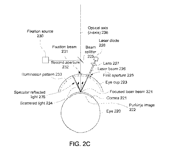
100621 Figure 2C shows alignment of the eye with a ranging beam such as a
laser beam
focused on the cornea, in accordance with some embodiments. In some
embodiments, an
eyecup 223 comprises a first aperture 225 to pass the ranging laser beam and a
second
aperture 232 to pass the scattered light 234 from ranging laser beam, The
laser beam 226
may comprise a laser beam from any suitable laser source such as a laser diode
228. Light
from the laser source may be passed through a lens 227 to focus the laser beam
to a waist
near the cornea 221. In some embodiments, the laser beam is inclined relative
to the optical
axis of the system. at any suitable angle, such as an angle from about 20
degrees to about 60
degrees, such that the laser beam spot moves across the cornea 221 as the
topography system
moves relative to the eye along the optical axis (Z-axis) 236, as described
herein. Prior to
measuring the eye, the laser beam angle may be adjusted to cross the optical
axis where the
vertex of the cornea is to be positioned when aligned with the topography
system along the
optical axis The laser beam 226 may also be focused where it crosses the
optical axis 236,
so as to decrease the spot size and improve positioning accuracy. In some
embodiments,
when the vertex of the cornea is positioned along the optical axis at the
intended position
along the optical axis, the focused waist of the laser beam 226 may appear as
a spot of light
- 17 -
CA 03134835 2021-09-23
WO 2020/205860
PCT/US2020/025957
in the camera image so as to overlap with the image of the reflection from the
fixation light
as described herein. In some embodiments, the focus of the laser beam at the
location where
the beam crosses the optical axis may be sufficiently small to allow accurate
alignment of the
eye along the optical axis and can be focused to any suitable size, for
example within a range
from about 10 microns to about 100 microns. Although reference is made to a
beam waist,
the focused spot need not comprise a diffraction limited spot, and the beam
waist may
correspond to an image of the output aperture of the laser diode 228, for
example.In some
embodiments, the light from the focused laser beam is back scattered from the
cornea
generally along the optical axis towards the second aperture 232 and the
imaging optics of
the corneal topography system. In some embodiments, the topography images may
comprise
the scattered light 234 from focused laser beam 226 illuminating the cornea.
In some
embodiments, the laser beam light reflected from the cornea 221 with specular
reflection 235
(i.e. mirror like reflection) may be reflected from the tear film on the
anterior surface of the
cornea 221 at an angle to the optical axis similar to the angle of the laser
beam toward the
cornea but in an opposite direction. This specular reflected laser beam light
235 may be
blocked by the eyecup 223 or other suitable structure. This reflection of the
specular light
away from the second aperture 232 can improve the contrast of the image of the
scattered
light from the cornea. In some embodiments, the eyecup 223 comprises the first
aperture 225
to pass the laser beam. The second aperture 232 is sized to pass the fixation
beam, the
pattern of reflected light from the cornea in order to image the pattern with
the camera, the
scattered light 234, and the fixation beam reflected from the cornea. The
illumination pattern
233 is passed through the second aperture 232 so as to form the Purkinje image
222
comprising light from the illumination pattern 233 and the fixation beam 231
as described
herein.
100631 In some
embodiments, the Purkinje image of the illumination pattern is located
farther from the cornea than the Purkinje image of the reflected fixation
beam. In some
embodiments, the location of the Purkinje image varies with the distance of
the object
reflected from the cornea. For objects that are located closer to the cornea
and reflected from
the eye, the Purkinje image is located farther from the cornea. For objects
that are farther
from the eye, the Purkinje image is located closer to the cornea. The fixation
beam may
comprise a substantially collimated beam of light that corresponds to an
object far from the
- 18 -
CA 03134835 2021-09-23
WO 2020/205860 PCT/US2020/025957
eye, e.g. approximating infinity. The illumination pattern reflected from the
eye corresponds
to a distance from the cornea that is closer to the cornea than the reflected
fixation beam, and
he Purkinje image of the illumination pattern is located farther from the
cornea than the
Purkinje image of the reflected fixation beam.
100641 The components, structures and features shown with reference to FIG. 2C
can be
combined with embodiments of the topography system as described herein. For
example, in
some embodiments, a fixation beam 231 may pass through the second aperture 232
that
receives the scattered light 234 from the cornea. In some embodiments, the
eyecup 223 may
comprise any suitable illumination pattern such as Placido disks, point
sources of light, point
sources of light arranged along circles to approximate a Placido disk, or a
grid pattern, for
example. In some embodiments, the illumination pattern 233 is configured to
reflect from
the cornea form a Purkinje image 222 (virtual image), such as the first
Purkinje image 222 at
a location below the cornea. The fixation light beam 231 may comprise
approximately
collimated light that reflects from the cornea to form a portion of Purkinje
image 222 that
forms near the center of the Purkinje image of the illumination pattern as
described herein.
In some embodiments, the scattered laser light 234 from the cornea 221
overlaps with the
Purkinje image of the fixation beam 231 near the center of the illumination
pattern as
described herein.
100651 Figure 2D illustrates a topography image 299 produced by the mobile
communications device-based corneal topography system. In some embodiments, a
light
pattern 297 comprises rings of concentric circles of varying diameters. The
size, shape and
location of the light pattern is related to the shape of the cornea and can be
used to derive
corneal topography data. For example, with steeper corneas the light pattern
is smaller in the
camera image and with flatter corneas the light pattern is larger. With
astigmatic corneas, the
light pattern can be distorted, being larger in one direction and smaller in
another direction.
100661 In some embodiments, the fixation beam Purkinje Image 296 is smaller
and inside
of the smallest boundary of the pattern, such as a circle 298.
100671 The light pattern reflected from the cornea can be shaped and
processed in many
ways. In some embodiments, the light pattern comprises a plurality of
continuous rings of
light, such as a rings of a Placido disk. Each of the continuous rings can be
processed with
image processing to determine a plurality of discrete points corresponding to
a plurality of
- 19 -
CA 03134835 2021-09-23
WO 2020/205860 PCT/US2020/025957
locations of each ring. Alternatively, the light pattern may comprise a
plurality of discrete
light sources located along circles corresponding to rings of a Placido disk,
and the locations
of each of these light sources determined, The light pattern locations may be
derived from
the rings or the discrete sources of the light pattern in order to generate
the corneal
topography data. The light pattern locations may corresponds to a plurality of
concentric
circles with deceasing diameters. In some embodiments, a plurality of LED
light elements
may form and/or generate the light pattern.
[0068] In some embodiments, the image of the fixation beam may overlap with
the
alignment beam which may include the second wavelength of light from the
alignment beam
scattered from the cornea and the first wavelength of light from the fixation
beam reflected
from the cornea. In some embodiments, a distance across the alignment beam on
the cornea
may be within a range of 5 microns to 200 microns, optionally within a range
of 10 microns
to 150 microns, or optionally within a range of 20 microns to 100 microns. In
some
embodiments, a distance across the fixation beam in a Purkinje image of the
eye may be
within a range of 10 microns to 300 microns, optionally within a range of 25
microns to 200
microns, or optionally within a range of 50 to 150 microns.
[0069] In some embodiments, a reticle may be displayed on the mobile
communication
device screen to facilitate alignment, for example when the alignment beam
overlaps the
fixation beam. While the beams can be sized in many ways, the overlapping area
of the
beams in the image may correspond to a distance across the cornea within a
range of 10
microns to 200 microns, optionally a range of 15 microns to 125 microns, or
optionally a
range of 20 microns to 75 microns.
[0070] In some embodiments, the illumination system may include a Placido
ring
assembly comprising a plurality of rings, wherein an innermost concentric ring
of the camera
image has a larger diameter than a distance across the fixation beam, or a
distance across the
ranging beam. In some embodiments, the illumination system may include a
Placido ring
assembly including a plurality of concentric rings, wherein the plurality of
concentric rings is
formed by a plurality of light-emitting diodes (LEDs) at discrete separated
locations along a
plurality of circles. In some embodiments, the illumination system may include
a Placido
ring assembly including a plurality of concentric rings, wherein the plurality
of concentric
rings is formed by a geometry of a Placido ring component, and the Placido
ring component
- 20 -
CA 03134835 2021-09-23
WO 2020/205860
PCT/US2020/025957
may be illuminated by a plurality of light-emitting diodes (LEDs). In some
embodiments, in
the corneal topography system, the luminescence intensity of pattern from the
illumination
system at the cornea may be within a range from 10 lux to 500 lux, optionally
from 25 lux to
250 lux and optionally from 50 lux to 125 lux In some embodiments, the
illumination
system may comprise or include a Placido ring assembly comprising a plurality
of concentric
rings, the plurality of concentric rings emitting a third wavelength of light,
the third
wavelength of light different from the first wavelength of light, e.g. of the
fixation beam, and
the second wavelength of light, e.g. of the ranging beam. In some embodiments,
when a
patient is being examined on the mobile communication device-based corneal
topography
system, the eye of a patient looks downward from horizontal at an angle within
a range of 2.5
degrees to 15 degrees towards the fixation target beam, or optionally looks
downward a
range of 5 degrees to 10 degrees towards a fixation target beam. In some
embodiments, the
alignment beam may be inclined relative to an optical axis and focused to a
cross-sectional
size on the cornea to position the vertex of the cornea along the optical axis
with an error of
no more than 150 microns when the fixation beam overlaps with the alignment
beam in the
image, and optionally wherein the error is no more than 100 microns,
optionally no more
than 50 microns and optionally no more than 25 microns. In some embodiments,
when a
patient is being examined on the mobile communication device-based corneal
topography
system, the eye of a patient looks at a fixation target beam along a
horizontal axis. In some
embodiments, when a patient is being examined on the mobile communication
device-based
corneal topography system, the eye of a patient looks down at an angle from
horizontal
within a range of 0.1 to 2.5 degrees towards the fixation beam.
[0071]
Referring again to Figure 2B, the mobile communications device-based corneal
topography system may comprise one or more ergonomic configurations, according
to some
embodiments. In some embodiments, the patient looks downward at an angle 890
relative to
horizontal toward the fixation target embodiments, an additional design
consideration may
be that an image of a subject's cornea on the mobile communication device's
display (e.g.,
the reflected image) be positioned at a downward angle with the horizontal
line connecting
an examiner and an examination subject (or patient). A term of art used by
movie directors
and cinematographers that pertains to this may be referred to as "eye line".
That is an
imaginary line connecting the eyes of two actors in a scene. In a corneal
topography system,
-21 -
CA 03134835 2021-09-23
WO 2020/205860 PCT/US2020/025957
an "eye-line" between an examiner and a subject has traditionally been in a
horizontal plane.
The "eye-line" refers to a condition where the eye of the examiner should be
aligned near a
horizontal plane with the eye of the subject being examined. Figure 2B
illustrates a
horizontal eye line 256 between an examiner and an examination subject. The
line between
the examiner's eye 282 and the image of the cornea on the mobile communication
device
display is in a downward direction. In other words, the examiner is looking
downward to the
corneal image as is illustrated by the line identified as angle 899 relative
to horizontal. In
embodiments of a mobile communication device-based corneal topography system,
it may be
preferable to have a corneal topography image on the mobile communication
display be
reasonably aligned both horizontally and vertically such that the eye-line
passes through a
center of a live camera image of mobile communication device display. In some
embodiments, the examiner may look along a horizontal axis towards the image
on the
mobile communication device display. In some embodiments, the examiner may
look down
at an angle from horizontal within the range of 0.1 to 2.5 degrees towards the
display of the
mobile communication device.
[0072] This allows ease-of-use for an examiner in that it may maintains the
same or
similar horizontal plane eye-line relationship that existed when the Examiner
utilized the slit-
lamp microscope. In other words, the examiner is used to such a horizontal
plane eye-line
positioning when the examiner operates the slit-lamp microscope. In some
embodiments, the
mobile communication device-based corneal topography system, which is attached
to the slit-
lamp microscope, does not change this horizontal plane eye-line relationship.
In some
embodiments, Aa mobile computing device-based corneal topography system may
comprise
a bulkhead and a slit lamp mounting plate and/or mounting assembly according
to
embodiments. In some embodiments, a bulkhead or positioning plate may be
utilized to
align and/or attach other pieces of a smartphone-based corneal topography
system in place in
order to enable efficient operation. In some embodiments, a bulkhead or a
positioning plate
may include a recess for a Placido illumination system 267 and/or eye piece
258. In some
embodiments, a mounting assembly (e.g., a positioning plate may attach to an
optical bench
or corneal topography optical housing) may be utilized to connect to a slit
lamp microscope
mounting assembly. In embodiments, a mobile communication device-based corneal
- 22 -
CA 03134835 2021-09-23
WO 2020/205860 PCT/US2020/025957
topography system may be attached (or piggy-backed) onto a slit-lamp
microscope in order
to maintain examination accuracy.
[0073] In some embodiments, a mobile communication device-based corneal
topography
system may also utilize infrared (IR) illumination (or a similar wavelength
illumination to
enable or initiate pupil edge detection. In some embodiments, the IR beam is
transmitted
through the pupil and reflected from the retina, such that the pupil of the
eye appears lighter
than the iris. This retro-illumination of the pupil can facilitate detection
of the edge of the
pupil. Figure 3 illustrates a corneal topography system or housing that
includes IR
illumination (e.g., an IR beam) and a green fixation beam being aligned on-
axis into a
patient's eye according to some embodiment. In some embodiments, the on-axis
alignment
of the infrared illumination and green fixation beam may allow for edge
detection of a
patient's pupil during dark conditions (e.g., without the Placido rings being
illuminated)
(scotopic conditions), during medium light conditions (mesopic conditions) and
during light
conditions ¨ photopic conditions (e.g., with the Placido rings being
illuminated). In other
words, the corneal topography software application may generate pupil edge
measurements
in light and/or dark conditions. In some embodiments, the IR light source may
introduce the
infrared beam coaxially, aligned with the green fixation beam and on axis with
a patient's
line of sight. In some embodiments, an advantage of coaxial illumination of
the IR beam and
the green fixation beam is that an operator and the corneal topography
smartphone software
may image the "red reflex" (retro-illumination) and see opacities in an
optically significant
part of the patient's visual system (e.g., a central ¨6mm diameter of the
cornea and lens,
which is an approximate measure depending on a patient's pupil size). This
advantage may
be in addition to the coaxial alignment of the infrared beam and green
fixation beam allowing
the corneal topography software application to image the pupil edge for pupil
size
measurement in dark and light conditions.
[0074] In Figure 3, in some embodiments, the corneal topography system or
housing may
include a fixation source (e.g., green LED) 305, an infrared light source 310
(IR LED), a first
lens assembly 315, a second lens assembly 320, and/or a doublet 335. In some
embodiments, the green fixation source 305 may a green LED that transmits a
green fixation
beam 325. In some embodiments, the green fixation beam 325 may be transmitted
on axis to
a patient's eye, as is illustrated in Figure 3. In some embodiments, the green
fixation beam
- 23 -
CA 03134835 2021-09-23
WO 2020/205860 PCT/US2020/025957
325 may be transmitted through a first lens 315, which may be a tilted lens.
In some
embodiments, the first lens 315 may be tilted which may introduce an
astigmatism in the
green fixation beam. In some embodiments, the green fixation beam 325 may then
pass
through a second lens 320, which may be a tilted lens. In some embodiments,
the second
tilted lens may correct for the astigmatism introduced by the first lens 315.
In some
embodiments, the green fixation beam 325 may pass through a doublet 335 on its
way to the
patient's cornea. In some embodiments, an infrared light beam 326 may be
introduced in
front of the first lens 315 and reflects off of the front surface of the first
lens 315. In some
embodiments, the infrared light beam 340 may pass through or be transmitted
through the
second lens 320 and/or the doublet 335 to the patient's cornea. In some
embodiments, the
infrared light source 310 may cast diffuse infrared light onto the patient's
pupil in order to
the illuminate the patient's pupil at an infrared spectrum. Because, the
infrared light beam
326 may be diffused, the system may not have to correct for an astigmatism. In
some
embodiments, the infrared light source may be an LED having a wavelength of
780 nm (+/-
15 nm). In some embodiments, the infrared light source may be a Thorlabs
LED780E. In
some embodiments, the light source may generate a light beam substantially
close to infrared
light spectrum as long as the light source illuminates the subject's eye.
[0075] In some embodiments, in order to perform pupil edge detection, the
fixation light
source (e.g., green LED) 305 and the infrared light source 310 may be
activated and/or
turned on. In some embodiments, the green fixation light source 305 and the
infrared light
source 310 may be activated by an operator turning on switches or controls of
the corneal
topography system or housing. In some embodiments, computer-readable
instructions
executable by one or more processors on a topography outboard or PCB of a
corneal
topography system or housing may cause signals to be transmitted to the
fixation light source
305 and the infrared light source 310 in order to turn on the fixation light
source 305 and/or
the infrared light source 310. In some embodiments, computer-readable
instructions
executable by one or more processors on the mobile communication device may
cause
signals, commands and/or instructions to be transmitted to the corneal
topography system or
housing to activate or turn on the fixation light source 305 and the infrared
light source 310.
Although Figure 3 illustrates a first lens, a second lens and a doublet, other
optical
components may be utilized by the corneal topography system in order to direct
the green
- 24 -
CA 03134835 2021-09-23
WO 2020/205860 PCT/US2020/025957
fixation beam and/or the IR beam to the patient's cornea. Although Figure 3
and the
discussion above identifies an IR beam and a green fixation beam, other
wavelengths and/or
colors may be utilized in place of or in addition to the IR beams and green
fixation beams as
long as these other light beams are detectable in light and/or dark conditions
and illuminate
the eye.
[0076] Figure 3A illustrate results of utilization of the IR illumination
system for pupil
edge detection according to some embodiments. As is illustrated in Figure 3A,
the patient's
pupil 360 may be illuminated by infrared illumination, which is reflected from
the retina,
such that the pupil appears lighter than the iris. In Figure 3A, the eye may
be in a dark or
non-illuminated setting or environment. In some implementations, the eye may
include an
iris 355, a pupil 360, a reflected illumination pattern 365 (e.g., a reflected
Placido ring
pattern), and a pupil illuminated by an infrared light source as described
herein. Work in
relation to the present disclosure suggests that retro-illumination of the
pupil is well suited
for combination with smart phone cameras as described herein, because the
retro illumination
of the pupil provides a sufficiently bright pupil for the edge of the pupil to
be readily visible
in the camera image, and the smart phone camera may comprise sufficient
sensitivity to
wavelengths that are barely perceptible or substantially imperceptible by the
human eye to
make the pupil readily visible in the camera image, such as wavelengths from
about 750 to
850 nm.
[0077] Although reference is made to edge detection with retro-illumination
of the pupil,
an IR light source can be used to illuminate the iris and detect the pupil
without
retroreflection. For example, IR light sources to transmit light obliquely
toward the cornea
so as to illuminate the iris and detect the boundary between the iris and the
pupil.
[0078] In some embodimentsõ the iPhone 7-Plus is a high-end mobile
communication
device that includes a high-end camera, a high-end lens and image processing
hardware
and/or software. Even with a high-level mobile communication device platform,
(such as
the Apple iPhone 7 and other similar Android-based smartphones made by
Motorola and
Samsung), the mobile communication device may have tiny variations in lens
position
relative to the mobile communication device camera sensor. For example, this
is true with
the Apple iPhone7. In addition, the high-level mobile communication device
platforms also
have very tiny variations in adjustment needed to set (or lock) focus and/or
zoom optimally
- 25 -
CA 03134835 2021-09-23
WO 2020/205860 PCT/US2020/025957
for the corneal topography system Keplerian telescope system resident or
installed in the
corneal topography system or housing. Accordingly, individual measurements or
settings
may need to be made for each individual mobile communication device (e.g.
iPhone)
camera-and-lens subsystem. Then, individual .ini files (e.g., configuration
files) may need to
be created for each individual mobile communication device to incorporate
those unique
settings. Such a setup and/or requirement is not useful in a production or
manufacturing
environment because time and/or resources would be necessary to measure the
variations in
the cameras and/or lens of the mobile communication device and then to record
or store the
identified settings for utilization later in configuring the corneal
topography system.
[0079] In a new and novel embodiment, as illustrated in Figure 4A, a new
configuration
of a mobile communication device-based corneal topography system includes
moving an
image sensor, one or more processors, one or more memory devices and/or image
processing
hardware and software into the corneal topography system or housing. In some
embodiments, the image sensor (or camera sensor), one or more processors, one
or more
memory devices and image processing hardware and/or software may be installed
on one or
more printed circuit boards (PCBs) or an outboard, and the PCBs or outboard
may be
installed in a corneal topography system or housing. The specification herein
refers to a
topography-specific PCB or a topography-specific outboard, but this apparatus
may also be
referred to as a topography outboard, a topography-specific chipset, or a
topography-specific
system on a chip (SoC). In some embodiments, the topography-specific PCB or
outboard
may be a single printed circuit board and/or maybe two or more PCBs coupled or
connected
to each other. In addition, the topography-specific PCB or outboard may also
have
components or assemblies that perform other functions including having a
communications
interface (e.g., such as a USB or Ethernet communication interface). In some
embodiments,
while the specification refers to a corneal topography system or housing, the
image sensor,
one or more processors, one or more memory devices, the image processing
hardware or
software, and/or other components or assemblies may be i) installed, located
or positioned
within a single physical housing or multiple physical housings or ii) have
some of the image
sensor, one or more processors, one or more memory devices, the image
processing hardware
or software, and/or components or assemblies mounted, attached, coupled or
connected to
one or more physical housings. In other words, the description herein is not
limited to all of
- 26 -
CA 03134835 2021-09-23
WO 2020/205860 PCT/US2020/025957
the above listed devices, components or assemblies being located within one
physical corneal
topography housing. For example, in some embodiments, some of the devices,
components
or assemblies may be partially contained within the corneal topography housing
and others
may be completely contained within the corneal topography housing. For
example, in some
embodiments, some of the devices, components or assemblies may be partially
contained
within one or more corneal topography housings while others are attached to,
coupled to or
connected to other devices, components and/or assemblies that are not within
corneal
topography housings.
[0080] In some embodiments, the image sensor (or camera sensor) and other
components
(e.g., processors, image processors, memory devices, computer-readable
instructions, etc.)
may be mounted on a circuit board. In some embodiments, a circuit board may be
mounted
to a rear surface of corneal topography system or housing, although the
location of the circuit
board may not be limited a rear surface. In some embodiments, the image sensor
(or camera
sensor) may be installed in a position that is a horizontal center of the rear
surface of the
corneal topography system or housing and may also be directly be aligned with
a rest of the
slit-lamp microscope which is aligned with the corneal topography system. In
some
embodiments, the image sensor (or camera sensor) may be installed in other
positions and on
other surfaces besides a horizontal center of a rear surface. Accordingly, the
specification
does not limit the location of the image sensor (or camera sensor) within the
corneal
topography system or housing as the location of the image sensor may be within
any location
of the corneal topography system or housing.
[0081] Figure 5, which will be described later, may utilize the mobile
communication
device camera rather than the image sensor (or camera sensor) of the corneal
topography
system of Figure 4A. However, in Figure 5, because the mobile communication
device may
be designed, customized and/or fabricated for the corneal topography system,
any variations
in the lens position and/or the adjustments for focus and/or zoom may be
eliminated because
the mobile communication device camera sensor will be affixed at the exact
focal plane of
the Keplerian telescope system of the corneal topography system, eliminating
the standard
lens typically installed in front of the camera sensor in most modern mobile
communication
device. Additionally, the mobile communication device may be be manufactured
according
- 27 -
CA 03134835 2021-09-23
WO 2020/205860
PCT/US2020/025957
to custom specifications provided by the maker or developer of the mobile
communication
device-based corneal topography system.
[0082] Figure
4A illustrates a block diagram of a corneal topography system including
system components (including an image sensor or camera sensor) for corneal
topography at
least partially contained within a housing according to according to some
embodiments. In
some embodiments, as illustrated in Figure 4A, the corneal topography system
407 may be
connected, coupled or attached to a custom-designed and fabricated mobile
communication
device 470. In some embodiments, the corneal topography system 407 may be
positioned
adjacent to a surface of the mobile communication device 470 and may be
connected via a
ribbon cable (e.g., a USB-3 ribbon cable). In some embodiments, the corneal
topography
system or housing 407 may be connected or coupled to a slit lamp microscope
mount 475 to
allow adjustment of the corneal topography system or housing with respect to
the patient's
eye. In some embodiments, as illustrated in Figure 4A, the corneal topography
system or
housing 407 may comprise a power supply 411, an image path 412, a
communications
interface or communications processor 425, a topography-specific PCB or
outboard 412, a
Placido ring illumination control system 450, a fixation light source 452, an
infrared light
source 453 and/or a ranging beam light source 451. In some embodiments, the
corneal
topography system or housing 407 may further comprise an illumination system
457 (e.g., a
Placido rings illumination system) and/or a rest 458. In some embodiments, a
patient's head
may rest on a chin rest of the slit lamp microscope with a curved plastic
strap to position the
forehead against the corneal topography system. In some embodiments, fixating
the chin and
forehead may allow for stabilization of the head relative to the microscope
(and thus the
corneal topography system.) In some embodiments, the illumination system 457
(e.g.,
Placido ring illumination system) may include one or more lights (e.g., LEDs)
to illuminate a
specific pattern that may be reflected off of a patient's cornea. In some
embodiments, the
custom-designed or fabricated mobile communication device 470 may comprise one
or more
processors, one or more memory devices, operating system software, application
software, a
display and/or a communication interface 425. In addition, although not shown
in Figure 4A,
the mobile communication device 470 may also include GPS transceivers,
cellular
transceivers (3G, 4G, or 5G), wireless local area network (Wi-Fi)
transceivers, NFC
transceivers, and/or other components and/or software. In some embodiments, as
illustrated
- 28 -
CA 03134835 2021-09-23
WO 2020/205860 PCT/US2020/025957
in Figure 4A, the topography-specific PCB / outboard or control circuitry 412
may comprise
an image processor 440, one or more processors 415, one or more memory devices
416,
computer-readable instructions stored in the one or more memory devices 417
and/or
firmware 418. In some embodiments, the topography-specific PCB or outboard 412
may
comprise communication circuitry and/or a communication interface 425. The
topography-
specific PCB or outboard 412 may not be required to utilize or include all the
components or
assemblies illustrated in Figure 4A. For example, in some embodiments the one
or more
processors 415 may include image processing capability (and so a separate
image processor
may not be needed). In some embodiments, for example, the one or more memory
devices
417 may include all the driver and/or application software and firmware 418
may not be
needed in certain embodiments of topography-specific PCB or outboard.
[0083] In this new and novel corneal topography system or housing, an
imaging
subassembly (which includes Keplerian telescope lenses and/or beam folding
mirrors) may
reflect a Placido rings image on a patient's cornea and the image sensor (or
camera sensor)
410 may capture a reflected Placido rings image (or an image of another
illuminated pattern).
In some embodiments, because the image sensor (or camera sensor) 410 may be
placed at a
specific position and because the imaging subassembly and resulting imaging
path 430 may
have specific dimensions, the reflected Placido rings image (or image of
another illuminated
pattern) may be received at the image sensor or camera sensor 410 at a corneal
image plane
at a desired vertex distance from the patient's eye (or cornea). In some
embodiments, the
Placido rings image (or image of another illuminated pattern) may be reflected
or projected
into the camera sensor without using any type of zooming functionality. In
some
embodiments, the image sensor, camera sensor or detector may be a CMOS-sensor
or may be
a CCD sensor (such as a Sony IMX250 CMOS sensor).
[0084] In some embodiments, a Keplerian telescope of the corneal topography
system
may project a Placido rings image directly into the image sensor or camera
sensor without
interposing a standard camera lens on an outer housing of a mobile
communication device in
front of the camera sensor. In some embodiments, the zoom may be set and
defined by the
optical components of the Keplerian telescope system. In some embodiments, the
zoom may
not able to be tweaked or altered by any adjustment of the mobile
communication device
camera optical zoom settings because there is no camera lens affixed in front
of the image
- 29 -
CA 03134835 2021-09-23
WO 2020/205860 PCT/US2020/025957
sensor of the corneal topography system. In some embodiments, software-
controller digital
zoom is still possible. In addition, in some embodiments, eliminating the
mobile
communication device camera lens and utilizing the Keplerian telescope system
(which is in
the corneal topography system) along with the image sensor or camera sensor
410 of the
corneal topography system or housing also eliminates mobile communication
device camera
lens positioning errors. In some embodiments, this leads to design where the
focus of the
corneal topography system may be locked during the manufacturing without
having to tweak
or adjust each unit in a post-manufacture calibration.
[0085] In some embodiments, the projected Placido rings image (or other
illuminated
pattern image) may be projected or reflected to the image sensor or camera
sensor 410 at a
perfect focus. This configuration eliminates the need to use the mobile
communication
device camera and/or lens (and the resulting variations therein) to capture
the reflected
Placido rings image (or other illumination pattern image). In addition,
because the corneal
topography system or housing may comprise the image or camera sensor, the
image
processing hardware and/or software and/or other corneal topography software
may be
moved into the corneal topography system or housing 407. In some embodiments,
the image
processing hardware and/or software and/or other corneal topography software
may be
located on the topography-specific outboard or housing.
[0086] In some embodiments, the topography-specific PCB or outboard 412 may
further
comprise computer-readable instructions stored on the one or more memory
devices that
were described above (e.g., non-volatile memory devices 416 and/or firmware
418). In some
embodiments, the computer-readable instructions may be accessed and executed
by one or
more processors 415 or 440 in order to control operation of other components
in the corneal
topography system or housing 407. In some embodiments, the computer-readable
instructions may be executed by one or more processors or controllers 415 or
440 to control
operation (e.g., activation or deactivation) of 1) a Placido rings
illumination subsystem (or
other illumination pattern subsystem); 2) fixation LED assembly (e.g. a Green
LED
assembly) and generated fixation beam; 3) a LED ranging laser assembly (e.g.,
a Red LED
assembly) and generated ranging beam and/or 4) an infrared LED assembly and
generated
infrared light beam. In some embodiments, the Bluetooth communication
transceiver (or
PAN communication transceiver) from the previously disclosed corneal
topography system
- 30 -
CA 03134835 2021-09-23
WO 2020/205860 PCT/US2020/025957
may be eliminated because the topography-specific PCB or outboard 412 may
either
communicate with the other components via wired connections (and/or wired
communication
protocols). In some embodiments, the components may be mounted or installed on
the
topography-specific PCB or outboard 412 and thus may be communicated with over
a wired
communications interface or communication circuitry on the topography-specific
PCB or
outboard 412.
[0087] In some embodiments, the corneal topography software may be stored
in the one
or more memory devices 416 and 418 on the topography-specific PCB or outboard
412. In
some embodiments, for example, topography library software (e.g., computer-
readable
instructions) may be stored in firmware 418 that may be executable by the one
or more
processors 415 or 440 that are installed on the topography-specific PCB (or
outboard) or in
other memory devices in the corneal topography housing. In some embodiments,
the one or
more processors may include an image processor that is specifically designed
to handle
imaging processing functions and/or analysis. In some embodiments, firmware
418 on the
topography-specific PCB (or outboard) 412 may store certain portions of the
corneal
topography software may include instructions that are executable by one or
more processors
or an image processor 440 to handle data intensive functionality (such as
executing and
initiating the corneal topography system auto-capture functionality, the
Placido rings image
capture, the Placido rings edge detection and/or the corneal topography power
mapping),
while allowing the one or more other processors 415 to initiate and execute
other
functionality such as activation of other components and/or transfer of
information between
the corneal topography system or housing 407 and the custom-designed and
fabricated
mobile communication device 470. In some embodiments, the one or more
processors
and/or related application software in the corneal topography system or
housing 407 may
then only communicate or transfer the corneal topography related images and
datafiles to the
custom-designed and fabricated mobile communication device for display on the
mobile
communication device display. In some embodiments, the custom-designed and
fabricated
mobile communication device may then upload the necessary corneal topography
images to a
cloud-based server, without having to perform any image processing at the
mobile
communication device.
- 31 -
CA 03134835 2021-09-23
WO 2020/205860 PCT/US2020/025957
[0088] In this new configuration or embodiment (as illustrated in Figure
4A), the
"intelligence" of the mobile communication device-based corneal topography
system may be
moved into a physical housing (or one or more physical housings) that is
outside of the
custom-designed or fabricated mobile communication device. Accordingly, a high-
end
mobile communication device with significantly processing power and/or an
image
processing chipset is no longer needed to perform the corneal topography
software
functionality. In some embodiments, a custom-designed and manufactured mobile
communication device may be utilized as the mobile communication device in the
mobile
communication device-based corneal topography system. In some embodiments, an
operating system may be created and developed for the custom-designed and
fabricated
mobile communication device by the developer and creator of the corneal
topography system
or housing (e.g., the corneal topography system), which is Intelligent
Diagnostics, LLC. In
some embodiments, the custom-designed and fabricated mobile communication
device 470
may only be required to have a monitor or display, a custom-designed and/or
developed (and
thus proprietary and closed) operating system, one or processors, a wired
communication
interface or communication circuitry (e.g., USB or Ethernet communication
circuitry), and/or
a wireless communication transceiver (e.g., a WiFi transceiver); a personal
area network
transceiver ¨ Bluetooth; and/or a cellular (3G, 4G, or 5G) transceiver,
although many other
components and/or software applications may also be resident within the mobile
communication device.
[0089] With this new system configuration, the corneal topography system
may still
utilize one or more mirrors to fold an image beam path created by the
Keplerian telescope
optical subassembly. However, it is not necessary to utilize the mobile
communication
device camera that was discussed in the prior ID patent applications. Thus, in
the
embodiment illustrated in Figure 4A, the reflected image beam path may be
decoupled from
the mobile communication device camera and/or the entrance pupil location of
the associated
lens. As discussed above, in Figure 4A, the image sensor or camera sensor 410
may integral
and/or integrated into the corneal topography system or housing 407 (and
specifically may be
integrated as part of the topography-specific PCB or outboard 412). In some
embodiments of
this new corneal topography system, the telescope optical system beam path may
be short
enough so that one mirror or two mirrors may be utilized for folding an image
beam path.
- 32 -
CA 03134835 2021-09-23
WO 2020/205860
PCT/US2020/025957
Figure 4B illustrates use of one mirror for folding an image beam path in a
corneal
topography system according to some embodiments. Figure 4C illustrates use of
two mirrors
for folding an image beam path in a corneal topography system according to
some
embodiments. In some embodiments, as illustrated in Figure 4B, a reflected
illuminated
pattern image (e.g., a reflected Placido rings image) may be transmitted or
reflected to an
image sensor 410 in the corneal topography system or housing after being
reflected by a
mirror 480. In some embodiments, as illustrated in Figure 4C, a reflected
illuminated pattern
image (e.g., a reflected Placido rings image) may be transmitted or reflected
to an image
sensor 410 in the corneal topography system or housing after being reflected
by a first mirror
481 and/or a second mirror 482. In some embodiments, no mirrors may be
necessary for
folding an image beam path. Thus, in the last embodiment, mirrors utilized for
folding an
image beam path may be eliminated from the corneal topography optical system.
In some
embodiments, mirrors and/or lenses may still be utilized to introduce or
direct the fixation
beam and/or infrared (IR) light to the patient's eye or cornea or for other
features or
functionality of the corneal topography system.
[0090] In this
new system configuration (Figure 4A), the topography-specific PCB or
outboard 412 may be placed in different locations relative to the corneal
topography system
or housing 407 and the mobile communication device to which this subsystem is
attached. In
some embodiments, the position of the topography-specific outboard or PCB 412
may 'float'
or be moved relative to a position of the custom-designed and fabricated
mobile
communication device 470. In some embodiments, the position of the topography-
specific
PCB 412 or outboard may be moved as long as the image sensor or camera sensor
410 may
be aligned to receive the reflected Placido rings image (or other illumination
pattern image).
Thus, in some embodiments, the position of the Placido rings assembly (or
other pattern
illumination assembly) may be exactly in a horizontal midline of the custom-
designed and
fabricated mobile communication device, even if the typical mobile
communication device
camera is off to one side or near to the top edge of the mobile communication
device (e.g., as
in the iPhone 7, 8 and 10-series phones among other Android-based phones).
This is because
in the embodiment illustrated in Figure 4A, the mobile communication device
camera may
not be utilized for image capture of the reflected Placido rings image (or
other illumination
pattern image). Thus, in some embodiments, the mobile communication device-
based
- 33 -
CA 03134835 2021-09-23
WO 2020/205860 PCT/US2020/025957
corneal topography system described in Figure 4A may be a bilaterally
symmetric product as
opposed to the slightly asymmetric design that was disclosed in the previously
submitted
patent applications.
[0091] In addition, the movement of the corneal topography application
software to
memory devices in the corneal topography system or housing 407 (and
specifically the
topography-specific PCB or outboard 412) may provide a number of advantages.
One
advantage is that the developer of the corneal topography system or housing
does not have to
worry about mobile communication device camera drivers changing (and/or
related mobile
communication device operating system software changing). In other words,
phone or
mobile communication device manufacturers may push out updates that contain
drivers
and/or other tweaks which may jeopardize the operational stability of the
corneal topography
system. In this new configuration (Figure 4A), the mobile communication device
operating
system is also custom-designed and/or developed by the corneal topography
system creator
or developer, so this situation should no longer be an issue. In addition, the
corneal
topography system developer may also have control of the operating system of
the custom-
designed mobile communication device (and thus updates of the OS or drivers
will not be
communicated to the mobile communication device unless the corneal topography
system or
housing developer is aware of the impact to the corneal topography system). In
other words,
the corneal topography system (and the software platform) may be controlled
end-to-end by
the developer of the corneal topography system. In some embodiments, the
custom-designed
and/or developed operating system may be Linux-based rather than a phone-
manufacturer
branded flavor of Android. In some embodiments, a version of Linux (named
Yocto ¨ which
is published by Intel) may be utilized as a base operating system to run the
topography-
specific PCB (or outboard) 412 and other components in the corneal topography
system or
housing and/or the custom-designed and fabricated mobile communication device.
[0092] Figure 4A illustrates a block diagram of a new configuration of a
smartphone
corneal topography system according to some embodiments. The block diagram
does not
represent a shape of the corneal topography system or housing and instead is
drawn as a
simple rectangle. In addition, the optical path labeled in the corneal
topography system or
housing 407 may not be indicative of the optical path (and/or components
utilized therein) to
transmit the reflected Placido rings image (or other illumination pattern
image) to the image
- 34 -
CA 03134835 2021-09-23
WO 2020/205860 PCT/US2020/025957
sensor or camera sensor 410. In addition, no lenses or mirrors are shown in
the block
diagram of the corneal topography system or housing although the lenses or
mirrors may be
present in the corneal topography system or housing 407. In other words, this
Figure (Figure
4A) is not directed to illustrating or describing the optical path in the
mobile communication
device-based corneal topography system. Instead, Figure 4A is illustrating a
new
configuration of the corneal topography system that brings the brains and
processing power
into the corneal topography system or housing 407.
[0093] The mobile communication device-based corneal topography system 400
comprises a corneal topography system or housing 407, a Placido rings assembly
or other
pattern illumination assembly 410, a custom-designed and fabricated smartphone
470, and a
slit-lamp microscope mounting assembly 475. In some embodiments, the Placido
assembly
(e.g., the Placido rings assembly or other pattern illumination assembly) may
be mounted on
one side of a corneal topography system or housing 407 and a custom-designed
mobile
communication device 470 may be mounted or connected to an opposite side of
the corneal
topography system or housing 407. In some embodiments, the corneal topography
system or
housing 407 may be connected or coupled to a slit lamp microscope mounting
assembly 475.
[0094] In some embodiments, the corneal topography system or housing 407
may
comprise an image path 430, where the image path 430 may be a path that a
reflected Placido
rings image (or other illuminated pattern image) travels in order to enter an
image sensor or
camera sensor 410. In some embodiments, the corneal topography system or
housing 407
may comprise a topography-specific PCB or outboard 412. In some embodiments,
the
topography-specific PCB or outboard 412 may receive power from a power source
411 in the
corneal topography system or housing 407. In some embodiments, the power
source 411
may be a rechargeable battery. In some embodiments, the power source 411 may
be
connected to an external power outlet or charging pad which provides power to
the power
source 411. In some embodiments, computer-readable instructions 417 executable
by one or
more processors 415 (or firmware 418 executable by one or more processors 415)
may
activate an image sensor or a camera sensor 410 to capture a reflected Placido
rings image
(or other illumination pattern image) transmitted via the image path 430. In
some
embodiments, computer-readable instructions 417 stored in one or more memory
devices 416
executable by one or more processors 415 (or the firmware 418 executable by
one or more
- 35 -
CA 03134835 2021-09-23
WO 2020/205860 PCT/US2020/025957
processors 415 or controllers) may generate instructions, commands or signals
to perform
operations in the corneal topography system or housing 407. For simplicity,
the specification
may refer to computer-readable instructions executable by one or more
processors 415 from
this point forward although the other embodiments described previously (e.g.,
firmware
executable by one or more processors or controllers) may also be utilized.
[0095] In some embodiments, the computer-readable instructions 417
executable by one
or more processors 415 may perform auto-capture of the reflected Placido rings
image (or
other illumination pattern image). In some embodiments, the computer-readable
instructions
417 executable by one or more processors 415 may communicate the captured
Placido rings
image (or other illumination pattern image) to an image processor 440. In some
embodiments, as described above, the image processor 440 may be a separate
processor or
device from the one or more processors 415 in order to offload intensive image
processing
operations from the one or more processors 415. In some embodiments, the
computer-
readable instructions 417 executable by one or more processors 415 may cause
the image
processor 440 to perform additional corneal topography functions such as
Placido rings edge
detection and/or the corneal topography power mapping, as well as other
corneal image
manipulation or processing. In some embodiments, firmware or computer-readable
instructions located in an integrated circuit or a printed circuit board
including the image
processor 440 may be executable by the image processor 440 to perform corneal
topography
functions such as the Placido rings image auto-capture, Placido rings edge
detection and/or
corneal topography power mapping. In other words, the image processor 440 may
have its
own embedded software or firmware to perform corneal topography functions.
[0096] In some embodiments, the corneal topography related images and files
(e.g., the
reflected Placido rings image, the Placido rings edge detection, data files
corresponding to
the Placido rings image and/or the corneal topography power map) may be
communicated to
the custom-designed and/or fabricated mobile communication device 470 for
display on the
mobile communication device display and/or further communication or
transmission to
additional computing devices. The corneal topography images and/or related
files may be
communicated to the custom-designed and fabricated mobile communication device
470 via
a communication interface or communication circuitry 425 (e.g., USB-3
interface), a cable
426 (e.g., a USB-3 Cable), and a mobile communication device communication
interface or
- 36 -
CA 03134835 2021-09-23
WO 2020/205860 PCT/US2020/025957
communication interface circuitry 427 (e.g., phone USB interface connector).
In some
embodiments, the topography-specific PCB or outboard 412 may comprise the
communication circuitry or communication interface 425. In some embodiments,
the
communication circuitry or communication interface 425 may be a socket on the
topography-
specific PCB or outboard 412 and the cable 426 may be a ribbon cable. This
eliminates the
need for a wireless communication transceiver (e.g., a Bluetooth transceiver)
in the corneal
topography system or housing 407. This configuration also provides additional
security for
the patient data (e.g., the patient corneal topography images and related
files) because the
patient data may not be hacked or stolen by obtaining patient data transmitted
via a Bluetooth
communications protocol. In other words, wired transmission of corneal
topography data is
more secure than wireless transmission of corneal topography data. In
addition, commands,
instructions, signals and messages may be transmitted or communicated between
the custom-
designed and fabricated mobile communication device 470 and the corneal
topography
system or housing 407 in order to control other components of the corneal
topography system
or housing 407.
[0097] In some embodiments, the computer-readable instructions 417 may be
executable
by one or more processors 415 of the topography-specific PCB 412 to control
operation of
components in the corneal topography system or housing 407 (or corneal
topography optical
bench). For example, in some embodiments, the one or more processors 415 of
the
topography-specific PCB 412 may generate commands, instructions or signals to
cause the
Placido rings (or other illumination pattern) to illuminate, the ranging beam
to be generated
and transmitted to the patient's eye, the fixation beam to be generated and
transmitted to the
patient's eye and/or the infrared beam to be generated and transmitted to the
patient's eye.
Similarly, in some embodiments, the one or more processors 415 of the
topography-specific
PCB or outboard 412 may generate commands, instructions and/or signals to
cause those
beams to cease to be generated and/or the Placido rings to be turned off.
[0098] In some embodiments, for example, the computer-readable instructions
417 may
be executable by one or more processors 415 to generate a signal, command or
instruction to
a fixation beam assembly 452 to cause the fixation beam assembly 452 to
generate a fixation
beam (e.g., a green fixation beam) which is transmitted to the patient's eye.
Similarly,
- 37 -
CA 03134835 2021-09-23
WO 2020/205860 PCT/US2020/025957
signals, commands and/or instructions may be generated and communicated to
turn off the
fixation beam.
[0099] In some embodiments, for example, the computer-readable instructions
417
executable by one or more processors 415 may generate a signal, command or
instruction to
a ranging beam assembly 451 to cause the ranging beam assembly 451 to generate
a ranging
beam (e.g., a red ranging beam) which is transmitted to the patient's eye.
Similarly, signals,
commands and/or instructions may be generated and communicated to turn off the
ranging
beam.
[0100] In some embodiments, for example, the computer-readable instructions
417
executable by one or more processors 415 may generate a signal, command or
instruction to
an infrared light assembly 453 to cause the infrared light assembly 453 to
generate an
infrared light beam (e.g., an infrared light beam) which is transmitted to the
patient's eye.
Similarly, signals, commands and/or instructions may be generated and
communicated to
turn off the infrared light beam.
[0101] In some embodiments, for example, the computer-readable instructions
417
executable by one or more processors 415 may generate a signal, command or
instruction to
a Placido rings controller or circuitry 450 to cause the Placido rings
controller 450 to
generate signals, commands or instructions to illuminate rings of the Placido
rings assembly
410. In some embodiments, the one or more processors may generate a signal,
command or
instruction directly to a Placido rings assembly 410 to illuminate the Placido
rings.
Similarly, signals, commands and/or instructions may be generated and
communicated to
turn off the illumination of the Placido rings in the Placido rings assembly
410.
[0102] Figure 5 illustrates an alternative embodiment utilizing a custom-
designed and
developed-mobile communication device according to some embodiments. In Figure
5, the
main difference with respect to Figure 4 is that the camera sensor and
potentially the corneal
topography software, may be located or resident in the customized-designed
and/or
fabricated mobile communication device 570. Because the corneal topography
system or
housing developer is also the developer of the custom-designed and/or
fabricated mobile
communication device, the developer can control a location or position of the
camera sensor
and/or lenses in the custom-designed and/or fabricated mobile communication
device and
thus will not have the variations that are present in other phone
manufacturer's cameras and
- 38 -
CA 03134835 2021-09-23
WO 2020/205860 PCT/US2020/025957
lenses (e.g., Apple, Samsung, Motorola, Google). Thus, the custom-designed
mobile
communication device camera may be located at a horizontal center of the
custom-designed
mobile communication device and may receive the reflected Placido rings image
via the
image path 530. Because the corneal topography system developer controls
and/or has
customized both pieces (e.g., the custom-designed and fabricated mobile
communication
device 570 and the corneal topography system and housing 507), tight
tolerances may be
maintained with the optical components in both devices. In addition, the
developer will also
control the custom-designed and developed mobile communication device
operating system
and/or the corneal topography system operating system, so unexpected driver
updates (and
potentially problematic updates) for components of either system (e.g., the
custom-designed
mobile communication device and/or the corneal topography system or housing)
will not be
an issue. In some embodiments, the topography-specific PCB or outboard that
was disclosed
in Figure 4A may be eliminated in Figure 5. In some embodiments, the custom-
designed
and/or fabricated mobile communication device 570 may communicate commands,
signals
and/or instructions with the corneal topography system or housing 507 via a
wired
communication interface or communication circuitry 526 (e.g., a USB-3
interface) utilizing a
cable 527 and the wired communication interface or communication circuitry 525
in the
corneal topography system or housing 507. Alternatively, the custom-designed
and/or
fabricated mobile communication device 570 may communicate commands, signals
and/or
instructions with the corneal topography system or housing 507 utilizing a
wireless
communication interface 526 such as Bluetooth or Wi-Fi without the need of a
physical
cable. In some embodiments, the custom-designed mobile communication device
communication interface or communication circuitry (whether wired or wireless)
may control
operations of components in the corneal topography system or housing 507, such
as the
fixation light source 552, the infrared light source 553, the ranging light
source 551 and/or
the Placido rings illumination assembly (or other pattern illumination system)
510. In some
embodiments, computer-readable instructions 517 stored in one or more memory
devices 516
and executable by the one or more processors 515 in the custom-designed mobile
communication device 570 may perform image processing of the reflected Placido
rings
image or other illuminated pattern image (the operations or which were
described
previously). In other words, in Figure 5, the corneal topography application
software would
- 39 -
CA 03134835 2021-09-23
WO 2020/205860 PCT/US2020/025957
be stored and executed by one or more processors on the custom-designed mobile
communication device.
[0103] Figure 6A illustrates a side view of components of a mobile
communication
device-based corneal topography system according to some embodiments. In some
embodiments, the mobile communication device-based corneal topography system
600 may
comprise a mobile communication device 605; a topography processor 615 and/or
a
topography printed circuit board 620; an image sensor 626 and/or an image
sensor printed
circuit board 626; one or more lens assemblies (e.g., a first lens assembly
630, a second lens
assembly 631, and/or a third lens assembly 632), a mirror 610, and/or an
optical tube that
includes an illumination pattern source 640. In some embodiments, the mobile
communication device-based corneal topography system 600 may further comprise
a fixation
beam source 635 and/or a fixation mirror 636. In some embodiments, the mobile
communication device-based corneal topography system 600 may further comprise
a ranging
beam source 710 and/or one or more proximity sensors 720 (both illustrated in
Figure 7A).
In some embodiments, the mobile communication device 605 may comprise a mobile
communication device display 606.
[0104] There is a significant advantage to moving to a mobile-communication
device-
based corneal topography system having an image sensor and/or topography
processing
hardware and/or software outside of the mobile communication device (which may
be
referred to as outboard). All mobile communication device (e.g., smartphone)
cameras or
sensors have their own integrated lens systems with auto-focus and zoom. These
features are
not needed in the corneal topography system and if the mobile communication
device camera
was utilized as the sensor in the corneal topography system, these features
would need to be
disabled and/or a work around would need to be developed. In addition, all
mobile
communication device cameras incorporate infrared and/or far red filters to
eliminate "red
eye" in photos. The corneal topography system described and claimed herein
desired to
eliminate this filter (infrared and/or far red) and instead utilize the red
light and infrared
spectrum for pupil edge detection, and potentially autorefraction. In
addition, even in high
end name-brand mobile communication devices such as Apple iPhone and Android
phones,
there are very tiny differences in spacing of the mobile communication device
lens(es) from
the image sensor or camera. For corneal topography features, if the mobile
communication
- 40 -
CA 03134835 2021-09-23
WO 2020/205860 PCT/US2020/025957
device camera is utilized, these very tiny differences would need to be
measured, recorded
and/or factored in calibration for each and every instrument (e.g., mobile
communication
device-based corneal topography system). Having to measure, record and factor
these very
tiny differences for each mobile communication device would be clunky,
cumbersome,
undersireable and costly in a production context. Thus, by utilizing an image
sensor outside
the mobile communication device, the image sensor may be bonded to the optical
bench
(which includes all of the lensing elements (e.g., lens)) so that the spacing
for each
instrument is uniform, reproducible and/or consistent. In addition, by not
having to guide the
reflected image of the illumination pattern through a mobile communication
device lens
system, the configurations described herein is optimized so that a design of
the imaging
system for imaging the reflected illumination pattern of the cornea to the
dedicated image
sensor.
[0105] In some embodiments, the image sensor 625 may communicate with the
topography processor 615) and/or other components on a topography PCB 620 via
an
interface, such as a MIPI interface. In some embodiments, the corneal
topography system
may comprise a battery or power source (e.g., such as a lithium ion battery)
that is included
in a housing. In some embodiments, the topography processor may be configured
with
instructions to communicate with other components or assemblies within a
housing or the
mobile communication device-based corneal topography system 600 such as one or
more
thermal or temperature sensors (not shown), a fixation beam source 635, the
illumination
source (or illumination pattern source) 640, the ranging beam source 710,
and/or an infrared
light source (shown in Figure 3). In some embodiments, the illumination
pattern source 640
may comprise two parts. In some embodiments, the illumination system may be
referred to
as an illumination pattern source. In some embodiments, the illumination
system 641 may
generate an illumination pattern that is reflected of a cornea of a subject or
patient. In some
embodiments, an imaging system may be coupled to the illumination system and
coupled to
an image sensor. In some embodiments, the imaging system may direct the
reflected
illumination pattern to the image sensor. In some embodiments, the image
sensor 625 may
capture an image of the reflected illumination pattern. An important advantage
of the
embodiments described in Figures 6A, 6B, 7A and 7B is that the image sensor
625 may be
located in an optical housing and is separate from an image sensor or camera
in the mobile
-41 -
CA 03134835 2021-09-23
WO 2020/205860 PCT/US2020/025957
communication device 605. As described previously, including the image sensor
in a
housing with the imaging system allows a fixed alignment of the image sensor
625 and the
imaging system. In addition, it eliminates the mobile communication device-
based corneal
topography system having to identify and/or address the different
characteristics and
specifications of the image sensors in the mobile communication device, as
well as potential
different mobile communication device image sensor locations.
[0106] In some embodiments, the mobile communication device-based corneal
topography system may further comprise an interface. In some embodiments, the
interface
may be the Mobile Industry Processor Interface (or MIPI interface) (not
shown). In some
embodiments, the MIPI interface may be coupled to the image sensor 625 and/or
the
topography processor 615. In some embodiments, the image sensor 625 may be
configured
with instructions to communicate, via the MIPI interface, the captured images
of the reflected
illumination pattern to the topography processor 615. In some embodiments, the
topography
processor 615 may be configured with instructions to communicate the captured
image of the
reflected illumination pattern to the mobile communication for presentation on
the display of
the mobile communication device 605 to allow for viewing by the Examiner. In
some
embodiments, the communication of the reflected illumination pattern image to
the mobile
communication device may occur in real time. In some embodiments, the
topography
processor 615 may be configured with instructions to control operation of the
image sensor
(e.g., to specify parameters or measurements of the image captured by the
image sensor 625).
In some embodiments, the topography processor 605 may communicate commands or
instructions to the image sensor 625 to control a size, a resolution and/or a
frequency of when
an image is refreshed or recaptured (which may be referred to as a frame
rate). In some
embodiments, the topography processor may communicate instructions to the
image sensor
625 to down-size an image. For example, for the auto-capture process described
above and
below, a high resolution (e.g., 3K x 3K) may be utilized for the image being
evaluated in the
auto-capture process whereas for a topography process (e.g., rings analysis
process that is
described below), a smaller resolution (e.g., 1K x 1K) of the captured image
of the reflected
illumination pattern may be utilized. During the auto-capture process, the
topography
processor 615 may be configured with instructions to enable or set different
regions of
interest in the reflected image of the fixation beam and/or the ranging beam.
In this
- 42 -
CA 03134835 2021-09-23
WO 2020/205860 PCT/US2020/025957
embodiment, then the topography processor 615 may only be evaluating a center
area of the
reflected image of the fixation beam and/or the ranging beam to find and
determine overlap
of the fixation beams and the ranging beam. In this embodiment, this may allow
the corneal
topography system described herein to utilize a higher frame rate to achieve
auto-capture
and/or also to utilize a higher resolution image at that frame rate.
[0107] In some embodiments, a topography processor 615 may be configured
with
instructions to process the image of the reflected illumination pattern to
generate topography
map images and/or one or more topography data files. In some embodiments, the
topography
data files may include 1) ring edge location measurements, 2) calibration
data, 3) patient
identifier data and/or 4) x, y and/or z-axis offset data. In some embodiments,
the topography
processor 615 may be configured with instructions to communicate the generated
topography
map images and the one or more data files to the mobile communication device
605. In some
embodiments, a processor on the mobile communication device 605 may be
configured with
instructions to present the generated one or more topography map images on a
display of the
mobile communication device.
[0108] In some embodiments, the mobile communication device-based corneal
topography system may utilize an auto-capture process to verify that accurate
positioning in
the x, y and z-axis of a cornea (of the subject) is present as the reflected
illumination pattern
is captured. In some embodiments, the pattern illumination source or component
640 may
not be initially illuminated. In some embodiments, the pattern illumination
source or
component 640 may be illuminated. In some embodiments, a fixation beam source
635 may
generate a fixation beam which may travel on a fixation path which forms
fixation axis
(which has two portions 655 and 656). In some embodiments, the fixation beam
defines a
fixation target visible to the eye of the subject, the fixation target beam
comprising a first
wavelength of light. In some embodiments, a ranging beam source 710 may
generate a
ranging beam 715 and direct the ranging beam to the cornea of the subject. In
some
embodiments, the ranging beam 715 may also be referred to an alignment beam
and the
ranging beam source 710 may be referred to as an alignment beam source. In
some
embodiments, the ranging beam 715 may comprise a second wavelength of light
that is
different from a first wavelength of light. In some embodiments, the ranging
beam 715 may
travel along a path which may be referred to as a ranging axis. In these
embodiments, the
- 43 -
CA 03134835 2021-09-23
WO 2020/205860 PCT/US2020/025957
image sensor 625 may capture a reflected image of a ranging beam and a
fixation beam on
the cornea of the subject. In some embodiments, as shown an angle between the
fixation axis
(beam) and the imaging axis (beam) is illustrated by reference number 716.
[0109] In these embodiments, the image sensor 625 may be configured with
instructions
to communicate the reflected image of the ranging beam and the fixation beam
to the
topography processor 615 via the interface. In these embodiments, the
topography processor
615 may communicate the reflected image of the alignment beam and the fixation
beam to
the mobile communication device 605 to display on the mobile communication
device
display 616 and to allow the examiner to move the corneal topography system.
In some
embodiments, multiple frames of the reflected image of the fixation beam and
the ranging
beam may be communicated from the image sensor 625 to the topography processor
615. In
some embodiments, the one or more frames of the reflected image of the
fixation beam and
the ranging beam may be communicated from the topography processor to the
mobile
communication device 605. In some embodiments, the one or more frames of the
reflected
image of the fixation beam and the ranging beam may include a mark or cross
hair (e.g., a
fiducial mark) which may be utilized to identify a center of an overlap of the
fixation beam
and the ranging beam by the operator (e.g., the examiner) of the mobile
communication
device-based corneal topography system. In other words, the mark or cross-hair
(e.g., yellow
cross-hairs) facilitate proper alignment, by providing visual cues to the
person performing the
topography exam. In some embodiments, a process of centering the ranging and
fixation
beams may require operator guidance of steering the mobile communication
device-based
corneal topography system when mounted on the slit lamp microscope so as to
achieve an
optical position of the fixation and ranging beam within the yellow cross-
hairs.
[0110] In these embodiments, the topography processor 615 may be configured
with
instructions to determine if the ranging beam and the fixation beam are
overlapping. In these
embodiments, for example, the topography processor 615 may be configured with
instructions to determine the beams are overlapping by tracking the first
wavelength of light
(e.g., the fixation beam) and the second wavelength of light (e.g., the
ranging beam) with
spectral analysis. In some embodiments, the topography processor 615 may also
be
configured with instructions to verify that an overlap of fixation beam and
the ranging beam
are in alignment with a fiducial mark or cross-hairs in the reflected image of
the fixation
- 44 -
CA 03134835 2021-09-23
WO 2020/205860 PCT/US2020/025957
beam and the ranging beam (the cross-hairs may be yellow cross-hairs in order
to stand out
or be distinct from a red ranging beam and green fixation beam). In some
embodiments, the
operator or user may move the fiducial mark or cross-hairs by moving the
mobile
communication device-based corneal topography system 600 utilizing the
joystick or similar
device on the slit lamp microscope (to which the system 600 is mounted). If
the topography
processor 615 determines that these conditions have been met (e.g., beams
overlapping and
aligned with fiducial mark or cross-hairs), the topography processor 615 may
be configured
with instructions to turn off or deactivate the fixation beam source 635
and/or the ranging
beam source 710. In other words, the topography processor 615 may send
shutdown or
deactivation commands or instructions to the fixation beam source 635 and/or
the ranging
beam source 710.
[0111] In some embodiments, once it is determined that the fixation beam
and the
alignment beam are overlapping each other, the topography processor 615 may be
configured
with instructions to instruct, command or signal the pattern illumination
component or source
640 to turn on and/or illuminate in order to project the illumination pattern
onto the cornea of
the subject. In some embodiments, the pattern illumination component or source
640 may
already be illuminated and thus be projecting an illumination pattern on a
cornea of the
subject. In these embodiments, the topography processor 615 may be configured
with
instructions to command, instruct and/or signal the image sensor 625 to
capture a reflected
illumination pattern image. In these embodiments, the image sensor 625 may be
configured
with instructions to automatically capture the reflected illumination pattern
image and to
communicate the captured reflected illumination pattern image to the
topography processor
615. In other words, no human intervention may be required in performing these
steps. The
auto-capture process described herein is an advantage over prior art systems
where multiple
tests have to be performed in order to an image of acceptable quality. This
auto-capture
process helps reduce human error in capturing images of the reflected
illumination pattern at
the correct corneal vertex. This auto-capture process will speed up
examinations of subjects
and improve the quality and accuracy of the captured images, as well as the
topography map
images and the one or more topography data files generated therefrom.
[0112] In these embodiments, the image sensor 625 may be configured with
instructions
to communicate, via the interface, the captured reflected illumination pattern
image to the
- 45 -
CA 03134835 2021-09-23
WO 2020/205860 PCT/US2020/025957
topography processor 615 for topography processing. In these embodiments, the
topography
processor 615 on a topography PCB 620 may be configured with instructions to
perform
topography processing and to generate one or more topography map images and
one or more
topography data files. In some embodiments, the topography processor 615 may
be
configured with instructions to communicate the generated one or more
topography map
images and the one or more topography data files to the mobile communication
device 605.
In some embodiments, the topography processor may also communicate the
captured image
of the reflected illumination pattern (or some derivative thereof) to the
mobile
communication device 605. In some embodiments, the processor of the mobile
communication device 605 may be configured with instructions to present the
one or more
topography map images on the display 606 (either by themselves or with the
reflected
illumination pattern image (e.g., the captured reflected illumination pattern
image)). In some
embodiments, the processor of the mobile communication device 605 may be
configured
with instructions to communicate the reflected illumination pattern image (or
a derivative
thereof) and/or the one or more topography data files to a cloud-based server
and/or remote
computing device for storage and/or analysis.
[0113] In some embodiments, the communication of 1) reflected illumination
pattern
images; 2) one or more topography map images; and 3) one or more topography
data files to
the mobile communication device may occur utilizing a wired communication
interface. In
some embodiments, the wired communication interface may operate according to
the USB-2
and/or the USB-3 communication protocol (although other communications
protocol may be
utilized). In some embodiments, the wired communication interface may be a USB-
2 and/or
USB-3 cable. The utilization of the wired communication interface provides
protection from
outside individuals being able to access and/or hack the reflected
illumination pattern images,
the topography map images and/or the one or more topography data files as they
are being
transferred to the mobile communication device. This protection is a
significant advantage
over other systems as it provides protection for subject's personal health-
related data. In
some embodiments, the mobile communication device 605 may utilize one or more
wireless
communication transceivers to communicate the reflected illumination pattern
image (or
derivative thereof) and the one or more topography data files to a cloud-based
server and/or
remote computing device. In some embodiments, the one or more wireless
communication
- 46 -
CA 03134835 2021-09-23
WO 2020/205860 PCT/US2020/025957
transceivers may be transceivers operating according to any one of a number of
802.11
protocols, WiFi transceivers and/or wireless LAN protocols. In some
embodiments, the one
or more wireless communication transceivers may be cellular transceivers which
operate
according to the 3G, 4G and/or 5G communication transceivers. In some
embodiments, the
one or more wireless communication transceivers may be personal area network
transceivers
(e.g., Zigbee, Bluetooth, and/or Bluetooth Low Energy transceivers, or
potentially NFC
transceviers).
[0114] In some embodiments, the topography processor 615 on a topography
PCB 620
may be configured with instructions to perform multiple steps as part of the
topography
processing of the captured image of the reflected illumination pattern. Below
is a
representative example of different steps in topography processing. However,
slight
variations to the steps or process described below (for topography processing)
may be
utilized with the claimed subject matter. In an embodiment, for example, the
topography
processor 615 may be configured with instructions to find and/or locate
centroids of central
rings of the reflected illumination pattern and then utilize the centroids
data to determine a
position of a vertex normal for the cornea being analyzed. In this embodiment,
for example,
the topography processor 615 may be configured with instructions to calculate
and/or
determine other data (e.g., such as x-y-z offset data from a perfect
position). In this
embodiment, the x-y-z offset data may be utilized as an indicator of test
accuracy and/or
reliability. In other words, the x-y-z offset data may be thought of as any
decentration, pitch
or yaw of the corneal apex from the expected position.
[0115] In this embodiment, for example, the topography processor 615 may be
configured
with instructions to 1) find and/or determine ring edge locations and/or 2)
represent these
ring edge location in polar coordinates, through 360 degrees of arc in 1-
degree increments for
all rings of the captured image of the reflected illumination pattern. In this
embodiment, for
example, if there are 28 rings in the image of the reflected illumination
pattern, that means
there are 56 ring edges.
[0116] In this embodiment, for example, a topography processor 615 may be
configured
with instructions to create a topography data file or one or more topography
data files. In
some embodiments, the topography data file may comprise data representative of
ring edge
locations (e.g., the polar coordinates described above), calibration reference
data, patient
- 47 -
CA 03134835 2021-09-23
WO 2020/205860 PCT/US2020/025957
identifier data and/or right eye/left eye data. In some embodiments, the
topography data file
may further comprise x-y-z offset data and/or vertex normal data. In some
embodiments, the
topography data file may be more than a single file and may be referred to as
one or more
topography data files.
[0117] In this embodiment, for example, the topography processor 615 may be
configured
with instructions to 1) analyze the topography data file (or the one or more
topography data
files) and 2) generate topography power maps (or topography map images) along
with
statistical data and derivative analyses data (which is based upon the
statistical data). In
some embodiments, the one or more topography data files described above may
further
comprise the statistical data and/or the derivative analysis data.
[0118] In some embodiments, other components and/or assemblies (e.g.,
memory devices
(volatile and/or non-volatile), controllers, flash memories, etc.) on a
topography PCB 620
may assist the topography processor 615 in performing the below listed
operations.
Although the topography PCB 620 is described as a single printed circuit
board, multiple
printed circuit boards and/or chipsets may be utilized to perform the
functions identified as
being performed by the topography PCB 620. Although the topography processor
is
described as a single processor, multiple processors and/or chipsets may be
utilized to
perform the functions identifier as being performed by the topography
processor 615. In
addition, in some embodiments, the topography PCB 620 may also comprise one or
more
interfaces to communicate with other components or assemblies within the
mobile
communication device-based corneal topography system.
[0119] Figure 6B illustrates axis' and/or planes in a mobile communication
device-based
corneal topography system according to some embodiments. In some embodiments,
the
mobile communication device-based corneal topography system 600 may comprise
an
imaging system. In some embodiments, the imaging system may comprise one or
more lens
assemblies (e.g., lens assemblies 630, 631 and 632), the optical tube
including the
illumination pattern source 640 (which may be a Placido rings illumination
source), the
mirror 610 (or beam mirror) and the image sensor 625. In some embodiments, the
illumination pattern source 640 may generate an illumination pattern and cause
an
illumination pattern to be reflected off a subject's cornea. In some
embodiments, the
reflected illumination pattern may be reflected off the mirror 610 through one
or more lens
- 48 -
CA 03134835 2021-09-23
WO 2020/205860 PCT/US2020/025957
assemblies (e.g., lens assemblies 630, 631 and 632) to the image sensor 625.
In some
embodiments, the path travelled by the reflected illumination pattern of the
subject's cornea
may be referred to as the imaging axis or imaging path. In some embodiments,
the imaging
axis may also be referred to as the optical axis. In some embodiments, a first
portion of the
imaging axis 660 may extend from the subject's cornea to the mirror 610. In
some
embodiments, a second portion of the imaging axis 661 may extend from the
mirror 610 to
the imaging sensor 625. In some embodiments, the one or more lens assemblies
(e.g., lens
assemblies 630, 631 and 632) may be positioned along the second portion of the
imaging axis
661or the optical axis to image the reflected illumination pattern so as to
fit a size of the
image sensor 625. In some embodiments, the one or more lens assemblies may be
positioned
along the second portion of the imaging axis 661 or the optical axis to image
the reflected
illumination pattern at a magnification so as to fit a size of the image
sensor 625, and wherein
the magnification is between 0.25 to 0.75, optionally 0.35 to 0.65, or
optionally 0.45 to 0.55.
In some embodiments, the one or more lens assemblies may be positioned along
the second
portion of the imaging axis 661 to image the reflected illumination pattern at
a magnification
so as to fit a size of the image sensor, wherein the magnification may be
between 0.75 to 1 or
alternatively greater than 1. With the single mirror configuration disclosed
in Figure 6A and
Figure 6B, the corneal topography system may fold the imaging beam path (or
imaging axis),
which shortens an otherwise uncomfortably long image path or imaging axis so
that it can be
utilized in the slit-lamp mounted context. This preserves the relative
position that is
normally occupied by the examiner and the patient on either side of the slit
lamp during an
examination.
[0120] In some embodiments, the optical path extending from the subject's
cornea to
mirror 610 may be referred to as a first portion of the optical axis. In some
embodiments, the
optical path extending from the mirror 610 through the one or more lens
assemblies and to
the image sensor 625 may be referred to as a second portion of the optical
axis. In some
embodiments, the optical axis may be aligned with the imaging axis.
[0121] In some embodiments, the topography processor 615 may be supported
by a
topography printed circuit board (PCB) 620. In some embodiments, the
topography printed
circuit board 620 may be inclined at an angle with respect to vertical. In
some embodiments,
the topography PCB may extend along a topography PCB plane 651. In some
embodiments,
- 49 -
CA 03134835 2021-09-23
WO 2020/205860 PCT/US2020/025957
the mobile communication device (MCD) 605 may be inclined at an angle with
respect to a
vertical axis. In some embodiments, the mobile communication device 605 may
comprise an
MCD printed circuit board supporting an MCD processor. In some embodiments,
the mobile
communication device may extend along an MCD plane 650. In some embodiments,
the
display 606 of the mobile communication device 605 may be inclined with
respect to a
vertical axis. In some embodiments, the display 606 may extend along a display
plane 652.
In some embodiments, the image sensor 625 may be supported by an image sensor
PCB 626.
In some embodiments, the image sensor PCB 626 may extend along an image sensor
plane
653.
[0122] In some embodiments, the mobile communication device 605 may further
comprise one or more memory devices, one or more wireless communication
transceivers,
one or more near-field communication (NFC) transceivers, one or more Global
Positioning
System (GPS) transceiver or receivers, and/or one or serial communication
transceivers
and/or interfaces. In some embodiments, a number of the above-mentioned
components may
be supported, coupled and/or attached to an MCD printed circuit board.
[0123] Figure 7A illustrates a top view of components and assemblies of a
mobile
communication device-based corneal topography system according to some
embodiments.
Figure 7B illustrates a front view of components and assemblies of a mobile
communication
device-based corneal topography system according to some embodiments. In some
embodiments, the imaging system of the mobile communication device-based
corneal
topography system may include a fixation beam source 635 to generate a
fixation beam, the
fixation beam defining a fixation target visible to the eye of the subject. In
some
embodiments, the fixation target beam may comprise a first wavelength of
light. In some
embodiments, a ranging beam source 710 may generate a ranging beam 715, the
ranging
beam 715 comprising a second wavelength of light and the second wavelength of
light may
be different from the first wavelength of light. In some embodiments, the
ranging beam 715
may travel along a ranging axis. In some embodiments, an angle between the
fixation beam
and the ranging beam 716 may be illustrated as 716 in Figure 7A.
[0124] In some embodiments, the image sensor 625 may be configured to image
a
reflection of the fixation beam and the ranging beam from the cornea of the
subject and
communicate the image of the reflection of the fixation beam and the ranging
beam to the
- 50 -
CA 03134835 2021-09-23
WO 2020/205860 PCT/US2020/025957
topography PCB 620 (via an interface). In some embodiments, the topography
processor 615
on a topography PCB 620 may be configured with instructions to determine when
the
fixation beam and the ranging beam are overlapping (e.g., as discussed in
detail above and as
illustrated in Figures 1 and 2). In some embodiments, when the fixation beam
and the
ranging beam are found to be overlapping, the topography processor 615 may be
configured
with instructions to turn off the fixation beam source and the ranging beam
source. The
fixation beam source and the ranging beam source may be turned off or
deactivated to
eliminate those beams from the reflected illumination pattern. In some
embodiments, the
topography processor 615 may be configured with instructions to instruct,
command or cause
the image sensor 625 to automatically capture an image of the reflected
illumination pattern.
As discussed previously, the overlapping of the fixation beam and alignment
beam allows the
image sensor 625 to automatically capture the reflected illumination pattern
at the correct
corneal vertex.
[0125] In some embodiments, the ranging beam 715 may travel along a ranging
axis and
the fixation beam may travel along a fixation axis 656 (e.g., a second portion
of the fixation
axis 656) and there may be an intersection (as illustrated by reference number
717 in Figure
7A). In some embodiments, the ranging axis may be at an angle 716 with respect
to the
fixation axis 656 within a range of 25 to 65 degrees, optionally 40 to 60
degrees and
optionally 45 degrees. In some embodiments, because an intersection may
involve two
beams (e.g., the fixation beam and the ranging beam), the intersection 717 may
not be a point
but more a spot or area or intersection as shown previously in Figures 1A, 1B
and 2.
[0126] In some embodiments, the image sensor 625 may comprise an array of
pixels, the
array comprising a first plurality of pixels more sensitive to the first
wavelength than the
second wavelength and a second plurality of pixels more sensitive to the
second wavelength
than the second wavelength. In some embodiments, the first wavelength may
comprise a
first color and the second wavelength may comprise a second color different
from the first
color. In some embodiments, the MCD processor may be configured with
instructions to
display a portion of the reflected fixation beam and the reflected ranging
beam on a display
606 of the mobile communication device 605. In some embodiments, the MDC
processor
may be configured with instructions to display where the first beam overlaps
with the second
beam with a different color than the first wavelength and the second
wavelength. In some
-51 -
CA 03134835 2021-09-23
WO 2020/205860 PCT/US2020/025957
embodiments, the ranging beam may be configured to overlap with the fixation
beam at a
vertex of the cornea.
[0127] In some embodiments, the fixation beam may comprise substantially
collimated
light prior to reflection off the subject's cornea. In some embodiments, the
image of the
fixation beam from an anterior surface of cornea may comprise a maximum size
across
within a range from about 10 um to about 1 mm. In some embodiments, the
fixation beam
may be collimated to within about 5 degrees. In some embodiments, the ranging
beam may
be focused to the waist at a full cone angle within a range from about 1
degree to about 45
degrees. In some embodiments, the ranging beam 715 may comprise an image of
scattered
light from the cornea when a tear film covers the cornea and optionally
wherein the scattered
light comprises light scattered from Bowman's membrane or corneal stroma of
the eye
beneath the tear film.
[0128] In some embodiments, the mobile communication device-based corneal
topography system 600 may further comprise a fixation beam source 635 and a
fixation
mirror 636. In some embodiments, the fixation beam source 635 may generate a
fixation
beam or fixation light beam which may travel along a fixation path or fixation
axis. In some
embodiments, a fixation axis or fixation path may include a first portion 655
and a second
portion 656, although in other embodiments the fixation path or fixation axis
may include
one portion or more than two portions. In some embodiments, as illustrated in
Figure 6B, a
first portion 655 of a fixation axis or path may be from the fixation beam
source 635 to the
fixation mirror 636. In some embodiments, as illustrated in Figure 6B, a
second portion 656
of the fixation axis or path may be from the fixation mirror 636 to the cornea
of the subject or
patient. In some embodiments, as illustrated in Figure 6B, the second portion
of the fixation
axis 656 may be aligned and/or coaxial with a first portion of the imaging
axis 660.
[0129] In some embodiments, the fixation beam source 635 may transmit a
fixation beam
to the fixation mirror 636. In some embodiments, the fixation beam is
reflected from the
fixation mirror 636 to a mirror 610 and onto to the cornea of the subject
being examined. In
some embodiments, the mirror 610 may be a dichroic mirror that transmits the
fixation beam
along a second portion 656 of the fixation axis to the cornea of the subject.
In some
embodiments, the dichroic mirror may also transmit (and not reflect) the
infrared beam
utilized in pupil edge detection to the cornea (as discussed with respect to
Figure 3). In some
- 52 -
CA 03134835 2021-09-23
WO 2020/205860 PCT/US2020/025957
embodiments, the dichroic mirror may reflect the reflected illumination
pattern or the
reflected image of the alignment beam and/or the fixation beam to the image
sensor 625. In
other words, the mirror 610 may be a partial transmittance, partial
reflectance mirror where
certain wavelengths are transmitted through the mirror, whereas other
wavelengths are
reflected off the mirror 610. In some embodiments, the mirror 610 may be
positioned or
have an angle of inclination of approximately 135 degrees with respect to the
second portion
of the fixation axis 656 (when being viewed from the fixation mirror 636).
Alternatively, in
some embodiments, the mirror 610 may have an angle of inclination with respect
to the
second portion 656 of the fixation axis in a range of 95 to 175 degrees,
optionally 110 to 160
degrees, or optionally 125 to 145 degrees.
[0130] In some embodiments, as is illustrated in Figure 7B, the mobile
communication
device-based corneal topography system 600 comprises an optical tube or
illumination
source component 640 and a ranging beam source 710. In some embodiments, the
ranging
beam source 710 may be coupled or connected to an outside surface of the
optical tube or
illumination source component 640. In some embodiments, the optical tube or
illumination
source component 640 may include an opening, where the opening extends from
the outside
surface of the optical tube 640 to the inside surface of the optical tube to
define an aperture
therebetween. In these embodiments, the ranging beam source 710 may transmit
the ranging
beam 715 through the aperture to the cornea of the subject. In some
embodiments, the
ranging beam source 710 may be coupled to the outside surface of the optical
tube 640 at a
position between 1 o'clock and 5 o'clock with respect to vertical, optionally
between 2
o'clock and 4 o'clock, or optionally at 3 o'clock with respect to vertical.
[0131] In some embodiments, a housing (which may be referred to as an
optical housing)
may enclose the illumination system 641, the imaging system (including the
image sensor
625) and the topography processor 615 (and/or the topography printed circuit
board (PCB)
620). In other embodiments, the housing may also enclose the mobile
communication device
605. In other embodiments, the housing may partially enclose the illumination
system 641,
the imaging system (including the image sensor 625) or the topography
processor 615
(and/or the topography printed circuit board (PCB) 620). In these embodiments,
the housing
may further partially enclose the mobile communication device 620. In other
words, the
mobile communication device-based corneal topography system 600 described
herein may
- 53 -
CA 03134835 2021-09-23
WO 2020/205860 PCT/US2020/025957
include a housing that has different combinations of components and/or
assemblies that are
enclosed and/or covered by the housing.
[0132] Figure 8 illustrates a side view of a housing enclosing portions of
the mobile
communication device-based corneal topography system 600 according to
embodiments. In
some embodiments, as illustrated by Figure 8, the housing 805 may enclose the
illumination
pattern source, the imaging system,e.g., mirror 610, fixation mirror 636,
fixation beam source
635, lens assemblies (e.g., 630, 631, 632), the image sensor 626 and the image
sensor PCB
626, the topography processor 615 and the topography PCB 620. In some
embodiments, the
housing 805 is coupled to a post 810 or assembly to insert into the slit-lamp
microscope stand
in order to mount the mobile communication device-based corneal topography
system 600 to
the slit-lamp microscope. In the embodiment illustrated in Figure 8, the
mobile
communication device 605 may be attached or mounted to a side of the housing
805 or may
be partially enclosed by the housing 805. Figure 8 is an illustrative
embodiment of the
housing of the mobile communication device-based corneal topography system 600
and
many other configurations may be utilized with the subject matter described
herein. In some
embodiments, certain assemblies or components or devices or boards may be
partially
enclosed by a housing, and in other embodiments, certain assemblies,
components, devices or
boards may be attached to the housing 805.
[0133] In some embodiments, the post 810 is coupled to a support 811, which
is
configured to support the housing 805 and internal components within the
housing. In some
embodiments, the housing 805 is configured to be removed while post 810 and
support 811
support the internal components, in order to allow alignment and servicing of
the topography
system. The support 811 may comprise any suitable structures to support the
internal
components. In some embodiments, the support is coupled to and supports the
imaging
system, e.g., mirror 610, fixation mirror 636, fixation beam source 635, lens
assemblies (e.g.,
630, 631, 632), the image sensor 626 and the image sensor PCB 626, the
topography
processor 615 and the topography PCB 620. The support may comprise one or more
of
extensions, rails, plates, optical mounts, rails or other structures to
support the mobile
communication device-based corneal topography system 600 with post 810 in
order to allow
the topography system to couple to a slit lamp base and pivot as described
herein.
- 54 -
CA 03134835 2021-09-23
WO 2020/205860 PCT/US2020/025957
[0134] In the Figures presented herein, the components and/or assemblies of
the mobile
communication device-based corneal topography system are configured in
specific
alignments in order to efficiently utilize space in the housing, in accordance
with some
embodiments. Other embodiments, may have different alignments and/or spacing
the
components assemblies and/or devices of the mobile communication device-based
corneal
topography system as described herein.
[0135] Referring again to FIGS. 6A and 6B, in some embodiments, an angle of
inclination
of a display plane 652 may be within 20 degrees of an angle of inclination of
the topography
PCB plane 651, optionally within 10 degrees of an angle of inclination of the
topography
PCB plane 651, or optionally parallel with an angle of inclination of the
topography PCB
plane 651. In some embodiments, an angle of inclination of a mobile
communication device
plane (MCD plane) 650 may be within 30 degrees of an angle of inclination of
the
topography PCB plane 651, optionally within 10 degrees of an angle of
inclination of the
topography PCB plane 651 or optionally parallel with an angle of inclination
of the
topography PCB plane 651. In some embodiments, an angle of inclination of the
image
sensor plane 653 with respect to the topography PCB plane 651 may be within a
range from
45 degrees to 135 degrees, optionally from 75 degrees to 105 degrees,
optionally from 85
degrees to 95 degrees, optionally at an oblique angle, or optionally
perpendicular. In some
embodiments, an angle of inclination of the image sensor plane 653 with
respect to the
display plane 652 may be within a range from 45 degrees to 135 degrees,
optionally from 75
degrees to 105 degrees, optionally from 85 degrees to 95 degrees, optionally
at an oblique
angle, or optionally perpendicular with respect to the topography PCB plane
651.
[0136] In some embodiments, the first portion of an imaging axis 661 may be
aligned
with an axis extending along the optical tube including the illumination
pattern source 640.
In some embodiments, the first portion of the imaging axis 661 may be inclined
at an angle
with respect to the second portion of the imaging axis 662, the angle within a
range from 60
to 120 degrees, optionally within a range from 80 to 100, optionally an
oblique angle or
optionally a perpendicular angle. In some embodiments, a second portion of the
fixation axis
656 may be within a range from 25 to 65 degrees with respect to a first
portion of the fixation
axis 655, optionally 35 to 55 degrees, or optionally 45 degrees. In some
embodiments, the
second portion of the fixation axis 656 may be within a range from 45 degrees
to 135 degrees
- 55 -
CA 03134835 2021-09-23
WO 2020/205860 PCT/US2020/025957
with respect to a second portion of the imaging axis 661, optionally 75
degrees to 105
degrees, optionally 85 degrees to 95 degrees, optionally at an oblique angle,
or optionally a
perpendicular angle. In some embodiments, the second portion of the fixation
axis 656 may
be aligned with a first portion of the imaging axis 660.
[0137] In some embodiments, a fixation beam may extend along a fixation
beam optical
path or fixation axis. In some embodiments, a portion of an optical path of
the imaging
system may overlap with the fixation beam axis or fixation beam optical path.
In some
embodiments, the MCD plane and the topography PCB plane may be inclined with
respect to
the portion of the fixation beam optical path or fixation axis. Although the
illumination
pattern illustrated in many diagrams is a Placido rings, the subject matter
described herein
may be utilized with other illumination patterns.
[0138] In some embodiments, the imaging system of mobile communication
device-based
corneal topography system may include an optical configuration to adjust the
image of the
reflected illumination pattern being evaluated and also to decrease an optical
path length
between the cornea of the subject and the image sensor 625. In some
embodiments, a surface
of the mobile communication device 605 may be tilted with respect to a
vertical axis to
provide enhanced viewing of the reflected illumination pattern image by the
examiner. In
some embodiments, the illumination system source or component may be tilted
upward with
respect to a horizontal axis to facilitate alignment with an eye of a subject
being examined.
In some embodiments, the illumination system 640, the housing 680 and the
mobile
communication device 605 may be adjustable on a base to maintain a horizontal
plane of
alignment between a subject and an examiner during operation of the corneal
topography
system. In some embodiments, the mobile communication device-based corneal
topography
system 600 may further comprise one or more proximity sensors 820, the one or
more
proximity sensors coupled to the illumination system to determine whether a
right eye or a
left eye of the subject is being examined by detecting a cheek or a node of a
subject. In some
embodiments, a fixation beam may traverse a ranging beam 715 at an angle and
wherein the
angle is more than an angle between the MCD 605 and the topography PCB 720.
[0139] Figure 8A illustrates that a corneal topography system as in FIG. 8
may rotate
about a pivot axis in order to examine both eyes of a patient according to
some embodiments.
Figure 8B illustrates the corneal topography system as in FIGS. 8 and 8A
mounted on a slit
- 56 -
CA 03134835 2021-09-23
WO 2020/205860
PCT/US2020/025957
lamp microscope according to some embodiments. In some embodiments, a method
of
operating a corneal topography system that is mounted onto a slit lamp
microscope a
positioning hole such as a universal positioning hole is described herein. In
some
embodiments, the mobile communication device-based corneal topography system,
which is
attached, mounted on or connected to a hole or opening in the slit-lamp
microscope, does not
substantially change a spatial relationship between the examiner and the
patient. In other
words, the examiner still feels comfortable utilizing the mobile communication
device-based
corneal topography system because the space between and orientation with
respect to the
patient and medical examiner is about the same.
[0140] In some
embodiments, a positioning post as described herein may be utilized to
connect to a slit lamp microscope mounting assembly. In embodiments, a mobile
communication device-based corneal topography system may be attached (or piggy-
backed)
onto a slit-lamp microscope in order to maintain examination accuracy. With
reference to
Fig. 8A, in some embodiments the Z axis of the topography system comprises the
optical
axis of the light pattern, (e.g. the placido disk or concentric rings pattern
or illumination
pattern), and the optical axis of the imaging system. In embodiments, a
corneal topography
system may rely on +/-100 micron z-axis positional accuracy in order to have
+/- 0.25
Diopter accuracy in calculating accurate corneal power. In some embodiments, a
mobile
computing device-based corneal topography system may be attached to a slit-
lamp
microscope and may utilize the slit-lamp microscope's built-in and existing x-
y-z positioning
system, where a roller-track and a joystick provides fine motor control of x-y-
z positioning.
Figure 8A illustrates a mobile-computing device-based corneal topography
system
configured to placed on a mounting hole within an available space of a slit
lamp microscope
according to some embodiments. As illustrated in Figures 8A and 8B, a user or
examiner
may utilize a joystick 855 for fine motor control of x-y-z positioning of the
coupled or
connected mobile computing device-based corneal topography system. In some
embodiments, the joystick 855, may move the slit lamp microscope (and thus the
connected
mobile communication device 857 and corneal topography optical housing 852).
The use of
a slit-lamp microscope in the mobile computing device-based corneal topography
system
takes advantage of the fact that many eye care professionals are already
trained in and
experienced with use of a slit-lamp microscope.
- 57 -
CA 03134835 2021-09-23
WO 2020/205860 PCT/US2020/025957
[0141] In some embodiments, a corneal topography system may be picked up.
In some
embodiments, a support post of the corneal topography system may be placed or
inserted into
a positioning hole such as a universal positioning hole of a slit-lamp
microscope. In some
embodiments, the corneal topography system may be pivoted in a first direction
in the
positioning hole to align an eye cup of the corneal topography system with a
first cornea of a
patient. In some embodiments, the corneal topography system may capture an
image of an
illuminated pattern on the first cornea of the patient. In some embodiments,
the corneal
topography system may be pivoted in a second direction in the positioning hole
to align the
eye cup with a second cornea of the patient. In some embodiments, the corneal
topography
system may capture an image of an illuminated pattern on the second cornea of
the patient.
Figure 8A illustrates a vertical pivot axis 850 running through a positioning
post 810 and
shows that the corneal topography system may rotate about the vertical axis in
a first
direction and/or a second direction. Figure 8B illustrates a mobile
communication device-
based corneal topography system mounted on a slit lamp microscope In some
embodiments,
the slit lamp microscope may include a slit lamp 860, one or more slit lamp
lenses 859, a slit
lamp arm 858, a slit lamp base 856, a joystick 855, and an assembly 854
comprising a
positioning hole to receive the post 810. In some embodiments, the pivot axis
850 may be a
substantially vertical axis, e.g. within +/- 10 degrees of vertical. In some
embodiments, the
mobile communication device-based corneal topography system may rotate in a
left direction
and/or a right direction about the pivot axis 850 in order to perform
examinations on both
eyes of the patient. In some embodiments, the mobile communication device-
based corneal
topography system may include an assembly 853 comprising positioning post 810,
an eye
cup 851, a housing 852 and/or a mobile communication device 857. In some
embodiments
assembly 853 is configured to engage assembly 854 with the post receiving in
the positioning
hole. Each of these assemblies may comprise a bearing surface configured to
engage the
other assembly when the post has been placed in the positioning hole. In some
embodiments,
the eye cup 851 may be on one side of the pivot axis 850 and the slim lamp
lenses 859 and/or
slit lamp 860 may be on another side of the pivot axis 850. In some
embodiments, other
components of the mobile communication device-based corneal topography system
(e.g., the
mobile communication device 857, an image sensor and/or portions of an imaging
system)
- 58 -
CA 03134835 2021-09-23
WO 2020/205860 PCT/US2020/025957
may be on an opposite side of the pivot axis 850 from the eye cup 851. In some
embodiments, the eye cup 851 may be on a side of the pivot axis where the
patient is located.
[0142] In some embodiments, a corneal topography system may include an
illumination
system configured to generate an illumination pattern reflected off a cornea
of a subject, an
imaging system coupled to an image sensor to capture an image of the reflected
illumination
pattern, a topography processor operatively coupled to the image sensor to
process the image
of the reflected illumination pattern and a mobile communication device. In
some
embodiments, the mobile communications device may includes a display, a mobile
communication device processor and may be operatively coupled to the image
sensor. In
some embodiments, the housing at least partially enclosing one or more of the
illumination
system, the imaging system, or the topography processor. In some embodiments,
the corneal
topography system may further include a mounting or positioning post 853
coupled to the
housing, the mounting post configured to be placed in a positioning hole 854
(e.g., the
universal positioning hole) of a slit lamp microscope. In some embodiments,
the positioning
hole may include a universal positioning hole of approximately 8 mm diameter.
In some
embodiments, the positioning post may be configured to support the
illumination system, the
imaging system, the topography processor and/or the mobile communication
device when
placed in the universal positioning hole 854. In some embodiments, the
positioning or
mounting post 853 may be less than 8 mm in diameter. In some embodiments, the
corneal
topography housing may maintain position in the universal positioning hole 854
due to
gravity. In some embodiments, the post may also maintain a position in the
positioning hole
via a fit of the positioning hole relative to the post (e.g., a snug fit or a
tight fit), which allows
the housing to maintain vertical alignment with decreased tilt and/or yaw. In
some
embodiments, the housing and the mounting or positioning post 853 may be
configured to be
able to pivot side to side about a vertical axis extending through a center of
the universal
positioning hole. In some embodiments, the imaging system may include an eye
cup 851
where an eye is placed during examination, the eye cup 851 positioned ahead of
the
positioning hole 854 of the slit-lamp microscope and toward the patient
relative to the
positioning hole 854.
[0143] In some embodiments, the slit-lamp microscope may include lenses
859, the lenses
859 being on an opposite side of the universal positioning hole 854 from the
eye cup 851. In
- 59 -
CA 03134835 2021-09-23
WO 2020/205860 PCT/US2020/025957
some embodiments, the eye cup 851 may be located toward the patient relative
to the
positioning hole 854 and the lenses 859 of slit lamp patient may be located
away from the
positioning hole 854 relative to the patient. In some embodiments, the image
sensor, the
topography processor and the mobile communication device 857 may be positioned
on the
opposite side of the pivot point or axis 850 from the eye cup 851. In some
embodiments, the
eye cup 851 may move in an opposite direction from the image sensor, the
topography
processor and a display of the mobile communication device 857 when the eye
cup 851
pivots about the pivot point or pivot axis 850.
[0144] In some embodiments, the corneal topography system may pivot in a
range of 0.1
to 20 degrees in the first direction from a center of the universal
positioning hole 854;
optionally in a range of 0.1 to 40 degrees in the first horizontal direction
from the center of
the universal positioning hole; or optionally in a range of 0.1 to 60 degrees
in the first
horizontal direction from the universal positioning hole 854. In some
embodiments, the
corneal topography system pivots in a range of 0.1 to 20 degrees in the second
direction from
a center of the universal positioning hole 854; optionally in a range of 0.1
to 40 degrees in
the second direction from a center of the universal positioning hole 854; or
optionally in a
range of 0.1 to 60 degrees in the second direction from a center of the
universal positioning
hole 854. In some embodiments, the first direction may be opposite the second
direction. In
some embodiments, the pivot in the first direction and the pivot in the second
direction are
about a substantially vertical axis. In some embodiments, the substantially
vertical axis is
within about 10 degrees of vertical.
[0145] In some embodiments, a diameter of the positioning post may be
within a range of
7.5 millimeters to 8.5 millimeters; optionally may be from within a range from
7.75 mm to
8.25 mm; optionally may be within a range 7.8 millimeters to 8 millimeters; or
optionally
may be within a range of 7.9 millimeters to 8 millimeters.
[0146] In some embodiments, the corneal topography system may also include
additional
modules or subsystems in order to perform multiple diagnostic tests on a
patient's eyes. This
brings at least some of the benefits described including, but not limited to
portability, ease of
use, lower cost and the ability to reach additional patients. In other words,
the housing of the
mobile communication device-based corneal topography system or the corneal
topography
system may also include other eye or cornea diagnostic modules. In some
embodiments, the
- 60 -
CA 03134835 2021-09-23
WO 2020/205860 PCT/US2020/025957
corneal topography system may also include an autorefractor module to perform
autorefraction on a left eye and a right eye of the patient. In some
embodiments, the
autorefractor module may be configured to pivot about the positioning post
along with the
eyecup of the corneal topography system in order to examine both the left eye
and the right
eye of the patient. In some embodiments, the corneal topography system may
include a
wavefront sensor module to identify aberrations in a left eye and a right eye
of the patient. In
some embodiments, the wavefront sensor module may be configured to pivot about
the
positioning post with the eye cup of the corneal topography system in order to
examine both
the left eye and the right eye of the patient. In some embodiments, the
corneal topography
system may include a fundus camera module to capture an image of a retina of a
patient's left
eye and right eye. In some embodiments, the fundus camera may be configured to
pivot
about the positioning post with the eyecup of the corneal topography system in
order to
examine both the left eye and the right eye of the patient. In some
embodiments, the corneal
topography system may include a Scheimpflug camera or corneal tomography
module to
capture images of a cornea of a patient's left eye and right eye. In some
embodiments, the
Scheimpflug camera or corneal tomography module may be configured to pivot
about the
positioning post with the eyecup of the corneal topography system in order to
examine both
the left eye and the right eye of the patient. In some embodiments, the
corneal topography
system may include a laser interferometer module to capture intraocular lens
(TOL) power
calculations of a patient's left eye and a right eye. In some embodiments, the
laser
interferometer may be configured to pivot about the positioning post with the
eyecup of the
corneal topography system in order to examine both the left eye and the right
eye of the
patient.
[0147] In some embodiments, other modules or systems may be placed into the
positioning hole of the slit lamp microscope like the corneal topography
system described
herein and/or may have mobile communication devices mounted to surfaces
thereof This
allows an eye doctor to be able to utilize the slit lamp microscope to perform
a number of eye
examinations without having to purchase additional special equipment. In some
embodiments, after the corneal topography system has performed diagnostic
examinations of
the right eye and the left eye, the corneal topography system may be removed
from the
positioning hole of the slit lamp microscope. In some embodiments, a mobile
- 61 -
CA 03134835 2021-09-23
WO 2020/205860 PCT/US2020/025957
communication device-based autorefractor system, a mobile communication device-
based
corneal tomography system, a mobile communication device-based Scheimpflug
system, a
mobile communication device-based wavefront sensor system, a mobile
communication
device-based fundus camera system, and/or a mobile communication device-based
laser
interferometer system may be lifted. In some embodiments, a mounting post of
the mobile
communication device-based autorefractor system, the mobile communication
device-based
corneal tomography system, the mobile communication device-based wavefront
sensor
system, a mobile communication device-based Scheimpflug system, the mobile
communication device-based fundus camera system, or the mobile communication
device-
based laser interferometer system may be placed in the universal positioning
hole of the slit
lamp microscope in order for examinations to be performed on the patient's
left eye and the
right eye. In some embodiments, this may continue for multiple mobile-
communication
device eye examination systems that utilize the same platform for mounting and
thus can be
easily removed if a different examination is requested.
[0148] In some embodiments, a topography processor may be configured to
generate
topography data and derived topography data. In some embodiments, the mobile
communication device to communicate the generated topography data and the
derived
topography data to a cloud-based computing device. In some embodiments, the
mobile
communication device to communicate the image of the reflected illumination
pattern to the
cloud-based computing device. In some embodiments, the examiner looks down at
an angle
from horizontal within a range of 2.5 degrees to 15 degrees towards a display
of the mobile
communication device in the mobile communication device-corneal topography
system, or
optionally within a range of 5 degrees to 10 degrees towards a display of the
mobile
communication device.
[0149] In some embodiments, the eye cup 851 may be located toward the
patient from the
mounting post to allow an angle of the eye cup 851 to change in response to
anatomical
differences between a left eye and a right eye of a patient. In some
embodiments, the slit
lamp microscope may include a slit lamp base, the slit lamp base 856 coupled
to the hole and
lenses of the slit lamp. In some embodiments, the slit lamp base 856 includes
a joy stick 855
configured to translate the hole of the slit lamp along two directions with
pivoting of the joy
- 62 -
CA 03134835 2021-09-23
WO 2020/205860 PCT/US2020/025957
stick along two corresponding directions and wherein rotation of the joystick
about an
elongate axis of the joystick raises or lowers the hole of the slit lamp
microscope.
[0150] As detailed above, the computing devices and systems described
and/or illustrated
herein broadly represent any type or form of computing device or system
capable of
executing computer-readable instructions, such as those contained within the
modules
described herein. In their most basic configuration, these computing device(s)
may each
comprise at least one memory device and at least one physical processor.
[0151] The term "memory" or "memory device," as used herein, generally
represents any
type or form of volatile or non-volatile storage device or medium capable of
storing data
and/or computer-readable instructions. In one example, a memory device may
store, load,
and/or maintain one or more of the modules described herein. Examples of
memory devices
comprise, without limitation, Random Access Memory (RAM), Read Only Memory
(ROM),
flash memory, Hard Disk Drives (HDDs), Solid-State Drives (SSDs), optical disk
drives,
caches, variations or combinations of one or more of the same, or any other
suitable storage
memory.
[0152] In addition, the term "processor" or "physical processor," as used
herein, generally
refers to any type or form of hardware-implemented processing unit capable of
interpreting
and/or executing computer-readable instructions. In one example, a physical
processor may
access and/or modify one or more modules stored in the above-described memory
device.
Examples of physical processors comprise, without limitation, microprocessors,
microcontrollers, Central Processing Units (CPUs), Field-Programmable Gate
Arrays
(FPGAs) that implement softcore processors, Application-Specific Integrated
Circuits
(ASICs), portions of one or more of the same, variations or combinations of
one or more of
the same, or any other suitable physical processor.
[0153] Although illustrated as separate elements, the method steps
described and/or
illustrated herein may represent portions of a single application. In
addition, in some
embodiments one or more of these steps may represent or correspond to one or
more
software applications or programs that, when executed by a computing device,
may cause the
computing device to perform one or more tasks, such as the method step.
[0154] In addition, one or more of the devices described herein may
transform data,
physical devices, and/or representations of physical devices from one form to
another. For
- 63 -
CA 03134835 2021-09-23
WO 2020/205860 PCT/US2020/025957
example, one or more of the devices recited herein may receive image data of a
sample to be
transformed, transform the image data, output a result of the transformation
and store the
result of the transformation to produce an output image of the sample.
Additionally or
alternatively, one or more of the modules recited herein may transform a
processor, volatile
memory, non-volatile memory, and/or any other portion of a physical computing
device from
one form of computing device to another form of computing device by executing
on the
computing device, storing data on the computing device, and/or otherwise
interacting with
the computing device.
[0155] The term "computer-readable medium," as used herein, generally
refers to any
form of device, carrier, or medium capable of storing or carrying computer-
readable
instructions. Referrals to instructions refers to computer-readable
instructions executable by
one or more processors in order to perform functions or actions. The
instructions may be
stored on computer-readable mediums and/or other memory devices. Examples of
computer-
readable media comprise, without limitation, transmission-type media, such as
carrier waves,
and non-transitory-type media, such as magnetic-storage media (e.g., hard disk
drives, tape
drives, and floppy disks), optical-storage media (e.g., Compact Disks (CDs),
Digital Video
Disks (DVDs), and BLU-RAY disks), electronic-storage media (e.g., solid-state
drives and
flash media), and other distribution systems.
[0156] A person of ordinary skill in the art will recognize that any
process or method
disclosed herein can be modified in many ways. The process parameters and
sequence of the
steps described and/or illustrated herein are given by way of example only and
can be varied
as desired. For example, while the steps illustrated and/or described herein
may be shown or
discussed in a particular order, these steps do not necessarily need to be
performed in the
order illustrated or discussed.
[0157] The various exemplary methods described and/or illustrated herein
may also omit
one or more of the steps described or illustrated herein or comprise
additional steps in
addition to those disclosed. Further, a step of any method as disclosed herein
can be
combined with any one or more steps of any other method as disclosed herein.
[0158] Unless otherwise noted, the terms "connected to" and "coupled to"
(and their
derivatives), as used in the specification and claims, are to be construed as
permitting both
direct and indirect (i.e., via other elements or components) connection. In
addition, the terms
- 64 -
CA 03134835 2021-09-23
WO 2020/205860 PCT/US2020/025957
"a" or "an," as used in the specification and claims, are to be construed as
meaning "at least
one of" Finally, for ease of use, the terms "including" and "having" (and
their derivatives),
as used in the specification and claims, are interchangeable with and shall
have the same
meaning as the word "comprising.
[0159] The processor as disclosed herein can be configured with
instructions to perform
any one or more steps of any method as disclosed herein.
[0160] As used herein, the term "or" is used inclusively to refer items in
the alternative
and in combination.
[0161] Embodiments of the present disclosure have been shown and described
as set forth
herein and are provided by way of example only. One of ordinary skill in the
art will
recognize numerous adaptations, changes, variations and substitutions without
departing
from the scope of the present disclosure. Several alternatives and
combinations of the
embodiments disclosed herein may be utilized without departing from the scope
of the
present disclosure and the inventions disclosed herein. Therefore, the scope
of the presently
disclosed inventions shall be defined solely by the scope of the appended
claims and the
equivalents thereof.
- 65 -