Note: Descriptions are shown in the official language in which they were submitted.
CA 03135087 2021-09-27
WO 2020/191428
PCT/AU2020/000024
METHOD AND DEVICE FOR MEASURING THE FIBRINOGEN
CONCENTRATION IN BLOOD SAMPLES.
BACKGROUND
[0001] The present invention relates to diagnostic methodologies and
associated
equipment and more specifically relates to a diagnostic method which enables
measurement of fibrinogen concentration in blood samples. More particularly
the
present invention also relates to a visual colorimetric method of testing for
fibrinogen concentration in a blood sample using a device having an indicator
which enables determination of fibrinogen concentration in the sample. The
invention further relates to a method of testing of blood samples using
fibrinogen
and thrombin solutions supplied to a device such as an indicator strip and
observing a dyed fluid elution. itie invention further relates to method for
determination of fibrinogen concentration in a blood sample using a cellulose
fiber
indicator strip charged with fibrinogen and thrombin solutions and applying a
dye
droplet to the strip to observe a lengthwise extent of elution.
PRIOR ART
[0002] Fibrinogen is a protein, (a coagulation factor) that is essential for
blood clot
formation. Fibrinogen is produced by the liver and released into circulation
along
with several other coagulation factor proteins. Normally, when a body tissue
or
blood vessel wall is injured, a process called hemostasis begins to arrest
bleeding by
forming a plug at the injury site. Small cell fragments called platelets
adhere to and
aggregate at the site and clotting factors are activated one after the other
eventually
forming a clot.
[0003] Fibrinogen is one of those clotting factors. Its concentration can be
measured
to ascertain a patient's capacity for blood coagulation in the event of a cut
or impact
injury. As clotting occurs, fibrinogen is converted into insoluble fibrin
threads
which crosslink together to form a fibrin net that stabilizes at the injury
site. The
fibrin net adheres to the site of injury along with the platelets to form a
stable blood
J.
CA 03135087 2021-09-27
WO 2020/191428
PCT/AU2020/000024
clot. For a stable clot to form there must be enough normally functioning
platelets
and coagulation factors. If there are dysfunctional factors or platelets, or
too little or
too much of them, this can lead to bleeding episodes and/or to formation of an
in
appropriate blood clot- known as thrombosis. It is therefore important to
determine
clotting capacity in a patient and particularly fibrinogen concentration.
[0004] Currently there are a number of tests available to evaluate fibrinogen
concentration in the blood and evaluate haemostasis. One such test is a
fibrinogen activity
test which evaluates how well fibrinogen functions in helping to form a blood
clot. A
second test known as a fibrinogen antigen test measures the amount of
fibrinogen in
the blood. The tests are intended to ( but do not necessarily ) reflect in
vivo
haemostasis. A laboratory test may not reflect the in vivo behaviour but its
aim is to
indicate ( if not simulate) fibrinogen concentration and it is also useful to
evaluate
specific components of haemostasis.
[0005] The known fibrinogen activity test evaluates that part of the
haemostatic
process in which soluble fibrinogen is converted into fibrin threads. With the
addition of thrombin to the test sample, the fibrinogen test focuses on the
function
of fibrinogen and measures the time that it takes for a fibrin clot to form
following
the addition of a standard amount of thrombin to blood plasma. The time that
is
required for a clot to form directly correlates with the amount of active
fibrinogen
that is present. This test evaluates the function of fibrinogen, its ability
to be
converted into fibrin. It is known that prolonged clot-formation times may be
due to
decreased concentrations of normal fibrinogen or due to dysfunctional
fibrinogen.
[0006] Another known test methodology is the fibrinogen antigen test which
uses a
fibrinogen antibody to bind to fibrinogen in a blood sample. This test allows
the
quantity, but not activity, of fibrinogen to be measured. Concentrations of
fibrinogen rise sharply with conditions causing acute tissue inflammation or
damage. Tests for these acute phase reactants, including fibrinogen, may be
performed using a blood sample to determine the extent of inflammation in the
body.
[0007] Included in the prior art are the disclosures in US application No.
20120107851A1 which is incorporated by reference herein. That publication
2
CA 03135087 2021-09-27
WO 2020/191428
PCT/AU2020/000024
teaches a device consisting of a "flow receiving sample" and a "flow path
zone"
coated with thrombin. Plasma is added to the flow receiving sample and travels
through the flow path zone via capillary action where it coagulates and
modifies in
flow rate. After a set period of time, the plasma stops moving and a distance
up the
flow path zone provides an indication of fibrinogen concentration. This
arrangement
works by determining how far the plasma moves through the device while
clotting,
primarily reliant on micropillar extrusions. The arrangement employs a
substrate
manufactured from one of plastics, silicon or glass but does not teach any use
of
cellulose fibre ( paper or cardboard).
[0008] For example, in the prior art, a plastics diagnostic device is used
which has
no inherent capillary action to facilitate fluid flow. Accordingly, as
plastics
diagnostic devices have no capillary action to facilitate fluid flow, it has
to be
artificially induced by the inclusion of micropillars. Also the plastics
diagnostic
must be coated with SiOx and treated with polyelectrolytes to increase the
hydrophilicity of the flow path zone.
[0009] Since this test essentially measures the clot formation time, it is
susceptible
to changes in thrombin kinetics. The shorter the clot formation time, the
higher the
fibrinogen concentration. The longer the clot formation time, the lower the
fibrinogen concentration. Therefore, plasma samples with levels of thrombin
activators are likely to pre-maturely clot the plasma sample and give high
fibrinogen
concentration readings. Likewise, high concentrations of inhibitors (such as
heparin
or warfarin) are likely to delay clotting and give falsely low fibrinogen
level
readings. The latter is especially a problem in western medicine where such
inhibitors are used as medications to treat heart attacks.
[0010] This disclosure does not teach or allude to an arrangement in which a
pigmented solution or dye moves under capillary a distance through clotted
plasma
after plasma is added to thrombin to react for a period of time and after
water is
added to the dye or pigment. The prior art does not teach a method in which
water
wicks a dye through clotted plasma. Nor does it teach a testing device having
one
part coated with thrombin and another part coated with a dye.
3
CA 03135087 2021-09-27
WO 2020/191428
PCT/AU2020/000024
Another device for measuring fibrinogen concentration is disclosed in the
following
publication:
[0011] Dudek, M.M., Lindahl, T.L. and Killard, A.J., "Developing a point of
care
lateral flow device for measuring human plasma fibrinogen", Analy. Chem., vol
82,
5, pp 2029-2035, March 1(2010).
[0012] This publication teaches a lateral flow assay to measure fibrinogen
concentrations using a thermoplastic resin as the substrate. As is described
in
US20120107851A1, the test described incorporates a flow receiving sample and a
flow path zone made of a thermoplastic resin. A flow path zone induces
capillary
action by the presence of micro pillar. This test pre-immobilises 250-1000 mU
of
thrombin in the flow path zone and adds 15 1.1L of plasma preheated at 37 C to
the
flow receiving sample. The plasma migrates through the flow path zone where it
is
clotted. The test stops when plasma movement is arrested. The distance the
plasma
moves up the flow path zone is correlated to the fibrinogen concentration
where
higher concentrations lead to lower migrations. The test measures from 0-11 mm
for >4 g/L fibrinogen, 12-20 mm for 2-4 g/L fibrinogen and 21-27 mm for 1-2
g/L
fibrinogen. The device doesn't reach further than 27 mm length. The mechanism
appears to be related to clotting time (as with Clauss Assay). Lower
concentration
fibrinogen solutions will take longer to form a clot that eventually halts
plasma
movement. This causes the plasma to travel further up the flow path zone
before
stopping.
[0013] The test method described, which relies of clot formation time, employs
micro-structured thermoplastic as a substrate and measures low (1-2 g/L),
medium
(2-4 g/L) and high (>4 g/L) fibrinogen to cover both haemorrhage and
cardiovascular disease risk. Also the prior art method separates 1-2 g/L
fibrinogen
in mm increments.
[0014] There is an ongoing need to constantly seek improvements in and
useful
alternatives to the known testing regimes to improve testing speed,
convenience
and efficiency. There is a further need to provide an alternative to and
overcome
the disadvantages of or at least ameliorate the shortcomings of the known
methods
to increase the efficiency and speed of Serological testing.
4
CA 03135087 2021-09-27
WO 2020/191428
PCT/AU2020/000024
INVENTION
[00/5] The present invention provides a diagnostic method which enables
measurement of fibrinogen concentration in blood samples using a visual
indicator.
More particularly the present invention provides a visual colorimetric method
of
testing for fibrinogen concentration in a blood sample using an indicator
which
enables determination of fibrinogen concentration in the sample. The invention
further provides a method of testing of blood samples using thrombin solutions
supplied to an indicator strip and observing a dyed fluid elution. More
particularly
the invention provides a method for determination of fibrinogen concentration
in a
blood sample using a cellulose fiber indicator strip charged with thrombin
solutions
and applying a dye to the strip to observe a lengthwise extent of
wicking/elution.
[0016] According to an apparatus aspect the present invention provides a
handheld
diagnostic indicator which may comprise a strip or plate which enables visual
measurement of fibrinogen concentration in human blood samples using an
increase
in hydrophobicity induced by the conversion of fibrinogen to fibrin. According
to a
method aspect, in a first step, a solution of fibrinogen and thrombin allowing
thrombosis to occur, is added to a paper strip. Following that, an aqueous dye
is
deposited on the strip allowing wicking/elution to occur, thereby enabling a
visual
indication of fibrinogen concentration of the solution related to a distance
the
dyed fluid wicks/elutes up each strip.
[0017] The visual test enables a determination of concentration of fibrinogen
from
the length of wicking. The dye is used to improve the visibility of the
solution
wicking. Preferably the plate or strip is comprised of paper (cellulose
fibre). The
increase in hydrophobicity is dramatic and depends on the concentration of
fibrinogen. If the elution is small the fibrinogen concentration is high and
if the
elution distance is large ( lower hydrophobicity) the concentration is small
or very
low. According to one embodiment the indicator is a paper strip of a
predetermined
CA 03135087 2021-09-27
WO 2020/191428
PCT/AU2020/000024
length and width. Wicking and capillary flow is induced by hydrophilic porous
and
fibrous media and films.
[0018] The parameters which impact on the testing methodolgy and outcomes
include:
the type of paper or porous media (including wettability and pore size
distribution),
the strip width, the volume of blood sample added, the individual
concentrations of
thrombin and FXIIIa used as well as the reaction and elution times. There are
3 other
aspects that also have a major effect:
1. The location where the blood sample/thrombin is added on the paper strip
(i.e. it is
better to add the sample/thrombin further into the flow receiving zone).
2. The width of the flow receiving zone (wider is better).
3. The volume of dye added to the strip (more is better).
[0019] Additionally, the effect of non-specific blood proteins on the test
to
emulate plasma-like conditions is allowed for and quantified. Throughout the
description a reference to elution can be taken to include a reference to
phenomena
including absorption, capillarity. The purpose of an elution is to cause the
release of
antibody molecules from the red blood cell membrane. Once free in solution
the eluted antibodies are tested against reagent red blood cells to determine
if an
immune antibody specificity is present. Elution is the process of removing
antibodies from the surface of red blood cells. Throughout the specification a
reference to a plasma sample can be taken to include a reference to blood
sample
and a reference to blood sample can be taken to include a reference to a
plasma
sample. Also a reference in the specification to elution can be taken to
include a
reference to wicking and a reference to wicking can be taken to include a
reference
to elution. A reference to a kit can be taken to include a reference to a
testing
device and any associated accessories or simply to a testing device. A
reference to
clotting can be taken to include a reference to coagulation.
[0020] In one broad form the present invention comprises:
a disposable diagnostic device which enables measurement of fibrinogen
concentration in a blood sample, the device comprising a wettable porous
testing
6
CA 03135087 2021-09-27
WO 2020/191428
PCT/AU2020/000024
substrate and viewing indicators which allow determination of a test status;
the
substrate having a first end and second end and intermediate therebetween a
flow
receiving zone, a flow path zone and a reaction zone; the reaction zone pre
charged
with Thrombin and an indicating dye or Thrombin and a fibrinogen solution and
a
dye; wherein when the sample for testing is deposited in said flow receiving
zone,
or in said reactions zone, porosity in said substrate urges said fluid a
distance along
said substrate, the distance travelled along said substrate providing a
measure of
concentration of fibrinogen in said blood sample under test.
[0021] According to one embodiment the device includes a housing which
includes the viewing windows that indicate to the user along the flow path
zone, the
fibrinogen concentration
[0022] In another broad form the present invention comprises:
a disposable diagnostic strip which enables measurement of fibrinogen
concentration in a blood plasma sample applied to said strip, the strip
comprising: a
wettable porous testing substrate, the substrate having a first end and second
end
and intermediate therebetween a flow receiving zone, a flow path zone and a
reaction zone; the reaction zone pre charged with Thrombin and an indicating
dye;
wherein when a sample fluid to be tested is deposited in said receiving zone
or
reaction zone, the sample reacts with said Thrombin inducing clotting; the
sample
when deposited creating a zone of hydrophobicity; whereby water added to the
dye
advances along a distance in the flow path zone; the distance travelled along
said
substrate providing a measure of concentration of fibrinogen in said blood
sample
under test.
[0023] In another broad form the present invention comprises:
a disposable diagnostic indicator device which enables measurement of
fibrinogen
concentration in a blood plasma sample applied to said device, the indicator
comprising: a wettable porous testing substrate, the porous testing substrate
having
a first end and second end and intermediate therebetween a flow receiving
zone, a
flow path zone and a reaction zone; the reaction zone of the substrate pre
charged
with Thrombin and an indicating dye; wherein, when said sample is deposited in
7
CA 03135087 2021-09-27
WO 2020/191428
PCT/AU2020/000024
said flow receiving zone or in said reaction zone, a zone of hydrophobicity is
created; and wherein when water mixes with the dye, the dye advances along the
flow path zone; also urging said sample a distance along said substrate, the
distance
travelled along said substrate of the indicating dye providing a measure of
concentration of fibrinogen in said blood sample under test.
[0024] In another broad form the present invention comprises:
a device to enable diagnosis of fibrinogen concentration of a blood sample by
measuring a hydrophobicity generated in the device after an initiating of
clotting of
said sample;
the device comprising; a porous testing substrate, the substrate having a
first end
and second end and intermediate therebetween a flow receiving zone, a flow
path
zone and a reaction zone; the reaction zone comprised of biological factors,
chemical factors and/or derivatives of the biological factors and/or chemical
factors
pre-applied to the substrate.
[0025] In its broadest form the present invention comprises:
a diagnostic device which enables measurement of fibrinogen concentration in a
blood sample, the device comprising; a wettable testing substrate including
viewing indicators which allow determination of a status of a test; the
substrate
having a first end and second end and intermediate therebetween a flow
receiving
zone, a flow path zone and a reaction zone; the reaction zone pre charged with
at
least one reagent; wherein, when a blood sample to be tested is deposited near
or in
either said flow receiving zone or said reaction zone, reacts with said
reagents
inducing clotting of the sample; a dye added to said reaction zone, advances a
distance along said substrate; the distance travelled along said substrate by
the dye
and through the sample providing a measure of concentration of fibrinogen in
said
blood sample under test.
[0026] According to a preferred embodiment the porous substrate is
manufactured
from cellulose fibre (paper). According to a preferred embodiment, the
reaction
zone is pre charged with Thrombin and an indicating dye; wherein when a sample
fluid is deposited in said flow receiving zone, porosity in said substrate
urges said
fluid under the action of capillary, a distance along said substrate. A dye
mixed
8
CA 03135087 2021-09-27
WO 2020/191428
PCT/AU2020/000024
with water advances along the substrate in the flow path zone with the
distance of
travel related of the dye providing an indicator from which concentration of
fibrinogen in said blood sample under test can be ascertained.
[0027] According to one embodiment, the wettable porous substrate is modified
with a physical factor and/or chemical factor or biological factor that will
or may
increase or decrease the hydrophobicity of the substrate. In one embodiment
the
chemical factor modification of the substrate comprises coating the substrate.
The
blood plasma is according to one embodiment applied on the porous substrate
outside the reaction zone. Alternatively the blood or plasma is applied to the
reaction zone to form a zone of hydrophobicity after initiating clot
formation.
Measuring the hydrophobicity refers to measuring the zone of hydrophobicity's
hydrophobicity.
[0028] The aforesaid biological factors, chemical factors and/or derivatives
of the
biological factors and/or chemical factors used are involved in the
initiation,
execution, amplification and/or acceleration of the clot formation.
Alternatively,
the biological factors, chemical factors and/or derivatives of the biological
factors
and/or chemical factors used are involved in the enhancement or diminishment
of
the clot's hydrophobicity. The biological factors, chemical factors and/or
derivatives of the biological factors and/or chemical factors used may be
applied
and/or pre-applied outside of the reaction zone. The aforesaid physical
factors used
will or may allow or prevent the initiation of the clot formation. Physical
factors
used may enhance or diminish the clot's hydrophobicity. Alternatively any
physical
factors used that will or may increase or decrease the execution,
amplification
and/or acceleration of the clot formation.
[0029] In another broad form the present invention comprises:
a method of diagnosis of concentration of fibrinogen in a blood or plasma
sample,
using at least one capillary in a centrifuge, said at least one capillary each
including
a reaction mixture of biological factors, chemical factors and/or derivatives
of the
biological factors and/or chemical factors applied and/or pre-applied inside
of the at
least one capillary, the method including the step of applying blood or plasma
to
9
CA 03135087 2021-09-27
WO 2020/191428
PCT/AU2020/000024
said reaction mixture to form a clot and measuring an extent of clotting after
initiating clotting of the sample. Measuring the extent of clotting refers to
quantifying the mass, volume or height of the clot in the capillary. An
indicating
dye is mixed with water and advances along a flow path with the distance
advanced
determinant of concentration of fibrinogen in the bold sample.
[0030] In another broad form according to a method aspect, the present
invention
comprises: a testing method for determining the concentration of fibrinogen in
a
test sample using a porous substrate; the substrate having a first end and
second end
and intermediate therebetween a flow receiving zone, a flow path zone and a
reaction zone; the method comprising the steps of:
a) pre charging said porous substrate with Thrombin chromogenic substance and
an indicating dye, in the reaction zone;
b) adding a plasma/blood sample to said reaction zone;
c) allowing the plasma/blood to react with the thrombin and undergo clotting;
d) allowing said plasma to create a zone of hydrophobicity,
e) adding water to the dye and allowing the dye to advance along the substrate
to
a distance;
f) measuring said distance to determine fibrinogen concentration of said
plasma
sample.
[0031] Preferably the method of quantifying the
zone of
hydrophobicity/hydrophobicity consists of measuring the distance travelled by
at
least one chromogenic marker through or away from the zone of hydrophobicity
in
a lateral flow set-up.
[0032] In other broad form of the method aspect, the present invention
comprises:
a method of testing for the concentration of fibrinogen in a blood sample
using
a diagnostic device which enables measurement of fibrinogen concentration in a
blood sample, the device comprising; a wettable testing substrate and a
housing
including viewing indicators which allow determination of a status of a test;
the
substrate having a first end and second end and intermediate therebetween a
flow
CA 03135087 2021-09-27
WO 2020/191428
PCT/AU2020/000024
receiving zone, a flow path zone and a reaction zone; the reaction zone pre
charged
with reagents;
the method comprising the steps of:
a) pre charging said porous substrate with Thrombin chromogenic substrate and
a
dye/buffer solution, to provide a reaction mixture in a reaction zone;
b) adding a blood or plasma sample near or in said receiving zone or near or
in
said reaction zone so that is engages with;
c) allowing the plasma to react with the thrombin in the reaction mixture to
create
a zone of hydrophobicity;
d) using the porosity in the substrate to transport the dye/buffer solution
along the
substrate and through the reaction zone,
e) observing a distance along the porous substrate that the dye/buffer
solution
passes;
f) determining fibrinogen concentration in said sample with reference to said
distance that the dye/buffer solution travels along the flow path zone
g) allowing blood or plasma sample to form a clot and measuring an extent of
clotting after initiating clotting of the sample, by measuring mass or volume
or
height of the clot.
[0033] In another broad form of the method aspect, the present invention
comprises:
a testing method for determining the concentration of fibrinogen in a test
sample
using a porous substrate; the substrate having a first end and second end and
intermediate therebetween a flow receiving zone, a flow path zone and a
reaction
zone; the method comprising the steps of:
h) pre charging said porous substrate with Thrombin chromogenic substrate to
provide a reaction zone and an indicating dye;
i) adding a plasma sample to said reaction zone;
j) allowing the plasma to react with the thrombin to clot and create a zone of
hydrophobicity;
k) washing the dye/buffer solution in the reaction zone,
11
CA 03135087 2021-09-27
WO 2020/191428
PCT/AU2020/000024
I) observing a distance that the dye/buffer solution travels along the flow
path
zone and colour change;
m) determining fibrinogen concentration in said sample with reference to said
distance and/or said colour change.
[0034] The hydrophobicity is induced by the polymerization of the fibrinogen
in
the blood sample to fibrin upon enzymatic reaction with thrombin and/or FXIIIa
deposited onto the receiving surface. According to one embodiment diagnosis
relies on a significant change of hydrophobicity induced by polymerization of
the
fibrinogen in the blood sample to fibrin upon enzymatic reaction with thrombin
and/or FXIIIA deposited in the receiving zone. The method of quantifying the
zone of hydrophobicity's surface hydrophobicity consists of measuring the
shape,
height and/or contact angle of any deposited liquid droplets on top of the non-
porous substrate's surface. According to one embodiment, capillarity
distributes the
blood sample into the receiving zone treated with thrombin, and a wash
solution is
transported through the receiving zone to remove dye deposited in the
receiving
zone out of the porous material. The colour intensity after washing is used to
measure and visualize the hydrophobicity of the zone. Therefore, colour
intensity is
correlative to the fibrinogen concentration in the blood sample.
[0035] According to an alternative embodiment of the method aspect, a
diagnosis
of fibrinogen concentration can be made using the following combined three
mechanisms. The first mechanism is a change in dye adhesiveness induced by
polymerization of the fibrinogen in the blood sample to fibrin upon enzymatic
reaction with thrombin and/or FXIIIa deposited onto the receiving zone. The
second mechanism is the adhesion of the dye deposited in the receiving zone to
the
fibrin directly (or indirectly via the assistance of dye binders). The third
mechanism
is capillarity used to: 1) distribute the blood sample into the receiving zone
treated
with thrombin, and 2) transport wash solution through the receiving surface to
remove dye out of the porous material. The colour intensity after washing is
used to
measure and visualize the quantity of fibrin-adhered dye remaining in the
zone.
Therefore, colour intensity is correlative to the fibrinogen concentration in
the
blood sample.
12
CA 03135087 2021-09-27
WO 2020/191428
PCT/AU2020/000024
[0036] According to a preferred embodiment, said indicator or said strip
comprises
cellulose fibre or other suitable porous and wettable material. Preferably a
blood
sample is deposited in the reaction zone of the indicator device and is
allowed to
react with a precharge of Thrombin. The indicator is preferably pre-charged
with
Thrombin and/or FXIIIa or similar. The indicator is either pre charged with a
dye/buffer solution to the reaction zone or to a different receiving zone.
Alternatively, the dye is added during or after introduction of the
blood/plasma
sample. The indicative dye or solution may contain a buffer. The dye allows a
visual indication of distance that fluid added to the flow receiving zone
travels
dictated by the zone of hydrophobicity which is in turn dictated by the
reaction
between a plasma sample and Thrombin. This allows a diagnostic result for
Fibrinogen concentration based on the measured distance.
[0037] Preferably said dye in said reaction zone, acts as a visual indicator
and is
blue in colour. Preferably said Thrombin is Lyophillised. Preferably said
reaction
zone is in said flow path zone. Plasma advances along the substrate through
capillary action.
Results suggest that the reaction zone ( and the eventual zone of
hydrophobicity) is
better spread across both the flow path and flow receiving zone.
[0038] These together with other objects of the invention, along with the
various
features of novelty which characterize the invention, are pointed out with
particularity in the claims annexed to and forming a part of this disclosure.
For a
better understanding of the invention, its operating advantages and the
specific
objects attained by its uses, reference should be had to the accompanying
illustrations and descriptive matter in which there is illustrated various
including
preferred embodiments of the invention.
[0039] The present invention provides an alternative to the known prior art
and
the shortcomings identified. The foregoing and other objects and advantages
will
appear from the description to follow. In the description reference is made to
the
accompanying representations, which forms a part hereof, and in which is shown
13
CA 03135087 2021-09-27
WO 2020/191428
PCT/AU2020/000024
by way of illustration specific embodiments in which the invention may be
practiced. These embodiments will be described in sufficient detail to enable
those
skilled in the art to practice the invention, and it is to be understood that
other
embodiments may be utilized and that structural changes may be made without
departing from the scope of the invention. In the accompanying illustrations,
like
reference characters designate the same or similar parts throughout the
several
views. The following detailed description is, therefore, not to be taken in a
limiting
sense, and the scope of the present invention is best defined by the appended
claims.
BRIEF DESCRIPTION OF DRAWINGS
[0040] The invention will be better understood and objects other than those
set
forth above will become apparent when consideration is given to the following
detailed description thereof. Such description will now be described in more
detail
according to a preferred but non -limiting embodiments and with reference to
the
accompanying illustrations; wherein
Figure 1 shows a schematic arrangement of a fibrinogen concentration
diagnostic
testing device including a porous substrate.
Figure 2 shows the device of figure 1 in a first stage of testing
Figure 3 shows the device of figure 1 at a second stage of testing.
Figure 4 shows the testing device of figure 1 with an outer casing completing
a
testing kit and with visual indicators indicating an unused state.
Figure 5 shows the casing of figure 4 indicating a first stage of testing with
visual
indicators indicating a reaction and a valid test.
Figure 6 shows the device of figure 4 at a second stage of testing with visual
indicators indicating a result.
14
CA 03135087 2021-09-27
WO 2020/191428
PCT/AU2020/000024
DETAILED DESCRIPTION
[0041] The present invention will now be described in more detail according to
a
preferred embodiment but non limiting embodiment and with reference to the
accompanying illustrations. The examples referred to herein are illustrative
and are
not to be regarded as limiting the scope of the invention. While various
embodiments of the invention are described herein, it will be appreciated that
these
are capable of modification, and therefore the disclosures herein are not to
be
construed as limiting of the precise details set forth, but to avail such
changes and
alterations as fall within the purview of the description.
TERMINOLOGY
[0042] Throughout the description a reference to substrate can be taken to
include a
reference to: Active Base/medium of diagnostic which facilitates assaying. A
reference to Factor can be taken to include a reference to: a component of
diagnostic that impacts on results. a reference to Biological factor can be
taken to
include a reference to: a Factor derived from biological source. Eg. Thrombin,
Platelets, etc... A reference to Chemical Factor: can be taken to include a
reference
to: a factor derived from non-biological source. Eg. Calcium Chloride, etc...A
reference to physical factor can be taken to include a reference to: a Factor:
not
composed of chemical matter. Eg. UV Radiation, Temperature, Pressure.
[0043] A reference to apply/applied can be taken to include a reference to:
the
addition of a chemical to diagnostic as a part of test procedure. A reference
to pre -
apply can be taken to include a reference to: incorporation of a chemical into
diagnostic as part of intrinsic design. A reference to clot can be taken to
include a
reference to: a Network composed of polymerized fibrin monomers and a
reference to clotting or clot formation can be taken to include a reference to
Conversion of fibrinogen into fibrin network. A reference to Amplification (of
clot
formation) can be taken to include a reference to: generation of a larger
magnitude
clot network in the time allowed for reacting.
CA 03135087 2021-09-27
WO 2020/191428
PCT/AU2020/000024
[0044] A reference to Chromogenic Marker, also termed a dye, can be taken to
include a reference to: a coloured chemical that is clearly visible to the
naked eye.
A reference to Lateral Flow can be taken to include a reference to: movement
of
liquid through a long, narrow channel via capillary forces. A reference to
Wicking
Liquid can also be taken to include a reference to: a liquid that moves
through a
substrate through the action of capillary forces. A wicking fluid is also a
fluid that
saturates the flow receiving zone in order to travel through the flow path
zone.
[0045] A reference to a flow receiving zone can be taken to include a
reference to:
a zone in which wicking liquid can be applied. A reference to a flow path
zone, can
be taken to include a reference to: a Zone in which wicking liquid can move
into
from the flow receiving zone.
[0046] A reference to a Washing Liquid, can be taken to include a reference
to: a
liquid used to dislocate the chromogenic marker(s) out of the substrate. The
plasma/blood solution too can be defined as washing liquid in this case as it
is able
to dislocate the dye out of the paper (if the dye is pre-charged Into the
reaction
zone).
[0047] A reference to a, Threshold Result can be taken to include a reference
to: a
simplified quantification in which the result provided from a measurement is
only
readable as above or below a certain value (i.e. the reading is positive or
negative).
[0048] A reference to a, surface (of substrate) can be taken to include a
reference to:
a solid interface of substrate's surface that is only exposable to liquid or
gas;
includes any chemical modifications or coatings applied and/or pre-applied to
this
interface. A reference to Capillary can be taken to include a reference to: a
thin
tube which can hold blood or plasma. A reference to a centrifuge or centrifuge
device, can be taken to include a reference to: a device that applies
centripetal or
centrifugal forces ( to a sample).
[0049] The invention is described herein with reference to paper strips for
the
purpose of illustration but it will be appreciated by persons skilled in the
art that the
invention has applications apart from fibrinogen testing. Throughout the
description
16
CA 03135087 2021-09-27
WO 2020/191428
PCT/AU2020/000024
a reference to wicking can be taken to include a reference to acting to absorb
or draw
off liquid by capillary.
[0050] According to one embodiment there is provided a paper indicator
using
thrombin and a dyed solution to ascertain concentration of fibrinogen in a
blood
sample using distance increments which can potentially be measured in mm or
cms.
The indicator is intended for measuring between 0-2 g/L to diagnose
hypofibrinogenemia (especially in the early stages of hemorrhage). According
to the
method aspect, the test employs plasma and measures based on clot
hydrophobicity
rather than clot formation time as in the known art.
[0051] According to a preferred embodiment a paper substrate is provided which
has one section coated with thrombin and another section coated with blue dye.
Plasma is added to the section with thrombin to react for a short period of
time, then
water is added to the section with blue dye. It is preferred to use the blue
dye in the
reaction zone (and causing it to wick in the eventual zone of hydrophobicity)
instead. The distance that is measured is how far that the water wicks the
blue dye
through the clotted plasma. In reality it also moves the zone of
hydrophobicity with
it (whilst simultaneously moving through it). This is to be distinguished from
the
prior art which measures how far the plasma moves through the device whilst
clotting. The front of the zone of hydrophobicity can be used as a distance
marker as
it can be much clearer than that produced by the wicking fluid moving through
it.
[0052] Figure 1 shows according to one embodiment, a schematic arrangement of
a fibrinogen concentration diagnostic testing device 1 ( see figure 2)
including a
porous substrate 2. Preferably substrate 2 is provided in the form of a strip
but it
will be appreciated that other geometries are feasible as long as it includes
a flow
path zone. According to the embodiment shown, substrate 2 comprises a first
end 3
and second end 4. In between ends 3 and 4 are a flow receiving zone 5 and a
flow
path zone 6 for analyzing test results. Substrate 2 includes in the flow path
zone 6 a
Thrombin Chromogenic substrate ( for example S2238). [Chromogenic substrates
are peptides that react with proteolytic enzymes under the formation of color.
They
are made synthetically and are designed to possess a selectivity similar to
that of
the natural substrate for the enzyme. A chromogenic substrate is acted on by
an
17
CA 03135087 2021-09-27
WO 2020/191428
PCT/AU2020/000024
enzyme so as to increase or decrease the absorption of light at a particular
wavelength as the substrate is converted to product.] Adjacent flow receiving
zone
for receiving water and near end 3 is a wax boundary 7 which provides a flow
limit for water introduced into the flow receiving zone 5 during testing.
Substrate 2
further comprises a reaction zone 8 which preferably incorporates Lyophilised
thrombin and a blue dye. Reaction zone 8 is pre charged with Thrombin and the
dye.
[0053] Figure 2 shows with corresponding numbering the substrate device 2 of
figure 1 in a first stage of testing to determine fibrinogen concentration in
a blood
plasma sample. According to one embodiment, plasma is added to reaction zone 8
preferably using a pipette. Capillary forces in the substrate 2 wicks the
plasma, blue
dye and thrombin laterally in the direction of either of ends 3 and/or 4.
Thrombin
reacts with plasma fibrinogen and the thrombin chromogenic substrate. This
creates
a zone of hydrophobicity 9. Indication zone 20 undergoes a colour change as
Thrombin cleaves the Chromogenic substrate.
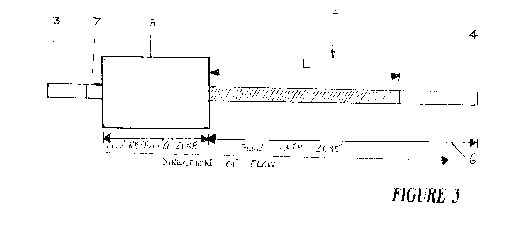
[0054] Figure 3 shows the device of figure 1 at a second stage of testing.
During
this stage, water is added to flow receiving zone 5. This may be done using a
pipette or alternatively placing the substrate 2 into a beaker of a wicking
fluid such
as but not limited to water. Capillary forces in porous substrate 2
urges/wicks the
wicking fluid along flow path zone 6. A distance L that the wicking fluid
travels
along flow path 6 is a function of a level of hydrophobicity in the zone of
hydrophobicity 9. The distance that the water travels along the substrate is a
visual
indication of the fibrinogen concentration in the plasma sample.
[0055] Figure 4 shows an unused testing device 1 comprising substrate 2 of
figure
1 inside and housed in an outer/housing/casing 11 completing testing device 1
and
with visual indicators indicating an unused state. Casing 11 is divided into
application indicator windows and analysis windows. In the application windows
are provided an indication window 13 which indicates plasma introduction and
window 12 which indicates location of water introduction. Upon addition of
plasma,
window 13 undergoes a colour change. A quality check window 14 is provided
which is used to assess pre reaction of a plasma/blood sample and Thrombin.
Window 14 posses the thrombin Chromogenic substrate. When thrombin is spread
18
CA 03135087 2021-09-27
WO 2020/191428
PCT/AU2020/000024
to it after the addition of the plasma or blood sample ( from window 13), it
changes
colour to indicate that the thrombin hasn't degraded indicating that the test
is valid.
Zone 6 (which contains the uncleaved thrombin chromogenic substrate) lies
under
Window 14. When the blood or plasma sample is added to window 13, it allows
the
pre-charged thrombin from the reaction zone to spread to zone 6. The inactive
thrombin chromogenic marker can be placed in a separate area upstream from the
reaction zone. This can be denoted by an extra box in figure 4 ( not shown)
without
shading to indicate its lack of colour instead. Then when the plasma/blood
sample
is added, it becomes coloured as indicated in figure 5. If the thrombin is
working, it
will then cleave the thrombin chromogenic substrate, and cause window 14 to
change colour to indicate a valid test. If the thrombin isn't working, then no
colour
change will occur and it will indicate to the user that the test device cannot
be used.
At completion of testing, window 15 will provide an indication of a low
fibrinogen
or window 16 will provide an indication of a very low fibrinogen. This is
preferably effected by a colour indication appearing in either window 15 or
16.
[0056] Figure 5 shows with corresponding numbering, the housing/casing 11 of
figure 4 indicating a first stage of testing with visual indicator quality
check
window 14 indicating a reaction has occurred between the sample and Thrombin
chromogenic substrate and that there is a valid test ( quality check). The
indication
of a valid test in window 14 is preferably achieved by a colour change- for
example,
but not limited to, white to green. A dye can also be incorporated into the
reaction
zone. Zone 6 (which contains the uncleaved thrombin chromogenic substrate)
lies
under Window 14. When the blood or plasma sample is added to window 13, it
allows the pre-charged thrombin from the reaction zone to spread to zone 6. If
the
thrombin is working, then it will cleave the thrombin chromogenic substrate,
and
cause window 14 to change colour to indicate a valid test. If the thrombin
isn't
working, then no colour change will occur and it will indicate to the user
that the
test cannot be used. When the blood/plasma sample is added, it will spread the
dye
under window 14 as well. Hence the window 14 after plasma addition may appear
blue initially before turning green once the thrombin chromogenic substrate
has
reacted.
19
CA 03135087 2021-09-27
WO 2020/191428
PCT/AU2020/000024
[0057] Figure 6 shows the indications at a second stage of testing. Referring
to
indicator windows, window 13 indicates that plasma was added and that from
window 14 a valid test was performed. In this case window 16 shows no
indication
( remains white) but window 15 shows that the patient tested has a low
concentration of Fibrinogen between 1-2g/L. of device of figure 4 at a second
stage of testing with visual indicators indicating a result. In this case
Fibrinogen
concentrate is needed.
[0058] The present invention employs paper, cellulose or any porous and
wettable
material as a medium for both the plasma and water to wick through. The porous
structure of the cellulose induces a capillary action but without reliance on
action
from micropillar extrusions. Paper is economic in comparison to plastics,
glass or
silicon (AU$5 cents per test vs AU$50 cents per test). Paper is a flexible
material
that can be cut into many different shapes, configurations and structures and
can be
easily incorporated with hydrophobic barriers and hydrophilic channels.
Therefore,
its fabrication costs are very economic compared to the prior art.
[0059] The present invention presents many advantages over the prior art. For
example, in the prior art, a plastics diagnostic device is used which has no
inherent
capillary action to facilitate fluid flow. Accordingly, fluid flow must be
artificially
induced by the inclusion of micropillars. In addition, the prior art plastics
diagnostic device needs to be coated with SiOx and treated with
polyelectrolytes to
increase the hydrophilicity of the flow path zone as in US20120107851A1. The
extra manufacturing adds significantly to the costs of producing the device.
According to the present invention, a paper indicator is used which requires
no pre
treatment to induce capillarity as the material itself has inherent
capillarity
requiring no pretreatment or modification to induce capillarity. One advantage
of
the present invention is that use of a paper indicator, has natural capillary
flow
without the need to add in parts or modify surfaces.
[0060] Another advantage is that the disadvantages of variability related to
thrombin kinetics is eliminated. Furthermore, the employment of a porous
material
such as paper, allows fibrinogen concentration to be measured in other ways
than a
lateral flow assay. For example, the test can be converted into a flow-through
type
CA 03135087 2021-09-27
WO 2020/191428
PCT/AU2020/000024
detection system that measures colour intensity after a certain number of
washes.
Another advantage of the present invention is that the sensitivity for the
test is
higher for concentration ranges of 1-2 g/L Fibrinogen. For example in the
cited
prior art US20120107851A1 a distance travelled for between 1 and 2 g/L plasma
is
only 0.6 cm before stopping.
[0061] According to the present invention there is for example, a 1.7 cm
separation
for the same concentrations after 7 minutes of elution. It is contemplated
that
substrate 2 includes a variety of user options for wicking fluid, including a
wetable
fibrous material and includes but is not limited to, non wetable materials
treated by
plasma treatment, radiation, surfactant coating and/or chemical reaction to
make it
wettable. The substrate may be woven or non -woven. It may be pre or post
treated
with thrombin and/or FXIIIa or derivatives of these enzymes. It may be treated
with
deposited desorbable dyes and/or desorbable dye binders and particles and
nanoparticles.
During testing the substrate is charged with water, buffer solution, dye
solution and/or
washing solution- collectively wicking fluids.
[0062] There are provided alternative methods for analyzing the testing and
test
results. A method of quantifying the zone of hydrophobicity/hydrophobicity
includes measuring the distance travelled by at least one chromogenic marker
with
he zone of hydrophobicity 9 in a lateral flow. Preferably the lateral flow
occurs in
porous substrate 2 in flow receiving zone 5 and flow path zone 6. According to
one
embodiment, flow path zone 6 has a length Li and a width Wl. Flow receiving
zone 5 has a length L2 and a width W2. Li may be the same or different length
from L2. Likewise W1 may be the same or a different length from W2. A lateral
flow regime traverses the flow receiving zone 5 and flow path zone 6 and as
described earlier. According to one embodiment, the biological factors,
chemical
factors and/or derivatives of the biological factors and/or chemical factors
pre charge
substrate 2 in flow path zone 6. Alternatively those factors may be applied
during testing to
at least part of the flow path zone 6 and/or the flow receiving zone 5. In
use, a blood or
plasma sample is introduced into either at least part of the flow path zone 6
and/or the flow
receiving zone 5. Chromogenic marker(s) can be applied at this time or pre-
applied to the
substrate 2.
21
CA 03135087 2021-09-27
WO 2020/191428
PCT/AU2020/000024
[0063] A wicking fluid is applied to the flow receiving zone 5 in order to
induce the
movement of the chromogenic marker(s) through the flow path zone 6 The wicking
fluid is
according to one embodiment applied to the flow receiving zone in the form of
a finite
reservoir or an infinite volume reservoir. The distance travelled by the
chromogenic
marker(s) is measured with ruling markers next to the flow path zone 6. When
substrate 2
is held in casing 11 to form device 1, the distance travelled by the
chromogenic marker(s)
is/are measured via by observing the flow path zone 6 through transparent
windows 12, 13,
14, 15 and 16 in casing 11. Windows 12, 14, 15 and 16 align with the flow path
zone 6 and
enable the observations described earlier with reference to figures 5 and 6.
The indicating
windows align with key distances up the flow path zone 6 and will show
wherever the
chromogenic marker(s) have reached and/or gone past the key distances or not.
[0064] According to one embodiment, hydrophobicity of the zone of
hydrophobicity 9 is
determined by measurement of degree of retention of at least one chromogenic
marker
applied and/or pre-applied in ( or outside of) the reaction zone, along with
all the other
biological factors, chemical factors and/or derivatives of the biological
factors and/or
chemical factors, after rinsing the porous substrate with a washing liquid.
Hydrophobicity
refers to the hydrophobicity at the surface of the non-porous substrate. The
substrate's
surface hydrophobicity is used to influence shape, height and/or contact angle
of
any deposited liquid droplets. Hydrophobicity can be modified with a physical
factor and/or chemical factor that will or may increase or decrease the
hydrophobicity of the substrate. One method of modifying the hydrophobicity is
to
apply a chemical coating to the substrate.
[0065] Preferably the clot formed acts as a hydrophobic barrier to prevent the
washing
liquid from dislocating of the chromogenic marker(s) out of the substrate. The
forming or
formed clot can coat the surface of the non-porous substrate to modify its
hydrophobicity.
Physical factors may allow or prevent the initiation of the clot formation.
Physical,
Biological factors, chemical factors and/or derivatives of the biological
factors and/or
chemical factors used are involved in the initiation, execution, amplification
and/or
acceleration of the clot formation. These factors also affect the clot's
hydrophobicity.
The retention of chromogenic marker(s) is measured by the absolute and/or
relative colour
intensity after washing the substrate with a given volume of washing liquid.
The
chromogenic marker(s) is applied and/or pre-applied in the reaction zone,
along
22
CA 03135087 2021-09-27
WO 2020/191428
PCT/AU2020/000024
with all the other biological factors, chemical factors and/or derivatives of
the
biological factors and/or chemical factors.
[0066] There are many variations in the methodology of performing the
fibrinogen
concentration testing. The variations include:
applying blood or plasma on the non-porous substrate outside the reaction
zone;
application of the blood or plasma to the reaction zone to form a zone of
hydrophobicity
after initiating clot formation;
removing clotted blood or plasma from the non-porous substrate's surface by
physical
factors;
enhancing the visibility of the liquid droplet with the use of at least one
chromogenic
maker...
a threshold result for height measurements which can be determined by placing
a porous,
absorbent substrate directly above the non-porous substrate and allowing any
deposited
liquid droplet to make contact with the porous, absorbent substrate.
[0067] The method contemplates a process of measuring chromogenic staining
which
refers to the binding of at least one chromogenic substrate directly and/or
indirectly to the
formed or forming clot. Blood or plasma is applied to the reaction zone to
form a zone of
stained clot after initiating clot formation. Biological factors, chemical
factors and/or
derivatives of the biological factors and/or chemical factors can be used
enhance the colour
intensity of the chromogenic marker(s).
[0068] According to an alternative embodiment, the present invention one
method
of diagnosis of concentration of fibrinogen in a blood or plasma sample,
involves
using at least one capillary in a centrifuge, each including a reaction
mixture of
biological factors, chemical factors and/or derivatives of the biological
factors
and/or chemical factors applied and/or pre-applied inside of the at least one
capillary. Blood or plasma is applied to the reaction mixture which forms a
clot and
the extent of clotting after initiating clotting is measured. Measuring the
extent of
clotting refers to quantifying the mass, volume or height of the clot in the
capillary.
[0069] A centrifugal force is applied to the capillaries such that it causes
the clot to
compress in the direction away from the axis of spin. The extent of clotting
is
determined by measuring the height of the centrifugally compressed clot in the
23
CA 03135087 2021-09-27
WO 2020/191428
PCT/AU2020/000024
capillary. The height of the compressed clot can be measured with ruling
markers
on top of and/or next to the capillar(ies). Alternatively the compressed clot
is
measured by containing the capillary in an external casing with transparent
windows that align with key heights up the capillary and hence will show
whether
the clot has reached and/or gone past the key heights or not.
[0070] As previously described, a chromogenic dye or other suitable marker is
used. Biological factors, chemical factors and/or derivatives of the
biological
factors and/or chemical factors are used enhance the colour intensity of the
chromogenic dye(s) and are involved in the initiation, execution,
amplification
and/or acceleration of the clot formation. Physical factors used that will or
may
allow or prevent the initiation of the clot formation. the biological factors,
chemical
factors and/or derivatives of the biological factors and/or chemical factors
used are
involved in the enhancement or diminishment of the clot's hydrophobicity.
Physical
factors used that will or may enhance or diminish the clot's hydrophobicity.
The
biological factors, chemical factors and/or derivatives of the biological
factors
and/or chemical factors used, are involved in the enhancement or diminishment
of
the clot's ability to bind directly and/or indirectly to at least one
chromogenic
marker.
[0071] It will be recognised by persons skilled in the art that numerous
variations and modification may be made to the invention broadly described
herein
without departing from the overall spirit and scope of the invention.
24