Note: Descriptions are shown in the official language in which they were submitted.
CA 03135407 2021-09-28
WO 2020/205359 PCT/US2020/024671
METHODS FOR PRODUCTION OF CAR-NK CELLS AND USE THEREOF
[0001] This application claims priority to U.S. Provisional Patent Application
Serial
No. 62/826,856, filed March 29, 2019, which is incorporated by reference
herein in its entirety.
BACKGROUND
1. Field
[0002] The present invention relates generally to the fields of immunology and
medicine. More particularly, it concerns methods of expanding natural killer
(NK) cells.
2. Description of Related Art
[0003] Despite technological advancements in the diagnosis and treatment
options
available to patients diagnosed with cancer, the prognosis still often remains
poor and many
patients cannot be cured. Immunotherapy holds the promise of offering a
potent, yet targeted,
treatment for patients diagnosed with various tumors with the potential to
eradicate the
malignant tumor cells without damaging normal tissues. In theory, the T cells
of the immune
system are capable of recognizing protein patterns specific for tumor cells
and to mediate their
destruction through a variety of effector mechanisms. Adoptive T cell therapy
is an attempt to
harness and amplify the tumor-eradicating capacity of a patient's own T cells
and then return
these effectors to the patient in such a state that they effectively eliminate
residual tumor,
however without damaging healthy tissue. Although this approach is not new to
the field of
tumor immunology, many drawbacks in the clinical use of adoptive T cell
therapy impair the
full use of this approach in cancer treatments. For example, chimeric antigen
receptor T cells
(CAR T) cells are patient-specific and have to be produced for each patient on
an individual
case basis.
[0004] On the other hand, cord blood (CB)-derived natural killer (NK) cells
provide an
off-the-shelf source of cells for immunotherapy and also harness the inherent
cytotoxicity of
NK cells against many tumors. While studies have been performed on CAR NK
cells derived
from peripheral blood, these cells are also not ideal for an 'off-the-shelf
approach. This is
because a donor has to be identified for NK cell donation in each case.
[0005] CAR-engineered NK92 cells have also been studied; however, NK92 is an
NK
cell line derived from a lymphoma patient which lacks many of the NK cell
receptors important
for NK cell cytotoxicity. In addition, since the cell line was derived from a
patient with
1
CA 03135407 2021-09-28
WO 2020/205359 PCT/US2020/024671
lymphoma, the cells must be irradiated prior to infusion. This lack of NK cell
receptors and
need for irradiation significantly impairs the ability of the cells to
proliferate and persist,
making them less effective than CAR-modified CB-NK cells that express the full
array of NK
cell receptors. Thus, there is an unmet need for methods of generating CAR NK
cells with high
efficiency for use in clinical therapies.
SUMMARY
[0006] In one embodiment, the present disclosure provides methods and
compositions
related to therapies for a medical condition, such as cancer. In particular
embodiments, the
methods and compositions concern immunotherapies and/or cell therapies. In a
specific
embodiment, the disclosure concerns an ex vivo method for producing natural
killer (NK) cells
engineered to express a chimeric antigen receptor (CAR) and/or T cell receptor
(TCR)
comprising culturing a starting population of NK cells in the presence of
artificial presenting
cells (APCs) or other feeder cells and at least one cytokine; introducing a
CAR and/or TCR
expression vector into the NK cells; and expanding the NK cells in a gas-
permeable bioreactor
in the presence of APCs and at least one cytokine, thereby obtaining an
expanded population
of engineered NK cells. In some aspects, the gas permeable bioreactor is G-Rex
100M. In
certain aspects, the method does not comprise performing HLA matching. In some
alternative
cases, any or all steps of the method occur in the absence of a gas-permeable
bioreactor.
[0007] In some aspects, the engineered NK cells express a CAR. In certain
aspects, the
engineered NK cells express a TCR. In particular aspects, the engineered NK
cells express a
CAR and TCR or multiple antigen receptors. In particular aspects, the
population of engineered
NK cells are GMP-compliant. In particular aspects, the complete method is
performed in less
than 2 weeks, such as 8 days, 9 days, 10 days, 11, days, 12 days, 13 days, or
14 days. In other
aspects, the complete method may take 3, 4 or more weeks. In some aspects, the
NK cells are
allogeneic with respect to an individual. In other aspects, the NK cells are
autologous with
respect to an individual.
[0008] In some aspects, the starting population of NK cells is obtained from
cord blood,
peripheral blood, bone marrow, CD34+ cells, or iPSCs. In particular aspects,
the starting
population of NK cells is obtained from cord blood. In some aspects, the cord
blood has
previously been frozen. In certain aspects, the starting population of NK
cells is obtained by
isolating mononuclear cells using a ficoll-paque density gradient. In some
aspects, the method
2
CA 03135407 2021-09-28
WO 2020/205359 PCT/US2020/024671
further comprises depleting the mononuclear cells of CD3, CD14, and/or CD19
cells to obtain
the starting population of NK cells. In some aspects, the method further
comprises depleting
the mononuclear cells of CD3, CD14, and CD19 cells to obtain the starting
population of NK
cells. In particular aspects, depleting comprises performing magnetic sorting.
In other aspects,
NK cells could be positively selected using sorting, magnetic bead selection
or other methods
to obtain the starting populations of NK cells.
[0009] In certain aspects, the APCs are gamma-irradiated APCs. In some
aspects, the
APCs are universal APCs (uAPCs). In some aspects, the uAPCs are engineered to
express (1)
CD48 and/or CS1 (CD319), (2) membrane-bound interleukin-21 (mbIL-21), and (3)
41BB
ligand (41BBL). In particular aspects, the NK cells and APCs are present at a
1:1 to 1:100 ratio,
such as a 1:1, 1:2, 1:3, 1:4, 1:5, 1:6, 1:7, 1:8, 1:9, or 1:10 ratio. In
certain aspects, the NK cells
and APCs are present at a 1:2 ratio.
[0010] In some aspects, at least one cytokine is IL-2, IL-21, IL-15, or IL-18.
In certain
aspects, the culturing and/or expanding of the NK cells is in the presence of
2, 3, or 4 cytokines.
In some aspects, the cytokines are selected from the group consisting of IL-2,
IL-21, IL-15,
and IL-18. In some aspects, at least one cytokine, such as IL-2, is present at
a concentration of
100-300 U/mL, such as 100, 125, 150, 175, 200, 225, 250, 275, or 300 U/mL. In
certain aspects,
the at least one cytokine is present at a concentration of 200 U/mL.
[0011] In certain aspects, introducing the CAR and/or TCR comprises
transduction or
electroporation. In some aspects, the transduction is retronectin
transduction. In particular
aspects, the transduction has an efficiency of at least 20%, such as at least
25%, 30%, 35%,
40%, 45%, 50%, 55%, 60%, 65%, 66%, 67%, 68%, 69%, 70%, 71%, 72%, 73%, 74%,
75%,
or higher. In some aspects, the CAR and/or TCR expression construct is a
lentiviral vector or
retroviral vector. In certain aspects, the method results in at least 1000-
fold expansion, such as
at least 1100-, 1200-, 1300-, 1400-, 1500-, 1600-, 1700-, 1800-, 1900-, 2000-,
2100-, 2200-,
2300-, 2400-, 2500-fold or higher expansion.
[0012] In some aspects, the CAR and/or TCR has antigenic specificity for CD19,
CD319/CS1, BCMA, CD38, CLL1, CD70, ROR1, CD20, CD5, CD70, CD20,
carcinoembryonic antigen, alphafetoprotein, CA-125, MUC-1, epithelial tumor
antigen,
melanoma-associated antigen, mutated p53, mutated ras, HER2/Neu, ERBB2, folate
binding
protein, HIV-1 envelope glycoprotein gp120, HIV-1 envelope glycoprotein gp41,
GD2,
3
CA 03135407 2021-09-28
WO 2020/205359 PCT/US2020/024671
CD123, CD23, CD30, CD56, c-Met, mesothelin, GD3, HERV-K, IL-11Ralpha, kappa
chain,
lambda chain, CSPG4, ERBB2, WT-1, EGFRvIII, TRAIL/DR4, and/or VEGFR2. In some
aspects, the CAR and/or expression construct further expresses a cytokine or
2, 3, or 4
cytokines. In certain aspects, the cytokine is IL-15, IL-21, IL-18, or IL-2.
[0013] In additional aspects, the method further comprises cryopreserving the
population of engineered NK cells. In some aspects, the engineered NK cells
are cryopreserved.
Further provided herein is a population of cryopreserved NK cells.
[0014] In another embodiment, there is provided a population of engineered NK
cells
produced according to the methods of present embodiments. Further provided
herein is a
pharmaceutical composition comprising the population of engineered NK cells of
the
embodiments and a pharmaceutically acceptable carrier. Another embodiment
provides a
composition comprising an effective amount of the engineered NK cells of the
embodiments
for use in the treatment of a disease or disorder in a subject. Also provided
herein is the use of
a composition comprising an effective amount of the engineered NK cells of the
embodiments
for the treatment of an immune-related disorder in a subject.
[0015] A further embodiment provides a method of treating an immune-related
disorder in a subject comprising administering an effective amount of
engineered NK cells of
the embodiments to the subject. In certain aspects, the method does not
comprise performing
HLA matching. In particular aspects, the NK cells are KIR-ligand mismatched
between the
subject and donor. In specific aspects, the method does not comprise
performing HLA
matching. In particular aspects, the absence of HLA matching does not result
in graft versus
host disease or toxicity.
[0016] In some aspects, the immune-related disorder is a cancer, autoimmune
disorder,
graft versus host disease, allograft rejection, or inflammatory condition. In
certain aspects, the
immune-related disorder is an inflammatory condition and the immune cells have
essentially
no expression of glucocorticoid receptor. In some aspects, the subject has
been or is being
administered a steroid therapy. In some aspects, the NK cells are autologous.
In certain aspects,
the NK cells are allogeneic.
[0017] In particular aspects, the immune-related disorder is a cancer. In some
aspects,
the cancer is a solid cancer or a hematologic malignancy.
4
CA 03135407 2021-09-28
WO 2020/205359 PCT/US2020/024671
[0018] In additional aspects, the method further comprises administering at
least a
second therapeutic agent. In some aspects, the at least a second therapeutic
agent comprises
chemotherapy, immunotherapy, surgery, radiotherapy, or biotherapy. In
particular aspects, the
NK cells and/or at least a second therapeutic agent are administered
intravenously,
intraperitoneally, intratracheally,
intrathecally, intratumorally, intramuscularly,
endoscopically, intralesionally, percutaneously, subcutaneously, regionally,
or by direct
injection or perfusion. The combination therapies may be administered
sequentially or
simultaneously.
[0019] A further embodiment provides a method of treating an infection of any
kind in
a subject comprising administering an effective amount of engineered NK cells
of the
embodiments to the subject. In certain aspects, the method does not comprise
performing HLA
matching. In particular aspects, the NK cells are KIR-ligand mismatched
between the subject
and donor. In specific aspects, the method does not comprise performing HLA
matching. In
some aspects, the NK cells are KIR-ligand mismatched between the subject and
donor. In
particular aspects, the absence of HLA matching does not result in graft
versus host disease or
toxicity. In some aspects, the NK cells are autologous. In certain aspects,
the NK cells are
allogeneic.
[0020] Other objects, features and advantages of the present invention will
become
apparent from the following detailed description. It should be understood,
however, that the
detailed description and the specific examples, while indicating preferred
embodiments of the
invention, are given by way of illustration only, since various changes and
modifications within
the spirit and scope of the invention will become apparent to those skilled in
the art from this
detailed description.
BRIEF DESCRIPTION OF THE DRAWINGS
[0021] The following drawings form part of the present specification and are
included
to further demonstrate certain aspects of the present invention. The invention
may be better
understood by reference to one or more of these drawings in combination with
the detailed
description of specific embodiments presented herein.
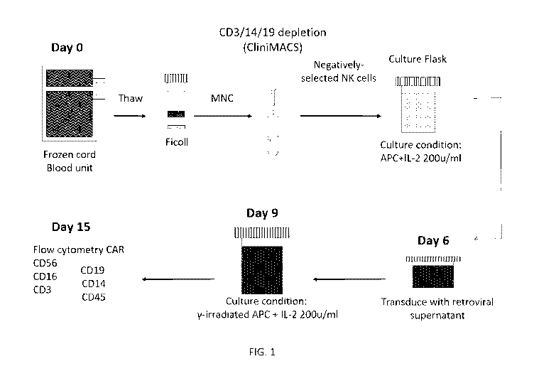
[0022] FIG. 1: Clinical GMP-grade CAR-NK transduction and expansion.
CA 03135407 2021-09-28
WO 2020/205359 PCT/US2020/024671
[0023] FIG. 2: Characteristics of GMP-grade CAR-transduced CB-NK cells
generated
from 5 different CB units after 14 days of culture.
[0024] FIG. 3: CAR NK cell expansion in flask versus G-Rex bioreactor.
[0025] FIG. 4: Average survival (days) of mice in groups treated with
different NK
cell preparations.
[0026] FIG. 5: Percent survival of mice engrafted with Raji tumors and treated
with
different NK cell preparations.
[0027] FIG. 6: Comparison of survival of mice engrafted with Raji tumors and
treated
with different NK cell preparations.
[0028] FIG. 7: Biofluorescent imaging of mice treated with indicated NK cell
preparations.
[0029] FIG. 8: Impact of blocking KIR-HLA interaction on activity of CAR NK
cells
against tumor targets.
[0030] FIG. 9: Table 1. Characteristics of Patients at Baseline.
[0031] FIG. 10: Table 2. Adverse Events in the 11 Study Patients.
[0032] FIG. 11: Clinical Response to CAR-NK Therapy and Postremission
Treatments. Shown are the clinical outcomes and subsequent therapies for the
11 patients who
were treated with anti-CD19 chimeric antigen receptor (CAR) natural killer
(NK) cells in the
study. Responses were confirmed and assessed according to the 2018 criteria of
the
International Workshop on Chronic Lymphocytic Leukemia and the 2014 Lugano
classification for non-Hodgkin's lymphoma. The indicated responses include
partial response
(PR) and complete response (CR); MRD denotes minimal residual disease, as
assessed on
multiparameter flow cytometry, with or without bone marrow (BM) infiltration.
Patient 3
received four doses (x4) of rituximab; for Patients 5 and 7, the dashed white
line indicates the
duration of postremission therapy. HSCT denotes hematopoietic stem-cell
transplantation.
[0033] FIGS. 12A and 12B: Persistence of CAR-NK Cells after Infusion. FIG. 12A
shows measurements of CAR-NK cells in peripheral-blood samples, as assessed on
quantitative polymerase-chain-reaction assay, according to the dose of CAR-NK
cells received
6
CA 03135407 2021-09-28
WO 2020/205359 PCT/US2020/024671
by the patient. The horizontal gray line at 3 copies per microgram of DNA
represents the lower
limit of quantification for this assay. The solid horizontal bars indicate the
median copy
numbers at the various time points for each dose level. After a single
infusion of CAR-NK
cells, CAR sequences could be detected in all 11 patients. The values
increased and remained
detectable in peripheral blood for up to 1 year after infusion, regardless of
the dose level. No
relationship was observed between the administered cell dose and the CAR-NK
copy number
beyond day 14 after infusion, which suggests that the persistence of CAR-NK
cells was driven
by in vivo proliferation of the infused cells. The length of follow-up varied
among the patients.
FIG. 12B shows the peak copy numbers of CAR-NK cells in the first 28 days
after infusion for
the 11 patients, according to their response to therapy. Patients who had a
response at day 30
had a significantly higher copy-number peak of CAR-NK cells after the infusion
than those
who did not have a response (median value, 31,744 vs. 903 copies per
microgram; P = 0.02).
The black horizontal bars indicate median values.
[0034] FIGS. 13A-13C: GMP-grade CAR-NK cells kill primary CLL targets in a
perforin-dependent manner. FIG. 13A shows lysis of primary CLL targets (n=4)
by GMP-grade
iC9/CAR19/IL-15 transduced CB NK cells (red line) compared to paired ex vivo
expanded
non-transduced NK cells (NT-NK cells; black line). *** represents p< 0.0001
and **p< 0.01.
FIG. 13B represents the fold change in mean fluorescence intensity of perforin
(red circles)
after treatment with concanomycin A (CMA), calculated as follows: perforin MFI
after culture
with CMA/ perforin MFI after culture with CMA. The MFI levels of CD56 (black
circles) and
CAR (green circles) on the surface of NK cells were measured as controls and
remained
unchanged after treatment with CMA (n=3). FIG. 13C shows lysis of primary CLL
targets
(n=4) by GMP-grade iC9/CAR19/IL-15 transduced CB NK cells before (solid
circles) and after
(open circles) treatment with CMA.; ** represents p< 0.01 and *p<0.05.
[0035] FIG. 14: Radiological response in patient 5. FDG PET-CT scans from
patient
performed at study enrollment, before (upper row) and 29 days after receiving
the CAR-NK
cell infusion (lower row). Upper right corner projection image showing FDG
uptake in nodes
above and below the diaphragm. Upper right middle showing FDG PET-CT scan with
abnormal uptake in enlarged mesenteric nodes (dark arrow). Upper left middle
PET-CT scan
showing enlarged mesenteric nodes (light arrow). Upper left "fused" PETCT scan
showing
FDG uptake localized to mesenteric adenopathy. Lower right corner projection
image showing
resolution of FDG uptake in nodes above and below the diaphragm. Lower right
middle
7
CA 03135407 2021-09-28
WO 2020/205359 PCT/US2020/024671
showing FDG PET-CT scan with no uptake in mesenteric nodes (dark arrow). Lower
left
middle PET-CT scan showing stable enlarged mesenteric nodes (light arrow).
Lower left
"fused" PET-CT scan showing no FDG uptake in mesenteric adenopathy (arrow).
[0036] FIG. 15: Persistence of CAR-NK cells after infusion according to the
degree of
HLA mismatch between the CB CAR-NK cells and the recipient. The persistence
and
expansion of iC9/CAR19/IL-15-modified CB-NK cells in peripheral blood samples
collected
from patients at multiple timepoints after infusion were assessed by qPCR. The
green dots
represent the CAR-NK copy numbers in peripheral blood samples for the nine
patients who
received a partially HLA-matched CAR-NK product (4/6 HLA match). The red dots
represent
the CAR-NK copy numbers for the two patients who received a non-HLA matched
product
(1/6 or 2/6 HLA match). The dotted black line represents the level of
detection of the PCR
assay.
[0037] FIGS. 16A-16D: Detection of CAR-NK cells by multiparameter flow
cytometry FIG. 16A shows the flow cytometry gating strategy for the detection
of donor CAR-
NK cells in the peripheral blood in a representative patient (patient 6, day
+3 after CAR-NK
infusion). Lymphocytes were selected using FSC-A and SSC-A (i); next doublets
were
excluded using SSCW vs SSC-H (ii); live cells were identified using a
Live/Dead dye (iii);
hematopoietic cells within the live population were then selected by gating on
CD45+ cells
(iv); myeloid cells were excluded by gating on the CD33 negative and CD14
negative cells (v);
NK cells were identified by gating on CD3- and CD56+ cells (vi). Within the
CD3-CD56+
subset, cord blood derived NK cells were identified based on expression of the
donor-specific
HLA-antigen (vii). Expression of CAR on donor NK cells was further determined
using an
antibody directed against the CH2-CH3 domain of the human IgG hinge
(109606088/ Jackson
Immuno Rsch) (viii). B cells were identified by expression of CD19 and/or CD20
by gating on
the CD45+CD33-CD14- lymphocyte population (ix). FIG. 16B shows the CAR-NK
frequencies, using the gating strategy described above for patient 6 on days
8, 14 and 21 after
infusion. FIG. 16C shows the CAR NK frequencies for patient 8 on days 3, 14
and 21 after
infusion. FIG. 16D shows the CAR NK frequencies for patient 10 on days 3, 7
and 14 after
infusion. PBMCs: peripheral blood mononuclear cells, FSC-A : forward scatter-
area, FSC-H
: forward scatter- height SSC-A : side scatter- area, SSC-H : side scatter-
height.
[0038] FIG. 17: Serial manual gating strategy for the detection of CAR-NK
cells in the
lymph node in a representative patient. Flow cytometry data to show the gating
strategy for the
8
CA 03135407 2021-09-28
WO 2020/205359 PCT/US2020/024671
detection of donor CAR NK cells in the lymph node for patient 6. The biopsy
was performed
105 days after the CAR-NK infusion to investigate residual FDG activity in a
single lymph
node. The lymphocyte population was selected based on FSC-A and SSC-A (i);
next doublets
were excluded using SSC-W vs SSC-H (ii); live cells were identified using a
Live/Dead dye
(iii); hematopoietic cells within the live population were then selected by
gating on CD45+
cells (iv); myeloid cells were excluded by gating on the CD33 negative and
CD14 negative
cells (v); NK cells were identified by gating on CD3- and CD56+ cells (vi).
Within the CD3-
CD56+ subset, cord blood derived NK cells were identified based on expression
of the donor-
specific HLA-antigen (in this case the CAR NK cells were HLAA3 positive and
the recipient
was HLA-A3 negative) (vii). PBMCs: peripheral blood mononuclear cells, FSC-A :
forward
scatter- area, FSC-H : forward; scatter- height SSC-A : side scatter- area,
SSC-H : side scatter-
height.
[0039] FIG. 18: CAR-NK copy numbers in peripheral blood, bone marrow and lymph
node in a representative patient. The figure shows the CAR-NK copy numbers
measured by
qPCR at various time points in peripheral blood (green circles), bone marrow
(red circles) and
lymph node (black circle) for patient 8. Lymph node biopsy was performed on
day 56 after the
CAR-NK infusion to investigate residual FDG activity in a single lymph node.
Biopsy showed
a necrotic mass with calcification and no evidence of lymphoma. CAR-NK
transcripts could
be detected by qPCR in the lymph node (118,897.4 copies/jig) at significantly
higher levels
(>25 fold) compared to peripheral blood and bone marrow samples collected
during the same
time period.
[0040] FIG. 19: Persistence of CAR-NK cells after infusion in peripheral blood
and
bone marrow samples. The figure shows measurements of CAR-NK cells in
peripheral blood
and bone marrow samples as assessed by qPCR. Green and red dots represent
peripheral blood
and bone marrow samples respectively. The green (peripheral blood) and the red
(bone
marrow) solid lines represent the median copy number at the various time
points. CAR-NK
transcripts were detectable at a similar levels in peripheral blood and bone
marrow.
[0041] FIG. 20: Gating strategy to detect donor CAR expressing T-cells after
CAR-
NK infusion. Flow cytometry gating strategy for the detection of donor T-cells
and donor-
derived CAR expressing T-cells in the peripheral blood in a representative
patient (patient 6,
day +8, panels i to viii, and day +21, panels ix and x, after CAR-NK
infusion). The lymphocyte
gate was selected using FSC-A and SSC-A (i); next doublets were excluded using
SSC-W vs
9
CA 03135407 2021-09-28
WO 2020/205359 PCT/US2020/024671
SSC-H (ii); live cells were identified using a Live/Dead dye (iii);
hematopoietic cells within
the live population were then selected by gating on CD45+ cells (iv); myeloid
cells were
excluded by gating on the CD33 negative and CD14 negative cells (v); T-cells
were identified
by gating on CD3+ CD56- cells (vi). Within the CD3+ subset, cord blood derived
T-cells were
identified based on expression of the donor-specific HLA-antigen (vii).
Expression of CAR on
donor T-cells was further determined using an antibody directed against the
CH2-CH3 domain
of the human IgG hinge (109606088/ Jackson Immuno Rsch). The percentage
indicates the
frequencies of CD3+ T cells expressing the CAR molecule (viii). The panel
shows minimal
contamination by CAR+ donor T-cells in PBMC samples collected from the patient
on days +
8 (vii and viii) and day +21 (ix and x) after CAR NK infusion. Similar results
were found in
two additional patients with available serial samples (data not shown). PBMCs:
peripheral
blood mononuclear cells, FSC-A: forward scatter- area, FSC-H: forward scatter-
height SSC-
A: side scatter- area, SSC-H: side scatter- height.
[0042] FIGS. 21A-21R: Levels of inflammatory cytokines in the peripheral
blood.
Panels show the time course for inflammatory cytokines in peripheral blood
samples after
CAR-NK infusion. Horizontal lines represent median values.
[0043] FIGS. 22A-22B: Patient Characteristics.
[0044] FIG. 23: Characteristics of the infused CAR-NK cell product.
[0045] FIG. 24: CAR-NK cell persistence during the follow up. CAR-NK
persistence
was measured in peripheral blood using qPCR.
[0046] FIG. 25: Measurement of donor-specific antibodies in patient samples at
multiple time points after CAR-NK infusion. (-) indicates that the results are
not available.
[0047] FIGS. 26A-26B. Antileukemic function of CB-NK cells transduced with
CAR19-CD28-zeta-2A-IL15 vector. (FIG. 26A) Transduction efficiency (85%) of CB
NK
cells (bottom panel) compared to non-transduced NK cells (top panel).
Transduction is stable.
(FIG. 26B) CAR-NK cells are more efficient at killing CD19+ Raji tumors and
primary CLL
compared to non-transduced (NT) ex vivo expanded and activated NK cells with
equal effector
function against K562 cells. P < 0.001 (iC9/CAR.CD19/IL15 + Raji vs NT-NKs +
Raji); P <
0.001 (iC9/CAR.CD19/IL15 + CLL vs NT-NKs + CLL); P=ns (iC9/CAR.CD19/IL15 +
K562
vs NT-NKs + K562).
CA 03135407 2021-09-28
WO 2020/205359 PCT/US2020/024671
[0048] FIGS. 27A-27C: In vivo homing, proliferation and antitumor activity of
iC9/CAR.19/IL15-transduced CB NK cells. (FIG. 27A) C9/CAR.19/IL15-tranduced
eGFP-
FFLuc-labeled CB-NK cells home to sites of disease (liver, spleen, bone marrow
BM]) more
efficiently than CAR.19 transduced CB-NK cells or NT-NK cells. (FIG. 27B)
Infusion of
iC9/CAR.19/IL15-transduced CB-NK cells into NSG mice engrafted with luciferase-
labeled
Raji cells results in tumor eradication, as evidenced by in vivo
bioluminescence imaging.
Colors indicate intensity of luminescence (red, highest; blue, lowest). (FIG.
27C). The in vivo
antitumor activity of a single dose of iC9/CAR.19/IL15-transduced CB NK is
significantly
better than that of CB-NK cells that were either not transduced or transduced
with a CAR.CD19
construct lacking IL-15. P= 0.001 (iC9/CAR.CD19/IL15 + Raji vs NT-NKs + Raji);
P= 0.044
(iC9/CAR.CD19/IL15 + Raji vs CAR.CD19 + Raji); P= 0.006 (CAR.CD19 + Raji vs NT-
NKs
+ Raji) P= 0.182 (NT-NKs + Raji vs Raji alone).
[0049] FIGS. 28A-28B: IL-15-transduced CB-NK cells do not show signs of
autonomous or dysregulated growth. (FIG. 28A) iC9/CAR.19/IL15-transduced CB NK
cells
stop expanding within 6 weeks of in vitro culture with no evidence of
autonomous growth.
(FIG. 28B) Photomicrographs of mesenteric lymph nodes show vestigial lymphoid
tissue with
no lymphocytes in any experimental mice, which is typical of NSG mice. Images
of the spleen
show rudimentary periarteriolar lymphoid tissue devoid of lymphocytes (black
arrows) and is
surrounded by hematopoietic tissue composed of various stages of erythroid and
myeloid series
cells, including megakaryocytes and hemosiderin-laden macrophages. Bone marrow
contains
normal hematopoietic cells and no abnormal lymphocytes. H&E stain,
magnification x200.
Slides from two representative groups of NSG mice treated with iC9/CAR.19/IL15-
transduced
CB NK cells.
[0050] FIG. 29: IL-15 production by iC9.CAR.19.CD28.CD3OL15-transduced CB
NK cells; iC9. CAR.19.CD28.CD3OL15-transduced CB NK cells produce IL-15 in
response
to antigenic stimulation in vitro.
[0051] FIGS. 30A-30B: Activation of the inducible caspase-9 suicide gene
eliminates
iC9/CAR.19/IL15+ CB-NK cells. (FIG. 30A) The addition of 10 nM of AP1903 to
cultures of
iC9-CAR-IL15+ CB-NK cells induced apoptosis/necrosis of transgenic cells
(bottom right
panel) within 4 hours as assessed by annexin-V-7AAD staining. NT, non-
transduced CB-NK
cells; CAR, iC9/CAR.19/IL15-transduced NK cells; (FIG. 30B) NSG mice engrafted
i.v. with
Raji cells, and infused with iC9/CAR.19/IL15+ CB-NK cells were treated 10-14
days later with
11
CA 03135407 2021-09-28
WO 2020/205359 PCT/US2020/024671
two doses of the AP1903 dimerizer (50 Ilg) i.p. two days apart.
iC9/CAR.19/IL15-expressing
NK cells were substantially reduced in all organs tested 3 days later.
DESCRIPTION OF ILLUSTRATIVE EMBODIMENTS
[0052] Encouraging clinical results have been seen with human umbilical cord
blood-
derived natural killer (CB-NK) cells transduced with retroviral vectors
targeting CD19+
lymphoid cancers. A number of additional CAR-NK cell constructs have been
generated
targeting myeloid tumors, multiple myeloma and solid tumor cancer antigens. In
some
embodiments, the present disclosure provides methods for the robust expansion
of NK cells.
The cells may be obtained from frozen or thawed CB units and expanded in gas
permeable
bioreactors containing co-cultures with antigen presenting cells (APCs), such
as universal
antigen presenting cells (uAPCs), or other feeder cells and cytokines, such as
interleukin (IL)-
2.
[0053] One limitation of using CAR NK cells for clinical therapy is because of
their
small numbers and their poor survival post thaw. The present studies have
addressed both of
these limitations by using GMP-compliant strategy for the ex vivo expansion of
CAR NK cells.
The present methods resulted in a median 2200-fold expansion in two weeks,
with an excellent
CAR transduction efficiency of around 66%. Using this strategy, up to 400
doses of 1 x10⁶
CAR NK cells per kg can be generated for the treatment of patients.
[0054] Accordingly, certain embodiments of the present disclosure provide
methods
and compositions concerning the manufacture, expansion, quality control, and
functional
characterization of clinical-grade NK cells intended for cell and
immunotherapy. Growing and
molding clinically relevant numbers of NK cells for infusion into patients
while meeting time
constraints are extremely challenging even in the best of circumstances. The
disclosed methods
and compositions detail the technical processes of NK cell manufacture,
details and kinetics of
achievable NK cell expansions, and molecular characterization to verify
successful cellular
molding.
[0055] The present methods provide high and consistent transduction levels of
the NK
cells with the CAR constructs and rapid production of highly potent CB-CAR-NK
cells which
can be infused fresh or frozen for subsequent infusion. The frozen CAR-NK cell
products
provided herein are truly "off-the-shelf' cell therapy which can be thawed and
infused into
patients with no delay needed for production.
12
CA 03135407 2021-09-28
WO 2020/205359 PCT/US2020/024671
[0056] In addition, the present studies have demonstrated the safety of
infusing NK
cells and CAR-NK cells which are not HLA-matched with the patients. Thus, the
combination
of no longer needing HLA matching and the robust efficacy of thawed products
has resulted in
a paradigm shift in the use of CAR-based therapy where CAR-NK cells can now be
prepared
as an "off-the-shelf' product that can be infused as a point of care product.
The present strategy
can also be applied to NK cells from any source, including peripheral blood,
bone marrow,
hematopoietic stem cells, induced pluripotent stern cells or NK cell lines.
[0057] In specific aspects, the NK cells may be isolated from umbilical CB of
healthy
donors co-cultured with APCs, such as K-562-based feeders or other feeder
cells such as
lymphoblastoid cells lines or beads, and one or more cytokines including IL-2,
IL-15, IL-12,
IL21 or 1L-18. The NK cells may then be transduced with retroviral,
lentiviral, adenoviral, or
adeno-associated viral vectors, or electroporated with sleeping beauty or
piggy-back constructs
that target hematologic and solid cancers. The transduced cells may then be
further expanded
in gas permeable bioreactors containing co-cultures with the APCs or other
feeders and IL-2
or other cytokines to obtain the potent CAR-transduced CB-NK cells. Those
cells can be
infused fresh, or can be frozen for thaw and infusion at a later date.
[0058] CAR-T cells for infusion in the allogeneic setting must be HLA-matched
or be
genetically manipulated to remove the T cell receptor in order to prevent
lethal GVHD.
Previous studies have used CB units to generate clinical NK cell and CAR-NK
cell products
that were matched at 4/6 HLA antigens for safety and consistency with the
requirements for
CB transplant matching. However, in the present studies, 2 patients were
treated with
allogeneic CB-derived NK cells that were not HLA-matched at any antigen to the
patients with
no toxicity. They were infused safely with no GVHD or other toxicities, and
comparable
persistence in the patients compared to 4/6 HLA-matched NK cells. This has
established the
present platform for using CB units to generate NK and CAR-NK cell products
without the
need for any HLA matching. Choosing CB units at random from CB banks for NK
cell
production markedly expands the number of CB units available and improves the
timing and
logistics of therapy by eliminating the need for HLA typing of the patient and
then matching
with the CB unit.
[0059] In further aspects, the present methods may comprise identifying and
selecting
CB units for CAR NK production which are typed for the killer immunoglobulin
receptor
(KIR) ligand and are mismatched with the recipient. The resulting
alloreactivity from KIR-
13
CA 03135407 2021-09-28
WO 2020/205359 PCT/US2020/024671
ligand mismatch may further enhance the activity of CAR-transduced NK cells by
synergizing
with the CAR-mediated recognition of the tumor cells. Indeed, the present
studies have shown
that blocking the KIR-ligand interaction using HLA blocking antibodies can
significantly
enhance the CAR-NK mediated cytotoxicity of CLL targets (FIG. 8).
[0060] The present CAR-transduced NK cells can provide an off-the-shelf source
of
cells for the immunotherapy of many cancers including both liquid and solid
tumors. Retroviral
transduction of CB derived NK cells allows for longer persistence and improved
efficacy of
the engineered cells for use in the immunotherapy of many cancers and
potentially for the
treatment of infections, including viruses, bacteria and fungi and autoimmune
disorders by
targeting autoreactive B or T cells.
I. Definitions
[0061] As used herein, "essentially free," in terms of a specified component,
is used
herein to mean that none of the specified component has been purposefully
formulated into a
composition and/or is present only as a contaminant or in trace amounts. The
total amount of
the specified component resulting from any unintended contamination of a
composition is
therefore well below 0.05%, preferably below 0.01%. Most preferred is a
composition in which
no amount of the specified component can be detected with standard analytical
methods.
[0062] As used herein the specification, "a" or "an" may mean one or more. As
used
herein in the claim(s), when used in conjunction with the word "comprising,"
the words "a" or
"an" may mean one or more than one.
[0063] The use of the term "or" in the claims is used to mean "and/or" unless
explicitly
indicated to refer to alternatives only or the alternatives are mutually
exclusive, although the
disclosure supports a definition that refers to only alternatives and
"and/or." As used herein
"another" may mean at least a second or more.
[0064] Throughout this application, the term "about" is used to indicate that
a value
includes the inherent variation of error for the device, the method being
employed to determine
the value, or the variation that exists among the study subjects.
[0065] An "immune disorder," "immune-related disorder," or "immune-mediated
disorder" refers to a disorder in which the immune response plays a key role
in the development
14
CA 03135407 2021-09-28
WO 2020/205359 PCT/US2020/024671
or progression of the disease. Immune-mediated disorders include autoimmune
disorders,
allograft rejection, graft versus host disease and inflammatory and allergic
conditions.
[0066] An "immune response" is a response of a cell of the immune system, such
as a
B cell, or a T cell, or innate immune cell to a stimulus. In one embodiment,
the response is
specific for a particular antigen (an "antigen-specific response").
[0067] An "autoimmune disease" refers to a disease in which the immune system
produces an immune response (for example, a B-cell or a T-cell response)
against an antigen
that is part of the normal host (that is, an autoantigen), with consequent
injury to tissues. An
autoantigen may be derived from a host cell, or may be derived from a
commensal organism
such as the micro-organisms (known as commensal organisms) that normally
colonize mucosal
surfaces.
[0068] "Treating" or treatment of a disease or condition refers to executing a
protocol,
which may include administering one or more drugs to a patient, in an effort
to alleviate signs
or symptoms of the disease. Desirable effects of treatment include decreasing
the rate of disease
progression, ameliorating or palliating the disease state, and remission or
improved prognosis.
Alleviation can occur prior to signs or symptoms of the disease or condition
appearing, as well
as after their appearance. Thus, "treating" or "treatment" may include
"preventing" or
"prevention" of disease or undesirable condition. In addition, "treating" or
"treatment" does
not require complete alleviation of signs or symptoms, does not require a
cure, and specifically
includes protocols that have only a marginal effect on the patient.
[0069] The term "therapeutic benefit" or "therapeutically effective" as used
throughout
this application refers to anything that promotes or enhances the well-being
of the subject with
respect to the medical treatment of this condition. This includes, but is not
limited to, a
reduction in the frequency or severity of the signs or symptoms of a disease.
For example,
treatment of cancer may involve, for example, a reduction in the size of a
tumor, a reduction in
the invasiveness of a tumor, reduction in the growth rate of the cancer, or
prevention of
metastasis. Treatment of cancer may also refer to prolonging survival of a
subject with cancer.
[0070] "Subject" and "patient" refer to either a human or non-human, such as
primates,
mammals, and vertebrates. In particular embodiments, the subject is a human.
CA 03135407 2021-09-28
WO 2020/205359 PCT/US2020/024671
[0071] The phrases "pharmaceutical or pharmacologically acceptable" refers to
molecular entities and compositions that do not produce an adverse, allergic,
or other untoward
reaction when administered to an animal, such as a human, as appropriate. The
preparation of
a pharmaceutical composition comprising an antibody or additional active
ingredient will be
known to those of skill in the art in light of the present disclosure.
Moreover, for animal (e.g.,
human) administration, it will be understood that preparations should meet
sterility,
pyrogenicity, general safety, and purity standards as required by FDA Office
of Biological
Standards.
[0072] As used herein, "pharmaceutically acceptable carrier" includes any and
all
aqueous solvents (e.g., water, alcoholic/aqueous solutions, saline solutions,
parenteral vehicles,
such as sodium chloride, Ringer's dextrose, etc.), non-aqueous solvents (e.g.,
propylene glycol,
polyethylene glycol, vegetable oil, and injectable organic esters, such as
ethyloleate),
dispersion media, coatings, surfactants, antioxidants, preservatives (e.g.,
antibacterial or
antifungal agents, anti-oxidants, chelating agents, and inert gases), isotonic
agents, absorption
delaying agents, salts, drugs, drug stabilizers, gels, binders, excipients,
disintegration agents,
lubricants, sweetening agents, flavoring agents, dyes, fluid and nutrient
replenishers, such like
materials and combinations thereof, as would be known to one of ordinary skill
in the art. The
pH and exact concentration of the various components in a pharmaceutical
composition are
adjusted according to well-known parameters.
[0073] The term "haplotyping or tissue typing" refers to a method used to
identify the
haplotype or tissue types of a subject, for example by determining which HLA
locus (or loci)
is expressed on the lymphocytes of a particular subject. The HLA genes are
located in the major
histocompatibility complex (MHC), a region on the short arm of chromosome 6,
and are
involved in cell-cell interaction, immune response, organ transplantation,
development of
cancer, and susceptibility to disease. There are six genetic loci important in
transplantation,
designated HLA-A, HLA-B, HLA-C, and HLA-DR, HLA-DP and HLA-DQ. At each locus,
there can be any of several different alleles.
[0074] A widely used method for haplotyping uses the polymerase chain reaction
(PCR) to compare the DNA of the subject, with known segments of the genes
encoding MHC
antigens. The variability of these regions of the genes determines the tissue
type or haplotype
of the subject. Serologic methods are also used to detect serologically
defined antigens on the
surfaces of cells. HLA-A, -B, and -C determinants can be measured by known
serologic
16
CA 03135407 2021-09-28
WO 2020/205359 PCT/US2020/024671
techniques. Briefly, lymphocytes from the subject (isolated from fresh
peripheral blood) are
incubated with antisera that recognize all known HLA antigens. The cells are
spread in a tray
with microscopic wells containing various kinds of antisera. The cells are
incubated for 30
minutes, followed by an additional 60-minute complement incubation. If the
lymphocytes have
on their surfaces antigens recognized by the antibodies in the antiserum, the
lymphocytes are
lysed. A dye can be added to show changes in the permeability of the cell
membrane and cell
death. The pattern of cells destroyed by lysis indicates the degree of
histologic incompatibility.
If, for example, the lymphocytes from a person being tested for HLA-A3 are
destroyed in a
well containing antisera for HLA-A3, the test is positive for this antigen
group.
[0075] The term "antigen presenting cells (APCs)" refers to a class of cells
capable of
presenting one or more antigens in the form of a peptide-MHC complex
recognizable by
specific effector cells of the immune system, and thereby inducing an
effective cellular immune
response against the antigen or antigens being presented. The term "APC"
encompasses intact
whole cells such as macrophages, B-cells, endothelial cells, activated T-
cells, and dendritic
cells, or molecules, naturally occurring or synthetic capable of presenting
antigen, such as
purified MHC Class I molecules complexed to y2-microglobulin.
II. Engineered NK Cells
[0076] In certain embodiments, the present disclosure provides methods for
producing
antigen receptor engineered (e.g., CAR and/or TCR) NK cells comprising
incubating the cells
with artificial presenting cells (APCs) and cytokines, transducing the cells
with a CAR
construct, and expanding the cells in the presence of APCs and cytokines. The
CAR and/or
TCR construct may be a retroviral or lentviral vector or may be
electroporated. The method
may comprise obtaining a starting population of cells from cord blood,
peripheral blood, bone
marrow, CD34+ cells, or iPSCs, particularly from cord blood. The starting cell
population may
then be subjected to a Ficoll-Paque density gradient to obtain mononuclear
cells (MNCs). The
MNCs can then be depleted of CD3, CD14, and/or CD19 cells for negative
selection of NK
cells or may be positively selected by CD56 selection. The NK cells may then
be incubated
with APCs and cytokines, such as IL-2, IL-21, and IL-18 followed by CAR
transduction, such
as retroviral transduction. The engineered NK cells can be further expanded in
the presence of
irradiated APCs and cytokines, such as IL-2.
17
CA 03135407 2021-09-28
WO 2020/205359 PCT/US2020/024671
[0077] The APCs used in the present methods may be K-562-based feeder cells,
lymphoblastoid cell lines, or universal antigen presenting cells (uAPCs), or a
non-cell based
approach, for instance using beads, cell particles or exosomes. "UAPC(s)"
refer herein to
antigen presenting cells designed for the optimized expansion of immune cells,
specifically NK
cells. The UAPCs may be generated by a unique combination of co-stimulatory
molecules to
overcome inhibitory signals and induce optimal and specific NK cell killing
function.
Exemplary APCs are generated by enforced expression of membrane-bound
interleukin
21(mbIL-21) and 4-1BB ligand in the NK cell-sensitive K562 antigen-presenting
cell line
(APC) (referred to as clone 46). In another embodiment, UAPCs were produced by
enforced
expression of mbIL-21, 4-1BB ligand, and CD48 in K562 cells (termed universal
APC
(UAPC)). In another embodiment, UAPCs were generated by enforced expression of
mbIL-
21, 4-1BB ligand, and CS1 in K562 cells (termed UAPC2). The UAPCs may be
generated to
express mbIL-21, 41BBL, and an NK-cell specific antigen, such as a SLAM family
antigen.
[0078] The engineered and expanded NK cells of the present disclosure are less
likely
to cause graft-versus-host disease (GVHD) than off-the-shelf CAR T cells in
the absence of
full HLA-matching. In addition, the CB-derived engineered NK cells, such as
CAR NK or TCR
NK cells, may be used to generate banks of NK cells for immunotherapy without
the need to
recruit donors for NK cell collection.
[0079] In certain embodiments, NK cells are derived from human peripheral
blood
mononuclear cells (PBMC), unstimulated leukapheresis products (PBSC), human
embryonic
stem cells (hESCs), induced pluripotent stem cells (iPSCs), bone marrow, or
umbilical cord
blood by methods well known in the art. Specifically, the NK cells may be
isolated from cord
blood (CB), peripheral blood (PB), bone marrow, or stem cells. In particular
embodiments, the
immune cells are isolated from pooled CB. The CB may be pooled from 2, 3, 4,
5, 6, 7, 8, 10,
or more units. The immune cells may be autologous or allogeneic. The isolated
NK cells may
be haplotype matched for the subject to be administered the cell therapy. NK
cells can be
detected by specific surface markers, such as CD16 and CD56 in humans.
[0080] In certain aspects, the starting population of NK cells is obtained by
isolating
mononuclear cells using ficoll density gradient centrifugation. The cell
culture may be depleted
of any cells expressing CD3, CD14, and/or CD19 cells and may be characterized
to determine
the percentage of CD56 /CD3- cells or NK cells.
18
CA 03135407 2021-09-28
WO 2020/205359 PCT/US2020/024671
[0081] The cells are expanded in the presence of APCs, particularly irradiated
APCs,
such as UAPCs. The expansion may be for about 2-30 days or longer, such as 3-
20 days,
particularly 12-16 days, such as 12, 13, 14, 15, 16, 17, 18, or 19 days,
specifically about 14
days. The NK cells and APCS may be present at a ratio of about 3:1-1:3, such
as 2:1, 1:1, 1:2,
specifically about 1:2. The expansion culture may further comprise cytokines
to promote
expansion, such as IL-2, IL-21, and/or IL-18. The cytokines may be present at
a concentration
of about 10-500 U/mL, such as 100-300 U/mL, particularly about 200 U/mL. The
cytokines
may be replenished in the expansion culture, such as every 2-3 days. The APCs
may be added
to the culture at least a second time, such as after CAR transduction.
[0082] Following expansion the immune cells may be immediately infused or may
be
stored, such as by cryopreservation. In certain aspects, the cells may be
propagated for days,
weeks, or months ex vivo as a bulk population within about 1, 2, 3, 4, 5 days.
[0083] In one embodiment, the starting population of cells are MNCs isolated
from a
single CB unit by ficoll density gradient. The cells can then be washed and
depleted of the
CD3, CD14 and CD19 positive cells, such as by using the CliniMACS
immunomagnetic beads
(Miltenyi Biotec). The unlabeled, enriched CB-NK cells can be collected,
washed with
CliniMACS buffer, counted, and combined with irradiated (e.g., 100 Gy) APCs,
such as in a
1:2 ratio. The cell mixture (e.gõ 1 x 10⁶ cells/mL) may be transferred to cell
culture flasks
containing NK Complete Medium (e.g., 90% Stem Cell Growth Medium, 10% FBS, 2
mM L-
glutamine) and IL-2, such as 50-500, such as 100-300, such as 200 U/mL. The
cells can be
incubated at 37 C in 5% CO2. On Day 3, a media change may be performed by
collecting the
cells by centrifugation and resuspending them in NK Complete Medium (e.g., 1 x
106 cells/mL)
containing IL-2, such as 50-500, such as 100-300, such as 200 U/mL. The cells
may be
incubated at 37 C in 5% CO2. On Day 5, the number of wells needed for
Retronectin
transduction can be determined by the number of CB-NK cells in culture. The
RetroNectin
solution may be plated to wells of 24-well culture plates. The plates can be
sealed and stored
in a 4 C refrigerator.
[0084] On Day 6, a 2nd NK selection as described on Day 0 can be performed
prior to
transduction of the CB-NK cells. The cells can be washed with CliniMACS
buffer, centrifuged
and resuspended in NK Complete Medium at 0.5 x 10⁶/mL with IL-2, such as 100-
1000,
particularly 600 U/mL. The RetroNectin plates can then be washed with NK
complete medium
and incubated at 37 C until use. The NK complete medium in each well can be
replaced with
19
CA 03135407 2021-09-28
WO 2020/205359 PCT/US2020/024671
retroviral supernatant, followed by centrifugation of plates at 32 C. The
retroviral supernatant
may then be aspirated and replaced with fresh retroviral supernatant. The CB-
NK cell
suspension containing 0.5 x 10⁶ cells and IL-2, 600 U/mL, may be added to each
well, and the
plates may be centrifuged. The plates can then be incubated at 37 C with 5%
CO2. On Day 9,
the CAR transduced CB-NK cells can be removed from the transduction plates,
collected by
centrifugation and stimulated with irradiated (e.g, 100 Gy) aAPCs, such as in
a ratio of 1:2, in
NK Complete Medium with IL-2, 200 U/mL. The cell culture flasks were incubated
at 37 C
with 5% CO2. On Day 12, media change may be performed. On Day 14, the cells
can be
collected by centrifugation, the supernatant may be aspirated and the cells
can be resuspended
in fresh NK Complete Medium containing IL-2, 200 U/mL. The cell culture flasks
are
incubated at 37 C with 5% CO2. If more than 1 x 10⁵ CD3+ cells/kg are
present, a magnetic
immunodepletion of CD3+ cells may be performed using CliniCliniMACS CD3
Reagent. On
Day 15, the cells are harvested and the final product is prepared for infusion
or
cryopreservation.
[0085] Expanded NK cells can secrete type I cytokines, such as interferon-y,
tumor
necrosis factor-a and granulocyte-macrophage colony-stimulating factor (GM-
CSF), which
activate both innate and adaptive immune cells as well as other cytokines and
chemokines. The
measurement of these cytokines can be used to determine the activation status
of NK cells. In
addition, other methods known in the art for determination of NK cell
activation may be used
for characterization of the NK cells of the present disclosure.
B. Bioreactor
[0086] The NK cells may be expanded in a functionally closed system, such as a
bioreactor. Expansion may be performed in a gas-permeable bioreactor, such as
G-Rex cell
culture device. The bioreactor may support between 1x10⁹ and 3x10⁹ total cells
in an average
450mL volume.
[0087] Bioreactors can be grouped according to general categories
including: static
bioreactors, stirred flask bioreactors, rotating wall vessel bioreactors,
hollow fiber bioreactors
and direct perfusion bioreactors. Within the bioreactors, cells can be free,
or immobilized,
seeded on porous 3-dimensional scaffolds (hydrogel).
[0088] Hollow fiber bioreactors can be used to enhance the mass transfer
during
culture. A Hollow fiber bioreactor is a 3D cell culturing system based on
hollow fibers, which
CA 03135407 2021-09-28
WO 2020/205359 PCT/US2020/024671
are small, semi-permeable capillary membranes arranged in parallel array with
a typical
molecular weight cut-off (MWCO) range of 10-30 kDa. These hollow fiber
membranes are
often bundled and housed within tubular polycarbonate shells to create hollow
fiber bioreactor
cartridges. Within the cartridges, which are also fitted with inlet and outlet
ports, are two
compartments: the intracapillary (IC) space within the hollow fibers, and the
extracapillary
(EC) space surrounding the hollow fibers.
[0089] Thus, for the present disclosure, the bioreactor may be a hollow
fiber
bioreactor. Hollow fiber bioreactors may have the cells embedded within the
lumen of the
fibers, with the medium perfusing the extra-lumenal space or, alternatively,
may provide gas
and medium perfusion through the hollow fibers, with the cells growing within
the
extralumenal space.
[0090] The hollow fibers should be suitable for the delivery of
nutrients and removal
of waste in the bioreactor. The hollow fibers may be any shape, for example,
they may be round
and tubular or in the form of concentric rings. The hollow fibers may be made
up of a resorbable
or non-resorbable membrane. For example, suitable components of the hollow
fibers include
polydioxanone, polylactide, polyglactin, polyglycolic acid, polylactic acid,
polyglycolic
acid/trimethylene carbonate, cellulose, methylcellulose, cellulosic polymers,
cellulose ester,
regenerated cellulose, pluronic, collagen, elastin, and mixtures thereof.
[0091] The bioreactor may be primed prior to seeding of the cells. The priming
may
comprise flushing with a buffer, such as PBS. The priming may also comprise
coating the
bioreactor with an extracellular matrix protein, such as fibronectin. The
bioreactor may then be
washed with media, such as alpha MEM.
[0092] In specific embodiments, the present methods use a G-Rex bioreactor.
The
base of the G-Rex flask is a gas permeable membrane on which cells reside.
Hence, cells are
in a highly oxygenated environment, allowing them to be grown to high
densities. The system
scales up easily and requires less frequent culture manipulations. G-Rex
flasks are compatible
with standard tissue culture incubators and cellular laboratory equipment,
reducing the
specialized equipment and capital investment required to initiate an ACT
program.
[0093] The cells may be seeded in the bioreactor at a density of about
100-1,000
cells/cm2, such as about 150 cells/cm2, about 200 cells/cm2, about 250
cells/cm2, about 300
cells/cm2, such as about 350 cells/cm2, such as about 400 cells/cm2, such as
about 450
21
CA 03135407 2021-09-28
WO 2020/205359 PCT/US2020/024671
cells/cm2, such as about 500 cells/cm2, such as about 550 cells/cm2, such as
about 600
cells/cm2, such as about 650 cells/cm2, such as about 700 cells/cm2, such as
about 750
cells/cm2, such as about 800 cells/cm2, such as about 850 cells/cm2, such as
about 900
cells/cm2, such as about 950 cells/cm2, or about 1000 cells/cm2. Particularly,
the cells may be
seeded at a cell density of about 400-500 cells/cm2, such as about 450
cells/cm2.
[0094] The total number of cells seeded in the bioreactor may be about
1.0x10⁶ to
about 1.0x10⁸ cells, such as about 1.0x10⁶ to 5Ø0x106, 5.0x10⁶ to 1.0x10⁷,
1.0x10⁷ to 5.0x10⁷,
5.0x10⁷ to 1.0x10⁸cells. In particular aspects, the total number of cells
seeded in the bioreactor
are about 1.0x10⁷ to about 3.0x10⁷, such as about 2.0x10⁷ cells.
[0095] The cells may be seeded in any suitable cell culture media, many
of which
are commercially available. Exemplary media include DMEM, RPMI, MEM, Media
199,
HAMS and the like. In one embodiment, the media is alpha MEM media,
particularly alpha
MEM supplemented with L-glutamine. The media may be supplemented with one or
more of
the following: growth factors, cytokines, hormones, or B27, antibiotics,
vitamins and/ or small
molecule drugs. Particularly, the media may be serum-free.
[0096] In some embodiments the cells may be incubated at room temperature. The
incubator may be humidified and have an atmosphere that is about 5% CO2 and
about 1% 02.
In some embodiments, the CO2 concentration may range from about 1-20%, 2-10%,
or 3-5%.
In some embodiments, the 02 concentration may range from about 1-20%, 2-10%,
or 3-5%.
C. Genetically Engineered Antigen Receptors
[0097] The NK cells of the present disclosure can be genetically engineered to
express
antigen receptors such as engineered TCRs and/or CARs. For example, the NK
cells are
modified to express a TCR having antigenic specificity for a cancer antigen.
Multiple CARs
and/or TCRs, such as to different antigens, may be added to the NK cells.
[0098] Suitable methods of modification are known in the art. See, for
instance,
Sambrook and Ausubel, supra. For example, the cells may be transduced to
express a TCR
having antigenic specificity for a cancer antigen using transduction
techniques described in
Heemskerk et al., 2008 and Johnson et al., 2009.
[0099] Electroporation of RNA coding for the full length TCR a and 0 (or y and
6)
chains can be used as alternative to overcome long-term problems with
autoreactivity caused
22
CA 03135407 2021-09-28
WO 2020/205359 PCT/US2020/024671
by pairing of retrovirally transduced and endogenous TCR chains. Even if such
alternative
pairing takes place in the transient transfection strategy, the possibly
generated autoreactive T
cells will lose this autoreactivity after some time, because the introduced
TCR a and f3 chain
are only transiently expressed. When the introduced TCR a and 0 chain
expression is
diminished, only normal autologous T cells are left. This is not the case when
full length TCR
chains are introduced by stable retroviral transduction, which will never lose
the introduced
TCR chains, causing a constantly present autoreactivity in the patient.
[00100] In some embodiments, the cells comprise one or more nucleic
acids
introduced via genetic engineering that encode one or more antigen receptors,
and genetically
engineered products of such nucleic acids. In some embodiments, the nucleic
acids are
heterologous, i.e., normally not present in a cell or sample obtained from the
cell, such as one
obtained from another organism or cell, which for example, is not ordinarily
found in the cell
being engineered and/or an organism from which such cell is derived. In some
embodiments,
the nucleic acids are not naturally occurring, such as a nucleic acid not
found in nature (e.g.,
chimeric).
[00101] In some embodiments, the CAR contains an extracellular
antigen-
recognition domain that specifically binds to an antigen. In some embodiments,
the antigen is
a protein expressed on the surface of cells. In some embodiments, the CAR is a
TCR-like CAR
and the antigen is a processed peptide antigen, such as a peptide antigen of
an intracellular
protein, which, like a TCR, is recognized on the cell surface in the context
of a major
histocompatibility complex (MHC) molecule.
[00102] Exemplary antigen receptors, including CARs and recombinant
TCRs,
as well as methods for engineering and introducing the receptors into cells,
include those
described, for example, in international patent application publication
numbers
W0200014257, W02013126726, W02012/129514, W02014031687, W02013/166321,
W02013/071154, W02013/123061 U.S. patent application publication numbers
US2002131960, US2013287748, US20130149337, U.S. Patent Nos.: 6,451,995,
7,446,190,
8,252,592, 8,339,645, 8,398,282, 7,446,179, 6,410,319, 7,070,995, 7,265,209,
7,354,762,
7,446,191, 8,324,353, and 8,479,118, and European patent application number
EP2537416,
and/or those described by Sadelain et al., 2013; Davila et al., 2013; Turtle
et al., 2012; Wu et
al., 2012. In some aspects, the genetically engineered antigen receptors
include a CAR as
23
CA 03135407 2021-09-28
WO 2020/205359 PCT/US2020/024671
described in U.S. Patent No.: 7,446,190, and those described in International
Patent
Application Publication No.: WO/2014055668 Al.
1. Chimeric Antigen Receptors
[00103] In some embodiments, the CAR comprises: a) an intracellular
signaling
domain, b) a transmembrane domain, and c) an extracellular domain comprising
an antigen
binding region.
[00104] In some embodiments, the engineered antigen receptors
include CARs,
including activating or stimulatory CARs, costimulatory CARs (see
W02014/055668), and/or
inhibitory CARs (iCARs, see Fedorov et al., 2013). The CARs generally include
an
extracellular antigen (or ligand) binding domain linked to one or more
intracellular signaling
components, in some aspects via linkers and/or transmembrane domain(s). Such
molecules
typically mimic or approximate a signal through a natural antigen receptor, a
signal through
such a receptor in combination with a costimulatory receptor, and/or a signal
through a
costimulatory receptor alone.
[00105] Certain embodiments of the present disclosure concern the
use of
nucleic acids, including nucleic acids encoding an antigen-specific CAR
polypeptide, including
a CAR that has been humanized to reduce immunogenicity (hCAR), comprising an
intracellular
signaling domain, a transmembrane domain, and an extracellular domain
comprising one or
more signaling motifs. In certain embodiments, the CAR may recognize an
epitope comprising
the shared space between one or more antigens. In certain embodiments, the
binding region
can comprise complementary determining regions of a monoclonal antibody,
variable regions
of a monoclonal antibody, and/or antigen binding fragments thereof. In another
embodiment,
that specificity is derived from a peptide (e.g., cytokine) that binds to a
receptor.
[00106] It is contemplated that the human CAR nucleic acids may be
human
genes used to enhance cellular immunotherapy for human patients. In a specific
embodiment,
the invention includes a full-length CAR cDNA or coding region. The antigen
binding regions
or domain can comprise a fragment of the VH and VL chains of a single-chain
variable fragment
(scFv) derived from a particular human monoclonal antibody, such as those
described in U.S.
Patent 7,109,304, incorporated herein by reference. The fragment can also be
any number of
different antigen binding domains of a human antigen-specific antibody. In a
more specific
24
CA 03135407 2021-09-28
WO 2020/205359 PCT/US2020/024671
embodiment, the fragment is an antigen-specific scFv encoded by a sequence
that is optimized
for human codon usage for expression in human cells.
[00107] The arrangement could be multimeric, such as a diabody or
multimers.
The multimers are most likely formed by cross pairing of the variable portion
of the light and
heavy chains into a diabody. The hinge portion of the construct can have
multiple alternatives
from being totally deleted, to having the first cysteine maintained, to a
proline rather than a
serine substitution, to being truncated up to the first cysteine. The Fc
portion can be deleted.
Any protein that is stable and/or dimerizes can serve this purpose. One could
use just one of
the Fc domains, e.g., either the CH2 or CH3 domain from human immunoglobulin.
One could
also use the hinge, CH2 and CH3 region of a human immunoglobulin that has been
modified
to improve dimerization. One could also use just the hinge portion of an
immunoglobulin. One
could also use portions of CD8alpha.
[00108] In some embodiments, the CAR nucleic acid comprises a
sequence
encoding other costimulatory receptors, such as a transmembrane domain and a
modified CD28
intracellular signaling domain. Other costimulatory receptors include, but are
not limited to
one or more of CD28, CD27, OX-40 (CD134), DAP10, DAP12, and 4-1BB (CD137). In
addition to a primary signal initiated by CD3t, an additional signal provided
by a human
costimulatory receptor inserted in a human CAR is important for full
activation of NK cells
and could help improve in vivo persistence and the therapeutic success of the
adoptive
immunotherapy.
[00109] In some embodiments, CAR is constructed with a specificity
for a
particular antigen (or marker or ligand), such as an antigen expressed in a
particular cell type
to be targeted by adoptive therapy, e.g., a cancer marker, and/or an antigen
intended to induce
a dampening response, such as an antigen expressed on a normal or non-diseased
cell type.
Thus, the CAR typically includes in its extracellular portion one or more
antigen binding
molecules, such as one or more antigen-binding fragment, domain, or portion,
or one or more
antibody variable domains, and/or antibody molecules. In some embodiments, the
CAR
includes an antigen-binding portion or portions of an antibody molecule, such
as a single-chain
antibody fragment (scFv) derived from the variable heavy (VH) and variable
light (VL) chains
of a monoclonal antibody (mAb).
CA 03135407 2021-09-28
WO 2020/205359 PCT/US2020/024671
[00110] In certain embodiments of the chimeric antigen receptor, the
antigen-
specific portion of the receptor (which may be referred to as an extracellular
domain comprising
an antigen binding region) comprises a tumor associated antigen or a pathogen-
specific antigen
binding domain. Antigens include carbohydrate antigens recognized by pattern-
recognition
receptors, such as Dectin-1. A tumor associated antigen may be of any kind so
long as it is
expressed on the cell surface of tumor cells. Exemplary embodiments of tumor
associated
antigens include CD19, CD20, carcinoembryonic antigen, alphafetoprotein, CA-
125, MUC-1,
CD56, EGFR, c-Met, AKT, Her2, Her3, epithelial tumor antigen, melanoma-
associated
antigen, mutated p53, mutated ras, and so forth. In certain embodiments, the
CAR may be co-
expressed with a cytokine to improve persistence when there is a low amount of
tumor-
associated antigen. For example, CAR may be co-expressed with IL-15.
[00111] The sequence of the open reading frame encoding the chimeric
receptor
can be obtained from a genomic DNA source, a cDNA source, or can be
synthesized (e.g., via
PCR), or combinations thereof. Depending upon the size of the genomic DNA and
the number
of introns, it may be desirable to use cDNA or a combination thereof as it is
found that introns
stabilize the mRNA. Also, it may be further advantageous to use endogenous or
exogenous
non-coding regions to stabilize the mRNA.
[00112] It is contemplated that the chimeric construct can be
introduced into
immune cells as naked DNA or in a suitable vector. Methods of stably
transfecting cells by
electroporation using naked DNA are known in the art. See, e.g., U.S. Patent
No. 6,410,319.
Naked DNA generally refers to the DNA encoding a chimeric receptor contained
in a plasmid
expression vector in proper orientation for expression.
[00113] Alternatively, a viral vector (e.g., a retroviral vector,
adenoviral vector,
adeno-associated viral vector, or lentiviral vector) can be used to introduce
the chimeric
construct into immune cells. Suitable vectors for use in accordance with the
method of the
present disclosure are non-replicating in the immune cells. A large number of
vectors are
known that are based on viruses, where the copy number of the virus maintained
in the cell is
low enough to maintain the viability of the cell, such as, for example,
vectors based on HIV,
5V40, EBV, HSV, or BPV.
[00114] In some aspects, the antigen-specific binding, or
recognition component
is linked to one or more transmembrane and intracellular signaling domains. In
some
26
CA 03135407 2021-09-28
WO 2020/205359 PCT/US2020/024671
embodiments, the CAR includes a transmembrane domain fused to the
extracellular domain of
the CAR. In one embodiment, the transmembrane domain that naturally is
associated with one
of the domains in the CAR is used. In some instances, the transmembrane domain
is selected
or modified by amino acid substitution to avoid binding of such domains to the
transmembrane
domains of the same or different surface membrane proteins to minimize
interactions with other
members of the receptor complex.
[00115] The transmembrane domain in some embodiments is derived
either from
a natural or from a synthetic source. Where the source is natural, the domain
in some aspects
is derived from any membrane-bound or transmembrane protein. Transmembrane
regions
include those derived from (i.e. comprise at least the transmembrane region(s)
of) the alpha,
beta or zeta chain of the T- cell receptor, CD28, CD3 zeta, CD3 epsilon, CD3
gamma, CD3
delta, CD45, CD4, CD5, CD8, CD9, CD 16, CD22, CD33, CD37, CD64, CD80, CD86, CD
134, CD137, CD154, ICOS/CD278, GITR/CD357, NKG2D, and DAP molecules.
Alternatively the transmembrane domain in some embodiments is synthetic. In
some aspects,
the synthetic transmembrane domain comprises predominantly hydrophobic
residues such as
leucine and valine. In some aspects, a triplet of phenylalanine, tryptophan
and valine will be
found at each end of a synthetic transmembrane domain.
[00116] In certain embodiments, the platform technologies disclosed
herein to
genetically modify immune cells, such as NK cells, comprise (i) non-viral gene
transfer using
an electroporation device (e.g., a nucleofector), (ii) CARs that signal
through endodomains
(e.g., CD28/CD3-c CD137/CD3-; or other combinations), (iii) CARs with variable
lengths of
extracellular domains connecting the antigen-recognition domain to the cell
surface, and, in
some cases, (iv) artificial antigen presenting cells (aAPC) derived from K562
to be able to
robustly and numerically expand CARP immune cells (Singh et al., 2008; Singh
et al., 2011).
2. T Cell Receptor (TCR)
[00117] In some embodiments, the genetically engineered antigen
receptors
include recombinant TCRs and/or TCRs cloned from naturally occurring T cells.
A "T cell
receptor" or "TCR" refers to a molecule that contains a variable a and 0
chains (also known as
TCRa and TCRP, respectively) or a variable y and 6 chains (also known as TCRy
and TCR,
respectively) and that is capable of specifically binding to an antigen
peptide bound to a MHC
receptor. In some embodiments, the TCR is in the af3 form.
27
CA 03135407 2021-09-28
WO 2020/205359 PCT/US2020/024671
[00118] Typically, TCRs that exist in c43 and y6 forms are generally
structurally
similar, but T cells expressing them may have distinct anatomical locations or
functions. A
TCR can be found on the surface of a cell or in soluble form. Generally, a TCR
is found on the
surface of T cells (or T lymphocytes) where it is generally responsible for
recognizing antigens
bound to major histocompatibility complex (MHC) molecules. In some
embodiments, a TCR
also can contain a constant domain, a transmembrane domain and/or a short
cytoplasmic tail
(see, e.g., Janeway et al, 1997). For example, in some aspects, each chain of
the TCR can
possess one N-terminal immunoglobulin variable domain, one immunoglobulin
constant
domain, a transmembrane region, and a short cytoplasmic tail at the C-terminal
end. In some
embodiments, a TCR is associated with invariant proteins of the CD3 complex
involved in
mediating signal transduction. Unless otherwise stated, the term "TCR" should
be understood
to encompass functional TCR fragments thereof. The term also encompasses
intact or full-
length TCRs, including TCRs in the af3 form or y6 form.
[00119] Thus, for purposes herein, reference to a TCR includes any
TCR or
functional fragment, such as an antigen-binding portion of a TCR that binds to
a specific
antigenic peptide bound in an MHC molecule, i.e. MHC-peptide complex. An
"antigen-binding
portion" or antigen- binding fragment" of a TCR, which can be used
interchangeably, refers to
a molecule that contains a portion of the structural domains of a TCR, but
that binds the antigen
(e.g. MHC-peptide complex) to which the full TCR binds. In some cases, an
antigen-binding
portion contains the variable domains of a TCR, such as variable a chain and
variable f3 chain
of a TCR, sufficient to form a binding site for binding to a specific MHC-
peptide complex,
such as generally where each chain contains three complementarity determining
regions.
[00120] In some embodiments, the variable domains of the TCR chains
associate
to form loops, or complementarity determining regions (CDRs) analogous to
immunoglobulins, which confer antigen recognition and determine peptide
specificity by
forming the binding site of the TCR molecule and determine peptide
specificity. Typically, like
immunoglobulins, the CDRs are separated by framework regions (FRs) (see, e.g.,
Jores et al.,
1990; Chothia et al., 1988; Lefranc et al., 2003). In some embodiments, CDR3
is the main
CDR responsible for recognizing processed antigen, although CDR1 of the alpha
chain has
also been shown to interact with the N-terminal part of the antigenic peptide,
whereas CDR1
of the beta chain interacts with the C-terminal part of the peptide. CDR2 is
thought to recognize
28
CA 03135407 2021-09-28
WO 2020/205359 PCT/US2020/024671
the MHC molecule. In some embodiments, the variable region of the 13-chain can
contain a
further hypervariability (HV4) region.
[00121] In some embodiments, the TCR chains contain a constant
domain. For
example, like immunoglobulins, the extracellular portion of TCR chains (e.g.,
a-chain, (3-chain)
can contain two immunoglobulin domains, a variable domain (e.g., Va or Vp;
typically amino
acids 1 to 116 based on Kabat numbering Kabat et al., "Sequences of Proteins
of
Immunological Interest, US Dept. Health and Human Services, Public Health
Service National
Institutes of Health, 1991, 5th ed.) at the N-terminus, and one constant
domain (e.g., a-chain
constant domain or Ca, typically amino acids 117 to 259 based on Kabat, 13-
chain constant
domain or Cp, typically amino acids 117 to 295 based on Kabat) adjacent to the
cell membrane.
For example, in some cases, the extracellular portion of the TCR formed by the
two chains
contains two membrane-proximal constant domains, and two membrane-distal
variable
domains containing CDRs. The constant domain of the TCR domain contains short
connecting
sequences in which a cysteine residue forms a disulfide bond, making a link
between the two
chains. In some embodiments, a TCR may have an additional cysteine residue in
each of the a
and 13 chains such that the TCR contains two disulfide bonds in the constant
domains.
[00122] In some embodiments, the TCR chains can contain a
transmembrane
domain. In some embodiments, the transmembrane domain is positively charged.
In some
cases, the TCR chains contains a cytoplasmic tail. In some cases, the
structure allows the TCR
to associate with other molecules like CD3. For example, a TCR containing
constant domains
with a transmembrane region can anchor the protein in the cell membrane and
associate with
invariant subunits of the CD3 signaling apparatus or complex.
[00123] Generally, CD3 is a multi-protein complex that can possess
three
distinct chains (7,6, and 6) in mammals and the -chain. For example, in
mammals the complex
can contain a CD37 chain, a CD38 chain, two CD3c chains, and a homodimer of
CD3t chains.
The CD37, CD38, and CD3c chains are highly related cell surface proteins of
the
immunoglobulin superfamily containing a single immunoglobulin domain. The
transmembrane regions of the CD37, CD38, and CD3c chains are negatively
charged, which is
a characteristic that allows these chains to associate with the positively
charged T cell receptor
chains. The intracellular tails of the CD37, CD38, and CD3c chains each
contain a single
conserved motif known as an immunoreceptor tyrosine -based activation motif or
ITAM,
whereas each CD3 chain has three. Generally, ITAMs are involved in the
signaling capacity
29
CA 03135407 2021-09-28
WO 2020/205359 PCT/US2020/024671
of the TCR complex. These accessory molecules have negatively charged
transmembrane
regions and play a role in propagating the signal from the TCR into the cell.
The CD3- and -
chains, together with the TCR, form what is known as the T cell receptor
complex.
[00124] In some embodiments, the TCR may be a heterodimer of two
chains a
and 0 (or optionally y and 6) or it may be a single chain TCR construct. In
some embodiments,
the TCR is a heterodimer containing two separate chains (a and 0 chains or y
and 6 chains) that
are linked, such as by a disulfide bond or disulfide bonds. In some
embodiments, a TCR for a
target antigen (e.g., a cancer antigen) is identified and introduced into the
cells. In some
embodiments, nucleic acid encoding the TCR can be obtained from a variety of
sources, such
as by polymerase chain reaction (PCR) amplification of publicly available TCR
DNA
sequences. In some embodiments, the TCR is obtained from a biological source,
such as from
cells such as from a T cell (e.g. cytotoxic T cell), T cell hybridomas or
other publicly available
source. In some embodiments, the T cells can be obtained from in vivo isolated
cells. In some
embodiments, a high-affinity T cell clone can be isolated from a patient, and
the TCR isolated.
In some embodiments, the T cells can be a cultured T cell hybridoma or clone.
In some
embodiments, the TCR clone for a target antigen has been generated in
transgenic mice
engineered with human immune system genes (e.g., the human leukocyte antigen
system, or
HLA). See, e.g., tumor antigens (see, e.g., Parkhurst et al., 2009 and Cohen
et al., 2005). In
some embodiments, phage display is used to isolate TCRs against a target
antigen (see, e.g.,
Varela-Rohena et al., 2008 and Li, 2005). In some embodiments, the TCR or
antigen-binding
portion thereof can be synthetically generated from knowledge of the sequence
of the TCR.
D. Antigen-Presenting Cells
[00125] Antigen-presenting cells, which include macrophages, B
lymphocytes,
and dendritic cells, are distinguished by their expression of a particular MHC
molecule. APCs
internalize antigen and re-express a part of that antigen, together with the
MHC molecule on
their outer cell membrane. The MHC is a large genetic complex with multiple
loci. The MHC
loci encode two major classes of MHC membrane molecules, referred to as class
I and class II
MHCs. T helper lymphocytes generally recognize antigen associated with MHC
class II
molecules, and T cytotoxic lymphocytes recognize antigen associated with MHC
class I
molecules. In humans the MHC is referred to as the HLA complex and in mice the
H-2
complex.
CA 03135407 2021-09-28
WO 2020/205359 PCT/US2020/024671
[00126] In some cases, aAPCs are useful in preparing therapeutic
compositions
and cell therapy products of the embodiments. For general guidance regarding
the preparation
and use of antigen-presenting systems, see, e.g., U.S. Pat. Nos. 6,225,042,
6,355,479, 6,362,001
and 6,790,662; U.S. Patent Application Publication Nos. 2009/0017000 and
2009/0004142;
and International Publication No. W02007/103009.
[00127] aAPC systems may comprise at least one exogenous assisting
molecule.
Any suitable number and combination of assisting molecules may be employed.
The assisting
molecule may be selected from assisting molecules such as co-stimulatory
molecules and
adhesion molecules. Exemplary co-stimulatory molecules include CD86, CD64
(FcyRI), 41BB
ligand, and IL-21. Adhesion molecules may include carbohydrate-binding
glycoproteins such
as selectins, transmembrane binding glycoproteins such as integrins, calcium-
dependent
proteins such as cadherins, and single-pass transmembrane immunoglobulin (Ig)
superfamily
proteins, such as intercellular adhesion molecules (ICAMs), which promote, for
example, cell-
to-cell or cell-to-matrix contact. Exemplary adhesion molecules include LFA-3
and ICAMs,
such as ICAM-1. Techniques, methods, and reagents useful for selection,
cloning, preparation,
and expression of exemplary assisting molecules, including co-stimulatory
molecules and
adhesion molecules, are exemplified in, e.g., U.S. Patent Nos. 6,225,042,
6,355,479, and
6,362,001.
E. Antigens
[00128] Among the antigens targeted by the genetically engineered
antigen
receptors are those expressed in the context of a disease, condition, or cell
type to be targeted
via the adoptive cell therapy. Among the diseases and conditions are
proliferative, neoplastic,
and malignant diseases and disorders, including cancers and tumors, including
hematologic
cancers, cancers of the immune system, such as lymphomas, leukemias, and/or
myelomas, such
as B, T, and myeloid leukemias, lymphomas, and multiple myelomas. In some
embodiments,
the antigen is selectively expressed or overexpressed on cells of the disease
or condition, e.g.,
the tumor or pathogenic cells, as compared to normal or non-targeted cells or
tissues. In other
embodiments, the antigen is expressed on normal cells and/or is expressed on
the engineered
cells.
[00129] Any suitable antigen may find use in the present method.
Exemplary
antigens include, but are not limited to, antigenic molecules from infectious
agents, auto-/self-
antigens, tumor-/cancer-associated antigens, and tumor neoantigens (Linnemann
et al., 2015).
31
CA 03135407 2021-09-28
WO 2020/205359 PCT/US2020/024671
In particular aspects, the antigens include NY-ESO, EGFRvIII, Muc-1, Her2, CA-
125, WT-1,
Mage-A3, Mage-A4, Mage-A10, TRAIL/DR4, and CEA. In particular aspects, the
antigens for
the two or more antigen receptors include, but are not limited to, CD19, EBNA,
WT1, CD123,
NY-ESO, EGFRvIII, MUC1, HER2, CA-125, WT1, Mage-A3, Mage-A4, Mage-A10,
TRAIL/DR4, and/or CEA. The sequences for these antigens are known in the art,
for example,
CD19 (Accession No. NG 007275.1), EBNA (Accession No. NG 002392.2), WT1
(Accession No. NG 009272.1), CD123 (Accession No. NC 000023.11), NY-ESO
(Accession
No. NC 000023.11), EGFRvIII (Accession No. NG 007726.3), MUC1 (Accession No.
NG 029383.1), HER2 (Accession No. NG 007503.1), CA-125 (Accession No.
NG 055257.1), WT1 (Accession No. NG 009272.1), Mage-A3 (Accession No.
NG 013244.1), Mage-A4 (Accession No. NG 013245.1), Mage-A10 (Accession No.
NC 000023.11), TRAIL/DR4 (Accession No. NC 000003.12), and/or CEA (Accession
No.
NC 000019.10).
[00130] Tumor-associated antigens may be derived from prostate,
breast,
colorectal, lung, pancreatic, renal, mesothelioma, ovarian, or melanoma
cancers. Exemplary
tumor-associated antigens or tumor cell-derived antigens include MAGE 1, 3,
and MAGE 4
(or other MAGE antigens such as those disclosed in International Patent
Publication No.
W099/40188); PRAME; BAGE; RAGE, Lage (also known as NY ESO 1); SAGE; and HAGE
or GAGE. These non-limiting examples of tumor antigens are expressed in a wide
range of
tumor types such as melanoma, lung carcinoma, sarcoma, and bladder carcinoma.
See, e.g.,
U.S. Patent No. 6,544,518. Prostate cancer tumor-associated antigens include,
for example,
prostate specific membrane antigen (PSMA), prostate-specific antigen (PSA),
prostatic acid
phosphates, NKX3.1, and six-transmembrane epithelial antigen of the prostate
(STEAP).
[00131] Other tumor associated antigens include Plu-1, HASH-1, HasH-
2,
Cripto and Criptin. Additionally, a tumor antigen may be a self peptide
hormone, such as whole
length gonadotrophin hormone releasing hormone (GnRH), a short 10 amino acid
long peptide,
useful in the treatment of many cancers.
[00132] Tumor antigens include tumor antigens derived from cancers
that are
characterized by tumor-associated antigen expression, such as HER-2/neu
expression. Tumor-
associated antigens of interest include lineage-specific tumor antigens such
as the melanocyte-
melanoma lineage antigens MART-1/Melan-A, gp100, gp75, mda-7, tyrosinase and
tyrosinase-related protein. Illustrative tumor-associated antigens include,
but are not limited to,
32
CA 03135407 2021-09-28
WO 2020/205359 PCT/US2020/024671
tumor antigens derived from or comprising any one or more of, p53, Ras, c-Myc,
cytoplasmic
serine/threonine kinases (e.g., A-Raf, B-Raf, and C-Raf, cyclin-dependent
kinases), MAGE-
A 1, MAGE-A2, MAGE-A3, MAGE-A4, MAGE-A6, MAGE-A10, MAGE-Al2, MART-1,
BAGE, DAM-6, -10, GAGE-1, -2, -8, GAGE-3, -4, -5, -6, -7B, NA88-A, MART-1,
MC1R,
Gp100, PSA, PSM, Tyrosinase, TRP-1, TRP-2, ART-4, CAMEL, CEA, Cyp-B, hTERT,
hTRT, iCE, MUC1, MUC2, Phosphoinositide 3-kinases (PI3Ks), TRK receptors,
PRAME,
P15, RU1, RU2, SART-1, SART-3, Wilms' tumor antigen (WT1), AFP, -catenin/m,
Caspase-
8/m, CEA, CDK-4/m, ELF2M, GnT-V, G250, HSP70-2M, HST-2, KIAA0205, MUM-1,
MUM-2, MUM-3, Myosin/m, RAGE, SART-2, TRP-2/INTT2, 707-AP, Annexin II,
CDC27/m,
TPI/mbcr-abl, BCR-ABL, interferon regulatory factor 4 (IRF4), ETV6/AML,
LDLR/FUT,
Pml/RAR, Tumor-associated calcium signal transducer 1 (TACSTD1) TACSTD2,
receptor
tyrosine kinases (e.g., Epidermal Growth Factor receptor (EGFR) (in
particular, EGFRvIII),
platelet derived growth factor receptor (PDGFR), vascular endothelial growth
factor receptor
(VEGFR)), cytoplasmic tyrosine kinases (e.g., src-family, syk-ZAP70 family),
integrin-linked
kinase (ILK), signal transducers and activators of transcription STAT3, STATS,
and STATE,
hypoxia inducible factors (e.g., HIF-1 and HIF-2), Nuclear Factor-Kappa B (NF-
B), Notch
receptors (e.g., Notch1-4), c-Met, mammalian targets of rapamycin (mTOR), WNT,
extracellular signal-regulated kinases (ERKs), and their regulatory subunits,
PMSA, PR-3,
MDM2, Mesothelin, renal cell carcinoma-5T4, 5M22-alpha, carbonic anhydrases I
(CAI) and
IX (CAIX) (also known as G250), STEAD, TEL/AML1, GD2, proteinase3, hTERT,
sarcoma
translocation breakpoints, EphA2, ML-IAP, EpCAM, ERG (TMPRSS2 ETS fusion
gene),
NA17, PAX3, ALK, androgen receptor, cyclin Bl, polysialic acid, MYCN, RhoC,
GD3,
fucosyl GM1, mesothelian, PSCA, sLe, PLAC1, GM3, BORIS, Tn, GLoboH, NY-BR-1,
RGsS, SART3, STn, PAX5, 0Y-TES1, sperm protein 17, LCK, HMWMAA, AKAP-4, 55X2,
XAGE 1, B7H3, legumain, TIE2, Page4, MAD-CT-1, FAP, MAD-CT-2, fos related
antigen 1,
CBX2, CLDN6, SPANX, TPTE, ACTL8, ANKRD30A, CDKN2A, MAD2L1, CTAG1B,
SUNC1, LRRN1 and idiotype.
[00133] Antigens may include epitopic regions or epitopic peptides
derived from
genes mutated in tumor cells or from genes transcribed at different levels in
tumor cells
compared to normal cells, such as telomerase enzyme, survivin, mesothelin,
mutated ras,
bcr/abl rearrangement, Her2/neu, mutated or wild-type p53, cytochrome P450
1B1, and
abnormally expressed intron sequences such as N-acetylglucosaminyltransferase-
V; clonal
rearrangements of immunoglobulin genes generating unique idiotypes in myeloma
and B-cell
33
CA 03135407 2021-09-28
WO 2020/205359 PCT/US2020/024671
lymphomas; tumor antigens that include epitopic regions or epitopic peptides
derived from
oncoviral processes, such as human papilloma virus proteins E6 and E7; Epstein
bar virus
protein LMP2; nonmutated oncofetal proteins with a tumor-selective expression,
such as
carcinoembryonic antigen and alpha-fetoprotein.
[00134] In other embodiments, an antigen is obtained or derived from
a
pathogenic microorganism or from an opportunistic pathogenic microorganism
(also called
herein an infectious disease microorganism), such as a virus, fungus,
parasite, and bacterium.
In certain embodiments, antigens derived from such a microorganism include
full-length
proteins.
[00135] Illustrative pathogenic organisms whose antigens are
contemplated for
use in the method described herein include human immunodeficiency virus (HIV),
herpes
simplex virus (HSV), respiratory syncytial virus (RSV), cytomegalovirus (CMV),
Epstein-Barr
virus (EBV), Influenza A, B, and C, vesicular stomatitis virus (VS V),
vesicular stomatitis virus
(VSV), polyomavirus (e.g., BK virus and JC virus), adenovirus, Staphylococcus
species
including Methicillin-resistant Staphylococcus aureus (MRSA), and
Streptococcus species
including Streptococcus pneumoniae. As would be understood by the skilled
person, proteins
derived from these and other pathogenic microorganisms for use as antigen as
described herein
and nucleotide sequences encoding the proteins may be identified in
publications and in public
databases such as GENBANK , SWISS-PROT , and TREMBL .
[00136] Antigens derived from human immunodeficiency virus (HIV)
include
any of the HIV virion structural proteins (e.g., gp120, gp41, p17, p24),
protease, reverse
transcriptase, or HIV proteins encoded by tat, rev, nef, vif, vpr and vpu.
[00137] Antigens derived from herpes simplex virus (e.g., HSV 1 and
HSV2)
include, but are not limited to, proteins expressed from HSV late genes. The
late group of genes
predominantly encodes proteins that form the virion particle. Such proteins
include the five
proteins from (UL) which form the viral capsid: UL6, UL18, UL35, UL38 and the
major capsid
protein UL19, UL45, and UL27, each of which may be used as an antigen as
described herein.
Other illustrative HSV proteins contemplated for use as antigens herein
include the ICP27 (H1,
H2), glycoprotein B (gB) and glycoprotein D (gD) proteins. The HSV genome
comprises at
least 74 genes, each encoding a protein that could potentially be used as an
antigen.
34
CA 03135407 2021-09-28
WO 2020/205359 PCT/US2020/024671
[00138] Antigens derived from cytomegalovirus (CMV) include CMV
structural
proteins, viral antigens expressed during the immediate early and early phases
of virus
replication, glycoproteins I and III, capsid protein, coat protein, lower
matrix protein pp65
(ppUL83), p52 (ppUL44), IE1 and 1E2 (UL123 and UL122), protein products from
the cluster
of genes from UL128-UL150 (Rykman, et al., 2006), envelope glycoprotein B
(gB), gH, gN,
and pp150. As would be understood by the skilled person, CMV proteins for use
as antigens
described herein may be identified in public databases such as GENBANK , SWISS-
PROT ,
and TREMBL (see e.g., Bennekov et al., 2004; Loewendorf et al., 2010;
Marschall et al.,
2009).
[00139] Antigens derived from Epstein-Ban virus (EBV) that are
contemplated
for use in certain embodiments include EBV lytic proteins gp350 and gp110, EBV
proteins
produced during latent cycle infection including Epstein-Ban nuclear antigen
(EBNA)-1,
EBNA-2, EBNA-3A, EBNA-3B, EBNA-3C, EBNA-leader protein (EBNA-LP) and latent
membrane proteins (LMP)-1, LMP-2A and LMP-2B (see, e.g., Lockey et al., 2008).
[00140] Antigens derived from respiratory syncytial virus (RSV) that
are
contemplated for use herein include any of the eleven proteins encoded by the
RSV genome,
or antigenic fragments thereof: NS 1, NS2, N (nucleocapsid protein), M (Matrix
protein) SH,
G and F (viral coat proteins), M2 (second matrix protein), M2-1 (elongation
factor), M2-2
(transcription regulation), RNA polymerase, and phosphoprotein P.
[00141] Antigens derived from Vesicular stomatitis virus (VSV) that
are
contemplated for use include any one of the five major proteins encoded by the
VSV genome,
and antigenic fragments thereof: large protein (L), glycoprotein (G),
nucleoprotein (N),
phosphoprotein (P), and matrix protein (M) (see, e.g., Rieder et al., 1999).
[00142] Antigens derived from an influenza virus that are
contemplated for use
in certain embodiments include hemagglutinin (HA), neuraminidase (NA),
nucleoprotein (NP),
matrix proteins M1 and M2, NS1, NS2 (NEP), PA, PB1, PB1-F2, and PB2.
[00143] Exemplary viral antigens also include, but are not limited
to, adenovirus
polypeptides, alphavirus polypeptides, calicivirus polypeptides (e.g., a
calicivirus capsid
antigen), coronavirus polypeptides, distemper virus polypeptides, Ebola virus
polypeptides,
enterovirus polypeptides, flavivirus polypeptides, hepatitis virus (AE)
polypeptides (a hepatitis
B core or surface antigen, a hepatitis C virus El or E2 glycoproteins, core,
or non-structural
CA 03135407 2021-09-28
WO 2020/205359 PCT/US2020/024671
proteins), herpesvirus polypeptides (including a herpes simplex virus or
varicella zoster virus
glycoprotein), infectious peritonitis virus polypeptides, leukemia virus
polypeptides, Marburg
virus polypeptides, orthomyxovirus polypeptides, papilloma virus polypeptides,
parainfluenza
virus polypeptides (e.g., the hemagglutinin and neuraminidase polypeptides),
paramyxovirus
polypeptides, parvovirus polypeptides, pestivirus polypeptides, picorna virus
polypeptides
(e.g., a poliovirus capsid polypeptide), pox virus polypeptides (e.g., a
vaccinia virus
polypeptide), rabies virus polypeptides (e.g., a rabies virus glycoprotein G),
reovirus
polypeptides, retrovirus polypeptides, and rotavirus polypeptides.
[00144] In certain embodiments, the antigen may be bacterial
antigens. In certain
embodiments, a bacterial antigen of interest may be a secreted polypeptide. In
other certain
embodiments, bacterial antigens include antigens that have a portion or
portions of the
polypeptide exposed on the outer cell surface of the bacteria.
[00145] Antigens derived from Staphylococcus species including
Methicillin-
resistant Staphylococcus aureus (MRSA) that are contemplated for use include
virulence
regulators, such as the Agr system, Sar and Sae, the Arl system, Sar
homologues (Rot, MgrA,
SarS, SarR, SarT, SarU, SarV, SarX, SarZ and TcaR), the Srr system and TRAP.
Other
Staphylococcus proteins that may serve as antigens include Clp proteins, HtrA,
MsrR,
aconitase, CcpA, SvrA, Msa, CfvA and CfvB (see, e.g., Staphylococcus:
Molecular Genetics,
2008 Caister Academic Press, Ed. Jodi Lindsay). The genomes for two species of
Staphylococcus aureus (N315 and Mu50) have been sequenced and are publicly
available, for
example at PATRIC (PATRIC: The VBI Path Systems Resource Integration Center,
Snyder
et al., 2007). As would be understood by the skilled person, Staphylococcus
proteins for use as
antigens may also be identified in other public databases such as GenB ank ,
Swiss-Prot , and
TrEMBL .
[00146] Antigens derived from Streptococcus pneumoniae that are
contemplated
for use in certain embodiments described herein include pneumolysin, PspA,
choline-binding
protein A (CbpA), NanA, NanB, SpnHL, PavA, LytA, Pht, and pilin proteins
(RrgA; RrgB;
RrgC). Antigenic proteins of Streptococcus pneumoniae are also known in the
art and may be
used as an antigen in some embodiments (see, e.g., Zysk et al., 2000). The
complete genome
sequence of a virulent strain of Streptococcus pneumoniae has been sequenced
and, as would
be understood by the skilled person, S. pneumoniae proteins for use herein may
also be
identified in other public databases such as GENBANK , SWISS-PROT , and TREMBL
.
36
CA 03135407 2021-09-28
WO 2020/205359 PCT/US2020/024671
Proteins of particular interest for antigens according to the present
disclosure include virulence
factors and proteins predicted to be exposed at the surface of the pneumococci
(see, e.g., Frolet
et al., 2010).
[00147] Examples of bacterial antigens that may be used as antigens
include, but
are not limited to, Actinomyces polypeptides, Bacillus polypeptides,
Bacteroides polypeptides,
Bordetella polypeptides, Bartonella polypeptides, Borrelia polypeptides (e.g.,
B. burgdorferi
OspA), Brucella polypeptides, Campylobacter polypeptides, Capnocytophaga
polypeptides,
Chlamydia polypeptides, Corynebacterium polypeptides, Coxiella polypeptides,
Dermatophilus polypeptides, Enterococcus polypeptides, Ehrlichia polypeptides,
Escherichia
polypeptides, Francisella polypeptides, Fusobacterium polypeptides,
Haemobartonella
polypeptides, Haemophilus polypeptides (e.g., H. influenzae type b outer
membrane protein),
Helicobacter polypeptides, Klebsiella polypeptides, L-form bacteria
polypeptides, Leptospira
polypeptides, Listeria polypeptides, Mycobacteria polypeptides, Mycoplasma
polypeptides,
Neisseria polypeptides, Neorickettsia polypeptides, Nocardia polypeptides,
Pasteurella
polypeptides, Peptococcus polypeptides, Peptostreptococcus polypeptides,
Pneumococcus
polypeptides (i.e., S. pneumoniae polypeptides) (see description herein),
Proteus polypeptides,
Pseudomonas polypeptides, Rickettsia polypeptides, Rochalimaea polypeptides,
Salmonella
polypeptides, Shigella polypeptides, Staphylococcus polypeptides, group A
streptococcus
polypeptides (e.g., S. pyo genes M proteins), group B streptococcus (S.
agalactiae)
polypeptides, Treponema polypeptides, and Yersinia polypeptides (e.g., Y
pestis Fl and V
antigens).
[00148] Examples of fungal antigens include, but are not limited to,
Absidia
polypeptides, Acremonium polypeptides, Altemaria polypeptides, Aspergillus
polypeptides,
Basidiobolus polypeptides, Bipolaris polypeptides, Blastomyces polypeptides,
Candida
polypeptides, Coccidioides polypeptides, Conidiobolus polypeptides,
Cryptococcus
polypeptides, Curvalaria polypeptides, Epidermophyton polypeptides, Exophiala
polypeptides, Geotrichum polypeptides, Histoplasma polypeptides, Madurella
polypeptides,
Malassezia polypeptides, Microsporum polypeptides, Moniliella polypeptides,
Mortierella
polypeptides, Mucor polypeptides, Paecilomyces polypeptides, Penicillium
polypeptides,
Phialemonium polypeptides, Phialophora polypeptides, Prototheca polypeptides,
Pseudallescheria polypeptides, Pseudomicrodochium polypeptides, Pythium
polypeptides,
Rhinosporidium polypeptides, Rhizopus polypeptides, Scolecobasidium
polypeptides,
37
CA 03135407 2021-09-28
WO 2020/205359 PCT/US2020/024671
Sporothrix polypeptides, Stemphylium polypeptides, Trichophyton polypeptides,
Trichosporon
polypeptides, and Xylohypha polypeptides.
[00149] Examples of protozoan parasite antigens include, but are not
limited to,
Babesia polypeptides, Balantidium polypeptides, Besnoitia polypeptides,
Cryptosporidium
polypeptides, Eimeria polypeptides, Encephalitozoon polypeptides, Entamoeba
polypeptides,
Giardia polypeptides, Hammondia polypeptides, Hepatozoon polypeptides,
Isospora
polypeptides, Leishmania polypeptides, Microsporidia polypeptides, Neospora
polypeptides,
Nosema polypeptides, Pentatrichomonas polypeptides, Plasmodium polypeptides.
Examples
of helminth parasite antigens include, but are not limited to,
Acanthocheilonema polypeptides,
Aelurostrongylus polypeptides, Ancylostoma polypeptides, Angiostrongylus
polypeptides,
Ascaris polypeptides, Brugia polypeptides, Bunostomum polypeptides, Capillaria
polypeptides, Chabertia polypeptides, Cooperia polypeptides, Crenosoma
polypeptides,
Dictyocaulus polypeptides, Dioctophyme polypeptides, Dipetalonema
polypeptides,
Diphyllobothrium polypeptides, Diplydium polypeptides, Dirofilaria
polypeptides,
Dracunculus polypeptides, Enterobius polypeptides, Filaroides polypeptides,
Haemonchus
polypeptides, Lagochilascaris polypeptides, Loa polypeptides, Mansonella
polypeptides,
Muellerius polypeptides, Nanophyetus polypeptides, Necator polypeptides,
Nematodirus
polypeptides, Oesophagostomum polypeptides, Onchocerca polypeptides,
Opisthorchis
polypeptides, Ostertagia polypeptides, Parafilaria polypeptides, Paragonimus
polypeptides,
Parascaris polypeptides, Physaloptera polypeptides, Protostrongylus
polypeptides, Setaria
polypeptides, Spirocerca polypeptides Spirometra polypeptides, Stephanofilaria
polypeptides,
Strongyloides polypeptides, Strongylus polypeptides, Thelazia polypeptides,
Toxascaris
polypeptides, Toxocara polypeptides, Trichinella polypeptides,
Trichostrongylus
polypeptides, Trichuris polypeptides, Uncinaria polypeptides, and Wuchereria
polypeptides.
(e.g., P. falciparum circumsporozoite (PfCSP)), sporozoite surface protein 2
(PfSSP2),
carboxyl terminus of liver state antigen 1 (PfLSA1 c-term), and exported
protein 1 (PfExp-1),
Pneumocystis polypeptides, Sarcocystis polypeptides, Schistosoma polypeptides,
Theileria
polypeptides, Toxoplasma polypeptides, and Trypanosoma polypeptides.
[00150] Examples of ectoparasite antigens include, but are not
limited to,
polypeptides (including antigens as well as allergens) from fleas; ticks,
including hard ticks
and soft ticks; flies, such as midges, mosquitoes, sand flies, black flies,
horse flies, horn flies,
38
CA 03135407 2021-09-28
WO 2020/205359 PCT/US2020/024671
deer flies, tsetse flies, stable flies, myiasis-causing flies and biting
gnats; ants; spiders, lice;
mites; and true bugs, such as bed bugs and kissing bugs.
F. Suicide Genes
[00151] The CAR of the immune cells of the present disclosure may comprise one
or more suicide genes. The term "suicide gene" as used herein is defined as a
gene which, upon
administration of a prodrug, effects transition of a gene product to a
compound which kills its
host cell. Examples of suicide gene/prodrug combinations which may be used are
Herpes
Simplex Virus-thymidine kinase (HSV-tk) and ganciclovir, acyclovir, or FIAU;
oxidoreductase and cycloheximide; cytosine deaminase and 5-fluorocytosine;
thymidine
kinase thymidilate kinase (Tdk::Tmk) and AZT; and deoxycytidine kinase and
cytosine
arabinoside. In specific embodiments, the suicide gene is a mutant TNF-alpha
that is
membrane bound and may be targeted by a drug or antibody.
[00152] The E.coli purine nucleoside phosphorylase, a so-called suicide gene
which
converts the prodrug 6-methylpurine deoxyriboside to toxic purine 6-
methylpurine. Other
examples of suicide genes used with prodrug therapy are the E. coli cytosine
deaminase gene
and the HSV thymidine kinase gene.
[00153] Exemplary suicide genes include CD20, CD52, EGFRv3, mutant
TNF-
alpha (including a membrane bound TNF-alpha) or inducible caspase 9. In one
embodiment,
a truncated version of EGFR variant III (EGFRv3) may be used as a suicide
antigen which can
be ablated by Cetuximab. Further suicide genes known in the art that may be
used in the present
disclosure include Purine nucleoside phosphorylase (PNP), Cytochrome p450
enzymes (CYP),
Carboxypeptidases (CP), Carboxylesterase (CE), Nitroreductase (NTR), Guanine
Ribosyltransferase (XGRTP), Glycosidase enzymes, Methionine-a,y-lyase (MET),
and
Thymidine phosphorylase (TP).
G. Methods of Delivery
[00154] One of skill in the art would be well-equipped to construct
a vector
through standard recombinant techniques (see, for example, Sambrook et al.,
2001 and Ausubel
et al., 1996, both incorporated herein by reference) for the expression of the
antigen receptors
of the present disclosure. Vectors include but are not limited to, plasmids,
cosmids, viruses
(bacteriophage, animal viruses, and plant viruses), and artificial chromosomes
(e.g., YACs),
such as retroviral vectors (e.g. derived from Moloney murine leukemia virus
vectors
39
CA 03135407 2021-09-28
WO 2020/205359 PCT/US2020/024671
(MoMLV), MSCV, SFFV, MPSV, SNV etc), lentiviral vectors (e.g. derived from HIV-
1, HIV-
2, SIV, BIV, FIV etc.), adenoviral (Ad) vectors including replication
competent, replication
deficient and gutless forms thereof, adeno-associated viral (AAV) vectors,
simian virus 40
(SV-40) vectors, bovine papilloma virus vectors, Epstein-Barr virus vectors,
herpes virus
vectors, vaccinia virus vectors, Harvey murine sarcoma virus vectors, murine
mammary tumor
virus vectors, Rous sarcoma virus vectors, parvovirus vectors, polio virus
vectors, vesicular
stomatitis virus vectors, maraba virus vectors and group B adenovirus
enadenotucirev vectors.
a. Viral Vectors
[00155] Viral vectors encoding an antigen receptor may be provided
in certain
aspects of the present disclosure. In generating recombinant viral vectors,
non-essential genes
are typically replaced with a gene or coding sequence for a heterologous (or
non-native)
protein. A viral vector is a kind of expression construct that utilizes viral
sequences to introduce
nucleic acid and possibly proteins into a cell. The ability of certain viruses
to infect cells or
enter cells via receptor mediated- endocytosis, and to integrate into host
cell genomes and
express viral genes stably and efficiently have made them attractive
candidates for the transfer
of foreign nucleic acids into cells (e.g., mammalian cells). Non-limiting
examples of virus
vectors that may be used to deliver a nucleic acid of certain aspects of the
present invention are
described below.
[00156] Lentiviruses are complex retroviruses, which, in addition to
the common
retroviral genes gag, poi, and env, contain other genes with regulatory or
structural function.
Lentiviral vectors are well known in the art (see, for example, U.S. Patents
6,013,516 and
5,994,136).
[00157] Recombinant lentiviral vectors are capable of infecting non-
dividing
cells and can be used for both in vivo and ex vivo gene transfer and
expression of nucleic acid
sequences. For example, recombinant lentivirus capable of infecting a non-
dividing cell¨
wherein a suitable host cell is transfected with two or more vectors carrying
the packaging
functions, namely gag, pol and env, as well as rev and tat¨is described in
U.S. Patent
5,994,136, incorporated herein by reference.
b. Regulatory Elements
[00158] Expression cassettes included in vectors useful in the
present disclosure
in particular contain (in a 5'-to-3' direction) a eukaryotic transcriptional
promoter operably
CA 03135407 2021-09-28
WO 2020/205359 PCT/US2020/024671
linked to a protein-coding sequence, splice signals including intervening
sequences, and a
transcriptional termination/polyadenylation sequence. The promoters and
enhancers that
control the transcription of protein encoding genes in eukaryotic cells are
composed of multiple
genetic elements. The cellular machinery is able to gather and integrate the
regulatory
information conveyed by each element, allowing different genes to evolve
distinct, often
complex patterns of transcriptional regulation. A promoter used in the context
of the present
disclosure includes constitutive, inducible, and tissue-specific promoters.
(i) Promoter/Enhancers
[00159] The expression constructs provided herein comprise a
promoter to drive
expression of the antigen receptor. A promoter generally comprises a sequence
that functions
to position the start site for RNA synthesis. The best known example of this
is the TATA box,
but in some promoters lacking a TATA box, such as, for example, the promoter
for the
mammalian terminal deoxynucleotidyl transferase gene and the promoter for the
SV40 late
genes, a discrete element overlying the start site itself helps to fix the
place of initiation.
Additional promoter elements regulate the frequency of transcriptional
initiation. Typically,
these are located in the region 30110 bp- upstream of the start site, although
a number of
promoters have been shown to contain functional elements downstream of the
start site as well.
To bring a coding sequence "under the control of' a promoter, one positions
the 5' end of the
transcription initiation site of the transcriptional reading frame
"downstream" of (i.e., 3' of) the
chosen promoter. The "upstream" promoter stimulates transcription of the DNA
and promotes
expression of the encoded RNA.
[00160] The spacing between promoter elements frequently is
flexible, so that
promoter function is preserved when elements are inverted or moved relative to
one another.
In the tk promoter, the spacing between promoter elements can be increased to
50 bp apart
before activity begins to decline. Depending on the promoter, it appears that
individual
elements can function either cooperatively or independently to activate
transcription. A
promoter may or may not be used in conjunction with an "enhancer," which
refers to a cis-
acting regulatory sequence involved in the transcriptional activation of a
nucleic acid sequence.
[00161] A promoter may be one naturally associated with a nucleic
acid
sequence, as may be obtained by isolating the 5' non-coding sequences located
upstream of the
coding segment and/or exon. Such a promoter can be referred to as
"endogenous." Similarly,
41
CA 03135407 2021-09-28
WO 2020/205359 PCT/US2020/024671
an enhancer may be one naturally associated with a nucleic acid sequence,
located either
downstream or upstream of that sequence. Alternatively, certain advantages
will be gained by
positioning the coding nucleic acid segment under the control of a recombinant
or heterologous
promoter, which refers to a promoter that is not normally associated with a
nucleic acid
sequence in its natural environment. A recombinant or heterologous enhancer
refers also to an
enhancer not normally associated with a nucleic acid sequence in its natural
environment. Such
promoters or enhancers may include promoters or enhancers of other genes, and
promoters or
enhancers isolated from any other virus, or prokaryotic or eukaryotic cell,
and promoters or
enhancers not "naturally occurring," i.e., containing different elements of
different
transcriptional regulatory regions, and/or mutations that alter expression.
For example,
promoters that are most commonly used in recombinant DNA construction include
the
Plactamase (penicillinase), lactose and tryptophan (trp-) promoter systems. In
addition to
producing nucleic acid sequences of promoters and enhancers synthetically,
sequences may be
produced using recombinant cloning and/or nucleic acid amplification
technology, including
PCRTM, in connection with the compositions disclosed herein. Furthermore, it
is contemplated
that the control sequences that direct transcription and/or expression of
sequences within non-
nuclear organelles such as mitochondria, chloroplasts, and the like, can be
employed as well.
[00162] Naturally, it will be important to employ a promoter and/or
enhancer
that effectively directs the expression of the DNA segment in the organelle,
cell type, tissue,
organ, or organism chosen for expression. Those of skill in the art of
molecular biology
generally know the use of promoters, enhancers, and cell type combinations for
protein
expression, (see, for example Sambrook et al. 1989, incorporated herein by
reference). The
promoters employed may be constitutive, tissue-specific, inducible, and/or
useful under the
appropriate conditions to direct high level expression of the introduced DNA
segment, such as
is advantageous in the large-scale production of recombinant proteins and/or
peptides. The
promoter may be heterologous or endogenous.
[00163] Additionally, any promoter/enhancer combination (as per, for
example,
the Eukaryotic Promoter Data Base EPDB, through world wide web at epd.isb-
sib.ch/) could
also be used to drive expression. Use of a T3, T7 or 5P6 cytoplasmic
expression system is
another possible embodiment. Eukaryotic cells can support cytoplasmic
transcription from
certain bacterial promoters if the appropriate bacterial polymerase is
provided, either as part of
the delivery complex or as an additional genetic expression construct.
42
CA 03135407 2021-09-28
WO 2020/205359 PCT/US2020/024671
[00164] Non-limiting examples of promoters include early or late
viral
promoters, such as, SV40 early or late promoters, cytomegalovirus (CMV)
immediate early
promoters, Rous Sarcoma Virus (RSV) early promoters; eukaryotic cell
promoters, such as, e.
g., beta actin promoter, GADPH promoter, metallothionein promoter; and
concatenated
response element promoters, such as cyclic AMP response element promoters
(cre), serum
response element promoter (sre), phorbol ester promoter (TPA) and response
element
promoters (tre) near a minimal TATA box. It is also possible to use human
growth hormone
promoter sequences (e.g., the human growth hormone minimal promoter described
at Genbank,
accession no. X05244, nucleotide 283-341) or a mouse mammary tumor promoter
(available
from the ATCC, Cat. No. ATCC 45007). In certain embodiments, the promoter is
CMV 1E,
dectin-1, dectin-2, human CD1 lc, F4/80, 5M22, RSV, 5V40, Ad MLP, beta-actin,
MHC class
I or MHC class II promoter, however any other promoter that is useful to drive
expression of
the therapeutic gene is applicable to the practice of the present disclosure.
[00165] In certain aspects, methods of the disclosure also concern
enhancer
sequences, i.e., nucleic acid sequences that increase a promoter's activity
and that have the
potential to act in cis, and regardless of their orientation, even over
relatively long distances
(up to several kilobases away from the target promoter). However, enhancer
function is not
necessarily restricted to such long distances as they may also function in
close proximity to a
given promoter.
(ii) Initiation Signals and Linked Expression
[00166] A specific initiation signal also may be used in the
expression constructs
provided in the present disclosure for efficient translation of coding
sequences. These signals
include the ATG initiation codon or adjacent sequences. Exogenous
translational control
signals, including the ATG initiation codon, may need to be provided. One of
ordinary skill in
the art would readily be capable of determining this and providing the
necessary signals. It is
well known that the initiation codon must be "in-frame" with the reading frame
of the desired
coding sequence to ensure translation of the entire insert. The exogenous
translational control
signals and initiation codons can be either natural or synthetic. The
efficiency of expression
may be enhanced by the inclusion of appropriate transcription enhancer
elements.
[00167] In certain embodiments, the use of internal ribosome entry
sites (IRES)
elements are used to create multigene, or polycistronic, messages. IRES
elements are able to
bypass the ribosome scanning model of 5' methylated Cap dependent translation
and begin
43
CA 03135407 2021-09-28
WO 2020/205359 PCT/US2020/024671
translation at internal sites. IRES elements from two members of the
picornavirus family (polio
and encephalomyocarditis) have been described, as well an IRES from a
mammalian message.
IRES elements can be linked to heterologous open reading frames. Multiple open
reading
frames can be transcribed together, each separated by an IRES, creating
polycistronic
messages. By virtue of the IRES element, each open reading frame is accessible
to ribosomes
for efficient translation. Multiple genes can be efficiently expressed using a
single
promoter/enhancer to transcribe a single message.
[00168] Additionally, certain 2A sequence elements could be used to
create
linked- or co-expression of genes in the constructs provided in the present
disclosure. For
example, cleavage sequences could be used to co-express genes by linking open
reading frames
to form a single cistron. An exemplary cleavage sequence is the F2A (Foot-and-
mouth diease
virus 2A) or a "2A-like" sequence (e.g., Thosea asigna virus 2A; T2A).
(iii)Origins of Replication
In order to propagate a vector in a host cell, it may contain one or more
origins of
replication sites (often termed "on"), for example, a nucleic acid sequence
corresponding to
oriP of EBV as described above or a genetically engineered oriP with a similar
or elevated
function in programming, which is a specific nucleic acid sequence at which
replication is
initiated. Alternatively a replication origin of other extra-chromosomally
replicating virus as
described above or an autonomously replicating sequence (ARS) can be employed.
c. Selection and Screenable Markers
[00169] In some embodiments, cells containing a construct of the
present
disclosure may be identified in vitro or in vivo by including a marker in the
expression vector.
Such markers would confer an identifiable change to the cell permitting easy
identification of
cells containing the expression vector. Generally, a selection marker is one
that confers a
property that allows for selection. A positive selection marker is one in
which the presence of
the marker allows for its selection, while a negative selection marker is one
in which its
presence prevents its selection. An example of a positive selection marker is
a drug resistance
marker.
[00170] Usually the inclusion of a drug selection marker aids in the
cloning and
identification of transformants, for example, genes that confer resistance to
neomycin,
puromycin, hygromycin, DHFR, GPT, zeocin and histidinol are useful selection
markers. In
44
CA 03135407 2021-09-28
WO 2020/205359 PCT/US2020/024671
addition to markers conferring a phenotype that allows for the discrimination
of transformants
based on the implementation of conditions, other types of markers including
screenable
markers such as GFP, whose basis is colorimetric analysis, are also
contemplated.
Alternatively, screenable enzymes as negative selection markers such as herpes
simplex virus
thymidine kinase (tk) or chloramphenicol acetyltransferase (CAT) may be
utilized. One of
skill in the art would also know how to employ immunologic markers, possibly
in conjunction
with FACS analysis. The marker used is not believed to be important, so long
as it is capable
of being expressed simultaneously with the nucleic acid encoding a gene
product. Further
examples of selection and screenable markers are well known to one of skill in
the art.
d. Other Methods of Nucleic Acid Delivery
[00171] In addition to viral delivery of the nucleic acids encoding
the antigen
receptor, the following are additional methods of recombinant gene delivery to
a given host
cell and are thus considered in the present disclosure.
[00172] Introduction of a nucleic acid, such as DNA or RNA, into the
immune
cells of the current disclosure may use any suitable methods for nucleic acid
delivery for
transformation of a cell, as described herein or as would be known to one of
ordinary skill in
the art. Such methods include, but are not limited to, direct delivery of DNA
such as by ex vivo
transfection, by injection, including microinjection); by electroporation; by
calcium phosphate
precipitation; by using DEAE-dextran followed by polyethylene glycol; by
direct sonic
loading; by liposome mediated transfection and receptor-mediated transfection;
by
microprojectile bombardment; by agitation with silicon carbide fibers; by
Agrobacterium-mediated transformation; by desiccation/inhibition-mediated DNA
uptake, and
any combination of such methods. Through the application of techniques such as
these,
organelle(s), cell(s), tissue(s) or organism(s) may be stably or transiently
transformed.
H. Modification of Gene Expression
[00173] In some embodiments, the immune cells of the present
disclosure are
modified to have altered expression of certain genes such as glucocorticoid
receptor, TGFP
receptor (e.g., TGFO-RII), and/or CISH. In one embodiment, the immune cells
may be modified
to express a dominant negative TGFP receptor II (TGFPRIIDN) which can function
as a
cytokine sink to deplete endogenous TGFP.
CA 03135407 2021-09-28
WO 2020/205359 PCT/US2020/024671
[00174] Cytokine signaling is essential for the normal function of
hematopoietic
cells. The SOCS family of proteins plays an important role in the negative
regulation of
cytokine signaling, acting as an intrinsic brake. CIS, a member of the SOCS
family of proteins
encoded by the CISH gene, has been identified as an important checkpoint
molecule in NK
cells in mice. Thus, in some embodiments, the present disclosure concerns the
knockout of
CISH in immune cells to improve cytotoxicity, such as in NK cells and CD8+ T
cells. This
approach may be used alone or in combination with other checkpoint inhibitors
to improve
anti-tumor activity.
[00175] In some embodiments, the altered gene expression is carried
out by
effecting a disruption in the gene, such as a knock-out, insertion, missense
or frameshift
mutation, such as biallelic frameshift mutation, deletion of all or part of
the gene, e.g., one or
more exon or portion therefore, and/or knock-in. For example, the altered gene
expression can
be effected by sequence-specific or targeted nucleases, including DNA-binding
targeted
nucleases such as zinc finger nucleases (ZFN) and transcription activator-like
effector
nucleases (TALENs), and RNA-guided nucleases such as a CRISPR-associated
nuclease (Cas),
specifically designed to be targeted to the sequence of the gene or a portion
thereof.
[00176] In some embodiments, the alteration of the expression,
activity, and/or
function of the gene is carried out by disrupting the gene. In some aspects,
the gene is modified
so that its expression is reduced by at least at or about 20, 30, or 40%,
generally at least at or
about 50, 60, 70, 80, 90, or 95% as compared to the expression in the absence
of the gene
modification or in the absence of the components introduced to effect the
modification.
[00177] In some embodiments, the alteration is transient or
reversible, such that
expression of the gene is restored at a later time. In other embodiments, the
alteration is not
reversible or transient, e.g., is permanent.
[00178] In some embodiments, gene alteration is carried out by
induction of one
or more double-stranded breaks and/or one or more single-stranded breaks in
the gene,
typically in a targeted manner. In some embodiments, the double-stranded or
single-stranded
breaks are made by a nuclease, e.g. an endonuclease, such as a gene-targeted
nuclease. In some
aspects, the breaks are induced in the coding region of the gene, e.g. in an
exon. For example,
in some embodiments, the induction occurs near the N-terminal portion of the
coding region,
e.g. in the first exon, in the second exon, or in a subsequent exon.
46
CA 03135407 2021-09-28
WO 2020/205359 PCT/US2020/024671
[00179] In some aspects, the double-stranded or single-stranded
breaks undergo
repair via a cellular repair process, such as by non-homologous end-joining
(NHEJ) or
homology-directed repair (HDR). In some aspects, the repair process is error-
prone and results
in disruption of the gene, such as a frameshift mutation, e.g., biallelic
frameshift mutation,
which can result in complete knockout of the gene. For example, in some
aspects, the disruption
comprises inducing a deletion, mutation, and/or insertion. In some
embodiments, the disruption
results in the presence of an early stop codon. In some aspects, the presence
of an insertion,
deletion, translocation, frameshift mutation, and/or a premature stop codon
results in disruption
of the expression, activity, and/or function of the gene.
[00180] In some embodiments, gene alteration is achieved using
antisense
techniques, such as by RNA interference (RNAi), short interfering RNA (siRNA),
short hairpin
(shRNA), and/or ribozymes are used to selectively suppress or repress
expression of the gene.
siRNA technology is RNAi which employs a double-stranded RNA molecule having a
sequence homologous with the nucleotide sequence of mRNA which is transcribed
from the
gene, and a sequence complementary with the nucleotide sequence. siRNA
generally is
homologous/complementary with one region of mRNA which is transcribed from the
gene, or
may be siRNA including a plurality of RNA molecules which are
homologous/complementary
with different regions. In some aspects, the siRNA is comprised in a
polycistronic construct.
2. ZFPs and ZFNs
[00181] In some embodiments, the DNA-targeting molecule includes a
DNA-
binding protein such as one or more zinc finger protein (ZFP) or transcription
activator-like protein
(TAL), fused to an effector protein such as an endonuclease. Examples include
ZFNs, TALEs, and
TALENs.
[00182] In some embodiments, the DNA-targeting molecule comprises
one or
more zinc-finger proteins (ZFPs) or domains thereof that bind to DNA in a
sequence-specific
manner. A ZFP or domain thereof is a protein or domain within a larger protein
that binds DNA in
a sequence-specific manner through one or more zinc fingers, regions of amino
acid sequence
within the binding domain whose structure is stabilized through coordination
of a zinc ion. The
term zinc finger DNA binding protein is often abbreviated as zinc finger
protein or ZFP. Among
the ZFPs are artificial ZFP domains targeting specific DNA sequences,
typically 9-18 nucleotides
long, generated by assembly of individual fingers.
47
CA 03135407 2021-09-28
WO 2020/205359 PCT/US2020/024671
[00183] ZFPs include those in which a single finger domain is
approximately 30
amino acids in length and contains an alpha helix containing two invariant
histidine residues
coordinated through zinc with two cysteines of a single beta turn, and having
two, three, four, five,
or six fingers. Generally, sequence-specificity of a ZFP may be altered by
making amino acid
substitutions at the four helix positions (-1, 2, 3 and 6) on a zinc finger
recognition helix. Thus, in
some embodiments, the ZFP or ZFP-containing molecule is non-naturally
occurring, e.g., is
engineered to bind to a target site of choice.
[00184] In some embodiments, the DNA-targeting molecule is or
comprises a
zinc-finger DNA binding domain fused to a DNA cleavage domain to form a zinc-
finger
nuclease (ZFN). In some embodiments, fusion proteins comprise the cleavage
domain (or
cleavage half-domain) from at least one Type liS restriction enzyme and one or
more zinc
finger binding domains, which may or may not be engineered. In some
embodiments, the
cleavage domain is from the Type liS restriction endonuclease Fok I. Fok I
generally catalyzes
double-stranded cleavage of DNA, at 9 nucleotides from its recognition site on
one strand and
13 nucleotides from its recognition site on the other.
[00185] Many gene-specific engineered zinc fingers are available
commercially.
For example, Sangamo Biosciences (Richmond, CA, USA) has developed a platform
(CompoZr) for zinc-finger construction in partnership with Sigma-Aldrich (St.
Louis, MO,
USA), allowing investigators to bypass zinc-finger construction and validation
altogether, and
provides specifically targeted zinc fingers for thousands of proteins (Gaj et
al., Trends in
Biotechnology, 2013, 31(7), 397-405). In some embodiments, commercially
available zinc
fingers are used or are custom designed. (See, for example, Sigma-Aldrich
catalog numbers
CSTZFND, CSTZFN, CTi1-1KT, and PZD0020).
3. TALs, TALEs and TALENs
[00186] In some embodiments, the DNA-targeting molecule comprises a
naturally occurring or engineered (non-naturally occurring) transcription
activator-like protein
(TAL) DNA binding domain, such as in a transcription activator-like protein
effector (TALE)
protein, See, e.g., U.S. Patent Publication No. 2011/0301073, incorporated by
reference in its
entirety herein.
[00187] A TALE DNA binding domain or TALE is a polypeptide
comprising
one or more TALE repeat domains/units. The repeat domains are involved in
binding of the
48
CA 03135407 2021-09-28
WO 2020/205359 PCT/US2020/024671
TALE to its cognate target DNA sequence. A single "repeat unit" (also referred
to as a "repeat")
is typically 33-35 amino acids in length and exhibits at least some sequence
homology with
other TALE repeat sequences within a naturally occurring TALE protein. Each
TALE repeat
unit includes 1 or 2 DNA-binding residues making up the Repeat Variable
Diresidue (RVD),
typically at positions 12 and/or 13 of the repeat. The natural (canonical)
code for DNA
recognition of these TALEs has been determined such that an HD sequence at
positions 12 and
13 leads to a binding to cytosine (C), NG binds to T, NI to A, NN binds to G
or A, and NO
binds to T and non-canonical (atypical) RVDs are also known. In some
embodiments, TALEs
may be targeted to any gene by design of TAL arrays with specificity to the
target DNA
sequence. The target sequence generally begins with a thymidine.
[00188] In some embodiments, the molecule is a DNA binding
endonuclease,
such as a TALE nuclease (TALEN). In some aspects the TALEN is a fusion protein
comprising
a DNA-binding domain derived from a TALE and a nuclease catalytic domain to
cleave a
nucleic acid target sequence.
[00189] In some embodiments, the TALEN recognizes and cleaves the
target
sequence in the gene. In some aspects, cleavage of the DNA results in double-
stranded breaks.
In some aspects the breaks stimulate the rate of homologous recombination or
non-homologous
end joining (NHEJ). Generally, NHEJ is an imperfect repair process that often
results in
changes to the DNA sequence at the site of the cleavage. In some aspects,
repair mechanisms
involve rejoining of what remains of the two DNA ends through direct re-
ligation or via the
so-called microhomology-mediated end joining. In some embodiments, repair via
NHEJ results
in small insertions or deletions and can be used to disrupt and thereby
repress the gene. In some
embodiments, the modification may be a substitution, deletion, or addition of
at least one
nucleotide. In some aspects, cells in which a cleavage-induced mutagenesis
event, i.e. a
mutagenesis event consecutive to an NHEJ event, has occurred can be identified
and/or
selected by well-known methods in the art.
[00190] In some embodiments, TALE repeats are assembled to
specifically
target a gene. (Gaj et al., 2013). A library of TALENs targeting 18,740 human
protein-coding
genes has been constructed (Kim et al., 2013). Custom-designed TALE arrays are
commercially available through Cellectis Bioresearch (Paris, France),
Transposagen
Biopharmaceuticals (Lexington, KY, USA), and Life Technologies (Grand Island,
NY, USA).
Specifically, TALENs that target CD38 are commercially available (See
Gencopoeia, catalog
49
CA 03135407 2021-09-28
WO 2020/205359 PCT/US2020/024671
numbers HTN222870-1, HTN222870-2, and HTN222870-3). Exemplary molecules are
described, e.g., in U.S. Patent Publication Nos. US 2014/0120622, and
2013/0315884.
[00191] In some embodiments the TALEN s are introduced as trans
genes
encoded by one or more plasmid vectors. In some aspects, the plasmid vector
can contain a
selection marker which provides for identification and/or selection of cells
which received said
vector.
4. RGENs (CRISPR/Cas systems)
[00192] In some embodiments, the alteration is carried out using one
or more
DNA-binding nucleic acids, such as alteration via an RNA-guided endonuclease
(RGEN). For
example, the alteration can be carried out using clustered regularly
interspaced short
palindromic repeats (CRISPR) and CRISPR-associated (Cas) proteins. In general,
"CRISPR
system" refers collectively to transcripts and other elements involved in the
expression of or
directing the activity of CRISPR-associated ("Cas") genes, including sequences
encoding a Cas
gene, a tracr (trans-activating CRISPR) sequence (e.g. tracrRNA or an active
partial
tracrRNA), a tracr-mate sequence (encompassing a "direct repeat" and a
tracrRNA-processed
partial direct repeat in the context of an endogenous CRISPR system), a guide
sequence (also
referred to as a "spacer" in the context of an endogenous CRISPR system),
and/or other
sequences and transcripts from a CRISPR locus.
[00193] The CRISPR/Cas nuclease or CRISPR/Cas nuclease system can
include
a non-coding RNA molecule (guide) RNA, which sequence-specifically binds to
DNA, and a
Cas protein (e.g., Cas9), with nuclease functionality (e.g., two nuclease
domains). One or more
elements of a CRISPR system can derive from a type I, type II, or type III
CRISPR system,
e.g., derived from a particular organism comprising an endogenous CRISPR
system, such as
Streptococcus pyogenes.
[00194] In some aspects, a Cas nuclease and gRNA (including a fusion
of crRNA
specific for the target sequence and fixed tracrRNA) are introduced into the
cell. In general,
target sites at the 5' end of the gRNA target the Cas nuclease to the target
site, e.g., the gene,
using complementary base pairing. The target site may be selected based on its
location
immediately 5' of a protospacer adjacent motif (PAM) sequence, such as
typically NGG, or
NAG. In this respect, the gRNA is targeted to the desired sequence by
modifying the first 20,
19, 18, 17, 16, 15, 14, 14, 12, 11, or 10 nucleotides of the guide RNA to
correspond to the
CA 03135407 2021-09-28
WO 2020/205359 PCT/US2020/024671
target DNA sequence. In general, a CRISPR system is characterized by elements
that promote
the formation of a CRISPR complex at the site of a target sequence. Typically,
"target
sequence" generally refers to a sequence to which a guide sequence is designed
to have
complementarity, where hybridization between the target sequence and a guide
sequence
promotes the formation of a CRISPR complex. Full complementarity is not
necessarily
required, provided there is sufficient complementarity to cause hybridization
and promote
formation of a CRISPR complex.
[00195] The CRISPR system can induce double stranded breaks (DSBs)
at the
target site, followed by disruptions or alterations as discussed herein. In
other embodiments,
Cas9 variants, deemed "nickases," are used to nick a single strand at the
target site. Paired
nickases can be used, e.g., to improve specificity, each directed by a pair of
different gRNAs
targeting sequences such that upon introduction of the nicks simultaneously, a
5' overhang is
introduced. In other embodiments, catalytically inactive Cas9 is fused to a
heterologous
effector domain such as a transcriptional repressor or activator, to affect
gene expression.
[00196] The target sequence may comprise any polynucleotide, such as
DNA or
RNA polynucleotides. The target sequence may be located in the nucleus or
cytoplasm of the
cell, such as within an organelle of the cell. Generally, a sequence or
template that may be used
for recombination into the targeted locus comprising the target sequences is
referred to as an
"editing template" or "editing polynucleotide" or "editing sequence". In some
aspects, an
exogenous template polynucleotide may be referred to as an editing template.
In some aspects,
the recombination is homologous recombination.
[00197] Typically, in the context of an endogenous CRISPR system,
formation
of the CRISPR complex (comprising the guide sequence hybridized to the target
sequence and
complexed with one or more Cas proteins) results in cleavage of one or both
strands in or near
(e.g. within 1, 2, 3, 4, 5, 6, 7, 8, 9, 10, 20, 50, or more base pairs from)
the target sequence. The
tracr sequence, which may comprise or consist of all or a portion of a wild-
type tracr sequence
(e.g. about or more than about 20, 26, 32, 45, 48, 54, 63, 67, 85, or more
nucleotides of a wild-
type tracr sequence), may also form part of the CRISPR complex, such as by
hybridization
along at least a portion of the tracr sequence to all or a portion of a tracr
mate sequence that is
operably linked to the guide sequence. The tracr sequence has sufficient
complementarity to a
tracr mate sequence to hybridize and participate in formation of the CRISPR
complex, such as
51
CA 03135407 2021-09-28
WO 2020/205359 PCT/US2020/024671
at least 50%, 60%, 70%, 80%, 90%, 95% or 99% of sequence complementarity along
the length
of the tracr mate sequence when optimally aligned.
[00198] One or more vectors driving expression of one or more
elements of the
CRISPR system can be introduced into the cell such that expression of the
elements of the
CRISPR system direct formation of the CRISPR complex at one or more target
sites.
Components can also be delivered to cells as proteins and/or RNA. For example,
a Cas enzyme,
a guide sequence linked to a tracr-mate sequence, and a tracr sequence could
each be operably
linked to separate regulatory elements on separate vectors. Alternatively, two
or more of the
elements expressed from the same or different regulatory elements, may be
combined in a
single vector, with one or more additional vectors providing any components of
the CRISPR
system not included in the first vector. The vector may comprise one or more
insertion sites,
such as a restriction endonuclease recognition sequence (also referred to as a
"cloning site").
In some embodiments, one or more insertion sites are located upstream and/or
downstream of
one or more sequence elements of one or more vectors. When multiple different
guide
sequences are used, a single expression construct may be used to target CRISPR
activity to
multiple different, corresponding target sequences within a cell.
[00199] A vector may comprise a regulatory element operably linked
to an
enzyme-coding sequence encoding the CRISPR enzyme, such as a Cas protein. Non-
limiting
examples of Cas proteins include Cas 1, Cas1B, Cas2, Cas3, Cas4, Cas5, Cas6,
Cas7, Cas8,
Cas9 (also known as Csnl and Csx12), Cas10, Csyl, Csy2, Csy3, Csel, Cse2,
Cscl, Csc2,
Csa5, Csn2, Csm2, Csm3, Csm4, Csm5, Csm6, Cmrl, Cmr3, Cmr4, Cmr5, Cmr6, Csbl,
Csb2,
Csb3, Csx17, Csx14, Csx10, Csx16, CsaX, Csx3, Csxl, Csx15, Csfl, Csf2, Csf3,
Csf4,
homologs thereof, or modified versions thereof. These enzymes are known; for
example, the
amino acid sequence of S. pyogenes Cas9 protein may be found in the SwissProt
database
under accession number Q99ZW2.
[00200] The CRISPR enzyme can be Cas9 (e.g., from S. pyogenes or S.
pneumonia). The CRISPR enzyme can direct cleavage of one or both strands at
the location of
a target sequence, such as within the target sequence and/or within the
complement of the target
sequence. The vector can encode a CRISPR enzyme that is mutated with respect
to a
corresponding wild-type enzyme such that the mutated CRISPR enzyme lacks the
ability to
cleave one or both strands of a target polynucleotide containing a target
sequence. For example,
an aspartate-to-alanine substitution (D10A) in the RuvC I catalytic domain of
Cas9 from S.
52
CA 03135407 2021-09-28
WO 2020/205359 PCT/US2020/024671
pyogenes converts Cas9 from a nuclease that cleaves both strands to a nickase
(cleaves a single
strand). In some embodiments, a Cas9 nickase may be used in combination with
guide
sequence(s), e.g., two guide sequences, which target respectively sense and
antisense strands
of the DNA target. This combination allows both strands to be nicked and used
to induce NHEJ
or HDR.
[00201] In some embodiments, an enzyme coding sequence encoding the
CRISPR enzyme is codon optimized for expression in particular cells, such as
eukaryotic cells.
The eukaryotic cells may be those of or derived from a particular organism,
such as a mammal,
including but not limited to human, mouse, rat, rabbit, dog, or non-human
primate. In general,
codon optimization refers to a process of modifying a nucleic acid sequence
for enhanced
expression in the host cells of interest by replacing at least one codon of
the native sequence
with codons that are more frequently or most frequently used in the genes of
that host cell while
maintaining the native amino acid sequence. Various species exhibit particular
bias for certain
codons of a particular amino acid. Codon bias (differences in codon usage
between organisms)
often correlates with the efficiency of translation of messenger RNA (mRNA),
which is in turn
believed to be dependent on, among other things, the properties of the codons
being translated
and the availability of particular transfer RNA (tRNA) molecules. The
predominance of
selected tRNAs in a cell is generally a reflection of the codons used most
frequently in peptide
synthesis. Accordingly, genes can be tailored for optimal gene expression in a
given organism
based on codon optimization.
[00202] In general, a guide sequence is any polynucleotide sequence
having
sufficient complementarity with a target polynucleotide sequence to hybridize
with the target
sequence and direct sequence-specific binding of the CRISPR complex to the
target sequence.
In some embodiments, the degree of complementarity between a guide sequence
and its
corresponding target sequence, when optimally aligned using a suitable
alignment algorithm,
is about or more than about 50%, 60%, 75%, 80%, 85%, 90%, 95%, 97.5%, 99%, or
more.
[00203] Exemplary gRNA sequences for NR3CS (glucocorticoid receptor)
include Ex3 NR3C1 sG1 5-TGC TGT TGA GGA GCT GGA-3 (SEQ ID NO:1) and Ex3
NR3C1 sG2 5-AGC ACA CCA GGC AGA GTT-3 (SEQ ID NO:2). Exemplary gRNA
sequences for TGF-beta receptor 2 include EX3 TGFBR2 sG1 5-CGG CTG AGG AGC GGA
AGA-3 (SEQ ID NO:3) and EX3 TGFBR2 sG2 5-TGG-AGG-TGA-GCA-ATC-CCC-3 (SEQ
ID NO:4). The T7 promoter, target sequence, and overlap sequence may have the
sequence
53
CA 03135407 2021-09-28
WO 2020/205359 PCT/US2020/024671
TTAATACGACTCACTATAGG (SEQ ID NO:5) + target sequence + gttttagagctagaaatagc
(SEQ ID NO:6).
[00204] Optimal alignment may be determined with the use of any
suitable
algorithm for aligning sequences, non-limiting example of which include the
Smith-Waterman
algorithm, the Needleman-Wunsch algorithm, algorithms based on the Burrows-
Wheeler
Transform (e.g. the Burrows Wheeler Aligner), Clustal W, Clustal X, BLAT,
Novoalign
(Novocraft Technologies, ELAND (IIlumina, San Diego, Calif.), SOAP (available
at
soap.genomics.org.cn), and Maq (available at maq. sourceforge.net).
[00205] The CRISPR enzyme may be part of a fusion protein comprising
one or
more heterologous protein domains. A CRISPR enzyme fusion protein may comprise
any
additional protein sequence, and optionally a linker sequence between any two
domains. Examples
of protein domains that may be fused to a CRISPR enzyme include, without
limitation, epitope
tags, reporter gene sequences, and protein domains having one or more of the
following activities:
methylase activity, demethylase activity, transcription activation activity,
transcription repression
activity, transcription release factor activity, histone modification
activity, RNA cleavage activity
and nucleic acid binding activity. Non-limiting examples of epitope tags
include histidine (His)
tags, V5 tags, FLAG tags, influenza hemagglutinin (HA) tags, Myc tags, VSV-G
tags, and
thioredoxin (Trx) tags. Examples of reporter genes include, but are not
limited to, glutathione-5-
transferase (GST), horseradish peroxidase (HRP), chloramphenicol
acetyltransferase (CAT) beta
galactosidase, beta-glucuronidase, luciferase, green fluorescent protein
(GFP), HcRed, DsRed,
cyan fluorescent protein (CFP), yellow fluorescent protein (YFP), and
autofluorescent proteins
including blue fluorescent protein (BFP). A CRISPR enzyme may be fused to a
gene sequence
encoding a protein or a fragment of a protein that bind DNA molecules or bind
other cellular
molecules, including but not limited to maltose binding protein (MBP), S-tag,
Lex A DNA binding
domain (DBD) fusions, GAL4A DNA binding domain fusions, and herpes simplex
virus (HSV)
BP16 protein fusions. Additional domains that may form part of a fusion
protein comprising a
CRISPR enzyme are described in US 20110059502, incorporated herein by
reference.
III. Methods of Treatment
[00206] In some embodiments, the present disclosure provides methods
for
immunotherapy comprising administering an effective amount of the NK cells of
the present
disclosure. In one embodiments, a medical disease or disorder is treated by
transfer of an NK
54
CA 03135407 2021-09-28
WO 2020/205359 PCT/US2020/024671
cell population that elicits an immune response. In certain embodiments of the
present
disclosure, cancer or infection is treated by transfer of an NK cell
population that elicits an
immune response. Provided herein are methods for treating or delaying
progression of cancer
in an individual comprising administering to the individual an effective
amount an antigen-
specific cell therapy. The present methods may be applied for the treatment of
immune
disorders including auto or alloimmunity, solid cancers, hematologic cancers,
and viral
infections.
[00207] Tumors for which the present treatment methods are useful
include any
malignant cell type, such as those found in a solid tumor or a hematological
tumor. Exemplary
solid tumors can include, but are not limited to, a tumor of an organ selected
from the group
consisting of pancreas, colon, cecum, stomach, brain, head, neck, ovary,
kidney, larynx,
sarcoma, lung, bladder, melanoma, prostate, and breast. Exemplary
hematological tumors
include tumors of the bone marrow, T or B cell malignancies, leukemias,
lymphomas,
blastomas, myelomas, and the like. Further examples of cancers that may be
treated using the
methods provided herein include, but are not limited to, lung cancer
(including small-cell lung
cancer, non-small cell lung cancer, adenocarcinoma of the lung, and squamous
carcinoma of
the lung), cancer of the peritoneum, gastric or stomach cancer (including
gastrointestinal cancer
and gastrointestinal stromal cancer), pancreatic cancer, cervical cancer,
ovarian cancer, liver
cancer, bladder cancer, breast cancer, colon cancer, colorectal cancer,
endometrial or uterine
carcinoma, salivary gland carcinoma, kidney or renal cancer, prostate cancer,
vulval cancer,
thyroid cancer, various types of head and neck cancer, and melanoma.
[00208] The cancer may specifically be of the following histological
type,
though it is not limited to these: neoplasm, malignant; carcinoma; carcinoma,
undifferentiated;
giant and spindle cell carcinoma; small cell carcinoma; papillary carcinoma;
squamous cell
carcinoma; lymphoepithelial carcinoma; basal cell carcinoma; pilomatrix
carcinoma;
transitional cell carcinoma; papillary transitional cell carcinoma;
adenocarcinoma; gastrinoma,
malignant; cholangiocarcinoma; hepatocellular carcinoma; combined
hepatocellular
carcinoma and cholangiocarcinoma; trabecular adenocarcinoma; adenoid cystic
carcinoma;
adenocarcinoma in adenomatous polyp; adenocarcinoma, familial polyposis coli;
solid
carcinoma; carcinoid tumor, malignant; branchiolo-alveolar adenocarcinoma;
papillary
adenocarcinoma; chromophobe carcinoma; acidophil carcinoma; oxyphilic
adenocarcinoma;
basophil carcinoma; clear cell adenocarcinoma; granular cell carcinoma;
follicular
CA 03135407 2021-09-28
WO 2020/205359 PCT/US2020/024671
adenocarcinoma; papillary and follicular adenocarcinoma; nonencapsulating
sclerosing
carcinoma; adrenal cortical carcinoma; endometroid carcinoma; skin appendage
carcinoma;
apocrine adenocarcinoma; sebaceous adenocarcinoma; ceruminous adenocarcinoma;
mucoepidermoid carcinoma; cystadenocarcinoma; papillary cystadenocarcinoma;
papillary
serous cystadenocarcinoma; mucinous cystadenocarcinoma; mucinous
adenocarcinoma; signet
ring cell carcinoma; infiltrating duct carcinoma; medullary carcinoma; lobular
carcinoma;
inflammatory carcinoma; paget's disease, mammary; acinar cell carcinoma;
adenosquamous
carcinoma; adenocarcinoma w/squamous metaplasia; thymoma, malignant; ovarian
stromal
tumor, malignant; thecoma, malignant; granulosa cell tumor, malignant;
androblastoma,
malignant; sertoli cell carcinoma; leydig cell tumor, malignant; lipid cell
tumor, malignant;
paraganglioma, malignant; extra-mammary paraganglioma, malignant;
pheochromocytoma;
glomangiosarcoma; malignant melanoma; amelanotic melanoma; superficial
spreading
melanoma; lentigo malignant melanoma; acral lentiginous melanomas; nodular
melanomas;
malignant melanoma in giant pigmented nevus; epithelioid cell melanoma; blue
nevus,
malignant; sarcoma; fibrosarcoma; fibrous his tiocytoma, malignant;
myxosarcoma;
liposarcoma; leiomyosarcoma; rhabdomyosarcoma; embryonal rhabdomyosarcoma;
alveolar
rhabdomyosarcoma; stromal sarcoma; mixed tumor, malignant; mullerian mixed
tumor;
nephroblastoma; hepatoblastoma; carcinosarcoma; mesenchymoma, malignant;
brenner
tumor, malignant; phyllodes tumor, malignant; synovial sarcoma; mesothelioma,
malignant;
dysgerminoma; embryonal carcinoma; teratoma, malignant; struma ovarii,
malignant;
choriocarcinoma; mesonephroma, malignant; hemangiosarcoma;
hemangioendothelioma,
malignant; kaposi's sarcoma; hemangiopericytoma, malignant; lymphangiosarcoma;
osteosarcoma; j uxtacortic al osteosarcoma; chondrosarcoma; chondroblastoma,
malignant;
mesenchymal chondrosarcoma; giant cell tumor of bone; ewing's sarcoma;
odontogenic tumor,
malignant; ameloblas tic odontosarcoma; ameloblastoma, malignant; ameloblastic
fibrosarcoma; pinealoma, malignant; chordoma; glioma, malignant; ependymoma;
astrocytoma; protoplasmic astrocytoma; fibrillary astrocytoma; astroblastoma;
glioblastoma;
oligodendroglioma; oligodendroblastoma; primitive neuroectodermal; cerebellar
sarcoma;
ganglioneuroblastoma; neuroblastoma; retinoblastoma; olfactory neurogenic
tumor;
meningioma, malignant; neurofibrosarcoma; neurilemmoma, malignant; granular
cell tumor,
malignant; malignant lymphoma; hodgkin's disease; hodgkin's; paragranuloma;
malignant
lymphoma, small lymphocytic; malignant lymphoma, large cell, diffuse;
malignant lymphoma,
follicular; mycosis fungoides; other specified non-hodgkin's lymphomas; B-cell
lymphoma;
low grade/follicular non-Hodgkin's lymphoma (NHL); small lymphocytic (SL) NHL;
56
CA 03135407 2021-09-28
WO 2020/205359 PCT/US2020/024671
intermediate grade/follicular NHL; intermediate grade diffuse NHL; high grade
immunoblastic
NHL; high grade lymphoblastic NHL; high grade small non-cleaved cell NHL;
bulky disease
NHL; mantle cell lymphoma; AIDS-related lymphoma; Waldenstrom's
macroglobulinemia;
malignant histiocytosis; multiple myeloma; mast cell sarcoma;
immunoproliferative small
intestinal disease; leukemia; lymphoid leukemia; plasma cell leukemia;
erythroleukemia;
lymphosarcoma cell leukemia; myeloid leukemia; basophilic leukemia;
eosinophilic leukemia;
monocytic leukemia; mast cell leukemia; megakaryoblastic leukemia; myeloid
sarcoma; hairy
cell leukemia; chronic lymphocytic leukemia (CLL); acute lymphoblastic
leukemia (ALL);
acute myeloid leukemia (AML); and chronic myeloblastic leukemia.
[00209] Particular embodiments concern methods of treatment of
leukemia.
Leukemia is a cancer of the blood or bone marrow and is characterized by an
abnormal
proliferation (production by multiplication) of blood cells, usually white
blood cells
(leukocytes). It is part of the broad group of diseases called hematological
neoplasms.
Leukemia is a broad term covering a spectrum of diseases. Leukemia is
clinically and
pathologically split into its acute and chronic forms.
[00210] In certain embodiments of the present disclosure, immune
cells are
delivered to an individual in need thereof, such as an individual that has
cancer or an infection.
The cells then enhance the individual's immune system to attack the respective
cancer or
pathogenic cells. In some cases, the individual is provided with one or more
doses of the
immune cells. In cases where the individual is provided with two or more doses
of the immune
cells, the duration between the administrations should be sufficient to allow
time for
propagation in the individual, and in specific embodiments the duration
between doses is 1, 2,
3, 4, 5, 6, 7, or more days.
[00211] Certain embodiments of the present disclosure provide
methods for
treating or preventing an immune-mediated disorder. In one embodiment, the
subject has an
autoimmune disease. Non-limiting examples of autoimmune diseases include:
alopecia areata,
ankylosing spondylitis, antiphospholipid syndrome, autoimmune Addison's
disease,
autoimmune diseases of the adrenal gland, autoimmune hemolytic anemia,
autoimmune
hepatitis, autoimmune oophoritis and orchitis, autoimmune thrombocytopenia,
Behcet's
disease, bullous pemphigoid, cardiomyopathy, celiac spate-dermatitis, chronic
fatigue immune
dysfunction syndrome (CFIDS), chronic inflammatory demyelinating
polyneuropathy, Churg-
Strauss syndrome, cicatrical pemphigoid, CREST syndrome, cold agglutinin
disease, Crohn's
57
CA 03135407 2021-09-28
WO 2020/205359 PCT/US2020/024671
disease, discoid lupus, essential mixed cryoglobulinemia, fibromyalgia-
fibromyositis,
glomerulonephritis, Graves' disease, Guillain-Barre, Hashimoto's thyroiditis,
idiopathic
pulmonary fibrosis, idiopathic thrombocytopenia purpura (ITP), IgA neuropathy,
juvenile
arthritis, lichen planus, lupus erthematosus, Meniere's disease, mixed
connective tissue disease,
multiple sclerosis, type 1 or immune-mediated diabetes mellitus, myasthenia
gravis, nephrotic
syndrome (such as minimal change disease, focal glomerulosclerosis, or
mebranous
nephropathy), pemphigus vulgaris, pernicious anemia, polyarteritis nodosa,
polychondritis,
polyglandular syndromes, polymyalgia rheumatica, polymyositis and
dermatomyositis,
primary agammaglobulinemia, primary biliary cirrhosis, psoriasis, psoriatic
arthritis,
Raynaud's phenomenon, Reiter's syndrome, Rheumatoid arthritis, sarcoidosis,
scleroderma,
Sjogren's syndrome, stiff-man syndrome, systemic lupus erythematosus, lupus
erythematosus,
ulcerative colitis, uveitis, vasculitides (such as polyarteritis nodosa,
takayasu arteritis, temporal
arteritis/giant cell arteritis, or dermatitis herpetiformis vasculitis),
vitiligo, and Wegener's
granulomatosis. Thus, some examples of an autoimmune disease that can be
treated using the
methods disclosed herein include, but are not limited to, multiple sclerosis,
rheumatoid
arthritis, systemic lupus erythematosis, type I diabetes mellitus, Crohn's
disease; ulcerative
colitis, myasthenia gravis, glomerulonephritis, ankylosing spondylitis,
vasculitis, or psoriasis.
The subject can also have an allergic disorder such as Asthma.
[00212] In yet another embodiment, the subject is the recipient of a
transplanted
organ or stem cells and immune cells are used to prevent and/or treat
rejection. In particular
embodiments, the subject has or is at risk of developing graft versus host
disease. GVHD is a
possible complication of any transplant that uses or contains stem cells from
either a related or
an unrelated donor. There are two kinds of GVHD, acute and chronic. Acute GVHD
appears
within the first three months following transplantation. Signs of acute GVHD
include a reddish
skin rash on the hands and feet that may spread and become more severe, with
peeling or
blistering skin. Acute GVHD can also affect the stomach and intestines, in
which case
cramping, nausea, and diarrhea are present. Yellowing of the skin and eyes
(jaundice) indicates
that acute GVHD has affected the liver. Chronic GVHD is ranked based on its
severity:
stage/grade 1 is mild; stage/grade 4 is severe. Chronic GVHD develops three
months or later
following transplantation. The symptoms of chronic GVHD are similar to those
of acute
GVHD, but in addition, chronic GVHD may also affect the mucous glands in the
eyes, salivary
glands in the mouth, and glands that lubricate the stomach lining and
intestines. Any of the
populations of immune cells disclosed herein can be utilized. Examples of a
transplanted organ
58
CA 03135407 2021-09-28
WO 2020/205359 PCT/US2020/024671
include a solid organ transplant, such as kidney, liver, skin, pancreas, lung
and/or heart, or a
cellular transplant such as islets, hepatocytes, myoblasts, bone marrow, or
hematopoietic or
other stem cells. The transplant can be a composite transplant, such as
tissues of the face.
Immune cells can be administered prior to transplantation, concurrently with
transplantation,
or following transplantation. In some embodiments, the immune cells are
administered prior to
the transplant, such as at least 1 hour, at least 12 hours, at least 1 day, at
least 2 days, at least 3
days, at least 4 days, at least 5 days, at least 6 days, at least 1 week, at
least 2 weeks, at least 3
weeks, at least 4 weeks, or at least 1 month prior to the transplant. In one
specific, non-limiting
example, administration of the therapeutically effective amount of immune
cells occurs 3-5
days prior to transplantation.
[00213] In some embodiments, the subject can be administered
nonmyeloablative lymphodepleting chemotherapy prior to the immune cell
therapy. The
nonmyeloablative lymphodepleting chemotherapy can be any suitable such
therapy, which can
be administered by any suitable route. The nonmyeloablative lymphodepleting
chemotherapy
can comprise, for example, the administration of cyclophosphamide and
fludarabine,
particularly if the cancer is melanoma, which can be metastatic. An exemplary
route of
administering cyclophosphamide and fludarabine is intravenously. Likewise, any
suitable dose
of cyclophosphamide and fludarabine can be administered. In particular
aspects, around 60
mg/kg of cyclophosphamide is administered for two days after which around 25
mg/m2
fludarabine is administered for five days.
[00214] In certain embodiments, a growth factor that promotes the
growth and
activation of the immune cells is administered to the subject either
concomitantly with the
immune cells or subsequently to the immune cells. The immune cell growth
factor can be any
suitable growth factor that promotes the growth and activation of the immune
cells. Examples
of suitable immune cell growth factors include interleukin (IL)-2, IL-7, IL-
15, and IL-12, which
can be used alone or in various combinations, such as IL-2 and IL-7, IL-2 and
IL-15, IL-7 and
IL-15, IL-2, IL-7 and IL-15, IL-12 and IL-7, IL-12 and IL-15, or IL-12 and
IL2.
[00215] Therapeutically effective amounts of immune cells can be
administered
by a number of routes, including parenteral administration, for example,
intravenous,
intraperitoneal, intramuscular, intrasternal, or intraarticular injection, or
infusion.
59
CA 03135407 2021-09-28
WO 2020/205359 PCT/US2020/024671
[00216] The therapeutically effective amount of immune cells for use
in adoptive
cell therapy is that amount that achieves a desired effect in a subject being
treated. For instance,
this can be the amount of immune cells necessary to inhibit advancement, or to
cause regression
of an autoimmune or alloimmune disease, or which is capable of relieving
symptoms caused
by an autoimmune disease, such as pain and inflammation. It can be the amount
necessary to
relieve symptoms associated with inflammation, such as pain, edema and
elevated temperature.
It can also be the amount necessary to diminish or prevent rejection of a
transplanted organ.
[00217] The immune cell population can be administered in treatment
regimens
consistent with the disease, for example a single or a few doses over one to
several days to
ameliorate a disease state or periodic doses over an extended time to inhibit
disease progression
and prevent disease recurrence. The precise dose to be employed in the
formulation will also
depend on the route of administration, and the seriousness of the disease or
disorder, and should
be decided according to the judgment of the practitioner and each patient's
circumstances. The
therapeutically effective amount of immune cells will be dependent on the
subject being
treated, the severity and type of the affliction, and the manner of
administration. In some
embodiments, doses that could be used in the treatment of human subjects range
from at least
3.8x10⁴, at least 3.8x10⁵, at least 3.8x10⁶, at least 3.8x10⁷, at least
3.8x10⁸, at least 3.8x10⁹, or
at least 3.8x101 immune cells/m2. In a certain embodiment, the dose used in
the treatment of
human subjects ranges from about 3.8x10⁹ to about 3.8x101 immune cells/m2. In
additional
embodiments, a therapeutically effective amount of immune cells can vary from
about 5x10⁶
cells per kg body weight to about 7.5x10⁸ cells per kg body weight, such as
about 2x10⁷ cells
to about 5x10⁸ cells per kg body weight, or about 5x10⁷ cells to about 2x10⁸
cells per kg body
weight. The exact amount of immune cells is readily determined by one of skill
in the art based
on the age, weight, sex, and physiological condition of the subject. Effective
doses can be
extrapolated from dose-response curves derived from in vitro or animal model
test systems.
[00218] The immune cells may be administered in combination with one
or more
other therapeutic agents for the treatment of the immune-mediated disorder.
Combination
therapies can include, but are not limited to, one or more anti-microbial
agents (for example,
antibiotics, anti-viral agents and anti-fungal agents), anti-tumor agents (for
example,
fluorouracil, methotrexate, paclitaxel, fludarabine, etoposide, doxorubicin,
or vincristine),
immune-depleting agents (for example, fludarabine, etoposide, doxorubicin, or
vincristine),
immunosuppressive agents (for example, azathioprine, or glucocorticoids, such
as
CA 03135407 2021-09-28
WO 2020/205359 PCT/US2020/024671
dexamethasone or prednisone), anti-inflammatory agents (for example,
glucocorticoids such
as hydrocortisone, dexamethasone or prednisone, or non-steroidal anti-
inflammatory agents
such as acetylsalicylic acid, ibuprofen or naproxen sodium), cytokines (for
example,
interleukin-10 or transforming growth factor-beta), hormones (for example,
estrogen), or a
vaccine. In addition, immunosuppressive or tolerogenic agents including but
not limited to
calcineurin inhibitors (e.g., cyclosporin and tacrolimus); mTOR inhibitors
(e.g., Rapamycin);
mycophenolate mofetil, antibodies (e.g., recognizing CD3, CD4, CD40, CD154,
CD45, IVIG,
or B cells); chemotherapeutic agents (e.g., Methotrexate, Treosulfan,
Busulfan); irradiation; or
chemokines, interleukins or their inhibitors (e.g., BAFF, IL-2, anti-IL-2R, IL-
4, JAK kinase
inhibitors) can be administered. Such additional pharmaceutical agents can be
administered
before, during, or after administration of the immune cells, depending on the
desired effect.
This administration of the cells and the agent can be by the same route or by
different routes,
and either at the same site or at a different site.
IV. Pharmaceutical Compositions
[00219] Also provided herein are pharmaceutical compositions and
formulations
comprising immune cells (e.g., T cells or NK cells) and a pharmaceutically
acceptable carrier.
[00220] Pharmaceutical compositions and formulations as described
herein can
be prepared by mixing the active ingredients (such as an antibody or a
polypeptide) having the
desired degree of purity with one or more optional pharmaceutically acceptable
carriers
(Remington's Pharmaceutical Sciences 22nd edition, 2012), in the form of
lyophilized
formulations or aqueous solutions. Pharmaceutically acceptable carriers are
generally nontoxic
to recipients at the dosages and concentrations employed, and include, but are
not limited to:
buffers such as phosphate, citrate, and other organic acids; antioxidants
including ascorbic acid
and methionine; preservatives (such as octadecyldimethylbenzyl ammonium
chloride;
hexamethonium chloride; benzalkonium chloride; benzethonium chloride; phenol,
butyl or
benzyl alcohol; alkyl parabens such as methyl or propyl paraben; catechol;
resorcinol;
cyclohexanol; 3-pentanol; and m-cresol); low molecular weight (less than about
10 residues)
polypeptides; proteins, such as serum albumin, gelatin, or immunoglobulins;
hydrophilic
polymers such as polyvinylpyrrolidone; amino acids such as glycine, glutamine,
asparagine,
histidine, arginine, or lysine; monosaccharides, disaccharides, and other
carbohydrates
including glucose, mannose, or dextrins; chelating agents such as EDTA; sugars
such as
sucrose, mannitol, trehalose or sorbitol; salt-forming counter-ions such as
sodium; metal
61
CA 03135407 2021-09-28
WO 2020/205359 PCT/US2020/024671
complexes (e.g. Zn- protein complexes); and/or non-ionic surfactants such as
polyethylene
glycol (PEG). Exemplary pharmaceutically acceptable carriers herein further
include
insterstitial drug dispersion agents such as soluble neutral-active
hyaluronidase glycoproteins
(sHASEGP), for example, human soluble PH-20 hyaluronidase glycoproteins, such
as
rHuPH20 (HYLENEX , Baxter International, Inc.). Certain exemplary sHASEGPs and
methods of use, including rHuPH20, are described in US Patent Publication Nos.
2005/0260186 and 2006/0104968. In one aspect, a sHASEGP is combined with one
or more
additional glycosaminoglycanases such as chondroitinases.
V. Combination Therapies
[00221] In certain embodiments, the compositions and methods of the
present
embodiments involve an immune cell population in combination with at least one
additional
therapy. The additional therapy may be radiation therapy, surgery (e.g.,
lumpectomy and a
mastectomy), chemotherapy, gene therapy, DNA therapy, viral therapy, RNA
therapy,
immunotherapy, bone marrow transplantation, nanotherapy, monoclonal antibody
therapy, or
a combination of the foregoing. The additional therapy may be in the form of
adjuvant or
neoadjuvant therapy.
[00222] In some embodiments, the additional therapy is the
administration of
small molecule enzymatic inhibitor or anti-metastatic agent. In some
embodiments, the
additional therapy is the administration of side- effect limiting agents
(e.g., agents intended to
lessen the occurrence and/or severity of side effects of treatment, such as
anti-nausea agents,
etc.). In some embodiments, the additional therapy is radiation therapy. In
some embodiments,
the additional therapy is surgery. In some embodiments, the additional therapy
is a combination
of radiation therapy and surgery. In some embodiments, the additional therapy
is gamma
irradiation. In some embodiments, the additional therapy is therapy targeting
PBK/AKT/mTOR pathway, HSP90 inhibitor, tubulin inhibitor, apoptosis inhibitor,
and/or
chemopreventative agent. The additional therapy may be one or more of the
chemotherapeutic
agents known in the art.
[00223] An immune cell therapy may be administered before, during,
after, or in
various combinations relative to an additional cancer therapy, such as immune
checkpoint
therapy. The administrations may be in intervals ranging from concurrently to
minutes to days
to weeks. In embodiments where the immune cell therapy is provided to a
patient separately
62
CA 03135407 2021-09-28
WO 2020/205359 PCT/US2020/024671
from an additional therapeutic agent, one would generally ensure that a
significant period of
time did not expire between the time of each delivery, such that the two
compounds would still
be able to exert an advantageously combined effect on the patient. In such
instances, it is
contemplated that one may provide a patient with the antibody therapy and the
anti-cancer
therapy within about 12 to 24 or 72 h of each other and, more particularly,
within about 6-12 h
of each other. In some situations it may be desirable to extend the time
period for treatment
significantly where several days (2, 3, 4, 5, 6, or 7) to several weeks (1, 2,
3, 4, 5, 6, 7, or 8)
lapse between respective administrations.
[00224] Various combinations may be employed. For the example below
an
immune cell therapy is "A" and an anti-cancer therapy is "B":
A/B/A B/A/B B/B/A A/A/B A/B/B B/A/A A/B/B/B B/A/B/B
B/B/B/A B/B/A/B A/A/B/B A/B/A/B A/B/B/A B/B/A/A
B/A/B/A B/A/A/B A/A/A/B B/A/A/A A/B/A/A A/A/B/A
[00225] Administration of any compound or therapy of the present
embodiments
to a patient will follow general protocols for the administration of such
compounds, taking into
account the toxicity, if any, of the agents. Therefore, in some embodiments
there is a step of
monitoring toxicity that is attributable to combination therapy.
1. Chemotherapy
[00226] A wide variety of chemotherapeutic agents may be used in
accordance
with the present embodiments. The term "chemotherapy" refers to the use of
drugs to treat
cancer. A "chemotherapeutic agent" is used to connote a compound or
composition that is
administered in the treatment of cancer. These agents or drugs are categorized
by their mode
of activity within a cell, for example, whether and at what stage they affect
the cell cycle.
Alternatively, an agent may be characterized based on its ability to directly
cross-link DNA, to
intercalate into DNA, or to induce chromosomal and mitotic aberrations by
affecting nucleic
acid synthesis.
[00227] Examples of chemotherapeutic agents include alkylating
agents, such as
thiotepa and cyclosphosphamide; alkyl sulfonates, such as busulfan,
improsulfan, and
piposulfan; aziridines, such as benzodopa, carboquone, meturedopa, and
uredopa;
ethylenimines and methylamelamines, including altretamine,
triethylenemelamine,
trietylenephosphoramide, triethiylenethiophosphoramide, and
trimethylolomelamine;
63
CA 03135407 2021-09-28
WO 2020/205359 PCT/US2020/024671
acetogenins (especially bullatacin and bullatacinone); a camptothecin
(including the synthetic
analogue topotecan); bryostatin; callystatin; CC-1065 (including its
adozelesin, carzelesin and
bizelesin synthetic analogues); cryptophycins (particularly cryptophycin 1 and
cryptophycin
8); dolastatin; duocarmycin (including the synthetic analogues, KW-2189 and CB
1 -TM 1);
eleutherobin; pancratistatin; a sarcodictyin; spongistatin; nitrogen mustards,
such as
chlorambucil, chlornaphazine, cholophosphamide, es
tramu stine, ifosfamide,
mechlorethamine, mechlorethamine oxide hydrochloride, melphalan, novembichin,
phenesterine, prednimustine, trofosfamide, and uracil mustard; nitrosureas,
such as carmustine,
chlorozotocin, fotemustine, lomustine, nimustine, and ranimnustine;
antibiotics, such as the
enediyne antibiotics (e.g., calicheamicin, especially calicheamicin gammalI
and calicheamicin
omegaIl); dynemicin, including dynemicin A; bisphosphonates, such as
clodronate; an
esperamicin; as well as neocarzinostatin chromophore and related chromoprotein
enediyne
antiobiotic chromophores, aclacinomysins, actinomycin, authrarnycin,
azaserine, bleomycins,
cactinomycin, carabicin, carminomycin, carzinophilin, chromomycinis,
dactinomycin,
daunorubicin, detorubicin, 6-diazo-5-oxo-L-norleucine, doxorubicin (including
morpholino-
doxorubicin, cyanomorpholino-doxorubicin, 2-pyrrolino-doxorubicin and
deoxydoxorubicin),
epirubicin, esorubicin, idarubicin, marcellomycin, mitomycins, such as
mitomycin C,
mycophenolic acid, nogalarnycin, olivomycins, peplomycin, potfiromycin,
puromycin,
quelamycin, rodorubicin, streptonigrin, streptozocin, tubercidin, ubenimex,
zinostatin, and
zorubicin; anti-metabolites, such as methotrexate and 5-fluorouracil (5-FU);
folic acid
analogues, such as denopterin, pteropterin, and trimetrexate; purine analogs,
such as
fludarabine, 6-mercaptopurine, thiamiprine, and thioguanine; pyrimidine
analogs, such as
ancitabine, azacitidine, 6-azauridine, carmofur, cytarabine, dideoxyuridine,
doxifluridine,
enocitabine, and floxuridine; androgens, such as calusterone, dromostanolone
propionate,
epitiostanol, mepitiostane, and testolactone; anti-adrenals, such as mitotane
and trilostane; folic
acid replenisher, such as frolinic acid; aceglatone; aldophosphamide
glycoside; aminolevulinic
acid; eniluracil; amsacrine; bestrabucil; bisantrene; edatrax ate ;
defofamine; demecolcine;
diaziquone; elformithine; elliptinium acetate; an epothilone; etoglucid;
gallium nitrate;
hydroxyurea; lentinan; lonidainine; maytansinoids, such as maytansine and
ansamitocins;
mitoguazone; mitoxantrone; mopidanmol; nitraerine; pentostatin; phenamet;
pirarubicin;
lo sox antrone ; podophyllinic acid; 2-ethylhydrazide; procarbazine; PS Kpolys
accharide
complex; razoxane; rhizoxin; sizofiran; spirogermanium; tenuazonic acid;
triaziquone; 2,2',2"-
trichlorotriethylamine; trichothecenes (especially T-2 toxin, verracurin A,
roridin A and
anguidine); urethan; vindesine; dacarbazine; mannomustine; mitobronitol;
mitolactol;
64
CA 03135407 2021-09-28
WO 2020/205359 PCT/US2020/024671
pipobroman; gacytosine; arabinoside ("Ara-C"); cyclophosphamide; taxoids,
e.g., paclitaxel
and docetaxel gemcitabine; 6-thioguanine; mercaptopurine; platinum
coordination complexes,
such as cisplatin, oxaliplatin, and carboplatin; vinblastine; platinum;
etoposide (VP-16);
ifosfamide; mitoxantrone; vincris tine ; vinorelbine; novantrone; teniposide;
edatrexate;
daunomycin; aminopterin; xeloda; ibandronate; irinotecan (e.g., CPT-11);
topoisomerase
inhibitor RFS 2000; difluorometlhylornithine (DMF0); retinoids, such as
retinoic acid;
capecitabine; carboplatin, procarbazine,plicomycin, gemcitabien, navelbine,
farnesyl-protein
tansferase inhibitors, transplatinum, and pharmaceutically acceptable salts,
acids, or
derivatives of any of the above,
2. Radiotherapy
[00228] Other factors that cause DNA damage and have been used
extensively
include what are commonly known as y-rays, X-rays, and/or the directed
delivery of
radioisotopes to tumor cells. Other forms of DNA damaging factors are also
contemplated,
such as microwaves, proton beam irradiation (U.S. Patents 5,760,395 and
4,870,287), and UV-
irradiation. It is most likely that all of these factors affect a broad range
of damage on DNA,
on the precursors of DNA, on the replication and repair of DNA, and on the
assembly and
maintenance of chromosomes. Dosage ranges for X-rays range from daily doses of
50 to 200
roentgens for prolonged periods of time (3 to 4 wk), to single doses of 2000
to 6000 roentgens.
Dosage ranges for radioisotopes vary widely, and depend on the half-life of
the isotope, the
strength and type of radiation emitted, and the uptake by the neoplastic
cells.
3. Immunotherapy
[00229] The skilled artisan will understand that additional
immunotherapies may
be used in combination or in conjunction with methods of the embodiments. In
the context of
cancer treatment, immunotherapeutics, generally, rely on the use of immune
effector cells and
molecules to target and destroy cancer cells. Rituximab (RITUXANC)) is such an
example.
The immune effector may be, for example, an antibody specific for some marker
on the surface
of a tumor cell. The antibody alone may serve as an effector of therapy or it
may recruit other
cells to actually affect cell killing. The antibody also may be conjugated to
a drug or toxin
(chemotherapeutic, radionuclide, ricin A chain, cholera toxin, pertussis
toxin, etc.) and serve
as a targeting agent. Alternatively, the effector may be a lymphocyte carrying
a surface
molecule that interacts, either directly or indirectly, with a tumor cell
target. Various effector
cells include cytotoxic T cells and NK cells
CA 03135407 2021-09-28
WO 2020/205359 PCT/US2020/024671
[00230] Antibody-drug conjugates have emerged as a breakthrough
approach to
the development of cancer therapeutics. Cancer is one of the leading causes of
deaths in the
world. Antibody¨drug conjugates (ADCs) comprise monoclonal antibodies (MAbs)
that are
covalently linked to cell-killing drugs. This approach combines the high
specificity of MAbs
against their antigen targets with highly potent cytotoxic drugs, resulting in
"armed" MAbs that
deliver the payload (drug) to tumor cells with enriched levels of the antigen.
Targeted delivery
of the drug also minimizes its exposure in normal tissues, resulting in
decreased toxicity and
improved therapeutic index. The approval of two ADC drugs, ADCETRIS
(brentuximab
vedotin) in 2011 and KADCYLA (trastuzumab emtansine or T-DM1) in 2013 by FDA
validated the approach. There are currently more than 30 ADC drug candidates
in various
stages of clinical trials for cancer treatment (Leal et al., 2014). As
antibody engineering and
linker-payload optimization are becoming more and more mature, the discovery
and
development of new ADCs are increasingly dependent on the identification and
validation of
new targets that are suitable to this approach and the generation of targeting
MAbs. Two
criteria for ADC targets are upregulated/high levels of expression in tumor
cells and robust
internalization.
[00231] In one aspect of immunotherapy, the tumor cell must bear
some marker
that is amenable to targeting, i.e., is not present on the majority of other
cells. Many tumor
markers exist and any of these may be suitable for targeting in the context of
the present
embodiments. Common tumor markers include CD20, carcinoembryonic antigen,
tyrosinase
(p97), gp68, TAG-72, HMFG, Sialyl Lewis Antigen, MucA, MucB, PLAP, laminin
receptor,
erb B, and p155. An alternative aspect of immunotherapy is to combine
anticancer effects with
immune stimulatory effects. Immune stimulating molecules also exist including:
cytokines,
such as IL-2, IL-4, IL-12, GM-CSF, gamma-IFN, chemokines, such as MIP-1, MCP-
1, IL-8,
and growth factors, such as FLT3 ligand.
[00232] Examples of immunotherapies currently under investigation or
in use
are immune adjuvants, e.g., Mycobacterium bovis, Plasmodium falciparum,
dinitrochlorobenzene, and aromatic compounds (U.S. Patents 5,801,005 and
5,739,169; Hui
and Hashimoto, 1998; Christodoulides et al., 1998); cytokine therapy, e.g.,
interferons a, r3,
and 7, IL-1, GM-CSF, and TNF (Bukowski et al., 1998; Davidson et al., 1998;
Hellstrand et
al., 1998); gene therapy, e.g., TNF, IL-1, IL-2, and p53 (Qin et al., 1998;
Austin-Ward and
Villaseca, 1998; U.S. Patents 5,830,880 and 5,846,945); and monoclonal
antibodies, e.g., anti-
66
CA 03135407 2021-09-28
WO 2020/205359 PCT/US2020/024671
CD20, anti-ganglioside GM2, and anti-p185 (Hollander, 2012; Hanibuchi et al.,
1998; U.S.
Patent 5,824,311). It is contemplated that one or more anti-cancer therapies
may be employed
with the antibody therapies described herein.
[00233] In some embodiments, the immunotherapy may be an immune
checkpoint inhibitor. Immune checkpoints either turn up a signal (e.g., co-
stimulatory
molecules) or turn down a signal. Inhibitory immune checkpoints that may be
targeted by
immune checkpoint blockade include adenosine A2A receptor (A2AR), B7-H3 (also
known as
CD276), B and T lymphocyte attenuator (BTLA), cytotoxic T-lymphocyte-
associated protein
4 (CTLA-4, also known as CD152), indoleamine 2,3-dioxygenase (IDO), killer-
cell
immunoglobulin (KIR), lymphocyte activation gene-3 (LAG3), programmed death 1
(PD-1),
T-cell immunoglobulin domain and mucin domain 3 (TIM-3) and V-domain Ig
suppressor of
T cell activation (VISTA). In particular, the immune checkpoint inhibitors
target the PD-1 axis
and/or CTLA-4.
[00234] The immune checkpoint inhibitors may be drugs such as small
molecules, recombinant forms of ligand or receptors, or, in particular, are
antibodies, such as
human antibodies (e.g., International Patent Publication W02015016718;
Pardo11, Nat Rev
Cancer, 12(4): 252-64, 2012; both incorporated herein by reference). Known
inhibitors of the
immune checkpoint proteins or analogs thereof may be used, in particular
chimerized,
humanized or human forms of antibodies may be used. As the skilled person will
know,
alternative and/or equivalent names may be in use for certain antibodies
mentioned in the
present disclosure. Such alternative and/or equivalent names are
interchangeable in the context
of the present disclosure. For example it is known that lambrolizumab is also
known under the
alternative and equivalent names MK-3475 and pembrolizumab.
[00235] In some embodiments, the PD-1 binding antagonist is a
molecule that
inhibits the binding of PD-1 to its ligand binding partners. In a specific
aspect, the PD-1 ligand
binding partners are PDL1 and/or PDL2. In another embodiment, a PDL1 binding
antagonist
is a molecule that inhibits the binding of PDL1 to its binding partners. In a
specific aspect,
PDL1 binding partners are PD-1 and/or B7-1. In another embodiment, the PDL2
binding
antagonist is a molecule that inhibits the binding of PDL2 to its binding
partners. In a specific
aspect, a PDL2 binding partner is PD-1. The antagonist may be an antibody, an
antigen binding
fragment thereof, an immunoadhesin, a fusion protein, or oligopeptide.
Exemplary antibodies
are described in U.S. Patent Nos. U58735553, U58354509, and U58008449, all
incorporated
67
CA 03135407 2021-09-28
WO 2020/205359 PCT/US2020/024671
herein by reference. Other PD-1 axis antagonists for use in the methods
provided herein are
known in the art such as described in U.S. Patent Application No.
US20140294898,
US2014022021, and US20110008369, all incorporated herein by reference.
[00236] In some embodiments, the PD-1 binding antagonist is an anti-
PD-1
antibody (e.g., a human antibody, a humanized antibody, or a chimeric
antibody). In some
embodiments, the anti-PD-1 antibody is selected from the group consisting of
nivolumab,
pembrolizumab, and CT-011. In some embodiments, the PD-1 binding antagonist is
an
immunoadhesin (e.g., an immunoadhesin comprising an extracellular or PD-1
binding portion
of PDL1 or PDL2 fused to a constant region (e.g., an Fc region of an
immunoglobulin
sequence). In some embodiments, the PD-1 binding antagonist is AMP- 224.
Nivolumab, also
known as MDX-1106-04, MDX-1106, ONO-4538, BMS-936558, and OPDIVO , is an anti-
PD-1 antibody described in W02006/121168. Pembrolizumab, also known as MK-
3475,
Merck 3475, lambrolizumab, KEYTRUDA , and SCH-900475, is an anti-PD-1 antibody
described in W02009/114335. CT-011, also known as hBAT or hBAT-1, is an anti-
PD-1
antibody described in W02009/101611. AMP-224, also known as B7-DCIg, is a PDL2-
Fc
fusion soluble receptor described in W02010/027827 and W02011/066342.
[00237] Another immune checkpoint that can be targeted in the
methods
provided herein is the cytotoxic T-lymphocyte-associated protein 4 (CTLA-4),
also known as
CD152. The complete cDNA sequence of human CTLA-4 has the Genbank accession
number
L15006. CTLA-4 is found on the surface of T cells and acts as an "off' switch
when bound to
CD80 or CD86 on the surface of antigen-presenting cells. CTLA4 is a member of
the
immunoglobulin superfamily that is expressed on the surface of Helper T cells
and transmits
an inhibitory signal to T cells. CTLA4 is similar to the T-cell co-stimulatory
protein, CD28,
and both molecules bind to CD80 and CD86, also called B7-1 and B7-2
respectively, on
antigen-presenting cells. CTLA4 transmits an inhibitory signal to T cells,
whereas CD28
transmits a stimulatory signal. Intracellular CTLA4 is also found in
regulatory T cells and may
be important to their function. T cell activation through the T cell receptor
and CD28 leads to
increased expression of CTLA-4, an inhibitory receptor for B7 molecules.
[00238] In some embodiments, the immune checkpoint inhibitor is an
anti-
CTLA-4 antibody (e.g., a human antibody, a humanized antibody, or a chimeric
antibody), an
antigen binding fragment thereof, an immunoadhesin, a fusion protein, or
oligopeptide.
68
CA 03135407 2021-09-28
WO 2020/205359 PCT/US2020/024671
[00239] Anti-human-CTLA-4 antibodies (or VH and/or VL domains
derived
therefrom) suitable for use in the present methods can be generated using
methods well known
in the art. Alternatively, art recognized anti-CTLA-4 antibodies can be used.
For example, the
anti-CTLA-4 antibodies disclosed in: US 8,119,129, WO 01/14424, WO 98/42752;
WO
00/37504 (CP675,206, also known as tremelimumab; formerly ticilimumab), U.S.
Patent No.
6,207,156; Hurwitz et al. (1998) Proc Natl Acad Sci USA 95(17): 10067-10071;
Camacho et
al. (2004) J Clin Oncology 22(145): Abstract No. 2505 (antibody CP-675206);
and Mokyr et
al. (1998) Cancer Res 58:5301-5304 can be used in the methods disclosed
herein. The
teachings of each of the aforementioned publications are hereby incorporated
by reference.
Antibodies that compete with any of these art-recognized antibodies for
binding to CTLA-4
also can be used. For example, a humanized CTLA-4 antibody is described in
International
Patent Application No. W02001014424, W02000037504, and U.S. Patent No.
8,017,114; all
incorporated herein by reference.
[00240] An exemplary anti-CTLA-4 antibody is ipilimumab (also known
as
10D1, MDX- 010, MDX- 101, and Yervoy ) or antigen binding fragments and
variants thereof
(see, e.g., WO 01/14424). In other embodiments, the antibody comprises the
heavy and light
chain CDRs or VRs of ipilimumab. Accordingly, in one embodiment, the antibody
comprises
the CDR1, CDR2, and CDR3 domains of the VH region of ipilimumab, and the CDR1,
CDR2
and CDR3 domains of the VL region of ipilimumab. In another embodiment, the
antibody
competes for binding with and/or binds to the same epitope on CTLA-4 as the
above-
mentioned antibodies. In another embodiment, the antibody has at least about
90% variable
region amino acid sequence identity with the above-mentioned antibodies (e.g.,
at least about
90%, 95%, or 99% variable region identity with ipilimumab).
[00241] Other molecules for modulating CTLA-4 include CTLA-4 ligands
and
receptors such as described in U.S. Patent Nos. U55844905, U55885796 and
International
Patent Application Nos. W01995001994 and W01998042752; all incorporated herein
by
reference, and immunoadhesins such as described in U.S. Patent No. US 8329867,
incorporated
herein by reference.
4. Surgery
[00242] Approximately 60% of persons with cancer will undergo
surgery of
some type, which includes preventative, diagnostic or staging, curative, and
palliative surgery.
Curative surgery includes resection in which all or part of cancerous tissue
is physically
69
CA 03135407 2021-09-28
WO 2020/205359 PCT/US2020/024671
removed, excised, and/or destroyed and may be used in conjunction with other
therapies, such
as the treatment of the present embodiments, chemotherapy, radiotherapy,
hormonal therapy,
gene therapy, immunotherapy, and/or alternative therapies. Tumor resection
refers to physical
removal of at least part of a tumor. In addition to tumor resection, treatment
by surgery includes
laser surgery, cryosurgery, electrosurgery, and microscopically-controlled
surgery (Mohs'
surgery).
[00243] Upon excision of part or all of cancerous cells, tissue, or
tumor, a cavity
may be formed in the body. Treatment may be accomplished by perfusion, direct
injection, or
local application of the area with an additional anti-cancer therapy. Such
treatment may be
repeated, for example, every 1, 2, 3, 4, 5, 6, or 7 days, or every 1, 2, 3, 4,
and 5 weeks or every
1, 2, 3, 4, 5, 6, 7, 8, 9, 10, 11, or 12 months. These treatments may be of
varying dosages as
well.
5. Other Agents
[00244] It is contemplated that other agents may be used in
combination with
certain aspects of the present embodiments to improve the therapeutic efficacy
of treatment.
These additional agents include agents that affect the upregulation of cell
surface receptors and
GAP junctions, cytostatic and differentiation agents, inhibitors of cell
adhesion, agents that
increase the sensitivity of the hyperproliferative cells to apoptotic
inducers, or other biological
agents. Increases in intercellular signaling by elevating the number of GAP
junctions would
increase the anti-hyperproliferative effects on the neighboring
hyperproliferative cell
population. In other embodiments, cytostatic or differentiation agents can be
used in
combination with certain aspects of the present embodiments to improve the
anti-
hyperproliferative efficacy of the treatments. Inhibitors of cell adhesion are
contemplated to
improve the efficacy of the present embodiments. Examples of cell adhesion
inhibitors are
focal adhesion kinase (FAKs) inhibitors and Lovastatin. It is further
contemplated that other
agents that increase the sensitivity of a hyperproliferative cell to
apoptosis, such as the antibody
c225, could be used in combination with certain aspects of the present
embodiments to improve
the treatment efficacy.
VI. Articles of Manufacture or Kits
[00245] An article of manufacture or a kit is provided comprising
immune cells
is also provided herein. The article of manufacture or kit can further
comprise a package insert
CA 03135407 2021-09-28
WO 2020/205359 PCT/US2020/024671
comprising instructions for using the immune cells to treat or delay
progression of cancer in an
individual or to enhance immune function of an individual having cancer. Any
of the antigen-
specific immune cells described herein may be included in the article of
manufacture or kits.
Suitable containers include, for example, bottles, vials, bags and syringes.
The container may
be formed from a variety of materials such as glass, plastic (such as
polyvinyl chloride or
polyolefin), or metal alloy (such as stainless steel or hastelloy). In some
embodiments, the
container holds the formulation and the label on, or associated with, the
container may indicate
directions for use. The article of manufacture or kit may further include
other materials
desirable from a commercial and user standpoint, including other buffers,
diluents, filters,
needles, syringes, and package inserts with instructions for use. In some
embodiments, the
article of manufacture further includes one or more of another agent (e.g., a
chemotherapeutic
agent, and anti-neoplastic agent). Suitable containers for the one or more
agent include, for
example, bottles, vials, bags and syringes.
VII. Examples
[00246] The following examples are included to demonstrate particular
embodiments of the disclosure. It should be appreciated by those of skill in
the art that the
techniques disclosed in the examples which follow represent techniques
discovered by the
inventor to function well in the practice of the methods of the disclosure,
and thus can be
considered to constitute particular modes for its practice. However, those of
skill in the art
should, in light of the present disclosure, appreciate that many changes can
be made in the
specific embodiments that are disclosed and still obtain a like or similar
result without
departing from the spirit and scope of the subject matter of the disclosure.
Example 1¨ CAR NK Cell Expansion
[00247] NK cells were derived from cord blood and their specificity
was
redirected by genetically engineering them to express tumor-specific chimeric
antigen
receptors (CARs) that could enhance their anti-tumor activity without
increasing the risk of
graft-versus-host disease (GVHD), thus providing an 'off-the-shelf source of
cells for therapy,
such as immunotherapy of any cancer expressing the target.
[00248] NK cells were isolated from umbilical cord blood (CB) of
healthy
donors and co-cultured with antigen presenting cells (APCs) and one or more
cytokines
including IL-2, IL-15, IL21 or IL-18. The NK cells were then transduced with a
retroviral
71
CA 03135407 2021-09-28
WO 2020/205359 PCT/US2020/024671
vector for CAR. The transduced cells were then further expanded in co-cultures
with the APCs
and IL-2 to obtain CAR-transduced CB-NK cells. These cells can be infused
fresh, or can be
frozen in media containing cytokines for thaw and infusion at a later date.
The procedure for
generating CAR CB-NK cells is summarized in FIG 1.
[00249] Specifically, on Day 0, mononuclear cells were isolated from
a single
CB unit, washed and the CD3, CD14 and CD19 positive cells depleted using the
CliniMACS
immunomagnetic beads (Miltenyi Biotec). The unlabeled, enriched CB-NK cells
were
collected, washed with CliniMACS buffer, counted, and combined with irradiated
(100 Gy)
APCs in a 1:2 ratio (1 NK ce11:2 APCs). The cell mixture (1 x 10⁶ cells/ml)
was transferred to
cell culture flasks containing NK Cell Complete Medium (NKCCM) (90% Stem Cell
Growth
Medium, 10% FBS, 2 mM L-glutamine) and IL-2, 200 U/mL.
[00250] The cells were incubated at 37 C in 5% CO2. On Day 3, a
media change
was performed by collecting the cells by centrifugation and resuspending them
in NKCCM (1
x 106 cells/ml) containing IL-2, 200 U/mL. The cells were then incubated at 37
C in 5% CO2.
On Day 5. the number of wells needed for transduction was determined by the
number of CB-
NK cells in culture. The Retronectin solution was plated in 24-well culture
plates. The plates
were sealed and stored in a 4 C refrigerator.
[00251] On Day 6, a second NK cell selection was performed as
described on
Day 0 prior to transduction of the CB-NK cells. The cells were washed with
CliniMACS buffer,
centrifuged and resuspended in NKCCM at 0.5 x 10⁶/m1 with IL-2, 600 U/ml The
Retronectin
plates were then washed with NKCCM incubated at 37 C until use. The NKCCM in
each well
was replaced with retroviral supernatant, followed by centrifugation of plates
at 32 C. The
retroviral supernatant was then aspirated and replaced with fresh retroviral
supernatant. The
CB-NK cell suspension containing 0.5 x 10⁶ cells and IL-2, 600 U/mL was added
to each well,
and the plates centrifuged. The plates were then incubated at at 37 C with 5%
CO2.
[00252] On Day 9, the CAR transduced CB-NK cells were removed from
the
transduction plates, collected by centrifugation and stimulated with
irradiated (100 Gy) aAPCs
in a ratio of 1:2 (1 NK ce11:2 APCs) in NKCCM with IL-2, 200 U/ml (final
concentration) in
the GMP-compliant G-Rex bioreactor and incubated at 37 C with 5% CO2. On Day
12, IL-
2 was added. On Day 15, the cells were harvested and final product prepared
for infusion or
cryopreservation.
72
CA 03135407 2021-09-28
WO 2020/205359 PCT/US2020/024671
[00253] The use of a G-Rex bioreactor following transduction of the
NK cells,
rather than tissue culture flasks for the entire culture period increased the
robustness and
reproducibility of the CAR NK cell expansions while reducing the chance of
microbial
contamination compared to the more open system of flasks. Additionally, it
also markedly
reduced the technologist time as with the culture flask system they had to
manipulate the
cultures every 2-3 days. With the G-Rex the cells are fed once as described
above and then
left unperturbed until the Day 15 harvest. As shown in FIG. 3, from a
transduced CB cell
fraction containing 28 million cells a median of 7.67 x 10⁹ CAR NK cells were
generated in
the G-Rex bioreactor compared with 0.91 x10⁹ CAR NK cells in flasks
(p=0.014). This
represents a median 274-fold expansion following transduction in the G-Rex
bioreactor
compared with a 78-fold expansion in the flasks (p=0.037) (from day 6 to day
15 of culture).
With either procedure, the transduction efficiencies were excellent median
around 67% (range
48-87%). Thus, the use of the G-Rex bioreactor following the NK cell
transduction step
provides a excellent strategy for CAR NK cell production.
[00254] Expanded CB CAR-NK cells were frozen in GMP-compliant NK
cell
cryopreservation media mix with 5% DMSO, and frozen in liquid nitrogen using a
rate-
controlled method. In vitro chromium release assays demonstrated comparable
killing of both
Raji and K562 cell lines with the fresh versus frozen CAR-NK cells. In vivo
killing assays
using a xenogeneic NSG mouse model also confirmed comparable anti-tumor
activity of frozen
versus fresh NK cells against Raji tumor as assessed using bioluminescence
imaging of
luciferase labelled Raji cells.
[00255] FIG. 4 shows the survival of the 7 different treatment arms
and the
relevant controls that were used in the in vivo NSG studies. Mice engrafted
with Raji tumor
and treated with the frozen CAR-NK cells had survival which was comparable to
animals
receiving fresh CAR-NK cells. FIG. 5 shows the survival curves for these
animals and FIG. 6
shows details of the statistical analysis. FIG. 7 shows the bioluminescence
imaging data
showing the most potent anti-tumor activity in Raji-bearing mice treated with
either fresh
CAR-NK cells or CAR-NK cells frozen with our novel cryopreservation media mix.
[00256] Using this strategy, more than 100 doses of lx106 CAR NK
cells/Kg can
be generated from each cord blood unit for the treatment of patients. Thus,
the CAR-transduced
cord blood derived NK cells can provide an off-the-shelf source of NK cells
that can recognize
and attack many cancers including both liquid and solid tumors. Retroviral
transduction of cord
73
CA 03135407 2021-09-28
WO 2020/205359 PCT/US2020/024671
blood derived natural killer cells allows for longer persistence and improved
efficacy of the
engineered cells for use in the immunotherapy of many cancers and potentially
for the treatment
of many viral infections.
Example 2¨ Use of CAR-Transduced Natural Killer Cells in CD19-Positive
Lymphoid Tumors
[00257] The present example concerns results of a phase 1 and 2
trial, in which
HLA-mismatched anti-CD19 CAR-NK cells derived from cord blood were
administered to 11
patients with relapsed or refractory CD19-positive cancers (non-Hodgkin's
lymphoma or
chronic lymphocytic leukemia [CLL]). NK cells were transduced with a
retroviral vector
expressing genes that encode anti-CD19 CAR, interleukin-15, and inducible
caspase 9 as a
safety switch. The cells were expanded ex vivo and administered in a single
infusion at one of
three doses (1x10⁵, 1x10⁶, or 1x10⁷ CAR-NK cells per kilogram of body weight)
after
lymphodepleting chemotherapy. As described herein, the administration of CAR-
NK cells was
not associated with the development of cytokine release syndrome,
neurotoxicity, or graft-
versus-host disease, and there was no increase in the levels of inflammatory
cytokines,
including interleukin-6, over baseline. The maximum tolerated dose was not
reached. Of the
11 patients who were treated, 8 (73%) had a response; of these patients, 7 (4
with lymphoma
and 3 with CLL) had a complete remission, and 1 had remission of the Richter's
transformation
component but had persistent CLL. Responses were rapid and seen within 30 days
after
infusion at all dose levels. The infused CAR-NK cells expanded and persisted
at low levels for
at least 12 months.
Study Design and Patients
[00258] The present example provides information on the first 11
patients in this
study, with a data cutoff of April 2019. Briefly, patients underwent
lymphodepleting
chemotherapy with fludarabine (at a dose of 30 mg per square meter of body-
surface area) and
cyclophosphamide (at a dose of 300 mg per square meter) daily for 3
consecutive days,
followed by a single infusion of the trial CAR-NK cells at escalating doses of
1x10⁵ cells,
lx106 cells, and lx107 cells per kilogram of body weight. Postremission
therapy was permitted
after the day 30 assessment at the treating physician's discretion.
[00259] The first 9 patients received a CAR-NK product that was
partially
matched with the HLA genotype of the recipient (4 of 6 matches at HLA loci A,
B, and DR(31)
74
CA 03135407 2021-09-28
WO 2020/205359 PCT/US2020/024671
(FIG. 9 and FIGS. 22A and 22B). The protocol was then amended to permit
treatment with no
consideration for HLA matching, which was the procedure used in Patients 10
and 11. When
possible, a cord-blood unit was selected with killer immunoglobulin-like
receptor (KIR) ligand
mismatch (Mehta and Rezvani, 2016) for CAR-NK production. (KIR mismatch
between the
donor and recipient may enhance the intrinsic [non-CAR¨mediated] antitumor
activity of NK
cells through a process known as missing-self recognition.) Clinical responses
to therapy were
based on the 2018 criteria of the International Workshop on Chronic
Lymphocytic Leukemia
(Hallek et al., 2018) and on the 2014 Lugano classification for non-Hodgkin's
lymphoma
(Cheson et al., 2014). (Further details are provided in Example 3.)
Manufacture of CAR-NK Cells from Cord Blood
[00260] Full details regarding the manufacture of the CAR-NK cells
are
provided in the Methods section in Example 3. Briefly, the cord-blood unit was
thawed and
NK cells were purified and cultured in the presence of engineered K562 feeder
cells and
interleukin-2. On day 6, cells were transduced with a retroviral vector
encoding the genes for
anti-CD19 CAR, the CD28.CD3t signaling endodomain, interleukin-15, and
inducible caspase
9 (Hoyos et al., 2010). The cells were expanded and harvested for fresh
infusion on day 15.
The efficiency of the final CAR-NK transduction for the infused product was
49.0% (range,
22.7 to 66.5). CAR-NK cells were tested in vitro and killed primary CLL
targets in a perforin-
dependent manner (FIG. 13). The median CD3-positive T-cell content in the
infused product
was 500 cells per kilogram (range, 30 to 8000), with a median of 0.01% (range,
0.01 to 0.002)
contaminating CAR T cells in the product (FIG. 23).
Statistical Analysis
[00261] The Wilcoxon rank-sum test was used to test the associations
between
the response to therapy and level of CAR-NK cells. A P value of less than 0.05
was considered
to indicate statistical significance.
Characteristics of the Patients
[00262] From June 2017 through February 2019, 15 consecutive
patients were
enrolled in accordance with the protocol. Of these patients, 4 withdrew before
the initiation of
treatment owing to disease progression, the development of graft-versus-host
disease, the
absence of detectable disease, and bacterial contamination of the product (in
1 patient each).
Thus, 11 patients received a single dose of CAR-NK cells (FIG. 9 and FIG. 22A
and 22B). The
CA 03135407 2021-09-28
WO 2020/205359 PCT/US2020/024671
median age of the patients was 60 years (range, 47 to 70). The 11 patients had
already received
a median of 4 lines of therapy (range, 3 to 11). Five patients had CLL
(including 2 who had
Richter's transformation or accelerated CLL), and all had a history of disease
progression while
receiving ibrutinib plus a minimum of 3 other lines of therapy; all 5 patients
had high-risk
genetic characteristics. Six patients had lymphoma, including 2 with diffuse
large B -cell
lymphoma and 4 with the follicular form; 3 of these patients underwent
transformation to high-
grade lymphoma. Of the 6 patients with lymphoma, 4 had undergone disease
progression after
autologous hematopoietic stem-cell transplantation and 2 had refractory
disease.
Safety
[00263] After the infusion of CAR-NK cells, none of the patients had
symptoms
of cytokine release syndrome, neurotoxicity, or hemophagocytic
lymphohistiocytosis.
Moreover, there was not observed any cases of graft-versus-host-disease,
despite the HLA
mismatch between the patients and their CAR-NK products. As expected, all of
the patients
had transient and reversible hematologic toxic events, which were mainly
associated with the
lymphodepleting chemotherapy. It cannot be determined whether the infusion of
CAR-NK
cells contributed to the hematologic toxicity. There were no cases of tumor
lysis syndrome or
grade 3 or 4 nonhematologic toxicity. The maximum tolerated dose of CAR-NK
cells was not
reached. Table 2 as FIG. 10 lists all the adverse events that were observed in
the study. No
patient was admitted to an intensive care unit (ICU) for management of adverse
events
associated with CAR-NK cells. However, Patient 2 was admitted to the ICU for
treatment of
progressive lymphoma and subsequently died. Given the absence of serious
toxicity in the
study, the inventors did not activate the caspase 9 safety switch (with
rimiducid) to eliminate
the CAR-NK cells.
Treatment Response
[00264] At a median follow-up of 13.8 months (range 2.8 to 20.0), 8
patients
(73%) had an objective response, including 7 patients (3 with CLL and 4 with
lymphoma) who
had a complete response (FIG. 11). An additional patient who had CLL with
Richter's
transformation (Patient 5) had a complete remission of high-grade lymphoma,
according to the
absence of lesions with fluorodeoxyglucose uptake on positron-emission
tomography¨
computed tomography (PET-CT) performed 30 days after the CAR-NK infusion, but
continued
to have cytopenia, with bone marrow infiltration by CLL (FIG. 14). Although
this patient
eventually had a complete response while receiving postremission therapy (see
below), the
76
CA 03135407 2021-09-28
WO 2020/205359 PCT/US2020/024671
invenotors did not attribute this response to the CAR-NK therapy. In all 8
patients, the response
to treatment occurred during the first month after infusion. Of the 11
patients who were treated,
received a KIR ligand¨mismatched product.
Po stremis s ion Therapy
[00265] Of the 8 patients who had a response to CAR-NK therapy, 5
underwent
postremission therapy (FIG. 11). Patient 3 (who had CLL) had subsequent
minimal residual
disease, as detected on flow cytometry of peripheral blood, 9 months after
infusion and received
rituximab. Patient 7 (who also had CLL) had a clinical complete response but
had persistent
minimal residual disease and received lenalidomide as an immunomodulatory
agent, beginning
6 weeks after infusion. Patient 8 (who had transformed follicular lymphoma)
and Patient 11
(who had follicular lymphoma) underwent hematopoietic stem-cell
transplantation after CAR-
NK therapy while in complete response without evidence of minimal residual
disease. Patient
5 (who had CLL with Richter's transformation) had remission of high-grade
lymphoma but
had persistent CLL and received venetoclax. All of these patients were alive
and in complete
remission on the date of the last assessment, although Patients 3, 5, and 7
continue to have
positive results for minimal residual disease.
B-Cell Aplasia
[00266] Because B-cell aplasia has been used as a surrogate for anti-
CD19 CAR
T-cell activity, the frequencies were measured of CD19-positive B cells in the
peripheral blood
of patients after the infusion of CAR-NK cells. All of the patients except for
Patients 1 and 5
had B-cell aplasia associated with previous B-cell¨depleting therapies at the
time of
enrollment. In Patient 1, B-cell aplasia developed after CAR-NK therapy and
lymphodepleting
chemotherapy. Patient 5 had persistent CLL in peripheral blood, despite having
had a complete
response with respect to the high-grade transformation, until he received
venetoclax. Patient 3
had evidence of B-cell recovery coincident with recurrent positivity for
minimal residual
disease. None of the remaining patients had recovery of a normal B-cell count
during the
follow-up period.
CAR-NK Expansion, Migration, and Persistence
[00267] A quantitative real-time polymerase chain- reaction assay
was used to
measure in vivo expansion of CAR-NK cells according to the number of vector
transgene
copies per microgram of genomic DNA. Expansion was seen as early as 3 days
after infusion,
77
CA 03135407 2021-09-28
WO 2020/205359 PCT/US2020/024671
with CAR-NK cells persisting for at least 12 months (FIG. 12A and FIG. 24).
The peak CAR-
NK copy number was measured 3 to 14 days after infusion and was dose-
dependent. Beyond
day 14, no dose-related differences were noted in the level of peripheral-
blood transcripts or in
the persistence of CAR-NK cells. As has been reported in patients treated with
CAR-T cells
(Turtle et al., 2017; Neelapu et al., 2017; Maude et al., 2014) patients in
our study who had a
response to therapy had a significantly higher early expansion of CAR-NK cells
than those
who did not have a response (FIG. 12B). A difference was not observed in the
persistence of
CAR-NK cells according to the degree of HLA mismatch with the recipient (Table
1 in FIG. 9
and FIG. 15). These results were confirmed by means of flow cytometry (FIG.
16) (Muftuoglu
et al., 2018).
[00268] In 2 patients with available lymph-node samples, more CAR-NK
cells
were found in the lymph nodes than in the bone marrow or peripheral blood
(FIGS. 17 and 18),
a finding that supports the notion that CAR-NK cells home in on disease sites.
Similar levels
of CAR-NK cells were detected in the bone marrow and peripheral blood in the
10 patients
with available samples (FIG. 19).
[00269] The minimal number of contaminating CAR-expressing T cells
in the
product did not result in detectable CAR T-cell expansion after infusion, nor
did the CD3+ T
cells result in the development of graft-versus-host disease (FIG. 20). CAR-NK
cells were still
detectable at low levels in patients who did not have a response or who had a
relapse, despite
the expression of CD19 in the tumor cells, which indicates in certain
embodiments the presence
of alternative immune escape mechanisms, such as induction of CAR-NK
exhaustion.
Functional studies of the residual CAR-NK cells in the patients with relapse
have not been
performed. The persistent CAR-NK cells did not expand in vivo at the time of
relapse.
Analysis of Serum Cytokines
[00270] The supernatants from serial peripheral-blood samples were
measured
for inflammatory cytokines as well as for interleukin-15, which was encoded by
the retroviral
vector that was used to produce the CAR-NK cells. There was no observed
increase in the
levels of inflammatory cytokines (e.g., interleukin-6 and tumor necrosis
factor a) as compared
with the baseline levels, nor was there an increase in the systemic levels of
interleukin-15 over
pretreatment values, which indicated that interleukin-15 was not released to
substantial
systemic levels by CAR-NK cells in the peripheral blood after infusion (FIG.
21).
78
CA 03135407 2021-09-28
WO 2020/205359 PCT/US2020/024671
Induction of Alloimmune Antibody Responses against the Donor
[00271] All of the patients received HLA-mismatched CAR-NK products.
Patients 1 through 9 received a product with partial matching at 4 of 6 HLA
molecules, whereas
Patients 10 and 11 were recipients of non¨HLA-matched CAR-NK cells. Thus, the
inventors
monitored for the induction of donorspecific HLA antibodies. At all the time
points when
testing was performed, no antibody induction against the mismatched HLA
alleles of the
infused product was observed (FIG. 25). Host cellular responses were not
assessed.
Example 3-Supplementary Materials
CAR-NK cell manufacture
[00272] The preclinical development of iC9/CAR19/IL15 CAR-NK cells
was
described previously (Hoyos et al., 2010; Liu et al., 2018). The clinical CB
units for CAR-NK
cell production were obtained from the MD Anderson Cancer Center (MDACC) CB
bank. The
CAR-NK cells were manufactured in the MDACC GMP facility. Briefly, the cord
unit was
thawed and NK cells were purified by CD3, CD19 and CD14 negative selection
(Miltenyi
beads) and cultured in the presence of engineered K562 feeder cells expressing
membrane-
bound IL-21 and 4-1BB ligand and exogenous IL-2 (200 U/ml). On day 6, cells
were
transduced with a retroviral vector carrying a single chain variant fragment
(scFv) against
CD19, a CD28 transmembrane domain, and a CD28.CD3t signaling endodomain, in
combination with the human IL15 gene and the inducible caspase-9 suicide gene.
The three
genes were linked together using 2A sequence peptides derived from foot-and-
mouth disease
virus, and cloned into the SFG retroviral vector to generate the
iC9/CAR.19/IL15 retroviral
vector (Hoyos et al., 2010; Liu et al., 2018). The cells were expanded for an
additional 9 days
and harvested for fresh infusion on day 15.
Study design
[00273] A phase I-II clinical trial was conducted at the institution
of the
inventors, and the trial was designed to identify the optimal dose and assess
the safety and
efficacy of escalating doses of iC9/CAR19/IL15 CB-NK cells as treatment for
relapsed/refractory CD19-positive malignancies. The dose was escalated using
the sequentially
adaptive phase I-II EffTox trade-off-based design (Thall and Cook, 2004; Thall
et al., 2006;
Thall et al., 2014). Dose limiting toxicity was defined as occurrence of CRS
within 2 weeks of
the cell infusion that required transfer to the intensive care unit or the
development of grade
79
CA 03135407 2021-09-28
WO 2020/205359 PCT/US2020/024671
III-IV acute GVHD within 40 days of the infusion or grade 3-5 allergic
reaction related to the
NK-CAR cell infusion. For the purpose of the EffTox model, efficacy was
defined as the
patient being alive and in at least a partial remission at day 30 post CAR-NK
cell infusion.
[00274] All adverse events in the first 40 days after infusion,
irrespective of their
attribution to the CAR-NK cell therapy, were collected and reported. From day
40 until 12
month after therapy, all adverse events deemed to be at least possibly related
to CAR-NK cells
were collected and reported. In addition, all patients are enrolled on an IRB-
approved long-
term follow-up study (for 15 years). The EffTox dose acceptability rules
contemplated an
upper limit of the probability of dose limiting toxicity of 0.50 (based on the
CAR-T cell
experience we anticipated that 20% of the patients will develop dose limiting
CRS) and a lower
limit of the probability of efficacy of 0.25. The three equivalent trade-off
probability pairs used
for computing the trade-off contours were (0.35, 0), (0.55, 0.30), (1, 0.075).
The prior hyper-
parameters were computed based on the assumed prior means Prob(Toxicity I
dose) =0.35,
0.40, 0.45 and Prob(Efficacy I dose) = 0.15, 0.20, 0.25 respectively with the
overall prior
effective sample size = 1. One can treat a maximum of 36 patients in up to 3
cohorts of size 12
starting at the lowest dose level (105cell/kg); subsequent doses were chosen
by the EffTox
method, and no untried dose level was skipped when escalating. The EffTox
design was
implemented using the MDACC Department of biostatistics Clinical Trial Conduct
website
iiUps://bioqatistics,rndandc,'Noli,o11.4/Clinic.alTrialConducti.
Clinical trial amendments and patient enrolment
[00275] Between June 2017 and February 2019, 15 consecutives
patients were
enrolled in the protocol (4 were screen failures and 11 received the therapy).
Patients were
enrolled sequentially with a staggering interval of 14 days from the day of
CAR NK infusion
to the start of the preparative regimen for the next patient within each
cohort, as well as a 2-
week interval as the dose was escalated to the next level. In March 2019 the
dose finding
portion of the study was considered to be complete and the protocol was
amended to allow for
repeated CAR-NK cell infusions at the 107 cells/kg dose. Examples 2 and 3 of
this disclosure
report on the 11 patients treated with a single infusion of CAR-NK cells on
the dose finding
part of the study. The data cutoff for this report was April 2019. The
clinical trial was designed
to follow the patients for 12 months, after which patients were followed on an
MB-approved
long term follow up study for patients treated with genetically-modified cell
products. All
patients consented for participation in the long term follow up study.
CA 03135407 2021-09-28
WO 2020/205359 PCT/US2020/024671
Response assessments
[00276] Bone marrow examinations and PET-CT imaging were performed
at 4,
8, 12, 16, 26, 48 and 52 weeks after the infusion and more frequently if
clinically indicated.
Responses were defined using the Lugano and iWCLL criteria for NHL and CLL
patients,
respectively (Cheson et al., 2014; Hallek et al., 2008; Hallek et al., 2018).
All bone marrow
samples were evaluated for MRD status using 6 color flow cytometry with a
sensitivity of 10-
4 nucleated cells or better in the MDACC CLIA-certified hematopathology
laboratory. Patients
were considered MRD negative if they had at least two consecutive negative
assessments.
[00277] Criteria for response assessment of Hodgkin and Non-Hodgkin
Lymphoma (Lugano criteria) (Cheson et al., 2014)
Complete response
PET-CT--Based Response CT-Based Response
Lymph:nodes and Complete metaiscc respianse: Coniplete ractiologic
response tall a ,:fle
extalymphatio SCO4:e 2? or :3_ With or widadar a residual 1338E8 on
ibildWingt
is recognized That in WaMeyer' s11119; or Tai get niadeglitocird ruitaset:
must:regress to -e: J. 5
3110481 sitei i high iiptake 031.111.1..ak.N.
extralynifitiaii.C.WcScif disoase
with activancin . . . spleen or 111.3:,e. with
lternotheiapy snycloiri colcaq-stimulating
fadtors), isp7aice may be eater than si-is mai
lig4iiiSt03011131Ki:or In this 6st:tin:stance,
comp/ate int e,1...313Fil may 1,e inferped if
uptake at ,ites of initial rii-oolvetnent is no ar.eater
than surrounding nenuai tissue viif the tissue
lih plintolbaie uptake
Nonineasored sitar, l'..ror 111pH:table Absent
NO( 3ppac:kie RegE etis to norma I
New lesions. None None
Bone man ow No evidence. of FDG a'i hse 1TOW Norrgal by
moipl3biogyi if itnieternitnate. IHC
negative
Partial response
81
CA 03135407 2021-09-28
WO 2020/205359 PCT/US2020/024671
PET-CT¨Based Response CT-Based MI'SpOilfie
Lymph miles and Paioal metabolic resi3C33.Se Pardal remizion (all oldie
following)
extralyriphatic sites Score 4 or 51- with reduced
uptake compared with 59% dectease in SID of up to 6 target
des a extr site
baseline and resift tal inass(eS) of any size meson-able no td anodal
s
When a lesion is too small to measure on CT,
At. interim, Mese findings steagest responding
assian 5 ram az 5 mm as the default value
disease When no longer visible, 0 a
0311111
Fm= a node > 5 mina 5 min, hut smaller than
As end of tr""itent, these findlugs. indic-ite re316.11a1' notinal, use
acrisal illeasumisent lb/ calenlatim
disease
Nonmeasured lesion Not ifpplic able Atit.entfaorima, i-otto:=.ssed,
tsa no rese
Cirgan enlargement Not applicable Spleen must have regressed by
50% in lenctth
beyond noirnal
New lesions. None None
Bone marrow Residual uptake bliOacr than uptake in normal Not
aplicable
marrow but reduced coinpared with baseline
dittlise nptake compatible with reactive changes
from chemotherapy allowed). If there are persistent
focal changes in the marrow in the context of a.
nodal response, cfarzideration should be given to
further evaluation with MRI or biopsy or an
imeival scan
No response or stable disease
PET-C T¨Based Response CT-Based. Response
Target nodesModal masses. No metabolic response
Stable disease
extranodal lesions Scare 4 or 5 with no aignificant change in Fi-na <
50% decrease from baseline in SRI) of up to
uptake from baseline at interim or end of treatment dominant, measurable
nodes and extranodal sites:
no criteria for proaressive disease are met
Nii,n treasured lesion Not applicable No increase
consistent with otogrossion
Organ enlargement Not applicable No increase consistent with
progression
New lesions None None
Bone marrow No change from baseline Not applicable
Progressive disease
82
CA 03135407 2021-09-28
WO 2020/205359 PCT/US2020/024671
PET-CT-Based Response CT-Based Response
Indisidual target .nodesmodal Pregressive metabolic disease
Progressive diseme requires at least I o.f the
masses Score 4 or 5 with an increase in intensity of uptake
following PPD progõressiom
Extranodal lesions from baseline andior An individual node/lesion
must be abnormal with.
New PDG-avid foci consistent with lymp Lt)i> 5 CM andhoma at
Increase by> 50% fwari PPD nadir and An
interim or end-of-treatment assessment i3Lcremie in E.Di SDi from
nadir
t=an for lesion5 <1 cm
1.0 cm for lesions : 2 CM
In the Vil=ttilig of splenornegaly, the splenic length
must increase by > 5% of the extent of its prim
increase beyond baseline (eg. a 15-cm spleen
must inemae to > oearl). If no prior
splenomegaly, must increase by at least 2 cm
from baseline
New t-a- recurrent splemainegaly
NOnNew or cia4r grogrealon of greexiiting momea,:urd
Eqsk;f35
New ien New FDG-Kdr1 f0c3 c3ilSi8tea uth lymphoma Re,4rawail of
previously reso1ve:1 teicals A new
her than another etiology (eg. infection, node > 1.5 cm in any axis
inflammation). If illiCertain regarding etiology of A new extinnodal site >
LO Call in any =is; if < 1.0
nesv lesions. biopsy or interval scan may be my
axis, its presence must be unequiwcal
considered and must be attributable. M
lymphoma
AseEscible disease of arty size unequivocally
attributable to 1y:33131101113
Bone n-knTow New or rec.'urrent FDG-avid fod w or rer:rotem
[00278] Abbreviations: 5PS, 5-point scale; CT, computed tomography; FDG,
fluorodeoxyglucose; IHC, immunohistochemistry; LDi, longest transverse
diameter of a
lesion; MRI, magnetic resonance imaging; PET, positron emission tomography;
PPD, cross
product of the LDi and perpendicular diameter; SDi, shortest axis
perpendicular to the LDi;
SPD, sum of the product of the perpendicular diameters for multiple lesions.
*A score of 3 in
many patients indicates a good prognosis with standard treatment, especially
if at the time of
an interim scan. However, in trials involving PET where de-escalation is
investigated, it may
be preferable to consider a score of 3 as inadequate response (to avoid
undertreatment).
Measured dominant lesions: Up to six of the largest dominant nodes, nodal
masses, and
extranodal lesions selected to be clearly measurable in two diameters. Nodes
should preferably
be from disparate regions of the body and should include, where applicable,
mediastinal and
retroperitoneal areas. Non-nodal lesions include those in solid organs (e.g.,
liver, spleen,
kidneys, lungs), GI involvement, cutaneous lesions, or those noted on
palpation. Non-measured
lesions: Any disease not selected as measured, dominant disease and truly
assessable disease
should be considered not measured. These sites include any nodes, nodal
masses, and
extranodal sites not selected as dominant or measurable or that do not meet
the requirements
for measurability but are still considered abnormal, as well as truly
assessable disease, which
is any site of suspected disease that would be difficult to follow
quantitatively with
measurement, including pleural effusions, ascites, bone lesions,
leptomeningeal disease,
83
CA 03135407 2021-09-28
WO 2020/205359 PCT/US2020/024671
abdominal masses, and other lesions that cannot be confirmed and followed by
imaging. In
Waldeyer's ring or in extranodal sites (eg, GI tract, liver, bone marrow), FDG
uptake may be
greater than in the mediastinum with complete metabolic response, but should
be no higher
than surrounding normal physiologic uptake (eg, with marrow activation as a
result of
chemotherapy or myeloid growth factors). t PET 5PS: 1, no uptake above
background; 2,
uptake < mediastinum; 3, uptake > mediastinum but < liver; 4, uptake
moderately > liver; 5,
uptake markedly higher than liver and/or new lesions; X, new areas of uptake
unlikely to be
related to lymphoma.
iwCLL response criteria for CLL (Hallek et al., 2018)
Complete remission
[00279] CR requires all of the following criteria:
[00280] 1. Peripheral blood lymphocytes (evaluated by blood and
differential count)
<4 x10⁹/L.
[00281] 2. Absence of significant lymphadenopathy by physical examination. In
clinical trials, a CT scan of the neck, abdomen, pelvis, and thorax is
desirable if previously
abnormal. Lymph nodes should be <1.5 cm in longest diameter. Once this is
determined,
further imaging should not be required until disease progression is apparent
by clinical
examination or on blood testing.
[00282] 3. No splenomegaly or hepatomegaly by physical examination. In
clinical
trials, a CT scan of the abdomen should be performed at response assessment
and should show
no evidence for lymphadenopathy and splenomegaly.
[00283] 4. Absence of disease-related constitutional symptoms.
[00284] 5. Neutrophils >1.5x 10⁹/L.
[00285] 6. Platelets >100 x 109/L.
[00286] 7. Hemoglobin >11.0 g/dL (without red blood cell transfusions).
[00287] Some patients fulfill all the criteria for a CR but have a persistent
anemia,
thrombocytopenia, or neutropenia apparently unrelated to CLL, but related to
drug toxicity.
84
CA 03135407 2021-09-28
WO 2020/205359 PCT/US2020/024671
These patients should be considered as a different category of remission, CR
with incomplete
marrow recovery (CRi).
[00288] Partial remission
[00289] To define a partial remission, at least 2 parameters of group A and 1
parameter of group B need to improve, if previously abnormal (Table below). If
only 1
parameter of both groups A and B was abnormal before therapy, only 1 needs to
improve.
================================================-.........-==......
GroupA
.......................................................
Lymph notlett
wleen size iymplotto, count iiiiONNHNHN
Decrease Decrease >50,c F.)
Any
\ .1m baseline t:fram baselia0 fiDDI bsefilat
................................. .................................
.......................................
Progressive disease
[00290] Progressive disease during or after therapy is characterized by at
least 1 of
the following, when compared with nadir values:
[00291] 1. Appearance of any new lesion such as enlarged lymph nodes (>1.5
cm),
splenomegaly, hepatomegaly, or other organ infiltrates.
[00292] 2. An increase by > 50% in greatest determined diameter of any
previous
lymph node (>1.5 cm).
[00293] 3. An increase in the spleen size by >50% or the de novo appearance of
splenomegaly. In the setting of splenomegaly, the splenic length must increase
by >50% of the
extent of its prior increase beyond baseline. If no prior splenomegaly was
observed at baseline
or if splenomegaly has resolved with treatment, the spleen must increase by at
least 2 cm from
baseline.
[00294] 4. An increase in the liver size of >50% of the extent enlargement of
the
liver below the costal margin defined by palpation, or the de novo appearance
of hepatomegaly.
[00295] 5. An increase in the number of blood lymphocytes by 50% or more with
at
least 5x10⁹/L B lymphocytes.
[00296] 6. Transformation to a more aggressive histology (Richter syndrome)
CA 03135407 2021-09-28
WO 2020/205359 PCT/US2020/024671
[00297] 7. Occurrence of cytopenia (neutropenia, anemia, or thrombocytopenia)
directly attributable to CLL and unrelated to autoimmune cytopenias.
[00298] a. Decrease of Hb levels >2 g/dL or <10 g/dL
[00299] b. Decrease of platelet counts >50% or <100x109/L
[00300] 8. Increase of CCL cells in the bone marrow >50% in successive
biopsies
Stable disease
[00301] Patients who have not achieved a CR or a partial remission, and who
have
not exhibited PD, will be considered to have stable disease
Relapse
[00302] Relapse is defined as evidence of disease progression in a patient who
has
previously achieved the above criteria of a CR or partial remission for >6
months.
CAR-NK cell cytotoxicity against primary CLL targets
[00303] PBMCs from 4 different CLL patients were thawed and pre-activated
overnight with CD4OL (2ng/m1) in SCGM media at a concentration of 2 x10⁶
PBMCs/ ml in a
humidified incubator at 37 C/5% CO2. A 4 h 51Cr-release assay was performed in
v-bottomed
96-well plates. Briefly, 0.5 x 10⁶ CLL cells were resupended in 1 ml of SCGM
and labeled
with 100 microcuri of 51Cr for 2 h in a humidified incubator at 37 C/5% CO2.
After labeling,
cells were washed two times with PBS and resuspended in SCGM and then used as
targets for
the assays. Paired non-transduced (NT) and CAR-transduced NK cells (CAR-NK)
were used
as effectors, at different effector-totarget cell ratios (E: T). Percent
specific lysis was defined
as [(mean of the test wells)-(mean of the spontaneous release wells) / (mean
of maximal release
wells)-(mean of the spontaneous release wells)] x 100. Student paired t-test
was used to
calculate the statistical significance.
Analysis of perforin-dependent CAR-NK cell cytotoxicity against primary CLL
targets
[00304] Concanamycin A (CMA) was used to prevent the maturation of perforin
and
to deplete NK cells of active perforin (Kataoka et al., 1996). Briefly, 2 x10⁶
CD19-CAR
transduced NK cells were treated with 100nM of CMA (Fisher) or DMSO (Sigma) as
a vehicle
control for 2 hours in a humidified incubator at 37 C/ 5% CO2. Perforin
expression in CAR-
NK cells was assessed by flow cytometry using a monoclonal antibody against
perforin (dG9
86
CA 03135407 2021-09-28
WO 2020/205359 PCT/US2020/024671
clone from BioLegen (Makedonas et al., 2009). A change in perforin expression
was defined
by comparing perforin MFI in CD56+ CAR-NK cells in the presence or absence of
CMA. The
cytotoxicity of CAR NK cells that were pre-treated with or without CMA against
primary CLL
targets was determined using 51Cr release assay as described above. Student's
paired t-test was
used to calculate the statistical significance.
qPCR
[00305] Genomic DNA was extracted using QIAamp DNA Blood Mini Kit
(Qiagen), following the manufacturer's recommendation. Copies of vector
transgene per
micrograms genomic DNA was determined by quantitative PCR (qPCR) using Applied
Biosystems 7500 Fast Real-Time PCR System. The amplified targets were detected
in real time
using TaqMan Universal PCR Master Mix and a DNA-based, custom designed
Applied
BiosystemsTM TaqMan MGB (minor groove binder) probe that incorporates a 5'
reporter
(FAM) and a 3' non-fluorescent quencher (NFQ), and quantified using a standard
curve. The
quantified copies of vector transgene per reaction are reported as copies per
1 1.tg DNA.
Fluorescence data was analyzed using 7500 Software v2.3.
[00306] Examples of Primer probe sequences:
[00307] Forward Primer GAACAGATTATTCTCTCACCATTAGCA (SEQ ID
NO:7)
[00308] Reverse Primer AGCGTATTACCCTGTTGGCAAA (SEQ ID NO:8)
[00309] TaqMan FAM-MGB Probe CCTGGAGCAAGAAG (SEQ ID NO:9)
[00310] The primers and probe were custom designed and synthesized by Thermo
Fisher Scientific.
Analysis of serum cytokines
[00311] Serum from serial peripheral blood samples collected before and after
CAR-
NK infusion were measured for cytokines using the Procartaplex kit from
Thermofisher
(Vienna, Austria) following the manufacturer's instructions.
Phenotyping and tracking of CAR-NK cells by multiparameter flow cytometry
87
CA 03135407 2021-09-28
WO 2020/205359 PCT/US2020/024671
[00312] To determine the persistence of CB-derived CAR-NK cells in the
peripheral
blood and their trafficking to the bone marrow and lymph nodes, a two-step
strategy was used.
First, the inventors took advantage of the existing HLA-mismatch between the
patient and the
donor to identify the infused cord-blood derived NK-cells; next a CAR-specific
antibody was
used to identify the CAR-expressing cells within the donor NK cell population.
Briefly, a flow-
chimerism assay was developed using fluorochrome-conjugated antibodies against
the
mismatched HLA alleles. In addition, an anti-CAR antibody (109606088/ Jackson
Immuno
Rsch) directed against the CH2-CH3 domain of the human IgG hinge was used in
the construct
as a second method to detect the CAR NK cells. Cells were stained with a
live/dead dye (Tonbo
BioScience: 13-0868-T100) in 1 ml of PBS, for 20 minutes at room temperature.
The cells
were then washed twice by centrifugation at 400 xg at room temperature for 5
minutes with
flow buffer containing PBS and 1% heat-inactivated FBS. Next, the cells were
stained with
AF-647-conjugated anti-CAR (109606088/ Jackson Immuno Rsch) antibody for 20
minutes at
4 C. Cells were washed and stained with the relevant anti-HLA antibody at 4
C for 10
minutes. Cells were then incubated with a cocktail of fluorescent-tagged
antibodies containing
CD19 PE-Cy5, CD20 FITC, CD3 APC-Cy7, CD14 BUV395, CD33 BUV395 (all BD
Biosciences), CD45 BV510 (BeckMan Coulter), CD56 PE-TX Red (BeckMan Coulter)
and
CD16 BV650 (Biolegend) for 15 minutes at room temperature. Cells were then
washed by
centrifugation at 400 x g and fixed with 1% Paraformaldehyde. Flow cytometry
was performed
on a BD LSRFortessa X-20 instrument, and data were analyzed with the use of
FlowJo
software, version 10Ø8 (TreeStar). The gating strategy for the detection of
HLA¨positive
CARpositive NK cells is shown in FIG. 16.
Detection of CAR-NK in lymph node samples
[00313] Fine needle lymph node biopsy samples were collected in PBS. Single
cell
suspension of the samples was prepared by mincing the sample between two
frosted end slides.
The cell suspension was filtered through a 50 micron mesh, spun down, counted
and lx 106
cells were stained with live dead dye in PBS. After viability staining, cells
were washed once
in PBS with 1% FBS, and stained with AF-647-conjugated anti-CAR (109606088/
Jackson
Immuno Rsch) antibody (4 C, 20 minutes) in PBS-FBS buffer. Next, cells were
washed in
PBS-FBS buffer, and sequentially stained with an HLA-specific antibody (4 C,
10 minutes)
followed by a cocktail of antibodies against CD19 PE-Cy5, CD20 FITC, CD3 APC-
Cy7, CD14
BUV395, CD33 BUV395 (all BD Biosciences), CD45 BV510 (BeckMan Coulter), CD56
PE-
88
CA 03135407 2021-09-28
WO 2020/205359 PCT/US2020/024671
TX Red (BeckMan Coulter) and CD16 BV650 (Biolegend) Cells were fixed in 2%
paraformaldehyde and analyzed on the X20 Fortessa analyzer.
Donor-specific antibody (DSA) measurement
[00314] Ten of the eleven patients were screened for the presence of donor-
specific
anti-HLA antibodies before and at multiple time points after CAR-NK infusion.
If the screen
was positive the specificity of the antibody was determined using semi-
quantitative solid phase
antibody detection on a Luminex platform.
Example 4-An Example of a Dose Escalation Study Phase I/II of Umbilical Cord
Blood-Derived CAR-Engineered NK Cells in Conjunction with Lymphodepleting
Chemotherapy in Patients with Relapsed/Refractory B-Lymphoid Malignancies
[00315] The
present example concerns determination of the safety and efficacy
of
CAR.CD19-CD28-zeta-2A-iCasp9-IL15-transduced CB -NK cells in patients with
relapsed/refractory CD19+ B lymphoid malignancies. This example allows for
assessment of
the overall response rate (complete and partial response rates),
quantification of the persistence
of infused allogeneic donor CAR-transduced CB-derived NK cells in the
recipient, and
performance of comprehensive immune reconstitution studies.
Background
[00316] The present example describes a clinical trial for investigating novel
immunotherapeutic strategies, using engineered natural killer (NK) cells to
improve the tumor-
free survival of patients with relapsed or refractory CD19+ B-cell
malignancies. There are an
annual average of 69,740 new cases of non-Hodgkin lymphoma (NHL), 15,680 new
cases of
chronic lymphocytic leukemia (CLL) and 6070 new cases of acute lymphoblastic
leukemia
(ALL) in the United States, with estimated annual death rates of 19,020, 4580
and 1,430
respectively (http:/www.cancer.org). Overall survival (OS) is determined
largely by disease
stage at presentation and response to chemotherapy. Standard therapy for
patients who relapse
following frontline therapy is allogeneic hematopoietic stem cell
transplantation (HSCT). The
expected OS for patients in 2nd complete remission is 25% based on
chemotherapy-sensitivity
at the time of HSCT. Thus, there is an urgent and unmet need to develop new
therapies for
patients with advanced B-lineage malignancies, especially because relapse
after allogeneic
HSCT is usually fatal.
89
CA 03135407 2021-09-28
WO 2020/205359 PCT/US2020/024671
[00317] Chronic lymphocytic leukemia (CLL) is the most common form of adult
leukemia in the United States, accounting for 25% of all leukemias. There are
more than 15,000
new cases of CLL and 4,500 deaths from CLL every year in the United States.
The natural
history of the disease is diverse. Patients with only lymphocytosis have a
median survival
greater than of 10 years, whereas those with evidence of marrow failure
manifested by anemia
or thrombocytopenia have a median survival of only 2-3 years. Since no
treatment has been
shown to be curative, nor is there objective evidence that a specific
treatment prolongs survival,
treatment is delayed (Cheson and Cassileth, 1990). The NCI-sponsored CLL
Working Group
proposed the following indications for initiating treatment: 1) weight loss of
more than 10%
over the preceding 6 months; 2) extreme fatigue attributable to progressive
disease; 3) fever or
night sweats without evidence of infection; 4) worsening anemia (Rai stage
III) or
thrombocytopenia (Rai stage IV); 5) massive lymphadenopathy (>10 cm) or
rapidly
progressive lymphocytosis (lymphocyte doubling time <6 months); or 6)
prolymphocytic or
Richter's transformation. Current treatments for newly diagnosed CLL include
chemotherapy
and antibody therapy either alone or in combination. A variety of novel
approaches such as
targeted therapy using ibrutinib for treating CLL are being developed (Burger
et al., 2015; Byrd
et al., 2015), but the disease is not yet curable. Moreover, even after
complete responses,
immunological abnormalities and minimal residual disease remain in most
patients. Ultimately,
chronic immunosuppression resulting in infectious complications occurs in 80%
of CLL
patients and is a major cause of mortality. Allogeneic stem cell
transplantation may be curative
in some patients with CLL, but success has been limited, primarily due to the
high incidence
of mortality and morbidity associated with the procedure. Non-myeloablative
allogeneic
transplant regimens hold promise, but patient eligibility is limited by
availability of matched
sibling donors.
[00318] Historically the initial treatment of patients with CLL requiring
treatment
has been with an alkylating agent, particularly chlorambucil, alone or in
combination with a
corticosteroid. The overall response rate has been 50-70%; however, the
complete remission
rates are low (5-20%). Newer agents like purine analogues, particularly
fludarabine, have
higher response rate (Rai et al., 2000) as initial treatment. Randomized
trials comparing
alkylating agent-based therapy with single-agent fludarabine have shown a
higher complete
response rate and longer disease-free (Rai et al., 2000; Johnson et al., 1996)
survival with the
nucleoside analogue, but have not shown a survival advantage. Fludarabine was
approved by
the U.S. Food and Drug Administration (FDA) for the treatment of patients with
B cell CLL
CA 03135407 2021-09-28
WO 2020/205359 PCT/US2020/024671
who have not responded to or progressed during treatment with at least one
alkylating agent-
based regimen.
[00319] Combination regimens such as cyclophosphamide, fludarabine and
Rituximab have been shown to improve response rates (Keating et al., 2005),
but these
regimens are highly immunosuppressive, and long-term benefit has not been
demonstrated.
Ibrutinib is a covalent inhibitor of Bruton's (Honigberg et al., 2010)
tyrosine kinase (BTK), a
member of the TEC tyrosine kinase family and a key enzyme in the B-cell
receptor signaling
pathway. Ibrutinib as monotherapy, as well as in combination with
immunotherapy or
chemotherapy, is a very effective therapy for lymphoid malignancies including
CLL (Burger
et al., 2015; Byrd et al., 2013). However, outcomes after ibrutinib failure
are dismal, with only
a 3.1 month survival after drug discontinuation (Jain et al., 2015).
[00320] Acute Lymphoblastic Leukemia. Allogeneic HCT is a curative approach
for
a select group of patients with ALL. Overall survival (OS) ranges from 30%-60%
depending
on the patients disease stage and risk profile at time of transplant (Fielding
et al., 2009;
Golstone et al., 2008). Increasingly, minimal residual disease (MRD), both
before and after
HCT, is becoming an important predictor for relapse (Gokbuget et al., 2012).
In a series of 149
ALL patients transplanted in remission at MD Anderson Cancer Center, patients
with MRD,
measured by multi-parameter flow cytometric immunophenotyping (FCI) with a
sensitivity of
0.01%, present at time of HCT had a shorter PFS compared to patients who were
MRD
negative, 28% vs. 47%, p=.08 (4). Furthermore, among 135 patients who had MRD
measured
following HCT, 20 became positive for MRD, and 18 of these patients developed
overt
hematologic relapse within a median of 3.8 months (Zhou et al., 2014). Of
note, among 32
patients with overt relapse following HCT, 41% did NOT have preceding MRD,
suggesting
that positive MRD post HCT essentially confirms eventual relapse, but negative
MRD post
HCT in a high-risk patient does not preclude relapse (Leung et al., 2012; Bar
et al., 2014). The
findings corroborate similar published studies. Patients transplanted beyond
second remission
routinely have a significantly lower PFS and OS rates. In a study of 97
patients (CR1 51, CR2
29, others 17) treated with busulfan and clofarabine chemotherapy conditioning
following a
matched sibling (MSD) or matched unrelated donor (MUD) transplant, patients in
CR1 had a
significantly better disease free survival (DFS) compared with others. For
patients in CR1, the
2-yr DFS rate was 61% with 9/51 patients relapsing at median 9 months, the 2-
yr DFS rate was
40% for CR2, with 10/29 relapsing at median 3 months, and for patients with
more advanced
91
CA 03135407 2021-09-28
WO 2020/205359 PCT/US2020/024671
disease, the 2-yr DFS rate was 33% with 3/17 progressing at a median of 3
months. Data from
the Center for International Blood and Marrow Transplant Research (CIBMTR)
corroborate
the findings. Between 1996 and 2001, in patients less than 20 years-old, OS
ranges from 25%
for patients transplanted beyond first remission to 50% for sibling
transplants in first remission.
Similarly, in adult patients, greater than 20 years-old, the best outcome is
noted in sibling
transplants done in first remission with OS of 60%, as compared to 35% if
transplants are
performed beyond CR1 (CIBMTR Registry). No effective treatment options exist
for patients
who relapse following HCT. Multiple published series report less than 10%
survival for these
patients, regardless of the treatment modality used, with a median survival of
2-3 months
(Fielding et al., 2009; Poon et al., 2013). To date, the most common strategy
employed to
reduce relapse rates after HCT has usually involved some form of immune
manipulation,
ranging from donor lymphocyte infusion (DLI) to second transplant (Sullivan et
al., 1989; Poon
et al., 2013; Bader et al,. 2004). However, although it has been consistently
shown that patients
with B-ALL who develop graft-versus-host-disease (GVHD) have less risk for
relapse
(Appelbaum, 1997), DLI has not shown appreciable efficacy in this patient
population;
remission rates have been less than 10%, and have been associated with a high
incidence of
GVHD (Passweg et al, 1998). Of note, the best responses to DLI in ALL occur
when the DLI
is administered prophylactically to prevent relapse (Bader et al,. 2004); this
approach has been
demonstrated in pediatric patients but no data for prophylactic DLI has been
reported in adults.
Thus, there is an unmet need for effective therapy for ALL patients at high
risk for relapse
following allogeneic HCT, with high risk defined as positive MRD and/or
disease beyond first
complete remission.
[00321] Non-Hodgkins Lymphoma (NHL). In the United States, B cell lymphomas
represent 80-85% of cases reported. In 2013 approximately 69,740 new cases of
NHL and over
19,000 deaths related to the disease were estimated to occur. Non-Hodgkin
lymphoma is the
most prevalent hematological malignancy and is the seventh leading site of new
cancers among
men and women and account for 4% of all new cancer cases and 3% of deaths
related to cancer
(SEER 2014). Diffuse Large B cell Lymphoma: Diffuse large B cell lymphoma
(DLBCL) is
the most common subtype of NHL, accounting for approximately 30% of NHL cases.
There
are approximately 22,000 new diagnoses of DLBCL in the United States each
year. First line
therapy for DLBCL typically includes an anthracycline-containing regimen with
rituximab
(Coiffier et al., 2002). The first line objective response rate and the
complete response (CR)
rate to R-CHOP (rituximab, cyclophosphamide, doxorubicin, vincristine, and
prednisone) is
92
CA 03135407 2021-09-28
WO 2020/205359 PCT/US2020/024671
approximately 80% and 50% respectively. However, approximately one-third of
patients have
refractory disease to initial therapy or relapse after R-CHOP (Sehn et al.,
2005). For those
patients who relapse after response to first line therapy, approximately 40-
60% of patients can
achieve a second response with additional chemotherapy. For patients who are
young and fit,
the goal of second line therapy is to achieve a response that will make the
patient eligible for
autologous stem cell transplant (ASCT). The standard of care for second-line
therapy for
transplant-eligible patients includes rituximab and combination chemotherapy
such as RICE
(rituximab, ifosfamide, carboplatin, and etoposide) or RDHAP (rituximab,
dexamethasone,
cytarabine, and cisplatin). In a large randomized trial of RICE versus RDHAP
in transplant-
eligible patients with DLBCL (the CORAL study) 63% of patients achieved an
objective
response to either regimen with a 26% CR rate. Patients who respond to second
line therapy
and who are considered fit enough for transplant receive consolidation with
high-dose
chemotherapy and ASCT. This combination can cure approximately 50% of
transplanted
patients (Gisselbrecht et al., 2010). Patients who fail ASCT have a very poor
prognosis and no
curative options. The majority of second line patients are not eligible for
ASCT due to
chemotherapy-refractory disease, age, or comorbidities such as heart, lung,
liver, or kidney
disease. Transplant-ineligible salvage patients do not have a curative option
available to them.
There is no standard definition of relapse/refractory DLBCL. This trial will
enroll patients with
chemo-refractory lymphoma, as evidenced by failure to achieve even a transient
or partial
response to prior biologic and combination chemotherapy or by early recurrence
after ASCT.
[00322] Transformed Follicular Lymphoma (TFL). Follicular lymphoma (FL), a B
cell lymphoma, is the most common indolent (slow-growing) form of NHL,
accounting for
approximately 20% to 30% of all NHLs. Some patients with FL will transform
(TFL)
histologically to DLBCL which is more aggressive and associated with a poor
outcome.
Histological transformation to DLBCL occurs at an annual rate of approximately
3% for 15
years with the risk of transformation continuing to drop in subsequent years.
The biologic
mechanism of histologic transformation is unknown. Initial treatment of TFL is
influenced by
prior therapies for follicular lymphoma but generally includes anthracycline-
containing
regimens with rituximab to eliminate the aggressive component of the disease
(NCCN practice
guidelines 2014). Treatment options for relapsed/refractory TFL are similar to
those in
DLBCL. Given the low prevalence of these diseases, no large prospective
randomized studies
in these patient populations have been 26 conducted. Patients with
chemotherapy refractory
disease have a similar or worse prognosis to those with refractory DLBCL.
93
CA 03135407 2021-09-28
WO 2020/205359 PCT/US2020/024671
[00323] Mantle cell lymphoma (MCL), an incurable subtype of B-cell lymphoma,
accounts for 7% of all Non-Hodgkin lymphoma cases in the United States
(Connors, 2013).
Most MCL patients experience disease progression after frontline therapy, with
a median
overall survival of approximately 1-2 years after relapse; therefore, novel
therapies for MCL
are urgently needed. Ibrutinib, a first-in-class, once-daily, oral covalent
inhibitor of Bruton's
tyrosine kinase (BTK), was recently approved by the FDA to treat this disease.
Although results
in relapsed/refractory MCL were superior and unprecedented compared to other
standard
therapies (Wang et al., 2013), the vast majority of patients experience
disease progression after
single agent ibrutinib and die within 12 months (Wang et al., 2015; Cheah et
al., 2015). Patients
with relapsed/refractory MCL can achieve long-term remission and cure with
immune cell-
based therapies (Hamadani et al., 2013), including allogeneic stem cell
transplantation (allo-
SCT). Additionally we have reported promising results in selected MCL patients
who received
an autologous SCT following rituximab, BCNU, etoposide ara-C, melphalan (R-
BEAM)
therapy (Tam et al., 2009). However in MCL patients with ibrutinib resistant
disease, the
outcome of standard allogeneic and autologous transplants are suboptimal and
clearly more
effective therapies are needed. In summary, subjects who have refractory,
aggressive B
lymphoid malignancies have a major unmet medical need and novel treatments are
warranted
in these populations.
[00324] NK cells:
[00325] Natural killer (NK) cells are an important component of the graft-
versus-
leukemia (GVL) response (Ruggeri et al., 2002; Savani et al., 2006), which is
critical to
preventing relapse after HSCT. Each mature NK cell expresses a wide array of
activating and
inhibitory killer immunoglobulin-like receptors (KIRs), which are specific for
different HLA
class-I molecules (Lanier, 2008; Yawata et al., 2008; Caligiuri, 2008). The
ability of NK cells
to recognize and kill malignant cells is governed by complex and poorly
understood
interactions between inhibitory signals resulting from the binding of
inhibitory KIRs with their
cognate HLA class-I ligands, and activating signals from activating receptors
(Ruggeri et al.,
2002; Caligiuri, 2008; Ljunggren et al., 1990). NK cell responses are mediated
by two major
effector functions: direct cytolysis of target cells and production of
chemokines and cytokines.
Through the latter mechanism (e.g., interferon-y), NK cells participate in the
shaping of the
adaptive T cell response, possibly by a direct interaction between naïve T
cells and NK cells
94
CA 03135407 2021-09-28
WO 2020/205359 PCT/US2020/024671
migrating to secondary lymphoid compartments from inflamed peripheral tissues
and by an
indirect effect on dendritic cells (DC) (Martin-Fontecha et al., 2004; Krebs
et al., 2009).
[00326] GMP-grade NK cell expansion from cord blood. Previous studies have
largely used freshly obtained peripheral blood NK cells. The low number of
circulating
peripheral blood NK cells severely limits their therapeutic utility. The
inventors have
developed a system for ex vivo expansion of NK cells from cord blood (CB),
which reliably
generates clinically relevant doses of GMP grade CB-NK cells for adoptive
immunotherapy,
using GMP-grade K562-based artificial antigen presenting cells (aAPCs)
expressing
membrane bound IL-21, 4-1BB ligand, CD64 (FcyRI) and CD86 (clone 9.mbIL21)
(Denman
et al., 2012). Cord blood is a novel, attractive source of NK cells for
cellular immune therapy.
The cells are already collected, stored and immediately available. The cord
blood donor can be
optimally selected for HLA type, KIR gene expression and other factors. The
methodology to
generate CB NK cells has been approved by the FDA. Our current protocol yields
a mean NK
expansion of 3127 fold (range, 1640 ¨ 4931 fold) (FIG. 26A), with very few
CD3+ cells (mean,
4.50 x 10⁶) (FIG. 26B).
[00327] Functional phenotype of ex vivo-expanded CB-NK cells and their
cytotoxic
activity against myeloid leukemia targets. The expanded CB-NK cells display
the full array of
activating and inhibitory receptors, continue to strongly express eomesodermin
(Eomes) and
T-bet (FIG. 26C-26D)(Gill et al., 2012; Intlekofer et al., 2005), two factors
necessary for NK
cell maturation and activation, lyze myeloid target cells in a dose-dependent
manner (FIG. 26E)
and upon adoptive transfer into non-obese diabetic severe combined
immunodeficient-gamma
null (NSG) mice, could home to the bone marrow, liver, spleen and multiple
lymphoid tissues
(FIG. 27).
Genetic modification of CB-derived NK cells to enhance their activity against
leukemia.
[00328] Chimeric antigen receptors (CARs) have been used extensively to
redirect
the specificity of T cells against leukemia (Sadelain et al., 2003; Rosenberg
et al., 2008; June
et al., 2009) with dramatic clinical responses in patients with acute
lymphoblastic leukemia
(ALL) (Brentjens et al., 2013; Kalos et al., 2011; Maude et al., 2015). These
infusions have
been primarily restricted to the autologous setting because activated T cells
from an allogeneic
source are likely to increase the risk of GVHD. In this present example one
can test the safety
and efficacy of engineered CB-derived NK cells, as an alternative to T cells,
for the
CA 03135407 2021-09-28
WO 2020/205359 PCT/US2020/024671
immunotherapy of B-lymphoid malignancies. CB-derived NK cells have multiple
potential
advantages over T cells: (i) allogeneic NK cells should not cause GVHD, as
predicted by
observations in murine models, as well as patients with leukemia and solid
malignancies treated
with haploidentical or CB-derived NK cells (Olson et al., 2010; Rubnitz et
al., 2010; Miller et
al., 2005); (ii) mature NK cells have a limited life-span of a few weeks,
allowing for antitumor
activity while reducing the probability of long-term adverse events such as
prolonged
cytopenias caused by on-target/off-tumor toxicity to normal tissues, or the
risk of malignant
transformation; (iii) Unlike T-cells, NK cells will also have activity through
their native
receptors to kill antigen-negative target cells, potentially preventing a
mechanism of immune
escape; (iv) the generation of an autologous T cell product for each patient
is logistically
cumbersome and restrictive (Ruggeri et al., 2002; Rubnitz et al., 2010). The
use of frozen, off-
the-shelf CB units stored in the large global cord blood bank inventory for
the generation of
NK cells has the potential for widespread scalability that would not be
possible with autologous
peripheral blood-derived T or NK cell products.
[00329] Thus, to improve the persistence and anti-leukemic potency of frozen
and
ex vivo expanded CBNK cells, the inventors genetically modified them with a
retroviral vector,
iC9.CAR19-CD28-zeta-2A-IL15 (iC9/CAR.19/IL15), that (i) incorporates the gene
for CAR-
CD19 to redirect their specificity to CD19; (ii) ectopically produces IL-15, a
cytokine crucial
for NK cell survival and proliferation (Hoyos et al., 2010; Tagaya et al.,
1996), and (iii)
expresses a suicide gene, based on inducible caspase-9 (iC9) (Di et al.,
2011), that can be
pharmacologically activated to eliminate transgenic cells as needed. Initial
data show that CB-
NK cells can be stably transduced to express the CAR molecule (FIG. 29A).
Using a standard
51Cr-release assay, we found that iC9/CAR.19/IL15-transduced CB-NK cells had
specific
cytotoxic activity against CD19+ Raji cells and primary CLL cells (n=18; FIG.
29B). The NK-
CAR and non-transduced NK cells showed equal effector function against K562
cells,
indicating that the genetic modification of CB-NK cells did not alter their
intrinsic cytotoxicity
against NK-sensitive targets (FIG. 29B).
[00330] It was next evaluated the trafficking and persistence of
iC9/CAR.19/IL15-
modified CB-NK cells in vivo, using a NSG mouse Raji xenograft model. NT and
iC9/CAR.19/IL15-transduced CB-NK cells were infused in mice engrafted with
Raji cells. As
shown in FIG. 30A, iC9/CAR.19/IL15+ CBNK cells homed to the spleen, liver and
bone
96
CA 03135407 2021-09-28
WO 2020/205359 PCT/US2020/024671
marrow (sites of tumor infiltration), while CAR.CD19+ CB-NK cells without the
IL-15 gene
in the construct, as well as NT CB-NK cells were barely detectable in the
tumor sites.
[00331] iC9/CAR.19/IL15-tranduced CB-NK cells exert enhanced anti-tumor
activity in vivo. To study the in vivo antitumor activity of iC9/CAR.19/IL15-
transduced CB-
NK cells, we injected NSG mice with FFLuc-labeled Raji cells at 2 x 10⁵/mouse.
On the same
day, mice received one 6 i.v infusion of control NT, CAR.19 or iC9/CAR.19/IL15-
transduced
CB-NK cells (10 x 106 /mouse). Tumor growth was monitored by measuring changes
in tumor
bioluminescence over time. As shown in FIG. 30B, tumor bioluminescence
increased rapidly
in mice engrafted with Raji cells and treated with control NT CB-NK cells. By
contrast,
infusion of either CAR.19+ or iC9/CAR.19/IL15+ CB-NK cells resulted in
significant
prolongation of survival compared to the effect of NT CB-NK cells (P=0.006 and
P=0.001,
respectively). Notably, iC9/CAR.19/IL15+ CB-NK cells controlled tumor
expansion and
prolonged survival (FIG. 30C) significantly better than the CAR.CD19 construct
lacking the
IL-15 gene, underscoring the important contribution of IL-15 to enhanced
antitumor activity.
[00332] iC9/CAR.19/IL15-transduced CB-NK cells do not show in vitro or in vivo
signs of autonomous or dysregulated growth. To investigate the possibility
that the IL-15 gene
in the vector may result in autonomous or dysregulated growth of transduced CB-
NK cells, we
cultured iC9/CAR.19/IL15-transduced CB-NK cells in complete Serum-free Stem
Cell Growth
Medium (SCGM) without the addition of exogenous IL-2 or clone 9.mbIL21
stimulation for
42 days (n=5). Viable cells were enumerated and passaged every three days by
replacing media
with fresh complete SCGM. As shown in FIG. 28A, the iC9/CAR.19/IL15-transduced
CB-NK
cell cultures did not show any signs of abnormal growth over 6 weeks, after
which, the cells
stopped expanding. Karyotyping performed on iC9/CAR.19/IL15-transduced CB NK
cells
cultured for up to 17 weeks (n=7) failed to detect any chromosomal alterations
(data not
shown). The inventors also performed chromosome and SNP microarray analyses on
paired
CB-NK cells (n=6) before (at baseline) and up to 22 weeks after CAR-
transduction and ex vivo
expansion, and did not observe any evidence of genetic instability. With a
follow-up exceeding
months, we did not observe any evidence of autonomous growth or leukemic
transformation
in mice treated with iC9/CAR.19/IL15 or CAR.19-transduced CB-NK cells.
Histopathologic
examination did not reveal any lymphocytic infiltration, proliferation or
lymphoma in any
tissue of these mice. The rudimentary lymphoid tissues of the spleen and lymph
nodes were
free of lymphocytes in all NSG mice from both groups of animals (FIG. 28B),
nor was there
97
CA 03135407 2021-09-28
WO 2020/205359 PCT/US2020/024671
any lymphocytic infiltration or proliferation in the bone marrow of these
mice. Hematologic
tests indicated normal numbers of white blood cells and lymphocytes, and no
evidence of
lymphocytic leukemia in both groups of mice.
IL-15 production by CAR-transduced NK cells
[00333] To verify that iC9/CAR.19/IL15+ CB-NK cells can produce IL-15, control
NT CB-NK and iC9/CAR.19/IL15+ CB-NK lymphocytes were cultured in triplicates
in the
presence or absence of CD19+ CLL B cells and culture supernatants were
collected to measure
IL15 release after 24, 48 and 72 hours of culture. As shown in FIG. 29, IL15
was undetectable
in supernatants collected from non-transduced CB-NK cells cultured alone or
with CLL targets.
By contrast, iC9/CAR.19/IL15+ CB-NK cells produced small amounts of IL15 in
the absence
of antigen stimulation [average 15.05 pg/mL/106 cells (range 6.2 ¨
23.47pg/mL)], which
significantly increased with antigen stimulation [average 27.61 pg/mL/106
cells (range 15.82 ¨
38.18 pg/mL)] (P=0.02). The ability was examined of iC9/CAR.19/IL15-transduced
NK cells
to produce IL-15 in vivo in NSG mice engrafted with Raji cells. Serum levels
of IL-15 levels
the height of NK cell expansion (2 weeks post expansion) were 40-50 pg/mL, and
equivalent
to levels detected in the supernatant of cultured cells.
[00334] Exogenous recombinant human IL-15 (RhIL-15) has been used in the
clinical setting. In a recent phase 1 study in patients with metastatic
melanoma or renal cell
carcinoma, bolus infusions of 3.0, 1.0, and 0.3 1.tg/kg per day of IL-15 were
administered for
12 consecutive days to patients with metastatic malignant melanoma or
metastatic renal cell
cancer (Conlon et al., 2015). RhIL-15 was shown to activate NK cells,
monocytes, y6, and CD8
T cells. The 3.0-, 1.0-, and 0.3-m/kg per day doses resulted in a maximum
serum concentration
(Cmax) of 43,800 18,300, 15,900 1,900, and 1,260 350 pg/mL,
respectively. Dose-
limiting toxicities observed in patients receiving 3.0 and 1.0 1.tg/kg per day
were grade 3
hypotension, thrombocytopenia, and elevations of ALT and AST, resulting in 0.3
1.tg/kg per
day being determined the maximum-tolerated dose. There was not observed
toxicity releated
to IL-15 release by iC9/CAR.19/IL15-transduced CB-NK cells in our clinical
study. This is
likely because the levels of IL-15 produced by the transduced NK cells are on
average 2-3 logs
lower than that achieved in the clinical trial of exogenous IL-15 treatment.
[00335] iC9/CAR.19/IL15+ CB-NK cells are eliminated after activation of the
suicide gene by exposure to a small-molecule dimerizer. To counteract the
possibility of
excessive toxicity mediated by the release of inflammatory cytokines by
transduced CB-NK
98
CA 03135407 2021-09-28
WO 2020/205359 PCT/US2020/024671
cells or uncontrolled NK cell growth, we incorporated a suicide gene based on
the inducible
caspase-9 gene in the construct (Di et al., 2011). As shown in FIG. 30A, the
addition of as little
as 10 nM of a small molecule dimerizer to cultures of iC9/CAR.19/IL15-
transduced CB-NK
cells induced apoptosis/necrosis of 60% of transgenic cells within 4 hours as
assessed by
annexin-V and 7AAD staining but had no effect on the viability of NT CB-NK
cells. The
suicide gene was also effective in vivo. Mice were engrafted i.v. with Raji
tumor cells and
treated with iC9/CAR.19/IL15-transduced CB-NK cells .Administration of the
small-molecule
dimerizer AP1903 (50 Ilg, i.p. 2 days apart) 10-14 days after NK cells had
localized and
expanded at different tumor sites later (FIG. 30B, left panel), resulted in a
striking reduction in
iC9/CAR.19/IL15-transduced CB-NK cells in the blood and tissues of the treated
mice (FIG.
30B, right panel), indicative of in vivo elimination of the transgenic cells.
Clinical trial to evaluate the safety and efficacy of CB-NK cells transduced
with
iC9/CAR.19/IL15 in patients with relapsed/refractory B -lymphoid malignancies.
[00336] This is a Phase I/II dose-escalation trial to evaluate the
safety and relative
efficacy of iC9/CAR.19/IL15-transduced CB-NK cells in patients with
relapsed/refractory B-
lymphoid malignancies (ALL, CLL, NHL). This clinical study will capitalize on
the synergistic
antitumor activity produced by CAR CB-NK cells and the favorable lymphopenic
environment
induced by a lymphodepleting regimen (Dudley et al., 2002; Dudley et al.,
2005). Thus,
patients are treated with cyclophosphamide at a dose 5 of 300 mg/m /day for 3
days. Escalating
doses of iC9/CAR.19/IL15-transduced CB-NK cells (10 7/kg-10 /kg) are infused
once, on day
0, to determine the highest dose at which iC9/CAR.19/IL15-transduced CB-NK
cells can be
safely infused into patients with relapsed/refractory B-lymphoid malignancies,
as defined by
standard NCI toxicity criteria. A CB unit matched at 4/6, 5/6, or 6/6 HLA
class I (serological)
and II (molecular) antigens with the patient are used for CB-NK expansion and
CAR
transduction. The CB units may be obtained from the MD Anderson cord blood
bank.
[00337] To gain insight into the persistence, functionality and antileukemic
potential
of adoptively transferred iC9/CAR.19/IL15-transduced CB-NK cells, one can
perform a series
of phenotypic and functional assays. One can evaluate the magnitude of
expansion and duration
of persistence for adoptively infused genetically-modified NK cells in
serially acquired PB
samples by Q-PCR, using a primer pair that specifically amplifies the unique
CAR transgene
with sensitivity to detect 1/10,000 CAR+ NK cells. If there are sufficient
numbers of circulating
NK cells we will quantify by flow cytometry using a mAb specific against the
CH2-CH3 region
99
CA 03135407 2021-09-28
WO 2020/205359 PCT/US2020/024671
of iC9/CAR.19/IL15 with sensitivity to detect 1/1,000 CAR+ NK cells. The flow
cytometry
measurements will be coupled with analysis of cell surface NK activating and
inhibitory
receptor expression. One can evaluate for maintenance of CD19-redirected
effector function,
using 51Cr release assay, CD107a degranulation (Rubio et al., 2003; Rezvani et
al., 2009),
cytokine release (determined by intracellular cytokine assay for IFNy and IL-
2) and chemokine
release (M1131-a and MIP-10), against CD19-expressing cell lines and, when
available, primary
CD19+ tumor cells collected and stored from recipients prior to treatment.
[00338] To counteract any potential complications (Porter et al., 2011; Grupp
et al.,
2013), one can incorporate a suicide gene based on the inducible caspase-9
gene (for example)
into the CAR19 vector (Hoyos et al., 2010). As shown in FIG. 30, the addition
of a small
molecule dimerizer, AP1903, induces rapid apoptosis of transgenic cells, such
that in the case
of prolonged B lymphopenia, the dimerizer could be introduced to induce
apoptosis of CAR19-
transduced CB-NK cells, allowing normal recovery of B cells. This strategy
would also be
useful if the transduced NK cells are found to induce GVHD.
Example of Patient Eligibility
Inclusion criteria:
[00339] 1. Patients with history of CD19 positive B-lymphoid malignancies
(ALL,
CLL, NHL) who have received at least 2 lines of standard chemoimmunotherapy or
targeted
therapy and have persistent disease.
[00340] 2. Patients with ALL, CLL, NHL with relapsed disease following
standard
therapy or a stem cell transplant.
[00341] 3. Patients at least 3 weeks from last cytotoxic chemotherapy at the
time of
starting lymphodepleting chemotherapy. Patients may continue tyrosine kinase
inhibitors or
other targeted therapies until at least two weeks prior to administration of
lymphodepleting
chemotherapy.
[00342] 4. Karnofsky/Lansky Performance Scale > 70.
[00343] 5. Adequate organ function:
[00344] a. Renal: Creatinine clearance (as estimated by Cockcroft Gault) >/,
60
cc/min.
100
CA 03135407 2021-09-28
WO 2020/205359 PCT/US2020/024671
[00345] b. Hepatic: ALT/AST <1= 2.5 x ULN or <1= 5 x ULN if documented liver
metastases, Total bilirubin <1= 1.5 mg/dL, except in subjects with Gilbert' s
Syndrome in whom
total bilirubin must be <1= 3.0 mg/dL.
[00346] c. Cardiac: Cardiac ejection fraction >1= 50%, no evidence of
pericardial
effusion as determined by an ECHO or MUGA, and no clinically significant ECG
findings.
[00347] d. Pulmonary: No clinically significant pleural effusion, baseline
oxygen
saturation > 92% on room air.
[00348] 6. Able to provide written informed consent.
[00349] 7. 7-80 years of age.
[00350] 8. All participants who are able to have children must practice
effective birth
control while on study. Acceptable forms of birth control for female patients
include: hormonal
birth control, intrauterine device, diaphragm with spermicide, condom with
spermicide, or
abstinence, for the length of the study. If the participant is a female and
becomes pregnant or
suspects pregnancy, she must immediately notify her doctor. If the participant
becomes
pregnant during this study, she will be taken off this study. Men who are able
to have children
must use effective birth control while on the study. If the male participant
fathers a child or
suspects that he has fathered a child while on the study, he must immediately
notify his doctor.
[00351] 9. Signed consent to long-term follow-up protocol PA17-0483.
Exclusion Criteria:
[00352] 1. Positive beta HCG in female of child-bearing potential
defined as not
postmenopausal for 24 months or no previous surgical sterilization or
lactating females.
[00353] 2. Known positive serology for HIV.
[00354] 3. Presence of Grade 3 or greater toxicity from the previous
treatment.
[00355] 4. Presence of fungal, bacterial, viral, or other infection requiring
IV
antimicrobials for management. Note: Simple UTI and uncomplicated bacterial
pharyngitis are
permitted if responding to active treatment.
[00356] 5. Presence of active neurological disorder(s).
101
CA 03135407 2021-09-28
WO 2020/205359 PCT/US2020/024671
[00357] 6. Concomitant use of other investigational agents.
Example of Treatment Plan
[00358] Lymphodepleting Chemotherapy (inpatient):
[00359] On or before
[00360] D-15 Begin NK cell production
[00361] D -6 Admit / IV Hydration
[00362] D -5 Fludarabine 30 mg/m2 IV / Cyclophosphamide 300 mg/m2 IV /
[00363] Mesna 300 mg/m2 IV
[00364] D -4 Fludarabine 30 mg/m2 IV / Cyclophosphamide 300 mg/m2 IV /
[00365] Mesna 300 mg/m2 IV
[00366] D -3 Fludarabine 30 mg/m2 IV / Cyclophosphamide 300 mg/m2 IV /
[00367] Mesna 300 mg/m2 IV
[00368] D -2 Rest
[00369] D -1 Rest
[00370] DO Infusion of iC9/CAR.19/IL15-transduced CB-NK cells (per dose level)
[00371] Between D7 Infusion of iC9/CAR.19/IL15-transduced CB-NK cells (per
dose level)*
[00372] and D14
[00373] Lymphodepleting Chemotherapy (outpatient):
[00374] On or before
[00375] D-15 Begin NK cell production
102
CA 03135407 2021-09-28
WO 2020/205359 PCT/US2020/024671
[00376] D -5 Fludarabine 30 mg/m2 IV / Cyclophosphamide 300 mg/m2 IV /Mesna
300 mg/m2 IV
[00377] D -4 Fludarabine 30 mg/m2 IV / Cyclophosphamide 300 mg/m2 IV /Mesna
300 mg/m2 IV
[00378] D -3 Fludarabine 30 mg/m2 IV / Cyclophosphamide 300 mg/m2 IV /Mesna
300 mg/m2 IV
[00379] D-2 Rest
[00380] D-1 Rest
[00381] DO Infusion of iC9/CAR.19/IL15-transduced CB-NK cells (per dose level)
[00382] Between D7 Infusion of iC9/CAR.19/IL15-transduced CB-NK cells (per
dose level)* and D14
[00383] * If no DLT is observed during the first 7 days following the initial
NK cell
infusion, the patient may be given a 2nd NK cell infusion between days 7 and
14, using the
same NK cell dose given initially.
[00384] Three dose levels may be tested: 10E5, 10E6, and 10E7 per kilogram
body
weight. CAR NK cell infusion is dosed per adjusted body weight for patients
weighing >20%
above their ideal body weight. For patients less than or equal to 20% above
their ideal body
weight, the actual body weight is used.
[00385] If the patient has relapsed or has persistent disease after a protocol
assessment, an additional CAR NK infusion may be given. If there are left over
cells from their
first production, they may be used or a new cord unit may be selected for CAR
NK generation.
Prescreening testing will not need to be repeated if within 45 days of the
previous tests, or at
physician discretion.
[00386] Cyclophosphamide is dosed per adjusted body weight for patients
weighing
> 20% above their ideal body weight. For patients less than or equal to 20%
above their ideal
body weight, the actual body weight is used.
103
CA 03135407 2021-09-28
WO 2020/205359 PCT/US2020/024671
[00387] On DO the NK cell infusion will be administered intravenously.
Premedicate
with Benadryl 25 mg po or IV and Tylenol 650 mg po. The use of steroids is
contraindicated
unless required for physiologic replacement.
[00388] Subjects may be either inpatient or outpatient for the CAR NK
infusion,
depending upon bed availability and/or patient's clinical situation. Vital
signs (temperature,
heart rate, blood pressure, and respiratory rate) will be obtained on all
patients per BMT
standard of care for cellular therapy.
[00389] = Start of the CAR NK infusion approximately every 15 minutes x 4
[00390] = Then approximately every 30 minutes x 2 or until 1 hour after
completion
of the CAR NK infusion.
[00391] = Then approximately every hour as indicated by patient's condition.
[00392] NK cells may be obtained by the following method:
[00393] Frozen cord blood units may be thawed and mononuclear cells may be
isolated by Ficoll density gradient centrifugation. NK cells will be CAR
transduced and
generated for 14 to 22 days in liquid cultures using APC feeder cells as
described in detail in
the Chemistry, Manufacturing and Controls (CMC).
[00394] NK Product Release Criteria
[00395] The following minimum criteria may be required for release of the
expanded
NK cells for reinfusion:
[00396] Stat Gram Stain: "No Organisms Seen".
[00397] CAR+ NK cells: > 15%
[00398] CD3+ number: <2 e5 CD3+ cells/kg.
[00399] CD32+ cell number (aAPV): <5%
[00400] NK cells (CD16+/56+): >80%
104
CA 03135407 2021-09-28
WO 2020/205359 PCT/US2020/024671
[00401] Visual Inspection: "No Evidence of Contamination" (turbidity; change
in
media color).
[00402] Endotoxin Assay: < 5EU/Kg.
[00403] Viability: >70%.
[00404] Other parameters that may be monitored include sterility culture for
bacteria
and fungi. If more than 2 x 10⁵ CD3+ cells/kg are present, a second cycle of
CD3 depletion
may be performed. The cell dose for infusion may be reduced so that the
infused CD3+ cells
are <2 x10 5/ kg. If adequate CAR+ NK cell dose is not generated, then all
available cells will
be infused. If this occurs for patients in the MTD finding stage of this
study, they will not be
counted in any cohort. If more than the required NK dose is generated, the
additional NK cells
may be cryopreserved for future infusions or may be used for research. If CAR
NK cells cannot
be released due to microbial contamination, another cord blood unit will be
selected and
production will start over. If the patient has already completed
lymphodepleting chemotherapy,
they may require a second dose of lymphodepleting chemotherapy prior to the
CAR NK
infusion.
[00405] Frozen cells: CAR NK cells that begin production prior to D-15 may be
cryopreserved and released for infusion after meeting release criteria. The
cryopreserved cells
can be thawed for infusion on DO per GMP standard operating procedures. Since
it has been
shown of the safety, efficacy, and in vivo expansion and persistence of
freshly infused CAR
NK cells in the first 9 patients, one can use a frozen and off-the-shelf CAR
NK product and
treat an additional 3 patients at the highest dose level of 1 x10⁷/kg with a
frozen CAR NK
product. The safety and efficacy of this approach will be assessed using the
effTox approach.
One can look for the presence of viable NK cells on approximately days +1, +3
and +7 post-
infusion. If the levels are comparable to those we have seen with fresh CAR NK
cells, we will
proceed to the Phase II portion of the study using frozen CAR NK cells.
Administration of the Dimerizer AP1903 for Cytokine Release Syndrome (CRS),
Neurotoxicity, or GVHD.
[00406] Steps can be taken to address CRS, neurotoxicity, and GVHD. One can
administer Tocilizumab 8 mg/kg IV q 6h as needed for up to 3 doses / 24h for
Grade 2 CRS or
Grade 2 Neurotoxicity not responding to standard supportive measures. For
Grade 3 CRS and
Grade 3 Neurotoxicity, in addition to the Tocilizumab, a single dose of AP1903
may be
105
CA 03135407 2021-09-28
WO 2020/205359 PCT/US2020/024671
administered (0.4 mg/kg as an intravenous infusion over approximately 2
hours). The AP1903
dose is based on published Pk data which show plasma concentrations of 10-1275
ng/mL over
the 0.01 mg/kg to 1.0 mg/kg dose range 65 with plasma levels falling to 18%
and 7% of
maximum at 0.5 and 2hrs post dose. The dimerizer can also be used for the
treatment of grades
I-TV GVHD. Responses in patients with GVHD who had received Capsase-9+ T cells
and then
the AP1903, responses have occurred within the first 24-48 hours. Patients who
do not
experience downgrading or CRS or neurotoxicity to Grade 2 or less within 12
hours may
receive a second dose of AP1903 but will also receive high dose steroids.
Evaluation: Any time before or during study: HLA typing (high resolution A,B,
DR).
[00407] The following evaluations may be obtained within 30 days of study
enrollment: History and physical examination; CBC w/diff and platelets, total
bilirubin, SGPT,
alkaline phosphatase, LDH, albumin, total protein, BUN, creatinine, glucose,
electrolytes,
PT/PTT, type and screen, immunoglobulin levels (IGG, IGM, IGA), and cytokine
panel 3 (IL6,
IFN gamma, TNF alpha); Serology for HIV; ECHO or MUGA; Pulmonary function
tests, if
clinically indicated; Chest x-ray; Urinalysis; CT brain; PET/CT scan as
clinically indicated;
Bone marrow aspiration as clinically indicated; EKG.
Evaluations within 7 days of starting lymphodepleting chemotherapy:
[00408] History and physical examination including weight and vital signs.
[00409] Laboratory examinations: CBC w/diff and platelets, total bilirubin,
SGPT,
alkaline phosphatase, LDH, albumin, total protein, BUN, creatinine, glucose,
electrolytes, and
cytokine analysis. Serum pregnancy test if female participant of childbearing
potential.
[00410] The following evaluations may be obtained on day 0, day 3 (+/- 1 day),
day
7 (+/- 2 days), day 14 (+/- 2 days), and day 21 (+/- 3 days), week 4 (+/-5
days), week 8 (+/-5
days), week 12 (+/-5 days), week 16 (+/- 14 days), month 6 (+/- 28 days),
month 9 (+/- 28 days)
and 1 year (+/- 28 days) after CAR-NK infusion:
[00411] Physical examination including weight and vital signs at Day 7 (+/-2
days)
only.
[00412] CBC w/diff and platelets, chemistry panel, and cytokine analysis.
106
CA 03135407 2021-09-28
WO 2020/205359 PCT/US2020/024671
[00413] Cytokine panel 3 (IL6, IFN gamma, TNF alpha) at all time points,
except
only as clinically indicated at month 6 (+/- 28 days), month 9 (+/- 28 days),
and month 12 (+/-
28 days).
[00414] HLA antibodies at 4 weeks (+/- 5 days) and 12 weeks (+/- 5 days) only.
[00415] Research Labs: CAR NK detection, phenotype and function
[00416] The following evaluations may be obtained on week 4 (+/- 5 days), week
8
(+1-5 days), week 12 (+/-5 days), week 16 (+/- 14 days), and 6 months (+/- 28
days), month 9
(+/- 28 days), and month 12 (+/- 28 days) after CAR-NK infusion:
[00417] PET/CT scan as clinically indicated.
[00418] The following evaluations may be obtained on day 7 (+/- 2), week 4 (+/-
5
days), week 8 (+/-5 days), week 12 (+/-5 days), week 16 (+/- 14 days), month 6
(+/- 28 days),
month 9 (+/- 28 days) and 1 year (+/- 28 days) after CAR-NK infusion:
[00419] Bone marrow aspiration and/or biopsy as clinically indicated.
[00420] Research Labs: 5 to 10 mL bone marrow aspirate.
[00421] Lymph node biopsy
[00422] If the patient has a diagnostic lymph node biopsy, a portion of the
specimen
may be analyzed, if available.
[00423] RCR testing on NK CAR cells
[00424] RCR testing for the NK culture cells is sent out on Day -4 by the GMP
lab.
If the RCR results come back positive, the NK CAR cells cannot be infused. The
patient must
then come off study. If the results are delayed, then the NK culture may
continue for an
additional week.
[00425] See Table of Evaluations in FIG. 34. In such case, Time frame windows:
Days 3 (+/- 1 day), 7 (+/- 2 days), 14 (+/- 2 days), and 21 (+/- 3 days),
Weeks 4 (+/-5 days), 8
(+1-5 days), 12 (+/- 14 days), 16 (+/- 14 days), and Months 6 (+/- 28 days), 9
(+/- 28 days), and
12 (+/- 28 days). 1History & Physical, CBC, chem panel: Within 30 days and 7
days of starting
lymphodepleting chemotherapy. Pregnancy test: Within 7 days of starting
lymphodepleting
107
CA 03135407 2021-09-28
WO 2020/205359 PCT/US2020/024671
chemotherapy. Physical at 2Day 7 (+/- 2 days) only. 3 As clinically indicated.
Drawn as part
of the research lab z code. Samples will be batched and run approximately
every 3 months for
results. 4Cytokine Panel 3: Day 0 prior to CAR NK infusion. 5Drawn as part of
protocol
LABOO-099. 6Replication-competent retrovirus (RCR): About 1, 3, and 6 months
post NK cell
infusion, and then once every 6 months for 5 years, and then one a year after
that for 10 years,
per long term follow up study PA17-0483.
* * *
[00426] All of the methods disclosed and claimed herein can be made and
executed
without undue experimentation in light of the present disclosure. While the
compositions and
methods of this invention have been described in terms of preferred
embodiments, it will be
apparent to those of skill in the art that variations may be applied to the
methods and in the
steps or in the sequence of steps of the method described herein without
departing from the
concept, spirit and scope of the invention. More specifically, it will be
apparent that certain
agents which are both chemically and physiologically related may be
substituted for the agents
described herein while the same or similar results would be achieved. All such
similar
substitutes and modifications apparent to those skilled in the art are deemed
to be within the
spirit, scope and concept of the invention as defined by the appended claims.
108
CA 03135407 2021-09-28
WO 2020/205359 PCT/US2020/024671
REFERENCES
The following references, to the extent that they provide exemplary procedural
or other
details supplementary to those set forth herein, are specifically incorporated
herein by
reference.
Appelbaum, Leukemia 1997;11 Suppl 4:S15-S17.
Arora et al. Blood. 2012;119(1):296.
Austin-Ward and Villaseca, Revista Medica de Chile, 126(7):838-845, 1998.
Ausubel et al., Current Protocols in Molecular Biology, Greene Publishing
Associates and
John Wiley & Sons, NY, 1994.
Bader et al. J.Clin.Oncol. 2004;22:1696-1705.
Bar et al. Leuk.Res.Treatment. 2014;2014:421723.
Bennekov et al., Mt. Sinai J. Med. 71(2): 86-93, 2004.
Brentjens et al., Sci.Transl.Med. 2013 ;5:177ra38.
Bukowski et al., Clinical Cancer Res., 4(10):2337-2347, 1998.
Burger et al. N.Engl.J.Med. 2015;373:2425-2437.
Byrd et al. N.Engl.J.Med. 2013;369:32-42.
Byrd et al. Blood 2015;125:2497-2506.
Caligiuri, Blood 2008;112:461-469.
Camacho et al. J Clin Oncology 22(145): Abstract No. 2505 (antibody CP-
675206), 2004.
Campbell, Curr. Top. Microbiol. Immunol., 298: 23-57, 2006.
Cheah et al., Ann.Oncol. 2015;26:1175-1179.
Cheson et al. J.Clin.Oncol. 1990;8:813-819.
Cheson et al. J Clin Oncol 2014; 32: 3059-68.
Chothia et al., EMBO J. 7:3745, 1988.
Christodoulides et al., Microbiology, 144(Pt 11):3027-3037, 1998.
Cohen et al. J Immunol. 175:5799-5808, 2005.
Coiffier. N.Engl.J.Med. 2002;346:235-242.
Conlon et al. J.Clin.Oncol. 2015;33:74-82.
Connors, Mod.Pathol. 2013;26 Suppl 1:S111-S118.
Davidson et al., J. Immunother., 21(5):389-398, 1998.
Davila et al. PLoS ONE 8(4): e61338, 2013.
Denman et al., PLoS.One. 2012;7:e30264.
109
CA 03135407 2021-09-28
WO 2020/205359
PCT/US2020/024671
Di et al., N.Engl.J.Med. 2011;365:1673-1683.
Doulatov et al., Cell Stem Cell. 10:120-36, 2012.
Dudley et al. Science 2002;298:850-854.
Dudley et al. J.Clin.Oncol. 2005;23:2346-2357.
European patent application number EP2537416
Fedorov et al., Sci. Transl. Medicine, 5(215), 2013.
Fielding et al. Blood 2016-0641 pg. 68 2009;113:4489-4496.
Frolet et al., BMC Microbiol. 10:190 (2010).
Gaj et al., Trends in Biotechnology 31(7), 397-405, 2013.
Gill et al., Blood. 2012 Jun 14;119(24):5758-68.
Gisselbrecht et al. J.Clin.Oncol. 2010;28:4184-4190.
Gokbuget et al. Blood 2012;120:1868-1876.
Goldstone et al. Blood 2008;111:1827-1833.
Gribben, Best.Pract.Res.Clin.Haematol. 2007;20:513-527.
Grupp et al. N.Engl.J.Med. 2013;368:1509-1518.
Hallek et al. Blood 2008;111(12):5446-5456.
Hallek et al. Blood 2018; 131: 2745-60.
Hamadani et al., Biol.Blood Marrow Transplant. 2013;19:625-631.
Hanibuchi et al., Int. J. Cancer, 78(4):480-485, 1998.
Heemskerk et al. Hum Gene Ther. 19:496-510, 2008.
Hellstrand et al., Acta Oncologica, 37(4):347-353, 1998.
Hollander, Front. Immun., 3:3, 2012.
Honigberg et al. Proc.Natl.Acad.Sci.U.S.A 2010;107:13075-13080.
Hoyos et al. Leukemia 2010; 24: 1160-70.
Hubert et al., Proc. Natl. Acad. Sci. USA 96 14523-28, 1999.
Hui and Hashimoto, Infection Immun., 66(11):5329-5336, 1998.
Hurwitz et al. Proc Natl Acad Sci USA 95(17): 10067-10071, 1998.
Ibrahim et al., Bayesian Survival Analysis. Springer, NY, 2001. 2015.
International Patent Publication No. WO 00/37504
International Patent Publication No. WO 01/14424
International Patent Publication No. WO 2007/069666
International Patent Publication No. WO 2007/069666
International Patent Publication No. WO 98/42752
International Patent Publication No. WO/2014055668
110
CA 03135407 2021-09-28
WO 2020/205359 PCT/US2020/024671
International Patent Publication No. W01995001994
International Patent Publication No. W01998042752
International Patent Publication No. W02000037504
International Patent Publication No. W0200014257
International Patent Publication No. W02001014424
International Patent Publication No. W02006/121168
International Patent Publication No. W02007/103009
International Patent Publication No. W02009/101611
International Patent Publication No. W02009/114335
International Patent Publication No. W02010/027827
International Patent Publication No. W02011/066342
International Patent Publication No. W02012/129514
International Patent Publication No. W02013/071154
International Patent Publication No. W02013/123061
International Patent Publication No. W02013/166321
International Patent Publication No. W02013126726
International Patent Publication No. W02014/055668
International Patent Publication No. W02014031687
International Patent Publication No. W02015016718
International Patent Publication No. W099/40188
Intlekofer et al., Natimmunol. 2005;6:1236-1244.
Iuliucci, et al. Journal of Clinical Pharmacology 41, 870-879 (2001).
Jain et al. Blood 2015;125:2062-2067.
Janeway et al, Immunobiology: The Immune System in Health and Disease, 3rd
Ed., Current
Biology Publications, p. 433, 1997.
Johnson et al. Lancet 1996;347:1432-1438.
Johnson et al. Blood 114:535-46, 2009.
Jores et al., PNAS U.S.A. 87:9138, 1990.
June et al., Nat.Rev.Immunol. 2009;9:704-716.
Kalos et al., Sci.Transl.Med. 2011;3:95ra73.
Kaplan, EL and Meier, P, J. American Statistical Association 53:457-481, 1958.
2015.
Kataoka et al. J Immunol 1996;156(10):3678.
Keating et al. J.Clin.Oncol. 2005;23:4079-4088.
Kim et al., Nature Biotechnology 31, 251-258, 2013.
111
CA 03135407 2021-09-28
WO 2020/205359
PCT/US2020/024671
Kirchmaier and Sugden, J. Virol., 72(6):4657-4666, 1998.
Krebs. Blood 2009;113:6593-6602.
Kuruvilla et al. Leuk.Lymphoma 2008;49:1329-1336.
Lanier LL. Natimmunol. 2016-0641 pg. 70, 2008;9:495-502.
Leal, M., Ann N Y Acad Sci 1321, 41-54, 2014.
Lefranc et al., Dev. Comp. Immunol. 27:55, 2003.
Leung et al. Blood 2012;120:468-472.
Li et al. Nat Biotechnol. 23:349-354, 2005.
Li et al. Proc. Natl. Acad. Sci. USA 89:4275-4279, 1992.
Linnemann, C. et al. Nat Med 21, 81-85, 2015.
Liu et al. Leukemia 2018; 32(2):520-531.
Ljunggren et al., Immunol.Today 1990;11:237-244.
Lockey et al., Front. Biosci. 13:5916-27, 2008.
Loewendorf et al., J. Intern. Med. 267(5):483-501, 2010.
Ludwig et al. Nature Biotech., (2):185-187, 2006a.
Ludwig et al. Nature Methods, 3(8):637-646, 2006b.
Marschall et al., Future Microbiol. 4:731-42, 2009.
Martin-Fontecha et al. Nat.Immunol. 2004;5:1260-1265.
Maude et al. N Engl J Med; 371: 1507-17, 2014.
Maude et al., Blood 2015;125:4017-4023.
Mehta RS, Rezvani K. Hematology Am Soc Hematol Educ Program 2016; 2016: 106-
18.
Miller et al. Blood 2005;105:3051-3057.
Mokyr et al. Cancer Res 58:5301-5304, 1998.
Muftuoglu et al. N Engl J Med 2018; 379: 1443-51.
Neelapu et al. N Engl J Med 2017; 377: 2531-44.
Notta et al., Science, 218-221, 2011.
Olson et al., Blood 2010;115:4293-4301.
Pardoll, Nat Rev Cancer, 12(4): 252-64, 2012
Parkhurst et al. Clin Cancer Res. 15: 169-180, 2009.
Passweg et al. Bone Marrow Transplant. 2016-0641,pg. 69 1998;21:153-158.
Poon et al. Bone Marrow Transplant. 2013;48:666-670.
Poon et al. Biol.Blood Marrow Transplant. 2013;19:1059-1064.
Porter et al., N.Engl.J.Med. 2011;365:725-733.
Qin et al., Proc. Natl. Acad. Sci. USA, 95(24):14411-14416, 1998.
112
CA 03135407 2021-09-28
WO 2020/205359 PCT/US2020/024671
Rai et al. N.Engl.J.Med. 2000;343:1750-1757.
Rezvani et al. Blood 2009;113:2245-2255.
Rieder et al., J. Interferon Cytokine Res. (9):499-509, 2009.
Rosenberg et al., Nat.Rev.Cancer 2008;8:299-308.
Rubio et al. Nat.Med. 2003;9:1377-1382.
Rubnitz et al., J.Clin.Oncol. 2010;28:955-959.
Ruggeri et al. Science 2002;295:2097-2100.
Rykman, et al., J. Virol. 80(2):710-22, 2006.
Sadelain et al., Nat.Rev.Cancer 2003;3:35-45.
Sadelain et al., Cancer Discov. 3(4): 388-398, 2013.
Sambrook et al., Molecular Cloning: A Laboratory Manual, 3rd ed., Cold Spring
Harbor Press,
Cold Spring Harbor, N.Y. 2001.
Savani et al., Blood 2006;107:1688-1695.
Sehn et al., J Clin Oncol. 2005 Aug 1;23(22):5027-33.
Shah et al., PLoS One. 2013 Oct 18;8(10):e76781.
Singh et al., Cancer Research, 68:2961-2971, 2008.
Singh et al., Cancer Research, 71:3516-3527, 2011.
Sullivan et al. N.Engl.J.Med. 1989;320:828-834.
Tagaya et al. Immunity. 1996;4:329-336.
Takahashi et al., Cell, 126(4):663-76, 2007.
Tam et al., Blood 2009;113:4144-4152.
Terakura et al. Blood. 1:72- 82, 2012.
Thall and Cook JD. Biometrics 2004;60(3):684-693.
Thall et al. J Biopharm Stat 2006;16(5):623-638.
Thall et al. Clin Trials 2014;11(6):657-666.
Turtle et al., Curr. Opin. Immunol., 24(5): 633-39, 2012.
Turtle et al. J Clin Oncol 2017; 35: 3010-20.
U.S. Patent No. 4,870,287
U.S. Patent No. 5,739,169
U.S. Patent No. 5,760,395
U.S. Patent No. 5,801,005
U.S. Patent No. 5,824,311
U.S. Patent No. 5,830,880
U.S. Patent No. 5,844,905
113
CA 03135407 2021-09-28
WO 2020/205359
PCT/US2020/024671
U.S. Patent No. 5,846,945
U.S. Patent No. 5,885,796
U.S. Patent No. 5,994,136
U.S. Patent No. 6,013,516
U.S. Patent No. 6,103,470
U.S. Patent No. 6,207,156
U.S. Patent No. 6,225,042
U.S. Patent No. 6,355,479
U.S. Patent No. 6,362,001
U.S. Patent No. 6,410,319
U.S. Patent No. 6,416,998
U.S. Patent No. 6,544,518
U.S. Patent No. 6,790,662
U.S. Patent No. 7,109,304
U.S. Patent No. 7,442,548
U.S. Patent No. 7,446,190
U.S. Patent No. 7,598,364
U.S. Patent No. 7,989,425
U.S. Patent No. 8,008,449
U.S. Patent No. 8,017,114
U.S. Patent No. 8,058,065
U.S. Patent No. 8,071,369
U.S. Patent No. 8,119,129
U.S. Patent No. 8,129,187
U.S. Patent No. 8,183,038
U.S. Patent No. 8,268,620
U.S. Patent No. 8,329,867
U.S. Patent No. 8,354,509
U.S. Patent No. 8,546,140
U.S. Patent No. 8,691,574
U.S. Patent No. 8,735,553
U.S. Patent No. 8,741,648
U.S. Patent No. 8,900,871
U.S. Patent No. 9,175,268
114
CA 03135407 2021-09-28
WO 2020/205359
PCT/US2020/024671
U.S. Patent Publication No. 2010/0210014
U.S. Patent Publication No. 12/478,154
U.S. Patent Publication No. 2002131960
U.S. Patent Publication No. 2003/0211603
U.S. Patent Publication No. 2005/0260186
U.S. Patent Publication No. 2006/0104968
U.S. Patent Publication No. 2009/0004142
U.S. Patent Publication No. 2009/0017000
U.S. Patent Publication No. 2009/0246875
U.S. Patent Publication No. 2011/0104125
U.S. Patent Publication No. 2011/0301073
U.S. Patent Publication No. 20110008369
U.S. Patent Publication No. 2012/0276636
U.S. Patent Publication No. 2013/0315884
U.S. Patent Publication No. 20130149337
U.S. Patent Publication No. 2013287748
U.S. Patent Publication No. 2014/0120622
U.S. Patent Publication No. 2014022021
U.S. Patent Publication No. 20140294898
Varela-Rohena et al. Nat Med. 14: 1390-1395, 2008.
Wang et al. J Immunother. 35(9):689-701, 2012.
Wang et al., N.Engl.J.Med. 2013;369:507-516.
Wang et al., Blood 2015;126:739-745.
Wu et al., Adv. Cancer Res., 90: 127-56, 2003.
Wu et al., Cancer, 18(2): 160-75, 2012.
Yamanaka et al., Cell, 131(5):861-72, 2007.
Yawata et al., Blood 2008;112:2369-2380.
Yu et al., Science, 318:1917-1920, 2007.
Zhou et al. Clin.Lymphoma Myeloma.Leuk. 2014;14:319-326.
Zysk et al., Infect. Immun. 68(6):3740-43, 2000.
115