Note: Descriptions are shown in the official language in which they were submitted.
CA 03137307 2021-10-18
WO 2020/227234
PCT/US2020/031358
SYSTEMS AND METHODS FOR DENOISING PHYSIOLOGICAL SIGNALS
DURING ELECTRICAL NEUROMODULATION
PRIORITY APPLICATION
[0001] This
application claims priority to US Provisional Application No.
62/843,772, filed May 6, 2019, the entire content of which is incorporated
herein by
reference.
FIELD
[0002] The
present disclosure relates generally to methods and systems for
denoising (e.g., removing unwanted noise or interference) from a display of a
physiological
signal (e.g., bio-signals obtained from ECG, EEG, EKG sensors). Such denoising
may be
performed during application of electromagnetic energy (e.g., electrical
stimulation of
nerves). The field also relates to methods and systems for facilitating
electrical stimulation
of one or more nerves in and around the heart or other organs or tissue
without significantly
affecting normal operation of patient monitors (e.g., during times when
electrical
stimulation is not being applied).
BACKGROUND
[0003] Acute
heart failure is a cardiac condition in which a problem with the
structure or function of the heart impairs its ability to supply sufficient
blood flow to meet
the body's needs. The condition impairs quality of life and is a leading cause
of
hospitalizations and mortality in the western world. Treating acute heart
failure is typically
aimed at removal of precipitating causes, prevention of deterioration in
cardiac function,
and control of the patient's congestive state.
[0004] It is
also desirable that monitoring of patient vital signs occurs to ensure
patient safety. Conventional patient monitors utilize one or more sensors with
wires
connecting the monitor to the patient.
SUMMARY
[0005] Patients
in intensive care units (ICUs) or critical care units (CCUs) may
require continuous monitoring of ECG and frequently other physiologic signals
(such as
invasive or non-invasive blood pressures, pulse oximetry, respiration, or CO2
levels, among
-1-
CA 03137307 2021-10-18
WO 2020/227234
PCT/US2020/031358
others). These signals are processed through their respective electronic
signal channels in
the patient monitor and may have differing amounts of time delay or latency as
each signal
undergoes different types of signal processing. Any differences in time delays
among the
signals are accounted for and corrected so the signals all align correctly
once they are
displayed on the patient monitor.
[0006] One
approach to removing or reducing the stimulation artifacts is by
adding additional filtering to the ECG signals prior to the ECG signals
entering the patient
monitor. This may be a viable approach in some circumstances, but any signal
filtering
applied external to the patient monitor will introduce some amount of time
delay that results
in the displayed ECG trace being misaligned with the other displayed
physiologic signals.
Small amounts of delay may be unnoticeable on the display, but larger delays
will cause a
noticeable ECG misalignment with respect to the other signals and potentially
cause
confusion or misinterpretation of a patient's condition. Typically, the more
extensive or
complex the filtering applied, the greater the resulting signal latency and
the greater the
onscreen misalignment of the ECG traces.
[0007] In some
configurations, pre-filtering may include application of a notch
filter or adaptive filter adapted to filter out 50 Hz and/or 60 Hz noise
(e.g., typical 50 Hz
and/or 60 Hz, 50 Hz ¨ 60 Hz, or other line frequency components) from the ECG
signals
or other biosignals. The pre-filtering may advantageously provide smoothing of
the signals
to which a denoising process (e.g., blanking and filtering) is to be applied.
[0008] If
stimulation parameters such as frequency, amplitude, charge and
recharge pulse widths, and waveform morphology are fixed, it may be possible
to design
filters with acceptable performance to satisfy both the artifact attenuation
and latency
requirements because these filters can be tailored to very specific signal
characteristics.
However, in an actual application these parameters are not always fixed, and
the signal
characteristics are not always predictable. For example, the stimulation
artifact amplitude
imposed on the ECG signal is not always predictable or even within known
bounds. Most
filters will either degrade or become ineffective if the ratio of artifact to
ECG amplitude
exceeds the capability of the filter. This is especially true if the artifact
amplitude saturates
the ECG signal channel, which may happen periodically.
[0009] ECG
waveforms on patient monitors can be viewed and interpreted by
qualified medical staff or automatically processed by algorithms in the
patient monitors to
identify, or detect, specific features or characteristics of the waveform. One
basic extracted
characteristic is heart rate. Others may be detection of certain cardiac
arrhythmias that will
-2-
CA 03137307 2021-10-18
WO 2020/227234
PCT/US2020/031358
automatically trigger alarms if they occur. For example, ventricular
tachycardia or
ventricular fibrillation can be deadly arrhythmias that require immediate
intervention by
the medical staff. They rely on the arrhythmia detection capability of the
patient monitors
to alert them to these conditions.
[0010] Patient
monitors are used to provide feedback to clinicians regarding
real-time patient health. The patient monitors are configured to display
output relating to
real-time physiological parameters or vital signs of the patient. Clinical
professionals
monitor the display output to determine the current status of the patient's
health and to
diagnose current patient conditions. In addition to visual display, the
patient monitors may
also be configured to generate audible output (e.g., alarms) if a particular
physiological
parameter or vital sign being monitored falls outside a threshold range to
alert the clinical
professionals of an unsafe condition that may require medical assistance or
attention.
[0011] One
example of a physiological parameter that is commonly monitored
and displayed on patient monitors is heartbeat. The heartbeat may be displayed
on a patient
monitor as an electrocardiograph, or electrocardiogram, waveform (ECG or EKG)
that is
indicative of the electrical activity in the heart. The ECG waveform can be
monitored by
clinical professionals to determine whether any deviations or abnormalities
occur that may
be indicative of an unsafe and potentially life-threatening condition (e.g.,
atrial fibrillation,
ventricular tachycardia, heart disease, cardiac arrest) that may require
immediate medical
attention or therapeutic treatment. The ECG waveform can be used to evaluate
heart rate,
rhythm, and other cardiac abnormalities and to make diagnoses.
[0012]
Accordingly, it is desirable that the ECG waveform that is displayed on
the patient monitor is clean and uncorrupted so as not to generate false
alarms or prompt
medical treatment that is not warranted and may cause harm to the patient. In
addition, a
clean ECG can ensure accurate diagnosis of patient conditions. The ECG
waveform is
obtained from multiple sensors (e.g., electrodes or leads) positioned on a
skin of a patient
at various locations on a patient's body (e.g., on chest, torso, neck, back,
legs, and/or arms).
The sensors transmit the heart's electrical activity to a ECG processing
device or system.
The ECG processing device or system generates a waveform or other output
representative
of the heart's electrical activity for display (e.g., on a patient monitor).
[0013]
Unfortunately, the presence of electromagnetic energy generated from
other electromagnetic energy sources in the vicinity of the ECG sensors can
cause
unwanted interference or noise to appear on the ECG waveform displayed on the
patient
monitor, especially if the frequency content of the ECG waveform, (e.g.,
electrical signals
-3-
CA 03137307 2021-10-18
WO 2020/227234
PCT/US2020/031358
generated by the heart) and/or other interfering source overlap. This
interference or noise
can cause the clinical professionals, who are trained to be wary of any
abnormalities on the
ECG signal, to be alarmed and can render the ECG signals difficult or
impossible to read,
decipher or interpret. In addition, the interference or noise may cause
automated "false"
alarms or alerts to be generated because the interference or noise may cause
the parameters
being monitored to fall outside of a normal, expected condition or threshold
range of values.
The increase in false alarms may result in alarm fatigue.
[0014] One
source of interference or noise on the ECG waveform can be a tissue
modulation system configured to provide electrical modulation (e.g.,
electrical stimulation)
to one or more nerves in and around a heart of a patient or to one or more
nerves in and
around vessels surrounding the heart (e.g., pulmonary arteries, pulmonary
veins) to treat
patients with acute decompensated heart failure. For example, catheters having
stimulating
elements (e.g., electrodes) may be temporarily inserted into or externally
adjacent vessels
surrounding the heart or into chambers of the heart to deliver electrical
stimulation (e.g.,
electrical current or electrical pulses) to stimulate nerves (e.g., autonomic
nerve fibers
surrounding a pulmonary artery). These catheters may also cause interference
or noise on
the ECG waveform or signal when stimulation is being applied. In some
implementations,
implantable stimulators (e.g., pacemakers implanted in the heart), fluorescent
lights in the
vicinity of the patient, or other electromagnetic energy-emitting devices or
structures (e.g.,
50 Hz and/or 60 Hz line, 50 Hz ¨ 60 Hz, or other line frequency noise sources,
magnetic
resonance imaging machines, speakers) may be the source of interference on ECG
waveforms when electrical stimulation (e.g., neurostimulation) is being
applied.
[0015] In
addition to being used in connection with neuromodulation (e.g.,
neurostimulation) systems for treatment of patients with acute decompensated
heart failure,
other applications can also benefit from several of the denoising techniques
and systems
described herein. For example, the denoising techniques and systems may be
used in
conjunction with systems adapted to perform any one or more of the following:
spinal
neuromodulation, pacing with a pacemaker, defibrillation with an implantable
defibrillator
or external defibrillation system, pulsed electrocautery, stimulation of
nerves to treat
urinary or fecal incontinence, muscle stimulation, prostate stimulation, brain
and other
neurological stimulation, stimulation of the vagus nerve, stimulation of
osteoblasts, joint
stimulation therapy to treat orthopedic conditions, iontophoresis, stimulation
to determine
tissue contact, electroanatomical mapping, other non-cardiac related
functions, etc.
-4-
CA 03137307 2021-10-18
WO 2020/227234
PCT/US2020/031358
[0016] Methods
of addressing the issue of electromagnetic energy (e.g.,
neurostimulator) device interference may vary depending on multiple factors.
Most devices
can be temporarily turned off in order to record short duration ECGs. Other
procedures
(such as imaging, electrophysiological, or ergometry) may require the devices
to be
inactivated for longer periods if this is tolerable for the patient. Some
devices may be left
operational if their stimulation artifacts (e.g., noise or interference on the
ECG waveform
caused by an electrical stimulation device or system) can be attenuated
sufficiently by
filtering within the front-end ECG instrument or system (e.g., low pass
filters, band pass
filters, notch filters, or other filters).
[0017] Several
examples of the present disclosure provide for systems and
methods of denoising (e.g., removing noise or interference from) a
physiological parameter
signal or waveform (e.g., biopotential, bio-signal, ECG, EKG) in real time
(e.g., with
minimal latency of less than 100 ms). The noise or interference may be caused
for example,
by application of electrical stimulation energy in the vicinity of the sensors
(e.g., electrodes
or leads) that are acquiring the physiological parameter signal (e.g.,
stimulation artifact).
The denoising system may receive the signals from the ECG sensors. If
electrical
stimulation is not being applied, then the denoising system may be bypassed
and not
perform any denoising methods, algorithms or processes and the ECG waveform
may be
output for display (e.g., directly to a patient monitor or indirectly through
telemetry units
that transmit the ECG waveform to other display devices and/or central
monitoring
stations) as normal so as to advantageously affect (e.g., corrupt or impact)
the ECG
waveform as little as possible to improve fidelity. If electrical stimulation
or other
modulation is being applied (e.g., as determined by the denoising system from
a signal or
by automated processing algorithms or techniques), then the denoising system
performs
algorithms, methods, or techniques to remove the noise or interference caused
by the
electrical stimulation or other modulation (e.g., stimulation artifact) before
the ECG
waveform is output for display.
[0018] In some
examples, the denoising system comprises, or alternatively
consists essentially of, a filter subsystem or assembly configured to
communicate with ECG
leads configured to monitor a subject. The filter subsystem may comprise a
digital signal
processing system that is configured to produce a noise-filtered signal
including the signals
from the ECG leads minus noise from the neuromodulation system and send the
noise-
filtered signals to the patient monitor, or to a central monitoring system or
other display via
a telemetry unit (e.g., wirelessly). For example, the filter subsystem may
include a filter
-5-
CA 03137307 2021-10-18
WO 2020/227234
PCT/US2020/031358
adapted to remove 50 Hz and/or 60 Hz, 50 Hz ¨60 Hz, or other line frequency
components
from the ECG signal prior to and/or after other steps of a denoising process
are performed
in order to improve fidelity and accuracy of interpolation techniques
performed during the
denoising process. The denoising system may alternatively include one or more
analog
stages (e.g., unity gain amplifiers, sample-and-hold circuitry) to process ECG
signals in an
analog domain instead of a digital domain.
[0019] In some
implementations, an apparatus for removing a transitory noise
(e.g., temporary or transient noise) from a digitized biopotential (e.g., ECG
waveform or
signal) of a living being (e.g., human or animal) is provided. The transitory
noise may be
generated synchronous with electrical stimulation of a portion of a body (e.g.
heart or
vessels surrounding the heart, such as a pulmonary artery or vein) of the
living being. The
apparatus includes one or more processors configured to, upon execution of
instructions
stored on a non-transitory computer-readable medium, receive a synchronization
signal
(e.g., blanking pulse signal) from an electrical stimulation system (e.g.,
neurostimulator)
indicative of timing of the electrical stimulation, remove the transitory
noise from the
digitized biopotential based upon the received synchronization signal, and
interpolate
across a gap created in the digitized biopotential to create a digitized
biopotential free (or
substantially free) of transitory noise. The transitory noise may only be
removed during the
synchronization signal (e.g., while the synchronization signal is indicative
of electrical
stimulation being applied to the portion of the body). In some
implementations, the
transitory noise is removed for a time corresponding to 0 to 5 (e.g., 1 ¨ 5)
milliseconds
before until 0 to 5 (e.g., 1 to 5) milliseconds after receipt of the
synchronization signal (e.g.,
due to time lag or delay). Interpolating across the gap may involve use of one
or more of a
linear, curvilinear, or cubic spline interpolation approach. Interpolation may
include
replacing removed data points or modifying existing data points with new
values. For
example, the interpolation may be based on known good values prior to and/or
after the
time period for which transitory noise is being removed from the digitized
biopotential.
[0020] In
accordance with several implementations, an apparatus for removing
transitory noise from a biopotential (e.g., ECG waveform) of a living being is
configured
to receive a synchronization pulse from an electrical stimulation system
indicative of timing
of the electrical stimulation and remove said transitory noise from the
biopotential based
upon the received synchronization pulse using an analog-based approach. For
example, a
unity gain amplifier (or amplifier with other gain values) may be applied to
the biopotential
(e.g., ECG waveform) and a voltage level of the ECG waveform (or signal(s)
thereof) may
-6-
CA 03137307 2021-10-18
WO 2020/227234
PCT/US2020/031358
be sampled and held during the synchronization pulse (e.g., while the
synchronization pulse
is in a state indicative of stimulation being applied by the electrical
stimulation system).
[0021] In
accordance with several implementations, a denoising system is
provided for denoising an ECG signal comprising transitory noise caused by
application of
electrical stimulation by an electrical stimulation device. The denoising
system is (or is
configured to be) communicatively coupled to an ECG electrode array (e.g., a
plurality of
ECG electrodes or sensors) configured to obtain ECG signals from a patient.
The denoising
system includes one or more processors (e.g., microcontrollers, signal
processing circuitry)
configured to, upon execution of stored instructions on a non-transitory
computer-readable
medium, detect portions of the ECG signal comprising the noise, and denoise
the detected
portions of the ECG signal comprising the noise. In this implementation,
denoising the
detected portions of the ECG signal includes blanking the detected portions of
the ECG
signal comprising the transitory noise (e.g., during a determined "blanking
window") and
modifying (e.g., reconstructing, interpolating) the blanked portions to
produce a
reconstructed ECG signal with reduced noise (e.g., free or substantially free
of noise).
[0022] The one
or more processors may be further configured to digitize the
detected portions of the ECG signal comprising the noise. The one or more
processors may
be further configured to refine the reconstructed ECG signal using filtering
in the digital or
analog domain, such as a linear phase filter (e.g., a 40 Hz low pass filter, a
band pass filter),
a finite impulse response (FIR) filter, an infinite impulse response (IIR)
filter, a Butterworth
filter, and/or a Chebyshev filter to create a denoised ECG signal. In some
implementations,
the one or more processors are further configured to output the reconstructed
ECG signal
or the denoised ECG signal for display. In some implementations, the one or
more
processors are further configured to convert the denoised ECG signal into an
analog signal
to facilitate output on a display. The system may include a patient monitor
comprising a
display configured to display the output. The system may further include the
ECG electrode
array. The denoising system may comprise a fully integrated system comprising
the
electrical stimulation device, the patient monitor or other display, and/or
the ECG front end
system (e.g., ECG electrode array, leadwires, discrete electrical components
and/or
integrated chips) in addition to the components performing the denoising or
may comprise
a separate system configured to connect to the electrical stimulation device,
patient monitor
or display, and/or ECG front end system. In some implementations, the blanking
step
includes temporarily removing values stored at memory locations corresponding
to the
detected portions of the ECG signal comprising the noise and the modifying
(e.g.,
-7-
CA 03137307 2021-10-18
WO 2020/227234
PCT/US2020/031358
reconstructing, interpolating) step includes calculating modified values to
replace the
removed values in the memory locations. For example, the modified values may
be based,
at least in part, on known good values obtained at memory locations of the ECG
signal
prior to and/or after the portion of the signal being blanked (e.g., before
and/or after the
blanking window). In some implementations, the blanking step includes
decimating the
detected portions of the ECG signal to remove data points (e.g., using down-
sampling
techniques) and then re-inserting data points during the modifying (e.g.,
reconstructing,
interpolating) step.
[0023] In
accordance with several implementations, a system for denoising an
ECG signal comprising transitory noise caused by application of electrical
stimulation by
an electrical stimulation device includes one or more processors (e.g.,
microcontrollers,
signal processing circuitry) configured to be communicatively coupled to an
ECG electrode
array (e.g., a plurality of ECG electrodes or sensors) configured to obtain
ECG signals from
a patient, the one or more processors configured to, upon execution of stored
instructions
on a non-transitory computer-readable medium, detect portions of a digitized
ECG signal
that comprise noise, and denoise the detected portions of the digitized ECG
signal that
comprise the noise. Denoising the detected portions of the digitized ECG
signal can include
blanking the detected portions of the ECG signal having the noise by
temporarily removing
values at memory locations corresponding to the detected portions of the ECG
signal
having the noise and replacing the removed values with modified values
determined by
modifying (e.g., reconstructing, interpolating) the detected portions of the
digitized ECG
signal comprising the noise to reconstruct the ECG signal as a denoised ECG
signal with
reduced noise (e.g., free or substantially free of transitory noise. The one
or more processors
are further configured to convert the denoised ECG signal to an analog signal
using a
digital-to-analog converter to facilitate display of the denoised ECG signal
on a display.
[0024] The
system may further include a patient monitor including the display.
The system may also include the ECG electrode array and/or a front-end ECG
monitoring
system or hub. The one or more processors may optionally be further configured
to refine
the denoised ECG signal by applying further filtering to smooth out the
denoised ECG
signal (e.g., using a 50 Hz and/or 60 Hz or 50 Hz ¨ 60 Hz notch filter, a
Butterworth notch
filter or an adaptive noise cancellation filter to minimize signal latency
that can be added
by a more traditional linear time-invariant filter completely in series with
the signal path)
to remove 50 Hz, 60 Hz,50 Hz ¨ 60 Hz, or other line frequency noise to smooth
out ECG
signals prior to and/or after blanking and/or modification (e.g.,
reconstruction,
-8-
CA 03137307 2021-10-18
WO 2020/227234
PCT/US2020/031358
interpolation). The optional further filtering may be performed in the digital
and/or analog
domain and may include application of a linear phase filter (e.g., a low pass
filter, a band
pass filter, a notch filter), an FIR filter, an IIR filter, a Butterworth
filter (e.g., Butterworth
notch filter), and/or a Chebyshev filter. A digital FIR or IIR filter may be
used that has a
linear phase response but that is not a classical linear time-invariant analog
filter. In some
implementations, the ECG electrode array or front-end ECG monitoring system
includes a
band pass filter to refine the denoised ECG signal such that the separate
optional further
filtering is not required.
[0025] In
accordance with several implementations, a method of denoising a
physiological signal (e.g., a cardiac-related signal (e.g., an ECG signal, an
intracardiac
electrogram acquired from leads placed directly on or near the heart), a blood
pressure
signal) obtained from a patient that includes transitory (e.g., temporary or
transient) noise
caused by application of electromagnetic energy by a source of electromagnetic
energy
(e.g., an electrical stimulation system or device) located within or adjacent
the patient is
provided. For example, the source may be a neurostimulator positioned within a
vessel
(e.g., pulmonary artery or vein) surrounding a heart or within a chamber of a
heart. The
method includes detecting portions of the physiological signal comprising the
transitory
noise, blanking the detected portions of the physiological signal comprising
the transitory
noise, and modifying (e.g., reconstructing, interpolating) the detected
portions of the
physiological signal comprising the transitory noise to reconstruct the
physiological signal
as a reconstructed physiological signal. The detecting, blanking and modifying
(e.g.,
reconstructing, interpolating) may be performed by one or more processors
(e.g.,
microcontrollers, signal processing circuitry) executing instructions stored
on a non-
transitory computer-readable medium. The blanking and modifying (e.g.,
reconstructing,
interpolating) may be performed using a digital-based or analog-based
approach. The
blanking may include temporarily removing values at memory locations
corresponding to
the detected portions of the physiological signal having the transitory noise
and the
modifying (e.g., reconstructing, interpolating) may include replacing the
removed values
during the blanking window with modified values. For example, the modified
values may
be based, at least in part, on known good values of the physiological signal
corresponding
to times prior to and/or after the blanking window during which blanking is
performed. In
accordance with at least several embodiments, the method of denoising may not
involve
applying wavelet transforms (e.g., discrete wavelet transforms, quadratic
spline wavelets)
and may not filter out only asynchronous noise.
-9-
CA 03137307 2021-10-18
WO 2020/227234
PCT/US2020/031358
[0026] In some
implementations, the step of detecting portions of the
physiological signal that comprise the transitory noise is based on a
synchronization pulse
(e.g., blanking pulse) received from an electrical tissue modulation system
that is likely to
generate the transitory noise on the physiological signal. The blanking pulse
may be
received prior to initiation of electrical neuromodulation therapy by the
electrical tissue
modulation system or generally coincident with initiation of electrical
neuromodulation
therapy by the electrical neuromodulation system. In some implementations, the
blanking
pulse is continuously in a state (e.g., "on" state) indicative of therapy
being applied for an
entire duration of the electrical neuromodulation therapy. The method may also
include
digitizing the portions of the physiological signal having the transitory
noise using an
analog-to-digital converter prior to blanking the detected portions of the
physiological
signal having the transitory noise. The method may also include converting the
denoised
physiological signal into an analog signal using a digital-to-analog converter
to facilitate
output of the denoised physiological signal on a display. In other
implementations, the
portions of the physiological signal are not digitized and the blanking is
performed using
an analog-based approach by passing the signal through a unity gain amplifier
(or amplifier
with other gain values) and then sampling and holding the voltage at a
constant level while
the blanking pulse is in a state indicative of therapy being applied. The
analog denoised
signal may be output to the display. The display may be a display on a patient
monitor or a
central monitoring system of a clinical facility.
[0027] In some
implementations, the method further includes determining
whether a physiological parameter of the physiological signal falls outside of
a threshold
range and generating an alert if the physiological parameter of the
physiological signal falls
outside of the threshold range. The method may also optionally include
refining the
reconstructed physiological signal to smooth out the reconstructed
physiological signal to
create a denoised physiological signal without the transitory noise. In
various
implementations, a quality of signal reconstruction (as defined, for example,
by the QSR
equation provided herein) of the denoised physiological signal is greater than
95% (e.g.,
greater than 96%, greater than 97%, greater than 98%, about 99%).
[0028] In
accordance with several implementations, a method of denoising an
ECG signal comprising transitory noise caused by application of electrical
stimulation by
an electrical stimulation device includes detecting portions of the ECG signal
comprising
the transitory noise, removing values at memory locations corresponding to the
detected
portions of the ECG signal comprising the transitory noise, and replacing the
removed
-10-
CA 03137307 2021-10-18
WO 2020/227234
PCT/US2020/031358
values with modified values by modifying (e.g., reconstructing, interpolating)
the detected
portions of the ECG signal comprising the transitory noise to reconstruct the
ECG signal
as a reconstructed ECG signal. The detecting, removing, and modifying (e.g.,
reconstructing, interpolating) steps may be performed by one or more
processors (e.g.,
microcontrollers, signal processing circuitry) executing instructions stored
on a non-
transitory computer-readable medium.
[0029] The
method may further optionally include refining the reconstructed
ECG signal with a filter to smooth out the reconstructed ECG signal to create
a denoised
ECG signal, wherein the filter is a linear phase filter, a Butterworth filter,
an FIR filter, an
IIR filter, and/or a Chebyshev filter. In some implementations, the method
includes
amplifying the ECG signal before removing values at memory locations
corresponding to
the detected portions of the ECG signal. The method may also include obtaining
the ECG
signal from a subject using at least two ECG leadwires (e.g., single
lead/channel having
two leadwires, two-channel ECG input having two vectors, single channel ECG
with a
single lead, 3 electrodes and 3 leadwires). The step of detecting the portions
of the ECG
signal including the transitory noise may advantageously be based on a
blanking pulse
signal received from the electrical stimulation device in some
implementations. The
method may include digitizing the portions of the ECG signal having the
transitory noise
(e.g., portions of the ECG signal during a determined blanking window) using
an analog-
to-digital converter prior to removing values at memory locations
corresponding to the
detected portions of the ECG signal having the transitory noise. The method
may further
include converting the denoised ECG signal into an analog signal using a
digital-to-analog
converter to facilitate output of the denoised ECG signal on a display. The
method may
also include outputting the analog signal to the display, which could be on a
patient monitor
or a central monitoring system.
[0030] In some
implementations, the method includes determining whether a
physiological parameter of the ECG signal falls outside of a threshold range
or is indicative
of an abnormal heart rhythm. The method may include generating an alert if the
physiological parameter of the ECG signal falls outside of the threshold range
or is
indicative of an abnormal heart rhythm, wherein the alert is at least one of
an audible alert
and a visual alert. The alert may be configured to terminate electrical
stimulation being
provided by the electrical stimulation device.
[0031] In
accordance with several implementations, a method of denoising an
ECG waveform obtained from a patient, wherein the ECG waveform comprises
transitory
-11-
CA 03137307 2021-10-18
WO 2020/227234
PCT/US2020/031358
noise caused by application of electrical stimulation by an electrical
stimulation system
located within or adjacent the patient, includes receiving a synchronization
pulse from the
electrical stimulation system indicative of initiation of stimulation by the
electrical
stimulation system and removing the transitory noise from the ECG waveform
based upon
the received synchronization pulse using an analog-based approach. The analog-
based
approach may include applying a unity gain amplifier (or amplifier with other
gain values)
to an input analog ECG signal, sampling a voltage level of the input analog
ECG signal at
a first time instance corresponding to the received synchronization pulse, and
holding at
the voltage level until the synchronization pulse transitions to a state
indicative of
termination of stimulation by the electrical stimulation system. The terms
"synchronization
pulse" and "blanking pulse" may be used interchangeably herein.
[0032] In
accordance with several implementations, a system for denoising
physiological signals indicative of a patient parameter is configured to
determine whether
a physiological signal (or at least portions of the physiological signal)
received by the one
or more processors comprises transitory noise. If it is determined that the
physiological
signal comprises transitory noise, the system is configured to denoise the
physiological
signal (or at least the portions of the physiological signal determined to
comprise transitory
noise). Denoising the physiological signal may include removing values in
memory
locations corresponding to portions of the physiological signal having the
transitory noise
and modifying (e.g., reconstructing, interpolating) the portions of the
physiological signal
having the transitory noise to replace the removed values with modified values
based on
said modifying (e.g., reconstructing, interpolating) to reconstruct the
physiological signal
as a denoised physiological signal. If it is determined that the physiological
signal (or at
least portions of the physiological signal) does not comprise transitory noise
caused by
application of electrical stimulation by an electrical stimulation device, the
denoising
system is configured to cause the physiological signal (or those portions of
the
physiological signal determined not to comprise transitory noise) to be output
for display
without modifying the physiological signal. The system may include one or more
processors (e.g., microcontrollers, digital signal processing circuitry)
configured to, upon
execution of stored instructions on a non-transitory computer-readable medium,
perform
the recited steps. In some implementations, some of the steps may
alternatively be
performed using analog circuitry or stages (e.g., unity gain amplifiers and
sample-and-hold
circuitry).
-12-
CA 03137307 2021-10-18
WO 2020/227234
PCT/US2020/031358
[0033] The
physiological signal may include at least one of: a cardiac-related
signal such as an ECG signal or intracardiac electrograms acquired from leads
placed
directly on the heart, a blood pressure signal, and a respiratory rate signal.
The denoised
physiological signal may have a quality of signal reconstruction of greater
than 95% (e.g.,
greater than 95%, greater than 96%, greater than 97%, greater than 98%, about
99%). The
denoising system may be configured to make the determination of whether the
physiological signal comprises the transitory noise based on a received
blanking pulse
signal indicative of application of electrical stimulation by the electrical
stimulation device.
The blanking pulse signal may be generated by the electrical stimulation
device and
transmitted to the system (e.g., one or more processors of the system) through
a physical
electrical connection or through a wireless connection. In some
implementations, the
denoising system is configured to make the determination of whether the
physiological
signal comprises the noise based on characteristics of the physiological
signal (e.g., ECG
signal). The modifying (e.g., reconstructing, interpolating) may include use
of one or more
of: linear, curvilinear, and cubic spline interpolation. The system may
further include an
analog-to-digital converter configured to digitize the physiological signal
and a digital-to-
analog converter configured to convert the denoised physiological signal into
an analog
signal. The system may optionally include a linear phase filter configured to
smooth out
the reconstructed physiological signal after interpolation. Other non-linear
phase filters
may alternatively be used. The system may also include a patient monitor
comprising a
display configured to display the denoised ECG signal. The system may
optionally include
an alert generation subsystem configured to generate an alarm event if a
characteristic of
the physiological signal is outside of a threshold range.
[0034] In
accordance with several embodiments, a system for denoising an
ECG waveform includes an ECG electrode array configured to obtain ECG signals
from a
patient and one or more processors configured to be communicatively coupled to
the ECG
electrode array. The one or more processors are configured to, upon execution
of stored
instructions on a non-transitory computer-readable medium, determine whether
an ECG
signal received from the ECG electrode array comprises transitory noise caused
by
application of electrical stimulation by an electrical stimulation device. If
it is determined
that the ECG signal comprises transitory noise caused by application of
electrical
stimulation by an electrical stimulation device, the one or more processors
are configured
to digitize the ECG signal using an analog-to-digital converter and denoise
the digitized
ECG signal. Denoising the digitized ECG signal may include removing values in
memory
-13-
CA 03137307 2021-10-18
WO 2020/227234
PCT/US2020/031358
locations corresponding to portions of the digitized ECG signal having the
transitory noise
and modifying (e.g., reconstructing, interpolating) the portions of the
digitized ECG signal
having the transitory noise to replace the removed values with modified values
based on
said interpolating to reconstruct the ECG signal as a reconstructed ECG
signal. The one or
more processors are further configured to refine the reconstructed ECG signal
using a linear
phase filter (e.g., a low pass filter, a band pass filter, and a notch filter)
to create a denoised
ECG signal, convert the denoised ECG signal to an analog signal using a
digital-to-analog
converter, and output the denoised ECG signal for display on a patient
monitor. If it is
determined that the ECG signal does not comprise transitory noise caused by
application
of electrical stimulation by the electrical stimulation device, the one or
more processors are
configured to cause the ECG signal to be output for display on the patient
monitor without
modifying the ECG signal. Non-linear phase filters (e.g., a Butterworth
filter, and/or a
Chebyshev filter) may also be used to refine the reconstructed ECG signal.
[0035] The
denoising system may include a stimulation detection subsystem
configured to make the determination of whether the ECG signal comprises the
transitory
noise caused by application of electrical stimulation by the electrical
stimulation device.
The stimulation detection subsystem may be configured to make the
determination based
on a received blanking pulse signal indicative of application of electrical
stimulation by the
electrical stimulation device. The blanking pulse signal may be generated by
the electrical
stimulation device and transmitted to the denoising system through a physical
electrical
connection or a wireless connection. The stimulation detection subsystem may
be
configured to make the determination based on characteristics of the ECG
signal (e.g., an
R-R interval between successive R waves of the ECG signal). The system may
include the
patient monitor comprising a display. The system can optionally include an
alert generation
subsystem configured to generate an alert if a characteristic of the ECG
signal is out of a
threshold range. In some implementations, the denoising system includes one or
more
switches configured to open and close based on the determination of whether
the ECG
signal received from the ECG electrode array comprises transitory noise.
[0036] In
accordance with several implementations, a therapeutic system
includes an electrical stimulation system configured to apply electrical
stimulation to a
nerve within or surrounding a vessel adjacent a heart of a patient. The
therapeutic system
further includes a denoising system configured to remove noise artifact caused
by the
electrical stimulation system from an ECG signal received by one or more ECG
leads (e.g.,
electrodes and leadwires) coupled to the patient by blanking, modifying (e.g.,
-14-
CA 03137307 2021-10-18
WO 2020/227234
PCT/US2020/031358
reconstructing, interpolating), and optionally refining the ECG signal to
construct a
denoised ECG signal. The blanking and/or interpolating may be performed
digitally by one
or more processors or using analog circuitry. The therapeutic system also
includes a
physiological parameter determination subsystem, the physiological parameter
determination subsystem including one or more processors configured to, upon
execution
of stored instructions on a non-transitory computer-readable medium determine
whether a
physiological parameter being monitored is outside of a threshold range based,
at least in
part, on signals indicative of the physiological parameter received from one
or more sensors
coupled to or positioned within the patient. When the physiological parameter
determination subsystem determines that the physiological parameter is outside
of the
threshold range, application of electrical stimulation by the electrical
stimulation system is
terminated and the denoising system is bypassed. When the physiological
parameter
determination subsystem determines that the physiological parameter is within
the
threshold range, application of electrical stimulation by the electrical
stimulation system
continues and the ECG signal is processed by the denoising system. The one or
more
sensors coupled to or positioned within the patient may include one or more
pressure
sensors positioned within a chamber of the heart and/or within a pulmonary
artery. The
physiological parameter may be heart rate.
[0037] In some implementations, when the physiological parameter
determination subsystem determines that the physiological parameter is outside
of the
threshold range, the physiological parameter determination subsystem generates
a
stimulation termination signal to be sent to the electrical stimulation system
and/or an alert.
The alert may be audible and/or visible (e.g., output on a display of a
patient monitor. The
alert may additionally or alternatively be transmitted to a central monitoring
station of a
patient care facility. In some implementations, the alert is transmitted to a
mobile
communications device of one or more clinical professionals over a
communication
network (e.g., wireless network, telecommunications network, paging network,
cellular
network).
[0038] Several
embodiments of the invention are particularly advantageous
because they include one, several or all of the following benefits: (i)
removal of unwanted
or undesired noise artifact or interference from biological or physiological
parameter
signals; (ii) reduced processing or computing times; (iii) deterministic
algorithms as
opposed to predictive or reactive algorithms; (iv) preservation of fidelity
and morphology
of original waveforms; (v) reduction in false alarms or alarm fatigue; (vi)
ensured accuracy
-15-
CA 03137307 2021-10-18
WO 2020/227234
PCT/US2020/031358
of patient diagnoses; (vii) increased patient safety; (viii) ability to
provide continuous
treatment and monitoring for several days; (ix) improved "real-time" behavior
(especially
in memory) of digital signal processing systems vs. other digital techniques
given the lag
time requirements, and/or (x) simpler solutions due to synchronization.
[0039] The methods summarized above and set forth in further detail
below
describe certain actions taken by a practitioner; however, it should be
understood that they
can also include the instruction of those actions by another party. Thus,
actions such as
"positioning an electrode" include "instructing positioning of an electrode."
[0040] For purposes of summarizing the invention and the advantages
that may
be achieved, certain objects and advantages are described herein. Not
necessarily all such
objects or advantages need to be achieved in accordance with any particular
example. In
some examples, the invention may be embodied or carried out in a manner that
can achieve
or optimize one advantage or a group of advantages without necessarily
achieving other
objects or advantages.
[0041] The examples disclosed herein are intended to be within the
scope of the
embodiments herein disclosed. These and other examples will be apparent from
the
following detailed description having reference to the attached figures, the
embodiments
not being limited to any particular disclosed example(s). Optional and/or
preferred features
described with reference to some examples may be combined with and
incorporated into
other examples.
BRIEF DESCRIPTION OF THE DRAWINGS
[0042] Figure 1 schematically illustrates a system that can be used
to apply
electrical stimulation to one or more nerves in and around the heart of a
subject.
[0043] Figure 2 schematically illustrates an example
electrocardiograph.
[0044] Figure 3A illustrates an example of an ECG signal when no
electrical
stimulation is being applied by a stimulation system.
[0045] Figure 3B illustrates an example of an ECG signal when
electrical
stimulation is being applied by a stimulation system.
[0046] Figure 4 schematically illustrates an example of a denoising
system.
[0047] Figures 5A and 5B illustrate examples of timing diagrams of a
blanking
pulse signal and a stimulation pulse signal.
[0048] Figure 6 schematically illustrates an example method of
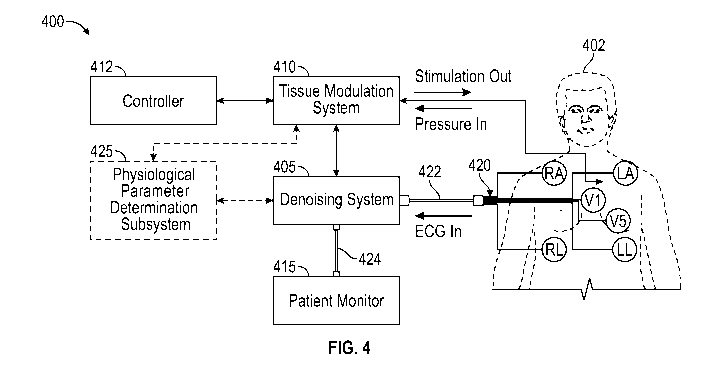
denoising an
ECG signal when electrical stimulation is being applied by a stimulation
system.
-16-
CA 03137307 2021-10-18
WO 2020/227234
PCT/US2020/031358
[0049] Figure
7A schematically illustrates a bypass signal path and examples
stages of the denoising system.
[0050] Figure
7B schematically illustrates example components of the
denoising system.
[0051] Figure
8A schematically illustrates an example ECG waveform
uncorrupted by application of neurostimulation.
[0052] Figure
8B schematically illustrates an example ECG waveform that is
corrupted by application of stimulation to a portion of a body of a living
subject.
[0053] Figure
8C schematically illustrates the example ECG waveform of
Figure 8B after blanking performed by the denoising system.
[0054] Figure
8D schematically illustrates the example ECG waveform of
Figure 8C after interpolating and optional refining performed by the denoising
system.
[0055] Figures
9A and 9B illustrate close-up exploded views of portions of the
corresponding ECG waveforms of Figures 8A and 8B, respectively.
[0056] Figures
9C and 9D schematically illustrate the portion of the example
ECG waveform of Figure 9B after blanking (Figure 9C) and interpolating and
optional
refining (Figure 9D) performed by the denoising system.
[0057] Figures
10A and 10B illustrate a normal sinus rhythm ECG waveform
during application of neurostimulation without denoising and with denoising,
respectively.
[0058] Figures
11A and 11B illustrate an ECG waveform indicative of
bigeminy during application of neurostimulation without denoising and with
denoising,
respectively.
[0059] Figures
12A and 12B illustrate an ECG waveform indicative of atrial
fibrillation during application of neurostimulation without denoising and with
denoising,
respectively.
[0060] Figures
13A and 13B illustrate an ECG waveform indicative of
ventricular fibrillation during application of neurostimulation without
denoising and with
denoising, respectively.
[0061] Figure
14 is a front view of an example tissue modulation system (e.g.,
neurostimulation system).
DETAILED DESCRIPTION
[0062] Patient
monitors are used to provide feedback to clinicians in hospitals,
nursing homes and other patient care facilities regarding real-time patient
health. The
-17-
CA 03137307 2021-10-18
WO 2020/227234
PCT/US2020/031358
patient monitoring devices are configured to display output relating to real-
time
physiological parameters or vital signs of the patient. Clinical professionals
monitor the
display output to determine the current status of the patient's health and
possibly increase
the level of medical care given to the patient based on the current status.
The clinical
professionals may diagnose patient conditions or illnesses or prescribe
treatments based on
the monitored physiological parameters, biopotentials, or vital signs. In
addition to visual
display of textual, numerical, or graphical information or data corresponding
to the
physiological parameters, biopotentials, or vital signs, the patient monitors
may also be
configured to generate visual or audible output (e.g., alerts or alarm events)
if a particular
physiological parameter, biopotential, or vital sign being monitored falls
outside a
threshold range (e.g., safety limits) to alert the clinical professionals of
an unsafe condition
that may require medical assistance or attention. Accordingly, it can be
advantageous to
make sure that the physiological parameters (or the output indicative of the
physiological
parameters) that are displayed and monitored by the patient monitors are
accurate and
reliable to reduce alarm fatigue and ensure accurate diagnosis.
[0063]
Physiological parameters can include heart rate, blood pressure,
temperature, or the like. One example of a physiological parameter that is
commonly
monitored and displayed on patient monitors is heartbeat. The heartbeat may be
displayed
on a patient monitor as an electrocardiograph, or electrocardiogram, waveform
(ECG or
EKG) that is indicative of the electrical activity in the heart. The ECG
waveform can be
monitored by clinical professionals to determine whether any deviations or
abnormalities
occur that may be indicative of an unsafe and potentially life-threatening
condition (e.g.,
atrial fibrillation, ventricular tachycardia, heart disease, cardiac arrest)
that may require
immediate medical attention or therapeutic treatment. The ECG waveform can be
used to
evaluate heart rate, rhythm, and other cardiac abnormalities and to make
diagnoses.
[0064]
Accordingly, it is desirable that the ECG waveform that is displayed on
the patient monitor is clean and uncorrupted so as not to generate false
alarms or prompt
medical treatment that is not warranted and may cause harm to the patient. In
addition,
ECG waveforms corrupted with noise may cause a practitioner to miss an
abnormal event
or occurrence (e.g., arrhythmia) and withhold therapy that should not have
been withheld.
The ECG waveform is derived from signals or measurements from multiple sensors
(e.g.,
electrodes and/or leads or leadwires) positioned on a skin of a patient at
various locations
on a patient's body. The sensors transmit the heart's electrical activity to
an ECG
processing device or system. The ECG processing device or system generates a
waveform
-18-
CA 03137307 2021-10-18
WO 2020/227234
PCT/US2020/031358
or other output representative of the heart's beats and electrical activity
for display (e.g.,
on a display of a patient monitor).
[0065]
Unfortunately, the presence of electromagnetic energy generated from
other electromagnetic energy sources in the vicinity of the ECG sensors can
cause
unwanted interference or noise to appear on the ECG waveform displayed on the
patient
monitor, especially if the frequency content of the ECG waveform (e.g.,
electrical signals
generated by the heart) and the other interfering source overlap. This
interference or noise
can cause the clinical professionals, who are trained to be wary of any
abnormalities on the
ECG signal, to be alarmed and can render the ECG signals difficult or
impossible to read,
decipher, or interpret. In addition, the interference or noise may cause
automated "false"
arrhythmia alarms or alerts to be generated because the interference or noise
may cause the
parameters being monitored to fall outside of a normal, expected condition or
threshold
range (e.g., safety limits). The increase in false alarms may result in alarm
fatigue.
[0066] One
source of temporary, transient, or transitory, interference or noise
can include a tissue modulation system configured to provide electrical
modulation (e.g.,
electrical stimulation, electrical ablation, electrical denervation) to one or
more nerves in
and around a heart of a patient or to one or more nerves in and around vessels
surrounding
the heart (e.g., pulmonary arteries, pulmonary veins) to treat patients with
acute
decompensated heart failure. Catheters having stimulating elements (e.g.,
stimulatory
electrodes) may be temporarily inserted into, or positioned externally
adjacent, vessels
surrounding the heart or chambers of the heart to deliver electrical
stimulation (e.g.,
electrical current or electrical pulses) to stimulate nerves (e.g., autonomic
nerve fibers
surrounding a pulmonary artery). These catheters may also cause interference
or noise (e.g.,
stimulation artifact) to appear on the ECG waveform when stimulation is being
applied by
the stimulating elements of the catheters. The degree of interference varies
depending on
the location of the stimulation electrodes and the characteristics of the
stimulation
waveform. As another example, pacemakers or other implantable stimulators
implanted
near the heart may cause interference (e.g., stimulation artifact) on a
display of the ECG
waveform when stimulation (e.g., electrical current or electrical pulses) is
being applied.
[0067] In
addition to being used in connection with neuromodulation (e.g.,
neurostimulation) systems for treatment of patients with acute decompensated
heart failure,
other applications can also benefit from several of the denoising techniques
and systems
described herein. For example, the denoising techniques and systems described
herein may
be used in conjunction with systems adapted to perform any one or more of the
following:
-19-
CA 03137307 2021-10-18
WO 2020/227234
PCT/US2020/031358
spinal neuromodulation, pacing with a pacemaker, defibrillation with an
implantable
defibrillator or external defibrillation system, pulsed electrocautery,
stimulation of nerves
to treat urinary or fecal incontinence, muscle stimulation, prostate
stimulation, brain or
other central or peripheral neurological stimulation, stimulation of the vagus
nerve,
stimulation of osteoblasts, joint stimulation therapy to treat orthopedic
conditions,
iontophoresis, stimulation to determine tissue contact, imaging,
electroanatomical mapping
or electrophysiology recordings, ergometry, etc.
[0068] Figure 1
schematically illustrates an example tissue modulation system
100 that can be used to apply electrical modulation to tissue (e.g., including
one or more
nerves) in and around the heart of a subject that may generate interference or
noise on an
ECG waveform while the electrical modulation is being applied. In one
implementation,
the tissue modulation system 100 comprises a cardio pulmonary nerve
stimulation (CPNS)
system that is intended to treat patients in acute decompensated heart
failure. The CPNS
system can cause electrical interference (e.g., stimulation artifact) to
appear on
biopotentials, such as ECG waveforms on a display that is being monitored by a
clinician
or practitioner).
[0069] In some
implementations, the location of the electrodes of the CPNS
system are intended to be near the heart, which is also the source of the ECG
signals, and
the stimulation waveform of the CPNS system has frequency components that
overlap that
of the ECG signals. Accordingly, the presence of the stimulation artifact on
the ECG signals
caused by the CPNS system makes the ECG waveform difficult to accurately
interpret by
trained practitioners and renders automatic arrhythmia detection functions on
ECG patient
monitors ineffective.
[0070] The
tissue modulation system 100 may be configured to deliver nerve
stimulation as either continuous or intermittent biphasic pulse trains through
a catheter
placed temporarily in the upper thorax near the heart. Patients receiving
therapy are
typically treated in intensive care units (ICUs) or cardiac care units (CCUs)
within hospitals
and are kept on continuous surface electrocardiogram (ECG) monitoring for up
to five days.
[0071] In
accordance with several implementations, dealing with CPNS-
generated interference is more challenging than with other neurostimulator
devices for
several reasons: (1) the neurostimulator provides therapy, and as such, can't
be turned off
during patient monitoring, (2) the neurostimulation therapy can be delivered
for a long
continuous duration of time (e.g., up to five days), while requiring patient
monitoring the
entire time, (3) the CPNS electrodes can be directly in line with the ECG
vectors, causing
-20-
CA 03137307 2021-10-18
WO 2020/227234
PCT/US2020/031358
a large amplitude interference artifact, and/or (4) the interference frequency
spectrum
typically overlaps the ECG frequency spectrum, thereby precluding the use of
ECG
instrument filters.
[0072] The
system 100 comprises a first component 102 and a second
component 104. The first component 102 may be positioned in a pulmonary artery
(e.g.,
the right pulmonary artery as shown in Figure 1, the left pulmonary artery,
and/or the
pulmonary trunk). The first component 102 may be endovascularly positioned via
a
minimally invasive, transdermal, percutaneous procedure, for example routed
through the
vasculature from a remote location such as a jugular vein (e.g., an internal
jugular vein, as
shown in Figure 1), an axial subclavian vein, a femoral vein, or other blood
vessels. Such
an approach can be over-the-wire, using a Swan-Ganz float catheter,
combinations thereof,
etc. In some examples, the first component may be positioned invasively, for
example
during conventional surgery (e.g., open-heart surgery), placement of another
device (e.g.,
coronary bypass, pacemaker, defibrillator, etc.), or as a stand-alone
procedure. As described
in further detail herein, the first component comprises a neuromodulator
(e.g., electrode,
transducer, drug, ablation device, ultrasound, microwave, laser, cryotherapy,
combinations
thereof, and the like) and may optionally comprise a stent or framework, an
anchoring
system, and/or other components. The first component 102 may be acutely
positioned in
the pulmonary artery for 24 to 120 hours. In some examples, the first
component 102
neuromodulates terminal branches within the cardiac plexus, which can increase
left and/or
right ventricle contractility and/or relaxation. The increase in ventricular
cardiac output due
to the contractility increase may occur without a corresponding increase in
heart rate, or
may be greater than (e.g., based on a percentage change) that due to an
increase in heart
rate alone. In some examples, the first component 102 may be adapted to ablate
tissue,
including nerves, in addition to or instead of stimulating tissue, such as
nerves.
[0073] The
first component 102 is electrically coupled to the second component
104 (e.g., via wires or conductive elements routed via a catheter, for example
as illustrated
in Figure 1, and/or wirelessly). The second component 104 may be positioned
extracorporeally (e.g., strapped to a subject's arm as shown in Figure 1,
strapped to another
part of the subject (e.g., leg, neck, chest), placed on a bedside stand,
etc.). In some
examples, the second component 104 may be temporarily implanted in the subject
(e.g., in
a blood vessel, in another body cavity, in a chest, etc.). The second
component 104 includes
electronics (e.g., pulse generator) configured to operate the electrode in the
first component
102. The second component 104 may include a power supply or may receive power
from
-21-
CA 03137307 2021-10-18
WO 2020/227234
PCT/US2020/031358
an external source (e.g., a wall plug, a separate battery, etc.). The second
component 104
may include electronics configured to receive sensor data.
[0074] The
system 100 may comprise one or more sensors (e.g., pressure
sensor). The sensor(s) may be positioned in one or more of a pulmonary artery
(e.g., right
pulmonary artery, left pulmonary artery, and/or pulmonary trunk), an atrium
(e.g., right
and/or left), a ventricle (e.g., right and/or left), a vena cava (e.g.,
superior vena cava and/or
inferior vena cava), and/or other cardiovascular locations. The sensor(s) may
be part of the
first component 102, part of a catheter, and/or separate from the first
component 102 (e.g.,
electrocardiogram chest monitor, pulse oximeter, etc.). The sensor(s) may be
in
communication with the second component 104 (e.g., wired and/or wireless). The
second
component 104 may initiate, adjust, calibrate, cease, etc. neuromodulation
based on
information from the sensor(s). Measurements obtained from the sensor(s)
(e.g., pressure
sensors) may be used to determine whether a patient condition is within a
"safe" or
acceptable range within which stimulation (and denoising processes) may be
applied.
Otherwise, stimulation may be halted and the denoising systems may be bypassed
to
increase patient safety and reduce processing times and complexity.
[0075] The
system 100 may comprise an "all-in-one" system in which the first
component 102 is integral or monolithic with the targeting catheter. For
example, the first
component 102 may be part of a catheter that is inserted into an internal
jugular vein, an
axial subclavian vein, a femoral vein, etc. and navigated to a target location
such as the
pulmonary artery. The first component 102 may then be deployed from the
catheter.
[0076] The
system 100 may comprise a telescoping and/or over-the-wire
system in which the first component 102 is different than the targeting
catheter. For
example, a targeting catheter (e.g., a Swan-Ganz catheter) may be inserted
into an internal
jugular vein, an axial subclavian vein, a femoral vein, etc. and navigated to
a target location
such as the pulmonary artery (e.g., by floating). A guidewire may be inserted
into a
proximal hub through the target catheter to the target location (e.g., having
a stiffest portion
exiting the target catheter distal end) and the first component 102 as part of
a separate
catheter than the target catheter may be tracked to the target location over
the guidewire or
using telescoping systems such as other guidewires, guide catheters, etc. The
first
component 102 may then be deployed from the separate catheter. Such systems
are known
by interventional cardiologists such that multiple exchanges may be of little
issue. Such a
system may allow customization of certain specific functions. Such a system
may reduce
overall catheter diameters, which can increase trackability, and/or allow
additional features
-22-
CA 03137307 2021-10-18
WO 2020/227234
PCT/US2020/031358
to be added, for example because not all functions are integrated into one
catheter. Such a
system may allow use of multiple catheters (e.g., removing a first separate
catheter and
positioning a second separate catheter without having to reposition the entire
system). For
example, catheters with different types of sensors may be positioned and
removed as
desired. The system 100 may be steerable (e.g., comprising a steerable
catheter) without a
Swan-Ganz tip. Some systems 100 may be compatible with one or more of the
described
types of systems (e.g., a steerable catheter with an optionally inflatable
balloon for Swan-
Ganz float, a steerable catheter that can be telescoped over a guidewire
and/or through a
catheter, etc.).
[0077] Figure 2
schematically illustrates a portion of an example
electrocardiograph, or electrocardiogram, (ECG or EKG) waveform. The ECG
waveform
includes P waves, Q waves, R waves, S waves, and T waves, which are each
indicative of
different events during a single heartbeat of a healthy subject (e.g.,
patient). The P wave
represents atrial depolarization, which causes the left atrium and the right
atrium to push
blood into the left ventricle and right ventricle, respectively. The flat
period until the Q
wave, the "PR Segment," and the start of the P wave to the start of the Q wave
is the "PR
Interval." The Q wave, the R wave, and the S wave, together the "QRS Complex,"
represent
ventricular depolarization, which causes the right ventricle to push blood
into the
pulmonary artery and towards the lungs and which causes the left ventricle to
push blood
into the aorta for distribution to the body. The T wave represents
repolarization of the left
and right ventricles. The flat period until the T wave is the "ST Segment"
during which the
ventricles are depolarized, and collectively the QRS Complex, the ST Segment,
and the T
wave are the "QT Interval." The duration between successive R waves or peaks
is the "R-
R interval." Some ECGs also have a U wave after the T wave. The timing,
amplitude,
relative amplitude, etc. of the various waves, segments, intervals, and
complexes can be
used to diagnose various conditions of the heart.
[0078]
Electrical modulation (e.g., stimulation) from a tissue modulation
system (such as the systems described herein) or from other electromagnetic
energy
generating systems or devices (e.g., radiofrequency energy delivery systems,
ultrasound
energy delivery systems, microwave energy delivery systems, laser devices,
implantable
stimulators, transcutaneous electrical stimulation devices, pacemakers,
defibrillators,
imaging devices, lighting equipment, electrophysiology recording or mapping
devices)
located in a region near one or more of the ECG leads or leadwires may
interfere with (e.g.,
cause distortion of, or noise to appear on) a display of a "clean" true ECG
waveform.
-23-
CA 03137307 2021-10-18
WO 2020/227234
PCT/US2020/031358
Figure 3A illustrates an example portion of a clean ECG waveform when no
electrical
stimulation is being applied by a stimulation system, such that no
interference or noise (e.g.,
stimulation artifact) is present on the displayed waveform. Figure 3B
illustrates an example
portion of the ECG waveform when electrical stimulation is being applied by a
stimulation
system. As can be seen in Figure 3B, the interference or noise created by the
stimulation
source makes it difficult to distinguish the normal ECG waveform (e.g., the
features
indicative of the heart beats) and the noise or interference (e.g.,
stimulation artifact). In
some examples, the portion of the ECG waveform may be modified to account for
(e.g.,
remove, filter out, suppress, cancel out) such interference or noise (e.g.,
stimulation artifact)
and display a true, or accurate, portion of the ECG waveform (e.g., high-
fidelity waveform
without compromising original morphology) even while electrical modulation
(e.g.,
electrical stimulation) is being applied to the patient (e.g., by a
neurostimulation system).
[0079] The ECG
waveform (e.g., one or more portions of the ECG waveform)
could be artificially flat-lined or ignored during periods of stimulation and
the clinical
professionals could rely on alternative physiological parameters or vital
signs to ensure
patient safety during periods of stimulation. However, many clinical
professionals may not
be comfortable with periods of time in which the true ECG waveform is not
being
accurately displayed. In addition, as mentioned previously, the periods of
artificial flat-
lining or "blanking" may cause false alarms to be generated, causing
unnecessary worry or
stress to the patient or clinicians, or even prompting spontaneous,
unwarranted medical
action that results in harm, or even death, to the patient. Accordingly,
several
implementations described herein denoise the ECG waveform by modifying or
replacing
ECG waveform values at certain time instances instead of zeroing the values
out or
removing the values at those time instances without replacing them with
alternative values.
Accordingly, the data sets before and after denoising may be the same size.
[0080] Figure 4
schematically illustrates an example treatment and patient
monitoring system 400 that includes a denoising system 405 configured to
advantageously
remove (e.g., filter out) the unwanted noise or interference (e.g.,
stimulation artifact)
generated by a stimulation system 410 while preserving the fidelity and
morphology of the
ECG waveform that is displayed on a patient monitor or other display device
415. The
treatment and patient monitoring system 400 includes a controller or control
unit 412
configured to control therapy delivery by the stimulation system 410 to a
living subject
(e.g., patient) 402. The controller or control unit 412 may comprise a
computing device
(e.g., computer, laptop, tablet, smartphone) that includes one or more
processing devices
-24-
CA 03137307 2021-10-18
WO 2020/227234
PCT/US2020/031358
(e.g., microcontrollers) and circuitry configured to execute one or more
stored programs or
algorithms (e.g., to generate electrical stimulation pulses of desired
patterns and durations).
The control unit 412 may include a touchscreen display configured to allow a
user to
provide user input by interacting with graphical user interfaces displayed on
the screen.
The display may also be configured to display stimulation system data and
stimulation
therapy data received from one or more sensors.
[0081] The
stimulation system 410 may comprise, for example, the
neurostimulation systems including catheters with electrode structures and the
like as
described herein. Other tissue modulation systems, including for other
indications other
than treatment of heart failure, are also possible. For example, the denoising
techniques and
systems described herein may be used in conjunction with systems adapted to
perform any
one or more of the following: spinal neuromodulation, pacing with a pacemaker,
defibrillation with an implantable defibrillator or external defibrillation
system, pulsed
electrocautery, stimulation of nerves to treat urinary or fecal incontinence,
muscle
stimulation, prostate stimulation, brain stimulation, stimulation of the vagus
nerve,
stimulation of osteoblasts, joint stimulation therapy to treat orthopedic
conditions,
iontophoresis, stimulation for tissue contact sensing, electroanatomical
mapping,
electrophysiology recording, etc. The denoising techniques and systems may
also be used
on conjunction with systems or devices employing motors, pumps, piezoelectric
actuators,
and/or the like. Interference sources that are synchronizable or periodic may
be filterable
or denoised using the techniques and systems described herein.
[0082] The
stimulation system 410 may be configured to generate a
programmable stimulation waveform to be applied to nerves of the subject 402
via one or
more electrodes or other stimulation elements. The stimulation system 410 may
optionally
also include sensors (e.g., sensors on a catheter) to sense pressure (e.g.,
pulmonary artery
pressure and right ventricle pressure) and receive signals indicative of the
sensed pressure
(as shown schematically in Figure 4). The system 410 may also include sensors
that directly
sense electrical activity, such as cardiac electrograms or nerve activity. As
discussed herein,
application of electrical stimulation to a subject 402 can affect an ECG
reading of the
subject 402. The subject 402 is also connected to leads or leadwires of an ECG
system 420
according to standard operation procedure to measure the rate and rhythm of
heartbeats.
Sometimes, an ECG amplifier (not shown) may optionally be used to amplify
input signals
from the ECG system 420.
-25-
CA 03137307 2021-10-18
WO 2020/227234
PCT/US2020/031358
[0083] The
system 400 shown in Figure 4 includes a denoising system 405
between the ECG system inputs 420 (e.g., electrodes, leads, leadwires, and/or
processing
hub) and the patient monitor 415. Instead of the patient monitor lead wire set
422 of the
ECG system inputs 420 connecting directly to the patient 402, the ECG inputs
420 (e.g.,
two-channel 24-bit 800 Hz ECG system inputs converted to a 32-b it unsigned
integer raw
ECG) are connected to the inputs of the denoising system 415 through the
patient monitor
lead wire set 422 and the patient monitor 415 then connects to the lead wire
set 424 output
of the denoising system 405. The denoising system 405 is configured to capture
and
manipulate data from the ECG system inputs 420 prior to sending such data to
an optional
ECG amplifier or to the patient monitor 415 for display. In some
implementations, the
denoising system 405 comprises one or more filters (e.g., 50 Hz notch filter,
60 Hz notch
filter, 50 Hz ¨ 60 Hz notch filter, Butterworth notch filter, adaptive filters
using
microcontrollers) to pre-filter typical 50 Hz line and/or 60 Hz line or 50 Hz
¨ 60 Hz or
other line frequency component noise (and possibly harmonics) out of the ECG
signals
received from the ECG system inputs 420 or other analog front-end ECG system
prior to
modification (e.g., reconstruction, interpolation) during the denoising
process so that values
just prior to and after blanking and/or interpolation are known good values
that do not
include 50 Hz and/or 60 Hz or 50 Hz ¨ 60 Hz noise frequency components (e.g.,
50 Hz
and/or 60 Hz or 50 Hz ¨ 60 Hz or other line artifacts or spikes), thereby
further enhancing
signal fidelity. The one or more filters may include a bandpass filter (e.g.,
linear phase
finite-impulse-response filter with a 3 dB cutoff of 0.05 to 40 Hz converted
to signed 16-
bit integers with a dynamic range of 6.25 mV). The denoising system 405 can
execute
stored instructions or algorithms using one or more processors (e.g., computer
circuitry or
computing circuits) to perform a process or set of processing steps for
denoising the ECG
signals or the waveform generated and output for display. The denoising system
405 may
include two signal outputs, one analog and one digital. The analog output
provides denoised
ECG signals to the patient monitor 415 for display. The analog output may have
a unity
gain or other gain values. The digital output provides a denoised ECG signal
to the
neuromodulation system 405. The denoising system 405 may also detect the QRS
complex
of the ECG and pass a corresponding marker to the neuromodulation system 410
for heart
rate computation, stimulation synchronization, and/or other functions. The
patient monitor
lead wire set 422 could be replaced with other types of lead sets connected to
other display
and/or processing devices besides patient monitor 415, such as a central
monitoring system.
-26-
CA 03137307 2021-10-18
WO 2020/227234
PCT/US2020/031358
[0084] The
denoising system 405 can process multiple ECG input channels
(e.g., 2, 3, 4, 5, 6, 7, 8, 9, 10 or more than 10 channels) and can support
multiple ECG
configurations. Not all channels are required to be used. In some examples,
the stimulation
system 410 or the ECG system 420 may comprise the denoising system 405 (e.g.,
the
denoising system 405 may be a component or subsystem of the stimulation system
410 or
the ECG system 420). In some implementations, the denoising system 405 is a
separate,
stand-alone component, or module, from the stimulation system 410 or the ECG
system
420. The denoising system 405 can inhibit or prevent a neurostimulation
waveform and/or
the effects of neurostimulation on an ECG signal from corrupting an ECG
signal, or
portions thereof.
[0085] The
denoising system 405 may receive stimulation timing information
or data (e.g., time during which stimulation is applied) from the
neuromodulation system
410 to facilitate the denoising process(es) (e.g., facilitate detection or
identification of
portions of the ECG signal or other physiological signal or biopotential that
comprise noise
or stimulation artifacts or are likely to comprise noise, such as stimulation
artifact). In some
implementations, the noise, or stimulation artifact, caused by an electrical
stimulation
system, for example, comprises periodic pulsatory type noise as opposed to
random or
continuous types of noise. The stimulation timing information may comprise a
blanking
pulse signal (e.g., synchronization pulse signal) 502 that is transmitted to
the denoising
system 405 coincident with the initiation of electrical stimulation pulses.
The pulses of the
blanking pulse signal 502 may advantageously be synchronized with the pulses
of the
stimulation pulse signal 504, as shown schematically in Figure 5A. The first
pulse of the
blanking pulse signal 502 may indicate to the denoising system 405 that
stimulation is about
to begin, or is beginning, and therefore that the portions of the ECG signal
corresponding
to the time after the blanking pulse is received are likely to have temporary,
or transitory,
noise or interference (e.g., stimulation artifact) caused by application of
electrical
stimulation or other electromagnetic energy or fields and that those portions
should be
denoised by the denoising system 405. The blanking pulse may be continuously
delivered
(e.g., in a state indicative of an "on" or "active" condition) for the
duration of the electrical
stimulation (e.g., duration of each pulse cycle occurring during an electrical
stimulation
treatment period, which could last, for example, for several minutes, several
hours, several
days, or several weeks).
[0086] The
pulses of the blanking pulse signal 502 may be used to identify, or
detect, both the beginning and duration of the pulses of the stimulation pulse
signal 504.
-27-
CA 03137307 2021-10-18
WO 2020/227234
PCT/US2020/031358
This is true in principle but may require modifications in actual use. For
example, it may
be necessary in some implementations for the timing of the leading edge and
trailing edge
of the blanking pulses to be adjusted due to perturbations that lead or follow
the stimulation
artifact on the ECG waveform. Figure 5B shows a blanking pulse signal 502 with
modified
leading and trailing edges. In several implementations, the leading edge of
the first "on"
pulse of the blanking pulse signal 502' is programmed or configured to occur a
short time
prior to the start of the first "on" pulse of the stimulation pulse signal 504
to allow for data
acquisition and processing time inherent in the ECG signal path of the
denoising system
405 and blanking pulse signal path. This leading offset value may be fixed as
determined
by the specifics of the internal denoising system 405 signal delays or
adjustable to
accommodate other encountered delay variables. For example, various
implementations of
the denoising system 405 and/or process 600, 605 may function best when the
leading edges
of the blanking pulses precede the leading edges of the stimulation pulses by
a short period
of time (e.g., 1-10 ms, 1- 5 ms, 6- 10 ms, 1 ms, 2, ms, 3 ms, 4 ms, 5 ms, 6
ms, 7 ms, 8 ms,
9 ms, 10 ms).
[0087] The
trailing edge of the blanking pulse signal 502' may also
advantageously extend beyond the trailing edge of the stimulation pulse signal
504 to
account for distortion that may occur to the stimulation artifact as it
appears on the ECG
waveform. The electrical transfer function through the body between the
implanted
stimulation electrodes and the surface ECG electrodes can be complex,
resulting in not only
amplitude changes but in time delays of the stimulation artifact on the ECG
waveform (e.g.,
spikes caused by stimulation) with respect to the stimulation pulses. In some
implementations, the entire stimulation artifact is delayed (including
initiation of the
artifact and the ending of the artifact). The time delay at the end of the
artifact causes the
artifact trailing edge to lag the stimulation pulse trailing edge by a small
amount. If the
pulse width of the synchronization pulses (e.g., blanking pulses) is not
extended to
compensate for this effect, the entire artifact may not be blanked during the
denoising
process, thereby allowing some of the artifact spike to "leak" through. As
with the
synchronization pulse (e.g., blanking pulse) leading edge, the trailing edge
offset may use
a fixed value or be adjustable to accommodate variations in artifact
distortion. The
adjustable synchronization pulse (e.g., blanking pulse) leading or trailing
edges may be
implemented either as manually controlled functions by a user (e.g., based on
artifact
observed in the baseline of the denoised ECG signal) or determined
automatically by
methods or algorithms within the denoising system 405 or process 600, 605. In
accordance
-28-
CA 03137307 2021-10-18
WO 2020/227234
PCT/US2020/031358
with several implementations, because the denoising system 405 receives an
indication in
advance as to when stimulation is occurring as a result of receipt of the
stimulation timing
information (e.g., synchronization pulse), the denoising system 405 and
processes 600, 605
can advantageously be deterministic and require less processing speed and/or
computing
resources. There may also be reduced signal latency compared to prior methods.
The
denoising processes and methods may be performed in real time (e.g., with
minimal latency
of less than 100 ms) such that the clinical professionals do not even realize
that the
denoising is being performed. In addition, the denoising system 405 and
processes 600, 605
may advantageously not require linear circuit operation during noise artifact
periods which
makes it tolerant to signal saturation at those times.
[0088] The
system 400 shown in Figure 4 may further optionally comprise a
physiological parameter determination subsystem or module. The physiological
parameter
determination subsystem or module may be an independent subsystem or module or
may
be a sub-component of the neuromodulation system 410 or the denoising system
405. The
physiological parameter determination subsystem or module may be configured
to, upon
execution of instructions stored on a computer-readable medium by one or more
processors,
determine whether a physiological parameter being monitored (e.g., heart rate
or other
cardiac-related parameter, pressure within a cardiac-related vessel or
chamber, such as a
pulmonary artery or a ventricle) is outside of a threshold range. The
threshold range may
correspond to a predetermined acceptable safe range that does not cause alarm
or require
medical attention. The physiological parameter value may be determined based
on signals
received from one or more sensors coupled to tissue of a patient (e.g., R-R
intervals
determined from signals received from ECG leads coupled to skin of a patient)
or
positioned within a lumen or cavity of a patient (e.g., pressure values
determined from
pressure sensors positioned within a blood vessel or a heart chamber).
[0089] If the
physiological parameter determination subsystem or module
determines that the physiological parameter is outside the threshold range,
the
physiological determination subsystem or module may cause the neuromodulation
system
410 to stop, or terminate, application of neuromodulation to the patient and
may cause the
denoising system 410 to be bypassed so as not to affect the ECG signals that
no longer
require denoising since the neuromodulation has been terminated. In some
implementations, the physiological determination subsystem or module generates
a control
signal that is sent to the neuromodulation system 410 and/or to the denoising
system 410.
The physiological parameter determination subsystem or module may also
comprise an
-29-
CA 03137307 2021-10-18
WO 2020/227234
PCT/US2020/031358
alert generation subsystem or module configured to generate an alert or alarm
event when
the physiological parameter is determined to be out of range. The alert may be
a visual
alarm output to a display (e.g., on a patient monitor or on a display of a
central monitoring
system in a patient care facility). The alert may additionally or
alternatively comprise an
audible alert or alarm. The alerts may generate a text message, electronic
mail message,
page, or other warning message to a display of a central monitoring system of
a health care
facility or to a mobile communications device (e.g., pager, smartphone) of one
or more
individual caregivers. The alerts may be transmitted through wired connections
or
wirelessly (e.g., via Bluetooth or cellular data communication protocols or
systems over a
communications network). If the physiological parameter determination
subsystem or
module determines that the physiological parameter is within the threshold
range, no action
is taken and the neuromodulation system 410 and the denoising system 405 may
continue
to operate as normal.
[0090] With
reference to Figure 6, an example of an algorithm or process 600
of denoising ECG signals or waveforms performed by the denoising system 405 is
schematically illustrated. The denoising system 405 can execute stored
instructions or
algorithms using one or more processors (e.g., computer circuitry or computing
circuits) to
perform the process 600. At Block 602, the various ECG signals are received
directly from
the ECG sensors, or from the ECG system inputs 420. At decision Block 604, the
denoising
system 405 determines whether stimulation or other electromagnetic energy is
currently
being applied. In accordance with several implementations, the process 600
advantageously
does not apply filtering (e.g., blanking and modifying (e.g., reconstructing,
interpolating)
to the entire ECG waveform. Instead, the process 600 applies filtering only to
portions of
the ECG waveform likely to have stimulation artifacts, thereby leaving those
portions that
are free (or substantially free) of stimulation artifact unaffected and
unmodified. In some
examples, the denoising system 405 can receive a signal from the
neuromodulation system
410 when the neuromodulation system 410 is applying neurostimulation through a
physical
electrical connection (e.g., the blanking pulse signal 502 described above).
The denoising
system 405 may be configured to correlate the time of receipt of the signal
indicative of
application of neurostimulation therapy or other modulation or electromagnetic
energy with
the timing of the ECG signals so as to know which portions of the ECG signals
comprise,
or are likely to comprise, noise or interference (e.g., stimulation
artifacts).
[0091] In some
implementations, a synchronization blanking pulse signal may
not be used and the determination of whether stimulation is being applied is
independently
-30-
CA 03137307 2021-10-18
WO 2020/227234
PCT/US2020/031358
determined by the denoising system 405 based on an analysis of the ECG
waveform to
predict or determine whether stimulation is being applied and/or to generate a
synchronization (e.g., blanking) pulse signal, or signal indicative of
stimulation being
applied. The synchronization (e.g., blanking) pulse signal can be generated
from the
stimulation-corrupted ECG signal directly, thereby eliminating the need for a
separate
synchronization pulse (e.g., blanking pulse). Because the stimulation signal,
and hence the
artifact, are periodic, they can be extracted from the original ECG signal
using one or more
clock extraction techniques, such as autocorrelation, or by using a phased
locked loop
(PLL). Additional methods can be employed to determine the correct blanking
pulse width
and the optimal phase relationship to use with the stimulation signal. Once
these values are
determined they can be saved and quickly reapplied for successive stimulation
pulses. If
the stimulation parameters change, the denoising system 405 can once again
determine the
correct denoising parameters to apply. This "extracted blanking pulse"
technique could be
useful, for example, in circumstances where a denoising function is applied to
a system not
originally designed to provide a synchronized blanking pulse signal.
[0092] As
another example, a peak detector (e.g., 20 Hz peak detector) could
be used in combination with other methods or techniques to detect the presence
of noise
caused by the neuromodulation system 410 as an indicator of whether electrical
stimulation
or other modulation is being applied at the current time. In various
implementations,
different wireless synch (including optical links), wired synch or synch
generation
techniques may be used. For example, a wireless connection such as Bluetooth
or a number
of other means can also be used in place of a physical electrical connection.
[0093] If it is
determined at decision Block 604 that stimulation is being
applied, then a denoising sub-process 605 is initiated by the denoising system
405. For
example, the blanking pulse signal 502 from the neuromodulation system 410 can
open a
circuit to interrupt the direct connection between the ECG system 420 and the
patient
monitor 415 and instead direct the original corrupted ECG signal to the
denoising system
405. If it is determined at decision block 604 that stimulation is not being
applied, then the
ECG waveform can be output for display at Block 610 as normal without going
through
the denoising sub-process 605. When no signal indicative of stimulation being
applied is
received from the neuromodulation system 410, the circuit between the ECG
system 420
and the ECG amplifier 425 can be re-closed and the denoising system 405 may be
bypassed.
In other words, the ECG signals are not directed through the denoising system
405 and are
processed without going through the processing of the denoising system 405.
The denoising
-31-
CA 03137307 2021-10-18
WO 2020/227234
PCT/US2020/031358
system 405 can include multiple switches that can be triggered (change the
state between
open and closed) depending on the determination of whether or not stimulation
is being
applied (and thus, whether the denoising system 405 should be bypassed or
not). For
example, when the ECG input signals are bypassing the denoising stages,
normally closed
switches between each ECG channel input and corresponding channel output can
be used
to pass the ECG signals directly to the output of the denoising system 405
without any
filtering or signal modification. When the denoising sub-process 605 is
active, the state of
the switches may be changed to direct the ECG input signals to the denoising
circuitry
stages.
[0094] In
implementations where a blanking or other synchronization pulse
signal is generated, the denoising process 500 may be treated as a system-
level operation
because the neuromodulation system 410 not only generates a stimulation
therapy signal
but also generates the synchronization pulse signal that facilitates the
denoising 600 (e.g.,
denoising sub-process 605). Turning briefly to Figure 7A, an example of a
bypass path in
which at least the major stages of the denoising sub-process 605 (e.g., ECG
analog front-
end stage, which may include an analog-to-digital-converter and/or amplifier,
a
microcontroller that performs the denoising sub-process, and an analog out
stage, which
may include a digital-to-analog converter and/or amplifier) are bypassed.
[0095] Turning
back to Figure 6, the denoising sub-process 605 may include
multiple processing steps. At Block 606, the portions of the ECG waveform or
signals that
are likely to include artifact spikes or interference noise during stimulation
(as determined
by, or as detected or identified based on the stimulation timing information
or data received
from the neuromodulation system 410) may be "blanked" from the ECG waveform or
signals (e.g., during a determined blanking period or window). The components
of the
denoising sub-process may be performed on all of the portions or components of
the ECG
waveform or signals identified or detected as having noise (e.g., during the
duration of each
stimulation or other modulation pulse cycle) at the same time (e.g.,
simultaneously) or may
be performed on each portion separately (e.g., sequentially).
[0096] The
denoising sub-process 605 may be performed for the entire length
of stimulation (e.g., the whole time the blanking pulse is in a state
indicative of stimulation
being "on" or "active") or for a portion of stimulation. The denoising sub-
process 605 may
or may not be performed during the time (either all or a portion of the time)
of the blanking
pulse that occurs prior to actual stimulation. For example, if the first
blanking pulse is
received 4 ms prior to actual stimulation, the denoising sub-process 605 may
start after 4
-32-
CA 03137307 2021-10-18
WO 2020/227234
PCT/US2020/031358
ms or after a time less than 4 ms (e.g., 2 ms, 3 ms, 1 ms). In one
implementation, the
"blanking" may involve application of one or more decimation filters to
permanently
eliminate data points during the time of stimulation (e.g., by performing down
sampling),
thereby resulting in a data set that is smaller in size than the original data
set. The eliminated
data points (e.g., data values and certain memory locations) may not be
replaced in these
implementations. However, the decimated data points could be reinserted with
new data
points whose values are calculated during a subsequent interpolation step.
Under this
approach, the final data set would once again be larger and match the size of
the original
data set prior to decimation. In another implementation, data points at
various intervals
(e.g., memory locations) are not permanently eliminated during the blanking
step but are
preserved and values at the data points are modified or substituted with
different values
(e.g., value of the preceding or succeeding memory location, or a mean of the
values in
preceding and/or succeeding memory locations, or a value between the value in
the
preceding and/or succeeding memory location) during a subsequent interpolation
step.
Because the denoising sub-process 605 removes the stimulation artifact through
blanking,
even artifacts that saturate the analog channel can be successfully removed or
modified
without adversely impacting the underlying ECG waveform. The blanking may
include
compressed, saturated, or clipped portions of the ECG signals. The ECG signals
may be
digitized prior to or at Block 606 using digitizing circuitry, such as an
analog-to-digital
converter (ADC). The digitized ECG signals may also be amplified.
[0097] At Block
607, interpolation or other modification or reconstruction may
be performed to fill in the gaps (e.g., insert straight or curved line
segments to connect the
dots) created by the blanking performed at Block 606. In some implementations,
interpolation involves taking a last known good value prior to blanking and
duplicating that
value at all data points during the blanking window. The interpolation may
involve taking
the last known good value prior to blanking and the first known good value
after blanking
and interpolating between those two values to insert interpolated values at
data points or
memory locations during the blanking. In some implementations, interpolation
may simply
involve inserting the last known good value prior to blanking and inserting
that same value
in all of the memory locations during the blanking. If decimation was
performed in the
blanking step, the decimated locations (e.g., data points) could be reinserted
with new data
points whose values are calculated using interpolation filters or techniques.
If no
decimation was performed in the blanking step, the values at the existing data
points may
simply be replaced using interpolation filters or techniques. In both
implementations, the
-33-
CA 03137307 2021-10-18
WO 2020/227234
PCT/US2020/031358
final data set may be the same size as the initial data set¨the difference
being that if
decimation is performed, new data points are added to replace data points
removed during
decimation (which may involve down-sampling) and if decimation is not
performed, no
new data points are added). Interpolation filters and digital filtering
techniques may be used
to perform the interpolation (including finite-impulse-response (FIR) filters
or adaptive
filters). Interpolation may include up-sampling (e.g., if decimation was
performed during
the blanking step). Refining filtering techniques may then optionally be
applied at Block
608 to smooth out the final waveform (or preserve the general original
waveform
appearance) for display. In some implementations, refining comprises
application of a
linear phase filter. In one example, band pass filtering is performed using a
linear phase
FIR filter with a 3 dB cutoff of 0.05 Hz to 40 Hz and converted to signed 16-
bit integers
with a dynamic range of +/- 6.25 mV. In some implementations, a 40 Hz low pass
filter is
used. In some implementations, the denoising sub-process 605 may involve
decomposing
the digitized ECG signals into subcomponents in different domains (e.g., time
domain and
frequency domain). The optional additional filtering at Block 608 may also
include
detection of R waves. The R-wave detection may be sent to the neuromodulation
system
410. The signals may be converted from digital to analog signals at or
following Block 608
(e.g., using a digital-to-analog converter) and before output for display at
Block 510.
[0098] In some
implementations, the filters involved in the denoising sub-
process 605 introduce a slight signal delay (e.g., 5 ¨ 20 ms, 10 ¨ 20 ms, 15 ¨
25 ms, 15 ¨
17 ms, overlapping ranges thereof, or any value within the recited ranges).
Use of an
optional digital-to-analog converter may add even more latency. In accordance
with several
implementations, total delay and latency is less than 100 ms (e.g., less than
90 ms, less than
80 ms, less than 70 ms, less than 60 ms, less than 50 ms, less than 40 ms,
less than 30 ms,
less than 25 ms). The modifications to the timing of the blanking pulse signal
502 described
above in connection with Figure 5B may help to account for the latency and
delay of the
denoising sub-process 605.
[0099] In some
implementations, the process 600 includes additional sub-
processes. In some implementations, stimulation or delivery of electrical
modulation by the
neuromodulation system 410 is halted if measured parameters (e.g., R-R
intervals or
relevant vessel or chamber pressures determined by the pressure sensors of the
neuromodulation system 410) are determined to be out of the acceptable safe
range, and
the denoising sub-process 605 is bypassed. For example, the process 600 may
include a
threshold preliminary sub-process (which may be carried out by the
physiological
-34-
CA 03137307 2021-10-18
WO 2020/227234
PCT/US2020/031358
parameter determination subsystem or module described above in connection with
Figure
4) that analyzes the input ECG waveform or signals and determines an R-R
interval (a
single R-R interval or an average R-R interval). If the R-R interval is
determined to be too
short or too long (e.g., below or above a threshold indicative of tachycardia,
bradycardia,
or other abnormal heart rhythm condition), the process 600, via an alert
generation
subsystem or module, may generate a control signal that is sent to the
neuromodulation
system 410 to automatically terminate modulation (e.g., stimulation) for
safety reasons.
Alternatively, an alert (e.g., audible, visible alert or alarm event) could be
generated to
prompt the clinician to manually terminate stimulation. If the R-R interval is
determined to
be within an acceptable "safe" range, then the process 600 may continue to the
denoising
sub-process 605. Hospital monitors' first level of detection can be based on R-
R intervals,
which are not impacted (not significantly impacted) by the denoising processes
described
herein.
[0100] Another
optional sub-process that may be performed prior to the
denoising sub-process 605 includes detection of pacemaker pulses on the ECG
waveform
or signals. This sub-process may involve stripping out the pacemaker pulses
and reinserting
them in the ECG waveform after the denoising sub-process 605. In some
implementations,
the process 600 may involve execution of a lead-off detection module or sub-
process that
triggers errors that generate a "lead off' condition if impedance measurements
are outside
a threshold range. The errors may result in generation of an alert, using an
alert generation
subsystem or module, that something is wrong that may require attention. In
some
implementations, the alerts may include indication of loss of contact between
a sensor and
tissue or between a stimulation electrode of the modulation system 410 and
tissue (e.g.,
based on impedance and/or force measurements) or indication of catheter
migration based
on a determined real-time position of a component (e.g., catheter tip,
stimulation electrode,
sensor) of the modulation system 410. Such alerts may be based on a detection
of changes
in stimulation artifact characteristics. The various alerts described herein
may be audible
and/or visible. The alerts may generate a text message, electronic mail
message, page, or
other warning message to a display of a central monitoring system of a health
care facility
or to a mobile communications device (e.g., pager, smartphone) of one or more
individual
caregivers. The alerts may be transmitted through wired connections or
wirelessly (e.g., via
Bluetooth or cellular data communication protocols or systems over a
communications
network). Another sub-process of process 600 may include the actual treatment
of the
-35-
CA 03137307 2021-10-18
WO 2020/227234
PCT/US2020/031358
patient using the neuromodulation system 410 by applying electrical
stimulation to nerves
to treat acute heart failure.
[0101] In some
configurations, a pre-filtering sub-process may optionally be
performed prior to or during the denoising sub-process 605. The pre-filtering
sub-process
may be performed prior to or during Block 606. The pre-filtering sub-process
may include
application of a notch filter or adaptive filter adapted to filter out 50 Hz
and/or 60 Hz noise
(e.g., typical 50 Hz and/or 60 Hz line frequency or 50 Hz ¨ 60 Hz frequency
components)
from the ECG signals or other biosignals received by the denoising system 405.
The pre-
filtering sub-process may advantageously provide smoothing of the signals
around the
"blanking" window (e.g., before and/or after the blanking window) to further
enhance
interpolation during the denoising sub-process 605 due to the absence of 50 Hz
and/or 60
Hz noise artifacts otherwise present on the signals or waveform during the
blanking
window. In some implementations, the pre-filtering sub-process comprises
application of
a moving average window before and/or after the blanking window to smooth out
the
portions of the signals passed on to the blanking and interpolation sub-
processes.
[0102] Figure
7B schematically illustrates example components of the
denoising system 405. The denoising system 405 includes one or more
input/output
interfaces, ports, or modules. The input/output interfaces may include one or
more input
interfaces 703 and one or more output interfaces 706. The input interfaces 703
may include
an ECG lead input interface through which the signals from the ECG system 420
(e.g.,
including sensors and leads and a processing hub) are received and/or a
stimulation timing
input interface through which stimulation timing information or data is
received from the
neuromodulation system 410. The denoising system 405 may optionally include a
stimulation detector module or subsystem 714 that is configured to determine
whether
stimulation, or other modulation, is being applied that is causing unwanted
interference or
noise (e.g., stimulation artifact) on the ECG waveform. As described above,
the stimulation
detector module or subsystem 714 can detect stimulation based on a received
blanking
pulse signal or based on analysis of the ECG signals or waveform without
receiving a
blanking pulse signal. In some implementations, the blanking pulse signal 502
is received
from the neuromodulation system 410 as a logic level signal via a micro USB
connector
and an optical isolator. The denoising system 405 may provide real-time
detection of the
R-wave and communicate a logic-level signal corresponding to the R-wave peak
to the
neuromodulation system 410 via the micro USB connector. The denoising system
405 may
also include an analog-to-digital converter (ADC) 709 that digitizes the
received analog
-36-
CA 03137307 2021-10-18
WO 2020/227234
PCT/US2020/031358
ECG signals for signal processing purposes and a digital-to-analog converter
(DAC) 712
that converts the processed and filtered digital signals back to analog
signals. The ECG
analog front-end stage 70 of Figure 7A may include one or more of the input
interface(s)
703, the stimulation detector 714, the lead-off detector 716, and ADC 709. The
filter
subsystem 715 may include the microcontroller 713 of Figure 7A and/or the
stimulation
detector 714. The analog out stage 711 of Figure 7A may include one or more of
the DAC
712 and output interface(s) 706.
[0103] The
denoising system 405 further includes a filter subsystem 715 that
performs various signal processing functions to remove the noise from the ECG
waveform.
The filter subsystem 715 may include a blanking subsystem or module and an
interpolation
subsystem or module, and may optionally include additional refining filtering
subsystems
or modules (such as the pre-filtering subsystems or modules to remove typical
50 Hz and/or
60 Hz line or 50 Hz ¨ 60 Hz frequency components prior to blanking and/or
interpolation
described herein). The blanking subsystem or module is configured to, upon
execution of
instructions stored on a non-transitory computer readable medium, blank
selected data
values of selected portions of the digitized signal corresponding to the time
during which
stimulation was, or is being, applied. In some implementations, the data
values at selected
memory locations are preserved and modified to new data values that replace
the
temporarily removed data values during subsequent interpolation. In some
implementations, the blanking subsystem or module is configured to perform
decimation,
whereby selected data points corresponding to portions of the digitized signal
identified as
having transitory noise (e.g., stimulation artifact) are eliminated (e.g.,
down-sampled), and
then perform up-sampling to pad with new data points of selected values. The
blanking
subsystem or module configured to reduce a sampling rate of the digitized ECG
signal (e.g.,
to reduce the computational complexity) to reduce the number of data points
during the
identified, or detected, stimulation periods in either approach. The blanking
subsystem or
module may perform anti-aliasing filtering and may include a low pass filter
with a
particular cutoff frequency. The interpolation subsystem or module is
configured to fill in
the gaps created by blanking during the stimulation periods. For example, in
some
implementations, the interpolation subsystem or module captures a last data
point (e.g., last
known good value) prior to blanking and a first data point (e.g., first known
good value)
after the blanking and then interpolates between these two data points. In
some
implementations, the interpolation subsystem or module may duplicate a value
of the last
known good data point prior to blanking and duplicate that value in all of the
data points
-37-
CA 03137307 2021-10-18
WO 2020/227234
PCT/US2020/031358
during the blanking period or window. The interpolation subsystem or module
may increase
the sampling rate of the digitized signal to add back in (e.g., pad) samples
that were
removed during decimation or fill in the data values at data points that were
preserved
during blanking with new modified values based on interpolation in order to
make the
signal more accurate and smooth. The interpolation may include performing one
or more
of linear, curvilinear, and cubic spline interpolation, as well as other
interpolation
techniques.
[0104] Although the denoising sub-process 605 (e.g., blanking and
interpolating techniques) have been described as being implemented in the
digital domain
with digital signal processing techniques, the denoising sub-process 605 may
also be
implemented with similarly useful results in the analog domain. For example,
one such
approach involves use of a unity gain amplifier (or amplifier with other gain
values) and
then the denoising system 405 is configured to sample and hold at a steady or
fixed voltage
level at the time the blanking pulse signal 502 is received and then return to
unity gain or
other gain value when the blanking pulse signal 502 is no longer being
received (e.g., is no
longer in an active state indicative of stimulation being applied). The
transient that might
occur as a result could be filtered to provide smoothing. In accordance with
several
implementations, a method of denoising an ECG waveform obtained from a
patient,
wherein the ECG waveform comprises transitory noise caused by application of
electrical
stimulation by an electrical stimulation system located within or adjacent the
patient,
includes receiving a synchronization pulse (e.g., blanking pulse) from the
electrical
stimulation system indicative of initiation of stimulation by the electrical
stimulation
system and removing the transitory noise from the ECG waveform based upon the
received
synchronization pulse using an analog-based approach. The analog-based
approach may
include applying a unity gain amplifier (or amplifier with other gain values)
to an input
analog ECG signal, sampling a voltage level of the input analog ECG signal at
a first time
instance corresponding to the received synchronization pulse, and holding at
the voltage
level until the synchronization pulse transitions to a state indicative of
termination of
stimulation by the electrical stimulation system.
[0105] The
optional additional refining filtering subsystem or modules may
include a linear phase filter (e.g., achieved using a finite impulse response
filter). The linear
phase filter may advantageously make re-creation of the wave shape of the
original ECG
input signal feasible (e.g., such that morphology of the ECG waveform is not
significantly
impacted by the denoising system 405 and processes). In one implementation,
band pass
-38-
CA 03137307 2021-10-18
WO 2020/227234
PCT/US2020/031358
filtering is performed using a linear phase FIR filter with a 3 dB cutoff of
0.05 Hz to 40 Hz
and converted to signed 16-bit integers with a dynamic range of +/- 6.25 mV.
In one
implementation, the additional filtering subsystem or modules may include a 40
Hz low
pass filter prior to being routed to the denoising system output lead wires
424, thereby
providing a connection point to the patient monitor 415. However, other
filters or filtering
techniques in the digital and/or analog domain may be used as desired and/or
required. For
example, a Butterworth filter may be used in certain implementations.
Chebyshev filters or
other filters or filtering techniques (e.g., a Wiener filter, a morphological
filter) may also
be used as desired and/or required. In some implementations, no additional
filtering is
required after interpolation or other modification or reconstruction. For
instance, the ECG
monitoring systems may itself include a band pass filter on the front end that
can eliminate
any residual transitory noise (e.g., stimulation artifact) following
interpolation. The
additional filtering subsystem or module may include a notch filter or
adaptive filter to
remove 50 Hz and/or 60 Hz or 50 Hz ¨ 60 Hz frequency components or noise prior
to
blanking and/or interpolation.
[0106] The
denoising system 405 may also include a lead-off detector module
or subsystem 716 configured to monitor contact impedance measurements and
detect when
one of the ECG leads is not properly attached or connected (and thus not
generating
accurate data) based on the monitored contact impedance measurements. The
denoising
system 405 may further include a power supply 718 adapted to power the
components of
the denoising system 405. The power supply 718 may include a battery,
capacitor, or other
energy storage device. The power supply 718 may be rechargeable.
[0107] Figures
8A-8D help schematically illustrate the effects on the ECG
waveform at various steps of denoising processes, such as those described
herein. Figure
8A illustrates an example clean ECG waveform without any noise (e.g., when
electrical
stimulation is not being applied). Figure 8B illustrates the same ECG waveform
but at a
time during electrical stimulation when the noise or interference caused by
the electrical
stimulation (e.g., stimulation artifact) is visible on the ECG waveform.
Figure 8C
schematically illustrates the ECG waveform of Figure 8B after a blanking step
is performed
by the denoising system 405. As can be seen in Figure 8C, the spikes caused by
the
stimulation have been removed from the ECG waveform. Figure 8D schematically
illustrates the example ECG waveform of Figure 8C after interpolating and
additional
filtering steps are performed by the denoising system 405.
-39-
CA 03137307 2021-10-18
WO 2020/227234
PCT/US2020/031358
[0108] The
accuracy of the blanking and interpolation steps of the denoising
sub-process 605 are illustrated better with a zoomed-in view of the portions
of the
waveforms with the stimulation artifact. Figures 9A-9D illustrate close-up,
exploded views
of a portion of the corresponding ECG waveforms of Figures 8A-8D. The portion
of the
ECG waveforms in Figures 9A-9D is the portion indicated by the rectangle
overlaid over
the waveform toward the right end of Figure 8B. This portion represents a
portion of the
ECG waveform correlating to a time period surrounding a single heart beat. For
example,
Figure 9A, which shows a portion of a clean ECG waveform without any noise
(e.g., when
electrical stimulation is not being applied), includes only a single QRS
complex. Figure 9B
illustrates how the same portion of the ECG waveform as in Figure 9A appears
during
application of electrical stimulation when the noise or interference caused by
the electrical
stimulation is visible on the ECG waveform. The stimulation spikes surrounding
the QRS
complex caused by the electrical stimulation are clearly visible in FIG. 9B.
Figures 9C and
9D schematically illustrate the same portion of the example ECG waveform of
Figure 9B
after blanking or decimation (Figure 9C) and interpolation and additional
filtration (Figure
9D) steps are performed by the denoising system 405. Figure 9C shows how the
initial lead
up portion of the QRS complex can be blanked and then recreated in Figure 9D
using
interpolation and additional filtering without compromising fidelity or
morphology of the
original ECG waveform.
[0109] The denoising processes and systems described herein can
advantageously and successfully be used to denoise ECG signals not only when a
heart is
in normal sinus rhythm but also when the heart is experiencing abnormal heart
rhythms or
rates (e.g., arrhythmia, bigeminy, trigeminy, atrial fibrillation, ventricular
fibrillation,
tachycardia, bradycardia, etc.). Thus, accurate patient diagnoses can
advantageously be
made even when the denoising processes are being performed. For complicated
heartbeats
(e.g., premature ventricular contraction (PVC), bigeminy, etc.), other ECG
signal
manipulation may be used. Bench testing was performed to evaluate fidelity and
performance of the denoising processes described herein. Stimulation spikes
were extracted
from surface recordings obtained during animal stimulation testing (e.g.,
using sheep
animal models). These extracted stimulation spikes were superimposed on stored
human
ECG waveforms from a database. The original ECG waveform and the denoised ECG
waveforms after application of the denoising processes described herein were
compared.
The denoising methods were found not to have an appreciable impact on
morphology or
fidelity of the ECG waveforms, as shown, for example in Figures 10A-13B.
Figures 10B,
-40-
CA 03137307 2021-10-18
WO 2020/227234
PCT/US2020/031358
11B, 12B and 13B show comparisons of the original ECG waveforms and the
denoised
ECG waveforms for various heart rhythms. For example, the mean quality of
signal
reconstruction (QSR) may be greater than or equal to 95% (greater than or
equal to: 95%,
96%, 97%, 98%, 99%) for the denoised ECG waveforms (e.g., for the entire
waveform or
signal, for the QRS waves, and/or for the P-T waves), where QSR is determined
by the
following equation:
QSR = 100% (1
(ECGC1ean¨ECGFt1tered)2
-
Zi(EcGc/ean)2
where ECGcleap is the data set prior to stimulation pulse interference and
where ECGriitered
is the stimulation corrupted data set after the denoising process 600. Tests
of data sets with
normal rhythms and data sets with arrhythmias including bigeminy, atrial
fibrillation, and
ventricular fibrillation described above resulted in QSR values between 99.16%
and
99.63%.
[0110] Figures
10A and 10B illustrate a normal sinus rhythm ECG waveform
during application of neurostimulation without denoising and with denoising,
respectively.
As shown, the pronounced T and P waves are not impacted (or not significantly
impacted)
by the denoising process. The line Speak in Figure 10B indicates the location
of the original
stimulation artifact, or transitory noise, spikes from Figure 10A prior to
denoising.
[0111] Figures
11A and 11B illustrate an ECG waveform indicative of
bigeminy during application of neurostimulation without denoising and with
denoising,
respectively. Again, the line Speak in Figure 11B indicates the location of
the original
stimulation artifact, or transitory noise, spikes from Figure 11A prior to
denoising.
[0112] Figures
12A and 12B illustrate an ECG waveform indicative of atrial
fibrillation during application of neurostimulation without denoising and with
denoising,
respectively. Again, the line Speak in Figure 12B indicates the location of
the original
stimulation artifact, or transitory noise, spikes from Figure 12A prior to
denoising. Figures
13A and 13B illustrate an ECG waveform indicative of ventricular fibrillation
during
application of neurostimulation without denoising and with denoising,
respectively.
[0113] The
denoising processes and systems described herein may also be used
to denoise ECG signals or other bio-signals or physiological signals (e.g.,
other cardiac-
related signals correlated to a cardiac cycle, biophysical signals, blood
pressure signals,
respiratory rate signals, or any other electrical or electrochemical signal)
when
electromagnetic energy or pulses (e.g., electrical stimulation pulses) are
applied to tissue
other than nerves surrounding the pulmonary artery. For example, the denoising
processes
-41-
CA 03137307 2021-10-18
WO 2020/227234
PCT/US2020/031358
and systems described herein may also be used to denoise signals when other
forms of
tissue modulation or electrical energy application or other therapy is
occurring or being
performed (e.g., spinal neuromodulation, pacing with a pacemaker,
defibrillation with an
implantable defibrillator or external defibrillation system, pulsed
electrocautery,
stimulation of nerves to treat urinary or fecal incontinence, muscle
stimulation, prostate
stimulation, brain stimulation, stimulation of the vagus nerve, stimulation of
osteoblasts,
joint stimulation therapy to treat orthopedic conditions, iontophoresis,
radiofrequency
tissue ablation, etc.). In various implementations, the denoising processes
and systems
described herein may be used to denoise multiple waveforms or signals obtained
from
multiple different sources.
[0114] Figure
14 is a front view of an example stimulation system 1400 (e.g.,
neuromodulation system 410). The stimulation system 1400 comprises a housing
1402, a
catheter connector 1404 including electrical connectors 1406, a display 1408,
and an input
button 1410 to allow a user to provide input with respect to the display 1408.
The housing
1402 can contain stimulation electronics including a switch matrix for
electrode
stimulation. In some examples, a minimum output of the stimulation matrix is
25 mA, up
to 8 ms, and 100 Hz. Other minimums, maximums, and specified parameters (e.g.,
number
of polarities, pulsing mode, amplitude, phase, voltage, duration, inter-pulse
interval, duty
cycle, dwell time, sequence, waveform, etc.) are also possible. A computing
device 1420
(e.g., networked computer terminal, desktop, laptop, tablet, smartphone,
smartwatch, etc.)
may be communicatively coupled to the stimulation system 1400 via wired or
wireless
system. The computing device 1420 may be the controller or control unit 412 in
the
schematic of Figure 4. In some examples, a tablet may be connected to the
stimulation
system 1400 via a USB connection 1422 (e.g., as shown in Figure 14). The
computing
device 1420 may include a display (e.g., touchscreen display) providing a
graphical user
interface configured to set stimulation parameters, present sensor data, view
waveforms,
store data, etc. The computing device 1420 may be networked to other computing
devices,
networks, the internet (e.g., via secured, HIPAA-compliant protocol), etc. The
stimulation
system may also include electrical connectors (not shown) that may be
configured to
interface with electrical connectors from ECG leads (e.g., three or more leads
from skin
ECG patches). The stimulation system 1400 may include additional electrical
connectors
that are not used to connect to current catheters, but that can provide the
ability to update
the system for future developments. The stimulation system 1400, the computing
device
1420, and/or another computing device may include embedded programs for
stimulation
-42-
CA 03137307 2021-10-18
WO 2020/227234
PCT/US2020/031358
and/or sensing. The stimulation system 1400, the computing device 1420, and/or
another
computing device may include safety alarms configured to alert a user at the
stimulation
system 1400, the computing device 1420, and/or another computing device of an
alarm
event, such as those described herein.
[0115] In some
implementations, the system comprises various features that are
present as single features (as opposed to multiple features). For example, in
one
implementation, the system includes a single ECG device, a single denoising
subsystem
and a single neuromodulation subsystem. A single pressure sensor may also be
included.
The system may comprise a single patient monitor or display as described
herein. Multiple
features or components are provided in alternate implementations.
[0116] In some
implementations, the system comprises one or more of the
following: means for tissue modulation (e.g., an electrical stimulation system
including a
stimulation pulse generator, a catheter with one or more electrodes and/or
sensors), means
for removing stimulation artifact from biological or physiological parameter
signals or
waveforms (e.g., denoising system including one or more of an ADC, a DAC,
amplifiers,
multi-domain signal processing subsystems that comprise multiple different
filters
implemented in hardware and/or software), etc.
[0117] The
foregoing description and examples has been set forth merely to
illustrate the disclosure and are not intended as being limiting. Each of the
disclosed aspects
and examples of the present disclosure may be considered individually or in
combination
with other aspects, examples, and variations of the disclosure. In addition,
unless otherwise
specified, none of the steps of the methods of the present disclosure are
confined to any
particular order of performance. Modifications of the disclosed examples
incorporating the
spirit and substance of the disclosure may occur to persons skilled in the art
and such
modifications are within the scope of the present disclosure.
[0118] While
the methods and devices described herein may be susceptible to
various modifications and alternative forms, specific examples thereof have
been shown in
the drawings and are herein described in detail. It should be understood,
however, that the
invention is not to be limited to the particular forms or methods disclosed,
but, to the
contrary, the invention is to cover all modifications, equivalents, and
alternatives falling
within the spirit and scope of the various examples described and the appended
claims.
Further, the disclosure herein of any particular feature, aspect, method,
property,
characteristic, quality, attribute, element, or the like in connection with an
example can be
used in all other examples set forth herein. Any methods disclosed herein need
not be
-43-
CA 03137307 2021-10-18
WO 2020/227234
PCT/US2020/031358
performed in the order recited. Depending on the example, one or more acts,
events, or
functions of any of the algorithms, methods, or processes described herein can
be
performed in a different sequence, can be added, merged, or left out
altogether (e.g., not all
described acts or events are necessary for the practice of the algorithm). In
some examples,
acts or events can be performed concurrently, e.g., through multi-threaded
processing,
interrupt processing, or multiple processors or processor cores or on other
parallel
architectures, rather than sequentially. Further, no element, feature, block,
or step, or group
of elements, features, blocks, or steps, are necessary or indispensable to
each example.
Additionally, all possible combinations, subcombinations, and rearrangements
of systems,
methods, features, elements, modules, blocks, and so forth are within the
scope of this
disclosure. The use of sequential, or time-ordered language, such as "then,"
"next," "after,"
"subsequently," and the like, unless specifically stated otherwise, or
otherwise understood
within the context as used, is generally intended to facilitate the flow of
the text and is not
intended to limit the sequence of operations performed. Thus, some examples
may be
performed using the sequence of operations described herein, while other
examples may be
performed following a different sequence of operations.
[0119] The
various illustrative logical blocks, modules, processes, methods,
and algorithms described in connection with the examples disclosed herein can
be
implemented as electronic hardware, computer software, or combinations of
both. To
clearly illustrate this interchangeability of hardware and software, various
illustrative
components, blocks, modules, operations, and steps have been described above
generally
in terms of their functionality. In some implementations, the modules are
modules for
processing data, wherein the module is stored in a memory. The module may
comprise
software in the form of an algorithm or machine-readable instructions. Whether
such
functionality is implemented as hardware or software depends upon the
particular
application and design constraints imposed on the overall system. The
described
functionality can be implemented in varying ways for each particular
application, but such
implementation decisions should not be interpreted as causing a departure from
the scope
of the disclosure.
[0120] The
various illustrative logical blocks and modules described in
connection with the examples disclosed herein can be implemented or performed
by a
machine, such as a general purpose processor, a digital signal processor
(DSP), an
application specific integrated circuit (ASIC), a field programmable gate
array (FPGA) or
other programmable logic device, discrete gate or transistor logic, discrete
hardware
-44-
CA 03137307 2021-10-18
WO 2020/227234
PCT/US2020/031358
components, or any combination thereof designed to perform the functions
described
herein. A general purpose processor can be a microprocessor, but in the
alternative, the
processor can be a controller, microcontroller, or state machine, combinations
of the same,
or the like. A processor can also be implemented as a combination of computing
devices,
e.g., a combination of a DSP and a microprocessor, a plurality of
microprocessors, one or
more microprocessors in conjunction with a DSP core, or any other such
configuration.
[0121] The
blocks, operations, or steps of a method, process, or algorithm
described in connection with the examples disclosed herein can be embodied
directly in
hardware, in a software module executed by a processor, or in a combination of
the two. A
software module can reside in RAM memory, flash memory, ROM memory, EPROM
memory, EEPROM memory, registers, hard disk, a removable disk, an optical disc
(e.g.,
CD-ROM or DVD), or any other form of volatile or non-volatile computer-
readable storage
medium known in the art. A storage medium can be coupled to the processor such
that the
processor can read information from, and write information to, the storage
medium. In the
alternative, the storage medium can be integral to the processor. The
processor and the
storage medium can reside in an ASIC. The ASIC can reside in a user terminal.
In the
alternative, the processor and the storage medium can reside as discrete
components in a
user terminal.
[0122]
Conditional language used herein, such as, among others, "can,"
"might," "may," "e.g.," and the like, unless specifically stated otherwise, or
otherwise
understood within the context as used, is generally intended to convey that
some examples
include, while other examples do not include, certain features, elements,
and/or states.
Thus, such conditional language is not generally intended to imply that
features, elements,
blocks, and/or states are in any way required for one or more examples or that
one or more
examples necessarily include logic for deciding, with or without author input
or prompting,
whether these features, elements and/or states are included or are to be
performed in any
particular example.
[0123] The
methods disclosed herein may include certain actions taken by a
practitioner; however, the methods can also include any third-party
instruction of those
actions, either expressly or by implication. For example, actions such as
"positioning an
electrode" include "instructing positioning of an electrode."
[0124] The
ranges disclosed herein also encompass any and all overlap, sub-
ranges, and combinations thereof. Language such as "up to," "at least,"
"greater than," "less
than," "between," and the like includes the number recited. Numbers preceded
by a term
-45-
CA 03137307 2021-10-18
WO 2020/227234
PCT/US2020/031358
such as "about" or "approximately" include the recited numbers and should be
interpreted
based on the circumstances (e.g., as accurate as reasonably possible under the
circumstances, for example 5%, 10%, 15%, etc.). For example, "about 1 V"
should
include "1 V." Phrases preceded by a term such as "substantially" include the
recited phrase
and should be interpreted based on the circumstances (e.g., as much as
reasonably possible
under the circumstances). For example, "substantially perpendicular" includes
"perpendicular." Unless stated otherwise, all measurements are at standard
conditions
including temperature and pressure. The phrase "at least one of' is intended
to require at
least one item from the subsequent listing, not one type of each item from
each item in the
subsequent listing. For example, "at least one of A, B, and C" can include A,
B, C, A and
B, A and C, B and C, or A, B, and C.
-46-