Note: Descriptions are shown in the official language in which they were submitted.
CA 03137721 2021-10-21
WO 2020/219473 PCT/US2020/029164
SYSTEM AND METHOD TO CONDUCT BONE SURGERY
CROSS-REFERENCE TO RELATED APPLICATION
[0001] This application claims the benefit of U.S. Provisional Patent
Application No.
62/836,824, filed in the U.S. Patent and Trademark Office on April 22, 2019,
and U.S. Provisional
Patent Application No. 63/012,617, filed in the U.S. Patent and Trademark
Office on April 20,
2020, each of which is incorporated herein by reference in its entirety for
all purposes.
FIELD
[0002] The present disclosure relates generally to systems and methods to
conduct bone
surgery. In at least one example, the present disclosure relates to systems
and methods to conduct
bone surgery with projected guidance.
BACKGROUND
[0003] In orthopedic surgery, surgeons can, before the actual surgery,
obtain images of the
patient's bone. For example, the images may be captured by X-ray, CT scan,
and/or MRI scan.
With the images, a 3D digital reconstruction of the bone can be obtained. The
surgeon can then
digitally determine a preoperative plan such as drawing on a computer
annotation lines and/or
resection plane(s) to outline precisely a surgical resection plan.
[0004] The surgeon then attempts to reproduce the preoperative plan at the
time of surgery.
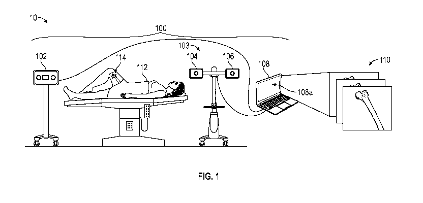
For example, a surgeon may use tools such as rulers and/or mechanically based
jigs and estimate
locations based on palpable or visible landmarks.
BRIEF DESCRIPTION OF THE DRAWINGS
[0005] Implementations of the present technology will now be described, by
way of example
only, with reference to the attached figures, wherein:
[0006] FIG. 1 is a diagram illustrating an example of an environment in
which a surgical
system may be used in accordance with the present disclosure.
[0007] FIG. 2 is a diagram of a controller which may be employed as shown
in FIG. 1.
[0008] FIG. 3 is a diagram illustrating a preoperative plan being displayed
from a controller.
[0009] FIGS. 4A-4G are diagrams illustrating a preoperative plan being
displayed from a
controller using a linear jig.
-1-
CA 03137721 2021-10-21
WO 2020/219473 PCT/US2020/029164
[0010] FIGS. 5A and 5B are diagrams illustrating a preoperative plan being
displayed from
a controller using a modular jig.
[0011] FIG. 5C are diagrams illustrating exemplary modular pieces which can
be used to
create a modular jig.
[0012] FIG. 5D is a diagram illustrating an exemplary coupling mechanism
for modular
pieces.
[0013] FIGS. 5E-5G are diagrams illustrating an exemplary modular jig where
the modular
pieces are extendable, rotatable, and/or retractable.
[0014] FIG. 6 is a diagram illustrating a jig with an exemplary alignment
base.
[0015] FIGS. 7A-7D are diagrams illustrating preparation of a bone surface
to attach a
marker.
[0016] FIGS. 8A-8D are diagrams illustrating exemplary markers.
[0017] FIG. 9 is a diagram illustrating a marker and bone being scanned by
a 3D surface
scanner.
[0018] FIGS. 10A-10D are diagrams illustrating an impression material
utilized to create an
impression of at least a portion of a marker and bone to be scanned by a 3D
surface scanner.
[0019] FIG. 11A is a diagram illustrating a bone image scanned by the 3D
surface scanner.
[0020] FIGS. 11B-11D are diagrams illustrating analysis and registration of
the bone image
with the preoperative image.
[0021] FIGS. 12A and 12B are diagrams illustrating a camera tracking in
real time a tracking
component of a marker and a projector projecting cutting lines and alignment
lines.
[0022] FIGS. 13A-13G are diagrams illustrating an exemplary position
mechanism operable
to adjust the positioning and alignment of a jig.
[0023] FIG. 14 is a flow chart illustrating an example of a bone surgery
that may be used in
accordance with the present disclosure.
DETAILED DESCRIPTION
[0024] It will be appreciated that for simplicity and clarity of
illustration, where appropriate,
reference numerals have been repeated among the different figures to indicate
corresponding or
analogous elements. In addition, numerous specific details are set forth in
order to provide a
thorough understanding of the embodiments described herein. However, it will
be understood by
-2-
CA 03137721 2021-10-21
WO 2020/219473 PCT/US2020/029164
those of ordinary skill in the art that the embodiments described herein can
be practiced without
these specific details. In other instances, methods, procedures and components
have not been
described in detail so as not to obscure the related relevant feature being
described. Also, the
description is not to be considered as limiting the scope of the embodiments
described herein. The
drawings are not necessarily to scale and the proportions of certain parts may
be exaggerated to
better illustrate details and features of the present disclosure.
[0025] Disclosed herein is a surgical system. Recreating preoperative
surgical plans during
surgery can be very difficult. For example, making a precise bone cut along a
plane determined
preoperatively on a CT scan image can be very difficult. Surgeons can use
visible or palpable
landmarks, rulers, and/or mechanical jigs at the time of surgery to help
recreate the preoperative
plan. However, these simple methods frequently result in inaccurate bone cuts.
During surgery, it
can be very difficult to visualize landmarks and accurately make precise bone
cuts, even with
mechanical jigs that may or may not be placed in the proper position.
[0026] The present surgical system can be utilized to assist surgeons with
accurately and
precisely recreating preoperative surgical plans during surgery. In at least
one example, a marker
coupled with the bone can be registered to the bone in a controller. The
marker can include a three-
dimensional body. At least a portion of the three-dimensional body and at
least a portion of the
bone can be scanned by a three-dimensional scanner to form a bone scan. The
bone scan can be
brought closer to and/or together with a bone image obtained preoperatively,
such as a CT scan
image. Accordingly, the marker and the bone can be registered more accurately
and simpler than
conventional methods. For example, some conventional systems require a surgeon
to use a hand
probe to touch the surface of bone dozens of times to manually generate a
point cloud which can
take time, is cumbersome, and, due to the relatively limited amount of data
points obtainable by
this method, can create significant inaccuracies in registration. Another
conventional way to
register a marker may be to obtain CT or X-rays during the surgical procedure
after the marker is
placed on the bone which can be expensive, require the use of large equipment,
and unnecessarily
expose the patient and hospital staff to radiation.
[0027] In some examples, the surgical system tracks the movement of the
bone to accurately
and precisely project light to guide the surgeon during surgery. For example,
the light may include
a cutting line to indicate the cutting point and/or plane for the surgeon to
cut the bone during
surgery. In some examples, the surgical system may track the movement of the
bone by the use of
-3-
CA 03137721 2021-10-21
WO 2020/219473 PCT/US2020/029164
a tracking component of the marker. For example, the tracking component may
include a two-
dimensional pattern and/or reflecting tracking features to be scanned by a
camera and recognized
by the controller. The position of the tracking component in relation to the
bone and/or the
registration component can be predetermined and/or known by the controller.
Accordingly, once
the bone is registered, the controller is able to track the movement of the
tracking component and
correspondingly track the movement of the bone in real-time.
[0028] In at least one example, one or more jigs can be utilized to guide
the surgical blade
during bone cuts. The components, orientation, and/or shape of jigs readily
available to the surgeon
can be stored in the memory of the controller. Accordingly, the surgeon can
prepare a preoperative
plan and determine the jig and/or make-up of the jig needed during surgery.
The surgeon can then
easily obtain and utilize the correct jig for surgery. For example, a modular
jig may be created out
of modular components. The bone cut may include an irregular shaped cut, and a
specific shaped
jig may be needed. The controller may be utilized to determine which modular
components readily
available can be combined and/or modified during preoperative planning. The
surgeon and/or
surgery staff can then create the modular jig without the need for
conventional 3D printed custom
jigs which can still result in substantial inaccuracies due to challenges in
jig placement on the bone
during surgery. Furthermore, conventional methods of producing custom jigs are
very expensive
and can take a significant time to generate - sometimes days or weeks; even
after such cost and
effort, the jig is single use and has to be discarded after just one surgery.
[0029] To ensure the accurate and precise placement of the jig, the jig may
include alignment
markers. The projector can then project light that includes alignment lines to
correspond with the
alignment markers. Accordingly, the jig simply needs to be positioned such
that the alignment
lines are aligned with the alignment markers.
[0030] As the surgical system is tracking the movement of the bone in real-
time, the
projected light such as the cutting line and/or the alignment lines may be
adjusted in real-time to
correspond with the movement of the bone. Accordingly, the preoperative
surgical plan can be
accurately recreated during surgery.
[0031] The disclosure now turns to FIG. 1, which illustrates a diagrammatic
view of an
exemplary surgical environment 10 for a surgical system 100, in which the
present disclosure may
be implemented. As illustrated in FIG. 1, a surgical system 100 can include a
controller 108, a 3D
surface scanner 102 communicatively coupled with the controller 108, and a
projector system 103
-4-
CA 03137721 2021-10-21
WO 2020/219473 PCT/US2020/029164
communicatively coupled with the controller 108. The surgical system 100 may
be utilized during
surgery on a patient 112. The patient 112, as illustrated in FIG. 1, has an
exposed bone surface 114
as the patient 112 is undergoing bone surgery such as a total knee
arthroplasty which can include
distal femur and proximal tibia cuts of the bone, total hip arthroplasty which
can include femoral
neck osteotomy, a tumor resection which can include custom, patient-specific
cuts of bone to
ensure removal of tumor, and/or procedures where specific placement of a
screw, pin, and/or
needle is required or skeletal deformity correction surgery.
[0032] The 3D surface scanner 102 is operable to optically scan an object,
for example a
bone, a marker, a mold, or any other surface. The 3D surface scanner 102
transmits the scan of the
object to the controller 108 which can then process the digitally scanned
surface of the object. The
3D surface scanner 102 can include, for example, a structured light projector
and one or more
cameras. An example of the 3D surface scanner 102 can be EinScan-SP.
[0033] As illustrated in FIG. 1, the 3D surface scanner 102 may be fixably
attached to a
rolling stand and brought into range of exposed bone surface 114 of patient
112 as required. In
some examples, the 3D surface scanner 102 may be fixably attached at a
location in the operating
room such that is in range of exposed bone surface 114 of patient 112. In some
examples, the 3D
surface scanner 102 may be disposed on a pivoting and/or swivelling arm which
can, for example,
be coupled to the ceiling or a mounting system above the patient 112.
[0034] In at least one example, during operation, the projector of the 3D
surface scanner 102
can project structured light pattern onto the target of the object, such as
the exposed bone surface
114. The cameras capture the distorted pattern of the structured light on the
target. Based on the
image with distorted structured light pattern, the 3D surface scanner 102 can
capture a 3D scanned
surface, and the controller 108 can digitally construct the 3D scanned surface
using a computer
algorithm. In some examples, each scan can take about 30-60 seconds. In some
examples, each
scan may take less than 30 seconds, for example substantially instantaneously.
[0035] The projector system 103 can include a camera 104 and a projector
106. The camera
104 is operable to capture images and/or video. For example, the camera 104
can include an 8MP
5-50mm Varifocal Lens USB Camera with a Sony IMX179 Sensor. The projector 106
can project
an array of desired patterns and/or colors onto a surface. For example, the
projector 106 can include
a BenQ TK800 projector. The camera 104 and the projector 106 have a
predetermined fixed
relative position to each other. For example, as illustrated in FIG. 1, the
camera 104 and the
-5-
CA 03137721 2021-10-21
WO 2020/219473 PCT/US2020/029164
projector 106 are disposed on the same stand or frame. In some examples, the
camera 104 and the
projector 106 may be separated such that the camera 104 and the projector 106
can move
independent from one another.
[0036] In at least one example, the projector system 103 can be calibrated
prior to the
surgery to obtain extrinsic parameters such as the relative position between
camera 104 and
projector 106 and to obtain intrinsic parameters of the camera 104 and/or the
projector 106 such
as lens focal length, lens distortion, and/or sensor pixel size. In at least
one example, when
projector system 103 is calibrated, the relative positions between the camera
104 and the projector
106 remain consistent and stable. In some examples, the projector system 103
can continuously
calibrate the relative positions between the camera 104 and the projector 106
as each one may
move independently from the other. For example, the camera 104 and/or the
projector 106 may
include sensors such as accelerometers and/or gyroscopes to sense positioning
and/or movement
of the camera 104 and/or the projector 106. Accordingly, when the camera 104
and/or the projector
106 move, the projector system 103 can re-calibrate the relative positions
between the camera 104
and the projector 106.
[0037] In at least one example, as illustrated in FIG. 1, the projector
system 103 may be
fixably attached to a rolling stand and brought into range of exposed bone
surface 114 and/or the
desired surface to track and/or have an image projected thereon as required.
In some examples, the
projector system 103 may be fixably attached anywhere in operating room such
that it is in range
of exposed bone surface 114 as required. In some examples, the projector
system 103 may be
disposed on a pivoting and/or swivelling arm which can, for example, be
coupled to the ceiling or
a mounting system above the patient 112.
[0038] In at least one example, surgical systems 100 where the 3D surface
scanner 102 and
the projector system 103 are a single, fully integrated system are
contemplated. Further, surgical
systems 100 where a plurality of 3D surface scanners 102 are integrated with
one or more cameras
104 and projectors 106 are also contemplated.
[0039] The controller 108 can include a monitor 108a that can be used to
view images and/or
video, for example, of exposed bone surface 114. In some examples, the images
and/or videos
displayed on monitor 108a can be captured in real-time by camera 104. In some
examples, the
monitor 108a may be used to display images and/or video, for example, of
manuals, instructions,
previous scans, or any other suitable information desired at the time of
surgery. For example,
-6-
CA 03137721 2021-10-21
WO 2020/219473 PCT/US2020/029164
preoperative images 110 may be displayed on the monitor 108a. Preoperative
images 110 may be
from any clinically relevant imaging modality a clinician may use such as
images obtained during
X-ray, CT scan, or MRI scan.
[0040] FIG. 2 is a block diagram of an exemplary controller 108. Controller
108 is
configured to perform processing of data and communicate with the surgical
components, for
example as illustrated in FIG. 1. In operation, controller 108 communicates
with one or more of
the above-discussed components and may also be configured to communication
with remote
devices/systems.
[0041] As shown, controller 108 includes hardware and software components
such as
network interfaces 210, at least one processor 220, sensors 260 and a memory
240 interconnected
by a system bus 250. Network interface(s) 210 can include mechanical,
electrical, and signaling
circuitry for communicating data over communication links, which may include
wired or wireless
communication links. Network interfaces 210 are configured to transmit and/or
receive data using
a variety of different communication protocols, as will be understood by those
skilled in the art.
[0042] Processor 220 represents a digital signal processor (e.g., a
microprocessor, a
microcontroller, or a fixed-logic processor, etc.) configured to execute
instructions or logic to
perform tasks in a surgical environment. Processor 220 may include a general
purpose processor,
special-purpose processor (where software instructions are incorporated into
the processor), a state
machine, application specific integrated circuit (ASIC), a programmable gate
array (PGA)
including a field PGA, an individual component, a distributed group of
processors, and the like.
Processor 220 typically operates in conjunction with shared or dedicated
hardware, including but
not limited to, hardware capable of executing software and hardware. For
example, processor 220
may include elements or logic adapted to execute software programs and
manipulate data
structures 245, which may reside in memory 240.
[0043] Sensors 260 typically operate in conjunction with processor 220 to
perform
measurements, and can include special-purpose processors, detectors,
transmitters, receivers, and
the like. In this fashion, sensors 260 may include hardware/software for
generating, transmitting,
receiving, detection, logging, and/or sampling temperature, bone alignment,
time, or other
parameters.
[0044] Memory 240 comprises a plurality of storage locations that are
addressable by
processor 220 for storing software programs and data structures 245 associated
with the
-7-
CA 03137721 2021-10-21
WO 2020/219473 PCT/US2020/029164
embodiments described herein. An operating system 242, portions of which may
be typically
resident in memory 240 and executed by processor 220, functionally organizes
the device by, inter
alia, invoking operations in support of software processes and/or services 244
executing on
controller 108. These software processes and/or services 244 may perform
processing of data and
communication with controller 108, as described herein. Note that while
process/service 244 is
shown in centralized memory 240, some examples provide for these
processes/services to be
operated in a distributed computing network.
[0045] It will be apparent to those skilled in the art that other processor
and memory types,
including various computer-readable media, may be used to store and execute
program instructions
pertaining to the surgical techniques described herein. Also, while the
description illustrates
various processes, it is expressly contemplated that various processes may be
embodied as modules
having portions of the process/service 244 encoded thereon. In this fashion,
the program modules
may be encoded in one or more tangible computer readable storage media for
execution, such as
with fixed logic or programmable logic (e.g., software/computer instructions
executed by a
processor, and any processor may be a programmable processor, programmable
digital logic such
as field programmable gate arrays or an ASIC that comprises fixed digital
logic. In general, any
process logic may be embodied in processor 220 or computer readable medium
encoded with
instructions for execution by processor 220 that, when executed by the
processor, are operable to
cause the processor to perform the functions described herein.
[0046] FIGS. 3-5B illustrate exemplary preoperative images 110 which may be
displayed,
for example, on a monitor 108a. During preoperative planning, the doctor is
able to determine,
using the controller 108, the actions required and desired during operation.
The doctor can
determine a cutting line 150 which can align with the desired cut or
interaction with the exposed
bone 114. However, during preoperative planning, in the preoperative image
110, the doctor can
place the desired cutting line 150 on the bone image 115 in the preoperative
image 110. In some
examples, as illustrated in FIG. 3, the cutting line 150 can include a dot
projection 300 which can
indicate, for example, a point for a needle, pin, screw, and/or a puncture to
be placed in the exposed
bone 114. FIGS. 4A-4G illustrate examples of the cutting line 150 including a
linear projection
400 along a cutting plane 410. FIGS. 5A and 5B illustrate examples of the
cutting line 150
including an irregularly shaped projection 500. While the cutting lines 150
can be shown as
substantially straight lines, the cutting line 150 can be shown as the
intersection of a top curve line
-8-
CA 03137721 2021-10-21
WO 2020/219473 PCT/US2020/029164
between the cutting plane 410 to the bone image 115. Accordingly, the cutting
line 150 at certain
viewing angles may appear straight while at other viewing angles may appear
curved to follow the
curvature of the bone 114, 115. In some examples, the cutting line 150 and
subsequent projection
can be any line, dot, circle, rectangle, triangle, and/or any shape as
clinically desired.
[0047] FIGS. 4A-4G illustrate exemplary preoperative images 110 which may
be displayed
during preoperative planning for a linear cut of the bone 114. FIG. 4A
illustrates a cutting plane
410 which correlates to the desired cut to be made during the operation. For
example, the cutting
plane 410 correlates with the cutting path of a surgical blade such as a saw
blade to cut the bone
in the proper location and angle. As illustrated in FIGS. 4A and 4B, the
cutting plane 410 may
correlate with a cut in a femur during a total knee arthroplasty.
[0048] To ensure proper alignment and provide a stable cut by the surgical
blade, a jig 117
can be used. FIGS. 4A-6 illustrated exemplary uses of different jigs 117 to
guide a surgical blade
during operation. The jig 117 can provide a surface for which the surgical
blade can abut such that
the jig 117 guides the blade during the cutting process.
[0049] The jig 117 can include a linear jig 118 which, as illustrated in
FIGS. 4A-4G, can be
formed in the shape of a T. As illustrated in FIG. 4C, the linear jig 118
includes one or more
coupling components 406 which are operable to couple the linear jig 118 with
the bone 114 such
that the linear jig 118 does not move and become misaligned. For example, the
coupling
components 406 can include recesses operable to receive couplers such as
screws to couple the
linear jig 118 with the bone 114. In some examples, the coupling components
406 can include one
or more straps, a secondary element intermediate the jig 117 with the bone
114, or any other
suitable component to couple the jig 117 with the bone 114.
[0050] As illustrated in FIGS. 4C and 4D, the linear jig 118 has a blade
surface 402 which
is aligned with the linear cutting plane 410 to serve as a guide to the
surgical blade. The blade
surface 402 of the linear jig 118 provides a linear surface to guide the blade
for a linear cut. In
other examples, the linear jig 118 can be any shape such that the linear jig
118 provides a guide
surface for a linear cutting plane 410, such as rectangular, triangular, or
any other suitable shape.
Additionally, in some examples, the linear jig 118 can include different
configurations for guiding
the surgical blade along the cutting plane 410. For example, the linear jig
118 can include a linear
aperture for which the surgical blade is inserted through ensure a stable and
straight cut in the bone
114.
-9-
CA 03137721 2021-10-21
WO 2020/219473 PCT/US2020/029164
[0051] During preoperative planning on the controller 108, the positioning
of an alignment
line 152 and/or a cutting line 150 can be determined. As illustrated in FIG.
4D, the controller 108
can determine the positioning of an alignment line 152. The alignment line 152
includes light
which can be projected during operation to indicate the precise alignment of
the linear jig 118. In
at least one example, the linear jig 118 can include a plurality of alignment
markers 404 which are
operable to ensure the correct and precise alignment of the linear jig 118.
When the alignment line
152 is aligned with the alignment markers 404, for example as illustrated in
FIGS. 4D and 4E, the
linear jig 118 is accurately and precisely positioned. The cutting plane 410
and blade surface 402
of the linear jig 118 are also accurately and precisely aligned.
[0052] In at least one example, as illustrated in FIGS. 4D and 4E, the
controller 108 can
determine the positioning of a cutting line 150 which includes light emitted
on the bone surface
114 to ensure the alignment of the cutting surface 402 and the cutting plane
410. As illustrated in
FIGS. 4D and 4E, the cutting line 150 can include a linear cutting line 400.
When the linear jig
118 is adequately positioned such that the cutting surface 402 is aligned with
the cutting plane 410
along the cutting projection, as illustrated in FIG. 4E, the surgical blade
126 can then make a
surgical cut with known parameters, for example cutting through cutting plane
410 and entering
the bone surface 114 through cutting plane 410, allowing for accurate
implementation of the
preoperative surgical plan.
[0053] As illustrated in FIGS. 4F and 4G, the preoperative planning can
include a simulation
of the positioning of the linear jig 118 on the bone image 115, for example,
with the alignment line
152. Additionally, FIGS. 4F and 4G illustrate the bone image 115 after a
simulated cut has been
made along the cutting plane 410 to determine whether a cut along the cutting
plane 410 produces
the desired bone cut to achieve the desired clinical results.
[0054] FIG. 5A illustrates an exemplary preoperative plan where the desired
projection line
150 is an irregularly shaped projection line 500 to indicate the desired
cutting path and/or cutting
plane of the bone. For example, as illustrated in FIG. 5A, a tumor 116 may
need to be resected,
and the projection line 500 needs to have three lines cut along different
angles to surround the
tumor 116. FIG. 5B illustrates the use of a jig 117 to guide a surgical blade
along the cutting path
and/or cutting plane and projection line 500. As the determined path along the
projection line 500
is not a single linear path, a modular jig 120 can be utilized. The modular
jig 120 is similar to the
-10-
CA 03137721 2021-10-21
WO 2020/219473 PCT/US2020/029164
linear jig 108 of FIGS. 4A-4G, however the modular jig 120 can be modified to
fit along a linear
and/or non-linear projection line 500 and cutting path and/or cutting plane.
[0055] In at least one example, similar to the linear jig 108, the modular
jig 120 can include
a plurality of alignment markers 504 which are operable to ensure the correct
and precise alignment
of the modular jig 120. When the alignment line 152 is aligned with the
alignment markers 504,
for example as illustrated in FIG. 5B, the modular jig 120 is accurately and
precisely positioned.
The cutting plane and blade surface of the modular jig 120 are also accurately
and precisely aligned
to execute the precise bone cut as determined in the preoperative plan.
[0056] As illustrated in FIG. 5B, the modular jig 120 includes one or more
coupling
components 506 which are operable to couple the modular jig 120 with the bone
114 such that the
modular jig 120 does not move and become misaligned. For example, the coupling
components
506 can include recesses operable to receive couplers such as screws to couple
the modular jig 120
with the bone 114.
[0057] As illustrated in FIG. 5C, the modular jig 120 can be composed of a
number of
different modular pieces 550. The modular pieces 550 can each have any
predetermined size,
shape, and/or design. For example, the modular pieces 550 can include a
plurality of left pieces
551 having different sizes and angles. The plurality of left pieces 551 as
illustrated in FIG. 5C can
include at least one bend or curve. A plurality of central pieces 552 can be,
for example, linear
pieces, and a plurality of right pieces 554 can include at least one bend or
curve. The left pieces
551 and the right pieces 554 can be configured to be coupled to each end of
the central pieces 552.
Any combination of modular pieces 550 can be linked together to form a modular
jig 120 that fits
the desired cutting path and/or cutting plane.
[0058] In at least one example, as illustrated in FIG. 5D, modular pieces
550 can be coupled
to one another by coupling portions 560, for example using a fastener 570 such
as a screw. A
coupling portion 560 of one modular piece 550 can include a male portion 561
while another
modular piece 550 can include a female portion 562 operable to receive the
male portion 561. Both
of the coupling portions 560 of the modular pieces 550 include fastening
apertures 565 aligned
with one another and operable to receive a fastener 570 such as a screw, a
bolt, a magnet, or any
other suitable fastener 570 to couple the modular pieces 550 together. In some
examples, different
coupling mechanisms can be utilized such that the modular pieces 550 are
coupled together and
maintain the shape of the modular jig 120.
-11-
CA 03137721 2021-10-21
WO 2020/219473 PCT/US2020/029164
[0059] FIGS. 5E-5G illustrates examples of a modular jig 120 in which the
modular pieces
550 can be pivoted in relation to one another, extended in length, and/or
retracted in length. In at
least one example, the precise angles and/or lengths of the modular pieces 550
of the modular jig
120 after pivoting and lengthening or retraction can be known by users, for
example, by visualizing
outputs of angles and lengths on modular jig 120. In at least one example, the
precise angles and/or
lengths of the modular pieces 550 of the modular jig 120 can be stored in
memory of the controller
108. For example, the controller 108 may record wirelessly the angles and
lengths through sensors
or optical scanning. In some examples, the modular jig 120 may include sensors
that measure the
lengths and/or angles of the modular pieces 550 of the modular jig 120, and
the sensors may
transmit the measurements to the controller 108. Accordingly, as in the
examples illustrated in
FIG. 5E-5G, the modular jig 120 can be further customizable and adaptable to
address any number
of surgical cuts.
[0060] In at least one example, the controller 108 can store each modular
piece 550 available
to the surgical team. After the cutting line 150 and/or the alignment line 152
is determined, the
controller 108 can determine the exact size and/or shape of the modular jig
120 needed by the
surgical team. Additionally, in some examples, the controller 108 can
construct, in preoperative
planning, a modular jig 120 using the known modular pieces 550 available to
the surgical team.
With the preoperative plan, the surgical team can then easily pick out the
modular pieces 550
identified by the controller 108 and construct or adjust the modular jig 120
exactly as determined
in the preoperative plan. In another example, modular jig 120 illustrated in
FIG. 5E-5G can be
configured to the precise angles and lengths required for the preoperative
plan by controller 108
by wired/wirelessly actuating modular jig 120 into configuration. For example,
the modular jig
120 may include one or more motors which are operable to move the modular
pieces 550 to the
precise angles and/or lengths as directed by the controller 108. The surgical
team does not then
have to wait for a customized jig 117 to be created by a fabrication company,
and can easily create
any jig 117 without delay.
[0061] FIG. 6 illustrates an exemplary alignment base 602 operable to
ensure the correct
and precise alignment of the jig 117. The alignment base 602 is operable to be
coupled with a jig
117, for example modular jig 120, to ensure the alignment of the jig 117.
Alignment of alignment
base 602 may create corresponding alignment of jig 117 which can be coupled to
alignment base
-12-
CA 03137721 2021-10-21
WO 2020/219473 PCT/US2020/029164
602, thereby eliminating the necessity of alignment markers 604 directly on
jig 117. However, in
at least one example, the jig 117 may still include alignment markers 604.
[0062] The alignment base 602 includes a plurality of alignment markers 604
which are
operable to correspond with an alignment line 152. When the alignment line 152
is aligned with
the alignment markers 604, the alignment base 602 is accurately and precisely
positioned.
Subsequently, the cutting plane and blade surface of the jig 117 are also
accurately and precisely
aligned.
[0063] The alignment base 602 can include one or more coupling components
606 which
are operable to couple the alignment base 602 with the bone 114 such that the
alignment base 602
does not move and become misaligned. For example, the coupling components 606
can include
recesses operable to receive couplers such as screws to couple the alignment
base 602 with the
bone 114.
[0064] In at least one example, during preoperative planning, the
controller 108 has stored
in memory the available jigs 117, such as the linear jig 118, the modular jig
120 and/or the modular
pieces 550 available to create the modular jig 120, and/or the alignment base
602. Accordingly,
the required jig 117 can be determined and/or created in the controller 108
during preoperative
planning such that the exact jig 117 and/or alignment base 602 can be utilized
and/or recreated
during surgery.
[0065] FIGS. 7A-7C illustrate steps during preparation of the bone 114 for
surgery. As
illustrated in FIG. 7A, the soft tissue 700 such as skin and muscle are
dissected and pulled back to
expose the bone 114. In some examples, residue small tissue 702 may remain on
the surface of the
bone 114. As illustrated in FIG. 7B, the surgeon 12 may utilize a tool 14 to
cut the residue small
tissue 702 from the surface of the bone 114. In some examples, as illustrated
in FIG. 7B, the tool
14 may include an instrument such as a Cobb surgical instrument, however, any
clinically
acceptable method to remove tissue and expose bone surface may be used. As
illustrated in FIG.
7C, the bone 114 is exposed and an area is cleaned of excess tissue. For
example, the area of the
exposed bone 114 may have a width 114W of about 3 centimeters and a length
114L of about 3
centimeters. The dimensions of the area of exposed bone 114 may vary as
desired or needed for
the surgery, or as needed by 3D surface scanner 102 to obtain enough
information to execute
accurate surface matching to preoperative images.
-13-
CA 03137721 2021-10-21
WO 2020/219473 PCT/US2020/029164
[0066] After the bone 114 is exposed, as illustrated in FIG. 7D, a marker
116 can be coupled
to the bone 114. In some examples, as illustrated in FIG. 7D, the marker 116
can be directly
coupled with the bone 114 by fasteners 710 such as screws, pins, or any other
clinically acceptable
device used for fixation to bone 114. In some examples, the marker 116 can be
indirectly coupled
with the bone 114 such that the marker 116 is coupled with another component
or device in which
the another component or device is directly or indirectly coupled to the bone
114. The marker 116
is affixed to the bone 114 proximate to the target bone area where there is no
tissue covering the
bone 114. The marker 116 is utilized to register and track the orientation and
movement of the
bone 114 during surgery. In some examples, the marker 116 is operable to be
scanned by a surgical
system 100 (shown in FIG. 1) such as the 3D surface scanner 102 and/or camera
104 of the
projector system 103. The surgical system 100 is operable to scan the marker
116 on the bone 114,
register the marker 116 in the controller 108 in reference to the preoperative
image 110, and/or
track the movement of the marker 116 and correlate the movement of the marker
116 in reference
to the movement of the bone 114 during surgery. Additionally, with the
understanding of the
orientation and movement of the bone 114 due to the marker 116, the controller
108 can control
the projector 106 of the projector system 103 to project light onto the bone
114 or one or more jigs
117 or corresponding components to align the jig 117 and subsequently
accurately align the
surgical blade to cut the bone 114.
[0067] FIGS. 8A-8D illustrate exemplary markers 116. The marker 116 can
include a
registration component 810 and a tracking component 850. The registration
component 810 is
operable to be scanned into a digital reproduction of at least a portion of
the registration component
810 and at least a portion of the bone 114. Because the precise location of
registration component
810 can be determined on bone 114 following surface extraction, the
registration component 810
can provide data to the controller 108 regarding the precise location and/or
orientation of the
marker 116 in relation to the bone 114. The tracking component 850 is disposed
a predetermined
distance with a predetermined orientation in relation to the registration
component 810 and is
known by controller 108. The controller 108, upon registering the location
and/or orientation of
the registration component 810 through surface extraction the digital
reproduction of the scan data
to preoperative images, can then scan the tracking component 850, for example
using the camera
104, to track the movement and/or orientation of the bone 114 in real time
during surgery. For
example, the tracking component 850 can include a 2-dimensional pattern 852
which can be
-14-
CA 03137721 2021-10-21
WO 2020/219473 PCT/US2020/029164
scanned by the camera 104 and recognized by the controller 108 to track the
movement of the
tracking component 850. In some examples, the 2-dimensional pattern 852 can
include a barcode,
a QR code, or any other suitable pattern.
[0068] As illustrated in FIG. 8A, the registration component 810 and the
tracking component
850 are disposed on one body 800. In some examples, the registration component
810 and the
tracking component 850 can be separately provided. For example, FIG. 8B
illustrates an exemplary
registration component 810 which is a separate component from the tracking
components 850
illustrated in FIGS. 8C and 8D. In some examples, the registration component
810 and the tracking
component 850 can be configured to be coupled to one another. Even if
separately provided, the
distance and/or relationship between the registration component 810 and the
tracking component
850 is predetermined and known. Accordingly, the controller 108 can track the
tracking component
850 and subsequently the bone 114 during surgery with only a scan of at least
a portion of the
registration component 810 and at least a portion of the bone 114.
[0069] For example, as illustrated in FIGS. 8B-8D, the registration
component 810 may
include a three-dimensional body 812 which is operable to be scanned into the
controller 108 to
register the location and/or orientation of the marker 116 in relation to the
bone 114. In at least one
example, the registration component 810 can include a pin receiver 814 which
is operable to
receive a pin 854 or a corresponding coupling mechanism to couple the
registration component
810 with the tracking component 850. The precise location of the registration
component 810 to
tracking component 850 can be precisely known. In some examples, the
registration component
810 can include a bone fastener 816 such as a screw, a pin, or any other
suitable fastener operable
to couple the registration component 810 to the bone 114.
[0070] FIGS. 8C and 8D illustrate exemplary tracking components 850. The
tracking
components 850 are operable to be scanned, for example, by a camera 104 such
that the controller
108 can track the movement of the tracking component 850 and subsequently the
corresponding
bone 114 in real time during surgery. As illustrated in FIGS. 8C, the tracking
component 850,
similar to FIG. 8A, can include a 2-dimensional pattern 852 such as a barcode,
a QR code, or any
other suitable pattern which can be scanned by the camera 104 and recognized
by the controller
108 to track the movement of the tracking component 850. In some examples, as
illustrated in FIG.
8D, the tracking component 850 can include one or more reflective tracking
features 853 operable
to be scanned by the camera 104 and recognized by the controller 108 to track
the movement of
-15-
CA 03137721 2021-10-21
WO 2020/219473 PCT/US2020/029164
the tracking component 850. The reflective tracking features 853 can be coated
such that the
reflective tracking features 853 shine when light is shone on the reflective
tracking features 853.
In some examples, the reflective tracking features 853 may be tracked by the
camera 104 which
can be equipped with an infrared pass filter.
[0071] In at least one example, referring also to FIG. 9, at least a
portion of the registration
component 810 and at least a portion of the bone 114 can be scanned directly
by the 3D surface
scanner 102 to detect the location and/or orientation of the marker 116 in
relation to the bone 114
in the controller 108. For example, as illustrated in FIG. 8A, the
registration component 810 is
substantially the shape of a flat-top 3-dimensional pyramid. In some examples,
the registration
component 810 can be any 3-dimensional shape that extends from the bone 114
such that the
location as well as 3-dimensional orientation of the registration component
810 in relation to the
bone 114 can be extracted. The controller 108 is operable to build a
registration component 810
local coordinate on the bone 114.
[0072] FIGS. 10A-10D illustrate a method to scan at least a portion of the
registration
component 810 and at least a portion of the bone 114 when the bone 114 is not
readily or easily
accessible to the 3D surface scanner 102. As illustrated in FIG. 10A, the
marker 116 is coupled
with the bone 114. An impression material 1000 is pressed against a portion of
the bone 114 and
at least a portion of the registration component 810 of the marker 116. The
impression material
1000 can cover enough of the registration component 810 such that defining
shapes, surfaces,
edges, and/or corners can be molded into the impression material 1000.
Similarly, the impression
material 1000 can cover enough of the bone 114 such that one or more defining
features of the
bone 114 can be molded into the impression material 1000.
[0073] In at least one example, as illustrated in FIG. 10B, an enforcement
component 1002
can be placed on and/or in the impression material 1000. The enforcement
component 1002 can
provide support to the impression material 1000 so that the impression
material 1000 is able to
better retain its shape after removal. For example, the enforcement component
1002 can function
similar to rebar supporting concrete in construction. In at least one example,
the enforcement
component 1002 can include or be formed as a handle to ease removal of the
impression material
1000 from the marker 116 and the bone 114. When the impression material 1000
either hardens or
captures an adequate impression of at least a portion of the registration
component 810 and the
bone 114, the impression material 1000 can be removed.
-16-
CA 03137721 2021-10-21
WO 2020/219473 PCT/US2020/029164
[0074] FIG. 10C illustrates the underside 1001 of the impression material
1000, showing the
bone negative impression 1004 and the marker negative impression 1006. The
underside 1001 of
the impression material 1000 including the bone negative impression 1004 and
the marker negative
impression 1006 is then scanned by the 3D surface scanner 102. The controller
108 can reverse
the normal vectors of the bone negative impression 1004 and the marker
negative impression 1006
such that the controller 108 has a scan of the bone 114 and the marker 116. By
using the impression
material 1000, the 3D surface scanner 102 does not have to be in direct sight
of the bone 114 and
marker 116. Accordingly, when the bone 114 and the marker 116 are not easily
accessible by the
3D surface scanner 102, the impression material 1000 can be utilized to
provide a scan of the bone
114 and the marker 116 for registration.
[0075] For example, as illustrated in FIG. 11A, the controller 108 can
create a digital
recreation 900 of at least a portion of the bone 114 and at least a portion of
the marker 116 based
on the scan of the marker 116 and bone 114. For example, the digital
recreation 900 can include a
3D point cloud. For example, each digital recreation 900 can capture at least
an area of 100
millimeters by 100 millimeters with about 1 million points such that the 3D
point cloud created
from the 3D surface scanner 102 can have an exemplary resolution of about 20
points/mm2. The
preoperative image 110 can have a resolution of about 2 points/mm2. In at
least one example, to
reduce the computational cost, the point cloud of the digital recreation 900
can be down-sampled
to the same resolution as the preoperative image 110. In at least one example,
the digital recreation
900 can include a 3D mesh. The 3D mesh can include, for example a 3D point
cloud where each
point is connected.
[0076] In some examples, in the digital recreation 900, many points may
originate from the
surrounding and/or background areas which are also captured and constructed by
the 3D surface
scanner 102, and are not relevant in the next alignment procedure. The images
of the surrounding
and/or background areas may be removed using computer software, for example
with the
controller 108, leaving only the exposed bone 114 and/or at least a portion of
the marker 116 such
as at least a portion of the registration component 810. In some examples, the
images of the
surrounding and/or background areas may be removed by an assistant and/or the
doctor. In some
examples, the images of the surrounding and/or background areas may be removed
automatically
without human assistance by the controller 108.
-17-
CA 03137721 2021-10-21
WO 2020/219473 PCT/US2020/029164
[0077] As illustrated in FIG. 11B, the orientation of the marker 116 and
subsequently the
bone 114 can be determined using the controller 108. For example, the flat
surfaces of the
registration component 810 can be selected and one or more intersected edges
and/or corners of
the registration component can be extracted. As the shape and/or size of the
registration component
810 is stored in the controller 108, the controller 108 can then determine the
orientation of the
registration component 810 along the X-axis, the Y-axis, and/or the Z-axis. As
illustrated in FIG.
11C, the exposed bone area 914 without tissue is selected to create a bone
scan 902. The marker
116 and the bone 114 are then registered, and the relationship between the
bone 114 and the marker
116 is determined.
[0078] After the digital recreation 900 of the bone 114 is obtained and
processed, as
illustrated in FIG. 11D, the bone scan 902 is aligned with the preoperative
bone image 115 such
that the preoperative plan can be correlated with the physical bone and
surgical process in real
time.
[0079] In at least one example, a surface matching algorithm can be
utilized by the controller
108 to align the bone scan 902 with the bone image 115. The surface matching
algorithm can
produce a number of highest possible rigid body homogenous transformations
that can potentially
align the two 3D models ¨ the bone scan 902 and the bone image 115. The
algorithm can build up
a descriptor, called the point pair feature (PPF), for every two points 950 on
the scanned surface
of the bone scan 902. The algorithm can then find the two corresponding points
952 in the CT-
scan model of the bone image 115 with similar or the same features as the bone
scan 902. For the
two corresponding pairs of points 950, 952 matched, the algorithm gives one
vote for the
homogenous transformation between the two corresponding pairs of points 950,
952. After
finishing a predetermined number of matched PPF to satisfy the algorithm, a
number of
homogenous transformations with the highest votes are likely to become the
best estimated
homogenous transformations. The outcome of a successful execution of the
surface matching
algorithm, is a predetermined number of homogenous transformations with the
highest votes.
[0080] An iterative closest point (ICP) algorithm can be applied to find
the best match
among the homogenous transformations obtained in the previous step by the
surface matching
algorithm. With each homogenous transformation, the bone scan 902 and the bone
image 115 are
brought closer. For example, the bone scan 902 and the bone image 115 can be
brought together.
Using the ICP algorithm, the corresponding points 950, 952 on the bone scan
902 and the bone
-18-
CA 03137721 2021-10-21
WO 2020/219473 PCT/US2020/029164
image 115 are identified by a nearest point search. The ICP algorithm then
computes the sum of
the errors and/or discrepancies between all such corresponding points 950, 952
on the bone scan
902 and the bone image 115 associated with each of the homogenous
transformations from the
previous step. Such errors may be minimized to find the best alignment and the
resulting
homogenous transformation.
[0081] Finally, after the ICP algorithm is completed for all homogenous
transformations
between the bone scan 902 and the bone image 115, the homogenous
transformation with the
smallest error between the bone scan 902 and the bone image 115 is selected as
final result. This
homogenous transformation is used for the subsequent alignment of the bone
scan 902 and the
bone image 115 and/or future procedures.
[0082] Once the bone scan 902 is aligned with the bone image 115, the
marker 116 is
registered in the controller 108. Accordingly, the controller 108 is able to
track, as illustrated in
FIG. 12A using the camera 104 to follow the movement of the tracking component
850, the
orientation and/or movement of the bone 114 during surgery. As the
relationship such as the
distance and orientation between the bone 114, the registration component 810,
and the tracking
component 850 is registered in the controller 108, as the tracking component
850 moves in the
images captured by the camera 104, the controller 108 is able to accurately
calculate the location
and/or orientation of the bone 114.
[0083] In some examples, other suitable registration systems and methods
may be utilized
to register a preoperative bone image 115 with one or more bone scans 902 so
that the movement
and/or orientation of the bone 114 can be tracked during surgery.
[0084] For example, the bone 114 can be touched with a probe that has known
dimensions
and one or more reflective markers. This method can rely on a motion tracking
device which
includes at least two infrared (or near-infrared) cameras. The relative pose
of each camera is fixed
and pre-defined (or pre-calibrated). Two probes may be needed to be tracked by
the motion
tracking device intraoperatively. First, the surgeon affixes a reference probe
to the target bone.
After that, the surgeon uses a hand probe to touch the surface of the target
bone 114, for example
a few dozen times. Each touch can correspond to one 3D point with respect to
the reference probe.
After touching the bone 114 with the probe, a 3D point cloud is built with
reference to the reference
probe and can be used for registration to the pre-operative image 115.
-19-
CA 03137721 2021-10-21
WO 2020/219473 PCT/US2020/029164
[0085] In other examples, registration of the bone 114 can be performed by
an imaging
device. For example, a motion tracking device can be utilized. The imaging
device can include
intraoperative computed tomography (CT). Markers can be fixed on the bone 114
to be tracked by
CT. One or more reference array probes can be fixed on the target bone I 14 to
be trackable by CT,
A CT scan can then be conducted, and a 3D image can be built with respect to
the reference probe.
This intraoperative image can be used to register with the preoperative image
115. In some
examples, the imaging device can instead include cone beam CT. In some
examples, the imaging
device can include magnetic resonance imaging (MRI). In some examples, the
imaging device can
include x-rays such as fluoroscopic x-rays, for example from multiple planes.
The acquired 2D
images, together with the obtained x-ray probe position and/or orientation on
each image, can be
used to generate an X-ray volume composed of regular spaced data and to form a
3D image. The
3D image can then be used for registration.
[0086] In other examples, a 3D ultrasound may be utilized. The images may
be acquired
with a probe having a passive position sensor. The sensor can use spherical,
retroreflective markers
that reflect infrared light emitted by illuminators on the tracker. The
tracker can measure the probe
spatial position and/or orientation. The acquired images, along with the
spatial position and/or
orientation on each image, can be used to generate an ultrasound volume
composed of regular
spaced data and to form a 3D image. A 3D point cloud and/or mesh can be
generated with the
ultrasound data and registered to the preoperative image 115.
[0087] In at least one example, as illustrated in FIG. 12A, the projector
106 is operable to
project a cutting line 150 and/or an alignment line 152 onto the bone 114. The
cutting line 150
and/or the alignment line 152 can be projected as lines of shapes of light.
The color of the cutting
line 150 and/or the alignment line 152 can vary as desired. In some examples,
the color of the
cutting line 150 and the color of the alignment line 152 can be different to
differentiate the lines.
For example, the color of the cutting line 150 can be red, and the color of
the alignment line 152
can be green. In some examples, the color of the cutting line 150 and the
color of the alignment
line 152 can be the same. The cutting line 150 is operable to indicate the
path and/or plane that the
surgical blade should cut the bone 114. As shown in FIGS. 12A and 12B, the
cutting line 150 is
shown as a straight line. In some examples, at different viewing angles, the
projected cutting line
150 can appear curved, representing the intersection line between the cutting
plane 410 and the
bone surface 114.
-20-
CA 03137721 2021-10-21
WO 2020/219473 PCT/US2020/029164
[0088] The alignment line 152 indicates the alignment of a jig 117 to
ensure the jig 117
provides an accurate guide for the surgical blade to cut the bone 114 along
the cutting line 150.
For example, FIG. 12B illustrates a jig 117 being coupled with the bone 114
using fasteners 119.
The fasteners 119 can include screws, pins, or any other suitable surgical
fastener to couple the jig
117 with the bone 114. As illustrated in FIG. 12B, the jig 117 is a linear jig
118, and the cutting
line 150 is aligned with the front surface 402 of the jig 117. As discussed
above, different jigs 117
can be utilized to accommodate different cutting lines 150 with different
shapes, paths, and/or
planes. The alignment line 152 is aligned with the alignment markers 404 of
the jig 117.
Accordingly, the jig 117 is in the correct and accurate placement and/or
alignment on the bone 114
to guide the correct and accurate cut of the bone 114 by the surgical blade
along the cutting line
150. As shown in FIG. 12A, the alignment line 152 being projected on the bone
114 is not a straight
line and has curves and waves differing from the lines desired to match the
alignment markers 404
on the jig 118 as illustrated in FIG. 12B. The alignment line 152 has this
appearance before the jig
117 is positioned because the alignment line 152 is being projected on the
bone 114 instead of the
jig 117. The bone 114 may have curves and angles that are different from the
jig 117. Additionally,
in some examples, the surface of the jig 117 with the alignment markers 404
may be at a different
depth or distance from the projector 106 than the surface of the bone 114.
Accordingly, the
alignment line 152 may have a different appearance on the bone 114 than on the
jig 117 when the
jig 117 is in position.
[0089] As the marker 116 is registered with the controller 108, the light
projected on the
bone 114 by the projector 106, such as the alignment line 152 and/or the
cutting line 150, can be
adjusted as the bone 114 is moved during surgery. The camera 104 captures
images and/or videos
in real-time, and the controller 108 can track the movement of the bone 114 in
real-time by
determining the movement and/or orientation of the tracking component 850 of
the marker 116.
As the bone 114 and correspondingly the marker 116 moves, the controller 108
can adjust the light
projected on the bone 114 by the projector 106 in real-time to ensure the
positioning of the light is
as desired. For example, the bone 114 and the marker 116 may move, and the
controller 108 can
control the projector 106 in real-time to adjust the light such that the
light, such as the cutting line
150 and/or the alignment line 152, corresponds with the preoperative plan.
[0090] Once the alignment of the jig 117 is confirmed such that the cutting
line 150 is
aligned with the front surface of the jig 117 and/or the alignment lines 152
are aligned with the
-21-
CA 03137721 2021-10-21
WO 2020/219473 PCT/US2020/029164
alignment markers 404 on the jig 117, the surgeon can proceed with cutting the
bone 114. The
surgical blade is guided by the front surface of the jig 117 to ensure an
accurate and precise cut of
the bone 114.
[0091] In at least one example, the jig 117 may be correctly aligned when
initially positioned
and prior to being fastened to the bone 114 or an intermediate component.
However, when the jig
117 is fastened in place, the jig 117 may become misaligned. In such a
scenario, the positioning
of the jig 117 may need to be fine-tuned and adjusted to bring the jig 117
back into the correct
alignment and positioning. The positioning adjustment of the jig 117 may be
along the X-axis, Y-
axis, Z-axis, and/or tilt along any combination of the X, Y, and/or Z axes.
FIGS. 13A-13G illustrate
an exemplary position mechanism 1300 which is operable to adjust, if desired
or needed, the
positioning of the jig 117 after the jig 117 has been secured.
[0092] FIGS. 13A and 13B illustrate the position mechanism 1300. The
position mechanism
1300 can include a base 1302 operable to be fixed to the bone 114. While the
disclosure discusses
fixing the position mechanism 1300 to the bone 114, in some examples, the
position mechanism
1300 can be positioned and fixed proximate to the bone 114 so long as the jig
117 can be correctly
positioned. For example, the base 1302 can include fixation components 1306
which are operable
to fix the position mechanism 1300 with the bone 114. The fixation components
1306 can include
recesses and/or apertures through which fixation elements 1350 (shown in FIGS.
13F and 13G)
can couple the position mechanism 1300 to the bone 114. For example, the
fixation elements 1350
can include screws, pins, or any other suitable mechanism operable to couple
the position
mechanism 1300 to the bone 114.
[0093] In some examples, as illustrated in FIGS. 13A and 13B, the position
mechanism 1300
can include a plurality of position markers 1304 operable to ensure the
correct and precise
alignment of the position mechanism 1300. When a position projection 154 (as
illustrated for
example in FIG. 13E) is aligned with the position markers 1304, the position
mechanism 1300 is
accurately and precisely positioned. The positioning and placement of the
position mechanism
1300 can, for example, be determined during preoperative planning by the
controller 108. In some
examples, the position mechanism 1300 may not include position markers 1304.
As illustrated in
FIGS. 13A-13G, four position markers 1304 are located in each of the four
corners of the base
1302. In some examples, the location of the position markers 1304 can vary as
desired so long as
-22-
CA 03137721 2021-10-21
WO 2020/219473 PCT/US2020/029164
the position markers 1304 can align with the position projection 154 to ensure
the accurate and
precise positioning of the position mechanism 1300.
[0094] In at least one example, the position mechanism 1300 can include a
platform 1320
which is operable to move relative to the base 1302 and/or the bone 114. The
platform 1320 is
operable to receive and/or be coupled with the jig 117. The platform 1320 can
include couplers
1322 operable to be coupled with the jig 117 to secure the jig 117. For
example, as illustrated in
FIGS. 13A-13D, the couplers 1322 can include at least one prong operable to be
inserted into
and/or through the jig 117 to secure the jig 117 to the platform 1320. As
illustrated in FIGS. 13A-
13D, the couplers 1322 can include two prongs disposed linearly from one
another to align the jig
117 and prevent undesired movement of the jig 117.
[0095] The platform 1320 can move relative to the base 1302 and/or the bone
114 along the
X-axis, Y-axis, Z-axis, and/or tilt along any combination of the X, Y, and/or
Z axes. In at least one
example, as illustrated in FIGS. 13A-13D, the position mechanism 1300 can
include position
controls 1330 which are operable to be adjusted to move the platform 1320. For
example, the
position controls 1330 can include knobs which are operable to be manually
adjusted, such as
twisted or moved. As illustrated in FIGS. 13A-13D, the position controls 1330
can include an X-
axis control 1338 operable to move the platform 1320 along the x-axis, a Y-
axis control 1336
operable to move the platform 1320 along the y-axis, a Z-axis control 1334
operable to move the
platform 1320 along the z-axis, and/or a tilt control 1332 operable to tilt
the platform 1320 along
any of the x-, y-, and/or z-axes. In some examples, the position controls 1330
can be powered by
one or more motors. In some examples, the position controls 1330 can be
controlled remotely by
a separate device such as a joystick, a remote controller, mouse, and/or
keyboard coupled to the
controller 108. In some examples, the controller 108 may automatically adjust
the position controls
1330 without human assistance until the jig 117 is correctly aligned and
positioned.
[0096] FIG. 13E illustrates a jig 117 which is coupled with a position
mechanism 1300. As
shown in FIG. 13E, the base 1302 of the position mechanism 1300 is not yet
fixed to the bone 114.
The jig 117 and the position mechanism 1300 are correctly positioned and
aligned, as the cutting
line 150 is aligned with the front surface of the jig 117, the alignment lines
152 are aligned with
the alignment markers 404 on the jig 117, and, optionally, the position
projection 154 is aligned
with the position markers 1304.
-23-
CA 03137721 2021-10-21
WO 2020/219473 PCT/US2020/029164
[0097] As illustrated in FIG. 13F, when the base 1302 of the position
mechanism 1300 is
fixed to the bone 114 by fixation elements 1350, the position mechanism 1300,
and subsequently
the jig 117, may become misaligned as shown in FIG. 13F. Accordingly, the
cutting line 150 is
not aligned with the front surface of the jig 117, the alignment lines 152 are
not aligned with the
alignment markers 404 on the jig 117, and, optionally, the position projection
154 is not aligned
with the position markers 1304. As illustrated in FIG. 13F, the cutting line
150, the alignment lines
152, and the position projection 154 is offset to the upper right from the
position mechanism 1300
and the jig 117. As the base 1302 of the position mechanism 1300 is fixed in
place, the position
mechanism 1300 cannot move. However, the positioning of the jig 117 needs to
be fine-tuned and
adjusted to adequately guide the surgical blade as the surgical blade cuts the
bone 114. As
illustrated in FIG. 13G, the platform 1320 is moved using the position
controls 1330. The jig 117,
being coupled with the platform 1320, moves along with the platform 1320. The
jig 117 is then
moved until the cutting line 150 aligns with the front surface of the jig 117
and/or the alignment
lines 152 align with the alignment markers 404 on the jig 117. As can be seen
in FIG. 13G, the
base 1302 of the position mechanism 1300 did not move as the position line 154
is still not aligned
with the position markers 1304.
[0098] In at least one example, the camera 104 continually monitors in real
time the tracking
component 850 of the marker 116 such that, even though the bone 114 may be
moved around
during surgery, the controller 108 controls the projector 106 to adjust the
projected location(s) of
the cutting line 150, the alignment lines 152, and/or the position projection
154. Accordingly, even
if the bone 114 is moved, the jig 117 and/or the position mechanism 1300 can
be positioned and
aligned to accurately follow the preoperative plan.
[0099] Once the alignment of the jig 117 is confirmed such that the cutting
line 150 is
aligned with the front surface of the jig 117 and/or the alignment lines 152
are aligned with the
alignment markers 404 on the jig 117, the surgeon can proceed with cutting the
bone 114. The
surgical blade is guided by the front surface of the jig 117 to ensure an
accurate and precise cut of
the bone 114.
[00100] Referring to FIG. 14, a flowchart is presented in accordance with
an example
embodiment. The method 1400 is provided by way of example, as there are a
variety of ways to
carry out the method. The method 1400 described below can be carried out using
the configurations
illustrated in FIGS. 1-13G, for example, and various elements of these figures
are referenced in
-24-
CA 03137721 2021-10-21
WO 2020/219473 PCT/US2020/029164
explaining example method 1400. Each block shown in FIG. 14 represents one or
more processes,
methods or subroutines, carried out in the example method 1400. Furthermore,
the illustrated order
of blocks is illustrative only and the order of the blocks can change
according to the present
disclosure. Additional blocks may be added or fewer blocks may be utilized,
without departing
from this disclosure. The example method 1400 can begin at block 1402.
[00101] At block 1402, images and/or video are received from a camera. The
camera can
capture the images and/or video in real-time during surgery and transmit the
images and/or video
to a controller.
[00102] At block 1404, the controller can track the movement of bone in
real-time during
surgery based on the images and/or video captured by the camera. In at least
one example, the
controller can track the movement of a tracking component of a marker coupled
with the bone in
the images and/or video captured by the camera. In some examples, the tracking
component can
include a two-dimensional pattern and/or one or more reflecting tracking
features operable to be
scanned by the camera and recognized by the controller to track the movement
of the tracking
component. The two-dimensional pattern can include a barcode and/or a QR code.
[00103] In at least one example, a registration component of the marker
coupled with the
bone can be registered into the controller such that the location and/or
orientation of the marker in
relation to the bone is determined. The location and/or orientation of the
marker in relation to the
bone can be registered into the controller, for example, by scanning at least
a portion of a three-
dimensional body of the registration component and at least a portion of the
bone. The registration
component can have a predetermined position relative to the tracking
component. Accordingly,
when the registration component is registered in relation to the bone, the
location and/or orientation
of the tracking component in relation to the bone is also then known.
[00104] At block 1406, a projector can project light including a cutting
line on the bone to
indicate a cutting plane for cutting the bone during surgery. The cutting
plane can be input into the
controller during preoperative planning prior to surgery. The cutting line can
form one or more
shapes including one or more of the following: one or more dots, one or more
lines, one or more
circles, one or more triangles, and/or one or more irregular shapes. In some
examples, the projector
can have a predetermined position relative to the camera. Accordingly, the
controller can
determine the relationship between the angles and/or distance of the bone
captured in the images
-25-
CA 03137721 2021-10-21
WO 2020/219473 PCT/US2020/029164
and/or video and accurately determine the light such as the cutting line to be
projected onto the
bone.
[00105] In at least one example, a jig can be coupled with the bone. The
jig can be operable
to guide a surgical blade during the cutting of the bone during surgery. In
some examples, the jig
can include a plurality of alignment markers. The controller can be further
operable to control the
projector to project the light to include one or more alignment lines to
correspond with the
alignment markers such that the alignment lines indicate a predetermined
position of the jig based
on preoperative planning.
[00106] In some examples, the light projected on the bone can be adjusted
in real time when
the bone is moved. As the marker is registered, the controller can track the
movement of the
tracking component of the marker to determine the movement of the bone in real-
time. The light
projected can then be adjusted in real-time to ensure the light such as the
cutting line and/or the
alignment lines are consistently accurately and precisely positioned. The
surgeon can then conduct
surgery with assurance that the cutting of the bone is exactly as desired
based on the preoperative
plan.
[00107] Numerous examples are provided herein to enhance understanding of
the present
disclosure. A specific set of statements are provided as follows.
[00108] Statement 1: A surgical system is disclosed comprising: a camera
operable to capture
images and/or video; a projector operable to project light; and a controller
communicatively
coupled with the camera and the projector, the controller operable to: track
movement of bone in
real-time during surgery based on the images and/or video captured by the
camera; and control the
projector to project the light including a cutting line on the bone to
indicate a cutting plane for
cutting the bone during surgery.
[00109] Statement 2: A surgical system is disclosed according to Statement
1, wherein the
projector has a predetermined position relative to the camera.
[00110] Statement 3: A surgical system is disclosed according to Statements
1 or 2, further
comprising a marker coupled with the bone, the marker including a tracking
component, wherein
the controller tracks the movement of the bone by tracking the movement of the
tracking
component in the images and/or video captured by the camera.
[00111] Statement 4: A surgical system is disclosed according to Statement
3, wherein the
tracking component includes a two-dimensional pattern and/or one or more
reflecting tracking
-26-
CA 03137721 2021-10-21
WO 2020/219473 PCT/US2020/029164
features operable to be scanned by the camera and recognized by the controller
to track the
movement of the tracking component.
[00112] Statement 5: A surgical system is disclosed according to Statement
4, wherein the
two-dimensional pattern includes a barcode and/or a QR code.
[00113] Statement 6: A surgical system is disclosed according to any of
preceding Statements
3-5, wherein the marker includes a registration component, wherein the
registration component is
operable to be registered with the controller such that the location and/or
orientation of the marker
in relation to the bone is determined.
[00114] Statement 7: A surgical system is disclosed according to Statement
6, wherein the
registration component has a predetermined position relative to the tracking
component.
[00115] Statement 8: A surgical system is disclosed according to Statements
6 or 7, wherein
the registration component includes a three-dimensional body, wherein at least
a portion of the
three-dimensional body and at least a portion of the bone is scanned into the
controller to register
the location and/or orientation of the marker in relation to the bone.
[00116] Statement 9: A surgical system is disclosed according to any of
preceding Statements
1-8, wherein the cutting plane is input into the controller during
preoperative planning prior to
surgery.
[00117] Statement 10: A surgical system is disclosed according to any of
preceding
Statements 1-9, wherein the light projected on the bone is adjusted in real
time when the bone is
moved.
[00118] Statement 11: A surgical system is disclosed according to any of
preceding
Statements 1-10, further comprising a jig coupled with the bone, the jig being
operable to guide a
surgical blade during the cutting of the bone during surgery.
[00119] Statement 12: A surgical system is disclosed according to Statement
11, wherein the
jig includes a plurality of alignment markers, wherein the controller is
further operable to control
the projector to project the light including one or more alignment lines to
correspond with the
alignment markers such that the alignment lines indicate a predetermined
position of the jig based
on preoperative planning.
[00120] Statement 13: A surgical system is disclosed according to any of
preceding
Statements 1-12, wherein the cutting line forms one or more shapes including
one or more of the
-27-
CA 03137721 2021-10-21
WO 2020/219473 PCT/US2020/029164
following: one or more dots, one or more lines, one or more circles, one or
more triangles, and/or
one or more irregular shapes.
[00121] Statement 14: A method is disclosed comprising: receiving, from a
camera, images
and/or video; tracking, by a controller, movement of bone in real-time during
surgery based on the
images and/or video captured by the camera; and projecting, by a projector,
light including a
cutting line on the bone to indicate a cutting plane for cutting the bone
during surgery.
[00122] Statement 15: A method is disclosed according to Statement 14:
wherein the tracking
of the movement of the bone further comprises: tracking, by the controller,
the movement of a
tracking component of a marker coupled with the bone in the images and/or
video captured by the
camera.
[00123] Statement 16: A method is disclosed according to Statements 14 or
15, further
comprising: registering, with the controller, a registration component of a
marker coupled with the
bone such that the location and/or orientation of the marker in relation to
the bone is determined.
[00124] Statement 17: A method is disclosed according to Statement 16,
further comprising:
registering, into the controller, the location and/or orientation of the
marker in relation to the bone
by scanning at least a portion of a three-dimensional body of the registration
component and at
least a portion of the bone.
[00125] Statement 18: A method is disclosed according to any of preceding
Statements 14-
17, further comprising: adjusting the light projected on the bone in real time
when the bone is
moved.
[00126] Statement 19: A method is disclosed according to any of preceding
Statements 14-
18, further comprising: coupling a jig with the bone, the jig operable to
guide a surgical blade
during the cutting of the bone during surgery.
[00127] Statement 20: A method is disclosed according to Statement 19,
wherein the light
projected by the projector includes one or more alignment lines to correspond
with alignment
markers on the jig such that the alignment lines indicate a predetermined
position of the jig based
on preoperative planning.
[00128] The embodiments shown and described above are only examples. Even
though
numerous characteristics and advantages of the present technology have been
set forth in the
foregoing description, together with details of the structure and function of
the present disclosure,
the disclosure is illustrative only, and changes may be made in the detail,
especially in matters of
-28-
CA 03137721 2021-10-21
WO 2020/219473 PCT/US2020/029164
shape, size and arrangement of the parts within the principles of the present
disclosure to the full
extent indicated by the broad general meaning of the terms used in the
attached claims. It will
therefore be appreciated that the embodiments described above may be modified
within the scope
of the appended claims.
-29-