Note: Descriptions are shown in the official language in which they were submitted.
CA 03137915 2021-10-22
WO 2020/223660
PCT/US2020/031093
TREATMENT OF KIDNEY DISEASE IN SUBJECTS WITH KIDNEY AND/OR
URINARY TRACT ANOMALIES
FIELD OF THE INVENTION
The present invention relates to, inter alia, methods, compositions, and cell
populations for treating subjects with kidney disease.
BACKGROUND
Anomalies of the kidney, such as congenital anomalies of the kidney and
urinary tract
(CAKUT) and/or acquired anomalies, can lead to renal disorders including
chronic and end-
stage kidney disease. CAKUT constitute approximately 20 to 30 percent of all
anomalies
identified in the prenatal period. See Queisser-Luft et al. (2002)
Malformations in newborn:
results based on 30,940 infants and fetuses from the Mainz congenital birth
defect monitoring
system (1990-1998). 2002;266(3):163, the entire content of which is
incorporated herein by
reference.
BRIEF SUMMARY
Provided herein are, inter alia, methods, cell populations, and compositions
for
treating kidney disease in subjects with a congenital anomaly of a kidney
and/or urinary tract.
In an aspect, provided herein is a method of treating kidney disease in a
subject who
has chronic kidney disease (CKD), the method comprising administering to the
subject an
effective amount of (i) a bioactive renal cell population; (ii) vesicles
secreted by the renal cell
population; and/or (iii) spheroids comprising the renal cell population and at
least one non-
renal cell population, wherein the subject has an anomaly of a kidney and/or
urinary tract.
In embodiments, the subject has an anomaly of a kidney. In embodiments, the
subject
has an anomaly of a urinary tract. In embodiments, the subject has an anomaly
of a kidney
and urinary tract. In embodiments, an anomaly is acquired before birth. In
embodiments, an
anomaly is acquired after birth. In embodiments, an anomaly is a congenital
anomaly. In
embodiments, the subject has a congenital anomaly of a kidney. In embodiments,
the subject
has a congenital anomaly of a urinary tract. In embodiments, the subject has a
congenital
anomaly of a kidney and urinary tract. As used herein, a "congenital" anomaly
is an
1
CA 03137915 2021-10-22
WO 2020/223660
PCT/US2020/031093
abnormality that is present at or before birth. In embodiments, a congenital
anomaly worsens
or gives rise to additional abnormalities after birth.
In an aspect, provided herein is a method of treating kidney disease in a
subject who
has an anomaly of a kidney and/or urinary tract, the method comprising,
consisting
essentially of, or consisting of administering to the subject an effective
amount of (i) a
bioactive renal cell population; (ii) one or more products secreted by the
renal cell
population; and/or (iii) spheroids comprising the renal cell population and at
least one other
cell population.
In an aspect, provided herein is a method of treating kidney disease in a
subject who
has an anomaly of a kidney and/or urinary tract, the method comprising
administering to the
subject an effective amount of (i) a bioactive renal cell population; (ii) one
or more products
(such as vesicles) secreted by the renal cell population; and/or (iii)
spheroids comprising the
renal cell population and at least one other cell population, such as a non-
renal cell
population.
In an aspect, provided herein is a method of treating kidney disease in a
subject who
has an anomaly of a kidney and/or urinary tract, the method comprising
administering to the
subject an effective amount of (i) a bioactive renal cell population; (ii)
vesicles secreted by
the renal cell population; or (iii) spheroids comprising the renal cell
population and at least
one non-renal cell population.
In an aspect, provided herein is a method of treating kidney disease in a
subject who
has an anomaly of a kidney and/or urinary tract, the method comprising
administering to the
subject an effective amount of (i) a bioactive renal cell population; (ii)
vesicles secreted by
the renal cell population; and (iii) spheroids comprising the renal cell
population and at least
one non-renal cell population.
In an aspect, provided herein is a method of treating kidney disease in a
subject who
has an anomaly of a kidney and/or urinary tract, the method comprising
administering to the
subject an effective amount of a composition comprising a bioactive renal cell
population. In
embodiments, the composition further comprises vesicles secreted by the renal
cell
population. In embodiments, the composition further comprises spheroids
comprising the
renal cell population and at least one non-renal cell population.
In an aspect, provided herein is a method of treating kidney disease in a
subject who
has an anomaly of a kidney and/or urinary tract, the method comprising
administering to the
subject an effective amount of vesicles secreted by a renal cell population.
2
CA 03137915 2021-10-22
WO 2020/223660
PCT/US2020/031093
In an aspect, provided herein is method of treating kidney disease in a
subject who has
an anomaly of a kidney and/or urinary tract, the method comprising
administering to the
subject an effective amount of spheroids comprising a renal cell population
and at least one
non-renal cell population.
In an aspect, provided herein is a bioactive renal cell population and uses
thereof for
treating kidney disease in a subject who has an anomaly of a kidney and/or
urinary tract.
In an aspect, provided herein are products (such as vesicles) secreted by a
bioactive
renal cell population and uses thereof for treating kidney disease in a
subject who has an
anomaly of a kidney and/or urinary tract.
In an aspect, provided herein are spheroids comprising a bioactive renal cell
population and uses thereof for treating kidney disease in a subject who has
an anomaly of a
kidney and/or urinary tract.
BRIEF DESCRIPTION OF THE DRAWINGS
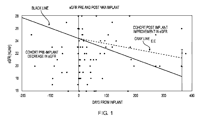
FIG. 1 is a graph showing estimated glomerular filtration rate (eGFR) pre- and
post-
REACT treatment.
FIG. 2 is a graph showing serum creatinine pre- and post-REACT treatment.
FIG. 3 is a photograph of a REACT product delivery system.
FIG. 4 is a photograph of a REACT shipping container.
FIG. 5 is study design flow diagram.
FIG. 6 is a flow diagram of a non-limiting example of an overall NKA
manufacturing
process.
FIG. 7 A-D are flow diagrams providing further details of the non-limiting
example
process depicted in FIG. 6.
FIG. 8 is a graph showing improvement in renal function as measured by eGFR in
a
patient receiving REACT treatment for kidney disease resulting from CAKUT;
star shows
patient's initial renal function before effect of CAKUT; solid gray line
(plotted -1 to 0
months relative to injection) patient's declining renal function as measured
by eGFR pre-
REACT injection; broken black line (plotted at 0 to 3 months relative to
injection), patient's
eGFR following REACT injection.
FIG. 9 is a graph showing improvement in renal function as measured by albumin-
to
creatinine ratio in a patient receiving REACT treatment for kidney disease
resulting from
CAKUT.
3
CA 03137915 2021-10-22
WO 2020/223660
PCT/US2020/031093
DETAILED DESCRIPTION
Reference is made herein to certain embodiments and examples encompassed by
the
invention. While the invention will be described in conjunction with exemplary
embodiments, it will be understood that they are not intended to limit the
invention to those
embodiments. On the contrary, the invention is intended to cover all
alternatives,
modifications, and equivalents which may be included within the scope of the
present
invention as defined by the claims. One skilled in the art will recognize many
methods and
materials similar or equivalent to those described herein, which could be used
in the practice
.. of the present invention. The present invention is in no way limited to the
methods and
materials described.
All references cited throughout the disclosure are expressly incorporated by
reference
herein in their entirety. In the event that one or more of the incorporated
literature, patents,
and similar materials differs from or contradicts this application, including
but not limited to
defined terms, term usage, described techniques, or the like, this application
controls.
Unless defined otherwise, technical and scientific terms used herein have the
same
meaning as commonly understood by one of ordinary skill in the art to which
this invention
belongs. One skilled in the art will recognize many methods and materials
similar or
equivalent to those described herein, which could be used in the practice of
the present
invention. Indeed, the present invention is in no way limited to the methods
and materials
described.
As used herein, the term "about" in the context of a numerical value or range
means
10% of the numerical value or range recited or claimed, unless the context
requires a more
limited range.
In the descriptions herein and in the claims, phrases such as "at least one
of' or "one
or more of' may occur followed by a conjunctive list of elements or features.
The term
"and/or" may also occur in a list of two or more elements or features. Unless
otherwise
implicitly or explicitly contradicted by the context in which it is used, such
a phrase is
intended to mean any of the listed elements or features individually or any of
the recited
elements or features in combination with any of the other recited elements or
features. For
example, the phrases "at least one of A and B;" "one or more of A and B;" and
"A and/or B"
are each intended to mean "A alone, B alone, or A and B together." A similar
interpretation
is also intended for lists including three or more items. For example, the
phrases "at least one
4
CA 03137915 2021-10-22
WO 2020/223660
PCT/US2020/031093
of A, B, and C" "one or more of A, B, and C" "A, B, and/or C" are each
intended to mean "A
alone, B alone, C alone, A and B together, A and C together, B and C together,
or A and B
and C together." In addition, use of the term "based on," above and in the
claims is intended
to mean, "based at least in part on," such that an unrecited feature or
element is also
permissible.
It is understood that where a parameter range is provided, all integers within
that
range, and tenths thereof, are also provided by the invention. For example,
"0.2-5 mg" is a
disclosure of 0.2 mg, 0.3 mg, 0.4 mg, 0.5 mg, 0.6 mg etc. up to and including
5.0 mg.
The transitional term "comprising," which is synonymous with "including,"
"containing," or "characterized by," is inclusive or open-ended and does not
exclude
additional, unrecited elements or method steps. By contrast, the transitional
phrase
"consisting of' excludes any element, step, or ingredient not specified in the
claim. The
transitional phrase "consisting essentially of' limits the scope of a claim to
the specified
materials or steps "and those that do not materially affect the basic and
novel
characteristic(s)" of the claimed invention.
As used herein, the singular forms "a," "an," and "the" include the plural
reference
unless the context clearly dictates otherwise.
As used herein, "treating" encompasses, e.g., inhibition, regression, or
stasis of the
progression of a disorder. Treating also encompasses the prevention or
amelioration of any
symptom or symptoms of the disorder. As used herein, "inhibition" of disease
progression or
a disease complication in a subject means preventing or reducing the disease
progression
and/or disease complication in the subject.
As used herein, a "symptom" associated with a disorder includes any clinical
or
laboratory manifestation associated with the disorder, and is not limited to
what the subject
can feel or observe.
As used herein, "effective" when referring to an amount of a therapeutic agent
refers
to the quantity of the agent that is sufficient to yield a desired therapeutic
response without
undue adverse side effects (such as toxicity, irritation, or allergic
response) commensurate
with a reasonable benefit/risk ratio when used in the manner of this
disclosure.
The term "bioactive renal cells" or "BRCs" as used herein refers to renal
cells having
one or more of the following properties when administered into the kidney of a
subject:
capability to reduce (e.g., slow or halt) the worsening or progression of
chronic kidney
disease or a symptom thereof, capability to enhance renal function, capability
to affect
(improve) renal homeostasis, and capability to promote healing, repair and/or
regeneration of
5
CA 03137915 2021-10-22
WO 2020/223660
PCT/US2020/031093
renal tissue or kidney. In embodiments, these cells may include functional
tubular cells (e.g.,
based on improvements in creatinine excretion and protein retention),
glomerular cells (e.g.,
based on improvement in protein retention), vascular cells and other cells of
the
corticomedullary junction. In embodiments, BRCs are obtained from isolation
and expansion
of renal cells from kidney tissue. In embodiments, BRCs are obtained from
isolation and
expansion of renal cells from kidney tissue using methods that select for
bioactive cells. In
embodiments, the BRCs have a regenerative effect on the kidney. In
embodiments, BRCs
comprise, consist essentially of, or consist of selected renal cells (SRCs).
In embodiments,
BRCs are SRCs.
In embodiments, SRCs are cells obtained from isolation and expansion of renal
cells
from a suitable renal tissue source, wherein the SRCs contain a greater
percentage of one or
more cell types and lacks or has a lower percentage of one or more other cell
types, as
compared to a starting kidney cell population. In embodiments, the SRCs
contain an
increased proportion of BRCs compared to a starting kidney cell population. In
embodiments, an SRC population is an isolated population of kidney cells
enriched for
specific bioactive components and/or cell types and/or depleted of specific
inactive and/or
undesired components or cell types for use in the treatment of kidney disease,
i.e., providing
stabilization and/or improvement and/or regeneration of kidney function. SRCs
provide
superior therapeutic and regenerative outcomes as compared with the starting
population. In
embodiments, SRCs are obtained from the patient's renal cortical tissue via a
kidney biopsy.
In embodiments, SRCs are selected (e.g., by fluorescence-activated cell
sorting or "FACS")
based on their expression of one or more markers. In embodiments, SRCs are
depleted (e.g.,
by fluorescence-activated cell sorting or "FACS") of one or more cell types
based on the
expression of one or more markers on the cell types. In embodiments, SRCs are
selected
from a population of bioactive renal cells. In embodiments, SRCs are selected
by density
gradient separation of expanded renal cells. In embodiments, SRCs are selected
by separation
of expanded renal cells by centrifugation across a density boundary, barrier,
or interface, or
single step discontinuous step gradient separation. In embodiments, SRCs are
selected by
continuous or discontinuous density gradient separation of expanded renal
cells that have
been cultured under hypoxic conditions. In embodiments, SRCs are selected by
density
gradient separation of expanded renal cells that have been cultured under
hypoxic conditions
for at least about 8, 12, 16, 20, or 24 hours. In embodiments, SRCs are
selected by
separation by centrifugation across a density boundary, barrier, or interface
of expanded renal
cells that have been cultured under hypoxic conditions. In embodiments, SRCs
are selected
6
CA 03137915 2021-10-22
WO 2020/223660
PCT/US2020/031093
by separation of expanded renal cells that have been cultured under hypoxic
conditions for at
least about 8, 12, 16, 20, or 24 hours by centrifugation across a density
boundary, barrier, or
interface (e.g., single-step discontinuous density gradient separation). In
embodiments, SRCs
are composed primarily of renal tubular cells. In embodiments, other
parenchymal (e.g.,
vascular) and stromal (e.g., collecting duct) cells may be present in SRCs. In
embodiments,
less than about 10%, 9%, 8%, 7%, 6%, 5%, 4%, 3%, 2%, or 1% of the cells in a
population of
SRCs are vascular cells. In embodiments, less than about 10%, 9%, 8%, 7%, 6%,
5%, 4%,
3%, 2%, or 1% of the cells in a population of SRCs are collecting duct cells.
In
embodiments, less than about 10%, 9%, 8%, 7%, 6%, 5%, 4%, 3%, 2%, or 1% of the
cells in
a population of SRCs are vascular or collecting duct cells.
The term "spheroid" refers to an aggregate or assembly of cells cultured to
allow 3-
dimensional growth as opposed to growth as a monolayer. It is noted that the
term "spheroid"
does not imply that the aggregate is a geometric sphere. In embodiments, the
aggregate may
be highly organized with a well defined morphology or the aggregate may be an
unorganized
mass. In embodiments, a spheroid may include a single cell type or more than
one cell type.
In embodiments, the cells may be primary isolates, or a permanent cell line,
or a combination
of the two. In embodiments, the spheroids (e.g., cellular aggregates or
organoids) are formed
in a spinner flask. In embodiments, the spheroids (e.g., cellular aggregates
or organoids) are
formed in a 3-dimensional matrix.
The term "native organ" shall mean the organ of a living subject. In
embodiments,
the subject may be healthy or unhealthy. In embodiments, an unhealthy subject
may have a
disease associated with that particular organ.
The term "native kidney" shall mean the kidney of a living subject. In
embodiments,
the subject may be healthy or unhealthy. In embodiments, an unhealthy subject
may have a
kidney disease. In embodiments, an unhealthy subject may have an anomaly of a
kidney
and/or urinary tract.
Provided herein are cell types and populations of cells (such as SRCs) that
provide a
benefit to a native organ, such as the kidney. In embodiments, the benefit
includes a halt or
slowing of the progression (e.g., the worsening of one or more symptoms) of
chronic kidney
disease. In embodiments, the benefit includes a regenerative effect, e.g.,
reduction in a
symptom of chronic kidney disease and/or an improvement in native kidney
function. In
embodiments, the benefit includes, without limitation, a reduction in the
degree of injury to a
native organ or an improvement in, restoration of, or stabilization of a
native organ function
7
CA 03137915 2021-10-22
WO 2020/223660
PCT/US2020/031093
or structure. Renal injury may be, e.g., in the form of fibrosis,
inflammation, glomerular
hypertrophy, atrophy, etc.
In embodiments, an enriched cell population or preparation is a cell
population
derived from a starting organ cell population (e.g., an unfractionated,
heterogeneous cell
population from a kidney) that contains a greater percentage of a specific
cell type than the
percentage of that cell type in the starting population. For example, a
starting kidney cell
population can be enriched for a first, a second, a third, a fourth, a fifth,
and so on, cell type
of interest.
The term "hypoxic" culture conditions as used herein refers to culture
conditions in
which cells are subjected to a reduction in available oxygen levels in the
culture system
relative to standard culture conditions in which cells are cultured at
atmospheric oxygen
levels (about 21%). Non-hypoxic conditions are referred to herein as normal or
normoxic
culture conditions.
Included herein are compositions comprising a biomaterial and one or more cell
types. In embodiments, the biomaterial is a natural or synthetic biocompatible
material that is
suitable for introduction into living tissue supporting cells in a viable
state. A natural
biomaterial is a material that is made by or originates from a living system.
Synthetic
biomaterials are materials which are not made by or do not originate from a
living system. In
embodiments, a biomaterial disclosed herein may be a combination of natural
and synthetic
biocompatible materials. As used herein, biomaterials include (but are not
limited to), for
example, polymeric matrices and scaffolds. Those of ordinary skill in the art
will appreciate
that the biomaterial(s) may be configured in various forms, for example, as
porous foam,
gels, liquids, beads, solids, and may comprise one or more natural or
synthetic biocompatible
materials. In embodiments, the biomaterial is the liquid form of a solution
that is capable of
becoming a hydrogel. In embodiments, the biomaterials is a hydrogel that is
capable of
becoming a liquid.
The term "kidney disease" as used herein includes disorders associated with
any stage
or degree of acute or chronic renal disease (e.g., acute or chronic renal
failure) that results in
a reduction or loss of the kidney's ability to perform the function of blood
filtration and
.. elimination of excess fluid, electrolytes, and wastes from the blood. In
embodiments, kidney
disease also includes endocrine dysfunctions such as anemia (erythropoietin-
deficiency), and
mineral imbalance (Vitamin D deficiency). In embodiments, kidney disease may
originate in
the kidney or may be secondary to a variety of conditions, including (but not
limited to)
congenital anomalies of the kidney and urinary tract (CAKUT), vesicoureteral
reflux, heart
8
CA 03137915 2021-10-22
WO 2020/223660
PCT/US2020/031093
failure, hypertension, diabetes, autoimmune disease, or liver disease. In
embodiments,
kidney disease may be a condition of chronic renal failure that develops after
an acute injury
to the kidney. For example, injury to the kidney by ischemia and/or exposure
to toxicants
may cause acute renal failure; incomplete recovery after acute kidney injury
may lead to the
development of chronic renal failure.
In embodiments, the term "treatment" may refer to therapeutic treatment and/or
prophylactic or preventative measures for kidney disease, anemia, tubular
transport
deficiency, or glomerular filtration deficiency wherein the object is to
reverse, prevent or
slow down (lessen) the targeted disorder. Those in need of treatment include
those already
having a kidney disease, anemia, tubular transport deficiency, or glomerular
filtration
deficiency as well as those prone to having a kidney disease, anemia, tubular
transport
deficiency, or glomerular filtration deficiency or those in whom the kidney
disease, anemia,
tubular transport deficiency, or glomerular filtration deficiency is to be
prevented. In
embodiments, a subject in need of treatment comprises a congenital anomaly of
a kidney
and/or urinary tract. The term "treatment" as used herein includes the
stabilization and/or
improvement of kidney function.
Included herein are constructs or formulations comprising one or more cell
types
(e.g., a cell population such as SRCs) deposited on or in a surface of a
scaffold or matrix
made up of one or more synthetic or naturally-occurring biocompatible
materials. In
embodiments, the one or more cell populations may be coated with, deposited
on, embedded
in, attached to, seeded, or entrapped in a biomaterial made up of one or more
synthetic or
naturally-occurring biocompatible biomaterials, polymers, proteins, or
peptides. In
embodiments, the one or more cell populations may be combined with a
biomaterial or
scaffold or matrix in vitro or in vivo. In embodiments, the one or more
biomaterials used to
generate the construct or formulation may be selected to direct, facilitate,
or permit dispersion
and/or integration of the cellular components of the construct with the
endogenous host
tissue, or to direct, facilitate, or permit the survival, engraftment,
tolerance, or functional
performance of the cellular components of the construct or formulation.
The term "Neo-Kidney Augment (NKA)" refers to a bioactive cell formulation
which
is an injectable product composed of autologous, homologous SRCs formulated in
a
biomaterial comprised of a gelatin-based hydrogel. The term "Advance Cell
Therapy
(ACT)" refers to treatment with NKA.
In embodiments, a subject is a living animal. In embodiments, a subject is a
mammal
such as a dog, cat, horse, rabbit, zoo animal, cow, pig, sheep, goat, camel,
mouse, rat, or
9
CA 03137915 2021-10-22
WO 2020/223660
PCT/US2020/031093
guinea pig. In embodiments, a subject is a primate such as a human, a
chimpanzee, an
orangutan, a monkey, or a baboon. In embodiments, a subject is a human. In
embodiments,
a subject is a patient, eligible for treatment, who is experiencing or has
experienced one or
more signs, symptoms, or other indicators of a kidney disease. Such subjects
include without
limitation subjects who are newly diagnosed or previously diagnosed and are
now
experiencing a recurrence or relapse, or are at risk for a kidney disease, no
matter the cause.
In embodiments, the subject may have been previously treated for a kidney
disease, or not so
treated. In embodiments, a subject has a congenital anomaly of a kidney and/or
urinary tract.
In embodiments, a subject is a human with congenital anomalies of the kidney
and urinary
tract. In embodiments, a subject is experiencing or has experienced one or
more signs,
symptoms, or other indicators of an organ-related disease, such as kidney
disease, anemia, or
erythropoietin (EPO) deficiency. In embodiments, the subject does not have
diabetes. In
embodiments, the subject does not have Type I diabetes. In embodiments, the
subject does
not have Type II diabetes.
Congenital anomalies of the kidney and urinary tract (CAKUT) includes a family
of
diseases of various anatomic spectrum, including renal anomalies, and
anomalies of the
bladder and urethra. In embodiments, the term "CAKUT" refers to one congenital
abnormality (e.g., when referring to a subject who has CAKUT). In embodiments,
the term
CAKUT refers to more than one congenital abnormality (e.g., when referring to
a subject
who has CAKUT). In embodiments, a subject with CAKUT has one or more
abnormalities
of the kidney, bladder, and/or urethra. In embodiments, a subject with CAKUT
has an
abnormality in one or two kidneys. In embodiments, a subject with CAKUT has an
abnormality in the urethra. In embodiments, the CAKUT has resulted from a
genetic
mutation or abnormality. In embodiments, the CAKUT has resulted from an
environmental
factor. In embodiments, a subject with CAKUT has an abnormality in the
bladder. Non-
limiting descriptions relating to CAKUT are provided in Ristoska-Bojkovska
(2017) Pril
(Makedon Akad Nauk Umet Odd Med Nauki) 38(1):59-62; and Rodriguez (2014) Fetal
Pediatr Pathol. 33(5-6):293-320, the entire contents of each of which are
incorporated herein
by reference. In embodiments, a subject who has CAKUT does not have diabetes.
In
embodiments, a subject who has CAKUT does not have Type I diabetes. In
embodiments, a
subject who has CAKUT does not have Type II diabetes.
CAKUT constitute approximately 20 to 30 percent of all anomalies identified in
the
prenatal period. See Queisser-Luft et al. (2002) Malformations in newborn:
results based on
30,940 infants and fetuses from the Mainz congenital birth defect monitoring
system (1990-
CA 03137915 2021-10-22
WO 2020/223660
PCT/US2020/031093
1998). Spranger J Arch Gynecol Obstet. 2002;266(3):163, the entire content of
which is
incorporated herein by reference. In embodiments, defects can be bilateral or
unilateral, and
different defects often coexist in an individual child.
In embodiments, CAKUT represent a broad range of disorders that result from
abnormal embryogenic renal development due to renal parenchymal malformations,
abnormalities in renal migration, or abnormalities in the developing
collecting system. In
embodiments, CAKUT represent a broad range of disorders and are the result of
abnormal
renal developmental processes. In embodiments, malformation of the renal
parenchyma
results in failure of normal nephron development, as seen in renal dysplasia,
rheumatoid
arthritis (RA), renal tubular dysgenesis, and some types of nephronophthisis.
Without being
bound by any scientific theory, investigation utilizing molecular genetics has
demonstrated
that renal malformation results from defects in genes that encode signaling
and transcription
factors. In embodiments, environmental factors, such as prenatal exposure to
teratogens, can
also disrupt renal morphogenesis resulting in CAKUT. In embodiments,
abnormalities
comprise abnormal embryonic migration of the kidneys, as seen in renal ectopy
(e.g., pelvic
kidney), and fusion anomalies, such as horseshoe kidney. In embodiments,
abnormalities of
the developing urinary collecting system, as seen in duplicate collecting
systems, posterior
urethral valves, and ureteropelvic junction obstruction may lead to CKD/ESRD.
Renal
dysplasia may be unilateral or bilateral and occurs in two to four per 1000
births. The male-
to-female ratio for bilateral renal dysplasia is 1.3:1, and for unilateral
dysplasia is 1.9:1
Because CAKUT play a causative role in 30 to 50 percent of cases of end-stage
renal
disease (ESRD) in children (Seikaly et al. 2003 Chronic renal insufficiency in
children: the
2001 Annual Report Pediatr Nephrol. 18(8):796), in embodiments it is important
to diagnose
these anomalies and initiate therapy to minimize renal damage, prevent or
delay the onset of
ESRD, and provide supportive care to avoid complications of ESRD. Patients
with
malformations involving a reduction in kidney numbers or size are most likely
to have a poor
renal prognosis (Sanna-Cherchi et al. 2009 Renal outcome in patients with
congenital
anomalies of the kidney and urinary tract. Kidney Int. 76(5):528). In
embodiments, by 30
years of age, most patients will have dialysis.
The risk for dialysis is significantly higher for patients with a solitary
kidney or with
renal hypodysplasia associated with posterior urethral valves compared to
patients with
unilateral or bilateral renal hypodysplasia, or multicystic or horseshoe
kidney. In
embodiments, sub-clinical defects of the solitary kidney maybe responsible for
a poorer
prognosis compared to more benign forms of CAKUT. In embodiments, and without
being
11
CA 03137915 2021-10-22
WO 2020/223660
PCT/US2020/031093
limited by any scientific theory, children with a solitary kidney are at risk
for long-term
CKD, which is thought to be due to glomerular hyperfiltration. In embodiments,
about one-
third of patients can have evidence of renal injury defined as proteinuria
(e.g., urine protein to
creatinine ratio >0.2 mg/mg [>22.6 mg/mmol in children greater than two years
of age]),
hypertension (e.g., blood pressure >95th percentile for age, gender, and
height), elevated
estimated creatinine clearance based on serum creatine and Schwartz equation,
or the use of
medication for renal protection (e.g., angiotensin-converting enzyme
inhibitors).
In embodiments, renal dysplasia may be discovered during routine antenatal
screening
or postnatally when renal ultrasonography is performed in a dysmorphic infant.
In
embodiments, bilateral dysplasia is likely to be diagnosed earlier than
unilateral dysplasia
especially if oligohydramnios is present. In embodiments, renal ultrasound
features include
increased echogenicity as a result of abnormal renal parenchymal tissue, poor
corticomedullary differentiation, and parenchymal cysts.
In embodiments, infants with bilateral dysplasia may have impaired renal
function at
birth, and subsequent progressive renal failure may occur. In embodiments,
associated
urological findings include abnormalities of the renal pelvis, calyces (e.g.,
congenital
hydronephrosis), and ureters e.g., duplicating collecting system megaureter,
ureteral stenosis,
and vesicoureteral reflux [VUR]. In embodiments, as a result, symptomatic
presentation may
occur due to complications associated with these urological anomalies,
including urinary tract
infection (UTI), hematuria, fever, and abdominal pain.
In embodiments, because of the frequent association of renal dysplasia with a
collecting system anomaly, voiding cystourethrography may be considered in
patients with
renal dysplasia with or without a UTI. In embodiments, if there is an
associated urological
abnormality such as VUR in the normal contralateral kidney, children with
unilateral renal
dysplasia may be at increased risk of long-term sequelae of renal scarring
from recurrent
UTI. In embodiments, a DMSA radionuclide scan can provide further information
on the
differential function of each kidney. In embodiments, multicystic dysplastic
kidney (MCDK)
typically has no viable functional renal tissue and, therefore, no detectable
renal blood flow
or renal function. However, in embodiments, there may be rare variations of
segmental
dysplasia. In embodiments, imaging studies may be useful in defining baseline
renal
function and risk of future renal damage and the ability to regenerate normal
functioning
renal parenchyma.
The term "sample" or "patient sample" or "biological sample" shall generally
include
any biological sample obtained from a subject or patient, body fluid, body
tissue, cell line,
12
CA 03137915 2021-10-22
WO 2020/223660
PCT/US2020/031093
tissue culture, or other source. The term includes tissue biopsies such as,
for example, kidney
biopsies. The term includes cultured cells such as, for example, cultured
mammalian kidney
cells. Methods for obtaining tissue biopsies and cultured cells from mammals
are well
known in the art. In embodiments, a sample may originate from various sources
in a
mammalian subject including, without limitation, blood, semen, serum, urine,
bone marrow,
mucosa, tissue, etc.
The term "control sample" refers a negative or positive control sample in
which a
negative or positive result is expected to help correlate a result in the test
sample. In
embodiments, a suitable control sample includes, without limitation, a sample
known to
exhibit indicators characteristic of normal kidney function, a sample obtained
from a subject
known not to have kidney disease, and a sample obtained from a subject known
to have
kidney disease. In embodiments, a control sample may be a sample obtained from
a subject
prior to being treated by a method provided herein. In embodiments, a control
sample may
be a test sample obtained from a subject known to have any type or stage of
kidney disease,
and a sample from a subject known not to have any type or stage of kidney
disease. In
embodiments, a control sample may be a normal healthy matched control. Those
of skill in
the art will appreciate other control samples suitable for use.
Provided herein are, inter alia, methods and compositions for treating chronic
kidney
disease in subjects with an anomaly of a kidney and/or urinary tract (e.g., a
subject who has
CAKUT).
In an aspect, provided herein is a method of treating kidney disease in a
subject who
has chronic kidney disease, the method comprising, consisting essentially of,
or consisting of
administering to the subject an effective amount of (i) a bioactive renal cell
population; (ii)
vesicles secreted by the renal cell population; and/or (iii) spheroids
comprising the renal cell
population and at least one non-renal cell population, wherein the subject has
an anomaly of a
kidney and/or urinary tract. In embodiments, the subject has an anomaly of a
kidney.
In an aspect, provided herein is a method of treating kidney disease in a
subject who
has an anomaly of a kidney and/or urinary tract, the method comprising,
consisting
essentially of, or consisting of administering to the subject an effective
amount of (i) a
bioactive renal cell population; (ii) one or more products secreted by the
renal cell
population; and/or (iii) spheroids comprising the renal cell population and at
least one other
cell population.
In an aspect, provided herein is a method of treating kidney disease in a
subject who
has an anomaly of a kidney and/or urinary tract, the method comprising
administering to the
13
CA 03137915 2021-10-22
WO 2020/223660
PCT/US2020/031093
subject an effective amount of (i) a bioactive renal cell population; (ii) one
or more products
(such as vesicles) secreted by the renal cell population; and/or (iii)
spheroids comprising the
renal cell population and at least one other cell population, such as a non-
renal cell
population.
In an aspect, provided herein is a method of treating kidney disease in a
subject who
has an anomaly of a kidney and/or urinary tract, the method comprising
administering to the
subject an effective amount of (i) a bioactive renal cell population; (ii)
vesicles secreted by
the renal cell population; or (iii) spheroids comprising the renal cell
population and at least
one non-renal cell population.
In an aspect, provided herein is a method of treating kidney disease in a
subject who
has an anomaly of a kidney and/or urinary tract, the method comprising
administering to the
subject an effective amount of (i) a bioactive renal cell population; (ii)
vesicles secreted by
the renal cell population; and (iii) spheroids comprising the renal cell
population and at least
one non-renal cell population.
In an aspect, provided herein is a method of treating kidney disease in a
subject who
has an anomaly of a kidney and/or urinary tract, the method comprising
administering to the
subject an effective amount of a composition comprising a bioactive renal cell
population. In
embodiments, the composition further comprises vesicles secreted by the renal
cell
population. In embodiments, the composition further comprises spheroids
comprising the
renal cell population and at least one non-renal cell population.
In an aspect, provided herein is a method of treating kidney disease in a
subject who
has an anomaly of a kidney and/or urinary tract, the method comprising
administering to the
subject an effective amount of vesicles secreted by the renal cell population.
In an aspect, provided herein is a method of treating kidney disease in a
subject who
has an anomaly of a kidney and/or urinary tract, the method comprising
administering to the
subject an effective amount of spheroids comprising the renal cell population
and at least one
non-renal cell population.
In an aspect, provided herein is a bioactive renal cell population and uses
thereof for
treating kidney disease in a subject who has an anomaly of a kidney and/or
urinary tract.
In an aspect, provided herein are products (such as vesicles) secreted by a
bioactive
renal cell population and uses thereof for treating kidney disease in a
subject who has an
anomaly of a kidney and/or urinary tract.
14
CA 03137915 2021-10-22
WO 2020/223660
PCT/US2020/031093
In an aspect, provided herein are spheroids comprising a bioactive renal cell
population and uses thereof for treating kidney disease in a subject who has
an anomaly of a
kidney and/or urinary tract.
In embodiments, the subject has an anomaly of a urinary tract. In embodiments,
the
subject has an anomaly of a kidney and a urinary tract. In embodiments, the
anomaly is
acquired before birth. In embodiments, the anomaly is acquired after birth. In
embodiments,
the anomaly is a congenital anomaly. In embodiments, the subject has a
congenital anomaly
of a kidney. In embodiments, the subject has a congenital anomaly of the
urinary tract. In
embodiments, the subject has a congenital anomaly of a kidney and a urinary
tract. In
embodiments, a congenital anomaly worsens or gives rise to additional
abnormalities after
birth. In embodiments, a subject has an abnormality in one kidney. In
embodiments, a
subject has one or more abnormalities in each kidney. In embodiments, the
subject has an
abnormality in the urinary tract, wherein the abnormality is in the urethra.
In embodiments,
the subject has an abnormality in the urinary tract, wherein the abnormality
is in the bladder.
In embodiments, the subject has an abnormality in the urinary tract, wherein
the abnormality
is in a ureter. In embodiments, an anomaly is present at birth, but does not
manifest or show
symptoms until after birth.
In embodiments, the kidney disease is CKD. In embodiments, the subject has CKD
from anomalies (e.g. congenitally) of the kidney and urinary tract.
In embodiments, the anomaly comprises a congenital anomaly. In embodiments,
the
subject has CAKUT.
In embodiments, the anomaly is a morphological anomaly. In embodiments, the
subject has an abnormally developed kidney.
In embodiments, the subject has or has had primary vesicoureteral reflux,
reflux
nephropathy, renal scaring, or renal hypodysplasia. In embodiments, the
subject has or has
had reflux nephropathy. In embodiments, the subject has or has had renal
scaring. In
embodiments, the subject has or has had renal hypodysplasia.
In embodiments, the subject is predisposed to urinary tract infections. In
embodiments, the subject has had at least about 1, 2, 3, 4, 5, 6, 7, 8, 9, or
10 urinary tract
infections.
In embodiments, the subject has hypertension or proteinuria.
In embodiments, the subject has had post-antireflux surgery.
In embodiments, the subject has a glomerular filtration rate (GFR) of less
than 90
mL/min/1.73 m2, microalbuminuria, or macroalbuminuria. In embodiments, the
subject has a
CA 03137915 2021-10-22
WO 2020/223660
PCT/US2020/031093
GFR of less than 80 mL/min/1.73 m2. In embodiments, the subject has a GFR of
less than 70
mL/min/1.73 m2. In embodiments, the subject has a GFR of less than 60
mL/min/1.73 m2. In
embodiments, the subject has a GFR of less than 50 mL/min/1.73 m2. In
embodiments, the
subject has a GFR of less than 40 mL/min/1.73 m2. In embodiments, the subject
has a GFR
of less than 30 mL/min/1.73 m2. In embodiments, the subject has a GFR of at
least 10
mL/min/1.73 m2. In embodiments, the subject has a GFR of at least 15
mL/min/1.73 m2. In
embodiments, the subject has a GFR of lat least 20 mL/min/1.73 m2. In
embodiments, the
subject has a GFR of at least 30 mL/min/1.73 m2. In embodiments, the subject
has a GFR of
from 10, 15, 20, 25 or 30 mL/min/1.73 m2 to 50, 60, 70, 80 or 90 mL/min/1.73
m2. In
embodiments, the GFR is the estimated GFR (eGFR). In embodiments, the subject
has
microalbuminuria. In embodiments, the subject has macroalbuminuria.
In embodiments, the subject is less than 18 years old. In embodiments, the
subject is
less than 60 years old. In embodiments, the subject is less than 50 years old.
In
embodiments, the subject is less than 40 years old. In embodiments, the
subject is less than
35 years old. In embodiments, the subject is less than 30 years old. In
embodiments, the
subject is less than 25 years old. In embodiments, the subject is less than 20
years old. In
embodiments, the subject is from 1 to 16 years old. In embodiments, the
subject is from 1, 2,
3, 4, 5, 6, 7, 8, 9, or 10 to 20, 25, 30, 35, or 40 years old.
In embodiments, the subject is from 20, 25, 30, 35, or 40 to 50, 55, 65, 70,
75, 80, 85,
90, 95, or 100 years old. In embodiments, the subject is at least 50 years
old. In
embodiments, the subject is at least 55 years old. In embodiments, the subject
is at least 60
years old. In embodiments, the subject is at least 65 years old. In
embodiments, the subject
is at least 70 years old.
In embodiments, the subject has a Renal Parenchymal Malformation.
In embodiments, the subject has a ureteral duplication, a ureteropelvic
junction
obstruction, renal agenesis, vesicoureteral reflux, renal dysplasia, renal
hypoplasia, renal
hypodysplasia, congenital hydronephrosis, a horseshoe kidney, posterior
urethral valve and
prune belly syndrome, obstructive renal dysplasia, or a nonmotile ciliopathy.
In embodiments, the abnormality has been caused by or has been correlated with
a
.. genetic factor. In embodiments, the CAKUT has been caused by or has been
correlated with a
genetic factor. In embodiments, the abnormality has been caused by or has been
correlated
with a non-genetic factor. In embodiments, the CAKUT has been caused by or has
been
correlated with a non-genetic factor. In embodiments, the non-genetic factor
is an
environmental factor.
16
CA 03137915 2021-10-22
WO 2020/223660
PCT/US2020/031093
In embodiments, the subject has a ureteral duplication, a ureteropelvic
junction
obstruction, renal agenesis, vesicoureteral reflux, renal hypodysplasia,
congenital
hydronephrosis, a horseshoe kidney, posterior urethral valve and prune belly
syndrome,
obstructive renal dysplasia, or a nonmotile ciliopathy. In embodiments, the
subject has a
ureteral duplication. In embodiments, the subject has a ureteropelvic junction
obstruction. In
embodiments, the subject has renal agenesis. In embodiments, the subject has
vesicoureteral
reflux. In embodiments, the subject has renal hypodysplasia. In embodiments,
the subject has
congenital hydronephrosis. In embodiments, the subject has a horseshoe kidney.
In
embodiments, the subject has posterior urethral valve and prune belly
syndrome. In
embodiments, the subject has obstructive renal dysplasia. In embodiments, the
subject has a
nonmotile ciliopathy. In embodiments, the CAKUT has been caused by or has been
correlated with a genetic factor.
In embodiments, the anomaly comprises Alagille syndrome, Apert syndrome,
Bardet-
Biedl syndrome, Beckwith-Wiedemann syndrome, Branchio-Oto-Renal syndrome
(BOR),
Campomelic dysplasia, Cenani-Lenz syndrome, DiGeorge syndrome, Fraser
syndrome,
hypoparathyroidism sensorineural deafness and renal anomalies (HDR), Kallmann
syndrome,
Mammary-Ulnar syndrome, Meckel Gruber syndrome, nephronophthisis, Okihiro
syndrome,
Pallister-Hall syndrome, Renal coloboma syndrome, hypoplasia, dysplasia, renal
dysplasia,
cystic dysplasia, non-cystic dysplasia, VUR Cystic dysplasia, renal
hypoplasia, isolated cystic
renal hypoplasia, isolated non-cystic renal hypoplasia, isolated renal tubular
dysgenesis,
Rubinstein-Taybi syndrome, Simpson-Golabi Behmel syndrome, Townes-Brock
syndrome,
Zellweger syndrome, Smith-Lemli-Opitz syndrome, hydronephrosis, medullary
dysplasia,
unilateral/bilateral agenesis/dysplasia, collecting system anomalies,
agenesis, ureteropelvic
junction obstruction (UPJO) agenesis, dysplasia agenesis, unilateral agenesis,
VUR,
malrotation, cross-fused ectopia, VUR Dysplasia, a dual Serine/Threonine And
Tyrosine
Protein Kinase (DSTYK) mutation, a DSTYK mutation associated with UPJO,
tubular
dysgenesis, cysts, and/or aplasia.
In embodiments, the kidney disease is chronic kidney disease. In embodiments,
the
chronic kidney disease is Stage I, II, III, IV, or V kidney disease. In
embodiments, the
chronic kidney disease is Stage I kidney disease. In embodiments, the chronic
kidney disease
is Stage II kidney disease. In embodiments, the chronic kidney disease is
Stage III kidney
disease. In embodiments, the chronic kidney disease is Stage IV kidney
disease. In
embodiments, the chronic kidney disease is Stage V kidney disease. In
embodiments, the
subject is receiving dialysis at least 1, 2, or 3 times per week.
17
CA 03137915 2021-10-22
WO 2020/223660
PCT/US2020/031093
In embodiments, a population of bioactive renal cells is administered to a
native organ
as part of a formulation described herein. In embodiments, a secreted product
of population
of bioactive renal cells is administered to a native organ as part of a
formulation described
herein. In embodiments, the cells are sourced from the native organ that is
the subject of the
administration or from a source that is not the target native organ.
In embodiments, cells of the renal cell population are in the form of
spheroids. In
embodiments, spheroids comprising bioactive renal cells are administered to a
subject. In
embodiments, the spheroids comprise at least one non-renal cell type or
population of cells.
In embodiments, the subject has renal disease as measured by microalbuminuria
which may be defined by a urinary albumin-creatinine ratio (UACR) > 30 mg/g or
urine
albumin excretion > 30 mg/day on 24 hour urine collection.
In embodiments, the patient's kidney function is improved as a result of the
treatment.
An improvement of the patient's kidney function may be a stabilization of the
patient's
kidney function or may be a change in kidney function that improves the kidney
function. In
embodiments, the improved kidney function is demonstrated by a reduction in
the rate of
decline, stabilization of, or an increase in estimated glomerular filtration
rate (eGFR). In
embodiments in which the improved kidney function is demonstrated by an
increase in
eGFR, the increase in eGFR may be an increase of at least 1%, at least 2 %, at
least 3%, at
least 4%, at least 5%, at least 6%, at least 7%, at least 8%, at least 9%, at
least 10%, at least
15%, at least 20%, or at least 25% relative to the patient's baseline eGFR. In
such
embodiments, a patient's baseline eGFR may be the patient's eGFR prior to a
first dose of the
treatment, e.g., may be the patient's eGFR as determined at most 3 months, 2
months, 1
month, 3 weeks, 2 weeks, 10 days, 7 days, 6 days, 5 days, 4 days, 3 days, 2
days, or 1 day
prior to the administration of a first dose of the treatment. The increase in
the patient's
baseline eGFR may be achieved within 1 to 6 months, or 2 to 6 months, or 3 to
6 months, or 4
to 6 months, or 5 to 6 months, or 1 to 5 months, or 1 to 4 months, or 1 to 3
months, or 2 to 5
months, or 2 to 4 months, or 3 to 4 months, or 2 to 3 months, or 2 months, or
3 months, or 4
months, or 5 months or 6 months following administration of a first dose of
the treatment.
The increase over the patient's baseline eGFR need not be at a constant level
or to a constant
degree, i.e., the patient need not maintain the same initial level of increase
over baseline for
the treatment to "improve" kidney function. The increase in patient's baseline
eGFR, once
achieved, may decline, provided however, that the patient's eGFR continues to
be increased
relative to the patient's baseline eGFR. The increase in patient's baseline
eGFR, once
achieved, may also be further increased or it may maintain its same level of
increase over
18
CA 03137915 2021-10-22
WO 2020/223660
PCT/US2020/031093
baseline as does its initial level of increase over baseline. The patient's
increase in eGFR
over baseline may be for over a period of time of at least 12 months, 12
months, at least 18
months, 18 months, at least 24 months, 24 months, at least 30 months, 30
months, at least 36
months, 36 months, at least 42 months, 42 months, at least 48 months, 48
months, at least 54
months, 54 months, at least 60 months, 60 months, at least 66 months, 66
months, at least 72
months, 72 months, at least 78 months, 78 months, or the remaining lifetime of
the patient.
In embodiments, the improved kidney function is demonstrated by a reduction in
albumin to creatinine ratio (ACR) in the patient. In embodiments in which the
improved
kidney function is demonstrated by a reduction in ACR in the patient, the
reduction may be
by at least 10%, at least 20%, at least 30%, at least 40%, at least 50%, at
least 60%, at least
70%, or at least 80%, or at least 90% relative to the patient's baseline ACR.
Alternatively,
the reduction in ACR may be such that if the patient's baseline ACR is
moderately increased,
e.g., between 30 mg/g and 300 mg/g, then the reduction in ACR may reduce the
patent's
ACR to levels in the mild to normal range, e.g., less than 30 mg/g.
Alternatively, the
reduction in ACR may such that if the patient's baseline ACR is severely
increased, e.g.,
greater than 300 mg/g, then the reduction in ACR may reduce the patient's ACR
to levels that
are moderately increased, e.g., between 30 mg/g and 300 mg/g, or mildly
increased to
normal, e.g., less than 30 mg/g. In such embodiments, a patient's baseline ACR
may be the
patient's ACR prior to a first dose of the treatment, e.g., may be the
patient's ACR as
determined at most 3 months, 2 months, 1 month, 3 weeks, 2 weeks, 10 days, 7
days, 6 days,
5 days, 4 days, 3 days, 2 days, or 1 day prior to administration of the first
dose of the
treatment. The reduction in the patient's baseline ACR may be achieved within
1 to 6
months, or 2 to 6 months, or 3 to 6 months, or 4 to 6 months, or 5 to 6
months, or 1 to 5
months, or 1 to 4 months, or 1 to 3 months, or 2 to 5 months, or 2 to 4
months, or 3 to 4
months, or 2 to 3 months, or 2 months, or 3 months, or 4 months, or 5 months
or 6 months
following administration of the first dose of the treatment. The reduction in
the patient's
baseline ACR need not be at a constant level or to a constant degree, i.e.,
the patient need not
maintain the same initial level of reduction over baseline for the treatment
to "improve"
kidney function. The reduction in patient's baseline ACR, once achieved, may
increase,
provided however, that the patient's ACR continues to be reduced relative to
the patient's
baseline ACR. The reduction in patient's baseline ACR, once achieved, may also
be further
reduced or it may maintain its same level of reduction over baseline as does
its initial level of
reduction over baseline. The patient's reduction in ACR over baseline may be
for over a
period of time of at least 12 months, 12 months, at least 18 months, 18
months, at least 24
19
CA 03137915 2021-10-22
WO 2020/223660
PCT/US2020/031093
months, 24 months, at least 30 months, 30 months, at least 36 months, 36
months, at least 42
months, 42 months, at least 48 months, 48 months, at least 54 months, 54
months, at least 60
months, 60 months, at least 66 months, 66 months, at least 72 months, 72
months, at least 78
months, 78 months, or the remaining lifetime of the patient.
In embodiments, the improved kidney function is demonstrated by reduction in
total
serum creatinine or the rate of increase in serum creatine (sCr), or
comparable measure (e.g.,
Cystatin-C, inulin, or other measures of glomerular filtration. In
embodiments, the improved
kidney function is demonstrated by improved renal cortical thickness. In
embodiments, the
improved kidney function may be demonstrated by structural and functional
alterations. In
embodiments, the improved kidney size and/or structure is determined by renal
imaging. In
embodiments, the method of renal imaging is ultrasound, MRI, or renal
scintigraphy. In
embodiments, the improved renal function is superior to the prior state of
kidney structure or
function.
In embodiments, the effective treatment of a kidney disease in a subject as
provided
herein can be observed through various indicators of kidney function. In
embodiments, the
indicators of kidney function include, without limitation, serum albumin
level, albumin to
globulin ratio (A/G ratio), serum phosphorous level, serum sodium level,
kidney size
(measurable by ultrasound), serum calcium level, phosphorous:calcium ratio,
serum
potassium level, proteinuria, urine creatinine level, serum creatinine level,
blood nitrogen
urea (BUN) level, cholesterol level, triglyceride levels and glomerular
filtration rate (GFR).
In embodiments, several indicators of general health and well-being include,
without
limitation, weight gain or loss, survival, blood pressure (mean systemic blood
pressure,
diastolic blood pressure, or systolic blood pressure), and physical endurance
performance.
In embodiments, an effective treatment with a bioactive renal cell formulation
is
evidenced by stabilization of one or more indicators of kidney function. In
embodiments, the
stabilization of kidney function is demonstrated by the observation of a
change in an indicator
in a subject treated by a method provided herein as compared to the same
indicator in a
subject that has not been treated by a method provided herein. In embodiments,
the
stabilization of kidney function may be demonstrated by the observation of a
change in an
indicator in a subject treated by a method provided herein as compared to the
same indicator
in the same subject prior to treatment. In embodiments, the change in the
first indicator may
be an increase or a decrease in value. In embodiments, the treatment provided
herein may
include stabilization of serum creatinine levels in a subject where the BUN
levels observed in
the subject are lower as compared to a subject with a similar disease state
who has not been
CA 03137915 2021-10-22
WO 2020/223660
PCT/US2020/031093
treated by the methods provided herein. In embodiments, the treatment may
include
stabilization of serum creatinine levels in a subject where the serum
creatinine levels
observed in the subject are lower as compared to a subject with a similar
disease state who
has not been treated by the methods provided herein. In embodiments, the
stabilization of
one or more of the above indicators of kidney function is the result of
treatment with a
selected renal cell formulation.
Those of ordinary skill in the art will appreciate that one or more additional
indicators
described herein or known in the art may be measured to determine the
effective treatment of
a kidney disease in the subject.
In embodiments, an effective treatment with a bioactive renal cell formulation
is
evidenced by improvement of one or more indicators of kidney function. In
embodiments, the
bioactive renal cell population provides an improved level of serum
creatinine. In
embodiments, the bioactive renal cell population provides an improved
retention of protein in
the serum. In embodiments, the bioactive renal cell population provides
improved levels of
serum cholesterol and/or triglycerides. In embodiments, the bioactive renal
cell population
provides an improved level of Vitamin D. In embodiments, the bioactive renal
cell population
provides an improved phosphorus:calcium ratio as compared to a non-enriched
cell
population. In embodiments, the bioactive renal cell population provides an
improved level
of hemoglobin as compared to a non-enriched cell population. In embodiments,
the bioactive
renal cell population provides an improved level of serum creatinine as
compared to a non-
enriched cell population. In embodiments, the improvement of one or more of
the above
indicators of kidney function is the result of treatment with a selected renal
cell formulation.
Included herein are methods for the regeneration of a native kidney in a
subject in
need thereof. In embodiments, the method includes the step of administering or
implanting a
bioactive cell population, formulation, or construct described herein to the
subject. In
embodiments, a regenerated native kidney may be characterized by a number of
indicators
including, without limitation, development of function or capacity in the
native kidney,
improvement of function or capacity in the native kidney, and the expression
of certain
markers in the native kidney. In embodiments, the developed or improved
function or
capacity may be observed based on the various indicators of kidney function
described above.
In embodiments, the regenerated kidney is characterized by differential
expression of one or
more stem cell markers. In embodiments, the stem cell marker may be one or
more of the
following: SRY (sex determining region Y)-box 2 (Sox2); Undifferentiated
Embryonic Cell
Transcription Factor (UTF1); Nodal Homolog from Mouse (NODAL); Prominin 1
(PROM1)
21
CA 03137915 2021-10-22
WO 2020/223660
PCT/US2020/031093
or CD133 (CD133); CD24; and any combination thereof (see Ilagan et al.
PCT/US2011/036347 incorporated herein by reference in its entirety, see also
Genheimer et
al., 2012. Molecular characterization of the regenerative response induced by
intrarenal
transplantation of selected renal cells in a rodent model of chronic kidney
disease. Cells
Tissue Organs 196: 374-384, incorporated by reference in its entirety. In
embodiments, the
expression of the stem cell marker(s) is up-regulated compared to a control.
In embodiments, the effect may be provided by the cells themselves and/or by
products secreted from the cells. In embodiments, a product secreted from the
cells is
administered to the subject. In embodiments, the product hs been isolated from
cells, e.g., the
cells that produced it. In embodiments, the product is a vesicle as described
herein. In
embodiments, the vesicle (e.g., an exosome), has been isolated from the renal
cell population
that produced it. In embodiments, the vesicles may include one or more of the
following:
paracrine factors, endocrine factors, juxtacrine factors, microvesicles,
exosomes, and RNA.
The secreted products may also include products that are not within
microvesicles including,
without limitation, paracrine factors, endocrine factors, juxtacrine factors,
and RNA. In
embodiments, the secreted products may be part of a vesicle derived from renal
cells. In
embodiments, the vesicles are secreted vesicles. In embodiments, the secreted
vesicles are
exosomes, microvesicles, ectosomes, membrane particles, exosome-like vesicles,
or apoptotic
vesicles. In embodiments, the secreted vesicles are exosomes. In embodiments,
the secreted
vesicles are microvesicles. In embodiments, the secreted vesicles contain or
comprise one or
more cellular components. In embodiments, the components may be one or more of
the
following: membrane lipids, RNA, proteins, metabolities, cytosolic components,
and any
combination thereof. In embodiments, the secreted vesicles comprise one or
more
microRNAs. In embodiments, the one or more miRNAs include one of or any
combination of
RNA (e.g., miRNA) molecules disclosed herein. In embodiments, the vesicles
comprise an
miRNA that inhibits Plasminogen Activation Inhibitor-1 (PAI-1) and/or TGF131.
In
embodiments, the secreted product that comprises a paracrine and/or juxtacrine
factor, such
as alpha-1 microglobulin, beta-2-microglobulin, calbindin, clusterin,
connective tissue growth
factor, cystatin-C, glutathione-S-transferase alpha, kidney injury moleculte-
1, neutraphil
gelatinase-associated lipocalin, osteopontin, trefoil factor 3, tam-horsfall
urinary
glycoprotein, tissue-inhibitor of metallo proteinase 1, vascular endothelial
growth factor,
fibronectin, interleukin-6, or mono cyte chemotactic protein-1.
In embodiments, the effect may be provided by the cells themselves and/or by
products secreted from the cells. In embodiments, regenerative effect may be
characterized
22
CA 03137915 2021-10-22
WO 2020/223660
PCT/US2020/031093
by one or more of the following: a reduction in epithelial-mesenchymal
transition (which
may be via attenuation of TGF-p signaling); a reduction in renal fibrosis; a
reduction in renal
inflammation; differential expression of a stem cell marker in the native
kidney; migration of
implanted cells and/or native cells to a site of renal injury, e.g., tubular
injury, engraftment of
.. implanted cells at a site of renal injury, e.g., tubular injury;
stabilization of one or more
indicators of kidney function (as described herein); de novo formation of S-
shaped
bodies/comma-shaped bodies associated with nephrogenesis, de novo formation of
renal
tubules or nephrons, restoration of erythroid homeostasis (as described
herein); and any
combination thereof. (see also Basu et al., 2011. Functional evaluation of
primary renal
cell/biomaterial neo-kidney augment prototypes for renal tissue engineering.
Cell
Transplantation 20: 1771-90; Bruce et al., 2015. Selected renal cells modulate
disease
progression in rodent models of chronic kidney disease via NF-KB and TGF-p 1
pathways.
Regenerative Medicine 10: 815-839, the entire content of each of which is
incorporated
herein by reference).
In embodiments, in addition to a tissue biopsy or as an alternative to a
tissue biopsy, a
regenerative outcome in a subject receiving treatment can be assessed from
examination of a
bodily fluid, e.g., urine. It has been discovered that microvesicles obtained
from subject-
derived urine sources contain certain components including, without
limitation, specific
proteins and miRNAs that are ultimately derived from the renal cell
populations. In
embodiments, these components may include factors involved in stem cell
replication and
differentiation, apoptosis, inflammation and immuno-modulation. In
embodiments, a
temporal analysis of microvesicle-associated miRNA/protein expression patterns
allows for
continuous monitoring of regenerative outcomes within the kidney of subjects
receiving the
cell populations or constructs described herein.
Also provided are methods of assessing whether a kidney disease patient is
responsive
to treatment with a therapeutic formulation. In embodiments, the method may
include the
step of determining or detecting the amount of vesicles or a luminal content
or contents
thereof in a test sample obtained from a kidney disease patient treated with
the therapeutic, as
compared to or relative to the amount of vesicles in a control sample, wherein
a higher or
.. lower amount of vesicles or one or more luminal contents thereof in the
test sample as
compared to the amount of vesicles or luminal content(s) in the control sample
is indicative
of the treated patient's responsiveness to treatment with the therapeutic.
In embodiments, these kidney-derived vesicles and/or the luminal contents of
kidney
derived vesicles may also be shed into the urine of a subject and may be
analyzed for
23
CA 03137915 2021-10-22
WO 2020/223660
PCT/US2020/031093
biomarkers indicative of regenerative outcome or treatment efficacy. In
embodiments, the
non-invasive prognostic methods may include the step of obtaining a urine
sample from the
subject before and/or after administration or implantation of a cell
population, composition,
formulation, or construct described herein. Vesicles and other secreted
products may be
isolated from the urine samples using standard techniques including without
limitation,
centrifugation to remove unwanted debris (Zhou et al. 2008. Kidney Int.
74(5):613-621; Skog
et al. U.S. Published Patent Application No. 20110053157, each of which is
incorporated
herein by reference in its entirety).
In embodiments, the vesicles may include one or more of the following:
paracrine
factors, endocrine factors, juxtacrine factors, microvesicles, exosomes, and
RNA. The
secreted products may also include products that are not within microvesicles
including,
without limitation, paracrine factors, endocrine factors, juxtacrine factors,
and RNA.
In embodiments, the secreted products may be part of a vesicle derived from
renal
cells. In embodiments, the vesicles are secreted vesicles. In embodiments, the
secreted
vesicles are exosomes, microvesicles, ectosomes, membrane particles, exosome-
like vesicles,
or apoptotic vesicles. In embodiments, the secreted vesicles are exosomes. In
embodiments,
the secreted vesicles are microvesicles. In embodiments, the secreted vesicles
contain or
comprise one or more cellular components. In embodiments, the components may
be one or
more of the following: membrane lipids, RNA, proteins, metabolities, cytosolic
components,
and any combination thereof. In embodiments, the secreted vesicles comprise
one or more
microRNAs. In embodiments, the one or more miRNAs include one of or any
combination of
miR-30b-5p, miR-449a, miR-146a, miR-130a, miR-23b, miR-21, miR-124, and miR-
151. In
embodiments, the one or more miRNAs include one of or any combination of let-
7a-1; let-7a-
2; let-7a-3; let-7b; let-7c; let-7d; let-7e; let-7f-1; let-7f-2; let-7g; let-
7i; mir-1-1; mir-1-2; mir-
7-1; mir-7-2; mir-7-3; mir-9-1; mir-9-2; mir-9-3; mir-10a; mir-10b; mir-15 a;
mir-15b; mir-
16-1; mir-16-2; mir-17; mir-18a; mir-18b; mir-19a; mir-19b-1; mir-19b-2; mir-
20a; mir-20b;
mir-21; mir-22; mir-23a; mir-23b; mir-23c; mir-24-1; mir-24-2; mir-25; mir-26a-
1; mir-26a-
2; mir-26b; mir-27a; mir-27b; mir-28; mir-29a; mir-29b-1; mir-29b-2; mir-29c;
mir-30a; mir-
30b; mir-30c-1; mir-30c-2; mir-30d; mir-30e; mir-31; mir-32; mir-33a; mir-33b;
mir-34a;
mir-34b; mir-34c; mir-92a-1; mir-92a-2; mir-92b; mir-93; mir-95; mir-96; mir-
98; mir-99a
mir-99b; mir-100; mir-101-1; mir-101-2; mir-103-1; mir-103-1-as; mir-103-2;
mir-103-2-as;
mir-105-1; mir-105-2; mir-106a; mir-106b; mir-107; mir-122; mir-124-1; mir-124-
2; mir-
124-3; mir-125a; mir-125b-1; mir-125b-2; mir-126; mir-127; mir-128-1; mir-128-
2; mir-129-
1; mir-129-2; mir-130a; mir-130b; mir-132; mir-132; mir-133a-1; mir-133a-2;
mir-133b; mir-
24
CA 03137915 2021-10-22
WO 2020/223660
PCT/US2020/031093
134; mir-135a-1; mir-135a-2; mir-135b; mir-136 M1101351120; mir-137; mir-138-
1; mir-
138-2; mir-139; mir-140; mir-141; mir-142; mir-143; mir-144; mir-145; mir-
146a; mir-146b;
mir-147; mir-147b; mir-148a; mir-148b; mir-149; mir-150; mir-151; mir-152; mir-
153-1;
mir-153-2; mir-154; mir-155; mir-181a-1; mir-181a-2; mir-181b-1; mir-181b-2;
mir-181c;
mir-181d; mir-182; mir-183; mir-184; mir-185; mir-186; mir-187; mir-188; mir-
190; mir-
190b; mir-191; mir-192; mir-193a; mir-193b; mir-194-1; mir-194-2; mir-195; mir-
196a-1;
mir-196a-2; mir-196b; mir-197; mir-198; mir-199a-1; mir-199a-2; mir-199b; mir-
200a; mir-
200b; mir-200c; mir-202; mir-203; mir-204; mir-205; mir-206; mir-208a; mir-
208b; mir-210;
mir-211; mir-212; mir-214; mir-215; mir-216a; mir-216b; mir-217; mir-218-1;
mir-218-2;
mir-219-1; mir-219-2; mir-221; mir-222; mir-223; mir-224; mir-296; mir-297;
mir-298; mir-
299; mir-300; mir-301a; mir-301b; mir-302a; mir-302b; mir-302c; mir-302d; mir-
302e; mir-
302f; mir-320a; mir-320b-1; mir-320b-2; mir-320c-1; mir-320c-2; mir-320d-1;
mir-320d-2;
mir-320e; mir-323; mir-323b; mir-324; mir-325; mir-326; mir-328; mir-329-1;
mir-329-2;
mir-330; mir-331; mir-335; mir-337; mir-338; mir-339; mir-340; mir-342; mir-
345; mir-346;
mir-361; mir-362; mir-363; mir-365-1; mir-365-2; mir-367; mir-369; mir-370;
mir-37; mir-
372; mir-373; mir-374a; mir-374b; mir-374c; mir-375; mir-376a-1; mir-376a-2;
mir-376b;
mir-376c; mir-377; mir-378; mir-378b; mir-378c; mir-379; mir-380; mir-381; mir-
382; mir-
383; mir-384; mir-409; mir-410; mir-411; mir-412; mir-421; mir-422a; mir-423;
mir-424;
mir-425; mir-429; mir-431; mir-432; mir-433; mir-448; mir-449a; mir-449b; mir-
449c; mir-
450a-1; mir-450a-2; mir-450b; mir-451; mir-452; mir-454; mir-455; mir-466; mir-
483; mir-
484; mir-485; mir-486; mir-487a; mir-487b; mir-488; mir-489; mir-490; mir-491;
mir-492;
mir-493; mir-494; mir-495; mir-496; mir-497; mir-498; mir-499; mir-500a; mir-
500b; mir-
501; mir-502; mir-503; mir-504; mir-505; mir-506; mir-507; mir-508; mir-509-1;
mir-509-2;
mir-509-3; mir-510; mir-511-1; mir-511-2; mir-512-1; mir-512-2; mir-513a-1;
mir-513a-2;
mir-513b; mir-513c; mir-514-1; mir-514-2; mir-514-3; mir-514b; mir-515-1; mir-
515-2; mir-
516a-1; mir-516a-2; mir-516b-1; mir-516b-2; mir-517a; mir-517b; mir-517c; mir-
518a-1;
mir-518a-2; mir-518b; mir-518c; mir-518d; mir-518e; mir-518f; mir-519a-1; mir-
519a-2;
mir-519b; mir-519c; mir-519d; mir-519e; mir-520a; mir-520b; mir-520c; mir-
520d; mir-
520e; mir-520f; mir-520g; mir-520h; mir-521-1; mir-521-2; mir-522; mir-523;
mir-524; mir-
525; mir-526a-1; mir-526a-2; mir-526b; mir-527; mir-532; mir-539; mir-541; mir-
542; mir-
543; mir-544; mir-544b; mir-545; mir-548a-1; mir-548a-2; mir-548a-3; mir-548ah-
1; mir-
548ah-2; mir-548b; mir-548c; mir-548d-1; mir-548d-2; mir-548e; mir-548f-1; mir-
548f-2;
mir-548f-3; mir-548f-4; mir-548f-5; mir-548g; mir-548h-1; mir-548h-2; mir-548h-
3; mir-
548h-4; mir-548i-1; mir-548i-2; mir-548i-3; mir-548i-4; mir-548j; mir-548k;
mir-5481; mir-
CA 03137915 2021-10-22
WO 2020/223660
PCT/US2020/031093
548m; mir-548n; mir-548o; mir-548p; mir-548s; mir-548t; mir-548u; mir-548v;
mir-548w;
mir-548x; mir-548y; mir-548z; mir-549; mir-550a-1; mir-550a-2; mir-550b-1; mir-
550b-2;
mir-551a; mir-551b; mir-552; mir-553; mir-554; mir-555; mir-556; mir-557; mir-
558; mir-
559; mir-561; mir-562; mir-563; mir-564; mir-566; mir-567; mir-568; mir-569;
mir-570; mir-
.. 571; mir-572; mir-573; mir-574; mir-575; mir-576; mir-577; mir-578; mir-
579; mir-580; mir-
581; mir-582; mir-583; mir-584; mir-585; mir-586; mir-587; mir-588; mir-589;
mir-590; mir-
591; mir-592; mir-593; mir-595; mir-596; mir-597; mir-598; mir-599; mir-600;
mir-601; mir-
602; mir-603; mir-604; mir-605; mir-606; mir-607; mir-608; mir-609; mir-610;
mir-611; mir-
612; mir-613; mir-614; mir-615; mir-616; mir-617; mir-618; mir-619; mir-620;
mir-621; mir-
622; mir-623; mir-624; mir-625; mir-626; mir-627; mir-628; mir-629; mir-630;
mir-631; mir-
632; mir-633; mir-634; mir-635; mir-636; mir-637; mir-638; mir-639; mir-640;
mir-641; mir-
642a; mir-642b; mir-643; mir-644; mir-645; mir-646; mir-647; mir-648; mir-649;
mir-650;
mir-651; mir-652; mir-653; mir-654; mir-655; mir-656; mir-657; mir-658; mir-
659; mir-660;
mir-661; mir-662; mir-663; mir-663b; mir-664; mir-665; mir-668; mir-670; mir-
671; mir-
675; mir-676; mir-708; mir-711; mir-718; mir-720; mir-744; mir-758; mir-759;
mir-760; mir-
761; mir-762; mir-764; mir-765; mir-766; mir-767; mir-769; mir-770; mir-802;
mir-873; mir-
874; mir-875; mir-876; mir-877; mir-885; mir-887; mir-888; mir-889; mir-890;
mir-891a;
mir-891b; mir-892a; mir-892b; mir-920; mir-921; mir-922; mir-924; mir-933; mir-
934; mir-
935; mir-936; mir-937; mir-938; mir-939; mir-940; mir-941-1; mir-941-2; mir-
941-3; mir-
941-4; mir-942; mir-942; mir-943; mir-944; mir-1178; mir-1179; mir-1180; mir-
1181; mir-
1182; mir-1183; mir-1184-1; mir-1184-2; mir-1184-3; mir-1185-1; mir-1185-2;
mir-1193;
mir-1197; mir-1200; mir-1202; mir-1203; mir-1204; mir-1205; mir-1206; mir-
1207; mir-
1208; mir-1224; mir-1225; mir-1226; mir-1227; mir-1228; mir-1229; mir-1231;
mir-1233-1;
mir-1233-2; mir-1234; mir-1236; mir-1237; mir-1238; mir-1243; mir-1244-1; mir-
1244-2;
mir-1244-3; mir-1245; mir-1246; mir-1247; mir-1248; mir-1249; mir-1250; mir-
1251; mir-
1252; mir-1253; mir-1254; mir-1255a; mir-1255b-1; mir-1255b-2; mir-1256; mir-
1257; mir-
1258; mir-1260; mir-1260b; mir-1261; mir-1262; mir-1263; mir-1264; mir-1265;
mir-1266;
mir-1267; mir-1268; mir-1269; mir-1270-1; mir-1270-2; mir-1271; mir-1272; mir-
1273; mir-
1273c; mir-1273d; mir-1273e; mir-1274a; mir-1274b; mir-1275; mir-1276; mir-
1277; mir-
1278; mir-1279; mir-1280; mir-1281; mir-1282; mir-1283-1; mir-1283-2; mir-
1284; mir-
1285-1; mir-1285-2; mir-1286; mir-1287; mir-1288; mir-1289-1; mir-1289-2; mir-
1290; mir-
1291; mir-1292; mir-1293; mir-1294; mir-1295; mir-1296; mir-1297; mir-1298;
mir-1299;
mir-1301; mir-1302-1; mir-1302-10; mir-1302-11; mir-1302-2; mir-1302-3; mir-
1302-4; mir-
1302-5; mir-1302-6; mir-1302-7; mir-1302-8; mir-1302-9; mir-1303; mir-1304;
mir-1305;
26
CA 03137915 2021-10-22
WO 2020/223660
PCT/US2020/031093
mir-1306; mir-1307; mir-1321; mir-1322; mir-1323; mir-1324; mir-1468; mir-
1469; mir-
1470; mir-1471; mir-1537; mir-1538; mir-1539; mir-1825; mir-1827; mir-1908;
mir-1909;
mir-1910; mir-1911; mir-1912; mir-1913; mir-1914; mir-1915; mir-1972-1; mir-
1972-2; mir-
1973; mir-1976; mir-2052; mir-2053; mir-2054; mir-2110; mir-2113; mir-2114;
mir-2115;
mir-2116; mir-2117; mir-2276; mir-2277; mir-2278; mir-2355; mir-2861; mir-
2909; mir-
3065; mir-3074; mir-3115; mir-3116-1; mir-3116-2; mir-3117; mir-3118-1; mir-
3118-2; mir-
3118-3; mir-3118-4; mir-3118-5; mir-3118-6; mir-3119-1;mir-3119-2; mir-3120;
mir-3121;
mir-3122; mir-3123; mir-3124; mir-3125; mir-3126; mir-3127; mir-3128; mir-
3129; mir-
3130-1; mir-3130-2; mir-3131; mir-3132; mir-3133; mir-3134; mir-3135; mir-
3136; mir-
3137; mir-3138; mir-3139; mir-3140; mir-3141; mir-3142; mir-3143; mir-3144;
mir-3145;
mir-3146; mir-3147; mir-3148; mir-3149; mir-3150; mir-3151; mir-3152; mir-
3153; mir-
3154; mir-3155; mir-3156-1; mir-3156-2; mir-3156-3; mir-3157; mir-3158-1; mir-
3158-2;
mir-3159; mir-3160-1; mir-3160-2; mir-3161; mir-3162; mir-3163; mir-3164; mir-
3165; mir-
3166; mir-3167; mir-3168; mir-3169; mir-3170; mir-3171; mir-3173; mir-3174;
mir-3175;
mir-3176; mir-3177; mir-3178; mir-3179-1; mir-3179-2; mir-3179-3; mir-3180-1;
mir-3180-
2; mir-3180-3; mir-3180-4; mir-3180-5; mir-3181; mir-3182; mir-3183; mir-3184;
mir-3185;
mir-3186; mir-3187; mir-3188; mir-3189; mir-3190; mir-3191; mir-3192; mir-
3193; mir-
3194; mir-3195; mir-3196; mir-3197; mir-3198; mir-3199-1; mir-3199-2; mir-
3200; mir-
3201; mir-3202-1; mir-3202-2; mir-3605; mir-3606; mir-3607; mir-3609; mir-
3610; mir-
3611; mir-3612; mir-3613; mir-3614; mir-3615; mir-3616; mir-3617; mir-3618;
mir-3619;
mir-3620; mir-3621; mir-3622a; mir-3622b; mir-3646; mir-3647; mir-3648; mir-
3649; mir-
3650; mir-3651; mir-3652; mir-3653; mir-3654; mir-3655; mir-3656mir-3657; mir-
3658;
mir-3659; mir-3660; mir-3661; mir-3662; mir-3663; mir-3664; mir-3665; mir-
3666; mir-
3667; mir-3668; mir-3669; mir-3670; mir-3670; mir-3671; mir-3671; mir-3673;
mir-3673;
.. mir-3675; mir-3675; mir-3676; mir-3663; mir-3677; mir-3678; mir-3679; mir-
3680; mir-
3681; mir-3682; mir-3683; mir-3684; mir-3685; mir-3686; mir-3687; mir-3688;
mir-3689a;
mir-3689b; mir-3690; mir-3691; mir-3692; mir-3713; mir-3714; mir-3907; mir-
3908; mir-
3909; mir-3910-1; mir-3910-2; mir-3911; mir-3912; mir-3913-1; mir-3913-2; mir-
3914-1;
mir-3914-2; mir-3915; mir-3916; mir-3917; mir-3918; mir-3919; mir-3920; mir-
3921; mir-
3922; mir-3923; mir-3924; mir-3925; mir-3926-1; mir-3926-2; mir-3927; mir-
3928; mir-
3929; mir-3934; mir-3935; mir-3936; mir-3937; mir-3938; mir-3939; mir-3940;
mir-3941;
mir-3942; mir-3943; mir-3944; mir-3945; mir-4251; mir-4252; mir-4253; mir-
4254; mir-
4255; mir-4256; mir-4257; mir-4258; mir-4259; mir-4260; mir-4261; mir-4262;
mir-4263;
mir-4264; mir-4265; mir-4266; mir-4267; mir-4268; mir-4269; mir-4270; mir-
4271; mir-
27
CA 03137915 2021-10-22
WO 2020/223660
PCT/US2020/031093
4272; mir-4273; mir-4274; mir-4275; mir-4276; mir-4277; mir-4278; mir-4279;
mir-4280;
mir-4281; mir-4282; mir-4283-1; mir-4283-2; mir-4284; mir-4285; mir-4286; mir-
4287; mir-
4288; mir-4289; mir-4290; mir-4291; mir-4292; mir-4293; mir-4294; mir-4295;
mir-4296;
mir-4297; mir-4298; mir-4299; mir-4300; mir-4301; mir-4302; mir-4303; mir-
4304; mir-
4305; mir-4306; mir-4307; mir-4308; mir-4309; mir-4310; mir-4311; mir-4312;
mir-4313;
mir-4314; mir-4315-1; mir-4315-2; mir-4316; mir-4317; mir-4318; mir-4319; mir-
4320; mir-
4321; mir-4322; mir-4323; mir-4324; mir-4325; mir-4326; mir-4327; mir-4328;mir-
4329;
mir-4329; and mir-4330.
In embodiments, the miRNAs include any one of, or two or more of, the
following:
miR-21; miR-23a; miR-30c; miR-1224; miR-23b; miR-92a; miR-100; miR-125b-5p;
miR-
195; miR-10a-5p; and any combination thereof. In embodiments, the miRNAs
include any
one of, or two or more of, the following: miR-30b-5p, miR-449a, miR-146a, miR-
130a, miR-
23b, miR-21, miR-124, miR-151, and any combination thereof. In embodiments,
the
miRNAs include any one of, or two or more of, the following: miR-24, miR-195,
miR-871,
miR-30b-5p, miR-19b, miR-99a, miR-429, let-7f, miR-200a, miR-324-5p, miR-10a-
5p, and
any combination thereof. In embodiments, the combination of miRNAs may include
2, 3, 4,
5, 6, 7, 8, 9, 10, or more individual miRNAs.
In embodiments the secreted product comprises a compound that attenuated a
NFkB
signaling pathway.
In embodiments, the secreted product comprises a paracrine factor. In
embodiments,
paracrine factors are molecules synthesized by a cell that can diffuse over
small distances to
induce or effect changes in a neighboring cell, i.e., a paracrine interaction.
In embodiments,
the diffusible molecules are referred to as paracrine factors. In embodiments,
juxtacrine
factors are molecules that facilitate intercellular communication that is
transmitted via
oligosaccharide, lipid, or protein components of a cell membrane, and may
affect either the
emitting cell or the immediately adjacent cells. In embodiments, juxtacrine
signaling
typically involves physical contact between the two cells involved.
In embodiments, the vesicles comprise an miRNA that inhibits Plasminogen
Activation Inhibitor-1 (PAI-1) and/or TGFp 1.
In embodiments, the secreted product that comprises a paracrine and/or
juxtacrine
factor, such as alpha-1 microglobulin, beta-2-microglobulin, calbindin,
clusterin, connective
tissue growth factor, cystatin-C, glutathione-S-transferase alpha, kidney
injury moleculte-1,
neutraphil gelatinase-associated lipocalin, osteopontin, trefoil factor 3, tam-
horsfall urinary
28
CA 03137915 2021-10-22
WO 2020/223660
PCT/US2020/031093
glycoprotein, tissue-inhibitor of metallo proteinase 1, vascular endothelial
growth factor,
fibronectin, interleukin-6, or mono cyte chemotactic protein-1.
In embodiments, the effective treatment of a kidney disease in a subject by
the
methods disclosed herein can be observed through various indicators of
erythropoiesis and/or
kidney function. In embodiments, the indicators of erythroid homeostasis
include, without
limitation, hematocrit (HCT), hemoglobin (HB), mean corpuscular hemoglobin
(MCH), red
blood cell count (RBC), reticulocyte number, reticulocyte %, mean corpuscular
volume
(MCV), and red blood cell distribution width (RDW). In embodiments, the
indicators of
kidney function include, without limitation, serum albumin, albumin to
globulin ratio (A/G
ratio), serum phosphorous, serum sodium, kidney size (measurable by
ultrasound), serum
calcium, phosphorous: calcium ratio, serum potassium, proteinuria, urine
creatinine, serum
creatinine, blood nitrogen urea (BUN), cholesterol levels, triglyceride levels
and glomerular
filtration rate (GFR). Furthermore, several indicators of general health and
well-being
include, without limitation, weight gain or loss, survival, blood pressure
(mean systemic
blood pressure, diastolic blood pressure, or systolic blood pressure), and
physical endurance
performance.
In embodiments, an effective treatment with a bioactive renal cell formulation
is
evidenced by stabilization of one or more indicators of kidney function. In
embodiments, the
stabilization of kidney function is demonstrated by the observation of a
change in an indicator
in a subject treated by a method provided for herein as compared to the same
indicator in a
subject that has not been treated by the method herein. In embodiments, the
stabilization of
kidney function may be demonstrated by the observation of a change in an
indicator in a
subject treated by a method herein as compared to the same indicator in the
same subject
prior to treatment. In embodiments, the change in the first indicator may be
an increase or a
decrease in value. In embodiments, the treatment provided by the present
disclosure may
include stabilization of blood urea nitrogen (BUN) levels in a subject where
the BUN levels
observed in the subject are lower as compared to a subject with a similar
disease state who
has not been treated by the methods of the present disclosure. In embodiments,
the treatment
may include stabilization of serum creatinine levels in a subject where the
serum creatinine
levels observed in the subject are lower as compared to a subject with a
similar disease state
who has not been treated by the methods of the present disclosure. In
embodiments, the
treatment may include stabilization of hematocrit (HCT) levels in a subject
where the HCT
levels observed in the subject are higher as compared to a subject with a
similar disease state
who has not been treated by the methods of the present disclosure. In
embodiments, the
29
CA 03137915 2021-10-22
WO 2020/223660
PCT/US2020/031093
treatment may include stabilization of red blood cell (RBC) levels in a
subject where the
RBC levels observed in the subject are higher as compared to a subject with a
similar disease
state who has not been treated by the methods of the present disclosure. In
embodiments, one
or more additional indicators described herein or known in the art may be
measured to
determine the effective treatment of a kidney disease in the subject.
In embodiments, a regenerated native kidney may be characterized by a number
of
indicators including, without limitation, development of function or capacity
in the native
kidney, improvement of function or capacity in the native kidney, and the
expression of
certain markers in the native kidney. In embodiments, the developed or
improved function or
capacity may be observed based on the various indicators of erythroid
homeostasis and
kidney function described herein. In embodiments, the regenerated kidney is
characterized by
differential expression of one or more stem cell markers. In embodiments, the
stem cell
marker may be one or more of the following: Sox2; UTF1; NODAL; PROM1 or CD133;
CD24; and any combination thereof (see Ilagan et al. PCT/US2011/036347
incorporated
herein by reference in its entirety). In embodiments, the expression of the
stem cell marker(s)
is up-regulated compared to a control.
In embodiments, the cell populations described herein, including enriched cell
populations and/or admixtures thereof, as well as constructs containing the
same may be used
to provide a regenerative effect to a native kidney. In embodiments, the
effect may be
provided by the cells themselves and/or by products secreted from the cells.
In embodiments,
the regenerative effect may be characterized by one or more of the following:
a reduction in
epithelial-mesenchymal transition (which may be via attenuation of TGF-f3
signaling); a
reduction in renal fibrosis; a reduction in renal inflammation; differential
expression of a
stem cell marker in the native kidney; migration of implanted cells and/or
native cells to a site
of renal injury, e.g., tubular injury, engraftment of implanted cells at a
site of renal injury,
e.g., tubular injury; stabilization of one or more indicators of kidney
function (as described
herein); restoration of erythroid homeostasis (as described herein); and any
combination
thereof.
In embodiments, a therapeutic composition or formulation provided herein
contains
an isolated, heterogeneous population of kidney cells that is enriched for
specific bioactive
components or cell types and/or depleted of specific inactive or undesired
components or cell
types. In embodiments, such compositions and formulations are used in the
treatment of
kidney disease, e.g., providing stabilization and/or improvement and/or
regeneration of
CA 03137915 2021-10-22
WO 2020/223660
PCT/US2020/031093
kidney function and/or structure. In embodiments, the compositions contain
isolated renal
cell fractions that lack cellular components as compared to a healthy
individual yet retain
therapeutic properties, e.g., provide stabilization and/or improvement and/or
regeneration of
kidney function. In embodiments, the cell populations described herein may be
derived from
healthy individuals, individuals with a kidney disease, or subjects as
described herein.
Included herein are therapeutic compositions of selected renal cell
populations that
are to be administered to a target organ or tissue in a subject. In
embodiments, a bioactive
selected renal cell population generally refers to a cell population
potentially having
therapeutic properties upon administration to a subject. In embodiments, upon
administration
to a subject in need, a bioactive renal cell population can provide
stabilization and/or
improvement and/or repair and/or regeneration of kidney function in the
subject. In
embodiments, the therapeutic properties may include a repair or regenerative
effect.
In embodiments, the renal cell population is an unfractionated, heterogeneous
cell
population or an enriched homogeneous cell population derived from a kidney.
In
embodiments, the heterogeneous cell population is isolated from a tissue
biopsy or from
whole organ tissue. In embodiments, the renal cell population is derived from
an in vitro
culture of mammalian cells, established from tissue biopsies or whole organ
tissue. In
embodiments, a renal cell population comprises subfractions or subpopulations
of a
heterogeneous population of renal cells, enriched for bioactive components
(e.g., bioactive
renal cells) and depleted of inactive or undesired components or cells.
In embodiments, the renal cell population expresses GGT and a cytokeratin. In
embodiments, the GGT has a level of expression greater than about 10%, about
15%, about
18%, about 20%, about 25%, about 30%, about 35%, about 40%, about 45%, about
50%,
about 55%, or about 60%. In embodiments, the GGT is GGT-1. In embodiments,
cells of the
renal cell population expresses GGT-1, a cytokeratin, VEGF, and KIM-1. In
embodiments,
greater than 18% of the cells in the renal cell population express GGT-1. In
embodiments,
greater than 80% of the cells in the renal cell population express the
cytokeratin. In
embodiments, the cytokeratin is selected from CK8, CK18, CK19 and combinations
thereof.
In embodiments, the cytokeratin is CK8, CK18, CK19, CK8/CK18, CK8/CK19,
CK18/CK19
or CK8/CK18/CK19, wherein the "/" refers to a combination of the cytokeratins
adjacent
thereto. In embodiments, the cytokeratin has a level of expression greater
than about 80%,
about 85%, about 90%, or about 95%. In embodiments, greater than 80% of the
cells in the
renal cell population express the cytokeratin. In embodiments, the renal cell
population
31
CA 03137915 2021-10-22
WO 2020/223660
PCT/US2020/031093
expresses AQP2. In embodiments, less than 40% of the cells express AQP2. In
embodiments, at least 3% of the cells in the renal cell population express
AQP2.
In embodiments, greater than 18% of the cells within the cell population
express
GGT-1 and greater than 80% of the cells within the cell population express a
cytokeratin. In
embodiments, the cytokeratin is CK18. In embodiments, 4.5% to 81.2% of the
cells in the
cell population express GGT-1, 3.0% to 53.7% of the cells within the cell
population express
AQP2, and 81.1% to 99.7% of the cells within the cell population express CK18.
In embodiments, the renal cell population comprises cells that express one or
more of
any combination of the biomarkers selected from AQP1, AQP2, AQP4, Calbindin,
Calponin,
CD117, CD133, CD146, CD24, CD31 (PECAM-1), CD54 (ICAM-1), CD73, CK18, CK19,
CK7, CK8, CK8, CK18, CK19, combinations of CK8, CK18 and CK19, Connexin 43,
Cubilin, CXCR4 (Fusin), DBA, E-cadherin (CD324), EPO (erythropoeitin) GGT1,
GLEPP1
(glomerular epithelial protein 1) , Haptoglobulin, Itgbl (Integrin 01), KIM-1
(kidney injury
molecule-1), T1M-1 (T-cell immunoglobulin and mucirs-containing molecule), MAP-
2(microtubule-associated protein 2), Megalin, N-cadherin, Nephrin, NKCC (Na-K-
C1-
cotransporters), OAT-1 (organic anion transporter 1), Osteopontin, Pan-
cadherin, PCLP1
(podocalyxin-like 1 molecule), Podocin, SMA (smooth muscle alpha-actin),
Synaptopodin,
THP (tamm-horsfall protein), Vinientin, and aGST-1 (alpha glutathione S-
transferase).
In embodiments, the renal cell population is enriched for epithelial cells
compared to
a starting population, such as a population of cells in a kidney tissue biopsy
or a primary
culture thereof (e.g., the renal cell population comprises at least about 5%,
10%, 15%, 20%,
or 25% more epithelial cells than the starting population). In embodiments,
the renal cell
population is enriched for tubular cells compared to a starting population,
such as a
population of cells in a kidney tissue biopsy or a primary culture thereof
(e.g., the renal cell
population comprises at least about 5%, 10%, 15%, 20%, or 25% more tubular
cells than the
starting population). In embodiments, the tubular cells comprise proximal
tubular cells. In
embodiments, the renal cell population has a lesser proportion of distal
tubular cells,
collecting duct cells, endocrine cells, vascular cells, or progenitor-like
cells compared to the
starting population. In embodiments, the renal cell population has a lesser
proportion of distal
tubular cells compared to the starting population. In embodiments, the renal
cell population
has a lesser proportion of collecting duct cells compared to the starting
population. In
embodiments, the renal cell population has a lesser proportion of endocrine
cells compared to
the starting population. In embodiments, the renal cell population has a
lesser proportion of
vascular cells compared to the starting population. In embodiments, the renal
cell population
32
CA 03137915 2021-10-22
WO 2020/223660
PCT/US2020/031093
has a lesser proportion of progenitor-like cells compared to the starting
population. In
embodiments, the renal cell population has a greater proportion of tubular
cells and lesser
proportions of EPO producing cells, glomerular cells, and vascular cells when
compared to
the non-enriched population (e.g., a starting kidney cell population). In
embodiments, the
renal cell population has a greater proportion of tubular cells and lesser
proportions of EPO
producing cells and vascular cells when compared to the non-enriched
population. In
embodiments, the renal cell population has a greater proportion of tubular
cells and lesser
proportions of glomerular cells and vascular cells when compared to the non-
enriched
population.
In embodiments, cells of the renal cell population, express hyaluronic acid
(HA). In
embodiments, the size range of HA is from about 5 kDa to about 20000 kDa. In
embodiments, the HA has a molecular weight of 5 kDa, 60 kDa, 800 kDa, and/or
3,000 kDa.
In embodiments, the renal cell population synthesizes and/or stimulate
synthesis of high
molecular weight HA through expression of Hyaluronic Acid Synthase-2 (HAS-2),
especially
after intra-renal implantation. In embodiments, cells of the renal cell
population express
higher molecular weight species of HA in vitro and/or in vivo, through the
actions of HAS-2.
In embodiments, cells of the renal cell population express higher molecular
weight species of
HA both in vitro and in vivo, through the actions of HAS-2. In embodiments, a
higher
molecular weight species of HA is HA having a molecular weight of at least 100
kDa. In
embodiments, the higher molecular weight species of HA is HA having a
molecular weight
from about 800 kDa to about 3,500 kDa. In embodiments, the higher molecular
weight
species of HA is HA having a molecular weight from about 800 kDa to about
3,000 kDa. In
embodiments, the higher molecular weight species of HA is HA having a
molecular weight
of at least 800 kDa. In embodiments, the higher molecular weight species of HA
is HA
having a molecular weight of at least 3,000 kDa. In embodiments, the higher
molecular
weight species of HA is HA having a molecular weight of about 800 kDa. In
embodiments,
the higher molecular weight species of HA is HA having a molecular weight of
about 3,000
kDa. In embodiments, HAS-2 synthesizes HA with a molecular weight of 2x10⁵ to
2x10⁶ Da.
In embodiments, smaller species of HA are formed through the action of
degradative
hyaluronidases. In embodiments, the higher molecular weight species of HA is
HA having a
molecular weight from about 200 kDa to about 2000 kDa. In embodiments, the
higher
molecular weight species of HA is HA having a molecular weight of about 200
kDa. In
embodiments, the higher molecular weight species of HA is HA having a
molecular weight
of about 2000 kDa. In embodiments, the higher molecular weight species of HA
is HA
33
CA 03137915 2021-10-22
WO 2020/223660
PCT/US2020/031093
having a molecular weight of at least 200 kDa. In embodiments, the higher
molecular weight
species of HA is HA having a molecular weight of at least 2000 kDa. In
embodiments, the
higher molecular weight species of HA is HA having a molecular weight of at
least 5000
kDa. In embodiments, the higher molecular weight species of HA is HA having a
molecular
weight of at least 10000 kDa. In embodiments, the higher molecular weight
species of HA is
HA having a molecular weight of at least 15000 kDa. In embodiments, the higher
molecular
weight species of HA is HA having a molecular weight of about 20000 kDa.
In embodiments, the population comprises cells that are capable of receptor-
mediated
albumin transport.
In embodiments, cells of the renal cell population are hypoxia resistant.
In embodiments, the renal cell population comprises one or more cell types
that
express one or more of any combination of: megalin, cubilin, N-cadherin, E-
cadherin,
Aquaporin-1, and Aquaporin-2.
In embodiments, the renal cell population comprises one or more cell types
that
express one or more of any combination of: megalin, cubilin, hyaluronic acid
synthase 2
(HAS2), Vitamin D3 25-Hydroxylase (CYP2D25), N-cadherin (Ncad), E-cadherin
(Ecad),
Aquaporin-1 (Aqpl), Aquaporin-2 (Aqp2), RAB17, member RAS oncogene family
(Rab17),
GATA binding protein 3 (Gata3), FXYD domain-containing ion transport regulator
4
(Fxyd4), solute carrier family 9 (sodium/hydrogen exchanger), member 4
(S1c9a4), aldehyde
dehydrogenase 3 family, member B1 (Aldh3b1), aldehyde dehydrogenase 1 family,
member
A3 (Aldhl a3), and Calpain-8 (Capn8).
In embodiments, the renal cell population comprises one or more cell types
that
express one or more of any combination of: megalin, cubilin, hyaluronic acid
synthase 2
(HAS2), Vitamin D3 25-Hydroxylase (CYP2D25), N-cadherin (Ncad), E-cadherin
(Ecad),
Aquaporin-1 (Aqpl), Aquaporin-2 (Aqp2), RAB17, member RAS oncogene family
(Rab17),
GATA binding protein 3 (Gata3), FXYD domain-containing ion transport regulator
4
(Fxyd4), solute carrier family 9 (sodium/hydrogen exchanger), member 4
(S1c9a4), aldehyde
dehydrogenase 3 family, member 81 (Aldh3b1), aldehyde dehydrogenase 1 family,
member
A3 (Aldhl a3), and Calpain-8 (Capn8), and Aquaporin-4 (Aqp4).
In embodiments, the renal cell population comprises one or more cell types
that
express one or more of any combination of: aquaporin 7 (Aqp7), FXYD domain-
containing
ion transport regulator 2 (Fxyd2), solute carrier family 17 (sodium
phosphate), member 3
(S1c17a3), solute carrier family 3, member 1 (S1c3a1), claudin 2 (Cldn2),
napsin A aspartic
peptidase (Napsa), solute carrier family 2 (facilitated glucose transporter),
member 2
34
CA 03137915 2021-10-22
WO 2020/223660
PCT/US2020/031093
(S1c2a2), alanyl (membrane) aminopeptidase (Anpep), transmembrane protein 27
(Tmem27),
acyl-CoA synthetase medium-chain family member 2 (Acsm2), glutathione
peroxidase 3
(Gpx3), fructose-1,6-biphosphatase 1 (Fbpl), alanine-glyoxylate
aminotransferase 2 (Agxt2),
platelet endothelial cell adhesion molecule (Pecam), and podocin (Podn).
In embodiments, the renal cell population comprises one or more cell types
that
express one or more of any combination of: PECAM, VEGF, KDR, HIF1a, CD31,
CD146,
Podocin (Podn), and Nephrin (Neph), chemokine (C-X-C motif) receptor 4
(Cxcr4),
endothelin receptor type B (Ednrb), collagen, type V, alpha 2 (Col5a2),
Cadherin 5 (Cdh5),
plasminogen activator, tissue (Plat), angiopoietin 2 (Angpt2), kinase insert
domain protein
receptor (Kdr), secreted protein, acidic, cysteine-rich (osteonectin) (Sparc),
serglycin (Srgn),
TIMP metallopeptidase inhibitor 3 (Timp3), Wilms tumor 1 (Wtl), wingless-type
MMTV
integration site family, member 4 (Wnt4), regulator of G-protein signaling 4
(Rgs4),
Erythropoietin (EPO).
In embodiments, the renal cell population comprises one or more cell types
that
express one or more of any combination of: PECAM, vEGF, KDR, HIF1a, podocin,
nephrin,
EPO, CK7, CK8/18/19.
In embodiments, the renal cell population comprises one or more cell types
that
express one or more of any combination of: PECAM, vEGF, KDR, HIF1a, CD31,
CD146.
In embodiments, the renal cell population comprises one or more cell types
that
express one or more of any combination of: Podocin (Podn), and Nephrin (Neph).
In embodiments, the renal cell population comprises one or more cell types
that
express one or more of any combination of: PECAM, vEGF, KDR, HIF1a, and EPO.
In embodiments, the presence (e.g., expression) and/or level/amount of various
biomarkers in a sample or cell population can be analyzed by a number of
methodologies,
many of which are known in the art and understood by the skilled artisan,
including, but not
limited to, immunohistochemical ("IHC"), Western blot analysis,
immunoprecipitation,
molecular binding assays, ELISA, ELIFA, fluorescence activated cell sorting
("FACS"),
MassARRAY, proteomics, biochemical enzymatic activity assays, in situ
hybridization,
Southern analysis, Northern analysis, whole genome sequencing, polymerase
chain reaction
.. ("PCR") including quantitative real time PCR ("qRT-PCR") and other
amplification type
detection methods, such as, for example, branched DNA, SISBA, TMA and the
like), RNA-
Seq, FISH, microarray analysis, gene expression profiling, and/or serial
analysis of gene
expression ("SAGE"), as well as any one of the wide variety of assays that can
be performed
by protein, gene, and/or tissue array analysis. Non-limiting examples of
protocols for
CA 03137915 2021-10-22
WO 2020/223660
PCT/US2020/031093
evaluating the status of genes and gene products include Northern Blotting,
Southern
Blotting, Immunoblotting, and PCR Analysis. In embodiments, multiplexed
immunoassays
such as those available from Rules Based Medicine or Meso Scale Discovery may
also be
used. In embodiments, the presence (e.g., expression) and/or level/amount of
various
biomarkers in a sample or cell population can be analyzed by a number of
methodologies,
many of which are known in the art and understood by the skilled artisan,
including, but not
limited to, "-omics" platforms such as genome-wide transcriptomics,
proteomics,
secretomics, lipidomics, phospatomics, exosomics etc., wherein high-throughput
methodologies are coupled with computational biology and bioinformatics
techniques to
elucidate a complete biological signature of genes, miRNA, proteins, secreted
proteins, lipids,
etc. that are expressed and not expressed by the cell population under
consideration.
In embodiments, a method of detecting the presence of two or more biomarkers
in a
renal cell population comprises contacting the sample with an antibody
directed to a
biomarker under conditions permissive for binding of the antibody to its
cognate ligand (i.e.,
biomarker), and detecting the presence of the bound antibody, e.g., by
detecting whether a
complex is formed between the antibody and the biomarker. In embodiments, the
detection
of the presence of one or more biomarkers is by immunohistochemistry. The term
"detecting" as used herein encompasses quantitative and/or qualitative
detection.
In embodiments, a renal cell population are identified with one or more
reagents that
allow detection of a biomarker disclosed herein, such as AQP1, AQP2, AQP4,
Calbindin,
Calponin, CD117, CD133, CD146, CD24, CD31 (PECAM-1), CD54 (ICAM-1), CD73,
CK18, CK19, CK7, CK8, CK8/18, CK8/18/19, Connexin 43, Cubilin, CXCR4 (Fusin),
DBA,
E-cadherin (CD324), EPO (erythropoeitin), GGT1, GLEPP1 (glomerular epithelial
protein
1), Haptoglobulin, Itgbl (Integrin p), KIM-1 (kidney injury molecule-1), T1M-1
(T-cell
immunoglobulin and mucin-containing molecule), MAP-2 (microtubule-associated
protein
2), Megalin, N-cadherin, Nephrin, NKCC (Na-K-Cl-cotransporters), OAT-1
(organic anion
transporter 1), Osteopontin, Pan-cadherin, PCLP1 (podocalyxin-like 1
molecule), Podocin,
SMA (smooth muscle alpha-actin), Synaptopodin, THP (tamm-horsfall protein),
Vimentin,
and aGST-1 (alpha glutathione 5-transferase). In embodiments, a biomarker is
detected by a
monoclonal or polyclonal antibody.
In embodiments, the source of cells is the same as the intended target organ
or tissue.
In embodiments, BRCs or SRCs may be sourced from the kidney to be used in a
formulation
to be administered to the kidney. In embodiments, the cell population is
derived from a
kidney biopsy. In embodiments, a cell populations is derived from whole kidney
tissue. In
36
CA 03137915 2021-10-22
WO 2020/223660
PCT/US2020/031093
embodiments, a cell population is derived from in vitro cultures of mammalian
kidney cells,
established from kidney biopsies or whole kidney tissue.
In embodiments, the BRCs or SRCs comprise heterogeneous mixtures or fractions
of
bioactive renal cells. In embodiments, the BRCs or SRCs may be derived from or
are
themselves renal cell fractions from healthy individuals. In embodiments,
included herein is
a renal cell population or fraction obtained from an unhealthy individual that
may lack certain
cell types when compared to the renal cell population of a healthy individual
(e.g., in a
kidney or biopsy thereof). In embodiments, provided herein is a
therapeutically-active cell
population lacking cell types compared to a healthy individual. In
embodiments, a cell
population is isolated and expanded from an autologous cell population.
In embodiments, SRCs are obtained from isolation and expansion of renal cells
from a
patient's renal cortical tissue via a kidney biopsy. In embodiments, renal
cells are isolated
from the kidney tissue by enzymatic digestion, expanded using standard cell
culture
techniques, and selected by centrifugation across a density boundary, barrier,
or interface
from the expanded renal cells. In embodiments, renal cells are isolated from
the kidney
tissue by enzymatic digestion, expanded using standard cell culture
techniques, and selected
by continuous or discontinuous single or multistep density gradient
centrifugation from the
expanded renal cells. In embodiments, SRCs are composed primarily of renal
epithelial cells
which are known for their regenerative potential. In embodiments, other
parenchymal
(vascular) and stromal cells may be present in the autologous SRC population.
In embodiments, bioactive renal cells are obtained from renal cells isolated
from
kidney tissue by enzymatic digestion and expanded using standard cell culture
techniques. In
embodiments, the cell culture medium is designed to expand bioactive renal
cells with
regenerative capacity. In embodiments, the cell culture medium does not
contain any
recombinant or purified differentiation factors. In embodiments, the expanded
heterogeneous
mixtures of renal cells are cultured in hypoxic conditions to further enrich
the composition of
cells with regenerative capacity. Without wishing to be bound by theory, this
may be due to
one or more of the following phenomena: 1) selective survival, death, or
proliferation of
specific cellular components during the hypoxic culture period; 2) alterations
in cell
granularity and/or size in response to the hypoxic culture, thereby effecting
alterations in
buoyant density and subsequent localization during density gradient separation
or during
centrifugation across a density boundary, barrier, or interface; and 3)
alterations in cell gene /
37
CA 03137915 2021-10-22
WO 2020/223660
PCT/US2020/031093
protein expression in response to the hypoxic culture period, thereby
resulting in differential
characteristics of the cells within the isolated and expanded population.
In embodiments, the bioactive renal cell population is obtained from isolation
and
expansion of renal cells from kidney tissue (such as tissue obtained from a
biopsy) under
culturing conditions that enrich for cells capable of kidney regeneration.
In embodiments, renal cells from kidney tissue (such as tissue obtained from a
biopsy)
are passaged 1, 2, 3, 4, 5, or more times to produce expanded bioactive renal
cells (such as a
cell population enriched for cells capable of kidney regeneration). In
embodiments, renal
cells from kidney tissue (such as tissue obtained from a biopsy) are passaged
1 time to
produce expanded bioactive renal cells. In embodiments, renal cells from
kidney tissue (such
as tissue obtained from a biopsy) are passaged 2 times to produce expanded
bioactive renal
cells. In embodiments, renal cells from kidney tissue (such as tissue obtained
from a biopsy)
are passaged 3 times to produce expanded bioactive renal cells. In
embodiments, renal cells
from kidney tissue (such as tissue obtained from a biopsy) are passaged 4
times to produce
expanded bioactive renal cells. In embodiments, renal cells from kidney tissue
(such as tissue
obtained from a biopsy) are passaged 5 times to produce expanded bioactive
renal cells. In
embodiments, passaging the cells depletes the cell population of non-bioactive
renal cells. In
embodiments, passaging the cells depletes the cell population of at least one
cell type. In
embodiments, passaging the cells depletes the cell population of cells having
a density greater
than 1.095 g/ml. In embodiments, passaging the cells depletes the cell
population of small
cells of low granularity. In embodiments, passaging the cells depletes the
cell population of
cells that are smaller than erythrocytes. In embodiments, passaging the cells
depletes the cell
population of cells with a diameter of less than 6 [tun. In embodiments,
passaging cells
depletes cell population of cells with a diameter less than 2 [tun. In
embodiments, passaging
the cells depletes the cell population of cells with lower granularity than
erythrocytes. In
embodiments, the viability of the cell population increases after 1 or more
passages. In
embodiments, descriptions of small cells and low granularity are used when
analyzing cells
by fluorescence activated cell sorting (FACs), e.g., using the X-Y axis of a
scatter-plot of
where the cells show up.
In embodiments, the expanded bioactive renal cells are grown under hypoxic
conditions for at least about 6, 9, 10, 12, or 24 hours but less than 48
hours, or from 6 to 9
hours, or from 6 to 48 hours, or from about 12 to about 15 hours, or about 8
hours, or about
12 hours, or about 24 hours, or about 36 hours, or about 48 hours. In
embodiments, cells
grown under hypoxic conditions are selected based on density. In embodiments,
the
38
CA 03137915 2021-10-22
WO 2020/223660
PCT/US2020/031093
bioactive renal cell population is a selected renal cell (SRC) population
obtained after
continuous or discontinuous (single step or multistep) density gradient
separation of the
expanded renal cells (e.g., after passaging and/or culture under hypoxic
conditions). In
embodiments, the bioactive renal cell population is a selected renal cell
(SRC) population
obtained after separation of the expanded renal cells by centrifugation across
a density
boundary, barrier, or interface (e.g., after passaging and/or culture under
hypoxic
condutions). In embodiments, a hypoxic culture condition is a culture
condition in which
cells are subjected to a reduction in available oxygen levels in the culture
system relative to
standard culture conditions in which cells are cultured at atmospheric oxygen
levels (about
21%). In embodiments, cells cultured under hypoxic culture conditions are
cultured at an
oxygen level of about 5% to about 15%, or about 5% to about 10%, or about 2%
to about 5%,
or about 2% to about 7%, or about 2% or about 3%, or about 4%, or about 5%. In
embodiments, the SRCs exhibit a buoyant density greater than approximately
1.0419 g/mL.
In embodiments, the SRCs exhibit a buoyant density greater than approximately
1.04 g/mL.
In embodiments, the SRCs exhibit a buoyant density greater than approximately
1.045 g/mL.
In embodiments, the BRCs or SRCs contain a greater percentage of one or more
cell
populations and lacks or is deficient in one or more other cell populations,
as compared to a
starting kidney cell population.
In embodiments, expanded bioactive renal cells may be subjected to density
gradient
separation to obtain SRCs. In embodiments, continuous or discontinuous single
step or
multistep density gradient centrifugation is used to separate harvested renal
cell populations
based on cell buoyant density. In embodiments, expanded bioactive renal cells
may be
separated by centrifugation across a density boundary, barrier or interface to
obtain SRCs. In
embodiments, centrifugation across a density boundary or interface is used to
separate
harvested renal cell populations based on cell buoyant density. In
embodiments, the SRCs are
generated by using, in part, OPTIPREP (Axis-Shield) medium, comprising a
solution of 60%
(w/v) of the nonionic iodinated compound iodixanol in water. One of skill in
the art,
however, will recognize that other media, density gradients (continuous or
discontinuous),
density boundaries, barriers, interfaces or other means, e.g., immunological
separation using
cell surface markers known in the art, comprising necessary features for
isolating cell
populations described herein may be used to obtain bioactive renal cells. In
embodiments, a
cellular fraction exhibiting buoyant density greater than approximately 1.04
g/mL is collected
after centrifugation as a distinct pellet. In embodiments, cells maintaining a
buoyant density
of less than 1.04 g/mL are excluded and discarded. In embodiments, a cellular
fraction
39
CA 03137915 2021-10-22
WO 2020/223660
PCT/US2020/031093
exhibiting buoyant density greater than approximately 1.0419 g/mL is collected
after
centrifugation as a distinct pellet. In embodiments, cells maintaining a
buoyant density of less
than 1.0419 g/mL are excluded and discarded. In embodiments, a cellular
fraction exhibiting
buoyant density greater than approximately 1.045 g/mL is collected after
centrifugation as a
distinct pellet. In embodiments, cells maintaining a buoyant density of less
than 1.045 g/mL
are excluded and discarded.
In embodiments, cell buoyant density is used to obtain an SRC population
and/or to
determine whether a renal cell population is a bioactive renal cell
population. In
embodiments, cell buoyant density is used to isolate bioactive renal cells. In
embodiments,
cell buoyant density is determined by centrifugation across a single-step
OptiPrep (7%
iodixanol; 60% (w/v) in OptiMEM) density interface (single step discontinuous
density
gradient). Optiprep is a 60% w/v solution of iodixanol in water. When used in
an exemplary
density interface or single step discontinuous density gradient, the Optiprep
is diluted with
OptiMEM (a cell culturing basal medium) to form a final solution of 7%
iodixanol (in water
and OptiMEM). The formulation of OptiMEM is a modification of Eagle's Minimal
Essential Medium, buffered with HEPES and sodium bicarbonate, and supplemented
with
hypoxanthine, thymidine, sodium pyruvate, L-glutamine or GLUTAMAX, trace
elements
and growth factors. The protein level is minimal (15 ttg/mL), with insulin and
transferrin
being the only protein supplements. Phenol red is included at a reduced
concentration as a pH
indicator. In embodiments, OptiMEM may be supplemented with 2-mercaptoethanol
prior to
use.
In embodiments, the OptiPrep solution is prepared and refractive index
indicative of
desired density is measured (R.I. 1.3456 +/- 0.0004) prior to use. In
embodiments, renal cells
are layered on top of the solution. In embodiments, the density interface or
single step
discontinuous density gradient is centrifuged at 800 g for 20 min at room
temperature
(without brake) in either a centrifuge tube (e.g., a 50m1 conical tube) or a
cell processor (e.g.
COBE 2991). In embodiments, the cellular fraction exhibiting buoyant density
greater than
approximately 1.04 g/mL is collected after centrifugation as a distinct
pellet. In embodiments,
cells maintaining a buoyant density of less than 1.04 g/mL are excluded and
discarded. In
embodiments, the cellular fraction exhibiting buoyant density greater than
approximately
1.0419 g/mL is collected after centrifugation as a distinct pellet. In
embodiments, cells
maintaining a buoyant density of less than 1.0419 g/mL are excluded and
discarded. In
embodiments, the cellular fraction exhibiting buoyant density greater than
approximately
1.045 g/mL is collected after centrifugation as a distinct pellet. In
embodiments, cells
CA 03137915 2021-10-22
WO 2020/223660
PCT/US2020/031093
maintaining a buoyant density of less than 1.045 g/mL are excluded and
discarded. In
embodiments, prior to the assessment of cell density or selection based on
density, cells are
cultured until they are at least 50% confluent and incubated overnight (e.g.,
at least about 8 or
12 hours) in a hypoxic incubator set for 2% oxygen in a 5% CO2 environment at
37 C.
In embodiments, cells obtained from a kidney sample are expanded and then
processed (e.g. by hypoxia and centrifugation separation) to provide a SRC
population. In
embodiments, an SRC population is produced using reagents and procedures
described
herein. In embodiments, a sample of cells from an SRC population is tested for
viability
before cells of the population are administration to a subject. In
embodiments, a sample of
cells from an SRC population is tested for the expression of one or more of
the markers
disclosed herein before cells of the population administration to a subject.
Non-limiting examples of compositions and methods for preparing SRCs are
disclosed in U.S. Patent Application Publication No. 2017/0281684 Al, the
entire content of
which is incorporated herein by reference.
In embodiments, the BRCs or SRCs are derived from a native autologous or
allogeneic kidney sample. In embodiments, the BRCs or SRCs are derived from a
non-
autologous kidney sample. In embodiments, the sample may be obtained by kidney
biopsy.
In embodiments, renal cell isolation and expansion provides a mixture of renal
cell
types including renal epithelial cells and stromal cells. In embodiments, SRC
are obtained by
continuous or discontinuous density gradient separation of the expanded renal
cells. In
embodiments, the primary cell type in the density gradient separated SRC
population is of
tubular epithelial phenotype. In embodiments, SRC are obtained by separation
of the
expanded renal cells by centrifugation across a density boundary, barrier, or
interface. In
embodiments, the primary cell type in the SRC population separated across a
density
boundary/barrier/interface is of tubular epithelial phenotype. In embodiments,
the
characteristics of SRC obtained from expanded renal cells are evaluated using
a multi-
pronged approach. In embodiments, cell morphology, growth kinetics and cell
viability are
monitored during the renal cell expansion process. In embodiments, SRC buoyant
density and
viability is characterized by centrifugation on or through a density gradient
medium and
Trypan Blue exclusion. In embodiments, SRC phenotype is characterized by flow
cytometry
and SRC function is demonstrated by expression of VEGF and KIM-1. In
embodiments, cell
function of SRC, pre-formulation, can also be evaluated by measuring the
activity of two
41
CA 03137915 2021-10-22
WO 2020/223660
PCT/US2020/031093
specific enzymes; GGT (7-glutamyl transpeptidase) and LAP (leucine
aminopeptidase),
found in kidney proximal tubules.
In embodiments, cellular features that contribute to separation of cellular
subpopulations via a density medium (size and granularity) can be exploited to
separate
-- cellular subpopulations via flow cytometry (forward scatter=a reflection of
size via flow
cytometry, and side scatter=a reflection of granularity). In embodiments, a
density gradient or
separation medium should have low toxicity towards the specific cells of
interest. In
embodiments, while the density medium should have low toxicity toward the
specific cells of
interest, the instant disclosure contemplates the use of mediums which play a
role in the
-- selection process of the cells of interest. In embodiments, and without
wishing to be bound by
theory, it appears that the cell populations disclosed herein recovered by the
medium
comprising iodixanol are iodixanol-resistant, as there is an appreciable loss
of cells between
the loading and recovery steps, suggesting that exposure to iodixanol under
the conditions of
the density gradient or density boundary, density, barrier, or density
interface leads to
-- elimination of certain cells. In embodiments, cells appearing after an
iodixanol density
gradient or density interface separation are resistant to any untoward effects
of iodixanol
and/or density gradient or interface exposure. In embodiments, a contrast
medium
comprising a mild to moderate nephrotoxin is used in the isolation and/or
selection of a cell
population, e.g. a SRC population. In embodiments, SRCs are iodixanol-
resistant. In
-- embodiments, the density medium should not bind to proteins in human plasma
or adversely
affect key functions of the cells of interest.
In embodiments, a cell population has been enriched and/or depleted of one or
more
kidney cell types using fluorescent activated cell sorting (FACS). In
embodiments, kidney
cell types may be enriched and/or depleted using BD FACSAriaTM or equivalent.
In
-- embodiments, kidney cell types may be enriched and/or depleted using
FACSAria 111TM or
equivalent.
In embodiments, a cell population has been enriched and/or depleted of one or
more
kidney cell types using magnetic cell sorting. In embodiments, one or more
kidney cell types
may be enriched and/or depleted using the Miltenyi autoMACS system or
equivalent.
In embodiments, a renal cell population has been subject to three-dimensional
culturing. In embodiments, the methods of culturing the cell populations are
via continuous
perfusion. In embodiments, the cell populations cultured via three-dimensional
culturing and
continuous perfusion demonstrate greater cellularity and interconnectivity
when compared to
cell populations cultured statically. In embodiments, the cell populations
cultured via three
42
CA 03137915 2021-10-22
WO 2020/223660
PCT/US2020/031093
dimensional culturing and continuous perfusion demonstrate greater expression
of EPO, as
well as enhanced expression of renal tubule-associate genes such as E-cadherin
when
compared to static cultures of such cell populations. In embodiments, a cell
population
cultured via continuous perfusion demonstrates a greater level of glucose and
glutamine
consumption when compared to a cell population cultured statically.
In embodiments, low or hypoxic oxygen conditions may be used in the methods to
prepare a cell population provided for herein. In embodiments, a method of
preparing a cell
population may be used without the step of low oxygen conditioning. In
embodiments,
normoxic conditions may be used.
In embodiments, a renal cell population has been isolated and/or cultured from
kidney
tissue. Non-limiting examples of methods are disclosed herein for separating
and isolating
the renal cellular components, e.g., enriched cell populations that will be
used in the
formulations for therapeutic use, including the treatment of kidney disease,
anemia, EPO
deficiency, tubular transport deficiency, and glomerular filtration
deficiency. In
embodiments, a cell population is isolated from freshly digested, i.e.,
mechanically or
enzymatically digested, kidney tissue or from a heterogeneous in vitro culture
of mammalian
kidney cells.
In embodiments, the renal cell population comprises EPO-producing kidney
cells. In
embodiments, a subject has anemia and/or EPO deficiency. In embodiments, EPO-
producing
kidney cell populations that are characterized by EPO expression and
bioresponsiveness to
oxygen, such that a reduction in the oxygen tension of the culture system
results in an
induction in the expression of EPO. In embodiments, the EPO-producing cell
populations are
enriched for EPO-producing cells. In embodiments, the EPO expression is
induced when the
cell population is cultured under conditions where the cells are subjected to
a reduction in
available oxygen levels in the culture system as compared to a cell population
cultured at
normal atmospheric (about 21%) levels of available oxygen. In embodiments, EPO-
producing cells cultured in lower oxygen conditions express greater levels of
EPO relative to
EPO-producing cells cultured at normal oxygen conditions. In general, the
culturing of cells
at reduced levels of available oxygen (also referred to as hypoxic culture
conditions) means
that the level of reduced oxygen is reduced relative to the culturing of cells
at normal
atmospheric levels of available oxygen (also referred to as normal or normoxic
culture
conditions). In embodiments, hypoxic cell culture conditions include culturing
cells at about
less than 1% oxygen, about less than 2% oxygen, about less than 3% oxygen,
about less than
4% oxygen, or about less than 5% oxygen. In embodiments, normal or normoxic
culture
43
CA 03137915 2021-10-22
WO 2020/223660
PCT/US2020/031093
conditions include culturing cells at about 10% oxygen, about 12% oxygen,
about 13%
oxygen, about 14% oxygen, about 15% oxygen, about 16% oxygen, about 17%
oxygen,
about 18% oxygen, about 19% oxygen, about 20% oxygen, or about 21% oxygen.
In embodiments, induction or increased expression of EPO is obtained and can
be
observed by culturing cells at about less than 5% available oxygen and
comparing EPO
expression levels to cells cultured at atmospheric (about 21%) oxygen. In
embodiments, the
induction of EPO is obtained in a culture of cells capable of expressing EPO
by a method that
includes a first culture phase in which the culture of cells is cultivated at
atmospheric oxygen
(about 21%) for some period of time and a second culture phase in which the
available
oxygen levels are reduced and the same cells are cultured at about less than
5% available
oxygen. In embodiments, the EPO expression that is responsive to hypoxic
conditions is
regulated by HIFla. In embodiments, other oxygen manipulation culture
conditions known in
the art may be used for the cells described herein.
In embodiments, the formulation contains enriched populations of EPO-producing
mammalian cells characterized by bio-responsiveness (e.g., EPO expression) to
perfusion
conditions. In embodiments, the perfusion conditions include transient,
intermittent, or
continuous fluid flow (perfusion). In embodiments, the EPO expression is
mechanically-
induced when the media in which the cells are cultured is intermittently or
continuously
circulated or agitated in such a manner that dynamic forces are transferred to
the cells via the
flow. In embodiments, the cells subjected to the transient, intermittent, or
continuous fluid
flow are cultured in such a manner that they are present as three-dimensional
structures in or
on a material that provides framework and/or space for such three-dimensional
structures to
form. In embodiments, the cells are cultured on porous beads and subjected to
intermittent or
continuous fluid flow by means of a rocking platform, orbiting platform, or
spinner flask. In
embodiments, the cells are cultured on three-dimensional scaffolding and
placed into a device
whereby the scaffold is stationary and fluid flows directionally through or
across the
scaffolding. Those of ordinary skill in the art will appreciate that other
perfusion culture
conditions known in the art may be used for the cells described herein.
In embodiments, a cell population is derived from a kidney biopsy. In
embodiments, a
cell population is derived from whole kidney tissue. In embodiments, a cell
population is
derived from an in vitro culture of mammalian kidney cells, established from
kidney biopsies
or whole kidney tissue. In embodiments, the renal cell population is a SRC
population. In
embodiments, a cell population is an unfractionated cell populations, also
referred to herein
as a non-enriched cell population.
44
CA 03137915 2021-10-22
WO 2020/223660
PCT/US2020/031093
Compositions containing a variety of active agents (e.g., other than renal
cells) are
included herein. Non-limiting examples of suitable active agents include,
without limitation,
cellular aggregates, acellular biomaterials, secreted products from bioactive
cells, large and
small molecule therapeutics, as well as combinations thereof. For example, one
type of
.. bioactive cells may be combined with biomaterial-based microcarriers with
or without
therapeutic molecules or another type of bioactive cells. In embodiments,
unattached cells
may be combined with acellular particles.
In embodiments, cells of the renal cell population are within spheroids. In
embodiments, the renal cell population is in the form of spheroids. In
embodiments,
spheroids comprising bioactive renal cells are administered to a subject. In
embodiments, the
spheroids comprise at least one non-renal cell type or population of cells. In
embodiments,
the a spheroids are produced in a method comprising (i) combining a bioactive
renal cell
population and a non-renal cell population, and (ii) culturing the bioactive
renal cell
population and the non-renal cell population in a 3-dimensional culture system
comprising a
spinner flask until the spheroids form.
In embodiments, the non-renal cell population comprises an endothelial cell
population or an endothelial progenitor cell population. In embodiments, the
bioactive cell
population is an endothelial cell population. In embodiments, the endothelial
cell population
is a cell line. In embodiments, the endothelial cell population comprises
human umbilical
.. vein endothelial cells (HUVECs). In embodiments, the non-renal cell
population is a
mesenchymal stem cell population. In embodiments, the non-renal cell
population is a stem
cell population of hematopoietic, mammary, intestinal, placental, lung, bone
marrow, blood,
umbilical cord, endothelial, dental pulp, adipose, neural, olfactory, neural
crest, or testicular
origin. In embodiments, the non-renal cell population is an adipose-derived
progenitor cell
population. In embodiments, the cell populations are xenogeneic, syngeneic,
allogeneic,
autologous or combinations thereof. In embodiments, the bioactive renal cell
population and
non-renal cell population are cultured at a ratio of from 0.1:9.9 to 9.9:0.1.
In embodiments,
the bioactive renal cell population and non-renal cell population are cultured
at a ratio of
about 1:1. In embodiments, the renal cell population and bioactive cell
population are
suspended in growth medium.
The expanded bioactive renal cells may be further subjected to continuous or
discontinuous density medium separation to obtain the SRC. Specifically,
continuous or
discontinuous single step or multistep density gradient centrifugation is used
to separate
harvested renal cell populations based on cell buoyant density. In
embodiments, the
CA 03137915 2021-10-22
WO 2020/223660
PCT/US2020/031093
expanded bioactive renal cells may be further subjected to separation by
centrifugation across
a density boundary, barrier, or interface to obtain the SRC. Specifically,
centrifugation
across a density boundary, barrier, or interface is used to separate harvested
renal cell
populations based on cell buoyant density. In embodiments, the SRC are
generated by using,
in part, the OPTIPREP (Axis-Shield) medium, comprising a 60% solution of the
nonionic
iodinated compound iodixanol in water. One of skill in the art, however, will
recognize that
any density gradient medium without limitation of specific medium or other
means, e.g.,
immunological separation using cell surface markers known in the art,
comprising necessary
features for isolating the cell populations encompassed by the instant
invention may be used.
For example, Percoll or sucrose may be used to form a density gradient or
density boundary.
In embodiments, the cellular fraction exhibiting buoyant density greater than
approximately
1.04 g/mL is collected after centrifugation as a distinct pellet. In
embodiments, cells
maintaining a buoyant density of less than 1.04 g/mL are excluded and
discarded. In
embodiments, the cellular fraction exhibiting buoyant density greater than
approximately
1.0419 g/mL is collected after centrifugation as a distinct pellet. In
embodiments, cells
maintaining a buoyant density of less than 1.0419 g/mL are excluded and
discarded. In
embodiments, the cellular fraction exhibiting buoyant density greater than
approximately
1.045 g/mL is collected after centrifugation as a distinct pellet. In
embodiments, cells
maintaining a buoyant density of less than 1.045 g/mL are excluded and
discarded.
The therapeutic compositions and formulations thereof may contain isolated,
heterogeneous populations of kidney cells, and/or admixtures thereof, enriched
for specific
bioactive components or cell types and/or depleted of specific inactive or
undesired
components or cell types for use in the treatment of kidney disease, i.e.,
providing
stabilization and/or improvement and/or regeneration of kidney function and/or
structure, for
example a previously described in Presnell et at. U.S. 8,318,484 and Ilagan et
at.
PCT/U52011/036347, the entire contents of which are incorporated herein by
reference. The
compositions may contain isolated renal cell fractions that lack cellular
components as
compared to a healthy individual yet retain therapeutic properties, i.e.,
provide stabilization
and/or improvement and/or regeneration of kidney function. The cell
populations, cell
fractions, and/or admixtures of cells described herein may be derived from
healthy
individuals, individuals with a kidney disease, or subjects as described
herein.
The disclosure contemplates therapeutic compositions of selected renal cell
populations that are to be administered to target organs or tissue in a
subject in need. A
bioactive selected renal cell population generally refers to a cell population
potentially having
46
CA 03137915 2021-10-22
WO 2020/223660
PCT/US2020/031093
therapeutic properties upon administration to a subject. For example, upon
administration to
a subject in need, a bioactive renal cell population can provide stabilization
and/or
improvement and/or repair and/or regeneration of kidney function in the
subject. The
therapeutic properties may include a regenerative effect.
In embodiments, the source of cells is the same as the intended target organ
or tissue.
For example, BRCs and/or SRCs may be sourced from the kidney to be used in a
formulation
to be administered to the kidney. In embodiments, the cell populations are
derived from a
kidney biopsy. In embodiments, the cell populations are derived from whole
kidney tissue.
In one other embodiment, the cell populations are derived from in vitro
cultures of
.. mammalian kidney cells, established from kidney biopsies or whole kidney
tissue. In
embodiments, the BRCs and/or SRCs comprise heterogeneous mixtures or fractions
of
bioactive renal cells. The BRCs and/or SRCs may be derived from or are
themselves renal
cell fractions from healthy individuals. In addition, the present disclosure
provides renal cell
fractions obtained from an unhealthy individual that may lack certain cellular
components
when compared to the corresponding renal cell fractions of a healthy
individual, yet still
retain therapeutic properties. The present disclosure also provides
therapeutically-active cell
populations lacking cellular components compared to a healthy individual,
which cell
populations can be, In embodiments, isolated and expanded from autologous
sources in
various disease states.
In embodiments, the SRCs are obtained from isolation and expansion of renal
cells
from a patient's renal cortical tissue via a kidney biopsy. Renal cells are
isolated from the
kidney tissue by enzymatic digestion, expanded using standard cell culture
techniques, and
selected by centrifugation of the expanded renal cells across a density
boundary, barrier, or
interface. In this embodiment, SRC are composed primarily of renal tubular
epithelial cells
which are known for their regenerative potential (Bonventre JV.
Dedifferentiation and
proliferation of surviving epithelial cells in acute renal failure. J Am Soc
Nephrol.
2003;14(Suppl. 1):S55-61; Humphreys BD, Czerniak S, DiRocco DP, et al. Repair
of injured
proximal tubule does not involve specialized progenitors. PNAS. 2011;108:9226-
31;
Humphreys BD, Valerius MT, Kobayashi A, et al. Intrinsic epithelial cells
repair the kidney
.. after injury. Cell Stem Cell. 2008;2:284-91). Other parenchymal (vascular)
and stromal cells
may be present in the autologous SRC population. In embodiments, renal cells
are selected
by centrifugation through a continuous or discontinuous single step or
multistep gradient.
47
CA 03137915 2021-10-22
WO 2020/223660
PCT/US2020/031093
As described herein, the present invention is based, in part, on the
surprising finding
that certain subfractions of a heterogeneous population of renal cells,
enriched for bioactive
components and depleted of inactive or undesired components, provide superior
therapeutic
and regenerative outcomes than the starting population.
Renal cell isolation and expansion provides a mixture of renal cell types
including
renal tubular epithelial cells and stromal cells. As noted above, SRC are
obtained by
separation of the expanded renal cells by centrifugation across a density
boundary, barrier, or
interface. The primary cell type in the separated SRC population is of tubular
epithelial
phenotype. The characteristics of SRC obtained from expanded renal cells is
evaluated using
a multi-pronged approach. Cell morphology, growth kinetics and cell viability
are monitored
during the renal cell expansion process. SRC buoyant density and viability is
characterized by
density interface and Trypan Blue exclusion. SRC phenotype is characterized by
flow
cytometry and SRC function is demonstrated by expression of VEGF and KIM-1.
Those of ordinary skill in the art will appreciate that other methods of
isolation and
culturing known in the art may be used for the cells described herein. Those
of ordinary skill
in the art will also appreciate that bioactive cell populations may be derived
from sources
other than those specifically listed above, including, without limitation,
tissues and organs
other than the kidney, body fluids and adipose.
In embodiments, one or more of a variety of biomaterials may be combined with
an
active agent (such as a renal cell population, a product thereof, or a
spheroid comprising a
renal cell population and one or more non-renal cell types or populations) to
provide a
therapeutic formulations. In embodiments, the biomaterials may be in any
suitable shape
(e.g., beads) or form (e.g., liquid, gel, etc.). Non-limiting examples of
suitable biomaterials
in the form of polymeric matrices are described in Bertram et al. U.S.
Published Application
20070276507 (incorporated herein by reference in its entirety). In
embodiments, the
polymeric matrix may be a biocompatible material formed from a variety of
synthetic or
naturally-occurring materials including, but not limited to, open-cell
polylactic acid
(OPLAO), cellulose ether, cellulose, cellulosic ester, fluorinated
polyethylene, phenolic,
poly-4-methylpentene, polyacrylonitrile, polyamide, polyamideimide,
polyacrylate,
polybenzoxazole, polycarbonate, polycyanoarylether, polyester,
polyestercarbonate,
polyether, polyetheretherketone, polyetherimide, polyetherketone,
polyethersulfone,
polyethylene, polyfluoroolefin, polyimide, polyolefin, polyoxadiazole,
polyphenylene oxide,
polyphenylene sulfide, polypropylene, polystyrene, polysulfide, polysulfone,
48
CA 03137915 2021-10-22
WO 2020/223660
PCT/US2020/031093
polytetrafluoroethylene, polythioether, polytriazole, polyurethane, polyvinyl,
polyvinylidene
fluoride, regenerated cellulose, silicone, urea-formaldehyde, collagens,
gelatin, alginate,
laminins, fibronectin, silk, elastin, alginate, hyaluronic acid, agarose, or
copolymers or
physical blends thereof. In embodiments, the biomaterial is a hydrogel.
Scaffolding
configurations may range from soft porous scaffolds to rigid, shape-holding
porous scaffolds.
In embodiments, a scaffold is configured as a liquid solution that is capable
of becoming a
hydrogel, e.g., hydrogel that is above a melting temperature.
In embodiments, the scaffold is derived from an existing kidney or other organ
of
human or animal origin, where the native cell population has been eliminated
through
application of detergent and/or other chemical agents and/or other enzymatic
and/or physical
methodologies known to those of ordinary skill in the art. In this embodiment,
the native
three dimensional structure of the source organ is retained together with all
associated
extracellular matrix components in their native, biologically active context.
In embodiments,
the scaffold is extracellular matrix derived from human or animal kidney or
other organ. In
embodiments, the configuration is assembled into a tissue-like structure
through application
of three dimensional bioprinting methodologies. In embodiments, the
configuration is the
liquid form of a solution that is capable of becoming a hydrogel.
Hydrogels may be formed from a variety of polymeric materials and are useful
in a
variety of biomedical applications. Hydrogels can be described physically as
three-
dimensional networks of hydrophilic polymers. Depending on the type of
hydrogel, they
contain varying percentages of water, but altogether do not dissolve in water.
Despite their
high water content, hydrogels are capable of additionally binding great
volumes of liquid due
to the presence of hydrophilic residues. Hydrogels swell extensively without
changing their
gelatinous structure. Hydrogels swell extensively without changing their
gelatinous structure.
The basic physical features of a hydrogel can be specifically modified,
according to the
properties of the polymers used and the device used to administer the
hydrogel.
In embodiments, a hydrogel is formed when an organic polymer (e.g., natural or
synthetic) is crosslinked via covalent, ionic, or hydrogen bonds to create a
three-dimensional
open-lattice structure which entraps water molecules to form a gel. In
embodiments, the
material used to form a hydrogel includes a polysaccharide such as alginate,
polyphosphazines, and polyacrylates, which are crosslinked tonically, or block
copolymers
such as PluronicsTM or TetronicsTm, polyethylene oxide-polypropylene glycol
block
copolymers which are crosslinked by temperature or pH, respectively. In
embodiments, a
hydrogel comprises gelatin (e.g., the hydrogel is a biodegradable gelatin-
based hydrogel).
49
CA 03137915 2021-10-22
WO 2020/223660
PCT/US2020/031093
In embodiments, the hydrogel material does not induce an inflammatory
response.
Non-limiting examples of other materials which can be used to form a hydrogel
include (a)
modified alginates, (b) polysaccharides (e.g. gellan gum and carrageenans)
which gel by
exposure to monovalent cations, (c) polysaccharides (e.g., hyaluronic acid)
that are very
viscous liquids or are thixotropic and form a gel over time by the slow
evolution of structure,
(d) gelatin or collagen, and (e) polymeric hydrogel precursors (e.g.,
polyethylene oxide-
polypropylene glycol block copolymers and proteins). U.S. Pat. No. 6,224,893
B1 provides a
detailed description of various polymers, and the chemical properties of such
polymers, that
are suitable for making hydrogels in accordance with certain embodiments
described herein.
In embodiments, the hydrogel used to formulate a biomaterial is gelatin-based.
Gelatin is a non-toxic, biodegradable and water-soluble protein derived from
collagen, which
is a major component of mesenchymal tissue extracellular matrix (ECM). Gelatin
retains
informational signals including an arginine-glycine-aspartic acid (RGD)
sequence, which
promotes cell adhesion, proliferation and stem cell differentiation. A
characteristic property
of gelatin is that it exhibits Upper Critical Solution Temperature behavior
(UCST). In
embodiments, above a specific temperature threshold of 40 C, gelatin can be
dissolved in
water by the formation of flexible, random single coils. Upon cooling,
hydrogen bonding and
Van der Waals interactions occur, resulting in the formation of triple
helices. In
embodiments, these collagen-like triple helices act as junction zones and thus
trigger the sol-
gel transition. Gelatin is widely used in pharmaceutical and medical
applications.
Collagen is the main structural protein in the extracellular space in the
various
connective tissues in animal bodies. As the main component of connective
tissue, it is the
most abundant protein in mammals, making up from 25% to 35% of the whole-body
protein
content. Depending upon the degree of mineralization, collagen tissues may be
rigid (bone),
compliant (tendon), or have a gradient from rigid to compliant (cartilage).
Collagen, in the
form of elongated fibrils, is mostly found in fibrous tissues such as tendons,
ligaments and
skin. It is also abundant in corneas, cartilage, bones, blood vessels, the
gut, intervertebral
discs and the dentin in teeth. In muscle tissue, it serves as a major
component of the
endomysium. Collagen constitutes one to two percent of muscle tissue, and
accounts for 6%
.. of the weight of strong, tendinous muscles. Collagen occurs in many places
throughout the
body. Over 90% of the collagen in the human body, however, is type I.
To date, 28 types of collagen have been identified and described. They can be
divided
into several groups according to the structure they form: Fibrillar (Type I,
II, III, V, XI). Non-
fibrillar FACIT (Fibril Associated Collagens with Interrupted Triple Helices)
(Type IX, XII,
CA 03137915 2021-10-22
WO 2020/223660
PCT/US2020/031093
XIV, XVI, XIX). Short chain (Type VIII, X). Basement membrane (Type IV).
Multiplexin
(Multiple Triple Helix domains with Interruptions) (Type XV, XVIII). MACIT
(Membrane
Associated Collagens with Interrupted Triple Helices) (Type XIII, XVII). Other
(Type VI,
VII). The five most common types are: Type I: skin, tendon, vascular ligature,
organs, bone
(main component of the organic part of bone). Type II: cartilage (main
collagenous
component of cartilage) Type III: reticulate (main component of reticular
fibers), commonly
found alongside type I.Type IV: forms basal lamina, the epithelium-secreted
layer of the
basement membrane. Type V: cell surfaces, hair and placenta.
Gelatin retains informational signals including an arginine-glycine-aspartic
acid
(RGD) sequence, which promotes cell adhesion, proliferation and stem cell
differentiation. A
characteristic property of gelatin is that it exhibits Upper Critical Solution
Temperature
behavior (UCST). Above a specific temperature threshold of 40 C, gelatin can
be dissolved
in water by the formation of flexible, random single coils. Upon cooling,
hydrogen bonding
and Van der Waals interactions occur, resulting in the formation of triple
helices. These
collagen-like triple helices act as junction zones and thus trigger the sol-
gel transition.
Gelatin is widely used in pharmaceutical and medical applications.
In embodiments, the hydrogel used to formulate the injectable cell
compositions
herein is based on porcine gelatin, which may be sourced from porcine skin and
is
commercially available, for example from Nitta Gelatin NA Inc (NC, USA) or
Gelita USA
Inc. (IA, USA). Gelatin may be dissolved, for example, in Dulbecco's phosphate-
buffered
saline (DPBS) to form a thermally responsive hydrogel, which can gel and
liquefy at different
temperatures. In embodiments, the hydrogel used to formulate the injectable
cell
compositions herein is based on recombinant human or animal gelatin expressed
and purified
using methodologies known to those of ordinary skill in the art. In
embodiments, an
expression vector containing all or part of the cDNA for Type I, alpha I human
collagen is
expressed in the yeast Pichia pastoris. Other expression vector systems and
organisms will be
known to those of ordinary skill in the art. In a particular embodiment, the
gelatin-based
hydrogel may be one that is liquid at and above room temperature (22-28 C) and
that gels
when cooled to refrigerated temperatures (2-8 C).
In embodiments, the gelatin-based hydrogel biomaterial used to formulate SRC
into
NKA is a porcine gelatin dissolved in buffer to form a thermally responsive
hydrogel. In
embodiments, this hydrogel is fluid at room temperature but gels when cooled
to refrigerated
temperature (2-8'C). SRC are formulated with the hydrogel to obtain NKA. In
embodiments,
NKA is gelled by cooling and is shipped to the clinic under refrigerated
temperature (2-8'C).
51
CA 03137915 2021-10-22
WO 2020/223660
PCT/US2020/031093
In embodiments, NKA has a shelf life of 3 days. In embodiments, at the
clinical site, the
product is warmed to room temperature before injecting into the patient's
kidney. In
embopdiments, NKA is implanted into the kidney cortex using a needle and
syringe suitable
for delivery of NKA via a percutaneous or laparoscopic procedure. In
embodiments, the
hydrogel is derived from gelatin or another extracellular matrix protein of
recombinant
origin. In embodiments, the hydrogel is derived from extracellular matrix
sourced from
kidney or another tissue or organ. In embodiments, the hydrogel is derived
from a
recombinant extracellular matrix protein. In embodiments, the hydrogel
comprises gelatin
derived from recombinant collagen (i.e., recombinant gelatin).
In embodiments, scaffolding or biomaterial characteristics may enable cells to
attach
and interact with the scaffolding or biomaterial material, and/or may provide
porous spaces
into which cells can be entrapped. In embodiments, the porous scaffolds or
biomaterials
allow for the addition or deposition of one or more populations of cells on a
biomaterial
configured as a porous scaffold (e.g., by attachment of the cells) and/or
within the pores of
the scaffold (e.g., by entrapment of the cells). In embodiments, the scaffolds
or biomaterials
allow or promote for cell:cell and/or cell:biomaterial interactions within the
scaffold to form
constructs as described herein.
In embodiments, the biomaterial is comprised of hyaluronic acid (HA) in
hydrogel
form, containing HA molecules ranging in size from 5.1 kDA to >2x10⁶kDa. In
embodiments, the biomaterial is comprised of hyaluronic acid in porous foam
form, also
containing HA molecules ranging in size from 5.1 kDA to >2x10⁶ kDa. In
embodiments, the
biomaterial is comprised of a poly-lactic acid (PLA)-based foam, having an
open-cell
structure and pore size of about 50 microns to about 300 microns. In
embodiments, a renal
cell population provides directly and/or stimulate synthesis of high molecular
weight
Hyaluronic Acid through Hyaluronic Acid Synthase-2 (HAS-2), especially after
intra-renal
implantation.
In embodiments, the biomaterials described herein respond to certain external
conditions, e.g., in vitro or in vivo. In embodiments, the biomaterials are
temperature-
sensitive (e.g., either in vitro or in vivo). In embodiments, the biomaterials
respond to
exposure to enzymatic degradation (e.g., either in vitro or in vivo). In
embodiments, a
biomaterial's response to external conditions can be fine-tuned as described
herein. In
embodiments, temperature sensitivity of the formulation described can be
varied by adjusting
the percentage of a biomaterial in the formulation. For example, the
percentage of gelatin in
a solution can be adjusted to modulate the temperature sensitivity of the
gelatin in the final
52
CA 03137915 2021-10-22
WO 2020/223660
PCT/US2020/031093
formulation (e.g., liquid, gel, beads, etc.). In embodiments, the gelatin
solution may be
provided in PBS, DMEM, or another suitable solvent. In embodiments,
biomaterials may be
chemically crosslinked to provide greater resistance to enzymatic degradation.
For instance,
a carbodiimide crosslinker may be used to chemically crosslink gelatin beads
thereby
.. providing a reduced susceptibility to endogenous enzymes.
In embodiments, the response by the biomaterial to external conditions
concerns the
loss of structural integrity of the biomaterial. Although temperature-
sensitivity and resistance
to enzymatic degradation are provided herein, other mechanisms exist by which
the loss of
material integrity may occur in different biomaterials. These mechanisms may
include, but
.. are not limited to, thermodynamic (e.g., a phase transition such as
melting, diffusion (e.g.,
diffusion of an ionic crosslinker from a biomaterial into the surrounding
tissue)), chemical,
enzymatic, pH (e.g., pH-sensitive liposomes), ultrasound, and photolabile
(light penetration).
In embodiments, the exact mechanism by which the biomaterial loses structural
integrity will
vary but typically the mechanism is triggered either at the time of
implantation or post-
.. implantation.
In embodiments, the formulations described herein incorporate biomaterials
having
properties which create a favorable environment for the active agent (such as
a renal cell
population, a product thereof, or a spheroid comprising a renal cell
population and one or
more non-renal cell types or populations) to be administered to a subject. In
embodiments,
the formulation contains a first biomaterial that provides a favorable
environment from the
time the active agent is formulated with the biomaterial up until the point of
administration to
the subject. In embodiments, the favorable environment concerns the advantages
of having
one or more active agents (such as a renal cell population, a product thereof,
or a spheroid
comprising a renal cell population and one or more non-renal cell types or
populations)
suspended in a substantially solid state versus a fluid (as described herein)
prior to
administration to a subject. In embodiments, the first biomaterial is a
temperature-sensitive
biomaterial. In embodiments, the temperature-sensitive biomaterial may have
(i) a
substantially solid state at about 8 C or below, and (ii) a substantially
liquid state at ambient
temperature or above. In embodiments, the ambient temperature refers to the
temperature at
which a composition will be administered. In embodiments, the ambient
temperature is the
temperature of a temperature-controlled environment. In embodiments, the
ambient
temperature is about room temperature. In embodiments, ambient temperature
ranges from
about 18 C to about 30 C. In embodiments, ambient temperature is about 18
C, about 19
C, about 20 C, about 21 C, about 22 C, about 23 C, about 24 C, about 25
C, about 26
53
CA 03137915 2021-10-22
WO 2020/223660
PCT/US2020/031093
C, about 27 C, about 28 C, about 29 C, or about 30 C. In embodiments, one
or more
active agents (such as a renal cell population, a product thereof, or a
spheroid comprising a
renal cell population and one or more non-renal cell types or populations)
described herein
may be coated with, deposited on, embedded in, attached to, seeded, suspended
in, or
entrapped in a temperature-sensitive biomaterial.
In embodiments, one or more active agents (such as a renal cell population, a
product
thereof, or a spheroid comprising a renal cell population and one or more non-
renal cell types
or populations) is uniformly dispersed throughout the volume of the cell-
stabilizing
biomaterial.
In embodiments, the formulation is an injectable formulation comprising one or
more
active agents (such as a renal cell population, a product thereof, or a
spheroid comprising a
renal cell population and one or more non-renal cell types or populations) and
a temperature-
sensitive cell-stabilizing biomaterial that maintains (i) a substantially
solid state at 8 C or
below, and (ii) a substantially liquid state at ambient temperature or above,
wherein the
.. biomaterial comprises a hydrogel, wherein the biomaterial is in a solid-to-
liquid transitional
stage between 8 C and ambient temperature or above; and wherein the one or
more active
agents is suspended in and dispersed throughout the cell-stabilizing
biomaterial. In
embodiments, the ambient temperature ranges from 18 C to 30 C. In embodiments,
the
biomaterial is in a liquid state at 37 C. In embodiments, the substantially
solid state is a gel
.. state. In embodiments, the hydrogel comprises gelatin. In embodiments, the
gelatin is present
in the formulation at 0.5% to 1% (w/v). In embodiments, the gelatin is present
in the
formulation at 0.75% (w/v).
In embodiments, the formulation further comprises an antioxidant, an oxygen
carrier,
an immunomodulatory factor, a cell recruitment factor, a cell attachment
factor, an anti-
.. inflammatory agent, an immunosuppressant, an angiogenic factor, or a wound
healing factor.
In embodiments, the formulation further comprises an antioxidant. In
embodiments,
the antioxidant is 6-hydroxy-2,5,7,8-tetramethylchroman-2-carboxylic acid. In
embodiments,
the 6-hydroxy-2,5,7,8-tetramethylchroman-2-carboxylic acid is present at 50 M
to 150 M.
In embodiments, the 6-hydroxy-2,5,7,8-tetramethylchroman-2-carboxylic acid is
present at
100 M.
In embodiments, the formulation further comprises an oxygen carrier. In
embodiments, the oxygen carrier is a perfluorocarbon.
In embodiments, the formulation further comprises an immunomodulatory factor.
In embodiments, the formulation further comprises an immunosuppressant.
54
CA 03137915 2021-10-22
WO 2020/223660
PCT/US2020/031093
In embodiments, the formulation comprises 0.75% (w/v) gelatin and 100 ttlVI 6-
hydroxy-2,5,7,8-tetramethylchroman-2-carboxylic acid.
In embodiments, the formulation further comprises biocompatible beads
comprising a
biomaterial. In embodiments, the beads are crosslinked. In embodiments, the
crosslinked
beads have a reduced susceptibility to enzymatic degradation as compared to
non-crosslinked
biocompatible beads. In embodiments, the crosslinked beads are carbodiimide-
crosslinked
beads. In embodiments, the carbodiimide is selected from the group consisting
of 1-Ethy1-3-
[3-dimethylaminopropyl] carbodiimide hydrochloride (EDC), DCC - N,N'-
dicyclohexylcarbodiimide (DCC), and N,N'-Diisopropylcarbodiimide (DIPC). In
.. embodiments, the carbodiimide is 1-Ethyl-343-dimethylaminopropyl]
carbodiimide
hydrochloride (EDC). In embodiments, the crosslinked beads comprise a reduced
number of
free primary amines as compared to non-crosslinked beads. In embodiments, the
number of
free primary amines is detectable spectrophotometrically at 355 nm. In
embodiments, the
beads are seeded with the active agent (such as a renal cell population, a
product thereof, or a
spheroid comprising a renal cell population and one or more non-renal cell
types or
populations). In embodiments, the formulation further comprises additional
biocompatible
beads that comprise a temperature-sensitive biomaterial that maintains (i) a
substantially solid
state at ambient temperature or below, and (ii) a substantially liquid state
at 37 C or above. In
embodiments, the biomaterial comprises a solid-to-liquid transitional state
between ambient
temperature and 37 C. In embodiments, the substantially solid state is a gel
state. In
embodiments, the biomaterial comprises a hydrogel. In embodiments, the
hydrogel
comprises gelatin. In embodiments, the beads comprise gelatin at 5% (w/v) to
10% (w/v). In
embodiments, the additional biocompatible beads are spacer beads. In
embodiments, the
spacer beads are not seeded with active agent (such as a renal cell
population, a product
thereof, or a spheroid comprising a renal cell population and one or more non-
renal cell types
or populations).
In embodiments, the formulation comprises or further comprises a product
secreted by
a renal cell population. In embodiments, the product comprises a paracrine
factor. In
embodiments, the product comprises an endocrine factor. In embodiments, the
product
comprises a juxtacrine factor. In embodiments, the products comprise vesicles.
In
embodiments, the vesicles comprise microvesicles. In embodiments, the vesicles
comprise
exosomes.
In embodiments, the vesicles comprise a secreted product selected from the
group
consisting of paracrine factors, endocrine factors, juxtacrine factors, and
RNA. In
CA 03137915 2021-10-22
WO 2020/223660
PCT/US2020/031093
embodiments, the RNA is an miRNA. In embodiments, the vesicles comprise an
miRNA that
inhibits Plasminogen Activation Inhibitor-1 (PAT-1) and/or TGF131.
In embodiments, the secreted product that comprises a paracrine and/or
juxtacrine
factor, such as alpha-1 microglobulin, beta-2-microglobulin, calbindin,
clusterin, connective
tissue growth factor, cystatin-C, glutathione-S-transferase alpha, kidney
injury moleculte-1,
neutraphil gelatinase-associated lipocalin, osteopontin, trefoil factor 3, tam-
horsfall urinary
glycoprotein, tissue-inhibitor of metallo proteinase 1, vascular endothelial
growth factor,
fibronectin, interleukin-6, or mono cyte chemotactic protein-1.
Further included by the disclosure herein are formulations that contain
biomaterials
which degrade over a period time on the order of seconds, minutes, hours, or
days. This is in
contrast to a large body of work focusing on the implantation of solid
materials that then
slowly degrade over days, weeks, or months. In embodiments, the biomaterial
has one or
more of the following characteristics: biocompatibility,
biodegradeable/bioresorbable, a
substantially solid state prior to and during implantation into a subject,
loss of structural
integrity (substantially solid state) after implantation, and cytocompatible
environment to
support cellular viability. In embodiments, he biomaterial's ability to keep
implanted
particles spaced out during implantation enhances native tissue ingrowth. In
embodiments,
the biomaterial also facilitates implantation of solid formulations. In
embodiments, the
biomaterial provides for localization of the formulation described herein
since inserted of a
.. solid unit helps prevent the delivered materials from dispersing within the
tissue during
implantation. In embodiments, for cell-based formulations, a solid biomaterial
also improves
stability and viability of anchorage dependent cells compared to cells
suspended in a fluid. In
embodiments, a short duration of the structural integrity means that soon
after implantation,
the biomaterial does not provide a significant barrier to tissue ingrowth or
integration of the
.. delivered cells/materials with host tissue.
In embodiments, a construct includes a biomaterial configured as a three-
dimensional
(3D) porous biomaterial suitable for entrapment and/or attachment of the
admixture. In
embodiments, a construct includes a biomaterial configured as a liquid or semi-
liquid gel
suitable for embedding, attaching, suspending, or coating mammalian cells. In
embodiments,
.. a construct includes a biomaterial configured comprised of a predominantly
high-molecular
weight species of hyaluronic acid (HA) in hydrogel form. In embodiments, a
construct
includes a biomaterial comprised of a predominantly high-molecular weight
species of
hyaluronic acid in porous foam form. In embodiments, a construct includes a
biomaterial
comprised of a poly-lactic acid-based foam having pores of between about 50
microns to
56
CA 03137915 2021-10-22
WO 2020/223660
PCT/US2020/031093
about 300 microns. In embodiments, a construct includes one or more cell
populations that
may be derived from a kidney sample that is autologous to the subject in need
of improved
kidney function. In embodiments, a the sample is a kidney biopsy. In
embodiments, a the
subject has a kidney disease. In embodiments, a the cell population is derived
from a non-
autologous kidney sample. In embodiments, a construct provides increased renal
function. In
embodiments, a construct provides kidney regeneration. In embodiments, a
construct
provides erythroid homeostasis.
In embodiments, a formulation contains bioactive cells combined with a second
biomaterial that provides a favorable environment for the combined cells from
the time of
formulation up until a point after administration to the subject. In
embodiments, the
favorable environment provided by the second biomaterial concerns the
advantages of
administering cells in a biomaterial that retains structural integrity up
until the point of
administration to a subject and for a period of time after administration. In
embodiments, the
structural integrity of the second biomaterial following implantation is
minutes, hours, days,
or weeks. In embodiments, the structural integrity is less than one month,
less than one week,
less than one day, or less than one hour. In embodiments, the relatively short
term structural
integrity provides a formulation that can deliver the active agent and
biomaterial to a target
location in a tissue or organ with controlled handling, placement or
dispersion without being
a hindrance or barrier to the interaction of the incorporated elements with
the tissue or organ
into which it was placed.
In embodiments, the second biomaterial is a temperature-sensitive biomaterial
that
has a different sensitivity than the first biomaterial. The second biomaterial
may have (i) a
substantially solid state at about ambient temperature or below, and (ii) a
substantially liquid
state at about 37 C or above. In embodiments, the ambient temperature is about
room
temperature.
In embodiments, the second biomaterial is crosslinked beads. In embodiments,
the
crosslinked beads may have finely tunable in vivo residence times depending on
the degree of
crosslinking, as described herein. In embodiments, the crosslinked beads
comprise bioactive
cells and are resistant to enzymatic degradation as described herein.
In embodiments, the formulations of the present disclosure may include the
first
biomaterial combined with an active agent, e.g., bioactive cells, with or
without a second
biomaterial combined with an active agent, e.g., bioactive cells. In
embodiments, where a
formulation includes a second biomaterial, it may be a temperature sensitive
bead and/or a
crosslinked bead.
57
CA 03137915 2021-10-22
WO 2020/223660
PCT/US2020/031093
In embodiments, the bioactive cell preparations and/or constructs described
herein can
be administered as bioactive cell formulations. In embodiments, the
formulations include the
cells and one or more biomaterials that provide stability to the bioactive
cell preparations
and/or constructs described herein. In embodiments, the biomaterial is a
temperature-
sensitive biomaterial that can maintain at least two different phases or
states depending on
temperature. In embodiments, the biomaterial is capable of maintaining a first
state at a first
temperature, a second state at a second temperature, and/or a third state at a
third temperature.
In embodiments, the first, second or third state may be a substantially solid,
a substantially
liquid, or a substantially semi-solid or semi-liquid state. In embodiments,
the biomaterial has
a first state at a first temperature and a second state at a second
temperature, wherein the first
temperature is lower than the second temperature.
In embodiments, the state of a temperature-sensitive biomaterial is a
substantially
solid state at a temperature of about 8 `V or below. In embodiments, the
substantially solid
state is maintained at about 1 `V, about 2 `V, about 3 `V, about 4 `V, about 5
`V, about 6 `V,
about 7 `V, or about 8 'C. In embodiments, the substantially solid state has
the form of a gel.
In embodiments, the state of the temperature-sensitive biomaterial is a
substantially liquid
state at ambient temperature or above. In embodiments, the substantially
liquid state is
maintained at about 25 C, about 25.5 C, about 26 C, about 26.5 C, about 27
C, about
27.5 C, about 28 C, about 28.5 C, about 29 C, about 29.5 C, about 30 C,
about 31 `V,
about 32 C, about 33 C, about 34 C, about 35 C, about 36 C, or about 37 C. In
embodiments, the ambient temperature is about room temperature.
In embodiments, the state of a temperature-sensitive biomaterial is a
substantially
solid state at a temperature of about ambient temperature or below. In
embodiments, the
ambient temperature is about room temperature. In embodiments, the
substantially solid state
is maintained at about 17 `V, about 16 `V, about 15 `V, about 14 `V, about 13
`V, about 12 `V,
about 11 `V, about 10 `V, about 9 `V, about 8 `V, about 7 `V, about 6 `V,
about 5 `V, about 4
`V, about 3 `V, about 2 `V, or about 1 'C. In embodiments, the substantially
solid state has the
form of a bead. In embodiments, the state of the temperature-sensitive
biomaterial is a
substantially liquid state at a temperature of about 37 `V or above. In
embodiments, the
.. substantially solid state is maintained at about 37 `V, about 38 `V, about
39 `V, or about 40
'C.
In embodiments, a temperature-sensitive biomaterial may be provided in the
form of a
solution, in the form of beads, or in other suitable forms described herein
and/or known to
those of ordinary skill in the art. In embodiments, the cell populations and
preparations
58
CA 03137915 2021-10-22
WO 2020/223660
PCT/US2020/031093
described herein may be coated with, deposited on, embedded in, attached to,
seeded,
suspended in, or entrapped in a temperature-sensitive biomaterial. In
embodiments, the
temperature-sensitive biomaterial may be provided without any cells, such as,
for example in
the form of spacer beads.
In embodiments, the temperature-sensitive biomaterial has a transitional state
between
a first state and a second state. In embodiments, the transitional state is a
solid-to-liquid
transitional state between a temperature of about 8 'V and about ambient
temperature. In
embodiments, the ambient temperature is about room temperature. In
embodiments, the solid-
to-liquid transitional state occurs at one or more temperatures of about 8 'V,
about 9 'V, about
10 'V, about 11 'V, about 12 C, about 13 'V, about 14 'V, about 15 'V, about
16 'V, about 17
'V, and about 18 C.
In embodiments, a temperature-sensitive biomaterial has a certain viscosity at
a given
temperature measured in centipoise (cP). In embodiments, the biomaterial has a
viscosity at
25 'V of about 1 cP to about 5 cP, about 1.1 cP to about 4.5 cP, about 1.2 cP
to about 4 cP,
about 1.3 cP to about 3.5 cP, about 1.4 cP to about 3.5 cP, about 1.5 cP to
about 3 cP, about
1.55 cP to about 2.5 cP, or about 1.6 cP to about 2 cP. In embodiments, the
biomaterial has a
viscosity at 37 'V of about 1.0 cP to about 1.15 cP. The viscosity at 37 'V
may be about 1.0
cP, about 1.01 cP, about 1.02 cP, about 1.03 cP, about 1.04 cP, about 1.05 cP,
about 1.06 cP,
about 1.07 cP, about 1.08 cP, about 1.09 cP, about 1.10 cP, about 1.11 cP,
about 1.12 cP,
about 1.13 cP, about 1.14 cP, or about 1.15 cP. In embodiments, the
biomaterial is a gelatin
solution. In embodiments, the gelatin is present at about 0.5%, about 0.55%,
about 0.6%,
about 0.65%, about 0.7%, about 0.75%, about 0.8%, about 0.85%, about 0.9%,
about 0.95%
or about 1%, (w/v) in the solution. In embodiments, the biomaterial is a 0.75%
(w/v) gelatin
solution in PBS. In embodiments, the 0.75% (w/v) solution has a viscosity at
25 'V of about
1.6 cP to about 2 cP. In embodiments, the 0.75% (w/v) solution has a viscosity
at 37 'V of
about 1.07 cP to about 1.08 cP. In embodiments, the gelatin solution may be
provided in
PBS, DMEM, or another suitable solvent.
In embodiments, the bioactive cell formulation also includes a cell viability
agent. In
embodiments, the cell viability agent is selected from the group consisting of
an antioxidant,
an oxygen carrier, an immunomodulatory factor, a cell recruitment factor, a
cell attachment
factor, an anti-inflammatory agent, an angiogenic factor, a matrix
metalloprotease, a wound
healing factor, and products secreted from bioactive cells.
In embodiments, antioxidants are characterized by the ability to inhibit
oxidation of
other molecules. Antioxidants include, without limitation, one or more of 6-
hydroxy-2,5,7,8-
59
CA 03137915 2021-10-22
WO 2020/223660
PCT/US2020/031093
tetramethylchroman-2-carboxylic acid (Trolox0), carotenoids, flavonoids,
isoflavones,
ubiquinone, glutathione, lipoic acid, superoxide dismutase, ascorbic acid,
vitamin E, vitamin
A, mixed carotenoids (e.g., beta carotene, alpha carotene, gamma carotene,
lutein, lycopene,
phytopene, phytofluene, and astaxanthin), selenium, Coenzyme Q10, indole-3-
carbinol,
proanthocyanidins, resveratrol, quercetin, catechins, salicylic acid,
curcumin, bilirubin, oxalic
acid, phytic acid, lipoic acid, vanilic acid, polyphenols, ferulic acid,
theaflavins, and
derivatives thereof. Those of ordinary skill in the art will appreciate other
suitable
antioxidants for use in the present disclosure.
In embodiments, oxygen carriers are agents characterized by the ability to
carry and
release oxygen. They include, without limitation, perfluorocarbons and
pharmaceuticals
containing perfluorocarbons. Suitable perfluorocarbon-based oxygen carriers
include, without
limitation, perfluorooctyl bromide (C8F17Br); perfluorodichorotane (C8F16C12);
perfluorodecyl bromide; perfluobron; perfluorodecalin; perfluorotripopylamine;
perfluoromethylcyclopiperidine; Fluosol® (perfluorodecalin &
perfluorotripopylamine);
Perftoran0 (perfluorodecalin & perfluoromethylcyclopiperidine); Oxygent0
(perfluorodecyl
bromide & perfluobron); OcycyteTM (perfluoro (tert-butylcyclohexane)). Those
of ordinary
skill in the art will appreciate other suitable perfluorocarbon-based oxygen
carriers for use in
the present disclosure.
Immunomodulatory factors include, without limitation, osteopontin, FAS Ligand
factors, interleukins, transforming growth factor beta, platelet derived
growth factor,
clusterin, transferrin, regulated upon action, normal T-cell expressed,
secreted protein
(RANTES), plasminogen activator inhibitor-1 (Pai-1), tumor necrosis factor
alpha (TNF-
alpha), interleukin 6 (IL-6), alpha-1 microglobulin, and beta-2-microglobulin.
Those of
ordinary skill in the art will appreciate other suitable immunomodulatory
factors for use in
the present disclosure.
In embodiments, anti-inflammatory agents or immunosuppressant agents may also
be
part of the formulation. Those of ordinary skill in the art will appreciate
other suitable
antioxidants for use in the present formulations and/or treatments.
Cell recruitment factors include, without limitation, monocyte chemotatic
protein 1
(MCP-1), and CXCL-1. Those of ordinary skill in the art will appreciate other
suitable cell
recruitment factors for use in the present formulations and/or treatments.
Cell attachment factors include, without limitation, fibronectin, procollagen,
collagen,
ICAM-1, connective tissue growth factor, laminins, proteoglycans, specific
cell adhesion
peptides such as RGD and YSIGR. Those of ordinary skill in the art will
appreciate other
CA 03137915 2021-10-22
WO 2020/223660
PCT/US2020/031093
suitable cell attachment factors for use in the present formulations and/or
treatments.
Angiogenic factors include, without limitation, vascular endothelial growth
factor F
(VEGF) and angiopoietin-2 (ANG-2). Those of ordinary skill in the art will
appreciate other
suitable angiogenic factors for use in certain embodiments of the present
disclosure.
Matrix metalloproteases include, without limitation, matrix metalloprotease 1
(MMP1), matrix metalloprotease 2 (MMP2), matrix metalloprotease 9 (MMP-9), and
tissue
inhibitor and matalloproteases - 1 (TIMP-1).
Wound healing factors include, without limitation, keratinocyte growth factor
1
(KGF-1), tissue plasminogen activator (tPA), calbindin, clusterin, cystatin C,
trefoil factor 3.
Those of ordinary skill in the art will appreciate other suitable wound
healing factors for use
in the present formulations and/or treatments.
The disclosure also provides bioactive cell formulations containing
implantable
constructs comprising a biomaterial and bioactive renal cells for the
treatment of kidney
disease. In embodiments, the construct is made up of a biocompatible material
or
biomaterial, scaffold or matrix composed of one or more synthetic or naturally-
occurring
biocompatible materials and one or more cell populations described herein
deposited on or
embedded in a surface of the scaffold by attachment and/or entrapment. In
embodiments, the
construct is made up of a biomaterial and one or more cell populations
described herein
coated with, deposited on, deposited in, attached to, entrapped in, embedded
in, seeded, or
combined with the biomaterial component(s). Any of the cell populations
described herein,
including enriched cell populations (e.g., SRCs), may be used in combination
with a matrix to
form a construct. In embodiments, the bioactive cell formulation is made up of
a
biocompatible material or biomaterial and an SRC population described herein.
In embodiments, the bioactive cell formulation is a Neo-Kidney Augment (NKA),
which is an injectable product composed of autologous, homologous selected
renal cells
(SRC) formulated in a Biomaterial (gelatin-based hydrogel). In embodiments,
autologous,
homologous SRC are obtained from isolation and expansion of renal cells from
the patient's
renal cortical tissue via a kidney biopsy and selection by separation of the
expanded renal
cells across a density boundary, barrier, or interface (e.g., single-step
discontinuous density
gradient separation). In embodments, autologous SRC are obtained from
isolation and
expansion of renal cells from the patient's renal cortical tissue via a kidney
biopsy and
selection of the expanded renal cells over a continuous or discontinuous
single step or
multistep density gradient. In embodiments, the SRC are composed primarily of
renal
epithelial cells which are well known for their regenerative potential
(Humphreys et al.
61
CA 03137915 2021-10-22
WO 2020/223660
PCT/US2020/031093
(2008) Intrinsic epithelial cells repair the kidney after injury. Cell Stem
Cell. 2(3):284-91).
In embodiments, injection of SRC into recipient kidneys results in significant
improvement in
animal survival, urine concentration, and filtration functions. In
embodiments, SRC have
limited shelf life and stability. In embodiments, formulation of SRC in a
gelatin-based
hydrogel biomaterial provides enhanced stability of the cells thus extending
product shelf
life, improved stability of NKA during transport and delivery of NKA into the
kidney cortex
for clinical utility.
In embodiments, NKA is manufactured by first obtaining renal cortical tissue
from a
donor using a standard-of-clinical-care kidney biopsy procedure. In
embodiments, the donor
is the subject to be treated. In embodiments, renal cells are isolated from
the kidney tissue by
enzymatic digestion and expanded using standard cell culture techniques. In
embodiments, a
cell culture medium used expand primary renal cells does not contain any
differentiation
factors. In embodiments, harvested renal cells are subjected to separation
across a density
boundary or interface or density gradient separation to obtain SRC.
In embodiments, a formulation comprises biomaterials designed or adapted to
respond
to external conditions as described herein. As a result, the nature of the
association of the
bioactive cell population with the biomaterial in a construct changes
depending upon the
external conditions. In embodiments, a cell population's association with a
temperature-
sensitive biomaterial varies with temperature. In embodiments, the construct
contains a
bioactive renal cell population and biomaterial having a substantially solid
state at about 8 C
or lower and a substantially liquid state at about ambient temperature or
above, wherein the
cell population is suspended in the biomaterial at about 8 C or lower. In
embodiments, the
cell population is substantially free to move throughout the volume of the
biomaterial at
about ambient temperature or above. In embodiments, having the cell population
suspended
in the substantially solid phase at a lower temperature provides stability
advantages for the
cells, such as for anchorage-dependent cells, as compared to cells in a fluid.
In embodiments,
having cells suspended in the substantially solid state provides one or more
of the following
benefits: i) prevents settling of the cells, ii) allows the cells to remain
anchored to the
biomaterial in a suspended state; iii) allows the cells to remain more
uniformly dispersed
throughout the volume of the biomaterial; iv) prevents the formation of cell
aggregates; and
v) provides better protection for the cells during storage and transportation
of the formulation.
A formulation that can retain such features leading up to the administration
to a subject is
advantageous at least because the overall health of the cells in the
formulation will be better
and a more uniform and consistent dosage of cells will be administered.
62
CA 03137915 2021-10-22
WO 2020/223660
PCT/US2020/031093
In embodiments, the manufacturing process for the bioactive cell formulations
is
designed to deliver a product in approximately four weeks from patient biopsy
to product
implant. In embodiments, patient-to-patient tissue variability poses a
challenge to deliver
product on a fixed implant schedule. In embodiments, expanded renal cells are
cryopreserved
during cell expansion to accommodate for this patient-dependent variation in
cell expansion.
In embodiments, cryopreserved renal cells provide a continuing source of cells
in the event
that another treatment is needed (e.g., delay due to patient sickness,
unforeseen process
events, etc.) and to manufacture multiple doses for re-implantation, as
required.
In embodiments, the bioactive cell composition is composed of autologous,
homologous cells formulated in a biomaterial (gelatin-based hydrogel). In
embodiments, the
composition comprises about 20x106 cells per mL to about 200x106 cells per mL
in a gelatin
solution with Dulbecco's Phosphate Buffered Saline (DPBS). In embodiments, the
number of
cells per mL of product is about 20 x106 cells per mL, about 40 x106 cells per
mL, about
60x106 cells per mL, about 100 x106 cells per mL, about 120 x106 cells per mL,
about 140
x106 cells per mL, about 160 x106 cells per mL, about 180 x106 cells per mL,
or about 200
x106 cells per mL. In embodiments, the gelatin is present at about 0.5%, about
0.55%, about
0.6%, about 0.65%, about 0.7%, about 0.75%, about 0.8%, about 0.85%, about
0.9%, about
0.95% or about 1%, (w/v) in the solution. In embodiments, the biomaterial is a
0.88% (w/v)
gelatin solution in DPBS. In embodiments, the injectable formulation comprises
a
biomaterial comprising about 0.88% (w/v) gelatin, and a composition comprising
a bioactive
renal cell population (BRC), wherein the BRC comprise an enriched population
of tubular
renal cells and having a density greater than about 1.04 g/mL. In embodiments,
the injectable
formulation comprises a biomaterial comprising about 0.88% (w/v) gelatin, and
a
composition comprising a bioactive renal cell population (BRC), wherein the
BRC comprise
.. an enriched population of tubular renal cells and having a density greater
than about 1.0419
g/mL or about 1.045 g/mL.
In embodiments, NKA is presented in a sterile, single-use 10 mL syringe. In
embodiments, the final volume is calculated from the concentration of 100x106
SRC/mL of
NKA and a target dose of 3.0x10⁶ SRC/g kidney weight. In embodiments, the
kidney weight
is the weight estimated by MRI. In embodiments, therapeutic dosage is
determined (e.g., by a
medical professional such as surgeon) at the time of injection based on the
patient's kidney
weight. In embodiments, the dose is about 2.5x10⁶ SRC/g kidney weight to about
of 3.5x10⁶
SRC/g kidney weight.
63
CA 03137915 2021-10-22
WO 2020/223660
PCT/US2020/031093
In embodiments, a total number of cells may be selected for the formulation
and the
volume of the formulation may be adjusted to reach the proper therapeutic
dosage. In
embodiments, the formulation may contain a dosage of cells to a subject that
is a single
dosage or a single dosage plus additional dosages. In embodiments, the dosages
may be
provided by way of a construct as described herein. In embodiments, a
therapeutically
effective amount of a bioactive renal cell population described herein can
range from the
maximum number of cells that is safely received by the subject to the minimum
number of
cells necessary for treatment of kidney disease, e.g., stabilization, reduced
rate-of-decline, or
improvement of one or more kidney functions.
In embodiments, a therapeutically effective amount of a bioactive renal cell
population described herein can be suspended in a pharmaceutically acceptable
carrier or
excipient. Such a carrier includes, but is not limited to basal culture medium
plus 1% serum
albumin, saline, buffered saline, dextrose, water, collagen, alginate,
hyaluronic acid, fibrin
glue, polyethyleneglycol, polyvinylalcohol, carboxymethylcellulose and
combinations
thereof. The formulation should suit the mode of administration.
In embodiments, a bioactive renal cell preparation or composition is
formulated in
accordance with routine procedures as a pharmaceutical composition adapted for
administration to human beings. In embodiments, compositions for intravenous
administration, intra-arterial administration or administration within the
kidney capsule, for
example, are solutions in sterile isotonic aqueous buffer. In embodiments, the
composition
can also include a local anesthetic to ameliorate any pain at the site of the
injection. In
embodiments, the ingredients are supplied either separately or mixed together
in unit dosage
form, for example, as a cryopreserved concentrate in a hermetically sealed
container such as
an ampoule indicating the quantity of active agent. In embodiments, when the
composition is
to be administered by infusion, it can be dispensed with an infusion bottle
containing sterile
pharmaceutical grade water or saline. In embodiments, where the composition is
administered by injection, an ampoule of sterile water for injection or saline
can be provided
so that the ingredients can be mixed prior to administration.
In embodiments, pharmaceutically acceptable carriers may be determined in part
by
.. the particular composition being administered, as well as by the particular
method used to
administer the composition. Accordingly, there are a wide variety of suitable
formulations of
pharmaceutical compositions (see, e.g., Alfonso R Gennaro (ed), Remington: The
Science
and Practice of Pharmacy, formerly Remington's Pharmaceutical Sciences 20th
ed.,
Lippincott, Williams & Wilkins, 2003, incorporated herein by reference in its
entirety). In
64
CA 03137915 2021-10-22
WO 2020/223660
PCT/US2020/031093
embodiments, the pharmaceutical compositions are generally formulated as
sterile,
substantially isotonic and in full compliance with all Good Manufacturing
Practice (GMP)
regulations of the U.S. Food and Drug Administration.
In embodiments, the bioactive cell formulation includes a cell viability agent
selected
from the group consisting of an antioxidant, an oxygen carrier, an
immunomodulatory factor,
a cell recruitment factor, a cell attachment factor, an anti-inflammatory
agent, an angiogenic
factor, a wound healing factor, and products secreted from bioactive cells.
In embodiments, secreted products from bioactive cells described herein may
also be
added to the bioactive cell formulation as a cell viability agent.
In embodiments, the formulation includes a temperature-sensitive biomaterial
described herein and a population of biocompatible beads containing a
biomaterial. In
embodiments, the beads are crosslinked. Crosslinking may be achieved using any
suitable
crosslinking agent known to those of ordinary skill in the art, such as, for
example,
carbodiimides; aldehydes (e.g. furfural, acrolein, formaldehyde,
glutaraldehyde, glyceryl
aldehyde), succinimide-based crosslinkers IBis(sulfosuccinimidyl) suberate
(B53),
Disuccinimidyl glutarate (DSG), Disuccinimidyl suberate (DSS),
Dithiobis(succinimidyl
propionate), Ethylene glycolbis(sulfosuccinimidylsuccinate), Ethylene
glycolbis(succinimidylsuccinate) (EGS), Bis(Sulfosuccinimidyl) glutarate (B
52G),
Disuccinimidyl tartrate (DST)); epoxides (Ethylene glycol diglycidyl ether,
1,4 Butanediol
diglycidyl ether); saccharides (glucose and aldose sugars); sulfonic acids and
p-toluene
sulfonic acid; carbonyldlimidazole; genipin; imines; ketones;
diphenylphosphorylazide
(DDPA); terephthaloyl chloride; cerium (III) nitrate hexahydrate; microbial
transglutaminase;
and hydrogen peroxide. Those of ordinary skill in the art will appreciate
other suitable
crosslinking agents and crosslinking methods for use in the present methods,
formulations
and/or treatments.
In embodiments, the beads are carbodlimide-crosslinked beads. In embodiments,
the
carbodiimide-crosslinked beads may be crosslinked with a carbodiimide selected
from the
group consisting of 1-Ethyl-3[3-dimethylaminopropyl] carbodiimide
hydrochloride (EDC),
DCC--N,N'-dicyclohexylcarbodiimide (DCC), and N,N'-Diisopropylcarbodiimide
(DIPC).
In embodiments, crosslinked beads have a reduced susceptibility to enzymatic
degradation as compared to non-crosslinked biocompatible beads, thereby
providing beads
with finely tunable in vivo residence times. In embodiments, the crosslinked
beads are
resistant to endogenous enzymes, such as collagenases. In embodiments, the
provision of
crosslinked beads is part of a delivery system that facilitates one or more
of: (a) delivery of
CA 03137915 2021-10-22
WO 2020/223660
PCT/US2020/031093
attached cells to the desired sites and creation of space for regeneration and
ingrowth of
native tissue and vascular supply; (b) ability to persist at the site long
enough to allow cells to
establish, function, remodel their microenvironment and secrete their own
extracellular
matrix (ECM); (c) promotion of integration of the transplanted cells with the
surrounding
tissue; (d) ability to implant cells in a substantially solid form; (e) short
term structural
integrity that does not provide a significant barrier to tissue ingrowth or
integration of
delivered cells/materials with the host tissue; (f) localized in vivo delivery
in a substantially
solid form thereby preventing dispersion of cells within the tissue during
implantation; (g)
improved stability and viability of anchorage dependent cells compared to
cells suspended in
a fluid; and (h) biphasic release profile when cells are delivered i) in a
substantially solid
form (e.g., attached to beads), and ii) in a substantially liquid form (e.g.,
suspended in a
fluid).
In embodiments, the present disclosure provides crosslinked beads containing
gelatin.
In embodiments, non-crosslinked gelatin beads are not suitable for a bioactive
cell
formulation because they rapidly lose integrity and cells dissipate from the
injection site. In
embodiments, highly crosslinked gelatin beads may persist too long at the
injection site and
may hinder the de-novo ECM secretion, cell integration and tissue
regeneration. In
embodiments, the present disclosure allows for the in vivo residence time of
the crosslinked
beads to be finely tuned. In embodiments, in order to tailor the
biodegradability of
biomaterials, different crosslinker concentrations of carbodiimide are used
while the overall
reaction conditions were kept constant for all samples. In embodiments, the
enzymatic
susceptibility of carbodiimide-crosslinked beads can be finely tuned by
varying the
concentration of crosslinking agent from about zero to about 1M. In
embodiments, the
concentration is about 5 mM, about 6 mM, about 7 mM, about 8 mM, about 9 mM,
about 10
mM, about 11 mM, about 12 mM, about 13 mM, about 14 mM, about 15 mM, about 16
mM,
about 17 mM, about 18 mM, about 19 mM, about 20 mM, about 21 mM, about 22 mM,
about
23 mM, about 24 mM, about 25 mM, about 26 mM, about 27 mM, about 28 mM, about
29
mM, about 30 mM, about 31 mM, about 32 mM, about 33 mM, about 34 mM, about 35
mM,
about 36 mM, about 37 mM, about 38 mM, about 39 mM, about 40 mM, about 41 mM,
about
42 mM, about 43 mM, about 44 mM, about 45 mM, about 46 mM, about 47 mM, about
48
mM, about 49 mM, about 50 mM, about 55 mM, about 60 mM, about 65 mM, about 70
mM,
about 75 mM, about 80 mM, about 85 mM, about 90 mM, about 95 mM, or about 100
mM.
The crosslinker concentration may also be about 0.15 M, about 0.2 M, about
0.25 M, about
0.3 M, about 0.35 M, about 0.4 M, about 0.45 M, about 0.5 M, about 0.55 M,
about 0.6 M,
66
CA 03137915 2021-10-22
WO 2020/223660
PCT/US2020/031093
about 0.65 M, about 0.7 M, about 0.75 M, about 0.8 M, about 0.85 M, about 0.9
M, about
0.95 M, or about 1 M. In another embodiment, the crosslinking agent is 1-Ethy1-
343-
dimethylaminopropyl] carbodiimide hydrochloride (EDC). In embodiments, the EDC-
crosslinked beads are gelatin beads.
In embodiments, crosslinked beads may have certain characteristics that favor
the
seeding, attachment, or encapsulation. In embodiments, the beads may have a
porous surface
and/or may be substantially hollow. In embodiments, the presence of pores
provides an
increased cell attachment surface allowing for a greater number of cells to
attach as compared
to a non-porous or smooth surface. In embodiments, the pore structure can
support host tissue
integration with the porous beads supporting the formation of de novo tissue.
In
embodiments, the beads have a size distribution that can be fitted to a
Weibull plot
corresponding to the general particle distribution pattern. In embodiments,
the crosslinked
beads have an average diameter of less than about 120 [ma, about 115 [ma,
about 110 [ma,
about 109 [ma, about 108 [ma, about 107 [ma, about 106 ttm, about 105 [ma,
about 104 [ma,
.. about 103 [ma, about 102 [ma, about 101 [ma, about 100 lam, about 99 [ma,
about 98 [ma,
about 97 [tun, about 96 [tun, about 95 [ma, about 94 [ma, about 93 [ma, about
92 [ma, about 91
lam, or about 90 [ma. In embodiments, the characteristics of the crosslinked
beads vary
depending upon the casting process. In embodiments, a process in which a
stream of air is
used to aerosolize a liquid gelatin solution and spray it into liquid nitrogen
with a thin layer
chromatography reagent sprayer (ACE Glassware) is used to provide beads having
the
aforementioned characteristics. Those of skill in the art will appreciate that
modulating the
parameters of the casting process provides the opportunity to tailor different
characteristics of
the beads, e.g., different size distributions.
In embodiments, the cytocompatibility of the crosslinked beads is assessed in
vitro
prior to formulation using cell culture techniques in which beads are cultured
with cells that
correspond to the final bioactive cell formulation. In embodiments, the beads
are cultured
with primary renal cells prior to preparation of a bioactive renal cell
formulation and
live/dead cell assays are used to confirm cytocompatibility. In embodiments,
the
biocompatible crosslinked beads are combined with a temperature-sensitive
biomaterial in
.. solution at about 5% (w/w) to about 15% (w/w) of the volume of the
solution. In
embodiments, the crosslinked beads may be present at about 5% (w/w), about
5.5% (w/w),
about 6% (w/w), about 6.5% (w/w), about 7% (w/w), about 7.5% (w/w), about 8%
(w/w),
about 8.5% (w/w), about 9% (w/w), about 9.5% (w/w), about 10% (w/w), about
10.5%
(w/w), about 11% (w/w), about 11.5% (w/w), about 12% (w/w), about 12.5% (w/w),
about
67
CA 03137915 2021-10-22
WO 2020/223660
PCT/US2020/031093
13% (w/w), about 13.5% (w/w), about 14% (w/w), about 14.5% (w/w), or about 15%
(w/w)
of the volume of the solution.
In embodiments, the present disclosure provides formulations that contain
biomaterials which degrade over a period time on the order of minutes, hours,
or days. This is
.. in contrast to a large body of work focusing on the implantation of solid
materials that then
slowly degrade over days, weeks, or months. In embodiments, the biomaterial
has one or
more of the following characteristics: biocompatibility,
biodegradeability/bioresorbablity, a
substantially solid state prior to and during implantation into a subject,
loss of structural
integrity (substantially solid state) after implantation, and cytocompatible
environment to
support cellular viability and proliferation. The biomaterial's ability to
keep implanted
particles spaced out during implantation enhances native tissue ingrowth. The
biomaterial
also facilitates implantation of solid formulations. The biomaterial provides
for localization
of the formulation described herein since insertion of a solid unit helps
prevent the delivered
materials from dispersing within the tissue during implantation. For cell-
based formulations,
a solid biomaterial also improves stability and viability of anchorage
dependent cells
compared to cells suspended in a fluid. However, the short duration of the
structural integrity
means that soon after implantation, the biomaterial does not provide a
significant barrier to
tissue ingrowth or integration of the delivered cells/materials with host
tissue.
In an aspect, the present disclosure provides formulations that contain
biomaterials
which are implanted in a substantially solid form and then liquefy/melt or
otherwise lose
structural integrity following implantation into the body. This is in contrast
to the significant
body of work focusing on the use of materials that can be injected as a
liquid, which then
solidify in the body.
In embodiments, the present disclosure provides formulations having
biocompatible
crosslinked beads seeded with bioactive cells together with a delivery matrix.
In
embodiments, the delivery matrix has one or more of the following
characteristics:
biocompatibility, biodegradable/bioresorbable, a substantially solid state
prior to and during
implantation into a subject, loss of structural integrity (substantially solid
state) after
implantation, and cytocompatible environment to support cellular viability. In
embodiments,
.. the delivery matrix's ability to keep implanted particles (e.g.,
crosslinked beads) spaced out
during implantation enhances native tissue ingrowth. In embodiments, if the
delivery matrix
is absent, then compaction of cellularized beads during implantation can lead
to inadequate
room for sufficient tissue ingrowth. In embodiments, the delivery matrix
facilitates
implantation of solid formulations. In embodiments, in addition, the short
duration of the
68
CA 03137915 2021-10-22
WO 2020/223660
PCT/US2020/031093
structural integrity means that soon after implantation, the matrix does not
provide a
significant barrier to tissue ingrowth or integration of the delivered
cells/materials with host
tissue. In embodiments, the delivery matrix provides for localization of the
formulation
described herein since inserted of a solid unit helps prevent the delivered
materials from
dispersing within the tissue during implantation. In embodiments, for cell-
based
formulations, a solid delivery matrix improves stability and viability of
anchorage dependent
cells compared to cells suspended in a fluid.
In embodiments, the delivery matrix is a population of biocompatible beads
that is not
seeded with cells. In embodiments, the unseeded beads are dispersed throughout
and in
between the individual cell-seeded beads. In embodiments, the unseeded beads
act as "spacer
beads" between the cell-seeded beads prior to and immediately after
transplantation. In
embodiments, the spacer beads contain a temperature-sensitive biomaterial
having a
substantially solid state at a first temperature and a substantially liquid
state at a second
temperature, wherein the first temperature is lower than the second
temperature. In
embodiments, the spacer beads contain a biomaterial having a substantially
solid state at
about ambient temperature or below and a substantially liquid state at about
37 'V, such as
that described herein. In embodiments, the ambient temperature is about room
temperature.
In embodiments, the biomaterial is a gelatin solution. In embodiments, yhe
gelatin solution is
present at about 4%, about 4.5%, about 5%, about 5.5%, about 6%, about 6.5%,
about 7%,
about 7.5%, about 8%, about 8.5%, about 9%, about 9.5%, about 10%, about
10.5%, or about
11%, (w/v). In embodiments, the gelatin solution may be provided in PBS, cell
culture media
(e.g., DMEM), or another suitable solvent.
In embodiments, the present disclosure provides formulations that contain
biomaterials which are implanted in a substantially solid form (e.g., spacer
beads) and then
liquefy/melt or otherwise lose structural integrity following implantation
into the body.
In embodiments, the temperature-sensitivity of spacer beads can be assessed in
vitro
prior to formulation. In embodiments, spacer beads can be labeled and mixed
with unlabeled
non-temperature-sensitive beads. In embodiments, the mixture is then Incubated
at 37 'V to
observe changes in physical transition. In embodiments, the loss of shape of
the labeled
temperature-sensitive beads at the higher temperature is observed over time.
In embodiments,
temperature-sensitive gelatin beads may be made with Alcian blue dye to serve
as a marker
of physical transition. In embodiments, the blue gelatin beads are mixed with
Cultispher S
beads (white), loaded into a catheter, then extruded and incubated in IX PBS,
pH 7.4, at 37
'C. In embodiments, the loss of shape of the blue gelatin beads is followed
microscopically at
69
CA 03137915 2021-10-22
WO 2020/223660
PCT/US2020/031093
different time points. In embodiments, changes in the physical state of the
blue gelatin beads
are visible after 30 min becoming more pronounced with prolonged incubation
times. In
embodiments, the beads do not completely dissipate because of the viscosity of
the material.
In embodiments, the bioactive cell formulations described herein may be used
to
prepare renal cell-based formulations for injection into the kidney. However,
those of
ordinary skill in the art will appreciate that the formulations will be
suitable for many other
types of bioactive cell populations. For example, the present disclosure
contemplates
formulations for bioactive cells for injection into any solid organ or tissue.
In embodiments, the bioactive cell formulations described herein will contain
a set
number of cells. In embodiments, the total number of cells for the formulation
is about 104,
about 105, about 106, about 107, about 108, or about 109. In embodiments, the
dosage of cells
for a formulation described herein may be calculated based on the estimated
mass or
functional mass of the target organ or tissue. In embodiments, the bioactive
cell formulations
contain a dosage corresponding to a number of cells based upon the weight of
the host organ
that will be the subject of treatment by the formulation. In embodiments, a
bioactive renal
cell formulation is based upon an average weight of about 150 grams for a
human kidney. In
embodiments, the number of cells per gram (g) of kidney is about 600 cells/g
to about
7.0x10⁷ cells/g. In embodiments, the number of cells per gram of kidney is
about 600 cells/g,
about 1000 cells/g, about 1500 cells/g, about 2000 cells/g, about 2500
cells/g, about 3000
cells/g, about 3500 cells/g, about 4000 cells/g, about 4500 cells/g, about
5000 cells/g, about
5500 cells/g, about 6000 cells/g, about 6500 cells/g, about 7000 cells/g,
about 7500 cells/g,
about 8000 cells/g, about 8500 cells/g, about 9000 cells/g, about 9500
cells/g, or about
10,000 cells/g.
In embodiments, the number of cells per gram of kidney is about 1.5x10⁴
cells/g,
about 2.0x10⁴ cells/g, about 2.5x10⁴ cells/g, about 3.0x10⁴ cells/g, about
3.5x10⁴ cells/g,
about 4.0x10⁴ cells/g, about 4.5x10⁴ cells/g, about 5.0x10⁴ cells/g, about
5.5x10⁴ cells/g,
about 6.0x10⁴ cells/g, about 6.5x10⁴ cells/g, about 7.0x10⁴ cells/g, about
7.5x10⁴ cells/g,
about 8.0x10⁴ cells/g, about 9.5x10⁴ cells/g.
In embodiments, the number of cells per gram of kidney is about 1.0x10⁵
cells/g,
about 1.5x10⁵ cells/g, about 2.0x10⁵ cells/g, about 2.5x10⁵ cells/g, about
3.0x10⁵ cells/g,
about 3.5x10⁵ cells/g, about 4.0x10⁵ cells/g, about 4.5x10⁵ cells/g, about
5.0x10⁵ cells/g,
about 5.5x10⁵ cells/g, about 6.0x10⁵ cells/g, about 6.5x10⁵ cells/g, about
7.0x10⁵ cells/g,
about 7.5x10⁵ cells/g, about 8.0x10⁵ cells/g, about 8.5x10⁵ cells/g, about
9.0x10⁵ cells/g, or
about 9.5x10⁵ cells/g.
CA 03137915 2021-10-22
WO 2020/223660
PCT/US2020/031093
In embodiments, the number of cells per gram of kidney is about 1.0x10⁶
cells/g,
about 1.5x10⁶ cells/g, about 2.0x10⁶ cells/g, about 2.5x10⁶ cells/g, about
3.0x10⁶ cells/g,
about 3.5x10⁶ cells/g, about 4.0x10⁶ cells/g, about 4.5x10⁶ cells/g, about
5.0x10⁶ cells/g,
about 5.5x10⁶ cells/g, about 6.0x10⁶ cells/g, about 6.5x10⁶ cells/g, about
7.0x10⁶ cells/g,
about 7.5x10⁶ cells/g, about 8.0x10⁶ cells/g about 8.5x10⁶ cells/g, about
9.0x10⁶ cells/g,
about 9.5x10⁶ cells/g, 1.0x10⁷ cells/g, or about 1.5x10⁷ cells/g.
In embodiments, a total number of cells may be selected for the formulation
and the
volume of the formulation may be adjusted to reach the proper dosage.
In embodiments, the formulation may contain a dosage of cells to a subject
that is a
single dosage or a single dosage plus additional dosages. In embodiments, the
dosages may
be provided by way of a construct as described herein. In embodiments, the
therapeutically
effective amount of the renal cell populations described herein can range from
the maximum
number of cells that is safely received by the subject to the minimum number
of cells
necessary for treatment of kidney disease, e.g., stabilization, reduced rate-
of-decline, or
improvement of one or more kidney functions.
In embodiments, the therapeutically effective amount of the renal cell
populations
described herein can be suspended in a pharmaceutically acceptable carrier or
excipient. Such
a carriers include, but are not limited to basal culture medium plus 1% serum
albumin, saline,
buffered saline, dextrose, water, collagen, alginate, hyaluronic acid, fibrin
glue,
polyethyleneglycol, polyvinylalcohol, carboxymethylcellulose and combinations
thereof. The
formulation should suit the mode of administration.
In embodiments, the disclosure provides a use of a formulation containing a
renal cell
population for the manufacture of a medicament to treat kidney disease in a
subject. In
embodiments, the medicament further comprises recombinant polypeptides, such
as growth
factors, chemokines or cytokines. In embodiments, the medicaments comprise a
human
kidney-derived cell population. In embodiments, the cells used to manufacture
the
medicaments can be isolated, derived, or enriched using any of the variations
provided for the
methods described herein.
In embodiments, the renal cell preparation(s) or compositions disclosed herein
are
formulated in accordance with routine procedures as a pharmaceutical
composition adapted
for administration to human beings. In embodiments, compositions for
intravenous
administration, intra-arterial administration or administration within the
kidney capsule, for
example, are solutions in sterile isotonic aqueous buffer. In embodiments, the
composition
can also include a local anesthetic to ameliorate any pain at the site of the
injection. In
71
CA 03137915 2021-10-22
WO 2020/223660
PCT/US2020/031093
embodiments, the ingredients are supplied either separately or mixed together
in unit dosage
form, for example, as a cryopreserved concentrate in a hermetically sealed
container such as
an ampoule indicating the quantity of active agent. In embodiments, when the
composition is
to be administered by infusion, it can be dispensed with an infusion bottle
containing sterile
pharmaceutical grade water or saline. In embodiments, where the composition is
administered by injection, an ampoule of sterile water for injection or saline
can be provided
so that the ingredients can be mixed prior to administration.
In embodiments, pharmaceutically acceptable carriers are determined in part by
the
particular composition being administered, as well as by the particular method
used to
administer the composition. Accordingly, there are a wide variety of suitable
formulations of
pharmaceutical compositions (see, e.g., Alfonso R Gennaro (ed), Remington: The
Science
and Practice of Pharmacy, formerly Remington's Pharmaceutical Sciences 20th
ed.,
Uppincott, Williams & Wilkins, 2003, incorporated herein by reference in its
entirety). In
embodiments, the pharmaceutical compositions are generally formulated as
sterile,
substantially isotonic and in full compliance with all Good Manufacturing
Practice (GMP)
regulations of the U.S. Food and Drug Administration.
In embodiments, a formulation of the present disclosure is provided as a
modified
release formulation. In embodiments, the modified release is characterized by
an initial
release of a first active agent upon administration following by at least one
additional,
subsequent release of a second active agent. In embodiments, the first and
second active
agents may be the same or they may be different. In embodiments, the
formulations provide
modified release through multiple components in the same formulation. In
embodiments, the
modified release formulation contains an active agent as part of a first
component that allows
the active agent to move freely throughout the volume of the formulation,
thereby permitting
immediate release at the target site upon administration. In embodiments, the
first component
may be a temperature-sensitive biomaterial having a substantially liquid phase
and a
substantially solid phase, wherein the first component is in a substantially
liquid phase at the
time of administration. In embodiments, the active agent in the substantially
liquid phase
such that it is substantially free to move throughout the volume of the
formulation, and
therefore is immediately released to the target site upon administration.
In embodiments, the modified release formulation has an active agent as part
of a
second component in which the active agent is attached to, deposited on,
coated with,
embedded in, seeded upon, or entrapped in the second component, which persists
before and
after administration to the target site. In embodiments, the second component
contains
72
CA 03137915 2021-10-22
WO 2020/223660
PCT/US2020/031093
structural elements with which the active agent is able to associate with,
thereby preventing
immediate release of the active agent from the second component at the time of
administration. In embodiments, the second component is provided in a
substantially solid
form, e.g., biocompatible beads, which may be crosslinked to prevent or delay
in vivo
enzymatic degradation. In embodiments, the active agent in the substantially
solid phase
retains its structural integrity within the formulation before and after
administration and
therefore it does not immediately release the active agent to the target site
upon
administration. Suitable carriers for modified release formulations have been
described herein
but those of ordinary skill in the art will appreciate other carriers that are
appropriate for use
herein.
In embodiments, the formulation provides an initial rapid delivery/release of
delivered
elements, including cells, nanoparticles, therapeutic molecules, etc. followed
by a later
delayed release of elements. In embodiments, the formulations of the present
disclosure can
be designed for such biphasic release profile where the agent to be delivered
is provided in
both an unattached form (e.g., cells in a solution) and an attached form
(e.g., cells together
with beads or another suitable carrier). In embodiments, upon initial
administration, the
unencumbered agent is provided immediately to the site of delivery while
release of the
encumbered agent is delayed until structural integrity of the carrier (e.g.,
beads) fails at which
point the previously attached agent is released. As discussed herein, other
suitable
mechanisms of release will be appreciated by those of ordinary skill in the
art.
In embodiments, the time delay for release can be adjusted based upon the
nature of
the active agent. In embodiments, the time delay for release in a bioactive
cell formulation
may be on the order of seconds, minutes, hours, or days. In embodiments, a
delay on the
order of weeks may be appropriate. In embodiments, for other active agents,
such as small or
large molecules, the time delay for release in a formulation may be on the
order of seconds,
minutes, hours, days, weeks, or months. In embodiments, it is also possible
for the
formulation to contain different biomaterials that provide different time
delay release profiles.
In embodiments, a first biomaterial with a first active agent may have a first
release time and
a second biomaterial with a second active agent may have a second release
time. In
embodiments, the first and second active agent may be the same or different.
In embodiments, the time period of delayed release may generally correspond to
the
time period for loss of structural integrity of a biomaterial. However, those
of ordinary skill
in the art will appreciate other mechanisms of delayed release. In
embodiments, an active
agent may be continually released over time independent of the degradation
time of any
73
CA 03137915 2021-10-22
WO 2020/223660
PCT/US2020/031093
particular biomaterial, e.g., diffusion of a drug from a polymeric matrix. In
embodiments,
bioactive cells can migrate away from a formulation containing a biomaterial
and the
bioactive cells to native tissue. In embodiments, bioactive cells migrate off
of a biomaterial,
e.g., a bead, to the native tissue.
In embodiments, biodegradable, biocompatible polymers can be used, such as
ethylene vinyl acetate, polyanhydrides, polyglycolic acid, collagen,
polyorthoesters, and
polylactic acid. In embodiments, prolonged absorption of injectable
formulations can be
brought about by including in the formulation an agent that delays absorption,
for example,
monostearate salts and gelatin. Many non-limiting examples of methods for the
preparation
of such formulations are patented or generally known to those skilled in the
art. See, e.g.,
Sustained and Controlled Release Drug Delivery Systems, J. R. Robinson, ed.,
Marcel
Dekker, Inc., New York, 1978. Additional non-limiting examples of methods
applicable to
the controlled or extended release of polypeptide agents are described, for
example, in U.S.
Pat. Nos. 6,306,406 and 6,346,274, as well as, for example, in U.S. Patent
Application Nos.
US20020182254 and US20020051808, all of which are incorporated herein by
reference.
In embodiments, a formulation provided herein is administered alone. In
embodiments, a formulation provided herein is administered in combination with
one or more
other active compositions. In embodiments, a formulation is suitable for
injection or
implantation of incorporated tissue engineering elements to the interior of a
solid organ to
regenerate tissue. In embodiments, the formulations are used for the injection
or implantation
of tissue engineering elements to the wall of a hollow organ to regenerate
tissue.
Also provided by the disclosure herein are methods of providing a bioactive
cell
formulation to a subject. In embodiments, the source of the bioactive cell may
be autologous,
allogeneic, syngeneic (autogeneic or isogeneic), and any combination thereof.
In
embodiments, in instances where the source is not autologous, the methods may
include the
administration of an immunosuppressant agent. (see e.g. U.S. Patent No.
7,563,822).
Examples of immunosuppressant drugs include, without limitation, azathioprine,
cyclophosphamide, mizoribine, ciclosporin, tacrolimus hydrate, chlorambucil,
Iobenzarit
disodium, auranofin, alprostadil, gusperimus hydrochloride, biosynsorb,
muromonab,
alefacept, pentostatin, daclizumab, sirolimus, mycophenolate mofetil,
leflonomide,
basiliximab, dornase a, bindarid, cladribine, pimecrolimus, ilodecakin,
cedelizumab,
efalizumab, everolimus, anisperimus, gavilimomab, faralimomab, clofarabine,
rapamycin,
siplizumab, saireito, LDP-03, CD4, SR-43551, SK&F-106615, IDEC-114, IDEC-131,
FTY-
720, TSK-204, LF-080299, A-86281, A-802715, GVH-313, HMR-1279, ZD-7349, IPL-
74
CA 03137915 2021-10-22
WO 2020/223660
PCT/US2020/031093
423323, CBP-1011, MT-1345, CNI-1493, CBP-2011, J-695, UP-920, L-732531, ABX-
RB2,
AP-1903, IDPS, BMS-205820, BMS-224818, CTLA4-1g, ER-49890, ER-38925, ISAtx-
247,
RDP-58, PNU-156804, UP-1082, TMC-95A, TV-4710, PTR-262-MG, and AGI-1096 (see
U.S. Pat. No. 7,563,822). Those of ordinary skill in the art will appreciate
other suitable
immunosuppressant drugs.
In embodiments, at least one active agent (such as a renal cell population, a
product
thereof, or a spheroid comprising a renal cell population and one or more non-
renal cell types
or populations) is directly administered to the site of intended benefit,
e.g., by injection. In
embodiments, a subject may be treated by in vivo contacting of a native kidney
with a
bioactive cell formulation described herein together with products secreted
from one or more
enriched renal cell populations, and/or a mixture or construct containing the
same. In
embodiments, the step of in vivo contacting provides a regenerative effect to
the native
kidney.
A variety of means for administering compositions of active agents such as
selected
renal cells to subjects will, in view of this specification, be apparent to
those of skill in the
art. Such methods include injection of the cells into a target site in a
subject.
Modes of administration of the formulations include, but are not limited to,
systemic,
intra-renal (e.g., parenchymal), intravenous or intra-arterial injection and
injection directly
into the tissue at the intended site of activity. Additional modes of
administration to be used
in accordance with certain embodiments herein include single or multiple
injection(s) via
direct laparotomy, via direct laparoscopy, transabdominal, or percutaneous.
Still yet
additional modes of administration to be used in accordance with embodiments
include, for
example, retrograde and ureteropelvic infusion. Surgical means of
administration include
one-step procedures such as, but not limited to, partial nephrectomy and
construct
implantation, partial nephrectomy, partial pyelectomy, vascularization with
omentum
peritoneum, multifocal biopsy needle tracks, cone or pyramidal, to cylinder,
and renal pole-
like replacement, as well as two-step procedures including, for example,
organoid-internal
bioreactor for replanting. In embodiments, formulations containing different
active agents
are delivered via the same route at the same time. In embodiments, active
agents are
delivered separately to specific locations or via specific methodologies,
either simultaneously
or in a temporally-controlled manner, by one or more of the methods described
herein. In
embodiments, at least one active agent (such as a renal cell population, a
product thereof, or a
spheroid comprising a renal cell population and one or more non-renal cell
types or
populations) is percutaneously injected into the renal cortex of a kidney. In
embodiments, a
CA 03137915 2021-10-22
WO 2020/223660
PCT/US2020/031093
guiding cannula is inserted percutaneously and used to puncture the kidney
capsule prior to
injection of the composition into the kidney.
In embodiments, a laparoscopic or percutaneous technique may be used to access
the
kidney for injection of formulated BRC or SRC population. In embodiments, use
of
laparoscopic surgical techniques allows for direct visualization of the kidney
so that any
bleeding or other adverse events can be spotted during injection and addressed
immediately.
In embodiments, use of a percutaneous approach to the kidney has been in use
for over a
decade, primarily for ablating intrarenal masses. In embodiments, these
procedures insert an
electrode or cryogenic needle into a defined mass in the kidney, and remain in
contact for
(typically) 10 to 20 minutes while the lesion is ablated. In embodiments, for
injection of the
therapeutic formulation, the percutaneous instrumentation is no larger nor
more complex, and
this approach offers the safety advantages of no surgery (avoiding abdominal
puncture
wounds and inflation with gas) and minimal immobilization time. In
embodiments, the
access track can have hemostatic biodegradable material left in place, to
further reduce any
chance of significant bleeding.
In embodiments, the therapeutic formulation is injected into the renal cortex.
In
embodiments, it is important to distribute the therapeutic formulation in the
renal cortex as
widely as possible. In embodiments, distributing the therapeutic formulation
in the renal
cortex is achieved by entering the renal cortex at an angle allowing
deposition of the
.. therapeutic formulation in the renal cortex as widely as feasible. In
embodiments, the kidney
is imaged in a longitudinal or transverse approach using ultrasound guidance
or with axial
computed tomography (CT) imaging, depending upon individual patient
characteristics. In
embodiments, the injection will involve multiple deposits as the injection
needle/cannula is
gradually withdrawn. In embodiments, the full volume of the therapeutic
formulation may be
deposited at a single or multiple entry points. In embodiments, up to two
entry points may be
used to deposit the full volume of therapeutic formulation into the kidney. In
embodiments,
the injection may be administered to a single kidney, using one or more entry
points, e.g. one
or two entry points. In embodiments, the injection is made into both kidneys,
in each kidney
using one or more entry point, e.g. one or two entry points. In embodiments, a
composition
provided herein is administered to a subject multiple times over a given time
period, e.g., two
or more times, wherein each administration is at least about 1, 2, 3, 4, 5, 6
or 12 months after
the previous administration. In embodiments, the SRCs are administered as a
single
treatment into one kidney. In embodiments, the BRCs (e.g., SRCs) are
administered as a
single treatment with injections into both kidneys. In embodiments, the BRCs
(e.g., SRCs)
76
CA 03137915 2021-10-22
WO 2020/223660
PCT/US2020/031093
are administered as repeated or multiple injections into one or both kidneys.
In embodiments,
the first and second injections are administered at least 3 months apart, at
least 6 months
apart, or at least one year apart. In embodiments, the BRCs (e.g., SRCs) are
administered
over more than 2 injections. In embodiments, the composition administered as a
single
injection or multiple injections over a specified time period. In embodiments,
the
composition is administered with a minimum of one injection in one kidney. In
embodiments, the composition is administered as two or more injections. In
embodiments,
the first and second injections may be administered at any time up to 3 months
apart, any
time up to 6 months apart or at annual intervals. In embodiments, the second
injection is
administered any time up to 3 years after the 1 st injection. In embodiments,
the composition
may also be administered as one, two or more injections in one or both
kidneys. In
embodiments, the composition is administered to subjects who contemporaneously
receive
standard-of-care treatment for CKD prior to receiving injections of NKA. In
embodiments,
the two or more injections do not result in adverse immunogenic effects. In
embodiments,
the composition is injected into one kidney of the patient. In embodiments,
the composition
is injected into both kidneys of the patient. In embodiments, single or
multiple entry points
may be used to inject the composition into the kidney of the patient. In
embodiments, the
injection is into the renal parenchyma. In embodiments, the patient receives a
therapeutic
dose at any given injection site. In embodiments, the patient receives a dose
of 1-9 x 10⁶
SRC/g of kidney at any given injection site.
In embodiments, the step of contacting a native kidney in vivo with secreted
products
may be accomplished through the use/administration of a formulation containing
a population
of secreted products from cell culture media, e.g., conditioned media, and/or
by implantation
of an enriched cell population, and/or a construct capable of secreting the
products in vivo. In
embodiments, the step of in vivo contacting provides a regenerative effect to
the native
kidney.
A variety of means for administering cells and/or secreted products to
subjects will, in
view of this specification, be apparent to those of skill in the art. In
embodiments, such
methods include injection of the cells into a target site in a subject.
In embodiments, cells and/or secreted products can be inserted into a delivery
device
or vehicle, which facilitates introduction by injection or implantation into
the subjects. In
embodiments, the delivery vehicle can include natural materials. In
embodiments, the
delivery vehicle can include synthetic materials. In embodiments, the delivery
vehicle
provides a structure to mimic or appropriately fit into the organ's
architecture. In
77
CA 03137915 2021-10-22
WO 2020/223660
PCT/US2020/031093
embodiments, the delivery vehicle is fluid-like in nature. In embodiments,
such delivery
devices can include tubes, e.g., catheters, for injecting cells and fluids
into the body of a
recipient subject. In embodiments, the tubes additionally have a needle, e.g.,
a syringe,
through which the cells can be introduced into the subject at a desired
location. In
.. embodiments, mammalian kidney-derived cell populations are formulated for
administration
into a blood vessel via a catheter (where the term "catheter" is intended to
include any of the
various tube-like systems for delivery of substances to a blood vessel). In
embodiments, the
cells can be inserted into or onto a biomaterial or scaffold, including but
not limited to
textiles, such as weaves, knits, braids, meshes, and non-wovens, perforated
films, sponges
and foams, and beads, such as solid or porous beads, microparticles,
nanoparticles, and the
like (e.g., Cultispher-S gelatin beads-Sigma). In embodiments, the cells can
be prepared for
delivery in a variety of different forms. In embodiments, the cells can be
suspended in a
solution or gel. In embodiments, cells can be mixed with a pharmaceutically
acceptable
carrier or diluent in which the cells remain viable. In embodiments,
pharmaceutically
acceptable carriers and diluents include saline, aqueous buffer solutions,
solvents and/or
dispersion media. The use of such carriers and diluents is well known in the
art. In some
embodiments, the solution is a sterile fluid, and will often be isotonic. In
embodiments, the
solution is stable under the conditions of manufacture and storage and
preserved against the
contaminating action of microorganisms such as bacteria and fungi through the
use of, for
example, parabens, chlorobutanol, phenol, ascorbic acid, thimerosal, and the
like. One of skill
in the art will appreciate that the delivery vehicle used in the delivery of
the cell populations
and/or admixtures thereof can include combinations of the above-mentioned
characteristics.
In an aspect, provided herein is a method of treating kidney disease in a
subject, the
method comprising injecting a formulation, composition, or cell population
disclosed herein
into the subject. In embodiments, the formulation, composition, for cell
population is
injected through a 18 to 30 gauge needle. In embodiments, the formulation,
composition, for
cell population is injected through a needle that is smaller than 20 gauge. In
embodiments,
the formulation, composition, for cell population is injected through a needle
that is smaller
than 21 gauge. In embodiments, the formulation, composition, for cell
population is injected
through a needle that is smaller than 22 gauge. In embodiments, the
formulation,
composition, for cell population is injected through a needle that is smaller
than 23 gauge. In
embodiments, the formulation, composition, for cell population is injected
through a needle
that is smaller than 24 gauge. In embodiments, the formulation, composition,
for cell
population is injected through a needle that is smaller than 25 gauge. In
embodiments, the
78
CA 03137915 2021-10-22
WO 2020/223660
PCT/US2020/031093
formulation, composition, for cell population is injected through a needle
that is smaller than
26 gauge. In embodiments, the formulation, composition, for cell population is
injected
through a needle that is smaller than 27 gauge. In embodiments, the
formulation,
composition, for cell population is injected through a needle that is smaller
than 28 gauge. In
embodiments, the formulation, composition, for cell population is injected
through a needle
that is smaller than 29 gauge. In embodiments, the formulation, composition,
for cell
population is injected through a needle that is about 20 gauge. In
embodiments, the
formulation, composition, for cell population is injected through a needle
that is about 21
gauge.
In embodiments, the formulation, composition, for cell population is injected
through
a needle that is about 22 gauge. In embodiments, the formulation, composition,
for cell
population is injected through a needle that is about 23 gauge. In
embodiments, the
formulation, composition, for cell population is injected through a needle
that is about 24
gauge. In embodiments, the formulation, composition, for cell population is
injected through
a needle that is about 25 gauge. In embodiments, the formulation, composition,
for cell
population is injected through a needle that is about 26 gauge. In
embodiments, the
formulation, composition, for cell population is injected through a needle
that is about 27
gauge. In embodiments, the formulation, composition, for cell population is
injected through
a needle that is about 28 gauge. In embodiments, the formulation, composition,
for cell
population is injected through a needle that is about 29 gauge.
In embodiments, the inter diameter of the needle is less than 0.84 mm. In
embodiments, the inter diameter of the needle is less than 0.61 mm. In
embodiments, the inter
diameter of the needle is less than 0.51 mm. In embodiments, the inter
diameter of the needle
is less than 0.41 mm. In embodiments, the inter diameter of the needle is less
than 0.33 mm.
In embodiments, the inter diameter of the needle is less than 0.25 mm. In
embodiments, the
inter diameter of the needle is less than 0.20 mm. In embodiments, the inter
diameter of the
needle is less than 0.15 mm. In embodiments, the outer diameter of the needle
is less than
1.27 mm . In embodiments, the outer diameter of the needle is less than 0.91
mm. In
embodiments, the outer diameter of the needle is less than 0.81 mm. In
embodiments, the
outer diameter of the needle is less than 0.71 mm. In embodiments, the outer
diameter of the
needle is less than 0.64 mm. In embodiments, the outer diameter of the needle
is less than
0.51 mm. In embodiments, the outer diameter of the needle is less than 0.41
mm. In
embodiments, the outer diameter of the needle is less than 0.30 mm. In certain
embodiments, a needle has one of the sizes in the following table:
79
CA 03137915 2021-10-22
WO 2020/223660 PCT/US2020/031093
ID Size OD Size
Guage in mm in mm
14 0.060 1.55 0.070 1.83
15 0.054 1.37 0.065 1.65
16 0.047 1.19 n/a n/a
18 0.033 0.84 0.050 1.27
20 0.023 0.61 0.036 0.91
21 0.020 0.51 0.032 0.81
22 0.016 0.41 0.028 0.71
23 0.013 0.33 0.025 0.64
25 0.010 0.25 0.020 0.51
27 0.008 0.20 0.016 0.41
30 0.006 0.15 0.012 0.30
32 0.004 0.10 0.009 0.23
A non-liminting example of a cell-containing therapeutic product is the Neo
Kidney
Augment (NKA). NKA comprises SRCs (i.e., homologous, autologous selected renal
cells)
as a biologically active component. Without being bound by any scientific
theory, this cell
population is naturally involved in renal repair and regeneration. (Bruce et
al. Regen Med.
2015;10:815-39; Bruce et al. Experimental Biology Meeting, Washington, DC,
2011;
Genheimer et al. Cells Tissues Organs. 2012;196:374-84; Ilagan et al. TERMIS
Conference,
Orlando, FL, 2010; Ilagan et al. TERMIS Conference, Orlando, FL, 2010; Ilagan
et al.
KIDSTEM Conference, Liverpool, UK, 2009; Kelley et al. Cell Transplant.
2013;22:1023-
39; Kelley et al. ADA Conference, San Diego, CA, 2011; Kelley et al. ISCT
Conference,
Philadelphia, PA, 2010; Kelley et al. KIDSTEM Conference, Liverpool, UK, 2008;
Kelley et
al. TERMIS Conference, Orlando, FL, 2010; Presnell et al. Tissue Engineering
Part C
Methods. 2010;17:261-73; Presnell et al. Experimental Biology Meeting, New
Orleans, LA,
2009; Wallace et al. ISCT Conference, Philadelphia, PA, 2010; Yamaleyeva et
al. TERMIS
Conference, Orlando, FL, 2010). In embodiments, therapeutic intervention with
NKA
improves renal function in subjects with CKD and CAKUT and delays the need for
renal
dialysis or transplantation which, based on the current standard-of-care, is
inevitable for
ESRD.
NKA is made from expanded autologous selected renal cells (SRC) obtained from
each individual subject's kidney biopsy. In embodiments, to manufacture NKA,
kidney
biopsy tissue from a subject is processed to have renal cells expanded and SRC
selected.
In embodiments, NKA is presented in a sterile, single-use syringe. In
embodiments,
SRC is formulated in a gelatin based hydrogel at a concentration of 100 x 106
cells/mL,
packaged in a 10 mL syringe, and shipped to the clinical site for use. In
embodiments, the
CA 03137915 2021-10-22
WO 2020/223660
PCT/US2020/031093
final volume is calculated from the concentration of 100x106 SRC/mL of NKA and
the target
dose of 3.0x10⁶ SRC/g kidney weight (estimated by, e.g., MRI). In embodiments,
as
described in the literature, volume measurements of the kidney in mLs obtained
by different
methods are approximately 92 ¨ 97% of dry weight measurements in grams
obtained by
measuring isolated organs trimmed of perirenal fat. In embodiments, a dose of
NKA is
calculated using a conversion of 1 g equals 1 mL. In embodiments, dosage is
determined at
the time of injection based on the patient's kidney weight. In embodiments,
the maximum
volume for any patient will be 8.0 mL; that is, if any subject has a left
kidney with a
calculated weight > 259 g, then that subject will receive 8 mL of NKA.
Expanded renal cells can be cryopreserved during cell expansion to accommodate
for
patient-dependent variation in cell expansion. Cryopreserved renal cells
provide a continuing
source of cells to manufacture multiple doses of the bioactive cell
formulation for re-injection
and in the event that another treatment is needed (e.g., delay due to patient
sickness,
unforeseen process events, etc.).
Examples are provided below to facilitate a more complete understanding of the
invention. The following examples illustrate the exemplary modes of making and
practicing
the invention. However, the scope of the invention is not limited to specific
embodiments
disclosed in these Examples, which are for purposes of illustration only,
since alternative
methods can be utilized to obtain similar results.
81
CA 03137915 2021-10-22
WO 2020/223660
PCT/US2020/031093
EXAMPLES
EXAMPLE 1 - PHASE I, Open-Label Safety, Tolerability, and Early Efficacy Study
of
Renal Autologous Cell Therapy (REACT) in Patients with Chronic Kidney Disease
from Congenital Anomalies of the Kidney and Urinary Tract (CAKUT) (REGEN-044)
PROTOCOL SYNOPSIS
Therapeutic Product
REACT is made from expanded autologous selected renal cells (SRC) obtained
from
each individual subject's kidney biopsy. To manufacture REACT, kidney biopsy
tissue from
each enrolled subject is sent to Twin City Bio LLC, where renal cells are
expanded and SRC
selected. SRC are formulated in a gelatin based hydrogel at a concentration of
100 x
106ce11s/mL, packaged in a 10 mL syringe, and shipped to the clinical site for
use.
Study Objectives
Primary Objective: The primary objective of the study is to assess the safety
of REACT injected
in one recipient kidney.
Primary Endpoints:
- Change in eGFR through 6 months following two REACT injections
- Incidence of renal-specific procedure and/or product related adverse
events (AEs)
through 6 months post-injection
Secondaty Objective: The secondary objective of the study is to assess the
safety and
tolerability of REACT administration by assessing renal-specific adverse
events over a 24
month period following injection.
Secondary endpoint:
- Renal-specific laboratory assessments through 24 months post-injection.
Exploratory Objective: Exploratory objectives of the study are designed to
assess the impact of
REACT on renal function over a 24 month period following injection.
Exploratory endpoints:
- Clinical diagnostic and laboratory assessments of renal structure and
function
(including eGFR, serum creatinine, and proteinuria) to assess changes in the
rate of
progression of renal disease.
- Vitamin D levels
- Iohexol imaging
- Blood pressure control
82
CA 03137915 2021-10-22
WO 2020/223660
PCT/US2020/031093
- MRI assessment of kidney volume
Study Design
Multi-center, prospective, open-label, single-group study. All subjects are
treated with
two REACT injections 3months (+12 weeks) apart after biopsy.
Randomization
Open-label, non-randomized.
Control Group
Each subject serves as his or her own control; the patient's previous medical
history,
which must include a minimum 6 month period of observation of renal function,
serves as the
control for rate of progression of renal insufficiency.
Sample Size
Up to 15 patients are treated with REACT. As this is a Phase I safety study,
robust
statistical analysis is not to be performed. Therefore, the sample size
proposed for this study is
a size typical for in Phase 1 studies, allowing for identification of safety
outcomes in a limited
population.
Study Population
Male or female patients 18 to 65 years of age with CKD defined as eGFR between
14
and 50 mL/min/1.73m2 as a result of CAKUT. Patients should have sufficient
historical clinical
data (no fewer than three eGFR measurements) to determine their individual
rate of CKD
progression.
Inclusion Criteria: Unless otherwise noted, subjects must satisfy each
inclusion
criterion to participate in the study. Inclusion criteria is to be assessed at
the Screening Visit,
prior to renal biopsy, and before each REACT injection unless otherwise
specified.
1. The patient is male or female, 18 to 65 years of age on the date of
informed consent.
2. The patient has a documented history of abnormality of the kidney and/or
urinary tract
in addition to documented history of CAKUT.
3. The patient has an established diagnosis of Stage III/IV CKD not
requiring renal
dialysis, defined as having an eGFR between 14 and 50 mL/min/1.73 m2 inclusive
at the
Screening Visit prior to REACT injection.
4. The subject has blood pressure less than 140/90 at the Screening Visit,
prior to renal
biopsy, and prior to REACT injection(s). Note BP should not be significantly
below 115/70.
5. A minimum of three measurements of eGFR or sCr should be obtained at
least 3 months
apart prior to the Screening Visit and within the previous 24 months to define
the rate of
progression of CKD.
83
CA 03137915 2021-10-22
WO 2020/223660
PCT/US2020/031093
6. The patient is willing and able to refrain from NSAID consumption
(including aspirin)
as well as clopidogrel, prasugrel, or other platelet inhibitors during the
period beginning 7 days
before through 7 days after both the renal biopsy and REACT injection(s).
7. The patient is willing and able to refrain from consumption of fish
oil and platelet
aggregation inhibitors, such as dipryridamole (i.e., Persantine0), during the
period beginning
7 days before through 7 days after both the renal biopsy and REACT
injection(s).
8. The patient is willing and able to cooperate with all aspects of the
protocol.
9. The patient is willing and able to provide signed informed consent.
Exclusion Criteria: Subjects who satisfy any exclusion criterion listed below
are not
eligible to participate in the study. Exclusion criteria is assessed at the
Screening Visit, before
renal biopsy, and before each REACT injection unless otherwise noted.
1. The patient has a history of renal transplantation.
2. The patient has a diagnosis of hydronephrosis, SFU Grade 4 or 5.
3. The patient has an uncorrected VUR Grade 5.
4. The patient's cortical thickness measures less than 5 mm on MRI
5. The patient has a known allergy or contraindication(s), or has
experienced severe
systemic reaction(s) to kanamycin or structurally similar aminoglycoside
antibiotic(s)
6. The patient has a history of anaphylactic or severe systemic reaction(s)
or
contraindication(s) to human blood products or materials of animal origin
(e.g., bovine,
porcine).
7. The patient has a history of severe systemic reaction(s) or any
contraindication to local
anesthetics or sedatives.
8. The patient has a clinically significant infection requiring parenteral
antibiotics within
6 weeks of REACT injection.
9. The patient has acute kidney injury or has experienced a rapid decline
in renal function
during the last 3 months prior to REACT injection.
10. The patient has any of the following conditions prior to REACT
injection: renal tumors,
polycystic kidney disease, anatomic abnormalities that would interfere with
the REACT
injection procedure or evidence of a urinary tract infection.
Note: anatomic abnormalities are not exclusionary if kidney remains accessible
and meets the
criteria to receive REACT injection
11. The patient has class III or IV heart failure (NYHA Functional
Classification)
12. The patient has FEV1/FVC >70%.
84
CA 03137915 2021-10-22
WO 2020/223660
PCT/US2020/031093
13. The patient has a history of cancer within the past 3 years (excluding
non-melanoma
skin cancer and carcinoma in situ of the cervix).
14. The patient has clinically significant hepatic disease (ALT or AST
greater than 3 times
the upper limit of normal) as assessed at the Screening Visit.
15. The patient is positive for active infection with Hepatitis B Virus
(HBV), or Hepatitis
C Virus (HCV), and/or Human Immunodeficiency Virus (HIV) as assessed at the
Screening
Visit.
16. The patient has a history of active tuberculosis (TB) requiring
treatment within the past
3 years.
17. The patient is immunocompromised or is receiving immunosuppressive
agents,
including individuals treated for chronic glomerulonephritis within 3 months
of REACT
injection.
Note: inhaled corticosteroids and chronic low-dose corticosteroids (less than
or equal to 7.5 mg
per day) are permitted as are brief pulsed corticosteroids for intermittent
symptoms (e.g.,
asthma).
18. The patient has a life expectancy less than 2 years.
19. The female patient is pregnant, lactating (breast feeding), or planning
a pregnancy
during the course of the study. Or, the female patient is of child-bearing
potential and is not
using a highly effective method(s) of birth control, including sexual
abstinence. Or, the female
patient is unwilling to continue using a highly-effective method of birth
control throughout the
duration of the study.
20. The patient has a history of active alcohol and/or drug abuse that, in
the judgment of
the Investigator, would impair the patient's ability to comply with the
protocol.
21. The patient's health status would, in the judgment of the Investigator,
be jeopardized
by participating in the study.
22. The patient has used an investigational product within 3 months prior
to REACT
injection without receiving written consent from the Medical Monitor.
Study Duration
Treatment begins as soon as the REACT product is made available and assuming a
one
month interval prior to receiving the first REACT injection, and assuming a 3
month interval
before receiving the second injection, plus a 24 month follow up period after
the final injection,
the duration of treatment would be:
- 28 months for a series of 2 REACT injections
Study Enrollment
CA 03137915 2021-10-22
WO 2020/223660
PCT/US2020/031093
Up to 15 subjects are enrolled into the study. Patients who complete screening
procedures satisfying all I/E criteria are enrolled into the study immediately
prior to the biopsy.
Patients who do not meet all criteria before the biopsy is taken are
considered screen failures.
Patients who have a biopsy but are not injected for whatever reason are
discontinued from the
study and may be replaced. Once a patient has been injected, the patient
completes treatment
and every effort is made to ensure the patient completes all follow-up visits.
Investigational Plan
Screening: Subjects who satisfy eligibility criteria and provide written
informed consent may
be entered into the study. The subject should have adequate, historical
clinical data to provide
a reasonable estimate of the rate of progression of CKD following consultation
with the
Medical Monitor. Screening procedures include a full physical exam, ECG, and
laboratory
assessments (hematology, clinical chemistry, and urinalysis). An ultrasound is
performed to
confirm anatomic features of the kidney to be biopsied and injected. An MRI or
Ultrasound is
completed to determine kidney size and volume to determine dose volume.
Renal Biopsy: Three days or less before undergoing renal biopsy, enrolled
subjects report to
the clinic and undergo an interim physical exam along with an ECG and renal
MRI (if not
completed during or after the Screening Visit). Laboratory tests, including
renal function,
hemoglobin, and a pregnancy test for females also are performed. Eligible
subjects satisfying
all inclusions and exclusion criteria are admitted to the hospital /clinical
research center to
undergo a kidney biopsy. A minimum of 2 tissue cores measuring at 1.5cm a
piece must be
collected using a 16 gauge biopsy needle to provide sufficient material for
the manufacture of
REACT. Subjects who do not experience complications from the biopsy may be
discharged
the same day consistent with site standard practice. Each individual subject's
kidney biopsy
tissue is sent to Twin City Bio LLC.
REACT Injection: Ten to 14 days before the scheduled injection date, subjects
undergo an
interim physical exam for ongoing verification of inclusion and exclusion
criteria. Subjects
also undergo renal scintigraphy (i.e., split kidney function scan) to find out
what percentage
each kidney contributes to total baseline kidney function. On the day of the
scheduled REACT
injection, eligible subjects are admitted into the hospital /clinical research
unit. After warming
to liquefy the hydrogel, REACT is injected into the same kidney that was
previously biopsied
using a percutaneous approach. This procedure will follow a standardized
technique, such as
that used in the ablation of renal masses by radiofrequency or cryogenic
methods. Subjects
without complications may be discharged the same day consistent with site
standard practice.
An ultrasound is performed the day after injection to detect possible,
subclinical AEs. Subjects
86
CA 03137915 2021-10-22
WO 2020/223660
PCT/US2020/031093
receive 2 REACT injections given 3 months (+12 weeks) apart. The first and
second injections
occur in the same kidney in which the biopsy was taken. Therefore, only one
kidney is used
for the duration of this study.
Follow-up: Subjects complete follow-up evaluations on Days 1, 7, 14, 28 ( 3
days) and Month
.. 2 ( 7 days) after the first and second REACT injections. Depending on when
the second
injection is administered (i.e., at 3 months [+12 weeks]), subjects may
undergo evaluations at
3 and 6 months after the first REACT injection. Following the final REACT
injection, subjects
complete long-term, follow-up assessments of safety and efficacy through 6, 9,
12, 15, 18, 21,
and 24 months post-treatment.
Safety Monitoring: Hemorrhage following REACT injection is a known and
foreseeable risk
to subjects participating in this study. Therefore, hemoglobin is measured by
the site's local
laboratory at the following times: a) before, b) after procedure per site
standard practice
Investigational Product, Dosage and Mode of Administration
Investigational Product: REACT is made from expanded autologous selected renal
cells
obtained from each individual subject's kidney biopsy. To manufacture REACT,
biopsy tissue
from each enrolled subject is sent to Twin City Bio LLC, in whose facilities
renal cells are
expanded and SRC selected. SRC are formulated in a gelatin-based hydrogel at a
concentration
of 100 x 106ce11s/mL, packaged in a 10 mL syringe, and shipped to the clinical
site.
Dosage: The volume of REACT to be administered is determined by pre-procedure
MRI
volumetric 3D evaluation or ellipsoid formula (Length x width AP plane x width
Transverse
plan x .62). Based on pre-clinical data, the dose of REACT will be 3 x
106cells/g estimated
kidney weight (g KW't). Since the concentration of SRC per mL of REACT is 100
x 106
cells/mL, the dosing volume will be 3.0 mL for each 100 g of kidney weight.
Using this dosing
paradigm, the following table shows the dosing volume and number of SRC to be
delivered
relative to estimated kidney weight. The maximum volume of REACT injected into
the
biopsied kidney will be 8.0 mL.
Estimated
Kidney Weight (gKWest)* Dosing SRC Delivered
Median Weight Weight Range Volume (Number of Cells x
(g) (g) (mL) 106)
100 95 ¨ 108 3.0 300
117 109 ¨ 125 3.5 350
133 126 ¨ 141 4.0 400
150 142 ¨ 158 4.5 450
167 159 ¨ 175 5.0 500
183 176 ¨ 191 5.5 550
200 192 ¨ 208 6.0 600
87
CA 03137915 2021-10-22
WO 2020/223660
PCT/US2020/031093
217 209 ¨ 225 6.5 650
233 226 ¨ 241 7.0 700
250 242 ¨ 258 7.5 750
>259 8.0 800
*Kidney weight will be estimated from the results of an MRI study performed
Subjects receive two planned REACT injections to allow dose-finding and
evaluate the
duration of effects. The first and second injections occur in the same kidney
in which the biopsy
was taken. In some cases, a subject or the Investigator may decide to postpone
or withhold the
second REACT injection. For example, if there appears to be any untoward
safety risk, or rapid
deterioration of renal function, or the development of uncontrolled diabetes
or uncontrolled
hypertension, or the development a malignancy or an intercurrent infection,
then the second
REACT injection should not be administered.
Mode of Administration: REACT is injected into the biopsied kidney using a
percutaneous
approach. The percutaneous method employs a standardized technique (such as
that utilized in
the ablation of renal masses by radiofrequency or cryogenic methods).
Statistical Analysis Methods
Statistical analyses is primarily descriptive in nature and no statistical
hypothesis testing
is planned for the study. Unless otherwise specified, continuous variables are
summarized by
presenting the number of non-missing observations (n), mean, standard
deviation, median,
minimum, and maximum. Categorical variables are summarized by presenting
frequency count
and percentage for each category.
88
68
CD
1.-1:J1-1:JI-cJr-)41-1:J1-1:JJ000c-)
-t -trL t rL t rL t c D CD rL t 2
8' S.
5g g gggaggf'-''<2
2 1 C- ID
g,D , rD'
CD '-' lIt rD = =
8-
CD P4 E, /,
CD
`G
______________________________________________________________________ 5.
,
4- Day Screening P
- 60 to - 3" Visit =
_________________ 4, S:1==
Day M
- 3 to -
1 C
rD
Day 0 Renal Biopsy c4
4, ___________________________________ Biopsy
4, P
,-1 Day 1 cr
i77'
Follow-up
Preparation and Shipment of REACT Product*
- Day
- 14 to - 10
4, Day First REACT
___ 4, 0 REACT Injection Injection
4,
,-1 Day
1 Follow-up
Day
7 ( ) 3 days
Day Follow-up
14 ( ) 3 days First REACT
Day Injection
28 ( ) 3 days
Month 2
( ) 7 days
Month 3
Month 6 Optional'
( ) 7 days
Interval Between REACT Injections = 3 Months (+) 12 Weeks**
- Day**
- 14 to - 10
4, Day 0 Last REACT
___ 4, REACT Injection Injection
4,
,-1 Day 1
Follow-up
Day 7
( ) 3 days
Day 14
( ) 3 days Follow-up
Last REACT
Day 28
( ) 3 days Injection
Month 2
Month 3
( ) 7 days
Months 6, 9,
12, 15, 18, 21
Follow-up
( ) 7 days
________________________________________________________ Long-Term
1 . Month 24
60i0/0ZOZSII/I3c1 i_L v ^7 .4 ...,.,
099ZZ/OZOZ OM
ZZ-0T¨TZOZ ST6LETE0 VD
CA 03137915 2021-10-22
WO 2020/223660
PCT/US2020/031093
Abbreviations: AE (adverse event); ConMeds (concomitant medications); DMSB
(Data Safety
and Monitoring Board); ECG (electrocardiogram); EOS (End-of-Study Visit); TIE
(inclusion
/exclusion); KDQOL (Kidney Disease Quality of Life Survey); MRI (magnetic
resonance
imaging); REACT (-Kidney Augment); PE (physical examination).
Notes:
**Every attempt is made to ensure that the second REACT injection is
administered 3 months
after the first injection.
a. If the screening assessment falls outside of the 60-day window before renal
biopsy, re-
screening is performed as described in Screening (Section 6.1).
b. Because the second REACT injection occurs 3 months (+12 weeks) after the
first injection,
the 3-month visit or the 6-month visit may not be scheduled.
c. In the event that a second REACT injection is not administered, the subject
undergoes all
follow-up assessments after the last REACT injection at the 12-month EOS
Visit.
d. The EOS Visit takes place 12 months after the last REACT injection, or when
the subject is
terminated from the study by the Investigator (Section 8.4) or when the
subject voluntarily
discontinues from the study (Section 5.4).
e. The Informed Consent Form must be signed and dated prior to conducting any
study-specific
procedures, including those at the Screening Visit.
f. The PE and interim PE are described in Section 7.2.2).
g. Vital signs include heart rate, resting blood pressure, respiration rate,
and body temperature.
(Section 7.2.1).
h. Vital signs are measured throughout the procedure.
i. Refer to Table 2 for a schedule of laboratory assessments
j. Ultrasound is performed at the Screening Visit to verify subject
eligibility, confirm anatomic
features of the kidney to be biopsied and injected, and to obtain baseline
echogenicity reading.
Subsequent Ultrasounds monitor echogenicity.
k. Ultrasound is performed following the in-patient renal biopsy on Day 0 and
Day 1, and
following the in-patient REACT injection(s) on Day 0 and Day 1 with the aim of
monitoring
possible, subclinical AEs.
1. A MRI study without contrast is performed at the Screening Visit through
Day -1 before renal
biopsy to determine kidney size and volume. This MRI is repeated before the
second injection to
ensure proper dose calculation. An ultrasound i done to determine kidney size
to calculate dose if
patient is unable to undergo MRI or if site feels ultrasound is adequate for
measurement.
m. Renal scintigraphy is performed before the first REACT injection, before
the last REACT
injection, at the 6-Month Visit after the last REACT injection, and at the EOS
Visit.
n. The REACT preparation is handled and injected according to procedures
described in the
Study Reference Manual.
o. CT Scan can be used in conjunction with ultrasound for the REACT injection
procedure.
p. Subjects who do not experience complications may be discharged the same day
consistent
with site standard practice.
T6
V. 4 1-14
. P D 1 .
2 ,',':A- S. ,..,,,
, L AA 7-,
n , 1-t ,...
-Es fa,
73 '", M p Z
I' - - 3 2- -- = C'S ,, C , r - ) -t, ' , ¨ C... E,
".='`i Z
M 5 5 ,... m
- - E:,, ,--.
A, ,,,
Cr. -
`Z q 5 i77'
______________________________________________________________________ P
Cr
0
Day Screening ee
- 60 to - 3"
Visit P
=-r-
Day
- 3 to - 1
1-..
Day 0 Renal Biopsy f.D
Biopsy P
=
Day 1
Follow-up M
C
m
Preparation and Shipment of REACT Product*
Day
- 14 to -
10 P
Cr
Day 0 First REACT 'ep
____________________________________________ REACT Injection
Day 1
Follow-up
Day 7
( ) 3 days
Day 14 Follow-up
( ) 3 days First REACT
Day 28 Injection
( ) 3 days
Month 2
( ) 7 days
Month 3
Month 6 Optional"
( ) 7 days
Interval Between REACT Injections = 3 Months (+) 12 Weeks**
Day**
- 14 to - 10
Day 0 Last REACT
__________________________________________________________ REACT Injection
Injection
Day 1
Follow-up
Day 7
( ) 3 days
Day 14
( ) 3 days Follow-up
Day 28 Last REACT
__________________________________________________________ ( ) 3 days
Injection
Month 2
Month 3
( ) 7 days
Months 6, 9,
12, 15, 18, 21
( ) 7 days Follow-up
___________________________________________________________ Long-Term
Month 24 cp t'l
.. c
( ) 7 days
¨60i0/0ZOZSII/I3c1 099ZZ/OZOZ OM-
ZZ-0T-TZOZ ST6LETE0 VD
CA 03137915 2021-10-22
WO 2020/223660
PCT/US2020/031093
Abbreviations: APTT (Activated Partial Thromboplastin Time); FSH (Follicle
Stimulating
Hormone; HbAlc (glycosylated hemoglobin); HIV (Human Immunodeficiency Virus);
HBV
(Hepatitis B Virus); HCV (Hepatitis C Virus ); NGAL (Neutrophil Gelatinase-
Associated
Lipocalin); REACT ( Kidney Augment); i PTH (Parathyroid Hormone, intact); PT-
INR
(Prothrombin Time-International Normalized Ratio)
Notes:
* Day 1 Follow up after Biopsy is an optional lab kit to be used only if
patient was admitted
and kept overnight in hospital per site standard practice
**Every attempt is made to ensure that the second REACT injection is
administered 3
months after the first injection.
a. If the screening assessment falls outside of the 60-day window before renal
biopsy, re-
screening is performed as described in Screening (Section 6.1).
b. Because the second REACT injection occurs 3 months (+12 weeks) after the
first injection,
the 3-month visit or the 6-month visit may not be scheduled.
c. In the event that a second REACT injection is not administered, the subject
undergoes all
follow-up assessments after the last REACT injection at the 12-month EOS
Visit.
d. The EOS Visit takes place 12 months after the last REACT injection, or when
the subject
is terminated from the study by the Investigator (Section 8.4), or when the
subject
voluntarily discontinues from the study (Section 5.4).
e. The clinic performs a urine dip-strip pregnancy test. If positive, then a
confirmatory test is
performed by the central laboratory.
f. Post-menopausal women with a confirmatory FSH test do not have to undergo
pregnancy
testing throughout the study.
g. Within 24 hours before Days 0 for renal biopsy and REACT injection(s),
hemoglobin
levels are verified as > 9 g/dL per site standard practices.
h. On Days 0 for renal biopsy and REACT treatment (s), hemoglobin and
hematocrit are
measured before and after procedure per site standard practice at the local.
These samples are
processed by the site's local laboratory to accelerate notification of results
and subsequent
decisions affecting clinical care. Additionally, blood samples for hemoglobin
and hematocrit
after procedure are sent to the central laboratory where results can be
entered into the study
database.
i. Prior to REACT injection, microscopic urinalysis are performed to confirm
the absence of
infection using a dip stick
j. f32-microglobulin is assessed in both serum and urine samples.
k. Research samples (serum/plasma and urine) are collected, frozen, and stored
for the
evaluation of novel biomarkers.
Table 3: Abbreviations and Specialist Terms
Abbreviation or Specialist Term Explanation
ACEI Angiotensin-Converting-Enzyme Inhibitor
92
CA 03137915 2021-10-22
WO 2020/223660
PCT/US2020/031093
Abbreviation or Specialist Term Explanation
AE Adverse Event
ALT Alanine Transaminase
APTT Activated Partial Thromboplastin Time
ARB Angiotensin Receptor Blocker
AST Aspartate Transaminase
BMI Body Mass Index
BP Blood Pressure
BUN Blood Urea Nitrogen
CAKUT Congenital Anomalies of the Kidney and Urinary
Tract
CKD Chronic Kidney Disease
ConMed(s) Concomitant Medication(s)
CRF Case Report Form
CRP C-Reactive Protein
CTCAE Common Terminology Criteria for Adverse Events
DMSA Dimercaptosuccinic acid
ECG Electrocardiogram
EC Ethics Committee
eGFR Estimated Glomerular Filtration Rate
EOS End-of-Study
ESRD End Stage Renal Disease
FDA Food And Drug Administration
Gram(s)
GCP Good Clinical Practice
GFR Glomerular Filtration Rate
gKwest Gram(s) of Estimated Kidney Weight
GLP Good Laboratory Practice
GMP Good Manufacturing Practice
Hb Hemoglobin
HbA lc Glycosylated Hemoglobin
HBV Hepatitus B Virus
HCV Hepatitus C Virus
HIV Human Immunodeficiancy Virus
HR-QoL Health-Related Quality of Life
TB Investigator's Brochure
ICH International Conference On Harmonization
I/E Inclusion /Exclusion
IND Investigational New Drug
INR International Normalization Ratio
iPTH Intact Parathyroid Hormone
KDQOL Kidney Disease Quality-of-Life Survey
MedDRA Medical Dictionary for Regulatory Activities
Mo Month
MRI Magnetic Resonance Imaging
NCI National Cancer Institute
NKA Neo Kidney Augment
NOAEL No-Observed-Adverse-Effect-Level
93
CA 03137915 2021-10-22
WO 2020/223660
PCT/US2020/031093
ithbreviation or Specialist Term Expl:anation
NSAIDs Non-Steroidal Anti-Inflammatory Drugs
Nx Nephrectomy
PE Physical Examination
PI Principal Investigator
PT Prothrombin Time
PT-INR Prothrombin Time-International Normalization
Ratio
QA Quality Assurance
REACT Renal Autologous Cell Therapy
RBC Red Blood Cell
SAE /SAR Serious Adverse Event /Serious Adverse
Reaction
sCr Serum Creatinine
SPIO Superparamagnetic Iron Oxide
SRC Selected Renal Cells
SUSAR Suspect, Unexpected Serious Adverse Reaction
T2DM Type 2 Diabetes Mellitus
TB Tuberculosis
UACR Urinary Albumin / Creatinine Ratio
WBC White Blood Cell
1. CAKUT AND CHRONIC KIDNEY DISEASE
A common component of CAKUT is vesicoureteric reflux is defined as the back
flow
of urine from the urinary bladder into one or both ureters, the renal
pelvises, or both. Primary
vesicoureteral reflux (VUR) is the commonest congenital urinary tract
abnormality in
childhood, which is diagnosed mostly after an episode of urinary tract
infection (UTI). VUR
is believed to predispose to urinary tract infection (UTI) and renal scarring.
Renal scarring
associated with VUR is also known as reflux nephropathy (RN). Long-term
potential
complications of RN include hypertension, proteinuria, and progression to end-
stage renal
disease (ESRD) [5]. Patients with abnormally developed kidneys are most
vulnerable to
development of ESRD as kidneys continue to worsen even after VUR has been
corrected
(Brakeman). Ardissino et al. found that nearly 26% of end stage renal disease
in patients with
hypodysplasia was associated with vesicoureteral reflux. The exact incidence
of RN in
children or adults is not known. RN is responsible for 12% to 21% of all
children with
chronic renal failure [1, 2]. According to the 2008 North American Pediatric
Renal Trials and
Collaborative Studies report, RN is the fourth commonest cause for chronic
kidney disease in
8.4% of the children and is the primary pathology in 5.2% of transplanted
patients and 3.5%
of dialysis patients [3]. In the CKID study that involved a cohort of 586
children aged 1 to 16
years with an estimated GFR of 30 to 90 mL/min/1.73 m2. RN was the underlying
cause for
CKD in 87 (14.8%) patients. In adults, obstructive uropathy accounted for 0.3%
of the point
prevalent cases of ESRD for 2005, a fraction of which may be due to RN. Few
cohort studies
94
CA 03137915 2021-10-22
WO 2020/223660
PCT/US2020/031093
have performed long-term follow-up of patients post-antireflux surgery. In one
cohort from
Israel, only 1 of 100 patients developed CKD after 20 years. In another cohort
of patients
identified as having renal scarring by IVP, 18% developed CKD after 20 years.
Regardless,
the incidence of ESRD in adults due to RN is low.
Chronic Kidney Disease (CKD) is characterized by progressive nephropathy that
without therapeutic intervention will worsen until the patient reaches ESRD.
CKD is defined
as reduced kidney function, demonstrated by a decreased glomerular filtration
rate (GFR) or
evidence of kidney damage, such as increased excretion of urinary albumin.
Global
prevalence of CKD is estimated at 8-16%. To survive, ESRD patients require
renal
replacement therapy (dialysis or kidney transplantation). Preventing or
delaying adverse
outcomes of CKD via early intervention is the primary strategy in CKD
management.
Nevertheless, early treatments have been less than optimal, resulting in a
significant unmet
medical need for improved interventional strategies to manage CKD and delay
progression to
ESRD.
Treatment of patients with CKD is focused on slowing progression and preparing
for
kidney failure /replacement. For many patients, CKD occurs as part of a
complex
comorbidity cluster. When a patient reaches ESRD, renal replacement therapy
(i.e., dialysis
or transplantation) is indicated. The vast majority of Stage 5 patients
receive hemodialysis.[4]
Dialysis replaces about 5-15% of kidney function, depending on the intensity
and frequency
of use; dialysis also helps to restore fluid and electrolyte balance when
kidneys fail. However,
the life expectancy of an ESRD patient initiating hemodialysis is only 4-5
years.[5]
Additionally, hemodialysis has been associated with multiple, serious
complications as well
as interference with quality of life, such as the need to undergo dialysis up
to three times per
week. Although kidney transplantation remains the most effective form of
therapy at this
time; there is a chronic shortage of organs. If a patient is able to secure a
kidney for
transplantation, long-term immunosuppressive therapy is required to prevent
rejection. Use of
these regimens results in a higher incidence of infection and, over the long
term, some types
of cancer. [6] Taken together, there is a critical medical need for improved
therapies for CKD
that could dramatically slow the progression of disease and significantly
delay, or reduce the
need for renal transplantation. Table 4 defines CKD stages according to GFR
measurements.
The initial stage of nephropathy (Stage 1) occurs over a period of several
years and is
characterized by microalbuminuria (30-300 mg/24 hr) followed by
macroalbuminuria (> 300
mg/24 hr). As the ability of the kidney to filter blood waste products
declines, serum
creatinine rises. With increasing kidney damage (Stages 2-4), rising blood
pressure further
CA 03137915 2021-10-22
WO 2020/223660
PCT/US2020/031093
exacerbates kidney disease. When the kidneys cease to function entirely (Stage
5 [ESRD]),
renal replacement therapy (dialysis or transplantation) is required.
Table 4: Summary of Classification and Prevalence Estimates for CKD
Stage* Description GFR
(mL/min/1.73m2)
1 Kidney damage with normal or increased >90
-
GFR
2 Kidney damage with mild decrease in 60-89
GFR
3 Moderate decrease in GFR 30-59
4 Severe decrease in GFR 15-29
Kidney failure <15 (or dialysis)
5 * Source:
National Kidney Foundation, 2002. National Kidney Foundation. 2002. KDOQI
clinical practice
guidelines for chronic kidney disease: evaluation, classification, and
stratification. Am õI Kidney Dis. 39:S1-
266.
1.1 Non-clinical Pharmacology Studies
In a series of pre-clinical studies, Tengion (a former regenerative medicine
company)
defined the pharmacological characteristics of SRC and delayed the progression
of
experimental models of CKD by augmenting renal structure and function. [7-12]
Tengion
subsequently conducted safety pharmacology and GLP toxicology studies. An
overview of
these non-clinical studies is presented in Table S.
Table 5: Summary of Non-clinical Pharmacology and Toxicology Studies
Dose per Total
Kidney Number Kidney Totalb Total SRC -,
Number Study Weight Kidneys Weight (g) Dose SRC x Conc.
Model Animals Length 10bCe11/g Injected Injected" (mL)
106 1 06/mL
Pharmacology (efficacy, kinetics, migration, and persistence)
5/6 Nephrectomy
3 6 mo 5-10 I (I) I 0.1 5-10
50-100 I
Lewis Ratd
70% Nephrectomy
4 10 ma 6 I (2) 57.7 5 334 66.8 2
Canined
ZSF-1 7 12 mo 3 2(2) 3.2 0.4 10 25 3
5/6 Nephrectomy
77 4 days 5-15 I (I) I 0.1 5-15
50-150 4
Lewis Rate
Canines I 30 min 12.5 2 (2) 120 10 1500 150
5
Canines 92.77-
4 30 min 1.5-9.2 2 (1-2) 120
2.5-10 37-55 6
553.5
GLP Toxicology
ZSF-1 ratd 5M/5F 3 ma 3.13 2 (2) 2 .25 6.25 25
5M/5F 6 mo 3.13 2(2) 2 .25 6.25 25
7
5M/5F 3 mo 6.25 2(2) 2 .25 12.5 50
5M/5F 6 mo 6.25 2(2) 2 .25 12.5 50
Canines 2M/2F I ma 2.75 I (I) 60 3 330 110
2111/2F 3 ma 2.75 I (I) 60 3 330 110
2M/2F I ma 11.0 2 (I) 120 12 1320 110
8
2M/2F 3 mo 11.0 2(1) 120 12 1320 110
Canine' , 0
2M/2F n/a 5.5 2 (2) 120 6 660 110 9
months
96
CA 03137915 2021-10-22
WO 2020/223660 PCT/US2020/031093
3
6 nio 5.5 2(2) 120 6 660 110 10
months
Notes:
a. Estimated kidney weight based on animal model. Actual weights are listed in
the study
reports where applicable.
b. Dose refers to SRC or REACT.
c. Two doses of REACT were administered, one at 0 months and the second at
3 months.
d. Delivered REACT to rodents using syringe affixed to sharp needle that
pierced the
capsule and deposited REACT.
e. Delivered REACT to dogs using cannula to pierce the capsule plus blunt-end
delivery
cannula to deposit REACT.
1.1.1. Pharmacodynamics
Proof of principle for SRC as the biologically active component of REACT was
established in multiple animal models of CKD. For example, the 5/6th
nephrectomy (Nx)
rodent mass reduction model of CKD allowed for an optimized selection of a
therapeutically
relevant SRC cell population. A 70% Nx canine model of CKD confirmed SRC
activity in a
large mammal, while the ZSF-1 rat served as proof-of-principle for
demonstrating the effects
of SRC in a model relevant to T2DM.[13] SRC delivered directly into the kidney
cortex in
multiple experimental models of CKD induced a regenerative response through
direct
engraftment or tissue replacement, and also induced secretory factors via a
putative paracrine
mechanism. [9, 14-17]
This intervention strategy significantly improved survival, stabilized disease
progression, and extended the longevity in both the 5/6th Nx model and the ZSF-
1 rodent
models of CKD. Morphological normalization of multiple nephron structures was
accompanied by functional improvements, including glomerular filtration,
tubular protein
handling, electrolyte balance, and the ability to concentrate urine. Lowered
blood pressure
and reduced levels of circulating renin were also observed in the ZSF-1 rat
model. The
observed functional improvements following SRC treatment were accompanied by
significant reductions in glomerular sclerosis, tubular degeneration and
interstitial
inflammation and fibrosis. No toxicologically significant in-life, clinical
pathology, or
histological changes were noted in the target organ or other tissues. Based on
results from
multiple pre-clinical studies conducted in different CKD animal models, SRC
(ie, active
component of REACT) were effective in significantly delaying progression of
CKD when
injected in the diseased organ prior to irreversible nephropathy. These
results provide a
rationale for investigating the effects of this cell-based intervention in
patients prior to ESRD.
1.1.2. Safety Pharmacology
97
CA 03137915 2021-10-22
WO 2020/223660
PCT/US2020/031093
1.1.2.1. Extra-Renal Activity
REACT (i.e., SRC formulated in a gelatin-based hydrogel) was administered in
various rat and canine models to assess immediate cardiovascular and
respiratory
pharmacologic effects. The acute effects of lower and higher SRC
concentrations formulated
in varying percentages of gelatin (0.75-1.0%) were evaluated in the rodent
5/6th Nx model.
Potential changes in blood pressure were assessed immediately before, during,
and shortly
after REACT delivery in the normal canine model. No studies on the effects of
REACT on
the central nervous system were performed since: 1) animals exhibited normal
behavior
before, during, and after REACT injection; 2) no effects on the central
nervous system were
expected from an investigational product containing intact renal cells; and,
3) REACT was
delivered into the kidney.
1.1.2.2. Hemodynamic Effects
Rats in the 5/6th Nx study (Study #4) received REACT or vehicle control, and
potential hemodynamic effects were monitored over 4 days. Among the 77 animals
treated in
this REACT formulation study, 16 animals experienced apnea during or
immediately after
REACT delivery. A total of 9 animals died; the causes of death were classified
as apnea
(n=3), renal hemorrhage (n=2), and deaths associated with CKD (n=4). Six of
the 16 animals
that experienced apnea were not pre-treated with atropine; of these animals
that experienced
apnea, two died under the influence of anesthesia prior to the use of
atropine. Ten of the 16
animals that experienced apnea were treated with atropine, and all recovered
from the
surgical procedure and REACT injection.
On the other hand, apnea, renal hemorrhage, and deaths that occurred in the
5/6th Nx
rat study were not observed in the ZSF-1 rat study, or in the canine
pharmacology study, or in
two (intact) canine pilot studies that assessed the short-term effects of
volume administration
on blood pressure. Without being bound by any theory, taken together, this
model-specific
hemodynamic response can be potentially attributed to: 1) altered hemodynamics
of the
severely mass-reduced rodent remnant kidney; [18] 2) transient changes in
kidney interstitial
pressure administration triggering a central autonomic response; [19, 20] and,
3) under
perfusion of tissue or acute hypoxia from bleeding following delivery into the
kidney. Pre-
treatment with atropine, a competitive antagonist of the parasympathetic
nervous system,
helped mitigate the model-specific hemodynamic changes. The effect of atropine
suggested a
possible autonomic response to REACT delivery that was specific to the
severely mass-
reduced, 5/6th Nx rodent model of CKD.
1.1.2.3. Dose Volume
98
CA 03137915 2021-10-22
WO 2020/223660
PCT/US2020/031093
Using a range of REACT doses, volumes, and concentrations, (Study #5), the
normal
canine was selected to evaluate blood pressure immediately before, during, and
after REACT
delivery into the kidney. In this study, each pole of each kidney received 2.5
mL of REACT;
therefore, a total of 10 mL /120 g, or 0.083 mL /g, was delivered at a dosage
of 12.5
x106ce11s /gram of kidney mass. REACT treatment was well tolerated; there were
no adverse
systemic effects (physical or serological), nor were there any significant
toxicological or
histomorphological changes indicative of kidney injury or other tissue injury
as a result of
REACT delivery. In contrast to the severe mass reduction rodent model of CKD,
no apnea,
renal hemorrhage, or deaths were noted.
1.1.3. Kinetics, Migration, and Persistence
As with other cell-based therapies targeting soft organs, data on
biodistribution of the
investigational product has been limited. To that end, three additional
studies provide
evidence concerning the potential migration and persistence of REACT within
the kidney at
selected sampling times post-delivery.[16, 17, 21] The results of these
studies are
summarized in this section; detailed information is provided in the
Investigator's Brochure.
1.1.3.1. ZSF-1 Rat
SRCs were labeled with the Rhodamine-B superparamagnetic iron oxide (SPIO)
particle. This contrast agent is specifically formulated for cell labeling and
is readily
internalized by non-phagocytic cells. SPIO-labeled cells were administered to
the ZSF-1 rat
kidney. Twenty-four hours after delivery, SPIO-labeled cells were detected by
MRI and
whole organ optical imaging. In addition, ZSF-1 rats received SRCs labeled
with CelSense-
19F, which were quantified by Nuclear Magnetic Resonance at 3 hr, 24 hr, and 7
days after
injection. [16, 22]
Both acute ZSF-1 detection and long-term donor cell detection using the 5/6th
Nx
model of CKD showed significant retention of SPIO-labeled cells. Clinically
relevant MRI
detection at 24 hr following cell delivery revealed a region at the anterior
pole of the kidney
where SPIO-labeled cells had been injected. Sectioning of the whole kidney and
staining with
Prussian blue demonstrated a bolus of iron-labeled SPIO cells migrating and
distributing
from the cortical injection site, which confirmed their presence in tubular
and peritubular
spaces of the renal cortex and medulla.
Likewise, whole organ fluorescent imaging highlighted cell detection at and
around
the site of injection located at the upper cortex of the anterior pole of the
ZSF-1 rat kidney.
Detection of 19F-labeled SRC at 3 and 24 hr after delivery confirmed nearly
100% retention
99
CA 03137915 2021-10-22
WO 2020/223660
PCT/US2020/031093
in the kidney. After 7 days, detection of 19F-labeled SRC was diminished by an
order of
magnitude, consistent with continuous urinary excretion.
1.1.3.2. Porcine Non-GLP Analysis of Non-Renal Tissue
In a pre-clinical, non-GLP study, SPIO-labeled SRC were delivered to the
kidneys of
living swine (n=11). Cellular distribution was monitored over the course of
the 30-day study
period using MRI. Labelled SRC were distributed in two major compartments:
urinary
bladder and renal parenchyma. The major route of excretion was urine. Notably,
there was no
evidence of ectopic SRC migration or site-specific engraftment at non-target
organ sites. [23]
1.1.3.3. Canine Non-GLP Analysis of Non-Renal Tissue
In a pre-clinical, non-GLP study, SPIO-labeled SRC were delivered to the
kidneys of
living canine hosts. Cellular distribution was monitored at 30 minutes post-
injection via MRI.
Consistent with observations from the living porcine model demonstrating that
injected
SPIO-labeled SRC was retained in renal parenchyma or excreted in urine, SPIO-
labeled SRC
likewise were retained within the renal parenchyma at the injection site after
30 minutes.
1.1.3.4. Conclusions
- SRC were distributed at the site of injection (renal parenchyma) and
excreted via the
urine, based on SRC labeling studies with SPIO and CelSense-19F.
- SRC delivered into rat, swine, and canine kidneys were not detected in
non-target
organs (other than urinary tract during excretion), based on extensive
histological
evaluation.
- Based on reports concerning allogenic mesenchymal stem cells as well as
published
data concerning the safety of autologous mesenchymal stem cells in clinical
trials,
autologous REACT-related materials should also not give rise to ectopic tissue
growth, organ dysfunction, or tumor development. [24-28]
1.2. Toxicology Studies
To assess the safety of REACT, three GLP safety studies were conducted, ie,
one
study was conducted in the rat ZSF-1 disease model of CKD and the other two
studies were
conducted in normal canines.
1.2.1. ZSF-1 Rat Single Dose Study
The purpose of this study was to assess the safety of a single administration
of
REACT in ZSF-1 rats, a model of uncontrolled metabolic syndrome including
T2DM,
hypertension, and severe obesity. The rats received: 1) high dose REACT; 2)
low dose
REACT; 3) sham; or, 4) biomaterial only. Each animal received 4 injections of
test article,
100
CA 03137915 2021-10-22
WO 2020/223660
PCT/US2020/031093
one into each pole of each kidney. The results were assessed at 3 and 6 months
post-
treatment.
1.2.1.1. Renal-Related Findings
No treatment-related kidney findings were noted following evaluation of 8
areas of
each kidney (3 stains per area), including assessment and scoring of 150
glomeruli per
kidney. Apart from changes related to injection and/or injection site linear
scars, all kidneys
were considered normal within the context of the disease model. No test
article-related
kidney finding was observed at 3 or 6 month post-treatment. All macroscopic
and
microscopic kidney changes were considered related to the natural progression
of renal
disease in the ZSF-1 obese rat, or to the injection procedure.
Kidney changes in all groups were more severe in males, and consistent with
differences in the disease stage between genders. Overall, there was an
apparent trend of
lower renal histological severity scores (ie, lower glomerular injury score,
tubule-interstitial
injury score, and global nephron score) that was consistently noted in the low-
concentration
REACT treatment group when compared to the Sham control group 6 months post-
procedure.
Based on the absence of differences across study groups, the No-Observed-
Adverse-
Effect-Level (NOAEL) was the high dose, 6.25 x 10⁶ce11s /g KWest.
1.2.1.2. Non-Renal Findings
No REACT safety-related findings were observed in non-target tissues. No
ureteral or
bladder (primary routes of REACT excretion) REACT-related changes were
observed. There
were no REACT-related effects, and no observable REACT cellular materials in
any of the
draining (lymph nodes) or filtering (liver, lung, spleen) tissues examined.
1.2.1.3. Clinical Pathology
The results of clinical laboratory tests (including hematology, clinical
chemistry, and special
urinalysis panels) were evaluated for differences between baseline and end of
study (3 or 6
months post-treatment), and between treated and control groups. No REACT-
related
clinically significant laboratory abnormalities were identified.
1.2.1.4. Conclusions
- All animals survived to the end of study (3 or 6 months post-treatment).
- There were no significant safety-related clinical pathology findings or
safety-related
findings of toxicological significance attributable to treatment.
- No REACT-related clinically significant laboratory abnormalities were
identified.
- The observed NOAEL was 6.25 x 10⁶ce11s /g KWest.
101
CA 03137915 2021-10-22
WO 2020/223660
PCT/US2020/031093
1.2.2. Initial Single Dose Canine Study
The initial canine toxicology study assessed the safety of a single
administration of
two different doses of REACT compared to sham treatment or treatment with the
biomaterial.
The test article was delivered into one pole of each kidney. A total of 32
mongrel dogs were
entered into the study; 16 were assessed at one month and 16 were assessed at
3 months.
1.2.2.1. General Results
All 32 animals survived to their designated termination time point at one or 3
months
after treatment. The animals appeared to be in good health throughout the
study. There were
no significant clinical pathology findings. There were no signs of renal
insufficiency
(azotemia), and there were no indications of decreased GFR.
1.2.2.2. Renal-Related Results
No REACT delivery-related macroscopic or microscopic findings were observed at
the one or 3 month endpoint. No treatment-related kidney findings were noted
following
enhanced evaluation of 8 areas of each kidney (3 stains per area), including
evaluation and
scoring of 150 glomeruli per kidney. Apart from changes related to injection
site scars all
kidneys were normal. All macroscopic and microscopic kidney changes were
considered
background findings or related to the injection procedures.
1.2.2.3. Non-Renal Findings
No test article-related findings were identified in other (non-kidney)
tissues. All
macroscopic and microscopic changes were considered background changes and
within
normal limits.
1.2.2.4. Procedure-Related Findings
The most common abnormalities included swelling at the incision sites (seroma
formation) and weight loss at study termination. With regard to incision site
swelling, ten of
sixteen (10/16) animals had sterile seroma formation post-injection, and nine
of sixteen
(9/16) post treatment.
The animals had varying degrees of swelling at their retroperitoneal
incisions, and
were treated as deemed necessary by a veterinarian.
Numerous animals had mild inappetence following REACT administration (29 of
32)
and treatment procedures (18 of 32). Most animals (28 of 32) experienced
weight loss from
baseline (prior to injection) to termination. Of note, a greater amount of
weight loss occurred
between baseline (2 weeks prior to treatment) and treatment (Day 0; renal
injections) than
between treatment and termination. Weight loss also occurred across all
treatment groups and
was judged to be related to the stressful nature of the study.
102
CA 03137915 2021-10-22
WO 2020/223660
PCT/US2020/031093
1.2.2.5. Conclusions
- All animals survived to their designated termination time point and
were in good
general health throughout the study based on clinical pathology, urinalysis,
and
veterinary assessment.
- Neither the low or high dose of REACT produced macroscopic or microscopic
adverse effects at one or 3 months post- treatment, similar to observations
following
sham treatment or treatment with the biomaterial.
- Pathological evaluation showed no REACT safety-related (macroscopic
or
microscopic) findings in the target (kidney) or non-target organs examined.
- Based on anatomic pathology, the observed NOAEL was the higher concentration
tested, ie, 11.7 x 10⁶cells/g KWest.
1.2.3. Repeat Dose Canine Study
The second canine toxicology study assessed the safety of administering two
repeat
doses of REACT. Each dose was delivered into both kidneys at baseline (time
zero) and 3
months. All animals were subjected to two renal biopsies per kidney 4 to 6
weeks prior to the
baseline injection procedure. Control animals were injected with PBS. Animals
were
monitored for 6 months following the baseline injection.
1.2.3.1. Study Results
All 8 animals were in good clinical health throughout the study and survived
to their
designated termination time point at 6 months. There was mild or insignificant
weight loss in
5 of 8 animals, with 2 animals losing >3% body weight for the duration of the
study. Greater
weight loss occurred between the renal injection and initial treatment than
between the initial
treatment and termination. The clinical pathology and urinalysis data revealed
no abnormal
trends. There were no signs of renal insufficiency, and no indications of
decreased GFR.
1.2.3.2. Kidney-Related Findings
No REACT injection safety-related macroscopic or microscopic findings were
observed at the 6-month time point. No treatment-related kidney findings were
noted
following enhanced evaluation of 8 areas of each kidney (3 stains per area),
including
evaluation and scoring of 150 glomeruli per kidney. All kidneys appeared
normal, apart from
changes related to injection site scars (fibrosis/chronic inflammation in the
capsule; linear
fibrosis/chronic inflammation and inflammatory cells in the cortex/medulla).
1.2.3.3. Non-Kidney Related Findings
103
CA 03137915 2021-10-22
WO 2020/223660
PCT/US2020/031093
No test article safety-related findings were identified in non-target tissues.
All
macroscopic and microscopic changes were considered background changes and
thus,
considered within normal limits.
1.2.3.4. Conclusions
- All animals survived to their designated termination time point, and
appeared in good
health based on clinical pathology, urinalysis, and veterinary assessment
data.
- Pathological assessment showed no REACT safety-related (macroscopic
or
microscopic) findings in either the target organ (kidney) or non-target organs
examined.
- At the 6-month time point, no detrimental effects of two repeat doses of
REACT were
observed in comparison to the control animals injected with PBS.
1.3. Non-clinical Conclusions
Evidence from multiple animal studies over a wide range of doses (3 to 15
million
SRC/g of kidney tissue injected), and extended periods of time post-REACT
treatment (up to
one year), including three GLP studies, indicated that the potential risk of
complications from
REACT delivery into the kidney was similar to the potential risk of
complications associated
with standard renal biopsy practice. [1-3]
Apart from changes related to injection procedures and cardiovascular findings
specific to 5/6th nephrectomy rodent mass reduction model of CKD, no
unanticipated in-life,
hematological, urological, serological, or histological changes were found in
the target organ
or non-target tissues following delivery of REACT.
1.4. Phase 1 Clinical Trial: Interim Results
1.4.1.
In April 2013, a first-in-human clinical trial was initiated at the Karolinska
University
Hospital Huddinge in Stockholm, Sweden: A Phase 1, Open-Label Safety and
Delivery
Optimization Study of an Autologous -Kidney Augment (REACT) in Patients with
Chronic
Kidney Disease (RMTX-CL001). This is a Phase 1, open-label, safety and
delivery
optimization study of REACT injected into subjects with CKD. REACT is
manufactured
from SRC obtained from a subject's renal biopsy, formulated with gelatin
biomaterial, and
injected back into the subject's left kidney. The primary objective is to
assess the safety and
optimal delivery of REACT injected at one site in a recipient kidney as
measured by
procedure- and/or product-related adverse events (AEs) through 12 months post-
treatment.
The secondary objective is to assess renal function by comparing the results
of laboratory
tests from baseline through 12 months following REACT injection, followed by
an additional
104
CA 03137915 2021-10-22
WO 2020/223660
PCT/US2020/031093
observational period of 18 months. Each subject's baseline rate of CKD disease
progression
serves as his/her own "control" to monitor for changes in renal insufficiency
over time. Six
subjects, recruited from the Karolinska University Hospital, were enrolled
into the study. In
addition, one subject was enrolled in the study at the University of North
Carolina.
1.4.2. Adverse Events
Among a cohort of 7 male subjects, 53 to 70 years of age, with pre-dialysis
diabetic
nephropathy (Stage 3b/4 of CKD), all subjects recovered from the laparoscopic
REACT
delivery procedure without immediate perioperative complications. Notably, no
subject
experienced hematuria, which was prospectively considered to be the most
likely untoward
event. One subject developed an intestinal volvulus on Day 2 after REACT
injection, and
required a partial colonic resection that was complicated by anastomotic
hemorrhage. In the
judgment of the Investigator, this event was not related to the
investigational product or the
procedure. One subject experienced a skin infection that was associated with
the laparoscopic
injection procedure. Another subject recovered from the surgical procedure
with
inflammation of the respiratory tract. All serious adverse events (SAEs)
associated with the
clinical trial are presented in Table 6.
Eight of the nine SAEs were considered possibly related to the injection
surgery.
There were no AEs or SAEs considered related to the biopsy procedure. To date,
no delayed
or late-onset adverse reactions related to REACT or other study procedures
have been
identified (e.g., negative immune mediated reactions). Based on the current
data, the highest
risk associated with REACT treatment appears to be from the injection surgery.
Consequently, steps are being taken to decrease the duration of the surgical
procedure and
improve surgical outcomes.
105
CA 03137915 2021-10-22
WO 2020/223660 PCT/US2020/031093
Table 6: Serious Adverse Events Reported in Study RMTX-CL001
Subject MedDRA Time to Relationship to
Number Preferred Term Intensity Onset Outcome of SAE REACT
001-002 Fatigue Mild 4 days Recovered /Resolved Not Related
001-001 Fatigue Mild 5 days Recovered /Resolved Not Related
Postoperative woun
001-001 infection Mild 20 days Recovered /Resolved Possibly
Related
001-002 Pneumonia Moderate 41 days Recovered /Resolved Possibly
Related
Urinary tract
001-003 infection Mild 7 days Recovered /Resolved Not Related
001-004 Fatigue Mild 4 days Recovered /Resolved Not Related
001-005 Volvulus Moderate 2 days Recovered /Resolved Not Related
001-003 Fluid retention Moderate 61 days Recovered /Resolved Not Related
Anastomotic
001-005 hemorrhage Moderate 40 days Recovered /Resolved Not Related
1.4.3. Estimated Glomerular Filtration Rate (eGFR)
Seven male patients with T2DM and Stage 3b/4 of CKD were injected with REACT
in the left kidney. Pre-injection information from these subjects indicated
that their average
decline in eGFR was 6.1 ml/min/year. Following REACT treatment, eGFR decline
for the
combined group (all subjects) was -3.1 ml/min/year (gray line in FIG. 1).
After monitoring the potential impact of REACT treatment on CKD progression in
this cohort for approximately one year, the expected decline in renal function
appears to have
been modified by a single injection of REACT into a single kidney. In FIG. 1,
a comparison
of eGFR following REACT treatment (gray line) versus eGFR before REACT
treatment
(black-line) showed that 6 of 7 subjects had a reduction in the rate of eGFR
decline post-
treatment. The annual rate of change for eGFR, before and after REACT
treatment, is
presented for each subject in Table 7.
Table 7: Estimated Glomerular Filtration Rate by Subject (RMTX-CL001)
Subject Change in eGFR (mL/min/year)
Number
Pre-REACT Post-REACT
001-001 -14.8 1.5
001-002 -0.2 -1.3
001-003 -6.7 -5.9
001-004 -16.3 -7.5
001-005 -3.9 -2.6
001-006 -11.4 -5.9
106
CA 03137915 2021-10-22
WO 2020/223660
PCT/US2020/031093
Subject Change in eGFR (mL/min/year)
Number
Pre-REACT Post-REACT
001-007 -7.7 1.4
1.4.4. Serum Creatinine
Pre-treatment levels of serum creatinine (sCR) were generally elevated in this
cohort,
which would be expected from subjects with T2DM and moderate to severe renal
insufficiency (Stage 3b/4 of CKD). The annual rate of change for sCR,
before and after
REACT treatment, is presented for each subject in Table 8. All subjects
demonstrated a
reduction in their individual rate of increase for sCr following REACT
treatment compared to
the rate of sCr increase that had been observed before REACT treatment.
Table 8: Serum Creatinine by Subject (RMTX-CL001)
Subject Change in Serum Creatinine ( mole/L/year)
Number
Pre-REACT Post-REACT
001-001 153 -41
001-002 17 -21
001-003 214 200
001-004 16 -39
001-005 69 23
001-006 216 95
001-007 48 -40
The collective pre-treatment level of sCR for this cohort was >100
ttmole/L/yr.
Following REACT treatment, sCR decreased to <50 ttmole/L/yr. As shown in
Figure 2, a
comparison of sCR after REACT treatment (gray line) versus sCr before REACT
treatment
(black-line) showed that the cohort experienced a reduction in the rate of
increase for sCr
post-REACT treatment. This change was consistent for each subject.
1.4.5. Kidney Cortical Thickness
Patients suffering from chronic kidney disease undergo a thinning of the
functional
portion of the kidney, i.e., the cortex. Renal cortical thickness is reduced
in CKD as a result
of fibrosis and scarring as the disease progresses. An increase in cortical
thickness was
associated with kidney regeneration in pre-clinical studies of REACT and was
confirmed
histologically in all 4 animal species studied. In the clinical trials TNG-
CL010 and TNG-
107
CA 03137915 2021-10-22
WO 2020/223660
PCT/US2020/031093
CL011, cortical thickness was evaluated using imaging technologies; no
biopsies were taken
to confirm the basis for the increased thickness. Cortical thickness was
measured in both the
right and left kidney to determine if the injected left kidney exhibited any
change in cortical
thickness that could be attributed to REACT injection. The right kidney served
as a non-
injected control.
On average, cortical thickness increased in the left kidney from 14 mm at
baseline to
approximately 16 mm after one year of REACT treatment. This change in cortical
thickness
was not sufficient to cause an increase in the total volume of the left kidney
(data not
presented). No change in cortical thickness was observed in the right kidney
cortex.
1.4.6. Hemoglobin
CKD can be associated with anemia due to an alteration in renal erythropoietin
production as well as metabolic abnormalities resulting from chronic uremia.
[29] In the
clinical trial RMTX-CL001, 3 of 7 subjects exhibited improvement in hemoglobin
levels
after REACT treatment, while the remaining 4 subjects maintained normal levels
during the
study.
1.4.7. Blood Pressure
Blood pressure was monitored during the course of clinical trials TNG-CL010
and
TNG-CL011. Subjects received medication to control their blood pressure.
Notably, intake of
antihypertensive medication was reduced in 3 of 6 subjects during the first
six months
following REACT treatment.
1.5. Potential Risks
In general, potential risks associated with the clinical use of REACT can be
broadly
divided into 3 categories: kidney biopsy, REACT product, and delivery into the
recipient
kidney. An assessment of potential risks associated with each of these steps
is presented in
this Section.
At this time, there are no specific warnings or precautions associated with
the use of
REACT. However, warnings and precautions for a renal biopsy and the
percutaneous
injection procedure must be considered with use of this product. The risks of
renal biopsy
have been well characterized in the 100 years that this procedure has been
used and
developed. Percutaneous needle instrumentation of the kidney has a shorter
history.
The risks of renal biopsy and percutaneous needle kidney injection include:
1. Pain in flank / injection/biopsy site
2. Bleeding at injection/biopsy which may occur around the kidney or anywhere
along
the needle track and which may be sufficient to entail clinically significant
anemia,
108
CA 03137915 2021-10-22
WO 2020/223660
PCT/US2020/031093
acute kidney injury (AKI), hematoma, and in the case of subcapsular bleeding,
a
"Page kidney" and acute hypertension
3. Surgical damage to the kidney from needle injury, as well as injuries
of other
structures that include connective tissues, bone, and intra-abdominal viscera.
1.5.1. Potential Risks Associated with Renal Biopsy
Autologous kidney cells will be obtained from individual subjects via a kidney
biopsy
performed according to standard medical practice [1-3] and consistent with
standard
operating procedures at participating hospitals /medical institutions. A
minimum of 2 tissue
cores from a single kidney biopsy is needed to obtain sufficient renal
cortical tissue for the
production of REACT. A 16-gauge biopsy needle measuring approximately 10 mm in
length
will remove 0.01-0.02% of the average total volume of the diseased kidney.
Since
approximately 0.001% of the total number of renal glomeruli will be
harvested,[3] the biopsy
is not expected to adversely affect kidney function.
Kidney biopsies for diagnostic procedures are of low risk and often conducted
under
sedation on an outpatient basis in the US.[30, 31] When performed by qualified
interventional physicians, a renal biopsy properly targeted towards the cortex
produces
limited renal damage.[27] On the other hand, reports of kidney damage at the
biopsy site
describe vascular injury and varying degrees of ischemia and infarction. The
severity of
damage depends on the size and number of vessels injured during the biopsy
procedure. [32]
Hemorrhage is the most common adverse event associated with a routine kidney
biopsy. Nearly all patients experience microscopic hematuria as a result of
the biopsy, but
this is not clinically significant.[2, 31] On the other hand, gross hematuria
occurs in 3-9% of
patients, [30, 31] and generally resolves by 24 hr post-biopsy. The most
serious complication
is severe bleeding that requires transfusion and/or results in patient death.
Transfusions are
needed in less than 1% of renal biopsies, and death occurs in less than 0.01%
of cases. [33-35]
1.5.2. Potential Risks Associated with REACT Product
The investigational product, REACT, is composed of autologous renal cells
obtained
from the same subject via kidney biopsy. Based on experience with autologous
stem cell
transplantation, the risk of an immune response (e.g., graft rejection) caused
by REACT
injection into the kidney seems unlikely.
Since the kidney is a highly perfused organ, it is doubtful that the injected
SRC will
remain localized at the injection site. The three locations considered to be
the most likely
destinations of migrated SRC are: 1) the sub-capsular space; 2) the systemic
circulation; and,
3) the urinary tract. Leakage of SRC into the sub-capsular space is not
expected to pose a risk
109
CA 03137915 2021-10-22
WO 2020/223660
PCT/US2020/031093
to the subject. For example, the sub-capsular space is commonly used to inject
endocrine
tissue, such as islet cells.[36] The renal capsule also serves as a niche for
native stem cells
capable of migrating into the renal parenchyma.[37] Additionally, direct
injection into the
kidney mitigates the possible entry of SRC into the systemic circulation by
providing a
natural route of elimination via the urinary tract. Furthermore, intravenous
administration of
heterologous, allogeneic stem cells (ie, mesenchymal stem cells) has been
evaluated in
clinical trials and yielded no significant risk to subjects.[24-28]
Porcine Skin Type B gelatin used in the formulation of REACT meets
Pharmaceutical
and Edible Gelatin Monograph (European Pharmacopeia 7.0, US Pharmacopeia-
National
Formulary USP35 NF30) requirements. Gelatin is widely used in pharmaceutical
and medical
applications, including cellular transplantation for regenerative products.
Gelatin would not
be expected to cause adverse effects in study subjects based on its
biocompatible nature,
widespread use, and results of GLP toxicology studies with REACT-containing
porcine
gelatin.
1.5.3. Potential Risks Associated with REACT Treatment
A percutaneous technique is used to access the kidney for REACT delivery. The
percutaneous approach has been used for over a decade in ablation of renal
masses. A concise
review of this method can be found in Salagierski and Salagierski (2010).[38]
Safety
measures will be executed during REACT treatment and post-surgical follow-up
to reduce
the potential for excessive bleeding and other adverse events. Patients will
be closely
monitored as discussed in Section 6 .
Cain and coworkers (1976) [39] reported that renal cell homogenates injected
into
rodent kidneys produced no significant adverse events. Similarly, the observed
morphological
effects following REACT delivery into the kidney were consistent with those
reported for
repeated kidney biopsies taken from canines, ie, the presence of a mature
connective tissue
track with no functional deficits linked to minimal structural changes. [32]
Increasing
intracapsular kidney water volume in canines can elevate intra-kidney pressure
as well as
transient increases in kidney weight and systemic blood pressure.[19, 20]
However, in the
pilot canine studies that assessed the short-term effects of volume
administration on blood
pressure, there were no adverse effects on blood pressure following volume
escalation up to 6
mL per kidney of REACT.
1.6. Potential Benefits
The potential to achieve clinically significant improvement in CKD is
supported by
studies that tested REACT in pre-clinical animal models of kidney
insufficiency, i.e., surgical
110
CA 03137915 2021-10-22
WO 2020/223660
PCT/US2020/031093
models for decreased kidney function in otherwise healthy rats and dogs plus
the ZSF-1 rat
model of T2DM. The main finding was that REACT significantly decreased the
rate of
structural and functional deterioration in already compromised kidneys to an
extent that was
clinically relevant in the animal model. Therefore, the potential exists for
subjects
participating in this clinical trial to realize therapeutic benefit from REACT
treatment, such
as a possible reduction in the rate of progression of CKD.
2. PHASE I TRIAL OBJECTIVES AND PURPOSE
This clinical trial relates to a regenerative cell-based product, -Kidney
Augment
(REACT), with the aim of improving renal function in subjects who have CKD and
T2DM.
Therapeutic intervention with REACT is intended to delay the need for renal
replacement
therapy (dialysis or transplant) which, based on the current standard-of-care,
is inevitable for
patients with end-stage CKD. The purpose of the present study is to compare
the safety and
efficacy of up to 2 injections of REACT given 3 months (+12 weeks) apart
(maximum) in
subjects who are randomized to receive their first treatment as soon as the
REACT product is
made available versus subjects who are randomized to undergo contemporaus,
standard-of-
care treatment for CKD during the first 12-18 months prior to receiving up to
2 injections of
REACT. In addition, each subject's annual rate of renal decline, based on
adequate historical,
clinical data from 18 months prior to the Screening Visit, serves as a
comparator to monitor
the rate of progression of renal insufficiency pre- and post- REACT injection.
REACT treatment reduces the rate (slope) of eGFR decline and improves renal
function over the 24 month period following the last REACT injection.
2.1. Primary Objective
To assess the safety of REACT injected in one recipient kidney.
Primary Endpoints:
- Change in eGFR through 6 months following two REACT injections
- Incidence of renal-specific procedure and/or product related adverse
events (AEs)
through 6 months post-injection
2.2. Secondary Objective
To assess the safety and tolerability of REACT administration by assessing
renal-
specific adverse events over a 24 month period following injection.
Secondaty Endpoint: Renal-specific laboratory assessments through 24 months
post-
injection.
2.3. Exploratory Objective
111
CA 03137915 2021-10-22
WO 2020/223660
PCT/US2020/031093
To assess the impact of REACT on renal function over a 24 month period
following
injection.
Exploratory Endpoints:
- Clinical diagnostic and laboratory assessments of renal structure and
function
(including eGFR, serum creatinine, and proteinuria) to assess changes in the
rate of
progression of renal disease.
- Vitamin D levels
- Iohexol imaging
- Blood pressure control
- MRI assessment of kidney volume
3. INVESTIGATIONAL PRODUCT
3.1. Description of Study Drug
REACT is an injectable product composed of SRC formulated in a biomaterial
(gelatin-based hydrogel). Table 9 presents an overview of the investigational
product. Refer
to the Investigator's Brochure for a detailed description of SRC and REACT as
well as the
manufacturing process.
Table 9: Investigational Product
Investigational Product
Product Name: REACT (-Kidney Augment)
Dosage Form: Renal cells obtained from autologous kidney biopsy
tissue are
expanded and SRC selected. SRC are formulated in a
gelatin-based hydrogel at a concentration of 100 x 106 cells/mL.
This sterile cell preparation (REACT) is contained in a sterile
10 mL syringe and shipped to the clinical site for use.
Unit Dose The dose of REACT is adjusted to 3 x 10⁶ cells/g
estimated
kidney weight determined from MRI study.
Route of Administration Percutaneous injection into the cortex of the biopsied
kidney
Physical Description Sterile, labelled, 10 mL syringe containing up to 8
mL of REACT
Manufacturer Twin City Bio LLC, Winston-Salem, North Carolina,
USA
3.2. Procurement and Manufacture of REACT
REACT is manufactured in a GMP facility at Twin City Bio LLC located in in
Winston-Salem, North Carolina, USA.
112
CA 03137915 2021-10-22
WO 2020/223660
PCT/US2020/031093
3.2.1. Biopsy
The biopsy material is collected using standard surgical techniques to assess
the left
or right kidney. A minimum of 2 tissue cores each measuring in 1.5cm must be
collected
using a 16 gauge biopsy needle to provide sufficient material for the
manufacture of
autologous REACT. When the biopsy material is received at Twin City Bio LLC,
the samples
are labeled and strict documentation measures followed to insure that product
traceability is
maintained. Twin City Bio LLC notifies the site concerning the adequacy and
quality of the
biopsy sample for the manufacture of REACT, and confirms the scheduled date
for REACT
injection. If the biopsy material cannot be used, the subject should be
discontinued from the
study.
3.2.2. Selection of SRC
Approximately 4 weeks prior to the subject's planned REACT treatment,
autologous
renal cells are removed from the vapor phase of a liquid nitrogen freezer,
thawed, and
isolated from kidney tissue by enzymatic digestion. Cells are cultured and
expanded using
standard techniques.
The cell culture medium is designed to expand primary renal cells and does not
contain any differentiation factors. Harvested renal cells are subjected to
density gradient
separation to obtain SRC, which are composed primarily of renal epithelial
cells known for
their regenerative potential. [40] Other parenchymal (vascular) and stromal
(collecting duct)
cells may be sparsely present in the autologous SRC population.
If sufficient cells are available, the same biopsy material is used to make
additional
REACT preparations for research studies and stored, under GMP conditions, in
the vapor
phase of a liquid nitrogen freezer.
All subjects receive a series of 2 REACT injections. The time and events table
shows
.. that the series of 2 REACT injections are administered 3 months apart with
a study visit
window of 12 weeks. Regardless, every attempt is made to ensure that the
second REACT
injection is administered 3 months after the first injection. Twin City Bio
LLC notifies the
site to obtain information about the scheduled date for the second injection.
3.2.3. Formulation
SRC is formulated in a gelatin-based hydrogel to improve stability during
transport
and delivery upon injection into the renal cortex. Porcine gelatin is
dissolved in buffer to
form the thermally responsive hydrogel. Although fluid at room temperature,
this biomaterial
gels when cooled to refrigerated temperature (2 to WC). Prior to injection,
the REACT
investigational product must be warmed to >20C up to 26C to liquefy the
hydrogel.
113
CA 03137915 2021-10-22
WO 2020/223660
PCT/US2020/031093
3.2.4. REACT Product for Injection
Ten to 14 days prior to the scheduled date for REACT injection, the subject
reports to
the clinic and undergoes assessments to verify continued eligibility. If the
subject does not
qualify for REACT injection, the Investigator and the Sponsor discusses
possible options, for
example, if there is sufficient stability to attempt an REACT injection at a
future date. If the
subject still qualifies, the REACT product is manufactured and shipped to the
clinical center.
It is the responsibility of the site to ensure REACT shipments are delivered
directly to site
personnel. REACT is injected into the biopsied kidney of eligible subjects
using a
percutaneous approach. The percutaneous method employs a standardized
technique (such as
that utilized in the ablation of renal masses by radiofrequency or cryogenic
methods). [38]
Two REACT injections are planned for each subject. However, if there appears
to be
any untoward safety risk, or rapid deterioration of renal function, or
development of
uncontrolled diabetes or uncontrolled hypertension, or development of a
malignancy or an
intercurrent, then the second REACT injection should not be administered.
Renal cells that may have been frozen but not used to manufacture REACT remain
in
the vapor phase of a liquid nitrogen freezer at Twin City Bio LLC until the
EOS Visit. At that
time, if these renal cells are no longer needed, they are de-identified of all
personal
information and stored in the vapor phase of a liquid nitrogen freezer for a
maximum of 5
years. The aim is to test these renal cells in laboratory research studies.
During the informed
consent process, each subject provides written consent for the storage and
future use of
autologous cells not used for REACT injection. Subjects have the option of
having these cells
destroyed upon study completion.
3.2.5. REACT Dose
The dose of REACT for subjects in the Phase 1 clinical trials (TNG-CL010 and
TNG-
CL011) was 3 x 10⁶SRC /g estimated kidney weight (g KWest). Similarly, in the
present
study, each REACT injection contains 3 x 10⁶cells/g KWest. Since the
concentration of SRC
is 100 x 106 cells /mL of REACT, the dosing volume is 3.0 mL for each 100 g of
kidney
weight. The volume of REACT to be administered is determined by pre-procedure
MRI
volumetric 3D evaluation or ellipsoid formula (Length x width AP plane x width
Transverse
plan x .62). Examples of dosing volumes based on estimated kidney weight are
shown in
Table 10.
Table 10: REACT Dosing Relative to Estimated Kidney Weight
114
CA 03137915 2021-10-22
WO 2020/223660 PCT/US2020/031093
Estimated Kidney Weight (gKWest)b
REACT Dosing SRC Delivered
Median Weight (g) Weight Range (g) Volume (mL) (Number
of Cells x
100 95 ¨ 108 3.0 300
117 109 ¨ 125 3.5 350
133 126 ¨ 141 4.0 400
150 142 ¨ 158 4.5 450
167 159 ¨ 175 5.0 500
183 176 ¨ 191 5.5 550
200 192 ¨ 208 6.0 600
217 209 ¨ 225 6.5 650
233 226 ¨ 241 7.0 700
250 242 ¨ 258 7.5 750
¨ ¨ ¨ >259 8.0c 800
Abbreviations: Estimated Kidney Weight (g KW"); SRC (Selected Renal Cells).
Notes:
a. The dose of REACT will be 3 x 10⁶ cells/g estimated kidney weight.
b. Kidney weight will be estimated from an MRI study performed before renal
biopsy.
c. 8 mL will be the maximum dosing volume (mL).
The dose of REACT is based on kidney volume calculated via MRI. In contrast to
other methods, measurements of renal volume using MRI are more accurate, and
acquire true
tomographic data along any orientation without the risk of ionizing radiation
or nephrotoxic
contrast agents. Renal volume measurements (mL) estimated from MRI are
approximately 92
to 97% of dry weight measurements in grams for isolated organs trimmed of
perirenal fat. As
a conservative approach, the REACT dose is calculated using a conversion of
one g equals
one mL. The volume of REACT to be administered is determined by pre-procedure
MRI
volumetric 3D evaluation or ellipsoid formula (Length x width AP plane x width
Transverse
plan x .62). This ensures that subjects do not receive REACT doses higher
than those
previously tested in animal studies.
3.2.5.1. Rationale for Two REACT Injections
All subjects are intended to receive two planned REACT injections to allow
dose-
finding and evaluate the duration of effects. The scientific rationale, based
on non-clinical
studies, is that the biologically active component of REACT (homologous,
autologous, SRC)
delays progression of experimental models of CKD by augmenting renal structure
and
function. [7-12] As a result, the more cells that can be infused, the greater
the potential
improvement in renal function. The total number of cells that can be delivered
into a kidney
at one time is limited by the size of the kidney, however, as well as the
inelasticity of the
renal capsule. Consequently, it may be possible to improve therapeutic benefit
by
115
CA 03137915 2021-10-22
WO 2020/223660
PCT/US2020/031093
administering greater numbers of SRC via a second injection, given after cells
from the first
injection have become incorporated into the kidney.
Apart from increasing SRC numbers by administering 2 REACT injections into the
same kidney, the duration of effects can be evaluated. The processes by which
functional
nephrons become disabled in kidneys with CKD may, over time, adversely affect
"new" cells
delivered via REACT injection. Consequently, REACT might not result in long-
term,
therapeutic benefit. Exploring the effects from a second REACT injection,
given at an
appropriate interval after the first injection, would address this question.
In the present study, subjects are administered a second REACT injection 3
months
after the first injection, with a study visit window of 12 weeks. Regardless,
every attempt
should be made to ensure that the second REACT injection is administered 3
months after the
first injection. However, if there appears to be any untoward safety risk, or
rapid deterioration
of renal function, or development of uncontrolled diabetes or uncontrolled
hypertension, or
development of a malignancy or an intercurrent infection, then the second
REACT injection
is not administered.
3.2.5.2. Safety of Two REACT Injections
To assess the safety of administering two doses of REACT into the biopsied
kidney, a
canine GLP toxicology study was conducted (Refer to Section 1.2.2). Similar to
the clinical
study design, study animals (n=8) underwent renal biopsies at 4 to 6 weeks
prior to baseline.
Each dose was delivered into both kidneys at baseline and 3 months; animals
were observed
for 6 months following the baseline injection. While control animals received
PBS, REACT-
treated animals received a two-fold greater dose than that used in the present
clinical study.
Briefly, no detrimental effects of two doses of REACT into the biopsied kidney
were
observed in comparison to control animals 6 months after baseline treatment.
Pathological
assessment showed no REACT safety-related (macroscopic or microscopic)
findings in either
the target organ (kidney) or non-target organs examined. No treatment-related
kidney
findings were noted following enhanced evaluation of 8 areas of each kidney (3
stains per
area), including assessment and scoring of 150 glomeruli per kidney. All
kidneys appeared
normal, apart from changes related to injection site scars. There were no
signs of renal
insufficiency, and no indications of decreased GFR. Detailed information is
provided in the
Investigator's Brochure.
3.3. Study Drug Packaging
The product delivery system consists of 3 components:
1) 10 mL standard, Luer-LokOsyringe
116
CA 03137915 2021-10-22
WO 2020/223660 PCT/US2020/031093
2) Package for containment of the syringe
3) REACT shipping container for transportation of the package to the clinical
site
The syringe containing REACT is shipped to the clinical site encased in a
package
designed to maintain integrity of the product as well as sterility of the
product and syringe. A
representative image of the product delivery system is shown in FIG. 3.
The product delivery system is made from components listed in Table 11 .
Materials
that come into contact with the REACT product are USP class VI or equivalent.
The syringe,
tubing and ancillary parts are obtained from vendors listed in Table 11 or
other vendors that
satisfy the biocompatibility classification and product compatibility testing
requirements. The
syringe is pre-sterilized in the package by gamma sterilization. After
filling, the tubing is
sealed and cut.
Table 11: Product Delivery System Components
Production REACT Biocompatibility
Components Vendor Material Contact Test Reference*
Merit Polycarbonate,
Medical or Silicone or ISO 10993
Syringe Direct
Becton- Polypropylene, USP Class VI
Dickinson Silicone
Saint-Gobain Ph Eur 3.1.1.2
Tubing Performance Polyvinyl Direct ISO 10993
Chloride
Plastics USP Class VI
PAW
Polyethylene Direct USP Class VI
Luer-Lok BioScience
Fittings Polypropylene
Value Plastics Direct USP Class VI
MABS
*Additional testing has been performed (Cytotoxicity, MEM Elution, in vitro:
USP <87>;
Rabbit Blood Cell Hemolysis: ASTM F756-00; Physicochemical Test for Plastics:
USP
<661>).
3.4. Study Drug Label
The REACT product is made from expanded autologous SRC obtained from each
individual subject's kidney biopsy and is, therefore, subject-specific. Each
package
containing the syringe has affixed to it a label containing the following:
"FOR
AUTOLOGOUS USE ONLY". In addition, the label indicates that this drug (REACT)
is for
"Investigational Use ONLY".
3.5. Study Drug Transportation
117
CA 03137915 2021-10-22
WO 2020/223660
PCT/US2020/031093
All biopsy specimens are transported to Twin City Bio LLC using packaging
mandated in the Code of Federal Regulations (42 CFR Part 72) and according to
individual
carrier guidelines.
Because the REACT hydrogel formulation must maintain a temperature from 2 to 8
C
during shipping, REACT product is transported from Twin City Bio LLC to the
clinical site
in a shipping container validated to maintain temperature at 2 to 8 C. The
REACT package is
placed in a plastic outer containment bag and then in a refrigerated shipping
container from
Minnesota Thermal Sciences. A temperature recorder is also included in the
shipping
container. A representative image of the shipping container is shown in FIG.
4.
When the shipping container arrives at the clinic in time for a scheduled
injection, the
inner REACT package is removed from the shipping container and equilibrated to
controlled
room temperature (> 20 C up to 26 C).
Two individuals independently verify identifying information in the presence
of the
subject, thereby confirming that the information is correctly matched to the
specific study
participant.
Once the hydrogel becomes liquid, the surgical assistant opens the container
in a
sterile field, and transfers the syringe to the physician who performs the
percutaneous
injection of REACT into the renal cortex of the biopsied kidney.
3.5. 1. Disposition of Stored Specimens
Specimens are stored in the vapor phase of a liquid nitrogen freezer.
4. INVESTIGATIONAL PLAN
4.1. Overall Study Design
An overview of the study flow is shown in the diagram in FIG. 5.
After patients have signed the ICF, they are screened for entry into the
study.
Screening assessments include laboratory assessments, physical examination,
and an ECG
and MRI study, all of which are performed before the biopsy is taken. A biopsy
of the left
kidney is taken from patients who meet all I/E criteria within 45 days of the
first screening
assessment. During the biopsy procedure, two tissue cores are collected and
sent to Twin City
Bio for manufacture of REACT. If the patient experiences significant AEs/SAEs
following
biopsy (e.g., excessive bleeding, development of AV fistula) that precludes
safe injection,
then the patient is discontinued from the study.
One-two weeks after receipt at Twin City Bio's GMP facility in North Carolina,
USA,
Twin City Bio notifies the site if the tissue received was of sufficient size
and quality for
118
CA 03137915 2021-10-22
WO 2020/223660
PCT/US2020/031093
manufacture of REACT. If results are positive, the site confirms the scheduled
date of
injection. If the biopsy is not able to be used for manufacture of REACT (for
whatever
reason), the patient is discontinued from the study.
Ten to 14 days before the scheduled injection, the patient reports to the
clinic for a
pre-injection qualification visit including final review of I/E criteria and a
renal scintigraphy
study. The site notifies Twin City Bio to manufacture REACT product from
frozen renal cells
if the patient is eligible for injection. On the day of injection (Day 0), the
patient arrives at the
hospital and receives an REACT injection into the kidney that was biopsied.
4.2. Number of Subjects
Up to 15 subjects who complete screening procedures and satisfy all inclusion
and
exclusion criteria are enrolled.
4.3. Treatment Compliance
Eligible subjects receive their autologous REACT preparations via a series of
up to
two injections. The investigational product is administered into the biopsied
kidney using a
percutaneous approach. REACT product preparation and dosing procedures are
specified in
this protocol as well as the Study Reference Manual.
All subjects are intended to receive two REACT injections. If there appears to
be any
untoward safety risk, or if the subject's health status would be jeopardized,
then the second
REACT injection is not administered.
4.4. Study Duration
Subjects begin their series of REACT injection(s) as soon as the autologous
REACT
preparation is made available. With a one-month interval prior to the first
REACT injection,
and a 3-month interval before the second injection, plus a 24-month follow-up
period after
the final injection, the study duration is 28 months for a series of 2 REACT
injections
5. STUDY POPULATION
5.1. Subject Inclusion Criteria
Unless otherwise noted, subjects must satisfy each inclusion criterion to
participate in
the study. Inclusion criteria are assessed at the Screening Visit, prior to
renal biopsy, and
before each REACT injection unless otherwise specified.
1. The patient is male or female, 18 to 65 years of age on the date of
informed consent.
2. The patient has a documented history of abnormality of the kidney and/or
urinary
tract in addition to documented history of CAKUT.
119
CA 03137915 2021-10-22
WO 2020/223660
PCT/US2020/031093
3. The patient has an established diagnosis of Stage III/IV CKD not requiring
renal
dialysis, defined as having an eGFR between 14 and 50 mL/min/1.73 m2 inclusive
at
the Screening Visit prior to REACT injection.
4. The subject has blood pressure less than 140/90 at the Screening Visit,
prior to renal
biopsy, and prior to REACT injection(s). Note BP should not be significantly
below
115/70.
5. A minimum of three measurements of eGFR or sCr are obtained at least 3
months
apart prior to the Screening Visit and within the previous 24 months to define
the rate
of progression of CKD.
6. The patient is willing and able to refrain from NSAID consumption
(including
aspirin) as well as clopidogrel, prasugrel, or other platelet inhibitors
during the period
beginning 7 days before through 7 days after both the renal biopsy and REACT
injection(s).
7. The patient is willing and able to refrain from consumption of fish oil and
platelet
aggregation inhibitors, such as dipryridamole (i.e., Persantine0), during the
period
beginning 7 days before through 7 days after both the renal biopsy and REACT
injection(s).
8. The patient is willing and able to cooperate with all aspects of the
protocol.
9. The patient is willing and able to provide signed informed consent.
5.2. Subject Exclusion Criteria
Subjects who satisfy any exclusion criterion listed below are not eligible to
participate
in the study. Exclusion criteria is assessed at the Screening Visit, before
renal biopsy, and
before each REACT injection unless otherwise noted.
1. The patient has a history of renal transplantation.
2. The patient has a diagnosis of hydronephrosis, SFU Grade 4 or 5.
3. The patient has an uncorrected VUR Grade 5.
4. The patient's cortical thickness measures less than 5 mm on MRI
5. The patient has a known allergy or contraindication(s), or has experienced
severe
systemic reaction(s) to kanamycin or structurally similar aminoglycoside
antibiotic(s)
6. The patient has a history of anaphylactic or severe systemic reaction(s) or
contraindication(s) to human blood products or materials of animal origin
(e.g.,
bovine, porcine).
7. The patient has a history of severe systemic reaction(s) or any
contraindication to
local anesthetics or sedatives.
120
CA 03137915 2021-10-22
WO 2020/223660
PCT/US2020/031093
8. The patient has a clinically significant infection requiring parenteral
antibiotics within
6 weeks of REACT injection.
9. The patient has acute kidney injury or has experienced a rapid decline in
renal
function during the last 3 months prior to REACT injection.
10. The patient has any of the following conditions prior to REACT injection:
renal
tumors, polycystic kidney disease, anatomic abnormalities that would interfere
with
the REACT injection procedure or evidence of a urinary tract infection.
Note: anatomic abnormalities are not exclusionary if kidney remains accessible
and meets
the criteria to receive REACT injection
11. The patient has class III or IV heart failure (NYHA Functional
Classification)
12. The patient has FEV1/FVC >70%.
13. The patient has a history of cancer within the past 3 years (excluding non-
melanoma
skin cancer and carcinoma in situ of the cervix).
14. The patient has clinically significant hepatic disease (ALT or AST greater
than 3
times the upper limit of normal) as assessed at the Screening Visit.
15. The patient is positive for active infection with Hepatitis B Virus (HBV),
or Hepatitis
C Virus (HCV), and/or Human Immunodeficiency Virus (HIV) as assessed at the
Screening Visit.
16. The patient has a history of active tuberculosis (TB) requiring treatment
within the
past 3 years.
17. The patient is immunocompromised or is receiving immunosuppressive agents,
including individuals treated for chronic glomerulonephritis within 3 months
of
REACT injection.
Note: inhaled corticosteroids and chronic low-dose corticosteroids (less than
or equal to
7.5 mg per day) are permitted as are brief pulsed corticosteroids for
intermittent
symptoms (e.g., asthma).
18. The patient has a life expectancy less than 2 years.
19. The female patient is pregnant, lactating (breast feeding), or planning a
pregnancy
during the course of the study. Or, the female patient is of child-bearing
potential and
is not using a highly effective method(s) of birth control, including sexual
abstinence.
Or, the female patient is unwilling to continue using a highly-effective
method of
birth control throughout the duration of the study.
20. The patient has a history of active alcohol and/or drug abuse that would
impair the
patient's ability to comply with the protocol.
121
CA 03137915 2021-10-22
WO 2020/223660
PCT/US2020/031093
21. The patient's health status would be jeopardized by participating in the
study.
22. The patient has used an investigational product within 3 months prior to
REACT
injection.
5.3. Prohibited and Concomitant Medications
- The consumption of NSAIDs (including aspirin) as well as clopidogrel,
prasugrel, or
other platelet inhibitors is prohibited during the study beginning 7 days
before through
7 days after both the renal biopsy and REACT injection(s).
- Aspirin, up to a dose of 100 g/day, is accepted for primary prevention of
heart disease
in subjects with diabetes who are greater than 40 years of age or have
additional risk
factors for cardiovascular disease or stroke, and for whom the perceived
benefits of
aspirin therapy outweigh the risks associated with treatment.
- Intake of fish oil and platelet aggregation inhibitors, such as
dipryridamole (ie,
Persantine), is prohibited during the study beginning 7 days before through 7
days
after both the renal biopsy and REACT injection(s).
- Subjects who are undergoing treatment with an ACEI or an ARB must have
initiated
therapy at least 8 weeks prior to renal biopsy. Treatment must be stable
during the 6-
week period immediately prior to REACT injection. Stable treatment is defined
as
dose adjustment no less than one-half of the current dosage and no more than 2
times
the current dosage. In addition, except where medically necessary, no changes
should
be made to the ACEI or ARB dosing regimen from Screening through the 12-month
EOS Visit. Dose interruptions up to 7 days due to medical necessity are
allowed.
- Medications that interfere with measurements of sCr should be avoided
during the
study, such as trimethoprim, dronedarone, and cimetidine. If such medications
are
required based on medical necessity, then the circumstance should be discussed
with
the Medical Monitor and documented within the CRF.
- Use of investigational drugs is prohibited during the course of the
study.
Investigational drugs are defined as drugs that have not been approved for use
by the
FDA.
5.4. Subject Withdrawal
If a subject withdraws from the study before having the renal biopsy, the
subject is
considered a screen failure. If a subject withdraws from the study following
the renal biopsy
but before the first REACT injection, the subject is not be a screen failure,
but is not
considered enrolled and may be replaced. If a subject withdraws from the study
after REACT
injection but before the end of the follow-up period, the subject cannot be
replaced.
122
CA 03137915 2021-10-22
WO 2020/223660
PCT/US2020/031093
Every effort should be made to ensure that subjects who have been injected
with AKA
return for all subsequent follow-up visits and procedures, including the EOS
Visit.
6. STUDY VISITS
The schedules of clinical assessments and procedures to be performed during
the
study are displayed in the time and events tables, i.e., Table 1. Similarly,
the schedules of
sample collection and clinical laboratory evaluations planned for the study
are displayed in
the laboratory time and events tables, i.e. Table 2. Before conducting any
study specific
assessments or procedures (including screening), the subject provides written
informed
consent in accordance with ICH GCP guidelines and 21 CFR Part 50.
6.1. Screening
All screening assessments take place in a timeframe that allows for scheduling
of the
renal biopsy within 60 days of the Screening Visit. For example, if a subject
signs the consent
form and then goes to the laboratory for his/her blood draw two days later,
then the date of
the blood draw is considered the date of the first screening assessment (i.e.,
not the date that
the consent form was signed).
Renal ultrasound is performed at the Screening Visit to verify subject
eligibility (i.e.,
no evidence of renal tumors, polycystic kidney disease, renal cysts or other
anatomic
abnormalities that would interfere with the REACT injection procedure) along
with obtaining
a baseline echogenicity reading. Additionally, a MRI study without contrast is
performed
from the time of Screening Visit through Day -1 before renal biopsy to
determine kidney size
and volume.
To qualify for study enrollment, the subject's eGFR must be between 15 and 50
mL/min/1.73m2 inclusive at the Screening Visit. To define the eGFR for entry
criteria, the
site uses the eGFR assessed during screening and calculate using the CKD-EPI
equation. [41]
6.2. Biopsy
The biopsy is scheduled within 60 days of the Screening Visit. The biopsy
takes place
in a time frame that allows for scheduling of the first REACT injection
approximately one
month later. Renal biopsy cores are typically collected on a Wednesday or
Thursday.
Subjects report to the hospital or clinical research center one to three days
before the
biopsy for pre-biopsy assessments. As much as possible, pre-biopsy laboratory
samples are
collected and assessed to verify continued eligibility. After admission and
final verification of
inclusion and exclusion criteria, the biopsy is performed as described in
Section 7.5.1.
123
CA 03137915 2021-10-22
WO 2020/223660
PCT/US2020/031093
A minimum of 2 biopsy cores measuring 1.5cm in length a piece and collected
using a
16 gauge biopsy needle under sterile conditions from each enrolled subject are
sent to Twin
City Bio LLC using a refrigerated shipping container. Twin City Bio LLC
contacts the site to
confirm receipt of sufficient biopsy material to manufacture REACT. If the
biopsy cannot be
used to manufacture REACT, the subject is discontinued from the study.
Subjects who do not experience complications from the biopsy are discharged
the
same day consistent with site standard practice. Otherwise, the subject
remains in the hospital
overnight for observation. The subject is discharged on the day after the
biopsy so long as
any biopsy-related AEs have resolved, stabilized, or returned to baseline.
6.3. REACT Injection
Subjects receive two planned REACT injections to allow dose-finding and to
evaluate
the duration of effects. As indicated on the time and events tables (i.e.,
Table 1), the series of
2 REACT injections is administered 3 months apart with a study visit window of
12 weeks.
Regardless, every attempt is made to ensure that the second REACT injection is
administered
.. 3 months after the first injection.
If there appears to be any untoward safety risk, or rapid deterioration of
renal
function, or the development of uncontrolled diabetes or uncontrolled
hypertension, or the
development a malignancy or an intercurrent infection, then the second REACT
injection is
not be administered.
Eligible subjects arrive at the hospital or clinical research center on the
morning of
REACT treatment. The subject is injected with autologous REACT using a
percutaneous
approach as discussed in Section 7.5.2.
6.4. Discharge After REACT Injection
On the day after REACT injection and prior to discharge, an ultrasound is
performed
to detect possible, subclinical adverse effects (e.g., swelling, fluid
accumulation). If product-
or procedure- related AE's occurred following REACT injection, the subject is
discharged
until the AE's have resolved, stabilized, or returned to baseline. If
consistent with the site's
standard practice, the subject is discharged the same day as the REACT
injection after no less
than 2 hours of observation and monitoring.
6.5. Follow-up Visits
The subject returns to the clinic for follow-up visits on Days 7, 14, and 28 (
3 days)
and Month 2 ( 7 days) after the first and second REACT injections. When the
series of 2
REACT injections are administered 3 months apart, the subject does not attend
follow-up
visits scheduled at 3 and 6 months. These visits are shown as "optional" on
the time and
124
CA 03137915 2021-10-22
WO 2020/223660
PCT/US2020/031093
events tables (Table 1) as well as the laboratory time and events tables
(Table 2). Instead, the
subject reports to the clinical center 10 to 14 days before the planned, final
REACT injection
to undergo pre-treatment assessments.
Following the final REACT injection, subjects complete long-term, follow-up
assessments of safety and efficacy through Months 6, 9, 12, 15, 18, 21, and 24
( 7 days)
post-treatment.
6.6. End-of-Study Visit
This section describes situations in which a subject undergoes the EOS visit;
for
example, due to premature discontinuation from the study or completion of all
protocol-
specified follow-up visits.
- If a subject discontinues from the study after undergoing the renal
biopsy but before
REACT injection, then that subject completes all EOS assessments except for
the
MRI and/or renal scintigraphy studies. If the subject is experiencing an
investigational
product- or study procedure-related SAE, then the subject is not discontinued
until the
SAE has resolved, stabilized, or returned to baseline.
- If a subject discontinues from the study after undergoing one or two
REACT
injections but before completing all of the protocol-specified follow-up
visits, then
he/she has the EOS Visit at the time of discontinuation. If the subject is
experiencing
an investigational product- or study procedure-related SAE, then the subject
is not
discontinued until the SAE has resolved, stabilized, or returned to baseline.
- If a subject completes all of the protocol-specified follow-up visits,
he/she undergoes
all EOS assessments 24 months after the final REACT injection at the EOS
Visit. If
the subject is experiencing an investigational product- or study procedure-
related
SAE, then the subject is not discontinued until the SAE has resolved,
stabilized, or
returned to baseline.
6.7. Study Completion
Completion of the study is defined as the time when the last subject completes
the
EOS Visit, or when the last subject is considered lost to follow-up, withdraws
consent, or
dies.
7. STUDY ASSESSMENTS AND PROCEDURES
7.1. Demography and Medical History
Demographics characteristics are obtained for each subject at the Screening
Visit.
125
CA 03137915 2021-10-22
WO 2020/223660
PCT/US2020/031093
All CKD-related medical history and all other significant medical history is
recorded
in the CRF beginning at the Screening Visit. Throughout the study, medical
conditions that
are still ongoing are regularly updated in the CRF.
7.2. Clinical Evaluations
7.2.1. Vital Signs
Vital signs to be measured include systolic/diastolic blood pressure, heart
rate,
respiration rate, and temperature. Blood pressure is measured after the
subject has rested in a
sitting position for a minimum of 5 minutes. At the Pre-Biopsy Visit (Day -3
to Day-1) and
Pre-Injection Visit (Day -14 to Day -10 Visit), three BP measurements are
taken and the
average of the 3 measurements (for systolic and diastolic pressure) used to
satisfy entry
criteria and entered into the CRF.
7.2.2. Physical Examination
The comprehensive examination assesses all pertinent body systems while the
interim
examination includes specific assessments of those body systems deemed
appropriate for that
subject. As a general rule for the interim examination, the subject's adverse
events are
reviewed prior to, or in conjunction with, the examination and include
assessment of related
body systems as appropriate. Only clinically significant abnormalities are
recorded in the
CRF.
The subject's weight is measured at every visit that includes a complete or
interim
.. physical examination. Body Mass Index (BMI) will be calculated as kg/m2.
7.2.3. ECG
A 12-lead ECG is obtained after the subject rests on his or her back for 5
minutes with
the blood pressure cuff applied but not inflated at the level of the heart.
ECG recordings are
assessed and the results entered into the CRF.
7.2.4. Concomitant Medications
Concomitant medications are recorded in the CRF as follows:
- Screening Visit until first REACT injection: Record any CKD-specific
medications
as well as medications that may affect renal hemodynamics and/or serum
creatinine
measurements. In addition, record any medications used to treat an AE that is
documented in the CRF. Surgical medications used during the biopsy procedure
do
not need to be captured in the CRF unless their use falls outside of expected
dosages
and/or frequencies of administration.
- First REACT injection until 3 to 6 months of follow-up: Record any
medications
taken until 3 to 6 months after treatment, depending on when the second REACT
126
CA 03137915 2021-10-22
WO 2020/223660
PCT/US2020/031093
injection is administered. Surgical medications used during the REACT
injection
procedure do not need to be captured in the CRF unless their use falls outside
of
expected dosages and/or frequencies of administration.
- Second (Final) REACT injection until 6 months of follow-up: Record any
medications taken until 6 months after the last REACT treatment. Surgical
medications used during the REACT injection procedure do not need to be
captured
in the CRF unless their use falls outside of expected dosages and/or
frequencies of
administration.
- 6 months of follow-up through the EOS Visit: Record CKD-specific
medications,
that may affect renal hemodynamics and medications that may affect serum
creatinine
measurements. Record medications used to treat any AE documented in the CRF.
7.3. Laboratory Assessments
Planned clinical laboratory evaluations are listed in Table 12. Analyses will
be
conducted by a central laboratory, except as noted. The scheduled for
collecting biological
samples during the study are shown in Table 2.
127
CA 03137915 2021-10-22
WO 2020/223660
PCT/US2020/031093
Table 12: Clinical Laboratory Evaluations
Clinical Chemistry Hematology Urinalysis
Alkaline phosphatase Hematocrit Albumin
ALT Hemoglobin B2-Microglobulin
AST RBC count & indices Creatinine
B2-Microglobulin WBC count & Protein
Bilirubin differential Protein & Albumin: Creatinine
Creatinine kinase Pregnancy Ratio
FSH (females only) NGAL
GGT hCG (serum) ¨
HbA lc confirmatory
LDH Serology Standard Panel
PTH (intact) HBV pH
HCV Ketones
HIV Protein
Renal Analytes Coagulation Status Blood
Glucose
Albumin APTT Pregnancy
BUN PT-INR Microscopic analysis
Calcium Platelet count
CO2, total Lipid Panel Drug Screen
Creatinine
Cystatin-C Cholesterol Amphetamine
CRP LDL Barbiturates
eGFR (calculated) HDL benzodiazepines
Glucose LDL:HDL ratio Cocaine
Phosphorus Triglycerides Opiates
Potassium Tetrahydrocannabinol
Sodium Phencyclidine
7.3.1. eGFR
GFR is estimated using the Chronic Kidney Disease Epidemiology Collaboration
(CKD-EPI) equation that incorporates both serum creatinine and Cystatin C.[41]
For
comparison to each subject's historical values, it may be necessary to perform
a second
analysis at the site laboratory used to generate the historical data.
7.3.2. Routine Urinalysis
Urine is collected and analyzed via standard panel. The schedules for
collecting each
type of urine sample are shown in Table 2.
Urine is collected over two different time periods: 24 hour collection and
"spot" urine.
Spot urine collections are used for dipstick urinalysis (test stick)
assessments. The schedules
for collecting each type of urine sample are shown in Table 2. To provide a
comprehensive
picture of protein and albumin excretion, both total protein and albumin are
assessed in all
samples.
128
CA 03137915 2021-10-22
WO 2020/223660
PCT/US2020/031093
7.3.3. Hematology
Hemorrhage following REACT injection is a known and foreseeable risk to
subjects
participating in this study. Therefore, hemoglobin and hematocrit are measured
by the sites
local laboratory a) before, b) 4 hours after, each REACT injection and
compared to baseline
levels. Other bleeding parameters (e.g., APTT, PTT-INR, platelets) are also
measured.
7.3.4. Virus Serology
The biopsy cores obtained from each subject are used for the expansion and
selection
of SRC. Contamination with HIV, HBV, and/or HCV would prevent manufacturing of
REACT product for that subject. Therefore, each subject undergoes testing for
viral blood-
borne pathogens, including HIV, HBV, and HCV.
7.3.5. Drug Screen
Consistent with Exclusion Criterion #24 (See Section 5.2), subjects are not
eligible to
participate in the study if they have an" active history of drug abuse that
would impair
the subject's ability to comply with the protocol." Therefore, subjects
undergo testing for
drugs of abuse.
7.3.6. Pregnancy Screen
A qualitative urine pregnancy test is performed at the site using a test-
strip. If the test
is positive, then a confirmatory test is performed by the clinical laboratory.
If site practices do
not accept the results of a test-strip, then a urine sample is sent to the
central laboratory for
analysis. Post-menopausal women with a confirmatory FSH test do not have to
undergo
pregnancy testing throughout the study.
7.4. Renal Imaging
7.4.1. Ultrasound
Renal ultrasound is performed at the Screening Visit to verify subject
eligibility (i.e.,
no evidence of renal tumors, polycystic kidney disease, renal cysts or other
anatomic
abnormalities that would interfere with the REACT injection procedure) along
with obtaining
a baseline echogenicity reading. Ultrasound is also performed following the in-
patient renal
biopsy on Day 0 and Day 1, and following the in-patient REACT injection(s) on
Day 0 and
Day 1 with the aim of monitoring possible, subclinical AEs. Findings from the
ultrasound
(e.g., resistance index, length, etc.) are recorded on the CRF.
7.4.2. Computerized Tomography
Computerized tomography (CT) may be used in conjunction with ultrasound during
the REACT injection procedure, according to the usual standards of care at the
investigative
site.
129
CA 03137915 2021-10-22
WO 2020/223660
PCT/US2020/031093
7.4.3. Magnetic Resonance Imaging
An MRI study without contrast is performed from the Screening Visit through
Day -1
before renal biopsy to determine kidney size and volume. During the site
initiation visit, the
MRI process is defined for each site, depending on the MRI equipment
available. Generally,
a 1.5-T unit should be used. MRI imaging studies help determine kidney volume
(for dosing
calculations). MRI is performed using standard sequences without injection of
contrast
agents. Renal volume measurements may be calculated, for example, using a fast
3D
gradient-echo sequence, VIBE, with an acquisition time of 22 seconds and
spatial resolution
of 2 x 1.4 x 1.2 mm. Imaging parameters are recorded in the source documents
and CRF. A
total of four MRIs are performed on patients.
7.4.4. Renal Scintigraphy
Renal scintigraphy is used to assess left and right kidney function using the
radioactive tracer 99mTc- dimercaptosuccinic acid (DMSA) or Tc99m MAG3
(Mercaptoacetyl
triglycine) This method is considered as the most reliable for measuring renal
cortical
function. If the site's standard practice is considered sufficiently
equivalent to the procedure
using 99mTc-DMSA or Tc99m MAG3, then the site follows its procedure. IAll
patients in this
study receive four renal scintigraphy studies. Renal scintigraphy is performed
before the first
REACT injection, before the last REACT injection, at the 6-Month Visit after
the last
REACT injection, and at the EOS Visit for all patients.
7.5. Surgical Procedures
7.5.1. Biopsy
Renal biopsy is performed under sterile conditions using an ultrasound- or CT-
guided
approach consistent with site practices. Two biopsy cores are needed to
provide sufficient
material for the selection of SRC and manufacture of REACT. Likewise, a 16-
gauge needle is
used to insure adequate cortical material is obtained. If required, a 15-gauge
needle may be
used. Bedside examination of the biopsy cores may be performed, if available,
to ensure
sufficient cortical material has been obtained.
Since the biopsy tissue is used to manufacture REACT, the site ensures that
the tissue
cores are harvested using sterile conditions so that the risk of contamination
during
subsequent cell expansion and selection is minimized.
The subject will remain supine for 4 hours with monitoring of hemoglobin,
blood
pressure, gross hematuria, abdominal /flank pain, and flank ecchymosis. As
long as any
biopsy-related AEs have resolved, stabilized, or returned to baseline, the
subject is discharged
from the hospital on the day after the biopsy consistent with site standard
practice.
130
CA 03137915 2021-10-22
WO 2020/223660
PCT/US2020/031093
Importantly, any pain medication administered after the renal biopsy is
selected carefully,
avoiding medications with nephrotoxic potential.
If a subject experiences significant adverse events following the biopsy that
would put
the subject at increased risk for significant adverse effects following REACT
injection, then
.. he/she is not treated with REACT but is followed until resolution of the
event(s) and then
discontinuation from the study.
7.5.2. REACT Injection
Before performing the REACT injection, the operating physician evaluates the
subject
as follows:
- Perform a physical examination to determine the feasibility of the
procedure.
- Evaluate bleeding parameters, including coagulation panel, PTT-INR,
platelets,
hemoglobin, hematocrit, and other pertinent laboratory studies.
o Note: Hemorrhage following REACT injection is a known and foreseeable risk
to
subjects participating in this study. Therefore, hemoglobin and hematocrit are
measured a) before, b) 4 hours after, and c) 24 hours after each REACT
injection
and compared to baseline levels.
- Review imaging studies, including ultrasound, MRI, and/or CT, to
determine route of
access, depth of kidney, and appearance of cortical-medullary junction.
- Map potential REACT cell deposition sites.
- Determine classification and associated perioperative /post-operative risk
according to
the American Society of Anesthesiologists (ASA) with respect to airway
assessment,
medical history, allergies, and medications.
- Interview the subject and the subject's family/supporters to discuss the
procedure, its
risks and possible complications. Answer questions, and obtain written
informed
consent.
Prophylactic antibiotics are given intravenously according to site standard
practice.
An initial CT scan may be ordered, if necessary, to evaluate adjacent viscera,
renal location,
and the presence of renal cysts. In conjunction with ultrasound, a CT scan
also may help
locate the cortical-medullary junction.
REACT is targeted for injection into the kidney cortex via a needle /cannula
and
syringe compatible with cell delivery. The intent is to introduce REACT via
penetration of
the kidney capsule and deposit REACT into multiple sites of the kidney cortex.
Initially, the
kidney capsule is pierced using a 15- to 20-gauge trocar/access cannula
inserted
approximately 1 cm into the kidney cortex.
131
CA 03137915 2021-10-22
WO 2020/223660
PCT/US2020/031093
In the Phase 1 clinical study, REACT was administered via an 18-gauge needle.
The
proposed Phase 2 study utilizes an 18-gauge or smaller needle for REACT
delivery. The
needle is threaded inside the access cannula and advanced into the kidney,
from which the
REACT is administered. Injection of the REACT will be at a rate of 1 to 2
ml/min. After each
1 to 2 minute injection, the inner needle is retracted along the needle course
within the cortex
to the second site of injection, and so forth, until the needle tip reaches
the end of the access
cannula or until the entire REACT product has been injected. Using a
percutaneous delivery
approach, placement of the access cannula /trocar and delivery needle is
performed using
direct, real-time imaging. Options include ultrasound alone or ultrasound with
.. complementary CT.
During the procedure, moderate conscious sedation is employed; vital signs are
measured continuously. REACT injection ceases if there is imaging evidence of
cell
extravasation into central or peripheral renal blood vessels, the medullary
portion of the
kidney, or through the renal cortex and into the retroperitoneal soft tissues,
or evidence of
active bleeding.
Following completion of the REACT injection, the inner needle is withdrawn and
the
outer cannula remains in place for track embolization. During removal of the
outer cannula
(trocar), the site of the renal cortex puncture and needle track through the
retroperitoneum are
embolized with absorbable gelatin particle/pledgets (e.g., GelfoamO[Pfizer])
or fibrin sealant
(e.g., TISSEEL [Baxter]) or other suitable agent to prevent excessive renal
bleeding.
Upon completion of the procedure, non-contrast CT scan or ultrasound with
color
Doppler evaluation is performed to image puncture site cell injection and any
hematoma or
bleeding events. The subject is monitored for 2 to 3 hours post-procedure in a
recovery-room
environment with nursing assessment and measurement of vital signs. Subjects
who do not
experience complications are discharged the same day as REACT injection,
consistent with
site standard practice.
8. SAFETY ASSESSMENTS AND MANAGEMENT
8.1. Adverse and Serious Adverse Events
8.1.1. Definition of Adverse Events
An AE is the development of an undesirable medical condition (including
abnormal
laboratory findings) or the deterioration of a pre-existing medical condition
following or
during exposure to a study treatment, whether or not considered to have a
causal relationship
with study procedures or the investigational product. A pre-existing condition
is a clinical
132
CA 03137915 2021-10-22
WO 2020/223660
PCT/US2020/031093
condition (including a condition being treated) that is diagnosed before the
subject signs the
Informed Consent Form and is documented as part of the subject's medical
history. Pre-
existing conditions that are stable or unchanged should not be considered
adverse events.
The Investigator is responsible for ensuring that all AEs observed by the
Investigator
or reported by the subject that occur from the day of the biopsy procedure
through 12 months
after the final injection of REACT are monitored and recorded in the subject's
medical record
as well as the CRF provided by the Sponsor or its designee. AEs that occur
from the time of
consent and prior to the day of the biopsy procedure should be recorded as
medical history
for all subjects.
Treatment-emergent adverse events (TEAEs) are defined as any AE that started
after
the first injection of REACT, or started prior to the first injection but
increased in severity or
frequency after the first injection of REACT.
Unscheduled visits may be performed at any time during the study as judged
necessary to assess and conduct follow-up on AEs.
8.1.1.1. Definition of Serious Adverse Events
A serious adverse event (SAE) is an adverse event that occurs during any phase
of the
study (i.e., baseline, treatment, washout, or follow-up) at any dose of the
investigational
product, comparator, or placebo, and fulfils one or more of the following:
- Results in death
- It is immediately life-threatening
- It requires in-patient hospitalization or prolongation of existing
hospitalization
- It results in persistent or significant disability or incapacity
- Results in a congenital abnormality or birth defect
- It is an important medical event that may jeopardize the subject or may
require
medical intervention to prevent one of the outcomes listed above.
All SAEs that occur from the day of the biopsy procedure, during treatment, or
within
12 months following the final REACT injection, whether or not they are related
to study
procedures or the investigational product, must be recorded in the subject's
medical record as
well as the CRF.
8.1.1.2. Other Significant Adverse Events
Significant events of particular clinical importance include SAEs and AEs
leading to
premature discontinuation of subjects from the study. These events are
recorded in the
subjects' medical records as well as the CRF. Narratives of these events may
be prepared for
inclusion in the Clinical Study Report.
133
CA 03137915 2021-10-22
WO 2020/223660
PCT/US2020/031093
The following sections describe "adverse events of special interest"
concerning
procedure- and product-related events. Subjects are carefully monitored for
the occurrence of
these potential AEs.
8.1.1.3. Procedure-Related Events
Post-procedure pain: If the subject experiences pain following the biopsy or
REACT
injection, administration of paracetamol or paracetamol-codeine combinations
is
recommended. More severe pain in the loin or abdomen requires ultrasonography
to exclude
significant perirenal hemorrhage. If severe pain occurs, administration of
opiates may be
necessary. If analgesic doses higher than the maximum authorized doses are
required to
alleviate pain, then the Investigator must perform additional clinical
evaluations to ascertain
the probable cause(s) of excessive pain.
Hemorrhage: Following renal biopsy and REACT injection procedures, subjects
undergo regular hemoglobin and blood pressure monitoring. Subjects are
confined to bed and
monitored for maintenance of normal coagulation indices. If bleeding occurs
and the subject
is hypotensive despite bed rest, a blood transfusion may be considered. If the
bleeding is still
not controlled, surgery may be considered. In rare cases, renal angiography
may be
performed to identify the source of bleeding. Coil embolization can be
performed during the
same procedure.
Other complications: In very rare cases, other organs (such as liver,
gallbladder and
lungs) may be penetrated during the biopsy procedure. In these cases,
appropriate treatment
and follow-up may be discussed with consulting surgeons.
Death: Deaths resulting from renal biopsies occur in <0.01% of patients.[26,
34, 35]
Adherence to strict inclusion /exclusion criteria ensures that subjects who
may be predisposed
to uncontrolled or excessive bleeding will not be enrolled in this trial.
8.1.1.4. Product-Related Events
No REACT product-related events are occur.
8.2. Adverse Event Intensity and Relationship Assessment
8.2.1. Intensity Scale
Intensity is assessed using the "Common Terminology Criteria for Adverse
Events"
(CTCAE) version 4.03, from the US National Cancer Institute (refer to
evs.nci.nih.gov/ftpl/CTCAE/CTCAE_4.03_2010-06-14_QuickReference_8.5x11.pdf).
If the AE is not included in the CTCAE, then the intensity of the AE is
determined according
to the following criteria:
134
CA 03137915 2021-10-22
WO 2020/223660
PCT/US2020/031093
- Mild (Grade 1): The AE is noticeable to the subject but does not
interfere with
routine activity.
- Moderate (Grade 2): The AE interferes with routine activity but responds
to
symptomatic therapy or rest.
- Severe (Grade 3): The AE significantly limits the subject's ability to
perform routine
activities despite symptomatic therapy. Severe events are usually
incapacitating.
- Life-Threatening (Grade 4): The subject is at immediate risk of death.
- Death (Grade 5)
If the intensity (grade) changes within a day, the maximum intensity (grade)
is
recorded. If the intensity (grade) changes over a longer period of time, the
changes are
recorded as separate events (having separate onset and stop dates for each
grade).
It is important to distinguish between serious and severe AEs. Severity is a
measure of
intensity whereas seriousness is defined by the criteria under Definition of
Serious Adverse
Events (See Section 8.1.1.1). Therefore, an AE of severe intensity may not
necessarily meet
the criteria for seriousness.
8.2.2. Relationship Assessment
The Investigator should judge whether there is a reasonable possibility that
the AE
may have been caused by the study procedure or investigational product. If no
valid reason
exists for suggesting a relationship, then the AE is classified as "not
related." If there is any
valid reason, even if undetermined, for suspecting a possible cause-and-effect
relationship,
then the AE is considered "possibly related" or "related" to the study
procedure or
investigational product.
Definitions of relatedness categories are:
- Not Related: Exposure to the study treatment did not occur, or the
occurrence of the
AE is not reasonably related in time, or the AE is considered unlikely to be
related to
the study treatment.
- Unlikely Related: The study treatment and the AE were not closely related
in time,
and/or the AE could be explained more consistently by causes other than
exposure to
the study treatment product.
- Possibly Related: The study treatment and the AE were reasonably related in
time,
and the AE could be explained equally well by causes other than exposure to
the
study treatment product.
135
CA 03137915 2021-10-22
WO 2020/223660
PCT/US2020/031093
- Related: The study treatment and the AE were reasonably related in
time, and the AE
is more likely explained by exposure to the study product than by other
causes, or the
study treatment was the most likely cause of the AE.
For the purpose of safety analyses, all AEs judged to be "possibly related" or
"related" are considered treatment-related adverse events.
8.3. Recording and Reporting Adverse Events
Adverse events spontaneously reported by the subject and/or in response to an
open
question from study personnel, or revealed by observation, or documented via
laboratory
reports, imaging reports, consult notes, survey instruments and other data
collection tools, are
recorded in the subject's medical records and CRF.
An adverse event is reported using standard medical terminology, whenever
possible.
A clinically significant change in laboratory values or vital signs need not
be reported as an
AE unless the abnormal change constitutes an SAE and/or leads to
discontinuation of
treatment or withdrawal from the study.
For each AE, the start date, the stop date, the intensity of each reportable
event, the
judgment of the relationship to the study procedure or investigational
product, the action
taken, severity (if applicable), and whether the event resulted in
discontinuation of treatment
or withdrawal from the study is recorded.
8.3.1. Pregnancy
Pregnancy is neither an AE nor an SAE, unless a complication relating to the
pregnancy occurs. All reports of congenital abnormalities/birth defects are
SAEs. Spontaus
miscarriages should be reported and handled as SAEs. However, elective
abortions without
complications should not be handled as AEs.
All pregnancies experienced by female subjects enrolled in this study are to
be
reported in the same time frame as SAEs using the Pregnancy Form of the CRF.
The course
of all pregnancies, including perinatal and natal outcome, regardless of
whether the subject
has discontinued participation in the study, are followed until resolution,
including follow-up
of the health status of the newborn to 6 weeks of age.
The effects of administration of the investigational product on the pregnant
female or
the developing fetus are unknown. Therefore, female subjects of child-bearing
potential who
are planning a pregnancy during the course of the study, or who are not using
a highly
effective method(s) of birth control, or who are unwilling to continue using a
highly-effective
method of birth control throughout the duration of the study are not eligible
to participate in
the study. (Refer to Exclusion Criterion # 23; Section 5.2)
136
CA 03137915 2021-10-22
WO 2020/223660
PCT/US2020/031093
8.4. Stopping Rules for an Individual Subject
TA subject may be removed from the study for:
- Any clinical adverse event, laboratory abnormality, intercurrent illness,
other medical
condition or situation whereby continued participation in the study would not
be in
the best interest of the subject.
- Development of any exclusion criterion.
If a subject is terminated from the study, EOS assessments should be conducted
at the
last visit.
If any of the following events occur, no additional subjects can receive REACT
injections until review has been completed:
- An SAE that is rated as severe or life-threatening and is related to
REACT or study
procedures
- Death of an enrolled subject
- Similar SAE's in more than one subject that are related to REACT
- Inability to deliver a minimum of 50% of the dose of REACT in more than one
subject due to surgical or other issues
9. STATISTICAL METHODS AND PLANNED ANALYSES
9.1. Sample Size
Up to 15 subjects who complete screening procedures and satisfy all inclusion
and
exclusion criteria are enrolled.
Statistical analyses are primarily descriptive in nature. Unless otherwise
specified,
continuous variables are summarized by presenting the number of non-missing
observations
(n), mean, standard deviation, median, minimum, and maximum. Categorical
variables are
summarized by presenting frequency count and percentage for each category.
9.2. Criteria for Evaluation
9.2.1. Analysis Objectives and Endpoints
9.2.1.1. Primary
To assess the safety of REACT injected in one recipient kidney.
Primary Endpoint: Procedure and/or product related adverse events (AEs)
through 24
months post-injection.
9.2.1.2. Secondary
137
CA 03137915 2021-10-22
WO 2020/223660
PCT/US2020/031093
To assess the safety and tolerability of REACT administration by assessing
renal-
specific adverse events over a 24 month period following injection.
Secondary Endpoints: Renal-specific laboratory assessments through 24 months
post
injection.
9.2.1.3. Exploratory
To assess the impact of REACT on renal function over a 24 month period
following
injection.
Exploratory Endpoint: Clinical diagnostic and laboratory assessments of renal
structure and function (including eGFR, serum creatinine, and proteinuria) to
assess changes
__ in the rate of progression of renal disease.
9.3. Demographic and Baseline Characteristics
Demographic data and baseline characteristics are summarized via sample size,
mean,
standard deviation, median, minimum, and maximum for the continuous variables
as well as
the frequency and proportion for categorical variables. These summaries are
produced for
both the full analysis set and the injection analysis set. Demographic and
baseline
characteristic information are presented as descriptive statistics; generating
inferential
statistics is not planned. These data are provided in a tabular listing.
9.4. Efficacy Analysis
The primary efficacy endpoint is serial measurements of eGFR obtained at 1, 3,
6, and
__ 12 months after the last REACT injection. GFR are estimated using the
Chronic Kidney
Disease Epidemiology Collaboration (CKD-EPI) equation that incorporates both
serum
creatinine and Cystatin C. [1]
Estimated GFR measured at each time point are summarized by presenting
descriptive
statistics of raw data and change from baseline values for each treatment
group.
9.5. Exploratory Analysis
An exploratory analysis is conducted to examine potential changes in health-
related
Quality of Life (HR-QoL).
9.6. Statistical Methods
Subjects complete the KDQOL-SFTM survey (i.e., Kidney Disease and Quality of
Life
Short Form). The KDQ0L-SE is a 36-item, validated, /-1R-Qol., instrument
relevant to
patients with kidney disease. [42] This disease-specific, IIR-QoL insLniment
consists of the
following subscaies:
138
CA 03137915 2021-10-22
WO 2020/223660
PCT/US2020/031093
- The "SF-12 measure of physical (PCS) and mental (MCS) functioning"
contains
items about general health, activity limits, ability to accomplish desired
tasks,
depression and anxiety, energy level, and social activities.
- The "Burden of Kidney Disease subscale" contains items about how kidney
disease
interferes with daily life, takes up time, causes frustration, or makes the
respondent
feel like a burden.
- The "Symptoms and Problems subscale" contains items about how bothered a
respondent feels by sore muscles, chest pain, cramps, itchy or dry skin,
shortness of
breath, faintness/dizziness, lack of appetite, feeling washed out or drained,
numbness
in the hands or feet, nausea, or problems with dialysis access.
- The "Effects of Kidney Disease on Daily Life subscale" contains items
about how
bothered the respondent feels by fluid limits, diet restrictions, ability to
work around
the house or travel, feeling dependent on doctors and other medical staff,
stress or
worries, sex life, and personal appearance.
9.7. Safety Analysis
9.8. Laboratory Evaluations
Baseline values are collected immediately prior to REACT injection. Observed
and
change from baseline laboratory data is summarized via sample size, mean,
standard
deviation, median, minimum, and maximum for the continuous variables as well
as frequency
and proportion for the categorical variables. Laboratory abnormalities are
defined using the
NCI CTCAE grading scheme. Abnormal laboratory values are flagged as above or
below the
normal range. The results of laboratory testing for renal function,
specifically sCr, BUN, and
urinary albumin, are of particular interest for this study.
The incidence of treatment-emergent laboratory abnormalities, defined as
values that
increase at least one toxicity grade between baseline and any time post-
baseline up to six
months following REACT injection, are summarized. If baseline data are
missing, then the
latest value between biopsy and injection is used as the baseline value. If
baseline and pre-
injection data are missing, then any graded abnormality (i.e., at least a
Grade 1) is considered
as treatment- emergent. These values are summarized for both the full analysis
set and the
injection analysis set. Observed and change from baseline values are presented
as descriptive
statistics; generating inferential statistics is not planned. These data are
provided in a tabular
listing.
9.9. Adverse Events
139
CA 03137915 2021-10-22
WO 2020/223660
PCT/US2020/031093
Clinical and laboratory AEs are coded using the Medical Dictionary for
Regulatory
Activities (MedDRA) version 18.1 by System Organ Class (SOC) and Preferred
Term (PT).
Adverse events are graded using the CTCAE version 4.03 from the US National
Cancer
Institute.
A treatment-emergent AE is defined as any adverse event that started after the
first
injection of REACT, or started prior to the first injection but increased in
severity or
frequency after the first injection of REACT. Summaries (frequency and
proportion) of
treatment-emergent AE's are presented by SOC and PT. Additional summaries
include, but
are not limited to, treatment-emergent AE judged to be related to the
procedure and/or
-- investigational product, intensity, reason for subject withdrawal, SAEs,
and deaths. The
numbers of events (occurrence) and the number of subjects (incidence) who
experienced
treatment-emergent AEs are reported by treatment group. Adverse event data is
provided in a
data listing.
9.10. Other Safety Evaluations
Change from baseline for vital signs are calculated for each subject and
provided in a
data listing. The number and percent of subjects who exhibit change(s) in
their physical
examinations (such as from normal to abnormal) are summarized via a data
listing. The
number and percent of subjects who develop abnormal heart rhythms or QT-
interval
prolongation during the study are provided in a data listing. Data from
medical history,
-- concomitant medications, ultrasound, renal scintigraphy, and MRI
assessments are provided
in a data listing. Descriptive statistics for these evaluations are generated
as warranted.
9.11. Biopsy and REACT Injection(s)
Biopsy and REACT injection data is provided in a data listing.
10. RESULTS
A patient having kidney disease resulting from CAKUT was injected with REACT.
The patient was a 55-year old male having a posterior urethral valve. At three
months post-
REACT injection, the most recent time point for which data was available, the
patient
exhibited detectable improvements in kidney function as demonstrated by an
increase in
-- eGFR and decrease in abumin to creatinine ratio (ACR). As shown in Figure
8, injection of
the patient with REACT improved, i.e., increased, the patient's renal function
as measured by
eGFR. In the month preceding REACT injection, the patient's eGFR had been in
decline,
declining from approximately 40 mL/min/1.73 m2, 1 month prior to injection, to
approximately 33 mL/min/1.73 m2, at the time of injection (Figure 8, solid
gray line).
140
CA 03137915 2021-10-22
WO 2020/223660
PCT/US2020/031093
Injection of the patient with REACT increased the patient's eGRF, relative to
its at-time-of-
injection value, to approximately 34 mL/min/1.73 m2 2 months post-injection
and
approximately 35 mL/min/1.73 m2 3 months post injection (Figure 8, broken
black line).
Figure 9 provides further evidence of the patient's improved kidney function
via an
observed decrease in the patient's Albumin-to-Creatinine Ratio (ACR). At the
time of
REACT injection, the patient's ACR was elevated, at a level of approximately
47 mg/g.
However, 3 months following REACT injection the patient's ACR was reduced by
over 50%,
to approximately 21 mg/g, which can be considered an ACR value in the
"normal", i.e.,
below 30 mg/g, range.
These observations of improved kidney function post-REACT injection are
clinically
significant; current therapeutic options to treat CKD, regardless of
underlying cause, are
unable to improve kidney function and, rather, merely reduce the rate of
decline of the
organ's function.
141
CA 03137915 2021-10-22
WO 2020/223660
PCT/US2020/031093
REFERENCES
1. Lees, G.E., R.E. Cianciolo, and F.J. Clubb, Jr., Renal biopsy and
pathologic evaluation of
glomerular disease. Top Companion Anim Med, 2011. 26(3): p. 143-53.
2. Nass, K. and W.C. O'Neill, Bedside renal biopsy: ultrasound guidance by the
nephrologist.
Am J Kidney Dis, 1999. 34(5): p. 955-9.
3. Oberholzer, M., et al., Minimum sample size of kidney biopsies for
semiquantitative and
quantitative evaluation. Nephron, 1983. 34(3): p. 192-5.
4. Dhingra, R.K., et al., Type of vascular access and mortality in U.S.
hemodialysis patients.
Kidney Int, 2001. 60(4): p. 1443-51.
5. USRDS, 2012 Annual Data Report: Atlas of Chronic Kidney Disease and End-
Stage Renal
Disease in the United States. National Institutes of Health, National
Institute of Diabetes and
Digestive and Kidney Diseases, Bethesda, MD, 2012.
6. Almeida, C.C., et al., Safety of immunosuppressive drugs used as
maintenance therapy in
kidney transplantation: a systematic review and meta-analysis. Pharmaceuticals
(Basel),
2013. 6(10): p. 1170-94.
7. Atala, A., et al., Tissue-engineered autologous bladders for patients
needing cystoplasty.
Lancet, 2006. 367(9518): p. 1241-6.
8. Bruce, A.T., et al., Exposure of Cultured Human Renal Cells Induces
Mediators of cell
migration and attachment and facilitates the repair of tubular cell monolayers
in vitro.
Experimental Biology, Washington, DC, 2011.
9. Bruce, A.T., et al., Selected renal cells modulate disease progression in
rodent models of
chronic kidney disease via NF-kappaB and TGF-betal pathways. Regen Med, 2015.
10(7): p.
815-39.
10. Jayo, M.J., et al., Long-term durability, tissue regeneration and neo-
organ growth during
skeletal maturation with a neo-bladder augmentation construct. Regen Med,
2008. 3(5): p.
671-82.
11. Jayo, M.J., et al., Early cellular and stromal responses in regeneration
versus repair of a
mammalian bladder using autologous cell and biodegradable scaffold
technologies. J Urol,
2008. 180(1): p. 392-7.
12. Jayo, M.J., et al., Tissue engineering and regenerative medicine: role of
toxicologic
pathologists for an emerging medical technology. Toxicol Pathol, 2008. 36(1):
p. 92-6.
13. Bilan, V.P., et al., Diabetic nephropathy and long-term treatment effects
of rosiglitazone
and enalapril in obese ZSF1 rats. J Endocrinol, 2011. 210(3): p. 293-308.
142
CA 03137915 2021-10-22
WO 2020/223660
PCT/US2020/031093
14. Ilagan, R., et al., Exosomes derived from primary renal cells contain
microRNAs that can
potentially drive therapeutically-relevant outcomes in models of chronic
kidney disease.
TERMIS Conference, Orlando, FL, 2010.
15. Ilagan, R., et al., Secreted Factors from Bioactive Kidney Cells Attenuate
NF-kappa-B.
TERMIS Conference, Orlando, FL, 2010.
16. Kelley, R., et al., A Population of Selected Renal Cells Augments Renal
Function and
Extends Survival in the ZSF1 model of Progressive Diabetic Nephropathy Cell
Transplantation (in Press), 2012.
17. Kelley, R., et al., Bioactive Renal Cells Augment Kidney Function In a
Rodent Model Of
Chronic Kidney Disease. ISCT Conference, Philadelphia, PA, 2010.
18. Brenner, B.M., Nephron adaptation to renal injury or ablation. Am J
Physiol, 1985.
249(3 Pt 2): p. F324-37.
19. Montgomery, A.V., et al., The intrarenal pressure; its relation to age,
weight, blood
pressure, and sex. J Exp Med, 1950. 92(6): p. 637-42.
20. Swann, H.G., et al., The intrarenal pressure during experimental renal
hypertension. J
Exp Med, 1952. 95(5): p. 281-91.
21. Kelley, R.W., et al., Enhanced renal cell function in dynamic 3D culture
system.
KIDSTEM Conference, Liverpool, UK, 2008.
22. Kelley, R., et al., Intra-renal Transplantation of Bioactive Renal Cells
Preserves Renal
Functions and Extends Survival in the ZSFI model of Progressive Diabetic
Nephropathy.
ADA Conference, San Diego, CA, 2011.
23. Basu J, S.N., Rivera E, Guthrie K, Bertram T, Jain D. . Dynamic
distribution of
therapeutically bioactive selected renal cells in large animal pre-clinical
model. in
International Society for Stem Cell Research. 2014. Vancouver, BC.
24. Hare, J.M., et al., Comparison of allogeneic vs autologous bone marrow-
derived
mesenchymal stem cells delivered by transendocardial injection in patients
with ischemic
cardiomyopathy: the POSEIDON randomized trial. JAMA, 2012. 308(22): p. 2369-
79.
25. Hare, J.M., et al., A randomized, double-blind, placebo-controlled, dose-
escalation study
of intravenous adult human mesenchymal stem cells (prochymal) after acute
myocardial
infarction. J Am Coll Cardiol, 2009. 54(24): p. 2277-86.
26. Heldman, A.W., et al., Transendocardial mesenchymal stem cells and
mononuclear bone
marrow cells for ischemic cardiomyopathy: the TAC-HFT randomized trial. JAMA,
2014.
311(1): p. 62-73.
143
CA 03137915 2021-10-22
WO 2020/223660
PCT/US2020/031093
27. Lee, J.S., et al., A long-term follow-up study of intravenous autologous
mesenchymal stem
cell transplantation in patients with ischemic stroke. Stem Cells, 2010.
28(6): p. 1099-106.
28. Malliaras, K., et al., Intracoronaty cardiosphere-derived cells after
myocardial
infarction: evidence of therapeutic regeneration in the final 1-year results
of the CADUCEUS
trial (CArdiosphere-Derived aUtologous stem CElls to reverse ventricUlar
dySfunction). J
Am Coll Cardiol, 2014. 63(2): p. 110-22.
29. Babitt, J.L. and H.Y. Lin, Mechanisms of anemia in CKD. J Am Soc Nephrol,
2012.
23(10): p. 1631-4.
30. Khajehdehi, P., et al., Percutaneous renal biopsy in the 1990s: safety,
value, and
.. implications for early hospital discharge. Am J Kidney Dis, 1999. 34(1): p.
92-7.
31. Manno, C., et al., Desmopressin acetate in percutaneous ultrasound-guided
kidney
biopsy: a randomized controlled trial. Am J Kidney Dis, 2011. 57(6): p. 850-5.
32. Groman, R.P., et al., Effects of serial ultrasound-guided renal biopsies
on kidneys of
healthy adolescent dogs. Vet Radiol Ultrasound, 2004. 45(1): p. 62-9.
33. Hergesell, 0., et al., Safety of ultrasound-guided percutaneous renal
biopsy-retrospective
analysis of 1090 consecutive cases. Nephrol Dial Transplant, 1998. 13(4): p.
975-7.
34. Lin, W.C., et al., Outpatient versus inpatient renal biopsy: a
retrospective study. Clin
Nephrol, 2006. 66(1): p. 17-24.
35. Walker, P.D., The renal biopsy. Arch Pathol Lab Med, 2009. 133(2): p. 181-
8.
36. Medarova, Z., et al., In vivo imaging of autologous islet grafts in the
liver and under the
kidney capsule in non-human primates. Transplantation, 2009. 87(11): p. 1659-
66.
37. Park, H.C., et al., Renal capsule as a stem cell niche. Am J Physiol Renal
Physiol, 2010.
298(5): p. F1254-62.
38. Salagierski, M. and M.S. Salagierski, Radiofrequency ablation: a minimally
invasive
approach in kidney tumor management. Cancers (Basel), 2010. 2(4): p. 1895-900.
39. Cain, H., E. Egner, and M. Redenbacher, Increase of mitosis in the tubular
epithelium
following intrarenal doses of various kidney homogenates and hemogenate
fractions in the
rat. Virchows Arch B Cell Pathol, 1976. 22(1): p. 55-72.
40. Humphreys, B.D., et al., Intrinsic epithelial cells repair the kidney
after injury. Cell Stem
Cell, 2008. 2(3): p. 284-91.
41. Inker, L.A., et al., Estimating glomerular filtration rate from serum
creatinine and
cystatin C. N Engl J Med, 2012. 367(1): p. 20-9.
144
CA 03137915 2021-10-22
WO 2020/223660
PCT/US2020/031093
42. Hays RD, K.J., Mapes DL. . Kidney Disease Quality of Life Short Form KDQOL-
SF)
Version 1.3. A Manual for Use and Scoring. RAND; Santa Monica, Calif: 1995.
www.rand.org/health/surveys_tools/kdqol.html. Accessed 28 April 2016.
145
CA 03137915 2021-10-22
WO 2020/223660
PCT/US2020/031093
EXAMPLE 2 ¨ Non-limiting Examples of Methods and Compositions for Producing
SRCs
Example 2.1 - Preparation of Solutions
This example section provides the compositions of the various media
formulations
and solutions used for the isolation and characterization of the heterogeneous
renal cell
population, and manufacture of the regenerative therapy product, in this
example.
Table 13: Culture Media and Solutions
Material Composition
Tissue Transport Medium = ViaspanTM or HypoThermosol-FRS or DMEM
= Kanamycin: 100 ug/mL
Renal Cell Growth Medium = DMEM:KSFM (50:50)
= 5% FBS
= Growth Supplements:
= HGF: 10 mg/L
= EGF: 2.5 ug/L
= Insulin: 10.0 mg/L
= Transferrin: 5.5 mg/L
= Selenium: 670 ug/L
= Kanamycin: 10 ug/L
Tissue Wash Solution = DMEM
= Kanamycin: 10 ug/mL
Digestion Solution = Collagenase IV: 300 Units
= Dispase: 5 mg/mL
= Calcium Chloride: 5 mM
Cell Dissociation Solution = TrypLETm
Density Gradient Solution = 7% OptiPrep
= OptiMEM
Cryopreservation Solution = DMEM or HypoThermosol ¨ FRS
= 10% DMSO
= 10% FBS
Dulbecco's Phosphate Buffered Saline (DPBS) was used for all cell washes.
146
CA 03137915 2021-10-22
WO 2020/223660
PCT/US2020/031093
Example 2.2 - Isolation of the Heterogeneous Unfractionated Renal Cell
Population
This example section illustrates the isolation of an unfractionated (UNFX)
heterogeneous renal cell population from human. Initial tissue dissociation
was performed to
__ generate heterogeneous cell suspensions from human kidney tissue.
Renal tissue via kidney biopsy provided the source material for a
heterogeneous renal
cell population. Renal tissue comprising one or more of cortical,
corticomedullary junction or
medullary tissue may be used. In one embodiment, the corticomedullary junction
tissue is
used. Multiple biopsy cores (minimum 2), avoiding scar tissue, were required
from a CKD
__ kidney. Renal tissue was obtained by the clinical investigator from the
patient at the clinical
site approximately 4 weeks in advance of planned implantation of the final
NKA. The tissue
was transported in the Tissue Transport Medium of Example 2.1.
The tissue was then washed with Tissue Wash Solution of Example 2.1 in order
to
reduce incoming bioburden before processing the tissue for cell extractions.
Renal tissue was minced, weighed, and dissociated in the Digestion Solution of
Example 2.1. The resulting cell suspension was neutralized in Dulbecco's
Modified Eagle
Medium (D-MEM)+10% fetal bovine serum (FBS) (Invitrogen, Carlsbad Calif.),
washed,
and resuspended in serum-free, supplement-free, Keratinocyte Media (KSFM)
(Invitrogen).
Cell suspensions were then subjected to a 15% (w/v) iodixanol (OptiPrepTM,
Sigma) gradient
__ to remove red blood cells and debris prior to initiation of culture onto
tissue culture treated
polystyrene flasks or dishes at a density of 25,000 cells per cm2 in Renal
Cell Growth
Medium of Example 2.1. For example, cells may be plated onto T500 Nunc flask
at 25x106
cells/flask in 150 ml of 50:50 media.
__ Example 2.3 - Cell Expansion of the Isolated Renal Cell Population
Renal cell expansion is dependent on the amount of tissue received and on the
success
of isolating renal cells from the incoming tissue. Isolated cells can be
cryopreserved, if
required (see infra). Renal cell growth kinetics may vary from sample to
sample due to the
inherent variability of cells isolated from individual patients.
A defined cell expansion process was developed that accommodates the range of
cell
recoveries resulting from the variability of incoming tissue Table 14.
Expansion of renal cells
involved serial passages in closed culture vessels (e.g., T-flasks, Cell
Factories,
HyperStacks0) in Renal Cell Growth Medium Table 13 using defined cell culture
procedures.
147
CA 03137915 2021-10-22
WO 2020/223660 PCT/US2020/031093
A BPE-free medium was developed for human clinical trials to eliminate the
inherent
risks associated with the use of BPE. Cell growth, phenotype (CK18) and cell
function (GGT
and LAP enzymatic activity) were evaluated in BPE-free medium and compared to
BPE
containing medium used in the animal studies. Renal cell growth, phenotype and
function
were equivalent in the two media. (data not shown)
Table 14 Cell Recovery from Human Kidney Biopsies
Renal cells
Source (cells/10 mg tissue)
(passage 0) (passage 1)
Human Kidney Tissue Samples
1.44¨ 10.80x 10⁶ 4.61 ¨ 23.10 x 10⁷
(n=7)
Once cell growth was observed in the initial T-flasks (passage 0) and there
were no
visual signs of contamination, culture medium was replaced and changed
thereafter every 2-4
days (FIG. 7B). Cells were assessed to verify renal cell morphology by visual
observation of
cultures under the microscope. Cultures characteristically demonstrated a
tight pavement or
cobblestone appearance, due to the cells clustering together. These
morphological
characteristics vary during expansion and may not be present at every passage.
Cell culture
confluence was estimated using an Image Library of cells at various levels of
confluence in
the culture vessels employed throughout cell expansions.
Renal cells were passaged by trypsinization when culture vessels are at least
50%
confluent (FIG. 7B). Detached cells were collected into vessels containing
Renal Cell Growth
Medium, counted and cell viability calculated. At each cell passage, cells
were seeded at 500-
4000 cells/cm2 in a sufficient number of culture vessels in order to expand
the cell number to
that required for formulation of NKA (FIG. 7B). Culture vessels were placed in
a 37 C.
incubator in a 5% CO2 environment. As described above, cell morphology and
confluence
was monitored and tissue culture media was replaced every 2-4 days. Table 15
lists the
viability of human renal cells observed during cell isolation and expansion of
six kidney
biopsies from human donors.
Table 15 Cell Viability of Human Renal Cells in Culture
Passage (n=6) Cell Viability (Average ')/0) Range CYO
PO 88 84-93
148
CA 03137915 2021-10-22
WO 2020/223660 PCT/US2020/031093
P1 91 80-98
P2 94 92-99
P3 98 97-99
Inherent variability of tissue from different patients resulted in different
cell yield in
culture. Therefore, it was not practical to strictly define the timing of cell
passages or number
and type of culture vessels required at each passage to attain target cell
numbers. Typically
renal cells undergo 2 or 3 passages; however, duration of culture and cell
yield can vary
depending on the cell growth rate.
Cells were detached for harvest or passage with 0.25% Trypsin with EDTA
(Invitrogen). Viability was assessed via Trypan Blue exclusion and enumeration
was
performed manually using a hemacytometer or using the automated
Cellometer®
counting system (Nexcelom Bioscience, Lawrence Mass.).
Example 2.4 Clyopreservation of Cultured Cells
Expanded renal cells were routinely cryopreserved to accommodate for inherent
variability of cell growth from individual patients and to deliver product on
a pre-determined
clinical schedule. Cryopreserved cells also provided a backup source of cells
in the event that
another NKA was needed (e.g., delay due to patient sickness, unforeseen
process events,
etc.). Conditions were established that have been used to cryopreserve cells
and recover
viable, functional cells upon thawing.
For cryopreservation, cells were suspended to a final concentration of about
50x106
cells/mL in Cryopreservation Solution (see Example 2.1) and dispensed into
vials. One ml
vials containing about 50x106 cells/mL were placed in the freezing chamber of
a controlled
rate freezer and frozen at a pre-programmed rate. After freezing, the cells
were transferred to
a liquid nitrogen freezer for in-process storage.
Example 2.5 Preparation of SRC Cell Population
Selected Renal Cells (SRC) can be prepared from the final culture vessels that
have
grown from cryopreserved cells or directly from expansion cultures depending
on scheduling
(FIG. 7B).
If using cryopreserved cells, the cells were thawed and plated on tissue
culture vessels
for one final expansion step. When the final culture vessels were
approximately 50-100%
149
CA 03137915 2021-10-22
WO 2020/223660
PCT/US2020/031093
confluent cells were ready for processing for SRC separation. Media exchanges
and final
washes of NKA dilute any residual Cryopreservation Solution in the final
product.
Once the final cell culture vessels had reached at least 50% confluence the
culture
vessels were transferred to a hypoxic incubator set for 2% oxygen in a 5% CO2
environment
at 37 C (FIG. 7C) and cultured overnight. Cells may be held in the oxygen-
controlled
incubator set to 2% oxygen for as long as 48 hours. Exposure to the more
physiologically
relevant low-oxygen (2%) environment improved cell separation efficiency and
enabled
greater detection of hypoxia-induced markers such as VEGF.
After the cells had been exposed to the hypoxic conditions for a sufficient
time (e.g.,
overnight to 48 hours), the cells were detached with 0.25% Trypsin with EDTA
(Invitrogen).
Viability was assessed via Trypan Blue exclusion and enumeration was performed
manually
using a hemacytometer or using the automated Cellometer0 counting system
(Nexcelom
Bioscience, Lawrence Mass.). Cells were washed once with DPBS and resuspended
to about
850x106 cells/mL in DPBS.
Density gradient centrifugation was used to separate harvested renal cell
populations
based on cell buoyant density. Renal cell suspensions were separated on single-
step 7%
iodixanol Density Gradient Solution (OptiPrep; 60% (w/v) in OptiMEM; see
Example 2.1).
The 7% OptiPrep gradient solution was prepared and refractive index indicative
of
desired density was measured (R.I. 1.3456+/-0.0004) prior to use. Harvested
renal cells were
layered on top of the gradient solution. The density gradient was centrifuged
at 800 g for 20
min at room temperature (without brake) in either centrifuge tubes or a cell
processor (e.g.,
COBE 2991). The cellular fraction exhibiting buoyant density greater than
approximately
1.045 g/mL was collected after centrifugation as a distinct pellet. Cells
maintaining a buoyant
density of less than 1.045 g/mL were excluded and discarded.
The SRC pellet was re-suspended in DPBS (FIG. 7C). The carry-over of residual
OptiPrep, FBS, culture medium and ancillary materials in the final product is
minimized by 4
DPBS wash and 1 Gelatin Solution steps.
150