Note: Descriptions are shown in the official language in which they were submitted.
WO 2020/236588
PCT/US2020/033130
ENDOTRACHEAL TUBE
Cross Reference to Related Applications
[0001] The present disclosure claims the benefit of U.S. Provisional
Application No. 62/850384
filed on May 21, 2019, the contents of which are incorporated herein in their
entirety.
Field of the Disclosure
100021 The present disclosure relates to an endotracheal tube, and more
specifically to an
endotracheal tube that has a resistant layer.
BACKGROUND
[0003] Often during medical procedures such as surgeries and the like the
physician desires to
control the flow of fluids to and from the patient. In one such example, an
endotracheal tube is
utilized to control the flow of fluid to the patient's lungs. As part of the
insertion process, the
endotracheal tube is passed through the patient's mouth, past the vocal
chords, and partially into
the trachea. The endotracheal tube often has an inflatable cuff on a distal
end that can be selectively
inflated to provide a fluid seal between the distal end of the endotracheal
tube and the surrounding
walls of the trachea. The inflated cuff fluidly seals the endotracheal tube to
the trachea walls to
allow the endotracheal tube to provide a fluid channel with which the
physician can control the
volume and type of fluid entering and leaving the patient's lungs.
[0004] The fluid introduced through the endotracheal tube is often gaseous and
includes oxygen
to ensure the patients lungs are supplied sufficient oxygen during the
procedure. Further, many
procedures involve operating on soft tissue around the trachea or other
portion of the patient's
anatomy that is proximate to the endotracheal tube. In these procedures, the
physician must take
special care to ensure the endotracheal tube is not compromised with the
cutting device. Further
still, many procedures involve using a surgical laser or other heat-generating
device as the cutting
device.
100051 Some endotracheal tubes are made of a fire-resistant material around
the outside of the
endotracheal tube. While this may aid in preventing the endotracheal tube from
being
compromised by the heat-generating cutting device, the exterior surface is
abrasive to the patient's
soft tissue. More specifically, as the endotracheal tube is positioned within,
and removed from, the
1
CA 03138735 2021- 11- 19
WO 2020/236588
PCT/US2020/033130
patient's trachea it will pass over the vocal chords among other things.
Accordingly, the abrasive
exterior of the conventional fire-resistance endotracheal tube often causes
undue trauma to the soft
tissue of the patient during insertion and extraction.
SUMMARY
[0006] One embodiment is a tube for delivering fluid that has a tube extending
between a first
opening and a second opening, an expandable cuff formed on a distal end of the
tube, a resistant
member formed around a portion of the tube, and a sleeve formed around the
resistant member.
Wherein, the sleeve presents a substantially smooth outer surface.
[0007] One example of this embodiment has a second expandable cuff formed at
the distal end of
the tube. Further, this example has a first inflation lumen that is fluidly
coupleable to the
expandable cuff, the first inflation lumen being partially formed within a
wall of the tube. This
example may also have a second inflation lumen that is fluidly coupleable to
the second
expandable cuff, the second inflation lumen being partially formed within the
wall of the tube. In
one aspect of this example, the first inflation lumen and the second inflation
lumen are not fluidly
coupled to one another in the wall of the tube.
[0008] In yet another example, the resistant member is formed from a material
that resists
penetration by a laser. In one aspect of this example, the resistant member is
formed of an
aluminum material. In yet another example the resistant member is wrapped
around at least a
portion of the tube. In one aspect of this example, the sleeve is positioned
radially outside of the
resistant member and extends at least the length of the resistant member.
[0009] In another example of this embodiment, adhesive is applied to the
proximal and distal end
of the resistant member and the sleeve to couple the resistant member and the
sleeve to the tube.
In one example, the sleeve is formed of silicon.
[0010] Another embodiment is an endotracheal tube assembly that has an airway
tube forming a
fluid channel from a first opening to a second opening and defining a tube
wall, a resistant member
formed around a portion of the airway tube, a sleeve formed around the
resistant member, and a
first cuff formed along the airway tube proximate to the second opening.
Wherein, the resistant
member is formed around the airway tube and the sleeve is formed of a single
material having a
substantially smooth outer surface.
[0011] One example of this embodiment has a second cuff formed along the
airway adjacent to
2
CA 03138735 2021- 11- 19
WO 2020/236588
PCT/US2020/033130
the first cuff One aspect of this example includes a first inflation lumen
fluid channel defined at
least partially within the tube wall and fluidly coupled to the first cuff and
a second inflation lumen
fluid channel defined at least partially within the tube wall and fluidly
coupled to the second cuff.
In one aspect of this example, the first cuff and second cuff are inflatable
independent of one
another. In yet another aspect, the resistant member is positioned radially
inside of at least a portion
of the first cuff
[0012] In another example of this embodiment, the resistant material is
wrapped around the airway
tube and the sleeve radially compresses the resistant material towards the
airway tube. In another
aspect of this example, the resistant material and the sleeve are adhesively
coupled to the airway
tube at a proximal and a distal end.
[0013] Another embodiment includes a method for manufacturing an endotracheal
tube that
includes forming an airway tube with at least two inflation lumen passageways
defined in a wall
of the airway tube, winding a resistant member around an outer portion of the
airway tube,
expanding a sleeve and positioning the sleeve around the resistant member,
allowing the sleeve to
contract to thereby compress the resistant member against the airway tube, and
coupling a proximal
and distal cuff to the airway tube and fluidly coupling a different one of the
two inflation lumen
passageways to each of the proximal and distal cuffs.
[0014] One example of this embodiment includes applying adhesive to a proximal
and distal end
of both the sleeve and resistant member to thereby adhesively couple the
sleeve and resistant
member to the airway tube.
BRIEF DESCRIPTION OF THE DRAWINGS
[0015] The above-mentioned aspects of the present disclosure and the manner of
obtaining them
will become more apparent and the disclosure itself will be better understood
by reference to the
following description of the embodiments of the disclosure, taken in
conjunction with the
accompanying drawings, wherein:
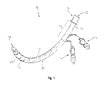
[0016] Fig. 1 is an elevated perspective view of an endotracheal tube
assembly;
[0017] Fig. 2 is a side view of the endotracheal tube assembly of Fig. 1 with
two cuffs inflated;
[00111] Fig. 3 is a cross-sectional view of the endotracheal tube assembly of
Fig. 2;
[0019] Fig. 4a is a partial side view of the endotracheal tube assembly of
Fig. 1 with a distal cuff
inflated;
3
CA 03138735 2021- 11- 19
WO 2020/236588
PCT/US2020/033130
[0020] Fig. 4b is a partial side view of the endotracheal tube assembly of
Fig. 2 with a proximal
cuff inflated; and
[0021] Fig. 5 is a method for manufacturing the endotracheal tube assembly of
Fig. 1.
[0022] Fig. 6a is another embodiment of an endotracheal tube assembly;
[0023] Fig. 6b is a partial side view of the endotracheal tube assembly of
Fig. 6a with a distal cuff
inflated;
[0024] Fig. 6c is a partial side view of the endotracheal tube assembly of
Fig. 6a with a proximal
cuff inflated;
[0025] Fig. 7 is another side view of the endotracheal tube assembly of Fig.
6a in a straightened
orientation;
[0026] Fig. 8 is a partial sectional view of one cuff of the endotracheal tube
assembly of Fig. 6a;
and
100271 Fig. 9 is a method for fluidly coupling a cuff to a lumen for the
endotracheal tube assembly
of Fig. 6a.
[0028] Corresponding reference numerals are used to indicate corresponding
parts throughout the
several views.
DETAILED DESCRIPTION
[0029] For the purposes of promoting an understanding of the principles of the
present disclosure,
reference will now be made to the embodiments described herein and illustrated
in the drawings
and specific language will be used to describe the same. It will nevertheless
be understood that no
limitation of the scope of the present disclosure is thereby intended, such
alterations and further
modifications in the illustrated devices and methods, and such further
applications of the principles
of the present disclosure as illustrated therein being contemplated as would
normally occur to one
skilled in the an to which the present disclosure relates.
[0030] Referring now to Figs. 1-3, an endotracheal tube assembly 100 is
illustrated isolated from
a ventilator or any other medical device. The endotracheal tube assembly 100
may be formed from
an airway tube 106 that has a first opening 102 on a proximal end and a second
opening 108 on a
distal end. The first opening 102 may have a coupler 104 coupled thereto. The
coupler 104 may
provide a location to fluidly couple the first opening 102 to a ventilator or
other medical device to
selectively provide fluid through the airway tube 106 and out the second
opening 108. In one
4
CA 03138735 2021- 11- 19
WO 2020/236588
PCT/US2020/033130
aspect of this disclosure, the airway tube 106 provides a fluidly sealed
channel 306 (see Fig. 3)
between the first opening 102 and the second opening 108.
100311 The airway tube 106 may be formed of silicon or any other material that
can provide a
sterile, fluidly sealed inner channel 306. In one aspect of this disclosure,
the airway tube 106 may
be a substantially tubular structure that has an airway wall 302 (see Fig. 3).
The airway wall 302
may have a thickness that is sufficient to define a first and second inflation
tube passageway or
lumen 304 (see Fig. 3) therein. The inflation lumen passageways 304 may
provide a fluidly isolated
passageway from a corresponding first and second pilot balloon 114, 116 to a
proximal and distal
cuff 118, 120. In one aspect of this disclosure, the first inflation pilot
balloon 114 can be fluidly
coupled to one of the proximal or distal cuffs 118, 120 through one of the
inflation lumen
passageways 304 to thereby allow the cuff to be selectively inflated (see
Figs. 4a and 4b).
Similarly, the second pilot balloon 116 can be fluidly coupled to the other of
the proximal or distal
cuffs 118, 120 through the other of the inflation lumen passageway 304 to
thereby allow the other
cuff to be selectively inflated.
100321 The airway tube 106 may also have a resistant member 110 coupled to a
radially outer
surface of the airway tube 106. The resistant member 110 may be any material
that resist
penetration by a surgical laser or any other surgical instrument that may
compromise the airway
tube 106. The resistant member 110 may extend a length of the airway tube 106
that corresponds
with the portions of the airway tube 106 that are intended to be positioned
within a patient during
use. However, in one embodiment the resistant member 110 may extend
substantially the entire
length of the airway tube 106 less an exposed portion 112 thereof. The exposed
portion 112 may
be the portion of the airway tube 106 located adjacent to the coupler 104. In
one example, the
resistant member 110 extends from a radially inner portion of the distal cuff
120, past the proximal
cuff 118, and along the airway tube 106 to the exposed portion 112. In yet
another embodiment,
the resistant member 110 extends between the proximal cuff 118 and the exposed
portion 112.
100331 In one aspect of this disclosure, the resistant member 110 may be
formed of a material that
resists penetration from a KTP laser. More specifically, a KTP laser with 400
millimeter lens
microbeam coupled to a microscope may be spaced 35 centimeters from the
endotracheal tube
assembly 100 and deliver a focused spot of 380 micron. The KTP laser may have
an angle of
incidence of about ninety degrees relative to the resistance layer. Under this
scenario, a laser
resistant material may be any material that can resist a continuous beam for
at least three minutes
CA 03138735 2021- 11- 19
WO 2020/236588
PCT/US2020/033130
with the laser delivering a maximum power of about fifteen watts,
continuously. More specifically,
oxygen gas can be pumped through the endotracheal tube assembly 100 with the
fire resistant
member 110 positioned there around and the fire resistant member 110 may
prevent combustion
of the oxygen in the above conditions.
[0034] Alternatively or additionally, the fire resistant member 100 may be any
material capable of
resisting penetration by a CO2 laser. For example, an endotracheal tube
assembly 100 may
positioned about 35.5 centimeters from the CO2 laser wherein the CO2 laser can
deliver a constant
spot of about .38 millimeters. The CO2 laser may produce a continuous beam for
at least three
minutes with the CO2 laser delivering a maximum output of at least about forty
to forty-five watts.
In other examples, the CO2 laser may deliver a maximum output of about 60
watts. The CO2 laser
may be applied multiple times at differing angles relative to the endotracheal
tube assembly 100.
Under this scenario, a laser resistant material may be any material that can
prevent combustion of
oxygen gas as it is pumped through the endotracheal tube assembly 100 while
the resistant later is
being exposed to the CO2 laser.
[0035] The above examples of a laser resistant material are not exhaustive.
The resistant member
110 may be formed of any material that can protect the fluid flowing within
the airway tube 106
from being affected by an external ignition. Accordingly, any known resistant
material may be
used for the resistant member 110.
[0036] In one aspect of this disclosure, the resistant member 110 is wrapped
around the airway
tube 106 in a helical pattern. In this configuration, adjacent portions of the
resistant member 110
partially overlap one another along the airway tube 106 to ensure the
resistant member 110 fully
covers the outer surface of the airway tube 106 where wrapped there around. In
other words, the
resistant member 110 is wrapped around the airway tube 106 to ensure that
there are no sections
of airway tube 106 that are not covered due to gaps in the wrapping pattern or
caused by bending
the airway tube 106.
[0037] While a wrapping application of the resistant member 110 is described
herein, other
applications are also considered. More specifically, the resistant member 110
may be formed in a
sheet that is wrapped around the airway tube 106 a single time, instead of in
a helical configuration.
In this embodiment, the resistant member 110 may be formed from a
substantially rectangular
sheet that is the desired length of the resistant member 110. The sheet may
then be wrapped slightly
greater than three-hundred and sixty degrees around the airway tube 106 to
substantially cover the
6
CA 03138735 2021- 11- 19
WO 2020/236588
PCT/US2020/033130
outer surface. Accordingly, any known method of applying a resistant member
110 is considered
herein.
100381 Regardless of the wrapping pattern or method, the resistant member 110
may have an outer
surface that is abrasive to soft tissue. Accordingly, in one aspect of this
disclosure a sleeve 122
may be positioned around the radially exterior portion of the resistant member
110. The sleeve 122
may be formed from a substantially continuous material that has a smooth outer
surface. In one
non-exclusive example, the sleeve 122 is a tube having an inner diameter that
is slightly less than
the outer diameter of the resistant member 110 when positioned around the
airway tube 106. In
this configuration, the sleeve 122 may be stretched or otherwise expanded to
be positioned around
the outer surface of the resistant member 110. Once positioned there around,
the sleeve 122 may
return to the un-stretched size to thereby provide a radial compression to the
underlying resistant
member 110 compressing the resistant member 110 against the airway tube 106.
100391 In another aspect of this disclosure, the sleeve 122 and resistant
member 110 may be
coupled to the underlying airway tube with an adhesive. In this aspect of the
disclosure, the
resistant member 110 and sleeve 122 may be positioned around the airway tube
106 as discussed
herein. However, in addition to the sleeve 122 applying a compressive load to
the resistant member
110 to couple the resistant member 110 and the sleeve 122 to the airway tube
106, an adhesive
may be applied to the proximal and distal ends of the resistant member 110 and
sleeve 122 to
further couple them to the airway tube 106 with the adhesive. In one non-
exclusive example, the
adhesive may be a Room-Temperature-Vulcanizing ("RTV") silicone. However, any
known
adhesive is considered herein.
100401 The sleeve 122 may have a wall thickness that is any suitable size.
More specifically, the
wall thickness may be thin enough to allow the sleeve to be stretched over the
resistant member
110 but thick enough to ensure that the sleeve is not torn during
manufacturing of the endotracheal
tube assembly 100. In one non-exclusive example, the sleeve may be formed of
silicon and have
a wall thickness of between about eight thousandths of an inch and ten
thousandths of an inch.
However, in other embodiments the sleeve 122 may be formed of a material other
than silicon.
Further still, the sleeve 122 may have a wall thickness less than eight
thousandths of an inch or
greater than ten thousandths of an inch. Accordingly, this disclosure
considers many different sizes
and materials for the sleeve 122.
7
CA 03138735 2021- 11- 19
WO 2020/236588
PCT/US2020/033130
100411 Referring now to Figs. 4a and 4b, two different configurations of the
cuffs 118, 120 are
illustrated. In Fig. 4a, the proximal cuff 118 is illustrated deflated while
the distal cuff 120 is
illustrated inflated. The inflation of the proximal and distal cuffs 118, 120
may be altered by
selectively providing fluid to the corresponding cuffs 118, 120 through one of
the first or second
pilot balloon 114, 116. In the example illustrated in Fig. 4a, the first pilot
balloon 114 may be
provided pressurized fluid that travels through one of the inflation lumen
passageways 304 in the
wall of the airway tube 106 and to an inner chamber of the distal cuff 120.
Similarly, in Fig. 4b,
the second pilot balloon 116 may be provided pressurized fluid that travels
through the other of
the inflation lumen passageways 304 in the wall of the airway tube 106 and to
an inner chamber
of the proximal cuff 118. Further, both the proximal and distal cuffs 118, 120
may be deflated as
in Fig. 1, or inflated as in Fig. 2, depending on the amount of fluid volume
and pressure provided
to the inner chambers of the corresponding cuffs 118, 120 as discussed herein.
100421 Referring now to Fig. 5, one non-exclusive example of a manufacturing
method 500 is
illustrated. Initially in box 502, the airway tube 106 may be formed utilizing
known extrusion
techniques. As part of forming the airway tube 106, the inflation lumen
passageways 304 may also
be formed in the wall of the airway tube 106. Next, in box 504, the airway
tube 106 may be cut to
any desired length depending on the desired application. In box 506, the
airway tube 106 may be
formed into an arc by utilizing an arc-shaped mandrel and applying a heat
process thereto. In box
508, the resistant member 110 may be formed around the airway tube 106
utilizing any of the
techniques described herein among others. Next, the sleeve 122 may be expanded
and pulled over
the resistant member 110 and the airway tube 106 in box 510. In box 512, an
adhesive may be
applied to the sleeve 122, the resistant member 110, and the airway tube 106
at both a proximal
and distal end thereof.
100431 In box 514, the inflation lumen passageways 304 may be skived at a
proximal end of the
airway tube 106. Next, in box 516 the first and second pilot balloon 114, 116
may be coupled to
the corresponding skived locations of the inflation lumen passageways 304.
Adhesive or the like
may be utilized to ensure the first and second pilot balloon 114, 116 are
fluidly coupled to the
inflation lumen passageways 304. In box 518, holes may be formed through a
portion of the airway
tube 106 to fluidly couple the inner portion of each cuff 118, 120 to the
corresponding inflation
lumen passageway 304. The holes may be fluid ports that fluidly couple the
corresponding
inflation lumen passageway 304 to the external surface of the airway tube 106
within the cuff 118,
8
CA 03138735 2021- 11- 19
WO 2020/236588
PCT/US2020/033130
120. Accordingly, in box 520 the proximal and distal cuffs 118, 120 may be
positioned around the
corresponding fluid port and coupled to the airway tube 106. Adhesive may be
used to fluidly
couple the corresponding cuffs 118, 120 to the airway tube 106 to further
fluidly couple the
corresponding cuffs 118, 120 to the corresponding first and second pilot
balloon 114, 116.
100441 In box 522, any inflation lumen may be back filled and in box 524 a
murphy eye may be
formed in the distal end of the airway tube 106. Next, inflation valves may be
inserted in the pilot
balloon 114, 116 in box 526. Finally, in box 528 the endotracheal tube
assembly 100 may be tested
for leaks.
[0045] Referring now to Fig. 6a, another embodiment of an endotracheal tube
assembly 600 is
illustrated. In this embodiment, the resistant member 110 may extend from a
starting portion 604
of the endotracheal tube 106 to an ending portion 602 of the endotracheal tube
106. In one aspect
of this disclosure, the ending portion 602 may be distal to the distal cuff
120. In other words, the
resistant material 110 may extend underneath both cuffs 118, 120 towards the
second opening 108.
In this embodiment, the resistant material 110 may extend a resistant length
702 that is
substantially the entire length of the airway tube 106 less the exposed
portion 112. In this
configuration, the portion of the airway tube 106 that is intended to be
positioned within the patient
is surrounded by the resistant material 110 up to the distal cuff 120.
[0046] In one aspect of this disclosure, the resistant material 110 is
positioned radially between
the cuffs 118, 120 and the inflation lumen 304. Accordingly, to fluidly couple
the inner portion of
the cuff 118, 120 with the corresponding inflation lumen 304 a fluid
passageway must be formed
through the sleeve 122, resistant material 110, and part of the airway wall
302. One aspect of this
disclosure is a method of forming this fluid connection while ensuring the
passageway from the
inflation lumen 304 through the airway wall 302, resistant material 110, and
sleeve 122 is fluidly
sealed.
[0047] Referring now to Figs. 8 and 9, one non-exclusive method for fluidly
coupling a cuff 118,
120 to the corresponding inflation lumen 304 is described. In box 902, the
airway tube 106 may
be wrapped with the resistant material 110 as discussed herein. In box 902,
the resistant material
110 is wrapped to be positioned axially along the airway tube 106 passed the
proximal cuff 118
and at least partially into the distal cuff 120. In one aspect of this
disclosure, the resistant material
110 is wrapped to extend along the airway tube 106 axially past both the
proximal and distal cuffs
9
CA 03138735 2021- 11- 19
WO 2020/236588
PCT/US2020/033130
118, 120. Next in box 904, the sleeve 122 is positioned over the resistant
material 110 and coupled
to the airway tube 106 and resistant material 110 as discussed herein.
100481 In box 906, a cutter having a first diameter 802 is used to cut a first
hole 806 through the
sleeve 122 and the resistant material 110 at a location radially outward of a
corresponding inflation
lumen 304. The first hole 806 may be radially outward of the inflation lumen
304 relative to an
airway axis 804 defined longitudinally through a center portion of the airway
tube 106. In one
aspect of this disclosure, the cutter is not advanced substantially radially
into the airway wall 302
of the airway tube 106 but rather is only advanced far enough to cut holes
through the sleeve 122
and the resistant material 110. While a portion of the airway tube 106 may be
slightly contacted
when cutting the first hole 806, the cutter for the first hole 806 does not
advance into the inflation
lumen 304. In other words, the cutter only advances far enough radially inward
to cut the first hole
806 in the sleeve 122 and the resistant member 110_ All portions of the sleeve
122 and resistant
material 110 are removed from the first hole 806 to expose a radially outer
surface of the airway
tube 106.
100491 Next, in box 908, a second hole 808 may be cut partially through the
airway tube 106 into
the underlying inflation lumen 304. The second hole 808 may be aligned to be
substantially coaxial
with the first hole 806 In one non-exclusive embodiment, the second hole 808
may have a second
diameter 810 that is slightly less than the first diameter 802. The second
hole 808 is only defined
partially through the airway wall 302 of the airway tube 106 in order to
provide an outlet (the
second hole 808) for the underlying inflation lumen 304 to be routed radially
outwardly from the
airway tube 106. As discussed herein, ultimately one of the cuffs 118, 120
will be positioned along
the outlet to fluidly couple the inflation lumen 304 to an inner chamber of
the corresponding cuff
118, 120.
100501 In box 910, a stopper plug 822 may be positioned through the second
hole 808 to
substantially fill the second hole 808. The stopper plug 822 may have a
diameter that is the same
or slightly greater than the second diameter 810 to substantially fill the
second hole 808 when
placed therein. Further, the stopper plug 822 may extend radially away from
the sleeve 122 to
define an annular channel 820 around the stopper plug 822. The annular channel
820 may be
defined by the stopper plug 822, outer surface of the airway tube 106 between
the first and second
hole, the resistant member 110 along the perimeter of the first hole 806, and
the sleeve 122 along
the perimeter of the first hole 806. In box 910, an adhesive can be applied
into the annular channel
CA 03138735 2021- 11- 19
WO 2020/236588
PCT/US2020/033130
820 while the stopper plug 822 is positioned through the second hole 808. By
placing the adhesive
in the annular channel 820, any gaps between the sleeve 122, resistant member
110, and airway
tube 106 may be substantially filled with adhesive to ensure no fluid can pass
through the first hole
806 to occupy the space between the sleeve 122 and the outer surface of the
airway tube 106. In
other words, applying adhesive to the annular channel 820 prevents fluid from
bleeding into the
resistant material 110 layer when the corresponding cuff 118, 120 is inflated.
[0051] The adhesive may be any known adhesive and in one non-exclusive
embodiment is RTV
silicone. Further, the plug may be formed from a material that resists
adhesion by the adhesive. In
one non-exclusive example, the plug may be formed of a Teflon material.
However, any known
adhesive and plug material may be used, and this disclosure considers all
known adhesives and
plug materials.
[0052] Referring now to box 912, the stopper plug 822 may be removed from the
second hole 808
after the adhesive has at least partially cured. As discussed herein, the
stopper plug 822 may be
formed of a material that substantially resists adhesion to the adhesive.
Accordingly, after the
adhesive has at least partially cured, the plug may be removed from the second
hole 808. Further,
since the adhesive is at least partially cured, the second hole 808 may remain
defined partially
through the airway tube 106 to fluidly couple the inflation lumen 304 to the
outer portion of the
sleeve 122.
[0053] Next, in box 914 the corresponding cuff 118, 120 may be positioned
around the sleeve 122
at a location axially aligned with the second hole 808. More specifically, the
cuff 118, 120 may be
aligned with the second hole 808 so the second hole 808 fluidly couples a
corresponding inflation
lumen 304 with a cavity 818 of the cuff 118, 120. In box 916, each cuff 118,
120 may be coupled
to the radially outer surface of the sleeve 122 at a proximal and distal end
812, 814 with adhesive.
The cavity 818 is formed between the inner surface of the cuff 118, 120 and
the outer surface of
the sleeve 122 between the proximal and distal ends 812, 814 of the cuff 118,
120. Further, a cap
816 may be formed at a distal end of the inflation lumen 304 with adhesive to
prevent fluid flow
out the distal end. Accordingly, fluid provided through the inflation lumen
304 passes through the
second hole 808 and into the cavity 818 to thereby expand the cuff 118, 120
under certain pressure
and volume conditions
[0054] Finally, in box 918 a leak test may be executed to ensure that the
inflation lumen 304 is
fluidly coupled to the cuff 118, 120. More specifically, fluid may be provided
to the inflation
11
CA 03138735 2021- 11- 19
WO 2020/236588
PCT/US2020/033130
lumen 304 at an established fluid pressure to fill the cavity 818. The fluid
may be provided at a
test pressure and monitored for a period of time to ensure that the test
pressure does not drop. A
drop in test pressure may indicate a leak between the inflation lumen 304 and
the cuff 118, 120.
As discussed herein, one aspect of the leak test may be to ensure that fluid
is not passing through
the perimeter of the first hole 806 and into the space between the sleeve 122
and the airway tube
106. In other words, one aspect of the leak test is to ensure that the
adhesive applied to the annular
channel 820 is properly sealing the sleeve 122, resistant member 110, and
airway tube 106 about
the perimeter of the first hole 806.
[0055] While Figs. 8 and 9 illustrate and describe a method for fluidly
coupling a single cuff to an
inflation lumen, this disclosure contemplates utilizing substantially the same
methodology
discussed herein to fluidly couple two or more cuffs thereto as well. More
specifically as illustrated
in Fig. 3, two inflation lumen 304 may be defined in the airway wall 302. In
this embodiment, the
methods discussed herein can be implemented to couple the proximal cuff 118 to
a first inflation
lumen and then couple the distal cuff 120 to a second inflation lumen. In this
configuration, the
first and second holes 806, 808 would be located along different portions of
the airway tube 106
to thereby fluidly couple the corresponding cuff 118, 120 to one of the first
or second inflation
lumen. Accordingly, the teachings of this disclosure may be applied to
endotracheal tubes with
any number of cuffs.
[0056] In use, the endotracheal tube assembly 100 described herein may be
utilized for procedures
that may expose the endotracheal tube assembly 100 to a surgical laser or the
like. In these types
of procedures, the endotracheal tube assembly 100 may be inserted partially
past the vocal chords
and into the trachea of a patient without abrasively contacting the soft
tissue. The cuff or cuffs may
be inflated to fluidly seal the endotracheal tube assembly 100 to the walls of
the trachea. Then, the
physician can perform the procedure utilizing a surgical laser or the like
while fluid is passed
through the endotracheal tube assembly 100 and into the patient.
[0057] If the physician unintentionally contacts the endotracheal tube
assembly 100 with the
surgical laser, the resistant member 110 may substantially reflect or
otherwise block the laser from
entering the fluid passageway of the airway tube 106. Once the procedure is
complete, the
physician may deflate the cuff or cuffs and remove the endotracheal tube
assembly 100 from the
patient without abrasively contacting the trachea or vocal chords. More
specifically, the sleeve 122
12
CA 03138735 2021- 11- 19
WO 2020/236588
PCT/US2020/033130
and cuffs 118, 120 may cover substantially the entire outer surface of the
resistant member 110 to
ensure the endotracheal tube assembly 100 can be smoothly inserted and removed
from the patient.
100581 As discussed herein, the resistant member 110 may extend axially along
the airway tube
106 past the proximal cuff and partially into, or past, the distal cuff By
extending the resistant
member 110 substantially the entire length of the airway tube 106, the
physician can utilize a
surgical laser or the like along portion of the patient that are adjacent to
the cuffs 118, 120. More
specifically, if the physician inadvertently redirects the surgical laser into
the cuffs 118, 120 and
toward the airway tube 106, the resistant member 110 may still substantially
prevent the surgical
laser from passing into the airway tube 106.
100591 While exemplary embodiments incorporating the principles of the present
disclosure have
been described herein, the present disclosure is not limited to such
embodiments. Instead, this
application is intended to cover any variations, uses, or adaptations of the
disclosure using its
general principles. Further, this application is intended to cover such
departures from the present
disclosure as come within known or customary practice in the art to which this
disclosure pertains.
13
CA 03138735 2021- 11- 19