Note: Descriptions are shown in the official language in which they were submitted.
CA 03140397 2021-11-12
WO 2020/247907 PCT/US2020/036577
SYSTEMS, DEVICES, AND METHODS FOR TREATING HEART VALVES
CROSS REFERENCE TO RELATED APPLICATIONS
[0001] This application claims the benefit of U.S. Provisional Application
Ser. No.
62/908,402, entitled "Systems, Devices, and Methods for Treating Heart
Valves," filed
September 30, 2019 and U.S. Provisional Application Ser. No. 62/858,875,
entitled "Systems,
Devices, and Methods for Treating Heart Valves," filed June 7, 2019; the
disclosures of which
are herein incorporated by reference in their entireties.
FIELD OF THE DISCLOSURE
[0002] The present disclosure relates to systems and methods for treating
valvular
regurgitation and/or other valve issues.
BACKGROUND OF THE DISCLOSURE
[0003] Prosthetic heart valves can be used to treat cardiac valvular
disorders. The native heart
valves (the aortic, pulmonary, tricuspid and mitral valves) serve critical
functions in assuring
the forward flow of an adequate supply of blood through the cardiovascular
system. These
heart valves can be rendered less effective by congenital, inflammatory,
infectious, and other
conditions. Such conditions can eventually lead to serious cardiovascular
compromise or death.
[0004] A transcatheter technique can be used for introducing and implanting a
prosthetic heart
valve using a flexible catheter in a manner that is less invasive than open
heart surgery. In this
technique, a prosthetic valve can be mounted in a crimped state on the end
portion of a flexible
catheter and advanced through a blood vessel of the patient until the valve
reaches the
implantation site. The valve at the distal end of the catheter can then be
expanded to its
functional size at the site of the defective native valve, such as by
inflating a balloon on which
the valve is mounted. Alternatively, the valve can have a resilient, self-
expanding stent or frame
that expands the valve to its functional size when it is advanced from a
delivery sheath at the
distal end of the catheter. Optionally, the valve can have a mechanically
expandable frame, or
the valve can have a combination of expansion mechanism, such as balloon
expandable, self-
expandable, and/or mechanically expandable portions.
[0005] Transcatheter heart valves (THVs) could theoretically be appropriately
sized, or
shaped to be placed inside native mitral and tricuspid valves. However, mitral
and tricuspid
valve anatomy can vary significantly from person to person and it can be
difficult to
appropriately size and shape a valve for many patients. Further, when treating
valve
- 1 -
CA 03140397 2021-11-12
WO 2020/247907 PCT/US2020/036577
insufficiency, the surrounding tissue may not be strong enough to hold certain
types of valves
in position as desired. It would be beneficial to have a docking system and/or
apparatus to
secure prosthetic valves in the proper position and appropriate delivery
systems to ensure safe
and effective delivery. Additionally, the shape of the native valve may allow
for paravalvular
leakage around the prosthetic valve (i.e., blood flow bypassing the prosthetic
valve). As such,
solutions to increase efficiency of prosthetic valve placement and to reduce
paravalvular
leakage would be beneficial.
SUMMARY OF THE DISCLOSURE
[0006] This summary is meant to provide examples and is not intended to be
limiting of the
scope of the invention in any way. For example, any feature included in an
example of this
summary is not required by the claims, unless the claims explicitly recite the
feature. The
description discloses exemplary embodiments of prosthetic valves, docking
stations for
prosthetic valves, delivery devices for docking stations, and packaging for
delivery devices.
The docking stations, catheters, and handles can be constructed in a variety
of ways. Also, the
features described can be combined in a variety of ways. Various features and
steps as
described elsewhere in this disclosure can be included in the examples
summarized here.
[0007] In some embodiments, systems and/or apparatuses herein include a
docking device
(e.g., anchor, etc.), a delivery system, a prosthetic or implantable heart
valve, a pusher device,
other components, or combinations of one or more of these. The docking device,
delivery
system, prosthetic valve, etc. can be the same as or similar to those
described below or
elsewhere herein.
[0008] In one representative embodiment, a suture lock assembly for a delivery
system for
an implantable medical device can include: a spool configured to receive a
suture and
including a gear; a rotatable handle coupled to the spool and configured to
rotate the spool
and gear; a pawl configured to engage with teeth of the gear and allow
rotation of the gear,
spool, and handle in only one direction; and a directional selector coupled to
the pawl and
movable between two positions, each of the two positions corresponding to a
different
direction of rotation of the gear, the directional selector configured to
pivot the pawl to adjust
an orientation of the pawl relative to the gear and adjust a direction of
rotation of the gear.
[0009] In some embodiments, the pawl is pivotable between a first orientation
which allows
rotation of the gear in only a first direction and a second orientation which
allows rotation of
the gear in only an opposite, second direction. In some embodiments, the first
direction is
counterclockwise and the second direction is clockwise.
- 2 -
CA 03140397 2021-11-12
WO 2020/247907 PCT/US2020/036577
[0010] In some embodiments, the pawl is held in the first orientation and the
second
orientation by a spring plunger engaged with the pawl at a back side of the
pawl and where, in
the first orientation, the pawl is arranged on a first side of the spring
plunger and, in the second
orientation, the pawl is arranged on a second side of the spring plunger.
[0011] In some embodiments, the pawl includes two teeth spaced apart from one
another and
arranged on a front side of the pawl and the two teeth of the pawl are
configured to engage with
teeth of the gear.
[0012] In some embodiments, the suture lock assembly further includes hard
stops arranged
within a housing of the suture lock assembly, the gear and pawl arranged
within the housing,
and the pawl is configured to interface with one of the hard stops when the
gear is rotated in a
direction that is opposite a selected direction of rotation set by the
directional selector.
[0013] In some embodiments, the suture lock assembly further includes a
housing including
a top housing and a bottom housing coupled to one another, the gear and pawl
arranged within
a space arranged between the top housing and bottom housing. The rotatable
handle and the
directional selector can extend outward from the top housing. The top housing
can include a
first icon indicating a slack position of the directional selector and a
second icon indicating a
tension position of the directional selector, and where the directional
selector is movable
between a first of the two position that points toward the first icon and a
second of the two
positions that points toward the second icon.
[0014] In some embodiments, a suture lock assembly further includes a release
bar including
a suture cutting location arranged at a distal end of the release bar, the
release bar configured
to receive a suture through an interior of the release bar and across the
suture cutting location,
the suture extending from the spool.
[0015] In some embodiments, the release bar includes one or more supporting
ribs arranged
on a center portion of the release bar, the center portion arranged between
the distal end and
proximal end of the release bar.
[0016] In some embodiments, the distal end of the release bar is shaped to
form a first keyed
connection with an adaptor of the delivery system and a proximal end of the
release bar is
shaped to form a second keyed connection with a bottom housing of the suture
lock assembly,
where the spool is arranged within an interior of the bottom housing.
[0017] In some embodiments, the suture lock assembly further includes a
flushing port
coupled to the bottom housing and extending outward from the bottom housing in
an opposite
direction from a direction which the release bar extends from the bottom
housing.
- 3 -
CA 03140397 2021-11-12
WO 2020/247907 PCT/US2020/036577
[0018] In some embodiments, the suture lock assembly further includes a
plurality of annular
sealing elements, including a first annular sealing element arranged around a
distal end portion
of the release bar, proximate to the suture cutting location, and a second
annular sealing element
arranged around a proximal end portion of the release bar, the second annular
sealing element
arranged between, in a radial direction, the release bar and a bottom housing
of the suture lock
assembly, where the spool is arranged within the bottom housing. In some
embodiments, the
plurality of annular sealing elements further includes a third annular sealing
element arranged
around a portion of the spool and arranged between the portion of the spool
and the bottom
housing.
[0019] In some embodiments, a proximal end of the release bar is bonded to a
bottom housing
of the suture lock assembly.
[0020] In some embodiments, the release bar includes a divider arranged within
the suture
cutting location, where the divider is configured to separate two lines of a
suture extending
longitudinally through the release bar and expose only one line of the two
lines of the suture to
an exterior of the suture lock assembly at the suture cutting location.
[0021] In some embodiments, the spool includes a gap in a flange arranged
around a bottom
of the spool and the rotatable handle includes an indicator on its outer
surface configured to
track a number of turns applied to the spool and locate the gap.
[0022] In some embodiments, the gap is arranged adjacent to one or more
apertures arranged
within the spool, the one or more apertures configured to route the suture
from inside the spool
to an exterior surface of the spool that is configured to receive the suture
thereon.
[0023] In some embodiments the rotatable handle is coupled to the spool via a
central screw
extending longitudinally through the rotatable handle and the spool and the
suture lock
assembly can further include one or more friction pads arranged around the
central screw,
adjacent to the central portion of the spool, and a friction nut coupled to
the central screw,
below a lower friction pad of the one or more friction pads. The one or more
friction pads can
be configured to increase friction on the central screw to stop rotation of
the central screw and
the rotatable handle when a tension in the suture increases above a
predetermined threshold.
[0024] In some embodiments, a suture lock assembly further includes a pin-
based clutch
system including a spring plunger extending longitudinally through and coupled
to a portion
of the rotatable handle, the spring plunger including an end extending into
the gear and
configured to extend into and mate with a plurality of detents arranged in an
outer-facing
surface of the gear to allow rotation of the gear by the rotatable handle. The
spring plunger can
- 4 -
CA 03140397 2021-11-12
WO 2020/247907 PCT/US2020/036577
be configured to slip out of the detents in response to a tension in the
suture above a
predetermined threshold.
[0025] In another representative embodiment, a delivery system for delivering
a docking
device to a native valve annulus of a patient's heart can include: an outer
shaft and a sleeve
shaft at least partially arranged within the outer shaft. The sleeve shaft can
include: a distal
section configured to cover the docking device, the distal section including a
flexible material
with a lubricous outer surface; and a proximal section including a rigid
material and including
a tubular portion and a cut portion, the cut portion having an open, u-shaped
cross-section. The
delivery system can further include a pusher shaft at least partially arranged
within the outer
shaft, the pusher shaft including: a main tube arranged interior to, in a
radial direction that is
relative to a central longitudinal axis of the delivery system, the sleeve
shaft; an annular shell
surrounding a proximal end portion of the main tube and spaced away from, in
the radial
direction, an outer surface of the main tube; and a proximal extension
connected to and
extending proximally from a proximal end of the main tube, proximal to the
shell, the proximal
extension including a flexible material and extending along a portion of an
inner surface of the
cut portion of the proximal section of the sleeve shaft.
[0026] In some embodiments the pusher shaft further comprises an annular plug
arranged
within the annular shell, at a proximal end of the shell, and surrounding the
main shaft, where
the plug includes a crescent-shaped portion extending across and filling a
first portion of an
annular space arranged between the main tube and the shell.
[0027] In some embodiments, the annular space includes a second portion that
is open and not
filled by the plug, where the proximal section of the sleeve shaft is
configured to slide within
the annular space, and where the cut portion of the proximal section is
configured to slide
through the second portion of the annular space.
[0028] In some embodiments, the tubular portion of the proximal section has an
end surface
at an interface between the tubular portion and the cut portion, the end
surface arranged normal
to the central longitudinal axis, and the plug is configured to interface with
the end surface of
the proximal section and stop the sleeve shaft from traveling further in the
proximal, axial
direction.
[0029] In some embodiments, the sleeve shaft further includes a middle section
arranged
between the distal section and the proximal section of the sleeve shaft, the
middle section
forming a transition between the flexible material of the distal section and
the rigid material of
the proximal section.
- 5 -
CA 03140397 2021-11-12
WO 2020/247907 PCT/US2020/036577
[0030] In some embodiments, the sleeve shaft further includes a flexible
polymer jacket
forming an outer surface of the distal section and the middle section, the
flexible polymer jacket
including the flexible material, an inner liner forming an inner surface of
each of the distal
section and the middle section, and a rigid tube including a first section
forming an entirety of
the proximal section and a second section forming a proximal portion of the
middle section.
[0031] In some embodiments, the rigid tube is a metal tube, where the second
section includes
a plurality of apertures arranged around a circumference of the rigid tube,
along the second
section, and where the rigid tube is coupled to the inner liner and the
flexible polymer jacket
via a bonding connection between the inner liner and the flexible polymer
jacket, through the
plurality of apertures.
[0032] In some embodiments, the delivery system further includes a handle
assembly include
a handle portion and a hub assembly extending proximally from a proximal end
of the handle
portion, where the outer shaft extends distally from a distal end of the
handle portion, and where
the hub assembly includes an adaptor with a straight section coupled to a
suture lock assembly
and a branch section coupled to sleeve actuating handle.
[0033] In some embodiments, the proximal extension of the pusher shaft extends
into and
through a portion of the branch section of the adaptor.
[0034] In some embodiments, the delivery system further includes a first
flushing port coupled
to the branch section of the adaptor and fluidly coupled with an inner lumen
of the proximal
extension of the pusher shaft. In some embodiments, the delivery system
further includes a
second flushing port coupled to the branch section, distal to the first
flushing port, and fluidly
coupled with a lumen formed between an outer surface of the proximal extension
and an inner
surface of the branch section.
[0035] In some embodiments, the delivery system further includes a first
flushing port coupled
to a proximal end of the suture lock assembly and fluidly coupled with an
inner lumen of the
proximal extension of the pusher shaft and a second flushing port coupled to
the branch section,
distal to the first flushing port, and fluidly coupled with a lumen formed
between an outer
surface of the proximal extension and an inner surface of the branch section.
[0036] In some embodiments, the cut portion of the sleeve shaft extends into
the straight
section of the adapted and is coupled to the sleeve actuating handle.
[0037] In some embodiments, the pusher shaft and the sleeve shaft are coaxial
with one
another, along the central longitudinal axis of the delivery system, and each
of the sleeve shaft
and the pusher shaft are configured to slide axially along the central
longitudinal axis, relative
to the outer shaft.
- 6 -
CA 03140397 2021-11-12
WO 2020/247907 PCT/US2020/036577
[0038] In some embodiments, a distal section of the main tube of the pusher
shaft includes a
plurality of cuts therein, spaced apart from one another along a length of the
distal section,
where the plurality of cuts is configured to increase a flexibility of the
distal section of the main
tube. In some embodiments, spacing between adjacent cuts of the plurality of
cuts varies along
the length of the distal section and where the spacing between adjacent cuts
increases from a
distal end to a proximal end of the distal section.
[0039] In another representative embodiment, a delivery system for delivering
a docking
device to a native valve annulus of a patient's heart includes: a handle
portion; an outer shaft
extending distally from a distal end of the handle portion; a sleeve shaft
extending through an
interior of the outer shaft and configured to cover the docking device; a
pusher shaft including
a main tube extending through an interior of the sleeve shaft; and a hub
assembly extending
proximally from a proximal end of the handle portion. The hub assembly can
include: an
adaptor coupled to the handle portion and including a first section and a
second section that
branches off from the first section, where a portion of the pusher shaft
extends into the second
section and a proximal section of the sleeve shaft extends through the first
section; a suture
lock assembly coupled to a proximal end of the second section and configured
to adjust tension
in a suture extending from the suture lock assembly, through the pusher shaft,
to the docking
device; a first flushing port coupled to the second section and fluidly
coupled to a first fluid
flow lumen arranged within an interior of the pusher shaft and to a second
fluid flow lumen
arranged between the sleeve shaft and the docking device; and a second
flushing port coupled
to the second section and fluidly coupled to a third fluid flow lumen arranged
between the outer
shaft and the sleeve shaft.
[0040] In some embodiments, the delivery system further includes a sleeve
actuating handle
arranged at a proximal end of the first section and coupled to an end of the
proximal section of
the sleeve shaft, the sleeve actuating handle configured to adjust an axial
position of the sleeve
shaft relative to the outer shaft.
[0041] In some embodiments, the first fluid flow lumen extends through an
interior of a
proximal extension of the pusher shaft and an interior of the main tube of the
pusher shaft, the
main tube coupled to the proximal extension and extending through an interior
of the outer
shaft and the proximal extension extending through a portion of the outer
shaft and into the
second section.
[0042] In some embodiments, the first fluid flow lumen extends to a distal end
of the pusher
shaft, the distal end arranged adjacent to but spaced away from a proximal end
of the docking
device when the docking device is arranged within the outer shaft.
- 7 -
CA 03140397 2021-11-12
WO 2020/247907 PCT/US2020/036577
[0043] In some embodiments, the second flushing port is fluidly coupled to the
third fluid
flow lumen via an annular cavity arranged between a shell of the pusher shaft
and the main
tube of the pusher shaft, and a fourth fluid flow lumen formed between an
outer surface of the
proximal extension and an inner surface of the second section, the fourth
fluid flow lumen
fluidly coupled to the annular cavity. In some embodiments, the third fluid
flow lumen is
arranged between an inner surface of the outer shaft and a distal portion of
the sleeve shaft, the
distal portion configured to cover the docking device while the docking device
is arranged
inside the outer shaft and being implanted at the native valve annulus.
[0044] In some embodiments, the delivery system further includes a third
flushing port
coupled to the handle portion and fluidly coupled to the annular cavity.
[0045] In some embodiments, the delivery system further includes a gasket
arranged within
and across a diameter of the second section, between where the first flushing
port is coupled to
the second section and where the second flushing port is coupled to the second
section. The
gasket is configured to fluidly separate the first fluid flow lumen and the
third fluid flow lumen
from one another.
[0046] In some embodiments, the first flushing port and the second flushing
port are
connected to a single fluid source. In some embodiments, the single fluid
source is an infusion
pump and where the infusion pump is coupled to the first flushing port and the
second flushing
port via a y-connector.
[0047] In some embodiments, the first flushing port and the second flushing
port are
connected to different fluid sources.
[0048] In some embodiments, the first flushing port is directly coupled to the
second section
of the adaptor, distal to the suture lock assembly and proximal to the second
flushing port.
[0049] In some embodiments, the first flushing port is part of the suture lock
assembly and
arranged at a proximal end of the suture lock assembly.
[0050] In some embodiments, the delivery system further includes a hemostatic
seal arranged
within the first section of the adaptor, proximate to the sleeve actuating
handle, where the
hemostatic seal includes an opening surrounding a cut portion of the sleeve
shaft that extends
through the first section, to the sleeve actuating handle, the hemostatic seal
configured to seal
around the cut portion of the sleeve shaft. In some embodiments, the delivery
system further
includes a locking cap assembly arranged on the first section, around the
hemostatic seal, the
locking cap assembly configured to apply inward pressure on the hemostatic
seal and lock axial
translation of the sleeve shaft relative to a remainder of the hub assembly.
- 8 -
CA 03140397 2021-11-12
WO 2020/247907 PCT/US2020/036577
[0051] In some embodiments, the pusher shaft is configured to deploy the
docking device
from inside a distal end portion of the outer shaft upon reaching the native
valve annulus and
a distal end of the sleeve shaft is spaced away from a distal end of the outer
shaft, within the
outer shaft, while the docking device is arranged within the outer shaft
during navigating the
delivery system to the native valve annulus.
[0052] In some embodiments, the docking device is configured to receive and
secure a
prosthetic heart valve at the native valve annulus.
[0053] In one representative embodiment, a method of delivering a docking
device to a native
valve of a heart can include: deploying the docking device from a distal end
of a delivery
system, the docking device covered by a distal section of a sleeve shaft of
the delivery system,
the docking device including a coil extending along a central axis and
including a central region
including a plurality of turns, a leading turn extending from a first end of
the central region,
and a stabilization turn extending from an opposite, second end of the central
region, where a
covering extends around and along a top turn of the central region, the top
turn arranged at the
second end of the central region; positioning the covered docking device at
the native valve,
such that the covering of the top turn of the central region crosses and plugs
a medial
commissure of the native valve, at least a portion of the leading turn is
positioned in a ventricle
of the heart, and at least a portion of the stabilization turn is positioned
in an atrium of the heart;
and after positioning the covered docking device, retracting the sleeve shaft,
in a proximal
direction, to uncover the docking device.
[0054] In some embodiments, deploying the docking device from the distal end
of the delivery
system includes pushing the covered docking device outside of the outer shaft
of the delivery
system with the pusher shaft of the delivery system.
[0055] In some embodiments, retracting the sleeve shaft to uncover the docking
device
includes moving the sleeve actuating handle in the proximal direction.
[0056] In some embodiments, the method can further include maintaining a
position of the
pusher shaft while retracting the sleeve shaft to uncover the docking device
and, after
uncovering the docking device, retracting the pusher shaft back into the outer
shaft of the
delivery system.
[0057] In some embodiments, the method can further include, during deploying
the covered
docking device and positioning the covered docking device at the native valve,
flushing a
plurality of lumens of the delivery system including a first lumen arranged
between the distal
section of the sleeve shaft and the docking device and a second lumen arranged
between an
outer shaft of the delivery system and the sleeve shaft.
- 9 -
CA 03140397 2021-11-12
WO 2020/247907 PCT/US2020/036577
[0058] In some embodiments flushing the first lumen includes providing flush
fluid to a
pusher shaft lumen extending through the pusher shaft from a proximal end of
the pusher shaft
arranged within a branch section of a hub assembly, where a suture lock is
coupled to the
branch section, to a distal end of the pusher shaft, the distal end arranged
proximate to, but
spaced away from, a proximal end of the docking device and flowing the flush
fluid through
the pusher shaft lumen and into and through the first lumen.
[0059] In some embodiments, the flush fluid is provided to the pusher shaft
lumen via a flush
port coupled to the branch section, distal to the suture lock.
[0060] In some embodiments, the flush fluid is provided to the pusher shaft
lumen via a flush
port that is part of the suture lock and arranged at a proximal end of the
suture lock.
[0061] In some embodiments, flushing the second lumen includes providing flush
fluid to a
first cavity formed between an outer surface of the pusher shaft and an inner
surface of a
conduit of the branch section, flowing the flush fluid from the first cavity
into a second cavity
formed between a shell of the pusher shaft and a main tube of the pusher
shaft, and flowing the
flush fluid from the second cavity to the second lumen.
[0062] In some embodiments, the method can further include, during the
deploying and
positioning of the covered docking device, arranging a distal tip of the
distal section of the
sleeve shaft to extend a distance past, in the distal direction, a distal end
of the docking device.
[0063] In some embodiments, the method can further include deploying a
prosthetic heart
valve within the central region of the docking device.
[0064] In another representative embodiment, a method for providing flush
fluid to a delivery
system configured to deliver a docking device to a native valve of a heart can
include: flowing
flush fluid through an inner, pusher shaft lumen extending through an interior
of a pusher shaft
of the delivery system to a distal end of the pusher shaft, where the pusher
shaft is arranged
coaxial with and at least partially within a sleeve shaft of the delivery
system, the sleeve shaft
and pusher shaft arranged within an outer shaft of the delivery system that
extends distally from
a handle assembly of the delivery system, the sleeve shaft include a distal
section that surrounds
and covers the docking device within the outer shaft; flowing flush fluid from
the pusher shaft
lumen into a sleeve shaft lumen formed between an outer surface of the docking
device and an
inner surface of the distal section of the sleeve shaft; and flowing flush
fluid through a delivery
shaft lumen formed between an outer surface of the sleeve shaft and an inner
surface of the
outer shaft.
[0065] In some embodiments, flowing flush fluid through the pusher shaft lumen
and into the
sleeve shaft lumen and flowing fluid through the delivery shaft lumen includes
flowing flush
- 10 -
CA 03140397 2021-11-12
WO 2020/247907 PCT/US2020/036577
fluid continuously, from a common fluid source to the pusher shaft lumen, the
sleeve shaft
lumen, and the delivery shaft lumen.
[0066] In some embodiments, flowing flush fluid through the pusher shaft lumen
and into the
sleeve shaft lumen and flowing fluid through the delivery shaft lumen includes
flowing flush
fluid continuously from a first fluid source to the pusher shaft lumen and the
sleeve shaft lumen
and flowing flush fluid continuously from a separate, second fluid source to
the delivery shaft
lumen.
[0067] In some embodiments, flowing flush fluid through the pusher shaft lumen
and into the
sleeve shaft lumen and flowing fluid through the delivery shaft lumen occurs
during advancing
a distal end portion of the delivery system, including the docking device
arranged therein, to
the native valve and positioning the docking device, while covered by the
sleeve shaft, at the
native valve.
[0068] In some embodiments, flowing flush fluid through the pusher shaft lumen
and into the
sleeve shaft lumen and flowing fluid through the delivery shaft lumen occurs
during preparing
the delivery device for an implantation procedure, prior to inserting the
delivery device into a
patient.
[0069] In some embodiments, flowing the flush fluid through the delivery shaft
lumen
includes flowing flush fluid from a first flushing port coupled to a conduit
of a hub assembly
of the delivery system to a first cavity formed between an outer surface of
the pusher shaft and
an inner surface of the conduit, flowing flush fluid from the first cavity
into a second cavity
arranged between an inner surface of a shell of the pusher shaft and an outer
surface of a main
tube of the pusher shaft, and flowing flush fluid from the second cavity to
the delivery shaft
lumen.
[0070] In some embodiments, flowing the flush fluid through the delivery shaft
lumen
includes flowing flush fluid from a first flushing port coupled to the conduit
and in direct fluid
communication with the first cavity, into the first cavity.
[0071] In some embodiments, flowing the flush fluid through the pusher shaft
lumen and into
the sleeve shaft lumen includes flowing the flush fluid from a second flushing
port coupled to
the conduit, proximal to where the first flushing port is coupled to the
conduit, and in direct
fluid communication with the pusher shaft lumen, into the pusher shaft lumen.
[0072] In some embodiments, the method can further include maintaining the
flush fluid flow
from the first flushing port into the first cavity separate from the flush
fluid flow from the
second flushing port into the pusher shaft lumen.
- 11-
CA 03140397 2021-11-12
WO 2020/247907 PCT/US2020/036577
[0073] In some embodiments, a docking device for docking a prosthetic valve at
a native heart
valve includes a coil extending along a central axis, including a leading
coil, a central region,
and a stabilization coil, where the central region possesses a plurality of
turns having
substantially equal inner diameters, the leading turn extends from one end of
the central region
and has a diameter greater than the diameter of the central region, and the
stabilization turn has
a diameter greater diameter than the diameter of the central region and
extends from the
opposing end of the central region from the leading turn.
[0074] In some embodiments of a docking device, the stabilization turn is
designed to create
three points of contact in a native anatomy.
[0075] In some embodiments of a docking device, the stabilization turn is
designed to sit lower
in free space than the central region thus lifting the central region.
[0076] In some embodiments of a docking device, the stabilization turn has a
diameter larger
than an opening of a native mitral valve but smaller enough to rest on the
mitral plane.
[0077] In some embodiments of a docking device, the stabilization turn is
configured to create
a ring around a deployed prosthetic valve.
[0078] In some embodiments of a docking device, the central region possesses
at least three
full turns.
[0079] In some embodiments of a docking device, the stabilization turn
possesses a covering
to form a seal against a prosthetic valve.
[0080] In some embodiments of a docking device, the covering is and/or
comprises a foam.
[0081] In some embodiments of a docking device, the covering is and/or
comprises a braided
structure, such as a nitinol braided structure and/or a covered nitinol
braided structure (e.g.,
covered in cloth, fabric, polymer, foam, etc.).
[0082] In some embodiments of a docking device, the covering possesses pores
sized to be
atraumatic to native tissues and allow tissue ingrowth into the covering.
[0083] In some embodiments of a docking device, the docking device further
includes a soft
covering over the entire length of the coil to reduce friction and maintain
retention forces for a
prosthetic valve.
[0084] In some embodiments of a docking device, the soft covering comprises a
plurality of
layers of ePTFE bonded together.
[0085] In some embodiments of a docking device, the bonding is intermittent to
increase
gumminess of the soft covering.
[0086] In some embodiments of a docking device, the central region forms
comprises at least
3 turns, including a proximal turn, a distal turn, and at least 1 intermediate
turn, where the
- 12-
CA 03140397 2021-11-12
WO 2020/247907 PCT/US2020/036577
proximal turn is the turn nearest the stabilization turn and the distal turn
is the turn nearest the
leading turn, and where the central region forms a generally hourglass
structure, where the
distal turn and the proximal turn have a greater diameter than the at least 1
intermediate turn.
[0087] In some embodiments of a docking device, the central region forms
comprises at least
3 turns, including a proximal turn, a distal turn, and at least 1 intermediate
turn, where the
proximal turn is the turn nearest the stabilization turn and the distal turn
is the turn nearest the
leading turn, and where the central region forms a generally barrel structure,
where the at least
1 intermediate turn has a greater diameter than the distal turn and the
proximal turn.
[0088] In a some embodiments of a docking device, the docking device includes
a flange
created by linking the stabilization turn to the next adjacent turn in the
central region using
cloth.
[0089] In some embodiments of a docking device, the coil incorporates a
radiopaque marker.
[0090] In some embodiments of a docking device, the radiopaque marker is
located at one-
quarter turn around the leading turn.
[0091] In some embodiments, an implantable prosthetic heart valve includes an
annular frame
having an inflow end and an outflow end and being radially collapsible and
expandable
between a radially collapsed configuration and a radially expanded
configuration, the frame
defining an axial direction extending from the inflow end to the outflow end,
a leaflet structure
positioned within the frame and secured thereto, and a flange attached to the
inflow end of the
annular frame and designed to extend outwardly therefrom.
[0092] In some embodiments, an implantable prosthetic heart valve has a flange
constructed
of and/or comprising a memory material (e.g., a shape memory alloy, a shape
memory metal,
nitinol, etc.).
[0093] In one embodiment of an implantable prosthetic heart valve, the flange
is made of
and/or comprises nitinol.
[0094] In some embodiments of an implantable prosthetic heart valve, the
flange is attached
to the annular frame with a cloth intermediary.
[0095] In some embodiments of an implantable prosthetic heart valve, the
implantable
prosthetic heart valve further includes a skirt attached to an outer surface
of the annular frame.
[0096] In some embodiments of an implantable prosthetic heart valve, the skirt
is constructed
of and/or comprises at least one of foam and cloth.
[0097] In some embodiments of an implantable prosthetic heart valve, the foam
is selected
from at least one of the group consisting of polyurethane and polyurethane-
polycarbonate
matrix.
- 13 -
CA 03140397 2021-11-12
WO 2020/247907 PCT/US2020/036577
[0098] In some embodiments of an implantable prosthetic heart valve, the skirt
is expandable.
[0099] In some embodiments of an implantable prosthetic heart valve, the skirt
comprises both
cloth and foam.
[0100] In some embodiments of an implantable prosthetic heart valve, the
annular frame
includes a memory material incorporated with or located under the skirt to aid
in expansion of
the skirt is manufactured using cloth and foam.
[0101] In some embodiments of an implantable prosthetic heart valve, the skirt
possesses a
larger diameter near the inflow end of the prosthetic valve than near the
outflow end of the
prosthetic valve.
[0102] In some embodiments of an implantable prosthetic heart valve, the skirt
possesses a
pocket for the placement of an embolic material.
[0103] some embodiments of an implantable prosthetic heart valve, the pocket
possesses a
pore to allow for insertion of the embolic material.
[0104] some embodiments of an implantable prosthetic heart valve, the pocket
possesses a
permeable or semipermeable covering to allow for the exchange of fluids
between the embolic
material and native blood.
[0105] some embodiments of an implantable prosthetic heart valve, the embolic
material is
selected from a hydrogel, an ethylene vinyl alcohol dissolved in dimethyl
sulfoxide, and an n-
butyl cyanoacrylate.
[0106] In some embodiments, a system for implanting a docking device at a
native valve
includes a delivery catheter, an elongated coiled docking device having an end
portion, a pusher
shaft disposed in the delivery catheter and coupled to the end portion of the
coiled docking
device, and a sleeve shaft coaxially located with the pusher shaft and
disposed between the
delivery catheter and the pusher shaft, where the system is configured such
that the pusher shaft
and sleeve shaft to operate in parallel.
[0107] In some embodiments of a system for implanting a docking device at a
native valve,
the sleeve shaft comprises a distal section, a middle section, and a proximal
section, where the
distal section forms a lubricous sleeve covering the docking device, and the
proximal section
is used to actuate the position of the lubricous sleeve.
[0108] In some embodiments of a system for implanting a docking device at a
native valve,
the lubricous sleeve is and/or comprises a low friction material.
[0109] In some embodiments of a system for implanting a docking device at a
native valve,
the lubricous sleeve possesses a hydrophilic coating.
- 14 -
CA 03140397 2021-11-12
WO 2020/247907 PCT/US2020/036577
[0110] In some embodiments of a system for implanting a docking device at a
native valve,
the lubricous sleeve possesses a hydrogel coating.
[0111] In some embodiments of a system for implanting a docking device at a
native valve,
the proximal section is rigid and possesses a cut portion to allow access to
the pusher shaft.
[0112] In some embodiments of a system for implanting a docking device at a
native valve,
the distal section and the middle section are flexible and are each
constructed of a polymer and
braid structure.
[0113] In some embodiments of a system for implanting a docking device at a
native valve,
the polymer is and/or comprises a polyether-amide block copolymer or blend of
two or more
polyether-amide block copolymers.
[0114] In some embodiments of a system for implanting a docking device at a
native valve,
the braid is and/or comprises stainless steel.
[0115] In some embodiments of a system for implanting a docking device at a
native valve,
the distal section possesses a high density braid.
[0116] In some embodiments of a system for implanting a docking device at a
native valve,
the middle section possesses a lower density braid than the distal section.
[0117] In some embodiments of a system for implanting a docking device at a
native valve,
the pusher shaft includes a main hypo tube having a distal end affixed to the
docking device
and a proximal end opposite the distal end, a shell, a plug, and a proximal
extension, where the
shell runs coaxially to the main hypo tube and sleeve shaft, is welded to the
proximal end of
the main hypo tube using the plug, and is disposed between the catheter and
the sleeve shaft,
and where the proximal extension extends from the proximal end of the main
hypo tube.
[0118] In some embodiments of a system for implanting a docking device at a
native valve,
the proximal extension is constructed of a flexible material.
[0119] In some embodiments of a system for implanting a docking device at a
native valve,
the shell and the plug are welded to the main hypo tube to allow the cut
portion of the sleeve
shaft to slide between the main hypo tube and the shell.
[0120] In some embodiments of a system for implanting a docking device at a
native valve,
the system for implanting a docking device at a native valve further includes
a handle assembly.
[0121] In some embodiments of a system for implanting a docking device at a
native valve,
the handle assembly includes a general Y-shape connector.
[0122] In some embodiments of a system for implanting a docking device at a
native valve,
the Y-shaped connector possesses a straight section and a branch, where the
sleeve shaft
- 15 -
CA 03140397 2021-11-12
WO 2020/247907 PCT/US2020/036577
extends to the end of the straight section, and the proximal extension extends
to the end of the
branch.
[0123] In some embodiments of a system for implanting a docking device at a
native valve,
the handle assembly further includes a flushing port.
[0124] In some embodiments of a system for implanting a docking device at a
native valve,
the flushing port is configured such that a plurality of lumens formed between
the catheter, the
sleeve shaft, and the pusher shaft are simultaneously flushable from a single
port.
[0125] In some embodiments of a system for implanting a docking device at a
native valve,
the handle assembly includes a hemostatic seal located in the straight section
formed and
possessing a first end located proximal to an opening in the shape of the
sleeve shaft.
[0126] In some embodiments of a system for implanting a docking device at a
native valve,
the sleeve shaft possesses a laser cut portion forming a general U-shape
structure, and the
opening possesses a U-shape.
[0127] In some embodiments of a system for implanting a docking device at a
native valve,
the handle assembly further includes a first rigid washer located on one end
of the hemostatic
seal and a second rigid washer on the second end of the hemostatic seal.
[0128] In some embodiments of a system for implanting a docking device at a
native valve,
the first and second rigid washers place inward pressure on the hemostatic
seal to form a seal
between the hemostatic seal and the sleeve shaft.
[0129] In some embodiments of a system for implanting a docking device at a
native valve,
the handle assembly further includes a locking cap assembly.
[0130] In some embodiments of a system for implanting a docking device at a
native valve,
the locking cap assembly allows adjustment of inward pressure between the
first and second
rigid washers and the hemostatic seal to immobilize the sleeve shaft.
[0131] The present disclosure provides for methods of delivering implants to
native valves of
a heart. The methods can be used to deliver any of the implants described
herein, including the
docking devices described herein. In some embodiments the methods can comprise
positioning
the selected docking device at the native valve of the heart, such that at
least a portion of the
leading turn of the docking device is positioned in a ventricle of the heart
and around one or
more valve leaflets of the native valve. In certain embodiments, the
implantation of the docking
device can act to reshape one or more tissues in the heart to repair the
function of the native
valve. In some embodiments, the methods can comprise delivering the docking
device to a
native mitral valve to repair the left ventricle and associated heart
function. In some
embodiments, the methods can reduce the annulus diameter and place tension on
the chordae.
- 16-
CA 03140397 2021-11-12
WO 2020/247907 PCT/US2020/036577
In some embodiments, the methods can further include performing an edge to
edge repair on
the native leaflets of the native mitral valve, such as by attaching a clip to
attach a free edge of
the anterior mitral valve leaflet to a free edge of the posterior mitral valve
leaflet.
[0132] In some embodiments, the methods can comprise delivering an implantable
prosthetic
heart valve within the docking device after the docking device is positioned
at the native valve
of the heart in the desired position. The methods can be used to deliver any
of the implantable
prosthetic heart valves described herein. In some embodiments, suitable
implantable prosthetic
heart valves that can be used in the methods can have an annular frame with an
inflow end and
an outflow end that is radially collapsible and expandable between a radially
collapsed
configuration and a radially expanded configuration, with the frame defining
an axial direction
extending from the inflow end to the outflow end; a leaflet structure
positioned within the frame
and secured thereto; and a flange attached to the inflow end of the annular
frame and designed
to extend outwardly therefrom. In certain embodiments, the methods can further
comprise
positioning the implantable prosthetic heart valve in a radially collapsed
configuration within
the docking device and expanding the implantable prosthetic heart valve from
the radially
collapsed configuration to a radially expanded configuration, such that a
radially outward
pressure is applied by the frame of the implantable prosthetic heart valve on
at least a portion
of a central region of the docking device.
[0133] In some aspects, the present disclosure further provides for methods of
delivering
implants using the delivery systems described elsewhere herein. In certain
embodiments, the
delivery systems suitable for use in the methods can include a delivery
catheter, the docking
device with an end portion at the end of the stabilization turn located
opposite the central
region, a pusher shaft disposed in the delivery catheter and coupled to the
end portion of the
docking device, and a sleeve shaft coaxially located with the pusher shaft and
disposed between
the delivery catheter and the pusher shaft. In some embodiments, the delivery
system can be
configured such that the pusher shaft and sleeve shaft to operate in parallel.
In certain
embodiments, the positioning step of the methods can comprise pushing the
docking device
out of the catheter with the pusher shaft.
[0134] In various embodiments, the methods can be performed on a living animal
or on a non-
living cadaver, cadaver heart, simulator (e.g. with the body parts, tissue,
etc. being simulated),
anthropomorphic ghost, etc.
[0135] The foregoing and other objects, features, and advantages of the
disclosed technology
will become more apparent from the following detailed description, which
proceeds with
reference to the accompanying figures.
- 17 -
CA 03140397 2021-11-12
WO 2020/247907 PCT/US2020/036577
BRIEF DESCRIPTION OF THE DRAWINGS
[0136] FIG. 1 shows a schematic cross-sectional view of a human heart in
accordance with
various embodiments.
[0137] FIG. 2 shows a schematic top view of a mitral valve annulus of a heart
in accordance
with various embodiments.
[0138] FIG. 3A illustrates a perspective view of an embodiment of a prosthetic
heart valve
possessing a flange in accordance with various embodiments.
[0139] FIG. 3B illustrates a side view of an embodiment of a prosthetic heart
valve possessing
a flange in accordance with various embodiments.
[0140] FIG. 3C illustrates a perspective view of an example embodiment of a
prosthetic heart
valve possessing commissure flanges in accordance with various embodiments.
[0141] FIGS. 4A-C illustrates views of example embodiments of a prosthetic
heart valve
possessing a covering in accordance with various embodiments.
[0142] FIGS. 5A-5F illustrate views of example embodiments possessing sculpted
coverings
in accordance with various embodiments.
[0143] FIG. 6A illustrates a side view of an embodiment of a prosthetic heart
valve possessing
a woven cloth covering in accordance with various embodiments
[0144] FIG. 6B illustrates a side view of an embodiment of a prosthetic heart
valve possessing
a hybrid covering in accordance with various embodiments.
[0145] FIGS. 6C-6E illustrate views of an embodiment of a prosthetic heart
valve possessing
an edge covering in accordance with various embodiments.
[0146] FIGS. 7A-7C illustrate views of a prosthetic heart valve possessing a
flexible flange
support in accordance with various embodiments.
[0147] FIG. 8 illustrates a prosthetic heart valve possessing outward struts
in accordance with
various embodiments.
[0148] FIG. 9A illustrates a top view of an example embodiment of a docking
device or a core
of a docking device with three points of contact in the left atrium in
accordance with various
embodiments.
[0149] FIGS. 9B and 9C illustrate side views of an example embodiment of a
docking device
or a core of a docking device with three points of contact in the left atrium
in accordance with
various embodiments.
[0150] FIGS. 10A and 10B illustrate top views of example embodiments of
docking devices
with a flat stabilization or atrial turn in accordance with various
embodiments.
- 18 -
CA 03140397 2021-11-12
WO 2020/247907 PCT/US2020/036577
[0151] FIGS. 10C and 10D illustrate side views of an example embodiment of a
docking
device or a core of a docking device with a flat stabilization or atrial turn
in accordance with
various embodiments.
[0152] FIG. 11A illustrates a top view of an example embodiment of a hybrid
docking device
or a core of a hybrid docking device in accordance with various embodiments.
[0153] FIGS. 11B and 11C illustrate side views of an example embodiment of a
hybrid
docking device or a core of a hybrid docking device in accordance with various
embodiments.
[0154] FIGS. 12A-12D illustrate top views of example embodiments of a docking
device
possessing a covering on the stabilization or atrial turn in accordance with
various
embodiments.
[0155] FIG. 12E illustrates a perspective view of an example embodiment of a
docking device
possessing a covering on a functional turn in accordance with various
embodiments.
[0156] FIG. 12F illustrates a cross-sectional view of a first portion of the
covering of the
docking device of FIG. 12E in accordance with various embodiments.
[0157] FIG. 12G illustrates a cross-sectional view of a second portion of the
covering of the
docking device of FIG. 12E in accordance with various embodiments.
[0158] FIG. 12H illustrates a superior or plan view of a mitral valve, with
the leaflets closed
and coapting and indicating primary anatomical landmarks as well as diagram
lines indicating
features of the docking device of FIG. 12E in accordance with various
embodiments.
[0159] FIG. 13A illustrates a schematic view of example embodiments of a
docking device
possessing a covering in accordance with various embodiments.
[0160] FIGS. 13B-13C illustrate cross sectional views of example embodiments
of a docking
device possessing a covering in accordance with various embodiments.
[0161] FIGS. 14A and 14B illustrate cross-sectional views of example
embodiments of a
docking device possessing soft coverings in accordance with various
embodiments.
[0162] FIG. 14C illustrates an elongated linear view of a bonding schematic of
a soft covering
in accordance with various embodiments.
[0163] FIGS. 15A and 15B illustrate side views of example embodiments of
docking devices
possessing an hourglass shape in the central region in accordance with various
embodiments.
[0164] FIGS. 15C and 15D illustrate side views of example embodiments of
docking devices
possessing a barrel shape in the central region in accordance with various
embodiments.
[0165] FIG. 16 illustrates a perspective view of an example embodiment of a
docking device
possessing a flange on the stabilization or atrial turn in accordance with
various embodiments.
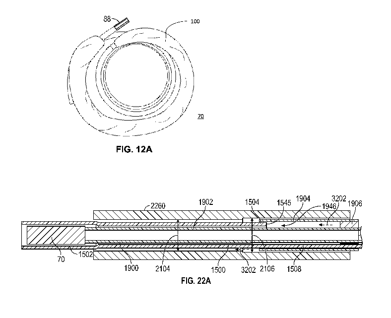
- 19-
CA 03140397 2021-11-12
WO 2020/247907 PCT/US2020/036577
[0166] FIG. 17A illustrates an example embodiment of a sleeve shaft in
accordance with
various embodiments.
[0167] FIG. 17B illustrates a side cross-sectional view of the sleeve shaft of
FIG. 17A.
[0168] FIG. 17C illustrates a detail view of a portion of the sleeve shaft of
FIG. 17B, showing
an interface between different materials of the sleeve shaft.
[0169] FIG. 17D illustrates a side view of an example embodiment of a flexible
polymer
jacket of the sleeve shaft of FIG. 17B.
[0170] FIG. 17E illustrates a side view of an example embodiments of a more
rigid tube
portion of the sleeve shaft of FIG. 17B.
[0171] FIG. 18 illustrates an example embodiment of a layered construction of
a lubricous
sleeve in accordance with various embodiments.
[0172] FIG. 19 illustrates a side cross-sectional view of an example
embodiment of a flexible
tip for the sleeve shaft of FIG. 17B.
[0173] FIG. 20A illustrates a side view of an example embodiment of a proximal
section of a
sleeve shaft in accordance with various embodiments.
[0174] FIG. 20B illustrates a perspective view of an example embodiment of a
more rigid tube
portion of a proximal section of a sleeve shaft in accordance with various
embodiments.
[0175] FIG. 20C illustrates a perspective view of an example embodiment of an
interface
between the tube portion of FIG. 20B and an inner liner at the proximal
section of the sleeve
shaft in accordance with various embodiments.
[0176] FIG. 20D illustrates a perspective view of an example embodiment of an
outer flexible
polymer layer arranged over the tube portion and inner liner of FIG. 20C at
the proximal section
of the sleeve shaft in accordance with various embodiments.
[0177] FIG. 21A illustrates a first side cross-sectional view of an example
embodiment of a
pusher shaft in accordance with various embodiments.
[0178] FIG. 21B illustrates a second side cross-sectional view of an example
embodiment of
a pusher shaft in accordance with various embodiments.
[0179] FIG. 21C illustrates a detail view of a distal end of the pusher shaft
of FIG. 21B.
[0180] FIG. 21D illustrates a proximal end view of the pusher shaft of FIG.
21B.
[0181] FIG. 21E illustrates a side view of a tube portion of the pusher shaft
of FIG. 21B.
[0182] FIG. 21F illustrates a side view of a shell of the pusher shaft of FIG.
21B.
[0183] FIG. 21G illustrates an end view of a plug of the pusher shaft of FIG.
21B.
[0184] FIGS. 22A-22C illustrate an example embodiment of a sleeve shaft and a
pusher shaft
interoperating in accordance with various embodiments.
- 20 -
CA 03140397 2021-11-12
WO 2020/247907 PCT/US2020/036577
[0185] FIG. 23A illustrates a side view of an example embodiment of a proximal
extension of
a pusher shaft in accordance with various embodiments.
[0186] FIG. 23B illustrates a perspective view of the pusher shaft including
the proximal
extension of FIG. 23A.
[0187] FIG. 24A illustrates an example embodiment of a portion of a handle
assembly for a
delivery system for a docking device in accordance with various embodiments.
[0188] FIG. 24B illustrates an example embodiment of a delivery system for a
docking device.
[0189] FIG. 25 illustrates an example embodiment of a flushing plate in
accordance with
various embodiments.
[0190] FIGS. 26A and 26B illustrate example embodiments of a hemostatic seal
in accordance
with various embodiments.
[0191] FIG. 27A illustrates an example embodiment of a portion of a handle
assembly for a
delivery system including a suture lock and sleeve handle in accordance with
various
embodiments.
[0192] FIG. 27B illustrates a perspective view of the suture lock of FIG. 27A,
disconnected
from a branch of the handle assembly.
[0193] FIG. 27C illustrates an exploded view of the suture lock of FIG. 27A.
[0194] FIG. 28A illustrates a side view of an embodiment of the suture lock of
FIGS. 27A-
27C including a flushing port at a proximal end of the suture lock.
[0195] FIG. 28B illustrates a perspective view of a detail portion of the
suture lock of FIGS.
27A-27C, showing a release knob and internal release bar.
[0196] FIG. 28C illustrates a side cross-sectional view of the release bar of
the suture lock of
FIG. 28B.
[0197] FIG. 28D illustrates a perspective view of the release bar of FIGS. 28B
and 28C.
[0198] FIG. 28E illustrates a detail cross-sectional view of a suture cutting
portion of the
release bar of FIGS. 28B-28D.
[0199] FIGS. 29A-29E illustrate example embodiments of directional mechanisms
for a
suture lock in accordance with various embodiments.
[0200] FIGS. 30A-30C illustrate an example embodiment of a suture cutting and
removal
mechanism in accordance with various embodiments.
[0201] FIGS. 31A and 31B illustrate example embodiments of a coil holder in
accordance
with various embodiments.
[0202] FIGS. 32A-32C illustrate a method to expand a covering over a docking
device in
accordance with various embodiments.
- 21 -
CA 03140397 2021-11-12
WO 2020/247907 PCT/US2020/036577
[0203] FIG. 33 illustrates a perspective view of an example embodiment of a
sleeve shaft
covering a docking device and extending outside of a delivery catheter of a
delivery system.
[0204] FIG. 34 illustrates the sleeve shaft surrounding a pusher shaft after
deploying the
docking device from the delivery system of FIG. 33 and removing the sleeve
shaft from the
docking device.
[0205] FIG. 35 illustrates a side cross-sectional view of a portion of a
handle assembly and
fluid flow through lumens of the handle assembly.
[0206] FIG. 36 illustrates a perspective cross-sectional view of a more
detailed portion of the
handle assembly of FIG. 35 and fluid flow through lumens of the handle
assembly.
[0207] FIG. 37 illustrates a perspective cross-section view of a portion of a
delivery system,
including a pusher shaft and sleeve shaft, arranged coaxially with one
another, and fluid flow
through lumens arranged between the coaxial components.
[0208] FIG. 38 illustrates a schematic of fluid flow through lumens of a
distal end portion of
a delivery system, the delivery system including a pusher shaft, sleeve shaft,
and docking
device arranged in an outer shaft of the delivery system.
[0209] FIG. 39 is a flow chart of a method for delivering a docking device to
a native valve
of a heart and implanting the docking device and an associated prosthetic
heart valve at the
native valve.
DETAILED DESCRIPTION OF THE DISCLOSURE
[0210] Disclosed herein are various systems, apparatuses, methods, etc.,
including anchoring
or docking devices, which can be used in conjunction with expandable
prosthetic valves (e.g.,
transcatheter heart valves (THV)) at a native valve annulus (e.g., mitral or
tricuspid valve
annulus), in order to more securely implant and hold the prosthetic valve at
the implant site.
Anchoring/docking devices according to embodiments of the invention provide or
form a more
circular and/or stable anchoring site, landing zone, or implantation zone at
the implant site, in
which prosthetic valves can be expanded or otherwise implanted. Many of these
docking
devices and prosthetic valves have circular or cylindrically-shaped valve
frames or stents that
can be expanded or otherwise implanted into locations with naturally circular
cross sections.
However, further embodiments of docking devices and prosthetic valves have
other geometries
(e.g., oblong, ovular, longitudinally curved, etc.) which are more appropriate
for non-circular
and/or non-cylindrical anatomies. In addition to providing an anchoring site
for the prosthetic
valve, the anchoring/docking devices can be sized and shaped to cinch or draw
the native valve
(e.g., mitral, tricuspid, etc.) anatomy radially inwards. In this manner, one
of the main causes
- 22 -
CA 03140397 2021-11-12
WO 2020/247907 PCT/US2020/036577
of valve regurgitation (e.g., functional mitral regurgitation), specifically
enlargement of the
heart (e.g., enlargement of the left ventricle, etc.) and/or valve annulus,
and consequent
stretching out of the native valve (e.g., mitral, etc.) annulus, can be at
least partially offset or
counteracted. Some embodiments of the anchoring or docking devices further
include features
which, for example, are shaped and/or modified to better hold a position or
shape of the docking
device during and/or after expansion of a prosthetic valve therein. By
providing such anchoring
or docking devices, replacement valves can be more securely implanted and held
at various
valve annuluses, including at the mitral annulus which does not have a
naturally circular cross-
section.
[0211] Referring first to FIGS. 1 and 2, the mitral valve 10 controls the flow
of blood between
the left atrium 12 and the left ventricle 14 of the human heart. After the
left atrium 12 receives
oxygenated blood from the lungs via the pulmonary veins, the mitral valve 10
permits the flow
of the oxygenated blood from the left atrium 12 into the left ventricle 14.
When the left ventricle
14 contracts, the oxygenated blood that was held in the left ventricle 14 is
delivered through
the aortic valve 16 and the aorta 18 to the rest of the body. Meanwhile, the
mitral valve should
close during ventricular contraction to prevent any blood from flowing back
into the left atrium.
[0212] When the left ventricle contracts, the blood pressure in the left
ventricle increases
substantially, which serves to urge the mitral valve closed. Due to the large
pressure differential
between the left ventricle and the left atrium during this time, a large
amount of pressure is
placed on the mitral valve, leading to a possibility of prolapse, or eversion
of the leaflets of the
mitral valve back into the atrium. A series of chordae tendineae 22 therefore
connect the leaflets
of the mitral valve to papillary muscles located on the walls of the left
ventricle, where both
the chordae tendineae and the papillary muscles are tensioned during
ventricular contraction to
hold the leaflets in the closed position and to prevent them from extending
back towards the
left atrium. This helps prevent backflow of oxygenated blood back into the
left atrium. The
chordae tendineae 22 are schematically illustrated in both the heart cross-
section of Fig. 1 and
the top view of the mitral valve of Fig. 2.
[0213] A general shape of the mitral valve and its leaflets as viewed from the
left atrium is
shown in Fig. 2. Commissures 24 are located at the ends of the mitral valve 10
where the
anterior leaflet 26 and the posterior leaflet 28 come together. Various
complications of the
mitral valve can potentially cause fatal heart failure. One form of valvular
heart disease is mitral
valve leak or mitral regurgitation, characterized by abnormal leaking of blood
from the left
ventricle through the mitral valve back into the left atrium. This can be
caused, for example,
by dilation of the left ventricle causing the native mitral leaflets to not
coapt completely,
-23 -
CA 03140397 2021-11-12
WO 2020/247907 PCT/US2020/036577
resulting in a leak, by damage to the native leaflets, or weakening of (or
damage to) the chordae
tendineae and/or papillary muscles. In these circumstances, it may be
desirable to repair the
mitral valve or to replace the functionality of the mitral valve with that of
a prosthetic heart
valve.
[0214] The field of transcatheter aortic valve replacement has developed much
more and has
gained widespread success than transcatheter mitral valve replacement. This
discrepancy
stems, in part, from replacement of a mitral valve being more difficult than
aortic valve
replacement in many respects, such as, for example, due to the non-circular
physical structure
of the mitral valve, its sub-annular anatomy, and more difficult access to the
valve.
Additionally, the mitral valve often lacks calcification limiting the ability
of prosthetic valves
to anchor within the mitral valve.
[0215] One of the most prominent obstacles for mitral valve replacement is
effective
anchoring or retention of the valve at the mitral position, due to the valve
being subject to a
large cyclic load. As noted above, another issue with mitral valve replacement
is the size and
shape of the native mitral annulus, as can be seen in Fig. 2. Aortic valves
are more circular or
cylindrical in shape than mitral valves. Also, the mitral and tricuspid valves
are both larger than
the aortic valve, and more elongate in shape, making them more difficult and
unconventional
sites for implanting a replacement valve with a generally circular or
cylindrical valve frame. A
circular prosthetic valve that is too small can result in leaking around the
implant (i.e.,
paravalvular leakage) if a good seal is not established around the valve,
while a circular
prosthetic valve that is too large can stretch out and damage the narrower
parts of the native
mitral annulus. Further, in many cases, the need for aortic valve replacement
arises due, for
example, to aortic valve stenosis, where the aortic valve narrows due to
calcification or other
hardening of the native leaflets. Therefore, the aortic annulus generally
forms a more compact,
rigid, and stable anchoring site for a prosthetic valve than the mitral
annulus, which is both
larger than the aortic annulus and non-circular. Instances of mitral valve
regurgitation are
unlikely to provide such a good anchoring site. Also, the presence of the
chordae tendineae and
other anatomy at the mitral position can form obstructions that make it much
more challenging
to adequately anchor a device at the mitral position.
[0216] Other obstacles to effective mitral valve replacement can stem from the
large cyclic
loads the mitral valve undergoes and the need to establish a sufficiently
strong and stable
anchoring and retention. Also, even a slight shift in the alignment of the
valve can still lead to
blood flow through the valve or other parts of the heart being obstructed or
otherwise negatively
impacted.
- 24 -
CA 03140397 2021-11-12
WO 2020/247907 PCT/US2020/036577
Embodiments of a Prosthetic Valve
[0217] Prosthetic valves according to exemplary embodiments are shown in FIGS.
3A to 6B.
While specific examples of prosthetic heart valves are discussed herein,
general structure,
method of manufacture, and methods of use of various prosthetic valves, which
can be adapted
for use with the anchoring/docking devices herein, are described in at least
U.S. Pat. No.
10,195,025 entitled "Prosthetic Heart Valve;" U.S. Pat. Pub. No. US
2018/0206982 entitled
"Covered Prosthetic Heart Valve," and U.S. Pat. App. No. 16/252,890 entitled
"Covered
Prosthetic Heart Valve," the disclosure of each of which is incorporated
herein by reference in
its entirety.
[0218] FIGS. 3A and 3B illustrate an example prosthetic valve 30 with a flange
32 attached to
the atrial (inflow end) 34 of the prosthetic valve 30 and extending radially
outward in 360 in
accordance with various embodiments. In many of these embodiments, the flange
32 is
designed to rest on a plane of the native valve, such as on a plane of the
native mitral valve,
tricuspid valve, etc. The flange 32 of some embodiments is designed to
encourage flow through
the prosthetic valve 30 to prevent and/or reduce paravalvular leakage. FIG. 3C
illustrates a
prosthetic valve 30 which includes flanges 32 and 32' that are designed to
only cover the mitral
commissures rather than the entire mitral plane. Flanges 32 and 32' that only
cover the
commissures could beneficially reduce the crimped or compressed size of the
valve for
narrower profile during delivery, but may require the repositioning or
adjusting of the
prosthetic valve 30 during deployment. In various embodiments, the flange 32
(or flanges 32
and 32') is made of a resilient material that is capable of being compacted on
a catheter for
delivery. In certain embodiments, the flange 32 (or flanges 32 and 32') is
made of and/or
comprises a memory material that can be compressed or manipulated and returns
to a specific
shape once a force is removed. An example of a memory material is nitinol (or
NiTi), but other
shape memory alloys or shape memory metals can be used. The memory material
can be
formed into a weave or a frame that is compressible and returns to its formed
shape (e.g., a
flange) once released from a catheter. In some embodiments, the flange 32 is
attached to the
frame of the prosthetic valve via a cloth intermediary 36.
[0219] Turning to FIGS. 4A to 4B, exemplary embodiments of prosthetic valves
40
comprising a covering 42, such as a skirt, on the outer surface of the
prosthetic valve 40 are
illustrated. In covered embodiments, the covering can be designed and/or
configured to prevent
paravalvular leakage between the prosthetic valve 40 and the native valve, to
protect the native
anatomy, to promote tissue ingrowth, and/or designed/configured for other
purposes. Due to
- 25 -
CA 03140397 2021-11-12
WO 2020/247907 PCT/US2020/036577
the general D-shape of the mitral valve (see FIG. 2) and relatively large
annulus compared to
the aortic valve, the covering 42 acts as a seal around the prosthetic valve
40 (e.g., when the
valve is sized smaller than the annulus) and allows for smooth coaptation of
the native leaflets
against the prosthetic valve 40. In various embodiments, the covering 42 is
comprised of a
material that can be crimped for transcatheter delivery of the prosthetic
valve and is expandable
to prevent paravalvular leakage around the prosthetic valve. Examples of
possible materials
include foam, cloth, fabric, one or more polymers, and/or an encapsulated
material, such as an
encapsulated hydrogel. In certain embodiments, the covering is attached via
loop-over stitching
63, as illustrated in FIG. 4A, while additional embodiments will utilize an
edge covering strip
65 with radial, horizontal stitching, as illustrated in FIG. 4B. The edge
covering strip 65 of
many embodiments is constructed of any suitable material that is biocompatible
and atraumatic
to native tissue, including ePTFE, bovine pericardium, porcine pericardium,
equine
pericardium, woven PTFE, knitted PTFE, braided PTFE, polyurethane, electrospun
ePTFE,
dipped thermoplastic, sprayed thermoplastic, other organic tissues, other non-
organic tissues,
and combinations thereof.
[0220] Turning to FIG. 4C, covering (e.g., in cloth, fabric, etc.) solutions
of some
embodiments will form a pocket 46, such as a cup or purse shape, to allow for
insertion,
injection, or encapsulation of an embolic material after placement of a valve
and allow free
exchange of fluid with native blood. Certain embodiments including a pocket
include one or
more pores 44 in a layer of the covering to allow an outer layer to inflate
during systole. In
some embodiments, an encapsulated material may provide an appealing mechanism
of
expansion. Certain embodiments inflate by receiving blood from the atrial side
of the prosthetic
valve. In additional embodiments, a pore 44 or port allows for insertion of
the embolic material
with limited exposure to native blood, while further embodiments encapsulate
the embolic
material completely or nearly completely. In some encapsulated embodiments,
the skirt can
possess a permeable or semipermeable covering to allow for the exchange of
fluids between
the encapsulated material and blood. In some embodiments, the embolic material
is injected
through a catheter or syringe. Further embodiments expand the pocket using
monofilament
warping and/or buckling. An example of monofilament warping is discussed
further herein in
reference to FIG. 8.
[0221] In a number of embodiments, the embolic material will be a hydrogel.
Some hydrogels
expand at body temperature; thus, selecting a body-temperature-expandable
hydrogel allows
the natural heat of a patient to provide the expansion of the hydrogel around
a prosthetic valve
to prevent paravalvular leakage. Further embodiments will possess a hydrogel
that expands by
- 26 -
CA 03140397 2021-11-12
WO 2020/247907 PCT/US2020/036577
absorbing a fluid, e.g., blood. In such embodiments, the hydrogel can be
inserted into the skirt
prior to valve deployment, and the presence of blood after deployment will
allow the hydrogel
to expand. Additional embodiments will utilize a precipitating composition,
such as ethylene
vinyl alcohol (EVOH) dissolved in dimethyl sulfoxide (DMSO). Certain EVOH¨DMSO
compositions are known in the art, including ONYX LIQUID EMBOLIC SYSTEMTm
(Micro Therapeutics, Inc., Irvine, California, U.S.A.) formulations ONYX 18
(6% EVOH),
ONYX 34 (8% EVOH), ONYX HD-500 (20% EVOH), or blends thereof. In such
embodiments, the EVOH¨DMSO composition will be inserted into the skirt after
or during
valve deployment. The DMSO in these compositions will be carried away by the
blood, leaving
EVOH behind, thus forming an embolic to prevent paravalvular leakage.
[0222] In certain embodiments, the embolic material can be an n-butyl
cyanoacrylate. Some
suitable embolic materials can be liquid alkyl-2-cyanoacrylate monomers that,
on contact with
ionic mediums (e.g., water, blood), form flexible polymers that can form
adhesive bonds to
soft tissues. These liquid monomers in isolation are nonviscous, radiolucent,
and can rapidly
polymerize. In certain embodiments, the embolic material can be a multi-
component
formulation including the cyanoacrylate and a radiopaque material, ethiodized
oil, or both. In
some embodiments, the additional components can prolong polymerization time,
opacify the
liquid agent, and allow for visualization under fluoroscopy. Certain n-butyl
cyanoacrylates are
known in the art, including TRUFILL n-butyl-2-cyanoacrylate (n-BCA) liquid
embolic
system (DePuy Synthes Companies, Raynham, Massachusetts, U.S.A.).
[0223] In further embodiments, the embolic material can include one or more
radiopaque
materials that provide for visualization under fluoroscopy. In certain
embodiments, the
radiopaque materials can comprise one or more salts, compounds, or
nanoparticles containing
iodine, barium, tantalum, bismuth, or gold. In some embodiments, the
radiopaque material can
be a tantalum powder.
[0224] Embodiments incorporating foam solutions provide a covering that is
attached to the
exterior of the valve frame in order to provide a substantial paravalvular
leakage solution, while
maintaining a low crimp profile enabling the device to be delivered via a
catheter. In certain
embodiments, one or more foam materials can be used to achieve a low device
profile while
crimped, and provide for expansion in the mitral position and a soft, smooth
surface to interact
with the native mitral valve. Possible foam materials include polyethylene
terephthalate,
polyurethane, and polyurethane-polycarbonate matrix intended for long-term
implantation.
Foam may be advantageous over a cloth covering, because foam typically is able
to compress
to a smaller crimp profile, and the amount of swelling in the mitral valve is
substantially more
- 27 -
CA 03140397 2021-11-12
WO 2020/247907 PCT/US2020/036577
effective in reducing the amount of paravalvular leakage based on the
increased volume of the
foam. Additionally, foam can be extremely compliant and atraumatic against the
coaptation of
the mitral anatomy. Further advantages of foam include tissue ingrowth and
echogenicity. The
tissue ingrowth advantages arise because foam is typically more porous than
other materials,
and the porosity can allow better or improved tissue ingrowth. The improved
echogenicity is
advantageous because it allows a user, such as a physician, cardiologist,
surgeon, or other
medical professional to view the placement of the prosthetic valve based on
where the foam
has expanded.
[0225] A covering for the prosthetic valve can further be altered to allow for
alterations in the
inflow and outflow portions of a prosthetic valve (e.g., the shapes need not
always be perfectly
cylindrical, as shown previously), as seen in FIGS. 5A-5F. In these figures,
frame 50 possesses
a covering 52 that is machined, thermally molded, or otherwise manufactured
into a shape to
allow for a larger outer diameter at the inflow portion 54. Various
embodiments will possess
certain shapes, as illustrated in FIGS. 5A-5F. Some embodiments will possess a
generally
conical shape, as illustrated in FIG. 5A, having a larger outer diameter at
the inflow portion 54
that gradually tapers to a smaller outer diameter at the outflow portion 56.
Another set of
embodiments will possess a curved and tapered covering 52, such as in FIG. 5B,
where the
covering 52 is wider near the inflow portion 54 and has a generally curved
taper toward the
outflow portion 56.
[0226] Additional embodiments will possess generally hourglass shapes, such as
those
illustrated in FIGS. 5C to 5E. As illustrated in FIG. 5C, some embodiments
will have larger
outer diameters at the inflow portion 54 and at the outflow portion 56, while
possessing a
narrower outer diameter at the middle portion 58 of the prosthetic valve 50.
Another shape of
coverings 52 used in some embodiments is illustrated in FIG. 5D, where the
covering 52 is
retracted slightly from the inflow portion 54 of the prosthetic valve 50 and
possesses a larger
outer diameter toward the inflow portion 54; additionally, the covering 52
possesses a larger
outer diameter at the outflow portion 56 and has a smaller outer diameter at a
position 58
proximal to the outflow portion 56. FIG. 5E illustrates embodiments where the
covering 52
possesses a constriction 53 designed to prevent compression of the foam from
influencing the
volume and/or shape of a first portion 57. Because the constriction 53 is
designed to prevent
compression of first portion 57, the constriction 58 can be located at any
position along the
body to effectuate this goal. For example, constriction 53 can be proximal to
inflow portion (as
illustrated), or it can be located proximal to the outflow portion 56 or in a
middle position
between outflow 56 and inflow 54 portions. In many of these embodiments,
constriction 53 is
- 28 -
CA 03140397 2021-11-12
WO 2020/247907 PCT/US2020/036577
a machined slit. In many embodiments with constrictions 53, the overall shape
of covering 52
is cylindrical (e.g., similar to FIG. 4A), while many embodiments will add
contours or other
shapes to covering 52. For example, first portion 57 has a gradual increase in
thickness from
the inflow portion 54, and the second portion 59 has a generally curved taper
toward the
outflow portion 56, as illustrated in FIG. 5E.
[0227] Turning to FIG. 5F, further embodiments of prosthetic valve 50 will
possess a covering
52 with a general mushroom shape, where covering 52 possesses a first portion
57 with a
curved increase in thickness from inflow portion 54 toward a position 58. The
covering 52 will
also possess a second portion 59 extending toward the outflow portion 56 from
position 58,
which has a generally cylindrical shape.
[0228] It should be noted that while some of the embodiments illustrated in
FIGS. 5A to 5F
are illustrated with loop-over stitching and other embodiments are illustrated
with an edge strip
and radial, horizontal stitching, these illustrations are not meant to be
limiting the stitching type
on any specific embodiment, and many embodiments will possess loop-over
stitching or an
edge strip independent of the shape of the covering 52 on the prosthetic valve
50.
[0229] Turning to FIG. 6A, numerous embodiments of a covered valve 60 possess
a covering
made of a woven cloth or fabric possessing a plurality of floated yarn
sections 61 (e.g.,
protruding or puffing sections). Details of exemplary covered valves with a
plurality of floated
yarn sections 61 are further described in U.S. Patent Pre-Grant Publication
Nos.
U52019/0374337 Al, U52019/0192296 Al, and U52019/0046314 Al, the disclosures
of
which are incorporated herein in their entireties for all purposes. In certain
embodiments, the
float sections are separated by one or more horizontal bands 63. In many
embodiments,
horizontal bands 63 are constructed via a leno weave, which improve the
strength of the woven
structure. In some embodiments of woven cloth, vertical fibers (e.g., running
along the
longitudinal axis of the valve 60) comprise a yarn or other fiber possessing a
high level of
expansion, such as a texturized weft yarn, while horizontal (e.g., running
circumferentially
around valve 60) fibers in a leno weave comprise a low expansion yarn or
fiber.
[0230] Floated yarn sections 61 can be heat set to obtain a desired size and
texture, e.g., to
make them softer and more texturized. Texturizing can be achieved by having
the constituent
fibers of the strands/yarns used in section 61 twisted, heat set, and
untwisted such that the fibers
retain their deformed, twisted shape and create a voluminous fabric. In some
embodiments,
floated yarn section 61 can be formed from textured PET yarns without any
weave structure.
In certain embodiments, the covering of covered valve 60 can be heat shrunk to
achieve a
stretchability between 80-160%.
- 29 -
CA 03140397 2021-11-12
WO 2020/247907 PCT/US2020/036577
[0231] In a variety of embodiments, a woven cloth resembles a greige fabric
when assembled
and under tension (e.g., when stretched longitudinally on a compressed valve
60 prior to
delivery of a valve 60). When a valve 60 is deployed and expanded, tension on
floats 61 is
relaxed allowing expansion of the floats 61. In many embodiments, the number
and sizes of
floats 61 is optimized to provide a level of expansion to prevent paravalvular
leakage across
the mitral plane (e.g., to have a higher level of expansion thickness) and/or
a lower crimp
profile (e.g., for delivery of the valve), as further described in U.S. Patent
Pre-Grant Publication
Nos. US2019/0374337 Al, US2019/0192296 Al, and US2019/0046314 Al.
Additionally,
bands 63 can be optimized to allow for attachment of the covering to the valve
based on the
specific size or position of struts or other structural elements on the valve.
[0232] In some embodiments, a covered valve 60 (e.g., as shown in FIG. 6B)
possesses a
hybrid covering 62, which can comprise a plurality of different types of
coverings working
together. In the example shown in FIG. 6B, a first portion 64 of the hybrid
covering 62 near
the inflow portion 66 of the valve 60 comprises foam or other expandable
material, while a
second portion 68 of the hybrid covering 62 near the outflow portion 69 of the
valve 60
comprises woven cloth or fabric, which can have one or more expandable
portions 61. Further,
in FIG. 6B, the foam or other expandable material has a larger expanded
profile at the inflow
portion to increase the ability to form a seal and prevent paravalvular
leakage around the mitral
plane. Due to the compressibility of the foam/expandable material coupled with
the low-profile
ability of the woven cloth, this foam-cloth hybrid skirt beneficially achieves
a low profile when
the valve is crimped. The expanded valve expands radially outwardly at the
inflow end to allow
for good paravalvular sealing.
[0233] Additionally, FIG. 6B illustrates a variation on edge covering 65. In
particular, edge
covering 65 in many embodiments will possess a series of openings 67 placed in
edge covering
65 (FIGS. 6C-6E). In certain embodiments, the openings are created via die
cutting, laser
cutting, punching, or any other method of creating an opening in the material
of the edge
covering 65. Turning to FIGS. 6C and 6D, perspective views of the inflow
portion 66 (FIG.
6C) and outflow portion 69 (FIG. 6D) are illustrated. As seen in these
figures, the edge covering
65 will possess openings 67 on both ends of a prosthetic valve of many
embodiments. Further,
FIG. 6E illustrates a perspective view from within the outflow portion 69,
illustrating edge
covering 65 with a series of openings 67. As illustrated in FIG. 6E, the frame
63 of a prosthetic
valve of many embodiments possesses angled struts, which allow compressibility
around a
catheter or other delivery device. The openings 67 are cut, such that they
align between apices
in frame 63. By placing openings between apices, the edge covering 65 will not
bulge, thus
-30-
CA 03140397 2021-11-12
WO 2020/247907 PCT/US2020/036577
minimize the outer diameter of a crimped valve, when the prosthetic valves of
certain
embodiments are crimped around a catheter or other delivery device.
[0234] The various embodiments illustrated in FIGS. 3A-6E are described as
being of foam
and/or woven cloth, additional embodiments are constructed of materials
capable of providing
the same effect, including woven PET, knitted PET, braided PET, woven PTFE,
knitted PTFE,
braided PTFE, ePTFE membrane, electrospun ePTFE, thermoplastic membrane,
dipped
thermoplastic, sprayed thermoplastic, foam, and combinations thereof.
[0235] In some embodiments, prosthetic valves with coverings will utilize a
material that is
placed under and/or incorporated with the covering that can be compressed or
manipulated and
returns to a specific shape once a force is removed. FIGS. 7A to 7B illustrate
a flange support
structure 71 to allow a covering to expand into its full position.
Specifically, FIGS. 7A to 7B
illustrate a flange support structure 71 secured to a frame 63 of many
embodiments. In a number
of embodiments, the flange support structure 71 is secured near the inflow
portion 72 and at a
middle position 73 of frame 63. In some embodiments, the securing is
accomplished via
stitching, welding, or any other suitable method for securing a flange support
structure 71 to
the frame 63. In a number of embodiments, the flange support structure 71
possess a series of
windows 75, which allow the flange support structure 71 to bulge radially
outward from the
frame 63 and allow the covering to expand to its full position. FIG. 7C
illustrates an
embodiment of prosthetic valve with a flange support structure 71 crimped onto
a delivery
device, such as a catheter. In FIG. 7C, the compressed frame 63 and flange
support structure
71 do not drastically increase the outer diameter (double arrow) of the
crimped frame 63. The
flange support structure 71 can be constructed of any suitable material, such
as biocompatible
and/or atraumatic to native tissue. In certain embodiments, the flange support
structure 71 is
constructed from ePTFE. Additionally, the number of windows 75 placed in
flange support
structure 71 can be any number that is capable of allowing the flange support
structure 71 to
bulge outwardly from frame 63. In some embodiments, 8 windows will be cut, but
additional
embodiments will possess 10, 12, 16, 24, or more windows.
[0236] Additional embodiments, such as illustrated in FIG. 8, outward struts
77 will be placed
on the frame 63 of many embodiments that expand via monofilament warping
and/or buckling.
In many of these embodiments, the outward struts 77 are constructed of a
memory material,
such as nitinol, and placed under or incorporated within a foam or
cloth/fabric covering, the
memory material can aid in the resilient expansion of the covering. Any number
of outward
struts 77 of many embodiments can be placed at positions to allow for the
expansion of the
covering. Some embodiments will place outward struts 77 at 3 positions around
the frame 63,
-31 -
CA 03140397 2021-11-12
WO 2020/247907 PCT/US2020/036577
while additional embodiments will place outward struts 77 at 4, 6, or 8
locations around the
frame 63. In some embodiments, two outward struts 77 are joined to frame 63 at
approximately
the same position and expand in a general V-shape outwardly from the frame 63,
while other
embodiments will possess a single outward strut 77 at a specific location
expanding outwardly
from the frame 63. While FIG. 8 is illustrated for expansion at or near the
atrial or inflow side
of frame 63, a similar mechanism is used to expand pocket coverings (e.g.,
FIG. 4C) in the
ventricular or outflow side of frame 63 to assist in expansion of a pocket for
encapsulating
blood or embolic material.
[0237] It should be noted that the embodiments illustrated in FIGS. 3A to 8
are illustrative
and not meant to be exclusive to or limiting on any other embodiment, unless
the features
illustrated are not combinable. For example, several embodiments will combine
flange support
structures, such as illustrated in FIGS. 7A to 7C along with a foam covering,
such as illustrated
in any of FIGS. 4A to 6A and/or with a flange as illustrated in any of FIGS.
3A to 3C.
Docking Devices
[0238] Anchors/docking devices (e.g., docks) according to example embodiments
of the
invention are shown in FIGS. 9A to 16, and these can comprise a coiled shape.
It is possible
that certain docking devices in an atrio-ventricular position may migrate
further or deeper into
the ventricle post-deployment of the docking device than desired. It may be
beneficial to avoid
too much "dock drop" to help avoid paravalvular leakage, e.g., sealing the
docking device and
prosthetic valve seal together higher on the native leaflets may help prevent
paravalvular leaks
that might occur if done lower on the chordae tendineae. Additionally, higher
placement (e.g.,
by avoiding dock drop) may help long term stability of the valve, because the
leaflets are
thicker and more robust near the annulus rather than near their distal end,
thus anchoring at or
near the annulus is expected to be stronger and provide longer term stability.
Also, higher
placement of the prosthetic valve may help avoid abrasion of the valve against
the native tissue
(e.g., against the native leaflets and/or chordae tendineae), by making the
artificial valve less
likely to rub against the leaflets and/or chordae tendineae.
[0239] Paravalvular leakage can occur due to a number of causes, including
where the native
annulus is too large in comparison the prosthetic valve; the commissural
leaflets are too short
and/or are damaged; the implantation of a docking device did not completely
capture the native
leaflets; the crossing of a docking device from one side of the valve to the
other causes a small
gap (e.g., in the commissure); the placement of a prosthetic valve is too
biased toward the
lateral, anterior, posterior, and/or medial sides of a native valve; and/or
anatomical
-32-
CA 03140397 2021-11-12
WO 2020/247907 PCT/US2020/036577
abnormalities in certain patients (e.g., clefts, those associated with
degenerative mitral
regurgitation, etc.). Various embodiments of this disclosure are designed to
compensate for,
avoid, reduce, and/or obviate many of these issues, including by holding the
dock up on both
sides of the native mitral valve (thus reducing and/or inhibiting dock drop by
maintaining the
dock and prosthetic valve close to the native annulus plane), by creating a
better seal around a
prosthetic valve, by creating a better seal above the native annulus, etc..
For example, FIGS.
9A-9C illustrate a version of the dock or a core of the dock configured to
provide one or more
points or regions of contact between the dock and the left atrial wall, such
as at least three
points of contact in the left atrium or complete contact on the left atrial
wall, while FIGS. 10A-
10C illustrate a flat dock or core of a flat dock, where the atrial portion
lies on the mitral plane,
and FIGS. 11A-11C illustrate a hybrid dock or core of the hybrid dock where
the stabilization
or atrial turn creates a ring around a deployed prosthetic valve to seal the
valve and reduce
paravalvular leakage. In the examples of FIGS. 9A to 11C, the docking device
70 includes a
coil or coiled portion with a plurality of turns extending along a central
axis of the docking
device 70. The coil or coiled portion can be continuous and can extend
generally helically, with
various differently sized and shaped sections, as described in greater detail
below. The docking
devices 70 shown in FIGS. 9A to 11C can be configured to fit at the mitral
position but can be
shaped and/or adapted similarly or differently in other embodiments for better
accommodation
at other native valve positions as well, such as at the tricuspid valve.
Advantageously, the
docking device geometries of the present disclosure provide for engagement
with the native
anatomy that can provide for increased stability and reduction of relative
motion between the
docking device, the prosthetic valve docked therein, and the native anatomy.
Reduction of such
relative motion can prevent material degradation of components of the docking
device and/or
the prosthetic valve docked therein and can prevent damage/trauma to the
native tissues.
[0240] The docking device 70 of many embodiments includes a central region 80
with a coil,
coiled portion, or multiple coils (e.g., 2 coils, 3 coils, 4 coils, between 2-
5 coils, or more). The
coiled portion or coils of the central region 80 can be similarly sized and
shaped or vary in size
and/or shape. In some implementations, the central region 80 comprises three
or approximately
three full coil turns having substantially equal inner diameters. The central
region 80 of the
docking device 70 serves as the main landing region or holding region for
holding the
expandable prosthetic valve when the docking device 70 and the valve
prosthesis are implanted
into a patient's body. In some embodiments, the docking device 70 has a
central region 80 with
more or less than three coil turns, depending for example, on the patient's
anatomy, the amount
of vertical contact desired between the docking device 70 and the valve
prosthesis (e.g.,
- 33 -
CA 03140397 2021-11-12
WO 2020/247907 PCT/US2020/036577
transcatheter heart valve or THV), and/or other factors. The coiled portion or
coil(s) of the
central region 80 can also be referred to as the "functional coils" or
"functional turns" since the
properties of these coils contribute the most to the amount of retention force
generated between
the valve prosthesis, the docking device 70, and the native mitral leaflets
and/or other
anatomical structures.
[0241] Various factors can contribute to the total retention force between the
docking device
70 and the prosthetic valve held therein. A main factor is the number of turns
included in the
functional coils, while other factors include, for example, an inner diameter
of the functional
coils, friction force (e.g., between the coils and the prosthetic valve), and
the strength of the
prosthetic valve and the radial force the valve applies on the coil. A docking
device can have a
variety of numbers of coils and/or turns. The number of functional turns can
be in ranges from
just over a half turn to 5 turns, or one full turn to 5 turns, or more. In one
embodiment with
three full turns, an additional one-half turn is included in the ventricular
portion of the docking
device. In another embodiment, there can be three full turns total in the
docking device. In one
embodiment, in the atrial portion of the docking device, there can be one-half
to three-fourths
turn or one-half to three-fourths of a circle. While a range of turns is
provided, as the number
of turns in a docking device is decreased, the dimensions and/or materials of
the coil and/or the
wire that the coil is made from can also change to maintain a proper retention
force. For
example, the diameter of the wire can be larger and/or the diameter of the
function coil turn(s)
in a docking device with fewer coils. There can be a plurality of coils in the
atrium and in the
ventricle.
[0242] A size of the functional coils or coils of the central region 80 is
generally selected
based on the size of the desired THV to be implanted into the patient.
Generally, the inner
diameter 90 of the functional coils/turns (e.g., of the coils/turns of the
central region 80 of the
docking device 70) will be smaller than the outer diameter of the expandable
heart valve, so
that when the prosthetic valve is expanded in the docking device, additional
radial tension or
retention force will act between the docking device and the prosthetic valve
to hold the
prosthetic valve in place. The retention force needed for adequate
implantation of a prosthetic
valve varies based on the size of the prosthetic valve and on the ability of
the assembly to
handle mitral pressures of approximately 180 mm Hg. For example, based on
hemodynamic
data using a prosthetic valve with a 29 mm expanded outer diameter, a
retention force of at
least 15.8 N can be needed between the docking device and the prosthetic valve
in order to
securely hold the prosthetic valve in the docking device and to resist or
prevent valve
regurgitation or leakage. However, under this example, to meet this 15.8 N
retention force
- 34-
CA 03140397 2021-11-12
WO 2020/247907 PCT/US2020/036577
requirement with statistical reliability, a target average retention force
should be substantially
greater, for example, approximately 30 N.
[0243] In many embodiments, the retention force between the docking device and
the valve
prosthesis reduces dramatically when a difference between the outer diameter
of the prosthetic
valve in its expanded state and the inner diameter of the functional coils is
less than about 5
mm, since the reduced size differential can be too small to create sufficient
retention force
between the components. For example, when, in one embodiment, a prosthetic
valve with a 29
mm expanded outer diameter was expanded in a set of coils with a 24 mm inner
diameter, the
retention force observed was about 30 N, but when the same prosthetic valve
was expanded in
a set of coils with a 25 mm inner diameter (e.g., only 1 mm larger), the
retention force observed
dropped significantly to only 20 N. Therefore, in some embodiments, in order
to create a
sufficient retention force between the docking device and a 29 mm prosthetic
valve, the inner
diameter of the functional coils (e.g., the coils of the central region 10 of
docking device 1)
should be 24 mm or less. Often, the inner diameter of the functional coils
(e.g., central region
80 of the docking device 70) should be selected to be at least about 5 mm less
than the prosthetic
valve that is selected for implantation, though other features and/or
characteristics (e.g., friction
enhancing features, material characteristics, etc.) can be used to provide
better retention if other
sizes or size ranges are used, as various factors can affect retention force.
[0244] However, diameter of the functional coils should be selected based on
consideration
and balancing of several factors to obtain optimal results. For example, the
native anatomy
between the mitral annulus at the mitral plane and the papillary muscle heads
forms a generally
trapezoidal shape, and the tissue of the mitral leaflets is thicker near the
mitral plane and thins
the further below the mitral plane. Smaller diameters of the central region 80
may encourage
the docking device 70 to install further below the mitral plane than desirable
(a similar effect
can be observed at the tricuspid valve as well). When docking occurs at a
location where the
mitral leaflets are thinner, this may result in a suboptimal anchoring
position for the prosthetic
valve. Accordingly, size, diameters, and other features that help hold the
prosthetic valve higher
on the leaflets can be beneficial. In addition, a size of the inner diameter
of the functional coils
or central region 80 can also be selected to draw the native anatomy closer
together, in order
to at least partially offset or counteract valve regurgitation that is caused
by stretching out of
the native valve annulus as a result of, for example, left ventricular
enlargement.
[0245] It is noted that the desired retention forces discussed above are
applicable to
embodiments for mitral valve replacements. Therefore, other embodiments of the
docking
device that are used for replacement of other valves can have different size
relationships based
- 35 -
CA 03140397 2021-11-12
WO 2020/247907 PCT/US2020/036577
on the desired retention forces for valve replacement at those respective
positions. In addition,
the size differentials can also vary, for example, based on the materials used
for the valve and/or
the docking device, whether there are any other features to prevent expansion
of the functional
coils or to enhance friction/locking, and/or based on various other factors.
[0246] In embodiments where the docking device 70 is used at the mitral
position, the docking
device can first be advanced and delivered to the native mitral valve annulus,
and then set at a
desired position, prior to implantation of the prosthetic heart valve. In some
embodiments, the
docking device 70 is flexible and/or made of a shape memory material, so that
the coils of the
docking device 70 can be straightened for delivery via a transcatheter
approach as well. In some
embodiments, the coil is made of another biocompatible material, such as
stainless steel. Some
of the same catheters and other delivery tools can be used for both delivery
of the docking
device 70 and the prosthetic valve, without having to perform separate
preparatory steps,
simplifying the implantation procedure for the end user.
[0247] Since the functional coils/turns or coils/turns of the central region
80 of the docking
device 70 are kept relatively small in diameter (e.g., the central region 80
in one embodiment
can have an inner diameter of between approximately 21-24 mm (e.g., 2 mm) or
another
diameter smaller than the prosthetic valve and/or the native annulus) in order
to increase
retention force with the prosthetic valve, it might be difficult to advance
the docking device 70
around the existing leaflets and/or chordae tendineae to a desired position
relative to the native
mitral annulus. This is especially true, if the entire docking device 70 is
made to have the same
small diameter as the central region 80. Therefore, the docking device 70 can
have a distal or
lower region 82 that comprises and/or consists of a leading coil/turn
(sometimes referred to as
an encircling turn or a leading ventricular coil/turn) of the docking device
70, which has a lower
diameter that is greater than the diameter of the functional coils/turns or of
the coils/turns of
central region 80.
[0248] Features of the native anatomy, especially in the right and left
ventricles, have variable
dimensions. For example, native mitral anatomy can have an approximately 35 mm
to 45 mm
greatest width on a long axis. The diameter or width of the encircling turn or
leading coil/turn
(e.g., ventricular coil/turn) of the lower region 82 can be selected to be
larger to more easily
navigate a distal or leading tip 84 of the docking device 70 around and
encircle the features of
the native anatomy (e.g., leaflets and/or chordae tendineae).
[0249] Various sizes and shapes are possible, for example, in one embodiment,
the diameter
could be any size from 25 mm to 75 mm. The term "diameter" as used in this
disclosure does
not require that a coil/turn be a complete or perfectly-shaped circle but is
generally used to
- 36 -
CA 03140397 2021-11-12
WO 2020/247907 PCT/US2020/036577
refer to a greatest width across opposing points of the coil/turn. For
example, with respect to
the leading coil/turn, diameter can be measured from the distal tip 84 to the
opposite side, as if
the lower region or leading coil/turn 82 formed a complete rotation, as shown
as diameter 91
in FIG. 9A. Alternatively, the diameter can be considered double a radius of
curvature of the
leading coil/turn. In various embodiments, the diameter 91 of the lower region
82 is enough to
encircle anatomical features within the ventricle, including mitral leaflets
and chordae, such
that the inner diameter of the lower region 82 is equal to or greater than the
inner diameter of
the central region 80 (e.g., diameter 90 shown in FIG. 10A). Further
embodiments are designed
to be atraumatic to other ventricular anatomy, including walls or septa within
the ventricle. As
such, the diameter 91 of the lower region 82 is small enough to not contact
walls or septa. In
certain embodiments, the diameter 91 of the lower region 82 ranges from
approximately 33-37
mm (e.g., 2 mm). In one embodiment, the lower region 82 of the docking
device 70 (e.g., the
leading coil/turn) has a diameter 91 of 43 mm or approximately 43 mm (e.g.,
2 mm), in other
words the radius of curvature at the leading coil/turn can be 21.5 mm or
approximately 21.5
mm (e.g., 2 mm). In other embodiments, the diameter 91 of the lower region 82
of the docking
device 70 is in a range of 28-38mm, 30-36mm, 31-35mm, 32-34mm, or 32.5-33.5mm
(e.g.,
radius of curvature in a range of 16.25-16.75mm).
[0250] Having a leading coil/turn with a larger size than the functional coils
can help more
easily guide the coils around and/or through the chordae tendineae geometry,
and most
importantly, adequately around both native leaflets of the native valve (e.g.,
the native mitral
valve, tricuspid valve, etc.). Once the distal tip 84 is navigated around the
desired native
anatomy, the remaining coils of the docking device 70 can also be guided
around the same
features. In some embodiments, the size of the other coils can be reduced
sufficiently to cause
the corralled anatomical features to be pulled radially inwardly or slightly
radially inwardly.
Meanwhile, the length of the enlarged lower region 82 or the leading coil/turn
can be kept
relatively short, to prevent or avoid obstruction or interference of the flow
of blood along the
ventricular outflow tract by the lower region 82 or the leading coil/turn. For
example, in one
embodiment, the enlarged lower region 82 or the leading coil/turn extends for
only about half
a loop or rotation. With a lower region 82 or the leading coil/turn having
this relatively short
length, when a prosthetic valve is expanded into the docking device 70 and the
coils of the
docking device 70 start to unwind slightly due to the size differential
between the docking
device and the prosthetic valve, the lower region 82 or the leading coil/turn
may also be drawn
in and shift slightly. Under this example, after expansion of the prosthetic
valve, the lower
region 82 or the leading coil/turn can be similar in size and be aligned
substantially with the
- 37 -
CA 03140397 2021-11-12
WO 2020/247907 PCT/US2020/036577
functional coils of the docking device 70, rather than continuing to project
away from the
functional coils, thereby reducing any potential flow disturbances. Other
docking device
embodiments can have lower regions that are longer or shorter, depending on
the particular
application.
[0251] In various embodiments, the docking device 70 illustrated in FIGS. 9A
to 11 also
includes an enlarged proximal or upper region 86 that comprises and/or
consists of a stabilizing
coil/turn (e.g., which can be an atrial coil/turn) of the docking device 70.
During a transient or
intermediate stage of the implantation procedure, that is, during the time
between the
deployment and release of the docking device 70 and final delivery of the
prosthetic valve,
there is a possibility that the coil could be shifted and/or dislodged from
its desired position or
orientation, for example, by regular heart function. Shifting of the docking
device 70 could
potentially lead to a less secure implantation, misalignment, and/or other
positioning issues for
the prosthetic valve. A stabilization feature or coil can be used to help
stabilize the docking
device in the desired position. For example, the docking device 70 can include
the upper region
86 with an enlarged stabilization coil/turn (e.g., an enlarged atrial
coil/turn having a greater
diameter 92 and/or 94 than the functional coils) intended to be positioned in
the circulatory
system (e.g. in the left atrium) such that it can stabilize the docking
device. For example, the
upper region 86 or stabilization coil/turn can be configured to abut or push
against the walls of
the circulatory system (e.g., against the walls of the left atrium), in order
to improve the ability
of the docking device 70 to stay in its desired position prior to the
implantation of the prosthetic
valve.
[0252] The stabilization coil/turn (e.g., atrial coil/turn) at the upper
region 86 of the docking
device 70 in the embodiments shown can extend up to about one full turn or
rotation, and
terminates at a proximal tip 88. In other embodiments, the stabilization
coil/turn (e.g., atrial
coil) can extend for more or less than one turn or rotation, depending for
example on the amount
of contact desired between the docking device and the circulatory system
(e.g., with the walls
of the left atrium) in each particular application. The radial size of the
stabilization coil/turn
(e.g., atrial coil) at the upper region 86 can also be significantly larger
than the size of the
functional coils in the central region 80, so that the stabilization coil/turn
(e.g., atrial coil or
atrial turn) flares or extends sufficiently outwardly in order to contact the
walls of the
circulatory system (e.g., the walls of the left atrium). Additionally, the
stabilization coil/turn of
various embodiments will be configured to be less abrasive to the native
tissue and/or anatomy.
For example, the surface texture can be made smoother and/or softer, such that
movement of
the docking device against the native anatomy will not damage the native
tissue.
- 38 -
CA 03140397 2021-11-12
WO 2020/247907 PCT/US2020/036577
[0253] The proximal tip 88 as shown in these figures also includes an eyelet
or eyehole. The
eyelet at the proximal tip will be used to secure the docking device 70 to a
delivery system (as
described below) through various means, including a suture. As such, various
embodiments
comprising an eyelet at the proximal tip 88 will utilize different shapes and
sizes of eyelets. As
such, some embodiments will utilize larger eyelets, while other embodiments
will use smaller
eyelets. Additionally, the shape will vary in certain embodiments, such that
some embodiments
will possess round eyelets, while others will utilize D-shaped eyelets.
Further, various
embodiments will not possess eyelets such as those illustrated, but will
possess holes drilled
into the docking device itself, such as laser-drilled holes.
[0254] Turning to FIGS. 9A to 9C, these figures are representative of a
docking device, but
are also representative of a core that can be covered and/or added to in order
to form a docking
device. The stabilization coil 86 is designed to create a plane formed by at
least three points of
contact in the atrium. These three anchoring points or points of contact are
the posterior shelf
of the native valve (e.g., of the mitral valve, etc.), the anterior wall of
the atrium, and either an
atrial appendage of the atrium or the lateral shelf of the native valve. This
plane formed by
three points of contact will be parallel to a plane of the native valve, which
will maintain the
dock position parallel to the native valve plane. In some embodiments, the
diameter of
stabilization coil is desirably larger than the annulus, native valve plane,
and atrium for better
stabilization, but the stabilization coil is flexible with a thin and weak
cross section to prevent
damage to the atrium of a patient over a long-term placement of the docking
device 70.
[0255] Suitable materials for the docking device include a nitinol core with a
core size range
from approximately 0.3mm to approximately lmm. The flexible core will allow
the
stabilization coil to conform to varying atrium shapes and sizes.
Additionally, in some
embodiments with a three-point contact design as illustrated in FIGS. 9B and
9C, the
stabilization coil is designed, when unconstrained (e.g., when not installed
into a patient's
native valve), to sit lower in free space than the functional coils or cross
downwardly across
the functional coils. This can beneficially lift the functional coils (central
region 80) relative to
the native anatomy, a plane of the native valve, and the leaflets when
implanted. For example,
a stabilization coil that crosses from a proximal side of the central region
or functional coils to
or toward a distal side of the central region/functional coils, when deployed
in an atrium, push
down on the atrium and/or native valve plane to cause the central
region/functional coils to
move up or be biased up higher on the ventricular side of the valve and higher
under the leaflets.
[0256] With respect to FIGS. 10A to 10D, these figures are representative of a
docking device,
but are also representative of a core that can be covered and/or added to in
order to form a
- 39 -
CA 03140397 2021-11-12
WO 2020/247907 PCT/US2020/036577
docking device. In FIGS. 10A-10D, the docking device 70 is designed to have a
flat
stabilization coil 86 that sits on a plane of the native valve (e.g., on a
native mitral plane or a
native tricuspid plane). In these embodiments, the stabilization coil 86 is
designed to be larger
than the opening of the native valve (e.g., of the mitral valve or tricuspid
valve), yet not so
large that the stabilization coil 86 does not sit on a plane of the native
valve. The stabilization
coil 86 can form a continuous curve, such as illustrated in FIG. 10A or can be
flared out or
biased toward the posterior wall of the atrium, as seen in FIG. 10B, thus
taking advantage of
the posterior shelf, to prevent the docking device 70 from falling into the
ventricle prior to
deployment of an artificial or prosthetic valve therein. Additionally, in
certain flat
embodiments, the stabilization coil will have a smooth cover to prevent trauma
to native
anatomy in regions that exhibit relative motion (e.g., where the docking
device 70 crosses from
the atrium to the ventricle through the mitral valve).
[0257] Turning to FIGS. 11A to 11C, these figures are representative of a
docking device, but
are also representative of a core that can be covered and/or added to in order
to form a docking
device. In FIGS. 11A to 11C, a hybrid dock design is illustrated that is
configured to improve
prevention and/or inhibition of paravalvular leakage. In embodiments of this
hybrid docking
device 70, the stabilization coil 86 is designed to create a closer ring
around a deployed
prosthetic valve, thus helping to seal the valve and preventing paravalvular
leakage. In some
embodiments, this sealing effect is maximized by offsetting radially outwardly
the dock core
from the maximum valve outer diameter plus half the cross section of the
docking device. In
some embodiments, the stabilization coil 86 allows for the largest outer
diameter (e.g., optimal
contact with the atrium) to help also with dock drop prior to prosthetic valve
deployment.
Additionally, in some embodiments, the docking device is configured such that,
after prosthetic
valve deployment, the stabilization coil will be flush with the outer diameter
of the implanted
and expanded prosthetic valve. This hybrid design can be manufactured to rely
on different
prosthetic valve outer diameters to prevent paravalvular leakage close around
the prosthetic
valve. For example, the docking device can be designed to accommodate a valve
with an outer
diameter of approximately 30mm or approximately 34mm or any diameter
therebetween.
Additionally, the dimensions of the docking device 70 can be adjusted to
handle prosthetic
valves with outer diameters ranging from approximately 20mm to approximately
50mm.
[0258] In some embodiments, the various docking devices herein are configured
to have a
small enough cross section during delivery to fit inside a
catheter/sleeve/sheath of a delivery
device (discussed in greater detail below), but expand post-deployment to
maximize OD and
create an improved seal around a prosthetic valve after implantation.
Additionally, some
- 40 -
CA 03140397 2021-11-12
WO 2020/247907 PCT/US2020/036577
embodiments comprise and/or utilize a material configured to optimize tissue
ingrowth (e.g.,
with pores and/or other openings sized to provide more available surface area
to assist in
encouraging such ingrowth). In some embodiments, the pore and/or opening sizes
are
approximately 30i.tm - 1000 p.m, which can encourage optimal tissue ingrowth.
The tissue
ingrowth can allow for improved sealing and better integration with the native
valve anatomy
to stabilize the docking device and prosthetic valve and prevent abrasions
and/or damage over
time. In certain embodiments, the docking devices can comprise a material
having openings
(e.g., pores) with sizes in a range of about 400-800 p.m, 500-750m, 500-660m,
600-650m,
or 625-650m.
[0259] The various docking devices herein can also incorporate additional
modifications to the
functional coils (e.g., central region 80 in FIGS. 9A to 11C) and/or the
stabilization coil (e.g.,
item 86 in FIGS. 9A to 11C) to improve the functionality of the docking
devices. Examples of
some such additional modifications are illustrated in FIGS. 12A to 16.
[0260] FIGS. 12A to 12G illustrate examples of possible coverings 100 that can
be placed on
all or only a portion of a docking device (e.g., docking device 70 as
illustrated in FIGS. 9A to
11C or elsewhere herein) to form a seal against the prosthetic valve and
reduce paravalvular
leakage. In many embodiments, covering 100 covers predominately or only the
stabilization
turn/coil (e.g., atrial turn/coil) or a portion thereof. In some embodiments,
the covering 100 is
attached on the atrial turn and extends toward the functional turns in the
central region and/or
onto a portion of the functional turns. In some embodiments, the covering 100
is attached on
the functional turn and extends towards the atrial turn. Certain embodiments
possess the
covering 100 on only the functional turns. In some embodiments, covering 100
extends over
the entirety of the docking device 70. When on the stabilization coil/turn or
atrial coil/turn,
covering 100 can help cover an atrial side of an atrioventricular valve to
prevent and/or inhibit
blood from leaking through the native leaflets, commissures, and/or around an
outside of the
prosthetic valve by blocking blood in the atrium from flowing in an atrial to
ventricular
direction other than through the prosthetic valve. In some embodiments, the
covering 100 is
configured to have a compressed configuration for delivery through vasculature
to the heart
valve with a narrow profile and an expanded configuration in which the
covering 100 is
expanded to a larger outer diameter (which can beneficially help prevent
and/or inhibit
paravalvular leakage).
[0261] In some embodiments, the covering 100 can expand to a diameter of
approximately
5mm (e.g., 4mm) to prevent and/or inhibit paravalvular leakage. In some
embodiments, the
covering 100 is configured to expand such that an improved seal is formed
closer to and/or
- 41 -
CA 03140397 2021-11-12
WO 2020/247907 PCT/US2020/036577
against a prosthetic valve deployed therein (such as describe above in regard
to FIGS. 11A to
11C). In some embodiments, covering 100 is configured to prevent and/or
inhibit leakage at
the location where the docking device 70 crosses between leaflets of the
native valve (e.g.,
without covering 100, the docking device may push the leaflets apart at the
point of crossing
the leaflets and allow for leakage at that point (e.g., along the docking
device or to its sides),
but covering 100 can be configured to expand to cover and/or fill any opening
at that point and
inhibit leakage along the docking device).
[0262] In some embodiments, e.g., as illustrated in FIG. 12A, covering 100 can
comprise
and/or consist of an expandable foam. In some embodiments, the covering
comprises and/or
consists of an expandable foam that is a memory foam, such that it will expand
to a specific
shape or specific pre-set shape upon removal of a crimping pressure prior to
delivery of the
docking device 70. Examples of such foams are polyethylene terephthalate
(PET),
polyurethane, and polyurethane-polycarbonate matrix. In some embodiments, the
foam is
configured to expand the cross-sectional diameter of the docking device 70,
such that the cross-
sectional diameter in the region of the covering is 2mm to 7mm. Additionally,
in some
embodiments utilizing foam, the foam comprises large enough pores to be
atraumatic to the
native anatomy and allow tissue ingrowth.
[0263] In some embodiments covering 100 comprises an expandable, non-foam
structure over
the docking device 70. For example, as illustrated in FIG. 12B, covering 100
can comprise a
braided structure over the docking device 70. A braided structure can be
stretched inside of a
sleeve or covering prior to deployment of the docking device 70, but after the
deployment, the
braided structure can be allowed to expand to its largest possible diameter to
create a seal. In
some embodiments, the braided structure is a braided shape memory material
(e.g., shape
memory alloy, shape memory metal, nitinol, etc.) that is shape set and/or pre-
configured to
expand to a particular shape and/or size when unconstrained and when deployed
at a native
valve.
[0264] In FIG. 12C, some embodiments the covering 100 comprises multiple
layers of the
same and/or different materials. In these embodiments, the covering (such as a
braided
structure, foam, or expandable non-foam structure) can be covered with a
second covering 102.
In these embodiments, the second covering 102 is designed to be atraumatic to
native tissue
and/or promote tissue ingrowth into the second covering 102 and possibly the
first covering
100. The second covering can be constructed of any suitable material,
including foam, cloth,
fabric, and/or polymer, which is flexible to allow for compression and
expansion of the first
100 and second 102 coverings.
- 42 -
CA 03140397 2021-11-12
WO 2020/247907 PCT/US2020/036577
[0265] Motion between a docking device 70 and a covering 100 can cause trauma
to native
tissue, as such, several embodiments of docking devices 70 will incorporate
means to limit
motion, thus reducing the risk of trauma to native tissue. Turning to FIG.
12D, an embodiment
of a covered docking device with a braided stabilization coil 86 is
illustrated. In embodiments
such as illustrated in FIG. 12D, a braided texture on the stabilization coil
86 of the dock can
interact with the covering 100 surrounding the stabilization coil 86. By
forming an interaction
between the stabilization coil 86 and the covering 100, motion of the docking
device 70 can be
reduced, thus limiting trauma to the native tissue.
[0266] In FIG. 12E, an embodiment of a docking device 70 is illustrated with a
covering 100
on a functional coil in the central region of the docking device. Like the
example illustrated in
FIG. 12B, covering 100 can comprise a braided structure over the docking
device 70. A braided
structure can be stretched inside of a sleeve or covering prior to deployment
of the docking
device 70, but after the deployment, the braided structure can be allowed to
expand to its largest
possible diameter to create a seal. In some embodiments, the braided structure
is a braided
shape memory material (e.g., shape memory alloy, shape memory metal, nitinol,
etc.) that is
shape set and/or pre-configured to expand to a particular shape and/or size
when unconstrained
and when deployed at a native valve. As shown in FIG. 12E, the docking device
70 can have
an extension 140 substantially positioned between the central region 142
having the functional
turns and the upper region 144 having the atrial turn. As described elsewhere
herein, the
docking device 70 can have a lower region 146 having an encircling turn with a
larger diameter
relative to the functional turns in the central region 142. In FIG. 12E, the
extension 140 is
made up of or includes a vertical part of the coil that extends substantially
parallel to a central
axis of the docking device 70. In some embodiments, the extension 140 can be
angled relative
to the central axis of the docking device 70, but will generally serve as a
vertical or axial spacer
that spaces apart the adjacent connected portions of the docking device 70 in
a vertical or axial
direction, so that a vertical or axial gap is formed between the coil portions
on either side of
the extension 140 (e.g., a gap can be formed between an upper or atrial side
and a lower or
ventricular side of the docking device 70). In certain embodiments, the
extension 140 is
intended to be positioned at or near the anterolateral commissure AC when the
docking device
70 is implanted, with covering 100 crossing the mitral annulus plane and
positioned such that
a portion of covering 100 is positioned in the posteromedial commis sure PC.
Additional details
of exemplary shapes for the docking device 70 having an extension 140 can be
seen in U.S.
Pre-Grant Publication No. US2018/0055628A1, the entirety of which is
incorporated herein
for all purposes.
-43 -
CA 03140397 2021-11-12
WO 2020/247907 PCT/US2020/036577
[0267] Turning to FIG. 12E, an embodiment of a docking device 70 is
illustrated with covering
100 extending on the top functional turn of the central region. In such
embodiments, covering
100 can help cover the ventricular side of an atrioventricular valve to
prevent and/or inhibit
blood from leaking through the native leaflets, commissures, and/or around an
outside of the
prosthetic valve by blocking blood in the atrium from flowing in an atrial to
ventricular
direction other than through the prosthetic valve. Like the example
illustrated in FIG. 12B,
covering 100 can comprise a braided structure over the docking device 70. A
braided structure
can be stretched inside of a sleeve or covering prior to deployment of the
docking device 70,
but after the deployment, the braided structure can be allowed to expand to
its largest possible
diameter to create a seal. In some embodiments, the braided structure is a
braided shape
memory material (e.g., shape memory alloy, shape memory metal, nitinol, etc.)
that is shape
set and/or pre-configured to expand to a particular shape and/or size when
unconstrained and
when deployed at a native valve. In some embodiments, the covering 100 is
attached on the
atrial turn and extends toward the functional turns in the central region
and/or onto a portion
of the functional turns. In some embodiments, the covering 100 is attached on
the functional
turn and extends towards the atrial turn. In further embodiments, the covering
100 can be
attached to the docking device core at both ends of the covering 100 and have
a free-floating
portion therebetween. Additionally, covering 100 can comprise and/or consist
of an expandable
foam. In some embodiments, the covering comprises and/or consists of an
expandable foam
that is a memory foam, such that it will expand to a specific shape or
specific pre-set shape
upon removal of a crimping pressure prior to delivery of the docking device
70, like the
example in FIG. 12A.
[0268] FIGS. 12F and 12G illustrate different arrangements of the various
components that can
be integrated on or around the stabilization coil 86 of many embodiments. In
particular, FIG.
12F illustrates a cross section of the stabilization coil 86 along with
covering 100 and second
covering 102. As illustrated, as the covering 100 and second covering 102
expand, a cavity 104
is formed between stabilization coil 86 and the coverings 100, 102. Also
illustrated are the
construction of the atrial turn including a core 106, which can be for
example, a NiTi core, or
a core that is made of or includes one or more of various other biocompatible
materials. FIG.
12F also illustrates tubular layer 108 to provide a cushioned, padded-type
layer for the atrial
turn to be atraumatic against native tissue. In certain embodiments, tubular
layer 108 is
constructed of ePTFE. FIG. 12F further illustrates braided layer 110 placed
over the tubular
layer 108. As noted above, the braided layer 110 is designed to interact with
covering 100 to
limit motion and/or limit trauma to native tissue. It should be noted that
FIG. 12F illustrates a
- 44 -
CA 03140397 2021-11-12
WO 2020/247907 PCT/US2020/036577
variety of options that can be utilized in construction of docking devices of
various
embodiments, and the particular arrangement is only illustrative of some
embodiments. As
such, a number of embodiments will not possess all of the components
illustrated in FIG. 12F
in constructing a docking device.
[0269] In some embodiments, the cross-section of FIG. 12F can be implemented
in the coils
shown FIGS. 12B-12D. Further, in some embodiments, the stabilization coil 86
can have two
portions, a first portion having the cross-section shown in FIG. 12F and an
adjacent, second
portion having a cross-section shown in FIG. 12G. The cross-section shown in
FIG. 12G may
be the same as the cross-section shown in 12F, except it does not include the
braided layer 110.
In some embodiments, as shown in FIG. 12E, the area of covering 100 can be
split into two
portions (as denoted by the dashed line), including a first portion extending
in the direction of
arrow 12F and a second portion extending in the opposite direction, as shown
by arrow 12G.
The first portion can have the cross-section shown in FIG. 12F while the
second portion can
have the cross-section shown in FIG. 12G. In certain embodiments, the first
portion having the
cross-section shown in 12F can have a smaller total cross-sectional diameter
than the total
cross-sectional diameter of the second portion having the cross-section shown
in FIG. 12G.
One potential advantage of these embodiments is a reduced potential for LVOT
obstruction
due to the reduced cross-sectional diameter of a portion of the coverings 100,
102 that are
present in the left ventricle under the anterior leaflet.
[0270] FIG. 12H illustrates a circumferential span 130 around the mitral
annulus generally
illustrating exemplary ranges of covering 100 that can be included in certain
embodiments of
the docking device 70. FIG. 12H is a plan view of the mitral valve with
posterior being down
and anterior being up. In a healthy heart, the annulus of the mitral valve MV
creates an
anatomic shape and tension such that a posterior leaflet PL and an anterior
leaflet AL coapt in
the flow orifice, forming a tight junction, at peak contraction or systolic
pressures, as seen in
FIG. 12H. The mitral valve MV annulus has a posterior aspect to which the
posterior leaflet
PL attaches and an anterior aspect to which the anterior leaflet AL attaches.
Where the
leaflets meet at the opposing medial and lateral sides of the annulus are
called the leaflet
commissures: the anteriorolateral commissure AC, and the posteromedial
commissure PC.
The posterior leaflet is divided into three scallops or cusps, sometimes
identified as Pl, P2,
and P3, starting from the anterior commissure and continuing in a
counterclockwise direction
to the posterior commissure. The posterior scallops Pl, P2, and P3
circumscribe particular
arcs around the periphery of the posterior aspect of the annulus, which may
vary depending
on a variety of factors, including actual measurement of the mitral valve
posterior leaflet
- 45 -
CA 03140397 2021-11-12
WO 2020/247907 PCT/US2020/036577
scallops, and surgeon preference. As a rule, however, a major axis 122 of the
mitral annulus
intersects both the first and third posterior scallops P1 and P3,
approximately at the
commissures AC, PC, and a minor axis 124 intersects and generally bisects the
middle
posterior scallop P2. The anterior leaflet also features scallops or regions
labeled Al, A2, and
A3 as indicated in FIG. 12H. The mitral anterior leaflet AL attaches to the
fibrous portion FA
of the mitral annulus, which makes up about one-third of the total mitral
annulus
circumference. The muscular portion of the mitral annulus constitutes the
remainder of the
mitral annulus, and the posterior leaflet PL attaches thereto. The anterior
fibrous annulus FA,
the two ends of which are called the fibrous trigones T, forms part of the
central fibrous body
of the heart. The anterior commissure AC and the posterior commissure PC are
located just
posterior to each fibrous trigone. The fibrous mitral valve annulus FA is
intimate with or
adjacent to the aortic valve AV, in particular the left coronary sinus LCS and
non-coronary
sinus NCS. The central fibrous body is fairly resistant to elongation, and
thus the great
majority of mitral annulus dilation occurs in the posterior two-thirds of the
annulus, or around
the muscular mitral annulus. The covering 100, with or without additional
covering 102, can
be provided on the docking device 70 with a desired length upon implantation
that has a
circumferential span 130. In some embodiments, the covering 100 can extend
from a first
radial angular location 134 in the left ventricle, through the PC and into the
left atrium, and to
a second radial angular location 136 in the left atrium. In FIG. 12H, the
first angular location
134 is shown at a point between the PC and the AC, but in other
implementations, the
circumferential span 130 can extend further around the annulus towards or past
the AC, or
can extend less of a radial angle around the annulus as shown as exemplary
angular location
138 in FIG. 12H.
[0271] The first radial angular location can be at one of various locations
relative to the
anatomy of the mitral annulus in various embodiments upon implantation. In
some
embodiments, the first radial angular location 134 can be at a radial angular
location that
corresponds to a point in Al, a point in A2, or a point in A3. In certain
embodiments, upon
implantation the first radial angular location 134 is underneath the A2 region
of the AL,
which can provide an advantage of reducing the risk of LVOT obstruction. In
some
embodiments, the first angular location 134 is selected to avoid overlapping
with the adjacent
aortic valve structures of the left coronary sinus LCS and non-coronary sinus
NCS. In other
embodiments, the first radial angular location 134 can be at a point
representing a percentage
of the circumferential distance from the PC to the AC (in the counter-
clockwise direction in
FIG. 12H), of about 10%, about 20%, about 30%, about 40%, about 50% (at about
the middle
- 46 -
CA 03140397 2021-11-12
WO 2020/247907 PCT/US2020/036577
of A2 at about the point intersected by minor axis 124), about 60%, about 70%,
about 80%,
about 90%, or about 100% (at about the AC).
[0272] The second radial angular location 136 can be at one of various
locations relative to
the anatomy of the mitral annulus in various embodiments upon implantation. In
some
embodiments, the second radial angular location 136 can be at radial angular
location 132 at
or near the AC. In other embodiments, the second radial angular location 136
can be at a
point in Pl, at a point in P2, or at a point in P3. In yet other embodiments,
the second radial
angular location 136 can be at a point representing a percentage of the
circumferential
distance from the PC to the AC (in the clockwise direction as shown in FIG.
12H), of about
10%, about 20%, about 30%, about 40%, about 50% (at about the middle of P2 at
about the
point intersected by minor axis 124), about 60%, about 70%, about 80%, about
90%, or about
100% (at about the AC). In further embodiments, the covering 100 can extend
all the way to
the radial angular location 132 of the AC and upwards onto a portion of the
extension 140 of
the docking device 70 that extends into the left atrium.
[0273] In certain embodiments, the first radial angular location 134 and the
second radial
angular location 136 can be selected such that the covering 100 forms a
complete
circumferential span around the MV. In some embodiments, both radial angular
locations
134, 136 can be at or near the AC. In certain embodiments, the first radial
location 134 can be
selected such that the portion of covering 100 in the left ventricle extends
counter-clockwise,
as seen in FIG. 12H, beyond the second radial angular location 136 such that
the covering
100 is implanted with a total radial angular length more than one complete
circumference of
the MV.
[0274] In some embodiments, a sleeve or sheath to prevent the covering 100
from expanding
until the docking device 70 is deployed is provided. This additional sleeve or
sheath can be
integrated into a delivery system and/or delivery device (e.g., such as will
be described below
with reference to FIGS. 17A-20D, 22A-22C, 24A-24B, and 33-34). In some
embodiments, the
sleeve or sheath can be a biodegradable or bioabsorbable material, such that
the sleeve or sheath
will degrade over a period of time after deployment without additional
requirements on the
manufacture of a delivery device or on the user withdrawing the sleeve or
sheath. In these
cases, the sleeve or sheath can be more of a coating on the covering. In
embodiments using a
bioabsorbable sleeve, the material can be designed to biodegrade over a period
of time
sufficient to allow deployment and/or redeployment of the docking device 70
and/or a
prosthetic valve without obstructing the work of a doctor, surgeon, or other
medical
professional deploying the docking device 70 or a prosthetic valve.
- 47 -
CA 03140397 2021-11-12
WO 2020/247907 PCT/US2020/036577
[0275] In some embodiments of docking devices with coverings 100, such as
illustrated in
FIGS. 12A to 12G, the covering is made of braided NiTi, braided NiTi covered
with braided
PET, braided NiTi covered with woven PET, braided NiTi covered with knitted
PET, braided
NiTi covered with ePTFE membrane, braided NiTi covered with electrospun ePTFE,
braided
NiTi dipped in an elastomer, braided NiTi sprayed with an elastomer, braided
NiTi between
thermally compressed layers of thermoplastic membranes, foam, PET braid, PET
woven cloth,
PET knitted cloth, composite braided materials having yarns of NiTi and PET,
composite
braided materials having yarns of NiTi and PET covered with one or more of
braided PET,
woven PET, knitted PET, ePTFE membrane, electrospun ePTFE, composite braided
materials
having yarns of NiTi and PET dipped in or sprayed with an elastomer, composite
braided
materials having yarns of NiTi and PET between thermally compressed layers of
thermoplastic
membranes, or combinations thereof. In certain embodiments the composite
braided materials
can comprise a braided composite having 48 yarn ends, with 30 of the yarn ends
comprising
PET and 18 of the yarn ends comprising NiTi. In further embodiments, the
covering 100 can
be impregnated with growth factors to stimulate or promote tissue ingrowth,
such as
transforming growth factor alpha (TGF-alpha), transforming growth factor beta
(TGF-beta),
basic fibroblast growth factor (bFGF), vascular epithelial growth factor
(VEGF), and
combinations thereof.
[0276] The various docking devices herein can comprise weaved or braided
textures or
coverings on various surfaces of the docking device. For example, docking
devices with a
weaved or braided texture or coverings on the central region or on the
functional coils can
beneficially help raise friction between the docking device, the native
anatomy, and/or the
prosthetic heart valve when the prosthetic heart valve is deployed in the
docking device, which
can help improve the retention forces. This can also provide more surface area
for tissue
ingrowth. While these textures provide benefits such as better retention force
for prosthetic
valves, the texture may also cause unwanted friction on the native anatomy
while the docking
device is being positioned at the native valve, which can slow down deployment
of a docking
device and/or may cause damage to the native anatomy. In some embodiments, the
weaved or
braided textures or covering are part of and/or are tightly held against the
outer wall of the
docking device to maintain low profile and secure location. In some
embodiments, the weaved
or braided textures or covering comprises an ePTFE covering and/or a PET
covering.
[0277] FIGS. 13A to 13C show schematic and cross-sectional views of a portion
of an
example docking device configured for improving retention forces between the
docking device
and a replacement valve. FIG. 13A illustrates portions of three turns of a
docking device 70
- 48 -
CA 03140397 2021-11-12
WO 2020/247907 PCT/US2020/036577
(such as the central region 80) are illustrated, while FIG. 13B illustrates a
cross-sectional view
of a docking device 70. The docking device 70 includes a main coil or core
1102, which can
be for example, a NiTi coil/core, or a coil/core that is made of or includes
one or more of
various other biocompatible materials. The docking device 70 further includes
a covering 1104
that covers the coil/core 1102. The covering 1104 can be made of or include a
high friction
material, so that when the expandable valve is expanded in the docking device
70, an increased
amount of friction is generated between the valve and the covering 1104 to
hold a shape of the
docking device 400 and prevent or inhibit/resist the docking device 70 from
unwinding. In
some embodiments, the covering also or alternatively increases the amount of
friction between
the docking device and native leaflets and/or the prosthetic valve to help
retain the relative
positions of the docking device, leaflets, and/or prosthetic valve. In some
embodiments, the
covering 1104 is made from one or more high friction materials that is placed
over the coil
wire/core 1102. In some embodiments, the covering 1104 is made of or includes
a PET braid.
[0278] In additional embodiments, the covering 1104 is made of or includes a
PET braid over
an ePTFE tube (e.g., 1106, FIG. 13C), the latter of which serves as a core for
the covering
1104. The ePTFE tube is porous, providing a cushioned, padded-type layer for
struts or other
portions of a frame of the expandable valve to dig into, improving engagement
between the
valve and the docking device 70. Meanwhile, the PET layer provides additional
friction against
the native valve leaflets when the prosthetic valve is expanded, and the
struts or other portions
of the valve frame apply outward pressure on the docking device 70. These
features can work
together to increase radial forces between the docking device 70 and the
native leaflets and/or
prosthetic valve, thereby also increasing retention forces and preventing the
docking device 70
from unwinding.
[0279] In other embodiments, the covering 1104 can be made from one or more
other high
friction materials that covers the coil 1102 in a similar manner. The material
or materials
selected for making the covering 1104 can also promote rapid tissue ingrowth.
In addition, in
some embodiments, an outer surface of a frame of the replacement valve can
also be covered
in a cloth material or other high friction material to further increase the
friction force between
the docking device and the valve, thereby further reducing or preventing the
docking device
from unwinding. The friction provided by the covering can provide a
coefficient of friction
greater than 1. The covering can be made of ePTFE and can be a tube that
covers the coil and
can be smooth or can have pores (or be braided or have other structural
features that provide a
larger accessible surface area like pores do) to encourage tissue ingrowth.
The covering can
also have a PET braid over the ePTFE tube when the ePTFE tube is smooth. The
outermost
- 49 -
CA 03140397 2021-11-12
WO 2020/247907 PCT/US2020/036577
surface of the covering or braid over the covering can be any biocompatible
material that
provides friction, such as a biocompatible metal, silicone tubing, or PET.
Pore size in the
covering can range from 30 to 100 microns. In embodiments where there is a PET
covering on
top of the ePTFE, the PET layer can be only attached to the ePTFE covering,
and not directly
to the coil of the docking device. The ePTFE tube covering can be attached to
the docking
device coil at the coverings proximal and distal ends. The ePTFE tube covering
can also be
laser welded on to the coil, or swaged to hold them in place to the coil,
including using
radiopaque markers placed on the outside of the ePTFE tube covering or PET
braid as the
swaging material
[0280] The covering 1104 can be added to any of the docking devices described
herein and
can cover all or a portion of the docking device. For example, the covering
can be configured
to only cover the functional coils, the leading coil, the stabilization coil,
or just a portion of one
or more of these (e.g., just a portion of the functional coils).
[0281] FIGS. 14A to 14C illustrate embodiments that utilize a smooth and/or
soft covering
1200 over the core 1202 of a docking device to reduce friction while
maintaining retention
forces for a prosthetic valve. In certain embodiments, the soft cover 1200 is
expanded
polytetrafluoroethylene (ePTFE), but other materials are also possible. In
FIG. 14A, the core
1202 is surrounded by two layers, which can be two layers of ePTFE. Outer
layer 1204 can be
a low density ePTFE that allows a prosthetic valve to embed itself into the
ePTFE, thus
providing a retention force for the prosthetic valve. Additionally, inner
layer 1206 can be a
higher density ePTFE that prevents ripping and bunching of the ePTFE at the
distal and
proximal ends of a docking device during deployment (e.g., items 21 and 31,
FIGS. 9A to 11C).
An exemplary lower density ePTFE can be an ePTFE with approximately 0.2g/cm3.
An
exemplary higher density ePTFE can be an ePTFE with approximately 1.3 g/cm3,
1.4 g/cm3,
1.5 g/cm3, 1.6g/cm3, 1.7g/cm3, 1.8g/cm3, 1.85g/cm3, or 1.9g/cm3. In some
embodiments using
a 2-layer soft and/or smooth covering, the core diameter will be approximately
0.84mm (e.g.,
+0.5mm), while some embodiments will possess a core diameter of at least
0.83mm.
Additionally, the outer diameter of inner layer 1206 of some embodiments will
be
approximately will be 1.34mm (e.g., +1mm). Further, the outer diameter of
outer layer 1104
will be 3.1mm (e.g., +1mm). In certain embodiments, the outer diameter of
outer layer 1204
will be no greater than 3.1mm.
[0282] In some embodiments, a soft and/or smooth covering utilizes a 3-layer
method, as
illustrated in FIG. 14B. In the 3-layer example, an intermediate layer 1208 of
ePTFE possessing
an intermediate layer of ePTFE between the inner layer 1206 and outer layer
1204. In some
- 50 -
CA 03140397 2021-11-12
WO 2020/247907 PCT/US2020/036577
embodiments, the intermediate layer 1208 will utilize an ePTFE with a higher
density than the
outer layer 1204 but lower density than the inner layer 1206. For example, a 3-
layer
embodiment as illustrated in FIG. 14B can have an outer layer 1204 of about
0.2g/cm3 ePTFE,
an inner layer 1206 of ePTFE having a density of between about 1.3 g/cm3 and
about 1.9 g/cm3,
and an intermediate layer 1208 using ePTFE having a density of between about
0.2g/cm3 and
about 1.9g/cm3. In some embodiments using a 2-layer soft and/or smooth
covering, the core
diameter will be approximately 0.84mm (e.g., +0.5mm), while some embodiments
will possess
a core diameter of at least 0.83mm. Additionally, the outer diameter of inner
layer 1206 of
some embodiments will be approximately will be 1.34mm (e.g., +1mm). Also, the
outer
diameter of intermediate layer 1208 of some embodiments will be approximately
1.62mm (e.g.,
+1mm), while the outer diameter of outer layer 1204 will be 3.1mm (e.g., +1mm)
in various
embodiments. In some embodiments, the outer diameter of outer layer 1204 will
be no greater
than 3.1mm.
[0283] Some embodiments comprising multiple layers of ePTFE to create a soft
and/or
smooth covering will have the layers bonded together for the entire length of
the covering
and/or docking device. However, additional embodiments will use an
intermittent bonding
pattern to increase gumminess of the covering, such as illustrated in FIG.
14C. In FIG. 14C,
the length of the docking device with a smooth and/or soft covering is shown.
Bonding 1210
is shown at various positions along the length of the soft covering 1200. The
distance between
bonding can be every 5mm, 8mm, or 12mm in various embodiments. In some
embodiments,
the bonding can be at variable distances to adjust properties along the length
of the docking
device.
[0284] The functional coils of the docking devices herein can be similar or
the same in size
and shape or can vary in sized and/or shape. Turning to FIGS. 15A to 15D,
variations to the
functional coils of docking device 70 are illustrated. In FIGS. 15A and 15B,
the central region
80 possesses a generally hourglass shape in the functional coils, such that
functional coils
possess a larger inner diameter at the inflow portion 1302 and outflow portion
1304 of the
central region 80 with a smaller inner diameter at the medial position 1306.
Conversely, FIGS.
15C and 15D illustrates a central region 80 possessing a generally barrel
shape possessing a
larger inner diameter at the medial position 1306 and smaller inner diameters
at the inflow
portion 1302 (e.g., atrial or proximal) and the outflow portion 1304 (e.g.,
ventricular or distal).
The hourglass and/or barrel designs of FIGS. 15A to 15D, respectively, may
allow better
washout of leaflets of the prosthetic valve, which may help prevent thrombus
from forming.
- 51 -
CA 03140397 2021-11-12
WO 2020/247907 PCT/US2020/036577
[0285] The hourglass and barrel shapes of FIGS. 15A to 15D can be formed a
variety of ways,
including by forming the docking device 70 with a uniform cross section from
the proximal tip
88 to distal tip 84, but the shape of the central region 80 maintains the
hourglass or barrel shape
illustrated in FIGS. 15A to 15D, respectively. Another method to create these
shapes is to vary
the cross section from the proximal tip 88 to distal tip 84, such that the
inner diameter in the
central region 80 possesses either the hourglass or barrel shapes.
Additionally, the number of
functional coils can be increased to form the hourglass or barrel shapes of
FIGS. 15A to 15D.
For example, some embodiments of the hourglass shape will utilize a 3-coil
central region 80
(as illustrated in FIG. 15A), where the most distal and proximal functional
coils possess the
larger inner diameter, while the intermediate coil possesses a relatively
smaller inner diameter,
while other embodiments may utilize a 5-coil central region 80 (as illustrated
in FIG. 15B),
while other embodiments will use 7- or more coils in the central region 80
with more gradual
changes in inner diameter between the larger diameters of the most proximal
and distal
functional coils an the intermediate coil. Conversely, embodiments of the
barrel shape will
utilize similar numbers of coils as described for the hourglass shape,
including a 3-coil form
(FIG. 15C), a 5-coil form (FIG. 15D), or 7- or more coils; however, it should
be noted that the
barrel shape will increase the inner diameter for the intermediate coils
relative the most distal
and proximal functional coils. Further, these examples of 3-, 5-, and 7-coils
are merely for
illustration and should not be construed to limit the number of coils to odd
numbers of coils
nor limit them only to these examples.
[0286] In some embodiments, the docking devices herein can further incorporate
a flange 1402
on the stabilization coil of docking device 70, as shown for example in FIG.
16. In FIG. 16, the
stabilization coil 86 possesses cloth or other fabric connected to the next
adjacent turn 1404 in
the central region 80. This cloth will act as a flange 1402 to reduce
paravalvular leakage and/or
increase the amount of blood flowing through a prosthetic valve.
[0287] In some embodiments, the various docking devices herein include one or
more
radiopaque markers along the length of the docking device. For example, a
radiopaque marker
can be placed at the distal tip of some embodiments, and some embodiments will
include a
radiopaque marker at a location approximately one-quarter turn through the
coils of the docking
device. Additional embodiments include a plurality of radiopaque markers
located at positions
throughout the docking device. For example, radiopaque markers could be placed
every 25mm,
29mm, 30mm, 34mm, or more, which could be used by a medical professional to
identify the
amount of expansion in the diameter of the functional turns, such as when a
prosthetic valve is
subsequently deployed in the docking device. Radiopaque marker bands can be
laser welded
- 52 -
CA 03140397 2021-11-12
WO 2020/247907 PCT/US2020/036577
on to the coil, or radiopaque markers can be placed on the outside of the
ePTFE tube covering
or PET braid and swaged to the materials to hold them in place to the coil.
[0288] It should be noted that various embodiments will encompass multiple
features, such as
those described in reference to FIGS. 12A to 16 and all combinations of these
features are
contemplated herein, unless certain features are mutually exclusive and/or
physically cannot
be combined (e.g., a docking device possessing both the hourglass and barrel
shapes of FIGS.
15A to 15D). Additionally, while the stabilization coil/turn and atrial
coil/turn are used
simultaneously or interchangeably herein, it should be noted that the docking
device can also
be used in other locations, where similar shapes would be beneficial, and the
use of these terms
is not meant to limit the use of embodiments described herein for atrial
deployment.
Delivery system
[0289] Certain embodiments are directed to delivery systems and/or devices to
deliver
anchors/docking devices (such as one of the docking devices described above
with reference
to FIGS. 9A-16) to a heart and/or native valve of an animal, human, cadaver,
cadaver heart,
anthropomorphic ghost, and/or simulation/simulator. Such devices include
transcatheter
devices that can be used to guide the delivery of a docking device through
vasculature.
[0290] An exemplary delivery system 2220 configured to deliver a docking
device 2232 to a
target implantation site is shown in FIG. 24B. In some embodiments, the
docking device 2232
can be one of the docking devices described above with reference to FIGS. 9A-
16. The delivery
system can include a handle assembly 2200 and an outer shaft (e.g., delivery
catheter) 2260
extending distally from the handle assembly 2200. The handle assembly 2200 can
include a
handle 2222 including one or more knobs, buttons, wheels, or the like. For
example, in some
embodiments, as shown in FIG. 24B, the handle 2222 can include knobs 2224 and
2226 which
can be configured to control flexing of the delivery system (e.g., the outer
shaft 2260). Further
details on delivery systems, such as delivery system 2220, that are configured
to deliver a
docking device to a target implantation site can be found in U.S. Patent
Publication Nos.
US2018/0318079, U52018/0263764, and U52018/0177594, which are all incorporated
by
reference herein in their entireties.
[0291] During delivery of some docking devices at the target implantation
site, the docking
device risks catching, getting stuck on, and/or being obstructed by native
portions of the
anatomy, such as on the heart wall, trabeculae, native leaflets, chordae
tendineae, etc. due to a
number of factors such as friction forces relative to the native anatomy,
getting a distal end or
tip caught in trabeculae and/or chordae, size differentials between the inner
diameter of
- 53 -
CA 03140397 2021-11-12
WO 2020/247907 PCT/US2020/036577
functional turns of a docking device and the outer diameter of native
leaflets, etc. Some docking
devices have a woven or braided texture and/or covering on the surface of the
docking device
to increase friction. This friction can create difficulties in advancing a
docking device around
that native anatomy. Further, the native leaflets can have a diameter of up to
about 55 mm,
whereas functional turns of the docking device are generally designed to be
considerably
smaller (e.g., as little as approximately 22 mm). When the functional turns of
a docking device
are smaller, the native leaflets can push out on a docking device, increasing
friction forces
between the native leaflets and a docking device.
[0292] Once the docking device runs into an obstacle such as these, the
doctor, surgeon, or
other medical professional may need to retract the docking device into the
delivery system
(e.g., transcatheter device) and try again to deploy the docking device. This
trial-and-error
methodology can cause damage to the native tissue due to textures or braids
existing on the
docking device rubbing against and/or catching portions of tissue and dragging
it back into a
transcatheter delivery system, which can damage or clog the transcatheter
delivery system.
Further, this may extend the amount of time for the deployment procedure.
[0293] To overcome these challenges, it is desirable to provide a docking
device having a
lubricous outer surface (e.g., such as on the functional turns and/or other
portions), but also
having higher-friction functional coils/turns once properly positioned and
during subsequent
deployment of a prosthetic valve therein. In some embodiments, this is
accomplished with a
temporary lubricous sleeve or sheath that can be placed over the docking
device during
delivery, and which is retractable from off of the docking device after the
docking device is in
a desired position/location. In some embodiments, a lubricous or low-friction
sleeve/sheath can
be incorporated into a transvascular and transcatheter delivery system, such
as the delivery
system 2220 of FIG. 24B.
[0294] Embodiments of delivery systems including a lubricous sleeve, such as
delivery system
2220, can comprise one, some, or all of the following characteristics: a
durably lubricous, kink-
resistant sleeve that is capable of sustaining numerous cycles of
repositioning (e.g., more than
30 cycles); a sleeve able to advance into the anatomy simultaneously with the
docking device
but move independently of the docking device and the delivery system's pusher
shaft; a sleeve
that increases the ease of encircling the mitral leaflets and reduces risk of
damage to the mitral
anatomy; a sleeve that can be retracted prior to releasing the docking device
without impacting
the position of the docking device; a sleeve that will not significantly
increase the length of the
delivery system and/or the cross section of the docking device; a sleeve that
does not increase
the deployment or retrieval forces of the docking device; a sleeve that is
ergonomic and does
- 54 -
CA 03140397 2021-11-12
WO 2020/247907 PCT/US2020/036577
not significantly increase the number of procedural steps or include
simultaneous steps; the
delivery system allows a lumen inside and a lumen outside of the sleeve in
order to have a
continuous flush to avoid thrombosis; and a sleeve that has a radial strength
to compress a
paravalvular leakage solution (e.g., foam or braid as discussed above) on the
dock prior to
retracting the sleeve. To include a retractable sleeve to cover the docking
device, certain
embodiments include two main shafts for delivery of a docking device, which
can be actuated
independently of each other: a pusher shaft to push a docking device into
place and a sleeve
shaft which actuates a lubricous sleeve surrounding the docking device with a
minimal increase
in outer diameter of the delivery system. In many embodiments, the two shafts
run coaxially
inside of a delivery catheter.
[0295] For example, in some embodiments, the delivery system 2220 can include
a pusher
shaft 2238 and a sleeve shaft (not visible in FIG. 24B) which are coaxially
located within the
outer shaft 2260 and each have portions that extend into the handle assembly
2200. The pusher
shaft 2238 can be configured to deploy the docking device 2232 from inside a
distal end portion
of the outer shaft 2260, upon reaching the target implantation site, and the
sleeve shaft can be
configured to cover the docking device while inside the delivery system 2220
and while being
implanted at the target implantation site. Further, the delivery system 2220
can be configured
to adjust an axial position of the sleeve shaft to remove a sleeve portion
(e.g., distal section) of
the sleeve shaft from the docking device 2232, after implantation at the
target implantation site,
as explained further below. As shown in FIG. 24B, during delivery, the docking
device 2232
can be coupled to the delivery system via a release suture (or other retrieval
line comprising a
string, yarn, or other material that can be configured to be tied around the
docking device and
cut for removal) 2236 that extends through the pusher shaft 2238. As explained
further below
with reference to FIG. 24A and 27A-30C, the release suture 2236 can extend
through the
delivery system 2220, through an inner lumen of the pusher shaft 2238, to a
suture lock
assembly 2206 of the delivery system 2220. Further details regarding the
pusher shaft and
sleeve shaft are discussed below with reference to FIGS. 24A, FIGS. 17A-23B,
and FIGS. 33-
34.
[0296] The handle assembly 2200 can further include a hub assembly 2230 with
the suture
lock assembly (e.g., suture lock) 2206 and a sleeve handle 2234 attached
thereto. The hub
assembly can be configured to control the pusher shaft and sleeve shaft of the
delivery system
2220 while the sleeve handle 2234 can control a position of the sleeve shaft
relative to the
pusher shaft. In this way, operation of the various components of the handle
assembly 2200
can actuate and control operation of the components arranged within the outer
shaft 2260. In
- 55 -
CA 03140397 2021-11-12
WO 2020/247907 PCT/US2020/036577
some embodiments, the hub assembly 2230 can be coupled to the handle 2222 via
a connector
2240.
[0297] The handle assembly 2200 can further include one or more flushing ports
to supply
flush fluid to one or more lumens arranged within the delivery system 2220
(e.g., annular
lumens arranged between coaxial components of the delivery system 2220) in
order to reduce
potential thrombus formation. One embodiment where the delivery system 2220
includes three
flushing ports (e.g., flushing ports 2210, 2216, and 2218) is shown in FIG.
24B. Further details
on these flushing ports and the components of the handle assembly 2200 are
discussed below
with reference to FIG. 24A.
Sleeve Shaft
[0298] An example sleeve shaft 1500 in accordance with various embodiments,
which can be
implemented within a docking device delivery system, such as delivery system
2220 of FIG.
24B, is illustrated in FIGS. 17A-20D. Other variations of a sleeve shaft with
only some of the
illustrated features in these figures and/or with additional non-illustrated
features are also
possible. In some embodiments, as illustrated in FIG. 17A, the sleeve shaft
1500 comprises
three sections: a distal or sleeve section 1502, which comprises the lubricous
sleeve to cover
the docking device during deployment, a proximal section 1504 used to
manipulate or actuate
the sleeve position, and a middle section 1506 to connect the distal 1502 and
proximal 1504
sections. A portion of the proximal section 1504 can be arranged in the handle
assembly (as
discussed further below with reference to FIGS. 24A and 35-37). Further, the
sections 1502,
1504, and 1506 of the sleeve shaft 1500 can be formed by a plurality of
components and/or
materials, including a flexible polymer jacket 1516 (FIG. 17D), a more rigid
tube 1530 (FIG.
17E), an inner liner 1540 (FIGS. 17B, 17C, 19, and 20C), and a metal braid
1542 (which may
be part of or imbedded within portions of the polymer jacket 1516). For
example, as explained
further below, the polymer jacket 1516 can be part of the distal section 1502
and middle section
1506, the inner liner 1540 can extend along and form an interior surface of
the distal section
1502 and the middle section 1506, and the tube 1530 can form the proximal
section 1504, with
a portion that extends into a proximal portion of the middle section 1506. In
this way, each of
the distal section 1502, proximal section 1504, and middle section 1506 of the
sleeve shaft
1500 can include different layers and compositions of materials, as explained
further below.
[0299] As the distal section 1502 is configured to cover the docking device,
the distal section
of various embodiments can be flexible, have a lower durometer (e.g.,
hardness), and have a
hydrophilic coating. The hydrophilic coating of some embodiments acts as a
lubricous surface
- 56 -
CA 03140397 2021-11-12
WO 2020/247907 PCT/US2020/036577
to improve the ease of encircling the native anatomy, reduce risk of damage to
the native
anatomy, and reduce procedure time. The lubricious sleeve can cover higher-
friction areas of
the docking device during implantation. Additionally, the distal section 1502
can act as a cover
for a foam or braided paravalvular leakage solution that may exist on the
docking device, as
discussed above. As the distal section 1502 acts as a sleeve or cover for a
docking device, it
can form a tubular structure in many embodiments (e.g., as shown in FIG. 17D,
as discussed
further below). This tubular structure comprises an inner diameter sufficient
to surround a
docking device and an outer diameter that is not too much larger than the
diameter of the
docking device. For example, in some embodiments, the inner diameter of the
distal section
1502 of the sleeve shaft 1500 is approximately 2.4mm (e.g., +0.3mm), while the
outer diameter
is approximately 3.4mm (e.g., +0.5mm). In some embodiments, the inner diameter
is 2.4mm
+0.1mm and the outer diameter is 3.4mm +0.2mm. Further, in some embodiments,
the length
of the distal section 1502 is sufficient to cover the full length of the
docking device from distal
end to proximal end thereof. In some embodiments, the distal section 1502 will
be longer than
the docking device to allow some room to cover connecting regions of the
docking device or
to give extra space for added flexibility or any other reasonable purpose. For
example, in some
embodiments, during delivery, a distal tip (or end) 1512 of the distal section
1502 can extend
past a distal end of the docking device (labeled as 1514 in FIG. 17B; however,
in alternate
embodiments, the location 1514 of the distal end of the docking device can be
farther away
from the distal tip 1512), thereby providing the distal section 1502 of the
sleeve shaft 1500
with a more atraumatic tip that can bend, squeeze, deform, or the like, as it
is navigated around
the native architecture of the implantation site for the docking device. This
is explained further
below with reference to FIG. 33. In some embodiments, the distal section 1502
will be
approximately 400mm (e.g., +10mm) in length. In some embodiments, the length
of the distal
section 1502 can be in a range of 385mm to 415mm.
[0300] As illustrated in FIG. 18, in some embodiments, the distal section 1502
comprises
multiple different components. In some embodiments, the distal section 1502 is
constructed of
a flexible polymer 1602 over a supporting braid 1604. The flexible polymer
1602 (which may
be part of the polymer jacket 1516) can be selected from a variety of
elastomeric materials,
while the braid needs to be supportive and flexible, including high density
braids (as measured
by picks per inch; e.g., 80ppi, 90ppi, or the like). In some embodiments, the
braid 1604 can be
constructed of metals, such as nitinol or stainless steel. In some
embodiments, the braid 1604
can a stainless steel braid having a density of approximately 90ppi. In
certain embodiments,
the flexible polymer can be a polyether-amide block copolymer or a blend of
two or more
- 57 -
CA 03140397 2021-11-12
WO 2020/247907 PCT/US2020/036577
polyether-amide block copolymers. The flexible polymer can have a Shore D
hardness
measured according to ISO 868:2003 of between about 20 and about 40, between
about 20 and
about 30, about 22, or about 25. In some embodiments, the flexible polymer can
have a flexural
modulus measured according to ISO 178:2010 of between about 10 MPa and about
80 MPa,
between about 10 MPa and about 25 MPa, between about 10 MPa and about 20 MPa,
between
about 10 MPa and about 15 MPa, between about 10 MPa and about 12 MPa, about 10
MPa,
about 11 MPa, about 12 MPa, about 13 MPa, about 14 MPa, or about 15 MPa. In
certain
embodiments, the flexible polymer can be one of or a blend of two or more of
PEBAX grades
2533, 3533, 4033, 4533, and 5513 (Arkema S.A., France) and VESTAMID grade E40
(Evonik Industries AG, Germany). In some embodiments, the flexible polymer can
be
PEBAX 2533.
[0301] Additional embodiments of the distal section 1502 can include an inner
layer (e.g., inner
liner) 1606 to provide an inner layer (which may be part of the inner liner
1540) against the
docking device, which can be made of various polymeric materials, such as
PTFE. Finally, in
some embodiments, if the flexible polymer 1602 is not sufficiently lubricous,
a hydrophilic
coating 1608, such as a hydrogel, is applied on the outer side of the sleeve.
The hydrophilic
coating can serve various purposes, such as allowing a sleeved docking device
to navigate more
easily around the native valve anatomy without significant friction.
Additionally, hydrophilic
compounds increase echogenicity, thus allowing visualization of the sleeve
using sonography.
Further, the distal section 1502 of some embodiments can include a radiopaque
material to
increase the ability to visualize the sleeve during deployment of a docking
device, as described
further below with reference to FIG. 19.
[0302] While FIG. 18 illustrates one exemplary construction of the distal
section 1502, other
embodiments can utilize a cut (such as laser cut), higher durometer material.
In such laser cut
and higher durometer material embodiments, the cuts may allow the distal
section 1502 to be
more flexible and bend, while the higher durometer material can provide
integrity to the distal
section.
[0303] Additionally, the distal section 1502 of the sleeve shaft 1500 of
various embodiments
includes a distal tip 1520, as illustrated in FIG. 17B and shown in more
detail in FIG. 19. The
distal tip 1520 can incorporate a thinner and/or softer material to help
deflect the sleeve, if the
distal tip contacts an obstruction. In some embodiments, the distal tip 1520
is also tapered such
that it has a smaller diameter at its distal end 1512. As shown in FIG. 19, in
some embodiments,
the inner liner 1540 may not extend to the distal end 1512, thereby leaving
the distal end portion
of the distal tip 1520 to be comprised of only the flexible polymer (e.g., the
flexible polymer
- 58 -
CA 03140397 2021-11-12
WO 2020/247907 PCT/US2020/036577
material of the polymer jacket 1516). Further, several embodiments incorporate
a radiopaque
material in the distal tip 1520 to increase the visibility of the distal tip
1520 of the sleeve shaft
1500 during deployment from the delivery system (e.g., at the target
implantation site). In some
embodiments, as shown in FIG. 19, the radiopaque material can be in the form
of one or more
marker bands 1552, embedded within the polymer jacket 1516 and spaced away
from the distal
end 1512. In some embodiments, a metal braid or braided portion of the polymer
jacket 1516
can terminate a distance before a distal end of the marker band 1552, such as
at location 1554
shown in FIG. 19.. In some embodiments, the radiopaque material of the marker
band 1552 is
a platinum-iridium marker, while other embodiments will utilize a section of
flexible polymer
loaded with bismuth or BaSO4, 60% BaSO4.
[0304] The middle section 1506 of the sleeve shaft 1500 of various embodiments
serves to
provide column strength to push the distal section with the dock and retract
the distal section
1502 after the docking device encircles the native valvular anatomy as well as
navigate the
anatomy of a patient from the point of insertion of the delivery system to the
heart. Therefore,
the middle section 1506 of various embodiments can be both flexible and
possess a braided
polymer shaft. Additionally, in some embodiments, the middle section 1506 can
comprise a
flexible polymer of varying durometer along its length, as explained further
below with
reference to FIG. 17D. The middle section 1506 of many embodiments can be
constructed of
a flexible polymer over a supporting braid. In certain embodiments, the
flexible polymer can
be a polyether-amide block copolymer or a blend of two or more polyether-amide
block
copolymers. The flexible polymer can have a Shore D hardness measured
according to ISO
868:2003 of between about 35 and about 70, between about 45 and about 65,
between about
50 and about 60, or about 55. In some embodiments, the flexible polymer can
have a flexural
modulus measured according to ISO 178:2010 of between about 75 MPa and about
400 MPa,
between about 100 MPa and about 250 MPa, between about 150 MPa and about 200
MPa,
between about 160 MPa and about 180 MPa, between about 160 MPa and about 170
MPa,
about 160 MPa, about 165 MPa, about 170 MPa, about 175 MPa, about 180 MPa, or
about 185
MPa. In certain embodiments, the flexible polymer can be one of or a blend of
two or more of
PEBAX grades 4033, 4533, 5533, 6333, and 7033 (Arkema S.A., France) and
VESTAMID
grades E40, E47, E55, E58, and E62 (Evonik Industries AG, Germany). In some
embodiments,
the flexible polymer can be PEBAX 5533. In other embodiments, the flexible
polymer can
be VESTAMID E55. The braid can be the same density (e.g., 80ppi, 90ppi, or
the like) or a
lower density (e.g., 60ppi) braid than the distal section 1502. Additionally,
in some
embodiments, the middle section 1506 is a tubular structure adapted and/or
configured such
- 59 -
CA 03140397 2021-11-12
WO 2020/247907 PCT/US2020/036577
that the sleeve shaft can operate over a pusher shaft. As a tubular structure,
the inner diameter
can be approximately 2.25mm (e.g., +0.3mm), while the outer diameter is
approximately
3.0mm (e.g., +0.5mm). In some embodiments, the inner diameter is 2.21mm and
the outer
diameter is 3.07mm. In various embodiments the length of the middle section
will be sufficient
to navigate through a patient's anatomy. In many embodiments, the length of
the middle section
will be approximately 940mm (e.g., +50mm).
[0305] In some embodiments, the distal section 1502 and the middle section
1506 are formed
as a single, continuous unit with varying properties (e.g., dimensions,
polymers, braids, etc.)
along the length of the singular unit. For example, FIG. 17D shows an
exemplary embodiment
of a flexible polymer jacket (or covering) 1516 and its relative location on
the above-described
sections of the sleeve shaft 1500. The polymer jacket 1516 can be included on
and/or at least
partially forms the distal section 1502 and middle section 1506 of the sleeve
shaft 1500. The
dashed lines in FIG. 17D illustrate the proximal section 1504 of the sleeve
shaft 1500, which
does not include the flexible polymer jacket. In some embodiments, as
explained above, the
polymer jacket 1516 can comprise different grades or hardness of the same
flexible polymer
(e.g., PEBAX ), along its length. Said another way, the polymer jacket 1516
can have a
varying (e.g., increasing) hardness (may also be referred to as durometer)
along its length, from
its distal end 1518 to its proximal end 1522.
[0306] As an example, the distal section 1502 can comprise a flexible polymer
(e.g.,
PEBAX ) with a first hardness (e.g., shore D hardness). Possible grades and
shore D hardness
for the distal section 1502 are discussed above. The portion of the polymer
jacket 1516 forming
the middle section 1506 can comprise a first portion 1524 comprising the same
flexible
polymer with a second hardness, which is greater (e.g., less flexible) than
the first hardness of
the distal section 1502, and a second portion 1526 comprising the same
flexible polymer with
a third hardness, which is greater (e.g., less flexible) than the second
hardness. Possible grades
and shore D hardness for the middle section 1506 are discussed above. In some
embodiments,
the first hardness can be from about 20 to about 24, the second hardness can
be from about 50
to about 60, and the third hardness can be from about 55 to about 65. As such,
the polymer
jacket 1516 can increase in hardness and decrease in flexibility toward its
proximal end 1522.
In alternate embodiments, the polymer jacket 1516 can comprise more sections
than those
shown in FIG. 17D with varying hardness. For example, in some embodiments, the
portion of
the polymer jacket 1516 forming the middle section 1506 can comprise more than
two sections
with different hardness (e.g., three sections, each having a different
hardness).
- 60 -
CA 03140397 2021-11-12
WO 2020/247907 PCT/US2020/036577
[0307] In some embodiments, the inner liner 1540 can be arranged along an
inner surface of
the polymer jacket 1516, in the distal section 1502 and middle section 1506.
As explained
above, in some embodiments the inner liner 1540 can comprise a thin layer of
polymer, such
as PTFE. The polymeric materials of the inner layer 1540 and the polymer
jacket 1516 can be
configured to bond to one another.
[0308] The proximal section 1504 of the sleeve shaft is designed to be more
rigid and provide
column strength to actuate the position of the lubricous sleeve by pushing the
middle section
1506 and distal section 1502 with the docking device (e.g., docking device 70,
as shown in
FIGS. 9A to 11C) and retracting the distal section 1502 after the docking
device encircles the
native anatomy. As the sleeve shaft 1500 of various embodiments operates
surrounding the
pusher shaft (e.g., pusher shaft 1900 of FIGS. 21A-21G, as described further
below), the
structure can be shaped and configured to be generally tubular in structure
and more rigid. For
example, the proximal section 1504 can be formed by a relatively rigid tube
1530, as shown in
FIG. 17E. In some embodiments, the tube 1530 can be constructed of a surgical
grade metal,
such as stainless steel. In some embodiments the tube 1530 can be a hypo tube.
[0309] The tube 1530 can include a first section 1532 (which can form the
entirety of the
proximal section 1504) and a second section 1534 which extends into the middle
section 1506
(see FIGS. 17E and 20B). As explained further below, the first section 1532
includes a cut
portion 1508 which has a cross-section (in a plane normal to a central
longitudinal axis 1501
of the sleeve shaft 1500) that is not a complete circle (e.g., is open and
does not form a closed
tube). A remainder of the tube 1530 can be tubular (e.g., a closed tube having
a relatively
circular cross-section). As also explained further below, the second section
1534 can be
configured to facilitate boding between the inner liner 1540, arranged on an
inner surface of
the second section 1534, to the polymer jacket 1516, arranged on an outer
surface of the second
section 1534.
[0310] As a tubular structure, the tube 1530 of various embodiments can have
an inner
diameter of approximately 2.4mm (e.g., +0.3mm), while the outer diameter can
be
approximately 3.0mm (e.g., +0.5mm). In some embodiments, the inner and outer
diameters of
the tube 1530 can vary over a length of the tube 1530. For example, in some
embodiments, the
proximal end 1536 of the tube 1530 can have an inner diameter of 2.21mm
(+0.02mm) and an
outer diameter of 3.07mm (+0.02mm). In some embodiments, the distal end 1538
of the tube
1530 can have an inner diameter of 2.67mm (+0.3mm) and an outer diameter of
2.87mm
(+0.3mm).
- 61 -
CA 03140397 2021-11-12
WO 2020/247907 PCT/US2020/036577
[0311] As introduced above, the first section 1532 of the tube 1530 can
include the cut portion
1508, proximate to the proximal end 1536. As shown in FIGS. 24A, 35, and 36
(which are
described in further detail below), the cut portion 1508 of the sleeve shaft
1500 extends into
the hub assembly 2230 of the handle assembly 2200 and a portion (e.g.,
proximal extension
1910) of the pusher shaft 1900 extends along an inner surface of the cut
portion 1508. The cut
(e.g., open) profile of the cut portion 1508 can allow the proximal extension
1910 of the pusher
shaft 1900 to extend out of a void space 1544 formed in the cut portion 1508
(FIGS. 20A and
20B) and branch off, at an angle relative to the cut portion 1508, into the
branch 2204 of the
hub assembly (e.g., a suture lock 2206 can be connected at an end of the
branch 2204, as shown
in FIG. 24A). As such, the pusher shaft 1900 and sleeve shaft 1500 can be
operated in parallel
with one another and an overall length of the delivery system in which the
sleeve shaft 1500
and pusher shaft 1900 are incorporated can be maintained similar to or only
minimally longer
than previous delivery systems that do not incorporate sleeves.
[0312] In some embodiments, the cut portion 1508 can have a generally U-shaped
cross-
section with a portion of the complete tubular structure removed. For example,
the cut portion
1508 can form an open channel or conduit. In various embodiments, the cut
portion 1508 can
be cut using a laser, although any other means for removing part of the
tubular structure can be
used. Example embodiments of a shape of the cut portion 1508 can be seen in
FIGS. 20A and
20B. However, in alternate embodiments, a different portion of the
circumference of the tube
1530 can be removed/cut to form the cut portion 1508 than that shown in FIGS.
20A and 20B.
[0313] An end surface 1545 (FIGS. 20A and 20B) is formed (e.g., exposed) on
the full, tubular
portion of the first section 1532, at an interface between the cut portion
1508 and the remainder
of the first section 1532. This end surface 1545 can be arranged normal to the
central
longitudinal axis 1501 and can be configured to come into face-sharing contact
with a stop
element (e.g., plug 1906) of the pusher shaft (e.g., as shown in FIG. 22B, as
explained further
below)
[0314] As shown in FIG. 17E, the second section 1534 of the tube 1530 can
include a plurality
of apertures 1546 that are configured to enable bonding of a proximal end of
the second portion
1526 of the polymer jacket 1516, arranged on the outer surface of the second
section 1534, to
the inner liner 1540, arranged on the inner surface of the second section
1534. For example, as
shown in FIG. 20C, the inner liner 1540 can extend along the inner surface of
the second section
1534, to an edge 1556 of the second section 1534 which forms an interface
between the first
section 1532 and the second section 1534 of the tube 1530. However, in FIG.
20C the inner
liner 1540 and the second section 1534 of the tube 1530 are not bonded
together. As shown in
- 62 -
CA 03140397 2021-11-12
WO 2020/247907 PCT/US2020/036577
FIG. 20D, the polymer jacket 1516 can be reflowed over the outer surface of
the second section
1534 and bond, through the apertures 1546, to the inner liner 1540. Thus, in
FIG. 20D the
second section 1534 of the tube 1530 is sandwiched between the polymer jacket
1516 and the
inner liner 1540.
[0315] For example, the polymers of the polymer jacket 1516 and the inner
liner 1540 may not
be able to bond (e.g., adhere) directly to the material (e.g., metal) of the
tube 1530, but can
bond to one another. Thus, the size and shape of each aperture 1546 and the
relative
arrangement of apertures 1546 on the second section 1534 can be selected to
allow the outer
polymer jacket 1516 to bond securely to the inner liner 1540, with the second
section 1534 of
the tube 1530 arranged therebetween. As such, the tube 1530 can be secured to
the polymer
jacket 1516 and the inner liner 1540.
[0316] In some embodiments, each of the plurality of apertures 1546 can extend
through an
entire thickness of the tube 1530. In some embodiments, the apertures 1546 can
be formed as
through-holes that are punched or cut through an entirety of the second
section 1534 of the tube
1530 (e.g., through-and-through apertures). As such, in some embodiments, at
each axial
location of one visible aperture 1546 in FIG. 17E, another aperture 1546 can
be located 180
degrees around a circumference of the tube 1530 from the visible aperture
1546. For example,
as shown in FIG. 17E, the second section 1534 can include 28 apertures 1546,
with adjacent
sets of apertures 1546 offset from one another by 90 degrees. In some
embodiments, along the
length of the second section 1532, in the axial direction, the apertures can
be spaced apart from
one another at a first (center-to-center) distance 1548 and each set of
apertures 1546 at the same
axial position can be spaced apart from an adjacent set of apertures 1546 at a
second distance
1550. In some embodiments, the first distance 1548 is approximately 3mm and
the second
distance 1550 is 1.5mm. In some embodiments, the first distance 1548 is in a
range of 2.5mm
to 3.5mm and the second distance 1550 is in a range of 1.0mm to 2.0mm. In some
embodiments
the second distance 1550 is half the first distance 1548. In alternate
embodiments, a different
number of apertures 1546 and/or relative spacing between and arrangement of
the apertures
1546 than that shown in FIG. 17E and described is above is possible, while
still providing
adequate bonding between the inner liner 1540 and the polymer jacket 1516.
[0317] In some embodiments, the apertures 1546 can be circular with a diameter
in a range of
0.5 to 1.5mm, 0.8mm to 1.2mm, or 0.95 to 1.05mm. In some embodiments, the
diameter of the
apertures 1546 can be approximately 1.0mm. In some embodiments, the apertures
1546 can
have another shape, such as oblong, square, rectangular, star-shaped,
triangular, or the like.
The diameter or width of each aperture 1546 can be selected so that a flexible
polymer jacket
- 63 -
CA 03140397 2021-11-12
WO 2020/247907 PCT/US2020/036577
1516 can be reflowed over the outer surface of the tube 1530, flow into the
apertures 1546, and
securely bond to the inner liner 1540 arranged on the inner surface of the
tube 1530, as shown
in the detail view 1510 of FIG. 17C, at an interface between the middle
section 1506 and
proximal section 1504.
[0318] In some embodiments, as shown in FIG. 20A, a gasket 1804 can be located
within the
tubular portion of the distal portion 1504 of the sleeve shaft 1500, to form a
seal between the
sleeve shaft 1500 and a pusher shaft (e.g., pusher shaft 1900 shown in FIGS.
21A-21G, as
explained further below) extending through sleeve shaft 1500. A seal formed by
the gasket
1804, in accordance with some embodiments, is to prevent fluids being flushed
through the
delivery system to back flow or find a lower resistance path through another
lumen than
intended, as explained further below.
Pusher Shaft
[0319] An example pusher shaft 1900 that can be used in a delivery system for
a docking
device, such as delivery system 2220 of Figure 24B, in accordance with various
embodiments,
is illustrated in FIGS. 21A-G and 23A-23B. FIG. 21A illustrates the four major
components
of the pusher shaft 1900, while FIG. 21B illustrates a more detailed
embodiment of the pusher
shaft 1900. A side view of an exemplary distal end of the pusher shaft 1900 is
shown in FIG.
21C and a proximal end view of the pusher shaft 1900 is shown in FIG. 21D.
FIGS. 21E-21G
show some of the individual components of the pusher shaft 1900, including a
main tube
(which in some embodiments, can be a hypo tube) 1902 (FIG. 21E), a shell 1904
(FIG. 21F),
and a plug 1906 (FIG. 21G). FIGS. 23A-23B show views of a portion of the
pusher shaft 1900
where the shell 1904, main tube 1902, and a proximal extension 1910 of the
pusher shaft 1900
interface with one another. These figures of the pusher shaft 1900 show a
central longitudinal
axis 1901 of the pusher shaft 1900, which can be coaxial with the central
longitudinal axis 1501
of the sleeve shaft 1500 and outer shaft 2260 of the delivery system, as
explained further below
with reference to FIGS. 22A-22C.
[0320] As shown in FIGS. 21A-21G, the example pusher shaft 1900 can comprise
four sections
or components: the main tube (e.g., shaft) 1902 for advancing and retracting a
docking device
(such as one of the docking devices described herein) and housing the release
suture that
secures the docking device to the pusher shaft, the shell 1904 that surrounds
the pusher shaft
1900 and allows for locking the shaft and provides a hemostatic seal on the
pusher shaft without
interfering with the movement of the sleeve shaft, the plug 1906 that connects
the main tube
1902 to the shell 1904 and acts as a stop for the sleeve shaft, and the
proximal extension 1910
- 64 -
CA 03140397 2021-11-12
WO 2020/247907 PCT/US2020/036577
(as best shown in FIGS. 23A-23B) that allows for the pusher shaft to route
from the inside of
the sleeve shaft to the outside of the sleeve shaft allowing the two shafts to
be actuated in
parallel and reducing an overall length of the delivery system.
[0321] The main tube 1902 can extend from a distal end of an outer shaft
(e.g., outer shaft 2260
shown in FIG. 24B) of the delivery system into a handle assembly (e.g., handle
assembly 2200
of FIGS. 24A and 24B) of the delivery system. For example, as shown in FIGS.
35 and 36, as
described further below, a proximal end portion 1912 of the pusher shaft 1900,
which includes
the interface between the main tube 1902, shell 1904, plug 1906, and proximal
extension 1910
(as shown in FIGS. 21A, 21B, and 21D), can be arranged within or proximate to
the hub
assembly (e.g., hub assembly 2230) of the handle assembly. Thus, the main tube
1902 can be
an elongate tube that extends along a majority of the delivery system.
[0322] In some embodiments, the main tube 1902 can be a hypo tube. Hypo tubes
are
components that can be utilized for deploying docking devices and have been
previously
described in U.S. Pat. Pub. No. 2018/0318079 entitled "Deployment systems,
tools, and
methods for delivery an anchoring device for a prosthetic valve," the
disclosure of which is
incorporated herein by reference in its entirety. In some embodiments, the
main tube 1902 can
comprise a biocompatible metal, such as stainless steel.
[0323] In various embodiments, the main tube 1902 (shown by itself, in greater
detail in FIG.
21E) is a relatively rigid tube that provides column strength for actuating
deployment of a
docking device. The main tube 1902 can possess a distal end 1914 at the point
of interfacing
with a docking device and a proximal end 1916, where the proximal extension
1910 is attached
(as discussed further below).
[0324] In some embodiments, as shown in FIG. 21E, the main tube 1902 can have
a distal
section 1918 including a plurality of cuts 1920 therein that provide the main
tube 1902 with
increased flexibility at its distal end. Thus, the distal section 1918 may be
referred to as a
flexible section or portion of the main tube 1902. In some embodiments, the
cuts 1920 can be
laser cuts formed by laser cutting into a surface (e.g., outer surface) of the
main tube 1902. In
alternate embodiments, the cuts 1920 can be another type of cut formed by
another cutting
process (e.g., via etching, scoring, through-cutting, etc., into the outer
surface of the main tube
1902). A width and depth of the cuts 1920 can be configured to add flexibility
to the main tube
1902. In some embodiments, each of the cuts 1920 can be through-and-through
cuts that
penetrate through an entirety of the main tube 1902 (e.g., from one side to
the other, in a
direction perpendicular to the central longitudinal axis 1901). In some
embodiments, the width
- 65 -
CA 03140397 2021-11-12
WO 2020/247907 PCT/US2020/036577
of each cut 1920 can be approximately 0.05mm. In some embodiments, the width
of each cut
1920 can be in a range of 0.03mm to 0.08 mm.
[0325] In some embodiments, a spacing between adjacent cuts 1920 can vary
along a length
of the distal section 1918. For example, as shown in FIG. 21E, adjacent cuts
1920 can be
arranged closest together at the distal end 1914 and then the spacing between
adjacent cuts
1920 can increase from the distal end 1914 to the proximal end of the distal
section 1918. In
some embodiments, the cuts 1920 can be formed as helical threads cut into (and
through) the
outer surface of the distal section 1918 of the main tube 1902. Thus, in these
embodiments, the
spacing or distance between adjacent cuts 1920 can be defined as the pitch of
the cuts. In an
exemplary embodiment, as shown in FIG. 21E, a first portion 1922 of the distal
section 1918
can have a pitch in a range of 0.4mm to 0.64mm, a second portion 1924 of the
distal section
1918 can have a pitch in a range of 0.64 to 1.2mm, a third portion 1926 of the
distal section
1918 can have a pitch of 1.2mm, and a fourth portion 1928 of the distal
section 1918 can have
a pitch in a range of 1.2mm to 3.0mm. In some embodiments, the pitch of the
first portion
1922 can increase from 0.4mm (at its distal end 1914) to 0.64mm along its
length, the pitch of
the second portion 1924 can increase from 0.64mm to 1.2mm along its length,
the pitch of the
third portion 1926 can be approximately 1.2mm along its length, and the pitch
of the fourth
portion 1928 can increase from 1.2mm to 3.0mm along its length. It should be
noted that the
above pitch values for the distal section 1918 are exemplary and other pitches
may be possible,
where the pitch values can be selected to provide the main tube 1902 with
increased flexibility
at its distal end 1914 and a decreasing amount of flexibility along the length
of the distal section
1918. In this way, the distal section 1918 can be configured to flex and/or
bend along with the
outer shaft 2260 of the delivery system, as it is navigated through an inner
lumen of a patient,
to the target implantation site.
[0326] The main tube 1902, in some embodiments, can include one or more
portions or
sections that include a plurality of apertures 1934 that are configured to
enable bonding of an
outer, flexible polymer layer (e.g., covering or jacket), arranged along a
portion of an outer
surface of the main tube 1902, to an inner liner, the inner liner arranged
along an inner surface
of the main tube 1902 (e.g., similar to apertures 1546 of the sleeve shaft
1500). At the same
time, the apertures 1934 can be configured to provide rigidity to the pusher
shaft 1900.
[0327] The embodiment of the main tube 1902 shown in FIG. 21E includes a first
section 1930
and a second section 1932, spaced apart from one another, each including one
or more apertures
1934 extending through a thickness of the main tube 1902 (e.g., through-holes
extending from
and through an outer surface to an inner surface of the main tube 1902). The
apertures 1934
- 66 -
CA 03140397 2021-11-12
WO 2020/247907 PCT/US2020/036577
can be spaced around a circumference of the main tube 1902. In some
embodiments, as shown
in FIG. 21E, each aperture 1934 can extend through an entirety of the main
tube 1902, thereby
creating two apertures 1934 arranged 180 degrees apart from one another around
the
circumference of the main tube 1902. Further, in some embodiments, adjacent
sets of apertures
1934 can be offset from one another by 90 degrees (e.g., as shown in FIG. 21E,
the first section
1930 may include 20 apertures).
[0328] The size and/or shape of each aperture 1934 and a number and spacing
between the
apertures 1934 of each of the first section 1930 and the second section 1932
can be selected to
allow the outer, flexible polymer layer to bond (e.g., bind) to the inner
liner, with the main tube
1902 arranged therebetween and still, providing rigidity to the pusher shaft
1900. For example,
in some embodiments, the apertures 1934 can be circular with a diameter in a
range of 0.4 to
0.6mm. In some embodiments, the diameter of the apertures 1934 can be
approximately
0.5mm. In some embodiments, the apertures 1934 can have another shape, such as
oblong,
square, rectangular, star-shaped, triangular, or the like.
[0329] In some embodiments, along the length of the first section 1930 and the
second section
1932, in the axial direction, the apertures can be spaced apart from one
another at a first (center-
to-center) distance 1952 and each set of apertures 1934 at the same axial
position can be spaced
apart from an adjacent set of apertures 1934 at a second distance 1954. In
some embodiments,
the first distance 1952 is approximately 2mm and the second distance 1550 is
approximately
1.0mm. In some embodiments, the first distance 1952 is in a range of 1.5mm to
2.5mm and the
second distance 1954 is in a range of 0.5mm to 1.5mm. In some embodiments the
second
distance 1954 is half the first distance 1952. In alternate embodiments, a
different number of
apertures 1934 and/or relative spacing between and arrangement of the
apertures 1934 than
that shown in FIG. 17E and described is above is possible, while still
providing adequate
bonding between the inner liner and the outer flexible polymer, while
providing rigidity to the
pusher shaft 1900.
[0330] As shown in FIG. 21E, the second section 1932 is arranged at the
proximal end 1916
of the main tube 1902 and includes fewer apertures 1934 than the first section
1930. However,
in alternate embodiments, the second section 1932 can include more apertures
1934 than shown
in FIG. 21E. In some embodiments, the first section 1930 can include 20
apertures 1934 and
the second section 1932 can include 8 apertures. In other embodiments, the
first section 1930
can include more or less than 20 apertures 1934 and the second section 1932
can include more
or less than 8 apertures 1934.
- 67 -
CA 03140397 2021-11-12
WO 2020/247907 PCT/US2020/036577
[0331] As shown in FIG. 21E, the main tube 1902 can include a third section
1936 arranged
and extending between the first section 1930 and the second section 1932 which
does not
include any apertures 1934.
[0332] FIG. 21B illustrates an exemplary embodiment of the materials and
components of the
pusher shaft 1900. As shown in FIG. 21B, the pusher shaft 1900 can include an
inner liner 1938
covering an inner surface of the main tube 1902 and forming an inner surface
of the proximal
extension 1910. In some embodiments, the inner liner 1938 can extend along an
entire length
of the pusher shaft 1900. The inner liner can be the same or similar to the
inner layer 1606
(shown in FIG. 18). In some embodiments, the inner liner can comprise PTFE.
Further, in
some embodiments, a thickness of the inner liner 1938 can be in a range of
0.012mm to
0.064mm.
[0333] Additionally, in some embodiments, a portion of the pusher shaft 1900
can include a
polymer layer (also referred to as an outer covering or jacket) 1940. The
polymer layer can be
a flexible polymer, as explained further below. In some embodiments, the outer
polymer layer
1940 is arranged over and along a fourth section 1942 (the fourth section 1942
including the
distal section 1918 and the first section 1930) of the main tube 1902, while
the third section
1936 of the main tube 1902 does not include the outer polymer layer 1940
(FIGS. 21B and
21E). In some embodiments, the outer polymer layer 1940 is also included on
the second
section 1932 of the main tube 1902 and forms an outer layer of the proximal
extension 1910.
For example, the proximal extension 1910 can comprise the inner liner 1938 and
the outer
polymer layer 1940.
[0334] The outer polymer layer 1940 can be reflowed over the cuts 1920 and the
apertures
1934. In certain embodiments, the outer polymer layer 1940 can comprise a
polyether-amide
block copolymer or a blend of two or more polyether-amide block copolymers.
The polymer
of the outer polymer layer 1940 can have a Shore D hardness measured according
to ISO
868:2003 of between about 60 and about 75, between about 65 and about 75,
between about
70 and about 75, or about 72. In some embodiments, the outer polymer layer
1940 can have a
flexural modulus measured according to ISO 178:2010 of between about 350 MPa
and about
550 MPa, between about 450 MPa and about 550 MPa, between about 500 MPa and
about 550
MPa, between about 500 MPa and about 525 MPa, between about 510 MPa and about
520
MPa, about 500 MPa, about 505 MPa, about 510 MPa, about 515 MPa, about 520
MPa, or
about 525 MPa. In certain embodiments, the outer polymer layer 1940 can be one
of or a blend
of two or more of PEBAX grades 7033 and 7233 (Arkema S.A., France) and
VESTAMID
grades E62, E72, and EX9200 (Evonik Industries AG, Germany). In some
embodiments, the
- 68 -
CA 03140397 2021-11-12
WO 2020/247907 PCT/US2020/036577
outer polymer layer 1940 can be PEBAX 7233. In other embodiments, the outer
polymer
layer 1940 can be VESTAMID EX9200.
[0335] In some embodiments, the main tube 1902 can possess a .uniform inner
diameter, from
its distal end 1914 to its proximal end 1916, in a range of about 1.0 mm to
about 1.34mm, while
the outer diameter can vary from approximately 1.8 to 2.0 mm (e.g., +0.2 mm)
in the proximal
and distal sections.
[0336] An exemplary embodiment of the distal tip 1942 of the pusher shaft 1900
is shown in
FIG. 21C. In some embodiments, the distal tip 1942 includes a more flexible,
polymeric tip or
distal end portion 1944 which comprises a flexible polymer. In some
embodiments, the
polymeric distal end portion 1944 can comprise the same flexible material as
and/or be
continuous with the outer polymer layer 1940. Thus, the polymeric distal end
portion 1944 of
the distal tip 1942 can be reflowed over the distal end 1942 of the main tube
1902 and bonded
to the inner liner 1938.
[0337] As shown in FIGS. 21A, 21B, and 21D, an inner diameter 1948 of the
shell 1904 is
larger than an outer diameter 1950 of the main tube 1902, thereby forming an
annular cavity
1946 between (in the radial direction) the main tube 1903 and the shell 1904.
As such, the
proximal portion 1504 of the sleeve shaft 1500 can slide within the annular
cavity (e.g., space)
1946, as described further below with reference to FIGS. 22A-22C. Further,
flush fluid
provided to a lumen on an exterior of the proximal extension 1910, in the hub
assembly, can
flow through the annular cavity 1946 and exit the distal end of the shell, as
shown by arrows
3202 to enter a lumen (delivery shaft lumen 3216 shown in FIG. 38) between the
sleeve shaft
1500 and outer shaft 2260 of the delivery system, as discussed further below
with reference to
FIGS. 35-38.
[0338] A side view of an exemplary embodiment of the shell 1904 of the pusher
shaft 1900 is
shown in FIG. 21F. The shell 1904 can include a distal section 1960, a middle
section 1962,
and a proximal section 1964. The distal section 1960 can be formed by the
inner liner 1938 and
an outer polymer layer 1966. In some embodiments, the outer polymer layer 1966
can comprise
one of the flexible polymers described herein, such as PEBAX . In some
embodiments, the
outer polymer layer 1966 can be a same or different grade of PEBAX than the
outer polymer
layer 1940 of the main shaft 1902 and/or have a same or different hardness
than that of the
outer polymer layer 1940. As shown in FIG. 21F, a distal end 1968 of the shell
1904 can have
a rounded edge. Together, the rounded edge of the distal end 1968 and the more
flexible nature
of the distal section 1960 (due to being comprised of the inner liner 1938 and
outer polymer
layer 1966 and not a more rigid tube), may provide a more atraumatic distal
tip to the shell
- 69 -
CA 03140397 2021-11-12
WO 2020/247907 PCT/US2020/036577
1904, thereby reducing or preventing abrasion to an inner surface of the outer
shaft of the
delivery system surrounding the shell 1904 (e.g., outer shaft 2260).
[0339] The middle section 1962 of the shell 1904 can comprise the inner liner
1938, the outer
polymer layer 1966, and a more rigid tube 1968 arranged between the inner
liner 1938 and the
outer polymer layer 1966 (in the radial direction). In some embodiments, the
tube 1968 can
comprise metal, such as stainless steel. In some embodiments, the tube 1968
can be a hypo
tube. The tube 1968 can comprise a plurality of apertures 1970 extending
through an entire
thickness of the tube 1968, similar to the apertures 1934 of the main tube
1902, as described
above. As described above, a size, number, and arrangement of the apertures
1970 can be
selected to provide rigidity to the second section 1962 while also allowing
the outer polymer
layer 1966 to flow through the apertures 1970 and form a secure bond to the
inner liner 1938.
In some embodiments, a diameter of the apertures 1970 can be in a range of 1.0
to 1.4mm. In
some embodiments, the dimeter of the apertures 1970 can be approximately
1.2mm.
[0340] The proximal section 1964 of the shell 1904 can comprise the tube 1968,
without any
apertures. Further, as shown in FIG. 21F, the proximal section 1964 does not
include the outer
polymer layer 1966 or the inner liner 1938. As shown in FIGS. 35-37, the
proximal section
1964 of the shell 1904 can extend to and/or into the hub assembly 2230,
proximate to where
the proximal extension 1910 of the pusher shaft 1900 angles off and away from
the cut portion
1508 of the sleeve shaft 1500. A proximal end 1905 of the proximal section
1964 of the shell
1904 can be configured to receive the plug 1906, as explained further below.
[0341] The plug 1906 can be configured to be arranged within the annular
cavity 1946, at the
proximal end 1905 of the shell 1904 (as shown in FIGS. 21A, 21B, 21D, and
23B). In some
embodiments, the plug 1906 can have a length 1907, extending in a direction of
the central
longitudinal axis 1901 (as shown in FIG. 21A). In some embodiments, the length
1907 is in a
range of 3.0mm to 9.0mm, of 4.0mm to 8.0mm, of 5.0mm to 7.0mm, or of 5.5 to
6.5mm. In
some embodiments, the length 1907 is approximately 6.0mm.
[0342] The plug 1906 can be configured to "plug" or fill a portion of the
annular cavity 1946,
at the proximal end 1905, while leaving a remainder of the portion of the
annular cavity open
to receive the cut portion 1508 of the sleeve shaft 1500 therein. For example,
as shown in the
end view of FIG. 21G, in some embodiments, the plug 1906 of the pusher shaft
1900 can
include an annular portion 1972 and a crescent-shaped portion 1974 extending
radially outward
from one side of the annular portion 1972. An inner diameter 1976 of the
annular portion 1972
can be selected such that the annular portion 1972 encircles an outer surface
of the main shaft
1902 and an outer diameter 1978 of the crescent-shaped portion 1974 can be
selected such that
- 70 -
CA 03140397 2021-11-12
WO 2020/247907 PCT/US2020/036577
the crescent-shaped portion 1974 fills the annular space 1946. For example,
the inner diameter
1976 can be selected to be slightly larger than the outer diameter 1950 of the
main shaft 1902
and the outer diameter 1978 can be selected to be slightly smaller than the
inner diameter 1948
of the shell 1904 (as shown in FIG. 21A). In some embodiments, the inner
diameter 1976 is
approximately 1.81mm and the outer diameter 1978 is approximately 3.42mm. An
arc length
of the crescent-shaped portion 1974 can be in a range of 60 to 140 degrees, 80
to 120 degrees,
90 to 110 degrees, or 95 to 105 degrees.
[0343] The shell 1904 and the plug 1906 of various embodiments are welded to
the main tube
1902 to allow the cut portion 1508 of the sleeve shaft (FIGS.20A and 20B) to
slide between
the main tube 1902 and the shell 1904. For example, as shown in the FIG. 21D,
a first weld
1980 can secure the annular portion 1972 of the plug 1906 to the main shaft
1902 and a second
weld 1982 can secure the crescent-shaped portion 1972 of the plug 1906 to the
shell 1904. In
some embodiments, each of the welds 1980 and 1982 can be tack welds that do
not extend
along an entirety of the mating surfaces between the plug 1906 and main shaft
1902 and shell
1904.
[0344] The proximal extension 1910, of certain embodiments, is illustrated in
FIGS. 23A and
23B. FIGS. 23A and 23B illustrate the proximal extension 1910 extending from
the proximal
end of the main tube 1902 and the shell 1904. As noted above, the proximal
extension 1910
provides the pusher shaft 1900 with flexibility such that it may be routed
from the inside of the
sleeve shaft (e.g., the cut portion 1508) to the outside of the sleeve shaft,
thereby allowing the
two shafts to be actuated in parallel. In many embodiments, as discussed
above, the proximal
extension 1910 can be made of a flexible polymer. In certain embodiments, the
flexible
polymer is a polyether-amide block copolymer or a blend of two or more
polyether-amide
block copolymers, such as PEBAX grades 2533, 3533, 4033, 4533, 5533, 6333,
and 7033,
and 7233 (Arkema S.A., France) and VESTAMID grades E40, E47, E55, E62, E72,
and
EX9200 (Evonik Industries AG, Germany).
Pusher Shaft and Sleeve Shaft Assembly
[0345] As introduced above, the pusher shaft 1900 and sleeve shaft 1500 can be
coaxial with
one another, at least within an outer shaft 2260 (e.g., catheter portion) of
the delivery system
(e.g., delivery system 2220 of FIG. 24B). FIGS. 22A-22C are assembly views
illustrating an
arrangement of the pusher shaft 1900 and sleeve shaft 1500 in the outer shaft
2260 of the
delivery system. Additionally, FIGS. 33 and 34 are perspective views showing
an exemplary
docking device 70 deployed from the outer shaft 2260 of the delivery system,
covered by a
-71 -
CA 03140397 2021-11-12
WO 2020/247907 PCT/US2020/036577
distal (or sleeve) portion 1502 of the sleeve shaft 1500 (FIG. 33), and the
exemplary docking
device 70 after the sleeve shaft 1500 has been retracted back into the outer
shaft 2260 (FIG.
34).
[0346] As shown in FIGS. 22A-22C, 33, and 34, the sleeve shaft 1500 can be
configured to
cover (e.g., surround) the docking device 70 and, together, the pusher shaft
1900 and sleeve
shaft 1500 can be configured to deploy the docking device 70 from the outer
shaft 2260 of the
delivery system, upon reaching the target implantation site. As described
further below, FIGS.
22A-22C, 33, and 34 illustrate different stages of the implantation process.
[0347] FIGS. 22A and 22B illustrate how the proximal section 1504 of sleeve
shaft 1500,
including the cut portion 1508, passes through the proximal end portion 1912
of the pusher
shaft 1900, between the main tube 1902 and the shell 1904, within the annular
cavity 1946.
Specifically, FIG. 22A illustrates an example of a first configuration of the
pusher shaft 1900
and sleeve shaft assembly, pre-deployment or during deployment of the docking
device 70,
where the sleeve shaft 1500 is arranged over the docking device 70 and the end
surface 1545
of the tube 1530 is positioned away from the plug 1906. During deploying the
docking device
70 from the outer shaft 2260 of the delivery system, the pusher shaft 1900 and
the sleeve shaft
1500 can move together, in the axial direction, with the docking device 70.
For example,
actuation of the pusher shaft 1900, to push against the docking device 70 and
move it out of
the outer shaft 2260 may also cause the sleeve shaft 1500 to move along with
the pusher shaft
1900 and the docking device 70. As such, the docking device 70 may remained
covered by the
distal section 1502 of the sleeve shaft 1500 during pushing the docking device
70 into position
at the target implantation site via the pusher shaft 1900, as also shown at
FIG. 33.
[0348] In some embodiments, as shown in FIG. 22A, the outer shaft 2260 can
have a first inner
diameter 2104 at a distal end portion of the outer shaft 2260 and a second
inner diameter 2106
at a more proximal end portion of the outer shaft 2260. The second inner
diameter 2106 can be
larger than the first inner diameter 2104 in order to accommodate the wider
shell 1904 therein.
[0349] Additionally, as introduced above with reference to FIG. 17B and as
shown in FIG. 33,
during delivery and implantation of the covered docking device 70 at the
target implantation
site, the distal tip 1512 of the distal section 1502 of the sleeve shaft 1500
can extend distal to
(e.g., past) a distal end 1514 of the docking device 70, thereby providing the
distal section 1502
of the sleeve shaft 1500 with a more atraumatic tip. In some embodiments, the
distance between
the distal tip 1512 of the sleeve shaft 1500 and the distal end 1514 of the
docking device 70,
during implantation at the target implantation site and prior to retracting
the sleeve shaft 1500
from the docking device 70, can be in a range of about 3mm to about lmm, of
about 2mm to
- 72-
CA 03140397 2021-11-12
WO 2020/247907 PCT/US2020/036577
about 1.2mm, or of about 1.7mm to about 1.4mm. As shown in FIG. 33, in some
embodiments,
the distal end 1514 of the docking device 70 can be arranged proximate to or
just distal to a
marker band 1552 of the sleeve shaft 1500.
[0350] FIG. 22B illustrates a second configuration of the pusher shaft 1900
and sleeve shaft
1500 assembly, after deploying the docking device 70 from the outer shaft 2260
at the target
implantation site and retracing the sleeve shaft 1500 away from the implanted
docking device
70. As shown in FIG. 22B, after implanting the docking device 70 at the target
implantation
site, in its desired position, the sleeve shaft 1500 can be pulled off the
docking device 70 and
retracted back into the outer shaft 2260. In some embodiments, as shown in
FIG. 22B, the
sleeve shaft 1500 can be stopped from further retraction into the delivery
system upon the end
surface 1545 coming into contact with the plug 1906.
[0351] FIG. 34 shows the sleeve shaft 1500 removed from the docking device,
leaving the
docking device 70 uncovered. As shown in FIG. 34, the distal tip 1512 of the
sleeve shaft 1500
can be arranged proximal to (e.g., retracted past) the distal end of the
pusher shaft 1900 which
can still be connected to the end of the docking device 70 via a suture 2236.
As explained
further below, after implanting the docking device 70 at the target
implantation site and
removing the distal portion 1502 of the sleeve shaft 1500 from covering the
docking device,
the docking device 70 can be disconnected from the delivery system by cutting
the suture 2236
via a suture lock assembly of the delivery system (e.g., suture lock assembly
2206 shown in
FIG. 24A and/or suture lock 2700 shown in FIGS. 27A-29D).
[0352] Turning to FIG. 22C, certain embodiments include a sealing mechanism
1908 located
on the main tube 1902 of the pusher shaft 1900 of some embodiments. A sealing
mechanism
1908 in some embodiments forms a seal between the main tube 1902 of the pusher
shaft 1900
and the sleeve shaft 1500 to prevent fluids being flushed through the system
to back flow or
find a lower resistance path through another lumen (as described further below
with reference
to FIGS. 35-38). Certain embodiments will use a gasket made of plastic,
rubber, PTFE, PBAX,
or another suitable material that is placed on the main tube 1902 of the
pusher shaft 1900. In
embodiments using a gasket, the gasket is bonded in place by fusing the gasket
to the main
tube 1902, while some embodiments will adhere the gasket using a glue or other
adhesive.
Additional embodiments will manufacture main tube 1902 to include a bump or
protrusion on
the main tube 1902 extending toward sleeve shaft 1500 as the sealing mechanism
1908, while
certain embodiments will form a bump or protrusion on sleeve shaft 1500
extending toward
the main tube 1902 as the sealing mechanism. Certain embodiments will include
multiple
sealing mechanisms 1908 to form a seal between the main tube 1902 of the
pusher shaft 1900
-73 -
CA 03140397 2021-11-12
WO 2020/247907 PCT/US2020/036577
and the sleeve shaft 1500 of any combination of bumps and/or gaskets.
Additional
embodiments will further include a gasket located toward the proximal end
(e.g., gasket 1804
of FIG. 20A) of the sleeve shaft 1500 in addition to one or more sealing
mechanisms 1908.
Handle System
[0353] As introduced above, the delivery system (e.g., delivery system 2220 of
FIG. 24B) can
include a handle assembly 2200 that is configured to control operation of the
delivery system,
including the pusher shaft and the sleeve shaft. The handle assembly can be
configured in a
variety of ways with one or more of a variety of components, handles, hubs,
connectors, knobs,
shafts, etc. An example embodiment of the complete handle assembly 2200 is
shown in FIG.
24B, as described above. As illustrated in FIG. 24A and introduced above, the
handle assembly
2200 of some embodiments comprises the hub assembly 2230, which in some
embodiments
can comprise a Y-shaped connector (e.g., adaptor) having a straight section
(e.g., straight
conduit) 2202 and at least one branch (e.g., branch conduit) 2204 (though, in
some
embodiments, it can include more than one branch).
[0354] In some embodiments, the suture lock assembly (e.g., suture lock) 2206
can be attached
to the branch 2204 and a sleeve actuating handle 2208 (which may be similar to
sleeve handle
2234 of FIG. 24B) can be arranged at a proximal end of the straight section
2202. The hub
assembly 2230 can be adapted and configured to allow the proximal extension
1910 of the
pusher shaft 1900 (or another, similar pusher shaft) to extend to the suture
lock assembly 2206
arranged at the end of the branch 2204, while the cut portion 1508 of the
sleeve shaft 1500
extends to the sleeve actuating handle 2208, arranged at the end of the
straight section 2202.
With this configuration, a medical professional can execute the deployment of
the docking
device (e.g., docking device 2232 of FIG. 24B and/or docking device 70 of
FIGS. 9A-12G and
22A-22C) by manipulating the position of the handle assembly 2200 (e.g.,
moving it in the
axial direction) and also execute retraction of the sleeve shaft (off of and
away from the
implanted docking device) by pulling back, in the axial direction, on the
sleeve actuating handle
2208. Thus, such a handle configuration may only add one additional step in
retracting the
sleeve shaft, as compared to delivery systems that do not include a sleeve
shaft or another
removable cover for the docking device.
[0355] The sleeve shaft and pusher shaft assembly can be configured to work
together such
that they can be moved simultaneously together when deploying and positioning
the docking
device at the native valve (e.g., by moving the entire hub assembly
2230forward and/or
backward, in the axial direction), but can also to move independently so the
pusher shaft 1900
- 74 -
CA 03140397 2021-11-12
WO 2020/247907 PCT/US2020/036577
can hold the docking device in position while the sleeve shaft 1500 is
retracted off of the
docking device (e.g., by holding the hub assembly 2230 in place relative to
the outer shaft 2260
of the delivery system and/or other parts of the delivery system and/or
docking device while
pulling proximally on the sleeve actuating handle 2208 to withdraw the
sleeve). As introduced
above and shown in FIGS. 22A-22C, the sleeve shaft 1500 and pusher shaft 1900
can be coaxial
along some, all, or a majority of the delivery system to facilitate this
working together.
[0356] The handle assembly 2200 can include one or more flushing ports that
enable flushing
of the various lumens (e.g., annular spaces arranged between components, such
as coaxial
shafts) arranged between the axially-extending components of the delivery
system. For
example, as shown in FIG. 38 which illustrates a distal end portion of a
delivery system (e.g.,
delivery system 2220) including a pusher shaft (e.g., pusher shaft 1900) and
sleeve shaft (e.g.,
sleeve shaft 1500) arranged within an outer shaft 2260 of the delivery system,
various lumens
configured to receive flush fluid during a delivery and implantation procedure
are formed
between the docking device 70, pusher shaft 1900, sleeve shaft 1500, and outer
shaft 2260. A
first, pusher shaft lumen 3210 can be formed within an interior of the pusher
shaft (e.g., within
an interior of the main tube 1902). The pusher shaft lumen 3210 can receive a
flush fluid from
a first fluid source, which may be fluidly coupled to a portion of the handle
assembly (e.g., the
branch 2204, as described further below). The flush fluid flow 3204 through
the pusher shaft
lumen 3210 can travel along a length of the main tube 1902 of the pusher shaft
1900, to the
distal end 1914 of the pusher shaft 1900. Since, as shown in FIG. 38, the
distal end 1914 of the
pusher shaft 1900 can be spaced away from a proximal end of the docking device
70, at least a
portion of the flush fluid flow 3204 can flow into a first portion of a
second, sleeve shaft lumen
3212, which is arranged between an outer surface of the docking device 70 and
an inner surface
of the distal section 1502 of the sleeve shaft 1500, as flush fluid flow 3208.
Further, in some
embodiments, a portion of the flush fluid flow 3204 can also flow into a
second portion of the
sleeve shaft lumen 3214, which is arranged between an outer surface of the
pusher shaft 1900
and an inner surface of the sleeve shaft 1500, as flush fluid flow 3206. In
this way, the same,
first fluid source may provide flush fluid to each of the pusher shaft lumen
3210, the first
portion of the sleeve shaft lumen 3212, and the second portion of the sleeve
shaft lumen 3214,
via the pusher shaft lumen 3210.
[0357] As also shown in FIG. 38, a third, delivery shaft lumen 3216 can be
formed in an
annular spaced between an inner surface of the outer shaft 2260 and an outer
surface of the
sleeve shaft 1500. The delivery shaft lumen 3216 can receive a flush fluid
from one or more
second fluid sources, which may be fluidly coupled to a portion of the handle
assembly (e.g.,
-75 -
CA 03140397 2021-11-12
WO 2020/247907 PCT/US2020/036577
branch 2204 and/or handle 2222, as described further below), and which may
result in flush
fluid flow 3202 flowing through the delivery shaft lumen 3216, to a distal end
of the outer shaft
2260.
[0358] Flushing the above-described lumens is important to prevent thrombosis
on and around
the docking device and other concentric parts of the delivery system during
deployment of the
docking device from the delivery system and implantation of the docking device
at a target
implantation site. To flush these lumens, various embodiments will possess one
or more
flushing (flush) ports arranged on and/or coupled to the handle assembly 2200
of the delivery
system. FIGS. 24A, 24B, 28A,35, and 36 show different embodiments of an
arrangement of
possible flushing ports configured to provide flush fluid to the lumens
described above with
reference to FIG. 38. Additionally, FIG. 37 illustrates a flow of the flush
fluid through a portion
of the delivery system arranged between the hub assembly 2230 (as shown in
FIGS. 24A, 35,
and 36) and the distal end portion of the delivery system (as shown in FIG.
38).
[0359] In a first embodiment of a flushing port arrangement, the handle
assembly 2200 can
include two flushing ports arranged on the branch 2204 (which may be referred
to as a suture
lock branch) of the hub assembly 2230, one of which provides the flush fluid
flow 3204 to the
pusher shaft lumen 3210 and another of which provides the flush fluid flow
3202 to the delivery
shaft lumen 3216. For example, the two flushing ports on the branch 2204 can
include a first
flushing port 2210 and a second flushing port 2216, the first flushing port
2210 arranged
proximal to the second flushing port 2216 on the branch 2204. In some
embodiments, the
location of the second flushing port 2216 on the branch 2204 can be closer to
or farther away
from the first flushing port 2210 than shown in FIGS. 24A, 35, and 36.
[0360] As shown in FIGS. 24A, 35, and 36, the first flushing port 2210 has an
inner flow lumen
that is fluidly connected to an internal cavity 2250 in the branch 2204. An
open, proximal end
2252 of the proximal extension 1910 of the pusher shaft 1900 can be fluidly
coupled to and/or
arranged within the internal cavity 2250 (as shown in FIGS. 24A, 35, and 36).
As explained
above, the proximal extension 1910 routes through the branch 2204, into the
straight section
2202 of the hub assembly 2230, and connects to the main tube 1902 of the
pusher shaft (FIG.
36). Thus, the pusher shaft lumen 3210 is formed by and within the main tube
1902 and the
proximal extension 1910. As such, the flush fluid flow 3204 from the first
flushing port 2210
enters the pusher shaft lumen 3210 at the proximal end 2252 of the proximal
extension 1910
and continues into and through an entirety of the main tube 1902 of the pusher
shaft, to the
distal end 1914 (as shown in FIG. 38.
- 76 -
CA 03140397 2021-11-12
WO 2020/247907 PCT/US2020/036577
[0361] The second flushing port 2216 has an inner flow lumen that is fluidly
connected to an
elongate space or cavity 2254 (which may be annular along at least a portion
of the cavity)
surrounding an exterior of the proximal extension 1910 within the branch 2204
and extending
into the straight section 2202, in a space between an inner surface of the cut
portion 1508 of
the proximal section 1504 of the sleeve shaft 1500 and the proximal extension
1910. Thus, the
flush fluid flow 3202 from the second flushing port 2216 can enter the cavity
2254 and flow
through the cavity 2254, around the proximal extension 1910, and into the
annular cavity 1946
(FIG. 37). As explained above with reference to FIGS. 21A and 22A, the flush
fluid flow 3203
can flow through the annular cavity 1946 and exit the distal end of the shell
1904, as shown by
arrows 3202 in FIGS. 21A and 22A, to enter the delivery shaft lumen 3216.
[0362] In some embodiments, as shown in FIGS. 24B and 35, the delivery shaft
lumen 3216
can be provided with additional flush fluid from a third flushing port 2218
(in addition to the
fluid from the second flushing port 2216) fluidly coupled to the annular
cavity 1946,
downstream of (e.g., distal to) the plug 1906. In this way, in some
embodiments, supplemental
flush fluid 3218 can be combined with the flush fluid flow 3202 and supplied
to the delivery
shaft lumen 3216. In some embodiments, as shown in FIGS. 24B and 35, the third
flushing
port 2218 can be arranged on a portion of the handle 2222. In alternate
embodiments, the third
flushing port 2218 can be arranged at a more distal location on the handle
than shown in FIGS.
24B and 35. In some embodiments, the third flushing port 2218 may not be used
during an
implantation procedure, but instead, may only be used for flushing the
delivery shaft lumen
3216 prior to insertion of the delivery system into a patient. In some
embodiments, the delivery
system may not include the third flushing port 2218.
[0363] Various embodiments of the hub assembly 2230, including the first
embodiment of the
flushing port arrangement described above, can include a gasket 2211 located
within branch
2204, between the two flushing ports on the branch 2204, to create separate
and distinct fluid
flow lumens fed by the two flushing ports on the branch 2204 (e.g., first
flushing port 2210 and
second flushing port 2216 shown in FIGS. 24A, 35, and 36 or the second
flushing port 2216
and a flushing port 2806 shown FIG. 28A). For example, the gasket 2211 can be
configured as
a disc with a single (e.g., central in some embodiments) hole configured to
tightly receive the
proximal extension 1910 therein. The gasket 2211 may not included any
additional holes and
can be further configured to provide a seal between the internal cavity 2250
and the cavity
2254. As a result, all of the flush fluid flow 3204 entering the internal
cavity 2250 from the
first flushing port 2210 (or, alternatively, from the flushing port 2806, as
described further
below) can enter the pusher shaft lumen 3210, without entering the cavity 2254
and flowing to
- 77 -
CA 03140397 2021-11-12
WO 2020/247907 PCT/US2020/036577
the delivery shaft lumen 3216. Likewise, all of the flush fluid flow 3202
entering the cavity
2254 from the second flushing port 2216 can enter the annular cavity 1946 and
the delivery
shaft lumen 3216.
[0364] In a second embodiment of a flushing port arrangement, the handle
assembly 2200 can
include two flushing ports arranged on the branch 2204 (which may be referred
to as a suture
lock branch) of the hub assembly 2230, one of which provides the flush fluid
flow 3204 to the
pusher shaft lumen 3210 and another of which provides the flush fluid flow
3202 to the delivery
shaft lumen 3216. However, in the second embodiment, the flushing port
providing the flush
fluid flow 3204 to the pusher shaft lumen 3210 can be arranged on a proximal
end of the branch
2204, at an end of a suture lock assembly (e.g., suture lock assembly 2206 of
FIGS. 24A and
24B or suture lock assembly 2700 of FIGS. 27A-29D). For example, as shown in
FIG. 28A,
the flush fluid flow 3204 can be provided via a flushing port 2806 arranged at
a proximal end
of a suture lock assembly 2700). In this way, the flush fluid flow 3204 can be
provided to the
pusher shaft lumen 3210 via a flushing port (e.g., flushing port 2806) with a
flow lumen
arranged in parallel with the pusher shaft lumen 3210 (instead of
perpendicular to the pusher
shaft lumen 3210, as shown in FIGS. 24A, 24B, 35, and 36).
[0365] Flushing port arrangement embodiments possessing multiple flushing
ports, such as the
first and second embodiments described above, can be supplied with flush fluid
independently
(e.g., with two separate fluid supply sources) or together with a common fluid
supply source.
For example, in some embodiments, each flushing port (e.g., first flushing
port 2210 and
second flushing port 2216 or flushing port 2806 and second flushing port 2216)
can be supplied
with flush fluid from two separate infusion pumps (one fluidly coupled to each
of the flushing
ports) or another set of fluid sources. In alternate embodiments, a single
infusion device (e.g.,
pump) 3220 can be connected to multiple flushing ports, such as through a Y-
connector 3222
that connects a single fluid line to multiple flushing ports, as shown in FIG.
35. As shown in
FIG. 35, the first flushing port 2210 and the second flushing port 2216 are
supplied with fluid
from the same source (e.g., the infusion pump 3220). In some embodiments, the
infusion pump
3220 can supply fluid to the flushing port 2806 and the second flushing port
2216.
[0366] It may be desirable to have the flush fluid flow be balanced between
the lumens, such
that the flow of flush fluid is equal in each lumen. However, in some
embodiments, flush fluid
flow passing through the pusher shaft lumen 3210 can possess increased
resistance, relative to
the delivery shaft lumen 3216. In one example, this increased resistance may
be due to a
narrower flow lumen and/or friction between a covering (e.g., covering 100,
FIGS. 12A-12D)
and sleeve section of a sleeve shaft (e.g., sleeve 1502, FIG. 17). As one
example, an additional
- 78 -
CA 03140397 2021-11-12
WO 2020/247907 PCT/US2020/036577
flushing port can be added to supplement the flow to the pusher shaft lumen
3210, in order to
make the flow equal between the lumens. As another example, two separate
infusion devices
can be used to provide desired flow rates of flush fluid to the pusher shaft
lumen 3210 and the
delivery shaft lumen 3216. As yet another example, when using the single
infusion device to
supply both flushing ports, the resistance in the delivery shaft lumen 3216
can be increased in
order to equalize the relative resistance between the delivery shaft lumen
3216 and the pusher
shaft lumen 3210. For example, certain embodiments can alter a flow rate of
fluid received by
the delivery shaft lumen 3216 and the pusher shaft lumen 3210 from the single
infusion device
3220 by altering an inner diameter of one or both of the flushing port lumen
(e.g., decrease a
diameter of an inner lumen of the second flushing port 2216 relative to the
first flushing port
2210), a diameter of the branch portions of the Y-connector 3222, or other
another component
to alter the relative flows to the cavity 2254 (feeding the delivery shaft
lumen 3216) and the
pusher shaft lumen 3210.
[0367] In this way, it may be desirable to balance the fluid flow resistance
between the flow
paths in and/or to the pusher shaft lumen 3210 and the delivery shaft lumen
3216, such that
both of these lumens receives equal flow of flush fluid from a single source
(e.g., single
infusion device 3220). Various embodiments may include altering the resistance
of one or more
components in one of the two flow paths (e.g., pusher shaft lumen flow path or
delivery shaft
lumen flow path) and/or providing one or more devices that meter an even flow
rate of flush
fluid to each of the pusher shaft lumen 3210 and the delivery shaft lumen
3216. Thus, the flush
fluid flow into these two lumens can be controlled by any way known in the art
to ensure the
flow rate in the lumens is equal, based on their relative resistances.
Further, during an
implantation procedure, differences in flow resistance may be experienced
within each of and
between the pusher shaft lumen 3210 and the delivery shaft lumen. Thus, it may
be desirable
to either delivery flush fluid flow to these lumens individually (e.g., via
separately controlled
flow sources) or via the single infusion device 3220 with a mechanism for
balancing resistance
between the lumens (and providing a target flow rate).
[0368] Some embodiments can include a mechanism (such as a sensor, alarm, or
the like) for
detecting when a flow rate of flush fluid drops below a preset, threshold flow
rate within one
or more of the lumens receiving the flush fluid (e.g., the pusher shaft lumen
and the delivery
shaft lumen). For example, infusion devices may possess alarms to alert a
medical professional
or user of a blockage in flow, which may occur due to an occlusion in the
system from a
thrombus. Thrombi can cause a stroke if they are dislodged during installation
of a docking
device. Additionally, thrombi can increase a force experienced during removal
of the distal
- 79 -
CA 03140397 2021-11-12
WO 2020/247907 PCT/US2020/036577
portion of the sleeve shaft 1502 from the docking device due to causing
increased friction
between the sleeve and the docking device. As one example, using two infusion
devices allow
for certain embodiments to identify when a thrombus forms in one or more of
the lumens,
including when cross-lumen flow is prevented using gaskets or other sealing
mechanisms (e.g.,
gasket 1804, shown in FIG. 20 and sealing mechanism 1908 shown in FIG. 22C).
Further
embodiments utilize a single infusion device connected to multiple flushing
ports and the use
of flow sensors connected to the flow lines (and/or alarms) to notify a
medical professional of
changes in flow rate, which may indicate a blockage, such as a thrombus.
[0369] In a third embodiment of a flushing port arrangement, the handle
assembly 2200 can
include a single flushing port arranged on the branch 2204 of the hub assembly
2230, the single
flushing port configured to provide both the flush fluid flow 3204 to the
pusher shaft lumen
3210 and the flush fluid flow 3202 to the delivery shaft lumen 3216. For
example, certain
configurations are able to flush all of the lumens described above with only
one flushing line,
such as the first flushing port 2210 (or alternatively, the flushing port 2806
shown in FIG. 28A).
In such embodiments, the single flushing port can provide fluid to the two
separate lumens
(pusher shaft lumen 3210 and delivery shaft lumen 3216), by incorporating a
flushing plate
2300 (shown in FIG. 25) in the branch 2204, normal to the flow paths through
the pusher shaft
lumen 3210 and the cavity 2254. For example, in some embodiments, the flushing
plate 2300
can be arranged where the gasket 2211 is shown in FIG. 36 (e.g., in place of
the gasket 2211
and with no second flushing port 2216), or further downstream of where the
gasket is shown.
[0370] FIG. 25 illustrates the flushing plate 2300 which may be used in
various embodiments.
As shown in FIG. 25, the flushing plate 2300 can have openings or pores 2301a,
2301b, and
2301c cut into it to equalize resistance among the various lumens. The
openings/pores 2301a,
2301b, and 2301c cut into the flushing plate 2300 are designed to equalize the
flow of flush
fluid into each lumen from the pores, thereby ensuring that adequate flow of
the flushing fluid
is provided to both the pusher shaft lumen 3210 and the delivery shaft lumen
3216.
[0371] Returning to FIG. 24A, in some embodiments, a hemostatic seal (such as
hemostatic
seal 2400 illustrated in FIGS. 26A and 26B) is used to seal around the cut
portion 1508 of the
proximal section 1504 of the sleeve shaft 1500, proximate to the sleeve
actuating handle 2208.
FIG. 26A illustrates a hemostatic seal 2400 in accordance with various
embodiments. As seen
in FIG. 26A, the hemostatic seal 2400 can possess an opening 2406 in the shape
of a cross-
section of the cut section 1508 of the sleeve shaft 1500, such as a U-shape or
incomplete (e.g.,
partial) annulus, configured to receive the cut portion 1508 therein and to
seal on all sides of
the sleeve shaft 1500. FIG. 26B illustrates the hemostatic seal 2400 in
operation and arranged
- 80 -
CA 03140397 2021-11-12
WO 2020/247907 PCT/US2020/036577
within the straight section 2202 of the hub assembly 2230, in accordance with
many
embodiments. In some embodiments, as shown in FIG. 26B, two rigid washers 2402
and 2404
can support each end of the hemostatic seal 2400. The rigid washers 2402, 2404
can possess
the same profile as the hemostatic seal 2400 to maintain the integrity of the
hemostatic seal
2400. In several embodiments, the rigid washers 2402, 2404 place inward
pressure on the
hemostatic seal 2400 to ensure a seal between the hemostatic seal 2400 and the
cut portion
1508 of the sleeve shaft 1500. Turning back to FIG. 24A, this hemostatic seal
2400 can be
located near the sleeve actuating handle 2208, such as at point 2212 in many
embodiments. By
placing the hemostatic seal 2400 near the sleeve actuating handle 2208, some
embodiments
will incorporate a locking cap assembly 2214 into the handle assembly 2200 to
allow for the
adjustment of inward pressure placed on the hemostatic seal, in order to lock
and/or immobilize
the sleeve shaft 1500 (e.g., from axial translation relative to a remainder of
the hub assembly
2230 and the pusher shaft 1900) by placing additional pressure on the sleeve
shaft.
[0372] As shown in FIGS. 24A and 24B and introduced above, the delivery system
can include
a suture lock assembly 2206 located on the branch 2204 of the hub assembly
2230 of the handle
assembly 2200. FIGS. 27A-29D show embodiments of a ratcheting suture lock 2700
which
may be used as the suture lock assembly 2206 of the delivery system 2220 of
FIGS. 24A and
24B. The hub assembly 2230 can be adapted and configured to allow the proximal
extension
of a pusher shaft (e.g., proximal extension 1910) to extend to the suture lock
2700 at the end
of branch 2204, while the sleeve shaft (e.g., sleeve shaft 1500) extends to a
sleeve actuating
handle 2208 at the end of the straight section 2202 (e.g., as shown in FIG.
27A).
[0373] Additional embodiments of the hub assembly including the suture lock
2700, as shown
in FIG. 27A, include a flush line 2216 to allow flushing of one or more lumens
within the
delivery device (e.g., the delivery shaft lumen 3216) to maintain hemostasis
within the delivery
device and/or to sterilize a delivery device (as described above with
reference to FIGS. 35-38).
As with the system illustrated in FIGS. 24A and 24B, a medical professional
operates the
deployment of the docking device by manipulating the position of the handle
assembly 2200
and only adds one additional step to retract the sleeve by pulling back on the
sleeve actuating
handle 2208. The sleeve assembly and pusher assembly can be configured to work
together
such that they can be moved simultaneously together when deploying and
positioning the
docking device at the native valve (e.g., by moving the entire hub assembly
and/or Y-shaped
connector forward and/or backward), but can also to move independently so the
pusher/pusher
shaft can hold the docking device in position while the sleeve is retracted
off from the docking
device (e.g., by holding the hub assembly and/or Y-shaped connector in place
relative to the
- 81 -
CA 03140397 2021-11-12
WO 2020/247907 PCT/US2020/036577
main shaft of the delivery system and/or other parts of the delivery system
and/or docking
device while pulling proximally on the sleeve actuating handle 2208 to
withdraw the sleeve).
The sleeve shaft and pusher shaft can be coaxial along some, all, or a
majority of the delivery
system to facilitate this working together, as explained above.
[0374] As shown in FIGS. 27A-28A and 29C, suture lock 2700 of many embodiments
comprises a rotator 2702 (also may be referred to as a rotatable handle) to
increase and decrease
tension on a suture 2812 (shown in FIGS. 28B-28D) which can extend from the
suture lock
2700, through branch 2204, and through the delivery system to connect to the
docking device
(e.g., similar to release suture 2236 shown in FIG. 24B and FIG. 34).
[0375] In many embodiments, the suture 2812 is wrapped around a spool 2930 of
the suture
lock 2700 (FIGS. 27C, 29C, and 29D). The rotator (e.g., handle) 2702 can be
coupled to the
spool 2930, such that rotating rotator 2702 in a given direction will adjust
tension (e.g., increase
or decrease) tension on the suture 2812 traversing the delivery device (e.g.,
delivery system
2220). Providing tension or slack to the suture 2812 via rotating the rotator
2702 (and thus the
spool 2930) can bring the docking device closer to or further away from the
delivery system,
respectively.
[0376] As shown in FIG. 27B, in some embodiments, the rotator 2702 can include
one or more
gripping portions or grips that increase an ease of gripping the rotator 2702
(e.g., via a user's
hand), without slipping. For example, the rotator 2702 can include a first
gripping portion 2703
arranged around a circumference of the rotator and that is configured to be
gripped by a user
during turning of the rotator 2702. In some embodiments, the first gripping
portion 2703 can
include a plurality of ridges to increase traction and ease of gripping. The
rotator 2702 can
further include a second gripping portion 2701 arranged on a top surface of
the rotator 2702.
Further, in some embodiments, the first gripping portion 2703 and/or the
second gripping
portion 2701 can comprise a material having a lower durometer (e.g., reduced
hardness).
[0377] In some embodiments, the suture lock 2700 can further include a
directional control
mechanism which may include a directional selector 2704 (e.g., in a form of a
switch, as shown
in FIGS. 27A-27C) that allows a medical practitioner or other user to select
whether to increase
or decrease slack in the suture 2812 traversing the delivery device. For
example, the directional
selector 2704 of various embodiments will allow a medical practitioner or
other user to select
a direction (e.g., increase or decrease tension), which will allow the rotator
2702 to turn in only
one direction to prevent an incorrect direction by a medical practitioner or
other user.
[0378] For example, as shown in FIG. 27C and 29A, the spool 2930 can include a
gear 2902
that can engage with a pawl 2904 that allows rotation of the gear 2902, and
thus rotator 2702
- 82 -
CA 03140397 2021-11-12
WO 2020/247907 PCT/US2020/036577
and spool 2930, in only one direction. The direction the rotator 2702 can be
rotated depends
on the orientation of the pawl 2904, which is controlled by the directional
selector 2704. In
some embodiments, as shown in FIGS. 27A and 27C, a top housing 2710 can
include a first
icon 2706 indicating a slack position of the directional selector 2704 and a
second icon 2708
indicating a tension position of the directional selector 2704.
[0379] As shown in FIG. 29A, in some embodiments, the directional control
mechanism can
be a ratcheting mechanism that limits directional movement from the rotator
2702 by a medical
practitioner or other user. As shown in FIGS. 27B, 27C, and 29A, the gear 2902
is attached to
the rotator 2702, while the pawl 2904 is attached to the directional selector
2704. The pawl
2904 can be designed to engage with teeth 2910 of the gear 2902 such that the
gear 2902 can
only rotate in one direction at a time. When pawl 2904 is actuated (e.g.,
pivoted) to a position
(e.g., tension or slack), a spring plunger 2906 engages a back of the pawl
2904, thereby
retaining the pawl 2904 in the selected direction/position (as shown in FIGS.
27C and 29A).
When engaged in one direction, one or more teeth 2908 of pawl 2904 interact
with the teeth
2910 on gear 2902. Additionally, a stop 2912 can be created to prevent the
pawl 2904 from
moving bidirectionally, thus allowing gear 2902 to only move in one direction.
Stop 2912 can
be constructed in a number of ways including by making it part of the top
housing 2710 or by
adding additional materials (e.g., pins, spacers, etc.) inside of housing 2710
to prevent
bidirectional movement of pawl 2904.
[0380] FIG. 29E is a pictorial chart 2950 illustrating exemplary operation of
the directional
control mechanism shown in FIG. 29A. As shown in FIG. 29E, when the
directional selector
2704 in the slack position (e.g., pointing to first icon 2706, as shown in
FIG. 27A), when the
rotator 2702 is rotated counterclockwise, the pawl 2904 is pushed clockwise by
the gear 2902
to allow rotation (as shown in box 2952 of chart 2950). When the teeth 2910 of
the gear 2902
pass over the tooth 2908 of the pawl 2904, the spring plunger 2906 pushes the
pawl 2904
counterclockwise to engage with the next gear tooth of the gear 2902. When the
rotator 2702
is rotated clockwise (e.g., with the directional selector 2704 in the slack
position), the gear
2902 rotates the pawl 2904 counterclockwise until it hits the hard stop 2912
on the top
housing 2710 (e.g., as shown in FIG. 29A and box 2954 of chart 2950). As
introduced above,
this hard stop 2912 prevents further rotation of the spool 2930 and takes the
load from
resisting rotation rather than the tooth 2908 of the pawl 2904. When the
directional selector
2704 is moved to the tension position, the spool 2930 can only be rotated
clockwise due to
the same mechanisms described above for the slack position (as shown in boxes
2956 and
2958 of chart 2950). In some embodiments, the hard stop 2912 is designed to
engage while
- 83 -
CA 03140397 2021-11-12
WO 2020/247907 PCT/US2020/036577
the spring plunger 2906 is still engaged with the pawl 2904, which can prevent
a loose
feeling in the directional selector 2704 while at rest.
[0381] FIGS. 29B-D show additional embodiments of a directional control
mechanism for a
suture lock, such as suture lock 2700, which include a clutch system. The
clutch system can be
configured to limit the amount of tension that may be applied to the suture
(e.g., suture 2812)
and avoid potential damage or degradation to the delivery system and/or
docking device.
[0382] Turning to FIG. 29B, a directional control mechanism having a clutch
that disengages
the spool 2930 from the rotator 2702 and that uses friction pads to transfer
the torque from the
rotator 2702 to the spool 2930 is illustrated in accordance with some
embodiments. In
particular, FIG. 29B illustrates a side cross-sectional view of a portion of a
suture lock (e.g.,
suture lock 2700) where rotator 2702 is connected to a central screw 2916 and
a friction control
nut 2918 is connected near the distal end of the central screw 2916. The
central screw 2916
extends through a center of the spool 2930 and is coupled to the spool 2930.
Friction pads 2920
are arranged around the central screw 2916, above and below a central portion
of the spool
2930, such that rotating rotator 2702 too far in one direction will cause
increased friction on
central screw 2916, such that further rotation is prevented. For example, when
the tension in
the suture reaches a predetermined threshold, the increased friction from the
friction pads 2920
may prevent the spool 2930 from being rotated when the rotator 2702 is turned.
[0383] In alternate embodiments, as shown in FIGS. 29C-29D, a pin-based clutch
system used
in certain embodiments is illustrated. In such embodiments, a spring plunger
2922 transfers
torque from rotator 2702 to the spool 2928 (which may be similar to spool
2930). Spring
plunger 2922 rests in (e.g., mates with) detents 2924 in gear 2926 (which may
be similar to and
used similarly as gear 2902) to allow driving of the spool 2928 to increase or
decrease tension
in a suture. The detents 2924 may be arranged in an outer-facing surface of
the gear 2926,
where a line normal to the outer-facing surface is arranged perpendicular to a
circumferential
surface of the gear including the gear teeth. At a designed suture tension
(e.g., tension above a
predetermined threshold), the spring plunger 2922 can slip out of one of the
detents and move
to an adjacent (e.g., next) detent 2924. In this way, when rotator 2702 is
rotated beyond a
certain point, spring plunger 2922 retracts, thus preventing additional
rotation of rotator 2702
and reducing degradation to the docking device and/or delivery system due to
too much tension
being applied in the suture. Degradation to the docking device and/or delivery
system may only
be a risk when applying tension to the suture. Thus, the detents 2924 can be
designed to only
slip in the tension configuration, and may not slip in the slack
configuration.
- 84 -
CA 03140397 2021-11-12
WO 2020/247907 PCT/US2020/036577
[0384] Returning to FIGS. 27A-27C and 28A-28B, in some embodiments, the suture
lock can
include a connector or connecting portion to attach the suture lock 2700 to a
handle assembly
(e.g., handle assembly 2200 of FIG. 27A). For example, the suture lock 2700
can include a
release bar 2820 which extends into and couples with a bottom housing 2712 of
the suture lock
2702 (FIGS. 27B-28C). In some embodiments, the release bar 2820 is bonded to
the bottom
housing 2712 (e.g., via an adhesive, weld, or other non-removable fixing
means). As shown in
FIGS. 27B and 28A-28C, a release knob 2802 can be arranged around a portion of
the release
bar 2820, adjacent to a connecting portion 2822 of the bottom housing 2712.
The release knob
2802 can be configured to connect the suture lock 2700 to an adaptor 2270 of
the delivery
system. In some embodiments, as shown in FIG. 27A, the adaptor 2270 can
include branch
2204 and straight portion 2202, as discussed above. For example, the release
knob 2802 can
screw onto an end 2272 of the adaptor 2270 to secure the suture lock 2700 to
the adaptor 2270.
In some embodiments, a shape, size, and/or configuration of the adaptor 2270
may be different
than shown in FIG. 27A and may change based on the delivery system to which
the suture lock
2700 is configured to be attached to (and used with).
[0385] For example, in some embodiments, when teeth of the release knob 2802
engage both
the end 2272 of the adaptor 2270 (or another adaptor of a delivery system) and
the release bar
2820, the suture lock 2700 is coupled to the delivery system and a suture
cutting section 2804
is covered by the adaptor 2270 (as shown in FIGS. 27A, 28B, and 28C). In some
embodiments,
once the docking device (or other implant) is positioned in a desired position
for release from
the delivery system, the release knob 2802 can be unscrewed, toward the bottom
housing 2712
and the suture lock 2700 can be pulled proximally away from the adaptor (e.g.,
delivery system
adaptor) 2270 to expose a suture cutting section 2804. In alternate
embodiments, rotation of
the release knob 2802 toward the bottom housing 2712 can expose the suture
cutting section
2804 without pulling the entire suture lock 2700 away from the adaptor 2270.
[0386] The suture cutting section 2804 allows for a user or medical
practitioner to cut a suture
2812 that traverses the length of a delivery system (e.g., as shown as suture
2236 in FIGS. 24B
and 34), to allow for the disconnection of a docking device from the delivery
system upon its
installation in a heart or heart analog.
[0387] In some embodiments, once the suture 2812 is wrapped around the docking
device or
implant (e.g., as shown in FIGS. 24B and 34) and routed through the delivery
system, through
the release bar 2820 (including across the suture cutting section 2804, as
shown in FIG. 28B),
and into the bottom housing 2712, the two suture ends of the suture 2812 can
be threaded
through the two apertures 2932 arranged in a bottom end of the spool 2930 (or
2928 of FIG.
- 85 -
CA 03140397 2021-11-12
WO 2020/247907 PCT/US2020/036577
29D) and then tied to complete the loop. As shown in FIG. 29D, the spool 2928
(or 2930) can
include a gap 2934 in a flange at the bottom of the spool 2928 that can
prevent the suture 2812
from getting crushed during assembly of the top housing 2710 and the bottom
housing 2712.
[0388] In some embodiments, as shown in FIG. 27A, the rotator 2702 can include
an indicator
2714 to track a number of turns applied and locate the spool gap 2934.
[0389] In many embodiments, as shown in FIGS. 28C and 28D, suture 2812 runs
longitudinally through the release bar 2820 of the suture lock 2700 and the
two lines of the
suture split to cross divider 2814 arranged in the suture cutting section
2804. Various
embodiments use divider 2814 to separate the lines of suture 2812 such that
only one line can
be cut by a user or medical practitioner to release a docking device from the
delivery device.
For example, the exposed portion of the suture 2812, as shown in FIG. 28D, can
then be cut by
a cutting mechanism, such as the cutting mechanism of FIGS. 30A-30C. Once the
suture is cut,
it can be removed from the delivery system and the suture lock 2700 can be
attached back onto
the adaptor 2270 of the delivery system by screwing the release knob 2802 onto
the adaptor
2270.
[0390] Additional embodiments maintain a seal within suture lock 2700 by using
a plurality of
annular sealing elements (e.g., 0-rings) 2816a-c to prevent leakage of blood,
saline, or other
fluid through the system. For example, as shown in FIGS. 27C, 28C, 29B, and
29C, the suture
lock 2700 can include a first, distal release bar 0-ring 2816a (FIGS. 27C and
28C), a second,
proximal release bar 0-ring 2816b (FIGS. 27C and 28C), and a spool 0-ring
2816c (FIGS.
27C, 29B, and 29C). These 0-rings 2816a-c can be configured to seal the suture
path when the
suture lock 2700 is assembled, allowing for hemostasis when connected to a
properly sealed
delivery system. The spool 0-ring 2816c can prevent leaks past the end of the
suture routing.
The proximal release bar 0-ring 2816b can prevent leaks between the release
bar 2820 and the
bottom housing 2712. In some embodiments, this allows an adhesive or other
bonding agent
bonding the release bar 2820 to the bottom housing 2712 to act solely as a
bond and does not
require a sealing function. The distal release bar 0-ring 2816a can prevent
leaks between the
release bar 2820 and the delivery system adapter 2270 while the release knob
2802 is engaged.
The release knob 2802 can be designed such that the distal release bar 0-Ring
2816a seals the
suture lock mechanism when there is any thread engagement with the adaptor
2270 (e.g., there
may be no variable sealing dependent on how tight the release knob is). In
some embodiments,
there may be a hole in the bottom housing 2712 to act as a leak path in the
event of a seal
degradation.
- 86 -
CA 03140397 2021-11-12
WO 2020/247907 PCT/US2020/036577
[0391] As introduced above with reference to FIG. 38, additional embodiments
of suture lock
2700 comprise a flushing port 2806 to allow flushing of one or more lumens
within the delivery
device to reduce thrombus formation between components of the delivery system,
maintain
hemostasis within a delivery device, and/or to sterilize a delivery device.
The flushing port
2806 allows for certain embodiments of a delivery device to flush lumens
independently if a
single flush line becomes clogged and/or is not maintaining hemostasis in a
delivery device. In
certain embodiments flushing port 2806 is an open port to allow constant flow
through a
delivery device, while certain embodiments possess a self-sealing flushing
port 2806 such that
fluids can be introduced into a delivery device as needed by a practitioner
without requiring
constant flow. A flushing port 2806 as illustrated in FIG. 28A allows for an
additional flush
line to be connected akin to multiple flushing ports, such as illustrated in
FIG. 24B and
discussed above.
[0392] Turning to FIGS. 28B and 28D, some embodiments of suture lock 2700
possess
segments that are keyed to prevent rotation of a suture lock 2700 around a
handle assembly,
thus preventing twisting of suture lines and/or increasing ease of access for
a practitioner.
Keying of certain suture lock 2700 components can be accomplished various
ways, including
by creating a specific non-round shape in the components, use of pins,
grooves, or any other
methodology to maintain a non-rotating fit between suture lock 2700 and an
outer housings or
handle assembly. For example, in some embodiments, as shown in FIG. 28B,
either end of the
release bar can be shaped to form keyed connections 2808a and 2808b between
the release bar
2820 and the bottom housing 2712 and the release bar 2820 and the adaptor
2270, respectively.
For example, a proximal end 2824 of the release bar 2820 can be shaped to form
the first keyed
connection 2808a and a distal end 2826 of the release bar 2820 can be shaped
to form the
second keyed connection 2808b.
[0393] In some embodiments, as shown in FIGS. 28D, the release bar 2820 can
include one or
more supporting ribs 2828 arranged on a center portion of the release bar, the
center portion
arranged between the distal end 2826 and the proximal end 2824 of the release
bar 2820. For
example, in some embodiments, the supporting ribs 2828 can include a plurality
of axially-
extending ribs 2828 that are arranged around a circumference of the release
bar 2820, on either
side of a central ring element 2830 that extends around the circumference of
the release bar
2820.
[0394] FIGS. 30A-30C illustrate a cutting and suture removal system used in
various
embodiments. Such embodiments allow for a user to cut and remove a suture,
such as suture
2812 shown in FIGS. 28B-28E without breaking hemostasis of a system or relying
on a scalpel
- 87 -
CA 03140397 2021-11-12
WO 2020/247907 PCT/US2020/036577
or other cutting method to cut a suture. In particular, FIG. 30A illustrates a
resting position
with a cutting actuator 3002 attached to a blade 3004 and a suture removal
actuator 3006
attached to a loop 3008 or hook attached to suture 2812. FIG. 30B illustrates
cutting of suture
2812 by pressing down on cutting actuator 3002 to sever suture 2812. FIG. 30C
illustrates
removal of suture by removing suture removal actuator 3006, which brings along
suture 2812
using loop 2008 from within a delivery device.
Packaging for Delivery System
[0395] As discussed above, many embodiments utilize a coating, lubricious
coating, and/or
hydrophilic coating, such as a hydrogel, on the lubricous sleeve covering the
docking device.
In some embodiments, the docking device itself may have a coating. After
manufacture,
docking devices and delivery systems will be transported for use. During
transport or storage,
the environment may change over time, such as with different weather patterns,
and/or
geographic locations. These environment changes can include changes in
humidity. However,
many hydrophilic coatings may absorb moisture in the environment. As delivery
devices are
transported or stored, the hydrophilic coatings may go through one or more wet-
dry cycles.
Due to wet-dry cycles, the hydrophilic coatings on adjacent coils may stick
together. Other
coatings may also be prone to sticking together on adjacent coils. Coils
sticking together can
be problematic when preparing or loading the docking device into the delivery
system for use.
As such, certain embodiments of the invention are directed to packaging for
delivery systems
and docking devices as discussed herein.
[0396] Turning to FIGS. 31A and 31B, a coil holder 3100 in accordance with
various
embodiments is illustrated. As seen in Figure 31A, a series of fins 3102
protrude from a central
pillar 3104. In many embodiments, the fins 3102 separate individual turns or
coils of a docking
device or of a sleeved docking device. By separating the individual coils or
turns from each
other, the coils will be unable to stick together, should they undergo wet-dry
cycles during
storage or transport or otherwise. Adjacent fins 3102 in various embodiments
are separated by
a distance sufficient to allow a single turn of a sleeved docking device to
lay between them.
Additionally, the coil holder 3100 of many embodiments includes a central
opening 3106
formed in the central pillar 3104. In several embodiments, the central opening
3106 can be
used to attach the coil holder 3100 to an outer packaging, which can have a
complementary
protrusion on which to mound the coil holder 3100 using the central opening
3106. In some
embodiments, the coil holder 3100 comprises a central opening having an
irregular shape, such
as a winged circle, as illustrated in Figure 31B, or another feature which
will prevent the coil
- 88 -
CA 03140397 2021-11-12
WO 2020/247907 PCT/US2020/036577
holder 3100 from rotating in the outer packaging. Additionally, the coil
holder 3100 can be
made of any material suitable for maintain separation of individual coils and
preventing
sticking or agglomerating of the coils, including plastics and polymer, such
as an acetal
homopolymer.
[0397] The coil holder 3100, when mounted in the outer packaging, can be
placed in a low
point or reservoir formed in the outer packaging. In some embodiments, the
packaging and
location and alignment of the coil holder 3100 is configured to allow the
preparation and
loading of the sleeved docking device (e.g., retracting the sleeve and docking
device into an
outer catheter or outer sheath of the delivery system) to occur without
removing the delivery
device from the outer packaging or while it is in its packaged position.
Methods
[0398] The present disclosure provides for methods of delivering implants to
native valves of
a heart. The methods can be used to deliver any of the implants described
herein, including the
docking devices having aspects thereof shown in the non-limiting FIGS. 7A to
16 and further
described elsewhere herein. The methods can comprise positioning the selected
docking device
at the native valve of the heart, such that at least a portion of the leading
turn of the docking
device is positioned in a ventricle of the heart and around one or more valve
leaflets of the
native valve. In some implementations, the implantation of the docking device
can act to
reshape one or more tissues in the heart to repair the function of the native
valve. In certain
implementations, the methods can comprise delivering the docking device to a
native mitral
valve to repair the left ventricle and associated heart function. In further
implementations, the
methods can reduce the annulus diameter and place tension on the chordae. In
yet further
implementations, the methods can further include performing an edge to edge
repair on the
native leaflets of the native valve, such as by attaching a clip to attach a
free edge of an anterior
mitral valve leaflet to a free edge of a posterior mitral valve leaflet.
[0399] In some implementations, the methods can comprise delivering an
implantable
prosthetic heart valve within the docking device after the docking device is
positioned at the
native valve of the heart in the desired position. The methods can be used to
deliver any of the
implantable prosthetic heart valves described herein, including the valves
having aspects
thereof shown in the non-limiting FIGS. 3A to 6 and further described
elsewhere herein. In
some implementations, suitable implantable prosthetic heart valves that can be
used in the
methods can have an annular frame with an inflow end and an outflow end that
is radially
collapsible and expandable between a radially collapsed configuration and a
radially expanded
- 89 -
CA 03140397 2021-11-12
WO 2020/247907 PCT/US2020/036577
configuration, with the frame defining an axial direction extending from the
inflow end to the
outflow end; a leaflet structure positioned within the frame and secured
thereto; and a flange
attached to the inflow end of the annular frame and designed to extend
outwardly therefrom.
In certain implementations, the methods can further comprise positioning the
implantable
prosthetic heart valve in a radially collapsed configuration within the
docking device and
expanding the implantable prosthetic heart valve from the radially collapsed
configuration to a
radially expanded configuration, such that a radially outward pressure is
applied by the frame
of the implantable prosthetic heart valve on at least a portion of a central
region of the docking
device.
[0400] In some aspects, the present disclosure further provides for methods of
deliveringdocking devices using the delivery systems described elsewhere
herein, including
the delivery systems having aspects thereof shown in non-limiting FIGS. 17-29E
and 33-38. In
certain implementations, the delivery systems suitable for use in the methods
can include a
delivery catheter, the docking device with an end portion at the end of the
stabilization turn
located opposite the central region, a pusher shaft disposed in the delivery
catheter and coupled
to the end portion of the docking device, and a sleeve shaft coaxially located
with the pusher
shaft and disposed between the delivery catheter and the pusher shaft. In some
implementations, the delivery system can be configured such that the pusher
shaft and sleeve
shaft to operate in parallel. In certain implementations, the positioning step
of the methods can
comprise pushing the docking device out of the catheter with the pusher shaft.
In some
implementations, the positioning step of the methods can comprise using the
pusher to hold the
docking device in place while a sleeve and/or the catheter is retracted off
the docking device.
[0401] FIG. 39 illustrates a flow chart of a method 3300 for delivering a
docking device to a
native valve of a heart and implanting the docking device and an associated
prosthetic heart
valve at the native valve. Method 3300 begins at 3302 and can include
advancing a distal end
portion of a delivery system to a native valve of a heart of a patient, the
delivery system
configured to delivery and implant a docking device arranged within the distal
end portion
and covered by a distal section of a sleeve shaft of the delivery system. The
delivery system
may be one of the delivery systems described herein, including the delivery
system
components described above with reference to FIGS. 17A-29E. The docking device
can
comprise a coil extending along a central axis and including a central region
including a
plurality of turns, a leading turn extending from a first end of the central
region, and a
stabilization turn extending from an opposite, second end of the central
region, where a
covering extends around and along a top turn of the central region, the top
turn arranged at
- 90 -
CA 03140397 2021-11-12
WO 2020/247907 PCT/US2020/036577
the second end of the central region. For example, in some embodiments, the
docking device
may be one of the docking devices described herein with reference to FIGS. 9A-
12E. Further,
in some embodiments, the covering extending around and along the top turn of
the central
region may be the covering 100 shown in FIG. 12E. In some embodiments, the
native valve
can be a mitral valve of the heart.
[0402] At 3304, method 3300 can include deploying the docking device from a
distal end of
the delivery system, the docking device covered by a distal section of a
sleeve shaft of the
delivery system. As described herein with reference to FIGS. 17A-29E and 33-
37, deploying
the docking device can include pushing the covered docking device outside of
the outer shaft
of the delivery system with the pusher shaft of the delivery system. For
example, pushing the
docking device outside of the outer shaft with the pusher shaft can include
actuating the
pusher shaft to extend distally (along the axial direction) out of the outer
shaft of the delivery
system, in response to a user moving the hub assembly and/or handle assembly
in the distal
direction. As a result, both the pusher shaft and the sleeve shaft can move
axially together, in
the distal direction, out of the outer shaft.
[0403] The method at 3304 can further include positioning the covered docking
device at the
native valve (e.g., mitral valve 10 shown in FIGS. 1 and 2), such that the
covering of the top
turn of the central region crosses and plugs a medial commissure (e.g., the
lower, right
commissure 24 shown in FIG. 2) of the native valve, at least a portion of the
leading turn is
positioned in a ventricle of the heart (e.g., left ventricle 14 shown in FIG.
1), and at least a
portion of the stabilization turn is positioned in an atrium of the heart
(e.g., left atrium 12
shown in FIG. 1).
[0404] During the advancing, deploying, and positioning of the covered docking
device, as
introduced above and shown in FIGS. 33 and FIG. 17B, a distal tip of the
distal section of the
sleeve shaft can extend distal to (e.g., past) a distal end of the docking
device, thereby
providing the distal section of the sleeve shaft with a more atraumatic tip
that can deform and
bend as it navigates around the native anatomy.
[0405] The method at 3306 can include, during the deploying, flushing one or
more lumens
of the delivery system. The one or more lumens can include a first lumen
arranged between
the distal section of the sleeve shaft and the docking device and a second
lumen arranged
between an outer shaft of the delivery system and the sleeve shaft, as
described above with
reference to FIG. 38.
[0406] In some embodiments, flushing the first lumen can include providing
flush fluid to a
pusher shaft lumen extending through a pusher shaft from a proximal end of the
pusher shaft
- 91 -
CA 03140397 2021-11-12
WO 2020/247907 PCT/US2020/036577
arranged within a branch section of a hub assembly, where a suture lock is
coupled to the
branch section, to a distal end of the pusher shaft, the distal end arranged
proximate to, but
spaced away from, a proximal end of the docking device. Flushing the first
lumen can further
include flowing the flush fluid through the pusher shaft lumen and into and
through the first
lumen. In some embodiments, the flush fluid can be provided to the pusher
shaft lumen via a
flush port coupled to the branch section, distal to the suture lock. In
alternate embodiments,
the flush fluid can be provided to the pusher shaft lumen via a flush port
that is part of the
suture lock and arranged at a proximal end of the suture lock.
[0407] In some embodiments, flushing the second lumen can include providing
flush fluid to
a first cavity (e.g., cavity 2254 shown in FIG. 36) formed between an outer
surface of the
pusher shaft and an inner surface of a conduit of the branch section, flowing
the flush fluid
from the first cavity into a second cavity (e.g., cavity 1946 shown in FIG.
37) formed
between a shell of the pusher shaft and the sleeve shaft, and flowing the
flush fluid from the
second cavity to the second lumen.
[0408] In some embodiments, flushing the lumens of the delivery device, as
described above,
can additional occur during preparing the delivery device for an implantation
procedure, prior
to inserting the delivery device into a patient.
[0409] At 3308, method 3300 can include, after positioning the covered docking
device,
retracting the sleeve shaft, in a proximal direction, to uncover the docking
device. In some
embodiments, retracting the sleeve shaft to uncover the docking device can
include moving a
sleeve actuating handle of the delivery system in the proximal direction. The
method at 3308
can further include maintaining a position of the pusher shaft while
retracting the sleeve shaft
to uncover the docking device and, after uncovering the docking device,
retracting the pusher
shaft back into the outer shaft of the delivery system.
[0410] Method 3300 can continue to 3310 to release (e.g., disconnect) the
docking device
from the delivery system. As described herein, the delivery system can include
a suture lock
assembly (e.g., suture lock 2206 of FIG. 24A and/or suture lock 2700 of FIGS.
27A-29E)
which includes a suture cutting location for cutting a suture (or other
retrieval line) that
extends from the suture lock, through the delivery system, and loops around an
end of the
docking device. In some embodiments, as described above with reference to
FIGS. 27A-30C,
the method at 3310 can include exposing the suture cutting location of the
suture lock and
using a cutting mechanism (such as the mechanism shown in FIGS. 30A-30C) to
cut the
suture and then pulling the suture out of and away from the docking device. As
a result, the
docking device may be disconnected from the delivery system.
- 92 -
CA 03140397 2021-11-12
WO 2020/247907 PCT/US2020/036577
[0411] At 3312, the method 3300 can include deploying a prosthetic heart valve
(e.g., one of
the valves shown in FIGS. 3A-8) within the implanted docking device, as
described herein.
[0412] A method of delivering a docking device in accordance with certain
embodiments is
illustrated in FIGS. 32A-32C. Figure 32A illustrates delivery of a docking
device including a
covering 100. In particular, Figure 32A illustrates initial retraction of a
sleeve or distal section
into a delivery device. However, covering 100 is not fully expanded and
extends over part of
pusher shaft 1900. As such, Figure 32B illustrates partial reinsertion of
sleeve or distal section
1502 to push covering 100 into an expanded form, and Figure 32C illustrates a
full retraction
of sleeve or distal section into a delivery device with covering 100 in an
expanded form and no
longer covering pusher shaft 1900.
[0413] Additional steps described anywhere herein can also be added and the
systems and
assemblies described herein can be used with these methods. Any and all of the
methods,
operations, steps, etc. described herein can be performed on a living animal
or on a non-living
cadaver, cadaver heart, simulator (e.g. with the body parts, tissue, etc.
being simulated),
anthropomorphic ghost, etc.
General Considerations
[0414] For purposes of this description, certain aspects, advantages, and
novel features of the
embodiments of this disclosure are described herein. The disclosed methods,
apparatus, and
systems should not be construed as being limiting in any way. Instead, the
present disclosure
is directed toward all novel and nonobvious features and aspects of the
various disclosed
embodiments, alone and in various combinations and sub-combinations with one
another. The
methods, apparatus, and systems are not limited to any specific aspect or
feature or combination
thereof, nor do the disclosed embodiments require that any one or more
specific advantages be
present or problems be solved.
[0415] Although the operations of some of the disclosed embodiments are
described in a
particular, sequential order for convenient presentation, it should be
understood that this
manner of description encompasses rearrangement, unless a particular ordering
is required by
specific language set forth below. For example, operations described
sequentially may in some
cases be rearranged or performed concurrently. Moreover, for the sake of
simplicity, the
attached figures may not show the various ways in which the disclosed methods
can be used in
conjunction with other methods. Additionally, the description sometimes uses
terms like
"provide" or "achieve" to describe the disclosed methods. These terms are high-
level
abstractions of the actual operations that are performed. The actual
operations that correspond
- 93 -
CA 03140397 2021-11-12
WO 2020/247907 PCT/US2020/036577
to these terms may vary depending on the particular implementation and are
readily discernible
by one of ordinary skill in the art.
[0416] As used in this application and in the claims, the singular forms "a,"
"an," and "the"
include the plural forms unless the context clearly dictates otherwise.
Additionally, the term
"includes" means "comprises." Further, the terms "coupled" and "associated"
generally mean
electrically, electromagnetically, and/or physically (e.g., mechanically or
chemically) coupled
or linked and does not exclude the presence of intermediate elements between
the coupled or
associated items absent specific contrary language.
[0417] In the context of the present application, the terms "lower" and
"upper" are used
interchangeably with the terms "inflow" and "outflow", respectively. Thus, for
example, the
lower end of the valve is its inflow end and the upper end of the valve is its
outflow end.
[0418] As used herein with reference to the delivery systems, docking devices,
and prosthetic
heart valves, the term "proximal" refers to a position, direction, or portion
of a device that is
closer to the user and/or a handle of the delivery system that is arranged
outside the patient and
further away from the implantation site. As used herein, the term "distal"
refers to a position,
direction, or portion of a device that is further away from the user and/or
the handle of the
delivery system and closer to the implantation site. Thus, for example,
proximal motion of a
device is motion of the device toward the user, while distal motion of the
device is motion of
the device away from the user. The terms "longitudinal" and "axial" refer to
an axis extending
in the proximal and distal directions, unless otherwise expressly defined.
Further, the term
"radial" refers to a direction that is arranged perpendicular to the axis and
points along a radius
from a center of an object (where the axis is positioned at the center, such
as the central
longitudinal axis of the delivery system).
[0419] In view of the many possible embodiments to which the principles of the
disclosed
technology may be applied, it should be recognized that the illustrated
embodiments are only
preferred examples and should not be taken as limiting the scope of the
disclosure. Rather, the
scope of the disclosure is at least as broad as the following claims.
- 94 -