Note: Descriptions are shown in the official language in which they were submitted.
WO 2020/247576
PCT/US2020/036040
COMPOSITIONS AND METHODS RELATING TO ERYTHROCYTES WITH ADHERED
PARTICLES
CROSS-REFERENCE TO RELATED APPLICATIONS
100011 This application claims benefit under 35 U.S.C.
119(e) of US. Provisional Application
No. 62/858,478 filed June 7, 2019, the contents of which are incorporated
herein by reference in their
entirety.
TECHNICAL FIELD
100021 The technology described herein relates to methods
and compositions relating to
erythrocytes with particles adhered to their cell surface.
BACKGROUND
MOW] An ongoing problem with most pharmaceutical
treatments is the difficulty in achieving
targeted delivery of the therapeutic agent. Intravenous administration can
provide minimally invasive
administration that is capable of delivery to much of the body, but the dose
received by the diseased
tissue or organ is a mere fraction of the total dose. Additionally, existing
administration technology will
also result in the therapeutic agent being distributed to organs and tissue
which are not in need of
treatment, often causing deleterious side effects. Described herein are the
highly effective Erythrocyte
Leveraged Chemotherapy (ELeCt) and Erythrocyte Anchored Systemic
Irnmunotherapy (EASY)
platforms, consisting of biodegradable drug nanoparticles self-assembled onto
the surface of
erythrocytes, to permit efficient and even targeted drug delivery. For
example, in one embodiment, the
ELeCt platform significantly extended the circulation time of the drug
nanoparticles and delivered 10-
fold higher drug content to the target organ compared to the free
nanoparticles.
SUMMARY
100041 Described herein is an approach which permits a
red blood cell to delivery a therapeutic
agent to specific tissues and/or organs in the body. This approach is
demonstrated to be more
efficient, and therefore to carry less risk of side effects than dosing with
the agent alone, or
encapsulation of the agent in free polymeric particles.
100051 In one aspect of any of the embodiments, described
herein is an engineered cellular
composition comprising: a+ an erythrocyte; and b. a particle comprising PLGA
and at least one
therapeutic agent, wherein the particle is located on the cell surface of the
erythrocyte.
100061 In some emodiments of any of the aspects, the PLGA
comprises a L:G ratio of at least
50:50 or more L. In some emodiments of any of the aspects, the PLGA comprises
a L:G ratio of about
50:50. In some emodiments of any of the aspects, the PLGA comprises a L:G
ratio of about 85:15. In
some emodiments of any of the aspects, the PLGA comprises a L:G ratio of about
65:35.
1
CA 03140681 2021-12-6
WO 2020/247576
PCT/US2020/036040
[0007] In some emodiments of any of the aspects, the PLGA
comprises ester ends and/or acid
ends. hi some emodiments of any of the aspects, the PLGA comprises ester ends.
In some
emodiments of any of the aspects, the PLGA comprises acid ends.
[0008] In some emodiments of any of the aspects, the PLGA
comprises a L:G ratio of about
50:50 and ester ends. In some emodiments of any of the aspects, the PLGA
comprises a L:G ratio
of about 50:50 and acid ends. In some emodiments of any of the aspects, the
PLGA comprises a L:G
ratio of about 85:15 and ester ends. In some emodiments of any of the aspects,
the PLGA comprises a
L:G ratio of about 65:35 and acid ends.
[0009] In some emodiments of any of the aspects, the PLGA
comprises a L:G ratio of about
50:50 and ester ends, whereby the therapeutic agent is targeted to the spleen
and/or heart. In some
emodiments of any of the aspects, the PLGA comprises a L:G ratio of about
50:50 and acid ends,
whereby the therapeutic agent is targeted to the spleen and/or lung. In some
emodiments of any of the
aspects, the PLGA comprises a L:G ratio of about 85:15 and ester ends, whereby
the therapeutic agent
is argeted to the kidney and/or lung. In some emodiments of any of the
aspects, the PLGA comprises
a L:G ratio of about 65:35 and acid ends, whereby the therapeutic agent is
targeted to the lung, heart
and/or kidney. In some emodiments of any of the aspects, the PLGA comprises a
L:G ratio of more
than 50:50, whereby the therapeutic agent is targeted to the lung and/or
kidney. In some emodiments
of any of the aspects, the PLGA comprises a L:G ratio of less than 85:15 and
ester ends, whereby the
therapeutic agent is targeted to the spleen.
100101 In some emodiments of any of the aspects, the at
least one therapeutic agent is selected
from a chemotherapeutic agent; an antigen; a steroid; an immunosuppressant
agent; an
immunostimulatory agent; a virus; a small molecule; a peptide; a nucleic acid;
and a chemokine. In
some emodiments of any of the aspects, the at least one chemotherapeutic agent
is selected from the
group consisting of doxorubicin; camptothecin; paclitaxel; docetaxel; 5-
fluorouracil; gemcitabine;
methotrexate; or a combination thereof.
[0011] In some emodiments of any of the aspects, the
therapeutic agent is present at a
concentration of at least 100 Lig per 3 x 10⁸ erythrocytes. In some emodiments
of any of the aspects,
the therapeutic agent is present at a concentration of at least 150 pig per 3
x 108 erythrocytes. hi some
emodiments of any of the aspects, the therapeutic agent is present at a
concentration of at least 200 pig
per 3 x 10⁸ erythrocytes. In some emodiments of any of the aspects, the
therapeutic agent is present at
a concentration of at least 250 ug per 3 x 10⁸ erythrocytes.
[0012] In some emodiments of any of the aspects, the
diameter of the polymeric particle is from
about 100 rim to about 10 "um In some emodiments of any of the aspects, the
diameter of the
polymeric particle is from about 100 nrn to about 1 pm.
[0013] In some emodiments of any of the aspects, the
polymeric particle further comprises one or
more cell adhesive molecules. In some emodiments of any of the aspects, the
one or more cell
2
CA 03140681 2021-12-6
WO 2020/247576
PCT/US2020/036040
adhesive molecules is localized to a region of the particle surface. In some
emodiments of any of the
aspects, the cell adhesive molecule is selected from the group consisting of
an antibody reagent that
binds specifically to a molecule on a red blood cell; a peptide that binds
specifically to a molecule on
a red blood cell; a cell adhesive polymer; a cell adhesive polyelectrolyte,
and a ligand for a receptor
on a red blood cell. In some emodiments of any of the aspects, the cell
adhesive polyelectrolytes
comprise hyaluronic acid, hyaluronic acid-aldehyde, and/or poly(allylamine)
hydrochloride. In some
emodiments of any of the aspects, the hyaluronic acid is modified to comprise
aldehyde groups. In
some emodiments of any of the aspects, the cell adhesive polymer is a
polyphenol or metal-
polyphenol network.
[0014] In one aspect of any of the embodiments, described
herein is a method of delivering a
therapeutic agent to a cell in a subject, the method comprising administering
to the subject a
composition described herein, hi some emodiments of any of the aspects, the
cell is a cancer cell and
the therapeutic agent is a chemotherapeutic agent, chemokine, or
inununostimulatory agent (e.g.,
IFNs, IFN-y, TNFa, IL-1f), IL-6, IL-4, IL-10, IL-
13, IL-2, IL-12, IL-15, and IL-27, and other
immunostimulatory antagonists such as CpG ODN, imiquimod, Resiquimod (R848),
Monophosphoryl Lipid A (MPLA), and poly(I:C)).
[0015] In one aspect of any of the embodiments, described
herein is a method of treating cancer
and/or a tumor in a subject in need -thereof, the method comprising
administering to the subject a
composition as described herein. In some emodiments of any of the aspects, the
therapeutic agent is a
chemotherapeutic agent or chemokine.
[0016] In some emodiments of any of the aspects, the
cancer cell is in the lung of the subject
and/or the subject has lung cancer. In some emodiments of any of the aspects,
the PLGA comprises a
L:G ratio of about 65:35 and acid ends.
[0017] In some emodiments of any of the aspects, the
cancer cell is in the kidney of the subject
and/or the subject has kidney cancer. In sonic emodiments of any of the
aspects, the PLGA comprises
a L:G ratio of about 85:15 and ester ends.
[0018] In some emodiments of any of the aspects, the PLGA
comprises a L:G ratio of about
65:35 and acid ends. In some emodiments of any of the aspects, the PLGA
comprises a L:G ratio of
more than 50:50.
[0019] In some emodiments of any of the aspects, the
method finther comprising administering
radiation or at least one chemotherapy to the subject.
[0020] In one aspect of any of the embodiments, described
herein is a method of stimulating an
immune response in a subject in need thereof, the method comprising
administering to the subject a
composition as described herein, wherein the therapeutic agent is an
immunostimulatory agent or
chemokine. In some emodiments of any of the aspects, the immune response is
localized.
3
CA 03140681 2021-12-6
WO 2020/247576
PCT/US2020/036040
[0021] In one aspect of any of the embodiments, described
herein is a method of decreasing or
suppressing an immune response in a subject in need thereof, the method
comprising administering to
the subject a composition as described herein, wherein the therapeutic agent
is an inununomodulatory
agent (e.g., IL-4) or steroid. In some emodiments of any of the aspects, the
immune response is
localized. In some emodiments of any of the aspects, the subject is in need of
an immune response in
the lungs. In some emodiments of any of the aspects, the subject is in need of
treatment for acute lung
injury. In some emodiments of any of the aspects, the therapeutic agent is a
steroid or IL-4. In some
emodiments of any of the aspects, the PLGA comprises a L:G ratio of more than
50:50. In some
emodiments of any of the aspects, the PLGA comprises a L:G ratio of about
65:35 and acid ends.
[0022] In some emodiments of any of the aspects, a
therapeutically effective amount of the
composition is administered. In some emodiments of any of the aspects, the
dose of the therapeutic
agent administered is 50% or less of the amount that would be administered to
a subject if
administered in a free form. In some emodiments of any of the aspects, the
dose of the therapeutic
agent administered is 40% or less of the amount that would be administered to
a subject if
administered in a free form. In some emodiments of any of the aspects, the
dose of the therapeutic
agent administered is 30% or less of the amount that would be administered to
a subject if
administered in a free form. hi some emodirnents of any of the aspects, the
dose of the therapeutic
agent administered is 20% or less of the amount that would be administered to
a subject if
administered in a free form. In some emodiments of any of the aspects, the
dose of the therapeutic
agent administered is 10% or less of the amount that would be administered to
a subject if
administered in a free form.
BRIEF DESCRIPTION OF THE DRAWINGS
[0023] Figs. 1A-1B depict properties of PLGA influence
their binding to carrier erythrocytes_
Fig. 1A depicts graph of binding efficiency of different PLGA nanoparticles to
erythrocytes. Fig. 18
depicts scanning electron microscopic (SEM) images of erythrocytes with
different PLGA
nanoparticles hitchhiked on them.
[0024] Figs. 2A-2B demonstrate that properties of PLGA
influence their binding and detachment
on carrier erythrocytes. Fig. 2A depicts the percent nanoparticle detached
from the carrier
erythrocytes under a low shear stress (rotary shear stress, ¨ 1 Pa). Fig. 28
depicts the net percent
nanoparticles detached from erythrocytes under a high shear stress (6 Pa).
[0025] Fig. 3 demonstrates that properties of PLGA
hitchhiked on erythrocytes influence their
delivery to specific organs. The biodistribution of nanoparticles and
erythrocyte hitchhiked
nanoparticles in lung, kidney, spleen, liver, heart, and brain is shown.
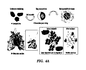
[0026] Figs. 4A-4K depict a schematic illustration of the
Erythrocyte Leveraged Chemotherapy
(EleCt) platform and characterization of drug (doxorubicin)-loaded
biodegradable PLGA
nanoparticles. (Fig. 4A) Schematic illustration of the composition and
mechanism of the
4
CA 03140681 2021-12-6
WO 2020/247576
PCT/US2020/036040
biodegradable drug nanoparticle self-assembling on erythrocyte platform
(EtcCt) for lung metastasis
treatment. (Figs. 4B-4D) Average size (Fig. 4B), zeta potential (Fig. 4C), and
drug loading contents
(Fig. 4D) of plain and drug-loaded nanoparticles. (Fig. 4E) SEM images showing
the morphological
features of the nanoparticles. Scale bar 200 nm. (Fig. 4F) Size distribution
of plain and drug-loaded
nanoparticles. (Fig. 4G) Drug release kinetics from the biodegradable
nanoparticles in a complete
medium (n=4). (Figs. 4H-41) Flow cytometry plots (Fig. 41-1) and confocal
laser scanning microscopic
images (Fig. 41) showing the interaction of drug-loaded nanoparticles with
BI6F10-Luc melanoma
cells. hi (Fig. 41), cell nuclei were stained using DAP!. (Figs. 41-4K) Dose-
response curve (Fig. 43)
and ICso values (Fig. 4K) of Bl6F10-Luc cells after being treated with
different formulations for 24
hours (n).
100271 Figs. 5A-5K demonstrate that doxorubicin-loaded
biodegradable PLGA nanoparticles
efficiently self-assemble to mouse and human erythrocytes. (Fig. 5A) Flow
cytometry analysis of self-
assembly of DOX-loaded PLGA nanoparticles to mouse erythrocytes at different
nanoparticle to
erythrocyte ratios (left to right: 0:1, 5th 1, 200:1, 400:1, and 800:1). (Fig.
5B) Percentage of mouse
erythrocytes carrying at least one nanoparticle. (Fig. 5C) Nanoparticle
binding efficiency and (Fig.
5D) drug dose on mouse erythrocytes at different nanoparticle to mouse
erythrocyte ratios. (Fig. 5E)
Confocal laser scanning microscopic (CLSM) and (Fig. 59 SEM images of mouse
erythrocytes with
drug-loaded nanoparticles self-assembled on them. Scale bars in (Fig. 5F): 2
gm. (Fig. 5G) CLSM and
(Fig. 5H) SEM images of human erythrocytes with drug-loaded nanoparticles self-
assembled on them.
Scale bars in (Fig. 5H): 2 pm. (Fig. 51) Flow cytometry assay of the self-
assembly of drug-loaded
nanoparticles to human erythrocytes at different nanoparticle to erythrocyte
ratios (left to right: 0:1,
200:1, 800:1, and 1600:1). (Fig. Si) Nanoparticle binding efficiency and (Fig.
5K) drug dose on
human erythrocytes at different nanoparticle to erythrocyte ratios.
100281 Figs. 6A-6E demonstrate that the ELeCt platform
enables enhanced and targeted delivery
of nanoparticle drugs to the lungs bearing metastasis. (Fig. 6A)
Pharmacokinetics of intravenously
administered doxorubicin formulations. Extended blood circulation time of
doxorubicin was achieved
by erythrocyte hitchhiking compared to using free drug or nanoparticles alone
(n=3). Significantly
different (ANOVA followed by Tukey's HSD analysis): * p < 0.05, ** p <0.01.
(Fig. 6B) Hitchhiked
drug-loaded nanoparticles could specifically detach from mouse and human
erythrocytes under the
lung-corresponding shear stress. Samples were sheared for 20 mins (n=3). Low
shear indicates rotary
shear (-1 Pa) while high shear was at 6 Pa. Significantly different (Student's
t test): *** p < 0.001.
(Fig. 6C) Drug accumulation in the lungs of mice bearing B16F10-Luc lung
metastasis at 20 min and
6 h after intravenous administration of different doxorubicin formulations
(n=3). (Fig. 6D)
Comparison of the drug concentration in the lungs of erythrocyte hitchhiking
group to that of the free
drug and nanoparticle alone groups (n=3). (Fig. 6E) Drug distribution in the
diseased lungs 20 min
CA 03140681 2021-12-6
WO 2020/247576
PCT/US2020/036040
after intravenous administration of doxorubicin formulations. Dashed lines
indicate the edge of
metastasis nodules.
100291 Figs. 7A-7H demonstrate that the ELeCt platform
inhibits lung metastasis progression
and improves survival in the early-stage B16F10-Luc metastasis model, (Fig.
7A) Schematic chart of
the treatment schedule. (Fig. 7B) Bioluminescence images of lung metastasis at
different time points.
(Fig. 7C) Lung metastasis progression curve as depicted from in vivo
bioluminescence signal
intensity. (Fig. 7D) Quantification of lung metastasis burden at different
time points (n=7). (Fig. 7E)
Scatter-plot comparing the lung metastasis burden in different treatment
groups as depicted from
bioluminescence signal intensity on day 16 (n=7). Significantly different
(Mann-Whitney test): * p <
0.05, ** p <0.01, *** p <0.001. (Fig. 7F) Scatter-plot comparison of the lung
metastasis burden on
day 23 (n=7). Significantly different (Mann-Whitney test): * p < 0.05, ** p <
0.01, *** p < 0.001;
n.s.: not significantly different. (Fig. 7G) Body weight change of mice during
the treatment period
(n=7). (Fig. 7H) Survival of mice under different treatments as displayed by
Kaplan-Meier curves
(n=7). Significantly different (log-rank test): * p <0.05, ** p < 0.01, *** p
< 0.001; n.s.: not
significantly different.
100301 Figs. 8A-8G demonstrate that the ELeCt platform
inhibits lung metastasis progression
and extends survival in the late-stage BI6F10-Luc metastasis model. (Fig. 8A)
Schematic illustration
of the treatment schedule. (Fig. 8B) Bioluminescence images of lung metastasis
progression at
different time points. (Fig. 8C) Lung metastasis growth curve in mice treated
with different
doxombicin formulations. (Fig. 8D) Quantitative analysis of lung metastasis
burden as depicted from
bioluminescence signal intensity (n=7). Significantly different (Mann-Whitney
test): * p <0+05, ** p
<0.01; n.s.: not significantly different. (Fig. 8E) Quantification of
metastasis nodule numbers on
excised lungs from mice in different treatment groups on day 16 (n=7).
Significantly different (Mann-
Whitney test): *** Pc 0.001; n.s.: not significantly different. (Fig. 8F) Body
weight change of mice
during the treatment period (n=7). (Fig. 8(3) Kaplan-Meier survival curves of
mice in different
treatment groups. Significantly different (log-rank test): ** p < 0.01, *** p
<0.001; n.s.: not
significantly different.
100311 Fig. 9 demonstrates that other chemotherapeutic
agent-loaded biodegradable
nanoparticles can efficiently bind to erythrocytes. The tested
chemotherapeutic agents include
camptothecin, paclitaxel, docetaxel, 5-fluorouracil, gemcitabine,
methotrexate, and the combination of
5-fluorouracil + methotrexate. Scale bars: 1 pm.
100321 Figs. 10A-10C depict representative H&E staining
images of lungs of mice. Mice treated
with (Fig. 10A) control (Saline), (Fig. 10B) DOX-loaded nanoparticles, and
(Fig. 10C) drug
nanoparticles self-assembled on erythrocytes (RBC-NPs) were scarified 16 days
after tumor
inoculation in the late-stage lung metastasis model. Lungs were processed by
H&E staining.
6
CA 03140681 2021-12-6
WO 2020/247576
PCT/US2020/036040
[0033] Fig. 11 depicts representative H&E staining images
of organs of mice treated with
different drug formulations. 16 days after tumor inoculation in the late-stage
lung metastasis model,
mice were scarified and organs were processed by H&E staining.
[0034] Fig. 12 depicts size distribution of different
chemotherapeutic agent-loaded biodegradable
PLGA nanoparticles.
[0035] Fig. 13 depicts a schematic illustration of
Erythrocyte Anchored Systemic
Immimotherapy (EASY) for inuuuno-restoration. (a) ImmunoBait (chemokine-loaded
polymeric
nanoparticles) assemble onto the surface of erythrocytes. (b) ImmunoBait
specifically dislodges from
erythrocytes in response to the mechano-physiological shear stress and gets
deposited in the vicinity
of lung metastatic sites. (c) hnmunoBait modulates the local microenvironments
by releasing
chemokine and restoring the chemokine gradient. (d) Effector cells including
Th1 CD4, effector C98
T, and Natural Killer (NK) cells infiltrate to the lung metastatic sites to
control their progression.
[0036] Figs. 14A-14R demonstrate modulation of the
material properties of PLGA nanoparticles
led to optimal targeted delivery to the lung. Fig. 14A depicts binding
efficiency of different PLGA
nanoparticles to erythrocytes (n=5). Significantly different (One-way ANOVA
followed by Tukey's
HSD test): ** p <0.01; &&& p < 0.001 compared to all other groups. (Fig. 14B)
Agglutination of the
carrier erythrocytes after being hitchhiked by different PLGA nanoparticles.
200 mn polystyrene (PS)
nanoparticles were used as a positive control. (Fig. 14C) Net percent
nanoparticles detached from
erythrocytes when experiencing a high rotary shear stress (6 Pa) (n=3).
Nanoparticles were
fluorescently labeled by encapsulating Alexa Fluor 647-ovalbumin. (Fig. 14D)
The amount (ID /0/g of
organ) of different PLGA nanoparticles deposited in the lung 20 mins after
being intravenously
administered (n=3 for free nanoparticles, n=6 for hitchhiked nanoparticles).
Significantly different
(One-way ANOVA followed by Tukey's HSD test): ** p < 0.01, *** p < 0.001, ****
p < 0.0001; ris:
not significantly different. (Fig. 14E) IVIS fluorescent images of excised
mouse lungs 20 min after the
administration of different PLGA nanoparticles hitchhiked on erythrocytes.
(Fig. 14F) Confocal laser
scanning microscopic (CLSM) images of lung microvascular endothelial cells
after being treated with
PLGA-d nanoparticles with or without anti-ICAM-1 antibody for 20 min or 6 h.
Nanopartieles were
labeled with Alexa Fluor 647 while cell nucleus was stained by DAPI. Scale
bar: 30 gm. (Fig. 14G)
IVIS fluorescent images of excised organs at different time points after
intravenous administration of
PLGA-d nanoparticle formulations. (Fig. 14th Kinetics of the amount (ID%/g of
organ) of PLGA-d
nanoparticles deposited in the lung (n=3). Series are, in order, NPs, EH-NPs
without aICAM-1, and
EH-NPs with aICAM-1. Significantly different (One-way ANOVA followed by
Tukey's HSD test): *
pc 0.05, ** PC 0.01, *** p c 0.001. In (Figs. 14D, 14E, 14G, and 14H),
nanoparticles were
fluorescently labeled by encapsulating Alexa Fluor 750-ovalbtunin. Data in
(Fig. 14A, 14C, 14D,
14H) are presented as mean s.e.m.. (Fig. 141) Scanning electron microscopic
(SEM) images of
ImmunoBait. Scale bar, 1 pm. (Fig. 14J) Transmission electron microscopic
(TEM) images of
7
CA 03140681 2021-12-6
WO 2020/247576
PCT/US2020/036040
ImmunoBait. Scale bar, 500 nni. (Fig. 14K) Size distribution of InununoBait.
(Fig. 14L) Chemokine
release kinetics from ImmunoBait in PBS, FBS, and complete medium (n=3). (Fig.
14M) CLSM
images of erythrocytes with ImmunoBait anchored on them. ImmunoBait was
labeled with Alexa
Fluor 647 which was conjugated to chemokine, (Fig. 14N) Flow cytometry
analysis of erythrocytes
carrying Alexa Fluor 647 labeled InirnimoBait. ImmunoBait was labeled with
Alexa Fluor 647 which
was conjugated to chemokine. unboxed dots: plain erythrocytes; Boxed dots:
erythrocytes carrying
ImmunoBait. (Fig. 140) SEM images of ImmunoBait anchored on erythrocytes.
Scale bar, 1 pm.
(Fig. 14P) Expression of phosphatidylserine on carrier erythrocytes after
being hitchhiked by
nanoparticles (n=3). (Fig. 14Q) Agglutination of carrier erythrocytes
hitchhiked by nanoparticles. 200
mu carboxylic polystyrene nanoparticles were used as a positive control in
(Fig. 14P) and (Fig. I4Q).
(Fig. 14R) Osmotic fragility of carrier erythrocytes after being hitchhiked by
nanoparticles. Percent
hemolysis of carrier erythrocytes at 73 mM NaCI was shown (n=3). Data in
(Figs. 14L, 14P, and 14R)
are presented as mean s.e.m.
100371 Figs. 15A-15.1 demonstate that ImmunoBait
assembled onto erythrocytes without causing
obvious side effects to the carrier erythrocytes. (Fig. 15A) Scanning electron
microscopic (SEM)
images of IrnmunoBait. Scale bar, 1 pin. (Fig. 15B) Transmission electron
microscopic (TEM) images
of ImmunoBait. Scale bar, 500 mn, (Fig. 15C) Size distribution of ImmunoBait,
(Fig, 15D)
Chemokine release kinetics from ImmunoBait (n=3). (Fig. 15E) CLSM images of
erythrocytes with
ImmunoBait assembled on them. ItnmunoBait was labeled with Alexa Fluor 647
which was
conjugated to chemokine. (Fig. 15F) Flow cytometry analysis of erythrocytes
carrying Alexa Fluor
647 labeled ImmunoBait. ImmunoBait was labeled with Alexa Fluor 647 which was
conjugated to
chemokine. Dots to the left of the box: plain erythrocytes; dots within the
box: erythrocytes carrying
ImmunoBait. (Fig. 15(i) SEM images of ImmunoBait assembled on erythrocytes.
Scale bar, 1 pm.
(Fig. 15H) Expression of phosphatidylserine on carrier erythrocytes after
being hitchhiked by
nanoparticles (n=3). Both plain nanoparticles and ImmunoBait caused negligible
phosphatidylserine
expression on the carrier erythrocytes. Series are, in order, plain NPs and
ImmunoBait. (Fig. 151)
Agglutination of carrier erythrocytes hitchhiked by nanoparticles. 200 nm
carboxylic polystyrene
nanoparticles were used as a positive control in (Fig. 15H) and (Fig. 15I).
(Fig. 15J) Osmotic fragility
of carrier erythrocytes after being hitchhiked by nanoparticles. Percent
hemolysis of carrier
erythrocytes at 73 inM NaCI was shown (n=3). Data in (Figs. 15D, 15H, and 15J)
are presented as
mean s.e.m..
100381 Figs. 16A-16M demonstrate that EASY precisely
delivered ImmunoBait to the lungs
bearing metastasis and achieved immuno-restoration. (Fig. 16A) SEM images of
ImmunoBait
hitchhiked erythrocytes before shear, after shear, or shear after fixation.
Samples were sheared at a
rotary shear stress of 6 Pa for 20 mins. (Fig. 16B) Percent ImmunoBait
nanoparticles detached from
the carrier erythrocytes when being sheared at 6 Pa for 5, 10, or 20 mins
(n=3). (Fig. 16C)
8
CA 03140681 2021-12-6
WO 2020/247576
PCT/US2020/036040
Biodistribution of ImmunoBait formulations 20 min or 6 h after intravenous
administration in the
early-stage lung metastasis model. At 20 min, n=3 for all groups. At 6 h, n=2
for NPs and NPs w/a
ICAM-1; n=3 for EH-NPs and EH-NPs w/ aICAM-1. Significantly different (One-way
ANOVA
followed by Tukey's HSD test): * p <0.05, ** p< 0.01, *** p <0.001, and ****
Pc 0,0001. (Fig.
16D) Lung to blood ratio of distributed ImmunoBait nanoparticles 20 min after
intravenous
administration in the early-stage lung metastasis model (n=3). Significantly
different (One-way
ANOVA followed by Tukey's HSD test): * p < 0.05. (Fig. 16E) IVIS fluorescent
images of lungs of
mice bearing early-stage lung metastasis 20 min after intravenous
administration of ImmunoBait
formulations. (Fig. 16F) IVIS fluorescent images of lungs of mice bearing late-
stage lung metastasis
20 min after intravenous administration of hnmunoBait formulations. (Fig. 166)
CLSM images of
metastatic lung sections 20 mins after intravenous administration of
ImmunoBait formulations. White
dash lines indicate the edge of the metastatic nodules. Nucleus were stained
by DAPI. In (Figs. 16B-
16G), all nanoparticles were labeled by Alexa Fluor 647 that was conjugated to
the albumin carrier
protein that was encapsulated into the nanoparticles. (Fig. 16H) Schedule for
monitoring the time-
course change of CXCL10 chemokine gradients in mice bearing breast cancer lung
metastasis. (Fig.
16I) Lung to blood ratio of CXCLIO chemokine on different days after primary
tumor resection
(n=5). Significantly different (One-way ANOVA followed by Tukey's HSD test): *
p <0.05. (Fig.
16J) Schedule for the chemokine gradient assay. (Fig. 16K) Concentration of
CXCL10 chemokine in
the blood 20 min or 6 h after intravenous administration of CXCL10
formulations (n=4-5). (Fig. 16L)
Concentration of CXCL10 chemokine in the blood 20 min or 6 h after intravenous
administration of
CXCL10 formulations (n=4-5). (Fig. 16M) Lung to blood ratio of CXCLIO
chemokine concentration
after administration (n=4-5). Significantly different (One-way ANOVA followed
by Tukey's HSD
test): * p <0,05, ** Pc 0.01. Data in (Figs. 16B-16D, 161, 16K-16M) are
presented as mean s_e.m.
Series for Figs. 16K-16M are, in order, control, Free chemokine (5x), NPs, and
RBC-NPs. (Fig. 160)
Percent area containing ImmunoBait NPs calculated from confocal images as
represented in (Fig.
166) (n=5). Calculation was conducted in hnageJ. Significantly different (One-
way ANOVA
followed by Tukey's HSD test): *** p <0.001, **** Pc 0.0001. (Fig. 16P)
Concentration of
CXCL10 chemokine in the blood 20 min, 6 h, 24 h, 48 h, and 72 h after
intravenous administration of
CXCL10 formulations (n=4-6). (Fig. 16Q) Concentration of CXCL10 chemokine in
the lung after
intravenous administration of CXCL10 formulations (n=4-6). (Fig, 16R) Lung to
blood ratio of
CXCL10 chemokine concentration after administration (n=4-6). In (Fig. 16P-
16R), the horizontal bars
indicate the mean s.e.m. of the corresponding levels in control mice before
treatments. Significantly
different compared to the control before treatment (One-way ANOVA followed by
Tukey's HSD test
or student's t test): * p c 0.05, ** p 0.01, *** p C 0.001. Significantly
different compared to the free
chemokine (5X) group (One-way ANOVA followed by Tukey's HSD test or student's
t test): ifit p
0.01. Significantly different compared to the ImmunoBait group (One-way ANOVA
followed by
9
CA 03140681 2021-12-6
WO 2020/247576
PCT/US2020/036040
Tukey's HSD test or student's t test): & p <0.05, && p <0.01, &&&& p < 0.0001.
Data in (Figs.
160-16R) are presented as mean s.e.m.
[0039] Figs. 17A-17H demonstrate that EASY led to
significant inhibition of progression of lung
metastasis and improvement of survival in abreast cancer lung metastasis
model. (Fig. 17A) Schedule
for the efficacy study. (Fig. 17B) Representative bioluminescence images of
mice receiving different
treatments at different time. (Fig. 17C) Nodules number on excised lungs of
mice on day 37 (n=8).
Significantly different (Mann-Whitney test): ** Pc 0.01, *** Pc 0.001. (Fig.
17D) Inhibition rate of
different treatments. (Fig. 17E) Representative images of excised lungs on day
37. (Fig. 17F) Lung
weight of mice on day 37 after being treated by different treatments (n=8).
Significantly different
(One-way ANOVA followed by Tukey's HSD test): * p < 0,05. (Fig. 176) Body
weight change of
mice during the treatment period (n=8). (Fig. 17H) Survival of mice under
different treatments as
displayed by Kaplan-Meier curves (n=6). Significantly different (log-rank
test): * p < 0.05. Data in
(Figs. 17C, 17F, 17G) are presented as mean s.e.m.. (Fig. 171) Nodules
number on excised lungs of
mice on day 37 (n=6-7) receiving different treatments. Significantly different
(Mann-Whitney test):
** p <001, *** p <0.001. Data are presented as mean s.e.m. (Fig. 17J)
Survival of mice under
different treatments as displayed by Kaplan-Meier curves (n=15-16).
Significantly different (Mantel-
Cox test): *** p <0.001.
[0040] Figs. 18A-180 demonshate that EASY resulted in
enhanced infiltration of effector
immune cells into the metastatic lungs. (Fig. 18A) Schedule for profiling the
immune cells of
metastatic lungs of mice under different treatments. (Fig. 18B) Representative
flow cytometry
analysis images of CD4+IFN-y+ cells. (Fig, 18C) The absolute percentage of IFN-
y+ Thl CD4 cells
in the lung (n=7-8). (Fig. 18D) Representative flow cytometry analysis images
of CD8+IFN-y+ cells.
(Fig. 18E) The absolute percentage of IFN-y+ CD8 cells in the lung (n=7-8).
(Fig. 18F)
Representative flow cytometry analysis images of Granzyme B+ CD8 cells. (Fig.
186) The absolute
percentage of Granzyme B+ effector CD8 cells in the lung (n=7-8). (Fig. 181-1)
Representative flow
cytometry analysis images of CD45+NKp46+ cells. (Fig. 181) The absolute
percentage of NKp46+
NK cells in the lung (n=7-8). (Fig. 18J) Representative flow cytometry
analysis images of
CD11c+CD86+ cells. (Fig. 18K) The absolute percentage of activated (CD86+)
dendritic cells in the
lung (n=7-8). (Figs. 18L-180) Concentrations of (Fig. 18L) IFN-y, (Fig. 18M)
TNF-a, (Fig. 18N) IL-
10, and (Fig. 180) CXCLIO in the metastatic lungs of mice following different
treatments. Data in
(Figs. 18C, 18E, 181, 18K, and 18L-0) are presented as mean s.e.m.
Significantly different in (Figs.
18C, 18E, 186, 181, 18K, and 18L-0) (One-way ANOVA followed by Tukey's HSD
test): * p <0.05,
** p < 0.01, **** < 0.0001.
[0041] Fig. 19 depicts scanning electron microscopic
(SEM) images of mouse erythrocytes
carrying different PLGA nanoparticles.
CA 03140681 2021-12-6
WO 2020/247576
PCT/US2020/036040
[0042] Figs. 20A-20C demonstrate the effect of different
PLGA nanoparticles on the carrier
erythrocytes. (Fig. 20A) Percent carrier erythrocytes lysed during the
hitchhiking process (n=3). (Fig.
20B) Osmotic fragility (hemolysis during osmotic shock) of the carrier
erythrocytes with different
PLGA nanoparticles hitchhiked on them (n=3). The enlarged panel shows the
percent hemolysis of
carrier erythrocytes at a NaCI concentration of 72.5 mM. (Fig. 20C) Percent
carrier erythrocytes
expressing phosphatidylserine on their surface after being hitchhiked by
different PLGA nanoparticles
(n=3). Data are presented as mean s.e.m.. Series for Figs. 20A and 20C are,
in order, PLGA-a,
PLGA-b, PLGA-c, and PLGA-d.
[0043] Fig. 21 depicts the percent nanoparticles detached
from the carrier erythrocytes under in
vitro shear conditions. Erythrocytes with different PLGA nanoparticles
hitchhiked on them were
sheared for 20 mins using a rheometer at a high rotary stress (6 Pa) (n=2-3).
Low shear indicates a
rotary stress the carrier erythrocyte experienced during rotation using a
revolver at 12 rpm/min. Data
in are presented as mean s.e.m.. Series are, in order, low shear and high
shear.
[0044] Fig. 22A-228 depict the biodistribution of PLGA
nanoparticles. (Fig. 22A)
Biodistribution of different PLGA nanoparticles either in a free form or
hitchhiked to erythrocytes 20
min after intravenous administration (n=3 for NPs, n=6 for EH-NPs). Series
are, in order NPs, and
EH-NPs. (Fig. 22B) Biodistribution of hitchhiked PLGA-d nanoparticles with or
without anti-ICAM-
1 antibody at different time points after intravenous administration (n=3).
Series are, in order, NPs,
EH-NPs without aICAM-1, and Ell-NPs with aICAM-1. Significantly different (One-
way ANOVA
followed by Tukey's HSD test): * p < 0.05, ** p< 0.01, *** p <0.001. Data are
presented as mean
s.e.m..
[0045] Figs. 23A-23C depict the assembly of ImmunoBait
onto the surface of mouse
erythrocytes. (Fig. 23A) Flow cytometry histogram plots of erythrocytes after
being incubated with
different amount of ImmunoBait nanoparticles. ImmunoBait to erythrocyte ratios
used in histograms
from left to right are 0:1, 50:1, 400:1, and 1000:1, respectively. (Fig. 23B)
Percentage of erythrocytes
carrying ImmunoBait nanoparticles after being incubated at different
ImmunoBait to erythrocyte
ratios (n=3). Data are presented as mean s.e.m. (Fig. 23C) CLSM images of
erythrocytes carrying
ImmunoBait nanoparticles when being incubated at a ImmunoBait to erythrocyte
ratio of 400:1.
ImmunoBait was labeled by Alexa Fluor 647 which was conjugated to the
encapsulated chemokine.
[0046] Fig. 24 demonstrates assembly of ImmunoBait
carrying other immunomodulatory agents
onto erythrocytes. GM-CSF, IL-2, IL-12, and IL-15 were encapsulated in
ImmunoBait. Anti-PD-1
antibody was conjugated to the surface of ImmunoBait. Scale bar: 1 pm.
[0047] Figs. 25A-25B demonstrate assembly of ImmunoBait
to human erythrocytes. (Fig. 25A)
SEM images of human erythrocytes with ImmunoBait nanoparticles assembled on
them. Scale bar: 1
pm. (Fig. 258) Number of ImmunoBait nanoparticles assembled onto the surface
of human
11
CA 03140681 2021-12-6
WO 2020/247576
PCT/US2020/036040
erythrocytes at different ImmunoBait to human erythrocyte incubation ratios
(n=3). Data are
presented as mean s.e.m.
[0048] Fig. 26 depicts the impact of ImmunoBait
hitchhiking on the sensitivity of the carrier
erythrocytes to osmotic stress. Representative osmotic fragility curves of
carrier erythrocytes were
shown (n=3). The carrier erythrocytes at all the tested nanoparticle
concentrations exhibited similar
fragility curves compared to Naive erythrocytes. Data are presented as mean
s.e.m.
[0049] Figs. 27A-27B depict concentrations of the CXCL10
chemokine in the blood and lung of
mice bearing breast cancer lung metastasis at different time after primary
tumor resection.
[0050] Fig. 28 depicts representative images of the back
side of lungs of mice on day 37 under
different treatments in the early-stage lung metastasis model.
[0051] Fig. 29 depicts H&E of mouse organs following
different treatments in the early-stage
lung metastasis model.
[0052] Figs. 30A-30C depict the efficacy of EASY in
inhibition of lung metastasis in a late-stage
breast cancer lung metastasis model. (Fig. 30A) Schedule for the efficacy
study in the late-stage lung
metastasis model. (Fig. 30B) Nodule number on excised lungs of mice on day 50
following different
treatments (n=3). Significantly different (student's t test): * p < 0.05. Data
are presented as mean
s.e.m. (Fig, 30C) Images of excised lungs on day 50,
[0053] Fig. 31A-31D Immune cells in the metastatic lungs
of mice under different treatments.
The absolute percentage of (Fig. 31A) CD4, (Fig. 318) CD8, and (Fig. 31C)
dendritic cells in the
lungs were shown. (Fig. 31D) The percentage of CD86+ cell in CD45+CD1 lc+
dendritic cells. Data
are presented as mean + s.e.m. Significantly different (One-way ANOVA followed
by Tukey's HSD
test): * p < 0.05, ** p < 0.01, *** p <0.001.
[0054] Fig. 32 depicts a heat-map showing the
concentrations of cytokines in the metastatic
lungs of mice being treated by different treatments. Unit: pg/mL.
[0055] Figs. 33A-33P demonstrate that EASI resulted in in
situ inununization and systemic
suppression of distant tumors. (Fig. 33A) Schedule for evaluating the local
and systemic immune
response. (Fig. 338) Viability of 4T1-Luc cells co-cultured with isolated lung
lymphocytes at 50:1 or
100:1 effector cell to tumor cell ratios (n=6). Data were shown as normalized
to the "Control-saline"
group. Significantly different (student's t test): * p < 0.05, *** p < 0.001.
(Fig. 33C) Representative
flow cytometry analysis images of CD11c+CD86+ cells in the metastatic lung.
(Fig. 33D) The fold-
change of absolute percentage of activated (CD86+) dendritic cells in the lung
as compared to that of
the control group (n=14-15). (Fig. 33E) Representative flow cytometry analysis
images of CD8 T
cells in the blood. (Fig. 33F) The absolute percentage of CD8 T cells in the
blood. (Fig. 33(1)
Representative flow cytometry analysis images of IFN-y+ CD8 cells in the
blood. (Fig. 33H) The
absolute percentage of !FN-y CD8 cells in the blood. (Fig. 331)
Representative flow cytometry
analysis images of Granzyme B+ CBS cells in the blood. (Fig. 33J) The absolute
percentage of
12
CA 03140681 2021-12-6
WO 2020/247576
PCT/US2020/036040
Granzyme B+ CD8 cells in the blood. In (Figs. 33B, 33D, 33F, 33H, and 33J),
significantly different
(student's t test): * p <0.05, ** p < 0.01, **** p <0.0001. (Fig. 33K)
Schedule for the tumor re-
challenge study. (Fig. 33L) Individual growth curve of the re-inoculated
tumors in the tumor re-
challenge study. (Fig. 33M) Overall growth curve of re-inoculated tumors.
(Fig. 33N) Overall growth
curve of re-inoculated tumors in the first 10 days after inoculation. (Fig.
330) Weight of extracted
tumors 16 days after tumor reehallenge. (Fig. 33P) Photographs of extracted
tumors 16 days after
tumor rechallenge. "EXP" indicates "Expired". In (Figs. 33K-33P), n=8 for the
"Healthy" group; n=5
for the "Control-saline" and "EASI" groups. In (Figs. 33M-330), significantly
different (student's t
test): * p < 0.05, ** p < 0.01, **** p <0+0001+ Data in (Figs. 33B, 33D, 33F,
33H, 33J, and 33M-
330) are presented as mean &cm,
[0056] Figs. 34A-34E depict the mechanisms of
nanoparticles anchoring to erythrocytes.
Relative binding efficiency and representative SEM images of nanoparticles
anchoring to erythrocytes
under different conditions. (Fig. 34A) When erythrocytes were fixed by 25%
glutaraldehyde. (Fig.
34B) When the surface of erythrocytes was PEGylated. (Fig. 34C) When the
hitchhiking was
conducted in serum. (Fig. 349) When PLGA NPs were PEGylated. (Fig. 34E) When
PLGA NPs had
different surface end (different hydrogen bonding capability).
[0057] Fig. 35 depicts the percent nanoparticles detached
from the carrier erythrocytes under in
vitro shear conditions. Erythrocytes with different PLGA nanoparticles
hitchhiked on them were
sheared for 20 mins using a rheometer at a high rotary stress (6 Pa) (n=6-7).
Low shear indicates a
rotary stress the carrier erythrocyte experienced during rotation using a
revolver at 12 rpm/min. Data
in are presented as mean + s.e.m. Significantly different (two-way ANOVA): * P
<0.05, ** p <0.01,
*** p < 0.0001. Significantly different compared to all other groups (two-way
ANOVA): && P <
0.01.
100581 Figs 36A-36F depict characterization of the
erythrocyte hitchhiking system in the blood
after i.v. administration. (Fig. 36A) Schematic illustration of the dual-
labeling strategy for the tracking
of carrier erythrocytes and nanoparticles. The carrier erythrocytes were
labeled with CellTraceTM Far
Red and PLGA-d nanoparticles were labeled with FITC. Blood was collected at
different time points
after i.v. administration of the dual-labeled system. The carrier erythrocytes
and the nanoparticles on
them were analyzed by flow cytometry. (Fig. 36B) Representative flow plots of
the blood at different
time points (n=3). (Fig. 36C) Representative flow plot of the administered
erythrocytes at different
time points (n=3). (Fig. 36D) Percentage of the carrier erythrocytes remaining
in the blood at different
time points as normalized to 0 min (n=3). (Fig. 36E) Percentage of the carrier
erythrocytes carrying
hitchhiked nanoparticles at different time points as normalized to 0 min
(n=3). (Fig. 36F) 1VIS images
of organs of mice 5 min after i.v. administration of the erythrocyte
hitchhiking system. In (Fig. 36F),
nanoparticles were labeled by encapsulating AF647 while the carrier
erythrocytes were not labeled.
13
CA 03140681 2021-12-6
WO 2020/247576
PCT/US2020/036040
[0059]
Fig. 37 depicts the effect of the
binding of aICAM-1 antibody to EH-NP complex on
the detachment of nanoparticles from carrier erythrocytes under shear stress.
Erythrocytes with
PLGA-d nanoparticles hitchhiked on them w/ or w/o aICAM-1 binding were sheared
for 20 mins
using a rheometer at a high rotary stress (6 Pa) (n=4-7). Low shear indicates
a rotary stress the carrier
erythrocyte experienced during rotation using a revolver at 12 rpm/min. Data
in are presented as mean
s.e.m.. n.s.: not significantly different (two-way ANOVA).
[0060] Figs. 38A-3811 depict the stability of CXCL10
following encapsulation into ImmunoBait.
(Fig. 38A) MALDI-TOF spectrum of native CXCLIO and CXCL10 released from
ImmunoBait. (Fig.
3813) Circular Dichroism Spectrum of native CXCL10 and CXCLIO released from
ImmunoBait.
[0061] Figs. 39A-39 B demonstrate that ImmunoBait was
released from earner erythrocytes
under shear. (Fig. 39A) SEM images of ImmunoBait hitchhiked erythrocytes
before shear, after shear,
or shear after fixation. Samples were sheared at a rotary shear stress of 6 Pa
for 20 mins. (Fig. 39B)
Percent ImmunoBait nanoparticles detached from the carrier erythrocytes when
being sheared at low
shear stress (rotary shear at 12 rpm) or high shear stress (6 Pa) for 3, 10,
or 20 mins (n=3).
Significantly different compared to low shear (student's t test): *** p <
0.001, **** p <0.0001. Data
in (Fig. 3913) are presented as mean s.e.m.
[0062] Figs. 40A-40B depict lung accumulation of
ImmunoBait delivered by EH with the
progression of lung metastasis. On days 7, 14, and 21 after primary tumor
resection, different sets of
mice were IV administered with EASI with aICAM-1 (n=3-4). 20 mins after
injections, mice were
euthanized and organs were collected for fluorescence measurement using IVIS.
ImmunoBait NPs
were labeled by encapsulating AF647-Ovalburnin. Fig. 40A) Representative IVIS
images of lungs.
Fig. 4013) ID% of ImmunoBait NPs accumulated in the lungs. Data indicated that
with the progression
of lung metastasis, the amount of NPs delivered to lungs by EH remained
similar. Not significantly
different among all groups (One-way ANOVA followed by Tukey's HSD test): n.s.
Data are
presented as mean s.e..m.
[0063] Figs. 41A-41B demonstrate that ImmunoBait
Nanopartieles delivered to metastatic lungs
by erythrocyte hitchhiking were not entrapped in phagocytes. Phagocytes
(mainly tissue
macrophages) were depleted by IV administration of 200 piL of Clodrosome
containing 5 mg/mL
Clodronate 48 h before IV injection of EASI. InurtunoBait NPs were labeled by
encapsulating AF647-
Ovabumin. 20 min after EAST administration, mice were euthanized and major
organs including lung,
liver, spleen, and kidney were collected and imaged using IVIS. Fluorescence
intensity (radiant
efficiency) of ROT was quantified and analyzed. Fig. 41A) Representative IVIS
images of organs of
mice injected with saline, EAST, or EASI after phagocyte depletion. Fig. 41B)
Total radiant efficiency
per gram of organ in major organs of mice with or without phagocyte depletion.
The depletion of
phagocytes resulted in reduced NP accumulation in the liver and kidney,
indicating NPs accumulated
in these two organs were majorly entrapped in phagocytes. In contrast, the
accumulation of NPs in the
14
CA 03140681 2021-12-6
WO 2020/247576
PCT/US2020/036040
lung was not decreased when phagocytes were depleted, indicating the detached
ImmunoBait NPs in
the lung were not majorly entrapped in phagocytes. Data are presented as mean
s.e.m. Significantly
different (Student's t test): * p <0.01, ** p <0.01, *** Pc 0.001.
[0064] Figs. 42A-42C demonstrate delivery of RANTES (a
chemokine) by erythrocyte
hitchhiking. ImmunoBait-RANTES was prepared by encapsulating RANTES in PLGA
nanoparticles.
Anti-ICAM-1 antibody was coated to IrrununoBait-RANTES. 6 h after IV
administration of
formulations, RANTES concentrations in the serum and lung homogenates were
quantified by
ELISA. (Fig. 42A) Concentration of RANTES chemokine in the blood (n=5). (Fig.
428)
Concentration of RANTES chemokine in the lung (n=5). (Fig. 42C) Lung to blood
ratio of RANTES
chemokine concentration after administration (n=5). Data are presented as mean
s.e.m. Significantly
different (Student's t test): ** p < 0.01. Not significantly different
(Student's t test): n.s.
[0065] Figs. 43A-43B depict immune cells in the
metastatic lungs of mice under different
treatments. The absolute percentage of (Fig. 43A) CD4 and (Fig. 43B) CD8
dendritic cells in the
lungs were shown. Data are presented as mean + s.e.m. Significantly different
(One-way ANOVA
followed by Tukey's HSD test): * p <0.05.
[0066] Figs. 44A-448 depict immune cells in different
organs of mice under different treatments.
The absolute percentage of CD4 cells, Thl CD4 cells, CD8 cells, effector CD8
cells, NK cells,
dendritic cells, and activated dendritic cells in the (Fig. 44A) spleen and
(Fig. 44B) liver were shown.
Data are presented as mean s.e.m. Significantly d
[0067] Figs. 45A-45B depict the efficacy and safety
evaluation of different formulations in
inhibiting lung metastasis progression. (Fig. 45A) Representative images of
lungs of mice on day 37
under different treatments in the early-stage lung metastasis model. (Fig.
45B) Body weight change of
mice during the treatment. Data in (Fig. 45B) are presented as mean s.e.m.
[0068] Figs. 46A-46B depict dendritic cells in the
metastatic lungs of mice under different
treatments. (Fig. 46A) The absolute percentage of dendritic cells in the
lungs. (Fig. 46B) The
percentage of CD86-F cell in C045+CD1 1 c+ dendritic cells. Data are presented
as mean s.e.m.
Significantly different (student's t test): ** p <0.01, **** p <0.0001.
[0069] Figs. 47A-47F depict a schematic for engineering a
hand-off of nanoparticles at spleen
via erythrocyte hitchhiking. (Fig. 47A) Protein- capped polymeric
nanoparticles used for the study
(different size, material or coated with different proteins). (Fig. 47B)
Number of nanoparticles loaded
on erythrocytes were tuned for protein loading and to induce temporary
upregulation of phosphatidyl
choline. (Fig. 47C) Intravenous injection of hitchhiked nanoparticles leads to
high discharge in the
spleen. (Fig. 47D) Upregulated phosphatidyl choline and masking CD47 improves
interactions with
antigen presenting cells (APCs) in the spleen. (Fig. 47E) Improved erythrocyte
interactions facilitate
nanoparticle uptake by the APCs while the erythrocytes return back to the
circulation. (Fig. 47F)
Hand-off of nanoparticles at the spleen improves both humoral and cellular
immune responses.
CA 03140681 2021-12-6
WO 2020/247576
PCT/US2020/036040
100701 Figs. 48A-48G depict the characterization of
protein capped nanoparticles: (Fig. 48A)
Scheme of protein attachment to polystyrene carboxylate (PS-COOH)
nanoparticles using EDC
chemistry. (Fig. 4813) Antigen attached to PS-COOH (n=12). (Fig. 48C) Particle
size in rim of plain
and protein capped nanoparticles (n=6). Significantly different (Student's t
test): ****; p < 0,0001.
(Fig. 48D) Zeta potential in mV of plain and protein capped nanoparticles
(n=6). Significantly
different (Student's t test): ****: p < 0.0001. (Fig. 48E) Particle size
distribution of plain and protein
capped nanoparticles. (Fig 48F) Scanning electron micrographs (SEM) of plain
and protein capped
nanoparticles. Scale bar: 200 nm, (Fig. 48G) Dendritic cell maturation
evaluated in terms of % CD80
expression (normalized to basal expression) (n=3 for all groups).
Significantly different (One-way
ANOVA followed by Tukey's HSD test): *: p <0.05, ns: not significant. Data in
(Figs, 48B-48D,
48G) are expressed as mean s.e.m..
100711 Figs. 49A-49K depict engineering nanoparticle-
erythrocyte hitchhiking parameters to
achieve spleen targeting. (Fig. 49A) Nanoparticle loaded per erythrocyte for
different feed ratios of
nanoparticles to erythrocytes (n=3 for all groups). (Fig. 4913) Percentage of
erythrocytes carrying
nanoparticles (determined by flow cytometiy) for different feed ratios of
nanoparticles to erythrocytes
(n=3 for all groups). (Fig. 49C) Percentage of nanoparticles released from
erythrocytes following in
vitro shear studies at the lung corresponding shear stress (6 Pa).
Significantly different (One-way
ANOVA followed by Tukey's HSD test). *: p <0.05. (Fig. 49D) Erythrocyte
damaged caused by
nanoparticles, evaluated by changes in percentage of phosphatidyl serine
expression, for different feed
ratios of nanoparticles to erythrocytes (n=3 for all groups) Dotted line
indicates positive control
(polystyrene beads) mean value. Significantly different (One-way ANOVA
followed by Tukey's HSD
test). *: p < 0.05, **:p <0.01. (Fig. 49E) Optical agglutination assay
demonstrating minimal
aggregates induced by nanoparticles to erythrocytes. All the tested
nanoparticle to erythrocyte ratios
were similar to Naive control as opposed to polystyrene beads which induced
matrix shaped
aggregates. (Fig. 49F) IVIS images of lungs and spleen harvested from mice, 20
minutes after being
injected with erythrocytes incubated at different nanoparticle to erythrocyte
ratios. Scale indicates low
(maroon) to high (bright yellow) radiant efficiency. (Fig. 49G) Lung to spleen
accumulation ratios
computed by using radiant efficiencies of these organs from IVIS imaging (n=3
for all groups).
Dotted line indicates equal lung and spleen accumulation. Significantly
different (One-way ANOVA
followed by Tukey's HSD test). *: p <0.05. (Fig. 49H) Fraction of particles
and erythrocytes
remaining in circulation, evaluating by their parallel tracking using flow
cytometry (n=5). (Fig. 491)
Biodistribution of free nanoparticles and hitchhiked nanoparticles in
different organs, expressed in
terms of % Injected Dose per gram of tissue, harvested 20 minutes after
injection (n= 3 for all
groups). Significantly different. (Student's t test). *: p <0.05. First series
is NP, second series is RBC-
NPs. (Fig. 49J) Kinetics of spleen accumulation of free and hitchhiked
nanoparticles monitored over
24 hours after injection. (n= 3 for all groups). Significantly different.
(Student's t test). *: p <0.05.
16
CA 03140681 2021-12-6
WO 2020/247576
PCT/US2020/036040
(Fig. 49K) Effect of phagocyte depletion on hitchhiked nanoparticles
biodistribution in two most
important organs of the mononuclear phagocytic system, 20 minutes after
injection. (n=3 for all
groups). First series is +phagocytes, second series is -phagocytes.
Significantly different. (Student's t
test). *: p < 0.05, ns: not significant. Data in (Figs, 49A-49D, 496-491) are
expressed as mean
s.e.m.
100721 Figs. 50A-50I depict the immunological
consequences of nanoparticle spleen hand-off.
(Fig. 50A) Schedule for evaluating systemic antibody (humoral) responses of
hitchhiked
nanoparticles. (Fig. 508) Anti-OVA IgG titer evaluated one day before first
immune challenge (Day -
1) and 14 days after the second immune challenge (Day 27) (n= 5 for all
groups). Significantly
different. (One-way ANOVA followed by Tukey's HSD test). *: p < 0.05, (Fig.
50C) Schedule for
evaluating systemic cellular immune responses of hitchhiked nanoparticles.
(Fig. 50D) Representative
flow cytometry analysis images of CD3-F CD8-F cells in spleen. (Fig. 50E)
Quantitative analysis of
percentage of CD3+ CD8+ cells in spleen. (n=4 for EDIT group, n=5 for all
other groups).
Significantly different. (One-way ANOVA followed by Tukey's HSD test). *: p <
0.05. (Fig. 50F)
Representative flow cytometry analysis images of CCR7+ CD62L+ cells in spleen.
(Fig. 50G)
Quantitative analysis of percentage of CCR7+ CD62L+ cells in spleen. (n=4 for
EDIT group, n=5 for
all other groups). Significantly different. (One-way ANOVA followed by Tukey's
HSD test). **: p <
0.01, ***: p <0.001. (Fig. 50H) Representative flow cytometry analysis images
of CD25+ FOXP3+
cells in spleen. (Fig. 501) Quantitative analysis of percentage of CD25+
FOXP3+ cells in spleen. (n=4
for EDIT group, n=5 for all other groups). Significantly different. (One-way
ANOVA followed by
Tukey's HSD test). *: p <0+05, **: Pc 0.01, ****: p <0.0001. Data in (Figs.
508, 50E, 50G, 501) are
expressed as mean s.e.m.
100731 Figs. 51A-51I depict the therapeutic extension of
immune modulation of hitchhiked
nanoparticles for vaccination. (Fig, 51A) Schedule for prophylactic
vaccination studies. (Fig. 518) In
vitro cell killing data post immunizations by various treatment groups
evaluated as percent viability
normalized to the untreated control at different effector to target ratios (n=
3 mice for all groups).
Significantly different: Saline-OVA vs EDIT and NPs vs EDIT. (One-way ANOVA
followed by
Tukey's HSD test). *: PC 0.05, Sz.: PC 0.05. (Fig. 51C) Fold change in in
vitro cell killing assay,
comparison of fold change within each treatment group as a function of
effector to target ratio. (n= 3
mice for all groups). Significantly different. (One-way ANOVA followed by
Tukey's HSD). *: p <
0.05. On the x-axis, the first group is Saline-OVA, the second group is NP,
the third group is EDIT,
and the fourth group is CpG. (Fig. 51D) Tumor growth curves for mice
inoculated after prophylactic
vaccinations by different treatment groups. Statistical analysis within this
figure was carried on day
17. (Fig. 51E) Evaluation of tumor volumes for different groups on day 13. For
Figs. 51D-51E (n=8
for EDIT and CpG groups, n=7 for Saline and NP groups. Significant different
(One-way ANOVA
followed by Tukey's HSD). *: p< 0.05, **: p< 0.01, ***: PC 0.001. Figs. 51F-
51I Tumor growth
17
CA 03140681 2021-12-6
WO 2020/247576
PCT/US2020/036040
kinetics for individual mice in (Fig. 51F) Saline, (Fig. 51G) NP, (Fig. 51H)
EDIT, (Fig. 511) CpG
treatment groups. Data in (Figs. 51B-51E) are expressed as mean s.e.m.
100741 Figs. 52A-52B depict the attachment of different
amount of proteins on the surface of 200
inn polystyrene carboxylate nanoparticles. (Fig. 52A) Attachment efficiency of
protein to the PS-
COOH. (Fig. 52B) Amount of protein attached to PS-COOH expressed in terms of
pg attached per
mg of particles. Data expressed as mean s.e.m.
100751 Figs. 53A-53B depict the internalization of OVA-NP
by dendritic cells. (Fig. 53A) Flow
cytometry images of dendritic cells post internalization of Alexa Fluor 647
labelled ovalbumin, either
in free form or attached to PS-COOH. (Fig. 53B) Confocal light scanning
micrographs (CLSM) of
dendritic cells post internalization of Alexa Fluor 647 labelled ovalbumin
either in free form or
attached to PS-COOH. Scale bar. 10 pa
100761 Figs. 54A-54D depict particle combinations for
EDIT platform. (Fig. 54A) Quantification
of different protein attachment to different particles using EDC chemistry.
Attachment of ovalbumin
to 500 nm polystyrene carboxylate and 200 nm polylactic co glycolate, and
attachment of KLH to 200
run polystyrene carboxylateµ (n=6 for PS-KLH-200, n= 5 for all other groups).
(Fig. 54B) Scanning
electron micrographs of the different particles. Scale bar: 200 nm. (Fig. 54C)
Confocal light scanning
micrographs (CLSM) of dendritic cells post internalization of Alexa Fluor 647
labelled ovalbumin or
KLH attached to different particles. Scale bar: 10 gm. (Fig. 54D) Dendritic
cell maturation evaluated
in terms of % CD 80 expression (normalized to basal expression) (n=3 for all
groups). Significantly
different (Student's t test): ***: p <0.001. Data in (Figs. 54A-54D) are
expressed as mean s.e.m.
100771 Figs. 55A-55C depict mechanistic understanding of
erythrocytes-nanoparticle binding.
(Fig. 55A) Hitchhiking efficiency change when erythrocytes are fixed
indicating that physical
wrapping of nanoparticles is needed to achieve proper hitchhiking. (Fig. 55B)
Addition of serum
during hitchhiking process affects the process, indicating that even a minimum
amount of serum
affects the protein-protein interactions from happening, indicating they are
important interactions
governing hitchhiking. (Fig. 55C) Confocal laser scanning microscopy images of
hitchhiked
nanoparticles on erythrocytes. Scale bar: 10 pm. Data expressed in (Fig. 55A,
55B) as mean s.e.m.
100781 Figs. 56A-56C depict scanning electron micrographs
of nanoparticles hitchhiked on
erythrocytes. (Fig. 56A) Group of control erythrocytes. Scale bar: 10 run.
(Fig. 56B) Group of
hitchhiked erythrocytes. Scale bar: 101..un. (Fig. 56C) Single erythrocyte
showing hitchhiked
nanoparticles. Scale ban 2 gm
100791 Figs. 57A-57B depict changes in the surface marker
expressions on erythrocyte
membranes. (Fig. 57A) Percentage expression of CD47 on erythrocyte membrane.
Naive cells
expression indicated by dotted lines. (n=3 for all groups). Significantly
different. (One-way ANOVA
followed by Tukey's HSD test). *: p < 0.05. (Fig. 57B) Kinetics of changes in
phosphatidyl serine
expression as normalized to the basal. Groups indicate hitchhiked erythrocytes
before injection,
18
CA 03140681 2021-12-6
WO 2020/247576
PCT/US2020/036040
tracked erythrocytes immediately after injection and tracked erythrocytes 20
mins after injection
(when the nanoparticles are expected to be sheared off) (n=3). Significantly
different. (One-way
ANOVA followed by Tukey's HSD test). *: p <01)5. Data expressed as mean +
s.e.m.
[0080] Figs. 58A-588 depict the biodistribution of
hitchhiked and free nanoparticles 6 h and 24 h
after intravenous injections. (Fig. 58A) Biodistribution at 6h (n=3 for all
groups). Significantly
different. (Student's t test). *: p <0.05, ***: Pc 0.001. (Fig. 588)
Biodistribution at 24h. Data
expressed as mean s.e.m. In both, the first series is NP and the second
series is RBC-NPs.
[0081] Figs. 59A-59D depict the potential of EDIT to
deliver different nanoparticle combinations
to spleen. (Fig. 59A) Scanning electron micrographs of erythrocytes with
hitchhiked nanoparticles
(PS-OVA-500, PLGA-OVA-200, PS-KLH-200). All scale bars: 21AM (Fig. 598)
Erythrocyte damage
caused by nanoparticles, evaluated by changes in percentage of phosphatidyl
serine expression, for
different protein capped particles, at a nanoparticle to erythrocyte feed
ratio of 300:1 (n=3 for all
groups). Dotted lines indicate phosphatidyl serine expression on Naive
erythrocytes and the damaged
caused by positive control (polystyrene beads). (Fig. 59C) Optical
agglutination assay demonstrating
damage induced by nanoparticles to erythrocytes for different protein capped
particles, at a
nanoparticle to erythrocyte feed ratio of 300:1. All the tested particles were
similar to Naive control as
opposed to polystyrene induced matrix shaped aggregates. (Fig. 59D)
Biodistribution of hitchhiked
nanoparticles, 20 minutes injection, evaluated by IVIS imaging. All the
hitchhiked particles showed
high spleen delivery efficiency. (n=3 for all groups). Data in (Figs. 598,
59D) are expressed as mean
s.e.m.
[0082] Figs. 60A-608 depict the immunological
consequences of enhanced delivery in the lung.
(Fig. 60A) Quantitative analysis of CD8 + cells present in the lungs. (n=3 for
EDIT, n=5 for all other
groups). (Fig. 608) Quantitative analysis of antigen experienced CCR7+CD62L+
cells in the lungs
(n=4 for EDIT, n=5 for all other groups). Data in (Figs, 60A, 608) are
expressed as mean s.e.m.
[0083] Fig. 61 depicts body weight change after
vaccination treatments for the prophylactic
studies. Data expressed as mean s.e.m.
DETAILED DESCRIPTION
[0084] The methods and compositions described herein
relate to erythrocytes in combination
with certain polymeric particles, e.g., located on the surface of the
erythrocyte. These polymeric
particles comprise at least therapeutic agent and PLGA. When the polymeric
particles described
herein are adhered to the erythrocyte and administered to a subject, they are
less likely to be
phagocytosed and can accumulate preferentially in a tissue or organ, providing
drug delivery with
increased efficiency.
[0085] In one aspect of any of the embodiments, described
herein is an engineered composition
(e.g., cellular composition) comprising: a. an erythrocyte; and b. a particle
comprising PLGA and at
least one therapeutic agent, wherein the particle is located on the cell
surface of the erythrocyte. The
19
CA 03140681 2021-12-6
WO 2020/247576
PCT/US2020/036040
particles can be referred to interchangeably herein as "adhered particles",
"polymeric particles" or
"backpacks."
100861 An erythrocyte or red blood cell is a
hematopoietic cell lacking a cell nucleus and having
an oval biconcave shape in humans. Erythryocytes have high levels of
hemoglobin and are the means
by which oxygen is delivered throughout the body of vertebrates. In some
embodiments of any of the
aspects, the erythrocyte is a human erythrocyte. In some embodiments of any of
the aspects, the
erythrocyte is autologous to a subject ¨ e.g., it is one of the subject's own
erythrocytes to which the
particle has been adhered, either in vitro or in vivo. In some embodiments of
any of the aspects, the
erythrocyte has not been genetically engineered and/or no exogenous material
has been introduced to
the cytoplasm of the erythrocyte.
100871 The polymeric particles described herein comprise
poly(lactic-co-glycolic) acid (PLGA).
In some embodiments of any of the aspects, the particle comprises one or more
therapeutic agents and
PLGA. In some embodiments of any of the aspects, the particle consists
essentially of one or more
therapeutic agents and PLGA. In some embodiments of any of the aspects, the
particle consists of one
or more therapeutic agents and PLGA.
0
HOPtilECOtH
0
Structure of PLGA
100881 In some embodiments of any of the aspects, the
PLGA can be a random copolymer.
100891 In some embodiments of any of the aspects, the
PLGA can comprise, consist of, or
consist essentially of PLGA with a molecular weight of from about 10,000 to
about 90,000. In some
embodiments of any of the aspects, the PLGA can comprise, consist of, or
consist essentially of
PLGA with a molecular weight of from about 20,000 to about 60,000. In some
embodiments of any
of the aspects, the PLGA can comprise, consist of, or consist essentially of
PLGA with a molecular
weight of from about 35,000 to about 56,000. In some embodiments of any of the
aspects, the PLGA
can comprise, consist of, or consist essentially of PLGA with a molecular
weight of from about
38,000 to about 54,000. In some embodiments of any of the aspects, the PLGA
can comprise, consist
of, or consist essentially of PLGA with a molecular weight of from about
45,000 to about 80,000. In
some embodiments of any of the aspects, the PLGA can comprise, consist of, or
consist essentially of
PLGA with a molecular weight of from about 50,000 to about 75,000. In some
embodiments of any
of the aspects, the PLGA can comprise, consist of, or consist essentially of
PLGA with a molecular
weight of from about 20,000 to about 40,000. In some embodiments of any of the
aspects, the PLGA
CA 03140681 2021-12-6
WO 2020/247576
PCT/US2020/036040
can comprise, consist of, or consist essentially of PLGA with a molecular
weight of from about
24,000 to about 38,000.
[0090] In some embodiments of any of the aspects, the
PLGA can comprise, consist of, or
consist essentially of PLGA with a molecular weight of from 10,000 to 90,000.
In some embodiments
of any of the aspects, the PLGA can comprise, consist of, or consist
essentially of PLGA with a
molecular weight of from 20,000 to 60,000. In some embodiments of any of the
aspects, the PLGA
can comprise, consist of, or consist essentially of PLGA with a molecular
weight of from 35,000 to
56,000. In some embodiments of any of the aspects, the PLGA can comprise,
consist of, or consist
essentially of PLGA with a molecular weight of from 38,000 to 54,000. In some
embodiments of any
of the aspects, the PLGA can comprise, consist of, or consist essentially of
PLGA with a molecular
weight of from 45,000 to 80,000. In some embodiments of any of the aspects,
the PLGA can
comprise, consist of, or consist essentially of PLGA with a molecular weight
of from 50,00010
75,000. In some embodiments of any of the aspects, the PLGA can comprise,
consist of, or consist
essentially of PLGA with a molecular weight of from 20,000 to 40,000. In some
embodiments of any
of the aspects, the PLGA can comprise, consist of, or consist essentially of
PLGA with a molecular
weight of from 24,000 to 38,000.
[0091] In some embodiments of any of the aspects, the
PLGA comprises a L:G ratio of at least
about 50:50 or more L, e.g., a L:G of at least about 50:50, about 55:45, about
60:40, about 65:35,
about 70:30, about 75:25, about 80:20, about 85:15, about 90:10, or about 95:5
or more. In some
embodiments of any of the aspects, the PLGA comprises a L:G ratio of at least
50:50 or more L, e.g.,
a L:G of at least 50:50, 55:45, 60:40, 65:35, 70:30, 75:25, 80:20, 85:15,
90:10, or 95:5 or more. In
some embodiments of any of the aspects, the PLGA comprises a L:G ratio of at
least from about
50:50 to about 95:5, es., a L:G ratio of from about 50:50 to about 85:15, of
from about 50:50 to about
65:35. In some embodiments of any of the aspects, the PLGA comprises a L:G
ratio of at least from
50:50 to 95:5, e.g., a L:G ratio of from 50:50 to 85:15, of from 50:50 to
65:35. In some embodiments
of any of the aspects, the PLGA comprises a L:G ratio of about 50:50, about
65:35, or about 85:15. In
some embodiments of any of the aspects, the PLGA comprises a L:G ratio of from
45:55 to 55:45,
from 60:40 to 70:30 or from 80:20 to 90:10. In some embodiments of any of the
aspects, the PLGA
comprises a L:G ratio of 50:50, 65:35, or 85:15.
[0092] PLGA can terminate in an ester end or an acid end.
In some embodiments of any of the
aspects, the PLGA comprises ester ends and/or acid ends. In some embodiments
of any of the
aspects, the PLGA ends comprise at least 90% ester ends, e.g, at least 90%, at
least 95%, at least 98%,
at least 99% or more ester ends. In some embodiments of any of the aspects,
the PLGA ends
comprise at least 90% acid ends, .g, at least 90%, at least 95%, at least 98%,
at least 99% or more acid
ends. In some embodiments of any of the aspects, the PLGA ends consist of
ester ends. In some
embodiments of any of the aspects, the PLGA ends consist of acid ends.
21
CA 03140681 2021-12-6
WO 2020/247576
PCT/US2020/036040
[0093] Certain specific PLGA compositions, comprising
certain L:G ratios and end identities are
contemplated herein, and are demonstrated to target their polymeric particles
to certain organs.
Exemplary such compositions are provided in Table 3.
[0094] Table 3
Particle Ratio of L:G in PLGA end composition
Targeted organs
Composition PLGA
1 About 50:50 Comprises at least 90%
ester ends Spleen, heart
2 45:55 to 55:45 Comprises at least 90%
ester ends Spleen, heart
3 50:50 Comprises at least 90%
ester ends Spleen, heart
4 About 50:50 Consists of ester ends
Spleen, heart
45:55 to 55:45 Consists of ester ends Spleen, heart
6 50:50 Consists of ester ends
Spleen, heart
7 About 50:50 Comprises at least 90%
acid ends Spleen, lung
8 45:55 to 55:45 Comprises at least 90%
acid ends Spleen, lung
9 50:50 Comprises at least 90%
acid ends Spleen, lung
About 50:50 Consists of acid ends Spleen, lung
11 45:55 to 55:45 Consists of acid ends
Spleen, lung
12 50:50 Consists of acid ends
Spleen, lung
13 About 85:15 Comprises at least 90%
ester ends Kidney, lung
14 80:20 to 90:10 Comprises at least 90%
ester ends Kidney, lung
85:15 Comprises at least 90% ester ends Kidney, lung
16 About 85:15 Consists of ester ends
Kidney, lung
17 80:20 to 90:10 Consists of ester ends
Kidney, lung
18 85:15 Consists of ester ends
Kidney, lung
19 About 65:35 Comprises at least 90%
acid ends Lung, heart, kidney
60:40 to 70:30 Comprises at least 90% acid ends Lung, heart,
kidney
21 65:35 Comprises at least 90%
acid ends Lung, heart, kidney
22 About 65:35 Consists of acid ends
Lung, heart, kidney
23 60:40 to 70:30 Consists of acid ends
Lung, heart, kidney
24 65:35 Consists of acid ends
Lung, heart, kidney
Less than about Comprises at least 90% ester ends Spleen
85:15
26 Less than 85:15 Comprises at least 90%
ester ends Spleen
27 Less than about Consists of ester ends
Spleen
85:15
28 Less than 85:15 Consists of ester ends
Spleen
29 About 50:50 to Comprises at least 90%
ester ends Spleen
about 85:15
50:50 to 85:15 Comprises at least 90% ester ends Spleen
31 About 50:50 to Consists of ester ends
Spleen
about 85:15
32 50:50 to 85:15 Consists of ester ends
Spleen
[0095] Alternatively, or additionally, the ratio of
particles to erythrocyte during incubation to
form the cellular composition can influence the delivery target for the
therapeutic agent. For example,
in some embodiments of any of the aspects, a cellular composition formed by
incubation of particles
and erythrocytes at a ratio of between about 150 and about 600 particles per
erythrocyte will target the
22
CA 03140681 2021-12-6
WO 2020/247576
PCT/US2020/036040
therapeutic agent to the spleen. In some embodiments of any of the aspects, a
cellular composition
formed by incubation of particles and erythrocytes at a ratio of between 150
and 600 particles per
erythrocyte will target the therapeutic agent to the spleen. hi some
embodiments of any of the
aspects, a cellular composition formed by incubation of particles and
erythrocytes at a ratio of
between about 200 and about 400 particles per erythrocyte will target the
therapeutic agent to the
spleen. In some embodiments of any of the aspects, a cellular composition
formed by incubation of
particles and erythrocytes at a ratio of between 200 and 400 particles per
erythrocyte will target the
therapeutic agent to the spleen. In some embodiments of any of the aspects, a
cellular composition
formed by incubation of particles and erythrocytes at a ratio of about 300
particles per erythrocyte
will target the therapeutic agent to the spleen. In some embodiments of any of
the aspects, a cellular
composition formed by incubation of particles and erythrocytes at a ratio of
300 particles per
erythrocyte will target the therapeutic agent to the spleen. In some
embodiments of any of the
aspects, the particle is a nanoparticle_
100961 In some embodiments of any of the aspects, a
cellular composition comprising more than
18 particles per erythrocyte will target the therapeutic agent to the spleen.
In some embodiments of
any of the aspects, a cellular composition comprising more than 20 particles
per erythrocyte will
target the therapeutic agent to the spleen. In some embodiments of any of the
aspects, a cellular
composition comprising more than 22 particles per erythrocyte will target the
therapeutic agent to the
spleen. In some embodiments of any of the aspects, a cellular composition
comprising about 24
particles per erythrocyte will target the therapeutic agent to the spleen. In
some embodiments of any
of the aspects, a cellular composition comprising 24 particles per erythrocyte
will target the
therapeutic agent to the spleen. In some embodiments of any of the aspects,
the particle is a
nanoparticle.
100971 In some embodiments of any of the aspects, a
cellular composition formed by incubation
of particles and erythrocytes at a ratio of below about 200 or above about 500
particles per erythrocyte
will target the therapeutic agent to the lung. In some embodiments of any of
the aspects, a cellular
composition formed by incubation of particles and erythrocytes at a ratio of
below 200 or above 500
particles per erythrocyte will target the therapeutic agent to the lung. In
some embodiments of any of
the aspects, a cellular composition formed by incubation of particles and
erythrocytes at a ratio of
below about 150 or above about 600 particles per erythrocyte will target the
therapeutic agent to the
lung. In some embodiments of any of the aspects, a cellular composition fonned
by incubation of
particles and erythrocytes at a ratio of below 150 or above 600 particles per
erythrocyte will target the
therapeutic agent to the lung. In some embodiments of any of the aspects, the
particle is a
nanoparticle.
100981 In some embodiments of any of the aspects, a
cellular composition comprising less than
22 particles per erythrocyte will target the therapeutic agent to the lung. In
some embodiments of any
23
CA 03140681 2021-12-6
WO 2020/247576
PCT/US2020/036040
of the aspects, a cellular composition comprising less than 20 particles per
erythrocyte will target the
therapeutic agent to the lung. In some embodiments of any of the aspects, a
cellular composition
comprising 18 or less particles per erythrocyte will target the therapeutic
agent to the lung. In some
embodiments of any of the aspects, a cellular composition comprising about 18
particles per
erythrocyte will target the therapeutic agent to the lung. In some embodiments
of any of the aspects, a
cellular composition comprising 18 particles per erythrocyte will target the
therapeutic agent to the
lung. In some embodiments of any of the aspects, the particle is a
nanoparticle.
100991 In some embodiments of any of the aspects, the
diameter of the polymeric particle is
about 10 nm to about 100 gm in size. In some embodiments of any of the
aspects, the diameter of the
polymeric particle is 10 rim to 100 pun in size. In some embodiments of any of
the aspects, the
diameter of the polymeric particle is about 100 mu to about 10 pm in size. In
some embodiments of
any of the aspects, the diameter of the polymeric particle is 100 nm to 10 gm
in size. hi some
embodiments of any of the aspects, the diameter of the polymeric particle is
about 100 nm to about 1
pm in size. In some embodiments of any of the aspects, the diameter of the
polymeric particle is 100
run to 1 gni in size.
1001001 In some embodiments of any of the aspects, the
polymeric particle is substantially
discoidal in shape. In some embodiments of any of the aspects, the polymeric
particle is discoidal in
shape. As used herein, "discoidal" refers to a particle having a disk-like
shape, with substantially flat,
concave or convex face& In some embodiments of any of the aspects, the
polymeric particle has a
shape which is a rod, a cylinder, a cube, cuboid, hexahedron, or pyramid.
1001011 As used herein, the term "therapeutic agent"
refers to any agent that can be used in the
treatment, management or amelioration of a disease and/or a symptom related
thereto. An agent can
be selected from a group comprising: chemicals; small organic or inorganic
molecules; signaling
molecules; nucleic acid sequences; nucleic acid analogues; proteins; peptides;
enzymes; aptamers;
peptidomimetic, peptide derivative, peptide analogs, antibodies; intrabodies;
biological
macromolecules, extracts made from biological materials such as bacteria,
plants, fungi, or animal
cells or tissues; naturally occurring or synthetic compositions or functional
fragments thereof In some
embodiments of any of the aspects, the agent is any chemical, entity or
moiety, including without
limitation synthetic and naturally-occurring non-proteinaceous entities.
Agents can be known to have
a desired activity and/or property, or can be selected from a library of
diverse compounds. Preferably,
a therapeutic agent is an agent which is known to be useful for, or has been
or is currently being used
for the treatment, management or amelioration of a disease or one or more
symptoms related thereto.
Therapeutic compounds are known in the art for a variety of conditions, see,
e.g., the database
available on the world wide web at drugs.com or the catalog of FDA-approved
compounds available
on the world wide web at catalog.datn igov/dataset/drugsfda-database; each of
which is incorporated
by reference herein in its entirety. Non-limiting examples of therapeutic
agents include, a peptide, a
24
CA 03140681 2021-12-6
WO 2020/247576
PCT/US2020/036040
nucleic acid, an antigen (e.g., for stimulating an immune response against a
cell expressing the
antigen), an antibody reagent, a small molecule, a virus, a chemotherapeutic
agent, a steroid, a
chemokine, an immunosuppressant agent, an immumostimulatory agent, or
combinations thereof.
[00102] A nucleic acid molecule, as described herein, can
be a vector, an expression vector, an
inhibitory nucleic acid, an aptamer, a template molecule or cassette (e.g.,
for gene editing), or a
targeting molecule (e.g., for CRISPR-Cas technologies), or any other nucleic
acid molecule that one
wishes to deliver to a cell. The nucleic acid molecule can be RNA, DNA, or
synthetic or modified
versions thereof
[00103] As used herein the term "chemotherapeutic agent"
refers to any chemical or biological
agent with therapeutic usefulness in the treatment of diseases characterized
by abnormal cell growth.
Such diseases include tumors, neoplasms and cancer as well as diseases
characterized by hyperplastic
growth. These agents can function to inhibit a cellular activity upon which
the cancer cell depends for
continued proliferation. In some aspect of all the embodiments, a
chemotherapeutic agent is a cell
cycle inhibitor or a cell division inhibitor. Categories of chemotherapeutic
agents that are useful in
the methods of the invention include alkylating/alkaloid agents,
antimetabolites, hormones or
hormone analogs, and miscellaneous antineoplastic drugs. Most of these agents
are directly or
indirectly toxic to cancer cells. In one embodiment, a chemotherapeutic agent
is a radioactive
molecule. Non-limiting examples of chemotherapeutic agents are provided
elsewhere herein. In some
embodiments of any of the aspects, the at least one chemotherapeutic agent can
be doxorubicin;
camptothecin; paclitaxel; docetaxel; 5-fluorouracil; gemcitabine;
methotrexate; or a combination
thereof.
[00104] The term "immunomodulatory agent" and variations
thereof including, but not limited to,
immunomodulatory agents, as used herein refer to an agent that modulates a
host's immune system_ In
certain embodiments, an immunomodulatory agent is an Umnunosuppressant agent.
In certain other
embodiments, an immunomodulatory agent is an inmiunostimulatory agent. As used
herein,
"suppression of the immune system" refers to decreasing or inhibiting the
immune function of an
animal, as measured by any parameter of the various immune functions of the
immune system. Non-
limiting examples of parameters of immune function can include the magnitude
of the antibody
response, the response of a B cell, the response of a T cell, the
proliferation of T cells, the production
of immunomodulatory cytokines, and/or the response to an antigen (e.g. to
allogenic or xenogenic
cells). Conversely, "stimulation of the immune system" refers to an increase
or activation of the
immune fitction of an animal, as measured by any parameter of the various
immune functions of the
immune system. Exemplary, non-limiting immunostimulants include
immunostimulatory cytokines
such as IFNs, IFN-y, TNFa, TGF-0, IL-113, IL-6, IL-4, IL-10, IL-13, IL-2, IL-
12, IL-15, and IL-27,
and other immunostimulatory antagonists such as CpG ODN, imiquimod, Resiquimod
(R848),
Monophosphoryl Lipid A (MPLA), and poly(I:C).
CA 03140681 2021-12-6
WO 2020/247576
PCT/US2020/036040
1001051 As used herein, the term "steroid" refers to a
chemical substance comprising three
cyclohexane rings and a cyclopentane ring. The rings are arranged to form
tetracyclic
cyclopentaphenanthrene, i.e. gonane. In some embodiments of any of the
aspects, the steroid is a
corticosteroid. As used herein, the term "corticosteroid" refers to a class of
steroid hormones that are
produced in the adrenal cortex or produced synthetically. Corticosteroids are
involved in a wide range
of physiologic systems such as stress response, immune response and regulation
of inflammation,
carbohydrate metabolism, protein catabolism, blood electrolyte levels, and
behavior. Corticosteroids
are generally grouped into four classes, based on chemical structure. Group A
corticosteroids (short to
medium acting glucocorticoids) include hydrocortisone, hydrocortisone acetate,
cortisone acetate,
tixocortol pivalate, prednisolone, methylprednisolone, and prednisone. Group B
corticosteroids
include triamcinolone acetonide, triamcinolone alcohol, mometasone,
amcinonide, budesonide,
desonide, fluocinonide, fluocinolone acetonide, and halcinonide. Group C
corticosteroids include
betamethasone, betamethasone sodium phosphate, dexamethasone, dexamethasone
sodium phosphate,
and fluocortolone. Group D corticosteroids include hydrocortisone-17-butyrate,
hydrocortisone-17-
valerate, aclometasone dipropionate, betamethasone valerate, betamethasone
dipropionate,
prednicarbate, clobetasone-17-butyrate, clobetasol-17-propionate,
fluocortolone caproate,
fluocortolone pivalate, and fluprednidene acetate. Non-limiting examples of
corticosteroids include,
aldostemone, beclomethasone, beclomethasone dipropionate, betametahasone,
betametahasone-21-
phosphate disodium, betametahasone valerate, budesonide, clobetasol,
clobetasol propionate,
clobetasone butyrate, clocortolone pivalate, cortisol, coitisteron, cortisone,
deflazacort,
dexamethasone, dexamethasone acetate, dexamethasone sodium phosphate,
diflorasone diacetate,
dihydroxycortison, flucinonide, fludrocortisones acetate, flumethasone,
flunisolide, flucionolone
acetonide, flurticasone furate, fluticasone propionate, halcinonide,
halpmetasone, hydrocortisone,
hydroconrtisone acetate, hydrocortisone succinate, 16a-hydroxyprednisolone,
isoflupredone acetate,
medrysone, methylprednisolone, prednacinolone, predricarbate, prednisolone,
prednisolone acetate,
prednisolone sodium succinate, prednisone, triamcinolone, triamcinolone, and
triamcinolone
diacetate. As used herein, the term "corticosteroid" can include, but is not
limited to, the following
generic and brand name corticosteroids: cortisone (CORTONETm ACETATETm,
ADRESONTM,
ALTESONATm, CORTELANTm, CORTTSTABTm, CORTISYLTm, CORTOGENTm, CORTONETm,
SCHEROSONTm); dexamethasone-oral (DECADRONORALTM, DEXAMETHTm, DEXONETM,
HEXADROL-ORALTm, DEXAMETHASONETm INTENSOLTm, DEXONE 0.5Tm, DEXONE 075Th,
DEXONE 1.5"1, DEXONE 4Th9; hydrocortisone-oral (CORTEFTm, HYDROCORTONETm);
hydrocortisone cypionate (CORTEF ORAL SUSPENSIONTm); methylprednisolone-oral
(MEDROL-
ORALTm); prednisolone-oral (PRELONETM, DELTA-CORTEFTm, PEDIAPREDTm,
ADNISOLONETm, CORTALONETm, DELTACORTRILTm, DELTASOLONETm, DELTASTABTm,
DI-ADRESON Fm, ENCORTOLONETm, HYDROCORTANCYLTm, MEDISOLONETm,
26
CA 03140681 2021-12-6
WO 2020/247576
PCT/US2020/036040
METICORTELONETm, OPREDSONETm, PANAAFCORTELONETm, PRECORTISYLTm,
PRENISOLONATm, SCHERISOLONAlm, SCHERISOLONETm); prednisone (DELTASONETm,
LIQUID PREDTM, METICORTENTm, ORASONE 1, ORASONE 5114, ORASONE 10Th,
ORASONE 2OTM, ORASONE 5OTM, PREDNICEN-MTm, PREDNISONE INTENSOLTm,
STEFtAPREDTm, STERAPRED DS, ADASONETm, CARTANCYLTm, COLISONETm,
CORDROLTm, CORTANTm, DACORTINTm, DECORTINTm, DECORTISYLTm, DELCORTINTm,
DELLACORTTm, DELTADOMETm, DELTACORTENETm, DELTISONATm, DIADRESONTm,
ECONOSONETM, ENCORTONTm, FERNISONETM, NISONATm, NOVOPREDNISONETM,
PANAFCORTTm, PANASOLTm, PARACORTim, PARMENISONTm, PEHACORTTm,
PREDELTINTm, PREDNICOR'Tim, PREDNICOTTm, PREDNIDIBTM, PREDNIMENTim,
RECTODELTTm, ULTRACORTENTm, WINPREDTm); triamcinoloneoral (KENACORTTm,
ARISTOCORTTm, ATOLONETm, SHOLOG ATM, TRAMACORT-DTm, TRI-MEDTm,
TRIAMCOTTm, TRISTOPLEXTm, TRYLONE DTM, U-TRI-LONETm), In some embodiments of
any
of the aspects, a corticosteroid can be dexamethasone (e.g. a compound having
the structure of
Formula I); prednisone (e.g. a compound having the structure of Formula II);
prednisolone (e.g. a
compound having the structure of Formula III); triamcinolone (e.g. a compound
having the structure
of Formula IV); clobetasol propionate (e.g. a compound having the structure of
Formula V);
betamethasone vaderate (e.g. a compound having the structure of Formula VI);
betamethasone
dipropionate (e.g. a compound having the structure of Formula VII); or
mometasone furoate (e.g. a
compound having the structure of Formula VII). Methods of synthesizing
steroids and corticosteroids
are well known in the art and such compounds are also commercially available,
e.g. dexamethasone
(Cat. No. D4902, Sigma-Aldrich; St. Louis, MO) and predinsone (Cat. No. P6254,
Sigma-Aldrich; St.
Louis, MO).
OH
143C ,,OH
HO cH.
H3C .4eit
Formula I
H3C r
H3C H
H H
0
27
CA 03140681 2021-12-6
WO 2020/247576
PCT/US2020/036040
Formula II
o
OH
HO -'3w6Y -,t0H
Hae:TH
H H
04-. ==
Formula III
OH
HaC
HO õ..40H
H3C H '4 CH
F H
0 a..
Formula IV
H3CA0
r,.
Li Is,
HO aminaCiii
H3C CH3
400
0
Formula IV
9"
HaC
1:0604
---,,=
F
CrCL'etN---A
Fonnula V
28
CA 03140681 2021-12-6
WO 2020/247576
PCT/US2020/036040
Ho
no
0O.
011101
Formula VI
at
Hat 0 IA
HO
0
CH3. jt, 0
CH3
Wit
Formula VII
1001061 The term "chemokine" is a generic term for any of
the proteins that act on white blood
cells and induce them to move and/or become activated to carry out their
immune system functions.
Chemokines are well-known in the art. Exemplary chemokines include, for
example and not for
limitation, TECK, ELC, BLC-1, CTACK, RANTES, fractalkine, exotaxin, eotaxin-2,
Monocyte
chemoattractant protein-1 (MCP-1), MCP-2, MCP-3, MCP-4, MDC, leukotactin, SDF-
1. beta.,
lymphotactin, TARC, ITAC, ENA-70, ENA-78, IP-10, NAP-2, interleukin-8 (IL-8),
HCC-I, MIP-la,
MIP-1f3, MIP-16, 1-309, GRO-a, GRO-13, GRO-y, MPIF-1, I-LINK, GCP-2, CXCL9,
CXCL10,
CXCL11, XCL-1, and CCL-5. In some embodiments of any of the asepcts, the
therapeutic agent can
be present at a concentration of at least about 100 pg per 3 x 10⁸
erythrocytes, e.g., at least about 150
Kg per 3 x 10⁸ erythrocytes, at least about 200 pg per 3 x 10⁸ erythrocytes,
or at least about 250 pg per
3 x los erythrocytes. In some embodiments of any of the asepcts, the
therapeutic agent can be present
at a concentration of at least 100 pg per 3 x 10⁸ erythrocytes, e.g., at least
150 pg per 3 x 10⁸
erythrocytes, at least 200 pg per 3 x 10⁸ erythrocytes, or at least 250 pg per
3 x 10⁸ erythrocytes_
1001071 In some embodiments of any of the aspects, the
therapeutic agent(s) is present in
admixture with the PLGA. In some embodiments of any of the aspects, the
therapeutic agent(s) is
present only in part of the particle, e.g., it coats the surface of the
particle, and/or is present in a
portion of the interior volume of the particle.
29
CA 03140681 2021-12-6
WO 2020/247576
PCT/US2020/036040
1001081 In some embodiments of any of the aspects, the
particle can comprise two or more
different therapeutic agents. In some embodiments of any of the aspects, the
cellular composition
comprises two or more particles, each comprising a different therapeutic
agent. In some embodiments
of any of the aspects, the cellular composition comprises a mixture of two or
more erythrocyte-
particle combinations, each comprising a different therapeutic agent.
1001091 In some embodiments of any of the aspects, the
polymeric particle further comprises one
or more cell adhesive molecules. Cell adhesive molecules can be any molecule
which will adhere to
the surface of a cell, e.g., an erythrocyte. Non-limiting examples of suitable
cell adhesive molecules
include an antibody reagent that binds specifically to a molecule on a red
blood cell; a peptide that
binds specifically to a molecule on a red blood cell; a cell adhesive polymer;
a cell adhesive
polyelectrolyte, and a ligand for a receptor on a red blood cell.
1041101 Characteristics that can enhance cell adhesion
include, e.g., high surface free energy,
hydrophilic protein content, low surface hydration, and low surface charge
density. Exemplary, non-
limiting cell adhesive molecules can include poly (glycidyl methacrylate)
(PGMA); polycaprolactone
(PCL); polydimethylsiloxane (PDMS); poly(hexamethyldisiloxane) (PHMDS0);
superhydrophobic
perfluoro-substituted PEDOT (PEDOT-F); superhydrophobic polystyrene (PS);
plasma-treated poly
(methyl methacrylate) (PMMA); plasma-treated poly-3-hydroxybutyrate (P3HE);
phosphatidylethanolamine (PE); and carboxymethyl chitin (CMCH). Cell adhesive
molecules can also
include, or comprise, e.g., RGD peptides, collagen, fibronectin, gelatin, and
collagen. Further
discussion of cell adhesive molecules can be found, e.g., at Lih et al.
Progress in Polymer Science
44:28-61 (2015) and Chen etal. Materials Today (2017); which are incorporated
by reference herein
in their entireties.
1001111 In some embodiments of any of the aspects, cell
adhesive polyelectrolytes comprise
hyaluronic acid, poly(allylamine) hydrochloride, and/or hyaluronic acid
modified to comprise
aldehyde groups.
1001121 In some embodiments of any of the aspects, cell
adhesive polymers can be a polyphenol
or metal-polyphenol network.
11101131 Ligands for the receptors on a given cell surface
and/or which target a red blood cell are
known in the art and can include natural or synthetic ligands. Exemplary
ligands for red blood cells
can include, by way of non-limiting example, glucose transporter ligands like
glucose, BAND3
ligands like lectin, glycophorin A ligands like EBA-175, glycophorin B ligands
like EBL-1,
glycophorin C ligands like EBA-140, complement receptor 1 ligands like Rh4,
basigin ligands like
Rh5, and CD59 ligands like C9.
1001141 In some embodiments of any of the aspects, the
cell adhesive molecules can be specific
for one or more cell types, e.g., red blood cells. However, the particles can
be adhered to isolated cell
CA 03140681 2021-12-6
WO 2020/247576
PCT/US2020/036040
populations in vitro, and thus such specificity is not required in all
embodiments. In some
embodiments of any of the aspects, the cell adhesive molecules are not
specific for red blood cells.
1001151 In some embodiments of any of the aspects, the
cell adhesive molecule(s) is present in
admixture with the PLGA. In some embodiments of any of the aspects, the cell
adhesive molecule(s)
is present only in part of the particle, e.g., it coats the surface of the
particle. In some embodiments of
any of the aspects, the particle can comprise two or more different cell
adhesive molecules. In some
embodiments of any of the aspects, the cellular composition comprises two or
more particles, each
comprising a different cell adhesive molecule. In some embodiments of any of
the aspects, the
cellular composition comprises a mixture of two or more erythrocyte-particle
combinations, each
comprising a different cell adhesive molecule.
1001161 As used herein, the term "polymer" refers to
oligomers, co-oligomers, polymers and co-
polymers, e.g., random block, multiblock, star, grafted, gradient copolymers
and combination thereof
The average molecular weight of the polymer, as determined by gel permeation
chromatography, can
range from 500 to about 500,000, e.g., from 20,000 to about 500,000.
1001171 In some embodiments of any of the aspects, the
particle can comprise one or more additional
polymers. Without limitation, any polymeric material known in the art can be
used in the invention.
Accordingly, In some embodiments of any of the aspects, the polymer is
selected from the group
consisting of polysaccharides, polypeptides, polynucleotides, copolymers of
fumaric/sebacic acid,
poloxatners, polylactides, polyglycolides, polycaprolactones, copolymers of
polylactic acid and
polyglycolic acid, polyanhydrides, polyepsilon caprolactone, polyamides,
polyurethanes,
polyesteramides, polyorthoesters, polydioxanones, polyacetals, polyketals,
polycarbonates,
polyorthocarbonates, polydihydropyrans,
polyphosphazenes, polyhydroxybutyrates,
polyhydroxyvalerates, polyalkylene oxalates, polyalkylene succinates,
poly(malic acid), poly(amino
acids), polyvinylpyrrolidone, polyethylene glycol, polyhydroxycellulose,
polymethyl methaerylate,
chitin, chitosan, copolymers of polylactic acid and polyglycolic acid,
poly(glycerol seba nte) (PGS),
gelatin, collagen, silk, alginate, cellulose, poly-nucleic acids, cellulose
acetates (including cellulose
diacetate), polyethylene, polypropylene, polybutylene, polyethylene
terphthalate (PET), polyvinyl
chloride, polystyrene, polyamides, nylon, polycarbonates, polysulfides,
polysulfones, hydrogels (e.g.,
acrylics), polyaetylonitrile, polyvinylacetate, cellulose acetate butyrate,
nitrocellulose, copolymers of
urethane/carbonate, copolymers of styrene/ maleic acid, poly(ethylenimine),
hyaluron, heparin, agarose,
pullulan, and copolymers, terpolymers, and copolymers comprising any
combinations thereof
Exemplary polymers can include, by way of non-limiting example polylactide
(PLA): polyglycolide
(PGA); poly-(a-caprolactone) (PCL); polyvinyl alcohol (PVA), poly(lactic-co-
caprolactone) (PLCL),
hyaluronic acid (HA), gelatin, collagen; poly(glycerol sebacate) (PGS);
polyphosphazenes;
polyorthoesters; polyanhydrides; poly(a-hydroxy esters); poly(ether esters);
copolymers comprising
lactide of glycolide and a-caprolactone or trimethylene carbonate; poly(polyol
sebacate) elastomers;
31
CA 03140681 2021-12-6
WO 2020/247576
PCT/US2020/036040
elastomers; poly(polyol citrate); polyesters; poly(glycolic acid); poly(lactic
acid); poly(caprolactone);
poly(lactic-co-glycolic acid); poly(butylene succinate); poly(triinethylene
carbonate); poly(p-
dioxanone); poly(butylene terephthalate); poly(ester amide)s; HybraneTm S1200;
DegraPolTm;
polyurethanes; polyanhydrides; poly[(caboxyphenoxy) propane-sebacic acid];
polyphsophoesters;
poly [bis(hydroxyethyl) terephthalate-ethyl orthophosphorylatekerephthaloyl
chloride]; poly(ortho
esters); poly(alkyl cyanoacrylates); poly(butyl cyanoacrylate); polyethers;
poly(ethylene glycol);
poly(amino acids); tyrosine derived polycarbonate; microbial polyesters;
po1y(13-hydroxyalkanoate);
poly(hydroxybutyrate); poly(hydroxybutyrate-co-hydroxyvalerate); collagen;
albumin; gluten;
chitosan; hyaluronate; cellulose; alginate; and starch. Suitable structural
polymers are discussed in
more detail at, e.g., Bat et al. Regen. Med. 9:385-398 (2014) and Mann et al.
Int. I Nanomedicine
8:3071-3091 (2013); which are incorporated by reference herein in their
entireties.
1001181
In some embodiments of any of
the aspects, the polymer is a biocompatible polymer. As
used herein, the term "biocompatible" means exhibition of essentially no
cytotoxicity or imnnmogenicity
while in contact with body fluids or tissues. The term "biocompatible polymer"
refers to polymers which
are non-toxic, chemically inert, and substantially non-immunogenic when used
internally in a subject and
which are substantially insoluble in blood. The biocompatible polymer can be
either non-biodegradable
or preferably biodegradable. Preferably, the biocompatible polymer is also non-
inflaimnatory when
employed in situ.
1041191
Biodegradable polymers are
disclosed in the art. Examples of suitable biodegradable
polymers include, but are not limited to, linear-chain polymers such as
polypeptides, polynucleotides,
polysaccharides, polylactides, polyglycolides, polycaprolactones, copolymers
of polylactic acid and
polyglycolic acid, polyanhydrides, polyepsilon caprolactone, polyamides,
polyurethanes,
polyesteramides, polyorthoesters, polydioxanones, polyacetals, polyketals,
polycau-bonates,
polyorthocarbonates, polydihydropyrans,
polyphosphazenes, polyhydroxybutyrates,
polyhydroxyvalerates, polyalkylene oxalates, polyalkylene succinates,
poly(malic acid), poly(amino
acids), polyvinylpyrrolidone, polyethylene glycol, polyhydroxyc,ellulose,
polymethyl methacrylate,
chitin, chitosan, copolymers of polylactic acid and polyglycolic acid,
poly(g,lycerol sebacate) (PGS),
fumaric acid, sebacic acid, and copolymers, teipolyrners including one or more
of the foregoing. Other
biodegradable polymers include, thr example, gelatin, collagen, silk,
chitosan, alginate, cellulose, poly-
nucleic acids, etc.
1001201
Suitable non-biodegradable
biocompatible polymers include, by way of example, cellulose
acetates (including cellulose diacetate), polyethylene, polypropylene,
polybutylene, polyethylene
terphthalate (PET), polyvinyl chloride, polystyrene, polyamides, nylon,
polycarbonates, polysulfides,
polysulfones, hydrogels (e.g., acrylics), polyacrylonitrile, polyvinylacetate,
cellulose acetate butyrate,
nitrocellulose, copolymers of urethane/carbonate, copolymers of styrene/
maleic acid, poly(ethylenimine),
Poloxamers (e.g., Pluronic such as Poloxamers 407 and 188), hyaluronic acid,
heparin, agarose, Pulludan,
32
CA 03140681 2021-12-6
WO 2020/247576
PCT/US2020/036040
and copolymers including one or more of the foregoing, such as ethylene/vinyl
alcohol copolymers
(EVOH). In some embodiments of any of the aspects, the biocompatible polymer
is a copolymer of
polylactic acid and polyglycolic acid, poly(glycerol sebacate) (PUS),
poly(ethylenimine), Pluronic
(Poloxamers 407, 188), hyaluronic acid, heparin, agarose, or Pullulan.
1001211 In some embodiments of any of the aspects, the
polymer is a homopolymer, a copolymer or
a block polymer. In some embodiments of any of the aspects, the polymer
comprises side chains
selected from the group consisting of amide or ester groups. In some
embodiments of any of the aspects,
the polymer is biodegradable, biocompatible, and non-toxic.
1001221 The polymer can be derivatized with a second
polymer and the first polymer and the second
polymer can be the same or different. For example, the polymer can be
derivatized with a polyethylene
glycol (PEG).
1001231 In some embodiments of any of the aspects,
polymers or portions of polymers can be
connected by linkers. In some embodiments of any of the aspects, components of
a polymeric particle,
e.g., a therapeutic agent and/or cell adhesive molecule can be connected via a
linker. As used herein, the
term "linker" refers to a moiety that connects two parts of a compound.
Linkers typically comprise a
direct bond or an atom such as oxygen or sulfur, a unit such as NIZI, C(0),
C(0)0, C(0)NRI, SO, SO2,
SO2NH or a chain of atoms, such as substituted or unsubstituted alkyl,
substituted or unsubstituted
alkenyl, substituted or unsubstituted alkynyl, arylalkyl, arylalkenyl,
arylalkynyl, heteroarylalkyl,
heteroarylalkenyl, heteroarylalkynyl, heterocyclylallcyl, heterocyclylalkenyl,
heterocyclylalkynyl, aryl,
heteroaryl, heterocyclyl, cycloalkyl, cycloa1kenyl, alkylarylalkyl,
allcylarylalkenyl, alkylaryla1kynyl,
ancenylarylalkyl, ancenylarylancenyl, alkenylarylalicynyl, alkynylarylalkyl,
ancynylarylalkenyl,
alicynylarylalkynyl, alkylheteroarylalkyl, alkylheteroarylalkenyl,
alkylheteroarylalkynyl,
ancenylheteroarylalkyl, alkenylheteroarylancenyl, alkenylheteroarylalkynyl,
allcynylheteroarylalkyl,
alkynylheteroarylalkenyl, allcynylheteroarylalkynyl, alkylheterocyclylalkyl,
alkylheteroeyelylalkenyl,
alkylhererocyclylalkynyl, allcenylheterocyclylalkyl,
alkenylheterocyclylalkenyl,
alkenylheterocyclylalkynyl, alkynylheterocyclylalkyl,
alkynytheterocyclylalkenyl,
allcynylheterocyclylallcynyl, alkylaryl, alkenylaryl, alkynylaryl,
alkylheteroaryl, alkenylheteroaryl,
alkynylhereroaryl, where one or more methylenes can be interrupted or
terminated by 0, S, 5(0), 502,
N(R02, C(0), cleavable linking group, substituted or unsubstituted aryl,
substituted or unsubstituted
heteroaryl, substituted or unsubstituted heterocyclic; where RI is hydrogen,
acyl, aliphatic or substituted
aliphatic.
1001241 The linker can be a branched linker. The branch-
point of the branched linker can be at least
divalent, but can be a trivalent, tetravalent, pentavalent or hexavalent atom,
or a group presenting such
multiple valencies. In certain embodiments, the branch-point can be, -N, -N(Q)-
C, -0-C, -S-C, -SS-C, -
C(0)N(Q)-C, -0C(0)N(Q)-C, -N(Q)C(0)-C, or -N(Q)C(0)0-C; wherein Q is
independently for each
33
CA 03140681 2021-12-6
WO 2020/247576
PCT/US2020/036040
occurrence H or optionally substituted alkyl. In some embodiments of any of
the aspects, the branch-
point can be an acrylate, cyanoacrylate, or methylacrylate.
[00125] In various embodiments, the linker is a cleavable
linker. A cleavable linker means that the
linker can be cleaved to release the two parts the linker is holding together.
A cleavable linker can be
susceptible to cleavage agents, such as, but not limited to, enzymes, pH,
redox potential or the presence
of degradative molecules. Examples of such agents: redox agents which are
selected for particular
substrates or which have no substrate specificity, including, e.g., oxidative
or reductive enzymes or
reductive agents such as mercaptans, present in cells, that can degrade a
redox cleavable linking group by
reduction; esterases; amidases; endosomes or agents that can create an acidic
environment, e.g., those that
result in a pH of five or lower, enzymes that can hydrolyze or degrade an acid
cleavable linking group by
acting as a general acid, peptidases (which can be substrate specific) and
proteases, and phosphatases.
[00126] In some embodiments of any of the aspects, the
linker is polyethylene glycol. In some
embodiments of any of the aspects, the linker is a peptide comprising the
sequence DEVD (SEQ ID NO:
1). In a further embodiment, the linker is a peptide comprising the sequence
ICDEVDAP (SEQ ID NO:
2). In still a further embodiment, the linker is a peptide comprising the
sequence GICDE'VDAP (SEQ ID
NO: 3). In some embodiments of any of the aspects, the cleavable linker is
cleavable by an enzyme.
[00127] In some embodiments of any of the aspects, the
cleavable linker is selected from a group
consisting of small molecules. In some preferred embodiments, the cleavable
linker is selected from a
group consisting of peptides or polypeptides.
[00128] Examplary methods of making the particles and
adhereing them to erythrocytes are provided
in the examples herein.
[00129] In one aspect of any of the embodiments, described
herein is a method of delivering a
therapeutic agent to a cell in a subject, the method comprising administering
to the subject a cellular
composition as described herein. In some embodiments of any of the aspects,
the cell is a cancer cell and
the therapeutic agent is a chemotherapeutic agent, chemokine, or
immunostimulatory agent (e.g., IFN).
[00130] In one aspect of any of the embodiments, described
herein is a method of treating cancer
and/or a tumor in a subject in need thereof, the method comprising
administering to the subject a cellular
composition as described herein, wherein the therapeutic agent is a
chemotherapeutic agent, chemokine,
or immunostimulatory agent (e.g., IFN).
[00131] In some embodiments of any of the aspects, the
cancer cell is in the lung of the subject and/or
the subject has lung cancer. In some embodiments of any of the aspects, the
cancer cell is in the lung of
the subject and/or the subject has lung cancer and the PLGA is composition
selected from Table 3 which
targets the lung, e.g., wherein the PLGA comprises a L:G ratio of about 65:35
and acid ends.
[00132] In some embodiments of any of the aspects, the
cancer cell is in the kidney of the subject
and/or the subject has kidney cancer. In some embodiments of any of the
aspects, the cancer cell is in
the kidney of the subject and/or the subject has kidney cancer and the PLGA is
composition selected
34
CA 03140681 2021-12-6
WO 2020/247576
PCT/US2020/036040
from Table 3 which targets the kidney, e.g., wherein the PLGA comprises a L:6
ratio of about 85:15
and ester ends or the PLGA comprises a L:G ratio of about 65:35 and acid ends.
1001331 As used herein, the term "cancer" relates
generally to a class of diseases or conditions in
which abnormal cells divide without control and can invade nearby tissues.
Cancer cells can also spread
to other parts of the body through the blood and lymph systems. There are
several main types of cancer.
Carcinoma is a cancer that begins in the skin or in tissues that line or cover
internal organs. Sarcoma is
a cancer that begins in bone, cartilage, flit, muscle, blood vessels, or other
connective or supportive
tissue. Leukemia is a cancer that starts in blood-forming tissue such as the
bone marrow, and causes
large numbers of abnormal blood cells to be produced and enter the blood.
Lymphoma and multiple
myeloma am cancers that begin in the cells of the immune system. Central
nervous system cancers are
cancers that begin in the tissues of the brain and spinal cord.
1001341 In some embodiments of any of the aspects, the
cancer is a primary cancer. In some
embodiments of any of the aspects, the cancer is a malignant cancer. As used
herein, the term
"malignant" refers to a cancer in which a group of tumor cells display one or
more of uncontrolled
growth (i.e., division beyond normal limits), invasion (i.e., intrusion on and
destruction of adjacent
tissues), and metastasis (i.e., spread to other locations in the body via
lymph or blood). As used herein,
the term "metastasize" refers to the spread of cancer from one part of the
body to another. A tumor
formed by cells that have spread is called a "metastatic tumor" or a
"metastasis." The metastatic tumor
contains cells that are like those in the original (primary) tumor. As used
herein, the term "benign" or
"non-malignant" refers to tumors that may grow larger but do not spread to
other parts of the body.
Benign tumors are self-limited and typically do not invade or metastasize.
1001351 A "cancer cell" or "tumor cell" refers to an
individual cell of a cancerous growth or tissue.
A tumor refers generally to a swelling or lesion formed by an abnormal growth
of cells, which may be
benign, pre-malignant, or malignant. Most cancer cells form tumors, but some,
e.g , leukemia, do not
necessarily form tumors. For those cancer cells that form tumors, the terms
cancer (cell) and tumor
(cell) are used interchangeably.
1I*11361 As used herein the term "neoplasm" refers to any
new and abnormal growth of tissue, e.g.,
an abnormal mass of tissue, the growth of which exceeds and is uncoordinated
with that of the normal
tissues. Thus, a neoplasm can be a benign neoplasm, premalignant neoplasm, or
a malignant neoplasm.
1001371 A subject that has a cancer or a tumor is a
subject having objectively measurable cancer
cells present in the subject's body. Included in this definition are
malignant, actively proliferative
cancers, as well as potentially dormant tumors or micrometastatses. Cancers
that migrate from their
original location and seed other vital organs can eventually lead to the death
of the subject through the
functional deterioration of the affected organs.
1001381 Examples of cancer include but are not limited to,
carcinoma, lymphoma, blastoma,
sarcoma, leukemia, basal cell carcinoma, biliary tract cancer; bladder cancer;
bone cancer, brain and
CA 03140681 2021-12-6
WO 2020/247576
PCT/US2020/036040
CNS cancer; breast cancer; cancer of the peritoneum; cervical cancer;
choriocarcinoma; colon and
rectum cancer; connective tissue cancer; cancer of the digestive system;
endometrial cancer; esophageal
cancer; eye cancer; cancer of the head and neck; gastric cancer (including
gastrointestinal cancer);
glioblastoma ((BM); hepatic carcinoma; hepatoma; intra-epithelial neoplasm.;
kidney or renal cancer;
larynx cancer; leukemia; liver cancer; lung cancer (e.g., small-cell lung
cancer, non-small cell lung
cancer, adenocarcinoma of the lung, and squamous carcinoma of the lung);
lymphoma including
Hodgkin's and non-Hodgkin's lymphoma; melanoma; myeloma; neuroblastoma; on!
cavity cancer
(e.g., lip, tongue, mouth, and pharynx); ovarian cancer; pancreatic cancer;
prostate cancer;
retinoblastoma; rhabdomyosarcoma; rectal cancer; cancer of the respiratory
system; salivary gland
carcinoma; sarcoma; skin cancer; squamous cell cancer, stomach cancer;
testicular cancer; thyroid
cancer; uterine or endometrial cancer; cancer of the urinary system; vulvar
cancer; as well as other
carcinomas and sarcomas; as well as B-cell lymphoma (including low
grade/follicular non-Hodgkin's
lymphoma (NHL); small lymphocytic (SL) NHL; intermediate grade/follicular NHL;
intermediate
grade diffuse NHL; high grade inununoblastic NHL; high grade lymphoblastic
NHL; high grade small
non-cleaved cell NHL; bulky disease NHL; mantle cell lymphoma; AIDS-related
lymphoma; and
Waldenstrom's Macroglobulinemia); chronic lymphocytic leukemia (CLL); acute
lymphoblastic
leukemia (ALL); Hairy cell leukemia; chronic myeloblastic leukemia; and post-
transplant
lymphoproliferative disorder (PTLD), as well as abnormal vascular
proliferation associated with
phakomatoses, edema (such as that associated with brain tumors), and Meigs
syndrome.
1001391 A "cancer cell" is a cancerous, pre-cancerous, or
transformed cell, either in vivo, ex vivo,
or in tissue culture, that has spontaneous or induced phenotypic changes that
do not necessarily involve
the uptake of new genetic material. Although transformation can arise from
infection with a
transforming virus and incorporation of new genomic nucleic acid, or uptake of
exogenous nucleic acid,
it can also arise spontaneously or following exposure to a carcinogen, thereby
mutating an endogenous
gene. Transformation/cancer is associated with, e.g., morphological changes,
immortalization of cells,
aberrant growth control, foci formation, anchorage independence, malignancy,
loss of contact inhibition
and density limitation of growth, growth factor or serum independence, tumor
specific markers,
invasiveness or metastasis, and tumor growth in suitable animal hosts such as
nude mice.
1001401 In some embodiments of any of the aspects,
described herein is a method of stimulating
an immune response in a subject in need thereof, the method comprising
administering to the subject a
a cellular composition as described herein, wherein the therapeutic agent is
an antigen,
inununostimulatory agent or chemokine. In some embodiments of any of the
aspects, the immune
response is localized.
1001411 As described in the Examples herein, delivery of
an antigen using one of the cellular
compositions described herein can provide a vaccination effect. That is, the
therapeutic agent is an
36
CA 03140681 2021-12-6
WO 2020/247576
PCT/US2020/036040
antigen and the cellular composition stimulates the immune response to provide
vaccination against
the antigen.
1001421 As further described herein, the cellular
compositions described herein can provide a
vaccination effect without the use of a further adjuvant. As used herein in
the context of
immunization, immune response and vaccination, the term "adjuvant" refers to
any substance than
when used in combination with a specific antigen that produces a more robust
immune response than
the antigen alone. When incorporated into a vaccine formulation, an adjuvant
acts generally to
accelerate, prolong, or enhance the quality of specific immune responses to
the vaccine antigen(s).
Accordingly, in some embodiments of any of the aspects, a cellular composition
described herein
comprising an antigen is administered without a further adjuvant. In some
embodiments of any of the
aspects, a cellular composition described herein comprising an antigen does
not comprise a thither
adjuvant. In some embodiments of any of the aspects, a composition comprising
a cellular
composition described herein comprising an antigen does not comprise a further
adjuvant_ In some
embodiments of any of the aspects, the further adjuvant can be any of alum,
aluminium hydroxide,
aliuminium phosphate, calcium phosphate hydroxide, paraffin oil, squalene, a
detergent, a plant
saponin, a cytokine, Freund's complete adjuvant, Freund's incomplete adjuvant,
or an analgesic. In
some embodiments of any of the aspects, a composition comprising a cellular
composition described
herein comprising an antigen does not comprise exogenous or ectopie alum,
aluminium hydroxide,
aliuminium phosphate, calcium phosphate hydroxide, paraffin oil, squalene, a
detergent, a plant
saponin, a cytokine, Freund's complete adjuvant, Freund's incomplete adjuvant,
or analgesic. In some
embodiments of any of the aspects, a cellular composition described herein
comprising an antigen
does not comprise exogenous or ectopic alum, aluminium hydroxide, aliuminium
phosphate, calcium
phosphate hydroxide, paraffin oil, squalene, a detergent, a plant saponin, a
cytokine, Freund's
complete adjuvant, Freund's incomplete adjuvant, or analgesic.
1001431 As described herein, an "antigen" is a molecule
that is specifically bound by a B cell
receptor (BCR), T cell receptor (TCR), and/or antibody, thereby activating an
immune response. An
antigen can be pathogen-derived, or originate from a pathogen. An antigen can
be a polypeptide,
protein, nucleic acid or other molecule or portion thereof. The term
"antigenic determinant" refers to
an epitope on the antigen recognized by an antigen-binding molecule, and more
particularly, by the
antigen-binding site of said molecule.
1001441 In some embodiments of any of the aspects, the
cellular composition can be a subunit
vaccine, including a recombinant subunit vaccine. A subunit vaccine does not
comprise entire
disease-causing microbes, but only a subset of antigens obtained from or
derived from the disease-
causing microbe. A subunit vaccine can comprise multiple different antigens.
Subunit vaccines in
which the antigens are produced via recombinant technologies are termed
recombinant subunit
vaccines.
37
CA 03140681 2021-12-6
WO 2020/247576
PCT/US2020/036040
1001451 Exemplary, non-limiting vaccines suitable for use
in the methods and compositions
described herein can include a coronavirus vaccine; a SARS-CoV-2 vaccine; a
pneumococcal vaccine;
a hepatitis B (HBV) vaccine; an acellular pertussis (aP) vaccine; a diphtheria
tetanus acellular
pertussis (DTaP) vaccine; a hepatitis A (HAY) vaccine; a meningococcal (MV)
vaccine; and/or
pneuunococcal conjugate vaccine (PCV)13. In some embodiments of any of the
aspects, the antigen
can be an antigen of (obtained from, originating from, or found in) a
coronavinis; a SARS-CoV-2; a
pneumococcus; a hepatitis B (HBV) virus; Clostribium tetani; Bordetella
pertussis; Corynebacterium
diphtheria; a hepatitis A (I4AV) virus; and/or a meningococcus.
1001461 In some embodiments of any of the aspects,
multiple antigens are administered. In some
embodiments of any of the aspects, multiple vaccines are administered.
1001471 The compositions and methods described herein can
be administered to a subject in need
of vaccination, immunization, and/or stimulation of an immune response. In
some embodiments of
any of the aspects, the methods described herein comprise administering an
effective amount of
compositions described herein, e.g. to a subject in order to stimulate an
immune response or provide
protection against the relevant pathogen the antigen was derived from.
Providing protection against
the relevant pathogen is stimulating the immune system such that later
exposure to the antigen (e.g.,
on or in a live pathogen) triggers a more effective immune response than if
the subject was naive to
the antigen. Protection can include faster clearance of the pathogen, reduced
severity and/or time of
symptoms, and/or lack of development of disease or symptoms. As compared with
an equivalent
untreated control, such reduction is by at least 5%, 10%, 20%, 40%, 5004, 60%,
80%, 90%, 95%, 99%
or more as measured by any standard technique. A variety of means for
administering the
compositions described herein to subjects are known to those of skill in the
art. Such methods can
include, but are not limited to oral, parenteral, intravenous, intramuscular,
subcutaneous, transdermal,
airway (aerosol), pulmonary, cutaneous, injection, or topical, administration.
Administration can be
local or systemic. In some embodiments of any of the aspects, the
administration can be intramuscular
or subcutaneous.
1001481 In some embodiments of any of the aspects, when
the therapeutic agent is an antigen, the
cellular composition is targeted to the spleen. Cellular compositions can be
targeted the spleen when,
e.g., the PGLA of the composition comprises: a) a L:G ratio of about 50:50 and
ester ends; b) a L:G
ratio of about 50:50 and acid ends, ore) a L:G ratio of less than 85:15 and
ester ends.
1001491 Alternatively, or additionally, the ratio of
particles to erythrocyte during incubation to
form the cellular composition can influence the delivery target for the
therapeutic agent. For example,
in some embodiments of any of the aspects, a cellular composition formed by
incubation of particles
and erythrocytes at a ratio of between about 150 and about 600 particles per
erythrocyte will target the
therapeutic agent to the spleen. In some embodiments of any of the aspects, a
cellular composition
formed by incubation of particles and erythrocytes at a ratio of between 150
and 600 particles per
38
CA 03140681 2021-12-6
WO 2020/247576
PCT/US2020/036040
erythrocyte will target the therapeutic agent to the spleen. In some
embodiments of any of the
aspects, a cellular composition formed by incubation of particles and
erythrocytes at a ratio of
between about 200 and about 400 particles per erythrocyte will target the
therapeutic agent to the
spleen. In some embodiments of any of the aspects, a cellular composition
formed by incubation of
particles and erythrocytes at a ratio of between 200 and 400 particles per
erythrocyte will target the
therapeutic agent to the spleen. In some embodiments of any of the aspects, a
cellular composition
formed by incubation of particles and erythrocytes at a ratio of about 300
particles per erythrocyte
will target the therapeutic agent to the spleen. In some embodiments of any of
the aspects, a cellular
composition formed by incubation of particles and erythrocytes at a ratio of
300 particles per
erythrocyte will target the therapeutic agent to the spleen. In some
embodiments of any of the
aspects, the particle is a nanoparticle..
1001501 In some embodiments of any of the aspects, a
cellular composition comprising more than
18 particles per erythrocyte will target the therapeutic agent to the spleen.
In some embodiments of
any of the aspects, a cellular composition comprising more than 20 particles
per erythrocyte will
target the therapeutic agent to the spleen. In some embodiments of any of the
aspects, a cellular
composition comprising more than 22 particles per erythrocyte will target the
therapeutic agent to the
spleen. In some embodiments of any of the aspects, a cellular composition
comprising about 24
particles per erythrocyte will target the therapeutic agent to the spleen. In
some embodiments of any
of the aspects, a cellular composition comprising 24 particles per erythrocyte
will target the
therapeutic agent to the spleen. In some embodiments of any of the aspects,
the particle is a
nanoparticle.
1001511 In some embodiments of any of the aspects,
described herein is a method of decreasing or
suppressing an immune response in a subject in need thereof, the method
comprising administering to
the subject a a cellular composition as described herein, wherein the
therapeutic agent is an
immunosuppressant agent or steroid. In some embodiments of any of the aspects,
the immune
response is localized.
1001521 In some embodiments of any of the aspects, the
immune response to be stimulated,
decreased, or suppressed is in or to be in the lung of the subject. In some
embodiments of any of the
aspects, the immune response to be stimulated, decreased, or suppressed is in
or to be in the lung of the
subject and the PLGA is composition selected from Table 3 which targets the
lung, e.g., wherein the
PLGA comprises a L:G ratio of about 65:35 and acid ends.
1001531 As used herein, an "immune response" refers to a
response by a cell of the immune
system, such as a B cell, T cell (CD4 or CD8), regulatory T cell, antigen-
presenting cell, dendritic
cell, monocyte, macrophage, NKT cell, NK cell, basophil, eosinophil, or
neutrophil, to a stimulus
(e.g., to an a disease, an antigen, or healthy cells, e.g., in the case of
autoimmunity). In some
embodiments of the aspects described herein, an immune response is a T cell
response, such as a
39
CA 03140681 2021-12-6
WO 2020/247576
PCT/US2020/036040
CD4+ response or a CD8+ response. Such responses by these cells can include,
for example,
cytotoxicity, proliferation, cytokine or chemokine production, trafficking, or
phagocytosis, and can be
dependent on the nature of the immune cell undergoing the response.
Stimulation of an immune
response refers to an induction or increase of the immune response.
Suppression of an immune
response refers to an elimination or decrease of the immune response.
1001541 An immune response to an antigen can be the
development in a subject of a humoral
and/or a cell-mediated immune response to molecules present in the antigen or
vaccine composition
of interest. For purposes of the present invention, a "humoral immune
response" is an antibody-
mediated immune response and involves the induction and generation of
antibodies that recognize and
bind with some affinity for the antigen, while a "cell-mediated immune
response" is one mediated by
T-cells and/or other white blood cells. A "cell-mediated immune response" is
elicited by the
presentation of antigenic epitopes in association with Class I or Class II
molecules of the major
histocompatibility complex (MHC), CDI or other non-classical WIC-like
molecules. This activates
antigen-specific CD4+ T helper cells or CD8+ cytotoxic lymphocyte cells
("CTLs"). CTLs have
specificity for peptide antigens that are presented in association with
proteins encoded by classical or
non-classical MHCs and expressed on the surfaces of cells. CTLs help induce
and promote the
intracellular destruction of intracellular microbes, or the lysis of cells
infected with such microbes.
Another aspect of cellular immunity involves an antigen-specific response by
helper T-cells. Helper
T-cells act to help stimulate the function, and focus the activity of,
nonspecific effector cells against
cells displaying peptide or other antigens in association with classical or
non-classical MI-IC
molecules on their surface. A "cell-mediated immune response" also refers to
the production of
cytokines, chemokines and other such molecules produced by activated T-cells
and/or other white
blood cells, including those derived from CD4-F and CD8-F T-cells. The ability
of a particular antigen
or composition to stimulate a cell-mediated immunological response may be
determined by a number
of assays, such as by lymphoproliferation (lymphocyte activation) assays, CTL
cytotoxic cell assays,
by assaying for T-lymphocytes specific for the antigen in a sensitized
subject, or by measurement of
cytokine production by T cells in response to re-stimulation with antigen.
Such assays are well known
in the art. See, e.g., Erickson et al. (1993) J. Immunol. 151:4189-4199; and
Doe et al. (1994) Eur. J.
Immunol, 24:2369-2376,
1001551 The engineered cellular compositions can comprise
cells (cg, erythrocytes), which are
autologous to or heterologous to the subject to be treated. In some
embodiments of any of the aspects,
the method of treatment can comprise a first step of obtaining the cell from a
donor and/or the subject
and contacting the cell with the polymeric particle ex vivo. The cell can be
isolated, e.g., isolated from
a blood sample obtained from the donor/subject prior to performing the
contacting/adhering step, or the
contacting/adhering can take place in a sample comprising multiple cell types,
e.g., in a blood sample.
CA 03140681 2021-12-6
WO 2020/247576
PCT/US2020/036040
1001561 The compositions and methods described herein can
be administered to a subject having or
diagnosed as having one of the conditions described herein. In some
embodiments of any of the aspects,
the methods described herein comprise administering an effective amount of
compositions described
herein, e.g. an engineered cellular composition to a subject in order to
alleviate a symptom of a condition
described herein. In some embodiments of any of the aspects, a therapeutically
effective dose of the
composition is administered. As used herein, "alleviating a symptom" is
ameliorating any condition or
symptom associated with the disease. As compared with an equivalent untreated
control, such reduction
is by at least 5%, 10%, 20%, 40%, 50%, 60%, 80%, 90%, 95%, 99% or more as
measured by any
standard technique. A variety of means for administering the compositions
described herein to subjects
are known to those of skill in the art. Such methods can include, but am not
limited to parenteral,
intravenous, intramuscular, subcutaneous, transdermal, airway (aerosol),
pulmonary, cutaneous,
injection, or intratumoral administration. Administration can be local or
systemic.
1001571 The term "effective amount" as used herein refers
to the amount of a composition needed
to alleviate at least one or more symptom of the disease or disorder, and
relates to a sufficient amount
of pharmacological composition to provide the desired effect. The term
"therapeutically effective
amount" therefore refers to an amount of a composition that is sufficient to
provide a particular
therapeutic effect when administered to a typical subject. An effective amount
as used herein, in
various contexts, would also include an amount sufficient to delay the
development of a symptom of
the disease, alter the course of a symptom disease (for example but not
limited to, slowing the
progression of a symptom of the disease), or reverse a symptom of the disease.
Thus, it is not
generally practicable to specify an exact "effective amount". However, for any
given case, an
appropriate "effective amount" can be determined by one of ordinary skill in
the art using only routine
experimentation.
1001581 Effective amounts, toxicity, and therapeutic
efficacy can be determined by standard
pharmaceutical procedures in cell cultures or experimental animals, e.g., for
determining the LD50
(the dose lethal to 50% of the population) and the ED50 (the dose
therapeutically effective in 50% of
the population). The dosage can vary depending upon the dosage form employed
and the route of
administration utilized. The dose ratio between toxic and therapeutic effects
is the therapeutic index
and can be expressed as the ratio LD5OTED50. Compositions and methods that
exhibit large
therapeutic indices are preferred. A therapeutically effective dose can be
estimated initially from cell
culture assays. Also, a dose can be formulated in animal models to achieve a
circulating plasma
concentration range that includes the IC50 (i.e., the concentration of the
active ingredient which
achieves a half-maximal inhibition of symptoms) as determined in cell culture,
or in an appropriate
animal model. Levels in plasma can be measured, for example, by high
performance liquid
chromatography. The effects of any particular dosage can be monitored by a
suitable bioassay, e.g.
41
CA 03140681 2021-12-6
WO 2020/247576
PCT/US2020/036040
for tumor size and/or immune response markers, among others. The dosage can be
determined by a
physician and adjusted, as necessary, to suit observed effects of the
treatment.
1001591 In some embodiments of any of the aspects, the
dose of the therapeutic agent
administered is 50% or less of the amount that would be administered to a
subject if administered in a
free form (e.g., not in a particle and/or not adhered to a erythrocyte). In
some embodiments of any of
the aspects, the dose of the therapeutic agent administered is 40% or less of
the amount that would be
administered to a subject if administered in a free form (e.g., not in a
particle and/or not adhered to a
erythrocyte). In some embodiments of any of the aspects, the dose of the
therapeutic agent
administered is 30% or less of the amount that would be administered to a
subject if administered in a
free form (e.g., not in a particle and/or not adhered to a erythrocyte). In
some embodiments of any of
the aspects, the dose of the therapeutic agent administered is 20% or less of
the amount that would be
administered to a subject if administered in a free form (e.g., not in a
particle and/or not adhered to a
erythrocyte). In some embodiments of any of the aspects, the dose of the
therapeutic agent
administered is 10% or less of the amount that would be administered to a
subject if administered in a
free form (e.g., not in a particle and/or not adhered to a erythrocyte).
1001601 In some embodiments of any of the aspects, a
composition described herein can be a
pharmaceutical composition. In some embodiments of any of the aspects, the
technology described
herein relates to a pharmaceutical composition comprising an engineered
cellular composition as
described herein, and optionally a pharmaceutically acceptable carrier. In
some embodiments of any
of the aspects, the active ingredients of the pharmaceutical composition
comprise an engineered
cellular composition as described herein. In some embodiments of any of the
aspects, the active
ingredients of the pharmaceutical composition consist essentially of an
engineered cellular
composition as described herein_ In some embodiments of any of the aspects,
the active ingredients
of the pharmaceutical composition consist of an engineered cellular
composition as described herein.
Pharmaceutically acceptable carriers and diluents include saline, aqueous
buffer solutions, solvents
and/or dispersion media. The use of such carriers and diluents is well known
in the art. Some non-
limiting examples of materials which can serve as pharmaceutically-acceptable
carriers include: (1)
sugars, such as lactose, glucose and sucrose; (2) starches, such as corn
starch and potato starch; (3)
cellulose, and its derivatives, such as sodium carboxymethyl cellulose,
methylcellulose, ethyl
cellulose, microcrystalline cellulose and cellulose acetate; (4) powdered
tragacanth; (5) malt; (6)
gelatin; (7) lubricating agents, such as magnesium stearate, sodium lauryl
sulfate and talc; (8)
excipients, such as cocoa butter and suppository waxes; (9) oils, such as
peanut oil, cottonseed oil,
safflower oil, sesame oil, olive oil, corn oil and soybean oil; (10) glycols,
such as propylene glycol;
(11) polyols, such as glycerin, sorbitol, mannitol and polyethylene glycol
(PEG); (12) esters, such as
ethyl oleate and ethyl laurate; (13) agar, (14) buffering agents, such as
magnesium hydroxide and
aluminum hydroxide; (15) alginic acid; (16) pyrogen-free water; (17) isotonic
saline; (18) Ringer's
42
CA 03140681 2021-12-6
WO 2020/247576
PCT/US2020/036040
solution; (19) ethyl alcohol; (20) pH buffered solutions; (21) polyesters,
polycarbonates and/or
polyanhydrides; (22) bulking agents, such as polypeptides and amino acids (23)
serum component,
such as serum albumin, HDL and LDL; (22) C2-C12 alcohols, such as ethanol; and
(23) other non-
toxic compatible substances employed in pharmaceutical formulations. Wetting
agents, coloring
agents, release agents, coating agents, sweetening agents, flavoring agents,
perfuming agents,
preservative and antioxidants can also be present in the formulation. The
terms such as "excipient",
"carrier", "pharmaceutically acceptable carrier" or the like are used
interchangeably herein. In some
embodiments of any of the aspects, the carrier inhibits the degradation of the
active agent, as
described herein.
1001611 In some embodiments of any of the aspects, the pharmaceutical
composition comprising an
engineered cellular composition as described herein can be a parenteral dose
forrn. Since
administration of parenteral dosage forms typically bypasses the patient's
natural defenses against
contaminants, parenteral dosage forms are preferably sterile or capable of
being sterilized prior to
administration to a patient. Examples of parenteral dosage forms include, but
are not limited to,
solutions ready for injection, dry or lyophilized products ready to be
dissolved or suspended in a
pharmaceutically acceptable vehicle for injection, suspensions ready for
injection, and emulsions. In
addition, controlled-release parenteral dosage forms can be prepared for
administration of a patient,
including, but not limited to, DUROS`t-type dosage forms and dose-dumping.
1001621 Suitable vehicles that can be used to provide parenteral dosage forms
of an engineered
cellular composition as disclosed within are well known to those skilled in
the alt. Examples include,
without limitation: sterile water; water for injection USP; saline solution;
glucose solution; aqueous
vehicles such as but not limited to, sodium chloride injection, Ringer's
injection, dextrose Injection,
dextrose and sodium chloride injection, and lactated Ringer's injection; water-
miscible vehicles such
as, but not limited to, ethyl alcohol, polyethylene glycol, and propylene
glycol; and non-aqueous
vehicles such as, but not limited to, corn oil, cottonseed oil, peanut oil,
sesame oil, ethyl oleate,
isopropyl myristate, and benzyl benzoate.
1001631 In some embodiments of any of the aspects, the
engineered cellular composition
described herein is administered as a monotherapy, e.g., another treatment for
the condition is not
administered to the subject.
1001641 In some embodiments of any of the aspects, the
methods described herein can further
comprise administering a second agent and/or treatment to the subject, e.g. as
part of a combinatorial
therapy. Non-limiting examples of a second agent and/or treatment can include
radiation therapy,
surgery, gemcitabine, cisplastin, paclitaxel, carboplatin, bortezomib, AMG479,
FK506, vorinostat,
acriflavine, rituximab, temozolomide, rapamycin, ABT-737, PI-103; alkylating
agents such as
thiotepa and CYTOXAN cyclosphosphamide; alkyl sulfonates such as busulfan,
improsulfan and
piposulfan; aziridines such as benzodopa, carboquone, meturedopa, and uredopa;
ethylenimines and
43
CA 03140681 2021-12-6
WO 2020/247576
PCT/US2020/036040
methylamelamines including altretamine, triethylenemelamine,
trietylenephosphoramide,
triethiylenethiophosphoramide and trimethylolomelamine; acetogenins
(especially bullatarin and
bullatacinone); a camptothecin (including the synthetic analogue topotecan);
bryostatin; callystatin;
CC-1065 (including its adozelesin, carzelesin and bizelesin synthetic
analogues); cryptophycins
(particularly cryptophycin 1 and cryptophycin 8); dolastatin; duocannycin
(including the synthetic
analogues, KW-2189 and CB1-TM1); eleutherobin; pancratistatin; a sarcodictyin;
spongistatin;
nitrogen mustards such as chlorambucil, chlomaphazine, cholophosphamide,
estramustine,
ifosfamide, mechlorethamine, mechlorethamine oxide hydrochloride, melphalart,
novembichin,
phenesterine, prednimustine, trofosfamide, uracil mustard; nitrosureas such as
cammstine,
chlorozotocin, fotemustine, lomustine, nimustine, and ranimnustine;
antibiotics such as the enediyne
antibiotics (e.g., calicheamicin, especially calicheamicin gammal I and
calicheamicin omegaI I (see,
e.g., Agnew. Chem. Intl. Ed. Engl., 33: 183-186 (1994)); dynemicin, including
dynemicin A;
bisphosphonates, such as clodronate; an esperamicin; as well as
neocarzinostatin chromophore and
related chromoprotein enediyne antiobiotic chromophores), aclacinomysins,
actinomycin,
authramycin, azaserine, bleomycins, cactinomycin, carabicin, caminomycin,
carzinophilin,
chromomycinis, dactinomycin, dattnorubicin, detorubicin, 6-diazo-5-oxo-L-
norleucine,
ADRIAMYCIN doxorubicin (including morpholino-doxorubicin, cyanomorpholino-
doxorubicin, 2-
pyrrolino-cloxonthicin and deoxydoxorubicin), epintbicin, esombicin,
idarubicin, marcellomycin,
mitomycins such as mitomycin C, mycophenolic acid, nogalamycin, olivomycins,
peplomycin,
potfiromycin, puromycin, quelamycin, rodorubicin, streptonigrin, streptozocin,
tubercidin, ubenimex,
zinostatin, zorubicin; anti-metabolites such as methotrexate and 5-
fluorouracil (5-FU); folic acid
analogues such as denopterin, methotrexate, pteropterin, trimetrexate; purine
analogs such as
fludarabine, 6-mercaptopurine, thiamiprine, thioguanine; pyrimidine analogs
such as ancitabine,
azacitidine, 6-azauridine, carmofur, cytarabine, dideoxyuridine,
doxifluridine, enocitabine,
floxuridine; androgens such as calusterone, dromostanolone propionate,
epitiostanol, mepitiostane,
testolactone; anti-adrenals such as aminoglutethimide, mitotane, trilostane;
folic acid replenisher such
as fi-olinic acid; aceglatone; a1dophosphamide glycoside; aminolevulinic acid;
eniluracil; amsacrine;
bestrabucil; bisantrene; edatraxate; defofamine; demecolcine; diaziquone;
elformithine; elliptinium
acetate; an epothilone; etoglucid; gallium nitrate; hydroxy-urea; lentinan;
lonidainine; maytansinoids
such as maytansine and ansamitocins; mitoguazone; mitoxantrone; mopidamnol;
nitraerine;
pentostatin; phenamet; pirarubicin; losoxantrone; podophyllinic acid; 2-
ethylhydrazide; procarbazine;
PSKO polysaccharide complex (JHS Natural Products, Eugene, Oreg.); razoxane;
rhizoxin; sizofuran;
spirogermanium; tenuazonic acid; triaziquone; 2,2',2"-trichlorotriethylamine;
nichothecenes
(especially T-2 toxin, verracurin A, roridin A and anguidine); urethan;
vindesine; dacarbazine;
matutomustine; mitobronitol; mitolactol; pipobroman; gacytosine; arabinoside
("Am-C");
cyclophosphamide; thiotepa; taxoids, e.g., TAXOLO paclitaxel (Bristol-Myers
Squibb Oncology,
44
CA 03140681 2021-12-6
WO 2020/247576
PCT/US2020/036040
Princeton, N.J.), ABRAXANE Cremophor-free, albumin-engineered nanoparticle
formulation of
pachtaxel (American Phannaceutical Partners, Schaumberg,
and TAXOTEREO doxetaxel
(Rhone-Poulenc Rorer, Antony, France); chloranbucil; GEMZARO gemcitabine; 6-
thioguanine;
mercaptopurine; methotrexate; platinum analogs such as cisplatin, oxaliplatin
and carboplatin;
vinblastine; platinum; etoposide (VP-16); ifosfamide; mitoxantrone;
vincristine; NAVELBINE_RTM.
vinorelbine; novantrone; teniposide; edatrexate; daunomycin; aminopterin;
xeloda; ibandronate;
irinotecan (Camptosar, CPT-11) (including the treatment regimen of irinotecan
with 5-FU and
leuoovorin); topoisomerase inhibitor RFS 2000; difluoromethylomithine (DMF0);
retinoids such as
retinoic acid; capecitabine; combretastatin; leucovorin (LV); oxaliplatin,
including the oxaliplatin
treatment regimen (FOLFOX); lapatinib (Tykerb®); inhibitors of PKC-alpha,
Raf, H-Ras, EGFR
(e.g., erlotinib (Tarceva0)) and VEGF-A that reduce cell proliferation and
pharmaceutically
acceptable salts, acids or derivatives of any of the above.
1001651 In addition, the methods of treatment can further
include the use of radiation or radiation
therapy. Further, the methods of treatment can further include the use of
surgical treatments.
1001661 In certain embodiments, an effective dose of a
composition comprising a cellular
composition as described herein can be administered to a patient once. In
certain embodiments, an
effective dose of a composition can be administered to a patient repeatedly.
In some embodiments of
any of the aspects, after an initial treatment regimen, the treatments can be
administered on a less
frequent basis. For example, after treatment biweekly for three months,
treatment can be repeated
once per month, for six months or a year or longer. Treatment according to the
methods described
herein can reduce levels of a marker or symptom of a condition, e.g. by at
least 10%, at least 15%, at
least 20%, at least 25%, at least 30%, at least 40%, at least 50%, at least
60%, at least 70%, at least 80
% or at least 90% or more.
1001671 The dosage of a composition as described herein
can be determined by a physician and
adjusted, as necessary, to suit observed effects of the treatment. With
respect to duration and
frequency of treatment, it is typical for skilled clinicians to monitor
subjects in order to determine
when the treatment is providing therapeutic benefit, and to determine whether
to increase or decrease
dosage, increase or decrease administicition frequency, discontinue treatment,
resume treatment, or
make other alterations to the treatment regimen. The dosing schedule can vary
from once a week to
daily depending on a number of clinical factors, such as the subject's
sensitivity to the composition.
The desired dose or amount of activation can be administered at one time or
divided into subdoses,
e.g., 2-4 subdoses and administered over a period of time, e.g., at
appropriate intervals through the
day or other appropriate schedule. In some embodiments of any of the aspects,
administration can be
chronic, e.g., one or more doses and/or treatments daily over a period of
weeks or months. Examples
of dosing and/or treatment schedules are administration daily, twice daily,
three times daily or four or
more times daily over a period of 1 week, 2 weeks, 3 weeks, 4 weeks, 1 month,
2 months, 3 months, 4
CA 03140681 2021-12-6
WO 2020/247576
PCT/US2020/036040
months, 5 months, or 6 months, or more. A composition comprising an engineered
cellular
composition as described herein can be administered over a period of time,
such as over a 5 minute,
minute, 15 minute, 20 minute, or 25 minute period.
1001681 The dosage ranges for the administration of the
compositions described herein, according
to the methods described herein depend upon, for example, the potency of the
cells, and the extent to
which symptoms, markers, or indicators of a condition described herein are
desired to be reduced, for
example the percentage reduction desired for tumor growth is desired to be
induced. The dosage
should not be so large as to cause adverse side effects, such as excessive
inflammation or
immunosuppression. Generally, the dosage will vary with the age, condition,
and sex of the patient
and can be determined by one of skill in the art. The dosage can also be
adjusted by the individual
physician in the event of any complication.
100011 The efficacy of an cellular composition in, e.g. the treatment of a
condition described herein,
or to induce a response as described herein can be determined by the skilled
clinician. However, a
treatment is considered "effective treatment," as the term is used herein, if
one or more of the signs or
symptoms of a condition described herein are altered in a beneficial manner,
other clinically accepted
symptoms are improved, or even ameliorated, or a desired response is induced
e.g., by at least 10%
following treatment according to the methods described herein. Efficacy can be
assessed, for
example, by measuring a marker, indicator, symptom, and/or the incidence of a
condition treated
according to the methods described herein or any other measurable parameter
appropriate. Efficacy
can also be measured by a failure of an individual to worsen as assessed by
hospitalization, or need
for medical interventions (i.e., progression of the disease is halted).
Methods of measuring these
indicators are known to those of skill in the art and/or are described herein.
Treatment includes any
treatment of a disease in an individual or an animal (some non-limiting
examples include a human or
an animal) and includes: (1) inhibiting the disease, e.g., preventing a
worsening of symptoms (e.g.,
pain or inflammation); or (2) relieving the severity of the disease, e.g.,
causing regression of
symptoms. An effective amount for the treatment of a disease means that amount
which, when
administered to a subject in need thereof, is sufficient to result in
effective treatment as that term is
defined herein, for that disease. Efficacy of an agent can be determined by
assessing physical
indicators of a condition or desired response. It is well within the ability
of one skilled in the art to
monitor efficacy of administration and/or treatment by measuring any one of
such parameters, or any
combination of parameters. Efficacy can be assessed in animal models of a
condition described
herein, for example treatment of cancer. When using an experimental animal
model, efficacy of
treatment is evidenced when a statistically significant change in a marker is
observed, e.g., tumor
growth, tumor size, inflammation, wound size, etc.
1001691 For convenience, the meaning of some terms and
phrases used in the specification,
examples, and appended claims, are provided below. Unless stated otherwise, or
implicit from
46
CA 03140681 2021-12-6
WO 2020/247576
PCT/US2020/036040
context, the following terms and phrases include the meanings provided below.
The definitions are
provided to aid in describing particular embodiments, and are not intended to
limit the claimed
invention, because the scope of the invention is limited only by the claims.
Unless otherwise defined,
all technical and scientific terms used herein have the same meaning as
commonly understood by one
of ordinary skill in the art to which this invention belongs. If there is an
apparent discrepancy
between the usage of a term in the art and its definition provided herein, the
definition provided
within the specification shall prevail.
1001701 For convenience, certain terms employed herein, in
the specification, examples and
appended claims are collected here.
1001711 The terms "decrease", "reduced", "reduction", or
"inhibit" are all used herein to mean a
decrease by a statistically significant amount. In some embodiments of any of
the aspects, "reduce,"
"reduction" or "decrease" or "inhibit" typically means a decrease by at least
10% as compared to a
reference level (e.g. the absence of a given treatment or agent) and can
include, for example, a
decrease by at least about 10%, at least about 20%, at least about 25%, at
least about 30%, at least
about 35%, at least about 40%, at least about 45%, at least about 50%, at
least about 55%, at least
about 60%, at least about 65%, at least about 70%, at least about 75%, at
least about 80%, at least
about 85%, at least about 90%, at least about 95%, at least about 98%, at
least about 99%, or more.
As used herein, "reduction" or "inhibition" does not encompass a complete
inhibition or reduction as
compared to a reference level. "Complete inhibition" is a 100% inhibition as
compared to a reference
level. A decrease can be preferably down to a level accepted as within the
range of normal for an
individual without a given disorder.
1001721 The terms "increased", "increase", "enhance", or
"activate" are all used herein to mean an
increase by a statically significant amount. In some embodiments of any of the
aspects, the terms
"increased", "increase", "enhance", or "activate" can mean an increase of at
least 10% as compared to
a reference level, for example an increase of at least about 20%, or at least
about 30%, or at least
about 40%, or at least about 50%, or at least about 60%, or at least about
70%, or at least about 80%,
or at least about 90% or up to and including a 100% increase or any increase
between 10-100% as
compared to a reference level, or at least about a 2-fold, or at least about a
3-fold, or at least about a 4-
fold, or at least about a 5-fold or at least about a 10-fold increase, or any
increase between 2-fold and
10-fold or greater as compared to a reference level. In the context of a
marker or symptom, a
"increase" is a statistically significant increase in such level.
1001731 As used herein, a "subject means a human or
animal. Usually the animal is a vertebrate
such as a primate, rodent, domestic animal or game animal. Primates include
chimpanzees,
cynomolgus monkeys, spider monkeys, and macaques, e.g., Rhesus. Rodents
include mice, rats,
woodchucks, ferrets, rabbits and hamsters. Domestic and game animals include
cows, horses, pigs,
deer, bison, buffalo, feline species, e.g., domestic cat, canine species,
e.g., dog, fox, wolf, avian
47
CA 03140681 2021-12-6
WO 2020/247576
PCT/US2020/036040
species, e.g., chicken, emu, ostrich, and fish, e.g., trout, catfish and
salmon. In some embodiments of
any of the aspects, the subject is a mammal, e.g., a primate, e.g., a human.
The terms, "individual,"
"patient" and "subject" are used interchangeably herein.
[00174] Preferably, the subject is a mammal. The mammal
can be a human, non-human primate,
mouse, rat, dog, cat, horse, or cow, but is not limited to these examples.
Mammals other than
humans can be advantageously used as subjects that represent animal models of
a disease. A subject
can be male or female.
[00175] A subject can be one who has been previously
diagnosed with or identified as suffering
from or having a condition in need of treatment or one or more complications
related to such a
condition, and optionally, have already undergone treatment for the condition
or the one or more
complications related to the condition. Alternatively, a subject can also be
one who has not been
previously diagnosed as having the condition or one or more complications
related to the condition.
For example, a subject can be one who exhibits one or more risk factors for
the condition or one or
more complications related to the condition or a subject who does not exhibit
risk factors.
[00176] A "subject in need" of treatment for a particular
condition can be a subject having that
condition, diagnosed as having that condition, or at risk of developing that
condition.
[00177] The terms "compound" and "agent" refer to any
entity which is normally not present or
not present at the levels being administered and/or provided to a cell, tissue
or subject. An agent can
be selected from a group comprising: chemicals; small organic or inorganic
molecules; signaling
molecules; nucleic acid sequences; nucleic acid analogues; proteins; peptides;
enzymes; aptamers;
peptidomimetie, peptide derivative, peptide analogs, antibodies; intrabodies;
biological
macromolecules, extracts made from biological materials such as bacteria,
plants, fungi, or animal
cells or tissues; naturally occurring or synthetic compositions or functional
fragments thereof In some
embodiments of any of the aspects, the agent is any chemical, entity or
moiety, including without
limitation synthetic and naturally occurring non-proteinaceous entities. In
certain embodiments the
agent is a small molecule having a chemical moiety. For example, chemical
moieties include
unsubstituted or substituted alkyl, aromatic, or heterocyclyl moieties
including macrolides,
leptomycins and related natural products or analogues thereof. Agents can be
known to have a
desired activity and/or property or can be selected from a library of diverse
compounds.
[00178] As used herein, the term "small molecule" refers
to a chemical agent which can include,
but is not limited to, a peptide, a peptidomimetic, an amino acid, an amino
acid analog, a
polynueleotide, a polynucleotide analog, an aptamer, a nucleotide, a
nucleotide analog, an organic or
inorganic compound (i.e., including heteroorganic and organometallic
compounds) having a
molecular weight less than about 10,000 grams per mole, organic or inorganic
compounds having a
molecular weight less than about 5,000 grams per mole, organic or inorganic
compounds having a
molecular weight less than about 1,000 grams per mole, organic or inorganic
compounds having a
48
CA 03140681 2021-12-6
WO 2020/247576
PCT/US2020/036040
molecular weight less than about 500 grams per mole, and salts, esters, and
other pharmaceutically
acceptable forms of such compounds.
1001791 As used herein, the terms "protein" and
"polypeptide" are used interchangeably herein to
designate a series of amino acid residues, connected to each other by peptide
bonds between the
alpha-amino and carboxyl groups of adjacent residues. The terms "protein", and
"polypeptide" refer to
a polymer of amino acids, including modified amino acids (e.g.,
phosphorylatecl, glycated,
glycosylated, etc.) and amino acid analogs, regardless of its size or
function. "Protein" and
"polypeptide" are often used in reference to relatively large polypeptides,
whereas the term "peptide"
is often used in reference to small polypeptides, but usage of these terms in
the art overlaps. The terms
"protein" and "polypeptide" are used interchangeably herein when referring to
a gene product and
fragments thereof. Thus, exemplary polypeptides or proteins include gene
products, naturally
occurring proteins, homologs, orthologs, paralogs, fragments and other
equivalents, variants,
fragments, and analogs of the foregoing.
1001801 In the various embodiments described herein, it is
further contemplated that variants
(naturally occurring or otherwise), alleles, homologs, conservatively modified
variants, and/or
conservative substitution variants of any of the particular polypeptides
described are encompassed. As
to amino acid sequences, one of skill will recognize that individual
substitutions, deletions or
additions to a nucleic acid, peptide, polypeptide, or protein sequence which
alters a single amino acid
or a small percentage of amino acids in the encoded sequence is a
"conservatively modified variant"
where the alteration results in the substitution of an amino acid with a
chemically similar amino acid
and retains the desired activity of the polypeptide. Such conservatively
modified variants are in
addition to and do not exclude polymorphic variants, interspecies homologs,
and alleles consistent
with the disclosure.
1001811 A given amino acid can be replaced by a residue
having similar physiochemical
characteristics, e.g., substituting one aliphatic residue for another (such as
Ile, Val, Leu, or Ala for one
another), or substitution of one polar residue for another (such as between
Lys and Arg; Glu and Asp;
or Gin and Asn). Other such conservative substitutions, e.g., substitutions of
entire regions having
similar hydrophobicity characteristics, are well known. Polypeptides
comprising conservative amino
acid substitutions can be tested in any one of the assays described herein to
confirm that a desired
activity, e.g. the MI-polarizing activity and specificity of a native or
reference polypeptide is retained.
1001821 Amino acids can be grouped according to
similarities in the properties of their side chains
(in A. L. Lehninger, in Biochemistry, second ed., pp. 73-75, Worth Publishers,
New York (1975)): (1)
non-polar Ala (A), Val (V), Leu (L), Ile (I), Pro (P), Phe (F), Tip (W), Met
(M); (2) uncharged polar:
Gly (G), Ser (S), Thr (T), Cys (C), Tyr (Y), Asn (N), Gln (Q); (3) acidic: Asp
(D), Glu (E); (4) basic:
Lys (K), Arg (R), His (H). Alternatively, naturally occurring residues can be
divided into groups
based on common side-chain properties: (1) hydrophobic: Norleucine, Met, Ala,
Val, Leu, Ile; (2)
49
CA 03140681 2021-12-6
WO 2020/247576
PCT/US2020/036040
neutral hydrophilic: Cys, Ser, Thr, Asn, Gin; (3) acidic: Asp, Glu; (4) basic:
His, Lys, Arg; (5)
residues that influence chain orientation: Gly, Pro; (6) aromatic: Trp, Tyr,
Phe. Non-conservative
substitutions will entail exchanging a member of one of these classes for
another class. Particular
conservative substitutions include, for example; Ala into Gly or into Ser, Arg
into Lys; Asn into Gin
or into His; Asp into Glu; Cys into Ser; Ghi into Asn; Glu into Asp; Gly into
Ala or into Pro; His into
Asn or into Gin; Ile into Leu or into Val; Leu into Ile or into Val; Lys into
Arg, into Gin or into Glu;
Met into Leu, into Tyr or into Ile; Phe into Met, into Leu or into Tyr; Ser
into Thr; Thr into Ser; Tip
into Tyr; Tyr into Trp; and/or Phe into Val, into Ile or into Leu.
1001831 In some embodiments of any of the aspects, the
polypeptide described herein (or a nucleic
acid encoding such a polypeptide) can be a functional fragment of one of the
amino acid sequences
described herein. As used herein, a "functional fragment" is a fragment or
segment of a peptide,
which retains at least 50% of the wild type reference polypeptide's activity
according to the assays
described below herein. A functional fragment can comprise conservative
substitutions of the
sequences disclosed herein.
1001841 In some embodiments of any of the aspects, the
polypeptide described herein can be a
variant of a sequence described herein. In some embodiments of any of the
aspects, the variant is a
conservatively modified variant. Conservative substitution variants can be
obtained by mutations of
native nucleotide sequences, for example. A "variant," as referred to herein,
is a polypeptide
substantially homologous to a native or reference polypeptide, but which has
an amino acid sequence
different from that of the native or reference polypeptide because of one or a
plurality of deletions,
insertions or substitutions. Variant polypeptide-encoding DNA sequences
encompass sequences that
comprise one or more additions, deletions, or substitutions of nucleotides
when compared to a native
or reference DNA sequence, but that encode a variant protein or fragment
thereof that retains activity.
A wide variety of PCR-based site-specific mutagenesis approaches are known in
the art and can be
applied by the ordinarily skilled artisan.
1001851 A variant amino acid or DNA sequence can be at
least 90%, at least 91%, at least 92%, at
least 93%, at least 94%, at least 95%, at least 96%, at least 97%, at least
98%, at least 99%, or more,
identical to a native or reference sequence. The degree of homology (percent
identity) between a
native and a mutant sequence can be determined, for example, by comparing the
two sequences using
freely available computer programs commonly employed for this purpose on the
world wide web (e.g.
BLASTp or BLASTn with default settings).
1001861 Alterations of the native amino acid sequence can
be accomplished by any of a number of
techniques known to one of skill in the art. Mutations can be introduced, for
example, at particular
loci by synthesizing oligonucleotides containing a mutant sequence, flanked by
restriction sites
enabling ligation to fragments of the native sequence. Following ligation, the
resulting reconstructed
sequence encodes an analog having the desired amino acid insertion,
substitution, or deletion.
CA 03140681 2021-12-6
WO 2020/247576
PCT/US2020/036040
Alternatively, oligonucleotide-directed site-specific mutagenesis procedures
can be employed to
provide an altered nucleotide sequence having particular codons altered
according to the substitution,
deletion, or insertion required. Techniques for making such alterations are
very well established and
include, for example, those disclosed by Walder etal. (Gene 42:133, 1986);
Bauer etal. (Gene 37:73,
1985); Craik (Biorechniques, January 1985, 12-19); Smith et al. (Genetic
Engineering: Principles and
Methods, Plenum. Press, 1981); and U.S. Pat. Nos. 4,518,584 and 4,737,462,
which are herein
incorporated by reference in their entireties. Any cysteine residue not
involved in maintaining the
proper conformation of the polypeptide also can be substituted, generally with
serine, to improve the
oxidative stability of the molecule and prevent aberrant erosslinking.
Conversely, cysteine bond(s)
can be added to the polypeptide to improve its stability or facilitate
oligomerization.
1001871 In some embodiments of any of the aspects, a
polypeptide, nucleic acid, or cell as described
herein can be engineered. As used herein, "engineered" refers to the aspect of
having been manipulated
by the hand of man. For example, a polypeptide is considered to be
"engineered" when at least one
aspect of the polypeptide, e.g., its sequence, has been manipulated by the
hand of man to differ from
the aspect as it exists in nature. As is common practice and is understood by
those in the art, progeny
of an engineered cell are typically still referred to as "engineered" even
though the actual manipulation
was performed on a prior entity.
1001881 As used herein, the term "antibody" refers to
immunoglobulin molecules and
immunologically active portions of immunoglobulin molecules, i.e., molecules
that contain an antigen
binding site that immunospecifically binds an antigen. The term also refers to
antibodies comprised of
two immunoglobulin heavy chains and two immunoglobulin light chains as well as
a variety of forms
including full length antibodies and antigen-binding portions thereof;
including, for example, an
immunoglobulin molecule, a monoclonal antibody, a chimeric antibody, a CDR-
grafted antibody, a
humanized antibody, a Fab, a Fab', a F(ab')2, a Fv, a disulfide linked Fv, a
scFv, a single domain
antibody (dAb), a diabody, a multispecific antibody, a dual specific antibody,
an anti-idiotypic
antibody, a bispecific antibody, a functionally active epitope-binding portion
thereof, and/or
bifinictional hybrid antibodies. Each heavy chain is composed of a variable
region of said heavy
chain (abbreviated here as HCVR or VH) and a constant region of said heavy
chain. The heavy chain
constant region consists of three domains CH1, CH2 and CH3. Each light chain
is composed of a
variable region of said light chain (abbreviated here as LCVR or VL) and a
constant region of said
light chain. The light chain constant region consists of a CL domain. The VH
and VL regions may be
further divided into hype rvariable regions referred to as complementarity-
determining regions (CDRs)
and interspersed with conserved regions referred to as framework regions (FR).
Each VH and VL
region thus consists of three CDRs and four FRs which are arranged from the N
terminus to the C
terminus in the following order FR!, CDR1, FR2, CDR2, FR3, CDR3, FR4. This
structure is well
known to those skilled in the art.
51
CA 03140681 2021-12-6
WO 2020/247576
PCT/US2020/036040
1001891 As used herein, the term "antibody reagent" refers
to a polypeptide that includes at least
one immunoglobulin variable domain or immunoglobulin variable domain sequence
and which
specifically binds a given antigen. An antibody reagent can comprise an
antibody or a polypeptide
comprising an antigen-binding domain of an antibody. In some embodiments of
any of the aspects,
an antibody reagent can comprise a monoclonal antibody or a polypeptide
comprising an antigen-
binding domain of a monoclonal antibody. For example, an antibody can include
a heavy (H) chain
variable region (abbreviated herein as VH), and a light (L) chain variable
region (abbreviated herein
as VL). In another example, an antibody includes two heavy (H) chain variable
regions and two light
(L) chain variable regions. The term "antibody reagent" encompasses antigen-
binding fragments of
antibodies (e.g., single chain antibodies, Fab and sFab fragments, F(abt)2, Fd
fragments, Fv
fragments, scFv, and domain antibodies (dAb) fragments as well as complete
antibodies.
[00190] Antibodies and/or antibody reagents can include an
immunoglobulin molecule, a
monoclonal antibody, a chimeric antibody, a CDR-grafted antibody, a humanized
antibody, a fully
human antibody, a Fab, a Fab', a F(ab12, a Fv, a disulfide linked Fv, a scFv,
a single domain
antibody, a diabody, a multispecific antibody, a dual specific antibody, an
anti-idiotypic antibody, a
bispecific antibody, and a functionally active epitope-binding portion thereof
[00191] As used herein, the term "nanobody" or single
domain antibody (sdAb) refers to an
antibody comprising the small single variable domain (VHH) of antibodies
obtained from camelids
and dromedaries. Antibody proteins obtained from members of the camel and
dromedary (Camelus
bachianus and Calelus dromaderius) family including new world members such as
llama species
(Lama paccos, Lama glama and Lama vicugna) have been characterized with
respect to size,
structural complexity and antigenicity for human subjects. Certain IgG
antibodies from this family of
mammals as found in nature lack light chains, and are thus structurally
distinct from the typical four
chain quaternary structure having two heavy and two light chains, for
antibodies from other animals.
See PCT/EP93/ 02214 (WO 94/04678 published 3 Mar. 1994; which is incorporated
by reference
herein in its entirety).
[00192] A region of the camelid antibody which is the
small single variable domain identified as
VIAH can be obtained by genetic engineering to yield a small protein having
high afiinity for a target,
resulting in a low molecular weight antibody-derived protein known as a
"camelid nanobody". See
U.S. Pat. No. 5,759,808 issued Jun. 2, 1998; see also Stijlemans, B. et al.,
2004 J Biol Chem 279:
1256-1261; Dumoulin, M. et al., 2003 Nature 424: 783-788; Pleschberger, M. et
al. 2003
Bioconjugate Chem 14: 440-448; Coitez-Retamozo, V. et al. 2002 Int J Cancer
89: 456-62; and
Lauwereys, M. et al. 1998 EMBO J. 17: 3512-3520; each of which is incorporated
by reference herein
in its entirety. Engineered libraries of camelid antibodies and antibody
fragments are commercially
available, for example, from Ablynx, Ghent, Belgium. As with other antibodies
of non-human origin,
an amino acid sequence of a camelid antibody can be altered recombinantly to
obtain a sequence that
52
CA 03140681 2021-12-6
WO 2020/247576
PCT/US2020/036040
more closely resembles a human sequence, i.e., the nanobody can be
"humanized". Thus the natural
low antigenicity of camelid antibodies to humans can be further reduced.
1001931 The camelid nanobody has a molecular weight
approximately one-tenth that of a human
IgG molecule and the protein has a physical diameter of only a few nanometers.
One consequence of
the small size is the ability of camelid nanobodies to bind to antigenic sites
that are functionally
invisible to larger antibody proteins, i.e., camelid nanobodies are useful as
reagents detect antigens
that are otherwise cryptic using classical immunological techniques, and as
possible therapeutic
agents. Thus yet another consequence of small size is that a camelid nanobody
can inhibit as a result
of binding to a specific site in a groove or narrow cleft of a target protein,
and hence can serve in a
capacity that more closely resembles the function of a classical low molecular
weight drug than that
of a classical antibody. The low molecular weight and compact size further
result
in camelid nanobodies being extremely thermostable, stable to extreme pH and
to proteolytic
digestion, and poorly antigenic. See U.S. patent application 20040161738
publishedAug. 19,2004;
which is incorporated by reference herein in its entirety. These features
combined with the low
antigenicity to humans indicate great therapeutic potential.
1001941 As used herein, "inhibitory nucleic acid" refers
to a nucleic acid molecule which can
inhibit the expression of a target, e.g., double-stranded RNAs (dsRNAs),
inhibitory RNAs (iRNAs),
and the like.
1001951 As used herein, the terms "treat," "treatment,"
"treating," or "amelioration" refer to
therapeutic treatments, wherein the object is to reverse, alleviate,
ameliorate, inhibit, slow down or
stop the progression or severity of a condition associated with a disease or
disorder, e.g. cancer. The
term "treating" includes reducing or alleviating at least one adverse effect
or symptom of a condition,
disease or disorder associated with a condition. Treatment is generally
"effective" if one or more
symptoms or clinical markers are reduced. Alternatively, treatment is
"effective" if the progression of
a disease is reduced or halted. That is, "treatment" includes not just the
improvement of symptoms or
markers, but also a cessation of, or at least slowing of, progress or
worsening of symptoms compared
to what would be expected in the absence of treatment. Beneficial or desired
clinical results include,
but are not limited to, alleviation of one or more symptom(s), diminishment of
extent of disease,
stabilized (i.e., not worsening) state of disease, delay or slowing of disease
progression, amelioration
or palliation of the disease state, remission (whether partial or total),
and/or decreased mortality,
whether detectable or undetectable. The terrn "treatment" of a disease also
includes providing relief
from the symptoms or side effects of the disease (including palliative
treatment).
1001961 As used herein, the term "pharmaceutical
composition" refers to the active agent in
combination with a pharmaceutically acceptable carrier e.g. a carrier commonly
used in the
pharmaceutical industry. The phrase "pharmaceutically acceptable" is employed
herein to refer to
those compounds, materials, compositions, and/or dosage forms which are,
within the scope of sound
53
CA 03140681 2021-12-6
WO 2020/247576
PCT/US2020/036040
medical judgment, suitable for use in contact with the tissues of human beings
and animals without
excessive toxicity, irritation, allergic response, or other problem or
complication, commensurate with
a reasonable benefit/risk ratio. In some embodiments of any of the aspects, a
pharmaceutically
acceptable carrier can be a carrier other than water. In some embodiments of
any of the aspects, a
pharmaceutically acceptable carrier can be a cream, emulsion, gel, liposome,
nanoparticle, and/or
ointment. In some embodiments of any of the aspects, a pharmaceutically
acceptable carrier can be an
artificial or engineered carrier, e.g., a carrier that the active ingredient
would not be found to occur in
in nature.
[00197] As used herein, the term "administering," refers
to the placement of a compound as
disclosed herein into a subject by a method or route, which results in at
least partial delivery of the
agent at a desired site. Pharmaceutical compositions comprising the compounds
disclosed herein can
be administered by any appropriate route, which results in an effective
treatment in the subject.
[00198] The term "statistically significant" or
"significantly" refers to statistical significance and
generally means a two standard deviation (25D) or greater difference.
[00199] Other than in the operating examples, or where
otherwise indicated, all numbers
expressing quantities of ingredients or reaction conditions used herein should
be understood as
modified in all instances by the term "about." The term "about" when used in
connection with
percentages can mean 1%.
[00200] As used herein, the term "comprising" means that
other elements can also be present in
addition to the defined elements presented. The use of "comprising" indicates
inclusion rather than
limitation.
[00201] The term "consisting of" refers to compositions,
methods, and respective components
thereof as described herein, which are exclusive of any element not recited in
that description of the
embodiment.
[00202] As used herein the term "consisting essentially
of' refers to those elements required for a
given embodiment. The term permits the presence of additional elements that do
not materially affect
the basic and novel or functional characteristic(s) of that embodiment of the
invention.
[00203] As used herein, the term "specific binding" refers
to a chemical interaction between two
molecules, compounds, cells and/or particles wherein the first entity binds to
the second, target entity
with greater specificity and affinity than it binds to a third entity which is
a non-target. In some
embodiments of any of the aspects, specific binding can refer to an affinity
of the first entity for the
second target entity which is at least 10 times, at least 50 times, at least
100 times, at least 500 times,
at least 1000 times or greater than the affinity for the third nontarget
entity. A reagent specific for a
given target is one that exhibits specific binding for that target under the
conditions of the assay being
utilized.
[00204] In some events, such as with Hylauronic acid with
adehyde modifications, the specific
54
CA 03140681 2021-12-6
WO 2020/247576
PCT/US2020/036040
binding can be accompanied by covalent binding to stengthen the cell/particle
interaction.
1002051 The singular terms "a," "an," and "the" include plural referents
unless context clearly
indicates otherwise. Similarly, the word "or" is intended to include "and"
unless the context clearly
indicates otherwise. Although methods and materials similar or equivalent to
those described herein
can be used in the practice or testing of this disclosure, suitable methods
and materials are described
below. The abbreviation, "e.g." is derived from the Latin exempli gratia, and
is used herein to indicate
a non-limiting example. Thus, the abbreviation "e.g." is synonymous with the
term "for example".
1002061
Groupings of alternative elements
or embodiments of the invention disclosed herein are
not to be construed as limitations. Each group member can be referred to and
claimed individually or
in any combination with other members of the group or other elements found
herein. One or more
members of a group can be included in, or deleted from, a group for reasons of
convenience and/or
patentability. When any such inclusion or deletion occurs, the specification
is herein deemed to
contain the group as modified thus fulfilling the written description of all
Markush groups used in the
appended claims.
1002071
Unless otherwise defined herein,
scientific and technical terms used in connection with
the present application shall have the meanings that are commonly understood
by those of ordinary
skill in the art to which this disclosure belongs. It should be understood
that this invention is not
limited to the particular methodology, protocols, and reagents, etc.,
described herein and as such can
vary. The terminology used herein is for the purpose of describing particular
embodiments only, and
is not intended to limit the scope of the present invention, which is defined
solely by the claims.
Definitions of common terms in immunology and molecular biology can be found
in The Merck
Manual of Diagnosis and Therapy, 19th Edition, published by Merck Sharp &
Dohme Corp., 2011
(ISBN 978-0-911910-19-3); Robert S, Porter et aL (eds.), The Encyclopedia of
Molecular Cell
Biology and Molecular Medicine, published by Blackwell Science Ltd., 1999-2012
(ISBN
9783527600908); and Robert A. Meyers (ed.), Molecular Biology and
Biotechnology: a
Comprehensive Desk Reference, published by VCH Publishers, Inc., 1995 (ISBN 1-
56081-569-8);
Immunology by Werner Luttmann, published by Elsevier, 2006; Janeway's
Inununobiology, Kenneth
Murphy, Allan Mowat, Casey Weaver (eds.), Taylor & Francis Limited, 2014 (ISBN
0815345305,
9780815345305); Lewin's Genes XI, published by Jones & Bartlett Publishers,
2014 (ISBN-
1449659055); Michael Richard Green and Joseph Sambrook, Molecular Cloning: A
Laboratory
Manual, 4th ed., Cold Spring Harbor Laboratory Press, Cold Spring Harbor,
N.Y., USA (2012) (ISBN
1936113414); Davis et aL, Basic Methods in Molecular Biology, Elsevier Science
Publishing, Inc.,
New York, USA (2012) (ISBN 044460149X); Laboratory Methods in Enzymology: DNA,
Jon Lorsch
(ed.) Elsevier, 2013 (ISBN 0124199542); Current Protocols in Molecular Biology
(CPMB), Frederick
M. Ausubel (ed.), John Wiley and Sons, 2014 (ISBN 047150338X, 9780471503385),
Current
Protocols in Protein Science (CPPS), John E. Coligan (ed.), John Wiley and
Sons, Inc., 2005; and
CA 03140681 2021-12-6
WO 2020/247576
PCT/US2020/036040
Current Protocols in Immunology (CPI) (John E. Coligan, ADA M Kruisbeek, David
H Margulies,
Ethan M Shevach, Warren Strobe, (eds.) John Wiley and Sons, Inc., 2003 (ISBN
0471142735,
9780471142737), the contents of which are all incorporated by reference herein
in their entireties.
[00208] One of skill in the art can readily identify a
chemotherapeutic agent of use (e.g. see
Physicians' Cancer Chemotherapy Drug Manual 2014, Edward Chu, Vincent T. Dc
Vita Jr., Jones &
Bartlett Learning; Principles of Cancer Therapy, Chapter 85 in Harrison's
Principles of Internal
Medicine, 18th edition; Therapeutic Targeting of Cancer Cells: Era of
Molecularly Targeted Agents
and Cancer Pharmacology, Chs. 28-29 in Abeloff's Clinical Oncology, 2013
Elsevier; and Fischer D
S (ed): The Cancer Chemotherapy Handbook, 4th ed. St. Louis, Mosby-Year Book,
2003).
[00209] In some embodiments of any of the aspects, the
disclosure described herein does not
concern a process for cloning human beings, processes for modifying the germ
line genetic identity of
human beings, uses of human embryos for industrial or commercial purposes or
processes for
modifying the genetic identity of animals which are likely to cause them
suffering without any
substantial medical benefit to man or animal, and also animals resulting from
such processes.
[00210] Other terms are defined herein within the
description of the various aspects of the
invention.
[00211] All patents and other publications; including
literature references, issued patents,
published patent applications, and co-pending patent applications; cited
throughout this application
are expressly incorporated herein by reference for the purpose of describing
and disclosing, for
example, the methodologies described in such publications that might be used
in connection with the
technology described herein. These publications are provided solely for their
disclosure prior to the
filing date of the present application. Nothing in this regard should be
construed as an admission that
the inventors are not entitled to antedate such disclosure by virtue of prior
invention or for any other
reason. All statements as to the date or representation as to the contents of
these documents is based
on the information available to the applicants and does not constitute any
admission as to the
correctness of the dates or contents of these documents.
[00212] The description of embodiments of the disclosure
is not intended to be exhaustive or to
limit the disclosure to the precise form disclosed. While specific embodiments
of, and examples for,
the disclosure are described herein for illustrative purposes, various
equivalent modifications are
possible within the scope of the disclosure, as those skilled in the relevant
art will recognize. For
example, while method steps or functions are presented in a given order,
alternative embodiments
may perform functions in a different order, or functions may be performed
substantially concurrently.
The teachings of the disclosure provided herein can be applied to other
procedures or methods as
appropriate. The various embodiments described herein can be combined to
provide further
embodiments. Aspects of the disclosure can be modified, if necessary, to
employ the compositions,
functions and concepts of the above references and application to provide yet
further embodiments of
56
CA 03140681 2021-12-6
WO 2020/247576
PCT/US2020/036040
the disclosure. Moreover, due to biological functional equivalency
considerations, some changes can
be made in protein structure without affecting the biological or chemical
action in kind or amount.
These and other changes can be made to the disclosure in light of the detailed
description. All such
modifications are intended to be included within the scope of the appended
claims.
1002131 Specific elements of any of the foregoing
embodiments can be combined or substituted
for elements in other embodiments. Furthermore, while advantages associated
with certain
embodiments of the disclosure have been described in the context of these
embodiments, other
embodiments may also exhibit such advantages, and not all embodiments need
necessarily exhibit
such advantages to fall within the scope of the disclosure.
1002141 The technology described herein is further
illustrated by the following examples which in
no way should be construed as being further limiting.
1002151 Some embodiments of the technology described
herein can be defined according to any of
the following numbered paragraphs:
1. An engineered cellular composition comprising:
a. an erythrocyte; and
b. a particle comprising PLGA and at least one therapeutic agent, wherein the
particle is
located on the cell surface of the erythrocyte.
2. The composition of paragraph 1, wherein the PLGA comprises a L:G ratio
of at least 50:50 or
more L.
3. The composition of paragraph 2, wherein the PLGA comprises a L:G ratio
of about 50:50.
4. The composition of paragraph 2, wherein the PLGA comprises a L:G ratio
of about 85:15.
5. The composition of paragraph 2, wherein the PLGA comprises a L:G ratio
of about 65:35.
6. The composition of any of paragraphs 1-5, wherein the PLGA comprises
ester ends and/or
acid ends.
7. The composition of any of paragraphs 1-6, wherein the PLGA comprises
ester ends.
8. The composition of any of paragraphs 1-6, wherein the PLGA comprises
acid ends.
9. The composition of paragraph 2, wherein the PLGA comprises a L:G ratio
of about 50:50 and
ester ends.
10. The composition of paragraph 2, wherein the PLGA comprises a L:G ratio of
about 50:50 and
acid ends.
11. The composition of paragraph 2, wherein the PLGA comprises a L:G ratio of
about 85:15 and
ester ends.
12. The composition of paragraph 2, wherein the PLGA comprises a L:G ratio of
about 65:35 and
acid ends.
13. The composition of any of paragraphs 1-8, wherein the PLGA comprises a L:G
ratio of about
50:50 and ester ends, whereby the therapeutic agent is targeted to the spleen
and/or heart.
57
CA 03140681 2021-12-6
WO 2020/247576
PCT/US2020/036040
14. The composition of any of paragraphs 1-8, wherein the PLGA comprises a L:G
ratio of about
50:50 and acid ends, whereby the therapeutic agent is targeted to the spleen
and/or lung.
15. The composition of any of paragraphs 1-8, wherein the PLGA comprises a L:G
ratio of about
85:15 and ester ends, whereby the therapeutic agent is targeted to the kidney
and/or lung.
16. The composition of any of paragraphs 1-8, wherein the PLGA comprises a L:G
ratio of about
65:35 and acid ends, whereby the therapeutic agent is targeted to the lung,
heart and/or
kidney.
17. The composition of any of paragraphs 1-8, wherein the PLGA comprises a L:G
ratio of more
than 50:50, whereby the therapeutic agent is targeted to the lung and/or
kidney.
18. The composition of any of paragraphs 1-8, wherein the PLGA comprises a L:G
ratio of less
than 85:15 and ester ends, whereby the therapeutic agent is targeted to the
spleen.
19. The composition of any of paragraphs 1-18, wherein the at least one
therapeutic agent is
selected from:
a chemotherapeutic agent; an antigen; a steroid; an inununosuppressant agent;
an
immunostimulatory agent; a virus; a small molecule; a peptide; a nucleic acid;
and a
chemokine.
20. The composition of paragraph 19, wherein the at least one chemotherapeutic
agent is selected
from the group consisting of:
doxorubicin; camptothecin; paclitaxel; docetaxel; 5-fluorouracil; gemcitabine;
methotrexate; or a combination thereof.
21. The composition of any of paragraphs 1-20, wherein the therapeutic agent
is present at a
concentration of at least 100 pg per 3 x lOg erythrocytes.
22. The composition of any of paragraphs 1-21, wherein the therapeutic agent
is present at a
concentration of at least 150 pg per 3 x 10s erythrocytes.
23. The composition of any of paragraphs 1-22, wherein the therapeutic agent
is present at a
concentration of at least 200 pg per 3 x 10⁸ erythrocytes.
24. The composition of any of paragraphs 1-23, wherein the therapeutic agent
is present at a
concentration of at least 250 pg per 3 x 10⁸ erythrocytes.
25. The composition of any of paragraphs 1-24, wherein the diameter of the
polymeric particle is
from about 100 urn to about 10 pm.
26. The composition of any of paragraphs 1-24, wherein the diameter of the
polymeric particle is
from about 100 ntn to about 1 pm.
27. The composition of any of paragraphs 1-26, wherein the polymeric particle
further comprises
one or more cell adhesive molecules.
28. The composition of paragraph 27, wherein the one or more cell adhesive
molecules is
localized to a region of the particle surface.
58
CA 03140681 2021-12-6
WO 2020/247576
PCT/US2020/036040
29. The composition of any of paragraphs 27-28, wherein the cell adhesive
molecule is selected
from the group consisting of:
an antibody reagent that binds specifically to a molecule on a red blood cell;
a peptide
that binds specifically to a molecule on a red blood cell; a cell adhesive
polymer; a cell
adhesive polyelectrolyte, and a ligand for a receptor on a red blood cell.
30. The composition of paragraph 29, wherein the cell adhesive
polyelectrolytes comprise
hyaluronic acid, hyaluronic acid-aldehyde, and/or poly(allylamine)
hydrochloride.
31. The composition of paragraph 30, wherein the hyaluronic acid is modified
to comprise
aldehyde groups.
32. The composition of paragraph 29, wherein the cell adhesive polymer is a
polyphenol or
metal-polyphenol network.
33. A method of delivering a therapeutic agent to a cell in a subject, the
method comprising
administering to the subject a composition of any of paragraphs 1-32.
34. The method of paragraph 33, wherein the cell is a cancer cell and the
therapeutic agent is a
chemotherapeutic agent, chemokine, or inmumostimulatory agent (e.g., IFNs, IFN-
y, TNFct,
TGF-13, IL-113, IL-6, IL-4, IL-10, IL-13, IL-2, IL-12, IL-15, and IL-27, and
other
immunostimulatory antagonists such as CpG ODN, imiquimod, Resiquimod (R848),
Monophosphoryl Lipid A (MPLA), and poly(I:C)).
35. A method of treating cancer and/or a tumor in a subject in need thereof,
the method
comprising administering to the subject a composition of any of paragraphs 1-
32.
36. The method of paragraph 35, wherein the therapeutic agent is a
chemotherapeutic agent or
chemokine.
37. The method of any of paragraphs 33-36, wherein the cancer cell is in the
lung of the subject
and/or the subject has lung cancer.
38. The method of paragraph 37, wherein the PLGA comprises a L:G ratio of
about 65:35 and
acid ends.
39. The method of any of paragraphs 33-36, wherein the cancer cell is in the
kidney of the subject
and/or the subject has kidney cancer.
40. The method of paragraph 39, wherein the PLGA comprises a L:G ratio of
about 85:15 and
ester ends.
41. The method of paragraph 39, wherein the PLGA comprises a L:G ratio of
about 65:35 and
acid ends.
42. The method of any of paragraphs 33-41, wherein the PLGA comprises a L:G
ratio of more
than 50:50.
43. The method of any of paragraphs 33-42, further comprising administering
radiation or at least
one chemotherapy to the subject.
59
CA 03140681 2021-12-6
WO 2020/247576
PCT/US2020/036040
44. A method of stimulating an immune response in a subject in need thereof,
the method
comprising administering to the subject a composition of any of paragraphs 1-
32, wherein the
therapeutic agent is an immunostimulatory agent or chemokine.
45. The method of paragraph 44, wherein the immune response is localized.
46. A method of decreasing or suppressing an immune response in a subject in
need thereof, the
method comprising administering to the subject a composition of any of
paragraphs 1-32,
wherein the therapeutic agent is an immunomodulatory agent (e.g., IL-4) or
steroid.
47. The method of paragraph 46, wherein the immune response is localized.
48. The method of any of paragraphs 44-47, wherein the subject is in need of
an immune
response in the lungs.
49. The method of any of paragraphs 4648, wherein the subject is in need of
treatment for acute
lung injury.
50. The method of any of paragraphs 46-49, wherein the therapeutic agent is a
steroid or IL-4.
51. The method of any of paragraphs 46-50, wherein the PLGA comprises a L:G
ratio of more
than 50:50.
52. The method of any of paragraphs 46-51, wherein the PLGA comprises a L:G
ratio of about
65:35 and acid ends.
53. The method of any of paragraphs 33-52, wherein a therapeutically effective
amount of the
composition is administered.
54. The method of any of paragraphs 33-53, wherein the dose of the therapeutic
agent
administered is 50% or less of the amount that would be administered to a
subject if
administered in a free form.
55. The method of any of paragraphs 33-53, wherein the dose of the therapeutic
agent
administered is 40% or less of the amount that would be administered to a
subject if
administered in a free form.
56. The method of any of paragraphs 33-53, wherein the dose of the therapeutic
agent
administered is 30% or less of the amount that would be administered to a
subject if
administered in a free form.
57. The method of any of paragraphs 33-53, wherein the dose of the therapeutic
agent
administered is 20% or less of the amount that would be administered to a
subject if
administered in a free form.
58. The method of any of paragraphs 33-57, wherein the dose of the therapeutic
agent
administered is 10% or less of the amount that would be administered to a
subject if
administered in a free form.
CA 03140681 2021-12-6
WO 2020/247576
PCT/US2020/036040
1002161 Some embodiments of the technology described
herein can be defined according to any of
the following numbered paragraphs:
1. An engineered cellular composition comprising:
a. an erythrocyte; and
b. a particle comprising PLGA and at least one therapeutic agent, wherein the
particle is
located on the cell surface of the erythrocyte.
2. The composition of paragraph 1, wherein the PLGA comprises a L:G ratio
of at least 50:50 or
more L.
3. The composition of paragraph 2, wherein the PLGA comprises a L:G ratio
of about 50:50.
4. The composition of paragraph 2, wherein the PLGA comprises a L:G ratio
of about 85:15.
5. The composition of paragraph 2, wherein the PLGA comprises a L:G ratio
of about 65:35.
6. The composition of any of paragraphs 1-5, wherein the PLGA comprises
ester ends and/or
acid ends.
7. The composition of any of paragraphs 1-6, wherein the PLGA comprises
ester ends.
8. The composition of any of paragraphs 1-6, wherein the PLGA comprises
acid ends.
9. The composition of paragraph 2, wherein the PLGA comprises a L:G ratio
of about 50:50 and
ester ends.
10. The composition of paragraph 2, wherein the PLGA comprises a L:G ratio of
about 50:50 and
acid ends.
11. The composition of paragraph 2, wherein the PLGA comprises a L:G ratio of
about 85:15 and
ester ends.
12. The composition of paragraph 2, wherein the PLGA comprises a L:G ratio of
about 65:35 and
acid ends.
13. The composition of any of paragraphs 1-8, wherein the PLGA comprises a L:G
ratio of about
50:50 and ester ends, whereby the therapeutic agent is targeted to the spleen
and/or heart.
14. The composition of any of paragraphs 1-8, wherein the PLGA comprises a L:G
ratio of about
50:50 and acid ends, whereby the therapeutic agent is targeted to the spleen
and/or lung.
15. The composition of any of paragraphs 1-8, wherein the PLGA comprises a L:G
ratio of about
85:15 and ester ends, whereby the therapeutic agent is targeted to the kidney
and/or lung.
16. The composition of any of paragraphs 1-8, wherein the PLGA comprises a L:G
ratio of about
65:35 and acid ends, whereby the therapeutic agent is targeted to the lung,
heart and/or
kidney.
17. The composition of any of paragraphs 1-8, wherein the PLGA comprises a L:G
ratio of more
than 50:50, whereby the therapeutic agent is targeted to the lung and/or
kidney.
18. The composition of any of paragraphs 1-8, wherein the PLGA comprises a L:G
ratio of less
than 85:15 and ester ends, whereby the therapeutic agent is targeted to the
spleen.
61
CA 03140681 2021-12-6
WO 2020/247576
PCT/US2020/036040
19. The composition of any of paragraphs 1-18, wherein the at least one
therapeutic agent is
selected from:
a chemotherapeutic agent; an antigen; a steroid; an irmnunosuppressant agent;
an
inmaunostimulatory agent; a virus; a small molecule; a peptide; a nucleic
acid; and a
chemokine.
20. The composition of paragraph 19, wherein the at least one chemotherapeutic
agent is selected
from the group consisting of:
doxorubicin; camptothecin; paclitaxel; docetaxel; 5-fluorouracil; gemcitabine;
methotrexate; or a combination thereof.
21. The composition of any of paragraphs 1-20, wherein the therapeutic agent
is present at a
concentration of at least 100 pg per 3 x 10⁸ erythrocytes.
22. The composition of any of paragraphs 1-21, wherein the therapeutic agent
is present at a
concentration of at least 150 pg per 3 x 10⁸ erythrocytes.
23. The composition of any of paragraphs 1-22, wherein the therapeutic agent
is present at a
concentration of at least 200 pg per 3 x 10⁸ erythrocytes.
24. The composition of any of paragraphs 1-23, wherein the therapeutic agent
is present at a
concentration of at least 250 pg per 3 x 10⁸ erythrocytes.
25. The composition of any of paragraphs 1-24, wherein the diameter of the
polymeric particle is
from about 100 run to about 10 pm.
26. The composition of any of paragraphs 1-24, wherein the diameter of the
polymeric particle is
from about 100 nm to about 1 p.m.
27. The composition of any of paragraphs 1-26, wherein the polymeric particle
further comprises
one or more cell adhesive molecules.
28. The composition of paragraph 27, wherein the one or more cell adhesive
molecules is
localized to a region of the particle surface.
29. The composition of any of paragraphs 27-28, wherein the cell adhesive
molecule is selected
from the group consisting of:
an antibody reagent that binds specifically to a molecule on a red blood cell;
a peptide
that binds specifically to a molecule on a red blood cell; a cell adhesive
polymer; a cell
adhesive polyelectrolyte, and a ligand for a receptor on a red blood cell.
30. The composition of paragraph 29, wherein the cell adhesive
polyelectrolytes comprise
hyaluronic acid, hyaluronic acid-aldehyde, and/or poly(allylamine)
hydrochloride.
31. The composition of paragraph 30, wherein the hyaluronic acid is modified
to comprise
aldehyde groups.
32. The composition of paragraph 29, wherein the cell adhesive polymer is a
polyphonel or
metal-polyphenol network.
62
CA 03140681 2021-12-6
WO 2020/247576
PCT/US2020/036040
33. A method of delivering a therapeutic agent to a cell in a subject, the
method comprising
administering to the subject a composition of any of paragraphs 1-32.
34. The method of paragraph 33, wherein the cell is a cancer cell and the
therapeutic agent is a
chemotherapeutic agent, chemokine, or immunostimulatory agent (e.g., IFNs, IFN-
-y, TNFa,
TGF-13, IL-1[3, IL-6, IL-4, IL-10, IL-13, IL-2, IL-12, IL-15, and IL-27, and
other
immunostimulatory antagonists such as CpG ODN, imiquimod, Resiquimod (I(848),
Monophosphoryl Lipid A (MPLA), and poly(LC)).
35. A method of treating cancer and/or a tumor in a subject in need thereof,
the method
comprising administering to the subject a composition of any of paragraphs 1-
32.
36. The method of paragraph 35, wherein the therapeutic agent is a
chemotherapeutic agent or
chemokine.
37. The method of any of paragraphs 33-36, wherein the cancer cell is in the
lung of the subject
and/or the subject has lung cancer.
38. The method of paragraph 37, wherein the PLGA comprises a L:G ratio of
about 65:35 and
acid ends.
39. The method of any of paragraphs 33-36, wherein the cancer cell is in the
kidney of the subject
and/or the subject has kidney cancer.
40. The method of paragraph 39, wherein the PLGA comprises a L:G ratio of
about 85:15 and
ester ends.
41. The method of paragraph 39, wherein the PLGA comprises a L:G ratio of
about 65:35 and
acid ends.
42. The method of any of paragraphs 33-41, wherein the PLGA comprises a L:G
ratio of more
than 50:50.
43. The method of any of paragraphs 33-42, further comprising administering
radiation or at least
one chemotherapy to the subject.
44. A method of stimulating an immune response in a subject in need thereof,
the method
comprising administering to the subject a composition of any of paragraphs 1-
32, wherein the
therapeutic agent is an antigen, immunostimulatory agent, or chemokine.
45. The method of paragraph 44, wherein the immune response is localized.
46. The method of any of paragraphs 44-45 wherein the therapeutic agent is an
antigen and the
PGLA comprises: a) a L:G ratio of about 50:50 and ester ends; b) a L:G ratio
of about 50:50
and acid ends, or c) a L:G ratio of less than 85:15 and ester ends.
63
CA 03140681 2021-12-6
WO 2020/247576
PCT/US2020/036040
47. A method of decreasing or suppressing an immune response in a subject in
need thereof, the
method comprising administering to the subject a composition of any of
paragraphs 1-32,
wherein the therapeutic agent is an immunomodulatory agent (e.g., IL-4) or
steroid.
48. The method of paragraph 47, wherein the immune response is localized.
49. The method of any of paragraphs 44-48, wherein the subject is in need of
an immune
response in the lungs.
50. The method of any of paragraphs 44-49, wherein the subject is in need of
treatment for acute
lung injury.
51. The method of any of paragraphs 47-50, wherein the therapeutic agent is a
steroid or IL-4.
52. The method of any of paragraphs 47-51, wherein the PLGA comprises a L:G
ratio of more
than 50:50.
53. The method of any of paragraphs 47-51, wherein the PLGA comprises a L:G
ratio of about
65:35 and acid ends.
54. The method of any of paragraphs 33-53, wherein a therapeutically effective
amount of the
composition is administered.
55. The method of any of paragraphs 33-54, wherein the dose of the therapeutic
agent
administered is 50% or less of the amount that would be administered to a
subject if
administered in a free form.
56. The method of any of paragraphs 33-54, wherein the dose of the therapeutic
agent
administered is 40% or less of the amount that would be administered to a
subject if
administered in a free form.
57. The method of any of paragraphs 33-54, wherein the dose of the therapeutic
agent
administered is 30% or less of the amount that would be administered to a
subject if
administered in a free form.
58. The method of any of paragraphs 33-54, wherein the dose of the therapeutic
agent
administered is 20% or less of the amount that would be administered to a
subject if
administered in a free form.
59. The method of any of paragraphs 33-54, wherein the dose of the therapeutic
agent
administered is 10% or less of the amount that would be administered to a
subject if
administered in a free form.
60. A composition of any of paragraphs 1-32 for use in a method of delivering
a therapeutic agent
to a cell in a subject, the method comprising administering to the subject the
composition of
any of paragraphs 1-32.
61. The composition of paragraph 60, wherein the cell is a cancer cell and the
therapeutic agent is
a chemotherapeutic agent, chemokine, or imrnunostimulatory agent (e.g., 1FNs,
IFN-y, TNFa,
TGF-I3, IL-1I3, IL-6, ILA, IL-10, IL-13, IL-2, IL-12, IL-15, and IL-27, and
other
64
CA 03140681 2021-12-6
WO 2020/247576
PCT/US2020/036040
immunostimulatory antagonists such as CpG ODN, imiquimod, Resiquimod (R848),
Monophosphoryl Lipid A (MPLA), and poly(LC)).
62. A composition of any of paragraphs 1-32 for use in a method of treating
cancer and/or a
tumor in a subject in need thereof, the method comprising administering to the
subject the
composition of any of paragraphs 1-32.
63. The composition of paragraph 62, wherein the therapeutic agent is a
chemotherapeutic agent
or chemokine.
64. The composition of any of paragraphs 62-63, wherein the cancer cell is in
the lung of the
subject and/or the subject has lung cancer.
65. The composition of paragraph 64, wherein the PLGA comprises a L:G ratio of
about 65:35
and acid ends.
66. The composition of any of paragraphs 62-65, wherein the cancer cell is in
the kidney of the
subject and/or the subject has kidney cancer.
67. The composition of paragraph 66, wherein the PLGA comprises a L:G ratio of
about 85:15
and ester ends.
68. The composition of paragraph 66, wherein the PLGA comprises a L:G ratio of
about 65:35
and acid ends.
69. The composition of any of paragraphs 60-68, wherein the PLGA comprises a
L:G ratio of
more than 50:50.
70. The composition of any of paragraphs 60-69, further comprising
administering radiation or at
least one chemotherapy to the subject.
71. A composition of any of paragraphs 1-32 for use in a method of stimulating
an immune
response in a subject in need thereof, the method comprising administering to
the subject the
composition of any of paragraphs 1-32, wherein the therapeutic agent is an
antigen,
inununostimulatory agent, or chemokine.
72. The composition of paragraph 71, wherein the immune response is localized.
73. The composition of any of paragraphs 71-72, wherein the therapeutic agent
is an antigen and
the PGLA comprises: a) a L:G ratio of about 50:50 and ester ends; b) a L:G
ratio of about
50:50 and acid ends, ore) a L:G ratio of less than 85:15 and ester ends.
74. A composition of any of paragraphs 1-32 for use in a method of decreasing
or suppressing an
immune response in a subject in need thereof, the method comprising
administering to the
subject the composition of any of paragraphs 1-32, wherein the therapeutic
agent is an
inununomodulatory agent (e.g., IL-4) or steroid.
75. The composition of paragraph 74, wherein the immune response is localized.
76. The composition of any of paragraphs 74-75, wherein the subject is in need
of an immune
response in the lungs.
CA 03140681 2021-12-6
WO 2020/247576
PCT/US2020/036040
77, The composition of any of paragraphs 74-76, wherein the subject is in need
of treatment for
acute lung injury.
78. The composition of any of paragraphs 74-77, wherein the therapeutic agent
is a steroid or IL-
4,
79. The composition of any of paragraphs 74-78, wherein the PLGA comprises a
L:G ratio of
more than 50:50.
80. The composition of any of paragraphs 74-78, wherein the PLGA comprises a
L:G ratio of
about 65:35 and acid ends.
81. The composition of any of paragraphs 60-80, wherein a therapeutically
effective amount of
the composition is administered.
82. The composition of any of paragraphs 60-80, wherein the dose of the
therapeutic agent
administered is 50% or less of the amount that would be administered to a
subject if
administered in a free form.
83. The composition of any of paragraphs 60-80, wherein the dose of the
therapeutic agent
administered is 40% or less of the amount that would be administered to a
subject if
administered in a free form.
84. The composition of any of paragraphs 60-80, wherein the dose of the
therapeutic agent
administered is 30% or less of the amount that would be administered to a
subject if
administered in a free form.
85. The composition of any of paragraphs 60-80, wherein the dose of the
therapeutic agent
administered is 20% or less of the amount that would be administered to a
subject if
administered in a free form.
86. The composition of any of paragraphs 60-80, wherein the dose of the
therapeutic agent
administered is 10% or less of the amount that would be administered to a
subject if
administered in a free form.
EXAMPLES
EXAMPLE 1
1002171 Four PLGA compositions were tested with the
erythrocyte-nanoparticle approach
described herein. The four PLGA candidates differ in two parameters: L:G ratio
and end-group,
which determine their hydrophobicity and ability to form hydrogen-bond. PLGA
with a higher L:G
ratio is more hydrophobic. PLGA with an ester-end is more hydrophobic.
Overall, the two parameters
control the location of delivery of erythrocyte hitchhiked PLGA nanoparticles
via tuning the binding
strength of nanoparticles to erythrocytes and shear-dependent detachment of
nanoparticles from the
carrier erythrocytes. Table 1 depicts the four PLGA compositions, with the
ratio of L:G provided as a
molar ratio.
66
CA 03140681 2021-12-6
WO 2020/247576
PCT/US2020/036040
1002181 Table 1
PLGA-a PLGA-b PLGA-c PLGA-d
L:G ratio 50:50 50:50 85:15
65:35
End-group Ester-end Acid-end Ester-end Acid-end
1002191 Binding efficiency data indicated that both
hydrophobic interaction and hydrogen-
bonding are necessary for more PLGA nanoparticles to bind to erythrocytes
(Fig. 1A). SEM data
indicated that acid-end PLGA nanoparticles went deeper on erythrocytes than
ester-end PLGA
nanoparticles (Fig. 18). Variation of L:G ratio and end-group are correlated
to the detachment of
PLGA nanoparticles from carrier erythrocytes, and engineering both parameters
can tune the binding
strength and shear-dependent detachment of PLGA nanoparticles (Figs. 2A-2B).
1002201 The biodistribution data is correlated to the in
vitro shear study data (Figs. 2A-28).
Erythrocyte hitchhiking delivers PLGA nanoparticles to specific organs
depending on the properties
of PLGA (Fig. 3). Delivery of PLGA nanoparticles to high shear organs (lung
and kidney) is
correlated to the net nanoparticle detachment from erythrocytes under high
shear stress. For example,
PLGA-c and PLGA-d showed the highest detachment efficiency under high shear
stress and were
more delivered to the lung and kidney. Delivery of PLGA nanoparticles to low
shear organs (spleen)
is related to the premature nanoparticle release under low shear stress. For
example, PLGA-a showed
the highest premature detachment under low shear stress and were more
delivered to the spleen.
EXAMPLE 2: Erythrocyte Leveraged Chemotherapy (ELeCt): Erythrocyte surface
assembled
biodegradable nanoparticles to combat lung metastasis
1002211 In spite of being a mainstay of cancer treatment,
chemotherapy has shown limited efficacy
for the treatment of lung metastasis due to ineffective targeting and poor
tumor accumulation. Described
herein is a highly effective Erythrocyte Leveraged Chemotherapy (ELeCt)
platform, consisting of
biodegradable drug nanoparticles self-assembled onto the surface of
erythrocytes, to permit
chemotherapy for lung metastasis treatment. The ELeCt platform significantly
extended the circulation
time of the drug nanoparticles and delivered 10-fold higher drug content to
the lung compared to the
free nanoparticles. In both the early- and late-stage melanoma lung metastasis
models, the ELeCt
platform enabled substantial inhibition of tumor growth that resulted in
significant improvement of
survival. Further, the ELeCt platform can be used to deliver numerous approved
chemotherapeutic
drugs. Altogether, the findings indicate that the ELeCt platform offers a
versatile strategy to enable
chemotherapy for effective lung metastasis treatment.
67
CA 03140681 2021-12-6
WO 2020/247576
PCT/US2020/036040
[00222] Introduction
[00223] Cancer has been one of the leading causes of
mortality over the last few decades.[1] While
early detection of tumor cells in specific tissues or the blood has improved
the survival of cancer
patients, current standard of care interventions including surgery, radiation
therapy or chemotherapy
have limited efficacy of cancer is not detected early.[1-4] Early detection,
however, is not often feasible
and in most patients tumors have metastasized to secondary locations by the
time of diagnosis 42, 4]
[00224] According to National Cancer Institute (NCI), the
most common site of metastasis for a
variety of primary cancers is the lung, owing to its high vascular density.
Lung metastasis is highly fatal
if not treated and currently there is no specific treatment it.[5, 6] Systemic
chemotherapy is one of the
standard treatment options for lung metastasis]], 8] However, its efficacy has
been far from desirable
attributing to the ineffective targeting and poor accumulation to the lungs.
Nanotechnology has played
a pivotal role in enhancing the treatment of advanced metastatic cancers[9-11]
and therefore can be
applied in the case of lung metastasis as well. However, traditional
nanoparticle delivery often fails to
accumulate at the desired site of action due to the existence of biological
barriers that impede the
intravascularly injected nanoparticles.[12-17] Active targeting using tissue
specific ligands has often
been explored as a strategy to improve tissue accumulation but has only
resulted in modest improvement
of therapeutic efficacy and decreased translational capability due to
increased cost of production. [18-
26]
[00225] To achieve efficient drug delivery to enable
chemotherapy for effective lung metastasis
treatment, the unique physiology of the target site was considered herein and
a two-pronged strategy
(Erythrocyte Leveraged Chemotherapy (ELeCt))- biodegradable drug nanoparticles
self-assembled on
the surface of erythrocyte was developed (Fig. 4A). Erythrocytes act as a
primary drug delivery system,
capable of responsively dislodging the particles in the lung endothelium and
tumor nodules in response
to the high shear stress experienced by erythrocytes in narrow lung
capillaries.[27, 28] The
biodegradable nanoparticles Themselves are capable of encapsulating large
amounts of
chemotherapeutics and having a characteristic controlled release
mechanism.[29, 30] They act as a
secondary drug delivery system enabling sustained delivery of the cargo. In
this study, superior
accumulation and therapeutic efficacy of this lung physiology-assisted
nanoparticle strategy was
demonstrated using a model chemotherapeutic- doxorubicin. This concept was
successfully used to
combat lung metastasis and improve survival in early and late stage melanoma-
lung metastasis model.
The ability to incorporate a plethora of FDA- approved chemotherapy drugs and
drug combinations in
the biodegradable nanoparticles and subsequently self-assemble onto the
erythrocytes was
demonstrated. The particles also readily self-assemble to human erythrocytes
and dislodge in a shear-
dependent manner. Put altogether, Erythrocyte Leveraged Chemotherapy (ELeCt)
offers a versatile,
potent and translatable platform to combat lung metastasis.
[00226] Results
68
CA 03140681 2021-12-6
WO 2020/247576
PCT/US2020/036040
1002271 Drug-loaded biodegradable nanoparticles can
efficiently interact with target cancer
cells. Doxorubicin was used as a model drug and prepared drug-loaded
biodegradable polymeric
(poly(lactic-co-glycolic acid) (PLGA)) nanoparticles using the
nanoprecipitation method. The drug-
loaded PLGA nanoparticles had a diameter of 136.0 2.7 inn, which is slightly
larger than the plain
nanoparticles (Fig. 4B). Interestingly, the encapsulation of doxorubicin made
the surface of the drug-
loaded nanoparticles slightly positive (10.45 0.84 my) (Fig. 4C), and this
can be attributed to the
presence of doxorubicin on the nanoparticle surface. The drug-loaded PLGA
nanoparticles exhibited a
high drug loading capacity (196.7 w 5.8 mg/g) (Fig. 4D). The morphology of
nanoparticles was
characterized using scanning electron microscopy (SEM). SEM images shown in
Fig. 4E reveal that
both the plain and the drug-loaded PLGA nanoparticles are spherical and
relatively mono-dispersed.
The dynamic light scattering (DLS) data (Fig. 4F) confirmed the uniform size
distribution of the
prepared nanoparticles. To test whether the drug can be released from the PLGA
nanoparticles, their
release profile was assayed in a complete medium. A burst followed by
sustained release profile was
observed and most of the drug was released within the first 6 hours (Fig. 4G).
Efficient interaction of
drug nanoparticles with the target cancer cells is critical for successful
drug delivery and efficacy. In
this study, B16F10-Luc melanoma cells were used as a model to evaluate the
interaction between the
drug-loaded biodegradable PLGA nanoparticles and the target cancer cells. As
shown in Fig. 4H, the
drug-loaded PLGA nanoparticles appeared to be internalized by B16F10-Luc cells
quickly and
efficiently. Within 20 mins of the incubation, a substantial portion of the
cells had drug-loaded
nanoparticles in them. The confocal laser scanning microscopy (CLSM) images
shown in Fig. 41
confirmed the efficient interaction between the nanoparticles and the BI6F10-
Luc cells. Noticeably, the
increase of doxorubicin fluorescence within the cell nucleus indicated an
effective intracellular delivery
and sufficient release of the loaded drug. The in vitro antitumor efficacy of
the drug-loaded PLGA
nanoparticles was further evaluated in a 2D culture of the same cell line. As
indicated by the dose-
response curve (Fig. 4.1) and IC50 values (Fig. 4K), the drug-loaded PLGA
nanoparticles exhibited a
slightly weaker cell killing efficacy compared to the free drug. However, the
difference between them
is insignificant.
1002281 Drug-loaded biodegradable nanoparticles
efficiently self-assembled onto erythrocytes
1002291 It was first evaluated whether the drug-loaded
PLGA nanoparticles can efficiently self-
assemble onto the mouse erythrocytes. To do this, mouse erythrocytes were
incubated with the
nanoparticles at a range of nanoparticle to erythrocyte ratios (50:1 to 800:1)
and the binding of
nanoparticles detected using flow cytometry. As shown in Fig. SA and 5B, the
drug-loaded PLGA
nanoparticles indeed self-assembled onto the mouse erythrocytes efficiently.
Particularly, 81.6% of
erythrocytes were found to carry nanoparticles when being incubated with
nanoparticles at a ratio of
200:1, and this number increased to > 96% on further increasing the incubation
ratio. The binding
efficiency of the nanoparticles to the erythrocytes was also quantified.
Surprisingly, a substantial portion
69
CA 03140681 2021-12-6
WO 2020/247576
PCT/US2020/036040
(39.3-54.5%) of the incubated nanoparticles self-assembled onto the mouse
erythrocytes, depending on
the feed ratio of the nanoparticles to the erythrocytes (Fig. 5C). Because of
this high binding efficiency
and the high drug loading capacity of the nanoparticles, the mouse
erythrocytes were able to carry a
high drug dose (as high as 294.1 mg per 3 x 10g erythrocytes) (Fig. 5D). In
addition, the drug dose on
the mouse erythrocytes can be easily tuned by manipulating the feed ratio of
the nanoparticles to the
erythrocytes. Next, the self-assembly of drug-loaded PLGA nanoparticles onto
the mouse erythrocytes
was visualized using CLSM and SEM. As shown in Fig. 5E and 5F, both the CLSM
and SEM data
confirmed the efficient self-assembly of the nanoparticles onto the mouse
erythrocytes. Meanwhile, the
mouse erythrocytes maintained their bi-concave shapes after being hitchhiked
by the drug-loaded
PLGA nanoparticles (Fig. 5E and 5F), indicating the self-assembly of the
nanoparticles has caused
minimal damage to the carrier erythrocytes. To test the translational
potential of the erythrocyte
hitchhiking platform, the self-assembly of the drug-loaded PLGA nanoparticles
onto the human
erythrocytes was evaluated. Both the CLSM and SEM images shown in Fig. 5G and
5H indicated that
the drug nanoparticles efficiently self-assemble onto the human erythrocytes
as well.
1002301 In addition, the self-assembly of drug-loaded PLGA
nanoparticles to human erythrocytes
at different nanoparticle to erythrocyte feed ratios (200:1 to 1600:1) was
also evaluated). Similar to the
murine counterparts, the drug-loaded PLGA nanoparticles self-assembled onto
the human erythrocytes
with high efficiency (38.7-45.7%) at various nanoparticle to erythrocyte feed
ratios (Fig. 51 and 5J).
Moreover, the drug dose on human erythrocytes is tunable by changing the
incubation ratio and a very
high drug dose (209.1 Fig per 1.5 x 10g erythrocytes) can be hitchhiked to
human erythrocytes when
being incubated at a 1600:1 nanoparticle to erythrocyte ratio (Fig. 5K).
1002311 Erythrocyte Leveraged Chemotherapy (ELeCt) enables
enhanced and targeted
delivery of the nanoparticle drugs to the lungs bearing metastasis._ A
phannacokinetie study was
first conducted to examine the blood circulation time of different drug
formulations. As shown in Fig.
64, by self-assembling drug nanoparticles to erythrocytes, a higher drug
concentration in the blood was
achieved at all the tested time points, indicating an extended circulation
time of the hitchhiked
formulation. Mouse lung capillaries have an average diameter of 5 gm,
narrowing down up to sizes as
small as 1 pm, 3-4 times smaller than the mouse erythrocyte diameter.[27] Upon
intravenous
administration, the drug-loaded nanoparticles self-assembled onto the
erythrocytes are expected to
detach from the carrier erythrocytes because of the high shear stress and be
deposited in the narrow lung
capillaries. To test this hypothesis, an in vitro shear study was first
performed in which the erythrocytes
carrying the drug-loaded nanoparticles were sheared for 20 mins at a low (-1
Pa) or high (6 Pa) shear
stress. As shown in Fig. 6B, detachment of the drug nanoparticles from the
mouse erythrocytes is
evidently shear-dependent, providing a basis for specific delivery of drug
nanoparticles to the diseased
lungs. Particularly, 76% of the hitchhiked drug nanoparticles were sheared off
at the lung-corresponding
shear stress (6 Pa), using a rheometer. Moreover, this shear-dependent
detachment of drug nanoparticles
CA 03140681 2021-12-6
WO 2020/247576
PCT/US2020/036040
was also observed with the human erythrocytes, bolstering the translational
potential of this ELeCt
platform. To test whether the drug nanoparticles can be sheared-off and
deposited in the lungs that bear
metastasis in vivo, a biodistribution study was conducted in mice bearing
B16F10-Luc melanoma lung
metastasis and the amount of drug was quantified, in this case, doxorubicin,
Remarkably, as shown in
Fig. 6C and 6D, by self-assembly on erythrocytes, the drug-loaded
nanoparticles delivered 16.6-fold
higher drug content to the diseased lungs as compared to their free
nanoparticle counterparts, 20 mins
after administration. Even at a longer time point (6 h), erythrocyte
hitchhiking deposited 8.7-fold higher
drug content in the lungs as compared to their un-hitchhiked counterparts. In
addition, erythrocyte
hitchhiking delivered a 6.9-fold higher drug content to the lungs with
melanoma metastasis as compared
to the free drug injection, 20 mins after administration.
[00232] Next, the distribution of the drug nanoparticles
sheared-off from the carrier erythrocytes
within the lungs bearing metastasis was investigated. As shown in Fig. 6E,
consistent with the
biodistribution data, remarkably more drug nanoparticles were found in the
lung section being treated
with erythrocytes with nanoparticles self-assembled on them compared to that
being treated with the
nanoparticles alone. Evidently, a substantial portion, though not all, of the
deposited nanoparticles went
deep into the tumor metastasis nodules, indicating the biodegradable drug
nanoparticle self-assembling
on erythrocyte is able to precisely deliver the payload chemotherapeutic
agents to their desired site of
action.
[00233] The Erythrocyte Leveraged Chemotherapy (ELeCt)
platform inhibits lung metastasis
progression and improves survival. To evaluate the efficacy of the
biodegradable drug nanoparticle
self-assembly on erythrocyte platform, a B16F10-Luc melanoma lung metastasis
model was established
and used to test the anti-metastatic efficacies in both the early- and the
late-stage of the same model.
The efficacy of the developed platform was tested in controlling early-stage
lung metastasis. As shown
in Fig. 7A, the lung metastasis model was established by intravenously
injecting B16F10-Luc cells via
the tail vein. Four doses of treatments were given every other day with the
first dose being administered
one day after the tumor cell injection. The lung metastasis burden was
measured by the bioluminescence
intensity in the lung. As indicated by the bioluminescence images (Fig. 713)
and lung metastasis burden
growth curve of individual mouse (Fig. 7C), a significantly better inhibition
of the lung metastasis
progression was achieved by the ELeCt as compared to using the free drug or
nanoparticles alone.
Surprisingly, two mice remained completely free of lung metastasis after being
treated with the drug
nanoparticles self-assembled on erythrocytes up to day 31 post tumor
inoculation. The overall lung
metastasis burden was calculated based on the bioluminescence intensity in the
lungs. As shown in Fig.
7D, in the first 23 days after tumor inoculation, lung metastasis was almost
completely inhibited in all
mice being treated with the drug nanoparticles self-assembled on erythrocytes.
Particularly, as shown
in Fig. 7E, on day 16,94,2% and 45 ,6% of the lung metastasis burden was
inhibited with the mice being
treated with free drug or drug nanoparticles alone, respectively. In a sharp
contrast, the ELeCt achieved
71
CA 03140681 2021-12-6
WO 2020/247576
PCT/US2020/036040
a metastasis inhibition rate of 99.5%. Similar finding was also observed on
day 23. As shown in Fig.
7F, compared to using the drug nanoparticles alone, the treatment using drug
nanoparticles self-
assembled on erythrocytes led to a 423% higher inhibition rate of the lung
metastasis burden. The
Kaplan-Meier survival analysis (Fig. 7H) further confirmed the significantly
improved survival benefit
of the ELeCt approach over using the nanoparticles alone. Using the free drug
or nanoparticles alone
only improved the survival slightly, increasing the median survival time from
29 to 32 days. In a sharp
comparison, by the treatment with drug nanoparticles self-assembled on
erythrocytes, the animal
median survival time was extended from 29 days to 61 days. Moreover, one out
of seven mice continued
to survive for at least 70 days. The body weight change of mice was monitored
during the entire
treatment period. No significant body weight loss was detected for any of the
treatments as compared
to a sharp decline in the body weight during the free drug treatment (Fig.
7G), indicating that only the
free drug administration caused obvious toxicity at the current drug dose.
1002341 Next, the anti-metastatic activity of the
developed therapies was investigated in late-stage
lung metastasis. As shown in Fig. SA, after intravenous tumor cell injection,
mice were received four
doses of therapies every other day with the first dose being administered a
week after inoculation (day
7). According to the bioluminescence images (Fig. SB) and lung metastasis
growth curve (Fig. SC) of
individual mouse, using the drug nanoparticles alone did not lead to
significant inhibition of lung
metastasis progression. However, the drug nanoparticle self-assembled on
erythrocyte (ELeCt) was able
to slow down the lung metastasis progression, although not as strikingly as in
the early-stage metastasis
model. Particularly, two out of seven mice that received the treatment of drug
nanoparticles self-
assembled on erythrocytes remained completely free from lung metastasis up to
day 16 after tumor
inoculation. The overall lung metastasis burden data shown in Fig. SD
confirmed the better efficacy of
the hitchhiked drug nanoparticles over using the nanoparticles alone.
Especially, on day 16 after tumor
inoculation, the hitchhiked drug nanoparticles exhibited a 2.4-fold better
efficacy in terms of inhibiting
metastasis growth. On day 16, the lungs were excised and the surface
metastatic nodules on the lungs
were counted. The surface nodules data shown in Fig. SE are consistent with
the bioluminescence
metastasis burden data evaluated with bioluminescence. A 2.3-fold better
efficacy in reducing surface
nodules was achieved by self-assembling the drug nanoparticles to the
erythrocytes. The HetE analysis
of the lungs of mice confirmed this result (Fig. 10). In addition, the body
weight change data shown in
Fig. 5F and H&E analysis data shown in Fig. 11 indicated that no significant
toxicity was associated
with any of the treatments. A separate study was conducted to evaluate the
efficacy of the therapies in
terms of extending the animal survival time. As shown in Fig. 4G, unlike in
the early-stage metastasis
model, the use of drug nanoparticles alone did not provide any survival
benefit. However, the treatment
using drug nanoparticles self-assembled on the erythrocytes (ELeCt)
significantly improved the animal
survival, extending the median survival time from 28.5 days to 37 days.
Especially, one out of eight
mice received the hitchhiked drug nanoparticles continued to survive for at
least 48 days.
72
CA 03140681 2021-12-6
WO 2020/247576
PCT/US2020/036040
[00235] Several chemotherapeutic agent can be loaded
biodegradable nanoparticles efficiently
self-assembled onto the erythrocytes. To test the feasibility of using ELeCt
platform for delivery of
other chemotherapeutic agents, six other common chemotherapeutic agents or
their combinations were
selected, including camptothecin, paclitaxel, docetaxel, 5-fluorouracil,
gemcitabine, methotrexate, and
combination of 5-fluorouracil + methotrexate and loaded them into the
biodegradable PLGA
nanoparticles. In spite of possessing diverse physicochemical properties
(shown in Fig. 12 and Table
2), the different chemotherapeutic agent-loaded nanoparticles were able to
self-assemble to
erythrocytes, though at different binding efficiency (Fig. 9). These data
support that the biodegradable
drug nanoparticle self-assembling to erythrocyte approach (ELeCt) is a
versatile platform to deliver
selected chemotherapies to lung metastasis originated from different primary
tumors.
[00236] Discussion
[00237] Due to its unique physiological features like high
blood throughput and high density of
narrow capillaries, the lung is one of the major organs into which the evaded
tumor cells from primary
tumor sites can spread.[31] In fact, 30-55% of advanced cancer patients have
lung metastasis_[32]
Treating lung metastasis is more challenging than treating the primary tumors
because it typically
progresses more aggressively4331 Systemic chemotherapy is one standard
treatment option for lung
metastasis. However, its efficacy is usually far from desirable, attributing
to its ineffective targeting and
poor accumulation in the lungs. Conventional nanoparticle mediated drug
delivery also fails to achieve
good localization with the desired site of action.[34] Described herein is an
erythrocyte hitchhiking
platform- ELeCt consisting of drug-loaded biodegradable nanoparticles self-
assembled on erythrocytes
for promoting chemotherapy for effective lung metastasis treatment.
[00238] Conventional nanomedicines employ the attachment
of active targeting ligands to enhance
the targeted delivery of chemotherapeutic payloads,[10, 11, 35-391 The ELeCt
platform developed in
this work exploits a completely new paradigm, taking advantage of the unique
physiology of the target
sites (high shear stress) and responsive dislodging of the chemotherapeutic
payloads. Our
pharmacokinetic and biodistribution data indicate that the ELeCt platform
possesses two important
features compared to the free drug and nanoparticles alone - extended blood
circulation time and
improved accumulation to lung metastasis. Actually, both features are
favorable for lung metastasis
treatment. The ex-tended circulation time is consistent with previous reports.
[27, 40] By hitchhiking to
erythrocytes, nanoparticles experience less immune recognition by the
reficuloendothelial system
(RES) organs, enabling them to stay in circulation for a longer time.[27, 28,
40] The higher
concentration of payload drug in the blood endowed by the ELeCt would allow
more drug to interact
with and kill the circulating tumor cells. The in vitro shear study data
provided herein demonstrates that
the detachment of drug nanoparticles from erythrocytes is shear-dependent, and
this is the basis for
employing the platform to precisely deliver payload chemotherapeutics to the
target lung metastasis
sites, It should be noticed that a substantial portion of the drug
nanoparticles were also detached at the
73
CA 03140681 2021-12-6
WO 2020/247576
PCT/US2020/036040
low shear stress. This factor emphasized the need for investigating the
surface modification of the drug
nanoparticles to modulate the binding strength of drug nanoparticles to
erythrocytes for future
explorations with this technology. Our biodistribution data indicated that the
biodegradable nanoparticle
self-assembly on erythrocyte (ELeCt) platform was able to deliver a high
concentration of payload
chemotherapeutics to the lung metastatic sites in a short period of time.
Impressively, the ELeCt
platform delivered 16.6-fold more drug to the lungs bearing metastasis in 20
mins compared to using
the drug nanoparticles alone. In comparison, the conventional targeted
nanomedicine approach using
targeting ligands can rarely achieve a such high delivery enhancement.[17, 411
Moreover, it usually
shows a maximum tumor accumulation at a significantly longer time point (12-24
hours), depending on
the properties of the nanomedicine.[42] The quick and targeted delivery of
drug nanoparticles by the
ELeCt platform would bring benefits for inhibiting tumor growth. For instance,
typical nanomedicines,
independent of their material origins, usually have an initial burst drug
release and thus cause premature
drug lealcage,[43] potentially attenuating the therapeutic efficacy and often
leading to toxicity. The
quick and targeted delivery achieved by the EleCt platform has the potential
to circumvent this issue.
In addition, not surprisingly, the lung section imaging indicated that the
deposited nanoparticles were
distributed throughout the lung sections, both the inside and the outside of
the lung metastatic nodules.
The nanoparticles deposited outside of the metastasis nodules have the
potential to serve as a drug
reservoir to release drug that can relocate to the metastatic nodules within
close proximity.
1002391 Our in vivo efficacy data indicate that the
enhanced and targeted delivery of
chemotherapeutics by the ELeCt platform can bring benefits for inhibiting both
the early-stage and the
late-stage lung metastasis growth. In the early-stage lung metastasis model,
the treatments using free
drug or drug nanoparticles alone exhibited some slow-down of the progression
of lung metastasis.
However, their anti-metastatic efficacy is not potent enough to significantly
extend the animal survival.
In comparison, the ELeCt platform was able to provide a 100-300-fold better
anti-metastatic efficacy
compared to using the free drug or drug nanoparticles alone. More importantly,
its improved anti-
metastatic efficacy led to a significantly extended animal survival, extending
the median survival time
of mice bearing lung metastasis by 32 days, compared to the control group. The
data indicated that the
ELeCt platform has the potential to enable chemotherapy for effective
treatment of early-stage lung
metastasis. In the late-stage metastasis model, the administration of drug
nanoparticles alone failed to
significantly inhibit the lung metastasis growth and to improve the survival
time. The ELeCt platform
is able to significantly slow down the lung metastasis progression and
modestly improved the animal
survival. Evidently, the anti-metastatic efficacy of the therapies is closely
related to the start-time of the
therapies. The efficacy of the developed therapies to treat in an even later
stage lung metastasis has not
been known yet. In addition, future studies may also need to be done to unveil
the effect of drug dose
and schedule of the therapies on their anti-metastatic efficacy.
74
CA 03140681 2021-12-6
WO 2020/247576
PCT/US2020/036040
1002401 Based on the current understanding, hydrophobic
interactions, electrostatic interactions,
and hydrogen bonding may contribute to the self-assembly of drug-loaded
biodegradable nanoparticles
to erythrocytes[271. Our drug nanoparticle binding data indicate that the
model drug-loaded
nanoparticles, in this case, doxorubicin, can self-assemble onto the mouse
erythrocytes at a very high
binding efficiency. This feature is critical for making the ELeCt platform
work. The number of
erythrocytes that can be administered has an upper limit and only having a
high drug dose on individual
erythrocyte can achieve the therapeutic concentration of chemotherapeutics. In
addition, our data also
indicate that the drug dose on erythrocytes can be tuned by changing the feed
incubation ratios of drug
nanoparticles to erythrocytes, thus providing the possibility of changing
drutg dosage according to
specific lung metastasis conditions. Other than doxorubicin, different
commonly-used
chemotherapeutic agents or their combinations were able to be loaded to the
biodegradable
nanoparticles. Moreover, these drug-loaded nanoparticles self-assemble onto
the mouse erythrocytes as
well, though at different binding efficiencies. This opens a new window to
employ the ELeCt platform
to treat lung metastasis originating from different primary sites. Lung
metastasis can have different
primary tumor origins like breast cancer, bladder cancer, melanoma, and many
others. The metastasis
derived from different origins is preferably treated by specific
chemotherapeutic agents.[44, 45] The
ELeCt platform has the potential to be a versatile platform to treat different
lung metastasis by loading
optimal chemotherapeutic agents according to their primary tumor origins. More
interestingly, our data
also indicate that the drug-loaded biodegradable nanoparticles efficiently
self-assembled onto human
erythrocytes and were detached from them under lung-corresponding shear
stress. In addition, the
material used to prepare the biodegradable nanoparticles (PLGA) is part of
several FDA approved
products.[46] Therefore, this platform technology has a translational
potential.
1002411 In summary, Erythrocyte Leveraged Chemotherapy
(ELeCt) platform, drug-loaded
biodegradable nanoparticle self-assembling on erythrocyte, was developed which
enables lung
physiology assisted shear-responsive targeted delivery of chemotherapeutic
agents to treat lung
metastasis. The drug nanoparticles self-assembled on erythrocytes can be
precisely dislodged in the
lungs bearing metastasis in response to the intrinsic mechanical high shear
stress. Various commonly-
used chemotherapeutic agents can be loaded into the biodegradable
nanoparticles and further made to
successfully self-assemble onto the erythrocytes. This platform successfully
delivered one order of
magnitude higher content of the model drug (doxorubicin) to the diseased lungs
as compared to using
the nanoparticles alone. Most importantly, this platform enabled chemotherapy
to effectively inhibit
lung metastasis growth and significantly improve the survival. All in all, the
ELeCt platform can be a
versatile strategy to treating lung metastasis originating from different
primary tumors, with a strong
translational potential_
1002421 Materials and methods
CA 03140681 2021-12-6
WO 2020/247576
PCT/US2020/036040
1002431 Nanoparticle preparation and characterization.
PLGA nanoparticles encapsulating
doxorubicin (DOX) were prepared using a nanoprecipitation method. Briefly, 5
mg of DOX was
dissolved in 500 tiL of methanol and 5 tiL of Triethylamine (TEA). This was
added to 1 mL of acetone
containing 20 mg of PLGA. The mixture was then injected into 10 mL of 1%
polyvinyl alcohol (PVA)
solution under constant stirring using a syringe pump at 1 mL/min. The
particles were kept under
constant stirring overnight before removing the organic solvents using rotary
evaporation. The formed
particles were centrifuged at 12000 g for 15 mins and the supernatant was
analyzed for quantifying drug
loading. The particles were then resuspended in deionized water and assessed
for their size, zeta
potential and polydispersity index using dynamic light scattering (Malvern
Zen3600) and scanning
electron microscopy (Zeiss FESEM Supra 55VP, Zeiss FESEM Ultra 55). The
nanoparticles were
washed for a total of two washes with deionized water before their final
resuspension in PBS.
Nanoparticles containing other chemotherapeutic drugs were prepared using the
similar
nanoprecipitation technique described above with minor modifications.
1002441 Blood collection and processing. Murine whole
blood was collected via cardiac puncture
using a heparin pre-coated syringe and stored in BD Microtainer blood
collection tubes prior to use.
Whole blood was centrifuged at 1000 g for 10 mins at 4 C to remove the serum
and the buffy coat
layers from the erythrocyte compartment. The isolated erythrocytes were
further washed 3 times with
cold PBS and centrifuged at 650 g for 15 min at 4 C before their final
resuspension at a concentration
of 10 % hernatocrit in PBS (erythrocyte stock solution). Human whole blood
obtained from BioIVT
(NY, USA) was processed and stored using the same procedure as murine blood.
Freshly processed
erythrocytes were used for every experiment in this study.
1002451 Self-assembly of drug nanoparticles to
erythrocytes and characterization. Equal
volumes of erythrocyte stock solution and drug nanoparticle suspension were
mixed in Axygenni 1_5
mL Self-Standing Screw Cap Tubes and further thoroughly mixed by inversion and
pipetting. The tubes
were then allowed to rotate on a tube revolver (Thermo Fisher Scientific) for
40 mins. The hitchhiked
erythrocytes were then pelleted by centrifugation at 100 g for 5 mins at 4 C,
unabsorbed particles were
carefitlly removed, and the pellet was washed again with lmL of IX PBS to
remove loosely bound
particles. The hitchhiked erythrocytes were finally resuspended at 10 % v/v in
1X PBS and used for
further characterization or in vivo studies.
1002461 Hitchhiking efficiency and the drug loading on
erythrocytes were determined using
fluorescence measurements. For quantification using fluorescence, 25 tit of
erythrocytes were lysed
using deionized water and the drug content was quantified using DOX
fluorescence (Ex/Em 470/590
run) on a plate reader (Tecan %afire 2(k), NC, USA). The percentage of
erythrocytes carrying
nanoparticles for different nanoparticle to erythrocyte ratios was determined
using flow cytometry (BD
LSR Analyser Um', CA, USA) using DOX fluorescence (Em/Ex 470/590 run) and
confirmed by
confocal microscopy (Upright Zeiss LSM 710 NLOTm ready, Germany). Nanoparticle
self-assembly to
76
CA 03140681 2021-12-6
WO 2020/247576
PCT/US2020/036040
erythrocytes was confirmed using scanning electron microscopy (Zeiss FESEM
Supra S5VPTM, Zeiss
FESEM Ultra 55). Briefly, the hitchhiked erythrocytes were fixed using 2.5%
glutaraldehyde solution
and washed in an increasing ethanol gradient before being chemically dried
using hexaniethyldisilazane
(FfivIDS). Finally, the samples were sputter coated (EMT 150T ES metal sputter
coater, PA, USA) prior
to imaging.
1002471 In vitro serum stability and shear studies. For
serum stability studies, hitchhiked murine
and human erythrocytes were incubated in 1 mL of fetal bovine serum (FBS) or
human serum (from
BioIVT) on a tube revolver at 12 rpm at 37' C. These conditions simulate low
shear physiological
environment. After incubation for 20 mins, the cells were pelleted by
centrifugation at 250 g for 5 mins
and resuspended to 10 % v/v in IX PBS. 25 it of erythrocytes were then lysed
using deionized water
and the remaining drug content was quantified using DOX fluorescence (Ex/Em
470/590 run) on a plate
reader (Tecan Satire 20).
1002481 For shear studies, hitchhiked murine and human
erythrocytes were incubated in 10 mL of
FBS or human serum. A rotatory shear (6 Pa) was applied to erythrocytes in
serum using a cylindrical
coquette viscometer (1mm gap, AR-G2 rheometer, TA instruments, DE, USA) for 20
mins. The samples
were maintained at 370 C during the application of shear using a water jacket.
These conditions simulate
lung-corresponding high shear physiological environment. After 20 mins, the
cells were pelleted by
centrifugation at 250 g for 10 mins and resuspended to 10 % v/v in 1X PBS. 25
pL of erythrocytes were
then lysed using deionized water and the remaining drug content was quantified
using DOX
fluorescence (Ex/Em 470/590 inn) on a plate reader (Teem Satire 2 ).
1002491 Animals. Female C57BL/6 mice (7-9 weeks of age)
were purchased from Charles River
Laboratories (MA, USA). All experiments were performed according to the
approved protocols by the
Institutional Animal Care and Use Committee (IACUC) of the Faculty of Arts and
Sciences (FAS),
Harvard University, Cambridge.
1002501 In vivo pharmacokinetics and biodistribution
studies. For the phannacokinetics (PK)
study, healthy female C57BL/6 mice were used. Free DOX, DOX-loaded
nanoparticles (NPs) and drug
nanoparticles self-assembled on erythrocytes (RBC-NPs) (n=3 for all groups)
were injected
intravenously into the tail vein at a dose of 5.2 mg/kg. Blood samples were
collected from the mice by
submandibular bleed at 2 mins, 15 mins, 30 mins, 2 h, and 5 h after the
injection. The plasma was
separated from the cellular component by centrifuging at 5000 rpm for 10 mins.
DOX was extracted
from both the compartments (30 pL) using 150 pL of acetonitrile. The drug
content was quantified
using a reversed phase liquid chromatography-mass spectroscopy (LC-MS, Agilent
1290/6140 UHPLC,
CA, USA) ran through Agilent C-18 column (PoroshellTM 120, EC-C18, 3.0 x 100
mm, 2.7 pm)
employing a gradient mobile solvent.
1002511 For the biodistribution studies, 1 x 10⁵B16F10-Lue
cells were injected intravenously into
the tail vein of female C57BL/6 mice. 14 days after inoculation, mice were
intravenously injected with
77
CA 03140681 2021-12-6
WO 2020/247576
PCT/US2020/036040
free DOX, DOX-loaded nanoparticles (NPs) and drug nanoparticles self-assembled
on erythrocytes
(RBC-NPs) (n=3 for all groups) into the tail vein at a dose of 5.2 mg/kg. Mice
were sacrificed at 20
mins and 6 h after the injection and organs were harvested for further
processing. 1 mL of cold deionized
water was added to each organ and the organs were homogenized using a high
shear homogenizer (IKA
T-10 Basic Ultra turrax, NC, USA). DOX was extracted from the homogenates
using acetonitrile (1:4
homogenate: acetonitrile) and the drug content was quantified using DOX
fluorescence (Em/Ex
470/590 nm) on a plate reader (Tecan Safire 20) The data is expressed as drug
content (pg) normalized
to the organ weight.
1002521 For nanoparticle distribution within the diseased
lungs, 1 x 10⁵ B16F10-Luc cells were
injected intravenously into the tail vein of female C578L/6 mice. 28 days
after inoculation, mice were
injected with DOX-loaded nanoparticles (NPs) and drug nanoparticles self-
assembled on erythrocytes
(RBC-NPs). 20 mins after the injection, the mice were euthanized, and the
intact lungs were collected.
Lungs were washed twice with cold 1X PBS before being fixed in a 4 %
paraformaldehyde solution
overnight. The fixed lungs were then frozen in Tissue Tek OCTTm compound
(Sakura Finetek) and
sectioned using a cryostat (Leica CM1950Tm, IL, USA). The sectioned tissue was
mounted using
Fluroshield to stain for DAPI (Ex/Em 340/488 nm) and were analyzed using
confocal microscope
(Upright Zeiss LSM 710 NLOTM ready).
1002531 Efficacy studies on in vivo experimental lung
metastasis model. Experimental lung
metastasis model was established by intravenous injection of 1 x 10⁵ B16F10-
Luc cells in to the tail
vein of female C57BL/6 mice. Efficacy for the treatment groups was evaluated
in an early stage and
late stage metastatic models. Mice were randomized based on the
bioluminescence intensity in the lungs
one day before the first injection of therapies. A control (Saline) group and
three treatment groups
(DOX-NPs, RBC-NPs, free DOX) at a dose of 5.2 mg/kg were evaluated for their
efficacy (n= 7 for all
groups, unless otherwise specified).
1002541 For the early-stage metastatic model, treatments
were given starting the day after the
inoculation. Four injections were given over six days, i.e. day 1, 3, 5 and 7
after inoculation. On days
6, 8, 10, 12 ,18, 23, 31 after inoculation, the mice were imaged using in vivo
imaging (Perkin Elmer
WIS SpectrumTM, MA, USA). Briefly, mice were injected intraperitoneally with
150 ut of 30 mg/mL
XenolightTm-D-luciferin in saline. 15 mins after the injection, mice were
imaged using in vivo imaging.
The average radiance (bioluminescence intensity) was evaluated using the
software Living system .
The animals were further monitored for their survival.
100251 For the late-stage metastatic model, treatments
were given one week after the inoculation.
Four injections were given over six days, i.e. day 7, 9, 11, and 13 after the
inoculation. The mice were
imaged on days 6, 8, 10, 12, and 16 using in vivo imaging as described above.
The average radiance
was evaluated using the software Living system . On day 16, the mice were
euthanized, and the lungs
were excised and fixed using 10 % fonnalin. The fixed lungs were used for
counting of the surface
78
CA 03140681 2021-12-6
WO 2020/247576
PCT/US2020/036040
nodules and H&E analysis. Survival in the late-stage model was evaluated by
having the injection
schedule as described above. (n= 8 for control and the treatment groups).
1002561 Statistical analysis. All data are presented as
mean + SEM. Student's t test, one-way
ANOVA with Tukey's HSD analysis, or Mann-Whitney test were used to determine
significance. All
statistical analyses were carried out using Graphpad Prism 6 software. For the
analysis of Kaplan-
Meier survival curves, Log-rank (Mantel-Cox) analysis was used. p values
represent different levels of
significance; p< 0.05 *; p <0+01 **; p <0.001 ***. All the flow cytometry
analyses were carried out
using FlowJoTM software.
1002571 References
[1] R.L. Siegel, K.D. Miller, A. Jemal, Cancer statistics, 2019, CA Cancer J
Clin 69(1) (2019) 7-34,
[2] J.D. Emery, K. Shaw, B. Williams, D. Mazza, J. Fallon-Ferguson, M. Varlow,
L.J. Trevena, The
role of primary care in early detection and follow-up of cancer, Nat Rev Clin
Oncol 11(1) (2014) 38-
48.
[3] S.A. Eccles, D.R. Welch, Metastasis: recent discoveries and novel
treatment strategies, Lancet
369(9574) (2007) 1742-57.
[4] At. Society, Cancer Prevention & Early Detection Facts & Figures 2019-
2020. 2019).
[5] F. Cardoso, E. Senkus-Koneflca, L. Fallowfield, A. Costa, M. Castiglione,
E.G.W. Group, Locally
recurrent or metastatic breast cancer: ESMO Clinical Practice Guidelines for
diagnosis, treatment and
follow-up, Ann Oncol 21 Suppl 5 (2010) v15-9.
[6] M. Reck, S. Popat, N. Reinmuth, D. De Ruysscher, K.M. Kerr, S. Peters,
E.G.W. Group, Metastatic
non-small-cell lung cancer (NSCLC): ESMO Clinical Practice Guidelines for
diagnosis, treatment and
follow-up, Ann Oncol 25 Suppl 3 (2014) iii27-39.
[7] G. Morgan, R. Ward, M. Barton, The contribution of cytotoxic chemotherapy
to 5-year survival in
adult malignancies, Clin Oncol (R Coll Radiol) 16(8) (2004) 549-60,
[8] B.A. Chabner, T.G. Roberts, Jr., Timeline: Chemotherapy and the war on
cancer, Nat Rev Cancer
5(1) (2005) 65-72.
[9] A. Schroeder, D.A. Heller, M.M. Winslow, J.E. Dahlman, G.W. Pratt, R.
Langer, T. Jacks, D.G.
Anderson, Treating metastatic cancer with nanotectmology, Nat Rev Cancer 12(1)
(2011) 39-50.
[10] D. Peer, J.M. Karp, S. Hong, 0.C. FaroKHzad, R. Margalit, R. Langer,
Nanocarriers as an
emerging platform for cancer therapy, Nat Nanotechnol 2(12) (2007) 751-760,
[11] J. Shi, P.W.ICantoff, R. Wooster, O.C. Faroldizad, Cancer nanomedicine:
progress, challenges and
opportunities, Nat Rev Cancer 17(1) (2017) 20-37.
[12] Z. Zhao, A. Ukidve, V. Krishnan, S. Mitragotri, Effect of physicochemical
and surface properties
on in vivo fate of drug nanocarriers, Adv Drug Deliv Rev (2019).
[13] E. Blanco, H. Shen, M. Ferrari, Principles of nanoparticle design for
overcoming biological barriers
to drug delivery, Nat Biotechnol 33(9) (2015) 941-51.
79
CA 03140681 2021-12-6
WO 2020/247576
PCT/US2020/036040
[14] S. Mitragotri, P.A. Burke, R. Langer, Overcoming the challenges in
administering
biopharmaceuticals: formulation and delivery strategies, Nat Rev Drug Discov
13(9) (2014) 655-72.
[15] S. Barua, S. Mitragotri, Challenges associated with Penetration of
Nanoparticles across Cell and
Tissue Barriers: A Review of Current Status and Future Prospects, Nano Today
9(2) (2014) 223-243.
[16] J.W. Nichols, Y.H. Bac, Odyssey of a cancer nanoparticle: from injection
site to site of action,
Nano Today 7(6) (2012) 606-618.
[17] S. Wilhelm, A.J. Tavares, Q. Dai, S. Ohta, J. Audet, H.F. Dvorak, W.C.W.
Chan, Analysis of
nanoparticle delivery to tumours, Nat Rev Mater 1(5) (2016).
[18] Y. Zhong, F. Meng, C. Deng, Z. Zhong, Ligand-directed active tumor-
targeting polymeric
nanoparticles for cancer chemotherapy, Biomacromolecules 15(6) (2014) 1955-69.
[19] X.H. Peng, Y.Q. Wang, DR. Huang, Y.X. Wang, Hi. Shin, Z.J. Chen, M.B.
Spewak, H. Mao, X.
Wang, Y. Wang, Z. Chen, S.M. Nie, D.M. Shin, Targeted Delivery of Cisplatin to
Lung Cancer Using
ScFvEGFR-Heparin-Cisplatin Nanoparticles, Acs Nano 5(12) (2011) 9480-9493.
[20] F. Pastorino, C. thignole, D. Di Paolo, B. Nico, A. Pezzolo, D.
Marimpietri, G. Pagnan, F. Piccardi,
N. Cilli, R. Longhi, if Ribatti, A. Corti, TM. Allen, M. Ponzoni, Targeting
liposomal chemotherapy
via both tumor cell-specific and tumor vasculature-specific ligands
potentiates therapeutic efficacy,
Cancer Res 66(20) (2006) 10073-10082.
[21] J.D. Byrne, T. Betancourt, L. Brannon-Peppas, Active targeting schemes
for nanopartick systems
in cancer therapeutics, Adv Drug Deliv Rev 60(15) (2008) 1615-26.
[22] R. Kanasty, Lit Dorkin, A. Vegas, D. Anderson, Delivery materials for
siRNA therapeutics, Nat
Mater 12(11) (2013) 967-77.
[23] T. Lammers, W.E. Hennink, G. Storm, Tumour-targeted nanomedicines:
principles and practice,
Br J Cancer 99(3) (2008) 392-7.
[24] N. Bertrand, J. Wu, X. Xu, N. Kam*, O.C. Farokhzad, Cancer
nanoteclmology: the impact of
passive and active targeting in the era of modern cancer biology, Adv Drug
Deliv Rev 66 (2014) 2-25.
[25] Y. Zou, Y. Liu, Z. Yang, D. Zhang, Y. Lu, M. Zheng, X. Xue, J. Geng, R.
Chung, B. Shi, Effective
and Targeted Human Orthotopic Glioblastoma Xenograft Therapy via a
Multifunctional Biomimetic
Nanomedicine, Adv Mater 30(51) (2018) e1803717.
[26] S. Wang, G. Yu, Z. Wang, 0. Jacobson, R. Tian, L.S. Lin, F. Zhang, J.
Wang, X. Chen, Hierarchical
Tumor Microenviromnent-Responsive Nanomedicine for Programmed Delivery of
Chcmotherapeutics,
Adv Mater (2018) e1803926.
[27] A.C. Anselmo, V. Gupta, B.J. Zem, D. Pan, M. Zakrewsky, V. Muzykantov, S.
Mitragotri,
Delivering Nanoparticles to Lungs while Avoiding Liver and Spleen through
Adsorption on Red Blood
Cells, Acs Nano 7(12) (2013) 11129-11137.
[28] J.S. Brenner, D.C. Pan, LW. Myerson, 0.A. Marcos-Contreras, C.H. Villa,
P. Patel, H. Hekierski,
S. Chatterjee, J.Q. Tao, H. Parhiz, K. Bhamidipati, T.G. Uhler, RD, Hood, R.Y.
Kiseleva, V.S.
CA 03140681 2021-12-6
WO 2020/247576
PCT/US2020/036040
Shuvaev, T. Shuvaeva, M. Khoshnejad, I. Johnston, J.V. Gregory, J. Latham, T.
Wang, E. Cantu, W.M.
Amistead, S. Mitragotri, V. Muzykantov, Red blood cell-hitchhiking boosts
delivery of nanocarriers to
chosen organs by orders of magnitude, Nat Commun 9 (2018).
[29] Z. Zhao, S. Lou, Y. Hu, J. Zhu, C. Zhang, A Nano-in-Nano Polymer-
Dendrimer Nanoparticle-
Based Nanosystem for Controlled Multidrug Delivery, Mol Pharm 14(8) (2017)
2697-2710.
[30] S. Rezvantalab, N.I. Drude, M.K. Moraveji, N. Guvener, E.K. Koons, Y.
Shi, T. Lammers, F.
Kiessling, PLGA-Based Nanoparticles in Cancer Treatment, Front Phannacol 9
(2018) 1260.
[31] F. van Zijl, G. Krupitza, W. Mikulits, Initial steps of metastasis: cell
invasion and endothelial
transmigration, Mutat Res 728(1-2) (2011) 23-34.
[32] S. DS, Secondary Lung Tumors. 2019 2019).
[33] N.K. Altorki, G.J. Markowitz, D.C. Gao, J.L. Port, A. Saxena, B. Stiles,
T. McGraw, V. Mittal,
The lung microenviromnent: an important regulator of tumour growth and
metastasis, Nat Rev Cancer
19(1) (2019) 9-31.
[34] S. Ramalingam, C. Belani, Systemic chemotherapy for advanced non-small
cell lung cancer:
Recent advances and future directions, Oncologist 13 (2008) 5-13.
[35] V.P. Chauhan, R.K. Jain, Strategies for advancing cancer nanomedicine,
Nat Mater 12(11) (2013)
958-962,
[36] S.A. Costa, D. Mozhdehi, M.J. Dzuricky, F.J. Isaacs, E.M. Brustad, A.
Chilkoti, Active Targeting
of Cancer Cells by Nanobody Decorated Polypeptide Micelle with Rio-
orthogonally Conjugated Drug,
Nano Lett 19(1) (2019) 247-254.
[37] S.S. He, C. Le, Q.F. Zhang, J.X. Ding, X.J. Liang, X.S. Chen, H.H. Xiao,
X.Y. Chen, D.F. Zhou,
Y.B. Huang, Tailoring Platinum(IV) Amphiphiles for Self-Targeting All-in-One
Assemblies as Precise
Multimodal Theranostic Nanomedicine, Acs Nano 12(7) (2018) 7272-7281.
[38] P. Guo, J. Yang, D. Liu, L. Huang, G. Fell, J. Huang, M.A. Moses, D.T.
Auguste, Dual
complementary Liposomes inhibit triple-negative breast tumor progression and
metastasis, Sci Adv 5(3)
(2019) eaav5010.
[39] A.K. Kosmides, J.W. Sidhom, A. Fraser, C.A. Bessell, J.P. Schneck, Dual
Targeting Nanoparticle
Stimulates the Immune System To Inhibit Tumor Growth, Acs Nano 11(6) (2017)
5417-5429.
[40] E. Chambers, S. Mitragotri, Long circulating nanoparticles via adhesion
on red blood cells:
Mechanism and extended circulation, Exp Blot Med 232(7) (2007) 958-966.
[41] Q. Dai, S. Wilhelm, D. Ding, A.M. Syed, S. Sindhwani, Y.W. Zhang, Y.Y.
Chen, P. MacMillan,
W.C.W. Chan, Quantifying the Ligand-Coated Nanoparticle Delivery to Cancer
Cells in Solid Tumors,
Acs Nano 12(8) (2018) 8423-8435.
[42] D. Rosenblum, N. Joshi, W. Tao, J.M. Karp, D. Peer, Progress and
challenges towards targeted
delivery of cancer therapeutics, Nat Commun 9 (2018).
81
CA 03140681 2021-12-6
WO 2020/247576
PCT/US2020/036040
[43] N. Kamaly, B. Yameen, J. Wu, 0.C. Farokhzad, Degradable Controlled-
Release Polymers and
Polymeric Nanoparticles: Mechanisms of Controlling Drug Release, Chem Rev
116(4) (2016) 2602-
2661
[44] P.S. Steeg, Targeting metastasis, Nat Rev Cancer 16(4) (2016) 201-218.
[45] Y. Gao, J.J. Xie, H.J. Chen, S.E. Cu, Rt. Zhao, J.W. Shao, L. Jia,
Nanotechnology-based
intelligent drug design for cancer metastasis treatment, Biotechnol Adv 32(4)
(2014) 761-777.
[46] H.K. Makadia, Si. Siegel, Poly Lactic-co-Glycolic Acid (PLGA) as
Biodegradable Controlled
Drug Delivery Carrier, Polymers-Basel 3(3) (2011) 1377-1397.
[00258] Materials. Poly(lactic-co-glycolic acid) (PLGA)
(65:35) Resomerle 653, methotrexate,
camptothecin, 5- fluorouracil, sodium heparin, fluroshield , DMEM, fetal
bovine serum (FBS),
Penicillin Streptomycin (Pen Strep) were obtained from Sigma Aldrich (MO,
USA). Doxombicin
hydrochloride, docetaxel, paclitaxel and gemcitabine hydrochloride were
obtained from L.C.
Laboratories (MA, USA). NuncTm Lab-TekTm II Chamber SlideTm System, cell
staining buffer,
puromycin, phosphate buffered saline (IX), AxygenTm 1.5mL Self-Standing Screw
Cap tubes were
obtained from Thermo Fischer Scientific (MA, USA). B16-F10 melanoma cell line
(B16F10-Luc)
expressing lueiferase were obtained from Imanis Life Sciences (MN, USA). Human
whole blood and
serum was obtained from BiolVT (NY, USA). Xenolight-D-luciferin potassium salt
was obtained
from Perkin Elmer (MA, USA). Lithium heparin coated microtainer tubes were
obtained from BD
medical technology (NJ, USA). CellTiter 96 AQueous One Solution Cell
Proliferation Assay (MTS)
kit was obtained from Promega (CA, USA). Tissue Tek OCT compound was obtained
from Sakura
Finetek (CA, USA). 0.9 % saline solution was obtained from Teknova (CA, USA).
Paraformaldehyde
was obtained from Electron Microscopy sciences (PA, USA). All other chemicals
were reagent grade
and obtained from Sigma Aldrich (MO, USA).
[00259] Cell culture, B16F10-Luc cells were cultured in a
humidified incubator maintained at
37 C and 5 % CO2. They were cultured in DMEM media supplemented with 10 %
FBS, 1% Pen
Strep and 1 pg/int Puromycin. Cells were passaged 3-4 times before their use.
1002601 In vitro drug release study. DOX containing
nanoparticles were resuspended in 1 mL
complete medium (DMEM + 10% FBS) and incubated at 37 C on a tube revolver. At
regular time
points, the particles were centrifuged at 12000 g for 15 mins and the
supernatant was collected for
analysis. The particles were further resuspended in lmL of fresh release media
and incubated at 37 C
until the next time point. Samples were taken at 1, 2, 4, 6, 12 and 24 h after
starting the incubation.
The cumulative release was quantified using DOX as fluorophore (Ex/Em 470/590
mn) on a plate
reader (Tecan Safire 2 , NY, USA).
1002611 Particle internalization and cytotoxicity studies.
Particle internalization was confirmed
using flow cytometry and confocal microscopy. For flow cytornetry analysis, 2
x 106 Bl6F10-Luc
cells were plated in a 12-well plate and allowed to adhere overnight. Plates
were then aspirated, and 1
82
CA 03140681 2021-12-6
WO 2020/247576
PCT/US2020/036040
mL of fresh media was added to each well. 30 pg of nanoparticles were added to
each well and
allowed to incubate for 20 mins, 2 h or 6 h at 37 C in an incubator. After
the stipulated time points,
media in the wells was completely aspirated and washed 3 times with PBS and
the cells were
detached from plate using 0,25 Trypsin/EDTA solution. After being washed with
PBS, these cells
were analyzed by flow cytometry (BD LSR Analyser II, CA, USA) using DOX
fluorophore.
1002621 For confocal microscopy, 2 x 10⁵ B16F10-Luc cells
were plated in individual chambers
of Nunem Lab-TekTm II Chamber Slide"' System (Thernio Fischer Scientific) and
allowed to adhere
overnight. Plates were then aspirated, and 1 mL of fresh media was added to
each well. 30 jig of
nanoparticles were added to each well and allowed to incubate for 20 mins, 2 h
or 6 h at 370 C in an
incubator. After the stipulated time points, media in the wells was completely
aspirated and cells were
washed 3 times with PBS before fixing with 4% paraformaldehyde. The fixed
cells were mounted
using Fluroshield 0 to stain for DAPI (Ex/EM 340/488 mn) and were analyzed
using confocal
microscopy (Upright Zeiss LSM 710 NLO ready, Germany).
1002631 The cytotoxicity of loaded PLGA particles,
unloaded PLGA particles and free DOX was
assessed using CellTiter 96 AQueous One Solution Cell Proliferation Assay
(MTS) according to
manufacturer's instructions. Briefly, 2000 B16-F10-Luc cells were seeded in a
96-well plate and
allowed to adhere overnight. The media was then aspirated and replaced with
media containing
various formulations at different concentrations and allowed to incubate for
24 h at 37 C. 20 pl of
CellTiter 960 AQueous One Solution reagent was added to the wells and allowed
to incubate in a
humidified incubator at 37 C for 4 h. The absorbance was read at 490 nm using
a plate reader (Epoch
II, Biotek systems, VT, USA). Dose response curves were fit to each
formulation with the Variable
slope model (Four parameter-dose response curve) using Graphpad Prism 6 and
IC50 values were
calculated using the same software.
1002641 Preparation of different chemotherapeutic agent-
loaded biodegradable PLGA
nanoparticles. All chemotherapeutic agent-loaded biodegradable PLGA
nanoparticles were prepared
using a nanoprecipitation method with minor modifications. To prepare
nanoparticles loaded with
methotrexate, 5-fluorouracil, 5-fluorouracil + methotrexate, and camptothecin,
2 mg of drug (1 mg of
each for 5-fluorouracil + methotrexate) was dissolved in 200 gL of DMSO. The
drug solution was
then mixed with 20 mg of PLGA dissolving in 1 mL acetone. The following steps
are same as in
preparing DOX-loaded PLGA nanoparticles, To prepare nanoparticles loaded with
docetaxel and
paclitaxel, 20 mg of PLGA and 2 mg of drug was dissolved in 1 mL acetone to
form the organic
phase. The following steps are as in preparing DOX-loaded PLGA nanoparticles.
To prepare
nanoparticles loaded with gemcitabine, 2 mg of gemcitabine hydrochloride was
dissolved in 0.5 mL
methanol and 5 gL Triethylamine (TEA), and this drug solution was added to 20
mg of PLGA
dissolved in 1 mL acetone. The following steps are same as in preparing DOX-
loaded PLGA
nanoparticles. All particles were collected at 12000 g for 15 mins and washed
three times using
83
CA 03140681 2021-12-6
WO 2020/247576
PCT/US2020/036040
deionized water. Nanoparticle size and zeta potential were measured using
dynamic light scattering
(Malvern Zen3600).
1002651 Table 2. Physicochemical properties of different
chemotherapeutic agent-loaded PLGA
nanoparticles
Avenge diameter Zeta-potential (mV)
PDI
(ntn)
Camptothecin 231.3 6.5
-27.9 1.3 0.250 0.038
Paclitaxel 222.0 9.7 -303 1.4
0342 0.043
Docetaxel 1822 8.1 -26,5 12
0.256 0.092
5-Fluorouracil 167.7 4.7
-29.4 0.5 0.196 0.020
Gemcitabine 139,5+ 1.8
-28,0+0,8 0.068 + 0.044
Methotrexate 211.1 2.5
-21.8 0.5 0.236 0.016
5-Fluorouracil + Methotrexate 197.7 6.2
-25.7 0.3 0.278 0.021
1002661 PLGA a: 50:50, ester-end, random copolymer,
moleculr weight-38000-54000. PLGA b:
50:50, acid-end, random copolymer, molecular weight-38000-54000. PLGA c:
85:15, ester-end,
random copolymer, molecular weight-50000-75000. PLGA d: 65:35, acid-end,
random copolymer,
moleculat weight-24000-38000.
EXAMPLE 3: Erythrocyte Anchored Systemic Immunotherapy (EASY): Systemic
administration-enabled local immuno-restoration for lung metastasis treatment
1002671 Local immuno-restoration in tumor
microenviromnents is a promising strategy to employ
body's own immune system to treat tumors in patients with advanced stage
cancers like lung metastasis.
Described herein is a new strategy, Erythrocyte Anchored Systemic
hnmunotherapy (EASY), that
enables local immuno-restoration in the hard-to-reach metastatic sites for
controlling lung metastasis
progression. Briefly, Immunol3ait, nanoparticles pre-loaded with chemokine,
assembled onto
erythrocytes, hitch a ride, and are precisely deposited in the vicinity of
metastatic sites. InununoBait
gradually releases chemokine, modulates the local microenviroment by restoring
the chemokine
gradient and subsequently leads to significant enhancement in the infiltration
of effector immune cells
to the metastatic sites. EASY markedly inhibited the progression of lung
metastasis and significantly
extended the animal survival in a spontaneous breast cancer lung metastasis
model. Our findings
84
CA 03140681 2021-12-6
WO 2020/247576
PCT/US2020/036040
indicate that EASY is a potent strategy for local immuno-modulation at the
hard-to-reach lung
metastatic sites to combat lung metastasis.
1002681 Immunomodulation, fine tuning of the immune system
to accomplish the desired outcome,
has been at the forefront of therapeutic research in recent times.' s
Controlling body's own immune
system by manipulating the phenotype of immune cells can drastically impact
the pathological outcome
of disease conditions.2' 4' 5' 9-12 To that effect, significant scientific
effort has been devoted towards spatial
control of immunomodulation, i.e. local alteration of immune
microenvironment."-16 Conventionally,
local irnmunomodulation has been a challenge, primarily due to lack of site-
specific delivery of
immunomodulatory agents and limited accessibility to the deeper tissues of the
body.15 Intravenous
administrations do facilitate direct access to most of the tissues, but often
fail at spatially restricting the
immunomodulatory effect. Even with the advent of nanotechnology, targeted
immunomodulation has
been a challenge, owing to the mononuclear phagocytic system of the body,
which often entraps
nanocarriers encapsulating immunomodulatory agents, making the effects
systemic rather than local."'
18
1002691 Cancer immunotherapy has been a great beneficiary
of immunodulation strategies that
convert the tumor microenvironment from a "cold" to a "hot" type." 19-23
According to the cancer-
immunity cycle described by Chen and Mellman, right from the apoptotic cancer
cell death to release
antigens, antigen presentation to dendritic cells, cell homing to lymph nodes,
development of antigen
specific responses, site-specific homing of effector immune cells to the
eventual apoptotic tumor cell
death caused by cytotoxic immune responses, every step is capable of being
immunologically
intervened to drive more potent therapeutic responses.24 Of all these stages
in the cancer-immunity
cycle, site-specific homing of effector immune cells is a promising target for
achieving effective anti-
tumor response but has remained a great challenge.11, 20, 22 This can be
largely attributed to the tumor
microenvironment endogenously depleting itself of chemoattractant biologics,
chemokines, and the lack
of strategies that can locally achieve immune-restoration to re-establish the
chemokine gradient and
thus to achieve successful homing of effector immune cells.25. 26
1002701 Described herein is a new approach, Erythrocyte
Anchored Systemic Immunotherapy
(EASY), capable of achieving local immuno-restoration post systemic
administration, in advanced lung
metastasis model (Fig. 13). The nanoparticles anchored on erythrocytes
dislodge specifically in lungs,
in a shear dependent manner and accumulate in great amounts, in otherwise hard
to reach metastatic
sites. A CXC type chemokine, CXCLIO, capable of attracting effector immune
cells, was encapsulated
into PLGA nanocartiers (ImmunoBait). The site-specific delivery of ImmunoBait
in the vicinity of
metastatic nodules restores the local chemokine gradient and is able to
attract effector immune cells to
illicit cytotoxic yet local immune responses. This immune-restoration results
in decrease in the number
of surface metastatic nodules and improved survival in a breast cancer
spontaneous lung metastasis
CA 03140681 2021-12-6
WO 2020/247576
PCT/US2020/036040
model, highlighting the ability of EASY to achieve systemic administration
enabled local modulation
of the microenviromnent to drive potent therapeutic responses.
[00271] Engineering of the material properties of PLGA
nanoparticles for optimal lung targeting
[00272] A set of experiments was conducted to achieve
optimal lung targeting through material
design of PLGA nanoparticles. Composition (lactic to glycolic acid ratio, L: G
ratio) and surface
chemistry (acid or ester end), two of the most important material properties
of PLGA polymers,' were
selected as parameters for optimization. To that effect, four different PLGA
polymers (Table 4) were
selected to prepare nanoparticles. Though different PLGA nanoparticles
assemble onto the erythrocytes
(Fig. 19), the PLGA nanoparticle with a high L: G ratio and an acid-end (PLGA-
d), exhibited the highest
binding efficiency to erythrocytes (Fig. 14A). The biocompatibility assays
(Fig. 14B and Fig. 20A-20C)
indicated that PLGA-d caused relatively minimal damage to the carrier
erythrocytes compared to the
other counterparts. Interestingly, according to the in vitro shear study
experiments, an acid-ended
polymer with a higher L: G ratio led to a higher percentage of nanoparticle
detachment at a high shear
stress (6 Pa) and a lower premature detachment at a low shear stress (Fig. 14C
and Fig. 21). PLGA-d
showed the highest detachment at the high shear stress among all the PLGA
candidates. Furthermore,
in vivo biodistribution data (Fig. 22A) indicated that organ-specific
targeting of different PLGA
nanoparticles hitchhiked onto erythrocytes correlates with the in vitro shear
data. Specifically,
nanoparticles having high premature detachment (PLGA-a) were targeted to low
shear organs like
spleen, while nanoparticles showing high net nanoparticle detachment (PLGA-c
and PLGA-d) exhibited
enhanced accumulation in high shear organs like lung and kidney. Especially,
PLGA-d exhibited the
best lung targeting ability while being assembled onto erythrocytes (Fig. 14D
and 14E). Considering its
high binding efficiency, minimal damage to the carrier erythrocytes and
excellent targeting to the lung,
PLGA-d (65:35 L: (1 ratio, acid-end) was established as the lead candidate for
further studies_
1002731 Quick, efficient interactions between
nanoparticles and lung endothelium are a prerequisite
for nanoparticles' long retention after being sheared off from the carrier
erythrocytes. ICAM-1 has been
reported to be overexpressed in the lung endothelium, especially in the
diseased lungs."-" Based on
this, it was explored whether the attachment of anti-ICAM-1 antibody could
enhance the interactions
between the detached nanoparticles and the lung endothelium. In a 2-D cell
culture study, the CLSM
imaging data (Fig. 14F) indicated that the attachment of anti-ICAM-1 antibody
to the PLGA
nanoparticles led to their faster and enhanced interactions with the lung
endothelial cells. Moreover, the
time-course biodistribution data (Fig. 22B and Figs. 14G-14H) revealed that,
in the absence of anti-
ICAM-1 antibody, erythrocyte hitchhiking delivered significantly more
nanoparticles to the lung
compared to the free nanoparticles alone, but the nanoparticles only stayed
for upto 20 mins. In a sharp
comparison, the attachment of anti-ICAM-1 antibody to nanoparticles
significantly extended their
retention in the lung, for at least up to 6 hours.
[00274] ImmunoBait self-assembly onto erythrocytes
86
CA 03140681 2021-12-6
WO 2020/247576
PCT/US2020/036040
1002751 ImmunoBait, chemokine loaded PLGA nanoparticles,
with a loading of 1.88 jig/mg
chemokine and encapsulation efficiency of 75%, were prepared using a double-
emulsion method.
Monodisperse, spherical ImmunoBait nanoparticles had an average diameter of
187.3 inn and a zeta-
potential of -24.5 mV, (Fig. 15A-15C). Release of chemokine from ImmunoBait in
PBS, FBS, and
complete medium, which stimulates the in vitro hitchhiking condition, seruun
environment, and local
tissue environment, respectively, exhibited a burst followed sustained release
pattern, with most of the
releasable payloads being released within 6 hours (Fig. 15D). In addition,
chemokine was released much
slower in PBS than in FBS and complete medium, which is beneficial for
minimizing the chemokine
loss during the hitchhiking process. To test whether ImmunoBait can
efficiently assemble onto the
erythrocytes, chemokine was labeled with a fluorescent probe, Alexa Fluor 647.
The CLSM data shown
in Fig. 15E indicated efficient assembly of IimminoBait nanoparticles onto
mouse erythrocytes. By
increasing the ratio of ImmunoBait nanoparticles to mouse erythrocytes, the
percentage of mouse
erythrocytes carrying ImmunoBait nanoparticles increased significantly (Fig.
23A-23C). Particularly,
> 90% of the mouse erythrocytes carried ImmunoBait nanoparticles at an
ImmunoBait to erythrocyte
ratio of 1000:1 (Fig. 15F). The SEM imaging data confirmed the efficient
assembly of ImmunoBait
nanoparticles onto mouse erythrocytes (Fig. 15(1). Apart from CXCL10,
InununoBait carrying other
chemokine (GM-CSF), cytokines (IL-2, IL-12, and IL-15), and immune checkpoint
inhibitor (anti-PD-
1 antibody) were also efficiently bound to mouse erythrocytes (Fig. 24).
Furthermore, ImmunoBait
nanoparticles also assemble onto human erythrocytes, though at a lower binding
efficiency (Fig. 25A-
25B).
1002761 Next, a set of assays was performed to detect, if
any, adverse effects caused to the carrier
erythrocytes by the hitchhiking of ImmunoBait nanoparticles. Surface
expression of phosphatidylserine
marks erythrocytes as senescent or damaged and accelerates their in vivo
clearance."' 32 As shown in
Fig. 15H, hitchhiking of ImmunoBait nanoparticles at all tested ratios did not
cause obvious
overexpression of surface phosphatidylserine on the carrier erythrocytes. In
addition, the data from the
agglutination assay' (Fig. 151) and osmotic fragility assay' (Fig. 26 and Fig.
15J) revealed that
hitchhiking of ImmunoBait nanoparticles resulted in insignificant changes to
the agglutination and
sensitivity to osmotic stress of the carrier erythrocytes. These
biocompatibility studies indicated that the
assembly of ImmunoBait nanoparticles onto erythrocytes caused minimal damage
to the carrier
erythrocytes.
1002771 Targeted delivery of ImmunoBait into to the lung
metastatic sites using EASY
1002781 To test whether ImmunoBait can detach from the
carrier erythrocytes in response to the
physiological lung corresponding high shear stress, an in vitro shear study
was first conducted in which
erythrocytes carrying the ImmunoBait were sheared for 3, 10, or 20 mins. The
SEM imaging data (Fig.
16A) revealed that the number of nanoparticles present on the surface of
erythrocytes markedly
decreased after shear. Quantitative analysis confirmed this finding (Fig.
16B). The detachment of
87
CA 03140681 2021-12-6
WO 2020/247576
PCT/US2020/036040
ImmunoBait is shear-dependent and >60% of the nanoparticles were released
within 3 mins when being
sheared at the lung corresponding shear stress (6 Pa).
[00279] Next, a biodistribution study was conducted in an
early-stage breast cancer spontaneous
lung metastasis model. As shown in Figs. 16C-16E, by hitchhiking ImmunoBait
nanoparticles to the
erythrocytes, a decreased number of nanoparticles were accumulated in the
liver, which is consistent
with the previous reports.'im In addition, similar to that in a healthy mouse
model, significantly more
ImmunoBait was delivered to the lungs bearing metastasis when being assembled
onto erythrocytes as
compared to their free counterparts. In particular, in the presence of anti-
ICAM-1 antibody, ¨ 27-fold
more ImmunoBait accumulated in the diseased lung when being assembled onto
erythrocytes as
compared to their free counterparts, 20 mins following their intravenous
administration. Furthermore,
ImmunoBait retained in the lung bearing metastasis for at least 6 hours. As
shown in Fig. 16D, the
enhanced delivery of InununoBait to the metastatic lung resulted in a high
lung to blood ratio, as high
as 94, forming a basis for the establishment of payload gradients. Further
biodistribution study in a late-
stage lung metastasis model indicated that the ImmunoBait could also be
deposited in the late-stage
metastatic lungs in response to the physiological high shear stress even under
advanced pathological
conditions (Fig. 16F).
[00280] Lung sections were next analyzed to investigate
the distribution of hmnunoBait within the
lungs bearing metastasis. As shown in Fig. 16G, the erythrocyte hitchhiking
approach was able to
deliver a substantial amount of ImmunoBait to the "hard to reach" metastatic
sites. Particularly, most
of the InuntmoBait was distributed around the metastatic nodules while some
also went deep into the
metastatic nodules.
[00281] Immuno-restoration enabled by Erythrocyte Anchored
Systemic Immunotherapy (EASY)
1002821 Previous studies have reported that with the
progression of melanoma lung metastasis, the
infiltration of effector immune cells, like effector T cells, into the
metastatic sites is significantly
inhibited, which is largely attributed to the loss of chemokine (CXCL9 and
CXCL10) gradients.' A
time-course study was conducted to monitor the CXCL 10 chemokine gradients in
the breast cancer
spontaneous lung metastasis model (Fig. 161-1). As shown in Fig. 27A-27B and
Fig. 161, the lung to
blood CXCL10 chemokine gradient significantly dropped with the progression of
lung metastasis,
indicating the development of immune-inhibition in the metastatic lungs. To
test whether the
ImmunoBait delivered by EASY can lead to immuno-restoration, a chemokine
gradient assay was
performed (Fig. 16J). Specifically, 20 min or 6 h after the intravenous
administration of chemokine
formulations, mice were sacrificed and the CXCL10 chemokine concentrations in
the lung and blood
were assayed. As shown in Fig. 16K-16M, only the ImmunoBait delivered by EASY
led to long-lasting
restoration of chemokine gradients. The free ImmunoBait with anti-ICAM-1
antibody was not able to
deliver enough chemokine to the lung and failed to establish a strong
chemokine gradient. A challenging
comparative control of delivering 5-fold higher dose of free chemokine was
used. The 5X free
88
CA 03140681 2021-12-6
WO 2020/247576
PCT/US2020/036040
chemokine formulation delivered high contents of chemokine to both the blood
and lung, by virtue of
high bioavailability, and created a weak chemokine gradient 20 mins after
administration. However,
this gradient could not be maintained. In a sharp comparison, the ImmunoBait
delivered by EASY was
able to deliver high concentrations of CXCL10 chemokine to the lungs and a
strong chemokine gradient
could be maintained for at least 6 hours.
1002831 In vivo treatment efficacy of Erythrocyte Anchored
Systemic hnmunotherapy (EASY)
1002841 Immuno-restoration at the lung metastatic sites
can modulate the local microenvironment
to a "hot" state favoring cytotoxic immune responses and have the potential to
control the progression
of lung metastasis.36-38 To test this hypothesis, the efficacy of EASY for
controlling lung metastasis was
evaluated in a breast cancer spontaneous lung metastasis model, As shown in
Fig. 17A, mice received
a total of 4 doses of therapies over 10 days, with the first dose being
administered 7 days after the
resection of the primary tumors. As indicated by the bioluminescence imaging
data (Fig. 17B), the EH-
InummoBait group exhibited remarkably slower progression of lung metastasis
compared to other
groups. Two days after the last dose, mice were scarified and the surface
nodules on excised lungs were
counted. As shown in Fig. 17C, only the EH-hnmunoBait resulted in a
significantly lower lung
metastasis burden compared to the control group. Specifically, EH-ImmunoBait
exhibited a 3.5-fold
and 6.0-fold better efficacy in inhibiting the progression of lung metastasis
as compared to the 5X free
chemokine and free ImrnunoBait groups, respectively. The overall lung
metastasis inhibition rate
achieved by the EH-InuriunoBait was as high as 87.4% (Fig. 17D). Moreover, two
out of eight mice in
the EH-ImmunoBait group had less than 2 visible lung nodules on day 37. The
qualitative images of
excised lungs confirmed higher efficacy of EH-ImmunoBait over other treatments
(Fig. 17E and Fig,
28). Furthermore, the lung weight of mice treated by EH-himiunoBait is
remarkably lower than that of
the control and other treatment groups and is closer to that of the healthy
mice (Fig. 17F). Significant
body weight loss was not observed in any of the treatment groups during the
entire treatment, indicating
no obvious toxicity was associated with the treatments (Fig. 17G). The H&E
staining data of mouse
organs confirmed the safety of the treatments (Fig. 29). Next, a survival
study was conducted to evaluate
whether the EASY approach can extend the survival time of mice bearing breast
cancer spontaneous
lung metastasis. Mice received therapies according to the same schedule as
shown in Fig. 17A, The
treatment by EH-ImmunoBait significantly improved the animal survival compared
to the control and
other treatment groups, extending the median survival time by almost 3-fold
(Fig. 17H), In addition,
one out of six mice survived for at least 65 days. Next, the efficacy of EASY
in treating late-stage lung
metastasis in a late-stage breast cancer spontaneous lung metastasis model was
studied (Fig. 30A). As
shown in Fig. 30B-30C, the EH-ImmunoBait resulted in a significantly reduced
lung metastasis burden
compared to the free ImmunoBait alone, indicating EASY also showed efficacy in
the late-stage
metastasis model.
89
CA 03140681 2021-12-6
WO 2020/247576
PCT/US2020/036040
1002851 Mechanism of hnmuno-restoration enabled by EASY
for inhibiting lung metastasis
progression
1002861 To understand the underlying cellular mechanism of
the observed anti-metastatic efficacy
of the hnmunoBait delivered by EASY, a study to profile the immune cells in
the metastatic lungs
following different treatments was conducted (Fig. 18A). CXCL10 is a strong
chemoattractant to
specific classes of immune cells expressing CXCR3 receptors like Th 1 CD4,
effector CD8, and NK
cells, all of which favor an anti-tumor response.25' 3941 As shown in Fig.
31A, the EH-ImmunoBait led
to a 15-fold increase in the total CD4 cells as compared to the control
(saline). Moreover, significantly
more (2.0-fold increase) IFN-ri- Thl CD4 cells were observed in the mouse
metastatic lungs treated by
the EH-hnmunoBait as compared to the saline group (Fig. 18B). Thl CD4 cells
secrete type 1 cytokines
like IFN-y and TNF-a, maintaining a pro-inflammatory environment that is
favorable for the
immunological control of tumors! i 42 The total CD8 T cells in the metastatic
lungs were not
significantly different among various treatment groups (Fig. 31B). However, EH-
ImmunoBait induced
significantly enhanced infiltration of effector CD8 T cells into the
metastatic lungs over the other groups
(Fig. 18D-18G). Specifically, a 1.7-2.0-fold increase in IFN-y+ CD8 cells and
a 1.7-2.3-fold increase
in Granzyme B+ CD8 cells was achieved by EH-ImmunoBait as compared to the
control and other
treatments. Apart from the adaptive immune cells, it was also observed that
the hnmunoBait delivered
by EASY significantly changed the infiltration of innate immune cells into the
metastatic lungs,
especially the NK and the dendritic cells (Fig. 18H-18J and Fig. 31C). As
shown in Fig. 1811-181,
following the administration of EH-linmunoBait, a significantly improved
infiltration of NEC cells to
the metastatic lungs was observed. The total number of NK cells in the EH-
ImmunoBait group was 1.4-
1.8-fold higher than that in the control and other treatment groups. Notably,
a remarkably higher amount
of CD! 1 c+ dendritic cells was also observed in the EH-ImmunoRait group over
other groups (Fig.
31C). Moreover, the infiltrated CD11c+ dendritic cells in the EH-hrtmunoBait
group were in a
significantly more activated state (C986+) as compared to those in the control
and other treatment
groups (Fig. 31D and Fig. 18K). This finding is unexpected since CXCL 10 is
not a strong
chemoattractant for dendritic cells. It is contemplated herein that the tumor
cell death caused by the
infiltration of Thl, effector CD8 and NK cells generated more tumor associated
antigens, leading to the
infiltration of dendritic cells and their subsequent activation.43' 44 The
above immune cell profiling data
evidently indicated that the immuno-restoration enabled by EASY significantly
enhanced the
infiltration of effector immune cells into the metastatic lungs. Furthermore,
the improved infiltration of
effector immune cells significantly modulated the cytokine profile in the
metastatic lungs. The
inflammatory cytokine levels in EH-ImmunoBait group were generally higher than
in the control and
other treatment groups (Fig. 32). Especially, the concentration of IFN-y and
TNF-a in the EH-
ImmunoBait group is significantly higher than that in the control or other
treatment groups (Fig. 18L-
18M). In addition, the level of the anti-inflanunatory cytokine IL-10 was
lower in the EH-ImmtmoBait
CA 03140681 2021-12-6
WO 2020/247576
PCT/US2020/036040
group than in the other groups (Fig. 18N). More interestingly, the
concentration of CXCL10 chemokine
in the EH-ImmunoBait group was also remarkably higher than in other groups
(Fig. 180), further
confirming the successful immuno-restoration.
[00287] Discussion
[00288] In summary, described herein is Erythrocyte
Anchored Systemic Immunotherapy (EASY),
an erythrocyte-mediated systemic administration approach that permits local
inununo-restoration at the
"hard to reach" lung metastatic sites for the management of lung metastasis.
EASY consists of
ImmunoBait (chemokine-loaded nanoparticles) anchored onto the surface of
erythrocytes. Following
systemic intravenous administration, the ImmunoBait quickly dislodges from the
carrier erythrocyte in
response to the physiological high shear stress in the metastatic lungs and
gets precisely deposited at
the metastatic sites. The engineered ImmunoBait gets retained in the
metastatic lungs and continues to
release the payload chemokine, which enables the re-establishment of the lost
chemokine gradient and
thus locally restores the immunological microenvironment at the metastatic
sites. It has been further
demonstrated that the restored local immune microenvironment leads to a
significant improvement in
the infiltration of effector immune cells, including ml CD4, effector CD8, NK,
and activated dendritic
cells, into the metastatic site. Promisingly, EASY was able to significantly
inhibit the progression of
lung metastasis and remarkably improve the animal survival in a breast cancer
spontaneous lung
metastasis model. EASY is one of the very first systemic administration
approaches that can enable
local immuno-restoration at the hard-to-reach lung metastatic sites and can be
a potent strategy to
modulating the local lung microenvironments by site-specific delivering a
range of immunomodulatory
agents.
[00289] Methods
[00290] Cell lines and animals
[00291] 4T1 mammary carcinoma cell line (4T1-Luc)
expressing luciferase were obtained from
Imanis Life Sciences (MN, USA). 4T1-Luc cells were cultured in a humidified
incubator maintained at
37 C and 5 % CO2. They were cultured in RPMI-1640 media supplemented with 10
% FBS, 1% Pen-
Strep and 0.1 mg/mL G418. Human lung microvasculature endothelial cells
(HLMVEC) and HLMVEC
growth medium was purchased for Sigma Aldrich (MO, USA). HLMVEC were cultured
in HLMVEC
growth medium according to manufacturer's instructions. Cells were passaged 2-
3 times before their
use. Female Balb/c mice (7-8 weeks of age) were purchased from Charles River
Laboratories (MA,
USA). All experiments were performed according to the approved protocols by
the Institutional Animal
Care and Use Committee (IACUC) of the Faculty of Arts and Sciences (FAS),
Harvard University,
Cambridge.
[00292] ImmunoBait preparation and characterization
[00293] ImmunoBait, PLGA nanoparticles encapsulating
CXCL10 were prepared using a double
emulsion method. Briefly, 50 pg of CXCL10 was dissolved in 200 [iL of % bovine
serum albumin
91
CA 03140681 2021-12-6
WO 2020/247576
PCT/US2020/036040
in PBS. This was added to 1 mL of dichloromethane containing 20 mg of PLGA.
The mixture was
briefly sonicated and then added dropwise to 11 mL of 1.5 % polyvinyl alcohol
(PVA) solution under
constant stirring. The entire mixture was probe-sonicated for 40 seconds. The
formed particles were
kept under constant stirring overnight to remove the organic solvents. The
particles were centrifuged at
12,000 g for 10 mins and the supernatant was analyzed for quantifying drug
loading. The particles were
then resuspended in de-ionized water and assessed for their size, zeta
potential and polydispersity index
using dynamic light scattering (Malvern Zen3600, PA, USA), scanning electron
microscopy (Zeiss
FESEM Supra 55VP, Zeiss FESEM Ultra 55, Germany) and transmission electron
microscopy (JEOL
2100, MA, USA). The nanoparticles were washed for a total of two washes with
deionized water before
their final resuspension in PBS. For quantification and biodistribution
studies, fluorescent
ovalbumin/CXCLIO were encapsulated in PLGA using the method described above.
[00294] In vitro drug release study
1002951 CXCL10 containing nanoparticles (LmmunoBait) were
resuspended in 1 mL PBS, FBS and
complete medium (DMEM-I-10% FBS) and incubated at 370 C on a tube revolver. At
regular time
points, the particles were centrifuged at 12,000 g for 10 mins and the
supernatant was collected for
analysis. The particles were further resuspended in lmL of fresh release media
and incubated at 37 et
until the next time point. Samples were taken at 1, 2, 4, 6, and 12 h after
starting the incubation. The
cumulative release in each release media was quantified using ELISA.
1002961 Blood collection and processing
1002971 Murine whole blood was collected via cardiac
puncture using a heparin pre-cowed syringe
and stored in BD Microtainer blood collection tubes at 4 C prior to use.
Whole blood was centrifuged
at 1,000 g for 10 mins at 4 C to remove the serum and the buffy coat layers
from the erythrocyte
compartment. The isolated erythrocytes were further washed 3 times with cold
1X PBS and centrifuged
at 650 g for 15 min at 4 C before their final resuspension at a concentration
of 10 % hematocrit in 1X
PBS. This solution will be regarded as erythrocyte stock solution in this
study.
1002981 Assembly of ImmunoBait to erythrocytes
1002991 Equal volumes of erythrocyte stock solution and
hmnunoBait nanoparticle suspension were
mixed in AxygenTM 1.5 mL Self-Standing Screw Cap Tubes and further thoroughly
mixed by inversion
and pipetting. The tubes were then rotated on a tube revolver (Thermo Fisher
Scientific) for 40 mins.
The hitchhiked erythrocytes were then pelleted by centrifugation at 100 g for
5 mins at 4 C, unabsorbed
particles were carefully removed, and the pellet was washed again with 1 mL of
1X PBS to remove
loosely bound particles. For attachment of anti-ICAM-1 antibody, ImmutioBait
nanoparticles or
hitchhiked erythrocyte pellet were resuspended in 0.5 mg/mL anti-ICAM-1
antibody solution for
additional 20 mins. The hitchhiked erythrocytes were finally resuspended at 10
% v/v in IX PBS and
used for further characterization or at 20% v/v in IX PBS for in vivo studies.
[00300] Spontaneous lung metastasis model establishment
92
CA 03140681 2021-12-6
WO 2020/247576
PCT/US2020/036040
1003011 To establish the breast cancer spontaneous lung
metastasis model, 1 x 1064T1-Luc cells
were injected orthotopically into the left mammary fat pad of female Ball*
mice. Either 19 days (the
early-stage model) or 32 days (the late-stage model) after inoculation, the
primary tumor was surgically
resected. 6 days after the primary tumor resection, mice were injected
intraperitoneally with 150 ptL of
30 mg/mL Xenolight-D-luciferin in saline. 15 mins after the injection, mice
were imaged using in vivo
imaging (IVIS Spectrum). Mice were randomized into different groups based on
the primary tumor
volume and bioluminescence intensity in the lungs 6 days after primary tumor
resection.
1003021 In vivo biodistribution studies in disease model
1003031 The early-stage or late-stage breast cancer lung
metastasis model was established as
described before. Seven days after the primary tumor resection, mice were
injected intravenously with
ImmimoBait, hitchhiked IntimBait, anti-ICAM-1 attached ImmunoBait and
hitchhiked ImmunoBait
with anti-ICAM-1 attached (n=3 for all groups) into the tail vein. InununoBait
nanoparticles were
fluorescently labeled by Alexa Fluor 647 that was conjugated to the albumin
carrier protein. Mice were
sacrificed at 20 mins and 6 h after the injection (n= 3 per time point per
group) and organs (liver, lungs,
spleen, kidneys, heart, brain and blood) were harvested for further
processing. For processing, 1 mL of
cold RIPA lysis buffer (1X) was added to each organ and the organs were
homogenized using a high
shear homogenizer (IICA T-10 Basic *Ultra turrax, Germany) and the
nanoparticle content was
quantified by fluorescence on a plate reader (Tecan Safire 2,0, Switzerland).
1003041 Efficacy studies on in vivo breast cancer
spontaneous lung metastasis model
1003051 In the early-stage breast cancer spontaneous lung
metastasis model, treatments were given
starting one week after the primary tumor resection. Four injections were
given over 10 days, i.e. day
26, 29, 32, and 35. The representative mice were imaged on days 25, 28, 31,
34, and 37, using in vivo
imaging (IVIS SpeetrmnTm), Briefly, mice were injected intraperitoneally with
150 pt of 30 mg/mL
XenolightTM- D-luciferin in saline. 15 mins after the injection, mice were
imaged using in vivo imaging.
On day 37, the mice were euthanized, intratracheally injected with India ink
solution and the lungs were
excised and fixed using Feket's solution. The fixed lungs were used for
counting of the surface nodules.
Survival in the early-stage model was evaluated by having the injection
schedule as described above
(n=6). In the late-stage lung metastasis model, four doses of treatments were
given every two days with
the first dose being administered 7 days (on day 39) after primary tumor
resection. Two days after the
last dose (on day 50), mice were euthanized and nodules on excised lungs were
counted.
1003061 Chemokine gradient assay
1003071 The time-course change of CXCL10 concentration in
the blood and lung was measured in
the early-stage lung metastasis model. 19 days after the tumor inoculation,
tumors were surgically
resected. 4, 10, 22 days after tumor resection, mice were euthanized, blood
was collected by cardiac
puncture and the lungs were excised (n= 5 for all time points). For
processing, 0.5 inL of cold tissue
extraction buffer containing protease inhibitor was added to each lung and the
lungs were homogenized
93
CA 03140681 2021-12-6
WO 2020/247576
PCT/US2020/036040
using a high shear homogenizer (IKA T-10 Basic *Ultra turrax, Germany). CXCL10
content in the
lungs and blood were quantified using ELISA.
1003081 The chemokine gradient assay was conducted on the
early-stage lung metastasis model as
follows. To evaluate chemokine gradient restoration capability of the therapy,
early-stage spontaneous
lung metastasis model was established as described in the previous context.
One saline and three
treatment groups (5X free CXCLIO chemokine, hinnunoBait with anti-ICAM-1, and
erythrocyte
hitchhiked InuntinoBait with anti-ICAM-1) were included into the study.
Treatments were given
starting one week after surgery. Two injection sets were given on day 7 and
day 10 after surgery. (n=4-
for each time point for each group). Mice were sacrificed at 20 mins and 6 h
after the second injection,
blood was collected by cardiac puncture and lungs were excised. Chemokine
content in lungs and blood
were quantified as described above.
1003091 Immune cell and cytokine profiling
1003101 Different panels of antibody cocktails were made
from CD45 (Biolegend, Cat no: 103116,
Clone: 30-F11), CD3 (Biolegend, Cat no: 100218, Clone: 17A2), CD4 (Biolegend,
Cat no: 100421,
Clone: GK1.5), CD8a (Biolegend, Cat no: 100711, Clone: 53-63), NKp46
(Biolegend, Cat no: 137606,
Clone: 29A1.4), CD11c (Biolegend, Cat no: 117307, Clone: N418), Granzyme 13
(Biolegend, Cat no:
372208, Clone: QA16A02), IFN-y (Biolegend, Cat no: 505849, Clone: XMG1.2), IFN-
y (Biolegend,
Cat no: 505806, Clone: XMG1.2), CD86 (Biolegend, Cat no: 105011, Clone: GL-1),
and Am Cyan
Live/dead cell staining kit (Thermo Fischer Scientific, MA, USA). All
antibodies were diluted at
optimized dilutions prior to their use.
1003111 For immune cell profiling, the early-stage
spontaneous lung metastasis was established as
described above. One saline and three treatment groups (5X free CXCL10
chemokine, ImmunoBait
with anti-ICAM-1, and erythrocyte hitchhiked ImmunoBait with anti-ICAM-1) were
included into the
study. Treatments were given starting one week after surgery. Three injection
sets were given on day
7, 10 and 13 after primary tumor resection. One day after the last injection,
mice were euthanized, and
lungs were excised. A single cell suspension of lung cells was formed using a
Lung dissociation kit
(Miltenyi Biotec, Germany) according to manufacturer's instructions. The cells
were stained with
antibodies mentioned above and analyzed by flow eytometry (BD LSR Analyser
IITm, NJ USA).
[00312] For cyrokine profiling, model establishment and
treatments were the same as described
above. One day after the last injection, mice were euthanized, and lungs were
excised. For processing,
0.5 mL of cold tissue extraction buffer containing protease inhibitor was
added to each lung and the
lungs were homogenized using a high shear homogenizer (IKA T-10 Basic *Ultra
turrax, Germany).
Cytokine profiling was carried out using LEGENDplexTM Mouse Inflammation Panel
(Biolegend, CA,
USA) according to manufacturer's instructions and analyzed using flow
cytometry (131) LSR Analyser
II, NJ USA). CXCLIO concentration was quantified by ELISA.
[00313] Statistical analysis
94
CA 03140681 2021-12-6
WO 2020/247576
PCT/US2020/036040
1003141 All experiments were repeated at least three
times. All statistical analyses were carried out
using Graphpad prism"' 8 software. All data are presented as mean SUM.
Student's t test, one-way
ANOVA with Tukey's HSD analysis, or Mann-Whitney test were used to determine
significance. For
the analysis of Kaplan-Meier survival curves, Log-rank test for trend was
used. p values represent
different levels of significance; p < 0.05 *; p < 0.01 **; p < 0.001 ***, p <
0.0001 ****. All the flow
cytometry analyses were carried out using FlowJoTM 10 software.
1003151 Supplementary methods
1003161 Materials
1003171 All chemicals and reagents were obtained from
Sigma Aldrich (MO, USA) and used
without further purification, unless otherwise mentioned. CXCL10/IP-10, IL-10,
IL- 15, GM-CSF was
obtained from PeproTech (NJ, USA). NuncTm Lab-TekTm II Chamber SlideTm System,
cell staining
buffer, G418 (Geneticin), mouse IP-10 ELISA kit, Alexa Fluor 647 Ovalbiunin,
Alexa Fluor 750 NHS
reagent, Alexa Fluor 647 NHS reagent, phosphate buffered saline(lX), AxygenTM
1.5mL Self-Standing
Screw Cap Tubes were obtained from Thermo Fischer Scientific (MA, USA). Human
whole blood and
serum was obtained from BiolVT (NJ, USA). Xenolight-D4uciferin potassium salt
was obtained from
Perkin Elmer (MA, USA). Lithium heparin coated microtainer tubes were obtained
from BD medical
technology (MA, USA). LEGENDplexTM Mouse Inflammation Panel with Filter Plate
and anti-ICAM-
1 antibody (Cat no: 116102, Clone: YN 1/1.7_2) was obtained from BioLegend
(CA, USA). Tissue
dissociation tubes and lung dissociation kit were obtained from Miltenyi
Biotec (Germany). Tissue Tek
OCT compound was obtained from Sakura Finetek (CA, USA). 0.9 % saline solution
was obtained
from Teknova (CA, USA). Paraformaldehyde was obtained from Electron Microscopy
sciences (PA,
USA). Surgical equipment was obtained from Braintree Scientific, Inc. (MA,
USA).
1003181 Nanoparticle internalization studies
1003191 Nanoparticle internalization was confirmed using
flow cytometry and confocal
microscopy. For flow cytometry analysis, 2 x10⁶ HLMVEC cells were plated in a
12-well plate and
allowed to adhere overnight. Plates were then aspirated, and 1 ml of fresh
media was added to each
well. 30 fig of Alexa Fluor 647 labeled nanoparticles were added to each well
and allowed to incubate
for 20 mins or 6h at 37 C in an incubator. After the stipulated time points,
media in the wells was
completely aspirated and washed 3 times with PBS and the cells were gently
scrapped using a cell
scrapper. These cells were analyzed by confocal microscope (Upright Zeiss LSM
710 NLO ready).
1003201 Hitchhiked erythrocyte characterizations and
biocompatibility
1003211 Hitchhiking efficiency and the nanoparticles
loaded on erythrocytes were determined using
fluorescence measurements. For quantification using fluorescence, 25 AL of
erythrocytes were lysed
using deionized water and the nanoparticle content was quantified using
fluorescence on a plate reader
(Tecan Safire 20, Switzerland). Nanoparticle attachment to erythrocytes was
confirmed using scanning
electron microscopy (Zeiss FESEM Supra 55VP, Zeiss FESEM Ultra 55, Germany).
Briefly, the
CA 03140681 2021-12-6
WO 2020/247576
PCT/US2020/036040
hitchhiked erythrocytes were fixed using 2.5% glutaraldehyde solution and
washed in an increasing
ethanol gradient before being chemically dried using hexamethyldisilazane
(FINDS). Finally, the
samples were sputter coated (EMT 150T ES metal sputter coater, PA USA) prior
to imaging.
[00322] For biocompatibility studies, osmotic fragility
and agglutination assays were carried out as
previously described.'
[00323] In vitro shear studies
[00324] Nanoparticles were labeled by Alexa Fluor 647-OVA
and used for the shear studies. For
low shear studies, hitchhiked murine erythrocytes were incubated in 1 mL of
FBS on a tube revolver at
12 rpm at 370 C. After incubation for 20 mins, the cells were pelleted by
centrifugation at 250 g for 5
mins and resuspended to 10 % v/v in 1X PBS. 25 pL of erythrocytes were then
lysed using deionized
water and the remaining drug content was quantified using fluorescence on a
plate reader (Tecan Safire
20, Switzerland).
[00325] For high shear studies, hitchhiked murine
erythrocytes were incubated in 10 inL of FBS. A
rotatory shear (6 Pa) was applied to erythrocytes in serum using a
cylindirical couette viscometer (1mm
gap, AR-G2 rheometer, TA instruments) for 20 mins. The samples were maintained
at 37 C during the
application of shear using a water jacket. These conditions simulate high
shear physiological
environment. After 20 mins, the cells were pelleted by centrifugation at 250 g
for 10 mins and
resuspended to 10 % v/v in 1X PBS. 25 pL of erythrocytes were then lysed using
deionized water and
the remaining nanoparticle content was quantified using fluorescence on a
plate reader (Tecan Satire
20, Switzerland) and confirmed using scanning electron microscopy (Zeiss FESEM
Supra 55VP, Zeiss
FESEM Ultra 55, Germany),
[00326] Nanoparticle distribution within the lungs
[00327] For nanoparticle distribution within the diseased
lungs, 1 x 1064T1-Luc cells were injected
orthotopically into the left mammary fat pad of female Balb/c mice. 19 days
after inoculation, tumors
were surgically resected. 28 days after tumor resection, mice were injected
with Alexa Fluor 647 labeled
nanoparticles with anti-ICAM-1 and erythrocyte hitchhiked fluorescent
nanoparticles with anti-ICAM-
1. 20 mins after the injection, the mice were euthanized, and the intact lungs
were collected. Lungs were
washed twice with cold IX PBS before being fixed in a 4 % paraformaldehyde
solution overnight. The
fixed lungs were then frozen in Tissue Tek OCT compound (Sakura Finetek, CA,
USA) and sectioned
using a cryostat (Leica CM1950, Germany). The sectioned tissue was mounted
using Fluroshield to
stain for DAPI (Ex/Em 340/488 inim) and were analyzed using confocal
microscope (Upright Zeiss LSM
710 NLO ready, Germany).
1003281 In vivo biodistribution studies in healthy mice
[00329] For biodistribution study with different PLGAs,
healthy female Balb/c mice were used.
Alexa Fluor 750-OVA encapsulating NPs and hitchhiked NPs (n=3 for all groups)
were injected
intravenously into the tail vein_ Mice were sacrificed at 20 mins after the
injection and organs (liver,
96
CA 03140681 2021-12-6
WO 2020/247576
PCT/US2020/036040
lungs, spleen, kidneys, heart, brain and blood) were harvested for further
processing. For comprehensive
biodistribution, Alexa Fluor 750-OVA encapsulating NPs, hitchhiked NPs, Alexa
Fluor 750-OVA
encapsulating NPs with anti-ICAM-1 and hitchhiked NPs with anti-ICAM-1 (n=3
for free NPs and n=6
for hitchhiked NPs) were injected intravenously into the tail vein. Mice were
sacrificed at 20 mins, 2 h,
6 h, and 24 h after the injection and organs (liver, lungs, spleen, kidneys,
heart, brain and blood) were
harvested for further processing. For processing, 1 mL of cold RIPA lysis
buffer (IX) was added to
each organ and the organs were homogenized using a high shear homogenizer (IKA
T-10 Basic *Vitra
turrax, Germany) and the nanoparticle content was quantified by fluorescence
measurement on a plate
reader (Tecan Safire 2 , Switzerland),
1003301 Late stage efficacy in a breast cancer spontaneous
lung metastasis model
1003311 For the late stage efficacy, spontaneous lung
metastasis model was established by
orthotopic injection of 1 x 1064T1-Luc into the left mammary fat pad of female
Balb/C mice. 32 days
after the inoculation, tumors were surgically resected. Treatments were given
starting one week after
surgery. Four injections were given every two days. Two days after the last
injection (on day 50), the
mice were euthanized, intratracheally injected with India ink solution and the
lungs were excised and
fixed using Feket's solution as previously described.2 The fixed lungs were
used for counting of the
surface nodules.
1003321 Table 4. Properties of four PLGA candidates.
PLGA-a PLGA-b
PLGA-c PLGA-d
L: G ratio* 50:50 50:50
85:15 65:35
End-group Ester-end Acid-end
Ester-end Acid-end
* Molar ratio of lactic acid to glycolic acid in the polymer
1003331 References
1. thivennikov, Si., Greten, F.R. & Karin, M. Immunity, Inflammation, and
Cancer. Cell 140,
883-899 (2010).
2. Ha, D. et al. Differential control of human Treg and effector T cells in
tumor immunity by Fe-
engineered anti-CTLA4 antibody, Frac Nati Acad Sci USA 116, 609-618 (2019),
3. Hubbell, J.A., Thomas, S.N. & Swartz, MA. Materials engineering for
immunomodulation.
Nature 462, 449460 (2009).
4. Wing, LB., Tanaka, A. & Sakaguchi, S. Human FOXP3(+) Regulatory T Cell
Heterogeneity
and Function in Autoinununity and Cancer. immunity 50, 302-316 (2019).
5. Tanoue, T. et at. A defined commensal consortium elicits CD8 T cells and
anti-cancer
immunity. Nature 565, 600-605 (2019).
6. Singhal, S. et at. Human tumor-associated monocytes/macrophages and
their regulation of T
cell responses in early-stage lung cancer. Sc! Transl Med 11 (2019).
97
CA 03140681 2021-12-6
WO 2020/247576
PCT/US2020/036040
7. O'Brien, K.L. & Finlay, D.K. Immunometabolism and
natural killer cell responses. Nat Rev
Immunol 19, 282-290 (2019).
K. Li, Q. & Verma, LM. NF-kappaB regulation in the immune
system. Nat Rev Immunol 2, 725-
734 (2002).
9. Thu, W. Regulatory T cells, tumour immunity and
immunotherapy. Nat Rev Immunol 6, 295-
307 (2006).
10. Libby, P. & Lichtman, A.I. Modulating Adaptive
Immunity in Vascular Disease: CD4 to the
Fore. J Am Coll Cardio! 73, 1824-1826 (2019).
11. Restifo, N.P., Dudley, M.E. & Rosenberg, S.A. Adoptive
immunotherapy for cancer
harnessing the T cell response. Nat Rev Immunol 12, 269-281 (2012).
12. Carbo, A. et al. Systems modeling of molecular
mechanisms controlling cytokine-driven CD4+
T cell differentiation and phenotype plasticity. PLoS Comput Biol 9, el003027
(2013).
13. Salabarria, A.C. et al. Local VEGF-A blockade
modulates the micnaenvironment of the corneal
graft bed. Am J Transplant (2019).
14. Tan, R.P. et al. Bioactive Materials Facilitating
Targeted Local Modulation of Inflarmnationµ
JACC Basic Trans! Sei 4, 56-71(2019).
15. Dellacherie, M.O., Seo, B.R. & Mooney, D.J. Macroscale
biomaterials strategies for local
inununomodulation. Nature Reviews Materials (2019).
16. Zhao, Z.M., Ukidve, A., Dasgupta, A. & Mitragotri, S.
Transdermal iunmunomodulation:
Principles, advances and perspectives. Adv Drug Deliver Rev 127, 3-19 (2018).
17. Zhao, Z., Ukidve, A., Krishnan, V. & Mitragotri, S.
Effect of physicochemical and surface
properties on in vivo fate of drug nanocarriers. Adv Drug Deily Rev (2019).
18. Blanco, E, Shen, H. & Ferrari, M. Principles of
nanopaiticle design for overcoming biological
bathers to drug delivery. Nat Biotechnol 33, 941-951(2015).
19. Pardoll, D.M. The blockade of immune checkpoints in
cancer immunotherapy. Nat Rev Cancer
12, 252-264 (2012).
20. Rosenberg, S.A. Decade in review-cancer immunotherapy:
entering the mainstream of cancer
treatment. Nat Rev Chit Oncol 11, 630-632 (2014).
21. Qian, Y. et al. Molecular-Targeted Immunotherapeutic
Strategy for Melanoma via Dual-
Targeting Nanoparticles Delivering Small Interfering RNA to Tumor-Associated
Macrophages. Acs Nano 11, 9536-9549 (2017).
22. Riley, R.S., June, Cif, Langer, R. & Mitchell, M.J.
Delivery technologies for cancer
immunotherapy. Nat Rev Drug Discov 18, 175-196 (2019).
23. Yang, W. et al. In Situ Dendritic Cell Vaccine for
Effective Cancer Iimnunotherapy. Acs Nano
13, 3083-3094 (2019).
98
CA 03140681 2021-12-6
WO 2020/247576
PCT/US2020/036040
24. Chen, D.S. & Mellman, I. Oncology meets immunology: the cancer-immunity
cycle. Immunity
39, 1-10 (2013).
25. Nagarsheth, N., Wicha, M.S. & Zou, W.P. Chemokines in the cancer
microenvironment and
their relevance in cancer iimnunotherapy. Nat Rev Immunol 17, 559-572 (2017).
26. Homey, B., Muller, A. & Zlotnik, A. Chemokines: agents for the
immunotherapy of cancer?
Nat Rev Immunol 2, 175-184 (2002).
27. Makadia, H.K. & Siegel, Si. Poly Lactic-co-Glycolic Acid (PLGA) as
Biodegradable
Controlled Drug Delivery Carrier. Polymers-Basel 3, 1377-1397 (2011).
28. Muro, S. et at Control of endothelial targeting and intracellular
delivery oftherapeutic enzymes
by modulating the size and shape of ICAM-1-targeted carriers. Mol Ther 16,
1450-1458 (2008).
29. Scherpereel, A. et al. Cell-selective intracellular delivery of a
foreign enzyme to endothelium
in vivo using vascular immunotargeting. Faseb J15, 416-426 (2001).
30. Guo, P. et al_ ICAM-1 as a molecular target for triple negative breast
cancer. P Nall Acad Sci
USA 111, 14710-14715 (2014).
31. Lee, Si., Park, S.Y., Jung, M.Y., Bae, S.M. & Kim, I.S. Mechanism for
phosphatidylserine-
dependent erythrophagocytosis in mouse liver. Blood 117, 5215-5223 (2011).
32. Oldenborg, P.A. et al. Role of CD47 as a marker of self on red blood
cells. Science 288, 2051-
+ (2000).
33. Brenner, J.S. et al. Red blood cell-hitchhiking boosts delivery of
nanocarriers to chosen organs
by orders of magnitude. Nat Commun 9 (2018).
34. Pan, D.C. et al. Nanoparticle Properties Modulate Their Attachment and
Effect on Carrier Red
Blood Cells. Si Rep-Ulc 8 (2018).
35. Anselmo, A.C. et al. Delivering Nanoparticles to Lungs while Avoiding
Liver and Spleen
through Adsorption on Red Blood Cells. Acs Nano 7, 11129-11137 (2013).
36. Clancy-Thompson, E. et al. Melanoma Induces, and Adenosine Suppresses,
CXCR3-Cognate
Chemokine Production and T-cell Infiltration of Lungs Bearing Metastatic-like
Disease.
Cancer Immunol Res 3,956-967 (2015).
37. Binnewies, M. et al. Understanding the tumor immune microenviromnent
(TIME) for effective
therapy. Nat Med 24, 541-550 (2018).
38. Wang, H. & Mooney, D.J. Biomaterial-assisted targeted modulation of
immune cells in cancer
treatment. Nat Mater 17, 761-772 (2018).
39. Tokunaga, R. et al. CXCL9, CXCLIO, CXCL11/CXCR3 axis for immune
activation - A target
for novel cancer therapy. Cancer Treat Rev 63, 40-47 (2018).
40. Hu, J.M. et al. CXCL9, CXCL10 and IFN gamma favor the accumulation of
infused T cells in
tumors following IL-12 plus doxorubicin treatment. J Immunol 196 (2016).
99
CA 03140681 2021-12-6
WO 2020/247576
PCT/US2020/036040
41. Pfirschke, C., Siwicki, M., Liao, H.W. & Pittet, Mt Tumor
Microenvironrnent: No Effector T
Cells without Dendritic Cells. Cancer Cell 31, 614-615 (2017).
42. Zhu, IF. & Paul, W.E. CD4 T cells: fates, functions, and faults. Blood
112, 1557-1569 (2008).
43. Quail, D.F. & Joyce, LA. Microenvirortmental regulation of tumor
progression and metastasis.
Nat Med 19, 1423-1437 (2013).
44. Palucka, K. ct Banchereau, J. Cancer immunotherapy via dendritic cells.
Nat Rev Cancer 12,
265-277 (2012).
References for Supplementary Information
Pan, D. et al. The Effect of Polymeric Nanoparticles on Biocompatibility of
Carrier Red Blood
Cells. PLoS One 11, e0152074 (2016).
2. Miretti, S. et al. A Mouse Model of Pulmonary Metastasis from
Spontaneous Osteosarcoma
Monitored In Vivo by Luciferase Imaging. Plos One 3(2008).
1003341
EXAMPLE 4: Lung metastasis-
driven cancer Immunotherapy for local and systemic
tumor suppression
1003351
Eliciting an immune response
against tumor at their primary location is difficult due to
immurtosuppressive microenvironrnent and intrinsic immune inaccessibility.
Metastasis, especially in
lungs, exposes cancer cells and opens a unique opportunity for immunization.
Described herein is a
platform, erythrocyte anchored systemic immunotherapy, that leverages
metastasis, converting the
physiological adversity into a unique therapeutic opportunity for local and
systemic tumor suppression.
Briefly, chemokine nanoparticles were anchored to erythrocytes which enabled
their dominant
deposition in the vicinity of metastatic cancer cells in lungs while
minimizing systemic accumulation,
which led to a significant infiltration of effector immune cells and led to in-
situ immunization without
the need of any specific antigen. In vivo results based on a spontaneous
breast cancer lung metastasis
model showed that this strategy remarkably inhibited the progression of local
lung metastasis and
significantly extended the animal survival. Moreover, the in-situ immunization
by this strategy elicited
systemic immunity that significantly suppressed the growth of re-challenged
distant tumors. These
findings indicate that lung metastasis opens an effective opportunity for
cancer vaccination and the
erythrocyte anchored systemic immunotherapy is a potent strategy for
leveraging this opportunity for
treating various cancers.
1003361
Cancer immunotherapy has
induced a paradigm shift in the approaches to treat cancer.
Various immwrotherapeutic approaches have been explored in the last few
decades, amongst which,
immune checkpoint inhibitors and adaptive T cell therapy have achieved
significant clinical success
recently.1-7 Particularly, cancer immunotherapy has shown significant impact
in improving the overall
as well as progression-free survival of patients that were thought to be
medically incurable by
conventional interventions such as chemotherapy.2, 8 One of the advantages of
cancer immunotherapy
is engaging body's own immune system against the defective cells, thereby
having potential to induce
100
CA 03140681 2021-12-6
WO 2020/247576
PCT/US2020/036040
an immunological memory and prevent recurrence.9, 10 Despite these prominent
potentials of cancer
inmumotherapy, raising an iimnune response against tumor at their primary
location remains a
challenge. This is primarily attributed to the large size of the tumors that
reduces the exposure of tumor
cells and the immunosuppressive environment of the tumors that inhibits the
induction of an anti-tumor
immunity.11-13
1003371 In many patients, by the time cancer is diagnosed,
tumor cells have already metastasized to
a secondary organ.14, 15 Due to its unique physiological features, lung is a
primary site for metastasis,
with cells from different primary origins of the tumor, finally harboring into
lungs and growing
uncontrollably.16 Owing to its aggressive nature, metastasis is generally more
difficult to manage than
the primary tumors, with no standalone therapies currently available.17, 18
Induction of an effective
immune response at the metastasis sites is also hampered by the rapid
development of an
immunosuppressive microenviroiunent. Particularly, the chemokine landscape at
the metastatic sites
undergoes endogenous changes, limiting the homing of effector immune cells and
thereby blocking the
immune responses.19-21 However, even with the quick change of its
microenvironment, metastasis
(especially early metastasis) usually exposes cancer cells to highly perfused
and immunoactive organs
such as the lung. This opens an opportunity for taking advantage of this
unique physiology of metastasis
for eliciting an active anti-cancer immune response.
1003381 Here, we leverage exposed cancer cells in the
lungs using erythrocyte-mediated lung
targeting,22, 23. Specifically, we deliver an unprecedented amount of
immunoactive nanoparticles to
co-localize with the metastatic cancer cells in the lung, a method we refer to
as Erythrocyte Anchored
Systemic Immunotherapy (EASI, also referred to herein interchangeably as "EH-
ImmitnoBait"), which
controls progression of metastasis and generates an in situ adaptive response
for systemic tumor
suppression. We engineered ImmunoBaits, nanoparticles that encapsulate
chemokine (CXCL10),
capable of attracting effector immune cells, and anchored them onto the
surface of erythrocytes. By
engineering their interactions with the carrier erythrocytes, the ImmunoBaits
specifically dislodge from
the erythrocyte surface and accumulate in metastatic sites in the lungs. The
site-specific accumulation
of ImmunoBait restored the local chemokine gradient and attracts effector
immune cells to elicit a
localized cytotoxic immune response which in turn induced systemic immunity.
Our in vivo results
based on a spontaneous breast cancer lung metastasis model showed that EAST
resulted in a decrease
in the metastatic tumor burden and improved survival. Moreover, the in-situ
immunization in the
metastatic lungs led to systemic immunity, which suppressed distant tumor
growth after tumor re-
challenge. These findings highlight the ability of EASI to convert the
biological adversity of metastasis
into a unique therapeutic opportunity against metastatic cancers.
1003391 Engineering material properties of nanoparticles
for optimal targeting
1003401 The anchoring of nanoparticles to erythrocytes is
a non-covalent binding process which
requires efficient surface contact and erythrocyte membrane spreading, and
this process is mediated by
101
CA 03140681 2021-12-6
WO 2020/247576
PCT/US2020/036040
non-covalent interactions between the erythrocyte membrane and nanoparticles
including electrostatic
interactions, hydrophobic interactions, and hydrogen bonding (Figs. 34A-34E).
With this
understanding, we conducted a set of experiments to engineer the composition
(lactic to glycolic acid
ratio, L: G ratio) and surface chemistry (acid or ester end)24 of poly(lactic-
co-glycolic acid) (PLGA)
nanoparticles to achieve optimal organ targeting. To that effect, four
different PLGA polymers (Table
5) were selected to prepare nanoparticles. In general, a high L: G ratio leads
to a higher hydrophobicity.
An acid-end (rather than ester-end) enables the formation of hydrogen bonds
with cell membranes.
Though different PLGA nanoparticles could anchor to the erythrocytes (Fig.
19), the PLGA
nanoparticle with a high L: G ratio and an acid-end (PLGA-d), which has high
hydrophobicity and
ability to form hydrogen bonds, exhibited the highest binding efficiency to
erythrocytes (Fig. 14A). The
biocompatibility assays (Fig. 14B and Figs. 20A-20C) indicated that PLGA-d
caused relatively
minimum damage to the carrier erythrocytes compared to the other designs.
Interestingly, in vitro shear
study experiments indicated that an acid-ended polymer with a higher L: G
ratio led to a higher
percentage of nanoparticle detachment at a high shear stress corresponding to
that in the lung capillaries
(6 Pa)25-27 and a lower premature detachment at a lower shear stress
corresponding to that in the
veins25, 26 (Fig. 14C, Fig. 35). PLGA-d showed maximum detachment at the high
shear stress among
all the PLGA candidates. Furthermore, in vivo biodistribution data (Fig. 22A)
revealed that organ-
specific targeting of different PLGA nanoparticles that hitchhiked on
erythrocytes correlates with the
in vitro shear data. Different PLGA nanoparticles that hitchhiked on
erythrocytes were targeted to
specific organs depending on their binding strength to erythrocytes. Overall,
erythrocytes with PLGA-
d anchored on them, exhibited the best lung targeting ability (Fig. 14D and
Fig. 14E). Considering high
binding efficiency, minimum damage to the carrier erythrocytes, and excellent
targeting to the lung,
PLGA-d (65:35 L: G ratio, acid-end) was established as the lead candidate and
used for all the later
studies in this example.
1003411 In vivo tracking of the erythrocyte hitchhiking
system (Figs. 36A-36F) indicated that the
anchored nanoparticles were quickly detached from the carrier erythrocytes
within 5 mins and deposited
primarily in the lung while the carrier erythrocytes remained in circulation
for at least 24 hrs. Lung
deposition of hitchhiked nanoparticles is based on: 1) Mechano-induced
nanoparticle transfer to the
endothelium during mechanical squeezing of erythrocytes as they flow through
narrow capillaries and
2) Shear-induced detachment of nanoparticles that shear-off in narrow lung
capillaries. Presence of high
capillary density and being the organ of first pass after intravenous
administration, together, make lungs
the primary site for particle deposition.23, 28 Upon particle desorption from
the erythrocytes, robust
interactions between the nanoparticle and endothelium must be rapidly achieved
to assure prolonged
particle retention. ICAM-1 has been reported to be overexpressed in the lung
endothelium (especially
in the diseased lungs)29-31 and certain cancers such as triple negative breast
eancers.31, 32 Based on
this, we explored whether the attachment of anti-ICAM-1 antibody (data not
shown) could enhance the
102
CA 03140681 2021-12-6
WO 2020/247576
PCT/US2020/036040
interactions between the detached nanoparticles and the lung endothelium.
alCAM-1 antibody was
efficiently attached to free nanoparticles and hitchhiked nanoparticles.
Meanwhile, the attachment
efficiency of aICAM-1 to free nanoparticles and hitchhiked nanoparticles was
15.4 + 6.7 pg antibody
/mg nanoparticle and 6,5 3.4 p.g antibody /mg nanoparticle, respectively.
The attachment of anti-
ICAM-1 antibody did not significantly influence the in vitro detachment of
nanoparticles from
erythrocytes under shear (Fig. 37). In a 2-D cell culture study, confocal
laser scanning microscopy
(CLSM) imaging data (Fig. 14F) indicated that the attachment of anti-ICAM-1
antibody to PLGA
nanoparticles led to their enhanced interactions with the lung endothelial
cells. Additionally, the time-
course biodistribution data (Fig, 22B and Fig. 14G-14H) revealed that the
attachment of anti-ICAM-1
antibody to nanoparticles that hitchhiked on erythrocytes significantly
extended their retention in the
lung, increasing the retention time from 20 mins to at least 6 hours.
1003421 ImmunoBait anchoring onto erythrocytes
1003431 ImmunoBait particles with a loading of 1.88 1.
g/mg chemokine and encapsulation
efficiency of 75%, were prepared using a double-emulsion method. Monodisperse,
spherical
ImmunoBait nanoparticles had an average diameter of 187.3 run and a zeta-
potential of -24.5 my (Fig.
141-14K). Release of chemokine from ImmunoBait exhibited a burst followed by a
sustained release
pattern (Fig. 14L). The released chemokine maintained good structural
integrity, showing similar
molecular weight and secondary structural patterns as the native chemokine
(Figs. 38A-38B). To test
whether ImmunoBait can efficiently anchor to the erythrocytes, we labeled the
chemokine with a
fluorescent probe, Alexa Fluor 647. The CLSM data (Fig. 14M) indicated
efficient anchoring of
ImmunoBait onto mouse eiythrocytes. By increasing the feed ratio of hnmunoBait
to mouse
erythrocytes, > 90% of the mouse erythrocytes carried ImmunoBait (Figs. 23A-
23C and Fig. 14N). The
scanning electron microscopy (SEM) imaging data confirmed efficient anchoring
of ImmunoBait onto
mouse erythrocytes (Fig. 140). Apart from CXCL10, hmnunoBait that carried
another chemokine
(GM-CSF), cytokines (IL-2, IL-12, and IL-15), or an immune checkpoint
inhibitor (anti-PD-1 antibody)
were also efficiently anchored to mouse erythrocytes (Fig. 24). Furthermore,
hnmunoBait could also
anchor onto human erythrocytes, though at a lower binding efficiency (Figs.
25A-25B).
1003441 Next, we performed a set of assays to detect any
adverse effects caused to the carrier
erythrocytes by the anchoring of ImmunoBait. Hitchhiking of ImmunoBait at all
tested ratios did not
cause obvious overexpression of surface phosphatidyiserine (a marker for
erythrocyte senescence and
damage33, 34) on the carrier erythrocytes (Fig. 14P). In addition, the data
from the agglutination
assay28 (Fig. 14Q) and osmotic fragility assay35 (Fig. 26 and Fig. 14R)
revealed that anchoring of
ImmunoBait resulted in minimal changes to the agglutination and sensitivity to
osmotic stress of the
carrier erythrocytes. These biocompatibility studies indicated that the
anchoring of ImmunoBait onto
erythrocytes caused minimal damage to the carrier erythrocytes.
103
CA 03140681 2021-12-6
WO 2020/247576
PCT/US2020/036040
1003451 Targeted delivery of ImmunoBait to the metastatic
sites using EASI led to restoration
of local chemokine gradient
1003461 In vitro shear study data (Figs. 39A-39B) revealed
that detachment of ImmunoBait from
erythrocytes is shear-dependent and > 60% of the nanoparticles were released
from the carrier
erythrocytes within 3 mins at the shear stress expected in the lungs (6 Pa)23,
25-27. Next, we conducted
a biodistribution study in an early-stage breast cancer spontaneous lung
metastasis model. As shown in
Figs 16C-16E, by hitchhiking ImmunoBait on the erythrocytes, a decreased
number of nanoparticles
accumulated in the liver, which is consistent with the previous reports.23, 28
Significantly more
IrnmunoBait was delivered to the lungs that contained metastases in comparison
to their free
counterparts. The inclusion of anti-ICAM-1 antibody allowed ImmunoBait
anchored onto erythrocytes
to accumulate in diseased lungs ¨27-fold higher than the particles alone,
assessed 20 minutes after
intravenous administration. Furthermore, anti-ICAM-1 antibody containing
ImmunoBait was retained
in the lungs for at least 6 holm. As shown in Fig. 16D, the enhanced delivery
of ImmunoBait to the
metastatic lungs resulted in an impressively high lung to blood ratio of 108,
forming the basis for the
establishment of payload gradients. Further biodistribution study indicated
that ImmunoBait could be
deposited in metastatic lungs at various stages of metastases with a similar
efficiency (Figs. 40A-40B),
even in a late-stage model under advanced pathological conditions (Fig. 16F),
indicating that the change
of the tumor physiology minimally affects InmumoBait deposition. Moreover,
depletion of phagocytes
did not reduce the deposition of ImmunoBait in metastatic lungs (Figs. 41A-
41B), indicating that
ImmunoBait were not primarily taken up by lung phagocytes.
1003471 We then analyzed lung sections to investigate the
distribution of ImmunoBait within
metastatic lungs. As shown in Figs. 16G and 160, EASI was able to deliver a
substantial amount of
ImmunoBait to these hard-to-reach metastatic sites. Particularly, most of the
ImmunoBait was
distributed around the metastatic nodules while some also went deep into the
metastatic nodules.
1003481 Next, we conducted a time-course study to monitor
the CXCL10 chemokine gradients in
the breast cancer spontaneous lung metastasis model (Fig. 16H). As shown in
Figs. 27A-27B and Fig.
161, the inherent lung to blood CXCL10 chemokine gradient significantly
dropped with the progression
of lung metastasis, indicating the development of an inununosuppressive
microenvironment in the
metastatic lungs. To test whether EAST can lead to inununo-restoration, we
performed a chemokine
gradient assay (Fig. 16J). As shown in Fig. 16P-16R, EASI led to long-lasting
restoration of chemokine
gradients. The free ImmunoBait with anti-ICAM-1 antibody was not able to
deliver enough chemokine
to the lung and failed to establish a strong chemokine gradient. A challenging
comparative control that
delivered a 5X higher dose of free chemokine was used. The 5X free chemokine
formulation delivered
high contents of chemokine to both the blood and lung, by virtue of a high
bioavailability, and created
a weak chemokine gradient 20 mins after administration. However, this gradient
could not be
maintained. In a sharp comparison, EAST was able to deliver high
concentrations of CXCL10
104
CA 03140681 2021-12-6
WO 2020/247576
PCT/US2020/036040
chemokine to the lungs and a strong chemokine gradient could be maintained for
up to 72 hours. The
longer duration of the chemokine gradient compared to InmiunoBait's retention
time could have
originated from the possibility that immune-restoration leads to endogenous
expression of CXCL10 in
the tumor microenvirorunent. Moreover, chemokine gradient assay indicated that
apart from CXCL10,
EASI can restore gradients of other chemokines such as RANTES (Figs. 42A-42C).
1003491 EASI led to local effector cell infiltration that
significantly inhibited the progression
of metastasis and improved survival
1003501 To evaluate the in situ inunune response elicited
by the restoration of the chemokine
gradients at the metastatic sites, we conducted a study to profile the immune
cells in the metastatic lungs
following different treatments (Fig. 18A). CXCLIO is a strong chemoattractant
to specific classes of
immune cells including Th1 CD4, effector CD8, and NK cells, all of which favor
an anti-tumor
response.!!, 36-38 As shown in Fig. 43A, EASI led to a 1.4-fold increase in
the total CD4 cells as
compared to the control (saline). Moreover, we observed significantly more (21-
fold increase) IFN-y+
Tha CD4 cells in the metastatic lungs treated by EASI in comparison to the
saline group (Fig. 18B-
18B). The total CD8 T cells in the metastatic lungs were not significantly
different among various
treatment groups (Fig. 438). However, EAST significantly enhanced infiltration
of IFN-y-F CDR cells
(1.8-2.0-fold increase) and Granzyme B+ CD8 cells (1,6-2.2-fold increase) over
the other groups (Fig.
18D-18G). Apart from the adaptive immune cells, we also observed that EASI
significantly changed
the infiltration of innate immune cells (NK cells). A 1.4-1.8-fold higher NK
cell infiltration was
achieved by EASI as compared to the control and other treatment groups. The
above immune cell
profiling data evidently indicated that the immuno-restoration enabled by EASI
significantly enhanced
the infiltration of effector immune cells into the metastatic lungs.
Specifically, 'Th 1 CD4 cells secrete
type 1 cytokines like IFNI, and TNF-a, maintaining a pro-inflammatory
environment that is favorable
for the immunological control of tumors.39, 40 Effector CD8 cells and NK cells
are the major
contributors to drive direct cy-totoxic killing of cancer cells.41, 42
Further, immune profiling in other
organs (Figs. 44A-44B) indicated that the tested immune cells in other organs
including liver and spleen
were less affected by EASI compared to that in the lung. Furthermore, the
improved infiltration of
immune cells significantly modulated the cytokine profile in the metastatic
lungs. The inflammatory
cytokine levels in the EAST group were generally higher than those in the
control and other treatment
groups (Fig. 32), Especially, as compared to the control and other treatments,
EASI led to significantly
higher concentrations of IFN-y and TNF-a (Fig. 18L, 18M). More interestingly,
the concentration of
CXCL10 chemokine in the EASI group was also remarkably higher than in other
groups, further
confirming the successful immuno-restoration.
1003511 Immuno-restoration at the lung metastatic sites
can modulate the local microenvironment
to a "hot" state, which favors cytotoxic immune responses and has the
potential to control the
progression of lung metastasis.20, 43, 44 To test this hypothesis, we
evaluated the efficacy of EAST for
105
CA 03140681 2021-12-6
WO 2020/247576
PCT/US2020/036040
controlling lung metastasis in a breast cancer spontaneous lung metastasis
model (Fig. 17A). As
indicated by the bioluminescence imaging data (Fig. 17B), the EASI group
exhibited remarkably slower
progression of lung metastasis compared to other groups. Two days after the
last dose, mice were
euthanized and the surface nodules on excised lungs were counted. As shown in
Fig, 17C, EASI
exhibited a 3.5-fold and 6.0-fold better efficacy in inhibiting the
progression of lung metastasis as
compared to the 5X free chemokine and free ImmunoBait groups, respectively.
Moreover, two out of
eight mice in the EASI group had less than 2 visible nodules on day 37. In a
separate set of experiments
(Fig. 171), a higher dose of free ImmunoBait (10X) did not lead to any anti-
metastatic effect,
Meanwhile, EAST was found to lose its anti-metastatic efficacy when CXCL10 was
neutralized by the
anti-CXCL10 antibody (Fig. 171). The qualitative images of excised lungs
confirmed higher efficacy of
EASI over other treatments (Fig. 17E, Fig. 28 and Fig.45A). Furthermore, the
lung weight of mice
treated by EASI is closer to that of the healthy mice in comparison to that of
the other treatment groups
(Fig. 17F). In addition, the body weight data (Fig. 17G and Fig. 45B) and the
H&E staining data of
mouse organs (Fig. 29) indicated that no obvious toxicity was associated with
any of the treatments,
including the higher doses of free chemokine (5X) and free ImmunoBait (10X).
Next, we conducted a
survival study in which mice received therapies according to the same schedule
shown in Fig. 17A. The
treatment by EASI significantly improved the animal survival compared to the
control and other
treatment groups, extending the median survival time by almost 3-fold (Fig.
17J). In addition, three of
the sixteen mice in the EASI group survived throughout the entire study. Next,
we studied the efficacy
of EASI in inhibiting late-stage lung metastasis in a late-stage breast cancer
spontaneous lung metastasis
model (Fig. 30A). As shown in Figs. 30B-30C, EASI resulted in a significantly
reduced lung metastasis
burden compared to the free ImmunoBait alone, indicating EASI also showed
efficacy in the late-stage
metastasis model.
1003521 EASI induced in situ immunization leading to
systemic immunity and distant tumor
suppression
1003531 EASI resulted in enhanced infiltration of effector
immune cells which were co-localized
with the exposed metastatic cells in the lung that is an immune active organ.
This opens an opportunity
to take advantage of the exposed cancer cells to induce an in-situ
immunization which could generate a
systemic anti-tumor immunity. To test this hypothesis, we first isolated the
lymphocytes from the
metastatic lungs following EASI treatment (Fig. 33A) and co-cultured them with
4T1 cells. As shown
in Fig. 33B, lymphocytes isolated from the EASI group led to a significantly
higher tumor cell killing
as compared to those from the saline group. Next, we assayed the antigen
presenting cell (APC)
landscape in the metastatic lungs. As shown in Figs. 46A-46B, EASI resulted in
significantly more
dendritic cells (CD45+CD1 lc+) infiltrated into the metastatic lungs and these
cells were in a more
activated status. Particularly, 2.6-fold more activated dendritic cells
(CD45+CD11c+CD86+) were
present following the treatment by EAST as compared to the saline group (Fig.
33C-33D). These data
106
CA 03140681 2021-12-6
WO 2020/247576
PCT/US2020/036040
indicated that EASI was able to enable in-situ antigen presentation and APC
activation. The enhanced
infiltration of dendritic cells may be caused by the improved infiltration of
NK cells, as accumulating
literature evidences suggest that NK cells stimulate recruitment of dendritic
cells to tumor
microenvironments.45-48 Also, the exposed tumor antigens caused by the
enhanced tumor cell killing
might have led to APC activation. Next, we tested if systemic inununity is
generated by the in-situ
immunization. As shown in Fig. 33E-33F, 3.1-fold more CD8 cells were present
in the blood of mice
treated by EASI as compared to the control. In addition, EASI resulted in 2.93-
fold increase in IFN-y+
CD8 cells and 2.7-fold increase in Granzyme B+ CD8 cells in the blood as
compared to the control
(saline) group (Fig. 33G-33J). These blood immune profiling data revealed that
EASI induced a
systemic immune response and generated more CD8 and effector CD8 cells.
Further, we conducted a
tumor rechallenge study to test if the systemic immunity generated by the in-
situ immunization by EASI
could lead to suppression of distant tumors (Fig. 33K). Two days after the
last dose of therapies, distant
tumors were inoculated on the right flank of mice. Apart from the "EASI"
group, two control groups,
"Healthy" (age-matched mice that received no prior tumor inoculation) and
"Control-saline" (mice that
received saline as treatment), were included in the study. As shown in Fig.
33L-33N, EASI significantly
suppressed the growth of the re-inoculated distant tumors, as compared to the
Healthy and Control-
saline groups. The weight data (Fig. 331) and qualitative images (Fig. 33P) of
extracted tumors at the
end of the study confirmed the better efficacy of EASI. All data in the tumor
rechallenge study
demonstrated that the focused local immuno-restoration in the metastatic lungs
could induce in-situ
immunization that generated a systemic immunity to inhibit systemic tumor
development.
1003541 Discussion
1003551 Raising an immune response against tumor at their
natural location is difficult due to the
large size and immunosuppressive environment. Unlike the primary tumors,
metastasis exposes cancer
cells in immune active organs such as the lung and thus opens an opportunity
for immunization. This is
however difficult to implement due to the fact that metastatic sites quickly
undergo endogenous changes
to acquire an inununosuppressive status. In this work, by leveraging exposure
in lungs, we have
developed Erythrocyte Anchored Systemic Immunotherapy (EASI) that allows co-
localization of large
doses of immune activators with the exposed cancer cells in an immunoactive
organ of lung, to induce
in-situ immunization for local as well as systemic cancer suppression.
1003561 EASI builds on previously established concept of
erythrocyte-mediated lung targeting.22,
23 However, unlike previous reports that focused on alleviating local
conditions through lung targeting,
EASI demonstrates a systemic outcome mediated by immune interactions in the
lungs. Specifically,
EASI exploits the exposure of cancer cells in the lungs to achieve in situ
immunization that yields a
systemic response. In contrast to other elegantly reported cellular delivery
systems such as cellular
"backpacking"49-51 and ERY1-bound erythrocytes52, 53, which involve covalent
or specific binding,
EAST consists of ImmunoBait (chemokine nanoparticles) anchored onto
erythrocytes via non-covalent
107
CA 03140681 2021-12-6
WO 2020/247576
PCT/US2020/036040
spontaneous interactions. This design allows EASI to specifically dislodge and
deposit ImmunoBait on
the lung capillary endothelium, where circulatory tumor cells prefer to reside
during metastasis,
attributed to its narrow size and endogenous physiological changes. In
addition, the attachment of anti-
ICAM-1 antibody to ImmunoBait facilitated its co-localization with cancer
cells, as ICAM-1 is
overexpressed on lung endothelitun (during metastasis)54 and certain type of
cancer cells such as triple
negative breast cancer.31
1003571 We demons __ hated that the co-localization of
InuntmoBait with local metastasis environment
achieved by EASI could specifically restore the chemokine gradients at the
lung metastatic sites. This
local immune microenvironment modulation activated the immune cascade that
leads to significant
enhancement in the infiltration of effector immune cells, which subsequently
drives strong therapeutic
efficacy of inhibiting the progression of lung metastasis and improving animal
survival. Moreover, the
cytotoxic immune killing of cancer cells at the metastasis sites resulted in
in-situ antigen presenting cell
activation and subsequently induced an elevated generation of systemic CD8 and
effector CD8 cells,
which were able to resist the development of distant tumors. These data
demonstrate that EASI
converted the exposure of cancer cells during metastasis into a therapeutic
opportunity for in-situ
immunization. EAST offers a potential therapeutic approach to advanced cancer
stages. Particularly, the
systemic anti-cancer immune response elicited by the in-situ immunization can
prevent tumor relapse
or metastasis to other organs which often occurs in advanced cancer stage
patients. Previous studies
suggested that changing the injection sites, erythrocyte hitchhiking can
target nanoparticles to organs
immediately downstream the injection vessels.28 Considering this, EASI can be
applied to induce in-
situ immunization at metastasis in other organs other than the lung.
1003581 References
1. Restifo, N.P., Dudley, ME. & Rosenberg, S.A. Adoptive immunotherapy for
cancer:
harnessing the T cell response. Nat Rev Immunol 12, 269-281 (2012).
2. Pardoll, D.M. The blockade of immune checkpoints in cancer
immunotherapy. Nat Rev Cancer
12, 252-264 (2012).
3. Rosenberg, S.A. Decade in review-cancer immunotherapy: entering the
mainstream of cancer
treatment. Nat Rev Clin Oncol 11,630-632 (2014).
Qian, Y. et al. Molecular-Targeted Immunotherapeutic Strategy for Melanoma via
Dual-
Targeting Nanoparticles Delivering Small Interfering RNA to Tumor-Associated
Macrophages. Acs
Nano 11, 9536-9549 (2017).
5. Riley, R.S., June, Cif, Langer, R. & Mitchell, M.J. Delivery
technologies for cancer
immunotherapy. Nat Rev Drug Discov 18, 175-196 (2019).
6. Shah, N.J. et at A biomaterial-based vaccine eliciting durable tumour-
specific responses
against acute myeloid leukaemia. Nat Biomed Eng 4, 40-51 (2020).
108
CA 03140681 2021-12-6
WO 2020/247576
PCT/US2020/036040
7. Neelapu, S.S. et at. Axicabtagene Cdoknee' CART-Cell
Therapy in Refractory Large B-Cell
Lymphoma. N Engl J Med 377, 2531-2544 (2017).
K. Fesnak, A.D., June, CH. & Levine, B.L. Engineered T
cells: the promise and challenges of
cancer immunotherapy. Nat Rev Cancer 16, 566-581 (2016).
9. Sharma, P., Hu-Lieskovan, S., Wargo, J.A. & Ribas, A. Primary, Adaptive,
and Acquired
Resistance to Cancer Immunotherapy. Cell 168, 707-723 (2017).
10. Mellman, I., Coukos, G. & Dranoff, G. Cancer immunotherapy comes of
age. Nature 480, 480-
489 (2011).
11. Nagarsheth, N., Wicha, M.S. & Zou, W.P. Chemokines in the cancer
microenvironment and
their relevance in cancer immunotherapy. Nat Rev Immunol 17, 559-572 (2017).
12. Vijayan, D., Young, A., Teng, M.W.L. & Smyth, M.J. Targeting
inununosuppressive adenosine
in cancer. Nat Rev Cancer 17, 765 (2017).
13. Chauhan, V.P. et al. Reprogramming the microenvironment with tumor-
selective angiotensin
blockers enhances cancer immunotherapy. Proc Natl Acad Sci U S A 116, 10674-
10680 (2019).
14. Gupta, G.P. & Massague, J. Cancer metastasis: building a framework.
Cell 127, 679-695
(2006).
15. Schroeder, A. et al. Treating metastatic cancer with nanotechnology.
Nat Rev Cancer 12, 39-
50(2011).
16. Institute, N.C. (National Cancer Institute, 2019).
17. Cardoso, F. et al. Locally recurrent or metastatic breast cancer ESMO
Clinical Practice
Guidelines for diagnosis, treatment and follow-up, Ann Oncol 21 Suppl 5, v15-
19 (2010),
18. Eckhardt, DL., Francis, P.A., Parker, B.S. & Anderson, R.L. Strategies
for the discovery and
development of therapies for metastatic breast cancer. Nat Rev Drug Discov 11,
479497 (2012).
19. Homey, B., Muller, A, & Zlotnik, A. Chemokines: agents for the
immunotherapy of cancer?
Nat Rev Inununol 2, 175-184 (2002).
20. Clancy-Thompson, E. et al. Melanoma Induces, and Adenosine Suppresses,
CXCR3-Cognate
Chemokine Production and T-cell Infiltration of Lungs Bearing Metastatic-like
Disease. Cancer
Itnmunol Res 3, 956-967 (2015).
21. Altorki, N.K. et at The lung microenvironment: an important regulator
of tumour growth and
metastasis. Nat Rev Cancer 19, 9-31 (2019).
22. Zhao, Z., Ukidve, A., Gao, Y., Kim, J. & Mitragotri, S. Erythrocyte
leveraged chemotherapy
(ELeCt): Nanoparticle assembly on erythrocyte surface to combat lung
metastasis. Sci Adv 5, eaax9250
(2019).
23. Anselmo, A.C. et al. Delivering Nanoparticles to Lungs while Avoiding
Liver and Spleen
through Adsoiption on Red Blood Cells. Acs Nano 7, 11129-11137 (2013).
109
CA 03140681 2021-12-6
WO 2020/247576
PCT/US2020/036040
24. Makadia, H.K. & Siegel, S.J. Poly Lactic-co-Glycolic Acid (PLGA) as
Biodegradable
Controlled Drug Delivery Carrier. Polymers-Basel 3, 1377-1397 (2011).
25. Paszkowiak, J.J. & Dardik, A Arterial wall shear stress: observations
from the bench to the
bedside. Vase Endovascular Surg 37,47-57 (2003),
26. Chatterjee, S. Endothelial Meehanotransduction, Redox Signaling and the
Regulation of
Vascular Inflammatory Pathways. Front Physiol 9, 524 (2018).
27. Malek, KM., Alper, S.L. & Izurno, S. Hemodynamic shear stress and its
role in atherosclerosis.
Jama-J Am Med Assoc 282, 2035-2042 (1999).
28. Brenner, J.S. et al. Red blood cell-hitchhiking boosts delivery of
nanocarriers to chosen organs
by orders of magnitude. Nat Commun 9 (2018).
29. Muro, S. et al. Control of endothelial targeting and intracellular
delivery of therapeutic enzymes
by modulating the size and shape of ICAM-1-targeted carriers. Mol Ther 16,
1450-1458 (2008).
30. Scherpereel, A. et al. Cell-selective intracellular delivery of a
foreign enzyme to endothelitun
in vivo using vascular inununotargeting. Faseb J 15, 416-426 (2001).
31. Guo, P. et al. ICAM-1 as a molecular target for triple negative breast
cancer. P Nall Acacl Sci
USA 111, 14710-14715 (2014).
32. Quo, P. et al. Dual complementary Liposomes inhibit triple-negative
breast tumor progression
and metastasis. Science Advances 5 (2019).
33. Lee, S.J., Park, S.Y., Jung, M.Y., Rae, S.M. & Kim, I.S. Mechanism for
phosphatidylserine-
dependent erythrophagocytosis in mouse liver. Blood 117, 5215-5223 (2011).
34. Oldenborg, PA. et al. Role of CD47 as a marker of self on red blood
cells. Science 288, 2051-
+ (2000).
35. Pan, D.C. et at Nanoparticle Properties Modulate Their Attachment and
Effect on Carrier Red
Blood Cells, Sci Rep-Uk 8 (2018).
36. Tokunaga, R. et al. CXCL9, CXCL10, CXCL11/CXCR3 axis for immune
activation - A tnrget
for novel cancer therapy. Cancer Treat Rev 63, 40-47 (2018).
37. Hu, J.M. et al. CXCL9, CXCL10 and IFN gamma favor the accumulation of
infused T cells in
tumors following IL-12 plus doxorubicin treatment. J Inununol 196 (2016).
38. Pfirschke, C., Siwicki, M., Liao, H.W. & Pittet, Mt Tumor
Microenvironment: No Effector T
Cells without Dendritic Cells. Cancer Cell 31, 614-615 (2017).
39. Zhu, J.F. & Paul, W.E. CD4 T cells: fates, functions, and faults. Blood
112, 1557-1569 (2008).
40. Grivennikov, Si., Greten, F.R. & Karin, M. Immunity, Inflammation, and
Cancer. Cell 140,
883-899 (2010).
41. Hsu, J. et al. Contribution of NK cells to immunotherapy mediated by PD-
1/PD-L1 blockade.
J Clin Invest 128, 4654-4668 (2018).
110
CA 03140681 2021-12-6
WO 2020/247576
PCT/US2020/036040
42. Vivier, E, Ugolini, S., Blaise, D., Chabannon, C. & Brossay, L.
Targeting natural killer cells
and natural killer T cells in cancer. Nat Rev Immunol 12, 239-252 (2012).
43. Bilmewies, M. et al. Understanding the tumor immune microenviromnent
(TIME) for effective
therapy. Nat Med 24, 541-550 (2018).
44. Wang, H. & Mooney, DT Biomaterial-assisted targeted modulation of
immune cells in cancer
treatment. Nat Mater 17, 761-772 (2018).
45. Boucher, J.P. et al. NK Cells Stimulate Recruitment of cDC1 into the
Tumor Microenvironment
Promoting Cancer Immune Control. Cell 172, 1022-+ (2018).
46. Barry, K.C. et al. A natural killer-dendritic cell axis defines
checkpoint therapy-responsive
tumor microenvironments. Nat Med 24, 1178-1191 (2018).
47. Harizi, H. Reciprocal crosstalk between dendritic cells and natural
killer cells under the effects
of PGE2 in immunity and immunopathology. Cell Mol Immunol 10, 213-221 (2013).
48. Nguyen, K.B. & Spranger, S. Modulation of the immune microenvironment
by tumor-intrinsic
oncogenie signaling. J Cell Biol 219 (2020).
49. Schmid, D. et al. T cell-targeting nanoparticles focus delivery of
immunotherapy to improve
antitumor immunity. Nat Commun 8 (2017).
50. Tang, L. et al. Enhancing T cell therapy through TCR-signaling-
responsive nanoparticle drug
delivery. Nature Biotechnology 36, 707-+ (2018).
51. Stephan, M.T., Moon, J.J., Urn, S.H., Bershteyn, A. & Irvine, D.J.
Therapeutic Cell Engineering
Using Surface-Conjugated Synthetic Nanopaiticles. J Immunother 33, 866-866
(2010).
52. Kontos, S., Kourtis, I.C., Dane, KY. & Hubbell, LA. Engineering
antigens for in situ
erythrocyte binding induces T-cell deletion. P Nall Acad Sci USA 110, E60-E68
(2013).
53. Lorentz, KM., Kontos, S., Diaceri, if, Henry, H. & Hubbell, J.A.
Engineered binding to
erythrocytes induces immunological tolerance to E. con asparaginase. Sci Adv
1, e1500112 (2015).
54. Masuda, Y., Murata, Y., Hayashi, M. & Nanba, H. Inhibitory effect of MD-
Fraction on tumor
metastasis: Involvement of NK cell activation and suppression of intercellular
adhesion molecule
(ICAM)-1 expression in lung vascular endothelial cells. Biol Phann Bull 31,
1104-1108 (2008).
1003591 Methods
1003601 Cell lines and animals
1003611 4T1 mantmary carcinoma cell line (4T1-Luc)
expressing luciferase were obtained from
Imanis Life Sciences (MN, USA). 4T1-Luc cells were cultured in a humidified
incubator maintained at
37 C and 5 % CO2. They were cultured in RPMI-1640 media supplemented with 10
% FBS, 1% Pen-
Strep and 0.1 mg/mL G418. Human lung microvasculature endothelial cells
(HLMVEC) and HLMVEC
growth medium was purchased for Sigma Aldrich (MO, USA). HLMVEC were cultured
in HLMVEC
growth medium according to manufacturer's instructions. Cells were passaged 2-
3 times before their
use. Female Balb/c mice (7-8 weeks of age) were purchased from Charles River
Laboratories (MA,
111
CA 03140681 2021-12-6
WO 2020/247576
PCT/US2020/036040
USA). All experiments were performed according to the approved protocols by
the Institutional Animal
Care and Use Committee (IACUC) of the Faculty of Arts and Sciences (FAS),
Harvard University,
Cambridge,
[00362] ImmunoBait preparation and characterization
[00363] ImnumoBait, PLGA nanoparticles encapsulating
CXCL10 were prepared using a double
emulsion method. Briefly, 50 tig of CXCL10 was dissolved in 200 RL of 0.1 %
bovine serum albumin
in PBS. This was added to 1 mL of dichloromethane containing 20 mg of PLGA.
The mixture was
briefly sonicated and then added dropwise to 11 mL of 1.5 % polyvinyl alcohol
(PVA) solution under
constant stirring. The entire mixture was probe-sonicated for 40 seconds, The
formed particles were
kept under constant stirring overnight to remove the organic solvents. The
particles were centrifuged at
12,000 g for 10 mins and the supernatant was analyzed for quantifying drug
loading. The particles were
then resuspended in de-ionized water and assessed for their size, zeta
potential and polydispersity index
using dynamic light scattering (Malvem Zen3600, PA, USA), scanning electron
microscopy (Zeiss
FESEM Supra 55VP, Zeiss FESEM Ultra 55, Germany) and transmission electron
microscopy (JEOL
2100, MA, USA). The nanoparticles were washed for a total of two washes with
deionized water before
their final resuspension in PBS. For quantification and biodistribution
studies, fluorescent
ovalbumin/CXCLIO were encapsulated in PLGA using the method described above.
[00364] Blood collection and processing
[00365] Murine whole blood was collected via cardiac
puncture using a heparin pre-coated syringe
and stored in BD Microtainer blood collection tubes at 4 C prior to use.
Whole blood was centrifuged
at 1,000 g for 10 mins at 4 'V to remove the serum and the huffy coat layers
from the erythrocyte
compartment. The isolated erythrocytes were further washed 3 times with cold
1X PBS and centrifuged
at 650 g for 15 min at 4 C before their final resuspension at a concentration
of 10 % hematocrit in 1X
PBS, This solution will be regarded as erythrocyte stock solution in this
study.
[00366] Anchoring of ImmunoBait to erythrocytes
[00367] Equal volumes of erythrocyte stock solution and
InmiunoBait nanoparticle suspension were
mixed in Axygenni 1.5 mL Self-Standing Screw Cap Tubes and further thoroughly
mixed by inversion
and pipetting. The tubes were then rotated on a tube revolver (Thermo Fisher
Scientific) for 40 mins.
The hitchhiked erythrocytes were then pelleted by centrifugation at 100 g for
5 mins at 4 C, unabsorbed
particles were carefully removed, and the pellet was washed again with 1 mL of
1X PBS to remove
loosely bound particles. For attachment of anti-ICAM-1 antibody, ImmunoBait
nanoparticles or
hitchhiked erythrocyte pellet were resuspended in 0,5 mg/mL anti-ICAM-1
antibody solution for
additional 20 mins. The hitchhiked erythrocytes were finally resuspended at 10
% v/v in IX PBS and
used for further characterization or at 20% v/v in 1X PBS for in vivo studies.
The endotoxin levels of
the hitchhiked samples for in vivo efficacy studies were evaluated by a
PierceTM LAL Chromogenic
112
CA 03140681 2021-12-6
WO 2020/247576
PCT/US2020/036040
Endotoxin Quantification Kit (ThennoFisher). Assay results indicated that
endotoxin levels in the
samples were low (below 0.02 EU per dose).
1003681 Spontaneous lung metastasis model establishment
1003691 To establish the breast cancer spontaneous lung
metastasis model, 1 x 10⁶ 4T1-Luc cells
were injected orthotopic,ally into the left mammary fat pad of female Balb/c
mice. Either 19 days (the
early-stage model) or 32 days (the late-stage model) after inoculation, the
primary tumor was surgically
resected. 6 days after the primary tumor resection, mice were injected
intraperitoneally with 150 pL of
30 mg/mL Xenolig,ht-D-luciferin in saline. 15 mins after the injection, mice
were imaged using in vivo
imaging (IVIS Spectrum). Mice were randomized into different groups based on
the primary tumor
volume and bioluminescence intensity in the lungs 6 days after primary tumor
resection.
1003701 In vivo biodistribution studies in disease model
1003711 The early-stage or late-stage breast cancer lung
metastasis model was established as
described before. For all the biodistribution studies, formulations containing
17 pig of nanoparticles
were used. Seven days after the primary tumor resection, mice were injected
intravenously with
ImmunoBait, hitchhiked ImmunoBait, anti-ICAM-1 attached hninunoBait and
hitchhiked ImmunoBait
with anti-ICAM-1 attached into the tail vein. ImmunoBait nanoparticles were
fluorescently labeled by
Alexa Fluor 647 or Alexa Fluor 750 that was conjugated to the albumin carrier
protein. In the case of
phagocyte depletion, mice were i.v. injected with 200 pL of Clodrosome
containing 5 mg/mL
Clodronate 48 h before i.v. injection of hitchhiked samples, which has been
shown to effectively deplete
intravascular phagocytes28. Mice were sacrificed at 20 mins and 6 h after the
injection and organs
(liver, lungs, spleen, kidneys, heart, brain and blood) were harvested for
further processing. For
processing, 1 mL of cold RIPA lysis buffer (IX) was added to each organ and
the organs were
homogenized using a high shear homogenizer (IICA T-10 Basic Ultra turrax,
Germany) and the
nanoparticle content was quantified by fluorescence on a plate reader (Tecan
Safire 24D, Switzerland).
Percent injected dose (ID%) was defined as the total nanoparticle fluorescence
in a specific organ
divided by the total nanoparticle fluorescence of all organs.
1003721 Efficacy studies on in vivo breast cancer
spontaneous lung metastasis model
1003731 In the early-stage breast cancer spontaneous lung
metastasis model, treatments were given
starting one week after the primary tumor resection. ImmunoBait or hitchhiked
ImmunoBait containing
anti-ICAM-1 antibody was used in the efficacy and survival studies. Four
injections were given over
days, i.e. day 26,29, 32, and 35. For the hitchhiked ImmunoBait with anti-
CXCL10 antibody group,
immediately after Lv. administration of EASI, 100 pig of rat anti-mouse CXCLIO
neutralizing antibody
was administered intraperitoneally. The representative mice were imaged on
days 25, 28, 31, 34, and
37, using in vivo imaging (IVIS Spectrum). Briefly, mice were injected
intraperitoneally with 150 ELL
of 30 ing/mL Xenolight- D-luciferin in saline. 15 mins after the injection,
mice were imaged using in
vivo imaging. On day 37, the mice were euthanized, intratracheally injected
with India ink solution and
113
CA 03140681 2021-12-6
WO 2020/247576
PCT/US2020/036040
the lungs were excised and fixed using Feket's solution. The fixed lungs were
used for counting of the
surface nodules. Survival in the early-stage model was evaluated by having the
injection schedule as
described above (n=15-16). In the late-stage lung metastasis model, four doses
of treatments were given
every two days with the first dose being administered 7 days (on day 39) after
primary tumor resection.
Two days after the last dose (on day 50), mice were euthanized and nodules on
excised lungs were
counted. For the efficacy studies, the injected formulations contained 2.5 pg
of CXCLIO chemokine,
except that the free chemokine (5X) and free ImmunoBait (10X) contained 12.5
pg and 25 pg of
CXCL 10, respectively.
[00374] Chemokine gradient assay
[00375] The time-course change of CXCLIO concentration in
the blood and lung was measured in
the early-stage lung metastasis model. 19 days after the tumor inoculation,
tumors were surgically
resected. 4, 10, 22 days after tumor resection, mice were euthanized, blood
was collected by cardiac
puncture and the lungs were excised (n= 5 for all time points). For
processing, 0.5 mL of cold tissue
extraction buffer containing protease inhibitor was added to each lung and the
lungs were homogenized
using a high shear homogenizer (IKA T-10 Basic Ultra tuiTax, Germany). CXCL10
content in the
lungs and blood were quantified using ELISA.
[00376] The chemokine gradient assay was conducted on the
early-stage lung metastasis model as
follows. To evaluate chemokine gradient restoration capability of the therapy,
early-stage spontaneous
lung metastasis model was established as described in the previous context.
One saline and three
treatment groups (5X free CXCL10 chemokine containing 12.5 pg CXCL10,
ImmunoBait with anti-
ICAM-1 containing 2.5 lig CXCL10, and erythrocyte hitchhiked ImmunoBait with
anti-ICAM-1
containing 2.5 pg CXCL10) were included into the study. Treatments were given
starting one week
after surgery. Two injection sets were given on day 7 and day 10 after
surgery. (n=4-6 for each time
point for each group). Mice were sacrificed at 20 mins, 6 h, 24 h, 48 h, and
72 h after the second
injection, blood was collected by cardiac puncture and lungs were excised.
Chemokine content in lungs
and blood were quantified as described above.
[00377] Immune cell and cytokine profiling
[00378] Different panels of antibody cocktails were made
from CD45 (Biolegend, Cat no: 103116,
Clone: 30-F11), CD3 (Biolegend, Cat no: 100218, Clone: 17A2), CD4 (Biolegend,
Cat no: 100421,
Clone: OK! .5), CD8a (Biolegend, Cat no: 100711, Clone: 53-6.7), NKp46
(Biolegend, Cat no: 137606,
Clone: 29A1.4), CD! lc (Biolegend, Cat no: 117307, Clone: N418), Granzyme B
(Biolegend, Cat no:
372208, Clone: QA16A02), IFN-y (Biolegend, Cat no: 505849, Clone: XMGI .2),
IFN-y (Biolegend,
Cat no: 505806, Clone: XMG1.2), CD86 (Biolegend, Cat no: 105011, Clone: GL-1),
and Am Cyan
Live/dead cell staining kit (Thermo Fischer Scientific, MA, USA). All
antibodies were diluted at
optimized dilutions prior to their use.
114
CA 03140681 2021-12-6
WO 2020/247576
PCT/US2020/036040
1003791 For immune cell profiling, the early-stage
spontaneous lung metastasis was established as
described above. One saline and three treatment groups (5X free CXCL10
chemokine containing 12.5
Fig of CXCLIO, ImmunoBait with anti-ICAM-1 containing 2.5 Fig CXCL10, and
erythrocyte hitchhiked
ImmunoBait with anti-ICAM-1 containing 2.5 pg CXCL10) were included into the
study. Treatments
were given starting one week after surgery. Three injection sets were given on
day 7, 10 and 13 after
primary tumor resection. One day after the last injection, mice were
euthanized. Blood was collected
by cardiac puncture and organs including lung, liver, and spleen were excised.
A single cell suspension
of organ cells was formed using corresponding organ dissociation kits
(Miltenyi Biotec, Germany)
according to manufacturer's instructions. The cells were stained with
antibodies mentioned above and
analyzed by flow cytometry (BD LSR Analyser II, NJ USA).
[00380] For cytokine profiling, model establishment and
treatments were the same as described
above. One day after the last injection, mice were euthanized, and lungs were
excised. For processing,
0.5 mL of cold tissue extraction buffer containing protease inhibitor was
added to each lung and the
lungs were homogenized using a high shear homogenizer (IKA T-10 Basic *Ultra
turrax, Germany).
Cytokine profiling was carried out using LEGENDplexTM Mouse Inflammation Panel
(Biolegend, CA,
USA) according to manufacturer's instructions and analyzed using flow
cytometry (El) LSR Analyser
II, NJ USA). CXCLIO concentration was quantified by ELISA.
[00381] Statistical analysis
[00382] All experiments were repeated at least three
times. All statistical analyses were carried out
using Graphpad prism 8 software. All data are presented as mean s.e.m.
Student's t test, one-way
ANOVA with Tukey's HSD analysis, or Mann-Whitney test were used to determine
significance. For
the analysis of Kaplan-Meier survival curves, Mantel-Cox test was used. p
values represent different
levels of significance; p <0.05 *; p< 0.01 **; p <0.001 ***, p < 0.0001 ****.
All the flow cytometry
analyses were carried out using FlowJorm 10 software.
1003831 Supplementary Methods
1003841 Materials
1003851 All chemicals and reagents were obtained from
Sigma Aldrich (MO, USA) and used
without further purification, unless otherwise mentioned. PLGA (50:50 acid-
end, 50:50 ester-end, 85:15
ester-end, and 65:35 acid-end) was purchased from Sigma-Aldrich (MO, USA).
mPEG-PLGA was
purchased from PolySciTech (IN, USA). CXCL10/IP-10, IL-10, IL- 15, and GM-CSF
were obtained
from PeproTech (NJ, USA). NuncTm Lab-TekTm II Chamber SlideTm System, cell
staining buffer, G418
(Geneticin), mouse IP-10 ELISA kit, Alexa Fluor 647 Ovalbumin, Alexa Fluor 750
NHS reagent, Alexa
Fluor 647 NHS reagent, phosphate buffered saline( 1X), AxygenTM 1.5mL Self-
Standing Screw Cap
Tubes, Pierc,eTM LAL Chromogenic Endotoxin Quantitation Kit, and CXCL10
monoclonal antibody
were obtained from Thermo Fischer Scientific (MA, USA). Human whole blood and
serum was
obtained from BioIVT (NJ, USA). Xenolight-D-Iticiferin potassium salt was
obtained from Perkin
115
CA 03140681 2021-12-6
WO 2020/247576
PCT/U52020/036040
Elmer (MA, USA). Lithium heparin coated microtainer tubes were obtained from
BD medical
technology (MA, USA). LEGENDp1exTM Mouse Inflammation Panel with Filter Plate,
recombinant
mouse RANTES (Cat no: 594208), and anti-ICAM-1 antibody (Cat no: 116102,
Clone: YN 1/L72; Cat
no:353102, Clone: HA58) was obtained from BioLegend (CA, USA). Tissue
dissociation tubes and
lung dissociation kit were obtained from Miltenyi Biotec (Germany). Tissue Tek
OCT compound was
obtained from Sakura Finetek (CA, USA). 0.9 % saline solution was obtained
from Teknova (CA,
USA). Paraformaldehyde was obtained from Electron Microscopy Sciences (PA,
USA). Surgical
equipment was obtained from Braintree Scientific, Inc. (MA, USA). Clodrosome
was obtained from
Encapsula NanoSciences (TN, USA).
[00386] In vitro drug release study
[00387] CXCL10 containing nanoparticles (ImmunoBait) were
resuspended in 1 mL PBS, FBS and
complete medium (DMEM+10% FBS) and incubated at 37 C on a tube revolver. At
regular time
points, the particles were centrifuged at 12,000 g for 10 mins and the
supernatant was collected for
analysis. The particles were further resuspended in linL of fresh release
media and incubated at 37 C
until the next time point. Samples were taken at 1, 2, 4, 6, and 12 h after
starting the incubation. The
cumulative release in each release media was quantified using FLISA. In
addition, the released
CXCL10 chemokine was characterized by MALDI-TOF and Circular Dichroism
Spectrophotometer to
detect any molecular weight and structural changes.
[00388] Nanoparticle internalization studies
[00389] Nanoparticle internalization was confirmed using
confocal microscopy. For flow cytometry
analysis, 2 x10⁶ FILMVEC cells were plated in a 12-well plate and allowed to
adhere overnight. Plates
were then aspirated, and 1 ml of fresh media was added to each well. 30 pg of
Alexa Fluor 647 labeled
nanoparticles were added to each well and allowed to incubate for 20 mins or
6h at 37 C in an incubator.
After the stipulated time points, media in the wells was completely aspirated
and washed 3 times with
PBS and the cells were gently scrapped using a cell scrapper. These cells were
analyzed by confocal
microscope (Upright Zeiss LSM 710 NLO ready).
[00390] Erythrocyte PEGylation
[00391] PEGylation of erythrocytes were performed
according to a previously reported method
from our group.! In brief, eiythrocytes were incubated in PBS that contains 10
mg/mL Cyanuric
chloride-functionalized 5-kDa m-PEG (C-mPEG, sigma Aldrich) for 30 mins. The C-
mPEG formed
covalent bonds with the amine groups on erythrocyte surface. Unreacted C-mPEG
was eliminated by
pelleting the erythrocytes by centrifugation followed by two washes using PBS.
PEGylated erythrocytes
were resuspended in PBS for use.
[00392] Hitchhiked erythrocyte characterizations and
bioc,ompatibility
[00393] Hitchhiking efficiency and the nanoparticles
loaded on erythrocytes were determined using
fluorescence measurements. For quantification using fluorescence, 25 pL of
erythrocytes were lysed
116
CA 03140681 2021-12-6
WO 2020/247576
PCT/US2020/036040
using deionized water and the nanoparticle content was quantified using
fluorescence on a plate reader
(Tecan Satire 2(k), Switzerland). Nanoparticle attachment to erythrocytes was
confirmed using scanning
electron microscopy (Zeiss FESEM Supra 55VP, Zeiss FESEM Ultra 55, Germany).
Briefly, the
hitchhiked erythrocytes were fixed using 2.5% glutaraldehyde solution and
washed in an increasing
ethanol gradient before being chemically dried using hexamethyldisilazane
(HMDS). Finally, the
samples were sputter coated (EMT 150T ES metal sputter coater, PA USA) prior
to imaging. For
biocompatibility studies, osmotic fragility and agglutination assays were
carried out as previously
described.2
1003941 In vitro shear studies
1003951 Nanoparticles were labeled by Alexa Fluor 647-OVA
and used for the shear studies. For
low shear studies, hitchhiked murine erythrocytes were incubated in 1 mL of
FBS on a tube revolver at
12 rpm at 37 C. After incubation for 20 mins, the cells were pelleted by
centrifugation at 250 g for 5
mins and resuspended to 10 % v/v in 1X PBS. 25 ut of erythrocytes were then
lysed using deionized
water and the remaining drug content was quantified using fluorescence on a
plate reader (Tecan Satire
20, Switzerland). For high shear studies, hitchhiked murine erythrocytes were
incubated in 10 mL of
FBS. A rotatory shear (2 Pa, 6 Pa, or 10 Pa) was applied to erythrocytes in
serum using a cylindrical
couette viscometer (1 mm gap, AR-62 rheometer, TA instruments) for 20 mins.
The samples were
maintained at 37 C during the application of shear using a water jacket. These
conditions simulate high
shear physiological environment. After 20 mins, the cells were pelleted by
centrifugation at 250 g for
mins and resuspended to 10 % v/v in IX PBS. 25 pit of erythrocytes were then
lysed using deionized
water and the remaining nanoparticle content was quantified using fluorescence
on a plate reader (Tecan
Satire 20, Switzerland) and confirmed using scanning electron microscopy
(Zeiss FESEM Supra 55VP,
Zeiss FESEM Ultra 55, Gennany).
1003961 Nanoparticle distribution within the lungs
1003971 For nanoparticle distribution within the diseased
lungs, 1 x 10⁶ 4T1-Luc cells were injected
orthotopically into the left mammary fat pad of female Balb/c mice. 19 days
after inoculation, tumors
were surgically resected. 28 days after tumor resection, mice were injected
with 17 jig Alexa Fluor 647
labeled nanoparticles with anti-ICAM-1 and erythrocyte hitchhiked fluorescent
nanoparticles with anti-
ICAM-1. 20 mins after the injection, the mice were euthanized, and the intact
lungs were collected.
Lungs were washed twice with cold IX PBS before being fixed in a 4 %
paraformaldehyde solution
overnight. The fixed lungs were then frozen in Tissue Tek OCT compound (Sakura
Finetek, CA, USA)
and sectioned using a cryostat (Leica CM1950, Germany). The sectioned tissue
was mounted using
Fluroshield to stain for DAPI (Ex/Em 340/488 mu) and were analyzed using
confocal microscope
(Upright Zeiss LSM 710 NLO ready, Germany).
1003981 In vivo biodistribution studies in healthy mice
117
CA 03140681 2021-12-6
WO 2020/247576
PCT/US2020/036040
1003991 For biodistribution study with different PLGAs,
healthy female Balb/c mice were used.
Alexa Fluor 750-OVA encapsulating NPs and hitchhiked NPs (n=3 for all groups)
at a dose containing
17 rig NPs were injected intravenously into the tail vein. Mice were
sacrificed at 20 mins after the
injection and organs (liver, lungs, spleen, kidneys, heart, brain and blood)
were harvested for further
processing. For comprehensive biodistribution, Alexa Fluor 750-OVA
encapsulating NPs, hitchhiked
NPs, Alexa Fluor 750-OVA encapsulating NPs with anti-ICAM-1 and hitchhiked NPs
with anti-ICAM-
1 (n=3 for free NPs and n=6 for hitchhiked NPs) were injected intravenously
into the tail vein. Mice
were sacrificed at 20 mins, 211, 6 h, and 24 h after the injection and organs
(liver, lungs, spleen, kidneys,
heart, brain and blood) were harvested for further processing. For processing,
1 mL of cold RIPA lysis
buffer (1X) was added to each organ and the organs were homogenized using a
high shear homogenizer
(IICA T-10 Basic *Ultra turrax, Germany) and the nanoparticle content was
quantified by fluorescence
measurement on a plate reader (Tecan Safire 2 , Switzerland).
1004001 Late-stage efficacy in a breast cancer spontaneous
lung metastasis model
1004011 For the late-stage efficacy, spontaneous lung
metastasis model was established by
orthotopic injection of 1 x 10⁶ 4T1-Luc into the left mammary fat pad of
female Balb/C mice. 32 days
after the inoculation, tumors were surgically resected. Treatments were given
starting one week after
surgery. Four injections were given every two days. The injected doses are
same as in the early-stage
model. Two days after the last injection (on day 50), the mice were
euthanized, intratracheally injected
with India ink solution and the lungs were excised and fixed using Feket's
solution as previously
describe.3 The fixed lungs were used for counting of the surface nodules.
1004021 Tumor rechallenge study
1004031 The tumor rechallenge study was conducted in the
early-stage breast cancer spontaneous
lung metastasis model, treatments (Control-saline, EAST) were given starting
one week after the primary
tumor resection. Hitchhiked InuntmoBait containing anti-ICAM-1 antibody was
used in the efficacy
and survival studies. Four injections were given over 10 days, i.e. day 26,
29, 32, and 35. 2 days after
the last injection (day 37), 1 x 10⁶ 4T1-Luc cells were subcutaneously
inoculated to the right flank of
diseases mice or age matched healthy mice that received no prior tumor
inoculations. Mice in the
Control-saline group was euthanized 10 days after tumor re-inoculation due to
poor body conditions.
Tumor growth was measured till day 51 On day 53, mice were euthanized and the
weight of extracted
tumors was measured.
1004041 Supplementary Results
1004051 Mechanism of anchoring of nanoparticles to
erythrocytes
1004061 Our hypothesis is that 1) the hitchhiking process
is contact dependent, i.e. nanoparticles
should be able to approach the RBC membrane clearly, spread the membrane and
eventually get lodged
into it, primarily due to non-covalent forces. 2) Post the membrane spreading,
the particles are able to
remain attached to the RBC membrane due to thermodynamic equilibrium between
the membrane
118
CA 03140681 2021-12-6
WO 2020/247576
PCT/US2020/036040
surface tension and non-covalent forces between the membrane and nanoparticle
surface, including
electrostatic interactions, hydrophobic interactions, and hydrogen bonding.
1004071 To test the first hypothesis, we first conjugated
PEG onto the surface of erythrocytes to
inhibit the surface contact of nanoparticles and erythrocyte membrane.
According to Fig, 34B, the PEG
on the RBC surface hindered the approach of nanoparticles towards the RBC,
thereby reducing the
hitchhiking efficiency by 50 %. These data indicate that the anchoring of
nanoparticles to erythrocyte
surface is indeed contact dependent. Next, we fixed the erythrocytes to
inhibit the spreading of
erythrocyte membranes. As shown in Fig. 43A, in the case of fixed RBCs, the
hitchhiking efficiency of
RBCs dramatically reduced to zero, indicating that if the nanoparticles are
not able to spread the
membrane, they are unable to bind to the RBC membrane. This study indicates
that the anchoring of
nanoparticles requires membrane spreading, i.e. if the approach is hindered or
the membrane chemically
fixed to prevent stretching, the hitchhiking process gets drastically
affected.
1004081 To test the second hypothesis, we conducted
hitchhiking experiments in serum rather than
PBS to inhibit the overall non-covalent interactions between nanoparticles and
erythrocyte membranes.
The serum proteins would form corona around the nanoparticles and also cover
RBCs thereby
masking/reducing overall nanoparticle- RBC interactions. As shown in Fig. 43C,
in a sharp contrast to
PBS, utilizing serum as the hitchhiking media reduced hitchhiking efficiency
by 60 %, indicating non-
covalent interactions are largely responsible for the holding of nanoparticles
onto erythrocyte
membranes.
1004091 We hypothesize that the actual non-covalent forces
responsible for holding the
nanoparticles in place once the RBC membrane spreads include electrostatic
interactions, hydrophobic
interactions, and hydrogen bonding. In our previous study, we reported that
positively charged
nanoparticles bound to erythrocytes at a super high efficiency (> 40%),
suggesting the electrostatic
interactions play a critical role in binding of nanoparticles to
erythrocytes4. This is understandable
because the erythrocyte membrane carries negative charge. However, negatively
charged nanoparticles
could also efficiently bind to erythrocytes, in which case electrostatic
interactions do not involve. We
hypothesize that hydrophobic interactions between nanoparticles and
erythrocyte membranes are the
major players in this case to hold nanoparticles in place. To test this
hypothesis, we inhibited the
hydrophobic interactions by covering the nanoparticles with hydrophilic PEG.
As shown in Fig. 43D,
in the case of PEG modified nanoparticles, the overall hitchhiking efficiency
reduces by almost 75%,
indicating that hydrophobic forces are vital in order for the process to
happen efficiently. Apart from
electrostatic and hydrophobic interactions, we hypothesize that other non-
covalent interactions such as
hydrogen bonding also plays a significant role in holding nanoparticles on
erythrocyte membranes. To
test this hypothesis, we compared the binding of ester-end and acid-end PLGA
nanoparticles (of same
composition) to erythrocytes. Ester-end PLGA nanoparticles are more
hydrophobic while acid-end
nanoparticles are able to form hydrogen bonding with cell membranes. As shown
in Fig. 43E, when
119
CA 03140681 2021-12-6
WO 2020/247576
PCT/US2020/036040
using ester-end nanoparticles instead of acid-end nanoparticles, the
hitchhiking efficiency surprisingly
reduced by 25%. This was unexpected, given that hitchhiking primarily was
believed to be dominated
by hydrophobic forces. This result indicates that H-bonding also controls
hitchhiking to certain extent.
[00410] In summary, based on our data, the anchoring of
nanoparticles to erythrocytes can follow
the proposed mechanism: the nanoparticles approach the RBC membrane, and in
absence of competitive
shielding proteins/forces, spread the RBC membrane in a contact dependent
manner. After this,
nanoparticles are held in place due to non-covalent forces of attraction
(electrostatic interactions,
hydrophobic forces, and H-bonding) between the RBC membrane and the
nanoparticle surface.
[00411] Detachment of PLGA nanoparticles with different
materials properties from erythrocytes
[00412] Our mechanism study (Figs.43A-43E) indicates that
both hydrophobic interactions and
hydrogen bonding contribute to the binding of nanoparticles to erythrocytes.
Under non-shear
conditions, both H-bonding and hydrophobic interactions are sufficiently
strong to hold nanoparticle on
erythrocytes. PLGA-d has balanced hydrophobicity and hydrogen bonding ability,
and thus show
highest affinity to erythrocytes under non-shear conditions. However, H-
bonding is relatively weaker
than hydrophobic interactions. Therefore, under shear conditions, the weak
hydrogen bond is easier to
break down and leads to dislodgement of nanoparticles from erythrocytes. Since
hydrogen bonding is
a dominant force for PLGA-d binding to erythrocytes, more nanoparticles were
dislodged from
erythrocytes under shear conditions (Fig. 35).
[00413] Detachment of hnmunoBait from erythrocytes in vivo
[00414] To further characterize the hitchhiked erythrocyte
system in vivo, we double labeled the
system in which the carrier erythrocytes were labeled by CellTraceTM Far Red
and the nanoparticles
were labeled by FITC (Fig. 36A). We injected the double labeled hitchhiked
erythrocyte system and
collected the blood at different time points and analyzed the hitchhiked
system by flow cytometry. As
shown in in Fig. 36B and Fig. 36D, the carrier erythrocytes remained in the
blood for at least 24 hours.
In contrast, as shown in Fig. 36C and Fig. 36E, most of the nanoparticles on
the carrier erythrocytes (>
95%) were quickly detached from the carrier erythrocytes in <5 min and the
biodistribution data shown
in Fig. 36F indicated the detached nanoparticles were most deposited in the
lung. The data shown in
Figs. 36A-36F together with our previous studies5 indicate that nanoparticles
anchored on erythrocytes
are quickly sheared off at the first capillary bed they hit. In the case of
intravenous injection, lung is the
first capillary rich organ that the carrier erythrocytes see and hence deposit
the most anchored
nanoparticles there instead of other organs.
1004151 Table 5. Properties of four PLGA candidates.
PLGA-a PLGA-b PLGA-c PLGA-d
L: G ratio* 50:50 50:50 85:15
65:35
120
CA 03140681 2021-12-6
WO 2020/247576
PCT/US2020/036040
End-group Ester-end Acid-end Ester-end Acid-end
* Molar ratio of lactic acid to glycolic acid in the polymer
[00416] References for Supplementary Information
1. Chambers, E. & Mitragotri, S. Long circulating nanoparticles via
adhesion on red blood cells:
mechanism and extended circulation. Exp Biol Med (Maywood) 232, 958-966
(2007).
2. Pan, D. et al. The Effect of Polymeric Nanoparticles on Biocompatibility
of Carrier Red Blood
Cells. PLoS One 11, e0152074 (2016).
3. Miretti, S. et al. A Mouse Model of Pulmonary Metastasis from
Spontaneous Osteosarcoma
Monitored In Vivo by Luciferase Imaging. Plos One 3 (2008).
4. Zhao, Z., Ukidve, A., Gao, Y., Kim, J. & Mitragotri, S. Erythrocyte
leveraged chemotherapy
(ELeCt): Nanoparticle assembly on erythrocyte surface to combat lung
metastasis. Sci Adv 5, eaax9250
(2019).
5. Brenner, J.S. et al. Red blood cell-hitchhiking boosts delivery of
nanoearriers to chosen organs
by orders of magnitude. Nat Commun 9 (2018).
[00417] EXAMPLE 5: Erythrocyte-Driven Immunization via
Biomimicry of their Natural
Antigen Presenting Function
[00418] Erythrocytes naturally capture certain bacterial
pathogens in circulation, kill them through
oxidative stress, and present them to the antigen presenting cells (APCs) in
the spleen. By leveraging
this innate immune function of erythrocytes, we developed Erythrocyte Driven
Immune Targeting
(EDIT), which presents nanoparticles from the surface of erythrocytes to the
APCs in the spleen.
Antigenic nanoparticles were adsorbed on the erythrocyte surface. By
engineering the number density
of adsorbed nanoparticles, they were predominantly delivered in the spleen
rather than lungs which is
conventionally the target of erythrocyte-mediated delivery systems.
Presentation of erythrocyte-
delivered nanoparticles to the spleen led to improved antibody response
against the antigen, higher
central memory T cell response and lower regulatory T cell response, compared
to the controls.
Enhanced immune response slowed down tumor progression in a prophylaxis model.
These fmdings
indicate that EDIT is an effective strategy to enhance systemic immunity.
1004191 Red blood cells perform a unique function of
capturing certain pathogens in blood and
presenting them to the immune cells in the spleen. We developed a biornimetic
strategy based on this
innate immune function of red blood cells to deliver vaccine nanoparticles to
the spleen. This "natural
adjuvant" strategy induced strong vaccination responses without the need for
foreign adjuvants.
[00420] Introduction
[00421] Erythrocytes, accounting for over 80% cells in the
human body, serve the primary
function of oxygen delivery to tissues. In addition to the oxygen transport,
erythrocytes also perform
several additional functions that are of high immunological relevance. For
example, upon reaching the
121
CA 03140681 2021-12-6
WO 2020/247576
PCT/US2020/036040
end of their natural lifespan, senescent erythrocytes are phagocytosed in the
spleen in a non-
inflammatory pathway (I). This unique mechanism has been elegantly exploited
to develop tolerance
to antigens for applications in autoimmune disorders and reducing anti-drug
antibody production (2-
4). Specifically, antigenic peptides, attached to erythrocyte membranes, are
captured in the spleen
along with senescent erythrocytes, thus generating a tolerogenic response to
antigens due to non-
inflammatory pathway of the capture unique to erythrocytes.
1004221 Recently, erythrocytes have been implicated in
another interesting and contrasting innate
immune function (5, 6). Specifically, they capture immune complexes and
bacteria in circulation on
their surface and hand them to Kupffer cells in the liver and professional
antigen presenting cells
(APCs) in the spleen without the capture of the carrier erythrocyte (7-11).
Bacterial species in the
blood such as Staphylococcus and Propionibacterium attach to erythrocyte
membrane due to
electrostatic attraction and are killed by oxycytosis by the carrier
erythrocyte. Thereafter, erythrocytes
hand them over to the cells in the liver and spleen, without themselves being
sequestered (9, 12).
While the exact mechanism of selective rargo uptake by APCs is unclear,
transient membrane
alteration induced by the bacterial cargo is implicated in the increased
crosstalk between the
erythrocytes and APCs (13, 14). Here, we leverage this innate and unique
ability of erythrocytes to
present antigens in the spleen to develop a biomimetic strategy for generating
cellular and humoral
immune responses to antigens (Figs. 47A-47F).
1004231 Attachment of molecules to erythrocytes has been
leveraged for several biomedical
applications (15). A range of payloads including proteins (24), therapeutics
(16) and nanoparticles
(17-19) have been attached to erythrocyte surface or encapsulated within
erythrocytes (20) for
various therapeutic applications. The attachment of the cargo to the
erythrocyte surface has been
brought about by chemical conjugation (16), binding to specific receptors like
Glycophorin A (4),
sortagging (2) or passive adsorption (19), without compromising their
physiological function of
oxygen transport. All previous approaches of hitchhiking on erythrocytes are
based on induction of
minimal perturbation to the carrier erythrocytes, which has led to either
their extended circulation or
capture in the capillary endothelia after injection. (17, 19, 21). Here, we
engineered a hitchhiking
system that induces the delivery of the attached nanoparticles predominantly
to the spleen instead of
lungs to achieve cellular and humoral immunity, a process that we refer to as,
Erythrocyte Driven
Immune Targeting (EDIT),
1004241 Results
1004251 Synthesis and characterization ofantigenic cargo
1004261 Ovalburnin (OVA) was selected as a model antigen
and was capped on the surf-ace of 200
nm polystyrene carboxylate (PS-COOK) to generate protein-capped nanoparticles
(NPs) that were
attached to erythrocytes (Fig. 48A). OVA was attached to 200 mn NPs using the
1-Ethy1-3-(3-
dimethylaminopropyl) carbodiimide (EDC) chemistry, as previously reported
(22). Loading of OVA
122
CA 03140681 2021-12-6
WO 2020/247576
PCT/US2020/036040
on NPs could be controlled over a wide range (Fig. 52A-52B), however, an
intermediate loading of ¨
43 ug/mg of particles was used for the remainder of the studies (Fig. 48B).
OVA attaclunent to
nanoparticles was confirmed by size and zeta potential measurements. OVA
attachment increased the
hydrodynamic size of NPs from 191 tun to 234 tun (Fig. 48C). Further,
conjugation of the carboxylate
groups on the NPs was also evident from the decrease in zeta potential from ¨
40.4 mV to -21.4 mV
(Fig. 48D). OVA conjugation did not affect NP polydispersity (Fig. 48E). This
was further confirmed
by performing scanning electron microscopy (SEM). SEM images of plain and
conjugated
nanoparticles show monodisperse nanoparticles (Fig. 48F) in both cases,
indicating that OVA
conjugation had a negligible effect on polydispersity.
1004271 Apart from characterization of physicochemical
properties, we also characterized the
OVA-NPs for internalization by and activation of dendritic cells. Both OVA and
OVA-NPs were
taken up by dendritic cells (Fig. 53A). However, OVA-NPs were taken up in
significantly higher
quantities compared to free OVA, which was also confirmed by confocal scanning
laser microscopy
(CLSM) images (Fig. 53B). Co-stimulatory effect on dendrite cells, evaluated
in terms of CD80
upregulation, revealed that OVA-NPs significantly upregulated CD80 expression
compared to their
soluble counterpart and were comparable to positive control,
lipopolysaceharide (LPS) (Fig. 48G).
We also capped 500 mu PS with OVA, 200 nm PLGA with OVA (PLGA-OVA-200) and 200
nm PS-
COOH with subunit 1 of Keyhole limpet hemocyanin (ICLH) (PS-KLH-200) to
confirm the generality
of this approach. Respective proteins were attached to different particle
types using the same EDC
chemistry Physicochemical properties of these combination particles were
evaluated (Table 6) and
these particles were also characterized for their ability to get internalized
by the dendritic cells and
consequently activate them (Fig 54A-54D). All particles were monodispersed and
showed excellent
internalization by and activation of dendritic cells. Though bare
nanoparticles are themselves capable
of maturating the cells (23), they are not of specific consequence in
assessing the benefits of
hitchhiking OVA-NPs and hence were not included in the study.
1004281 Engineering nanoparticle-erythrocyte hitchhiking
to achieve a hand-off in the spleen
1004291 Hitchhiking of nanoparticles occurs through two
steps that are physical in nature (17, 19,
24); adsorption of nanoparticles on erythrocyte surface to initiate the
contact and spreading of the
membrane around the nanoparticles to enhance the adhesion strength. Either one
of them is not
sufficient. If nanoparticles don't make a contact with the erythrocyte, the
adhesion is not initiated and
if the membrane spreading is inhibited, the adhesion is weak, and the
nanoparticles fall off during
washing. Introduction of competitor proteins (serum) during attachment
essentially inhibits the
hitchhiking. This inhibitory effect is seen even at 25% addition of serum. At
the same time, by using
glutaraldehyde fixed RBCs, our data demonstrate that rigidifying the membrane
nearly eliminates
hitchhiking (Fig. 55A-55C). As the NP:Erythrocyte ratio during incubation
increased from 75:1 to
300:1, the number of nanoparticles that attached to erythrocytes increased
from 12 to 24 per cell.
123
CA 03140681 2021-12-6
WO 2020/247576
PCT/US2020/036040
However, further increasing the ratio to 600:1 surprisingly decreased
nanoparticle loading to about 18
per erythrocyte, possibly due to the presence of excessive nanoparticles in
the hitchhiking suspension,
thereby hampering the necessary erythrocyte-nanoparticle interactions (Fig.
49A). The presence of
nanoparticles on the erythrocytes was confirmed by SEM (Fig. 56A-56C) and flow
cytometry
analyses of hitchhiked erythrocytes. Particularly, percentage of erythrocytes
carrying nanoparticles
increased from 68% at a ratio of 75:1 to >95 % at a ratio of 600:1 (Fig. 49B).
[00430] Erythrocyte hitchhiking has been previously
explored for lung targeting since the
nanoparticles on the erythrocyte surface are sheared off in the lungs owing to
high shear stress and
squeezing of erythrocytes due to close contact with the endothelium in lung
capillaries (17, 18).
Reducing lung uptake is essential to enabling nanoparticle-carrying
erythrocytes escape lungs and
deliver their cargo in other organs, in this case, spleen. To that effect, we
tested the in vitro shear
resistance of hitchhiked nanoparticles as a fimction of NP:Erythrocyte ratio
at a shear stress of 6 Pa
which corresponds to lung capillaries. Release of NPs from erythrocytes
decreased with increasing
loading from 75:1 to 600:1 (Fig 49C), likely due to the stiffening of
erythrocytes at high particle
loadings (24). Thus, sufficient fluidity/shear resistance at higher
nanoparticle loadings is needed to
escape the mechanical dislodgement of particles in the lungs.
[00431] Spleen targeting was mediated by maintaining
sufficient loading, shear resistance to
escape mechanical dislodgement in the lungs and induction of erythrocyte
membrane alterations to
prompt capture in the spleen. The extent of alterations in the erythrocyte in
the membrane was
controlled by NP dose. Erythrocyte membrane alteration was quantified in terms
of expression of
phosphatidyl serine on the erythrocyte membrane. Incubation of erythrocytes at
an NP:Erythrocyte
ratio of 300:1 and 600:1 caused a moderate increase in the expression of
phosphatidyl serine
compared to unloaded naïve erythrocytes (Fig. 49D). Hitchhiking process also
decreased CD47
expression, possibly due to physical masking by the nanoparticles (Fig. 57A).
Further, optical
agglutination assay indicated that there is no visual aggregation/ rouleaux
forrnation of erythrocytes
incubated with nanoparticles compared to positive control polystyrene beads
which formed matrix
shaped aggregates (Fig. 49E). The lack of aggregation indicates that NP-
hitchhiking erythrocytes can
be injected in vivo (25).
[00432] Effect of the nanoparticle loading on in vivo
nanoparticle distribution was evaluated by
performing biodistribution 20 min after intravenous injection of all different
loading ratios but
injecting the same volume of erythrocytes. Fluorescent intensities of
harvested organs, particularly
lungs and the spleen were evaluated (Fig. 49F). Low NP:Erythrocyte ratios
(75:1 and 150:1) led to
high lung:spleen accumulation ratio (-3) whereas high loading (NP: Erythrocyte
ratio of 300:1
showed higher spleen accumulation than lung accumulation (lung: spleen ratio ¨
0.8). Increasing the
ratio further to 600:1 again favored lung accumulation possibly due to lower
nanoparticle attachment
than that of 300:1 (Fig. 49A). The PS expression data (Fig. 49D) indicate that
the erythrocyte
124
CA 03140681 2021-12-6
WO 2020/247576
PCT/US2020/036040
membrane is most impacted at an incubation ratio of 600:1. Collectively, these
fmdings indicate that
300:1 is the optimal loading ratio for spleen targeting, e.g., under these
conditions (Fig. 49G). Hence,
NP:Erythrocyte ratio of 300:1 was selected for the remainder of the studies.
The lung:spleen
accumulation ratio for our optimal system is less than 1 (<40%ID/g in lungs).
This ratio in a typical
study on erythrocyte hitchhiking nanoparticles targeting lungs is ¨10 (to >100
%ID/g in the lungs)
(17). We also evaluated phannacokinetics of the injected hitchhiked
nanoparticles (NP: Erythrocyte
ratio of 300:1) by separately tracking erythrocytes and nanoparticles by flow
cytometry. The fraction
of injected erythrocytes did not change with time (S24 h) after injection,
while the hitchhiked
nanoparticles rapidly disappeared out of the blood stream with less than 1%
remaining in the
circulation as early as 20 mins after the injection, indicating rapid
clearance from the bloodstream
(Fig. 4911). This clearly indicated that erythrocytes were able to quickly
deliver their payloads to
specific organs while themselves resisting clearance, possibly due to decrease
in the phosphatidyl
serine expression on hitchhiked erythrocytes after nanoparticle hand-off (Fig.
57B).
1004331 Next, we performed a time course biodistribution
of hitchhiked nanoparticles at 20 mins,
6h and 24h after intravenous injection and compared it to the biodistribution
of equivalent free
nanoparticles (Fig. 491, 58A-58B). Free nanoparticles accumulated in the liver
and spleen.
Erythrocyte-hitchhiked NPs exhibited higher spleen accumulation (Fig 49J). Our
study represents the
first time that a splenic dose of ¨150 %ID/g was achieved using erythrocyte
hitchhiking. The higher
accumulation (-1.5-fold improvement over control) in spleen was significant
even after 6h compared
to free nanoparticles and was maintained for up to 24h after injection (Fig.
49J). Further studies
revealed that other particle combinations studied were also able to induce
transient damage and this
strategy was capable of carrying out hand-offs for a variety of particles
(Fig. 59A-59D).
1004341 To assess whether the nanoparticles delivered by
erythrocytes to the spleen are picked up
by phagocytes or by professional antigen presenting cells (APCs), we carried
out phagocyte depletion
in mice using clodronate (26) and performed biodistribution at 20 mins post
the injection of
hitchhiked nanoparticles and two immunologically active organs, liver and
spleen were evaluated for
changes in delivery efficiency. Clodronate liposomes transiently incapacitate
the macrophages in the
reticuloendothelial system in hepatic sinuses and spleen (26). This
intervention leads to delegation of
the functions of recognition, phagocytosis and presentation of foreign
compounds to other cells
including dendritic cells taking over antigen-presenting functions in the host
defense. Phagocyte
depletion significantly reduced the liver uptake (-25 fold) but caused no
significant change in the
splenic uptake, indicating that nanoparticles in the spleen are viable and
internalized by APCs and not
phagocytosecl (Fig. 49K).
1004351 Immunological consequences of nanoparticle hand-off in the spleen
1004361 We characterized both the humoral and the cellular
responses of hitchhiked nanoparticles
delivered to the spleen from erythrocyte surface. For hmnoral inununity, we
used a vaccination
125
CA 03140681 2021-12-6
WO 2020/247576
PCT/US2020/036040
schedule comprising of one injection per week, for 3 weeks followed by two
alum based- htunoral
challenges (Fig. 50A). Anti-OVA antibody (IgG) titer, one day before the
challenge (Day -1),
indicated no significant differences between hitchhiked OVA-NPs, free OVA-NPs
or soluble OVA.
Antibody titers evaluated 13 days after the last challenge were highest for
hitchhiked OVA-NPs
(EDIT), significantly higher than those for free nanoparticles (-3 fold) and
soluble protein (-4 fold).
No difference was found between OVA-NPs and free OVA (Fig. 50B). This
demonstrated the ability
of EDIT to induce higher OVA specific humoral responses compared to the other
groups.
1004371 Cellular immunity generated by EDIT was also
assessed. Mice were immunized by EDIT
or OVA-NPs once a week for 3 weeks, and comprehensive immune profiling from
the harvested
splenocytes was performed 5 days after the last vaccination (Fig. 50C). Flow
cytometry analysis
indicated that EDIT showed significant enhancement in CD3+ CD8+ cells in the
spleen compared to
the control group (-1.7 fold). Interestingly, free nanoparticles (NPs) alone
did not show the same
effect (Fig. 50D-50E). Carrying out a deeper analysis of CD8 subtypes, we
found that CCR7+
CD62L+ T cells, which correspond to a group of antigen-experienced T cells
(27, 28), were
remarkably increased in EDIT compared to both free NPs and the control group.
Specifically, EDIT
had 8-fold and 2,2-fold more antigen-experienced cells than untreated and OVA-
NP groups,
respectively (Fig. 50F-50G). Furthermore, our analysis also revealed that the
increase in antigen-
experienced central memory T cells, is also associated with a corresponding
decrease in the CD25+
FOXP3+ regulatory T cell phenotype, with EDIT having 4-fold and 2.5-fold less
Treg cells than
untreated or OVA-NP group respectively (Fig. 50H-501). No significant cellular
immune effects were
seen locally in the lung tissue (Fig. 60A-60B), indicating that spleen
delivery and consequent
systemic effects are more dominant.
1004381 Enhanced immune response improves interventional
window in a prophylactic tumor
model
1004391 To test the ability of EDIT to induce a cellular
therapeutic response, we designed a
prophylactic vaccination study, where the mice were immunized once a week for
3 weeks with OVA,
EDIT or free OVA-NPs. CpG was used as a positive control. One day after the
last vaccination, mice
were challenged by subcutaneous inoculation of EG-7 OVA cells and tumor growth
was monitored
(Fig. MA). None of the treatment groups induced obvious toxicities during
vaccination as indicated
by the body weight (Fig. 61). Also, after the last vaccination, splenocytes
were isolated from mice
injected with different treatment groups to evaluate their in vitro specific
target cell killing ability.
Splenocytes from mice immunized with EDIT demonstrated significant specific
killing even at low
effector to target (E:T) ratios (Fig. 51B). Only EDIT group maintained the
fold-change of killing
efficiency above 1 for all the ratios tested (Fig. 51C). Both these studies
indicated that EDIT induced
higher OVA specific responses compared to any other vaccination. Tumor growth
kinetics clearly
demonstrate EDIT immunization was effective, Specifically, 17 days after tumor
inoculation, EDIT
126
CA 03140681 2021-12-6
WO 2020/247576
PCT/US2020/036040
immunization resulted in ¨2.9-fold slower growth as compared to the control
group, but the free NP
group exhibited no significant difference compared to the control group (Fig.
510), while on day 13,
EDIT resulted in ¨4.6 fold and ¨33 fold compared to the control and NP group
(Fig. ME). In other
words, central memory induced by EDIT immunization successfully manifests in
effector immune
responses against EG-7 OVA when stimulated with the antigen and is able to
significantly slow down
the tumor growth rate as effectively as the positive control, CpG, without the
need for a foreign
adjuvant. Free NPs on their own show no such memory effects. Individual tumor
growth curves from
all different treatment groups indicate that in control and NP groups, growth
curves exponentiate far
more quickly as compared to EDIT and CpG groups (Fig. 51F-511). Remarkably,
one mouse from the
EDIT group remained tumor free throughout the course of the study. EDIT
significantly prolonged the
tumor exponentiation, thereby increasing the window for therapeutic
interventions with alternate
strategies.
1004401 Discussion
1004411 Erythrocytes play an important role in maintaining
physiological homeostasis by carrying
out the process of oxygenation. However, erythrocytes are also an active
member of the innate
immune system. It has been reported that certain pathogens can attach to the
erythrocyte cell
membrane, get neutralized by oxidative species from within the erythrocytes
and ultimately are
physically handed off to the immune cells in spleen (8, 9). This offers a
genuine opportunity to
develop a biomimetic strategy to target spleen, Erythrocyte Driven Immune
Targeting (EDIT), which
leverages antigen presentation to spleen from the surface of erythrocyte.
1004421 Conventionally, erythrocyte hitchhiking has been
explored for lung targeting applications
since the shear stresses experienced by stretched erythrocytes in lung
capillaries is able to dislodge the
particles in lungs (17, 18). This makes it challenging to deliver the cargo to
the spleen. The dominant
factor in skewing the distribution of nanoparticles from the lung to the
spleen was the initial feed ratio
of nanoparticle to erythrocytes. Modulation of this parameter helped in
improving shear resistance in
the lungs, thus allowing a smaller fraction of nanoparticles to detach in the
lungs, and thus making a
larger fraction available to target elsewhere. At the same time, the slight
alteration induced to the
erythrocyte membrane enabled spleen as a natural target. In vitro shear
studies indicated that
increasing NP:Erythrocyte feed ratios significantly reduced shear-induced
detachment. Higher
nanoparticle density on hitchhiked erythrocytes for higher NP:Erythrocyte feed
ratios is the likely
cause for this improved shear resistance. Highly loaded erythrocytes are more
rigid, thus resisting the
biomechanical stretching in the lung capillaries (24) and thereby reducing
lung accumulation. The
natural pathway of pathogen transfer from the surface of erythrocytes to the
APCs in the spleen has
been unclear, however, membrane alteration caused by adherent pathogens has
been strongly
implicated. This attribute was engineered in our system by controlling the
NP:Erythrocyte ratio in the
feed and assessing temporary damage in terms of phosphatidyl serine
upregulation. Phosphatidyl
127
CA 03140681 2021-12-6
WO 2020/247576
PCT/US2020/036040
serine upregulation is known to promote interactions of dendritic cells with
the erythrocytes (13, 14).
This, combined with the masking of CD47 receptors at higher nanoparticle to
erythrocytes ratios,
likely makes the nanopauticles on the "missing self" erythrocyte more prone to
uptake by these cells
(29).
1004431 Based on the effect of NP:Erythrocyte ratio on in
vitro shear resistance and transient
phosphatidyl serine expression, an optimal NP:Erythrocyte ratio of 300:1 was
selected. This ratio also
led to efficient delivery and sustained presence of nanoparticles in the
spleen. In contrast to the past
studies involving erythrocytes or their membranes, where their senescence was
exploited for targeting
spleen, in our case, only particles are delivered to the spleen, while the
erythrocytes continue to
remain in circulation, indicating that the damage to erythrocyte membrane is
temporary, sufficient for
spleen hand-off but does not cause the erythrocytes themselves to be
sequestered. Thus, EDIT offers a
new pathway for targeting the spleen, particularly the antigen presenting
cells in the spleen.
Phagocyte depletion studies illustrated that particles in spleen are not
located within the phagocytes,
indicating their presence within APCs which could be exploited for
immunomodulation.
1004441 Overall, for therapeutic evaluation of the humoral
and cellular immune responses,
respective OVA challenges were received after the treatments were given and
therapeutic outcomes
were monitored. Thus, by the design of these experiments, we were able to
track the memory
responses to our prophylactic vaccinations. Humoral and cellular immune
responses showed a strong
vaccination effect, with EDIT exhibiting 3-fold higher antibody titer, 2.2-
fold higher antigen
experienced central memory T cells and 2.5-fold lower regulatory T cells,
compared to free
nanoparticles. Moreover, the outcomes were assessed by ELISA (for Anti- OVA
IgG antibody) and
specific cell killing assay (for splenocyte cytotoxicity), indicating that
these responses are highly
specific.
1004451 This adjuvant effect can be effectively used for
vaccinations against blood-bome
infections, such as malaria, and the overall concept can be extrapolated to
develop systemic or tissue-
specific memory responses following intravenous vaccinations (17, 30). As a
proof of concept, the
inunune response generated by EDIT was successfully utilized to drive
therapeutic responses in a
prophylaxis model, EDIT-mediated immunization was able to significantly slow
down the tumor
growth by increasing the equilibrium phase of cancer immunity cycle
(31),performing equally as good
as a foreign adjuvant CpG, thereby increasing the window of therapeutic
interventions. Several
differences can be noted between strategies of CpG and EDIT. Unlike CpG, which
is anon-native
molecule, RBC here acts a natural adjuvant. Further, CpG is generally admixed
with the vaccine, thus
allowing it to diffuse away from the injection site, which can raise potential
safety concerns. In
contrast, EDIT is active only when the nanoparticle is attached to the
perturbed erythrocyte, which
inherently improves the safety profile. Finally, our data confirm that EDIT
can incorporate
128
CA 03140681 2021-12-6
WO 2020/247576
PCT/US2020/036040
nanoparticles beyond 200 run PS including those of different sizes, synthetic
materials or biological
materials.
[00446] Thus, EDIT offers a new perspective for
vaccination strategies. Several adjuvants have
been reported in the literature and used in the clinic (32). Often, the
adjuvants are of non-human
origin and that is the principal reason why an immune response gets triggered.
Such adjuvant-based
strategies are based on mixing the antigen with some kind of foreign
chemical/material that stimulates
the immune system, as the first Gaston Ramon's alum adjuvant. In contrast, we
report erythrocyte-
mediated delivery of the antigen that stimulates the immune response acting as
a 'natural adjuvant.
Adjuvant free therapies based on the most "self' cell of the body represents a
unique way of
propelling developments of safe vaccines.
[00447] In summary, we have developed a biomimetic
strategy that exploits the innate immune
function of the erythrocytes to engineer an efficient nanoparticle hand-off to
the spleen.
Fundamentally, it represents a novel pathway to deliver nanoparticles to the
spleen, that does not
involve extensive modifications to the nanoparticles themselves. Nanoparticle
hand-off by EDIT led
to a strong immunological memory that can drive therapeutic responses. This
platform is a versatile
strategy to target nanoparticles to the spleen without specific modifications.
[00448] Materials and Methods
[00449] Materials
[00450] Carboxylic acid polystyrene nanoparticles were
purchased from Polysciences, Inc. (PA,
USA). PLGA nanoparticles and Hexamethyldisilazane (HMDS) were purchased from
Sigma Aldrich
(MO, USA). GM-CSF was obtained from PeproTech (NJ, USA). NuncTm Lab-TekTm II
Chamber
SlideTM System, cell staining buffer, Alexa Fluor 647 Ovalbumin, Alexa Fluor
647 NHS reagent, and
phosphate buffered saline (130, 1-Ethyl-3-(3-dimethylaminopropyl) carbodiimide
(EDC), 2-(N-
morpholino) ethanesulfonic acid (MES) were obtained from Thermo Fischer
Scientific (MA, USA).
Lithium heparin coated microtainer tubes were obtained from BD medical
technology (MA, USA).
Tissue dissociation tubes and lung dissociation kit were obtained from
Miltenyi Biotec (Germany).
0.9 % saline solution was obtained from Teknova (CA, USA). Paraformaldehyde
was obtained from
Electron Microscopy sciences (PA, USA). Clodrosome was obtained from
Eneapsula NanoScienees
(TN, USA). All fluorescent probe conjugated antibodies for immune cell
staining were purchased
from Biolegend (CA, USA).
[00451] Preparation and characterization of antigen-coated
polystyrene nanoparticles
[00452] Antigen-coated polystyrene nanoparticles were
prepared using an EDC-based method.
Briefly, 2 mg of polystyrene nanoparticles with carboxylic acid surface groups
(PS-COOH) was
suspended in MES buffer (pH 5.5) for 15 mins to activate the carboxylic group.
1 mg of antigen
protein was subsequently added and allowed for reaction for 4 h under gentle
shaking at room
temperature. Unconjugated protein was eliminated by centrifugation of the
nanoparticles at 12000 g
129
CA 03140681 2021-12-6
WO 2020/247576
PCT/US2020/036040
for 15 mins. Protein conjugation efficiency was measured by quantifying the
unconjugated protein in
the supernatant using a fluorescence-based method. Protein coated
nanoparticles were washed twice
using deionized (DI) water. The particles were resuspended in DI water and
assessed for their size,
zeta potential and polydispersity index using dynamic light scattering
(Malvern Zen3600, PA, USA)
and scanning electron microscopy (Zeiss FESEM Supra 55VP, Zeiss FESEM Ultra
55, Germany).
Nanoparticles were resuspended in 1X PBS immediately before their use. Antigen-
coated PLGA
nanoparticles were prepared using the same method.
1004531 Internalization of nanoparticles by dendritic
cells (DCs) and activation of DCs by
nanoparticles
1004541 JAW!! DCs (ATCCIO CRL-11904m) were obtained from ATCC (VA, USA). They
were
cultured in Alpha minimum essential medium with ribonucleosides,
deoxyribonucleosides, 4 mM L-
glutamine, 1 mM sodium pyruvate and 5 ng/ml murine GM-CSF, 80%; fetal bovine
serum, 20%.
Internalization of antigen-coated nanoparticles was evaluated by flow
cytometry and confocal
microscopy. For flow cytometry analysis, 2 x 10⁶ JAW!! DCs were seeded in a 12-
well plate and
allowed to adhere overnight. Media was replaced before adding nanoparticles.
30 itg of Alexa Fluor
647 labeled antigen-coated nanoparticles were added to each well and allowed
to incubate for 24h at
37 C. Media was then removed and cells were washed 3 times using PBS. The
cells were gently
scrapped using a cell scrapper. These cells were analyzed by flow cytometry
(BD LSR Analyzer II,
CA, USA). For confocal microscopy, 2 x 10⁵ JAW!! DCs were seeded to a 2-well
chamber and
treated similarly as for the flow cytometry analysis. After washing cells with
PBS, cells were fixed
with 4% (paraformaldehyde) PFA for 10 mins. Cells were then permeabilized with
0.01% Triton-
X100 and cell nucleus was stained with DAPI. The processed cells were imaged
by confocal
microscopy (Upright Zeiss LSM 710 NLO ready).
1004551 To evaluate the activation of DCs by antigen-
coated nanoparticles, 2 x 10⁶ JAW!! DCs
were seeded in a 12-well plate and allowed to adhere overnight. Cells were
incubated with antigen-
coated nanoparticles using the same protocol for flow cytometry analysis of
nanoparticle uptake.
After treatment, cells were washed three times with PBS and detached from the
wells using 0.25%
Tiypsin/EDTA solution. The cells were washed twice using flow staining buffer
and stained for CDSO
using PE-CD80 antibody (Biolegend, CA, USA). The stained cells were analyzed
by flow cytometry
(BD LSR Analyzer II, CA, USA).
1004561 Hitchhiking of antigen-coated nanoparticles to red
blood cells (RBCs)
1004571 Mouse whole blood was collected via terminal
cardiac puncture using a heparin coated
syringe and stored in BD Microtainer blood collection tube. After sitting for
> 30 min on ice, the
collected whole blood was centrifuged at 1000 g for 10 mins at 4 C to remove
the serum and buffy
coat layer. The RBC layer was washed three times using cold PBS and
centrifuged at 650 g for 15
130
CA 03140681 2021-12-6
WO 2020/247576
PCT/US2020/036040
mins at 4 C. The washed RBCs were resuspended in PBS at a hematocrit of 10%
(RBC stock
solution) and stored at 4 C for later use.
1004581 The hitchhiking of antigen-coated nanoparticles to
RBCs was conducted using a
previously reported method(18). In brief, equal volume of antigen-coated
nanoparticles was mixed
with equal volume of 10% RBC stock solution by inversion and pipetting. The
mixture was then
rotated on a revolver at 12 rpm for 40 mins. The hitchhiked RBCs were
separated from unbound
nanoparticles by centrifugation at 100 g for 5 mins at 4 'C. The hitchhiked
samples were then washed
twice using PBS and finally resuspended in PBS at a 10% (v/v) concentration
for further
characterization and later use. The number of hitchhiked nanoparticles on RBCs
was quantified using
a fluorescence-based method. 25 itL of hitchhiked RBC samples (with known
number of RBCs) were
lysed using DI water, and the nanoparticle concentration was quantified by
measuring the
fluorescence of nanoparticles on a plate reader. The percentage of RBCs
carrying nanoparticles for
different nanoparticle-to-RBC ratios was determined using flow cytometry (BD
LSR Analyzer II, CA,
USA) using Alexa Fluor 647 fluorescence and confirmed by confocal microscopy
(Upright Zeiss
LSM 710 NLO ready, Germany). Scanning electron microscopy (SEM) (Zeiss FESEM
Supra 55VP,
Zeiss FESEM Ultra 55) was used to confirm the hitchhiking of antigen-coated
nanoparticles to MEW&
Briefly, hitchhiked samples were fixed for lh using 4% glutaraldehyde. They
were washed twice with
PBS to remove unreacted glutaraldehyde. Next, fixed hitchhiked cells were
subjected to successive
washes with increasing ethanol concentration (50- 100% v/v) before finally
resuspending them in
Hexamethyldisilazane (HMDS) followed by imaging. In vitro shear studies were
performed as
described before (18) Briefly, hitchhiked RBCs were resuspended in 10 ml of
fetal bovine senun and
a rotary shear of 6 Pa was applied for 20 mins using a couette viscometer (AR-
G2,TA Instruments,
DE, USA). The nanoparticle remaining attached were quantified using
fluorescence as described
before.
1004591 The impact of nanoparticle hitchhiking on the
carrier RBCs was evaluated by the
agglutination assay(25) and the phosphatidylserine (PS) assay (24) as reported
before. In brief, for the
agglutination assay, naive or hitchhiked RBCs of 1% hematocrit were dispensed
onto a 96-well U-
bottom plate. The plate was allowed to sit at 37 C for 1 h and the
agglutination was then assessed.
200 nm carboxylic acid polystyrene nanoparticle hitchhiked RBCs were used as a
positive control
considering its reported damage to the carrier erythrocytes. For the PS assay,
naive and hitchhiked
RBCs of 0.01% hematocrit were incubated with fluorescent Annexin V-Alexa Fluor
488 (binding to
PS) for 15 min in buffer containing 2 mM CaC12. After staining, samples were
analyzed using flow
cytometry (BD LSR Analyzer II, CA, USA).
1004601 Animals
1004611 Female Balb/e and C57BL/6 mice (7-9 weeks of age)
were purchased from Charles River
Laboratories (MA, USA). All animal experiments were performed according to the
approved
131
CA 03140681 2021-12-6
WO 2020/247576
PCT/US2020/036040
protocols by the Institutional Animal Care and Use Committee of the Faculty of
Arts and Sciences,
Harvard University, Cambridge.
1004621 Biodistribution study
1004631 All biodistribution studies were performed in
healthy female Balb/c mice. Alexa Fluor
647 labeled antigen was used to prepare antigen-coated nanoparticles for the
biodistribution studies.
In brief, female Balb/c mice (7-9 weeks of age) were intravenously
administered with free or
hitchhiked antigen nanoparticles at a dose containing 7 ug antigen. For
studies involving phagocyte
depletion, phagocytes were depleted by i.v. administration of 200 p.L of
Clodrosome containing 5
mg/mL Clodronate 48 h before i.v. injection of formulations. 20 min, 6 h, or
24 h after formulation
administration, mice were euthanized and major organs including blood, liver,
spleen, kidney, heart,
lung, and brain were extracted. The extracted organs were imaged using in vivo
imaging (PerkinElmer
11/IS Spectrum, MA, USA). Fluorescence in organs were quantified using 11/IS
software by analyzing
the ROI of organs. Percent injected dose (ID%) of nanoparticles accumulated in
organs was estimated
by dividing the fluorescence in the organ of interest with the total
fluorescence in all the tested organs.
1004641 For the in vivo tracking of hitchhiked system,
RBCs were labeled by CellTracem CFSE
and antigen-coated nanoparticles were labeled by Alexa Fluor 647. The double
labeled hitchhiked
system was iv. administered to female Balb/c mice (7-9 weeks of age). Blood
was collected at
predetermined time points (0 min, 20 min, 6 h, and 24 h after administration).
The collected Blood
was diluted in flow staining buffer at a 1:100 dilution and analyzed by flow
cytometry (BD LSR
Analyzer II, CA, USA).
1004651 Characterization of immune responses induced by
EDIT
1004661 The humoral and cellular immune response induced
by EDIT were assessed in healthy
Balb/c mice. To evaluate the htunoral response, female Balb/c mice (7 weeks of
age) were i.v,
administered with free Ovalbumin (OVA), OVA-coated nanoparticles, and
hitchhiked OVA-coated
nanoparticles at a dose containing 7 ug of OVA, on days 0,7, and 14.
Subsequently, 7 and 14 days
after the three doses of immunization, mice were subcutaneously challenged
with two doses of OVA
adjuvanted with Alum (7 ug OVA and 70 ug Alum). Blood was collected one day
before the first dose
of challenge and 13 days after the second dose of challenge. Anti-OVA IgG
antibody titer in the
collected blood was measure by ELISA using a previously reported method.(33)
1004671 To evaluate the cellular response, female Balb/c
mice (7 weeks of age) were iv.
administered with saline, ovalbumin (OVA)-coated nanoparticles, and hitchhiked
OVA-coated
nanoparticles at a dose of 7 ug of OVA every week for three doses (on day 0,
7, and 14). 5 days after
the last dose (on day 19), spleen and lung of mice were collected. A single
cell suspension of organ
cells was formed using corresponding organ dissociation kits (Miltenyi Biotec,
Germany) according
to manufacturer's instructions. The cells were stained with antibodies and
analyzed by flow cytometry
(BD LSR Analyser II, NJ USA). Different panels of antibody cocktails were made
from CD45
132
CA 03140681 2021-12-6
WO 2020/247576
PCT/US2020/036040
(Biolegend, Cat no: 103116, Clone: 30-F11), CD3 (Biolegend, Cat no: 100218,
Clone: 17A2), CD4
(Biolegend, Cat no: 100421, Clone: GK1.5), CD8a (Biolegend, Cat no: 100711,
Clone: 53-6.7),
NKp46 (Biolegend, Cat no: 137606, Clone: 29A1,4), CD11c (Biolegend, Cat no:
117307, Clone:
N418), Granzyme B (Biolegend, Cat no: 372208, Clone: QA16A02), IFN-y
(Biolegend, Cat no:
505849, Clone: XMG1.2), IFN-y (Biolegend, Cat no: 505806, Clone: XMG1.2), CD86
(Biolegend,
Cat no: 105011, Clone: (IL-1), and Am Cyan Live/dead cell staining kit (Thermo
Fischer Scientific,
MA, USA). All antibodies were diluted at optimized dilutions prior to their
use.
1004681 Tumor studies
1004691 EG-7 OVA (ATCC CRL-2113m) was obtained for ATCC (VA, USA). Cells were
cultured in RPM! 1640 medium with 2 mM L-glutamine adjusted to contain 1.5 g/L
sodium
bicarbonate, 4.5 g/L glucose, 10 mM HEPES and 1.0 mM sodium pyruvate and
supplemented with
0.05 mM 2-mercaptoetlianol and 0.4 mg/m1 G418, 90%; fetal bovine serum, 10%.
Cells of low
passage number were passaged 2-3 times before their in vivo use.
1004701 The efficacy of EDIT in controlling the growth of
EG-7 OVA tumors was studied in a
prophylactic model. Female C57BL/6 mice (7 weeks of age) were immunized with
free OVA (in
saline), OVA nanoparticles, hitchhiked OVA nanoparticles, and free OVA + CpG
ODN 1826(10 ug)
at a dose containing 7 ug OVA, on days 0, 7, and 14. One day after the last
immunization (on day 15),
x 105 EG-7 OVA cells were subcutaneously inoculated into the right mammary fat-
pad. The tumor
size and body weight of mice were monitored after tumor inoculation.
1004711 Statistical analysis
1004721 All statistical analyses were carried out using
Graphpad prism 8 software. Normality tests
were used to determine normality. Student's t test and one-way ANOVA with
Tukey's HSD test were
used to determine significance: p <0.03 *; PC 0.01 **; p <(1001 ***, p <0.0001
****, All the flow
cytometry analyses were carried out using FCS Express 7.0 software.
1004731 References
1. T. R. Klei, S. M. Meinderts, T. K. van den Berg, R.
van Bruggen, From the Cradle to the Grave:
The Role of Macrophages in Erythropoiesis and Erythrophagocytosis. Front
Immunol 8, 73
(2017).
N. Pishesha et al., Engineered erythrocytes covalently linked to antigenic
peptides can protect
against autoimmune disease. Proc Nat! Acad Sci USA 114, 3157-3162 (2017).
3. K. M. Lorentz, S. Kontos, G. Diaceri, H. Henry, J. A. Hubbell,
Engineered binding to
erythrocytes induces immunological tolerance to E. coli asparaginase, Sci Adv
1, e 1.500112
(2015).
4. S. Kontos, I. C. Kourtis, K. Y. Dane, J. A. Hubbell, Engineering
antigens for in situ erythrocyte
binding induces T-cell deletion. Proc Nall Acad Sci USA 110, E60-68 (2013).
133
CA 03140681 2021-12-6
WO 2020/247576
PCT/US2020/036040
5. H. L. Anderson, I. E. Brodsky, N. S. Mangalmurti, The Evolving
Erythrocyte: Red Blood Cells
as Modulators of Innate Immunity. J Immunol 201, 1343-1351 (2018).
6. V. Pretini et al., Red Blood Cells: Chasing Interactions. Front Physiol
10, 945 (2019).
7, J. Baum, R. H. Ward, D. J. Conway, Natural selection
on the erythrocyte surface. Mol Biol Evol
19, 223-229 (2002).
8. H. Minasyan, Mechanisms and pathways for the clearance of bacteria from
blood circulation in
health and disease. Pathophysiology 23, 61-66 (2016).
9. H. Minasyan, Phagocytosis and oxycytosis: two arms of human innate
immunity. Immunol Res
66, 271-280 (2018).
10, C. Halma et al., Elimination of soluble 123I-labeled
aggregates of IgG in patients with systemic
lupus erythematosus. Effect of serum IgG and numbers of erythrocyte complement
receptor
type 1. Arthritis Rheum 34, 442-452 (1991).
11. J. C. Edberg, E. Wright, R. P. Taylor, Quantitative analyses of the
binding of soluble
complement-fixing antibody/dsDNA immune complexes to CR1 on human red blood
cells. .1-
Immunol 139, 3739-3747 (1987).
12. C. Damgaard et al, Viable bacteria associated with red blood cells and
plasma in freshly drawn
blood donations, PLoS One 10, e0120826 (2015),
13. D. B. Nguyen flat, Regulation of phosphatidylscrine exposure in red
blood cells. Cell Physiol
Biochem 28, 847-856 (2011).
14. D. Z. de Back, E. B. Kostova, M. van Kr-nit T. K. van den Berg, R. van
Bruggen, Ctf
macrophages and red blood cells; a complex love story. Front Physiol 5, 9
(2014).
15. C. H. Villa, A. C. Anselmo, S. Mitragotri, V. Muzykantov, Red blood
cells: Supercarriers for
drugs, biologicals, and nanoparticles and inspiration for advanced delivery
systems. Aciv Drug
Deliv Rev 106, 88-103 (2016),
16. K. Danielyan et at, Cercbrovascular thromboprophylaxis in mice by
erythrocyte-coupled
tissue-type plasminogen activator. Circulation 118, 1442-1449 (2008).
17. J. S. Brenner et al., Red blood cell-hitchhiking boosts delivery of
nanocarriers to chosen organs
by orders of magnitude. Nat Commun 9, 2684 (2018).
18. Z. Zhao, A. Ukidvc, Y. Gao, J. Kim, S. Mitragotri, Erythrocyte
leveraged chemotherapy
(ELeCt): Nanoparticle assembly on erythrocyte surface to combat lung
metastasis. Sc! Adv., 5,
eaax9250 (2019).
19. A. C. Anselmo et al., Delivering nanoparticles to lungs while avoiding
liver and spleen through
adsorption on red blood cells. ACS Nano 7, 11129-11137 (2013).
20. V. Bourgeaux., J. M. Lanao, B. E. Bax, Y. Godfrin, Drug-loaded
erythrocytes: on the road
toward marketing approval. Drug Des Devel Ther 10, 665-676 (2016).
134
CA 03140681 2021-12-6
WO 2020/247576
PCT/US2020/036040
21. Z. Zhao, A. Ukidve, V. Krishnan, S. Mitragotri, Effect of
physicochemical and surface
properties on in vivo fate of drug nanocarriers. Adv Drug Deily Rev 143, 3-21
(2019).
22. S. Kumar, A. C. Anselmo, A. Banerjee, M. Zakrewsky, S. Mitragotri,
Shape and size-dependent
immune response to antigen-canying nanoparticles. J Control Release 220, 141-
148 (2015).
23. S. U. Frick et at, Functionalized polystyrene nanoparticles trigger
human dendritic cell
maturation resulting in enhanced CD4+ T cell activation. Macromol Biosci 12,
1637-1647
(2012).
24. D. C. Pan et al., Natioparticle Properties Modulate Their Attachment
and Effect on Carrier Red
Blood Cells. Sci Rep 8, 1615 (2018).
25. D. Pan et at, The Effect of Polymeric Nanoparticles on Biocompatibility
of Carrier Red Blood
Cells. PLoS One 11, e0152074 (2016).
26. S. G. Moreno, Depleting Macrophages In Vivo with Clodronate-Liposomes.
Methods Mol Biol
1784, 259-262 (2018).
27. M. D. Martin, V. P. Badovinac, Defining Memory CD8 T Cell. Front
Immunol 9, 2692 (2018).
28. H. Unsoeld, H. Pircher, Complex memory T-cell phenotypes revealed by
coexpression of
CD62L and CCR7. J Virol 79,4510-4513 (2005).
29. T. Yi etal., Splenic Dendritic Cells Survey Red Blood Cells for Missing
Self-CD47 to Trigger
Adaptive Immune Responses. Immunity 43, 764-775 (2015).
30. P. A. Darrah et at, Prevention of tuberculosis in macaques after
intravenous BCG
immunization. Nature 577, 95-102 (2020).
31. D. S. Chen, I. Melhnan, Oncology meets immunology: the cancer-immunity
cycle. Immunity
39, 1-10 (2013).
32. A. Di Pasquale, S. Preiss, F. Tavares Da Silva, N. Garcon, Vaccine
Adjuvants: from 1920 to
2015 and Beyond, Vaccines (Basel) 3, 320-343 (2015).
33. Z. Zhao et al., Engineering of a hybrid nanoparticle-based nicotine
nanovaccine as a next-
generation inununotherapeutic strategy against nicotine addiction: A focus on
hapten density.
Biotnaterials 123, 107-117 (2017).
135
CA 03140681 2021-12-6
WO 2020/247576
PCT/US2020/036040
1004741 Table 6: Physicochemical properties of different
particle combinations
Particle Size (nm) *
Polydispersity (PDI)* Zeta potential (mV)*
PS-OVA-500 540.93 + 13.03
0.072 0.031 -21.5 0.1
PLGA-OVA-200 155.73 + 1.03 0.064 +0.033 -21.26 +
0.2
PS-KLH-200 261.43 + 2.98
0.073 + 0.01 -3.08 + 0.21
* Data presented as mean s.e.m.
136
CA 03140681 2021-12-6