Note: Descriptions are shown in the official language in which they were submitted.
CA 03142020 2021-11-25
WO 2020/257720 PCT/US2020/038828
EXOSOMES FOR DISEASE TREATMENT
[0001] This application claims benefit of U.S. Provisional Patent Application
Nos. 62/863,767
filed June 19, 2019; 62/891,700 filed August 26, 2019; 62/905,117 filed
September 24, 2019,
and 62/924,147 filed October 21, 2019, the disclosures of which are
incorporated by reference
herein in their entireties.
1. FIELD OF THE INVENTION
[0002] Methods of using exosomes to treat diseases or conditions in a patient
and specific
exosome populations as well as characteristics of said populations which are
particularly
effective for such treatment are taught in the subject application.
2. BACKGROUND OF THE INVENTION
[0003] Exosomes are nano-sized bi-lipid membrane vesicles secreted from living
cells, which
play important functions in cell-cell communications. During human pregnancy,
the placenta
plays a central role in regulating physiological homeostasis and supporting
fetal development. It
is known that extracellular vesicles and exosomes secreted by placenta
contribute to the
communication between placenta and maternal tissues to maintain maternal-fetal
tolerance.
Exosomes contain active biologics including lipids, cytokines, microRNA, mRNA
and DNA, as
well as, proteins, which can be presented on the surface of the exosomes.
Exosomes are thought
to be useful for many therapeutic approaches including immune modulation, the
promotion of
angiogenesis, and for the delivery of medicaments. The need for more
approaches that allow for
the isolation of large quantities of exosomes is manifest.
3. SUMMARY
[0004] Aspects of the present invention concern methods to produce, isolate,
and characterize
exosomes from a cultivated placenta or a portion thereof. The present
invention also provides
methods of treating diseases or disorders in a subject with populations of
exosomes; particularly
populations of exosomes produced as described herein or having characteristic
described herein.
1
CA 03142020 2021-11-25
WO 2020/257720 PCT/US2020/038828
[0005] The exosomes described herein comprise particular markers. Such markers
can, for
example, be useful in the identification of the exosomes and for
distinguishing them from other
exosomes, e.g., exosomes not derived from placenta. In certain embodiments,
such exosomes
are positive for one or more markers, e.g., as determinable by flow cytometry,
for example, by
fluorescence-activated cell sorting (FACS). In addition, the exosomes provided
herein can be
identified based on the absence of certain markers. Determination of the
presence or absence of
such markers can be accomplished using methods known in the art, e.g.,
fluorescence-activated
cell sorting (FACS).
[0006] The present invention provides methods of treating a disease, disorder
or condition in a
subject comprising administering to the subject a population of exosomes or a
composition
comprising a population of exosomes, wherein said population of exosomes is
positive for CD1c,
CD20, CD24, CD25, CD29, CD2, CD3, CD8, CD9, CD11c, CD14, CD19, CD31, CD40,
CD41b, CD42a, CD44, CD45, CD49e, CD4, CD56, CD62P, CD63, CD69, CD81, CD86,
CD105, CD133-1, CD142, CD146, CD209, CD326, HLA-ABC, HLA-DRDPDQ, MCSP,
ROR 1 , SSEA-4, or combinations thereof.
[0007] In some embodiments said population of exosomes is positive for CD1c,
CD20, CD24,
CD25, CD29, CD2, CD3, CD8, CD9, CD11c, CD14, CD19, CD31, CD40, CD41b, CD42a,
CD44, CD45, CD49e, CD4, CD56, CD62P, CD63, CD69, CD81, CD86, CD105, CD133-1,
CD142, CD146, CD209, CD326, HLA-ABC, HLA-DRDPDQ, MCSP, ROR1, and SSEA-4. In
some embodiments said population of exosomes is positive for 2, 3, 4, 5, 6, 7,
8, 9, 10, or more
markers selected from the group consisting of CD1c, CD20, CD24, CD25, CD29,
CD2, CD3,
CD8, CD9, CD11c, CD14, CD19, CD31, CD40, CD41b, CD42a, CD44, CD45, CD49e, CD4,
CD56, CD62P, CD63, CD69, CD81, CD86, CD105, CD133-1, CD142, CD146, CD209,
CD326,
HLA-ABC, HLA-DRDPDQ, MCSP, ROR1, and SSEA-4.
[0008] In some embodiments said population of exosomes is positive for CD9,
CD29, CD42a,
CD62P, CD63, CD81, CD133-1, CD146, HLA-DRP, or combinations thereof. In some
embodiments said population of exosomes is positive for CD9, CD29, CD42a,
CD62P, CD63,
CD81, CD133-1, CD146, and HLA-DRP. In some embodiments said population of
exosomes is
positive for 2, 3, 4, 5, 6, 7, 8, 9, 10, or more markers selected from the
group consisting of CD9,
CD29, CD42a, CD62P, CD63, CD81, CD133-1, CD146, and HLA-DRP.
2
CA 03142020 2021-11-25
WO 2020/257720 PCT/US2020/038828
[0009] In some embodiments said population of exosomes is CD3-, CD11b-, CD14-,
CD19-,
CD33-, CD192-, HLA-A-, HLA-B-, HLA-C-, HLA-DR-, CD11 c- or CD34 . In some
embodiments said population of exosomes is CD3-, CD11b-, CD14-, CD19-, CD33-,
CD192-,
HLA-A-, HLA-B-, HLA-C-, HLA-DR-, CD11 c- and CD34-.
[0010] In some embodiments said population of exosomes comprise non-coding RNA
molecules. In some embodiments said non-coding RNA molecules are microRNAs. In
some
embodiments said microRNAs are selected from the group consisting of the
microRNAs in
Table 7, and combinations thereof. In some embodiments said microRNAs are
selected from the
group consisting of hsa-mir-26b, hsa-miR-26b-5p, hsa-mir-26a-2, hsa-mir-26a-1,
hsa-miR-26a-
5p, hsa-mir-30d, hsa-miR-30d-5p, hsa-mir-100, hsa-miR-100-5p, hsa-mir-21, hsa-
miR-21-5p,
hsa-mir-22, hsa-miR-22-3p, hsa-mir-99b, hsa-miR-99b-5p, hsa-mir-181a-2, hsa-
mir-181a-1, hsa-
miR-181a-5p, and combinations thereof.
[0011] In some embodiments said population of exosomes comprise a cytokine
selected from
the group consisting of the cytokines in Table 3 or Table 11, and combinations
thereof.
[0012] In some embodiments said population of exosomes comprise a cytokine
receptor in
Table 4, and combinations thereof.
[0013] In some embodiments said population of exosomes comprise a protein
selected from
the group consisting of the proteins in Table 6, and combinations thereof
[0014] In some embodiments said population of exosomes comprise a protein
selected from
the group consisting of Cytoplasmic aconitate hydratase, Cell surface
glycoprotein MUC18,
Protein arginine N-methyltransferase 1, Guanine nucleotide-binding protein
G(s) subunit alpha,
Cullin-5, Calcium-binding protein 39, Glucosidase 2 subunit beta, Chloride
intracellular channel
protein 5, Semaphorin-3B, 60S ribosomal protein L22, Spliceosome RNA helicase
DDX39B,
Transcriptional activator protein Pur-alpha, Programmed cell death protein 10,
BRO1 domain-
containing protein BROX, Kynurenine--oxoglutarate transaminase 3, Laminin
subunit alpha-5,
ATP-binding cassette sub-family E member 1, Syntaxin-binding protein 3,
Proteasome subunit
beta type-7, and combinations thereof.
[0015] In some embodiments said population of exosomes is a placental-derived
population of
exosomes. In some embodiments said placental-derived population of exosomes is
derived from
a media of a whole placenta culture. In some embodiments said placental-
derived population of
exosomes is derived from a media of a culture comprising placental lobes or
portions of a
3
CA 03142020 2021-11-25
WO 2020/257720 PCT/US2020/038828
placenta. In some embodiments said placental-derived population of exosomes is
derived from a
media of a culture comprising placental stem cells, preferably placental-
derived adherent cells
(PDAC). In some embodiments the media is selected from the group consisting of
a tissue
culture media, a saline solution, and a buffered saline solution.
[0016] In some embodiments said population of exosomes comprise at least one
marker
molecule at a level at least two-fold higher than a population of exosomes
derived from
mesenchymal stem cells, cord blood, or placental perfusate. In some
embodiments said
population of exosomes comprise at least one marker molecule at a level at
least two-fold lower
than a population of exosomes derived from mesenchymal stem cells, cord blood,
or placental
perfusate.
[0017] The present invention also provides compositions comprising the
populations of
exosomes provided herein for use in the treatment of a disease, disorder, or
condition in a
subject.
[0018] The present invention also provides compositions comprising the
populations of
exosomes provided herein for use in the manufacture of a medicament for the
treatment of a
disease, disorder, or condition in a subject.
[0019] In some embodiments the disease, disorder or condition is a lung
disease disorder or
condition. In some embodiments the lung disease disorder or condition is
selected from the
group consisting of acute lung injury, acute and chronic diseases, asthma,
chronic obstructive
pulmonary disease (COPD), lung fibrosis, idiopathic pulmonary fibrosis,
recovery of lung
surgery after lung cancer, pulmonary embolism, acute respiratory distress
syndrome, pneumonia,
viral infection, coronavirus infection, Covid-19, and ventilator induced lung
injury.
[0020] In some embodiments the disease, disorder or condition is a liver
disease disorder or
condition. In some embodiments the liver disease disorder or condition is
selected from the
group consisting of acute liver injury, acute and chronic diseases, liver
cirrhosis, liver fibrosis,
liver inflammation, metabolic disorders, liver damages caused by drugs,
poisons, alcohol, virus
(e.g., hepatitis) or other infectious disease, and cholestatic liver diseases.
[0021] In some embodiments the disease, disorder or condition is a brain /
spinal cord disease
disorder or condition. In some embodiments the brain / spinal cord disease
disorder or condition
is selected from the group consisting of acute brain / spinal cord injury,
acute and chronic
diseases, stroke, transient ischemic attach, Parkinson's and other movement
disorders,
4
CA 03142020 2021-11-25
WO 2020/257720 PCT/US2020/038828
dementias, Alzheimer's diseases epilepsy / seizures, myelopathy, multiple
sclerosis, infections of
the central nervous system, spinal cord trauma, spinal cord inflammation,
amyotrophic lateral
sclerosis, spinal muscular atrophy.
[0022] In some embodiments the disease, disorder or condition is a kidney
disease disorder or
condition. In some embodiments the kidney disease disorder or condition is
selected from the
group consisting of acute kidney injury, acute and chronic diseases, kidney
injury or damage
induced by trauma, drugs (e.g., chemotherapeutic agents), kidney cysts, kidney
stones, and
kidney infections, recovery of kidney function after kidney transplant,
diabetic nephropathy, and
polycystic kidney disease.
[0023] In some embodiments the disease, disorder or condition is a
gastrointestinal disease
disorder or condition. In some embodiments the gastrointestinal disease
disorder or condition is
selected from the group consisting of acute gastrointestinal injury,
autoimmune disease, acute
and chronic diseases, Crohn's disease, irritable bowel syndrome, perianal
abscesses, colitis,
colon polyps and cancer.
[0024] In some embodiments the disease, disorder or condition is a bone marrow
disease
disorder or condition. In some embodiments the bone marrow disease disorder or
condition is
selected from the group consisting of acute and chronic diseases, anemia,
leukopenia,
thrombocytopenia aplastic anemia, myeloproliferative disorders, and stem cell
transplantation.
[0025] In some embodiments the disease, disorder or condition is an eye
disease disorder or
condition. In some embodiments the eye disease disorder or condition is
selected from the group
consisting of acute eye injury, chronic and acute eye diseases, dry-eye
syndrome and diabetic
retinopathy, and macular degeneration.
[0026] In some embodiments the disease, disorder or condition is a spleen
disease disorder or
condition. In some embodiments the spleen disease disorder or condition is
selected from the
group consisting of acute spleen injury, chronic and acute spleen diseases,
diseases associated
with enlarged or de-regulated spleen functions, and lupus.
[0027] In some embodiments the disease, disorder or condition is a skin
disease disorder or
condition. In some embodiments the skin disease disorder or condition is
selected from the group
consisting of acute skin injury, chronic and acute skin diseases, diabetic
foot ulcer, wound due to
chemical burn, fire burn, skin or tissue damage caused, e.g., by injury,
disease or surgical
CA 03142020 2021-11-25
WO 2020/257720 PCT/US2020/038828
procedures, hair loss, a hair follicle disease, disorder or condition,
wrinkles, and reduced
firmness.
[0028] In some embodiments the disease, disorder or condition is an ischemic
disease disorder
or condition. In some embodiments the ischemic disease disorder or condition
is selected from
the group consisting of acute ischemic injury, chronic and acute ischemic
diseases, ischemic
heart disease, ischemic vascular disease, ischemic colitis, mesenteric
ischemia, Brain ischemia
(e.g., stroke), acute or chronic limb ischemia, cutaneous ischemia, ischemic
kidney, and the
promotion of angiogenesis in tissues or organs in need thereof.
[0029] In some embodiments the disease, disorder or condition is a heart /
cardiovascular
disease disorder or condition. In some embodiments the heart / cardiovascular
disease disorder or
condition is selected from the group consisting of acute heart /
cardiovascular injury,
hypertension, atherosclerosis, myocardial infarction (MI), and chronic heart
failure.
[0030] In some embodiments the disease, disorder or condition is an aging
associated disease
disorder or condition. In some embodiments the ageing associated disease
disorder or condition
is selected from the group consisting of age related fragility, age related
diabetics, Alzheimer's
diseases; age related macular degeneration, age related hearing loss, age
related memory loss,
age related cognitive decline, age related dementia, age related nuclear
cataract, age associated
loss of function and other effects of ageing.
[0031] In some embodiments the disease, disorder or condition is a systemic
disease disorder
or condition. In some embodiments the systemic disease disorder or condition
is selected from
the group consisting of acute and chronic diseases, graft versus host disease,
and infections (e.g.,
ear infection).
[0032] In some embodiments the composition is formulated for intravenous
administration. In
some embodiments the composition is formulated for local injection. In some
embodiments the
composition is formulated for topical administration. In some embodiments the
composition is
formulated for inhalation. In some embodiments the composition is formulated
for oral
administration. In some embodiments the composition is formulated for
subcutaneous
administration. In some embodiments the composition is formulated for buccal
or sublingual
administration. In some embodiments the composition is formulated for
administration to the ear.
In some embodiments the composition is formulated for nasal administration. In
some
embodiments the composition is formulated for ocular administration.
6
CA 03142020 2021-11-25
WO 2020/257720 PCT/US2020/038828
[0033] In some embodiments the subject is a human.
[0034] In certain embodiments, purified exosomes are formulated into
pharmaceutical
compositions suitable for administration to a subject in need thereof In
certain embodiments,
said subject is a human. The placenta-derived exosome-containing
pharmaceutical compositions
provided herein can be formulated to be administered locally, systemically
subcutaneously,
parenterally, intravenously, intramuscularly, topically, orally,
intradermally, transdermally, or
intranasally to a subject in need thereof. In a certain embodiment, the
placenta-derived exosome-
containing pharmaceutical compositions provided herein are formulated for
local administration.
In a certain embodiment, the placenta-derived exosome-containing
pharmaceutical compositions
provided herein are formulated for systemic subcutaneous administration. In a
certain
embodiment, the placenta-derived exosome-containing pharmaceutical
compositions provided
herein are formulated for parenteral administration. In a certain embodiment,
the placenta-
derived exosome-containing pharmaceutical compositions provided herein are
formulated for
intramuscular administration. In a certain embodiment, the placenta-derived
exosome-containing
pharmaceutical compositions provided herein are formulated for topical
administration. In a
certain embodiment, the placenta-derived exosome-containing pharmaceutical
compositions
provided herein are formulated for oral administration. In a certain
embodiment, the placenta-
derived exosome-containing pharmaceutical compositions provided herein are
formulated for
intradermal administration. In a certain embodiment, the placenta-derived
exosome-containing
pharmaceutical compositions provided herein are formulated for transdermal
administration. In
a certain embodiment, the placenta-derived exosome-containing pharmaceutical
compositions
provided herein are formulated for intranasal administration. In a specific
embodiment, the
placenta-derived exosome-containing pharmaceutical compositions provided
herein are
formulated for intravenous administration.
[0035] In another aspect, provided herein are uses of the exosomes and/or
pharmaceutical
compositions comprising exosomes described herein.
[0036] In a specific embodiment, the exosomes and/or pharmaceutical
compositions
comprising exosomes described herein are used to treat and/or prevent diseases
and/or conditions
in a subject in need thereof In a specific embodiment, the exosomes and/or
pharmaceutical
compositions comprising exosomes described herein are used to promote
angiogenesis and/or
vascularization in a subject in need thereof In another specific embodiment,
the exosomes
7
CA 03142020 2021-11-25
WO 2020/257720 PCT/US2020/038828
and/or pharmaceutical compositions comprising exosomes described herein are
used to modulate
immune activity (e.g., increase an immune response or decrease an immune
response) in a
subject in need thereof In another specific embodiment, the exosomes and/or
pharmaceutical
compositions comprising exosomes described herein are used to repair tissue
damage, e.g., tissue
damage caused by an acute or chronic injury, in a subject in need thereof.
[0037] In another specific embodiment, the derived exosomes and/or
pharmaceutical
compositions comprising exosomes described herein are for use in a method for
treating and/or
preventing diseases and/or conditions in a subject in need thereof. In another
embodiment, the
pharmaceutical compositions comprising exosomes described herein are for use
in a method for
treating diseases and/or conditions in a subject in need thereof. In another
embodiment, the
pharmaceutical compositions comprising exosomes described herein are for use
in a method for
preventing diseases and/or conditions in a subject in need thereof. In a
specific embodiment, the
pharmaceutical compositions comprising exosomes described herein are for use
in a method for
promoting angiogenesis and/or vascularization in a subject in need thereof. In
another specific
embodiment, the pharmaceutical compositions comprising exosomes described
herein are for use
in a method for modulating immune activity (e.g., increase an immune response
or decrease an
immune response) in a subject in need thereof. In another specific embodiment,
the
pharmaceutical compositions comprising exosomes described herein are for use
in a method for
repairing tissue damage, e.g., tissue damage caused by an acute or chronic
injury, in a subject in
need thereof.
[0038] In another specific embodiment, the exosomes and/or pharmaceutical
compositions
comprising exosomes described herein are used as cytoprotective agents. In
another aspect, the
exosomes and/or pharmaceutical compositions comprising exosomes described
herein are
provided in the form of a kit suitable for pharmaceutical use.
4. BRIEF DESCRIPTION OF THE DRAWINGS
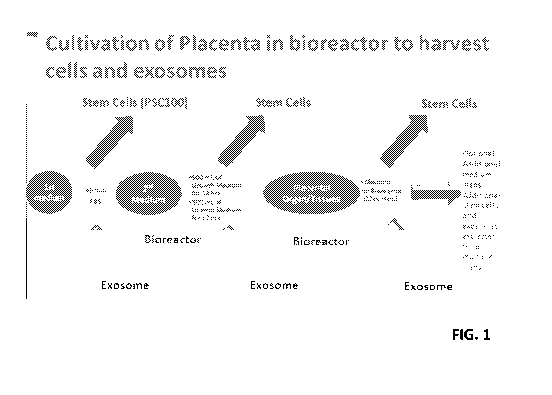
[0039] FIG. 1 shows a schematic for cultivating cells for exosome isolation.
[0040] FIG. 2A ¨ FIG.2C show three pExo isolates that were analyzed for their
size
distribution by NanoSight. This work was performed and reported by SBI Inc.
(System
Bioscience Inc.) using a contract service (www.systembio.com/services/exosome-
services/).
8
CA 03142020 2021-11-25
WO 2020/257720 PCT/US2020/038828
[0041] FIG. 3A ¨ FIG.3C show protein markers present on pExo (N=12) (FIG. 3A)
compared
with placenta perfusate exosomes (FIG. 3B) and cord blood serum derived
exosomes (FIG. 3C)
using the MACSPlex Kit.
[0042] FIG. 4 shows functional pathways of proteins identified in placental
exosome
populations.
[0043] FIG. 5 shows common and unique protein identified in three placenta
exosome
samples.
[0044] FIG. 6 shows that pExo promote migration of human dermal fibroblast
cells in a
transwell system.
[0045] FIG. 7 shows that pExo promote migration of human umbicical cord vessel
endothelial
cells.
[0046] FIG. 8 shows that pExo stimulate the proliferation of HUVEC.
[0047] FIG. 9 shows that pExo stimulate the proliferation of human CD34+
cells.
[0048] FIG. 10 shows that pExo stimulate the colony formation of human CD34+
cells.
[0049] FIG. 11 shows that pExo inhibit the proliferation of SKOV3 cancer
cells.
[0050] FIG. 12 shows that pExo inhibit the proliferation of A549 cancer cells.
[0051] FIG. 13 shows that pExo inhibit the proliferation of MDA321 cancer
cells.
[0052] FIG. 14 shows that pExo does not affect the proliferation of CD3+ T
cells in culture.
[0053] FIG. 15 shows that pExo increases expression of activation marker CD69
in UBC T
CD3+ cells.
[0054] FIG. 16 shows that pExo increases expression of activation marker CD69
in adult
PBMC T CD3+ cells.
[0055] FIG. 17 shows that pExo increases CD56+ NK cells in PBMC.
[0056] FIG. 18 shows protein markers present on pExo (N=10) using MACSPlex
Kit. Results
show pExo are positive for the following protein markers including pExo are
positive for CD2,
CD4, CD8, CD14, CD24, CD29, CD31, CD40, CD42a, CD42b, CD44, CD45, CD49e,
CD62P,
CD63, CD69, CD81, CD86, CD105, CD133-1, CD142, CD146, CD326, HLA-ABC, HLA-
DRDPDQ, MCSP, ROR1 and SSEA4.
[0057] FIG. 19 shows that pExo stimulate proliferation of human kidney
epithelial cells.
[0058] FIG. 20 shows that pExo stimulate proliferation of human lung
epithelial cells.
9
CA 03142020 2021-11-25
WO 2020/257720 PCT/US2020/038828
[0059] FIG. 21 Top panel shows that pExo stimulate proliferation of human
hepatic satellite
cells. Bottom panel shows that pExo improves cell recovery comparing to media
alone chemical-
induced injury of liver cells by acetaminophen (APAP) (2 mM) or APAP + pExo(10
ug/ml) and
acquire data with sweep interval every 15 minutes (N=3) in an xCELLigence Real
Time Cell
Analysis (RTCA).
[0060] FIG. 22 Top panel shows that pExo stimulate proliferation of human
dermal
fibroblasts.
[0061] FIG. 23 shows the study design of pExo biodistribution in vivo.
[0062] FIG. 24 shows the in vivo bio-distribution of pExo (whole body
imaging).
[0063] FIG. 25 shows persistence of pExo in mice (whole body imaging).
[0064] FIG. 26 shows bio-distribution of pExo in vivo (ex vivo imaging).
[0065] FIG. 27 shows the study design of pExo effect on rat stroke model.
[0066] FIG. 28 Top panel shows that pExo improved overall neuroscore
significantly in rat
after stroke induction. Bottom panels show that pExo-induced neurological
deficit reduction
compared to vehicle is superior than MSC-derived exosome in similar stroke
models (left) and
that pExo-induced neurological deficit reduction is superior than historic
PDAC data in the same
model (right).
[0067] FIG. 29 shows that pExo improved body-swing significantly in rat after
stroke
induction.
[0068] FIG. 30 shows that pExo improved forelimb placement score significantly
in rats after
stroke induction.
[0069] FIG. 31 shows that pExo improved stepping test score significantly in
rats after stroke
induction.
[0070] FIG. 32 shows pExo reduced lesion volume compared to vehicle control.
[0071] FIG. 33 shows no lesion volume reduction by MSC-derived Exo was
observed in a
similar stroke model (Xin et al. 2013).
[0072] FIG. 34 shows that pExo-induced lesion volume reduction is comparable
to historic
PDAC data in the same model.
[0073] FIG. 35 shows that pExo significantly increased doublecortin positive
cells in both
subventricular zone (SVZ) and hippocampus suggesting enhanced neurogenesis.
CA 03142020 2021-11-25
WO 2020/257720 PCT/US2020/038828
[0074] FIG. 36 shows that pExo significantly increased doublecortin positive
cells in both
subventricular zone (SVZ) and hippocampus suggesting enhanced neurogenesis.
[0075] FIG. 37 shows the study design of the effect of pExo on mice with
hindlimb ischemia
(HLI).
[0076] FIG. 38 shows that pExo improved the blood flow of mice with hindlimb
ischemia
(HLI) injury.
[0077] FIG. 39 shows that pExo improved the blood flow of mice with hindlimb
ischemia
(HLI) injury.
[0078] FIG. 40 shows the outline of an in vivo anti-aging study of pExo.
[0079] FIG. 41 shows that the pExo-treated group had a longer latency to fall
in rotarod test
than vehicle group in the rotarod study.
[0080] FIG. 42 shows that pExo-treated group had a quicker reduction of
glucose at 30min
after glucose administration than vehicle group as well as a lower glucose
AUC.
[0081] FIG. 43 shows the outline of an in vivo anti-GVHD study of pExo.
[0082] FIG. 44 shows single or multiple dosing of pExo improved survival in
GvHD model.
[0083] FIG. 45 shows single or multiple dosing of pExo improved weight loss in
GvHD
model.
[0084] FIG. 46 shows that multiple dosing of pExo inhibited the engraftment of
CD3+ human
T cells at Week 4 (mainly on CD4+ T cells).
[0085] FIG. 47 shows that multiple dosing of pExo inhibited the engraftment of
CD3+ human
T cells at Week 4 (mainly on CD4+ T cells).
[0086] FIG. 48 shows that pExo increases proliferation in PBTEC cells by
multiple pExo
cultivation methods.
[0087] FIG. 49 shows that pExo increases proliferation in a dose dependent
manner in PBTEC
cells.
5. DETAILED DESCRIPTION
5.1. Placenta-Derived Exosomes
[0088] The placenta-derived exosomes described herein can be selected and
identified by their
morphology and/or molecular markers, as described below. The placenta-derived
exosomes
described herein are distinct from exosomes known in the art e.g., chorionic
villi mesenchymal
11
CA 03142020 2021-11-25
WO 2020/257720 PCT/US2020/038828
stem cell-derived exosomes, e.g., those described in Salomon et al., 2013,
PLOS ONE, 8:7,
e68451. Accordingly, the term "placenta-derived exosome," as used herein, is
not meant to
include exosomes obtained or derived from chorionic villi mesenchymal stem
cells.
[0089] In certain embodiments, populations of placenta-derived exosomes
described herein do
not comprise cells, e.g., nucleated cells, for example placental cells.
5.1.1. Placenta-Derived Exosome Markers
[0090] The placenta-derived exosomes described herein contain markers that can
be used to
identify and/or isolate said exosomes. These markers may, for example, be
proteins, nucleic
acids, saccharide molecules, glycosylated proteins, lipid molecules, and may
exist in monomeric,
oligomeric and/or multimeric form. In certain embodiments, the markers are
produced by the
cell from which the exosomes are derived. In certain embodiments, the marker
is provided by
the cell from which the exosomes are derived, but the marker is not expressed
at a higher level
by said cell. In a specific embodiment, the markers of exosomes described
herein are higher in
the exosomes as compared to the cell of origin when compared to a control
marker molecule. In
another specific embodiment, the markers of exosomes described herein are
enriched in said
exosomes as compared to exosomes obtained from another cell type (e.g., the
chorionic villi
mesenchymal stem cells described in Salomon et al., 2013, PLOS ONE, 8:7,
e68451 and pre-
adipocyte mesenchymal stem cells), wherein the exosomes are isolated through
identical
methods.
[0091] The three-dimensional structure of exosomes allows for the retention of
markers on the
surface of the exosome and/or contained within the exosome. Similarly, marker
molecules may
exist partially within the exosome, partially on the outer surface of the
exosome and/or across the
phospholipid bilayer of the exosome. In a specific embodiment, the markers
associated with the
exosomes described herein are proteins. In certain embodiments, the markers
are transmembrane
proteins that are anchored within the exosome phospholipid bilayer, or are
anchored across the
exosome phospholipid bilayer such that portions of the protein molecule are
within the exosome
while portions of the same molecule are exposed to the outer surface of the
exosome. In certain
embodiments, the markers are contained entirely within the exosome. In another
specific
embodiment, the markers associated with the exosomes described herein are
nucleic acids. In
certain embodiments, said nucleic acids are non-coding RNA molecules, e.g.,
micro-RNAs
(miRNAs).
12
CA 03142020 2021-11-25
WO 2020/257720 PCT/US2020/038828
5.1.1.1. .. Surface markers
[0092] The exosomes described herein comprise surface markers that allow for
their
identification and that can be used to isolate/obtain substantially pure
populations of cell
exosomes free from their cells of origin and other cellular and non-cellular
material. Methods of
for determining exosome surface marker composition are known in the art. For
example,
exosomal surface markers can be detected by fluorescence-activated cell
sorting (FACS) or
Western blotting.
[0093] In certain embodiments, the exosomes described herein comprise a
surface marker at a
greater amount than exosomes known in the art, as determinable by, e.g., FACS.
5.1.1.2. Yield
[0094] The exosomes described herein may be isolated in accordance with the
methods
described herein and their yields may be quantified. In a specific embodiment,
the exosomes
described herein are isolated at a concentration of about 0.5-5.0 mg per liter
of culture medium
(e.g., culture medium with or without serum). In another specific embodiment,
the exosomes
described herein are isolated at a concentration of about 2-3 mg per liter of
culture medium (e.g.,
culture medium containing serum). In another specific embodiment, the exosomes
described
herein are isolated at a concentration of about 0.5-1.5 mg per liter of
culture medium (e.g.,
culture medium lacking serum).
5.1.2. Storage and Preservation
[0095] The exosomes described herein can be preserved, that is, placed under
conditions that
allow for long-term storage, or conditions that inhibit degradation of the
exosomes.
[0096] In certain embodiments, the exosomes described herein can be stored
after collection
according to a method described above in a composition comprising a buffering
agent at an
appropriate temperature. In certain embodiments, the exosomes described herein
are stored
frozen, e.g., at about -20 C or about -80 C.
[0097] In certain embodiments, the exosomes described herein can be
cryopreserved, e.g., in
small containers, e.g., ampoules (for example, 2 mL vials). In certain
embodiments, the
exosomes described herein are cryopreserved at a concentration of about 0.1
mg/mL to about 10
mg/mL.
[0098] In certain embodiments, the exosomes described herein are cryopreserved
at a
temperature from about -80 C to about -180 C. Cryopreserved exosomes can be
transferred to
13
CA 03142020 2021-11-25
WO 2020/257720 PCT/US2020/038828
liquid nitrogen prior to thawing for use. In some embodiments, for example,
once the ampoules
have reached about -90 C, they are transferred to a liquid nitrogen storage
area.
Cryopreservation can also be done using a controlled-rate freezer.
Cryopreserved exosomes can
be thawed at a temperature of about 25 C to about 40 C before use.
[0099] In certain embodiments, the exosomes described herein are stored at
temperatures of
about 4 C to about 20 C for short periods of time (e.g., less than two weeks).
5.2. Compositions
[00100] Further provided herein are compositions, e.g., pharmaceutical
compositions,
comprising the exosomes provided herein. The compositions described herein are
useful in the
treatment of certain diseases and disorders in subjects (e.g., human subjects)
wherein treatment
with exosomes is beneficial.
[00101] In certain embodiments, in addition to comprising the exosomes
provided herein, the
compositions (e.g., pharmaceutical compositions) described herein comprise a
pharmaceutically
acceptable carrier. As used herein, the term "pharmaceutically acceptable"
means approved by a
regulatory agency of the Federal or a state government or listed in the U.S.
Pharmacopeia or
other generally recognized pharmacopeia for use in animals, and more
particularly in humans.
The term "carrier," as used herein in the context of a pharmaceutically
acceptable carrier, refers
to a diluent, adjuvant, excipient, or vehicle with which the pharmaceutical
composition is
administered. Saline solutions and aqueous dextrose and glycerol solutions can
also be
employed as liquid carriers, particularly for injectable solutions. Suitable
excipients include
starch, glucose, lactose, sucrose, gelatin, malt, rice, flour, chalk, silica
gel, sodium stearate,
glycerol monostearate, talc, sodium chloride, dried skim milk, glycerol,
propylene, glycol, water,
ethanol and the like. Examples of suitable pharmaceutical carriers are
described in "Remington's
Pharmaceutical Sciences" by JP Remington and AR Gennaro, 1990, 18th Edition.
[00102] In certain embodiments, the compositions described herein additionally
comprise one
or more buffers, e.g., saline, phosphate buffered saline (PBS), Dulbecco's PBS
(DPBS), and/or
sucrose phosphate glutamate buffer. In other embodiments, the compositions
described herein
do not comprise buffers. In certain embodiments, the compositions described
herein additionally
comprise plasmalyte.
[00103] In certain embodiments, the compositions described herein additionally
comprise one
or more salts, e.g., sodium chloride, calcium chloride, sodium phosphate,
monosodium
14
CA 03142020 2021-11-25
WO 2020/257720 PCT/US2020/038828
glutamate, and aluminum salts (e.g., aluminum hydroxide, aluminum phosphate,
alum
(potassium aluminum sulfate), or a mixture of such aluminum salts). In other
embodiments, the
compositions described herein do not comprise salts.
[00104] The compositions described herein can be included in a container,
pack, or dispenser
together with instructions for administration.
[00105] The compositions described herein can be stored before use, e.g., the
compositions can
be stored frozen (e.g., at about -20 C or at about -80 C); stored in
refrigerated conditions (e.g., at
about 4 C); or stored at room temperature.
5.2.1. Formulations and Routes of Administration
[00106] The amount of exosomes or a composition described herein which will be
effective for
a therapeutic use in the treatment and/or prevention of a disease or condition
will depend on the
nature of the disease, and can be determined by standard clinical techniques.
The precise dosage
of exosomes, or compositions thereof, to be administered to a subject will
also depend on the
route of administration and the seriousness of the disease or condition to be
treated, and should
be decided according to the judgment of the practitioner and each subject's
circumstances. For
example, effective dosages may vary depending upon means of administration,
target site,
physiological state of the patient (including age, body weight, and health),
whether the patient is
human or an animal, other medications administered, and whether treatment is
prophylactic or
therapeutic. Treatment dosages are optimally titrated to optimize safety and
efficacy.
[00107] Formulations of exosomes, e.g., of pExo, can be prepared for
pharmaceutical or
cosmetic uses in any convenient form such as a liquid, paste, or suspension.
It can be formulated
for administration by any necessary or convenient route of administration for
a given indication
including those suitable for parenteral (e.g., subcutaneous, intramuscular,
intradermal,
intravenous, or direct local injection), oral, inhalation (in solid and liquid
forms or forms suitable
for administration by a nebulizer), rectal, topical, buccal (e.g., sub-
lingual), eyedrops, eardrops,
cavity rinses (e.g., oral rinses) and transdermal administration.
[00108] Although the subject experiments were performed using placenta derived
exosomes,
applicants have demonstrated the effective delivery of intravenous exosome
delivery to multiple
organ systems. Accordingly, exosomes from other sources can be readily be
delivered to these
organ systems as taught and contemplated herein, for the treatment of the
above conditions.
CA 03142020 2021-11-25
WO 2020/257720 PCT/US2020/038828
[00109] Administration of the exosomes described herein, or compositions
thereof can be done
via various routes known in the art. In certain embodiments, the exosomes
described herein, or
compositions thereof are administered by local, systemic, subcutaneous,
parenteral, intravenous,
intramuscular, topical, oral, intradermal, transdermal, or intranasal,
administration. In a specific
embodiment, said administration is via intravenous injection. In a specific
embodiment, said
administration is via subcutaneous injection. In a specific embodiment, said
administration is
topical. In another specific embodiment, the exosomes, or compositions
thereof, are
administered in a formulation comprising an extracellular matrix. In another
specific
embodiment, the exosomes, or compositions thereof, are administered in
combination with one
or more additional delivery device, e.g., a stent. In another specific
embodiment, the exosomes,
or compositions thereof, are administered locally, e.g., at or around the site
of an area to be
treated with said exosomes or compositions, such as hypoxic tissue (e.g., in
treatment of
ischemic diseases) or draining lymph nodes.
5.3. Methods of Use
5.3.1. Treatment of Diseases that Benefit from Angiogenesis
[00110] The exosomes described herein, and compositions thereof, promote
angiogenesis, and,
therefore can be used to treat diseases and disorders that benefit from
angiogenesis.
Accordingly, provided herein are methods of using the exosomes described
herein, or
compositions thereof, to promote angiogenesis in a subject in need thereof As
used herein, the
term "treat" encompasses the cure of, remediation of, improvement of,
lessening of the severity
of, or reduction in the time course of, a disease, disorder or condition, or
any parameter or
symptom thereof in a subject. In a specific embodiment, the subject treated in
accordance with
the methods provided herein is a mammal, e.g., a human.
[00111] In one embodiment, provided herein are methods of inducing
vascularization or
angiogenesis in a subject, said methods comprising administering to the
subject the exosomes
provided herein, or a composition thereof Accordingly, the methods provided
herein can be
used to treat diseases and disorders in a subject that that benefit from
increased
angiogenesis/vascularization. Examples of such diseases/conditions that
benefit from increased
angiogenesis, and therefore can be treated with the exosomes and compositions
described herein
included, without limitation, myocardial infarction, congestive heart failure,
peripheral artery
16
CA 03142020 2021-11-25
WO 2020/257720 PCT/US2020/038828
disease, critical limb ischemia, peripheral vascular disease, hypoplastic left
heart syndrome,
diabetic foot ulcer, venous ulcer, or arterial ulcer.
[00112] In one embodiment, provided herein are methods of treating a subject
having a
disruption of blood flow, e.g., in the peripheral vasculature, said methods
comprising
administering to the subject the exosomes provided herein, or a composition
thereof In a
specific embodiment, the methods provided herein comprise treating a subject
having ischemia
with the exosomes provided herein, or a composition thereof In certain
embodiments, the
ischemia is peripheral arterial disease (PAD), e.g., is critical limb ischemia
(CLI). In certain
other embodiments, the ischemia is peripheral vascular disease (PVD),
peripheral arterial
disease, ischemic vascular disease, ischemic heart disease, or ischemic renal
disease.
5.3.2. Patient Populations
[00113] In certain embodiments, the exosomes described herein are administered
to a subject
in need of therapy for any of the diseases or conditions described herein. In
another
embodiment, a composition described herein is administered to a subject in
need of therapy for
any of the diseases or conditions described herein. In certain embodiments
said subject is a
human.
[00114] In a specific embodiment, the exosomes or compositions described
herein are
administered to a subject (e.g., a human) in need of a therapy to increase
angiogenesis and/or
vascularization.
5.4. Kits
[00115] Provided herein is a pharmaceutical pack or kit comprising one or more
containers
filled with one or more of the ingredients of the pharmaceutical compositions
described herein,
i.e., compositions comprising the exosomes described herein. Optionally
associated with such
container(s) can be a notice in the form prescribed by a governmental agency
regulating the
manufacture, use or sale of pharmaceuticals or biological products, which
notice reflects
approval by the agency of manufacture, use or sale for human administration.
[00116] The kits described herein can be used in the above methods. The
compositions
described herein can be prepared in a form that is easily administrable to an
individual. For
example, the composition can be contained within a container that is suitable
for medical use.
Such a container can be, for example, a sterile plastic bag, flask, jar, or
other container from
which the compositions can be easily dispensed. For example, the container can
be a blood bag
17
CA 03142020 2021-11-25
WO 2020/257720 PCT/US2020/038828
or other plastic, medically acceptable bag suitable for the intravenous
administration of a liquid
to a recipient.
Exemplary Placenta Culture
[00117] The placenta is a reservoir of cells, including stem cells such as
hematopoietic stem
cells (HSC) and non-hematopoietic stem cells. Described herein are methods to
isolate exosomes
from a placenta or portion thereof, which is cultured in a bioreactor.
Exosomes are secreted by
the cells during the culture and the exosomes are secreted into the media,
which facilitates
further processing and isolation of the exosomes. Exosomes can be also
isolated from the
placenta or portion thereof at different stages of culture (e.g., at different
time points and
different perfusion liquids may be used at each recovery step). Once in the
media, the exosomes
can be further isolated using e.g., centrifugation, a commercially available
exosome isolation kit,
lectin affinity, and/or affinity chromatography (e.g., utilizing immobilized
binding agents, such
as binding agents attached to a substrate, which are specific for a small Rab
family GTPase,
annexin, flotillin, Alix, Tsg101, ESCRT complex, CD9, CD37, CD53, CD63, CD63A,
CD81,
CD82), Hsp70, Hsp90, epithelial cell adhesion molecules (EpCam), perforin,
TRAIL, granzyme
B, Fas, one or more cancer markers such as: Fas ligand, CD24, EpCAM, EDIL3,
fibronectin,
Survivin, PCA3, TMPRSS2:ERG, Glypican-1, TGF-01, MAGE 3/6, EGFR, EGFRvIII,
CD9,
CD147, CA-125, EpCam, and/or CD24, or one or more inflammatory or pathogenic
markers
such as: a viral, fungal, or a bacterial protein or peptide including but not
limited to a-synuclein,
HIV or HCV proteins, tau, beta-amyloid, TGF-beta, TNF-alpha, fetuin-A, and/or
CD133) . The
isolated exosomes can be used for therapeutics, diagnostics, and as
biotechnological tools.
[00118] "Exosomes" as described herein are vesicles that are present in many
and perhaps all
eukaryotic fluids, including ascites fluid, blood, urine, serum and breast
milk. They may also be
referred to as extracellular vesicles. Exosomes are bi-lipid membrane vesicles
secreted from
living cells that play important functions in cell-cell communications.
Exosomes are produced
by cells, such a stem cells, epithelial cells and a sub-type of exosomes,
defined as Matrix-bound
nanovesicles (MBVs), was reported to be present in extracellular matrix (ECM)
bioscaffolds
(non-fluid). The reported diameter of exosomes is between 30 and 100 nm, which
is larger than
low-density lipoproteins (LDL) but much smaller than, for example, red blood
cells. Exosomes
can be released from the cell when multivesicular bodies fuse with the plasma
membrane or
released directly from the plasma membrane.
18
CA 03142020 2021-11-25
WO 2020/257720 PCT/US2020/038828
[00119] Exosomes have been shown to have specialized functions and play a key
role in
processes such as coagulation, intercellular signaling, and waste management.
It is known that
extracellular vesicles and exosomes secreted by placenta contribute to the
communication
between placenta and maternal tissues to maintain maternal-fetal tolerance.
Exosomes isolated
from human placental explants was shown to have immune modulation activities.
Stem cell
derived exosomes were also shown to reduce neuroinflammation by suppressing
the activation of
astrocytes and microglia and promote neurogenesis possibly by targeting the
neurogenic niche,
both which contribute to nervous tissue repair and functional recovery after
TBI. (Review Yang
et al. 2017, Frontiers in Cellular Neuroscience). Exosomes derived from human
embryonic
mesenchymal stem cells also promote osteochondral regeneration (Zhang et al.
2016,
Osteoarthritis and Cartilage). Exosomes secreted by human placenta that carry
functional Fas
Ligand and Trail molecules were shown to convey apoptosis in activated immune
cells,
suggesting exosome-mediated immune privilege of the fetus. (Ann-Christin
Stenqvist et al.,
Journal of Immunology, 2013, 191: doi:10.4049).
[00120] Exosomes contain active biologics including lipids, cytokines,
microRNA, mRNA and
DNA. They may also function as mediators of intercellular communication via
genetic material
and/or protein transfer. Exosomes may also contain cell-type specific
information that may
reflect a cell's functional or physiological state. Consequently, there is a
growing interest in the
development of clinical and biological applications for exosomes.
[00121] Accordingly, exosomes isolated from human placenta or a portion
thereof using the
approaches described herein, optionally including characterization of said
exosomes (e.g., by
identifying the presence or absence of one or more proteins or markers on the
exosomes) can be
used to stimulate an immuno-modulation, an anti-fibrotic environment, and/or a
pro-regenerative
effect. Accordingly, exosomes isolated from human placenta or a portion
thereof using the
approaches described herein may be selected (e.g., according to markers
present or absent on the
exosomes), purified, frozen, lyophilized, packaged and/or distributed as a
therapeutic product
and/or a biotechnological tool.
[00122] In some alternatives, it may be beneficial to identify exosomes having
tumor markers
or peptides, pathogenic markers or peptides, such as viral, fungal, or
bacterial markers or
peptides, and/or inflammatory markers, such as inflammatory peptides, so that
such exosomes
can be removed from a population of exosomes (e.g., removal by affinity
chromatography with
19
CA 03142020 2021-11-25
WO 2020/257720 PCT/US2020/038828
binding molecules such as, antibodies or binding portions thereof, which are
specific for such
tumor markers or peptides, pathogenic markers or peptides, and/or inflammatory
markers or
peptides). Accordingly, in some alternatives, for example, a first population
of exosomes are
isolated from human placenta or a portion thereof by the methods described
herein and once the
first population of exosomes is isolated this population of exosomes is
further processed to
remove one or more subpopulations of exosomes using a substrate having an
immobilized
antibody or binding portion thereof (e.g., a membrane, a resin, a bead, or a
vessel having said
immobilized antibody or binding portion thereof), wherein the immobilized
antibody or binding
portion thereof is specific for a marker or peptide present on the
subpopulation of exosomes,
which are selected for further isolation, such as, one or more tumor markers
or peptides,
pathogenic markers or peptides, e.g., viral, fungal, or bacterial markers or
peptides, and/or
inflammatory markers or inflammatory peptides. In some alternatives, a first
population of
exosomes isolated from human placenta or a portion thereof by the methods
described herein are
contacted with a substrate having an immobilized antibody or binding portion
thereof (e.g., a
membrane, a resin, a bead, or a vessel having said immobilized antibody or
binding portion
thereof), wherein the immobilized antibody or binding portion thereof is
specific for one or more
cancer markers such as: Fas ligand, CD24, EpCAM, EDIL3, fibronectin, Survivin,
PCA3,
TMPRSS2:ERG, Glypican-1, TGF-01, MAGE 3/6, EGFR, EGFRvIII, CD9, CD147, CA-125,
EpCam, and/or CD24 so as to isolate a second population of exosomes from the
first population
of exosomes based on the affinity to the immobilized antibody or binding
portion thereof. In
some alternatives, a first population of exosomes isolated from human placenta
or a portion
thereof by the methods described herein are contacted with a substrate having
an immobilized
antibody or binding portion thereof (e.g., a membrane, a resin, a bead, or a
vessel having said
immobilized antibody or binding portion thereof), wherein the immobilized
antibody or binding
portion thereof is specific for one or more inflammatory or pathogenic markers
such as: a viral,
fungal, or a bacterial protein or peptide including but not limited to a-
synuclein, HIV or HCV
proteins, tau, beta-amyloid, TGF-beta, TNF-alpha, fetuin-A, and/or CD133 or
portions thereof
so as to isolate a second population of exosomes from the first population of
exosomes based on
the affinity to the immobilized antibody or binding portion thereof.
[00123] In some alternatives, the population of exosomes isolated and/or
selected by the
approaches described herein have markers or peptides that are useful for
therapeutics such as
CA 03142020 2021-11-25
WO 2020/257720 PCT/US2020/038828
perforin and/or granzyme B, which has been shown to mediate anti-tumor
activity both in vitro
and in vivo (I Cancer 2016; 7(9):1081-1087) or Fas, which has been found in
exosomes that
exert cytotoxic activity against target cancer cells. (Theranostics 2017;
7(10):2732-2745).
Accordingly, in some alternatives, a first population of exosomes isolated
from human placenta
or a portion thereof by the methods described herein are contacted with a
substrate having an
immobilized antibody or binding portion thereof (e.g., a membrane, a resin, a
bead, or a vessel
having said immobilized antibody or binding portion thereof), wherein the
immobilized antibody
or binding portion thereof is specific for perforin, TRAIL and/or granzyme B
and/or Fas and a
second population of exosomes from the first population of exosomes is
isolated based on the
affinity to the immobilized antibody or binding portion thereof to perforin,
TRAIL and/or
granzyme B and/or Fas. In some alternatives, a population of exosomes is
isolated, which
comprises CD63 RNAs, and/or a desired microRNA. In some alternatives, a
population of
exosomes is isolated and/or characterized after isolation using affinity
chromatography or
immunological techniques, wherein said population of exosomes comprise markers
or peptides
such as small Rab family GTPases, annexins, flotillin, Alix, Tsg101, ESCRT
complex, CD9,
CD37, CD53, CD63, CD63A, CD81, CD82), Hsp70, Hsp90) and/or epithelial cell
adhesion
molecules (EpCam). As detailed above, in some alternatives, a first population
of exosomes
isolated from human placenta or a portion thereof by the methods described
herein are contacted
with a substrate having an immobilized antibody or binding portion thereof
(e.g., a membrane, a
resin, a bead, or a vessel having said immobilized antibody or binding portion
thereof), wherein
the immobilized antibody or binding portion thereof is specific for small Rab
family GTPases,
annexins, flotillin, Alix, Tsg101, ESCRT complex, CD9, CD37, CD53, CD63,
CD63A, CD81,
CD82), Hsp70, Hsp90) and/or epithelial cell adhesion molecules (EpCam) and a
second
population of exosomes from the first population of exosomes is isolated based
on the affinity to
the immobilized antibody or binding portion thereof to small Rab family
GTPases, annexins,
flotillin, Alix, Tsg101, ESCRT complex, CD9, CD37, CD53, CD63, CD63A, CD81,
CD82),
Hsp70, Hsp90) and/or epithelial cell adhesion molecules (EpCam). In other
alternatives, a
population of exosomes isolated from human placenta or a portion thereof by
the methods
described herein are contacted with an antibody or binding portion thereof
specific for one or
more of small Rab family GTPases, annexins, flotillin, Alix, Tsg101, ESCRT
complex, CD9,
CD37, CD53, CD63, CD63A, CD81, CD82, Hsp70, Hsp90 and/or epithelial cell
adhesion
21
CA 03142020 2021-11-25
WO 2020/257720 PCT/US2020/038828
molecules (EpCam) and the binding of the antibody or binding portion thereof
is detected with a
secondary binding agent having a detectable reagent, which binds to said
antibody or binding
portion thereof (e.g., utilizing an ELISA or blotting procedure) so as to
confirm the presence of
the small Rab family GTPases, annexins, flotillin, Alix, Tsg101, ESCRT
complex, CD9, CD37,
CD53, CD63, CD63A, CD81, CD82), Hsp70, Hsp90 and/or epithelial cell adhesion
molecules
(EpCam) in the isolated exosome population.
[00124] "Isolation" as described herein is a method for separating the
exosomes from other
materials. Isolation of exosomes may be performed by high centrifugal force in
a centrifuge,
utilization of commercially available kits (e.g. SeraMir Exosome RNA
Purification kit (SBI
system biosciences), Intact Exosome Purification and RNA Isolation
(CombinationKit) Norgen
BioTek Corp.), and the use of lectin affinity or affinity chromatography with
binding agents
(e.g., an antibody or binding portion thereof) specific for markers or
peptides on the exosomes
such as the markers or peptides mentioned above (e.g., binding agents specific
for small Rab
family GTPases, annexins, flotillin, Alix, Tsg101, ESCRT complex, CD9, CD37,
CD53, CD63,
CD63A, CD81, CD82), Hsp70, Hsp90, epithelial cell adhesion molecules (EpCam),
perforin,
TRAIL, granzyme B, Fas, one or more cancer markers such as: Fas ligand, CD24,
EpCAM,
EDIL3, fibronectin, Survivin, PCA3, TMPRSS2:ERG, Glypican-1, TGF-01, MAGE 3/6,
EGFR, EGFRvIII, CD9, CD147, CA-125, EpCam, and/or CD24, or one or more
inflammatory
or pathogenic markers such as: a viral, fungal, or a bacterial protein or
peptide including but not
limited to a-synuclein, HIV or HCV proteins, tau, beta-amyloid, TGF-beta, TNF-
alpha, fetuin-
A, and/or CD133).
[00125] "Placenta" as described herein is an organ in the uterus of pregnant
eutherian
mammals, nourishing and maintaining the fetus through the umbilical cord. As
described herein,
the placenta may be used as a bioreactor for obtaining exosomes. In some
alternatives, a
decellularized placenta may be used as a scaffold and bioreactor, which
harbors an exogenous
cell population (e.g., a cell population that has been seeded onto and
cultured with the
decellularized placenta) so as to obtain a population of exosomes from said
cells, which are cell
specific. Accordingly, in some alternatives, decellularized placenta is seeded
with a regenerative
cell population (e.g., a population of cells comprising stem cells and/or
endothelial cells and/or
progenitor cells) and said regenerative cell population is cultured on said
decellularized placenta
in a bioreactor and cell specific exosomes are isolated from said cultured
cells using
22
CA 03142020 2021-11-25
WO 2020/257720 PCT/US2020/038828
centrifugation, a commercially available exosome isolation kit, lectin
affinity, and/or affinity
chromatography using a binding agents (e.g., an antibody or binding portion
thereof) specific for
markers or peptides on the exosomes such as the markers or peptides mentioned
above (e.g.,
binding agents specific for small Rab family GTPases, annexins, flotillin,
Alix, Tsg101, ESCRT
complex, CD9, CD37, CD53, CD63, CD63A, CD81, CD82), Hsp70, Hsp90, epithelial
cell
adhesion molecules (EpCam), perforin, TRAIL, granzyme B, Fas, one or more
cancer markers
such as: Fas ligand, CD24, EpCAM, EDIL3, fibronectin, Survivin, PCA3,
TMPRSS2:ERG,
Glypican-1, TGF-01, MAGE 3/6, EGFR, EGFRvIII, CD9, CD147, CA-125, EpCam,
and/or
CD24, or one or more inflammatory or pathogenic markers such as: a viral,
fungal, or a bacterial
protein or peptide including but not limited to a-synuclein, HIV or HCV
proteins, tau, beta-
amyloid, TGF-beta, TNF-alpha, fetuin-A, and/or CD133).
[00126] "Ascites fluid" as described herein is excess fluid in the space
between the membranes
lining the abdomen and abdominal organs (the peritoneal cavity). Ascites fluid
may be a source
of exosomes.
[00127] "Plasma" as described herein is the liquid part of the blood and
lymphatic fluid, which
makes up about half of the volume of blood. Plasma is devoid of cells and,
unlike serum, has not
clotted. Blood plasma contains antibodies and other proteins. Plasma may be a
source of
exosomes.
[00128] Several methods of culturing cells so as to produce copious amounts
exosomes are
provided herein. Culture media used for recovering or isolating the exosomes
may be provided
with one or more nutrients, enzymes or chelators. Chelators may be used to
facilitate release of
the exosomes from the cultured cells. Without being limiting, chelators used
in some of the
methods may include a phosphonate, BAPTA tetrasodium salt, BAPTA/AM, Di-
Notrophen TM
reagent tetrasodium salt, EGTA/AM, pyridoxal isonicotinoyl hydrazine,
N,N,N',N'-tetrakis-(2
Pyridylmethyl)ethylenediamine, 6-Bromo-N'-(2-hydroxybenzylidene)-2-
methylquinoline-4-
carbohydrazide, 1,2-Bis(2-aminophenoxy)ethane-N,N,N',N'-tetraacetic acid
tetrakis(acetoxymethyl ester), (Ethylenedinitrilo)tetraacetic acid, (EDTA),
Edathamil,
Ethylenedinitrilotetraacetic acid, Ethylene glycol-bis(2-aminoethylether)-
N,N,N,N1-tetraacetic
acid, or Ethylene glycol-bis(f3-aminoethyl ether)-N,N,N',N'-tetraacetic acid
(EGTA) or any
combination thereof. The chelator may be provided in the media used to culture
or isolate the
exosomes at a concentration of 1 mM, 2mM, 3mM, 4mM, 5mM, 6mM, 7mM, 8mM, 9mM,10
23
CA 03142020 2021-11-25
WO 2020/257720 PCT/US2020/038828
mM, 20mM, 30mM, 40mM, 50mM, 60mM, 70mM, 80mM, 90mM or 100mM or at a
concentration that is within a range defined by any two aforementioned
concentrations. As
shown herein, the presence of one or more chelators in the media unexpectedly
enhanced
recovery of exosomes from placenta cultured in a bioreactor. The media used to
culture and/or
recover the exosomes may also have a protease, which may further enhance the
release of
exosomes. In some alternatives, the protease provided in the media is trypsin,
collagenase,
chymotrypsin or carboxypeptidase. In some alternatives, the protease is
provided in the media at
a concentration of 1 mM, 2mM, 3mM, 4mM, 5mM, 6mM, 7mM, 8mM, 9mM, 10 mM, 20mM,
30mM, 40mM, 50mM, 60mM, 70mM, 80mM, 90mM or 100mM or at a concentration that
is
within a range defined by any two of the aforementioned concentrations. One or
more sugars
may also be added to the media used to culture and/or recover the exosomes. In
some
alternatives, the sugar added to the media is glucose. It is contemplated that
the presence of
glucose in the media enhances the release of the exosomes. In some
alternatives, the glucose is
provided in the media at a concentration of 1 mM, 2mM, 3mM, 4mM, 5mM, 6mM,
7mM, 8mM,
9mM, 10 mM, 20mM, 30mM, 40mM, 50mM, 60mM, 70mM, 80mM, 90mM or 100mM or at a
concentration that is within a range defined by any two of the aforementioned
concentrations.
The media may also include growth factors, cytokines, or one or more drugs
e.g., GM-CSF,
serum and/or an AHR antagonist.
Methods of collecting exosomes from a placenta or portion thereof
[00129] An exemplary method for recovery of exosomes from placenta is shown in
Figure 1.
Sources for the exosome isolation may be from cord blood plasma: PRP, placenta
perfusate (PS),
placenta tissue cultivate (PTS), placenta organ cultivate (PO), or exogenous
cells that may be
placed in the placenta or portion thereof, when the placenta is used as a
bioreactor for exosome
generation. By one approach, placenta or portion thereof is collected
(#200010323, collected
9/25/2017). Placenta is contacted with a media or perfused with normal PSC-100
collection
methods, collected as PS-1 (9/26/2017). The placenta or portion thereof is
incubated in a hood
for at least 4 hours. The placenta or portion thereof is contacted with media
(RPMI media) or
perfused with 500mL RPMI base medium (1% antibiotics), collected as PS-2. The
placenta or
portion thereof is then incubated in a hood overnight and is covered. The
placenta or portion
thereof is contacted with or perfused with 750mL saline solution and collected
as PS-3. The
24
CA 03142020 2021-11-25
WO 2020/257720 PCT/US2020/038828
samples were then shipped to a laboratory for analysis (Warren). PS1, PS2 and
PS3 were
analyzed by FACS at the same day after RBC lysis.
[00130] For the analysis, placenta tissue were cut into lx1x1 cm size, placed
in 100 mL of
solution (all with 1% P&S) in T75 flasks (each about 1/8 of the placenta).
Four solutions were
assayed: A: DMEM medium; B: PBS; C: PBS+5mM EDTA; D: PBS+0.025% Trypsin-EDTA.
This was then allowed to incubate in 37 C incubator overnight (0/N).
[00131] The supernatant was then harvested, passed through tissue filter and
spun down at
400g to harvest cells (pellet). The supernatant after the first centrifugation
was then spun down
for exosome isolation (3000g spin soup>10,000 spin soup: 100,000g pellet)
[00132] The cells collected were also used for FACS analysis. The cell samples
were in several
buffers (A=PTS1; B=PTS2; C=PTS-3, D=PTS4). Exosomes were recovered and were
then
assayed to identify the presence of an exosome marker confirming that the
exosomes were
obtained and isolated by the procedure.
Identification of a population of exosomes isolated from the placental
bioreactor using ELISA
and protein assays
[00133] Fractions of supernatant from the placental bioreactor were collected
by the methods
described above and the fractions were filtered. The supernatant was then
subjected to
centrifugation at 400g x 10 min to collect the cells. After the first
centrifugation, a second
centrifugation was performed at 3000g x 30 min to pellet cell debris. A third
centrifugation was
the performed at 10,000g x 1 hr to pellet micro vesicles. A fourth
centrifugation was then
performed at 100,000g x 1.5 hr to pellet exosomes. The centrifuge tube
containing the pelleted
exosomes was then placed upside-down on paper to drain residual liquid. The
exosome pellet
was then dissolved in an appropriate volume of sterile PBS (e.g. 2.0 mL) to
dissolve pellet, and
the solution containing the exosomes was then aliquoted in a sterile Eppendorf
tube and frozen in
a -20 C/-80 C freezer. Exosomes were then assayed for the presence of an
exosome-specific
marker CD63A using an ELISA-63A and Protein Quantification Kit
As shown, PRP, placenta perfusate and placenta tissue contain a population of
exosomes that are
CD63+ and can be efficiently isolated by ultracentrifiguation. For the exosome
isolation, first the
culture supernatant was filtered through a tissue filter and several
centrifugations were performed
as described above to obtain the exosomes, which were then frozen. For the
ELISA detection of
CA 03142020 2021-11-25
WO 2020/257720 PCT/US2020/038828
the exosomes, an anti-CD63 antibody was used. The sample was diluted 1:1 with
exosome binding
buffer (60uL + 60uL) in the assay. CD63+ exosomes were efficiently isolated by
this procedure.
Characterization of exosomes
[00134] Exosomes may contain protein, peptides, RNA, DNA and cytokines.
Methods such as
miRNA sequencing, surface protein analysis (MACSPlex Exosome Kit, Miltenyi),
proteomic
analysis, functional studies (enzyme assays in vitro wound healing assays
(scratch assay),
exosome-induced cell proliferation (human keratinocytes or fibroblast)
(comparing to 5 known
stimulants), exosome-induced collagen production (human keratinocyte or
fibroblast):
comparing to TGFb, includes serum and non-serum control, ELISA for pro-
collagen 1 C
peptide, exosome-induced inhibition of inflammatory cytokines: response cell
types include
human keratinocytes or human fibroblasts, and comparisons to lyophilized heat-
killed bacterial
or LPS) may be performed.
[00135] In some alternatives, isolated exosomes were concentrated with 100-Kda
Vivaspin
filter (Sartorius), washed once with PBS and approximately 40uL was recovered.
The
concentrated population of exosomes was mixed with lOuL of 5)MPA lysis buffer
containing
lxprotease inhibitor cocktail (Roche) and vortexed, which was then followed by
sonication at
20 C for 5 min at a water sonicator (Ultrasonic Cleaner, JSP). After
sonication, the tube was
incubated on ice for 20 min with intermittent mixing. Next, the mixture was
centrifuged at
10,000g for 10 min at 4 C. The isolated clear lysate was transferred to a
fresh tube. The protein
amount was measured with BCA kit and bug of protein was loaded per lane for
Western
blotting and an antibody is used for determination of a protein of interest.
[00136] In another alternative, exosome labeling and uptake by cells is
examined (e.g.
HEK293T). An aliquot of frozen eluted exosomes were resuspended in 1 mL of PBS
and labeled
using PKH26 Fluorescent cell linker Kits (Sigma-Aldrich). A 2x PNK26-dye
solution (4uL dye
in 1 mL of Diluent C) was prepared and mixed with 1 mL of exosomal solution
for a final dye
concentration of 2x10e-6M. The samples was immediately mixed for 5 min and
staining was
stopped by adding 1% BSA to capture excel PKH26 dye. The labeled exosomes was
transferred
into a 100-Kda Vivaspin filter and spun at 4000g then washed with PBS twice
and
approximately 50uL of sample was recovered for analysis of exosome
concentration using NTA
prior to storage at -80C. PBS was used as negative control for the labeling
reaction. To perform
the uptake studies, HEK293T cells were plated in 8-well chamber slide
(1x10e4/well) using
26
CA 03142020 2021-11-25
WO 2020/257720 PCT/US2020/038828
regular medium. After 24hr, the slides was washed twice with PBS and incubated
with DMEM-
exo-free FBS (10%) for 24hr. Following this, fresh DMEM media with 10% exo-
free PBS
(200uL) each labeled exosome sample, corresponding to 2x10e9 exosomes, was
added to each
well and incubated for 1.5 hr in a cell culture incubator. After incubation,
the slides was washed
twice with PBS (500u1) and fixed with 4% paraformaldehyde solution for 20 min
at room
temperature. The slides were washed twice with PBS (500uL) , dried, and
mounted using a
ProLong Gold Antifade Reagent with DAPI. The cells were visualized using an
Axioskop
microscope (Zeiss)
High yield isolation of exosomes from cultivated postpartum human placenta
[00137] Postpartum human placentas obtained with full donor consent were
perfused. Residual
blood from the placenta was washed off with a large volume of sterile saline
and then cultivated
in a 5-L bioreactor with serum free culture medium supplemented with
antibiotics and cultivated
at 37 C incubator (5% CO2) and alternated with rotating at refrigerated
conditions for extended
period unto to 4 days. Supernatant of the culture medium was processed by
sequential
centrifugation by 3000g and 10,000g to pellet tissue, cell and micro-vesicles.
Exosomes were
pelleted by 100,000g ultra-centrifugation from the supernatant of 10,000g
centrifugation and
dissolved with sterile PBS. The yield of exosome was quantified by BCA protein
assay.
[00138] Supernatants from the placenta organ culture were processed as
described in the
methods to isolate exosomes. An ELISA assay using anti-CD63A antibodies
demonstrated that
the isolated exosomes contain the CD63A protein, a specific protein marker for
exosomes. It is
estimated one placenta cultured in one liter of medium generated approximately
40mg of
exosomes, or approximately lx1013 CD63A positive exosome particles in 24
hours. Further
characterization of these placenta-organ derived exosomes including expression
of CD9, CD81,
size and functional activities are performed.
[00139] In another set of experiments, postpartum human placentas obtained
with full donor
consent are perfused to isolate exosomes with media's having different
concentrations of EDTA.
Serum free culture medium supplemented with antibiotics and varying
concentrations of EDTA
(e.g., 5, 10, 20, 30, 40, 50, 60, 70, 80, 90, or 100mM or within a range
defined by any two of the
aforementioned concentrations) are perfused into placenta through umbilical
cord veins via
peristaltic pump with a constant rate and cultivated another 24-48 hours under
controlled
conditions. Following this cultivation, 750mL of physiologic medium containing
the amount of
27
CA 03142020 2021-11-25
WO 2020/257720 PCT/US2020/038828
EDTA employed is perfused at controlled rate. Exosomes are then isolated by
sequential
centrifugation and ultracentrifugation, confirmed by the CD63A ELISA assay,
and quantified by
the BCA protein assay, all described above. It will be shown that the
concentration of EDTA in
the media used to recover the exosomes impacts the amount of exosomes
recovered from the
placenta cultured in the bioreactor.
Additional alternatives
[00140] In some alternatives, a method of exosome isolation from a placenta or
a portion
thereof is provided. The method comprises a) contacting the placenta or a
portion thereof with a
first medium; b) obtaining a first fraction comprising exosomes from said
placenta or portion
thereof; c) contacting said placenta or portion thereof with a second medium;
d) obtaining a
second fraction comprising exosomes from said placenta or portion thereof; e)
contacting said
placenta or portion thereof with a third medium; f) obtaining a third fraction
comprising
exosomes from said placenta or portion thereof and, optionally, isolating the
exosomes from said
first, second, and/or third fractions. In some alternatives, the method
further comprises multiple
steps of contacting the placenta or portion thereof with an additional medium;
and obtaining an
additional fraction comprising exosomes from said placenta or portion thereof
These two steps
may be repeated multiple times. Preferably, the placenta or portion thereof is
cultured and/or
maintained in a bioreactor. In some alternatives, the placenta or portion
thereof comprises
amniotic membrane. In some alternatives, the placenta or a portion thereof is
a human placenta
or a portion thereof. In some alternatives, the first, second, and/or third
mediums are in contact
with the placenta or portion thereof for at least 45 minutes, such as 45
minutes or 1, 2, 3, 4, 5, 6,
7, 8, 9, 10, 11 or 12 hours or any amount of time that is within a range
defined by any two of the
aforementioned time points. In some alternatives, the first, second, and/or
third mediums are in
contact with the placenta or portion thereof for at least 7, 14, 28, 35 or 42
days or any amount of
time that is within a range defined by any two of the aforementioned time
points. In some
alternatives, the placenta or a portion thereof has been minced, ground, or
treated with an
enzyme such as collagenase and/or a protease.
[00141] In some alternatives, a placenta or a portion thereof is provided as a
substantially flat
or sheet-like scaffold material, which has been decellularized and,
optionally, substantially dried.
The decellularized placenta or a portion thereof is used as a scaffold to
harbor exogenous cells
28
CA 03142020 2021-11-25
WO 2020/257720 PCT/US2020/038828
such as homogeneous cell populations obtained from cell culture or primary
isolation procedures
(e.g., regenerative cells including stem cells, endothelial cells, and/or
progenitor cells). The
method further comprises passaging fluid or fluid comprising the cells to be
seeded into the
decellularized placenta or portion thereof. Once the cells are established,
exosomes generated
from the cells are recovered and isolated using the procedures described
above. In some
alternatives, the fluid comprising the cells to be seeded on the
decellularized placenta or portion
thereof is ascites fluid, blood or plasma. In some alternatives, the cells are
from an organ. In
some alternatives, the cells are from liver, kidney, lung or pancreas. In some
alternatives, the
cells are immune cells. In some alternatives, the cells are T-cells or B-
cells.
[00142] In some alternatives, the first medium comprises Phosphate buffered
saline (PBS). In
some alternatives, the second medium comprises growth factors. In some
alternatives, the third
medium comprises a chelator. In some alternatives, the chelator is EDTA, EGTA,
a phosphonate,
BAPTA tetrasodium salt, BAPTA/AM, Di-Notrophen TM reagent tetrasodium salt,
EGTA/AM,
pyridoxal isonicotinoyl hydrazine, N,N,N',N'-tetrakis-(2
Pyridylmethyl)ethylenediamine, 6-
Bromo-N'-(2-hydroxybenzylidene)-2-methylquinoline-4-carbohydrazide, 1,2-Bis(2-
aminophenoxy)ethane-N,N,N,N1-tetraacetic acid tetrakis(acetoxymethyl ester),
(Ethylenedinitrilo)tetraacetic acid, EDTA, Edathamil,
Ethylenedinitrilotetraacetic acid, Ethylene
glycol-bis(2-aminoethylether)-N,N,N,N1-tetraacetic acid, or Ethylene glycol-
bis(fl-aminoethyl
ether)-N,N,N,N1-tetraacetic acid tetrasodium salt or any combination thereof.
In some
alternatives, the chelator is EDTA or EGTA or a combination thereof. In some
alternatives, the
chelator is provided in the third medium at a concentration of 1 mM, 2mM, 3mM,
4mM, 5mM,
6mM, 7mM, 8mM, 9mM, 10mM, 20mM, 30mM, 40mM, 50mM, 60mM, 70mM, 80mM, 90mM
or 100mM or at a concentration that is within a range defined by any two
aforementioned
concentrations. In some alternatives, the concentration of EDTA in the third
medium is provided
at a concentration of 1 mM, 2 mM, 3 mM, 4 mM, 5 mM, 6 mM, 7 mM, 8 mM, 9 mM 10
mM, 20
mM, 30 mM, 40 mM, 50 mM, 60 mM, 70 mM, 80 mM, 90 mM or 100 mM or at a
concentration
that is within a range defined by any two aforementioned concentrations.
[00143] In some alternatives, the third medium comprises a protease. In some
alternatives, the
protease is a trypsin, collagenase, chymotrypsin or carboxypeptidase or a
mixture thereof. In
some alternatives, the protease is trypsin. In some alternatives, the protease
is provided in the
third medium at a concentration of 1 mM, 2mM, 3mM, 4mM, 5mM, 6mM, 7mM, 8mM,
9mM,
29
CA 03142020 2021-11-25
WO 2020/257720 PCT/US2020/038828
mM, 20mM, 30mM, 40mM, 50mM, 60mM, 70mM, 80mM, 90mM or 100mM or at a
concentration that is within a range defined by any two of the aforementioned
concentrations.
[00144] In some alternatives, the method further comprises contacting the
placenta or portion
thereof with an additional plurality of mediums, wherein the contacting
results in obtaining
multiple fractions comprising exosomes. In some alternatives, the first,
second, third or
additional mediums comprise glucose. In some alternatives, the first, second,
third or additional
mediums comprise GM-CSF. In some alternatives, the first, second, third or
additional mediums
comprise serum. In some alternatives, the first, second, third or additional
mediums comprise
DMEM. In some alternatives, the first, second, third or additional medium
comprises an AHR
antagonist. In some alternatives, the AHR antagonist is SR1. In some
alternatives, the SR1 is at a
concentration of 1nM, 1 OnM, 100nM, 200nM, 300nM, 400nM, 500nM, 600nM, 700nM,
800nM,
900nM or 1mM or any other concentration within a range defined by any two
aforementioned
values.
[00145] In some alternatives, the first medium is in contact with the placenta
or portion thereof
while maintaining a temperature of 0 C, 5 C, 10 C, 15 C, 20 C, 25 C, 30
C, 35 C or 40 C
or a temperature that is within a range defined by any two of the
aforementioned temperatures. In
some alternatives, the second medium is in contact with the placenta or
portion thereof while
maintaining a temperature of 0 C, 5 C, 10 C, 15 C, 20 C, 25 C, 30 C, 35
C or 40 C or a
temperature that is within a range defined by any two of the aforementioned
temperatures. In
some alternatives, the third medium is in contact with the placenta or portion
thereof while
maintaining a temperature of 0 C, 5 C, 10 C, 15 C, 20 C, 25 C, 30 C, 35
C or 40 C or a
temperature that is within a range defined by any two of the aforementioned
values. In some
alternatives, the additional plurality of mediums is in contact with the
placenta or portion thereof
while maintaining a temperature of 0 C, 5 C, 10 C, 15 C, 20 C, 25 C, 30
C, 35 C or 40 C
or a temperature that is within a range defined by any two of the
aforementioned values.
[00146] In some alternatives, the first, second or third media or additional
plurality of mediums
comprise antibiotics.
[00147] In some alternatives, the exosomes are isolated from said first,
second, and/or third
fractions or multiple fractions by a method comprising:
(a) passing the first, second and/or third fractions or multiple
fractions through a
tissue filter;
CA 03142020 2021-11-25
WO 2020/257720 PCT/US2020/038828
(b) performing a first centrifugation of the filtrate collected in (a) to
generate a cell
pellet and a first supernatant;
(c) performing a second centrifugation on the first supernatant to generate
a second
supernatant; and
(d) performing a third centrifugation on the second supernatant to generate
an
exosome pellet; and, optionally,
(e) resuspending the exosomes in a solution.
[00148] In some alternatives, the population of isolated exosomes comprise
exosomes having
CD63, CD63-A, perforin, Fas, TRAIL or granzyme B Bor a combination thereof. In
some
alternatives, the population of isolated exosomes comprise exosomes that
comprise a signaling
molecule. In some alternatives, the population of isolated exosomes comprise
exosomes that
comprise cytokines, mRNA or miRNA.
[00149] In some alternatives, the method further comprises isolating exosomes
by affinity
chromatography, wherein affinity chromatography is selective for the removal
of exosomes
comprising viral antigens, viral proteins, bacterial antigens, or bacterial
protein fungal antigens
or fungal proteins.
[00150] In some alternatives, the method further comprises isolating exosomes
by an
alternative or additional affinity chromatography step, wherein the
alternative or additional
affinity chromatography step is selective for the removal of exosomes
comprising inflammatory
proteins. In some alternatives, the method further comprises enriching a
population of exosomes
comprising anti-inflammatory biomolecules.
[00151] In some alternatives, exosomes generated by any one of the embodiments
herein are
provided. In some alternatives, the exosomes are from ascites fluid, blood or
plasma. In some
alternatives, the exosomes are from cells from an organ. In some alternatives,
the exosomes are
from immune cells. In some alternatives, the exosomes are from T-cells or B-
cells.
[00152] It will be understood by those of skill within the art that, in
general, terms used herein,
and especially in the appended claims (e.g., bodies of the appended claims)
are generally
intended as "open" terms (e.g., the term "including" should be interpreted as
"including but not
limited to," the term "having" should be interpreted as "having at least," the
term "includes"
should be interpreted as "includes but is not limited to," etc.). It will be
further understood by
those within the art that if a specific number of an introduced claim
recitation is intended, such
31
CA 03142020 2021-11-25
WO 2020/257720 PCT/US2020/038828
an intent will be explicitly recited in the claim, and in the absence of such
recitation no such
intent is present. For example, as an aid to understanding, the following
appended claims may
contain usage of the introductory phrases "at least one" and "one or more" to
introduce claim
recitations. However, the use of such phrases should not be construed to imply
that the
introduction of a claim recitation by the indefinite articles "a" or "an"
limits any particular claim
containing such introduced claim recitation to embodiments containing only one
such recitation,
even when the same claim includes the introductory phrases "one or more" or
"at least one" and
indefinite articles such as "a" or "an" (e.g., "a" and/or "an" should be
interpreted to mean "at
least one" or "one or more"); the same holds true for the use of definite
articles used to introduce
claim recitations. In addition, even if a specific number of an introduced
claim recitation is
explicitly recited, those skilled in the art will recognize that such
recitation should be interpreted
to mean at least the recited number (e.g., the bare recitation of "two
recitations," without other
modifiers, means at least two recitations, or two or more recitations).
Furthermore, in those
instances where a convention analogous to "at least one of A, B, and C, etc."
is used, in general
such a construction is intended in the sense one having skill in the art would
understand the
convention (e.g., " a system having at least one of A, B, and C" would include
but not be limited
to systems that have A alone, B alone, C alone, A and B together, A and C
together, B and C
together, and/or A, B, and C together, etc.). In those instances where a
convention analogous to
"at least one of A, B, or C, etc." is used, in general such a construction is
intended in the sense
one having skill in the art would understand the convention (e.g.," a system
having at least one
of A, B, or C" would include but not be limited to systems that have A alone,
B alone, C alone,
A and B together, A and C together, B and C together, and/or A, B, and C
together, etc.). It will
be further understood by those within the art that virtually any disjunctive
word and/or phrase
presenting two or more alternative terms, whether in the description, claims,
or drawings, should
be understood to contemplate the possibilities of including one of the terms,
either of the terms,
or both terms. For example, the phrase "A or B" will be understood to include
the possibilities of
"A" or "B" or "A and B."
6. Examples
First Series of Experiments
6.1. Example 1: Cultivation of human placenta
32
CA 03142020 2021-11-25
WO 2020/257720 PCT/US2020/038828
[00153] Human placenta are received and washed with sterile PBS or saline
solution to remove
blood. The placenta is then cultivated in vessels as a whole organ in a large
container with
volume of 500 mL or 1000 mL of DMEM culture media supplemented with
antibiotics and 2mM
EDTA. In a different alternative, the placenta can be cut into different sizes
and placed in the
culture container. The cultivation is at 37oC in cell culture incubator with
5% CO2. The
cultivation time is 4 hour to 8 hours and the supernatant of the culture is
used for isolation of
exosomes. New media is added at each harvest time point (e.g., every 8 hours
or every 12 hours)
and the placenta organ and tissue is cultured for up to at least 5 days.
6.2. Example 2: Isolation and purification of placenta exosomes
[00154] The supernatant of the culture is centrifuged at 3,000g for 30minutes
to pellet the cell
and tissue debris. The supernatant is then centrifuged at 10,000 g for 1 hour
and the pellet (small
cell debris and organelles) is discarded. The supernatant is then centrifuged
at 100,000 g for 2
hours. The resulted pellet is exosomes. The exosomes pellet can be further
purified by the
following method: resuspended with different volume of sterile PBS and
centrifuged again at
100,000 for 2 hours and the final pellet is then resuspended with sterile PBS.
The resuspended
exosome is filtered through a syringe filter (0.2um), aliquoted at -80oC at
different volumes from
300uL to 1 mL.
[00155] Placental exosomes are characterized by size. Size distribution is
analyzed by a
nanoparticle tracking assay. Three representative samples of pExo were
measured with their size
using NanoSight. Each isolate has a mean size of 117, 101, and 96
respectively, consistent with
the reported size of exosomes. Results are shown in FIG. 2A ¨ FIG. 2C.
6.3. Example 3: Markers of pExos by FACS Analysis
[00156] Protein markers of pExo were analyzed with MACSPlex Exosome Kit
(Miltenyi
Biotec, Cat#130-108-813) following the protocol provided by the kit. Briefly,
the 120uL of pExo
isolates were incubated with 15 uL of exosome capture beads overnight at room
temperature
overnight. After washing once with 1 mL wash solution, the exosome were
incubated with
exosome detection reagents CD9, CD63 and CD81 cocktail and incubated for
additional 1 hrs.
After two washes, the samples were analyzed with FACS (BD Canto 10). There are
total 37
proteins markers included in this kit (Table 1) excluding mIgG1 and REA
control.
Table 1: List of protein markers used to detect pExo in MACSPlex Exosome Kit
33
CA 03142020 2021-11-25
WO 2020/257720 PCT/US2020/038828
No. Antibody Isotype No. Antibody Isotype
22 CO3 mIgG2a 65 CD81 REA
23 CD4 mIgG2a 66 MCSP mIgG1
24 C019 mIgG1 67 CD146 mIgG1
32 C08 mIgG2a 68 CD41b REA
33 HLA-DRDPDQ REA 74 CD42a REA
34 CD56 REA 75 CD24 mIgG1
35 CD105 mIgG1 76 CD86 mIgG1
42 CO2 mIgG2b 77 CD44 mIgG1
43 COlc mIgG2a 78 CD326 mIgG1
44 CO25 mIgG1 79 CD133/1 mIgG1 K
45 CD49e mIgG2b 85 CD29 mIgG1 K
46 ROR1 mIgG1 K 86 CD69 mIgG1 K
52 CD209 mIgG1 87 CD142 mIgG1 K
53 CD9 mIgG1 88 CD45 mIgG2a
54 SSEA-4 REA 89 CD31 mlgG1
55 HLA-ABC REA 96 REA Control REA
56 CD63 mIgG1K 97 CD20 mIgG1
57 C040 mIgG1 K 98 CD14 mIgG2a
63 CD62P REA 99 mIgG1 control mIgG1
64 C011c mIgG2b
[00157] pExo samples were identified to be highly positive for the following
protein markers
including CD1c, CD9, CD20, CD24, CD25, CD29, CD2, CD3, CD8, CD9, CD11c, CD14,
CD19, CD31, CD10, CD41b, CD42a, CD44, CD45, CD19c, CD4, CD15, CD19c, CD4,
CD56,
CD62P, CD83, CD69, CD81, CD86, CD105, CD133-1, CD142, CD148, HLA-ABC, HLA-
DRDPDQ, MSCP, ROR1, SSEA-4. pExo has very low level (2.6%) in CD209. Human
placenta
perfusate, which is obtained by perfuse the vasculature of placenta with
saline solution without
cultivation with medium and cell culture incubator, was also used to isolate
exosomes and
analyzed by the same methods for marker protein expression. The perfusate
derived exosomes
also express high levels of most of the markers found in pExo, but it has
significantly lower
CD11c (2.0%), MCSP (3.4%) and SSEA-4 (3.5%) comparing with pExos. pExo also
has
significantly higher levels of CD142 and CD81 comparing with placenta
perfusate exosomes.
Umbilical cord blood serum was also used to isolate exosomes and analyzed by
the same
34
CA 03142020 2021-11-25
WO 2020/257720 PCT/US2020/038828
methods for parker protei expression. Cord blood serum derived exosomes are
also positive in
most of the protein markers, but in general shows lower levels of each these
marker protein
expressions. Specifically, comparing with pExo, cord blood serum exosome has
lower levels of
CD56 (1.4%), CD3 (0.3%) and CD25 (3.9%). SSEA-4 and MSCP protein expression in
cord
blood serum is significantly lower than pExo but higher than placenta
perfusate exosomes. Cord
blood serum exosomes also has higher levels of MSCP protein comparing with
pExo. These data
indicate that cultivated placenta tissues can generate a unique exosome
population comparing
with non-cultured placenta and cord blood serum. Results for pExo samples,
compared to cord
blood serum derived exosomes and placenta perfusate exosomes are shown in FIG.
3A - FIG.
3C and Table 2.
Table 2
Protein Markers of Average Expression (%) on Exosomes from Three Different
Sources
Markers Cultivated Placenta Placenta Perfusate Cord Blood Serum
(N=12) (N=4) (N=4)
CD1c 9.80% 25.30% 15.60%
CD20 12.80% 10.80% 11.40%
CD24 61.90% 84.20% 12.50%
CD25 29.20% 26.50% 3.90%
CD29 69.80% 82.20% 11.20%
CD2 49.80% 67.20% 10.90%
CD3 12.00% 14.60% 0.40%
CD8 64.90% 86.90% 14.40%
CD9 66.20% 80.40% 10.40%
CD11c 37.90% 2.00% 11.50%
CD14 67.20% 29.50% 15.60%
CD19 29.30% 80.90% 8.90%
CD31 61.50% 81.50% 13.40%
CD40 67.30% 81.10% 15.60%
CD41b 64.70% 82.40% 12.50%
CD42a 66.10% 84.60% 13.00%
CD44 66.20% 86.30% 15.60%
CD45 24.70% 23.50% 6.20%
CD49e 60.60% 82.00% 15.30%
CD4 58.60% 77.40% 15.10%
CD56 24.20% 14.40% 1.40%
CD62P 64.10% 87.20% 15.60%
CD63 64.90% 81.10% 10.20%
CA 03142 02 0 2021-11-25
WO 2020/257720 PCT/US2020/038828
CD69 58.20% 65.80% 11.90%
CD81 56.40% 84.40% 15.60%
CD86 39.50% 17.30% 10.90%
CD105 53.60% 30.40% 10.00%
CD133-1 64.60% 44.20% 12.00%
CD142 67.80% 11.60% 13.30%
CD146 70.00% 79.40% 11.50%
CD209 2.60% 0% 9.70%
CD326 66.70% 75.50% 6.80%
HLA-ABC 64.60% 82.30% 13.70%
HLA- 60.80% 83.30% 12.80%
DRDPDQ
MCSP 44.60% 3.40% 8.10%
ROR1 64.20% 86.20% 14.40%
SSEA-4 58.80% 3.50% 10.80%
6.4. Example 4: Cytokines and growth factors of pExo samples
[00158] pExo samples were analyzed for their contents of cytokines with
MiltiPlex Luminex
kit that includes 41 different cytokines. The following tables show the data
of cytokines detected
on 15 different pExo preparations. The data shows that pExo contains
significant level of
cytokines (mean >50 pg/mL) including FGF2, G-CSF, Fractalkine, GDGF-AA/BB,
GRO, IL-
1RA, IL-8, VEGF, and RANTES. pExo also contains detectable levels of cytokines
(5 pg/mL to
49 pg/mL) of other cytokines including EGF, Flt-3L, IFNa3, MCP-3, PDGF-AA, IL-
15,
sCD40L, IL6, IP-10, MCP-1, MIP-alpha, MIP-lbeta, and TNF-alpha.
Table 3: Cytokines detected in pExo preparations
Sample ll3 EGF FGF-2 En):a TGF-a G-CSF Fiz-3i.
GM-CSF Fractalkine IF Na2 1FNg GRO 3.-10 MCP-3 11.-12P40
iTable 1-1)
pgitn1 pg/m1 P9fml pgimi pgimi Weil pgiall pgiml
pgant pgani pgimi Wail 99/1r8 99i'll'
3074-E1 279 17.11 3.77 <10.55i. 249.56 1 57 0 49
40.25 7.1 0.61 4044 0.59 5 :57 <0.74.
3315-E1 441 290.32 3.47 C55 4,49 6 ,33 1 12 83.56
11 3 1 2 mai <0 57,1. 432 <0.74i
941-El 1..59 17.11 43.203. '.55,i, 96.52 <0.62i c0.42i
17.66 2.22 0.87 6.6 0.7 0.85 <0.74i
944-E2 1.89. 12,33 43.203, (0.55 141,5 <3.82 i 0.45
22.66 54 0 1.01 6.6 0.62 1.59. <0.74i
988-E1 4.83 56.94 325 0 58 441.69 3.74 1 0 83.56
7.33 1 54 H.15 1.21 5.57 <3 74,
595-E2 12.75 120.3 11 42 2c3 267.34 5.42 22 227.72
13.al 1 g3 102.16 1.63 4.32 2.66
595-E3 .5 30.09 745 <0.55i 247.34 6.21 1.81 110,13
28.61 4.22 17.13 1.11 <0.36,i. 2.73
369-0 6 18 35937 9.36 1.27 34371 12 73 1 7/ 19758
7_35 146 13 2_33 9 97 1.96
405-E2 9 78 318.88 8.72 1.64 148.99 13 34 1 74 338.31
0.05 0.61 114.73 1.98 946 128
405-E3 7I 225.62 6.25 0.84 l7.5 45 1 53 225.33
7.47 0.48 was 1.21 675 1.73
52-E1 3.18 508.7 7.1 0.92 48.57 22.n 1.78 385.31
14.31 1.91 139.5 1% 11.19 4.13
352-E2 5./6 48327 6.20 078 7277 15.12 135 251.86
1368 1.31 10.976 1.14 5.57 2.21
789-El 13.45 2308 7.45 229 118.38 c0.62 I, 0.96
123.46 54 0 40 A6 3851 1.35 3 1.5
789- E2 5.72 24.5 5 :53 <0.5H 155.06 1 1 1.55 61.1
4.18 0 94 24.96 9.86 5.87 <8 74;,
313-E3 3.72 27.H 4 97 <0.55i 57.57 <0.621. 0 7 77.5
20.54 1 82 7.44 035 <0.33i, <0 74,
EGF FGF-2 Ea:axin TGF-a G-CSF F11-3L GM-CSF
Fractaikine iFt4a2 IFtig GRO 1-10 MCP-3 1-12P40
Mean r- S.07' 167,59 r 6.74r 1.31' 175.86 r 8.73r
1.38 r 14924 r 49.35r 1.42 P. 3.53'6 .25"4 5.72 r 228
SD 3.6 181.0 2.1 0.6 114,3
6.71 0.5 115.0 - O. J ,' 0.9 47,8 0.5 3.1 0.9
36
CA 03142020 2021-11-25
WO 2020/257720 PCT/US2020/038828
Sample ID MDC IL-12P70 PDGF-AA IL-13 PDGF-AB/BB I0-15
sCD4OL IL-17A IL-1RA IL-Is IL-9 IL-lb I0-2 IL-3
(Table 1-2)
pgirli pgirnt pg4111 pginl pgini pgirri pgfrol pg./ri
pgini pgiml pgirnt pg1m1 pgfrri pgdni
3074-E1 8.33 <0.710 395 1.3 203.83 2.19 0.81 0.41
10.87 077 087 0.48 <0.421 <0.31
3315-E1 <7.641 1.12 14.16 2.02 314.97 8.37 0.74 0.39
124.57 0.53 1.29 0.65 <0.421
941-E1 <7.641 <0.71; 1.41 1.01 35.31 0.95 1.5
<0.361. 9.47 0.39 0.36 1.23 <0.421
941-E2 <7.64.1. <0710 3.59 0.97 93.37 1.2 0.81 046
3.48 0.69 0.62 7 <0.421 <0.31j
988-E1 <7.64.1. 0.94 8.48 1.59 127 3.76 3.94 0.64
53.63 2.02 0.86 0.79 <0.420 <0.31j
595-E2 8.57 3.07 21.5 4.25 506.7 4.66 2566 0.95
92.19 2.7 1.84 8.2 <0.421 <0.31j
595-E3 11.62 1.65 126 3.9 317.14 3.72 205 0.98
18.86 2.09 2.92 3.37 0.53 0.51
368-E2 19.46 1.65 23.19 1.49 439.81 844 22 25 0.9
110.52 2.55 0.99 1.39 <0.44 9.37
405-E2 45.61 32 26.94 1.49 510.45 12 35 23.9 0.5
116.59 1.6 1.08 1.29 <0.421 <0.311
405-E3 24.28 1.16 18.87 1.28 335.8 10 21 1061 <0363
90.59 1.12 0.84 1.68 <0.421 <0.311
352-E1 271 32 33.76 2.04 492.97 33 13 18 13 1.01
169.21 2.48 1.49 15 0.45 <0.311
352-E2 14.83 2.14 28.14 1.21 442.07 23 78 11 72 098
107.61 1.87 1.18 1.38 <0.42.1 <0.311
789-E1 <764 1.86 7.02 1.61 192.21 219 10 47 0.77
91.16 1.71 0.78 0.39 <0.42.1 <0.311
789-E2 9.75 1.97 9.3 1.03 210.26 1.8 0.94 0.64
18.86 1.25 0.91 0.64 <0.421. <0.313
313-E3 <7840 0.94 5.01 2.89 167.09 0.95 <0.563
0.85 <3.201 1.05 2.93 0.35 <0.421. 0.42
MDC I1-12870 PDGF-AA I1-13 PDGF-AB/BB IL-15 sCD40L IL-17A IL-1RA IL-Is IL-9
IL-lb IL-2 0-3
Mean r 18.84' 1.91' 14.53' 1.87' 292.60 r 7.85' 9.55'
0.73' 72.69 r 1.52' 1.26' 2.02' 0.49' 0.43
SD 12.2 0.8 10.2 1.0 159.1 9.3 9.5 a 2 52.9
0.8 0.8 2.4 0.1 0.1
Sample ID 11,4 11.-5 IL-6 IL-7 IL-8 18-10 MCP-1 MIP-18
148-lb RANTES TNFa TN FL VEGF
(Table1-3)
139/91 wird 1 pert wird ped per 991lTi Pgini 139/rll
PO perl 1 wird ped
3074-El <3.200 <0.213 2.92 2.9 72.66 7.5 17.08 2.24
1.51 143.77 5.641 0.41 21.6
3315-El <3.201 <0.2111 6.3 6.2 215.72 27.63 85.9
11.98 8.27 292.91 2.1 0.44 56.06
941-El <3_201 027 1.15 1_45 6.08 <1.301 1.67
<1.311 <0_331 48.16 2.67 040 39.7
941-02 <3.20,!, <0.211.1 1.46 5.34 6.6 <1.303 1.53
1.48 0.89 30.32 16 58 0.38 43.8
908-El <3201 0.271 9.07 3.6 58.25 47.16 20.48
2.95 2.99 396.33 25.8 0.59 59.12
595-02 c3.20,1, 0.221 20.55 10.12 192,31 14,05 63.62
13.25 3.74 4462 23.97 0.41 51.98
595-03 e3.90 i 1.391 10.06 6.49 60.01 6.75 11,42
6.76 1.51 265,85 16.15 0.58 106.17
366-02 5.54 047 15.93 4.55 103,91 101.77 71.51 21.83
11.8 2413 5.41 1.73 64.19
405-02 4.01 0.381 17.02 5 105.05 92.1 9923 28.81
16.62 2463 638 0.97 54.71
405-03 <3.20; 032 13.3 3.6 159.18 53.34 59.98
27.54 17.83 1655 5.82 073 44.31
352-E1 8.08 0.451 24.21 7.24 167.95 156.45 138.26
9.99 8.19 3000 3201 1.12 67.45
352-02 <3201 0.381 18.92 5.4 198.95 89.91 103.45
12.06 6,69 2415 2.96 0,76 62.53
789-E1 <3.201 0.361 2.62 3.01 17,58 5.64 8.44
1.82 0.85 659.52 7.28 0.55 24.19
789-02 e3.90 i 0.271 5.69 2.85 84.19 4.65 8.74 3.7
1.04 417,14 5.5 0.52 22.25
313-03 <3.200 061 1.11 10.52 8.32 3.67 4.2
3.33 0.53 189.04 3.83 0.52 60.41
10-4 IL-5 1 IL-6 1L-7 I0-8 IP-10 MCP-1 MIP-1 a
M119-1b RANTES TNP'ft TN FO VEGF
Mean r 5.21' 0.45' 10.02' 5.22' 97.12' 46.97 r
46.37 r 10.55' 5.08' 1256.74 r 8.89 r 0.68' 51.90
SD 1.1 0.3 7.8 2.6 74.1 49.1 45.2 9.4
5.9 1380.0 7.81 0.4 21.4
[00159] pExo (11 samples) were also analyzed for the presence of soluble
cytokine receptors
by Multiplex Luminex analysis. The data are shown in the following table. The
data shows that
pExo contains high levels (>100 pg/mL) of sEFGR, sgp-130, sIL-1R1, sTNFR1,
sTNFRII,
sVEGRR1, sVEGFR1, sVEGFR3 and sCD30, sIL-2Ra, sIL-6R, sRAGE are also detected
in
some samples (>10ng/mL). Data shown as < are not detected and are regarded as
negative.
Table 4
37
CA 03142020 2021-11-25
WO 2020/257720 PCT/US2020/038828
Soluble cytokine receptors in placenta exosomes
pExo Samples sCD30 sEGFR sgp-130 sIL1-R1 s11,1R11 slL.2Ra
slIAR slIAR sRAGE sTNF RI sTNFRII sVEGF R1 sVEGF R2 sVEGF R3
Location pg/rri pg/rri pg/rri pg/rri pg/rri pg/rri
pg/rri pg/ml pg/ml pg/ni pg/ml pg/rri pg/rri pg/rri
1E3 988E1 1176 9626 326.15 <12.64 <35.04i 8.5
<59.19i 10.54 29.49 90.11 14.82 6556 75.84 462.01
1H3 595E1 <6.77i 3535 209.91 <12.64 <35.04i <5.94
<59.19i 9.78 8.23 29.88 <12.55i 2959 <70.59i 47.7
1C4 941E2 11.41 57601 132.85 <12.64 <35.04i 6.57
<59.19i 2.85 11.2 34.07 1966. 4929 <70.59i 119.96
1F4 941E1 8.03 >10030471 83.08 <12.64 <35.04i <5.94
<59.19i 3.65 7.57 25.57 <12.55i 803.42 <70.59i 56.61
1A5 405E1 12.84 5863 2316 <12.64 206.01 11.6
<59.19i 197.58 17.06 253.83 365.51 15179 436.1 64.11
1D5 366E1 9.34 10444 2806 <12.64 250.03 21.23
<59.19i 232.91 26.43 322.4 551.01 13823 419.27 10175
1G5 354E2 1912. 10627 4461 <12.64 327.31 17.59
<59.19i 172.5 1944. 249A7 308.79 19094 1378 86.58
1B6 352E1 14.68 7824 4108 <12.64 474A6 16.59
<59.19i 183.25 1331 297.87 473.55 16528 908.93 64.11
1E6 789E1 <6.74 25357 174.96 <12.64 <35.04i <5.94
<59.19i 7.18 11.93 28.6 15.44 3144 <70.59i 273.09
1H6 789E2 <6.77i 2499 206.92 <12.64 <35.04i <5.94
<59.19i 6.55 9.86 1933. <12.55i 6056 <70.59i 56.2
1C7 789E3 <6.74 2149 197.21 <12.64 <35.04i <5.94
<59.19i 4.05 6.12 15.19 <12.55i 9180 <70.59i 53.33
Mean 12.45 13552.50 1365.64 NA 314.45 13.68 NA 75.53 14.60 124.21
249.83 8931.95 643.63 125.95
SD 3.66 16858.13 1725.87 NA 117.87 5.70
NA 97.06 7.72 127.23 231.22 6238.32 506.33 128.64
1A3 QC1 465.86 2089 412.75 408.59 1948 420.27 200.54
138.73 252.82 201.08 210.06 4969 1984 1921
1C3 QC2 4219 18434 4012 4060 17200 4021 2290 1996
2165 2013 2005 18121 15711 18072
6.5. Example 5: Proteomic analysis of placenta exosomes
[00160] Three pExo samples were subjected to proteomic analysis. Submitted
samples were
lysed using a sonic probe (QSonica) with the following settings: amplitude
40%, pulse lOx 1
second on, 1 second off. The protein concentration was determined by Qubit
fluorometry. bug
of each sample was processed by SDS page and purified proteins were subject to
trypsin
digestion. Table 5 shows the total protein identified from each sample. Among
these samples,
there are total of 1814 proteins identified. Table 6 shows identification and
gene ID of top
identified proteins in pExo samples. Additional data is shown in FIG. 4 and
FIG. 5.
Table 5
32112 32113 32114
Total number of proteins ident&d 1313 1130 1362
Total number of spe,,etra matting 22408 2850 23248
Total number of unique peptides 12014 10761 13380
Table 6
38
CA 03142020 2021-11-25
WO 2020/257720 PCT/US2020/038828
Identified Proteins (1814) by Proteomics in Accession
Number Average
Placental Exosomes Relative
Abundanc
e
Cytoplasmic aconitate hydratase OS=Homo sp1P213991ACOC HUMAN 145
sapiens GN=AC01 PE=1 SV=3
Cell surface glycoprotein MUC18 OS=Homo sp113431211MUC18 HUMAN 131
sapiens GN=MCAM PE=1 SV=2
Protein arginine N-methyltransferase 1 sp9998731ANM1 HUMAN 119
OS=Homo sapiens GN=PRMT1 PE=1 SV=2
Guanine nucleotide-binding protein G(s) subunit splQ5JWF2IGNAS1 HUMA 99
alpha isoforms XLas OS=H sapiens GN=GNAS N
PE=1 SV=2
Cullin-5 OS=Homo sapiens GN=CUL5 PE=1 sp9930341CUL5 HUMAN 91
SV=4
Calcium-binding protein 39 OS=Homo sapiens sp99Y3761CAB39 HUMAN 83
GN=CAB39 PE=1 SV=1
Glucosidase 2 subunit beta OS=Homo sapiens sp113143141GLU2B HUMAN 72
GN=PRKCSH PE=1 SV=2
Chloride intracellular channel protein 5 splQ9NZA1ICLIC5 HUMA 72
OS=Homo sapiens GN=CLIC5 PE=1 SV=3 N
Semaphorin-3B OS=Homo sapiens sp9132141SEM3B HUMAN 72
GN=SEMA3B PE=2 SV=1
60S ribosomal protein L22 OS=Homo sapiens
sp113352681RL22 HUMAN 72
GN=RPL22 PE=1 SV=2
Spliceosome RNA helicase DDX39B sp9138381DX39B HUMAN 71
OS=Homo sapiens GN=DDX39B PE=1 SV=1
Transcriptional activator protein Pur-alpha splQ005771PURA HUMAN 68
OS=Homo sapiens GN=PURA PE=1 SV=2
Programmed cell death protein 10 OS=Homo splQ9BUL81PDC10 HUMA 66
sapiens GN=PDCD10 PE=1 SV=1 N
BRO1 domain-containing protein BROX splQ5VW321BROX HUMA 66
OS=Homo sapiens GN=BROX PE=1 SV=1 N
Kynurenine--oxoglutarate transaminase 3 splQ6YP211KAT3 HUMAN 65
OS=Homo sapiens GN=KYAT3 PE=1 SV=1
Laminin subunit alpha-5 OS=Homo sapiens sp10152301LAMA5 HUMA 64
GN=LAMA5 PE=1 SV=8 N
ATP-binding cassette sub-family E member 1 sp113612211ABCE1 HUMAN 61
OS=Homo sapiens GN=ABCE1 PE=1 SV=1
Syntaxin-binding protein 3 OS=Homo sapiens sp10001861STXB3 HUMAN 60
GN=STXBP3 PE=1 SV=2
Proteasome subunit beta type-7 OS=Homo sp9994361PSB7 HUMAN 60
sapiens GN=PSMB7 PE=1 SV=1
Glycogen [starch] synthase, muscle OS=Homo
sp113138071GYS1 HUMAN 59
sapiens GN=GYS1 PE=1 SV=2
39
CA 03142020 2021-11-25
WO 2020/257720 PCT/US2020/038828
Identified Proteins (1814) by Proteomics in Accession
Number Average
Placental Exosomes Relative
Abundanc
NAD(P)H-hydrate epimerase OS=Homo sapiens splQ8NCW5INNRE HUMA 59
GN=NAXE PE=1 SV=2
Hypoxia up-regulated protein 1 OS=Homo splQ9Y4L11HY0U1 HUMA 57
sapiens GN=HY0U1 PE=1 SV=1
Coagulation factor XI OS=Homo sapiens sp113039511FA11 HUMAN 57
GN=F11 PE=1 SV=1
Histone H1.0 OS=Homo sapiens GN=H1F0 sp113073051H10 HUMAN 56
PE=1 SV=3
COP9 signalosome complex subunit 4 splQ9BT781CSN4 HUMAN 56
OS=Homo sapiens GN=COPS4 PE=1 SV=1
40S ribosomal protein S15a OS=Homo sapiens sp113622441RS15A HUMAN 56
GN=RPS15A PE=1 SV=2
Protein ABHD11 OS=Homo sapiens splQ8NFV4IABHDB HUMA 54
GN=ABHD11 PE=1 SV=1
Retinal dehydrogenase 1 OS=Homo sapiens sp113003521AL1A1 HUMAN 53
GN=ALDH1A1 PE=1 SV=2
GDP-mannose 4,6 dehydratase OS=Homo sp10605471GMDS HUMAN 53
sapiens GN=GMDS PE=1 SV=1
Ketosamine-3-kinase OS=Homo sapiens splQ9HA641KT3K HUMAN 53
GN=FN3KRP PE=1 SV=2
Protein/nucleic acid deglycase DJ-1 OS=Homo sp9994971PARK7 HUMAN 52
sapiens GN=PARK7 PE=1 SV=2
Nectin-4 OS=Homo sapiens GN=NECTIN4 splQ96NY8INECT4 HUMA 51
PE=1 SV=1
Cdc42-interacting protein 4 OS=Homo sapiens
splQ156421CIP4 HUMAN 50
GN=TRIP10 PE=1 SV=3
WD repeat-containing protein 61 OS=Homo sp99GZS31WDR61 HUMA 49
sapiens GN=WDR61 PE=1 SV=1
CD59 glycoprotein OS=Homo sapiens sp113139871CD59 HUMAN 47
GN=CD59 PE=1 SV=1
Glycine dehydrogenase (decarboxylating), sp1P233781GCSP HUMAN 46
mitochondrial OS=Homo sapiens GN=GLDC
PE=1 SV=2
Guanine nucleotide-binding protein subunit sp113299921GNA11 HUMAN 43
alpha-11 OS=Homo sapiens GN=GNAll PE=1
SV=2
Serpin H1 OS=Homo sapiens GN=SERPINH1 sp113504541SERPH HUMAN 42
PE=1 SV=2
Alpha-2-antiplasmin OS=Homo sapiens sp113086971A2AP HUMAN 42
GN=SERPINF2 PE=1 SV=3
Heterogeneous nuclear ribonucleoprotein U splQ008391HNRPU HUMA 42
OS=Homo sapiens GN=HNRNPU PE=1 SV=6 N
CA 03142020 2021-11-25
WO 2020/257720 PCT/US2020/038828
Identified Proteins (1814) by Proteomics in Accession
Number Average
Placental Exosomes Relative
Abundanc
e
40S ribosomal protein Sll OS=Homo sapiens
sp113622801RS11 HUMAN 41
GN=RPS11 PE=1 SV=3
3-hydroxyacyl-CoA dehydrogenase type-2 splQ997141HCD2 HUMAN 41
OS=Homo sapiens GN=H5D17B 10 PE=1 SV=3
5H3 domain-binding glutamic acid-rich-like splQ9H2991SH3L3 HUMAN 40
protein 3 OS=Homo sapiens GN=SH3BGRL3
PE=1 SV=1
Heterogeneous nuclear ribonucleoprotein Q sp10605061HNRPQ HUMA 40
OS=Homo sapiens GN=SYNCRIP PE=1 SV=2 N
Bone marrow proteoglycan OS=Homo sapiens sp113137271PRG2 HUMAN 39
GN=PRG2 PE=1 SV=2
Lysosomal alpha-glucosidase OS=Homo sp113102531LYAG HUMAN 39
sapiens GN=GAA PE=1 SV=4
Mannan-binding lectin serine protease 1 sp113487401MASP1 HUMAN 38
OS=Homo sapiens GN=MASP1 PE=1 SV=3
Tubulin alpha-lA chain OS=Homo sapiens sp971U361TBA1A HUMA 37
GN=TUBA1A PE=1 SV=1 N
CD97 antigen OS=Homo sapiens GN=CD97 sp113489601CD97 HUMAN 35
PE=1 SV=4
V-type proton ATPase subunit B, brain isoform sp113212811VATB2 HUMAN 35
OS=Homo sapiens GN=ATP6V1B2 PE=1
SV=3
von Willebrand factor A domain-containing sp10005341VMA5A HUMA 34
protein 5A OS=Homo sapiens GN=VWA5A N
PE=2 SV=2
Integrin alpha-3 OS=Homo sapiens GN=ITGA3 sp1P260061ITA3 HUMAN 34
PE=1 SV=5
Leucine--tRNA ligase, cytoplasmic OS=Homo splQ9P2J5ISYLC HUMAN 34
sapiens GN=LARS PE=1 SV=2
Peptidyl-prolyl cis-trans isomerase FKBP3 splQ006881FKBP3 HUMAN 33
OS=Homo sapiens GN=FKBP3 PE=1 SV=1
GTP-binding protein SARI a OS=Homo sapiens splQ9NR311SAR1A HUMA 33
GN=SAR1A PE=1 SV=1 N
Ras-related protein Rab-10 OS=Homo sapiens sp113610261RAB10 HUMAN 33
GN=RAB 10 PE=1 SV=1
Immunoglobulin heavy variable 3-30 OS=Homo sp113017681HV330 HUMAN 32
sapiens GN=IGHV3-30 PE=1 SV=2 (+1)
Ubiquitin carboxyl-terminal hydrolase 14 sp113545781UBP14 HUMAN 32
OS=Homo sapiens GN=USP14 PE=1 SV=3
Mitochondrial-processing peptidase subunit beta sp10754391MPPB HUMAN 31
OS=Homo sapiens GN=PMPCB PE=1 SV=2
41
CA 03142020 2021-11-25
WO 2020/257720 PCT/US2020/038828
Identified Proteins (1814) by Proteomics in Accession
Number Average
Placental Exosomes Relative
Abundanc
e
Leucyl-cystinyl aminopeptidase OS=Homo sp1Q9UIQ61LCAP HUMAN 31
sapiens GN=LNPEP PE=1 SV=3
Serine/threonine-protein kinase 10 OS=Homo sp10948041STK10 HUMAN 31
sapiens GN=STK10 PE=1 SV=1
Protein MON2 homolog OS=Homo sapiens sp1Q7Z3U71M0N2 HUMAN 31
GN=MON2 PE=1 SV=3
Complement component C9 OS=Homo sapiens sp1P027481C09 HUMAN 31
GN=C9 PE=1 SV=2
Heat shock protein beta-6 OS=Homo sapiens sp10145581HSPB6 HUMAN 31
GN=HSPB6 PE=1 SV=2
Complement component C8 alpha chain sp1P073571C08A HUMAN 31
OS=Homo sapiens GN=C8A PE=1 SV=2
Tetratricopeptide repeat protein 37 OS=Homo sp1Q6PGP71TTC37 HUMAN 30
sapiens GN=TTC37 PE=1 SV=1
Gasdermin-E OS=Homo sapiens GN=GSDME sp10604431GSDME HUMA 30
PE=1 SV=2 N
Acyl-protein thioesterase 1 OS=Homo sapiens sp10756081LYPA1 HUMAN 30
GN=LYPLA1 PE=1 SV=1
Exportin-1 OS=Homo sapiens GN=XP01 PE=1 sp10149801XP01 HUMAN 29
SV=1
Membrane cofactor protein OS=Homo sapiens
sp1P155291MCP HUMAN 28
GN=CD46 PE=1 SV=3
Hydroxysteroid dehydrogenase-like protein 2 sp1Q6YN161HSDL2 HUMA 28
OS=Homo sapiens GN=HSDL2 PE=1 SV=1 N
ATPase ASNA1 OS=Homo sapiens sp10436811ASNA HUMAN 27
GN=ASNA1 PE=1 SV=2
Apolipoprotein D OS=Homo sapiens sp1P050901AP0D HUMAN 27
GN=APOD PE=1 SV=1
Tyrosine-protein kinase Lyn OS=Homo sapiens sp1P079481LYN HUMAN 27
GN=LYN PE=1 SV=3
Eukaryotic translation initiation factor 3 subunit sp1Q141521EIF3A HUMAN 27
A OS=Homo sapiens GN=EIF3A PE=1 SV=1
Hemopexin OS=Homo sapiens GN=HPX PE=1 sp1P027901HEM0 HUMAN 27
SV=2
Target of Myb protein 1 OS=Homo sapiens sp10607841T0M1 HUMAN 27
GN=T0M1 PE=1 SV=2
EH domain-containing protein 2 OS=Homo sp1Q9NZN41EHD2 HUMAN 26
sapiens GN=EHD2 PE=1 SV=2
Spectrin beta chain, erythrocytic OS=Homo sp1P112771SPTB1 HUMAN 26
sapiens GN=SPTB PE=1 SV=5
L-lactate dehydrogenase B chain OS=Homo sp1P071951LDHB HUMAN 26
sapiens GN=LDHB PE=1 SV=2
42
CA 03142020 2021-11-25
WO 2020/257720 PCT/US2020/038828
Identified Proteins (1814) by Proteomics in Accession
Number Average
Placental Exosomes Relative
Abundanc
e
Prefoldin subunit 2 OS=Homo sapiens sp1Q9UHV91PFD2 HUMAN 26
GN=PFDN2 PE=1 SV=1
[Pyruvate dehydrogenase[acetyl-transferring]]-
sp1Q9P0J11PDP1 HUMAN 26
phosphatase 1, mito. OS=H sapiens GN=PDP1
PE=1 SV=3
Lupus La protein OS=Homo sapiens GN=SSB sp1P054551LA HUMAN 26
PE=1 SV=2
DnaJ homolog subfamily B member 1 sp1P256851DNJB1 HUMAN 26
OS=Homo sapiens GN=DNAJB1 PE=1 SV=4
Receptor expression-enhancing protein 5 sp1Q007651REEP5 HUMAN 25
OS=Homo sapiens GN=REEP5 PE=1 SV=3
Calpain-1 catalytic subunit OS=Homo sapiens
sp1P073841CAN1 HUMAN 25
GN=CAPN1 PE=1 SV=1
2',3'-cyclic-nucleotide 3'-phosphodiesterase
sp1P095431CN37 HUMAN 25
OS=Homo sapiens GN=CNP PE=1 SV=2
Myoferlin OS=Homo sapiens GN=MYOF PE=1 sp1Q9NZM11MYOF HUMA 25
SV=1 N
Plasma kallikrein OS=Homo sapiens sp1P039521KLKB1 HUMAN 25
GN=KLKB1 PE=1 SV=1
Monocyte differentiation antigen CD14 sp1P085711CD14 HUMAN 24
OS=Homo sapiens GN=CD14 PE=1 SV=2
Golgin subfamily A member 3 OS=Homo sp1Q083781GOGA3 HUMA 24
sapiens GN=GOLGA3 PE=1 SV=2 N
Twinfilin-1 OS=Homo sapiens GN=TWF1 sp1Q127921TWF 1 HUMAN 24
PE=1 SV=3
Eukaryotic translation initiation factor 3 subunit sp1Q7L2H71EIF3M HUMAN 23
M OS=Homo sapiens GN=EIF3M PE=1 SV=1
Niban-like protein 1 OS=Homo sapiens sp1Q96TA1INIBL1 HUMAN 23
GN=FAM129B PE=1 SV=3
Guanine nucleotide-binding protein sp1P628731GBB1 HUMAN 23
G(I)/G(S)/G(T) subunit beta-1 OS=Homo
sapiens GN=GNB1 PE=1 SV=3
Galactoside-binding soluble lectin 13 OS=Homo sp1Q9UHV81PP13 HUMAN 22
sapiens GN=LGALS13 PE=1 SV=1
Integrin beta-1 OS=Homo sapiens GN=ITGB1 sp1P055561ITB1 HUMAN 22
PE=1 SV=2
Prostaglandin E synthase 3 OS=Homo sapiens
sp1Q151851TEBP HUMAN 22
GN=PTGES3 PE=1 SV=1
Isoleucine--tRNA ligase, cytoplasmic sp1P412521SYIC HUMAN 22
OS=Homo sapiens GN=IARS PE=1 SV=2
Pregnancy-specific beta-l-glycoprotein 1 sp1P114641PSG1 HUMAN 22
OS=Homo sapiens GN=PSG1 PE=1 SV=1
43
CA 03142020 2021-11-25
WO 2020/257720 PCT/US2020/038828
Identified Proteins (1814) by Proteomics in Accession
Number Average
Placental Exosomes Relative
Abundanc
Adipocyte plasma membrane-associated protein splQ9HDC91APMAP HUM 22
OS=Homo sapiens GN=APMAP PE=1 SV=2 AN
Coiled-coil domain-containing protein 93 sp9567U61CCD93 HUMAN 22
OS=Homo sapiens GN=CCDC93 PE=1 SV=2
Protein transport protein Sec31A OS=Homo sp10949791SC31A HUMAN 21
sapiens GN=SEC31A PE=1 SV=3
COP9 signalosome complex subunit 3 splQ9UNS2ICSN3 HUMAN 21
OS=Homo sapiens GN=COPS3 PE=1 SV=3
Uridine 5'-monophosphate synthase OS=Homo sp113111721UMPS HUMAN 21
sapiens GN=UMPS PE=1 SV=1
Cullin-4B OS=Homo sapiens GN=CUL4B splQ136201CUL4B HUMAN 20
PE=1 SV=4
La-related protein 7 OS=Homo sapiens sp94G0J31LARP7 HUMAN 20
GN=LARP7 PE=1 SV=1
Matrix metalloproteinase-9 OS=Homo sapiens sp113147801MMP9 HUMAN 20
GN=MMP9 PE=1 SV=3
Hepatocyte growth factor activator OS=Homo splQ047561HGFA HUMAN 20
sapiens GN=HGFAC PE=1 SV=1
AP-2 complex subunit alpha-2 OS=Homo sp10949731AP2A2 HUMAN 20
sapiens GN=AP2A2 PE=1 SV=2
Plasma protease Cl inhibitor OS=Homo sapiens sp113051551IC1 HUMAN 20
GN=SERPING1 PE=1 SV=2
6.6. Example 6: RNA analysis of placenta exosomes
[00161] Three pExo samples were analyzed for their RNA profile by sequencing.
Briefly,
RNA from pExo samples are extracted and covered to cDNA and sequenced. The
sequencing
data is then compared to the database to identify type and identify of each
sequencing data. Table
7 shows the overall profile of RNA sequencing results. The RNA in pExo
contains tRNA,
microRNA and other category of non-coding RNA. microRNA is the second most
abundant
RNA in the composition of pEXO samples. A total of 1500 different microRNA
have been
identified in these three pExo samples. Some commonly present in all three
samples and some
are uniquely present in one or two of the samples. The gene ID and relatively
frequency and
abundance of most abundant microRNAs are shown. MicroRNA are known to play
important
roles in the function of cell-cell communication.
44
CA 03142020 2021-11-25
WO 2020/257720
PCT/US2020/038828
Table 7
Gene _id Chromosome % of Total miRNA
hsa-mir-26b chr2 6.2606%
hsa-miR-26b-5p chr2 6.2598%
hsa-mir-26a-2 chr12 4.1329%
hsa-mir-26a-1 chr3 4.1306%
hsa-miR-26a-5p chr12 4.1306%
hsa-mir-30d chr8 2.7200%
hsa-miR-30d-5p chr8 2.7155%
hsa-mir-100 chr 1 1 2.3286%
hsa-miR-100-5p chr 11 2.3186%
hsa-mir-21 chr17 1.5647%
hsa-miR-21-5p chr17 1.5635%
hsa-mir-22 chr17 1.2528%
hsa-miR-22-3p chr17 1.2507%
hsa-mir-99b chr19 1.2358%
hsa-miR-99b-5p chr19 1.2230%
hsa-mir-18 la-2 chr9 1.0593%
hsa-mir-18 la-1 chrl 1.0014%
hsa-miR-181a-5p chrl 1.0004%
hsa-mir-199a-2 chrl 0.6194%
hsa-mir-199a-1 chr19 0.6193%
hsa-mir-199b chr9 0.6192%
hsa-miR-199a-3p chrl 0.6173%
hsa-miR-199b-3p chr9 0.6173%
hsa-mir-517a chr19 0.8630%
hsa-mir-517b chr19 0.8625%
hsa-mir-221 chrX 0.7610%
hsa-miR-221-3p chrX 0.7607%
hsa-mir-3 Oa chr6 0.7300%
hsa-miR-517b-3p chr19 0.6874%
hsa-miR-517a-3p chr19 0.6873%
hsa-mir-24-2 chr19 0.7529%
hsa-mir-24-1 chr9 0.7334%
hsa-miR-24-3p chr19 0.7329%
hsa-mir-512-1 chr19 0.7532%
hsa-mir-512-2 chr19 0.7532%
hsa-miR-512-3p chr19 0.7524%
hsa-mir-519a-1 chr19 0.7262%
hsa-mir-141 chr12 0.7506%
hsa-mir-103a-2 chr20 0.6143%
hsa-miR-103a-3p chr20 0.6130%
CA 03142020 2021-11-25
WO 2020/257720
PCT/US2020/038828
Gene _id Chromosome % of Total miRNA
hsa-mir-103a-1 chr5 0.6130%
hsa-miR-141-3p chr12 0.7479%
hsa-miR-30a-5p chr6 0.6009%
hsa-mir-200c chr12 0.6287%
hsa-miR-200c-3p chr12 0.6286%
hsa-mir-148a chr7 0.3417%
hsa-miR-148a-3p chr7 0.3408%
hsa-mir-519c chr19 0.6193%
hsa-mir-516b-1 chr19 0.7180%
hsa-miR-516b-5p chr19 0.7178%
hsa-mir-518e chr19 0.5433%
hsa-miR-320a chr8 0.9335%
hsa-mir-320a chr8 0.9335%
hsa-mir-522 chr19 0.5108%
hsa-mir-23a chr19 0.3359%
hsa-miR-23a-3p chr19 0.3356%
hsa-mir-27b chr9 0.3544%
hsa-miR-27b-3p chr9 0.3525%
hsa-mir-519b chr19 0.4531%
hsa-mir-523 chr19 0.4546%
hsa-miR-519a-5p chr19 0.4557%
hsa-mir-517c chr19 0.3725%
hsa-mir-486 chr8 0.4035%
hsa-miR-486-5p chr8 0.4028%
hsa-miR-519b-5p chr19 0.4490%
hsa-miR-519c-5p chr19 0.4490%
hsa-miR-522-5p chr19 0.4490%
hsa-miR-523-5p chr19 0.4490%
hsa-miR-518e-5p chr19 0.4487%
hsa-mir-143 chr5 0.2889%
hsa-miR-143-3p chr5 0.2887%
hsa-mir-516b-2 chr19 0.5721%
hsa-mir-519a-2 chr19 0.2933%
hsa-mir-10b chr2 0.2067%
hsa-miR-10b-5p chr2 0.2065%
hsa-miR-519a-3p chr19 0.2704%
hsa-mir-30e chrl 0.2635%
hsa-mir-92a-1 chr13 0.3218%
hsa-mir-516a-1 chr19 0.2681%
hsa-mir-516a-2 chr19 0.2681%
hsa-miR-516a-5p chr19 0.2676%
hsa-let-7a-3 chr22 0.3538%
46
CA 03142020 2021-11-25
WO 2020/257720
PCT/US2020/038828
Gene _id Chromosome % of Total miRNA
hsa-let-7a-1 chr9 0.3546%
hsa-let-7a-5p chr 1 1 0.3544%
hsa-let-7a-2 chr 1 1 0.3529%
hsa-mir-424 chrX 0.2370%
hsa-miR-92a-3p chr13 0.2961%
hsa-mir-92a-2 chrX 0.2961%
hsa-mir-93 chr7 0.2251%
hsa-miR-93-5p chr7 0.2249%
hsa-mir-526b chr19 0.2720%
hsa-miR-1323 chr19 0.3653%
hsa-mir-1323 chr19 0.3653%
hsa-miR-526b-5p chr19 0.2701%
hsa-let-7f-2 chrX 0.2072%
hsa-let-7f-5p chr9 0.2072%
hsa-let-7f-1 chr9 0.2055%
hsa-miR-517c-3p chr19 0.1967%
hsa-let-7b chr22 0.2197%
hsa-let-7b-5p chr22 0.2197%
hsa-mir-15 la chr8 0.2002%
hsa-miR-519c-3p chr19 0.1702%
hsa-mir-148b chr12 0.1442%
hsa-miR-107 chr10 0.1520%
hsa-mir-107 chr 1 0 0.1520%
hsa-miR-148b-3p chr12 0.1411%
hsa-let-7i chr12 0.1502%
hsa-let-7i-5p chr12 0.1502%
hsa-miR-101-3p chrl 0.1174%
hsa-mir-101-2 chr9 0.1174%
hsa-mir-101-1 chrl 0.1162%
hsa-miR-424-3p chrX 0.1552%
hsa-mir-519d chr19 0.1433%
hsa-mir-27a chr19 0.1629%
hsa-miR-517-5p chr19 0.1751%
hsa-miR-27a-3p chr19 0.1583%
hsa-mir-23b chr9 0.1206%
hsa-miR-23b-3p chr9 0.1205%
hsa-mir-10a chr17 0.0945%
hsa-miR-10a-5p chr17 0.0936%
hsa-miR-30e-3p chrl 0.1370%
hsa-mir-1283-2 chr19 0.1558%
hsa-miR-30e-5p chrl 0.1264%
hsa-miR-30a-3p chr6 0.1291%
47
CA 03142020 2021-11-25
WO 2020/257720
PCT/US2020/038828
Gene _id Chromosome % of Total miRNA
hsa-mir-191 chr3 0.1309%
hsa-miR-191-5p chr3 0.1305%
hsa-miR-1283 chr19 0.1416%
hsa-mir-1283-1 chr19 0.1416%
hsa-mir-423 chr17 0.1596%
hsa-mir-520a chr19 0.1325%
hsa-miR-15 la-3p chr8 0.1290%
hsa-mir-520d chr19 0.1287%
hsa-miR-520d-3p chr19 0.1263%
hsa-miR-520a-3p chr19 0.1242%
hsa-mir-518c chr19 0.1092%
hsa-miR-519d chr19 0.1026%
hsa-mir-335 chr7 0.0681%
hsa-mir-524 chr19 0.1320%
hsa-mir-16-2 chr3 0.0867%
hsa-mir-25 chr7 0.1007%
hsa-miR-25-3p chr7 0.1005%
hsa-miR-335-5p chr7 0.0645%
hsa-mir-16-1 chr13 0.0833%
hsa-miR-16-5p chr13 0.0829%
hsa-miR-192-5p chr 11 0.0956%
hsa-mir-192 chr 11 0.0956%
hsa-miR-518c-3p chr19 0.0930%
hsa-miR-423-3p chr17 0.1019%
hsa-miR-424-5p chrX 0.0818%
hsa-mir-140 chr16 0.0914%
hsa-miR-320b chrl 0.1382%
hsa-mir-320b-2 chrl 0.1382%
hsa-mir-320b-1 chrl 0.1374%
hsa-miR-140-3p chr16 0.0873%
hsa-miR-518e-3p chr19 0.0946%
hsa-mir-518b chr19 0.0883%
hsa-let-7g chr3 0.0762%
hsa-let-7g-5p chr3 0.0762%
hsa-miR-518b chr19 0.0823%
hsa-miR-222-3p chrX 0.0874%
hsa-mir-222 chrX 0.0875%
hsa-miR-524-3p chr19 0.1032%
hsa-miR-20a-5p chr13 0.0595%
hsa-mir-20a chr13 0.0595%
hsa-miR-15 la-5p chr8 0.0712%
hsa-miR-186-5p chrl 0.0752%
48
CA 03142020 2021-11-25
WO 2020/257720 PCT/US2020/038828
Gene _id Chromosome % of Total miRNA
hsa-mir-186 chrl 0.0752%
hsa-mir-660 chrX 0.0606%
hsa-miR-660-5p chrX 0.0604%
hsa-mir-125a chr19 0.0953%
hsa-miR-203a chr14 0.0536%
hsa-mir-203a chr14 0.0536%
hsa-mir-106b chr7 0.0669%
hsa-mir-520g chr19 0.0731%
hsa-miR-451a chr17 0.0587%
hsa-mir-45 la chr17 0.0589%
hsa-miR-522-3p chr19 0.0618%
hsa-mir-378a chr5 0.0840%
hsa-mir-3 Ob chr8 0.0724%
hsa-miR-181a-2-3p chr9 0.0589%
hsa-mir-18 lb-2 chr9 0.0656%
hsa-miR-378a-3p chr5 0.0836%
hsa-miR-181b-5p chrl 0.0650%
hsa-miR-125a-5p chr19 0.0842%
hsa-mir-584 chr5 0.0728%
hsa-miR-584-5p chr5 0.0728%
hsa-miR-29a-3p chr7 0.0496%
hsa-mir-29a chr7 0.0497%
hsa-mir-518a-1 chr19 0.0680%
hsa-mir-518a-2 chr19 0.0680%
hsa-mir-18 lb-1 chrl 0.0616%
hsa-miR-3 Ob-5p chr8 0.0685%
hsa-miR-518a-3p chr19 0.0662%
hsa-mir-28 chr3 0.0567%
hsa-mir-146b chr 1 0 0.0609%
hsa-miR-146b-5p chr10 0.0607%
hsa-miR-520g chr19 0.0636%
hsa-mir-515-1 chr19 0.0543%
hsa-mir-515-2 chr19 0.0543%
hsa-miR-106b-3p chr7 0.0554%
hsa-mir-30c-2 chr6 0.0559%
hsa-mir-30c-1 chrl 0.0555%
hsa-miR-30c-5p chrl 0.0547%
hsa-mir-518f chr19 0.0510%
6.7. Example 7: Placenta exosome promotes migration of human dermal fibroblast
cells
(HDF)
49
CA 03142020 2021-11-25
WO 2020/257720 PCT/US2020/038828
[00162] The cytokine profile shows pExo include chemotactic growth factors,
suggesting that
pExo should have the function to promote cell migration. To examine this,
transwell migration
assay was set up as the following: 750uL of DMEM basal medium (without serum)
was placed
on the bottom chamber of a transwell (24-well) plate, pExo was added at 50 uL.
PBS was added
at the same volume as control. lx10e5 HDF were seeded on the top chamber of
the transwells
(8um pore). After 6 to 24 hours, the cells on the top chamber of the transwell
were removed by
cotton swab. The transwells are then fixed in solution containing 1% ethanol
in PBS, followed
by stained with 1% crystal violet dissolved in 1% ethanol-PBS. The migrated
cells are visualized
with microscope. The data shows the example of results of HDF migrated to the
bottom side of
the transwell while there was significantly less cell migrated through the
well in the PBS control
transwell. The study demonstrates that pExo can promote the migration of human
dermal
fibroblast cells. See, FIG. 6.
6.8. Example 8: Placenta exosomes promote migration of human umbilical cord
blood
endothelial cells (HUVECs)
[00163] Transwell migration assay was also set up as the following: 750uL of
DMEM basal
medium (without serum) was placed on the bottom chamber of a transwell (24-
well) plate, pExo
was added at 50 uL. PBS was added at the same volume as control. 2x10e5 HUVEC
expressing
GFP proteins were seeded on the top chamber of the transwells (8um pore).
After 6 to 24 hours,
the migrated wells are visualized directly with an inverted fluorescence
microscope (AMG). The
study demonstrates that all three pExo sample tested can promote the migration
of HUVEC in all
three duplicated wells. Complete medium for HUVEC is used as a positive
control has
significant cell migration and PBS is used as an additional control has
significantly less cell
migrated through comparing with complete media or pExo tested wells. See, FIG.
7.
6.9. Example 9: Placenta exosomes stimulate proliferation of HUVECs
[00164] Cytokine profiles of pExo shows it has several growth factors (PGDF-
AA,BB, VEGF)
that are known to be involved in the growth of HUVECs. To examine the effect
of pExo on the
growth and proliferation of HUVEC. HUVEC expressing GFP were seeded at lx10e4
cells in
96-well plate (transparent bottom and non-transparent walls) in 100 uL of
complete HUVEC
growth medium. After seeding for 2 hours, cells were attached to the bottom of
the wells. The
wells are then added with 25uL of different pExo samples (N=6 per sample). The
plate is then
evaluated with their fluorescence intensity using a plate reader (Synergy H4,
excitation
CA 03142020 2021-11-25
WO 2020/257720 PCT/US2020/038828
395nm/emission 509 nm) at day-0 and day-2 after seeding. As shown in Figure
13, Complete
media demonstrate higher GFP signals (indicator of cell number) from day-0 to
day-2. PBS
control, in which the complete medium is 50% diluted, showed slight growth
comparing with
complete media. All eight different pExo samples all showed higher growth of
GFP at day 2.
See, FIG. 8.
6.10. Example 10: Placenta exosomes stimulate proliferation and colony
formatioin of human CD34+ cells
[00165] To test the effects of pExo on the proliferation of hematopoietic stem
cells, human
umbilical cord blood CD34+ cells (prepared in house) were thawed and cultured
in expansion
medium containing a cocktail of SCF, Flt-3, KL (medium A) with 10% FCS-IMDM at
lx10e4/cells per ml (N=4). Culture wells were added with either 25 uL of PBS
or 25 uL of pExo
samples (two pExo samples tested). After one week of culture, the total cell
number of each well
was counted and the percentage of CD34+ cells in the culture was evaluated by
flow cytometry
(FACS) using anti-CD34 antibodies. The total CD34+ cell number is calculated
as the total cell
number in the well to the % of CD34+ cell in the culture. The results showed
both pExo treated
culture has significantly higher number of CD34+ cells comparing with PBS
control culture.
pExo was also tested on their effect on CD34+ cells in a colony forming unit
culture (CFU).
CFU cultures were established with MethoCult H4434 media (Stem Cell
Technologies) and
pExo or PBS was added at 50uL/mL. After two weeks of culture, the total CFU
number in each
35-mm dish is counted (N=3). The data showed that at the presence of pExo,
there are
significantly higher number of CFU comparing with PBS control cultures. See,
FIG. 9 and FIG.
10.
6.11. Example 11: Inhibition of cancer cell proliferation
[00166] MicroRNA data and cytokine data suggest that pExo have the activities
to inhibit
cancer cell proliferation. pExo samples was used to examine its effect on the
growth of SKOV3
(Human ovarian cancer cell line) in 96-well plate. This SKOV3 cells is
engineered to express
Luciferase, therefore, measuring the luciferase activity is an index of cell
growth. A total of 8
different pExo samples were used. 2000 SKOV3 cells were added to 96-well plate
in 100 uL of
growth medium (DMEM-10% FCS). 2 hrs later, 40 uL of pExo was added to the well
(N=6) and
supplemented with 60 uL of growth media. 40 uL of PBS was used as control. The
complete
medium condition is by adding 100 uL of medium to the wells. After culturing
for 2 days in
51
CA 03142020 2021-11-25
WO 2020/257720 PCT/US2020/038828
incubator, the activity of the Luciferase are measured with Luciferase Assay
Kit (Promega) by
lysed the cells and the Luciferase activity was measured with the Luminescence
emission with a
plate reader (Synergy H4). The data shows that at each cell concentration,
pExo treated culture
had significantly less Luminex index comparing with PBS control. This data
indicates that pExo
inhibited the growth of SKOV3 cells. See, FIG. 11.
[00167] A549 cancer cell line (a human lung carcinoma cancer cell line) was
seeded at 1500
cells/well in a 96-well plate (Xiceligence). After seeding 24 hrs, pExo are
added at three
difference dose (5 uL, 25 and 50 uL) in the growth media (100uL). Same amount
of PBS was
added as control. The growth of the cells can be monitored from dayl to day3
after seeding
through the software that reflect the adherence of the cells on wells. The
data showed that at the
presence of pExo, the growth of the cells, as shown as normalized cell index,
was significantly
lower at the presence of pExo comparing with PBS controls. Each of the growth
curve is the
average cell index from three independent wells. See FIG. 12.
[00168] pExo sample was used to examine its effect on the growth of MDA231
(Human breast
cancer cell line) in 96-well plate with different cell doses. This MDA231
cells is engineered to
express Luciferase, therefore, measuring the luciferase activity is an index
of cell growth.
Different cell number of MDA231-Luciferase is seeded to 96-well plates
(triplicates) and added
with 25 uL of pExo#789. After culturing for 2 days in incubator, the activity
of Luciferase is
measured with Luciferase Assay Kit (Promega) by lysed the cells and the
Luciferase activity was
measured with the Luminescence emission with a plate reader (Synergy H4). The
data shows
that at each cell concentration, pExo treated culture had significantly less
Luminex index
comparing with PBS control. This data indicates that pExo inhibited the growth
of MDA231
cells. See, FIG. 13.
6.12. Example 12: Placenta exosomes modulate activation and
differentiation of
immune cells
[00169] To examine the effect of pExo on immune cells, human umbilical cord
blood T cells
were labeled with PKH Fluorescence dye and then incubated with pExo or PHA as
stimulation.
After culturing in RPMI+10% FCS for 5 days, cells are analyzed with FACS with
antibodies that
can distinguish total T cells as well as subtypes of different type of T cells
including CD4, CD8,
CD69, CD27. The data shows that at the presence of pExo, the MFI of CD3+ cells
are similar to
control culture, indicating that pExo alone do not affect the proliferation
activity on the T cells.
52
CA 03142020 2021-11-25
WO 2020/257720 PCT/US2020/038828
At PHA stimulation, the MFI significantly reduced, indicating that the cells
proliferated, at the
presence of both PHA and pExo, MFI is similar to PHA alone, indicating that
the cell
proliferation is not affected by the presence of pExo. It was found that CD69+
cells are
significantly higher in cells treated with pExo, CD69+ cells significantly
increased in CD3+ cells
(T cells), indicating that T cell activation was increased by pExo. This
observation was found in
both cord blood T cells and PBMC cells. In addition, pExo was found to
increase the percentage
of CD56+ cells (NK) cells in PBMC. See, FIG. 14, FIG. 15, FIG. 16, and FIG.
17.
6.13. Example 13: Yield of Exosomes from Cultivated placenta, placenta
perfusate
and PRP (cord blood serum)
[00170] Placenta perfusate and PRP (cord blood serum) were isolated by the
same method of
cultivated human placenta tissues. The table below shows the yield of exosome
from the placenta
perfusate and PRP are significantly less than cultivated placenta.
Table 8
Yield of exosomes (mg) isolated from Placenta perfusate, PRP and Cultivated
Placenta
Samples/Source Perfusate PRP Cultivated Placenta
1 0.30 0.07 114.7
2 0.02 0.39 88.8
3 0.21 0.67 103.4
4 0.25 0.47 70.0
0.36 63.1
6 1.35 97.45
7 0.23 70.46
Mean 0.39 0.40 86.84
SD 0.44 0.25 19.50
Discussion:
[00171] The subject methods are capable of producing large amounts of exosomes
with unique
and advantageous properties. The exosomes are shown to contain many proteins
and RNAs
which, due to the demonstrated function of the exosomes are believed to be
bioactive. The
exosomes express many cell surface markers which may act as binding partners,
e.g., as a
receptor or ligand, and thereby allow targeting of this biological activity to
desired cell types.
[00172] The data presented herein show utility for the exosomes of the for a
wide variety of
indications such as those described in Table 9.
53
CA 03142020 2021-11-25
WO 2020/257720
PCT/US2020/038828
Table 9
Functional Rationales References
Regeneration
Indication
Targets of pExo
Functional pExo contains cytokines and
regeneration growth factors that are
including but not involved in chemotaxis.
limiting to: pExo showed activity of
stroke, Spinal enhance cell migration.
cord injury, skin pExo showed activity in the
lesions, wound stimulation of HUVEC cell
healing, acute proliferation.
and chronic
myocardial
infarction
Orthopedic, pExo contains cytokines and
cosmetic and growth factors that are
regenerative involved in chemotaxis.
medicine pExo showed activity of
applications enhance cell migration.
pExo showed activity in the
stimulation of HUVEC cell
proliferation.
Anti-aging pExo contains cytokines and
applications growth factors that are
involved in chemotaxis.
pExo showed activity of
enhance cell migration.
pExo showed activity in the
stimulation of HUVEC cell
proliferation.
Hair pExo contains cytokines and
regeneration growth factors that are
involved in chemotaxis.
pExo showed activity of
enhance cell migration.
pExo showed activity in the
stimulation of HUVEC cell
proliferation.
Organ failure pExo contains cytokines and
growth factors that are
involved in chemotaxis.
pExo showed activity of
54
CA 03142020 2021-11-25
WO 2020/257720
PCT/US2020/038828
enhance cell migration.
pExo showed activity in the
stimulation of HUVEC cell
proliferation.
Vascular pExo contains cytokines and
disorders growth factors that are
involved in chemotaxis.
pExo showed activity of
enhance cell migration.
pExo showed activity in the
stimulation of HUVEC cell
proliferation.
Erectile pExo contains VEGF, Xie et al. (2008). Growth factors for
dysfunction PDGF, FGF2 which are pro- therapeutic angiogenesis in
angiogenesis. Degeneration hypercholesterolemic erectile dysfunction.
in the vasculature bed can Asian J Androl. 10:23-7
result in erectile dysfuntion.
pExo can enhance
angiogenesis.
Protection for pExo contains FGF2. FGF2 Kinoda J. et al. (2018). Protective
effect of
radiation were demonstrated to have FGF2 and low molecular-weight
induced wound protective effect on heparin/protamine nanoparticles on
repair radiation-induced healing- ratiation-induced healing-
impaired wound
impaired wound repair in repair in rats. J. Radiat Res. 59:27-
34.
rats.
Axonal pExo contains FGF2. FGF2 Nagashima et al. (2017). Sci Rep.
Priming
regeneration and were demonstrated to have with FGF2 stimulates human
dental pulp
locomotor the activity to stimulate cells to promote axonal
regeneration and
function human dental pulp cells to locomotor function recovery after
spinal
recovery after promote axonal cord injury. 7:13500.
Spinal cord regeneration and locomotor
injury fuction recovery after spinal
cord injury.
Liver diseases pExo contains FGF2. FGF2 Sato-Matsubara et al. (2017) et al.
were demonstrated to have Fibroblast growth factor-2 regulates
the activity to stimulate cytoglobin expression and activation
of
cytoglobin expression and human hepatic stellate cells via JNK
activation of human hepatic signaling. J. Biol Chem. 292:18961-18972.
stellate cells.
Axonal pExo contains FGF2. FGF2 Lee et al. (2017). Recombinant human
regeneration and were demonstrated to have fibroblast growth factor-2
promotes nerve
locomotor the activity to promote regeneration and functional recovery
after
function nerve regeneration and mental nerve crush injury. Neural
Regen
recovery after fuctional recovery after Res. 12:629-636.
Spinal cord mental nerve crush injury.
injury
CA 03142020 2021-11-25
WO 2020/257720 PCT/US2020/038828
Polycystic overy pExo contains Fractalkine. Huang et al. (2016).
Fractalkine restore the
syndrome Fractalkine were decreased expression of StAR and
demonstrated to have the progesterone in granulosa cells from
activity to restore the patients with polycystic ovary
syndrome.
expression of StAR and Sci. Rep. 6:26205.
progesterone in granulosa
cells from patients with
polycystic ovary syndrome.
Periodontal pExo contains FGF2 and Li et al. (2018). Evaluation of
recombinant
regeneration PDGF-BB. FGF2 and human FGF-2 and PDGF-BB in periodontal
PDGF-BB can enhance regeneration: A systemic review and
meta-
peridontal diseases. analysis. Sci Rep. 7:65..
Hair growth pExo contains FGF2 and Bak et al. (2018) Human umbilical
cord
PDGF-BB, VEGF. blood mesenchymal stem cells
engineered
to overexpress growth factors accelerate
outcomes in hair growth. Korea J. Physiol
Pharmcol. 22:555-566.
Axonal pExo contains micro-RNA Sun et al. (2018). Network analysis
of
regeneration and MIR-26a-5p, which have microRNAs, transcription factors,
and target
locomotor been implicated in the axon genes involved in axon
regeneration. J
function regeneration. Zhejiang Univer. Sci. 19:293-304.
recovery after
Spinal cord
injury
Anti Cancer pExo contains anti-tumor
Indication micro-RNA below
Targets of pExo
Anti-tumor microRNA-26b: microRNA Li YP et al. (2017). Effects of microRNA-
treatments (miR)-26b inhibits 26b on proliferation and invatioin of
glioma
including all neuroglioma (U87 glioma cells and related mechanisms. Mol
Med Rep
different types of cells) 16:4165-4170.
cancers eg.
Neuroglioma
Anti-tumor microRNA-26b: represses Zhang Y et al (2014). MicroRNA-26b
treatments colon cancer cell represses colon cancer cell
proliferation by
including all proliferation inhibiting lymphoid enhancer factor 1
different types of expression. Mol Cancer Ther. 13:1942-
51.
cancers eg.
Colan cancer
Anti-tumor microRNA-26b-5p: Fan et al. (2018). MicroRNA-26-5p
treatments inhibiting human regulates cell proliferation,
invasion, and
including all intrahepatic metastasis in human intrahepatic
different types of cholangiocarcinoma tumor cholangiocarcinoma by targeting
5100A7.
cancers: eg. cell lines RBE and HCCC- Oncol Lett. 15:386-392.
Liver cancer 9810.
56
CA 03142020 2021-11-25
WO 2020/257720 PCT/US2020/038828
Anti-tumor microRNA-26-a-5p and Niyamoto et al (2016). Tumor-
suppressive
treatments microRNA-26b-5p inhibits miRNA-26a-5p and miR-26-5p inhibit
cell
including all growth of bladder cancer aggressiveness by regulating
PLOD2 in
different types of cells. bladder cancer.
cancers. Eg.
Blader cancer
Anti-tumor microRAN-26b-5p inhibits Wang Y et al. (2016). Regulation of
treatments hepatocellular carcinoma proliferation, angiogenesis and
apoptosis in
including all hepatocellular carcinoma by miR-26b-
5p.
different types of Tumor Biol. 37:10965-79.
cancers
Anti-tumor mir-22 supppress Zhang X et al. (2017). miR-22 suppress
treatments tumorgenesis in breast tumorigenesis and improves
radiosensitivity
including all cancer of breast cancer cells by targeting
Sirtl.
different types of Biol Res. 50:27.
cancers
Anti-tumor mic-22 suppress colon Xia SS et al. (2017). MciroRNA-22
treatments cancer cells suppresses the growth, migration, and
including all invasion of colorectal cancer celsl
through a
different types of Spl negative feedback loop.
Oncotarget.
cancers 30:36266-36278.
Anti-tumor MiR-99B and Mir-99-B-5P Li W et al. (2015). miRNA-99-5p
treatments inhibits metastasis of suppresses liver metastasis of
colorectal
including all colorectal cancer cells to cancer by down-regulating mTOR.
different types of liver Oncotarget 6:24448-62.
cancers
Anti-tumor mir-181a and mir-181b Shi et al. (2008). Has-mir-181a and
has-mir-
treatments suppress human glioma 181b functions as tumor suppressors in
including all cells trigers growth human glioma cells. Brain Res.
1236:185-
different types of inhibition, induced 93.
cancers apoptosis and inhibited
invation in glioma cels.
Anti-tumor Mir-199a-2, mir-199-al, Koshizuka et al. (2017). Regulation
of
treatments mir-199-B, mir-199A-lp, ITGA3 by the anti-tumor miR-199
family
including all mir-199b-3p micro RNAs inhibits cancer migration and
invation in
different types of are anti-tumor miR199 head and neck cancer. Cancer Sci.
cancers family that inhibits cancer 108:1681-1692.
cell migration and invation
in head and neck cancer
Anti-tumor Mir-221 and Mir-221-2p Xie et al. (2018) MIR-221 inhibits
treatments inhibits proliferation of proliferation of pancreatic cell
cells via
including all pancreatic cancer cells down regualtion of SOCS3. Eur Rev
Med
different types of Pharmacol Sci. 22:1914-1921.
cancers
57
CA 03142020 2021-11-25
WO 2020/257720 PCT/US2020/038828
Anti-tumor MircoRNA-30a inhibits Liu YC et al. (2017) MicroRNA-30a
treatments colorectal cancer metastasis inhibits colorectal cancer
metastasis through
including all through down-regulation of down regulation of type 1 insulin
like
different types of type 1 insulin-like growth growth factor receptor.
cancers factor receptor
Anti-tumor miR-130-a-3p inhibits Kong et al. (2018). MiR-130-3p
inhibits
treatments migration and invation in migration and invation by
regulating
including all human breast cancer stem RAB5B in human breast cancer stem
cell-
different types of cell-like cells like cells. Biochem Biophys Res
Commun.
cancers 501:486-493.
Anti-tumor miR-24-2 inhibits breast Manvati et al. (2105). miR-24-2
regulates
treatments cancer cells growth. genes in survival pathway and
demonstrates
including all potentials in reducing cellular
viability in
different types of combination with docetaxel. Gene.
10:217-
cancers: eg. 24.
Breast cancer
Anti-tumor miR-24-2 inhibits growth of Pandita et al. (2015). Combined
effect of
treatments pancreatic cancer cell lines microRNA, nutraceuticals and
drug on
including all pancreatic cancer cell lines. Chem
Biol
different types of Interact. 233:56-64.
cancers: eg.
Pancreatic
cancer
Anti-tumor microRNA-24-1 inhibits Liu Y et al. (2017). MicroRNA-24-1
treatments hepatomal cell invasion and suppress mouse hepatoma cell
invasion and
including all metastasis metastasis via directly targeting 0-
G1cNAc
different types of transferase. Biomed Pharmacother.
91:731-
cancers: eg. 738.
Pancreatic
cancer
Anti-tumor microRNA-24-1 inhibits Inoguchi et al. (2014). Tumour
suppressive
treatments cancer cell proliferation. microRNA-24-1 inhibits cancer
cell
including all proliferation through targeting FOXM1
in
different types of bladder cancer. FEB S Lett. 588:3170-9
cancers: eg.
Bladder cancer
Anti-tumor miR-512-P contributes to Zhu et al. (2015). Inhibition of
RAC1-GEF
treatments suppression of metastasis in DOCK3 by miR-512-3p contributes to
including all non-small cell lung cancer suppression of metastasis in
non small cell
different types of lung cancer. Int. J. Biochem Cell
Biol.
cancers: eg. 61:103-14.
Small lung
cancer
Anti-tumor miR-141 inhibits Kim et al. (2018). Tumor-suppressing
miR-
treatments heptacocellular carcinoma 141- complex loaded tissue-
adhesive glue
including all
58
CA 03142020 2021-11-25
WO 2020/257720 PCT/US2020/038828
different types of for the locoregional treatment for
cancers: eg. hepatocellular carcinoma
Hepatocellular
carcinoma
Anti-tumor Mir-141-3p suppress tumor Fang et al (2018). MiR-141-3p
suppresses
treatments growth and metastasis tumor growth and metastasis in
Papillary
including all thyroid cancer via targeting Yin Yang
1.
different types of Anat Rec (Hoboken). Doi. 10.1002/ar.
cancers: eg. 23940.
Papillary thyroid
cancer
Anti-tumor Mir-141-3p suppress the Wang et al. (2108). miR-141-3p is a
key
treatments growth and migration of negative regulator of the EGFR
pathway in
including all osteosarcoma cells. osteosarcoma. Onco Targets Ther.
11:4461-
different types of 4478.
cancers: eg.
Papillary thyroid
cancer
Anti-tumor Mir-148a suppress the Liu et al. (2018). Long non-coading
RNA
treatments growth and migration of CCAT1/miR-148a/PKCzeta prevents cell
including all prostate cancer migration of prostate cancer by
altering
different types of macrophage polarization. Prostate.
cancers: eg. Doi:10.1002/pro.23716.
Papillary thyroid
cancer
Other
Indication
Targets of pExo
Wound healing pExo contains high IL-8.
IL-8, also known as
neutrophil chemotactic
factor, has two primary
functions. It induces
chemotaxis in target cells,
primarily neutrophils but
also other granulocytes,
causing them to migrate
toward the site of infection.
IL-8 also stimulates
phagocytosis once they have
arrived. IL-8 is also known
to be a potent promoter of
angiogenesis. In target cells,
IL-8 induces a series of
physiological responses
required for migration and
59
CA 03142020 2021-11-25
WO 2020/257720
PCT/US2020/038828
phagocytosis, such as
increases in intracellular
Ca2+, exocytosis (e.g.
histamine release), and the
respiratory burst.
Wound healing pExo contains PDGF-
AA/BB: Platelet-derived
growth factor (PDGF) is
one of numerous growth
factors that regulate cell
growth and division. In
particular, PDGF plays a
significant role in blood
vessel formation, the growth
of blood vessels from
already-existing blood
vessel tissue, mitogenesis,
i.e. proliferation, of
mesenchymal cells such as
fibroblasts, osteoblasts,
tenocytes, vascular smooth
muscle cells and
mesenchymal stem cells as
well as chemotaxis, the
directed migration, of
mesenchymal cells. Platelet-
derived growth factor is a
dimeric glycoprotein that
can be composed of two A
subunits (PDGF-AA), two
B subunits (PDGF-BB), or
one of each (PDGF-AB).
PDGF is a potent mitogen
for cells of mesenchymal
origin, including fibroblasts,
smooth muscle cells and
glial cells. In both mouse
and human, the PDGF
CA 03142020 2021-11-25
WO 2020/257720
PCT/US2020/038828
signalling network consists
of five ligands, PDGF-AA
through -DD (including -
AB), and two receptors,
PDGFRalpha and
PDGFRbeta. All PDGFs
function as secreted,
disulphide-linked
homodimers, but only
PDGFA and B can form
functional heterodimers
Anti- pExo contains IL-1RA. IL-
inflamamation 1RA is a member of the
interleukin 1 cytokine
family. IL1Ra is secreted by
various types of cells
including immune cells,
epithelial cells, and
adipocytes, and is a natural
inhibitor of the pro-
inflammatory effect of
IL1f3. This protein inhibits
the activities of interleukin
1, alpha (IL1A) and
interleukin 1, beta (IL1B),
and modulates a variety of
interleukin 1 related
immune and inflammatory
responses.
61
CA 03142020 2021-11-25
WO 2020/257720 PCT/US2020/038828
Anti infection, pExo contains high level of
anti HIV, anti RANTES (CCL5). CCL5 is
virus infection, an 8kDa protein classified
enhance ment of as a chemotactic cytokine or
NK cell chemokine. CCL5 is
cytotoxicity chemotactic for T cells,
eosinophils, and basophils,
and plays an active role in
recruiting leukocytes into
inflammatory sites. With the
help of particular cytokines
(i.e., IL-2 and IFN-y) that
are released by T cells,
CCL5 also induces the
proliferation and activation
of certain natural-killer
(NK) cells to form CHAK
(CC-Chemokine-activated
killer) cells. It is also an
HIV-suppressive factor
released from CD8+ T cells.
Second Series of Experiments
6.14 Example 14: Cultivation of human placenta and isolation of exosomes
[00173] Cultivation of human placenta for exosome isolation: Human placenta
are received
and washed off the blood with sterile PBS or saline solution. The placenta is
then processed to
tissue blocks (approximately lx1x1 cm) in 1000 mL of DMEM culture media
supplemented with
antibiotics. The placenta tissues are then placed in roller bottle bio-
bioreactors and placed in cell
culture incubator (humidified) with 5% CO2. The cultivation time varies from 4
hours to 16
hours and the supernatant of the culture is used for isolation of exosomes.
New media is added at
each harvest time point and the cultured for every 8 hours or 12 hours and up
to at least 3 days.
[00174] Isolation and purification of placenta exosomes: The supernatant of
the culture is
centrifuged at 3,000g for 30 minutes to pellet the cell and tissue debris. The
3000g supernatant
were frozen at -80oC freezer for further centrifugation. For further
centrifugation, frozen -80oC
supernatants are thawed at room temperature or at 4oC. For pooled samples,
media supernatant
from different placenta donors were mixed together. For single donor,
supernatants from a single
placenta donor is processed. The thawed 3000g supernatant is then centrifuged
at 10,000 g for 1
hour and the pellet (small cell debris and organelles) is discarded. The
supernatant is then
62
CA 03142020 2021-11-25
WO 2020/257720 PCT/US2020/038828
centrifuged at 100,000 g for 2 hours. The resulted pellet is then resuspended
with sterile PBS
aliquoted at -80 C.
6.15 Example 15: Characterization of placental exosomes
[00175] The size of pExo isolated is analyzed by Nano particle tracking assay
(performed by
Zen Bio Inc). A total of 10 different preparation was is shown. The data shows
the "mode" size
of pExo is 118 +/- 15 nm (nanometer). Both pooled donor (Lotl to Lot6) and
single donor (Lot 7
to Lot 10)
Table 10
Measurement of pExo Size (Zen Bio)
pExo Prep Mean size nM Mode size nM Donor
Lot#1 181 144 Pooled
Lotit2 185 118 il Pooled
Latta 190 127 Pooled
Lot#4 186 134 il Pooled
Lot#5 169 101 Pooled
Lot#6 170 117 il Pooled
LOW 175 99 Single
Lottgi 1.. 177 116 Single
bdt#9 18.2 117 Single
lot#10 180 103 Single
Mean , 180 118
SD 7 15
[00176] The protein markers of pExo were analyzed with MAC SPlex Exosome Kit
(Miltenyi
Biotec, Cat#130-108-813) following the protocol provided by the kit. Briefly,
the 120uL of pExo
isolates were incubated with 15 uL of exosome capture beads overnight at room
temperature
overnight. After washing once with 1 mL wash solution, the exosome was
incubated with
exosome detection reagents CD9, CD63 and CD81 cocktail and incubated for
additional 1 hrs.
After two washes, the samples were analyzed with FACS (BD Canto 10). There are
total 37
proteins markers included in this kit (FIG. 18) excluding mIgG1 and REA
control.
[00177] pExo samples were analyzed for their contents of cytokines with
MultiPlex Luminex
kit that includes 41 different cytokines. The following tables shows cytokines
and growth factors
63
CA 03142020 2021-11-25
WO 2020/257720
PCT/US2020/038828
from different pExo preparations (pooled or single donors). The data showed
pExo different
levels of cytokines including FGF2, G-CSF, Fractalkine, PDGF-AA/BB, GRO, IL-
1RA, IL-8,
VEGF, RANTES, IL-15, IL-4, IL-6, IP-10, MCP-1, MIP-la, MIP-lb, TNFa. These
cytokines
and growth factor are known to be involved in cell proliferation, tissue and
organ regeneration
and have immune-modulation activities.
Table 11
Analyte Sample 1L-15 sCD4OL 1L-17A 1L4RA L-la 1L-9 Lib
1L-2 1L-3 1L.4
pginIL pgIri mint pgInt pgIri opt Wilt phi mint pelt
pExo-LO 1 9.13 8.87 0.99 75.55 9.56 3.75 42.44
2.02 <0.421, 12t08
pExo-Lot44 7,58 7.93 1.35 50,2 6.72 2.8 37.46
1.61 1421 122,34
pExo-Lot 6 8,61 10.1 1.38 69,13 8.97 3.71 38,55
1.95 <0.421 14t09
pExo-LO 7 8.43 1112 0.79 37.91 375 3.84 12.73
111 <0.421 71,89
pExo- Lot410 717 16.14 1.12 4128 3.44 3.12 22.3
1.15 1421 134,89
pExo-6607 3,72 6.04 <0.67.i, 20.02 1.18 2.04 9,13
<0.4511 <0.421 38.24
Mean 7.54 10.03 1.13 49.02 5.60 3.21 27.10 1.57 #DIV/0! 104.92
SD 1.96 3,47 0.25 20.66 3.34 0.70 14.33 0,43 #D1V/O! 40.76
Analp Sample 1L-5 IL.6 1L-7 IL4 11%10 MCP.1 M1P-1a
MIP.11) RANTES INFa TNFb VEGF
pgint pgIni pgiaL pgini pgirni pgint pgint pgint gni. nit
pExo-Lol 9,45 1319 41.69 6056 258.3 4625 197,83
41.82 1080 49.23 1.16 62.97
pExo-Lot4 0.41 1121 38.7 7233 216.36 3341 85.69
36,44 799.41 4726 1,07 55.64
pExo-bi6 0.4 1202 35.48 5241 284.84 3981 112,42
47.83 1105 50.36 1.2 62.21
pExo-b:47 0.44 825,34 34.37 4450 65.75 1347 168:9
54.75 929.55 49.1 1.97 64.69
pExo-Lo 1419 0.37 1744 49.3 7840 131.61 3468 39.66
18,31 1424 9,17 OK 6812
pExo-6607 0.3 788.46 27.81 9194 5419 1650 25,83 12.68
g2181 4.28 0.66 49.33
Mean 0.40 1166.63 36,39 6669.00 168.63 3072.00 80.06 15.31 1043.30 34.90
1.01 60.59
SD 0.05 351,97 5.04 1755,21 98.71 1302.40 38.09 16.61 217.84 21.90
020 6.96
64
CA 0 3142 02 0 2 02 1-11-25
WO 2020/257720 PCT/US2020/038828
Analyste Sam pie EGF FGF-2 Eotaxin TGF-a G-CSF F11-
31_ GM-CS F Fractalkine ER4a2 I F Ng
P9irri p9/31 P9irri p9/31 P9irri p9/31 P9irri
139133 p9131 Pgirr
pExo-6548 4.03 200.91 18.1 3.76 598.64 21.81 41.95 154
30.83 2.12
pExo-6562 5.5 181.56 19.77 264 684.85 17.85 28.77
58.55 49.01 2.69
pExo--6565 5.18 208.82 15.1 4.91 1104 30.74 47.12
133.57 32.58 2.28
pExo-6570 486 30235 19.41 0.55 412.08 32.85 29.6
157.38 37.91 204
pExo-6726 2.22 60.26 3.11 <0543 107.99 <16.17 3 12.71
58.55 63.44 0.67
= =
pE3o-6678 2.22 14.31 10.11 0.78 90.88 <16.173 10.35
13.78 48.17 <0.631.
. .
pExo-6607 851 500 5 19.95 3.72 489.73 318 39.37
160.76 32.37 285
pExo-6680 5.96 161.77 12.71 284 699.75 22.82 14.84
106.11 32.83 1.3
pExo-6681 1.44 209.68 17.33 <0 543 631.44 21.61 244
76.7 31.55 1.44
pExo-6950 581 297 17.13 1.1 624.17 21.61 26.55 75
31 35.41 204
pExo -k)16,726 2.79 414 13.16 <0.54.', 245.78 <16.173
10.46 68.56 50.41 1.73
pExo- k.)I.9 9.03 689.59 20.31 446 599 6 <16. 173
49.95 135.99 46.65 2.36
One 100,000g Pellet EGF /0/-9 Eotaxin TGF-a 9-033 __ Flt-3L
__ '2131131 __ ;= __ 1.8;i;:n:i __ 1;= ;.,;=.2 __ I F Ng
Mean '.. 4.80 ' :=.7.0:1 '.. 16.27 ' 2.75 '.. 5.:3.0 '
25.11 '.. 23.n1 ' 190.15: 40.82 '.. 2.00
SD ' 2.41 -',30-'-:' ' 3.96 1.62 ' .1:::z2..:=,=0 5.75 '
4.' AsS..:1 -¶:.'..i': 0.69
pExo-6950p2 3.51 35.92 7.54 0.55 249.15 <18.173 9.87
30.13 21.38 0.91
pEx o-Lot3 p3 241 <8.093 10.84 2.06 272.37 <16.173
37.77 016 22.44 <0633
pExo-Lot1p2 0.6 <5891 <6.773 <0.541 44.2 <16.171 3.68
16.27 7.g6 <0.63,;,
. .
pExo-Lot2p3 2.97 <8093 <6.773 0.86 114.17 17.55 25.2
55.26 4.42 0.79
A.A.:...W. 608.4)9, 8.310 3 It...10 9:18.4.8 3
6....12111.8 __ MX; __ 1 4L-125178 __ 5433277431 1 __ 11...13 __ 41.187P-
41061-D __ D.-15 __ i
89611 t M4r1 i =
898:4 i 0,;.003 r-w.
898:4 i 0,.,1.69 1 nni i
f i. 499=41 332:54 -;
Pgai 3
tlec,;.,8548 2416 1 4 41.28 1 8 17 ,82.2i 1 2.4
18.84 I 3.65 648. '84 7.1 1
rfix,:-.37,82 300 I :IS 18.72. I 138 13.33 I
4.32. 18. 12. I 18.55. 1411 Ck 10.34 I
I-
ADx0 -9659 1 1-182 1 3.89 18.74 1 9.31 27.98 1
..9.11.2.1. 18.88 1 728 454913 428 I
3960 1 55 9.71 1 986.. 82.e9 1 1.11 6.88 1
8.51 :137.38 5.23 I
128 1736). I CM.; 1024 I .2",:t. Z .2;=4 . SS
t
...... g=Ex0-..., = 15:::.iµO : 41.6.5i x.1 int : 2 22 = ..55 __
xg: __ t ØsaZi __ 4.457 __ I __ 388 __ 29146 __ 41 14.
i
................ i"--- i = i ' - 1
333
I 20.34 i 12.21 10.16 i 1.11 10.38
i 0.3 312.8 327 1
063.8889 1 µ,7,888 : 2317 14.23 1 856 25.8F 1
1.27 . 4.12 1
................ + 12.25 1 9.16
22818 .. -....-....;
pFix,:....80=51 I 8401 I 2.07
r 14.83 I 522 18118 1 40.82.
A.* I 3.W. 15718 1948 i
.......... g=Exo-,,.:968 = :4163 : 2.85 =:cai I 5.33
xs.:a.:. : 1.27 3E95 1 9)16 4562 347 I
................ i---- 1 -i-- ,
p23<5-324 i 0020 i 3184 65.18 i 1512 9.48 . 55
1:1.57 i 094 3.25.45 796
1 8 894.5SI 115 5.=.,1 I
.335 -=';.5.;:'. : <3.,=-=2.;. 285.4 4.12'
,
I 513;.-0 L 11.30 i 94C5.3. 1._ 3...12943 i SAX: ..
i 73,17970 1 83548.789, I i..13. ,.. N.":133--PC 1 73,18
................. ,.....:: r 2.3.: .......................... r 2813 r 7.70
r 21638 r 3.,õ r 12,38 r 798 F 8'81.8: F 594
8333 r ms.> l' 3 0 Ps 1'7 V i 0 V 4.7 7 5.7
1.' 4 8 7 8 10 ts --....1
.: 0:K3 r 2 8 1
................... , = -4-- " ' ' ' --i- . " : -4-
, ' 1- ' , ..:...............4
VE>:=:=4:=.8.8...4 -= 194.831 4:65.: : =44.03i.1 8152:
=es,22z,1 40 ::,,a i ; 5.5 ; .?..:3.,?. 3%.23 = ZVI
i- ' i I
0.', i
4 ,==P.1s.=13 p3 380 811 C.031 0 8:1 t 181 .6.271' :1
14.92;1 4 741 6 :12 4W: 20 :
, 1 301
pe>55,1227ip7. 1554:4 .5.5..5,,.1 5.2.51 **fl .--.5. 22 ;4 __
'353.:3291 __ 1.3se+ __ :182 __ 141.83 __ i __ 3.154
AEx.-1...10.1,5 255.2:P1 <855, 1 1 5 :,v ,8143 1 __ 323 '4
__ = __ , : __ 332 __ 383 __ 377.18 __ i
::::;,:, . 1 1140 94:5.3 1 it. 42940 ; 4 816C ..
36-32479 1. 3173G8.11A n...13. 1. N>i3i.,--1,i ' 3,10
F i
6498o, = :802.91 1 088 7 .9. FS. i 1.73 : A.4et k NA
.4.76 4.38 383.33.8 155 i
*
............................ 154251</01 4.19 3 1.23 ' 9530/01 3
91A 8.47 0.79 838333757 0.91
Ana5k8 Sample 1143 1 IL-7 16-8 38.10 DIC.P.1 14381.1a
1918.1b 44347715 TWFa 1585 VEGF
pgfmt i pgirTO pgml; P2kr: pgirTO pgml; POkr: .
39154 qvg.i 9331111 pgit6
pExo--6545 35i-.873 -- -1 2501 54.1731 114.15 I 1257 __
33.54 __ 17.37 __ 1 __ 10E; __ 33.35 __ 0.72 __ 23.43
488o-6582 38129 ] 29.88 4840 344 09 ] 1670 18 77 1.94
1 894 28 14 193 0.72 51.50
p1333-5555 412.54 , _32.12 ......2......816 177.87 , 527.35
18..54 3.14 i 541.21 29.11 0.55 <13.431
953543570 297.78 1 284/8 5913 155.28 1 1124 94.95
38.87-1 7859 122.99 8.72 33.97
1 1
pEx0-5726 12.42 21.93 541.58 181 85 55.81 15 13
3.37 1 955.7 3838 19 043.434.
pExo-66T8 18.01 16.4 617.72 45.66 121.5? 3.42
<1.583 ' 671.83 282 048 013.434
pEx0-6607 544.31 2987 5748 84.1 1711 23.80 5.5 __
1263 __ 13.83 __ 1.19 __ 71.78
4E40-6580 108.28 15.02 1589 241.64 680.81 9.78 __ 2.91
__ 824.33 __ 6.11 __ 0.99 __ 413.433
p1.,:xo-6631 469.07 ' 31.75 6673) 245.06 998.66 6909 __ 13.5
__ 1 63314 __ 89.47 __ 1.99 __ <13.43 i
_
........._
pExo-8050 245.24 18.4 2875 425.1 558.12 35.5
8.54 - 1555 70.75 <0.432 23.43
pr.,x0- to4 85140 30.53 5557 76.15 1552 22.51 __ 7.75 __
1150 __ 11 73 __ 1.4 __ 24.54
pExo -1016725 47.14 27.3 1420 234.16 16399 36.52 __ 0.25 __
886.9 __ 41.84 __ <0431, __ <13.43i
23:8 3349 I19'.'71.1 81771 1, 1818-19
5.46189, 73494 113F b
Mean '<' 121.85 25.08 r 8;'.1.36 -v- 170.42 63824 r 32.61
<'996 361.12 7 3059-'.- 0.91 '''. Xt. r..;
SD ' 28033 6.89 v =1.21 P. 19257 86?.22 v 2681 '
10.71 ' 287.22 ' 38131 v 5.27 < 14.35
pExc.-5958p2 18.27 11.52 255.28 55.55 1 88.81 , 14.24
5.5 312.82 53.25 <5.432 <13432
pExo-LoS p3 157.74 5.45 758.70 .17.73 146.26. 1085 __ <1.363
__ 137.93 __ 35.07 __ <0.433 __ 413.433
4E3o-632142 5.97 849 82.36 <3673 20.33 3.85 <1.383 __
19.8 __ 9.42 __ 0.62 __ 013.431
p171x0-1..o.p3 5325 95 115 1<52 1 527 7.24 354
54.55 30.55 <O43;1 <13439
IL-8 I6-7 IL-8 IP-10 1 M328-1 4213-1a MI P-lb 1
R4NTE5 TNR9 INFb VEGF
Mean 1.3.3150 '2:32 H122.55 3357 75:553 323 5.72 __
'.3331,7.3 __ a ... . 24 __ 3172 __ 5453314.5;
80 63.09 3.78 236.97 32.34 51.61 4.76 2.52 125.88
22.15 ,
8D1v101 4173V,(L)!
6.16 Example 16: In vitro functional activities of placenta exosomes (pExo)
CA 03142020 2021-11-25
WO 2020/257720 PCT/US2020/038828
[00178] Placenta exosome promotes proliferation of human renal epithelial
cells (FIG. 19).
Ten different pExo preparations were used test their effect on the
proliferation of human renal
epithelial cells in a proliferation assay. In the assay, 5x10e4 cells were
seeded in 24-well plate
per well and each pExo treatment were tested at three concentrations. After 4
days, cells from
each well were harvested and counted. The proliferation fold is calculated vs
the input cell
number. The data showed all 10 pExo preps stimulated the proliferation of pExo
comparing with
the basal media. All is equivalent to the 20% of complete media (control
media) and some were
even higher than the complete media. The data indicate that pExo has the
activity to promote
growth of human kidney epithelial cells.
[00179] Placenta exosome promotes proliferation of human bronchial tracheal
epithelial cells
(PBTEC) (FIG. 20). in the following example pExo were used at 4 different
concentrations from
lug/mL to 25 ug/mL in 96-well plate to examine their effect on the
proliferation of human
primary bronchial tracheal lung epithelial cells (3000 cells/well). After 3-
days of treatment, cells
proliferation was measured with WST-1 proliferation kit (Sigma). The data
shows that pExo
promote the proliferation of PBTEC in a dose-dependent manner.
[00180] Placenta exosome promotes proliferation of human dermal fibroblast
(HDF) (FIG. 21).
in the following example pExo were used at 4 different concentrations from
lug/mL to 25 ug/mL
in 96-well plate to examine their effect on the proliferation of human primary
bronchial tracheal
lung epithelial cells (3000 cells/well). After 3-days of treatment, cells
proliferation was measured
with WST-1 proliferation kit (Sigma). The data shows that pExo promote the
proliferation of
human dermal fibroblast in a dose-dependent manner.
6.17 Example 17: In vivo distribution of placenta exosome (pExo)
[00181] To determine the bio-distribution of pExo in vivo, pExo were labeled
with a
fluorescent dye (Exo-Glow, SBI Inc) and 300ug of labeled pExo were injected
into mice via the
tail vein. The distribution of the dye was then observed with whole body live
imaging system
without sacrificing the animals. Free dye was used as a control. The data
showed that the signal
of pExo persist in mice significantly higher than the free dye up to 6-day in
the mice and the
pExo are present in both the upper and lower body of the mice.
[00182] To determine distribution of pExo in different organ and tissues, mice
were injected
with free Exo-Glow dye or labeled pExo (300ug). 48 hrs after injection, mice
were sacrificed and
organs were analyzed with ex vivo. The data shows that pExo are in lung,
liver, spleen, stomach,
66
CA 03142020 2021-11-25
WO 2020/257720 PCT/US2020/038828
GI track, and femur (bone marrow). Ex vivo analysis of the distribution of the
dye in different
organs were analyzed by ex vivo imaging.
6.18 Example 18: In vivo activity of pExo in tissue and organ repair
[00183] Stroke model: To determine if pExo can have in vivo biological
activities, two pExo
preparation from two single placenta donor were used for MCAO stroke model as
the following
illustrated study design. Each animal received three 10Oug of pExo at day-1,
day-6 and day-11
post induction of stroke induction. PBS (vehicle) is used as control. The rats
were evaluated with
neurological severity score, stepping test, forelimb placement and body score
up to day-35
weekly.
[00184] The neurological function of the animals show that rats with stroke
treated with pExo
showed improved neuroscore significantly from day-7 to day-35. Other
functional tests including
body swing, fore-limb placement, stepping test all show significant
improvement by both pExo
treatment.
[00185] Hind limb ischemia model (HLI): the functions of pExo for tissue and
organ repair
was tested in a second mice HLI model in which diabetic mouse were induced
with surgery to
have hindlimb ischemia. The mice were injected (i.v.) with 10Oug at days 1, 6
and 11 post
surgery and blood flow of the hind limb were measured at week2 and week4 post-
surgery. The
results show both pExo treatment improved the blood flow of the forelimb of
these animals.
[00186] Anti-aging study: Effects of pExo on aging were determined in 52-week-
old male
C57BL/6J mice. Endpoints were measures of T lymphocytes, plasma insulin and
glucose
tolerance, accelerating rotarod test, and clinical chemistry and hematology.
Results of the study
are forthcoming and are expected to continue to demonstrate in vivo, the anti-
aging effects of
pExo.
[00187] The rotarod assay was carried out using four EzRod test chambers. For
the
accelerating rotarod paradigm, mice were given 4 trials with the maximum
duration of 3 min and
a 30-sec ITT. Each mouse was placed on the EZRod machines and the latency to
fall was
recorded for all trials. If the mouse fell or 3 min elapsed, the mouse was
left in the bottom of
EzRod test chamber for 30 sec before starting the next trial.
[00188] For glucose tolerance analysis, Mice were fasted for 4 hours. Blood
glucose was
measured from the tail tip following removal of ¨1 mm of tail. The first drop
of blood was
checked via glucometer (One-Touch Ultra) for time 0. Blood was also collected
from the tail
67
CA 03142020 2021-11-25
WO 2020/257720 PCT/US2020/038828
snip at time 0 and processed into plasma for insulin measurements. Immediately
following the
time 0 procedures, glucose (20% solution in sterile water) was administered
via oral gavage (2
g/kg at 10 ml/kg) and subsequent glucose measurements and blood for insulin
were collected at
15, 30, 60 and 120 min following the glucose dose.
[00189] GVHD model: Single or multiple doses of pExo were administered IV to
mice
receiving 30 million human PBMC intravenously. Effects on GVHD were measured
by survival
and body weight analysis and cell engraftment was analyzed.
[00190] Based on the anti-aging effects and T cell suppression observed above,
PD-Li and
Visfatin kits were used to test pExo samples and data normalized to pg or
ng/mg. The results
show that pExo contains significant levels of PD-Li and Visfatin (eNAMPT).
Table 12
ilMatin Om, (nein)
11,1
pf-,-xo-6607 4.22
p6571) 2.64
wax Sf15 142
p1-7.-xo-6561 4.16
SD
pEN0-85M 3.82
Mi?a8 4,89
3.10
fAmtm P134.1 WIN 0,4m)
.......... itUzAM/
.......... r-gaksai
.411
__________ rxaeQ. at.4
29.9
pi=k61 $3.1
1118
6.19 Example 19: Treatment of Lung Injury with pExo
[00191] To further evaluate the role of pExo in the treatment of lung injury
we evaluated the
activity of pExo on proliferation of human primary cells (Pulmonary
bronchial/tracheal epithelial
cells) and compared DMEM cultivated and PBS cultivated pExo in the cell
proliferation assays.
Cells were seeded in 96-well plate at 3000 cells/well (n=3), washed with PBS
after overnight
culture, and treated with or without pExo for 2 days followed by WST assay,
data normalized to
68
CA 03142020 2021-11-25
WO 2020/257720
PCT/US2020/038828
Basal medium (BM). The results showed that pExo cultivated from DMEM (6
different donors)
and PBS (3 different donors) increase the proliferation of Pulmonary bronchial
tracheal epithelial
cells (PBTEC). These studies demonstrate that pExo could be used for lung
injury diseases such
as acute respiratory distress syndrome (ARDS) and / or ventilator induced
injury of lung
infection patients (e.g. COVID-19 patients).
[00192] To evaluate if pExo increases proliferation in dose dependent manner
in human
primary cells, cells were seeded in 96-well plate at 3000 cells/well (n=3)
followed by a wash
with PBS after overnight culture. Cells were treated with increasing
concentration of pExo (1 to
25 [tg/m1) for 2 days followed by WST assay, data normalized to Basal medium.
The results
demonstrate that pExo increases proliferation in dose dependent manner in
PBTEC, further
supporting their role in stimulating treatment and their utility as a
treatment for lung injury.
[00193] We next sought to evaluate selected cytokine and chemokine composition
with MSD
assays and to compare pExo from three different cultivation conditions: DMEM,
PBS and Saline
(0.89% NaCl). Briefly, Isolated pExo through sequential centrifugation as
established before
being resuspended in PBS or saline and added to MSD assay following
manufacturer's
instructions. Data were normalized to pg/mg of pExo according to individual
pExo concentration
and data are average of the samples tested as shown in the table below.
Table 13
tiiofExo
Ifif Fractalkino GICSF GliOa FIGF Rola 11.8 1L118 11'.10 CP.1 MINo RTES
Ifc
pExobyRialn:111 163.81 302.62 902.26 161.13 2018.50 13E5
3.11 393.16 1.12 5620.93 12.16 513.11 111.02 1369.96
9.11
pExobyPBSIn:61 111.89 139.96 123.81 3.82 3511 3136.13
0.13 161 0.69 231.09 5.26 187 1149 813.14 2.12
pExobySalinaln:5l 119.23 32921 PE 6.20 9E1 26116 2.02
8.02 231 123.12 33.26 15.16 113 RN 1.61
[00194] The results show that pExo contains each of the examined cytokine and
chemokine
tested. Hepatocyte growth factor (HGF) has the highest level among these
tested molecules
DMEM cultivated pExo are more enriched with most of these chemokine and
cytokines tested
compared to the other cultivation methods. This study demonstrates that pExo
contains high
level of HGF, which as regenerative activities to many cell types and that
pExo derived from
DMEM cultivation are more enriched with chemokines and cytokines.
6.20 Example 20: Treatment of Covid-19 induced or ventilator induced lung
injuries
69
CA 03142020 2021-11-25
WO 2020/257720 PCT/US2020/038828
[00195] Ventilator-associated lung injury (VALI) is an acute lung injury that
develops during
mechanical ventilation is also termed ventilator-induced lung injury (VILI).
During mechanical
ventilation, the flow of gas into the lung will take the path of least
resistance. Areas of the lung
that are collapsed or filled with secretions will be underinflated, while
those areas that are
relatively normal will be overinflated. These areas will become overdistended
and injured.
Another possible ventilator-associated lung injury is known as biotrauma.
Biotrauma involves
the lung suffering injury from any mediators of the inflammatory response or
from bacteremia.
Finally, oxygen toxicity contributes to ventilator-associated lung injury
through several
mechanisms including oxidative stress. VALI is most common in people receiving
mechanical
ventilation for acute lung injury or acute respiratory distress syndrome
(ALI/ARDS). 24 percent
of people mechanically ventilated will develop VALI for reasons other than ALT
or ARDS.
(https://en.wikipedia.org/wiki/Ventilator-associated lung injury)
[00196] Preclinical data support that mesenchymal stem cells can be used to
treat VILI by
promoting tissue repair following VILI. MSCs can reduce the injury related pro-
inflammatory
response to enhance the host response to bacterial infection. It has been
shown that MSCs effects
through multiple mechanism including direct cell-cell interaction as well as
paracrine dependent
resulting from both soluble secreted products and microvesicles or exosomes
(Hone and Laffrey.
(2016) Recent insights: mesenchymal stromal/stem cell therapy for acute
respiratory distress
syndrome. F1000Research. (doi : 10.12688/f1000research. 8217.1)).
[00197] We propose to use placenta exosome (pExo) for the treatment of the
Covid-19 induced
lung injury or VILI based on the following results:
1. pExo promotes the cell proliferation of human lung bronchial epithelial
cells in vitro.
2. pExo contains cytokines composition including HGF, PDGF-BB, FGF2. VEGF that
are pro-angiogenesis and pro-regeneration.
3. pExo contains chemokines that can attract the migration of HUVEC,
epithelial cells
for tissue repair.
4. pExo reduces the oxidation toxicity damage to cells.
5. pExo localize to lung in the preclinical animal model.
6. pExo improves mouse angiogenesis in vivo.
Conclusions
CA 03142020 2021-11-25
WO 2020/257720 PCT/US2020/038828
[00198] The results present here indicate that human placenta derived exosomes
(pExo)
contain important biological activities to stimulate the proliferation of
cells derived from
different human organ and tissues. In vivo data support that the pExo
distribute to different
organs including lung, liver, kidney, spleen, bone marrow, GI and stomach in
rodent models and
likely have similar results when administrated in humans. Administration of
pExo in human will
bring the biological molecules of pExo to these organs and they will persist
in these organs as
they do in the rodent models. In two animal models of tissue and organ
impairment (Stroke and
HLI), pExo showed significant benefit to the recovery of the animal comparing
with the control
group. These evidences support that pExo will be beneficial for the
therapeutics in humans
including but not limited to the following diseases or indications:
Table 14
Placenta Exosome (pExo) for the Treatment of Human Diseases
Demonstrated MOA Including but not Including but not Including but
and Functions of pExo limited to the limited to the not limited to
the
in vitro and in vivo following following indications following
routes
targeting and diseases of
organs/patient administration
population and/or formula
pExo improved the Aging population Aged related fragility, I.V.
suspension in
mobility, improved aged related diabetics, saline,
I.M,
glucose tolerance and Alzheimer's diseases; inhalation
neurogenesis in aged aged related macular
animals; pExo improves degeneration, aged
astrocyte proliferation; related hearing loss,
pExo improves survival aged related memory
of neuron cells; pExo loss, aged related
improves recovery of cognitive decline, age
animals in Stroke and related dementia, age
HLI models, promotes related nuclear cataract.
angiogenesis; pExo
contains cytokines and
chemokines and anti-
aging related protein E-
NAMP T.
71
CA 03142020 2021-11-25
WO 2020/257720 PCT/US2020/038828
Promotes proliferation of Liver Chronic and acute liver I.V. suspension
in
hepatic stellate cells; In diseases; liver cirrhosis; __ saline
vivo distribution to liver cholestatic liver
in animal model; diseases.
Contains hetocyte growth
factor and other growth
factors; Reduce the
chemical cytotoxicity
induced by oxidative
stress and chemicals;
Contains microRNA such
as miR-21-2, mir-26-2b
which can promotes liver
regeneration.
Promotes proliferation of Skin Wound healing. Skin Surface
fibroblasts and dermal wound due to chemical application;
or
fibroblasts. Contains burn, fire burn, diabetic I.M. or in
FGF, VEGF. Promotes foot ulcer; combination with
angiogenesis in vitro and placenta derived
in vivo. Contains biomaterials
microRNA such as miR-
21-2, mir-26-2b.
Promotes proliferation of Lung Chronic and acute lung I.V. suspension
in
pulmonary disease; acute lung saline
bronchial/tracheal injury, acute respiratory
epithelial cells. In vivo diseases, chronic
distribution to lung in obstructive pulmonary
animal model. diseases (COPD),
asthma; lung fibrosis;
improve recovery of
ventilator induced lung
injury
Promotes proliferation of Kidney Acute kidney injury I.V. suspension
in
renal epithelial cells. In (AKI), Chronic kidney saline
vivo distribution to diseases (CKD)
kidney in animal model.
Contains microRNA such
as miR-21-2, mir-26-2b
which can promote liver
regeneration.
72
CA 03142020 2021-11-25
WO 2020/257720 PCT/US2020/038828
Promotes proliferation of Brain, Nerve Stroke, spinal cord IV., local
astrocytes, improve system injury, injection,
survival of neuronal suspension in
cells. pExo improves saline, or in
recovery of stroke mouse combination with
model and neuron placenta derived
regeneration in animal biomaterials
model.
Promotes retinal pigment Eye Chronic and acute eye Local
application
epithelial cells; disease. Such as dry-eye
Demonstrated immune syndrome and diabetic
suppression activities in retinopathy.
vivo.
pExo distributes to Spleen Diseases that are I.V. suspension
in
spleen in animal model. associated with enlarged saline
or de-regulated spleen
functions such as lupus
pExo distributes to Bone marrow Anemia; Leukopenia; I.V. suspension
in
femur/bone marrow. Thrombocytopenia saline
pExo promotes
proliferation of
hematopoietic stem cells
(CD34+) cells. pExo
contains GM-CSF, G-
CSF that can promotes
the proliferation of
hematopoietic stem cells
pExo distributes to GI in GI Crohn's diseases; auto- I.V.
suspension in
animal model; pExo immune diseases saline
improves the survival of
GVHD animals and
suppress T cell
proliferation in vivo;
pExo contains immune-
regulatory protein PD-L1
pExo distributes to heart; Heart, Hypertension, I.V. suspension
in
pExo improves Cardiovascular Atherosclerosis, saline
proliferation of HUVEC, system Myocardial infarction
promotes angiogenesis, (MI), Chronic heart
promote migration of failure.
73
CA 03142020 2021-11-25
WO 2020/257720 PCT/US2020/038828
HUVEC. pExo improve
the recovery of stroke
and hind-leg ischemia
animal models.
Equivalents:
[00199] The present disclosure is not to be limited in scope by the specific
embodiments
described herein. Indeed, various modifications of the subject matter provided
herein, in
addition to those described, will become apparent to those skilled in the art
from the foregoing
description and accompanying figures. Such modifications are intended to fall
within the scope
of the appended claims.
[00200] Various publications, patents and patent applications are cited
herein, the disclosures
of which are incorporated by reference in their entireties.
74