Note: Descriptions are shown in the official language in which they were submitted.
CA 03142073 2021-11-25
WO 2019/236931 PCT/US2019/035922
RECOMBINANT HERPES SIMPLEX VIRUS FOR CANCER IMMUNOTHERAPY
Introduction
[0001] This application claims benefit of priority to U.S.
Provisional Patent Application Serial No. 62/682,202, filed
June 8, 2018, the content of which is incorporated herein
by reference in its entirety.
[0002] This invention was made with government support
under grant no. AI112755 awarded by the National Institutes
of Health. The government has certain rights in this
invention.
Background
[0003] Oncolytic herpes simplex virus 1 (HSV-1) is an
attractive agent for cancer immunotherapy (Peters & Rabkin
(2015) Mol. Ther. Oncolytics 2:15010; Chiocca & Rabkin
(2014) Cancer Immunol. Res. 2:295-300). Upon infection,
HSV-1 undergoes sequential gene expression, DNA
replication, assembly and egress, resulting in tumor cell
destruction. This is accompanied by release of danger
signals and neo-antigens that activate adaptive antitumor
immunity. A range of oncolytic HSV is under various stages
of development (Peters & Rabkin (2015) Mol. Ther.
Oncolytics 2:15010). The most clinically advanced agent is
talimogene laherparepvec (T-VEC) approved by FDA for
treating advanced melanoma (Andtbacka, et al. (2015) J.
Clin. Oncol. 33:2780-88). Additional examples of oncolytic
HSV are G207, 1716 and AG47 that have undergone or are in
clinical trials (Markert, et al. (2000) Gene Ther. 7:867-
74; Rampling, et al. (2000) Gene Ther. 357:525-6; Streby,
et al. (2017) Clin. Cancer Res. 23:3566-74; Fukuhara, et
al. (2016) Cancer Sci. 107:1373-79). Although differing in
backbone design, these oncolytic HSV viruses have
-1-
CA 03142073 2021-11-25
WO 2019/236931 PCT/US2019/035922
originally deleted the yi34.5 gene that codes for a
virulence factor.
[0004] HSV y134.5 contains a large amino-terminal domain
(aa 1-146) and carboxyl-terminal domain. In infected cells,
HSV-1 activates double-stranded RNA dependent kinase (PKR)
that shuts off protein synthesis by phosphorylation of
translation initiation factor 2ce (eIF-21a). As such, the
1(134.5 protein redirects protein phosphatase 1 (PP1) to
dephosphorylate elF-21a. Notably, site-specific disruption
of the y134.5-PP interaction abrogates viral virulence. HSV
1'134.5 is also reported to affect glycoprotein processing
and viral spread. In addition, evidence suggests that the
Y134.5 protein bears additional functions. These include
inhibition of autophagy, IFN induction by TANK binding
kinase 1, and dendritic cell maturation by Toll-like
receptors and acceleration of nuclear egress. Although the
Y134.5 protein shuttles between the nucleus and cytoplasm,
its precise interplay with host cells remains obscure.
[0005] Several lines of evidence demonstrate that HSV-1
mutants with deletion of the y134.5 gene exert antitumor
activity. This has been shown for tumors, including glioma,
colon, ovarian, breast, liver, and melanoma in immune-
deficient as well as in immune-competent pre-clinical
models (Mineta, et al. (1995) Nat. Med. 1:938-43; Toda, et
al. (1998) Hum. Gene Ther. 9:2177-85; Chambers, et al.
(1995) Proc. Natl. Acad. Sci. USA 92:1411-15; Randazzo, et
al. (1995) Virology 211:94-101; Thomas & Fraser (2003) Mol.
Ther. 8:543-51; Coukos, et al. (2000) Clin. Cancer Res.
6:3342-53; Braidwood, et al. (2014) J. Hepatocell.
Carcinoma 1:149-61; Wang, et al. (2016) Gene Ther. 23:135-
43; WO 2017/013419 Al; US 2017/0319638 Al; US 7,223,593).
However, the therapeutic outcome varies widely. Although
the underlying events are complex, the nature of virus-host
-2-
CA 03142073 2021-11-25
WO 2019/236931 PCT/US2019/035922
interactions seems a determinant. It has been suggested
that the activation of mitogen-activated protein kinase or
RAS oncogene in tumor cells inhibits PKR and thereby
permits viral replication. On the other hand, genetic or
epigenetic suppression of stimulated-interferon-gene
(STING) is reported to license the y134.5 null mutant for
tumor destruction as it mediates type I IFN production.
Accordingly, active PKR, STING or IFN production in the
tumor cells is believed to mitigate efficacy of oncolytic
HSV that lacks the y134.5 gene.
[0006] A major limitation in the use of attenuated,
replication-competent viruses to directly destroy tumors
continues to be the reduced growth in many cell types,
including cancer cells. Despite an initial wave of
oncolysis, host defenses limit the viral vectors to
replicate successfully for a long enough period of time to
eradicate the entire population of neoplastic cells.
Further, oncolytic viral backbones with improved
replication often dampen innate immune priming necessary
for antitumor immunity. As such, the surviving cancer cells
proliferate or re-establish their strangle-hold on the
patient. Thus, what is needed are viral anti-tumor agents
that lyse cancer cells and activate systemic antitumor
responses effectively.
Summary of the Invention
[0007] This invention provides a method for treating a
subject with cancer by administering to the subject a
therapeutically effective amount of a recombinant Herpes
Simplex Virus-1 (HSV-1) that expresses only a C-terminal
portion of y134.5 protein, e.g., a protein consisting of SEQ
ID NO:2, with no wild-type or intact y134.5 protein
expression thereby treating the subject's cancer. In some
-3-
CA 03142073 2021-11-25
WO 2019/236931 PCT/US2019/035922
embodiments, the recombinant HSV-1 further includes a
deletion of one or more non-essential genes or fragments
thereof, e.g., UL2, UL3, UL4, UL9.5, UL10, UL11, ULI2,
UL13, ULI4, UL20, UL21, UL23, UL24, UL39, UL40, UL41, UL43,
UL43.5, UL44, UL45, UL46, UL47, UL50, UL51, UL53, UL55,
Usl, Us1.5, Us2, Us3, Us4, Us5, Us7, Us8, Us8.5, Us9, Us10,
Us11, Us12, and ICPO. In other embodiments, the recombinant
HSV-1 further includes replacement of one or more non-
essential genes with one or more genes expressing a
therapeutic protein (e.g., interferon alpha (IFN-a),
interleukin-2 (IL-2), and granulocyte-colony stimulating
factor (G-CSF)), enzyme, antibody (e.g., anti-programmed
cell death protein 1 antibody (anti-PD1), anti-checkpoints
T-lymphocyte-associated protein 4 antibody (anti-CTLA4),
anti-0X40 (anti-CD134) antibody, and anti-CD40 antibody) or
nucleic acid for cancer therapy, wherein the non-essential
genes are selected from UL2, UL3, UL4, UL9.5, UL10, UL11,
ULI2, UL13, ULI4, UL20, UL21, UL23, UL24, UL39, UL40, UL41,
UL43, UL43.5, UL44, UL45, UL46, UL47, UL50, UL51, UL53,
UL55, Usl, Us1.5, Us2, Us3, Us4, Us5, Us7, Us8, Us8.5, Us9,
Us10, Us11, Us12, and ICP0. In still further embodiments,
the cancer is a solid tumor and is optionally a cancer
selected from breast, lung, liver, skin (melanoma), brain,
and colon cancer. In addition to the administration of the
recombinant HSV-1, the method may further include the
administration of an effective amount of a second
therapeutic agent useful for the treatment of cancer.
Brief Description of the Drawings
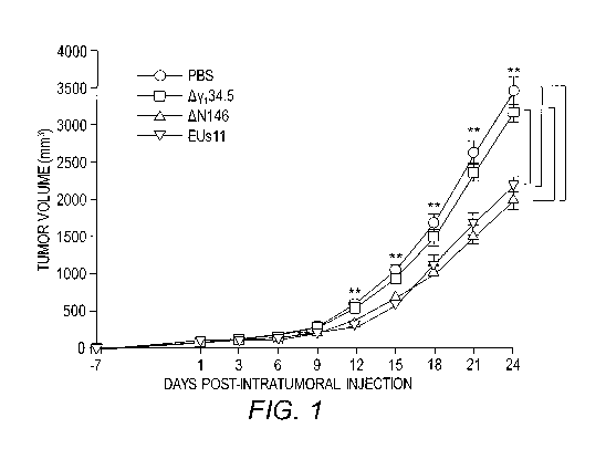
[0008] FIG. 1 provides data demonstrating that AN146
reduces local tumor growth. 4T1 cells were implanted
subcutaneously into mice (day -7). Tumors formed were
injected with PBS, Ay134.5, AN146, or EUsll suspended in PBS
-4-
CA 03142073 2021-11-25
WO 2019/236931 PCT/US2019/035922
on days 1, 3, and 6. Tumor sizes were measured periodically
(x axis) until day 24 (n = 6 each group). Average tumor
volumes over time are shown on the y axis. Asterisks
indicate statistical significance by nonparametric
analysis. The results shown are from one of three
independent experiments. Differences between the selected
groups were statistically assessed by a two-tailed Student
t test (**, P<0.01).
[0009] FIG. 2 provides data demonstrating that LN146
reduces metastasis. 4T1 cells were implanted subcutaneously
into mice (day -7). Tumors formed were injected with PBS,
ny134.5, 61\1146, or EUsll suspended in PBS on days 1, 3, and
6. Mice were sacrificed on day 24 after the initiation of
treatment and the lungs were collected and fixed in
formalin. The number of lung metastases was quantified by
counting under a light microscope. The results shown are
from one of three independent experiments. Differences
between the selected groups were statistically assessed by
a two-tailed Student t test (*, P<0.05; **, P<0.01).
[0010] FIG. 3 provides data demonstrating viral growth in
4T1 tumors. Tumors treated with PBS, Ay134.5, AN146, or
EUsll suspended in PBS were collected on day 9, and
infectious viruses present in tumors were quantified by
plaque assay (n=6). The results shown are from three
experiments with triplicate samples. Differences between
the selected groups were statistically assessed by a two-
tailed Student's t-test (**P<0.01).
[0011] FIG. 4 shows comparative analysis of N146 and EUsll
in vitro. Viral effects on the expression of IFN-al and
Cxcl9 were analyzed. 4T1 cells were mock-infected or
infected with AN146 or EUsll (5 pfu/cell). At 6 hours post-
infection, RNA samples were analyzed by quantitative
polymerase chain reaction. Data are representative of three
-5-
CA 01142073 2021-11-25
WO 2019/236931 PCT/US2019/035922
experiments among triplicate samples with standard
deviations.
Detailed Description of the Invention
[0012] The optimal intracellular environment for virus
replication develops through events that begin to take
place with attachment of virus to the cell membrane.
Binding of the herpes simplex virus to the cell membrane
receptor(s) is followed by a cascade of events that are
associated with biochemical, physiological, and
morphological changes in the cells. Following infection in
susceptible cells, lytic replication is regulated by a
temporally coordinated sequence of gene transcription.
Binding of the virus to a host cell membrane activates the
immediate-early (IE or a) genes (ICP0, ICP4, ICP22, ICP27,
and ICP47), which are transactivating factors allowing the
production of the next group of genes to be transcribed,
the early (p) genes. Expression of immediate-early gene
products is followed by the expression of proteins encoded
by the early and then, the late (y) genes. The entire
cascade of gene activation and viral replication in the
wild-type virus takes about 18-24 hours and invariably
results in cell death. The recombinant HSV mutant of the
present invention circumvents the protein synthesis shutoff
phenotype of y134.5 null viruses and activates STING
(interferon-stimulated genes) that mediate antitumor
immunity, creating a more robust HSV variant with targeted
y134.5 deletion.
[0013] It has now been discovered that recombinant HSV-1,
which expresses the C-terminal half of y134.5 (AN146),
robustly replicates in and lyses malignant cells that are
refractory to the y134.5 null mutant (Ay134.5). In infected
cells, AN146 but not Ay134.5 precludes phosphorylation of
-6-
CA 03142073 2021-11-25
WO 2019/236931 PCT/US2019/035922
translation initiation factor eIF2a, ensuing viral protein
synthesis. Remarkably, AN146 also activates interferon
regulatory factor 3 and the IFN response because it removes
the y134.5 inhibitory domain of STING, an immune factor
known to prime immunity against tumor. However, unlike
Ay134.5, AN146 replicates competently when exposed to IFN-
u/p. This is attributable to the activity associated with
the C-terminal half of y134.5. Although EUsll replicates
competently, it inactivates interferon regulatory IRF3.
Thus, its replication comes at cost of immune inhibition.
In a murine 4T1 tumor model, AN146 reduces tumor growth and
metastasis more effectively than Ay134.5. While comparable
in tumor growth reduction, AN146 reduces metastasis more
effectively than EUs11. This coincides with viral
replication, IFN induction and T cell infiltration in local
tumors. AN146 is undetectable in normal tissues and
avirulent in vivo. Thus, selective editing of HSV-1 alters
virus-cell interactions, which results in a unique anti-
neoplastic platform, namely, tumor
selectivity,
immunostimulation and resistance to clearance by IFN.
Accordingly, this invention is a recombinant HSV-1 virus
that expresses only the C-terminal half of y134.5 protein
with no wild-type or intact y134.5 protein expression and
its use in the treatment of cancer.
[0014] As is known in the art, yi34.5 is an HSV protein
that promotes viral replication in the peripheral tissues
and penetration to the peripheral nervous systems in
experimental models (Whitley, et al. (1993) J. Clin.
Invest. 91:2837-43; Perng, et al. (1996) J. Virol. 70:2883-
93; Mao & Rosenthal (2003) J. Virol. 77:3409-3417). In
addition, it facilitates HSV infection and replication in
the central nervous system (Chou, et al. (1990) Science
250:1262-66; MacLean, et al. (1991) J. Gen. Virol. 72:631-
-7-
CA 03142073 2021-11-25
WO 2019/236931 PCT/US2019/035922
39). HSV yi34.5 is known to include a large amino-terminal
domain (aa 1-146) and carboxyl-terminal domain (aa 147-
263), which binds protein phosphatase la (He, et al. (1998)
J. Biol. Chem. 273:20737-43). The nucleotide and amino acid
sequence for wild-type y134.5 are available under GENBANK
Accession No. NC 001806.1, which provides the complete
genome of HSV-1 strain 17; GENBANK Accession No.
GU734771.1, which provides the complete genome of HSV-1
strain F; and GENBANK Accession No. GU734772.1, which
provides the complete genome of HSV-1 strain H129. By way
of illustration, a wild-type or intact y134.5 has the amino
acid sequence:
MARRRRHRGPRRPRPPGPTGAVPTAQSQVTSTPNSEPAVRSAPAAAPPPPPASGPPPSC
SLLLRQWLHVPESASDDDDDDDWPDSPPPEPAPEARPTAAAPRPRSPPPGAGPGGGANP
SHPPSRPFRLPPRLALRLRVTAEHLARLRLRRAGGEGAPEPPATPATPATPATPATPAR
VRFSPHVRVRHLVVWASAARLARRGSWARERADRARFRRRVAEAEAVIGPCLGPEARAR
ALARGAGPANSV (SEQ ID NO:1).
[0015] As used herein, "recombinant HSV-1" refers to an
engineered or modified human herpes simplex virus 1 that
expresses only the C-terminal portion or half of y134.5
protein with no wild-type or intact yi34.5 protein
expression. As used herein, the C-terminal portion or half
of yi34.5 protein refers to the following amino acid
residues of y134.5 protein or its variants that retain or
enhance antitumor activity:
RLRRAGGEGAPEPPATPATPATPATPATPARVRFSPHVRVRHLVVWASAARLARRGSWA
RERADRARFRRRVAEAEAVIGPCLGPEARARALARGAGPANSV (SEQ ID NO:2).
[0016] In some aspects of the recombinant HSV-1 of this
invention, the endogenous yi34.5 gene has been modified such
that both copies of the y134.5 gene only express the C-
terminal portion of the y134.5 protein. In other aspects of
the recombinant HSV-1 of this invention, both endogenous
copies of the y134.5 gene have been deleted and nucleic
-8-
CA 01142073 2021-11-25
WO 2019/236931 PCT/US2019/035922
acids encoding the C-terminal portion of the y134.5 protein
have been inserted into one or more separate locations in
the HSV-1 genome, e.g., in non-essential genes. In this
respect, the HSV-1 genome has been modified so that the
wild-type y34.5 gene is non-functional, but the recombinant
HSV-1 can still infect, replicate within, and lyse tumor
cells in a mammal.
[0017] Expression of C-terminal portion of the y134.5
protein can be driven by the y134.5 protein promoter,
another endogenous HSV-1 promoter, or heterologous or
exogenous promoter of viral or cellular origin. Exemplary
promoters of use in the invention include, without
limitation, the herpes simplex virus immediate-early
promoters a27, a4, a0, a22, and a47; the herpes simplex
virus early promoters from ICP8 (or UL29), thymidine kinase
(tk or UL23), ICP6 (UL39) or any of the DNA replication
genes; or late promoter, e.g., the Usll promoter.
[0018] In some embodiments, the recombinant HSV-1 further
includes the deletion of one or more non-essential genes of
HSV-1. A non-essential gene is to be distinguished from an
essential gene, in whose absence the virus will not
replicate. A non-essential gene may be a beneficial gene,
in which case the replacement of such beneficial gene will
result in a virus that replicates at a much slower rate
than that of the wild-type virus. Representative non-
essential genes of HSV-1 include, but are not limited to,
UL2, UL3, UL4, UL9.5, UL10, UL11, UL12, UL13, ULI4, UL20,
UL21, UL23, UL24, UL39, UL40, UL41, UL43, UL43.5, UL44,
UL45, UL46, UL47, UL50, UL51, UL53, and UL55 in the UL
region; Usl, Us1.5, Us2, Us3, Us4, Us5, Us7, Us8, Us8.5,
Us9, Us10, Usll and Us12 in the Us region; and 'CPO in the
inverted repeat region.
-9-
CA 03142073 2021-11-25
WO 2019/236931 PCT/US2019/035922
[0019] In an alternative embodiment, one or more of non-
essential genes has been replaced with one or more nucleic
acids encoding and capable of expressing a therapeutic
protein, enzyme, antibody, nucleic acid (e.g., a nucleic
acid encoding said protein, enzyme, antibody, or a
microRNA, ribozyme, and the like), or the like for cancer
therapy. A therapeutic protein refers to a functional
protein (i.e., other than that of an enzyme or antibody),
which has a therapeutic benefit in the treatment of cancer.
Examples of suitable therapeutic proteins include, but are
not limited to, rsCD40L (Eliopoulos et al. (2000) Mol.
Cell. Biol. 20:5503-5515); Fas-ligand (Sharma et al. (2000)
Pharmacol. Ther. 88:333-347); TRAIL (Golstein (1997) Curr.
Biol. 7:R750-753); TNF (Baker & Reddy (1996) Oncogene 12:1-
9; Theys, et al. (1999) App/. Environ. Microbiol. 65:4295-
4300; Lammertyn, et al. (1997) App/. Environ. Microbiol.
63:1808-1813); GM-CSF for the treatment of melanoma, breast
carcinoma, colorectal carcinoma,
glioblastoma,
neuroblastoma, and prostate carcinoma (see, e.g., Eubank,
et al. (2009) Cancer Res. 69(5):2133-40); IFNu for the
treatment of ovarian carcinoma and solid tumors (see, e.g.,
Goto, et al. (1996) Br. J. Cancer 74:546-54); IL-2 for the
treatment of neuroblastoma and ovarian carcinoma (see,
e.g., Minor, et al. (2017) Gynecol. Oncol. Rep. 22:43-44);
and G-CSF for the treatment of breast carcinoma, bladder
carcinoma, ovarian carcinoma (see, e.g., Omura, et al.
(1996) Proc. Annu. Meet in. Soc. Clin. Oncol. 15:A755).
[0020] A therapeutic enzyme refers to an enzyme, which has
a therapeutic benefit in the treatment of cancer.
Therapeutic enzymes of particular use include enzymes
capable of converting a nontoxic prodrug into a toxic drug
which is cytotoxic to a tumor. Examples of suitable
therapeutic enzyme-prodrug pairs include, but are not
-10-
CA 03142073 2021-11-25
WO 2019/236931 PCT/US2019/035922
limited to, Herpes simplex virus thymidine kinase (HSV-TK)
+ Ganciclovir (GCV)(Moolten (1986) Cancer Res. 46:5276-
5281); HSV-TK A-5021
(11S,2'R)-91[1',2'-
bis(hydroxymethyl) cycloprop-11-yl]methyll
guanine
(Hasegawa, et al. (2000) Cancer Gene Ther. 7:557-562);
Horseradish peroxidase (HRP) + Indole-3-acetic acid
(IAA)(Greco, et al. (2000) Cancer Gene Ther. 7:1414-1420);
bacterial enzyme carboxypeptidase G2 (CPG2) + 4-([2-
chloroethyl][2-mesyloxyethyl]amino)benzoyl-L-glutamic acid
(CMDA) or + 4-[N,N-bis(2-iodoethyl) amino]phenoxycarbonyl
L-glutamic acid (ZD2767P)(Spooner, et al. (2000) Cancer
Gene Ther. 7:1348-1356; Webley, et al. (2001) Br. J. Cancer
84:1671-1676); Human cytochrome P450 CYPIA2 + acetaminophen
(Thatcher, et al. (2000) Cancer Gene Ther. 7:521-525);
Rabbit cytochrome P450 4B1 (CYP4B1) + 4-ipomeanol (4-IM)
(Mohr, et al. (2000) Cancer Gene Ther. 7:1008-1014; Heuser,
et al. (2000) Cancer Gene Ther. 7:806-12); Rat cytochrome
P450 4B1 (CYP2B1) + oxaphosporines, such as ifosfamide
(IF0)(Kammertoens, et al. (2000) Cancer Gene Ther. 7:629-
636); E. coli nitroreductase (NTR) + CB1954 (Djeha, et al.
(2000) Cancer Gene Ther. 7: 721-731; Djeha, et al. (2001)
Mol. Ther. 3:233-240); E. coil cytosine deaminase (CD), E.
coil uracil phosphoribosyltransferase (UPRT) + 5-
fluorocytosine (5-BC)(Kammertoens, et al. (2000) Cancer
Gene Ther. 7:629-636; Block, et al. (2000) Cancer Gene
Ther. 7:438-445; Bentires-Alj, et al. (2000) Cancer Gene
Ther. 7:20-6); Cytochrome P450 enzymes + cyclophosphamide
(CPA)(Huang, et al. (2000) Cancer Gene Ther. 7:1034-42;
Kan, et al. (2001) Cancer Gene Ther. 8:473-82); rabbit
carboxylesterase 7-
ethy1-10-[4-(1-piperidino)-1-
piperidino] carbonyloxycamptothecin (CPT-II) (Meck, et al.
(2001) Cancer Res. 61:5083-89); Mushroom tyrosinase + bis-
(2-chloroethyl)amino-4-hydroxyphenylaminomethanone 28
-11-
CA 031073 2021-11-25 2019/236931 PCT/US2019/035922
(Jordan, et al. (2001) Bioorg. Med. Chem. 9:1549-58); E.
coil p-galactosidase 1-
chloromethy1-5-hydroxy-1,2-
dihydro-3H-benz[e]indole (CC-1065) or + 1-(1'-chloroethyl)-
5-hydroxy-1,2-dihydro-3H-benz[e]indole (Tietze, et al.
(2001) Chembiochem. 2:758-765); a mutant of
carboxypeptidase G2 (CPG2, glutamate carboxypeptidase + 4-
[bis(2-iodoethyl)amino] phenyloxycarbonyl-L-glutamic acid
or + 3-fluoro-4-[bis(2-chlorethyl)amino]benzoyl-L-glutamic
acid or + 3,5-difluoro-4-[bis(2-iodoethyl)amino]benzoyl-L-
glutamic acid (Friedlos, et al. (2002) Cancer Res. 62:1724-
1729).
[0021] A therapeutic antibody refers to an antibody that
which has a therapeutic benefit in the treatment of cancer.
Examples of suitable therapeutic antibodies include, but
are not limited to, Atezolizumab (for the treatment of
bladder cancer and breast cancer, NSCLC, and small cell
lung cancer (SCLC)); Avelumab (for the treatment of bladder
cancer and Merkel cell carcinoma (MCC)); Durvalumab (for
the treatment of bladder cancer and NSCLC); Nivolumab (for
the treatment of bladder cancer, colorectal cancer, kidney
cancer, liver cancer, NSCLC, metastatic SCLC, Hodgkin
lymphoma, and melanoma); Pembrolizumab (for the treatment
of bladder cancer, cervical cancer, colorectal cancer,
esophageal cancer, liver cancer, NSCLC, Hodgkin lymphoma,
melanoma, and MCC); Bevacizumab (for the treatment of
glioblastoma, cervical cancer, colorectal cancer, kidney
cancer, non-small cell lung cancer (NSCLC), and ovarian
cancer); Dinutuximab (for the treatment of neuroblastoma);
Pertuzumab (for the treatment of breast cancer);
Trastuzumab (for the treatment of breast cancer and
esophageal cancer); Cetuximab (for the treatment of
colorectal cancer); Panitumumab (for the treatment of
colorectal cancer); Ramucirumab (for the treatment of
-12-
CA 03142073 2021-11-25
WO 2019/236931 PCT/US2019/035922
colorectal cancer and esophageal cancer); Alemtuzumab (for
the treatment of chronic lymphocytic leukemia (CLL));
Blinatumomab (for the treatment of acute lymphoblastic
leukemia (ALL)); Obinutuzumab (for the treatment of CLL and
non-Hodgkin lymphoma); Ofatumumab (for the treatment of
CLL); Rituximab (for the treatment of CLL and non-Hodgkin
Lymphoma); Necitumumab (for the treatment of NSCLC);
Ipilimumab (for the treatment of melanoma, pancreatic
cancer, prostate carcinoma and melanoma); Daratumumab (for
the treatment of multiple myeloma); Elotuzumab (for the
treatment of multiple myeloma); Denosumab (for the
treatment of bone cancer); Olaratumumab (for the treatment
of bone cancer); Cemiplimab (for the treatment of Merkel
cell carcinoma (MCC)); MEDI0562; GSK3174998; PF-04518600;
CP-870,893; dacetuzumumab; ADC-1013; and Ramucirumab (for
the treatment of stomach or gastroesophageal cancer).
[0022] Methods of preparing a recombinant virus are known
in the art. Briefly, to construct recombinant HSV, a gene
of interest is cloned into a transfer plasmid. This plasmid
is then co-transfected with HSV-1 genomic DNA (with a
target gene replaced with HSV thymidine kinase gene) into
rabbit skin cells. The progeny of the recombinant virus are
selected and plaque-purified on 143 TK mutant cells in
medium including of mixture 199V supplement with 100 pg of
bromodeoxyuridine/ml and 2% fetal calf serum. Next, the
thymidine kinase gene is restored by co-transfection of
progeny viral DNA and a plasmid encoding the thymidine
kinase gene in HAT medium. Preparation of viral stocks and
titrations of infectivity are done With Vero cells.
[0023] As demonstrated herein, a recombinant HSV-1, which
expresses only the C-terminal half of y134.5 protein with no
wild-type or intact yi34.5 protein expression, elicits
immune activation, and robustly replicates in and lyses
-13-
CA 03142073 2021-11-25
WO 2019/236931 PCT/US2019/035922
malignant cells that are refractory to the y134.5 null
mutant (Ay134.5). Accordingly, this invention provides a
method for treating a subject with cancer by administering
to the subject, e.g., a human, a therapeutically effective
amount of a recombinant HSV-1 that expresses only a C-
terminal portion of yi34.5 protein (in particular SEQ ID
NO:2) with no wild-type or intact y134.5 protein expression
thereby treating the subject's cancer. The recombinant HSV-
1 can be administered as the sole anticancer therapy, or in
conjunction with a therapeutically effective amount of a
second anticancer agent, such as radiation and/or
chemotherapy. Moreover, the method can also include the use
of a target-specific moiety (e.g., antibody or cell marker)
suitable for targeted administration of the recombinant
HSV-1 of the present invention to the desired tissue.
[0024] As used herein, the terms "treat," "treating,"
"treatment," and the like refer to eliminating, reducing,
relieving, reversing, and/or ameliorating a disease or
condition and/or symptoms associated therewith, in this
case treating cancer. Solid and non-solid tumors that can
be treated in accordance with the method herein, include
cancers of the bladder, breast, colon, kidney, liver, lung,
ovary, pancreas, stomach, cervix, including squamous cell
carcinoma; carcinoma, including thyroid and carcinomas of
the skin; leukemia, including acute lymphocytic leukemia,
acute lymphoblastic leukemia, acute and chronic myelogenous
leukemia and promyelocytic leukemia; lymphoma including B
cell lymphoma, T cell lymphoma, and Burkitt lymphoma;
fibrosarcoma and rhabdomyosarcoma; melanoma; and
neuroblastoma, astrocytoma and glioma. In certain
embodiments, the cancer being treated in accordance with
the method herein is a solid tumor. In other embodiments,
-14-
CA 03142073 2021-11-25
WO 2019/236931 PCT/US2019/035922
the cancer is selected from breast, liver, lung, skin
(melanoma), brain, and colon cancer.
[0025] Although not precluded, treating a disease or
condition does not require that the disease, condition, or
symptoms associated therewith be completely eliminated,
including the treatment of acute or chronic signs, symptoms
and/or malfunctions. "Treat," "treating," "treatment," and
the like may include "prophylactic treatment," which refers
to reducing the probability of redeveloping a disease or
condition, or of a recurrence of a previously-controlled
disease or condition, in a subject who does not have, but
is at risk of or is susceptible to, redeveloping a disease
or condition or a recurrence of the disease or condition.
"Treatment" therefore also includes relapse prophylaxis or
phase prophylaxis. The term "treat" and synonyms
contemplate administering a therapeutically effective
amount of the recombinant HSV-1 of the invention to an
individual in need of such treatment. A treatment can be
orientated symptomatically, for example, to suppress
symptoms. Treatment can be carried out over a short period,
be oriented over a medium term, or can be a long-term
treatment, for example within the context of a maintenance
therapy.
[0026] The term "therapeutically effective amount" or
"effective dose" as used herein refers to an amount of the
active ingredient(s) that, when administered, is (are)
sufficient, to efficaciously deliver the active
ingredient(s) for the treatment of a condition or disease
of interest to an individual in need thereof. In the case
of a cancer or other proliferation disorder, the
therapeutically effective amount of the agent may reduce
(i.e., retard to some extent and preferably stop) unwanted
cellular proliferation; reduce the number of cancer cells;
-15-
CA 03142073 2021-11-25
WO 2019/236931 PCT/US2019/035922
reduce the tumor size; inhibit (i.e., retard to some extent
and preferably stop) cancer cell infiltration into
peripheral organs; inhibit (i.e., retard to some extent and
preferably stop) tumor metastasis; inhibit, to some extent,
tumor growth; and/or relieve, to some extent, one or more
of the symptoms associated with the cancer. To the extent
the administered active ingredient(s) prevents growth
and/or kills existing cancer cells, it may be cytostatic
and/or cytotoxic.
[0027] The recombinant HSV-1 of the invention can be used
as is, provided via live carrier cells, or formulated in a
pharmaceutical composition containing a pharmaceutically
acceptable excipient. Pharmaceutical compositions provided
herein can be specially formulated for intravenous
administration in solid or liquid form or for intravenous
injection. Optimal pharmaceutical compositions can be
determined by one skilled in the art depending upon, for
example, the intended route of administration, delivery
format and desired dosage. See, for example, Remington's
Pharmaceutical Sciences (19th edition, 1995).
[0028] The recombinant HSV-1 can be incorporated in a
conventional systemic dosage form, such as an injectable
formulation. The dosage form may also include the necessary
physiologically acceptable carrier material, excipient,
lubricant, buffer, surfactant, antibacterial, bulking agent
(such as mannitol), antioxidants (ascorbic acid or sodium
bisulfite) or the like.
[0029] The primary carrier or excipient in a pharmaceutical
composition may be either aqueous or nonaqueous in nature.
For example, a suitable carrier or excipient may be water
for injection, physiological saline solution or artificial
cerebrospinal fluid, possibly supplemented with other
materials common in compositions for parenteral
-16-
CA 03142073 2021-11-25
WO 2019/236931 PCT/US2019/035922
administration. Neutral-buffered saline or saline mixed
with serum albumin are further exemplary vehicles.
Pharmaceutical compositions can include Tris buffer of
about pH 7.0-8.5, or acetate buffer of about pH 4.0-5.5,
which may further include sorbitol or a suitable substitute
therefor. Pharmaceutical compositions of the invention may
be prepared for storage by mixing the selected composition
having the desired degree of purity with optional
formulation agents (Remington's Pharmaceutical Sciences,
Id.) in the form of a lyophilized cake or an aqueous
solution. Further, the recombinant HSV-1 may be formulated
as a lyophilizate using appropriate excipients such as
sucrose.
[0030] Administration routes for the recombinant HSV-1, or
pharmaceutical compositions of the invention include
injection by intravenous, intraperitoneal, intracerebral
(intra-parenchymal),
intracerebroventricular,
intramuscular, intra-ocular, intraarterial, intraportal, or
intralesional routes; by sustained release systems or by
implantation devices. Compositions may be administered by
bolus injection or continuously by infusion, or by
implantation device. Compositions also can be administered
locally via implantation of a membrane, sponge or another
appropriate material onto which the desired molecule has
been absorbed or encapsulated. Where an implantation device
is used, the device may be implanted into any suitable
tissue or organ, and delivery of the desired molecule may
be via diffusion, timed-release bolus, or continuous
administration.
[0031] The compositions of the invention can be delivered
parenterally. When parenteral administration is
contemplated, the therapeutic compositions for use in this
invention may be in the form of a pyrogen-free,
-17-
CA 03142073 2021-11-25
WO 2019/236931 PCT/US2019/035922
parenterally acceptable aqueous solution including the
desired active ingredient(s) in a pharmaceutically
acceptable vehicle. A particularly suitable vehicle for
parenteral injection is sterile distilled water in which
the active ingredient(s) is formulated as a sterile,
isotonic solution, appropriately preserved. Preparation can
involve the formulation of the desired active ingredient(s)
with an agent, such as injectable microspheres, bio-
erodible particles, polymeric compounds (such as polylactic
acid or polyglycolic acid), beads or liposomes, that may
provide controlled or sustained release of the active
ingredient(s), which may then be delivered via a, depot
injection. Formulation with hyaluronic acid has the effect
of promoting sustained duration in the circulation.
Implantable drug delivery devices may be used to introduce
the desired active ingredient(s).
[0032] This invention also includes methods for treating
cancer by administering to an individual in need thereof
the recombinant HSV-1 of the invention and one or more
second therapeutic agents useful for the treatment of
cancer. The recombinant HSV-1 and the second therapeutic
agent can be administered simultaneously or sequentially.
In addition, the recombinant HSV-1 and second therapeutic
agent can be administered from a single composition or two
separate compositions.
[0033] The second therapeutic agent is administered in an
amount to provide its desired therapeutic effect. The
effective dosage range for each second therapeutic agent is
known in the art, and the second therapeutic agent is
administered to an individual in need thereof within such
established ranges.
[0034] In some embodiments, the second therapeutic agent is
an antibody. Suitable antibodies include, but are not
-18-
CA 031073 2021-11-25 2019/236931 PCT/US2019/035922
limited to, Atezolizumab; Avelumab; Durvalumab; Nivolumab
(anti-PD1); Pembrolizumab (anti-PD1);
Bevacizumab;
Dinutuximab; Pertuzumab; Trastuzumab;
Cetuximab;
Panitumumab; Ramucirumab; Alemtuzumab; Blinatumomab;
Obinutuzumab; Ofatumumab; Rituximab;
Necitumumab;
Ipilimumab (anti-CTLA4); Daratumumab;
Elotuzumab;
Denosumab; Olaratumumab; Cemiplimab; MEDI0562 (anti-0X40),
GSK3174998 (anti-0X40), PF-04518600 (anti-0X40), CP-870,893
(anti-CD40), dacetuzumumab (anti-CD40), ADC-1013 (anti-
CD40), and Ramucirumab.
[0035] In other embodiments, the second therapeutic agent
includes is a chemotherapeutic agent, radiotherapeutic
agent, anti-angiogenic agent, apoptosis-inducing agent,
anti-tubulin drug or a tumor-targeted chemotherapeutic
agent, radiotherapeutic agent, anti-angiogenic agent,
apoptosis-inducing agent or anti-tubulin drug. Exemplary
second therapeutic agents include, but are not limited to,
anti-angiogenic agents such as angiostatin, endostatin,
vasculostatin, canstatin and maspin and anti-tubulin drugs
such as colchicine, taxol, vinblastine, vincristine,
vindescine, a combretastatin or a derivative or prodrug
thereof. Other examples of second therapeutic agents
include, but are not limited to, alkylating agents,
nitrogen mustards, cyclophosphamide,
trofosfamide,
chlorambucil, nitrosoureas, carmustine (BCNU), lomustine
(CCNU), alkylsulphonates, busulfan, treosulfan, triazenes,
plant alkaloids, vinca alkaloids (vineristine, vinblastine,
vindesine, vinorelbine), taxoids, DNA topoisomerase
inhibitors, epipodophyllins, 9-
aminocamptothecin,
camptothecin, crisnatol, mitomycins, mitomycin C, anti-
metabolites, anti-folates, DHFR inhibitors, trimetrexate,
IMP dehydrogenase inhibitors, mycophenolic acid,
tiazofurin, ribavirin, EICAR, ribonuclotide reductase
-19-
CA 03142073 2021-11-25
WO 2019/236931 PCT/US2019/035922
inhibitors, hydroxyurea, deferoxamine, pyrimidine analogs,
uracil analogs, floxuridine, doxifluridine, ratitrexed, =
cytosine analogs, cytarabine (ara C), cytosine arabinoside,
fludarabine, purine analogs, mercaptopurine, thioguanine,
DNA antimetabolites, 3-HP, 2'-deoxy-5-fluorouridine, 5-HP,
alpha-TGDR, aphidicolin glycinate, ara-C, 5-
aza-
2'deoxycytidine, beta-TGDR, cyclocytidine,
guanazole
(inosine glycodialdehyde), macebecin II, pyrazoloimidazole,
hormonal therapies, receptor antagonists, anti-estrogen,
tamoxifen, raloxifene, megestrol, LHRH agonists, goserelin,
leuprolide acetate, anti-androgens,
flutamide,
bicalutamide, retinoids/deltoids, cis-retinoic
acid,
vitamin A derivatives, all-trans retinoic acid (ATRA-IV),
vitamin D3 analogs, CB1093, ICH1060, photodynamic
therapies, vertoporfin, BPD-MA,
phthalocyanine,
photosensitizer Pc4, demethoxy-hypocrellin A (2BA-2-DMHA),
cytokines, interferon-a, interferon-13, interferon-y, tumor
necrosis factor, angiogenesis inhibitors, angiostatin
(plasminogen fragment), antiangiogenic antithrombin UI,
angiozyme, ABT-627, Bay 12-9566, benefin, BMS-275291,
cartilage-derived inhibitor (CDI), CD59 complement
fragment, CEP-7055, Col 3, combretastatin A-4, endostatin
(collagen XVIII fragment), fibronectin fragment, Gro-beta,
halofuginone, heparinases, heparin hexasaccharide fragment,
HMV833, human chorionic gonadotropin (hCG), IM-862,
interferon inducible protein, interleukin-12, kringle 5
(plasminogen fragment), marimastat, metalloproteinase
inhibitors (UMPs), 2-methoxyestradiol, MMI270 (CGS 27023A),
neovastat, NM-3, panzem, PI-88, placental ribonuclease
inhibitor, plasminogen activator inhibitor, platelet
factor-4 (PF4), prinomastat, prolactin 161, proliferin
related protein (PRP), retinoids, solimastat, squalamine,
SS3304, SU5416, SU6668, SU11248, tetrahydrocortisol-S,
-20-
CA 03142073 2021-11-25
WO 2019/236931 PCT/US2019/035922
tetrathiomolybdate, thalidomide, thrombospondin-1 (TSP-1),
TNP-470, transforming growth factor-beta, vasculostatin,
vasostatin (calreticulin fragment), ZD6126, ZD6474, famesyl
transferase inhibitors (FTI), bisphosphonates, antimitotic
agents, allocolchicine, halichondrin B, colchicine,
colchicine derivative, dolstatin 10, maytansine, rhizoxin,
thiocolchicine, trityl cysteine, isoprenylation inhibitors,
dopaminergic neurotoxins, cell cycle
inhibitors,
staurosporine, actinomycins, actinomycin D, dactinomycin,
bleomycins, bleomycin A2, bleomycin B2, peplomycin,
anthracycline, adriamycin, epirubicin,
pirarnbicin,
zorubicin, mitoxantrone, MDR inhibitors, verapamil, Ca21A
TPase inhibitors, and thapsigargin.
[0036] The following non-limiting examples are provided to
further illustrate the present invention.
Example 1: Materials and Methods
[0037] Cells and Viruses. Vero, HT-29, SW480, C32, A375,
MDA-MB-231, 4T1, HepG2 and A549 cells were obtained from
the American Type Culture Collection. Vero, SW480, C32,
A375, MDA-MB-231 and A549 cells were propagated in
Dulbecco's modified Eagle's medium (DMEM) supplemented with
10% fetal bovine serum. HT-29, 4T1 and HepG2 cells were
propagated in RPMI1640 supplemented with 10% fetal bovine
serum. HSV-1(F) is a prototype HSV-1 strain used in this
study (Ejercito, et al. (1968) J. Gen. Virol. 2:357-364).
In recombinant virus 6,y134.5, a 1-kb fragment from the
coding region of the y134.5 gene was deleted (Chou, et al.
(1990) Science 250:1262-1266). In AN146, the sequences of
yi34.5 gene encoding amino acids 1 to 146 were deleted (Ma,
et al. (2012) J. Virol. 86:2188-2196). In EUs11, the y'34.5
gene was deleted but with the Us11 gene driven by the a-47
promoter (Liu, et al. (2018) J. Virol. 92). Preparation of
-21-
CA 03142073 2021-11-25
WO 2019/236931 PCT/US2019/035922
viral stock and titration of infectivity were carried out
as described previously (Ma, et al. (2012) J. Virol.
86:2188-2196).
[0038] Viral Infections. Viral infections were carried out
at indicated multiplicities of infection (Verpooten, et al.
(2009) J. Biol. Chem. 284:1097-1105). Cells were then
harvested and processed for immunoblot, real-time PCR
analysis or viral growth analysis (Ma, et al. (2012) J.
Virol. 86:2188-96; Wu, et al. (2016) J. Virol. 90:10414-
22). The cell viability was determined by CELLTITER-GLOO
Luminescent Cell Viability Assay (Promega) according to the
manufacture protocols. For the interferon assay, Vero and
MDA-MB-231 cells were untreated or treated with human
interferon-a (Sigma), and 4T1 cells were treated with mouse
interferon-a (Sigma) for 20 hours. Cells were then infected
with viruses and viral yields were determined at 48 hours
post-infection.
[0039] Immunoblot Analysis and ELISA. Cells were harvested,
washed with phosphate-buffered saline (PBS), and lysed with
ice-cold buffer (50 mM Tris-HC1 pH 7.4 ,150 mM NaCl, 5 mM
EDTA, 1.0% Tritonni X-100, and protease inhibitor cocktail)
on ice. After centrifugation, supernatants were mixed with
disruption buffer (50 mM Tris-HC1 pH 6.8, 2% (wt/vol) SDS,
0.1% bromophenol blue, 10% glycerol, and 100 nM p-
mercaptoethanol) and boiled. Samples were then subjected to
electrophoresis on denaturing polyacrylamide gels,
transferred to nitrocellulose membranes, and reacted with
antibodies against gC (Jing, et al. (2004) J. Virol.
78:7653-66), y34.5 (Cheng, et al. (2002) J. Virol. 76:9434-
45), ICP27 (Virusys Inc.), 'CPO (Santa Cruz), eIF-2a (Cell
Signaling Technology, Inc.), phosphorylated eIF-2a (Cell
Signaling Technology, Inc.), IRF3 (Cell Signaling
Technology, Inc.), phosphorylated IRF3 (Cell Signaling
-22-
CA 03142073 2021-11-25
WO 2019/236931 PCT/US2019/035922
Technology, Inc.) and 8-actin (Sigma). The membranes were
rinsed in PBS and reacted with either donkey anti-rabbit or
anti-mouse immunoglobulin conjugated to horseradish
peroxidase and developed with an enhanced chemiluminescence
western blot detection system kit (Amersham Pharmacia
Biotechnology, Inc.). To perform
enzyme-linked
immunosorbent assays (ELISA), supernatants of cell culture
were collected to analyze IFN-a and Cxcl9 according to the
manufacturer's instructions (R&D Systems).
[0040] Transcriptome Analysis. Monolayers of 4T1 cells were
mock-infected or infected with viruses (5 pfu/cell). At 6
hours post-infection, RNA was extracted from the cells
using the RNase plus mini kit (Qiagen) and treated with
DNase I (New England BioLabs). Duplicate RNA samples were
processed using ClariomTM S Affymetrix array by Center for
Genomic Research at University of Illinois at Chicago. Raw
data generated from Clarioml" S Mouse Array was processed in
R using package Oligo. Feature intensity values from each
CEL file was converted into normalized expression value
using Robust Multi-array Average (RMA) with default
settings. All the positive and negative control probes,
along with Affymetrix report genes (RPTR) were removed
before performing the downstream analysis. PCA (Principle
Component Analysis) plots were generated to check for any
batch-effect. Differential gene expression analysis was
performed using limma package. Significantly expressed
genes were filtered for adjusted-p value of <0.05. Heat
maps were produced from the primary data (the normalized
expression value) using the R package "pheatmap" v1Ø8.
[0041] Quantitative Real-Time PCR Assay. Cells were mock-
infected or infected with viruses. At 6 hours after
infection, total RNA was harvested from cells using an
RNase plus mini kit (Qiagen) and subjected to DNase I
-23-
CA 031073 2021-11-25 2019/236931
PCT/US2019/035922
digestion (New England BioLabs). cDNA was synthesized using
a high capacity cDNA reverse transcription kit (Applied
Biosystems). Quantitative real-time PCR was performed using
an Applied Biosystems ABI Prism 7900HT instrument with ABI
SYBR@ green master mix (Applied Biosystems). Gene
expression levels were normalized to endogenous control 18S
rRNA. Relative gene expression was determined by the 2-cT
method (Schmittgen & Livak (2008) Nat. Protoc. 3:1101-8).
Primers for each gene were chosen according to the
recommendation of the qPrimerDepot database. Primer
sequences are provided in Table 1.
TABLE 1
SEQ ID
Gene Primer Sequence
NO:
Mouse Forward GCCTTGACACTCCTGGTACAAATGAG 3
IFN-al Reverse CAGCACATTGGCAGAGGAAGACAG 4
Mouse Forward CAAGGCAGGTTTCTGAGGAG 5
IFIT1 Reverse AAGCAGATTCTCCATGACCTG 6
Mouse Forward CTGCTGCTTTGCCTACCTCT 7
Cc15 Reverse CACTTCTTCTCTGGGTTGGC 8
Mouse Forward TCCTTCCTTCCTTCCTTCCTTCC 9
Cxcl9 Reverse AGGCTCTTTTTCACCCTGTCTGG 10
Human Forward GGCCTTGACCTTTGCTTTACTG 11
IFN-al Reverse CACAGAGCAGCTTGACTTGCA
12
Human Forward CCTCCTTGGGTTCGTCTACA 13
IFITI Reverse AGTGGCTGATATCTGGGTGC 14
Human Forward CCTGCTGCTTTGCCTACATT 15
Cc15 Reverse ACACACTTGGCGGTTCTTTC 16
Human Forward CCCTGTTTCTTCCACAGTGCCTA 17
Cxcl9 Reverse GAGACAATGGTCTGGTTGCCATC 18
Forward CCTGCGGCTTAATTTGACTC
19
18s rRNA
Reverse AACCAGACAAATCGCTCCAC
20
[0042] Mice Studies. Five-week-old mice BALB/c mice were
purchased from Harlan Sprague Dawley Inc. and housed under
specific-pathogen-free conditions in a biosafety level 2
containment. All experimental procedures involving animals
were approved by the institutional animal care and use
committee of University of Illinois at Chicago. At 6 weeks
-24-
CA 03142073 2021-11-25
WO 2019/236931 PCT/US2019/035922
of age, 1x10⁵ viable 4T1 cells suspended in 0.1 ml of PBS
were inoculated subcutaneously into the right flank of mice
(day -7). When the tumor reached a volume of approximately
100 mm3 eight days after, mice were randomly assigned into
three groups for intra-tumor injections of Ay134.5, AN146 or
PBS on days 1, 3 and 6. Each tumor was injected slowly with
a total of 1x10⁷ PFU of virus or PBS in a volume of 0.1 ml.
The tumor growth was monitored every other day by measuring
two perpendicular tumor diameters with a digital caliper.
Tumor volumes were calculated using the following formula:
volume = (length x width x height)/2. On day 24 after tumor
inoculation, mice were euthanized by CO2 inhalation.
[0043] Tissue Analysis. On selected days after the last
intratumor injection, six mice from each treatment group
were sacrificed to collect the tumor, lung, liver, spleen
and blood. To measure viral load, the samples were minced,
homogenized and bead-beaten, freeze-thawed three times, and
sonicated in DMEM. After centrifugation, the tumor
supernatants were used for plaque assays. The supernatants
from the lung, liver, spleen and blood were used for
quantitative real-time PCR assay. Briefly, the supernatants
were suspended in buffer containing 1% SDS, 50 mM Tris (pH
7.5), and 10 mM EDTA. After incubation with proteinase K
(50 pg/m1) at 37 C, viral DNA was extracted and quantified
by real-time PCR using HSV-1 gD-specific primers:
TACAACCTGACCATCGCTTG (SEQ ID NO:21) and
GCCCCCAGAGACTTGTTGTA (SEQ ID NO:22).
[0044] For metastatic formation assays, lungs from mice
were excised, and fixed in formalin. The number of lung
metastases was quantified by counting under a light
microscope.
[0045] Immunohistochemistry Analysis. Tissue sections were
processed and HSV-1 antigens were detected with antibody
-25-
CA 03142073 2021-11-25
WO 2019/236931 PCT/US2019/035922
against HSV-1 (Dako). CD4 (Cell Signaling Technology, Inc.)
and CD8 (Cell Signaling Technology, Inc.) antibodies were
used according to the manufacture protocol. Samples were
incubated with primary antibody prior to the addition of
biotinylated anti-rabbit immunoglobulin secondary antibody,
avidin-horseradish peroxidase, and 3,3'-diaminobenzidine
tetrahydrochloride (0.04%) in 0.05 M Tris-HC1 (pH 7.4) and
0.025% H202 as a chromogen (Ventana Medical Systems, Tucson,
AZ).
Example 2: AN146 Mutant Replicates in Tumor Cells
[0046] It has been shown that an HSV yi_34.5 mutant (AN146),
with only amino acids 147-263, is substantially impaired
for viral growth in normal cells or tissues (Ma, et al.
(2012) J. Virol. 86:2188-2196; Ma, et al (2017) Sci. Rep.
7:41461; Pan, et al (2018) J. Virol. 92:e01015-18). To
determine activity of this mutant in malignant cells, viral
replication as assessed. This analysis indicated that in
4T1 (murine breast carcinoma) cells, wild-+type HSV-1
replicated to 1x10⁷ pfu/ml whereas the y134.5 null mutant
(Ay134.5) reached only 1x10³ pfu/ml. However, AN146 grew to
1x10⁶ pfu/ml, indicative of robust replication. A similar
trend was observed in MDA-MB-231 (human breast
adenocarcinoma) cells where AN146 replicated 100-fold
better than Ay134.5. Moreover, these phenotypes were
recapitulated in a range of other tumor cells including
human HT29 (colon), SW480 (colon), HepG2 (liver), C32
(melanoma), A375 (melanoma) and A549 (lung).
[0047] Subsequently, the kinetics of viral growth were
examined. HSV-1 grew steadily in 4T1 cells wild-type as
infection progressed, with a titer increasing to 1x10⁷
pfu/ml by 72 hours post infection. AN146 replicated to 1x10⁶
pfu though at a slightly lower level and Ay134.5 barely
-26-
CA 01142073 2021-11-25
WO 2019/236931 PCT/US2019/035922
replicated, with a titer of 1x10³ pfu/ml throughout
infection. A similar trend was observed in MDA-MB-231 cells
where nN146 replicated 100-fold better than ny134.5. To
assess viral cytolytic activity, cell viability was
measured. This analysis indicated that similar to wild-type
virus, nN146 lysed almost 95% of 4T1 cells by 72 hours,
with a slightly delayed kinetics, whereas ny134.5 destroyed
approximately 40% cells. Such effects were also mirrored in
MDA-MB-231 cells. Together, these results indicate that
nN146 replicates in and lyses tumor cells more effectively
than the y34.5 null mutant.
Example 3: Expression of the C-terminal Portion of yi34.5
Inhibits eIF2a Phosphorylation
[0048] HSV infection proceeds in a temporal manner, with
sequential expression of a, p, and y genes. Onset of viral
DNA replication invokes the cessation of protein synthesis
in the absence of yi_34.5 (Chou & Roizman (1992) Proc. Natl.
Acad. Sci. USA 89:3266-70). To assess the impact of AN146,
expression of representative proteins ICP27 (a protein) and
gC (y protein) was measured as the expression of these
proteins relies on viral DNA replication. Cells were mock-
infected or infected with HSV-1, nyi_34.5 or nN146 virus and
at 12 hours post-infection, samples were subjected to
western blot analysis. This analysis indicated that wild-
type virus expressed both ICP27 and gC in infected 4T1 and
MDA-MB-231 cells. Although ny134.5 expressed ICP27, little
gC was detectable in either of the 4T1 or MDA-MB-231 cells.
Under these same conditions, nN146 expressed a comparable
level of ICP27 and gC as wild-type HSV-1, indicating its
ability to block translational arrest initiated by viral
DNA replication.
-27-
CA 03142073 2021-11-25
WO 2019/236931 PCT/US2019/035922
[0049] As phosphorylation of eIF2u is coupled to protein
synthesis, phosphorylation of eIF2u by stress kinases PKR,
PERK or GCN2 was monitored in 4T1 and MDA-MB-231 tumor
cells. This analysis indicated that expression of eIF2u was
comparable in mock- or virus-infected tumor cells.
Interestingly, phosphorylated eIF2u was present in mock-
infected cells, likely due to oncogenic stress. Although
wild-type HSV-1 eliminated eIF2u phosphorylation Ay134.5
aggravated it and AN146 completely abrogated eIF2u
phosphorylation in 4T1 and MDA-MB-231 tumor cells.
Accordingly, the region spanning the C-terminal portion of
y134.5 is sufficient to inhibit eIF2u phosphorylation in
tumor cells.
Example 4: N146 Stimulates Interferon Responses in Tumor
Cells
[0050] To assess tumor cell responses to viral infection,
transcriptome analysis in 4T1 cells was carried out. It was
observed that numerous genes in diverse cellular pathways
were expressed differentially in 4T1 cells mock infected
and infected with viruses. Of note, many genes in the
innate immune pathways were evidently up-regulated in
response to AN146. Among the 46 genes tested, most remained
unchanged or marginally expressed in cells mock infected or
infected wild-type virus. However, they were upregulated in
cells infected with Ay134.5, albeit to a different extent.
Notably, gene induction was more pronounced in cells
infected with AN146, indicating that AN146 has a propensity
to stimulate the inflammatory response.
[0051] To confirm these results, the expression of selected
cytokines and interferon-stimulated genes was determined by
real-time PCR. As expected, wild-type virus triggered
little expression of IFN-al, IFIT1, Cc15, and Cxcl9 whereas
-28-
CA 03142073 2021-11-25
WO 2019/236931 PCT/US2019/035922
Ay134.5 or AN146 sharply induced these genes. This was
corroborated by the levels of cytokine production in ELISA
assay. To dissect the molecular basis, interferon
regulatory factor (IRF3), which activates immune responses,
was analyzed. IRF3 was un-phosphorylated in 4T1 cells mock
infected or infected with wild-type HSV-1. In contrast, it
became phosphorylated in cells infected with Ayi34.5 or
AN146. This was not due to differences in viral infectivity
as indicated by the normal expression of ICP0 and ICP27.
These results were confirmed in multiple experiments and
phenotypes were seen in human MDA-MB-231 cells as well. It
was concluded that like Ay134.5, AN146 is immune-stimulatory
upon infection of malignant cells.
Example 5: 1N146 is Resistant to IFN
[0052] Type I IFN is necessary to prime immunity against a
tumor. On the other hand, it mediates antiviral responses.
To determine whether AN146 is refractory to clearance by
IFN, viral growth was examined. As proof of concept, the
viral response to IFN was first determined in Vero cells,
which are devoid of IFN-a/ p genes. Treatment with IFN-cx had
little effect on replication of HSV-1(F) but drastically
reduced replication of Ay134.5 by approximately 1000-fold.
However, IFN-c only modestly decreased replication of
AN146. Furthermore, when tested in 4T1 and MDA-MB-231
cells, a similar trend was observed. While IFN-a reduced
viral replication in general, the effect was smaller on
wild-type HSV-1 or AN146. Indeed, AN146 consistently
replicated 500- to 1000-fold higher than Ay134.5 in the
presence of exogenous IFN-a. Thus, amino-acids 147-263 from
y134.5 are sufficient to confer viral resistance to IFN.
-29-
CA 03142073 2021-11-25
WO 2019/236931 PCT/US2019/035922
Example 6: AN146 Reduces Primary Tumor Growth and
Metastasis In Vivo
[0053] In light of the results presented in Examples 2-5,
it was posited that the capacity of AN146 to replicate and
activate inflammation would enhance tumor destruction in
vivo. To demonstrate this, an aggressive 4T1 mammary
carcinoma was selected that spontaneously metastasizes, a
process analogous to human mammary tumors. For comparison,
Ay134.5 was also as it resembles HSV1716 (Rampling, et al.
(2000) Gene Ther. 7:859-866; Streby, et al. (2017) Clin.
Cancer Res. 23:3566-3574). In addition, recombinant HSV
EUsll (Liu, et al. (2018) J. Virol. 92) was included as
this virus is structurally equivalent to the oncolytic
backbone for talimogene laherparepvec (Liu, et al. (2003)
Gene Ther. 10:292-303). Tumors established subcutaneously
in the flank of mice were thrice injected with PBS, Ay134.5,
AN146 or EUsll (1x10⁷ pfu) on days 1, 3, and 6. Tumor size
was then monitored. As illustrated in FIG. 1, control
tumors treated with PBS grew at a faster rate over time.
Treatment with yi34.5 null virus marginally reduced local
tumor growth. However, intra-tumor inoculation with AN146
or EUs11 markedly slowed tumor growth and a reduction in
tumor size became more apparent as treatment progressed. On
day 24, AN146 as well as EUs11 reduced the tumor size by
nearly 45% as compared to the mock control or Ay134.5.
Hence, while comparable to EUs11, AN146 displayed superior
activity against primary tumors when compared with Ay134.5.
[0054] To assess the viral impact on metastasis, lung tumor
formation was analyzed on day 24. FIG. 2 shows that
pulmonary metastasis was readily detectable in control
mice, with an average of 25 nodules per animal as measured
by microscopic analysis. Treatment with Ay134.5 or EUsll
reduced incidence, with an average of 15 nodules per
-30-
CA 03142073 2021-11-25
WO 2019/236931 PCT/US2019/035922
animal. Notably, AN146 further reduced metastatic burden,
with an average of 10 nodules. These results indicate that
Ay134.5 virus reduces pulmonary metastasis; however, AN146
exerted a more pronounced effect.
Example 7: AN146 Replicates in Primary Tumor but Not Normal
Tissues
[0055] To assess viral replication, viral yields in primary
tumors collected on day 9 were determined. This analysis
indicated that Ay134.5 replicated at an average titer of
1x10² pfu/g tumor tissue as measured by plaque assay (FIG.
3). On the other hand, EUsll grew at an average titer of
7x10³ pfu/g tumor tissue. Similarly, AN146 grew at an
average titer of 5x10³ pfu/g tumor tissue. Apparently, like
EUs11, AN146 replicated 50-fold better than Ay134.5. In line
with this, viral antigens were detected in thin sections of
the tumor beds, where AN146 and EUsll spread more
extensively than Ay134.5. This correlated with the degree of
necrosis of the tumor tissues.
[0056] To gauge whether viruses spread to the normal
tissues, it was determined whether Ay134.5, AN146 and EUsll
were present in the lung, blood, liver and spleen by qPCR
assay. This analysis indicated that none of the viruses was
detectable in these tissues on day 9 although they were
readily found in the tumors. These results indicate that
like that of Ay134.5 or EUs11, replication of AN146 is
limited to the tumor tissues in vivo.
[0057] To verify that viral replication indeed occurs
actively in the tumors, triple therapy of 4T1 primary
tumors was performed and viral yields on day 7, 9 and 15
were measured. This analysis indicated that viruses were
detectable at about 2x10² pfu/g tumor tissue on day 7 by
plaque assay. As treatment progressed, the quantity of
-31-
CA 01142073 2021-11-25
WO 2019/236931 PCT/US2019/035922
Ay134.5 remained unchanged initially and then reduced to 1
x10 pfu/g tumor tissue by day 15. However, under these same
conditions, the level of AN146 increased to 1x10⁴ pfu/g
tumor tissue on day 9, which subsequently decreased to 1x10³
pfu/g tumor tissue by day 15. EUsll displayed a similar
growth pattern. Therefore, unlike Ay134.5, AN146 as well as
EUsll are able to replicate within tumor in vivo.
Example 8: AN146 Induces Infiltration of CD4+ and CD8+ T
cells into the Primary Tumor
[0058] Previous evidence suggests that oncolytic HSV with
deletion of y134.5 activates systemic antitumor immunity
(Thomas & Fraser (2003) Mol. Ther. 8:543-51; Toda, et al.
(1999) Hum. Gene They. 10:385-93). As intra-tumor virus
injection reduced both local tumor growth and metastasis
formation, it was determined whether there was induction of
adaptive immunity. As such, CD4+ and CD8+ T cells were
assessed by immunohistochemistry analysis. Primary tumors
collected on day 24 were thin sectioned and stained for the
presence of CD4+ and CD8+ T cells. In mock-infected tumors,
a few CD4+ or CD8+ T cells (<4%) were detectable. However,
in tumor treated with Ay134.5, CD4+ T cells rose to 12% and
CD8+ T cells to 7%. Similarly, AN146 accounted for 15% of
CD4+ and 8% CD84-71 cells. Although EUsll triggered immune
cell infiltration, the observed effect was reduced for both
CD4+ (10%) and CD8+ T cells (5%). These results indicate
that similar to Ay134.5, AN146 induces T cell infiltration
whereas EUsll appears to dampen this process.
Example 9: AN146 and EUsll Interact with Tumor Cells
Differently
[0059] To determine whether AN146 and EUsll interact with
tumor cells differently, in vitro analyses were conducted.
-32-
CA 03142073 2021-11-25
W02019/236931 PCT/US2019/035922
As shown in FIG. 4, AN146 infection stimulated
transcription of IFN-al and cxc19 genes. In contrast, EUsll
suppressed gene expression. This paralleled with the levels
of cytokine production as measured by ELISA. Consistently,
AN146 stimulated phosphorylation of IRF3 whereas EUs11
failed to do so, suggesting EUsll
mediates
immunosuppression upon virus infection.
[0060] To assess the viral capacity to destruct tumor
cells, cell viability was measured. This analysis indicated
that like EUS11, AN146 lysed almost 95% of 4T1 cells by 72
hours. Thus, both AN146 and EUsll lysed tumor cells
efficiently. Further, viral replication in 4T1 cells with
or without IFN treatment was determined. This analysis
indicated that in the absence of IFN-a both AN146 and EUs11
replicated efficiently, with a titer reaching about
lx106pfu/ml. Addition of exogenous IFN-a modestly reduced
viral replication for AN146 and EUs11, with a titer of 5x10⁴
pfu/ml, indicating that they are equally resistant to type
I TEN.
-33-