Note: Descriptions are shown in the official language in which they were submitted.
CA 03143498 2021-12-14
WO 2020/257519
PCT/US2020/038526
BACTERIA-ENGINEERED TO ELICIT ANTIGEN-SPECIFIC T-CELLS
CROSS-REFERENCE TO RELATED APPLICATIONS
[0001] This application claims the benefit of and priority to U.S.
Provisional Patent
Application No. 62/863,594, filed June 19, 2019, and U.S. Provisional Patent
Application No.
63/033,811, filed June 2, 2020, the disclosures of which are hereby
incorporated by reference in
their entirety for all purposes.
STATEMENT AS TO RIGHTS TO INVENTIONS MADE UNDER FEDERALLY
SPONSORED RESEARCH AND DEVELOPMENT
[0002] This invention was made with Government support under Grant No:
DK113598
awarded by the National Institutes of Health (NIH). The Government has certain
rights in the
invention.
SEQUENCE LISTING
[0003] The instant application contains a Sequence Listing which has been
submitted
electronically in ASCII format and is hereby incorporated by reference in its
entirety. Said
ASCII copy, created on June 18, 2020, is named FBI-001WO_SL_5T25.txt and is
7,866 bytes in
size.
BACKGROUND OF THE INVENTION
[0004] Commensal microbiota reside primarily at barrier sites, such as the
gastrointestinal
tract, respiratory tract, urogenital tract and skin, where they functionally
tune the innate and
adaptive immune systems. Immune tolerance to these microbes must be
established at each of
these sites. In the gastrointestinal tract, a simple columnar epithelium is
coated by a thick mucus
layer that facilitates spatial segregation from luminal bacteria and also
diminishes the
immunogenicity of microbial antigens by delivering tolerogenic signals to
resident dendritic
cells. Innate lymphoid cells limit commensal-specific CD4+ T cell responses
via an MHC class
II-dependent mechanism and produce interleukin-22, which further promotes
anatomical
containment of microbes. Specialized gut-resident CD103 CD11b dendritic cells
also play an
important role in maintaining intestinal homeostasis by favoring induction of
regulatory T (Treg)
cells over pro-inflammatory CD4+ subsets (see Scharschmidt T.C. etal.,
Immunity 2015,
November 17; 43(5): 1011-1021). Interestingly, in other microbial niches such
as the skin,
1
CA 03143498 2021-12-14
WO 2020/257519
PCT/US2020/038526
certain commensal microbes (e.g., Staphylococcus epidermidis) have been
demonstrated to
selectively induce a CD8+ effector T cell response via interaction with dermal
dendritic cells
(see Naik S. et al., Nature 2015, 520:104-108).
[0005] Treg cells play a major role in establishing and maintaining immune
homeostasis in
peripheral tissues, particularly at barrier sites where they stably reside. In
the intestinal lamina
propria, Treg cells not only maintain self-tolerance but also play a crucial
role in mediating
tolerance to commensal organisms. A large percentage of gut-resident Treg
cells recognize
commensal antigens, and thymically derived Treg cells support tolerance to
intestinal microbes.
In addition, certain bacterial species expand Treg cells in the lamina propria
(Id.).
[0006] Tregs are a subset of T helper (TH) cells, and are considered to be
derived from the
same lineage as naive CD4 cells. Tregs are involved in maintaining tolerance
to self-antigens, and
preventing auto-immune disease. Tregs also suppress induction and
proliferation of effector T
cells (Teff). Tregs produce inhibitory cytokines such as TGF-0, IL-35, and IL-
10. Tregs express the
transcription factor Foxp3. In humans, the majority of Treg cells are MHC
class II restricted
CD4+ cells, but there is a minority population that are FoxP3+, MHC class I
restricted, CD8+
cells. Tregs can also be divided into subsets: "natural" CD4+ CD25+ FoxP3+
Treg cells (nTregs)
that develop in the thymus, and "inducible" regulatory cells (iTregs) which
arise in the periphery.
iTregs are also CD4+CD25+FoxP3+, and develop from mature CD4+ T cells in the
periphery (i.e.
outside of the thymus). iTregs can also express both RORyt and Foxp3 (see
Sefik E., et al.,
"Individual intestinal symbionts induce a distinct population of RORgamma(+)
regulatory T
cells," Science 2015;349:993-997). Research has shown that TGF-0 and retinoic
acid produced
by dendritic cells can stimulate naive T cells to differentiate into Tregs,
and that naive T cells
within the digestive tract differentiate into Tregs after antigen stimulation.
iTregs can also be
induced in culture by adding TGF-I3.
[0007] In contrast to Tregs, T effector (Teff) cells generally stimulate a
pro-inflammatory
response upon antigen-specific T Cell receptor (TCR) activation via the
expression or release of
an array of membrane-bound and secreted proteins that are specialized to deal
with different
classes of pathogen. There are three classes of Teff cell: CD8+ cytotoxic T
cells, TH1 cells, and
TH2 cells. CD8+ cytotoxic T cells recognize and kill target cells that display
peptide fragments
of intracellular pathogens (e.g., viruses) presented in the context of MHC
class I molecules at the
cell surface. CD8+ cytotoxic T cells store preformed cytotoxins in lytic
granules which fuse
with the membranes of infected target cells. CD8+ cytotoxic T cells
additionally express Fas
ligand, which induces apoptosis in Fas-expressing target cells. TH1 and TH2
cells both express
CD4 and recognize peptide fragments degraded within intracellular vesicles and
presented on the
2
CA 03143498 2021-12-14
WO 2020/257519
PCT/US2020/038526
cell surface in the context of MHC class II molecules. TH1 cells can activate
a number of other
immune cells, including macrophages and B cells, thereby promoting more
efficient destruction
and clearance of intracellular microorganisms. TH2 cells stimulate the
differentiation of B cells
and promote the production of antibodies and other effector molecules of the
humoral immune
.. response.
SUMMARY OF THE INVENTION
[0008] In one aspect, provided herein is a live, recombinant commensal
bacterium, wherein
the bacterium is engineered to express a non-native protein or peptide,
wherein the protein or
peptide is associated with a host disease or condition, wherein upon
administration of the
bacterium to the host resulting in colonization of a native host niche by the
bacterium, the host
mounts an adaptive immune response to the non-native protein or peptide,
wherein the adaptive
immune response is a regulatory T-cell (Treg) response or an effector T-cell
(Teffector) response. In
some embodiments, the colonization of the native host niche is persistent or
transient. In certain
embodiments, the native host niche is transiently colonized, and colonization
is for 1 day to 60
days. In certain embodiments, the native host niche is transiently colonized,
and colonization is
for 3.5 days to 60 days. In certain embodiments, the native host niche is
transiently colonized,
and colonization is for 7 days to 28 days. In some embodiments, colonization
is determined by
polymerase chain reaction or colony forming assay performed on a sample
obtained from the
host after 1 day, 3.5 days, 7 days, 14 days, 28 days, or 60 days after
administration to the host. In
some embodiments, the administration results in interaction of the bacterium
with a native
immune system partner cell. In certain embodiments, the native immune system
partner cell is an
antigen-presenting cell. In certain embodiments, the antigen-presenting cell
is selected from the
group consisting of a dendritic cell, a macrophage, a B-cell, and an
intestinal epithelial cell.
[0009] In some embodiments, the native host niche is selected from the
group consisting of
the gastrointestinal tract, respiratory tract, urogenital tract, and skin. In
some embodiments, the
non-native protein or peptide is a host protein or peptide. In some
embodiments, the bacterium is
a Gram-negative bacterium. In certain embodiments, the Gram-negative bacterium
is selected
from the group consisting of Bacteroides thetaiotaomicron, Helicobacter
hepaticus and
Parabacteroides sp. In certain embodiments, the bacterium is a Gram-positive
bacterium. In
certain embodiments, the Gram-positive bacterium is selected from the group
consisting of
Staphylococcus epidermidis, Faecalibacterium sp. and Clostridium sp.
[0010] In some embodiments, the administration is via a route selected
from the group
consisting of topical, enteral, parenteral and inhalation. In certain
embodiments, the
3
CA 03143498 2021-12-14
WO 2020/257519
PCT/US2020/038526
administration route is topical. In some embodiments, the bacterium is S.
epidermidis. In certain
embodiments, the administration route is enteral. In some embodiments, the
bacterium is
selected from the group consisting of Bacteroides spp., Clostridium spp.,
Helicobacter spp.,
Parabacteroides spp, and Prevotella spp. In some embodiments, the bacterium is
selected from
.. the group consisting of Bacteroides thetaiotaomicron, Bacteroides vulgatus
and Bacteroides
finegoldii.
[0011] In some embodiments, the adaptive immune response is a Treg
response and the
bacterium is selected from the group consisting of Bacteroides spp.,
Helicobacter spp.,
Parabacteroides spp., Clostridium spp., Staphylococcus spp., Lactobacillus
spp., Fusobacterium
spp., Enterococcus spp., Acenitobacter spp., Flavinofractor spp.,
Lachnospiraceae spp.,
Erysipelotrichaceae spp., Anaerostipes spp., Anaerotruncus spp., Coprococcus
spp.,
Clostridiales spp., Odoribacter spp., Collinsella spp., Bifidobacterium spp.,
Streptococcus spp.,
and Prevotella spp. In certain embodiments, the adaptive immune response is a
Treg response and
the bacterium is selected from the group consisting of Clostridium ramosum,
Staphylococcus
saprophyticus, Bacteroides thetaiotaomicron, Clostridium histolyticum,
Lactobacillus
rhamnosus, Parabacteroides johnsonii, Fusobacterium nucleatum, Enterococcus
faecium,
Lactobacillus casei, Acenitobacter lwofii, Bacteroides ovatusõBacteroides
vulgatus,
Bacteroides uniform's, Bacteroides finegoldii, Clostridium spiroforme,
Flavonifractor plautii,
Clostridium hathewayi, Lachnospiraceae bacterium, Clostridium bolteae,
Erysipelotrichaceae
bacterium, Anaerostipes caccae, Anaerotruncus colihominis, Coprococcus comes,
Clostridium
asparagiforme, Clostridium symbiosum, Clostridium ramosum, Clostridium sp. D5,
Clostridium
scindens , Lachnospiraceae bacterium, Clostridiales bacterium, Bacteroides
intestinalis,
Bacteroides caccae, Bacteroides massiliensis, Parabacteroides distasonis,
Odoribacter
splanchnicus, Collinsella aerofaciens, Acinetobacter lwoffii, Bifidobacterium
breve, Bacteroides
finegoldii, Bacteroides fragilis, Bacteroides massiliensis, Bacteroides
ovatus, Bifidobacterium
bifidum, Lactobacillus acidofilus, Lactobacillus casei, Lactobacillus reuteri,
Streptococcus
thermophilus, and Prevotella histicola. In certain embodiments, the bacterium
is selected from
the group consisting of Bacteroides thetaiotaomicron, Bacteroides vulgatus,
Bacteroides
finegoldii and Helicobacter hepaticus.
[0012] In some embodiments, the disease or condition is an autoimmune
disorder. In
certain embodiments, the autoimmune disorder is selected from the group
consisting of multiple
sclerosis, diabetes mellitus Type I, rheumatoid arthritis, systemic lupus
erythematosus,
inflammatory bowel disease, celiac disease, Graves' disease, Hashimoto's
autoimmune
thyroiditis, vitiligo, rheumatic fever, pernicious anemia/atrophic gastritis,
alopecia areata,
4
CA 03143498 2021-12-14
WO 2020/257519
PCT/US2020/038526
immune thrombocytopenic purpura, temporal arteritis, ulcerative colitis,
Crohn's disease,
scleroderma, antiphospholipid syndrome, autoimmune hepatitis type 1, primary
biliary cirrhosis,
Sjogren's syndrome, Addison's disease, dermatitis herpetiformis, Kawasaki
disease, sympathetic
ophthalmia, HLA-B27 associated acute anterior uveitis, primary sclerosing
cholangitis, discoid
.. lupus erythematosus, polyarteritis nodosa, CREST Syndrome, myasthenia
gravis,
polymyositis/dermatomyositis, Still's disease, autoimmune hepatitis type 2,
Wegener's
granulomatosis, mixed Connective tissue disease, microscopic polyangiitis,
autoimmune
polyglandular syndrome, Felty's syndrome, autoimmune hemolytic anemia, chronic
inflammatory demyelinating polyneuropathy, Guillain-Barre Syndrome, Behcet
disease,
autoimmune neutropenia, bullous pemphigoid, essential mixed cryoglobulinemia,
linear
morphea, autoimmune polyglandular syndrome 1 (APECED), acquired hemophilia A,
Batten
disease/neuronal ceroid lipofuscinoses, autoimmune pancreatitis, Hashimoto '5
encephalopathy,
Goodpasture's disease, pemphigus vulgaris, autoimmune disseminated
encephalomyelitis,
relapsing polychondritis, Takayasu arteritis, Churg-Strauss syndrome,
epidermolysis bullosa
acquisita, cicatricial pemphigoid, pemphigus foliaceus, autoimmune
hypoparathyroidism,
autoimmune hypophysitis, autoimmune inner ear disease, autoimmune
lymphoproliferative
syndrome, autoimmune oophoritis, autoimmune orchitis, autoimmune polyglandular
syndrome,
Cogan's syndrome, encephalitis lethartica, erythema elevatum diutinum, Evans
syndrome,
immunodysregulation polyendocrinopathy enteropathy X-linked (IPEX), Is sac's
syndrome/acquired neuromyotonia, Miller Fisher syndrome, Morvan's syndrome,
PANDAS,
POEMS syndrome, Rasmussen's encephalitis, stiff-person syndrome, Vogt-Koyanagi-
Harada
syndrome, neuromyelitis optica, graft vs host disease, and autoimmune uveitis.
In certain
embodiments, the autoimmune disorder is selected from the group consisting of
multiple
sclerosis and diabetes mellitus Type I.
[0013] In some embodiments, the non-native protein or peptide is selected
from the group
consisting of ovalbumin, myelin oligodendrocyte glycoprotein, insulin,
chromogranin A, hybrid
insulin peptides, proteolipid protein, myelin basic protein, villin,
epithelial cellular adhesion
molecule, collagen alpha-1, aggrecan core protein, 60kDa chaperonin 2,
vimentin, alpha-enolase,
fibrinogen alpha chain, fibrinogen beta chain, chitinase-3-like protein, 60kDa
mitochondrial heat
shock protein, matrix metalloproteinase-16, thyroid peroxidase, thyrotropin
receptor,
thyroglobulin, gluten, TSHR protein, glutamate decarboxylase 2, receptor-type
tyrosine-protein
phosphatase-like N, glucose-6-phosphatase 2, insulin isoform 2, zinc
transporter 8, glutamate
decarboxylase 1, GAD65, UniProt:A2RGMO, integrin alpha-lib, integrin beta-3,
EBV DNA
polymerase catalytic subunit, 2'3'-cyclic-nucleotide 3' phosphodiesterase,
myelin associated
5
CA 03143498 2021-12-14
WO 2020/257519
PCT/US2020/038526
oligodendrocyte basic protein, small nuclear ribonucleoprotein, Ul small
nuclear
ribonucleoprotein, histone H2B, histone H2A, histone H3.2, beta-2-
glycoprotein, histone H4,
60S ribosomal protein L7, TNF-alpha, myeloperoxidase, Cbirl, MS4Al2, DNA
topoisomerase,
CYP2D6, 0-phosphoseryl-tRNA selenium transferase, pyruvate dehydrogenase
complex,
.. spectrin alpha chain, steroid 21-hydroxylase, acetylcholine receptor, MMP-
16, keratin associated
proteins. Chondroitin sulfate proteoglycan 4, myeloblastin, Ul small nuclear
ribonucleoprotein
70 kDa, blood group Rh(D), blood group Rh(CE), myelin P2 protein, peripheral
myelin protein
22, myelin protein PO, S-arrestin, collagen Alpha-1, coagulation factor VIII,
collagen alpha-
3(IV), desmoglein-3, desmoglein-1, Insulin-2, major DNA-binding protein,
tyrosinase, 5,6-
dihydroxyindole-2-carboxylic acid oxidase, HLA-A2, aquaporin-4, myelin
proteolipid protein,
ABC transporter, HLA I B-27 alpha chain, HLA I B-7 alpha chain, and retinol-
binding protein 3.
In certain embodiments, the non-native protein or peptide is selected from the
group consisting
of ovalbumin, myelin oligodendrocyte glycoprotein, insulin, chromogranin A,
hybrid insulin
peptides, proteolipid protein, myelin basic protein, villin, epithelial
cellular adhesion molecule,
In some embodiments, the bacterium is engineered to secrete the expressed
protein or peptide. In
some embodiments, the bacterium is engineered to express a fusion protein
comprising the
protein or peptide and a native bacterial protein or portion thereof In some
embodiments, the
protein or peptide is fused to the N-terminus or the C-terminus of the native
bacterial protein or
portion thereof In some embodiments, the native bacterial protein is selected
from the group
consisting of sialidase, endonuclease, secreted endoglycosidase, anti-sigma
factor, thiol
peroxidase, hypothetical protein BT 2621, hypothetical protein BT_3223,
peptidase, Icc family
phosphohydrolase, exo-poly-alpha-D-galacturonosidase, and hypothetical protein
BT_4428. In
certain embodiments, the native bacterial protein is sialidase or anti-sigma
factor.
[0014] In
some embodiments, the adaptive immune response is a Teffector response and the
bacterium is selected from the group consisting of S. epidermidis,
Corynebacterium spp.,
Parabacteroides distasonis, Parabacteroides gordonii, Alistipes senegalensis,
Parabacteroides
johnsonii, Paraprevotella xylamphila, Bacteroides dorei, Bacteroides uniform's
JC115828,
Eubacterium limosum, Ruminococcaceae bacterium cv2, Phascolarcto bacterium
faecium,
Fusobacterium ulcerans, Klebsiella pneumoniae , Clostridium bolteae 90B3,
Clostridium cf
saccharolyticum K10, Clostridium symbiosum WAL-14673, Clostridium hathewayi
12489931,
Ruminococcus obeum A2-162, Ruminococcus gnavus AGR2154, Butyrate-producing
bacterium
SSC/2, Clostridium sp. A5F356, Coprobacillus sp. D6 cont1.1 , Eubacterium sp.
3131 cont1.1,
Erysipelotrichaceae bacterium 213, Subdoligranulum sp. 4 3 54A2FAA,
Ruminococcus
bromii L2-63, Firmicutes bacterium ASF500, Firmicutes bacterium ASF500,
Bacteroides dorei
6
CA 03143498 2021-12-14
WO 2020/257519
PCT/US2020/038526
1 36/D4 supercont2.3, Bifidobacterium animalis subsp. Lactis ATCC 27673, and
Bifidobacterium breve UCC2003. In certain embodiments, the bacterium is
selected from the
group consisting of S. epidermidis LM087 and Corynebacterium spp.
[0015] In some embodiments, the disease or condition is a proliferative
disorder. In some
5 embodiments, the proliferative disorder is cancer. In certain
embodiments, the cancer is selected
from melanoma, basal cell carcinoma, squamous cell carcinoma, and testicular
cancer. In certain
embodiments, the cancer is melanoma.
[0016] In some embodiments, the non-native protein or peptide is
selected from the group
consisting of PMEL, TRP2, MART-1, NY-ESO, MAGE-A, and a neoantigen. In certain
embodiments, the non-native protein or peptide is PMEL.
[0017] In some embodiments, the bacterium is engineered to secrete the
expressed protein
or peptide. In some embodiments, the bacterium is engineered to express a
fusion protein
comprising the protein or peptide and a native bacterial protein or portion
thereof In certain
embodiments, the protein or peptide is fused to the N-terminus or the C-
terminus of the native
bacterial protein or portion thereof In some embodiments, the native bacterial
protein is selected
from the group consisting of sialidase, endonuclease, secreted
endoglycosidase, anti-sigma
factor, thiol peroxidase, hypothetical protein BT 2621, hypothetical protein
BT_3223, peptidase,
Icc family phosphohydrolase, exo-poly-alpha-D-galacturonosidase, and
hypothetical protein
BT 4428. In certain embodiments, the native bacterial protein is sialidase or
anti-sigma factor.
[0018] In some embodiments, the bacterium is administered in combination
with a high-
complexity defined microbial community.
[0019] In some embodiments, the host is a mammal. In certain
embodiments, the mammal
is a human.
[0020] In another aspect, provided herein is a polynucleotide used to
engineer the
recombinant commensal bacterium disclosed herein.
[0021] In another aspect, provided herein is a method for generating an
antigen-presenting
cell displaying an antigen derived from a non-native protein or peptide,
comprising:
administering the recombinant commensal bacterium disclosed herein to a
subject, wherein the
administration results in colonization of the native host niche by the
bacterium, internalization of
the bacterium by an antigen-presenting cell, and presentation of the antigen
by the antigen-
presenting cell. In certain embodiments, the colonization of the native host
niche is persistent or
transient. In certain embodiments, the native host niche is transiently
colonized, and colonization
is for 1 day to 60 days. In certain embodiments, the native host niche is
transiently colonized,
and colonization is for 3.5 days to 60 days. In certain embodiments, the
native host niche is
7
CA 03143498 2021-12-14
WO 2020/257519
PCT/US2020/038526
transiently colonized, and colonization is for 7 days to 28 days. In certain
embodiments,
colonization is determined by polymerase chain reaction or colony forming
assay performed on a
sample obtained from the host after 1 day, 3.5 days, 7 days, 14 days, 28 days,
or 60 days after
administration to the host.
[0022] In some embodiments, the administration results in interaction of
the bacterium
with a native immune system partner cell. In some embodiments, the native
immune system
partner cell is the antigen-presenting cell. In certain embodiments, the
antigen-presenting cell is
selected from the group consisting of a dendritic cell, a macrophage, a B-
Cell, and an intestinal
epithelial cell. In certain embodiments, the native host niche is selected
from the group
consisting of the gastrointestinal tract, respiratory tract, urogenital tract,
and skin. In some
embodiments, the presentation is within an MHC II complex. In some
embodiments, the
presentation is within an MHC I complex. In some embodiments, the bacterium is
a Gram-
negative bacterium. In certain embodiments, the Gram-negative bacterium is
selected from the
group consisting of Bacteroides thetaiotaomicron, Helicobacter hepaticus,
Parabacteroides sp.,
and Prevotella spp. In some embodiments, the bacterium is a Gram-positive
bacterium. In
certain embodiments, the Gram-positive bacterium is selected from the group
consisting of
Staphylococcus epidermidis, Faecalibacterium sp. and Clostridium sp.
[0023] In some embodiments, the administration is via a route selected
from the group
consisting of topical, enteral, parenteral and inhalation. In some
embodiments, the route is
.. topical. In certain embodiments, the bacterium is S. epidermidis. In some
embodiments, the route
is enteral. In certain embodiments, the bacterium is selected from the group
consisting of
Bacteroides spp., Clostridium spp., Helicobacter spp., Parabacteroides spp,
and Prevotella spp.
In certain embodiments, the bacterium is selected from the group consisting of
Bacteroides
thetaiotaomicron, Bacteroides vulgatus and Bacteroides finegoldii. In some
embodiments, the
native bacterial protein is selected from the group consisting of sialidase,
endonuclease, secreted
endoglycosidase, anti-sigma factor, thiol peroxidase, hypothetical protein
BT_2621, hypothetical
protein BT_3223, peptidase, Icc family phosphohydrolase, exo-poly-alpha-D-
galacturonosidase,
and hypothetical protein BT_4428. In certain embodiments, the native bacterial
protein is
sialidase or anti-sigma factor. In certain embodiments, the non-native protein
or peptide is
melanocyte oligodendrocyte glycoprotein. In certain embodiments, the disease
or condition is
multiple sclerosis.
[0024] In another aspect, provided herein is a method for generating a T-
cell response in a
subject, comprising: administering the recombinant commensal bacterium
disclosed herein to the
subject, wherein the administration results in colonization of a native host
niche by the bacterium
8
CA 03143498 2021-12-14
WO 2020/257519
PCT/US2020/038526
and generation of the T-cell response, wherein the T-cell response is to an
antigen derived from
the non-native protein or peptide. In some embodiments, the colonization of
the native host
niche is persistent or transient. In certain embodiments, the native host
niche is transiently
colonized, and colonization is for 1 day to 60 days. In certain embodiments,
the native host niche
is transiently colonized, and colonization is for 3.5 days to 60 days. In
certain embodiments, the
native host niche is transiently colonized, and colonization is for 7 days to
28 days.
[0025] In some embodiments, colonization is determined by polymerase
chain reaction or
colony forming assay performed on a sample obtained from the host after 1 day,
3.5 days, 7
days, 14 days, 28 days, or 60 days after administration to the host. In some
embodiments, the
administration is via a route selected from the group consisting of topical,
enteral, parenteral and
inhalation. In certain embodiments, the route is topical. In certain
embodiments, the route is
enteral. In some embodiments, the bacterium is selected from the group
consisting of
Bacteroides spp., Clostridium spp., Helicobacter spp., Parabacteroides spp,
Prevotella spp., and
Staphylococcus epidermidis. In some embodiments, the T-cell response is a Treg
or a Teffector
response. In certain embodiments, the route is enteral and the T-cell response
is a Treg response.
In some embodiments, the bacterium is selected from the group consisting of
Bacteroides spp.,
Clostridium spp., Helicobacter spp., Parabacteroides spp, and Prevotella spp.
In certain
embodiments, the bacterium is selected from the group consisting of
Bacteroides
thetaiotaomicron, Bacteroides vulgatus, and Bacteroides finegoldii.
[0026] In some embodiments, the native bacterial protein is selected from
the group
consisting of sialidase, endonuclease, secreted endoglycosidase, anti-sigma
factor, thiol
peroxidase, hypothetical protein BT 2621, hypothetical protein BT_3223,
peptidase, Icc family
phosphohydrolase, exo-poly-alpha-D-galacturonosidase, and hypothetical protein
BT_4428. In
certain embodiments, the native bacterial protein is sialidase or anti-sigma
factor. In certain
embodiments, the non-native protein or peptide is myelin oligodendrocyte
glycoprotein. In
certain embodiments, the disease or condition is multiple sclerosis. In
certain embodiments, the
route is topical and the T-cell response is a Teffector response. In certain
embodiments, the
bacterium is S. epidermidis .
[0027] In another aspect, provided herein is a method of treating a
disease or condition in a
subject, comprising: administering the recombinant commensal bacterium
disclosed herein to the
subject, wherein the administration results in colonization of a native host
niche by the bacterium
and generation of a T-cell response, wherein the T-cell response is to an
antigen derived from the
non-native protein or peptide, and wherein the T-cell response treats the
disease or condition in
the subject.
9
CA 03143498 2021-12-14
WO 2020/257519
PCT/US2020/038526
[0028] In some embodiments, the colonization of the native host niche is
persistent or
transient. In certain embodiments, the native host niche is transiently
colonized, and colonization
is for 1 day to 60 days. In certain embodiments, the native host niche is
transiently colonized,
and colonization is for 3.5 days to 60 days. In certain embodiments, the
native host niche is
transiently colonized, and colonization is for 7 days to 28 days. In certain
embodiments,
colonization is determined by polymerase chain reaction or colony forming
assay performed on a
sample obtained from the host after 1 day, 3.5 days, 7 days, 14 days, 28 days,
or 60 days after
administration to the host.
[0029] In some embodiments, the disease or condition is selected from
the group consisting
of an autoimmune disorder and a proliferative disorder. In some embodiments,
the autoimmune
disorder is selected from the group consisting of multiple sclerosis, diabetes
mellitus Type I,
rheumatoid arthritis, systemic lupus erythematosus, inflammatory bowel
disease, celiac disease,
Graves' disease, Hashimoto's autoimmune thyroiditis, vitiligo, rheumatic
fever, pernicious
anemia/atrophic gastritis, alopecia areata, immune thrombocytopenic purpura,
temporal arteritis,
ulcerative colitis, Crohn's disease, scleroderma, antiphospholipid syndrome,
autoimmune
hepatitis type 1, primary biliary cirrhosis, Sjogren's syndrome, Addison's
disease, dermatitis
herpetiformis, Kawasaki disease, sympathetic ophthalmia, HLA-B27 associated
acute anterior
uveitis, primary sclerosing cholangitis, discoid lupus erythematosus,
polyarteritis nodosa,
CREST Syndrome, myasthenia gravis, polymyositis/dermatomyositis, Still's
disease,
autoimmune hepatitis type 2, Wegener's granulomatosis, mixed Connective tissue
disease,
microscopic polyangiitis, autoimmune polyglandular syndrome, Felty's syndrome,
autoimmune
hemolytic anemia, chronic inflammatory demyelinating polyneuropathy, Guillain-
Barre
Syndrome, Behcet disease, autoimmune neutropenia, bullous pemphigoid,
essential mixed
cryoglobulinemia, linear morphea, autoimmune polyglandular syndrome 1
(APECED), acquired
hemophilia A, Batten disease/neuronal ceroid lipofuscinoses, autoimmune
pancreatitis,
Hashimoto's encephalopathy, Goodpasture's disease, pemphigus vulgaris,
autoimmune
disseminated encephalomyelitis, relapsing polychondritis, Takayasu arteritis,
Churg-Strauss
syndrome, epidermolysis bullosa acquisita, cicatricial pemphigoid, pemphigus
foliaceus,
autoimmune hypoparathyroidism, autoimmune hypophysitis, autoimmune inner ear
disease,
autoimmune lymphoproliferative syndrome, autoimmune oophoritis, autoimmune
orchitis,
autoimmune polyglandular syndrome, Cogan's syndrome, encephalitis lethartica,
erythema
elevatum diutinum, Evans syndrome, immunodysregulation polyendocrinopathy
enteropathy X-
linked (IPEX), Issac's syndrome/acquired neuromyotonia, Miller Fisher
syndrome, Morvan's
syndrome, PANDAS, POEMS syndrome, Rasmussen's encephalitis, stiff-person
syndrome,
CA 03143498 2021-12-14
WO 2020/257519
PCT/US2020/038526
Vogt-Koyanagi-Harada syndrome, neuromyelitis optica, graft vs host disease,
and autoimmune
uveitis. In certain embodiments, the autoimmune disorder is selected from the
group consisting
of multiple sclerosis, and diabetes mellitus Type I. In some embodiments, the
proliferative
disorder is cancer. In certain embodiments, the cancer is selected from
melanoma, basal cell
carcinoma, squamous cell carcinoma, and testicular cancer. In certain
embodiments, the cancer
is melanoma.
[0030] In some embodiments, the administration is via a route selected
from the group
consisting of topical, enteral, parenteral and inhalation. In certain
embodiments, the route is
topical. In certain embodiments, the bacterium is S. epidermidis. In some
embodiments, the
disease is cancer. In some embodiments, the cancer is melanoma. In some
embodiments, the
non-native protein or peptide is selected from the group consisting of a
melanocyte-specific
antigen and a testis cancer antigen. In some embodiments, the melanocyte-
specific antigen is
selected from the group consisting of PMEL, TRP2 and MART-1. In some
embodiments, the
testis cancer antigen is selected from the group consisting of NY-ESO and MAGE-
A.
[0031] In some embodiments, the route is enteral. In some embodiments, the
bacterium is
selected from the group consisting of Bacteroides spp., Clostridium spp.,
Helicobacter spp.,
Parabacteroides spp, and Prevotella spp. In certain embodiments, the bacterium
is selected from
the group consisting of Bacteroides thetaiotaomicron, Bacteroides vulgatus,
and Bacteroides
finegoldii. In some embodiments, the native bacterial protein is selected from
the group
consisting of sialidase, endonuclease, secreted endoglycosidase, anti-sigma
factor, thiol
peroxidase, hypothetical protein BT 2621, hypothetical protein BT_3223,
peptidase, Icc family
phosphohydrolase, exo-poly-alpha-D-galacturonosidase, and hypothetical protein
BT_4428. In
certain embodiments, the native bacterial protein is sialidase or anti-sigma
factor.
[0032] In some embodiments, the autoimmune disorder is selected from the
group
consisting of multiple sclerosis, diabetes mellitus Type I, rheumatoid
arthritis, systemic lupus
erythematosus, inflammatory bowel disease, celiac disease, Graves' disease,
Hashimoto's
autoimmune thyroiditis, vitiligo, rheumatic fever, pernicious anemia/atrophic
gastritis, alopecia
areata, immune thrombocytopenic purpura, temporal arteritis, ulcerative
colitis, Crohn's disease,
scleroderma, antiphospholipid syndrome, autoimmune hepatitis type 1, primary
biliary cirrhosis,
Sjogren's syndrome, Addison's disease, dermatitis herpetiformis, Kawasaki
disease, sympathetic
ophthalmia, HLA-B27 associated acute anterior uveitis, primary sclerosing
cholangitis, discoid
lupus erythematosus, polyarteritis nodosa, CREST Syndrome, myasthenia gravis,
polymyositis/dermatomyositis, Still's disease, autoimmune hepatitis type 2,
Wegener's
granulomatosis, mixed Connective tissue disease, microscopic polyangiitis,
autoimmune
11
CA 03143498 2021-12-14
WO 2020/257519
PCT/US2020/038526
polyglandular syndrome, Felty's syndrome, autoimmune hemolytic anemia, chronic
inflammatory demyelinating polyneuropathy, Guillain-Barre Syndrome, Behcet
disease,
autoimmune neutropenia, bullous pemphigoid, essential mixed cryoglobulinemia,
linear
morphea, autoimmune polyglandular syndrome 1 (APECED), acquired hemophilia A,
Batten
disease/neuronal ceroid lipofuscinoses, autoimmune pancreatitis, Hashimoto's
encephalopathy,
Goodpasture's disease, pemphigus vulgaris, autoimmune disseminated
encephalomyelitis,
relapsing polychondritis, Takayasu arteritis, Churg-Strauss syndrome,
epidermolysis bullosa
acquisita, cicatricial pemphigoid, pemphigus foliaceus, autoimmune
hypoparathyroidism,
autoimmune hypophysitis, autoimmune inner ear disease, autoimmune
lymphoproliferative
syndrome, autoimmune oophoritis, autoimmune orchitis, autoimmune polyglandular
syndrome,
Cogan's syndrome, encephalitis lethartica, erythema elevatum diutinum, Evans
syndrome,
immunodysregulation polyendocrinopathy enteropathy X-linked (IPEX), Issac's
syndrome/acquired neuromyotonia, Miller Fisher syndrome, Morvan's syndrome,
PANDAS,
POEMS syndrome, Rasmussen's encephalitis, stiff-person syndrome, Vogt-Koyanagi-
Harada
syndrome, neuromyelitis optica, graft vs host disease, and autoimmune uveitis.
In certain
embodiments, the autoimmune disorder is multiple sclerosis. In some
embodiments, the
bacterium is selected from the group consisting of Bacteroides
thetaiotaomicron, Bacteroides
vulgatus, and Bacteroides finegoldii. In certain embodiments, the non-native
protein is myelin
oligodendrocyte glycoprotein.
[0033] In some embodiments, the bacterium is administered in combination
with a high-
complexity defined microbial community.
[0034] In some embodiments, the host is a mammal. In certain
embodiments, the mammal
is a human.
BRIEF DESCRIPTION OF THE DRAWINGS
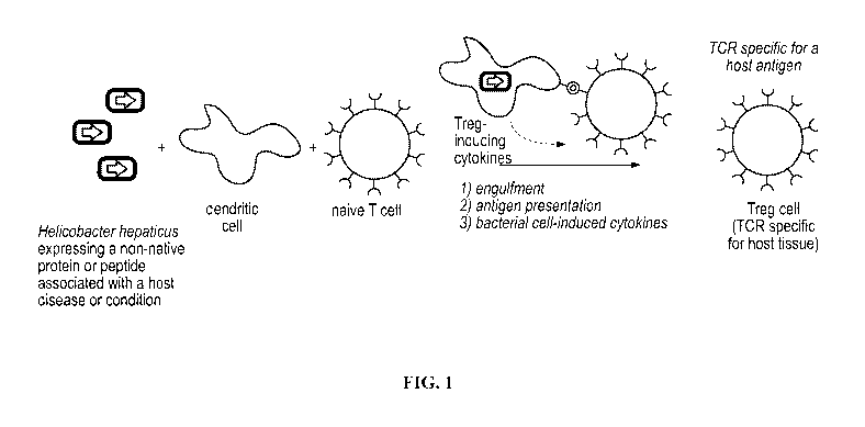
[0035] FIG. 1 illustrates an exemplary method for generating a regulatory T
cell response to an
exogenous antigen expressed by a recombinant bacterial strain of the
disclosure.
[0036] FIG. 2 shows Western blot data demonstrating expression of OVA antigen
peptide by
Bacteroides thetaiotaomicron engineered to express ovalbumin (OVA) peptide.
[0037] FIG. 3 shows flow cytometry analysis of OVA-specific T cells from the
spleen of OTII
transgenic mice co-cultured for 4 hours with B16-FLT3L stimulated DCs and OVA+
B.
thetaiotaomicron or WT B. thetaiotaomicron (negative control).
12
CA 03143498 2021-12-14
WO 2020/257519
PCT/US2020/038526
[0038] FIG. 4 shows Western blot data demonstrating expression of myelin
oligodendrocyte
glycoprotein (MOG) fusion constructs by B. thetaiotaomicron (Fig. 4A),
Bacteriodes vulgatus
(Fig. 4B), and Bacteroides finegoldii (Fig. 4C).
[0039] FIG. 5 shows flow cytometry data of CD4+ T cell activation in in vitro
co-cultures
comprising antigen presenting cells (APC; splenic dendritic cells), myelin
oligodendrocyte
glycoprotein (MOG)-specificT cells, and live or autoclaved wild-type B.
thetaiotaomicron or
recombinant B. thetaiotaomicron engineered to express M0G35-55 peptide.
[0040] FIG. 6 shows Experimental Autoimmune Encephalomyelitis (EAE) scores of
gnotobiotic mice administered with a mixture of B. vulgatus and B. finegoldii
expressing
wildtype MOG (BVF_WT) or a mixture of B. vulgatus and B. finegoldii expressing
MOG
fusion constructs (BVF MOG) two weeks prior to induction of EAE (Day 0).
[0041] FIG. 7 shows flow cytometry data of CD4+ T cell populations at Day 7 in
mice
administered with a mixture of wild-type B. vulgatus and B. finegoldii
(BVF_WT) or a mixture
of recombinant B. vulgatus and B. finegoldii engineered to express M0G35-55
fusion constructs
(BVF MOG) two weeks prior to induction of EAE (Day 0).
[0042] FIG. 8 shows flow cytometry data of CD8+ (FIG. 8A) and CD4+ (FIG. 8B) T
cell
activation in in vitro co-cultures comprising APCs, ovalbumin (OVA)-specificT
cells isolated
from OT-I or OT-II transgenic mice, and recombinant Staphylococcus epidermidis
engineered to
express OVA peptide.
[0043] FIG. 9 shows flow cytometry data of CD8+ T cell activation in in vitro
co-cultures
comprising APCs, PMEL antigen-specific T cells isolated from 8rest transgenic
mice, and
recombinant Staphylococcus epidermidis engineered to express PMEL antigen.
[0044] FIG. 10 shows OVA+ B16F0 melanoma tumor weights (FIG. 10A) and radiance
(FIGs.
10B and 10C) in mice topically administered with recombinant S. epidermidis
engineered to
express OVA +/- luciferase either 2 week before or 1 week after subcutaneous
or intraperitoneal
injection of melanoma cells.
DETAILED DESCRIPTION
1. Definitions
[0045] Unless defined otherwise, all technical and scientific terms used
herein have the
meaning commonly understood by one of ordinary skill in the art to which this
disclosure
belongs.
[0046] The term "a" and "an" as used herein mean "one or more" and
include the plural
unless the context is appropriate.
13
CA 03143498 2021-12-14
WO 2020/257519
PCT/US2020/038526
[0047] As used herein, the term "commensal" refers to a symbiotic
relationship between two
organisms of different species in which one derives some benefit while the
other is unharmed.
For example, a commensal microbe may be one that is normally present as a non-
pathogenic
member of a host gut microbiome, a host skin microbiome, a host mucosal
microbiome, or other
host niche microbiome.
[0048] As used herein, the term "bacteria" includes both singular and
plural forms, such as a
bacterium (single bacterial cell) and bacteria (plural), and genetically
modified (recombinant)
bacterial cells, bacteria and bacterial strains thereof
[0049] As used herein, the term "commensal bacteria" refers to a
bacterium, bacteria
(singular or plural), bacterial cell or bacterial strain that is commensal in
a vertebrate host. As
will be understood by one of ordinary skill in the art, most commensal
bacteria are typically
symbiotic, but a commensal strain can become pathogenic or cause pathology
under certain
conditions, such as host immunodeficiency, microbial dysbiosis or intestinal
barrier impairment.
For example, a commensal bacteria is normally present as a non-pathogenic
member of a host
.. gut microbiome, a host skin microbiome, a host mucosal microbiome, or other
host niche
microbiome.
[0050] As used herein, the terms "colonization," "colonized," or
"colonize" refers to the
occupation of a microbe, e.g., alive, recombinant, commensal bacteria, in a
niche of a host.
Colonization can be persistent, e.g. lasting over 60 days, or transient, e.g.
lasting between one to
.. 60 days.
[0051] As used herein, the term "heterologous" refers to a molecule
(e.g., peptide or protein)
that is not normally or naturally produced or expressed by a cell or organism.
[0052] The term "antigen" refers to a molecule (e.g., peptide or protein)
or immunologically
active fragment thereof that is capable of eliciting an immune response.
Peptide antigens are
.. typically presented by an antigen presenting cell (APC) to an immune cell,
such as a T
lymphocyte (also called a T cell).
[0053] The terms "heterologous antigen," or, in reference to proteins or
peptides, "non-
native," refer to an antigen that is not normally expressed by a cell or
organism. The term
includes antigens, or fragments thereof, that bind to a T cell receptor and
induce an immune
response. For example, protein or peptide antigens are digested by antigen
presenting cells
(APCs) into short peptides that are expressed on the cell surface of an APC in
the context of a
major histocompatibility complex (MHC) class I or MHC class II molecule. Thus,
the term
antigen includes the peptides presented by an APC and recognized by a T cell
receptor.
Heterologous antigens may be host-derived antigens, or non-host derived
antigens.
14
CA 03143498 2021-12-14
WO 2020/257519
PCT/US2020/038526
[0054] In reference to microbial niches in a host, the term "native"
refers to an environment
in or on a host in which a commensal microorganism or host immune cell is
naturally present
under normal, non-pathogenic conditions.
[0055] In reference to proteins expressed by a microorganism, e.g., a
bacterium, the term
.. "native" refers to a protein, or portion thereof, that is normally
expressed and present in a wild-
type microorganism in nature.
[0056] The term "effective amount," or "therapeutically effective
amount," refers to an
amount of a composition sufficient to prevent, decrease or eliminate one or
more symptoms of a
medical condition or disease when administered to a subject or patient in need
of treatment.
[0057] As used herein, the term "operably linked" refers to a functional
linkage between one
or more nucleic acid sequences, such as between a regulatory or promoter
sequence and a coding
region sequence, where transcription of the coding region sequence is
positively or negatively
regulated by the linked regulatory sequence.
[0058] As used herein, "antigen-specific" refers to an immune response
generated in a host
that is specific to a given antigen. The term includes responses to antigens
that are recognized
by antibodies capable of binding to the antigen of interest with high
affinity, and responses to
antigens by T cell receptors (TCRs) that recognize and bind to a complex
comprising an MHC
(molecule and a short peptide that is a degradation product of the antigen of
interest. Bacterial
antigens are typically degraded into peptides that bind to MHC class II
molecules on the surface
of APCs, which are recognized by the TCR of a T cell.
[0059] As used herein, "antigen-presenting cell (APC)" refers to an
immune cell that
mediates a cellular immune response in a subject by processing and presenting
antigens for
recognition by lymphocytes such as T cells. APCs display antigen complexed
with major
histocompatibility complexes (MI-ICs) on their surfaces, often referred to as
"antigen
presentation." So called "professional APCs" present antigen to helper T cells
(CD4+ T cells).
Examples of professional APCs include dendritic cells, macrophages, Langerhans
cells and B
cells.
[0060] The term "regulatory T cell" or "Treg" refers to a subpopulation
of T cells that
modulate the immune system, maintain tolerance to self-antigens, and prevent
autoimmune
disease. Tregs suppress activation, proliferation and cytokine production of
CD4+ T cells and
CD8+ T cells, and also suppress B cells and dendritic cells. There are two
types of Treg cells.
"Natural" Tregs are produced in the thymus, whereas Tregs that differentiate
from naïve T cells
outside the thymus (in the periphery) are called "adaptive" Tregs. Natural
Tregs express the
CD4 T cell receptor and CD25 (a component of the IL-2 receptor), and the
transcription factor
CA 03143498 2021-12-14
WO 2020/257519
PCT/US2020/038526
FOXP3. Tregs can also produce molecules, such as TGF-beta, IL-10 and
adenosine, that
suppress the immune response. Adaptive Tregs express CD4, CD45RO, Foxp3, and
CD25 (see
"Human CD4+ CD25hi Foxp3+ regulatory T cells are derived by rapid turnover of
memory
populations in vivo," Vukmanovic-Stejic M, etal., J Clin Invest. 2006
Sep;116(9):2423-33).
[0061] As used herein, the terms "T effector," "effector T," or "Tar" refer
to subpopulations
of T cells that exert effector functions upon cell activation, mediated by the
production of
membrane and secreted proteins which modulate the immune system to elicit a
pro-
inflammatory immune response. Teff cells include CD8+ cytotoxic T cells, TH1
cells, TH2 cells,
and TH17 cells.
[0062] As used herein, the term "modified" refers to an organism, cell, or
bacteria that does
not exist in nature. The term is used interchangeably with "recombinant" or
"engineered."
[0063] As used herein, an "autoimmune disease" refers to a disease or
pathological condition
associated with or caused by the immune system attacking the body's endogenous
organs,
tissues, and/or cells.
[0064] As used herein, an "autoimmune antigen" refers to an antigen
expressed by an
endogenous organ, tissue or cell that triggers an immune response against the
endogenous organ,
tissue or cell.
[0065] As used herein, "animal" refers to an organism to be treated with
a recombinant
commensal microbe (e.g., an engineered bacterium). Animals include, but are
not limited to,
mammals (e.g., murines, simians, equines, bovines, porcines, canines, felines,
and the like), and
more preferably include humans.
[0066] As used herein, "host" refers to a non-microbial organism in or on
which a
commensal microorganism (e.g., a commensal bacteria) colonizes. A host can be
a mammalian
host, e.g, a human host.
[0067] As used herein, the terms "subject" or "patient" are used
interchangeably, and refer to
an organism to which a modified microorganism, e.g., a live recombinant
commensal bacteria of
the present invention, is administered. In some cases, a subject has an
autoimmune or
proliferative disease, disorder or condition. A subject can be a mammalian
subject, e.g., a
human subject.
[0068] As used herein, the term "pharmaceutically acceptable carrier"
refers to any of the
standard pharmaceutical carriers, such as phosphate buffered saline (PBS)
solution, water,
emulsions (e.g., such as oil/water or water/oil emulsions), and various types
of wetting agents.
The compositions also can include stabilizers and preservatives. For examples
of carriers,
16
CA 03143498 2021-12-14
WO 2020/257519
PCT/US2020/038526
stabilizers, and adjuvants, see e.g., Martin, Remington's Pharmaceutical
Sciences, 15' Ed. Mack
Pub!. Co., Easton, PA [1975].
2. Modified Microorganisms
[0069] Described herein is a modified microorganism engineered to express a
heterologous
antigen, and methods of inducing an immune response to the heterologous
antigen in a subject.
In some embodiments, the modified microorganism includes live microorganisms
that colonize
or are commensal in humans, such as bacteria, Archaea and fungi. In some
embodiments, the
live modified microorganism is a live modified bacterium, live modified
bacteria or a live
modified bacterial strain engineered to express a heterologous antigen. In one
aspect, the
modified bacteria is a commensal bacteria that expresses a heterologous
antigen that is capable
of inducing an antigen-specific immune response in a subject. Unlike the
innate and adaptive
immune response to commensal bacteria, the present disclosure provides
engineered bacterial
strains that express a heterologous antigen, such as a mammalian antigen. In
some
embodiments, the heterologous antigen is a protein or peptide that is non-
native to the
commensal bacterium but is native to the host. In some embodiments, the
heterologous antigen
is a protein or peptide that is non-native to both the commensal bacterium and
the host. Because
the modified bacteria are derived from a bacteria that is commensal in the
host, they are not
expected to be pathogenic when administered to the subject.
[0070] In some embodiments, the modified microorganism, or pharmaceutical
composition
comprising the modified microorganism, are administered to a native host
niche. For example, a
live, recombinant commensal bacterium derived from a commensal bacterium
native to a host
gut niche, is administered to the same host gut niche for colonization. In
another example, an
engineered bacterium derived from a commensal bacterium native to a host skin
niche, is
administered to the same host skin niche for colonization.
[0071] In some embodiments, the modified microorganism, e.g., the live,
recombinant
commensal bacterium, persistently colonizes a native host niche when
administered to a subject.
For example, in some embodiments, the live, recombinant commensal bacterium
persists in the
native host niche for over 60 days, over 112 days, over 178 days, over 1 year,
over 2 years, or
over 5 years.
[0072] In some embodiments, the modified microorganism, e.g., the live,
recombinant
commensal bacterium, transiently colonizes a native host niche when
administered to a subject.
For example, in some embodiments, the live, recombinant commensal bacterium
transiently
colonizes the native host niche for between 1 and 60 days, 2 and 60 days, 10
and 60 days, 20 and
17
CA 03143498 2021-12-14
WO 2020/257519
PCT/US2020/038526
60 days, 40 and 60 days, 1 and 40 days, 2 and 40 days, 10 and 40 days, 20 and
40 days, 1 and 20
days, 2 and 20 days, 10 and 20 days, 1 and 10 days, or 2 and 10 days. In some
embodiments, the
modified microorganism transiently colonizes the native host niche in the
subject then migrates
to a different niche within the host.
[0073] In some embodiments, recombinant modification of a microorganism,
e.g., a live
commensal bacterium, does not affect the ability of the microorganism to
colonize its native host
niche when administered to a subject. For example, in some embodiments,
recombinant
modification of a live commensal bacterium to express a non-native protein or
peptide does not
substantially affect the native physiology of the commensal bacterium, thereby
maintaining the
ability of the commensal bacterium to participate in its native synergistic
interactions with the
host and/or other microbial flora present in its native host niche, and
facilitating the commensal
bacterium's colonization of its native host niche.
[0074] The engineered bacteria are useful for inducing an antigen-
specific immune response
to a heterologous antigen, which results in the generation of T cells that
express a T cell receptor
that specifically binds to the heterologous antigen or an immunologically
active fragment
thereof Thus, the engineered bacteria can be used to treat a disease or
condition in a subject by
administering an therapeutically effective amount of the engineered bacteria,
or a pharmaceutical
composition comprising the engineered bacteria, to a subject. Following
administration, the
subject's immune system responds by producing antigen-specific T cells that
bind the
.. heterologous antigen expressed by the bacteria. In some embodiments, the
immune system
responds by producing antigen-specific regulatory T cells (Treg), which reduce
the host's immune
response against a self-antigen or other antigen corresponding to the
expressed heterologous
protein or peptide. In some embodiments, the immune system responds by
producing antigen-
specific T cells (Teff), which promote an immune response against the
expressed heterologous
antigen, e.g. a tumor associated antigen.
[0075] The modified microorganism (e.g., bacteria, Archaea, and fungi)
and methods
described herein provide the advantage of generating an immune response
specific for a
heterologous antigen when administered to a subject. The disclosure also
provides advantages
over current approaches for generating antigen-specific immune cells, such as
chimeric antigen
receptor T cells (CAR-T cells), which are difficult and expensive to produce,
are of questionable
durability, and are potentially unsafe when administered to a patient because
of off-target effects
such as cytokine release syndrome and neurologic toxicity. In contrast,
commensal
microorganisms can be useful to trigger potent and long-lasting immune
responses, and can be
administered over the lifetime of a subject with no, or minimal, off-target
effects. Live,
18
CA 03143498 2021-12-14
WO 2020/257519
PCT/US2020/038526
commensal microorganisms thus provide advantages over attenuated, pathogenic
non-
commensal microorganisms, e.g., attenuated Listeria, which would be
undesirable to administer
to subjects over long time periods. Administering attenuated, pathogenic non-
commensal
bacteria introduces risk to a subject, especially over a long duration, due to
the potential of the
attenuated bacteria to revert back to a pathogenic form. In contrast, live,
commensal bacteria
can colonize the host subject in a non-pathogenic form for potentially long
time periods, and
thus provide an ongoing stimulus leading to a persistent antigen-specific T
cell population,
which is important since T cell responses can be short-lived.
[0076] In some embodiments, the modified microorganism is engulfed by an
antigen
presenting cell (APC), such as a dendritic cell, macrophage, B-cell,
intestinal epithelial cell,
and/or innate lymphoid cell. After being engulfed by an APC, the modified
microorganism is
lysed and the heterologous antigen is digested and presented to an immune
cell. In some
embodiments, the heterologous antigen is a protein or peptide and is digested
into smaller
peptide fragments, and the peptide fragments bind MHC molecules and are
displayed on the
surface of the APC for presentation to an immune cell. In some embodiments,
the immune cell is
a naïve T cell. The antigen-specific immune response can be elicited in vitro
or in vivo. In some
embodiments, the modified microorganism is engulfed, processed and presented
by an APC to
induce a Treg response to the heterologous antigen. In some embodiments, the
modified
microorganism is engulfed, processed and presented by an APC to induce a Teff
response to the
heterologous antigen.
3. Bacterial Strains
[0077] In some embodiments, the modified microorganism is a live,
recombinant bacteria or
bacterial strain. In some embodiments, the live, recombinant bacteria is
derived from a
commensal bacteria or bacterial strain. In some embodiments, the live,
recombinant bacteria is
derived from a commensal bacteria or bacterial strain in a mammal. In some
embodiments, the
live, recombinant bacteria or bacterial strain is derived from a commensal
bacteria or bacterial
strain in a human. In some embodiments, the live, recombinant bacteria or
bacterial strain is
derived from a commensal bacteria or bacterial strain native in a human niche,
for example, a
gastrointestinal tract, respiratory tract, urogenital tract, and/or skin.
[0078] In some embodiments, the live, recombinant bacteria is derived
from a commensal
bacteria that is normally non-pathogenic, for example, a bacteria that does
not cause a disease, or
adverse or undesired health condition, in a healthy subject that is
administered the commensal
bacteria (e.g., a subject having a competent immune system). In some
embodiments, the live,
19
CA 03143498 2021-12-14
WO 2020/257519
PCT/US2020/038526
recombinant bacteria is non-pathogenic if administered by oral, nasal,
vaginal, rectal,
subcutaneous, intradermal, intramuscular, or topical routes. In some
embodiments, the live,
recombinant bacteria is non-pathogenic if administered orally, topically or by
nasal inhalation. In
some embodiments, the bacteria is administered in an enteric-coated capsule.
[0079] In some embodiments, the live, recombinant bacteria is derived from
a commensal
bacteria that is native to the digestive tract of a mammal. For example, in
some embodiments,
the live, recombinant bacterium is derived from a Bacteroides spp.,
Clostridium spp.,
Faecalibacterium spp., Helicobacter spp., Parabacteroides spp., or Prevotella
spp. In some
embodiments, the live, recombinant bacterium is derived from Bacteroides
thetaiotaomicron,
Bacteroides vulgatus , Bacteroides finegoldii, or Helicobacter hepaticus
[0080] In some embodiments, the live, recombinant bacteria is derived
from a commensal
bacteria that is native to the skin of a mammal. For example, in some
embodiments, the live,
recombinant bacterium is derived from a Staphylococcus spp., or
Corynebacterium spp. In some
embodiments, the live, recombinant bacterium is derived from Staphylococcus
epidermidis. For
example, in some embodiments, the live, recombinant bacterium is derived from
S. epidermidis
LM087.
[0081] In some embodiments, the live, recombinant bacteria is derived
from a commensal
bacteria that is Gram negative. For example, in some embodiments, the Gram
negative bacteria
is a Bacteroides spp., a Helicobacter spp., or a Parabacteroides spp. In some
embodiments, the
live, recombinant bacterium is B. thetaiotaomicron, B. vulgatus, B.
finegoldii, or H hepaticus
[0082] In some embodiments, the live, recombinant bacteria is derived
from a commensal
bacteria that is Gram positive. For example, in some embodiments, the Gram
positive bacteria is
a Staphylococcus spp., a Faecalibacterium spp., or a Clostridium spp. In some
embodiments,
the live, recombinant bacterium is S. epidermidis.
[0083] In some embodiments, the live, recombinant bacteria is derived from
a commensal
bacteria that is known to induce a Treg response in a mammalian host. For
example, in some
embodiments, the live, recombinant bacteria is derived from a Bacteroides
spp., Helicobacter
spp., Parabacteroides spp., Clostridium spp., Staphylococcus spp.,
Lactobacillus spp.,
Fusobacterium spp., Enterococcus spp., Acenitobacter spp., Flavinofractor
spp.,
Lachnospiraceae spp., Erysipelotrichaceae spp., Anaerostipes spp.,
Anaerotruncus spp.,
Coprococcus spp., Clostridiales spp., Odoribacter spp., Collinsella spp.,
Bifidobacterium spp.,
Streptococcus spp., or Prevotella spp.
[0084] In some embodiments, the live, recombinant bacteria is derived
from Clostridium
ramosum, Staphylococcus saprophyticus, Bacteroides thetaiotaomicron,
Clostridium
CA 03143498 2021-12-14
WO 2020/257519
PCT/US2020/038526
histolyticum, Lactobacillus rhamnosus, Parabacteroides johnsonii,
Fusobacterium nucleatum,
Enterococcus faecium, Lactobacillus casei, Acenitobacter lwofii, Bacteroides
ovatus,
Bacteroides vulgatus, Bacteroides uniform's, Bacteroides finegoldii,
Clostridium spiroforme,
Flavonifractor plautii, Clostridium hathewayi, Lachnospiraceae bacterium,
Clostridium bolteae,
Erysipelotrichaceae bacterium, Anaerostipes caccae, Anaerotruncus colihominis,
Coprococcus
comes, Clostridium asparagiforme, Clostridium symbiosum, Clostridium ramosum,
Clostridium
sp. D5, Clostridium scindens , Lachnospiraceae bacterium, Clostridiales
bacterium,
Bacteroides intestinalis, Bacteroides caccae, Bacteroides massiliensis,
Parabacteroides
distasonis, Odoribacter splanchnicus, Collinsella aerofaciens, Acinetobacter
lwoffii,
Bifidobacterium breve, Bacteroides finegoldii, Bacteroides fragilis,
Bacteroides massiliensis,
Bacteroides ovatus, Bifidobacterium bifidum, Lactobacillus acidofilus,
Lactobacillus casei,
Lactobacillus reuteri, Streptococcus thermophilus, and Prevotella his ticola.
[0085] In some embodiments, the live, recombinant bacteria is derived
from a commensal
bacteria that is known to induce a Teff response in a mammalian host. For
example, in some
embodiments, the live, recombinant bacteria is derived from a Staphylococcus
spp.,
Parabacteroides spp., Alistipes spp., Bacteroides spp., Eubacterium spp.,
Runimococcaceae
spp., Phascolarctobacterium spp., Fusobacterium spp., Klebsiella spp.,
Clostridium spp.,
Coprobacillus spp., Erysipelotrichaceae spp., Subdoligranulum spp.,
Ruminococcus spp.,
Firmicutes spp., or Bifidobacterium spp.
[0086] In some embodiments, the live, recombinant bacteria is derived from
S. epidermidis,
Parabacteroides distasonis, Parabacteroides gordonii, Alistipes senegalensis,
Parabacteroides
johnsonii, Paraprevotella xylamphila, Bacteroides dorei, Bacteroides uniform's
JC115828,
Eubacterium limosum, Ruminococcaceae bacterium cv2, Phascolarcto bacterium
faecium,
Fusobacterium ulcerans, Klebsiella pneumoniae , Clostridium bolteae 90B3,
Clostridium cf
saccharolyticum K10, Clostridium symbiosum WAL-14673, Clostridium hathewayi
12489931,
Ruminococcus obeum A2-162, Ruminococcus gnavus AGR2154, Butyrate-producing
bacterium
SSC/2, Clostridium sp. A5F356, Coprobacillus sp. D6 cont1.1 , Eubacterium sp.
3131 cont1.1,
Erysipelotrichaceae bacterium 213, Subdoligranulum sp. 4 3 54A2FAA,
Ruminococcus
bromii L2-63, Firmicutes bacterium ASF500, Firmicutes bacterium ASF500,
Bacteroides dorei
5 1 36/D4 supercont2.3, Bifidobacterium animalis subsp. Lactis ATCC 27673,
and
Bifidobacterium breve UCC2003.
[0087] Exemplary commensal bacterial strains that can be engineered to
express
heterologous antigens are listed in Table 1.
21
CA 03143498 2021-12-14
WO 2020/257519
PCT/US2020/038526
TABLE 1: EXEMPLARY BACTERIAL STRAINS
Bacteroides Clostridium scindens Bacteroides dorei
thetaiotaomicron
Bacteroides Lachnospiraceae Bacteroides uniform's
.finegoldii bacterium JC115828
Bacteroides vulgatus Clostridiales Eubacterium limosum
bacterium
Helicobacter Bacteroides Ruminococcaceae
hepaticus intestinalis bacterium cv2
Clostridium ramosum Bacteroides caccae Phascolarctobacterium
.faecium
Staphylococcus Bacteroides Fusobacterium
sap rophyticus massiliensis ulcerans
Clostridium Parabacteroides Klebsiella pneumoniae
histolyticum distasonis
Lactobacillus Odoribacter Clostridium bolteae
rhamnosus splanchnicus 90B3
Parabacteroides Collinsella Clostridium cf
johnsonii aerofaciens saccharolyticum K10
Fusobacterium Acinetobacter lwoffii Clostridium
nucleatum symbiosum WAL-
14673
Enterococcus Bifidobacterium Clostridium hathewayi
.faecium breve 12489931
Lactobacillus casei Bacteroides fragilis Ruminococcus obeum
A2-162
Acenitobacter lwofii Bacteroides Ruminococcus gnavus
massiliensis AGR2154
Bacteroides ovatus Bacteroides ovatus Butyrate-producing
bacterium SSC/2
Bacteroides Bifidobacterium Clostridium sp.
uniform's bifidum ASF356
Clostridium Lactobacillus Coprobacillus sp. D6
spiroforme acidofilus cont1.1
Flavonifractor plautii Lactobacillus casei Eubacterium sp.
3131 cont1.1
Clostridium Lactobacillus reuteri Erysipelotrichaceae
hathewayi bacterium 21 3
Lachnospiraceae Streptococcus Subdoligranulum sp.
bacterium the rmophilus 4 3 54A2FAA
Clostridium bolteae Prevotella histicola Ruminococcus bromii
L2-63
Erysipelotrichaceae Staphylococcus Firmicutes bacterium
bacterium epidermidis LiVI097 ASF500
Anaerostipes caccae Corynebacterium Firmicutes bacterium
spp. ASF500
Anaerotruncus Parabacteroides Bacteroides dorei
colihominis distasonis 5 1 36/D4
supercont2. 3
22
CA 03143498 2021-12-14
WO 2020/257519
PCT/US2020/038526
Coprococcus comes Parabacteroides Bifidobacterium
gordonii animalis subsp. Lactis
ATCC 27673
Clostridium Alistipes senegalensis Bifidobacterium breve
asparagiforme UCC2003
Clostridium Parabacteroides Bacteroides dorei
symbiosum johnsonii
Clostridium ramosum Paraprevotella Bacteroides uniform's
xylamphila JOVI 5828
Clostridium sp. D5 Clostridium scindens Eubacterium limosum
4. Heterologous Antigens
[0088] In some embodiments, modified microorganisms, e.g., live,
recombinant commensal
bacteria, are engineered to express a heterologous antigen that is not
naturally expressed in a
bacteria. For example, in some embodiments, the heterologous antigen normally
exists in, is
present in, or is expressed by a non-bacterial host. In some embodiments, the
non-bacterial host
is an animal that is a natural host of the commensal bacteria from which the
modified
microorganism is derived. In some embodiments, the heterologous antigen
normally exists in, is
present in or is expressed by the host of the commensal bacteria. In some
embodiments, the
heterologous antigen is an antigen that exists in a vertebrate or mammal. In
some embodiments,
the heterologous antigen is a mammalian antigen, such as a mouse or human
antigen. In some
embodiments, the heterologous antigen is a protein or antigenic fragment
thereof
[0089] In some embodiments, the heterologous antigen is an autoimmune
antigen. For
example, in some embodiments, the heterologous antigen is myelin
oligodendrocyte
glycoprotein, insulin, chromogranin A, hybrid insulin peptides, proteolipid
protein, myelin basic
protein, villin, epithelial cellular adhesion molecule, collagen alpha-1,
aggrecan core protein,
60kDa chaperonin 2, vimentin, alpha-enolase, fibrinogen alpha chain,
fibrinogen beta chain,
chitinase-3-like protein, 60kDa mitochondrial heat shock protein, matrix
metalloproteinase-16,
thyroid peroxidase, thyrotropin receptor, thyroglobulin, gluten, TSHR protein,
glutamate
decarboxylase 2, receptor-type tyrosine-protein phosphatase-like N, glucose-6-
phosphatase 2,
insulin isoform 2, zinc transporter 8, glutamate decarboxylase 1, GAD65,
UniProt:A2RGMO,
integrin alpha-lib, integrin beta-3, EBV DNA polymerase catalytic subunit,
2'3'-cyclic-
nucleotide 3' phosphodiesterase, myelin associated oligodendrocyte basic
protein, small nuclear
ribonucleoprotein, Ul small nuclear ribonucleoprotein, histone H2B, histone
H2A, histone H3.2,
beta-2-glycoprotein, histone H4, 60S ribosomal protein L7, TNF-alpha,
myeloperoxidase, Cbirl,
MS4Al2, DNA topoisomerase, CYP2D6, 0-phosphoseryl-tRNA selenium transferase,
pyruvate
dehydrogenase complex, spectrin alpha chain, steroid 21-hydroxylase,
acetylcholine receptor,
23
CA 03143498 2021-12-14
WO 2020/257519
PCT/US2020/038526
MMP-16, keratin associated proteins. Chondroitin sulfate proteoglycan 4,
myeloblastin, Ul
small nuclear ribonucleoprotein 70 kDa, blood group Rh(D), blood group Rh(CE),
myelin P2
protein, peripheral myelin protein 22, myelin protein PO, S-arrestin, collagen
Alpha-1,
coagulation factor VIII, collagen alpha-3(IV), desmoglein-3, desmoglein-1,
Insulin-2, major
DNA-binding protein, tyrosinase, 5,6-dihydroxyindole-2-carboxylic acid
oxidase, HLA-A2,
aquaporin-4, myelin proteolipid protein, ABC transporter, HLA I B-27 alpha
chain, HLA I B-7
alpha chain, retinol-binding protein 3, or antigenic fragments thereof
[0090] In
some embodiments, the heterologous antigen is an antigen that is associated
with
an autoimmune disease. For example, in some embodiments, the heterologous
antigen is
associated with multiple sclerosis, diabetes mellitus Type I, rheumatoid
arthritis, systemic lupus
erythematosus, inflammatory bowel disease, celiac disease, Graves' disease,
Hashimoto's
autoimmune thyroiditis, vitiligo, rheumatic fever, pernicious anemia/atrophic
gastritis, alopecia
areata, immune thrombocytopenic purpura, temporal arteritis, ulcerative
colitis, Crohn's disease,
scleroderma, antiphospholipid syndrome, autoimmune hepatitis type 1, primary
biliary cirrhosis,
Sjogren's syndrome, Addison's disease, dermatitis herpetiformis, Kawasaki
disease, sympathetic
ophthalmia, HLA-B27 associated acute anterior uveitis, primary sclerosing
cholangitis, discoid
lupus erythematosus, polyarteritis nodosa, CREST Syndrome, myasthenia gravis,
polymyositis/dermatomyositis, Still's disease, autoimmune hepatitis type 2,
Wegener's
granulomatosis, mixed Connective tissue disease, microscopic polyangiitis,
autoimmune
polyglandular syndrome, Felty's syndrome, autoimmune hemolytic anemia, chronic
inflammatory demyelinating polyneuropathy, Guillain-Barre Syndrome, Behcet
disease,
autoimmune neutropenia, bullous pemphigoid, essential mixed cryoglobulinemia,
linear
morphea, autoimmune polyglandular syndrome 1 (APECED), acquired hemophilia A,
Batten
disease/neuronal ceroid lipofuscinoses, autoimmune pancreatitis, Hashimoto's
encephalopathy,
Goodpasture's disease, pemphigus vulgaris, autoimmune disseminated
encephalomyelitis,
relapsing polychondritis, Takayasu arteritis, Churg-Strauss syndrome,
epidermolysis bullosa
acquisita, cicatricial pemphigoid, pemphigus foliaceus, autoimmune
hypoparathyroidism,
autoimmune hypophysitis, autoimmune inner ear disease, autoimmune
lymphoproliferative
syndrome, autoimmune oophoritis, autoimmune orchitis, autoimmune polyglandular
syndrome,
Cogan's syndrome, encephalitis lethartica, erythema elevatum diutinum, Evans
syndrome,
immunodysregulation polyendocrinopathy enteropathy X-linked (IPEX), Issac's
syndrome/acquired neuromyotonia, Miller Fisher syndrome, Morvan's syndrome,
PANDAS,
POEMS syndrome, Rasmussen's encephalitis, stiff-person syndrome, Vogt-Koyanagi-
Harada
syndrome, neuromyelitis optica, graft vs host disease, or autoimmune uveitis.
24
CA 03143498 2021-12-14
WO 2020/257519
PCT/US2020/038526
[0091] For example, in some embodiments the heterologous antigen is
myelin
oligodendrocyte glycoprotein, or an antigenic fragment thereof, which is
associated with
multiple sclerosis (MS). In some embodiments, the heterologous antigen is a
pancreatic antigen,
or antigenic fragment thereof, that is associated with Type I Diabetes (e.g.,
insulin)
[0092] In some embodiments, the heterologous antigen is an antigen, or
antigenic fragment
thereof, associated with a proliferative disorder such as cancer. For example,
in some
embodiments the heterologous antigen is associated with melanoma, basal cell
carcinoma,
squamous cell carcinoma, or testicular cancer. In some embodiments, the
heterologous antigen
is a melanocyte-specific antigen such as PMEL, TRP2, or MART-1. In some
embodiments, the
heterologous antigen is a testis cancer antigen such as NY-ESO or MAGE-A. In
some
embodiments, the heterologous antigen is a neoantigen. In some embodiments,
the heterologous
antigen is not a neoantigen.
[0093] In some embodiments, the heterologous antigen is a protein or
antigenic peptide
fragment thereof that is not natively expressed by either a commensal bacteria
or a host. For
example, in some embodiments, the heterologous antigen is gluten, or an
antigenic fragment
thereof, which is associated with celiac disease in a host.
[0094] In some embodiments, the heterologous antigen comprises a peptide
having an
amino acid sequence as listed in Table 2.
TABLE 2: EXEMPLARY HETEROLOGOUS ANTIGEN PEPTIDES AND AMINO
ACID SEQUENCES.
Antigen Amino Acid Sequence SEQ ID NO.
OVA 323-329 ISQAVHAAHAEINEAGR 1
MOG 35-55 MEVGWYRSPFSRVVHLYRNGK 2
Insulin B9-23 (R22) SHLVEALYLVCGEEG 3
epitope
ChgA epitope SRLGLWVRME 4
2.5HIP epitope LQTLALWSRMD 5
PLP epitope 1 ECCARCLVGAPFASLVATGLCFFG 6
PLP epitope 2 LLLAEGFYTTGAVRQIFGDYK 7
PLP epitope 3 VYIYFNTWTTCQSIAFPSKTSASIGSLCADAR 8
PLP epitope 4 QMTFHLFIAAFVGAAATLVSLLTFM 9
MBP epitope RPSQRSKYLATASTMDHARHG 10
Villin epitope 1 KQHYLLYIWQGSQASQDEIAA 11
Villin epitope 2 MSPKVDVFTANTSLSSGPLPTFPLEQL 12
Villin epitope 3 STEDFTRALGMTPAAFSALPRWKQQ 13
Epcam epitope VKGESLFHSSKSMDLRVNGE 14
CA 03143498 2021-12-14
WO 2020/257519
PCT/US2020/038526
[0095] In some embodiments, the modified microorganism, e.g., a live,
recombinant
commensal bacteria, is capable of inducing a regulatory T cell response in the
host to the
heterologous antigen the modified microorganism is engineered to express. In
other words, when
the heterologous antigen is presented to a naive T cell on the surface of an
antigen presenting
cell, the naive T cell will differentiate into a Treg cell. As is known in the
art, differentiation into
a Treg cell can be induced under appropriate conditions, such as the presence
of cytokines
including TGF-0. Without intending to be bound by a particular mechanism, the
modified
microorganism, e.g. live, recombinant commensal bacteria, may induce
production of cytokines
by an APC that favor the differentiation of naive T cells to Treg cells. In
some embodiments, the
modified microorganism, e.g., a live, recombinant commensal bacteria, induces
a Treg response
to the heterologous antigen, but does not elicit an immune response mediated
by other subsets of
T cells, such as CD8+ or Th17 T cells.
[0096] In some embodiments, the modified microorganisms, e.g., live,
recombinant
commensal bacteria, express the heterologous antigen at a level that is
sufficient to trigger an
immune response when the microorganism is engulfed by an antigen presenting
cell (APC) and
the antigen, or antigenic fragment thereof, is presented to a T cell in the
context of an HLA
molecule. Methods for optimizing protein expression levels in bacteria are
described in Rosano
G., et al. "Recombinant protein expression in Escherichia coli: advances and
challenges," Front
Microbiol. 2014; 5: 172 (Published online 2014 Apr 17).
[0097] In some embodiments, the heterologous antigen comprises non-natural
amino acids.
A "non-natural amino acid" refers to an amino acid that is not one of the 20
common amino
acids and includes, but is not limited to, amino acids which occur naturally
by modification of a
naturally encoded amino acid (including but not limited to, the 20 common
amino acids) but are
not themselves incorporated into a growing polypeptide chain by the
translation complex.
Examples of naturally-occurring amino acids that are not naturally-encoded
include, but are not
limited to, N-acetylglucosaminyl-L-serine, N-acetylglucosaminyl-L-threonine,
and 0-
phosphotyrosine. Additionally, the term "non-natural amino acid" includes, but
is not limited to,
amino acids which do not occur naturally and may be obtained synthetically or
may be obtained
by modification of non-natural amino acids.
[0098] Expression of the heterologous antigen by the modified
microorganisms, e.g., live,
recombinant commensal bacteria, can be detected using assays that detect
expression of the
antigen RNA or protein, such as RT-PCR, Northern analysis, microarray, or
Western blot.
[0099] In some embodiments, a heterologous antigen described herein is
linked to an
endogenous protein, or functional fragment of an endogenous protein, expressed
by a
26
CA 03143498 2021-12-14
WO 2020/257519
PCT/US2020/038526
commensal bacteria or bacterial strain. For example, in some embodiments, a
heterologous
protein, or antigenic fragment thereof, can be linked to an endogenous
commensal bacterial
protein, or functional fragment thereof, to form a fusion protein that is
expressed by the live,
recombinant commensal bacteria. In some embodiments, the heterologous protein,
or antigenic
fragment thereof, is fused to the N-terminus of the endogenous commensal
bacterial protein, or
functional fragment thereof In some embodiments, the heterologous protein, or
antigenic
fragment thereof, is fused to the C-terminus of the endogenous commensal
bacterial protein, or
functional fragment thereof In some embodiments, the heterologous antigen, or
antigenic
fragment thereof, can be linked to the endogenous commensal bacterial protein,
or functional
portion thereof, by an amino acid linker.
[0100] In some embodiments, the heterologous antigen, or antigenic
fragment thereof, is
linked to sialidase, endonuclease, secreted endoglycosidase, anti-sigma
factor, thiol peroxidase,
hypothetical protein BT 2621, hypothetical protein BT_3223, peptidase, Icc
family
phosphohydrolase, exo-poly-alpha-D-galacturonosidase, hypothetical protein
BT_4428, or
functional fragments thereof
5. Nucleic Acids
[0101] In some embodiments, the modified microorganism, e.g., live,
recombinant
commensal bacteria, comprises a heterologous nucleic acid that is used to
express a heterologous
protein, or antigenic fragment thereof In some embodiments, the heterologous
nucleic acid is an
RNA that is translated to produce a heterologous protein, or antigenic
fragment thereof In some
embodiments, the heterologous nucleic acid is a DNA that encodes a
heterologous protein, or
antigenic fragment thereof (i.e., the DNA can be transcribed into mRNA that is
translated to
produce the heterologous protein or antigenic fragment thereof).
[0102] The heterologous nucleic acid typically includes regulatory
sequences and coding
region sequences. In some embodiments, the regulatory sequences are operably
linked to the
coding region sequences, such that the regulatory sequences control expression
(e.g.,
transcription or translation) of the coding region sequences. The regulatory
sequences can
include sequence elements such as promoters and enhancers that bind regulatory
proteins such as
transcription factors and influence the rate of transcription of operably
linked sequences. For
example, the regulatory sequences can be located upstream (5') or downstream
(3') of the coding
region sequences, or both.
[0103] In some embodiments, the coding region sequences encode a
heterologous protein
that is useful for eliciting an immune response in a mammal. As is known by
persons of skill in
27
CA 03143498 2021-12-14
WO 2020/257519 PCT/US2020/038526
the art, various online servers can used to predict epitope-coding sequences
that strongly bind to
MHCII and elicit a T cell response (for example, see the Technical University
of Denmark
Department of Bio and Health Informatics NetMHCIIpan). The nucleic acid can
also include
sequences that, when transcribed and translated, provide signals for
trafficking the heterologous
protein to a specific cellular location or compartment (e.g., intracellular,
secreted, or membrane
bound).
[0104] In some embodiments, the heterologous nucleic acid is an
expression vector
comprising regulatory sequences that upregulate or downregulate transcription
of the coding
region sequence into RNA. In some embodiments, the modified microorganism,
e.g., live
recombinant commensal bacteria, comprises the necessary components to
translate the RNA into
protein, such as amino acids and tRNA. The expression vector can contain
regulatory elements
that direct expression of the heterologous antigen anywhere in the live,
recombinant commensal
bacterial, for example, the cytoplasm (soluble, not inclusion bodies),
periplasm, fused to a cell
surface protein, or secreted by the bacteria. Nucleic acid vectors for the
expression of
recombinant proteins in bacteria are well known by persons of skill in the
art. For example, in
some embodiments, the expression vector is pNBU2-bla-ermGb, pNBU2-bla-tetQb,
or
pExchange-tdk (see, for example, Wang J. etal. (2000). J Bacteriol. 182. 3559-
71; pMM668,
Addgene; Mimee M. etal. (2015) Cell Syst. 1(1):62-71; and Koropatkin N. etal.
2008.
Structure. 16(7): 1105-1115).
[0105] In another example, in some embodiments, the expression vector is a
pWW3837
vector (Genbank# KY776532), which is used to integrate an antigenic epitope
coding region into
the bacterial genome, as described in Whitaker et al., "Tunable Expression
Tools Enable Single-
Cell Strain Distinction in the Gut Microbiome," Cell 169, 538-546, April 20,
2017.
[0106] In some embodiments, the heterologous nucleic acid is stably
integrated into the
genome of the bacteria. In some embodiments, the heterologous nucleic acid is
maintained as a
plasmid in the bacteria. In some embodiments, the heterologous nucleic acid is
an episomal
plasmid.
[0107] In some embodiments, the heterologous nucleic acid comprises an
epitope coding
region sequence as listed in Table 3.
TABLE 3. REPRESENTATIVE HETEROLOGOUS EPITOPE CODING REGION
SEQUENCES.
Antigen Nucleotide Sequence SEQ ID NO.
OVA 323-329 ATTTCCCAGGCTGTTCATGCCGCACATGCTGA 15
GATCAATGAGGCAGGACGT
28
CA 03143498 2021-12-14
WO 2020/257519
PCT/US2020/038526
Antigen Nucleotide Sequence SEQ ID NO.
MOG 35-55 ATGGAAGTCGGTTGGTATCGTTCCCCTTTTTCA 16
CGTGTGGTGCATCTTTACCGCAACGGGAAA
Insulin B9-23 (R22) AGCCACTTAGTCGAAGCCCTTTACCTGGTTTG 17
epitope CGGGGAAGAGGGT
ChgA epitope TCTCGCTTGGGATTATGGGTTCGTATGGAA 18
2.5 HIP epitope TTGCAAACCTTGGCGCTGTGGTCGCGCATGGA 19
PLP epitope 1 GAGTGTTGCGCCCGTTGCTTAGTTGGCGCTCC 20
TTTCGCGTCATTGGTAGCCACGGGTTTGTGCTT
CTTTGGC
PLP epitope 2 TTACTTTTAGCGGAGGGCTTTTACACAACGGG 21
TGCCGTTCGTCAGATCTTCGGTGACTATAAA
PLP epitope 3 GTATACATCTACTTCAACACGTGGACGACGTG 22
TCAATCGATCGCCTTTCCGTCGAAGACTTCAG
CCTCTATTGGAAGCCTGTGCGCTGACGCCCGC
PLP epitope 4 CAGATGACCTTTCACCTGTTCATCGCGGCTTTT 23
GTCGGAGCGGCTGCCACCTTAGTCAGTTTATT
AACATTTATG
MBP epitope CGCCCCAGCCAGCGCTCGAAATATCTGGCCAC 24
AGCCTCAACAATGGATCATGCTCGCCACGGA
Villin epitope 1 AAACAGCACTACCTGTTATACATCTGGCAAGG 25
CTCCCAGGCTTCTCAAGATGAAATTGCTGCT
Villin epitope 2 ATGAGTCCCAAAGTTGATGTTTTTACTGCAAA 26
TACCTCCCTTAGTTCGGGACCTTTACCAACTTT
TCCCTTGGAACAGTTG
Villin epitope 3 TCCACTGAGGATTTCACACGCGCCCTTGGTAT 27
GACCCCAGCAGCCTTCTCTGCTTTGCCACGTT
GGAAGCAACAG
Epcam epitope GTAAAAGGCGAATCCCTTTTCCACAGCTCTAA 28
GTCGATGGATCTTCGTGTGAATGGAGAA
[0108] In some embodiments, the heterologous nucleic acid comprises non-
natural
nucleotides or analogues of natural nucleotides. Nucleotide analogs or non-
natural nucleotides
include nucleotides containing any type of modification to a base, sugar or
phosphate moiety.
Modifications can include chemical modifications. Modifications can be, for
example, of the
3'0H or 5'0H groups of the backbone, sugar component or nucleotide base.
Modifications may
include the addition of non-naturally occurring linker molecules and / or
cross-strand or intra-
strand crosslinks. In one aspect, a modified nucleic acid comprises
modification of one or more
of a 3'0H or 5'0H group, backbone, sugar component, or nucleotide base, and /
or addition of a
non-naturally occurring linker molecule. In one aspect, the modified skeleton
includes a skeleton
other than the phosphodiester skeleton. In one aspect, modified sugars include
sugars other than
deoxyribose (in modified DNA) or sugars other than ribose (in modified RNA).
In one aspect,
29
CA 03143498 2021-12-14
WO 2020/257519
PCT/US2020/038526
modified bases include bases other than adenine, guanine, cytosine or thymine
(in modified
DNA) or bases other than adenine, guanine, cytosine or uracil (in modified
RNA).
6. Methods of Producing Live, Recombinant Commensal Bacteria
[0109] Commensal bacteria can be engineered to express heterologous
antigens, or antigenic
fragments thereof, using general molecular biology methods as described in
Green, M.R. and
Sambrook, J., eds., Molecular Cloning: A Laboratory Manual, 4th ed., Cold
Spring Harbor
Laboratory Press, Cold Spring Harbor, N.Y. (2012), and Ausubel, F. M., etal.
Current Protocols
in Molecular Biology (Supplement 99), John Wiley & Sons, New York (2012),
which are
incorporated herein by reference.
[0110] To produce a live, recombinant, commensal bacterial strain that
expresses a
heterologous antigen or antigenic fragment thereof, antigenic epitope coding
sequences can be
cloned into an expression vector. A representative expression vector is the
pWW3837 vector
(Genbank# KY776532), (see Whitaker etal., "Tunable Expression Tools Enable
Single-Cell
Strain Distinction in the Gut Microbiome," Cell 169, 538-546, April 20, 2017).
The antigenic
epitope coding sequences can be cloned into the expression vector by known
methods such as
Gibson assembly. The expression vector can then be electroporated into a
suitable bacterial
donor strain, such as an Escherichia coli S17 lambda pir donor strain. The E.
coli donor strain
can be co-cultured overnight with recipient live commensal bacteria for
conjugation, and
positive colonies screened for incorporation of the expression vector.
[0111] Expression of the heterologous antigen can be determined by
various assays,
including detecting expression of the RNA encoding the antigen, for example,
by Northern
analysis or RT-PCR, or by detecting expression of the protein antigen, for
example, by Western
analysis.
7. Pharmaceutical Compositions
[0112] In some embodiments, provided in the present disclosure are
pharmaceutical
compositions comprising a modified microorganism, e.g. a live, recombinant
commensal
bacteria, as described herein and a pharmaceutically acceptable carrier. In
some embodiments,
the pharmaceutical composition induces an antigen-specific T cell response to
a heterologous
antigen expressed by the modified microorganism described herein when ingested
by, or
otherwise administered to, a subject. In some embodiments, the composition
induces an antigen-
specific Treg response to the heterologous antigen expressed by the modified
microorganism
CA 03143498 2021-12-14
WO 2020/257519
PCT/US2020/038526
described herein. In some embodiments, the composition induces an antigen-
specific Teff
response to the heterologous antigen expressed by the modified microorganism
described herein.
[0113] In some embodiments, the pharmaceutical composition comprises a
live, recombinant
commensal bacteria comprising a heterologous nucleic acid that encodes a
heterologous antigen
that induces an antigen-specific T cell response when the composition is
administered to a
subject. In some embodiments, the pharmaceutical composition comprises a
modified
commensal bacteria comprising a heterologous nucleic acid that encodes a
heterologous antigen
that induces an antigen-specific Treg response when the composition is
administered to a subject.
In some embodiments, the pharmaceutical composition comprises a modified
commensal
bacteria comprising a heterologous nucleic acid that encodes a heterologous
antigen that induces
an antigen-specific Teff response when the composition is administered to a
subject.
[0114] The pharmaceutical compositions described herein can include a
pharmaceutically
acceptable excipient. Examples of pharmaceutically acceptable excipients
include, without
limitation, sterile solutions such as water, saline, and phosphate buffered
solutions. Additional
examples of pharmaceutical excipients are described in the Handbook of
Pharmaceutical
Excipients, 8' Edition, Authors/Editor: Sheskey, Paul J.; Cook, Walter G.;
Cable, Colin G.,
Pharmaceutical Press (ISBN: 978-0-857-11271-2). It will be understood that the
type of
excipient used will depend on the route of administration to a subject.
[0115] In some embodiments, the pharmaceutical composition comprises a
modified bacteria
that is derived from a commensal bacteria that is native to the digestive
tract of a mammal. In
some embodiments, the pharmaceutical composition comprises a live, recombinant
commensal
bacterium selected from a Bacteroides sp. or Helicobacter sp. In some
embodiments, the
pharmaceutical composition comprises a recombinant B. thetaiotaomicron, B.
vulgatus, B.
finegoldii or H. hepaticus.
[0116] In some embodiments, the pharmaceutical composition comprises a
modified bacteria
that is derived from a commensal bacteria that is native to the skin of a
mammal. In some
embodiments, the pharmaceutical composition comprises a Staphylococcus spp.
For example, in
some embodiemnts, the pharmaceutical composition comprises a recombinant S.
epidermidis.
[0117] The pharmaceutical composition disclosed herein can be
administered to a subject via
a suitable route that induces an antigen-specific immune response to the
heterologous antigen,
such as oral, nasal, subcutaneous, dermal, intradermal, intramuscular, mucosal
or rectal.
[0118] In some embodiments, the pharmaceutical composition disclosed
herein is
administered to a subject via a suitable route to allow the modified
microorganism, e.g., live,
recombinant commensal bacterium, to colonize a niche in the subject that the
microorganism
31
CA 03143498 2021-12-14
WO 2020/257519
PCT/US2020/038526
from which the modified microorganism was derived would natively inhabit. For
example, in
some embodiments, the pharmaceutical composition disclosed herein is orally
administered to a
subject to allow a modified microorganism, e.g., a live recombinant bacterium
derived from a
commensal bacterium native to the gastrointestinal tract of the subject, to
colonize the host's
gastrointestinal tract. In some embodiments, for example, the pharmaceutical
composition
disclosed herein is topically administered to a subject to allow a modified
microorganism, e.g., a
live recombinant bacterium derived from a commensal bacterium native to the
skin of the
subject, to colonize the host's skin.
[0119] In some embodiments, the pharmaceutical composition comprises a
material, such as
a delayed-release enteric coating, that permits transit through the stomach to
the small intestine
before the modified microorganisms described herein, e.g. live, recombinant
commensal
bacteria, are released. Thus, in some embodiments, the pharmaceutical
composition disclosed
herein comprises an enteric-coated capsule containing a modified
microorganism, e.g. a live,
recombinant commensal bacterium, described herein. In some embodiments, the
enteric coating
comprises a polymer that is stable at an acidic pH, such as the acidic pH of
the stomach, but
breaks down or dissolves rapidly at an alkaline pH, such as the pH in the
small intestine (pH 7-
9).
[0120] In some embodiments, the pharmaceutical composition can further
comprise
additional agents that are useful for treating a disease or pathological
condition in a subject.
Examples of additional agents include small molecule drugs or antibodies that
are useful for
treating a disease or pathological condition in a subject.
8. Synthetic Bacterial Communities Comprising Bacteria That Induce a
Regulatory T Cell
Response
[0121] Modified microorganisms produced according to the disclosure (e.g.,
a live
recombinant commensal bacteria) may be administered to a subject to induce an
antigen-specific
T cell immune response. It will be recognized that administering a cell does
not generally refer
to administration of a single cell, but encompasses administering a plurality
of cells, typically a
clonal population of cells with a desired property (i.e., expression of a
heterologous antigen or
antigenic fragment thereof).
[0122] U.S. Provisional Application No. 62/770,706, filed November 21,
2018, and related
International Patent Application No. PCT/US2019/062689, both entitled "High
Complexity
Synthetic Gut Bacterial Communities", and the content of each of which is
herein incorporated
by reference in its entirety, describe defined stable microbial communities
produced using in
32
CA 03143498 2021-12-14
WO 2020/257519
PCT/US2020/038526
vitro and in vivo back-fill methods, i.e. "back-fill communities," and methods
for making such
communities. These microbial communities comprise a cell of interest and are
stable when
engrafted into the mammalian (e.g., human) gut, such as a gut containing a
human microbiome
in the sense that the microbial ecosystem is at homeostasis such that a
microbe of interest does
not drop out of the community, is not over-grown by competing microbes in the
gut, and does
not overgrow and displace other microbes in the gut. If the combination of
strains in the
population is unstable, the population may change in unpredictable ways, which
may change the
metabolic phenotype of the community.
[0123] U.S. Provisional Application No. 62/770,706, and related
International Patent
Application No. PCT/US2019/062689, describe generation, screening and
engraftment of
communities with a desired "metabolic phenotype." In one aspect, a metabolic
phenotype may
be the ability of a microbial strain or microbial community to transform one
or more first
compounds into one or more second compounds. For illustration, in one example
a first
compound(s) is enzymatically converted by the microbe or community into a
second
.. compound(s), and the metabolic phenotype is an increase in the amount of
the second
compound(s).
[0124] In some embodiments, a modified microorganism as described herein,
e.g. a live,
recombinant commensal bacteria, can be administered in combination with a high-
complexity
defined microbial community as disclosed in International Application No.
PCT/U52019/062689. According to an aspect of the present disclosure, a desired
phenotype of a
high-complexity defined microbial community is the ability of a live,
recombinant commensal
bacterial cell as disclosed herein, to expresses a heterologous antigen, or
antigenic fragment
thereof, in sufficient amounts to induce an antigen-specific T cell response
to the heterologous
antigen. Thus, in one aspect of the present disclosure, a high-complexity
defined microbial
community comprising a modified microorganism, e.g., a live recombinant
commensal bacteria,
is administered to a subject (e.g., a mammal, such as a human) to allow
colonization of a niche
in the subject that a commensal bacteria from which the recombinant bacteria
was derived would
natively inhabit, resulting in induction of an antigen-specific T cell
response to the heterologous
antigen, or antigenic fragment thereof, expressed by the live recombinant
commensal bacteria. In
some embodiments, a high-complexity defined microbial community comprising a
live,
recombinant commensal bacteria described herein induces an antigen-specific
regulatory T cell
response in the subject into which the community is engrafted. In some
embodiments, a high-
complexity defined microbial community comprising a live, recombinant
commensal bacteria
33
CA 03143498 2021-12-14
WO 2020/257519
PCT/US2020/038526
described herein, induces an antigen-specific T effector cell response in the
subject into which
the community is engrafted.
[0125] One of ordinary skill in the art will appreciate that a high-
complexity defined
microbial community capable of inducing an antigen-specific T cell response to
a heterologous
antigen can produced as described in International Application No.
PCT/US2019/062689, with
the modification that the "metabolic phenotype" is the ability to elicit an
antigen-specific T cell
response. In this case, cultured or in vivo backfill communities are assayed
for the ability to
induce the desired antigen-specific T cell response. The desired antigen-
specific T cell response
may be considered a type of "metabolic phenotype." Alternatively it is
sometimes convenient to
refer to the phenotype as an "immune phenotype."
[0126] Assays for an immune phenotype are known in the art and are
described in this
disclosure including, without limitation, assays described in the section of
this disclosure entitled
"Methods for Detecting a T Cell Response."
9. Methods of Inducing an Antigen-Specific T Cell Response
[0127] In another aspect, provided are methods for inducing an antigen-
specific T cell
response to a heterologous antigen, or antigenic fragment thereof, expressed
by a modified
microorganism, e.g. a live, recombinant commensal bacteria, as described
herein. The methods
can be performed in vitro or in vivo. In some embodiments, a live, recombinant
commensal
bacteria expressing a heterologous antigen of interest is contacted with an
APC, wherein the
APC phagocytizes the recombinant bacteria and processes the heterologous
antigen, or antigenic
fragment thereof, for presentation on MHC class I or MHC class II molecules.
Examples of
APCs include dendritic cells, macrophages, Langerhans cells, B cells,
intestinal epithelial cells,
and innate lymphoid cells. In some embodiments, the APC is a dendritic cell,
such as a
CD103+CD11b+ dendritic cell. In some embodiments, the APC is an intestinal
macrophage,
such as a CX3CR1+ intestinal macrophage.
[0128] In some embodiments, the APC displaying the processed heterologous
antigen in
complex with an MHC molecule on its cell surface is then contacted with a T
cell, such as a
naïve T cell. In some embodiments, binding of the processed heterologous
antigen/MHC
complex to the T Cell Receptor (TCR) on the naïve T cell results in
differentiation of the naïve T
cell into a regulatory T cell (Treg). In some embodiments, activation of the T
Cell Receptor
(TCR) of the naïve T cell results in differentiation of the naïve T cell into
a regulatory T cell
(Treg). In some embodiments, binding of the processed heterologous antigen/MHC
complex to
34
CA 03143498 2021-12-14
WO 2020/257519
PCT/US2020/038526
the T Cell Receptor (TCR) on the naive T cell results in differentiation of
the naive T cell into an
effector T cell (Teff).
[0129] The induction of an antigen-specific T cell response can be
detected using a suitable
assay, such as cell surface marker expression analysis (e.g., by flow
cytometry analysis) for
specific T cell sub-populations. Suitable assays for detecting Treg cells are
described herein.
[0130] In an in vitro method of inducing an antigen-specific T Cell
response, live,
recombinant commensal bacteria expressing a heterologous antigen of interest
are cultured with
APCs in a suitable media under conditions that permit the APC to phagocytize
the bacteria,
process the heterologous antigen, and display the processed antigen on the
cell surface. Naive T
cells can be added to the in vitro culture of APCs and bacteria, or the APCs
can be isolated from
the bacteria and cultured with the naive T cells. The media can contain growth
factors and
cytokines that promote survival and differentiation of the T cells into a
given T cell subset. In
some embodiments, the media contains factors that promote the differentiation
of Treg cells, such
as TGF-0. In some embodiments, the media contains factors that promote the
differentiation of
.. Teff cells, such as IL-12, IL-2, and IFNy.
[0131] In some embodiments, the T cells are primary T cells. In some
embodiments, the T
cells are primary T cells isolated from the gut or spleen of a subject. In
some embodiments, the
isolated T cells include fully differentiated Tregs. In some embodiments,
freshly isolated primary
T cells are cultured in basic medium (i.e. DMEM+5%FBS) without growth factors
or cytokines.
[0132] In another embodiment of inducing an antigen-specific T cell
response, the method is
an in vivo method. In some embodiments, a subject or patient is administered a
pharmaceutical
composition comprising a modified microorganism, e.g., alive, recombinant
commensal bacteria
expressing a heterologous antigen of interest. The pharmaceutical composition
can be
administered by any suitable route, further described herein. For example, in
some embodiments
.. the pharmaceutical composition is ingested by the subject for delivery of
the recombinant
bacteria to a native gastrointestinal niche in the subject. In some
embodiments, for example, the
pharmaceutical composition is administered topically for delivery of the
recombinant bacteria to
an epidermal niche on the subject. While not being bound by theory, it is
expected that after the
pharmaceutical composition is administered to the subject, the modified
microorganism, e.g., the
live recombinant commensal bacteria, expressing the heterologous antigen of
interest will be
phagocytized by an APC in the subject, processed, and presented to naive T-
cells in the subject,
thereby inducing an antigen-specific T cell response. In some embodiments,
administration of
the pharmaceutical composition elicits an antigen-specific Treg response. In
some embodiments,
administration of the pharmaceutical composition elicits a Teri. response.
CA 03143498 2021-12-14
WO 2020/257519
PCT/US2020/038526
[0133] In some embodiments, differentiation into Tregs is influenced by
the type of bacteria
engulfed by an APC. In some embodiments, a heterologous antigen can induce the
differentiation of different T cell populations depending on the bacterial
strain the heterologous
antigen is expressed in. For example, in some embodiments, a live, recombinant
commensal
.. bacteria derived from a bacterial strain that is commensal to a mammalian
gut niche can induce a
Treg response specific for the heterologous antigen expressed by the
recombinant bacteria,
whereas the same heterologous antigen when expressed in a live, recombinant
commensal
bacteria derived from a bacterial strain that is commensal to a skin niche of
a mammal induces
the generation of an antigen-specific CD8+ Teff response.
10. Methods for Detecting a T Cell Response
[0134] An antigen-specific T cell response to the heterologous antigen
can be detected by a
variety of techniques known in the art. For example, the T cell response can
be detected by
isolating lymphocytes from a subject administered with a live, recombinant
commensal bacteria
disclosed herein, or a pharmaceutical composition comprising the same, and
assaying the
lymphocytes ex vivo for the presence of antigen-specific T cells. Methods for
detecting antigen-
specific T cells isolated from human subjects are described, for example, in
the "Manual of
Molecular and Clinical Laboratory Immunology, 7th Edition," Editors: B.
Detrick, R. G.
Hamilton, and J. D. Folds, 2006, e-ISBN : 9781555815905.
[0135] Methods for detecting a T cell response to antigens include flow
cytometry, cytokine
assays (e.g. ELISA) and TCR sequencing. Flow cytometry can be used to detect
expression of
cell surface and/or intracellular markers before and after differentiation of
a naïve T cell into an
activated T cell. For example, to detect an antigen-specific Treg response,
the cells can be labeled
with antibodies that bind CD3, CD4, CD25, FOXP3, and CD127, and gated on cells
that are
.. CD3+, CD4+, CD25hi, FOXP3+, and CD1271o. Because activated T cells often up-
regulate
CD25, and Foxp3 is expressed by effector (non-suppressive) T cell lineages,
another gating
strategy is to omit Foxp3 and sort cells that are CD3+, CD4+, CD25hi, and
CD12710 cells. The
population of sorted cells can then be assayed for Treg properties, for
example, by cytokine
analysis and/or suppression co-culture assays with non-Treg T cells (CD3+ CD4+
CD25-,
CD127hi). Inducible Tregs can also be detected by analyzing for expression of
both RORyt and
Foxp3 (see Xu M. et al., "c-Maf-dependent regulatory T cells mediate
immunological tolerance
to a gut pathobiont," Nature. 2018 Feb 15; 554(7692): 373-377).
[0136] Other assays to detect antigen-specific Treg cells include
suppression assays. For
example, responder CD4+ T cells are stimulated polyclonally and cocultured
with different
36
CA 03143498 2021-12-14
WO 2020/257519
PCT/US2020/038526
ratios of putative Treg cells, and the cultures are treated with 3H-thymidine
to monitor DNA
synthesis of responder T cells. Treg cells can also be detected by measuring
the production of
cytokines IL-2 and IFN-y in the coculture assays, as the level of these
cytokines is decreased by
Treg suppression of responder T cells. Another assay to detect an antigen-
specific Treg response is
to detect the expression of IL-2 and IFN-y mRNA or CD69 and CD154 surface
protein
expression in responder T cells, where suppression can be detected within 5-7
hours of
coculturing the responder T cells with putative Treg cells. (See McMurchy
etal., "Suppression
assays with human T regulatory cells: A technical guide," Eur. J. Immunol.
2012. 42: 27-34),
which is incorporated by reference herein.
[0137] Additional assays to detect an antigen-specific Treg responses
include sequence
analysis of single cell mRNA as described in Miragaia et al., "Single-Cell
Transcriptomics of
Regulatory T Cells Reveals Trajectories of Tissue Adaptation," Immunity 50,
493-504,
February 19, 2019; and transcriptome profiling as described in Bhairavabhotla
etal.,
Transcriptome Profiling of Human FoxP3+ Regulatory T Cells," Human Immunology,
Volume
77, Issue 2, February 2016, Pages 201-213. Another assay for detecting an
antigen-specific Treg
response comprises sequencing the TCR of Treg cells, as described in Rossetti
etal., "TCR
repertoire sequencing identifies synovial Treg cell clonotypes in the
bloodstream during active
inflammation in human arthritis," Ann Rheum Dis 2017;76:435-441
(doi:10.1136/annrheumdis-
2015-208992).
[0138] Yet another assay for detecting an antigen-specific Treg response
involves detecting
DNA methylation of the FoxP3 locus in T cells, as described in Baron U. et
al., "DNA
demethylation in the human FOXP3 locus discriminates regulatory T cells from
activated
FOXP3(+) conventional T cells," Eur J Immunol 2007;37:2378-89
(doi:10.1002/eji.200737594).
In some embodiments, the assay for detecting an antigen-specific Treg response
uses an APC,
heterologous antigen (or heterologous antigen expressing bacteria) and T cell
co-culture system.
After a suitable period of co-culture (e.g., about 1, 2, 3, 4, or 5 hours of
co-culture), expression
of Nur77 is monitored to detect antigen-specific TCR activation.
[0139] To detect an antigen-specific Tar response, cells can be labeled
with antibodies that
bind to T cell markers that are characteristic of specific T cell lineages and
the proportion of
different T cell subset populations can be analyzed using techniques known by
persons of skill in
the art (e.g., see Syrbe, etal. (1999) Springer Semin Immunopathol 21, 263-
285; Luckheeram
RV et al. (2012). Clin Dev Immunol. 2012;2012:925135; Mahnke YD etal. (2013)
Cytometry A
83(5):439-440). For example, in some embodiments, cells can be labelled with
one or more
antibodies that bind CD3, CD8, CCR7, IFNy, T-bet, CXCR3, CCR5, IL-4, IL-5,
GATA3,
37
CA 03143498 2021-12-14
WO 2020/257519
PCT/US2020/038526
STAT6, CCR4, CCR8, IL-17, RORyT, or CCR6. In a further example, to identify
CD8+ T cells,
cells can be labeled with antibodies that bind CD3, CD8, and CCR7 and gated on
cells that are
CD3+, CD8+, and CCR7-.
[0140] Assays for detecting an antigen-specific Teff response are well
known by persons of
skill in the art. For example, in some embodiments, the assay for detecting an
antigen-specific
Tar response uses an APC, heterologous antigen (or heterologous antigen
expressing bacteria)
and T cell co-culture system. After a suitable period of co-culture (e.g.,
about 1, 2, 3, 4, or 5
hours of co-culture), expression of Nur77 is monitored to detect antigen-
specific TCR activation
(e.g., see Ashouri JF and Weiss A (2017) J Immunol. 198 (2) 657-668).
[0141] Other assays to detect antigen-specific Tar cells include
proliferation assays. For
example, responder CD8+ T cells are stimulated polyclonally and cocultured
with different
ratios of putative Tar cells, and the cultures are treated with 31-1-thymidine
to monitor DNA
synthesis of responder T cells. Teff cells can also be detected by measuring
the production of
cytokines (e.g.., IFN-y) in coculture assays, as well as measuring the
production of perform and
granzyme.
11. Methods of Treatment
[0142] Also provided are methods of preventing or treating a disease,
disorder or condition
in a subject or patient with a pharmaceutical composition described herein. In
some
embodiments, the method comprises administering a therapeutically effective
amount of a
pharmaceutical composition comprising a modified microorganism, e.g., a live
recombinant
commensal bacterial cell or strain, described herein to the subject. The
pharmaceutical
composition can be administered to the subject by any suitable route that does
not trigger an
adverse reaction in the subject. For example, the pharmaceutical composition
can be
administered by oral, nasal, vaginal, rectal, topical, subcutaneous,
intradermal or intramuscular
routes. In some embodiments, the pharmaceutical composition is ingested orally
by the subject,
administered topically to the subject, inhaled by the subject, or injected
into the subject. In some
embodiments, the pharmaceutical composition is administered in a material,
such as a delayed
release enteric coating, that permits transit through the stomach to the small
intestine before the
pharmaceutical is released. Thus, in some embodiments, the pharmaceutical
composition
comprises a enteric-coated capsule containing a modified microorganism, e.g.,
a live,
recombinant commensal bacteria described herein.
[0143] In some embodiments, pharmaceutical compositions comprising a
modified
microorganism, e.g., a live recombinant commensal bacteria, described herein,
is used for the
38
CA 03143498 2021-12-14
WO 2020/257519
PCT/US2020/038526
prevention or treatment of an autoimmune disease. Examples of autoimmune
diseases that can
be treated by a modified microorganism disclosed herein include multiple
sclerosis, diabetes
mellitus Type I, rheumatoid arthritis, systemic lupus erythematosus,
inflammatory bowel
disease, celiac disease, Graves' disease, Hashimoto's autoimmune thyroiditis,
vitiligo, rheumatic
fever, pernicious anemia/atrophic gastritis, alopecia areata, immune
thrombocytopenic purpura,
temporal arteritis, ulcerative colitis, Crohn's disease, scleroderma,
antiphospholipid syndrome,
autoimmune hepatitis type 1, primary biliary cirrhosis, Sjogren's syndrome,
Addison's disease,
dermatitis herpetiformis, Kawasaki disease, sympathetic ophthalmia, HLA-B27
associated acute
anterior uveitis, primary sclerosing cholangitis, discoid lupus erythematosus,
polyarteritis
nodosa, CREST Syndrome, myasthenia gravis, polymyositis/dermatomyositis,
Still's disease,
autoimmune hepatitis type 2, Wegener's granulomatosis, mixed Connective tissue
disease,
microscopic polyangiitis, autoimmune polyglandular syndrome, Felty's syndrome,
autoimmune
hemolytic anemia, chronic inflammatory demyelinating polyneuropathy, Guillain-
Barre
Syndrome, Behcet disease, autoimmune neutropenia, bullous pemphigoid,
essential mixed
cryoglobulinemia, linear morphea, autoimmune polyglandular syndrome 1
(APECED), acquired
hemophilia A, Batten disease/neuronal ceroid lipofuscinoses, autoimmune
pancreatitis,
Hashimoto's encephalopathy, Goodpasture's disease, pemphigus vulgaris,
autoimmune
disseminated encephalomyelitis, relapsing polychondritis, Takayasu arteritis,
Churg-Strauss
syndrome, epidermolysis bullosa acquisita, cicatricial pemphigoid, pemphigus
foliaceus,
autoimmune hypoparathyroidism, autoimmune hypophysitis, autoimmune inner ear
disease,
autoimmune lymphoproliferative syndrome, autoimmune oophoritis, autoimmune
orchitis,
autoimmune polyglandular syndrome, Cogan's syndrome, encephalitis lethartica,
erythema
elevatum diutinum, Evans syndrome, immunodysregulation polyendocrinopathy
enteropathy X-
linked (IPEX), Issac's syndrome/acquired neuromyotonia, Miller Fisher
syndrome, Morvan's
syndrome, PANDAS, POEMS syndrome, Rasmussen's encephalitis, stiff-person
syndrome,
Vogt-Koyanagi-Harada syndrome, neuromyelitis optica, graft vs host disease,
and autoimmune
uveitis.
[0144] In some embodiments, pharmaceutical compositions comprising a
modified
microorganism, e.g., a live recombinant commensal bacteria, described herein,
is used for the
prevention or treatment of a proliferative disease. Examples of proliferative
diseases include
melanoma, basal cell carcinoma, squamous cell carcinoma, and testicular
cancer.
[0145] Any suitable animal model can be used to test the methods
described herein. In some
embodiments, the animal model is a mouse model, or a non-human primate model.
39
CA 03143498 2021-12-14
WO 2020/257519
PCT/US2020/038526
12. Kits Comprising the Bacterial Strains
[0146] In another aspect, a kit comprising the modified microorganism,
e.g., the live
recombinant commensal bacteria is provided. The kit can include a live,
recombinant commensal
bacterial that expresses a heterologous antigen described herein. In some
embodiments, the
heterologous antigen is an antigen normally present in a non-bacterial host of
the commensal
bacteria. For example, the heterologous antigen can be an antigen that is
expressed by or present
in a vertebrate or mammal.
[0147] In some embodiments, a kit comprises a pharmaceutical composition
described
herein. For example, the kit can include a pharmaceutical composition
comprising a modified
microogranisma, e.g., a live, recombinant commensal bacteria that expresses a
heterologous
antigen. In some embodiments, the pharmaceutical composition is capable of
inducing a
regulatory T cell response to the heterologous antigen. In some embodiments,
the
pharmaceutical composition is capable of inducing an effector T cell response
ot the
heterologous antigen.
[0148] In some embodiments, the kit can also include instructions for
administering the
pharmaceutical composition to a subject or patient. In addition, the kit can
include
pharmaceutical excipients that aid in administering the pharmaceutical
compositions.
[0149] In some embodiments, the kit can also include additional agents
that are useful for
treating a disease or pathological condition in a subject. Examples of
additional agents include
small molecule drugs or antibodies that are useful for treating a disease or
pathological condition
in a subject.
EXAMPLES
[0150] The disclosure now being generally described, will be more readily
understood by
reference to the following examples, which are included merely for purposes of
illustration of
certain aspects and embodiments of the present disclosure, and are not
intended to limit the
scope of the disclosure in any way.
EXAMPLE 1 ¨ Expression of OVA in Bacteroides Strains
[0151] Antigenic epitope coding sequences were cloned into the pWW3837
vector
(Genbank# KY776532), (see Whitaker et al., "Tunable Expression Tools Enable
Single-Cell
Strain Distinction in the Gut Microbiome," Cell 169, 538-546, April 20, 2017)
by Gibson
assembly. The vector was electroporated into E. coli S17 lambda pir donor
strains. E. coli donor
CA 03143498 2021-12-14
WO 2020/257519 PCT/US2020/038526
strains were co-cultured overnight with recipient bacteria for conjugation on
a BHI blood plate.
Biomass was scraped and plated onto BHI Blood + erm/gent plates. Positive
colonies were
screened by colony-PCR.
[0152] As shown in FIG. 2, Western blotting data demonstrates that
Bacteroides
thetaiotaomicron engineered to express an OVA epitope (OVA+ B.
thetaiotaomicron) showed
detectable levels of OVA whereas wild-type B. thetaiotaomicron (WT B.
thetaiotaomicron;
negative control) shows no signal.
EXAMPLE 2 ¨ in vitro Induction of OVA-Specific T Cells by Recombinant
Bacteroides Strains
[0153] OVA-specific T cells isolated from the spleens of OTII transgenic
mice were co-
cultured for 4 hours with B16-FLT3L stimulated DCs and OVA + B.
thetaiotaomicron or WT B.
thetaiotaomicron. As shown in FIG. 3, OTII T cells cultured with OVA + B.
thetaiotaomicron
upregulate the expression of Nur77 (two different Nur77 antibodies were used
to increase
specificity).
EXAMPLE 3 ¨ Expression of MOG Fusion Peptides in Recombinant Bacteroides
Strains
[0154] Myelin oligodendrocyte glycoprotein (MOG) 35-55 peptide sequences
were cloned
into the pWW3837 vector, electroporated into E. coli donor strains, and
conjugated with
commensal recipient strains using an analogous method as described in Example
1.
[0155] Commensal bacterial strains and expression constructs are summarized
in Table 4.
TABLE 4: MOG-Expressing Bacterial Strains and Constructs
Strain Name Commensal Strain Native Fusion Location of MOG
Protein Peptide Relative to
Native Fusion
Protein
BT _W Bacteroides thetaiotaomicron
VPI-5482
BT MOG#1 Bacteroides thetaiotaomicron BT0455 (Sialidase) N-
Terminal
VPI-5482
BT MOG#5 Bacteroides thetaiotaomicron BT1279 (Anti-Sigma N-
Terminal
VPI-5482 Factor)
BV W Bacteroides vulgatus
ATCC 8482
BV MOG#1 Bacteroides vulgatus BT0455 (Sialidase) N-
Terminal
ATCC 8482
BV MOG#5 Bacteroides vulgatus BT1279 (Anti-Sigma N-
Terminal
ATCC 8482 Factor)
41
CA 03143498 2021-12-14
WO 2020/257519 PCT/US2020/038526
Strain Name Commensal Strain Native Fusion Location of MOG
Protein Peptide Relative to
Native Fusion
Protein
BF _W Bacteroides finegoldii
DSM 17565
BF MOG#1 Bacteroides finegoldii BT0455 (Sialidase) N-Terminal
DSM 17565
BF MOG#5 Bacteroides finegoldii BT1279 (Anti-
Sigma N-Terminal
DSM 17565 Factor)
[0156] As shown in FIG. 4, Western blotting data using an anti-FLAG
antibody
demonstrates that B.thetaiotaomicron (FIG. 4A) engineered to express FLAG-
tagged M0G35-
55 peptide(BT_MOG#1 and BT_MOG#5), Bacteroides vulgatus (FIG. 4B) engineered
to
express FLAG-tagged MOG 35-55 peptide (BV_MOG#1 and BT_MOG#5), and Bacteroides
finegoldii (FIG. 4C) engineered to express FLAG-tagged MOG 35-55 peptide
(BF_MOG#1 and
BF MOG#5), all showed detectable levels of MOG peptide whereas wild-type B.
thetaiotaomicron, B. vulgatus, and B. finegoldii (BT_W, BV_W, and BF_W,
respectively), did
not show any signal.
EXAMPLE 4 ¨ in vitro Induction of MOG-Specific T Cells by Recombinant
Bacteroides Strains
[0157] To expand splenic dendritic cells (DCs), CD45.1 C57BL/6 (The
Jackson
Laboratory, strain #002014) mice were injected subcutaneously at the flank
with 5 x 10⁶ B16
melanoma cells overexpressing Flt3L. On day 11, spleens were harvested,
digested using a
spleen dissociation kit (Miltenyi) and splenic DCs were purified using CD1 lc
microbeads
(Miltenyi).
[0158] To prepare bacterial antigen, live, recombinant B. the
taiotaomicron expressing
M0G35-55 peptide (prepared by a method analogous to the method described in
Example 3)
were washed and resuspended in complete T cell media (DMEM, 10% FBS, 10 mM
HEPES, 50
[IM 2-ME). Heat-killing was performed at 65 C for 15 minutes and loss of
bacterial viability
was confirmed by culturing. Autoclaved antigen was prepared by autoclaving
bacterial
suspension at 121 C for 45 minutes at 15 psi. MOG-specific T cells were
isolated and purified
from spleens and peripheral lymph nodes of 2D2 TCR-Tg mice (The Jackson
Laboratory, strain
#006912) using a CD4 T cell isolation kit (Miltenyi).
[0159] To prepare APC-T cell co-cultures, 2 x 10⁵ splenic DCs were pulsed
with live, heat-
killed or autoclaved bacteria at a multiplicity of infection (MOI) of 10-50 or
40 pg/m1 of total
protein for 4 hours at 37 C. 2 x 10⁵ MOG-specific 2D2 CD4 T cells were added
to APCs. On
42
CA 03143498 2021-12-14
WO 2020/257519
PCT/US2020/038526
day 2 post-co-culture, cells were harvested, stained with fluorochrome
conjugated antibodies for
CD45.1, CD45.2, TCRb, CD4, CD25, CD44, CD69 (ThermoFisher Scientific or
BioLegend) and
assessed by flow cytometry (Attune NxT). Live cells were excluded by Live/Dead
Aqua
(ThermoFisher Scientific). Data analysis was performed using FlowJo v10.
[0160] As shown in FIGs. 5A and 5B, recombinant B. thetaiotaomicron strains
expressing
MOG35-55 peptide (L124, DR18.2, and DR1) induced a greater antigen-specific
induction of
CD4+ T cells than wild-type B. thetaiotaomicron (wt).
EXAMPLE 5 ¨ in vivo Induction of MOG-Specific T Cells by Recombinant
Bacteroides Strains
[0161] The Experimental Autoimmune Encephalomyelitis (EAE) model was used
as a
murine model for multiple sclerosis (MS). Germ-free 8-10 week old C57BL/6 mice
or C57BL/6-
Tg (Tcra2D2,Tcrb2D2)1Kuch/J mice were orally inoculated with M0G35-55 peptide-
expressing
bacteria (BVF-MOG = a mixture of B. vulgatus and B. finegoldii expressing
M0G35-55) or
wild-type commensal bacteria as a negative control (BVF-WT = a mixture of wild-
type B.
vulgatus and B. finegoldii) on day one. Wild-type and recombinant bacterial
strains were
obtained as previously described in Example 3. On day 14, these mice were
subcutaneously
immunized with the Hooke KitTmMOG35-55/CFA emulsion (EK-2110, Hooke Labs, St
Lawrence, MA, USA), which contains 200m M0G35-55 emulsified in 2004 Complete
Freund's Adjuvant (CFA). On day 14, 2 hours after M0G35-55/CFA immunization,
200 ng of
pertussis toxin (PTX) in phosphate buffered saline (PBS) was injected into the
intraperitoneal
cavity of each mouse. On day 15, 200 ng of pertussis toxin (PTX) in PBS was
injected
intraperitoneally. EAE scores and body weights were assessed daily from day 15
to day 34 in
order to evaluate the severity and stage of the disease. To alleviate the
distress from this
experiment, mice were euthanized when reaching a score of 3.5. Score 0 means
no obvious
changes in motor functions. Score 0.5 is a distal paralysis of the tail; score
1 complete tail
paralysis; score 1.5 mild paresis of one or both hind legs; score 2 severe
paresis of hind legs;
score 2.5 complete paralysis of one hindleg; score 3 complete paralysis of
both hind legs and
score 3.5 complete paralysis of hind legs and paresis of one front leg. Mice
reaching scores >3.5
will be euthanized.
[0162] On day 35, mice were euthanized; spinal cord samples were prepared
for
histological analysis; inguinal lymph nodes were collected, washed with PBS,
dissociated to
obtain a cell suspension, fixed used a FoxP3 staining buffer set
(eBioscience), and stained with
various fluorescently-labelled antibodies for flow cytometry analysis on a BD-
LSRII instrument.
43
CA 03143498 2021-12-14
WO 2020/257519
PCT/US2020/038526
[0163] As shown in FIG. 6, mice administered with a mixture of
recombinant B. vulgatus
and B. finegoldii expressing MOG35-55 peptide (BVF-MOG) had a significantly
reduced EAE
score as compared to mice administered with a mixture of wild-type B. vulgatus
and B.
finegoldii (BVF-WT). *p<0.05, ** p<0.01. Results are from three independent
experiments.
As shown in FIG. 7A, mice administered with a mixture of recombinant B.
vulgatus and B.
finegoldii expressing MOG35-55 peptide (BVF-MOG) had an increased number of
lymph node
FoxP3+Helios-CD4+ T cells as compared to mice administered with a mixture of
wild-type B.
vulgatus and B. finegoldii (BVF-WT). Mice administered with a mixture of
recombinant B.
vulgatus and B. finegoldii expressing M0G35-55 peptide (BVF-MOG) also
exhibited fewer
IL17+CD4+ T cells (FIG. 7B) and IFN-y+CD4+ T cells (FIG. 7C) as compared to
mice
administered with a mixture of wild-type B. vulgatus and B. finegoldii (BVF-
WT).
INCORPORATION BY REFERENCE
[0164] The entire disclosure of each of the patent documents and
scientific articles referred
to herein is incorporated by reference for all purposes.
EQUIVALENTS
[0165] The invention may be embodied in other specific forms without
departing from the
spirit or essential characteristics thereof. The foregoing embodiments are
therefore to be
considered in all respects illustrative rather than limiting the invention
described herein. Scope
of the invention is thus indicated by the appended claims rather than by the
foregoing
description, and all changes that come within the meaning and range of
equivalency of the
claims are intended to be embraced therein.
44