Note: Descriptions are shown in the official language in which they were submitted.
CA 03143910 2021-12-16
WO 2021/123912 PCT/IB2020/001063
Electrosurgical Tools, Electrosurgical Electrodes, and
Methods of Making an Electrode for an Electrosurgical Tool
CROSS-REFERENCE TO RELATED APPLICATIONS
[0001] This application claims the benefit of U.S. Provisional Patent
Application
No. 62/949,926, filed on December 18, 2019, the contents of which are hereby
incorporated by
reference in their entirety.
FIELD
[0002] The present disclosure generally relates to methods and
apparatus for
conveying electrical energy and, more specifically, to electrosurgical tools
and methods that
can activate select portions of an electrosurgical electrode according to a
selected mode of
operation.
BACKGROUND
[0003] Electrosurgery involves applying a radio frequency (RF) electric
current (also
referred to as electrosurgical energy) to biological tissue to cut, coagulate,
or modify the
biological tissue during an electrosurgical procedure. Specifically, an
electrosurgical generator
generates and provides the electric current to an active electrode, which
applies the electric
current (and, thus, electrical power) to the tissue. The electric current
passes through the tissue
and returns to the generator via a return electrode (also referred to as a
"dispersive electrode").
As the electric current passes through the tissue, an impedance of the tissue
converts a portion
of the electric current into thermal energy (e.g., via the principles of
resistive heating), which
increases a temperature of the tissue and induces modifications to the tissue
(e.g., cutting,
coagulating, ablating, and/or sealing the tissue).
1
CA 03143910 2021-12-16
WO 2021/123912 PCT/IB2020/001063
BRIEF DESCRIPTION OF THE FIGURES
[0004] The novel features believed characteristic of the illustrative
examples are
set forth in the appended claims. The illustrative examples, however, as well
as a preferred
mode of use, further objectives and descriptions thereof, will best be
understood by reference
to the following detailed description of an illustrative example of the
present disclosure when
read in conjunction with the accompanying drawings, wherein:
[0005] Figure 1 depicts a simplified block diagram of an
electrosurgical system,
according to an example.
[0006] Figure 2 depicts a perspective view of an electrosurgical
tool, according
to an example.
[0007] Figure 3A depicts a perspective view of an electrosurgical
electrode,
according to an example.
[0008] Figure 3B depicts an exploded view of the electrosurgical
electrode shown
in Figure 3A, according to an example.
[0009] Figure 3C depicts a cross-sectional view of the
electrosurgical electrode
shown in Figure 3A, according to an example.
[0010] Figure 4 depicts a schematic circuit diagram of the
electrosurgical system
of Figure 1, according to an example.
[0011] Figure 5 depicts a schematic circuit diagram of the
electrosurgical system
of Figure 1, according to another example.
[0012] Figure 6 depicts a schematic circuit diagram of the
electrosurgical system
of Figure 1, according to another example.
[0013] Figure 7 depicts a flowchart for a process of making an
electrosurgical
electrode for an electrosurgical tool, according to an example.
2
CA 03143910 2021-12-16
WO 2021/123912 PCT/IB2020/001063
DETAILED DESCRIPTION
[0014] Disclosed examples will now be described more fully
hereinafter with
reference to the accompanying drawings, in which some, but not all of the
disclosed examples
are shown. Indeed, several different examples may be described and should not
be construed
as limited to the examples set forth herein. Rather, these examples are
described so that this
disclosure will be thorough and complete and will fully convey the scope of
the disclosure to
those skilled in the art.
[0015] By the term "approximately" or "substantially" with reference
to amounts
or measurement values described herein, it is meant that the recited
characteristic, parameter,
or value need not be achieved exactly, but that deviations or variations,
including for example,
tolerances, measurement error, measurement accuracy limitations and other
factors known to
those of skill in the art, may occur in amounts that do not preclude the
effect the characteristic
was intended to provide.
[0016] As noted above, during an electrosurgical procedure, an
electrosurgical
generator generates and provides electrosurgical energy to an electrosurgical
electrode, which
applies the electrosurgical energy (and, thus, electrical power) to a
patient's tissue. In general,
the electrosurgical generator modifies the power and/or waveform of the
electrosurgical energy
supplied to the electrosurgical tool to operate the electrosurgical tool in
different modes of
operation.
[0017] In conventional electrosurgical systems, the electrosurgical
energy is
conducted through an entirety of the electrosurgical electrode in all modes of
operation. The
present disclosure provides for electrosurgical systems, tools, electrodes,
and methods that can
additionally enhance characteristics of the electrosurgical energy applied to
the patient's tissue
by selectively applying the electrosurgical energy to different portions of
the electrosurgical
electrode based on the mode of operation in which the electrosurgical tool is
operated. Within
examples, the different portions of the electrosurgical electrode can have a
plurality of different
sizes and/or a plurality of different shapes that can help to enhance one or
more properties of
the electrosurgical energy applied to the target tissue. In this way, the
electrosurgical electrode
can enhance and/or improve operational performance of the electrosurgical tool
relative to
conventional electrosurgical tools that conduct the electrosurgical energy
through an entirety
of the electrosurgical electrode for all modes of operation.
3
CA 03143910 2021-12-16
WO 2021/123912 PCT/IB2020/001063
[0018] In an example, an electrosurgical electrode for an
electrosurgical tool can
include a proximal end configured to receive electrosurgical energy from an
electrosurgical
tool and a distal end opposite the proximal end. The electrosurgical electrode
can also include
a cutting-electrode portion extending from the proximal end to the distal end.
The cutting-
electrode portion is configured for cutting tissue using the electrosurgical
energy received from
the electrosurgical tool. Additionally, the electrosurgical electrode can
include a coagulating-
electrode portion extending from the proximal end to the distal end. The
coagulating-electrode
portion is configured for coagulating tissue using the electrosurgical energy
received from the
electrosurgical tool. The electrosurgical electrode can further include an
insulator between the
cutting-electrode portion and the coagulating-electrode portion.
[0019] In another example, an electrosurgical system includes an
electrosurgical
electrode and an electrosurgical tool. The electrosurgical electrode includes
a proximal end
configured to receive electrosurgical energy from an electrosurgical tool and
a distal end
opposite the proximal end. The electrosurgical electrode also includes a
cutting-electrode
portion extending from the proximal end to the distal end, a coagulating-
electrode portion
extending from the proximal end to the distal end, and an insulator between
the cutting-
electrode portion and the coagulating-electrode portion. The cutting-electrode
portion is
configured for cutting tissue using the electrosurgical energy received from
the electrosurgical
tool, and the coagulating-electrode portion is configured for coagulating
tissue using the
electrosurgical energy received from the electrosurgical tool.
[0020] The electrosurgical tool includes a housing having a distal
end and
proximal end, at least one electrical conductor at the proximal end and
configured to couple to
an electrosurgical generator, a receptacle at the distal end and configured to
couple to the
proximal end of the electrosurgical electrode, and at least one user input
device configured to
select between a cutting mode of operation and a coagulation mode of
operation. In the cutting
mode of operation, the electrosurgical tool supplies the electrosurgical
energy from the at least
one electrical conductor to the cutting-electrode portion of the
electrosurgical electrode and not
the coagulating-electrode portion of the electrosurgical electrode. In the
coagulation mode of
operation, the electrosurgical tool supplies the electrosurgical energy from
the at least one
electrical conductor to at least the coagulating-electrode portion of the
electrosurgical
electrode.
[0021] In another example, a method of making an electrosurgical
electrode for
an electrosurgical tool includes forming a cutting-electrode portion, forming
a coagulating-
4
CA 03143910 2021-12-16
WO 2021/123912 PCT/IB2020/001063
electrode portion, positioning an insulator between cutting-electrode portion
and the
coagulating-electrode portion, and coupling the cutting-electrode portion to
the coagulating-
electrode portion with the insulator between the cutting-electrode portion and
the coagulating-
electrode portion. The cutting-electrode portion is configured for cutting
tissue using
electrosurgical energy received from an electrosurgical tool. The coagulating-
electrode portion
is configured for coagulating tissue using the electrosurgical energy received
from the
electrosurgical tool.
[0022] Referring now to Figure 1, an electrosurgical system 100 is
shown
according to an example. As shown in Figure 1, the electrosurgical system 100
includes an
electrosurgical generator 110 and an electrosurgical tool 112. In general, the
electrosurgical
generator 110 can generate electrosurgical energy that is suitable for
performing electrosurgery
on a patient. For instance, the electrosurgical generator 110 can include a
power converter
circuit 114 that can convert a grid power to electrosurgical energy such as,
for example, a radio
frequency (RF) output power. As an example, the power converter circuit 114
can include one
or more electrical components (e.g., one or more transformers) that can
control a voltage, a
current, and/or a frequency of the electrosurgical energy.
[0023] Within examples, the electrosurgical generator 110 can include
a user
interface 116 that can receive one or more inputs from a user and/or provide
one or more
outputs to the user. As examples, the user interface 116 can include one or
more buttons, one
or more switches, one or more dials, one or more keypads, one or more
touchscreens, and/or
one or more display screens.
[0024] In an example, the user interface 116 can be operable to
select a mode of
operation from among a plurality of modes of operation for the electrosurgical
generator 110.
As examples, the modes of operation can include a cutting mode, a coagulating
mode, an
ablating mode, and/or a sealing mode. Combinations of these waveforms can also
be formed
to create blended modes. In one implementation, the modes of operation can
correspond to
respective waveforms for the electrosurgical energy. As such, in this
implementation, the
electrosurgical generator 110 can generate the electrosurgical energy with a
waveform selected
from a plurality of waveforms based, at least in part, on the mode of
operation selected using
the user interface 116.
[0025] The electrosurgical generator 110 can also include one or more
sensors
118 that can sense one or more conditions related to the electrosurgical
energy and/or the target
CA 03143910 2021-12-16
WO 2021/123912 PCT/IB2020/001063
tissue. As examples, the sensor(s) 118 can include one or more current
sensors, one or more
voltage sensors, one or more temperature sensors and/or one or more
bioimpedance sensors.
Within examples, the electrosurgical generator 110 can additionally or
alternatively generate
the electrosurgical energy with an amount of electrosurgical energy (e.g., an
electrical power)
and/or a waveform selected from among the plurality of waveforms based on one
or more
parameters related to the condition(s) sensed by the sensor(s) 118.
[0026] In one example, the electrosurgical energy can have a
frequency that is
greater than approximately 100 kilohertz (kHz) to reduce (or avoid)
stimulating a muscle and/or
a nerve near the target tissue. In another example, the electrosurgical energy
can have a
frequency that is between approximately 300 kHz and approximately 500 kHz.
[0027] In Figure 1, the electrosurgical generator 110 also includes a
connector
120 that can facilitate coupling the electrosurgical generator 110 to the
electrosurgical tool 112.
For example, the electrosurgical tool 112 can include a power cord 122 having
a plug, which
can be coupled to a socket of the connector 120 of the electrosurgical
generator 110. In this
arrangement, the electrosurgical generator 110 can supply the electrosurgical
energy to the
electrosurgical tool 112 via the coupling between the connector 120 of the
electrosurgical
generator 110 and the power cord 122 of the electrosurgical tool 112.
[0028] As shown in Figure 1, the electrosurgical tool 112 can include
a housing
124 defining an interior chamber, a shaft 126 extending in a distal direction
from the housing
124, and an electrosurgical electrode 128 coupled to the shaft 126. In
general, the housing 124
can be configured to facilitate a user gripping and manipulating the
electrosurgical tool 112
while performing electrosurgery. For example, the housing 124 can have a shape
and/or a size
that can facilitate a user performing electrosurgery by manipulating the
electrosurgical tool 112
using a single hand. In one implementation, the housing 124 can have a shape
and/or a size
that facilitates the user holding the electrosurgical tool 112 in a writing
utensil gripping manner
(e.g., the electrosurgical tool 112 can be an electrosurgical pencil).
[0029] Additionally, for example, the housing 124 can be constructed
from one
or more materials that are electrical insulators (e.g., a plastic material).
This can facilitate
insulating the user from the electrosurgical energy flowing through the
electrosurgical tool 112
while performing the electrosurgery.
[0030] In some implementations, the shaft 126 can be fixedly coupled
to the
housing 124. In other implementations, the shaft 126 can be telescopically
moveable relative
6
CA 03143910 2021-12-16
WO 2021/123912 PCT/IB2020/001063
to the housing 124. For example, the shaft 126 can be telescopically moveable
in an interior
bore defined by the housing 124 to extend the shaft 126 in the distal
direction and retract the
shaft 126 in a proximal direction relative to the housing 124 (e.g., movable
along a longitudinal
axis of the electrosurgical tool 112). As noted above, the electrosurgical
electrode 128 is
coupled to the shaft 126 and, thus, the electrosurgical electrode 128 moves
together with the
shaft 126 relative to the housing 124. This can provide for adjusting a length
of the
electrosurgical tool 112, which can facilitate performing electrosurgery at a
plurality of
different depths within tissue (e.g., due to different anatomical shapes
and/or sizes of patients)
and/or at a plurality of different angles.
[0031] In some examples, the shaft 126 can additionally or
alternatively be
rotatable about an axis of rotation that is parallel to the longitudinal axis
of the electrosurgical
tool 112. In another example, the electrode 128 can be additionally or
alternatively rotatable
relative to the shaft 126. Rotating the shaft 126 and/or the electrosurgical
electrode 128 relative
to the housing 124 can facilitate adjusting an angle of the electrosurgical
electrode 128 relative
to one or more user input device(s) 130 of the electrosurgical tool 112. In
this arrangement, a
user can comfortably grip the housing 124 in a position in which their fingers
can comfortably
operate the user input device(s) 130 while the electrosurgical electrode 128
is set at a rotational
position selected from among a plurality of rotational positions relative to
the housing 124
based on, for example, a size and/or a shape of a surgical site in which the
user is operating.
[0032] The user input device(s) 130 can select between the modes of
operation
of the electrosurgical tool 112 and/or the electrosurgical generator 110. For
instance, in one
implementation, the user input device(s) 130 can be configured to select
between a cutting
mode of operation and a coagulation mode of operation. Responsive to actuation
of the user
input device(s) 130 of the electrosurgical tool 112, the electrosurgical tool
112 can (i) receive
the electrosurgical energy with a level of power and/or a waveform
corresponding to the mode
of operation selected via the user input device(s) 130 and (ii) supply the
electrosurgical energy
to the electrosurgical electrode 128.
[0033] In Figure 1, the electrosurgical tool 112 includes a plurality
of electrical
components that facilitate supplying the electrosurgical energy, which the
electrosurgical tool
112 receives from the electrosurgical generator 110, to the electrosurgical
electrode 128. For
example, the electrosurgical tool 112 can include a printed circuit board 132
(e.g., a flexible
printed circuit board), a housing conductor 134, one or more conductive leads
136, and/or a
receptacle 137 that can provide a circuit for conducting the electrosurgical
energy from the
7
CA 03143910 2021-12-16
WO 2021/123912 PCT/IB2020/001063
power cord 122 to the electrosurgical electrode 128. One or more of the
electrical components
can be positioned in the internal chamber defined by the housing 124.
[0034] Within examples, the user input device(s) 130 can include one
or more
buttons on an exterior surface of the housing 124. Each button of the user
input device(s) 130
can be operable to actuate a respective one of a plurality of switches 138 of
the printed circuit
board 132. In general, the switches 138 and/or the printed circuit board 132
are operable to
control a supply of the electrosurgical energy from the electrosurgical
generator 110 to the
electrosurgical electrode 128. For instance, in one implementation, when each
button is
operated (e.g., depressed), the respective switch 138 associated with the
button can be actuated
to cause the printed circuit board 132 to transmit a signal to the
electrosurgical generator 110
and cause the electrosurgical generator 110 to responsively supply the
electrosurgical energy
with a level of power and/or a waveform corresponding to a mode of operation
associated with
the button. In another implementation, operating the button and thereby
actuating the
respective switch 138 associated with the button can close the switch 138 to
complete a circuit
to the electrosurgical generator 110 to cause the electrosurgical generator
110 to responsively
supply the electrosurgical energy with a level of power and/or a waveform
corresponding to a
mode of operation associated with the button. In some examples of this
implementation, the
printed circuit board 132 can be omitted.
[0035] In both example implementations, the electrosurgical energy
supplied by
the electrosurgical generator 110 can be supplied from (i) the power cord 122,
the printed
circuit board 132, and/or the switches 138 to (ii) the electrosurgical
electrode 128 by the
housing conductor 134 and the conductive lead(s) 136. As such, as shown in
Figure 1, the
printed circuit board 132 can be coupled to the power cord 122, the housing
conductor 134 can
be coupled to the printed circuit board 132 and the conductive lead(s) 136,
and the conductive
lead(s) 136 can be coupled to the electrosurgical electrode 128 (e.g., via the
receptacle 137).
In this arrangement, the housing conductor 134 can conduct the electrosurgical
energy
(supplied to the housing conductor 134 via the printed circuit board 132) to
the conductive
lead(s) 136, the conductive lead(s) 136, and the receptacle 137 can conduct
the electrosurgical
energy to the electrosurgical electrode 128.
[0036] In general, the housing conductor 134 can include one or more
conductive elements that provide an electrically conductive bus for supplying
the
electrosurgical energy to the electrosurgical electrode 128. In one example,
the housing
conductor 134 can be formed in a helical shape. In this arrangement, the
housing conductor
8
CA 03143910 2021-12-16
WO 2021/123912 PCT/IB2020/001063
134 can be compressible and expandable such that the housing conductor 134 can
accommodate the shaft 126 telescopically moving into and/or out of the housing
124 to retract
and/or extend, respectively, the electrosurgical electrode 128 relative to the
housing 124. In
another example, the conductive lead(s) 136 can include one or more wires. In
another
example, the conductive lead(s) 136 can include one or more conductive traces
formed by, for
instance, screen printing, sputtering, electroplating, conductive paint and/or
laser ablation.
[0037] Within examples, the conductive lead(s) 136 can extend from
the housing
conductor 134 to the electrosurgical electrode 128. In one example, the
conductive lead(s) 136
can include one or more wires. In another example, the conductive lead(s) 136
can include one
or more conductive traces formed by, for instance, screen printing,
sputtering, electroplating,
conductive paint and/or laser ablation. The conductive lead(s) 136 can be
disposed in an
internal conduit of the shaft 126 and an exterior surface of the shaft 126 can
be formed of an
electrically insulating material. This can help reduce (or prevent) loss of
the electrosurgical
energy prior to the electrosurgical electrode 128.
[0038] The receptacle 137 can couple the electrosurgical electrode
128 to the
electrosurgical tool 112. As an example, the receptacle 137 and the
electrosurgical electrode
128 can be configured to couple to each other by friction-fit. Accordingly,
the receptacle 137
and the electrosurgical electrode 128 can have respective sizes and/or
respective shapes that
provide for a friction-fit coupling between the receptacle 137 and the
electrosurgical electrode
128 when the electrosurgical electrode 128 is inserted in the receptacle 137.
This can allow
for the electrosurgical electrode 128 to be releasably coupled to the
electrosurgical tool 112,
which can facilitate an interchangeability of a plurality of the
electrosurgical electrodes 128
with the electrosurgical tool 112. The receptacle 137 and electrosurgical
electrode 128 can be
mechanically keyed to ensure the correct electrical connections are made. In
other examples,
the electrosurgical electrode 128 can be coupled to the receptacle 137 by
another type of
releasable coupling (e.g., a threaded coupling) or a non-releasable coupling
(e.g., via welding
and/or soldering).
[0039] Within examples, the receptacle 137 can also include a conductor that
can
electrically couple the electrosurgical electrode 128 to the electrosurgical
energy supplied to
the electrosurgical tool 112 by the electrosurgical generator 110. For
instance, the receptacle
137 can be electrically coupled to the conductive lead(s) 136 (e.g., by a
conductive material).
9
CA 03143910 2021-12-16
WO 2021/123912 PCT/IB2020/001063
[0040] As shown in Figure 1, the electrosurgical tool 112 can
additionally
include a light source 140 that is configured to emit light. In the example of
Figure 1, the light
source 140 can optically coupled to an optical waveguide 142, which is
configured to receive
the light emitted by the light source 140 and transmit the light in a distal
direction toward a
surgical site to illuminate the surgical site while performing electrosurgery
using the
electrosurgical electrode 128. Within examples, the optical waveguide 142 can
transmit the
light in the distal direction via total internal reflection. For instance, the
optical waveguide can
include a cladding and/or an air gap on an exterior surface of the optical
waveguide 142. In
some implementations, the optical waveguide 142 can be formed as a single,
monolithic
structure. In another example, the electrosurgical tool 112 can omit the
optical waveguide 142
and instead emit the light from the light source 140 directly to the surgical
field without
transmitting the light through the optical waveguide 142.
[0041] In Figure 1, the light source 140 is coupled to the shaft 126.
As such, the
light source 140 can also move telescopically with the shaft 126 relative to
the housing 124.
However, in other examples, the light source 140 can be coupled to the housing
124. As
examples, the light source 140 can include one or more light emitting diodes
(LEDs), organic
light emitting diodes (OLEDs), optical fibers, non-fiber optic waveguides,
and/or lenses.
[0042] The optical waveguide 142 can be at a distal end of the shaft
126. In
some examples, the electrosurgical electrode 128 can extend from a central
portion of the
optical waveguide 142. As such, the optical waveguide 142 can
circumferentially surround the
electrosurgical electrode 128 to emit the light distally around all sides of
the electrosurgical
electrode 128. This can help to mitigate shadows and provide greater
uniformity of
illumination in all rotational alignments of the shaft 126 relative to the
housing 124 and/or the
electrosurgical tool 112 relative to the target tissue.
[0043] In implementations that include the light source 140, the user
input
device(s) 130, the printed circuit board 132, the switches 138, the housing
conductor 134,
and/or the conductive lead(s) 136 can additionally supply an electrical power
from a direct
current (DC) power source 144 to the light source 140. In one example, the DC
power source
144 can include a battery disposed in the housing 124 and/or the plug of the
power cord 122.
Although the electrosurgical tool 112 includes the DC power source 144 in
Figure 1, the DC
power source 144 can be separate and distinct from the electrosurgical tool
112 in other
examples. For instance, in another example, the electrosurgical generator 110
can include the
DC power source 144.
CA 03143910 2021-12-16
WO 2021/123912 PCT/IB2020/001063
[0044] Additionally, in implementations that include the light source
140, the
user input device(s) 130 can be operable to cause the light source 140 to emit
the light. In one
example, the user input device(s) 130 can include a button that independently
controls the light
source 140 separate from the button(s) that control the electrosurgical
operational modes of the
electrosurgical tool 112. In another example, the user input device(s) 130 and
the printed
circuit board 132 can be configured such that operation of the button(s) that
control the
electrosurgical operational mode simultaneously control operation of the light
source 140 (e.g.,
the light source 140 can be automatically actuated to emit light when a button
is operated to
apply the electrosurgical energy at the electrosurgical electrode 128).
[0045] As shown in Figure 1, responsive to operation of the user
input device(s)
130 to actuate the light source 140, the DC power source 144 can supply the
electrical power
(e.g., a DC voltage) to the light source 140 via the printed circuit board
132, the housing
conductor 134, and/or the conductive lead(s) 136. In this implementation, one
or more of the
conductive elements of the housing conductor 134 can be configured to supply
the electrical
power from the DC power source 144 to the light source 140 and/or return the
electrical power
from the light source 140 to the DC power source 144. Accordingly, the housing
conductor
134 can additionally or alternatively assist in providing electrical
communication between the
DC power source 144 and the light source 140 as the shaft 126 and the light
source 140
telescopically move relative to the housing 124.
[0046] As noted above, the electrosurgical tool 112 can additionally
include
features that provide for evacuating surgical smoke from a target tissue to a
location external
to the surgical site. Surgical smoke is a by-product of various surgical
procedures. For
example, during surgical procedures, surgical smoke may be generated as a by-
product of
electrosurgical units (ESU), lasers, electrocautery devices, ultrasonic
devices, and/or other
powered surgical instruments (e.g., bones saws and/or drills). In some
instances, the surgical
smoke may contain toxic gases and/or biological products that result from a
destruction of
tissue. Additionally, the surgical smoke may contain an unpleasant odor. For
these and other
reasons, many guidelines indicate that exposure of surgical personnel to
surgical smoke should
be reduced or minimized.
[0047] To reduce (or minimize) exposure to surgical smoke, a smoke
evacuation
system may be used during the surgical procedure. In general, the smoke
evacuation system
may include a pump 146 that can generate sufficient suction and/or vacuum
pressure to draw
the surgical smoke away from the surgical site. In some implementations, the
smoke
11
CA 03143910 2021-12-16
WO 2021/123912 PCT/IB2020/001063
evacuation system may be coupled to an exhaust system (e.g., an in-wall
exhaust system) that
exhausts the surgical smoke out of an operating room. In other
implementations, the smoke
evacuation system may filter air containing the surgical smoke and return the
air to the
operating room. Within examples, the pump 146 and the electrosurgical
generator 110 can be
provided as separate devices or integrated in a single device (e.g., in a
common housing).
[0048] As shown in Figure 1, the shaft 126 can include a smoke
evacuation
channel 148 at a distal end of the shaft 126. In an example, the smoke
evacuation channel 148
can extend circumferentially around the optical waveguide 142 at the distal
end of the shaft
126. The smoke evacuation channel 148 can also include a smoke inlet that
extends
circumferentially around the optical waveguide 142 at the distal end of the
shaft 126. In this
arrangement, the smoke inlet of the smoke evacuation channel can help to
receive surgical
smoke into the smoke evacuation channel 148 in all rotational alignments of
the shaft 126
relative to the housing 124 and/or the electrosurgical tool 112 relative to
the target tissue.
However, in another example, the smoke evacuation channel 148 can include one
or more
smoke inlets that do not extend circumferentially around the optical waveguide
142 and/or the
electrosurgical electrode 128.
[0049] In some implementations, the smoke evacuation channel 148 and
the
optical waveguide 142 can be coaxial. For instance, the smoke evacuation
channel 148 and
the optical waveguide 142 can each have a longitudinal axis that is aligned
with a central axis
of the shaft 126. In other implementations, the smoke evacuation channel 148
and the optical
waveguide 142 can have respective longitudinal axes that are offset relative
to each such that
the smoke evacuation channel 148 and the optical waveguide 142 are not
coaxial.
[0050] In an example, the smoke evacuation channel 148 can include an
outer
tube that is separated from the optical waveguide 142 by an air gap. For
instance, the shaft 126
can include a plurality of standoffs that extend between the optical waveguide
142 and the outer
tube of the smoke evacuation channel 148 to provide the air gap between the
outer tube and
the optical waveguide 142. In one implementation, the optical waveguide 142
can include the
standoffs such that the optical waveguide 142 and the standoffs are formed as
a single,
monolithic structure. In another implementation, the standoffs can be formed
as a single,
monolithic structure with the outer tube of the smoke evacuation channel 148.
In another
implementation, the standoffs can be separate from the outer tube of the smoke
evacuation
channel 148 and the optical waveguide 142.
12
CA 03143910 2021-12-16
WO 2021/123912 PCT/IB2020/001063
[0051] In an example, the smoke evacuation channel 148 of the shaft
126 defines
a first portion of a smoke flow path, and the interior chamber of the housing
124 defines a
second portion of a smoke flow path. In this arrangement, the surgical smoke
can be received
from the surgical site into the smoke evacuation channel 148 of the shaft 126,
and flow
proximally along the smoke evacuation channel 148 to the interior chamber of
the housing 124.
In the interior chamber of the housing 124, the smoke can further flow to a
smoke tube 150
that is coupled to a proximal end of the housing 124 and configured to convey
smoke from the
housing 124 to the pump 146.
[0052] Referring now to Figure 2, a perspective view of an
implementation of
the electrosurgical tool 112 is shown according to an example. As shown in
Figure 2, the
electrosurgical tool 112 includes the housing 124, the shaft 126
telescopically moveable in the
interior chamber of the housing 124, and the electrosurgical electrode 128
coupled to the shaft
126. However, as described above, the shaft 126 can be fixedly coupled to the
housing 124
such that the shaft 126 is not moveable relative to the housing 124 in other
examples.
[0053] Additionally, in Figure 2, the optical waveguide 142 is at a
distal end 252
of the shaft 126. In this arrangement, the optical waveguide 142 can
telescopically move with
the shaft 126 relative to the housing 124. In Figure 2, the optical waveguide
142 extends around
the electrosurgical electrode 128. This can help to emit the light in a
relatively uniform manner
by reducing (or preventing) shadows due to an orientation of the optical
waveguide 142 and
the electrosurgical electrode 128 relative to the surgical site. However, in
other examples, the
optical waveguide 142 may not extend entirely around the electrosurgical
electrode 128 at the
distal end 252 of the shaft 126, and/or the optical waveguide 142 can be at a
different position
on the shaft 126 and/or the housing 124.
[0054] In some examples, the electrosurgical tool 112 can include a
collar 254
at a proximal end of the housing 124. The collar 254 can be rotatable relative
to the housing
124 to increase and/or decrease friction between an outer surface of the shaft
126 and an inner
surface of the collar 254. In this way, the collar 254 to allow and/or inhibit
axial telescopic
movement and/or rotational movement of the shaft 126 relative to the housing
124.
[0055] As shown in Figure 2, the electrosurgical tool 112 includes
the power
cord 122. At a proximal end 256 of the power cord 122, the power cord 122
includes a plug
258 configured to couple to the connector 120 of the electrosurgical generator
110. A distal
end of the power cord 122 is coupled to the printed circuit board 132 in the
interior cavity of
13
CA 03143910 2021-12-16
WO 2021/123912 PCT/IB2020/001063
the housing 124. In this arrangement, the power cord 122 extends proximally
from the housing
124 to the plug 258.
[0056] Additionally, as shown in Figure 2, the user input device(s)
130 include
a first button 230A, a second button 230B, and a third button 230C on an
exterior surface of
the housing 124. In one implementation, the first button 230A can be actuated
to operate the
electrosurgical tool 112 in a cutting mode of operation, the second button
230B can be actuated
to operate the electrosurgical tool 112 in a coagulation mode of operation,
and the third button
230C can be actuated to operate the light source 140 (i.e., to cause the light
source 140 to emit
light or cease emitting light). As described above, the user input device(s)
130 can be
configured differently in other examples. For instance, the electrosurgical
tool 112 can be
operable in a lesser quantity of modes of operation, a greater quantity of
modes of operation,
and/or different types of modes of operation in other examples (e.g., such as
the example modes
of operation described above). Additionally, for instance, the at least one
user input device 130
can additionally or alternatively include the user interface 116 of the
electrosurgical generator
110 and/or another external device (e.g., a footswitch) for operating the
electrosurgical tool
112 in one or more modes of operation.
[0057] Within examples, the electrosurgical electrode 128 can provide
for
selectively applying the electrosurgical energy to different portions of the
electrosurgical
electrode 128 based on the mode of operation in which the electrosurgical tool
112 is operated.
The different portions of the electrosurgical electrode 128 can have a
plurality of different sizes
and/or a plurality of different shapes that can help to enhance one or more
properties of the
electrosurgical energy applied to the target tissue. In this way, the
electrosurgical electrode
128 can enhance and/or improve operational performance of the electrosurgical
tool 112
relative to conventional electrosurgical tools that conduct the
electrosurgical energy through
an entirety of the electrosurgical electrode 128 for all modes of operation.
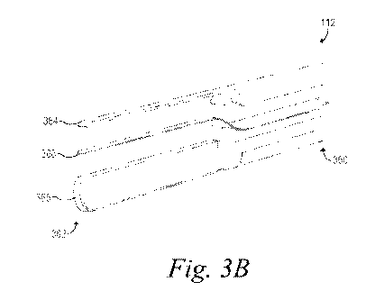
[0058] Figures 3A-3C depict the electrosurgical electrode 128
according to an
example. In particular, Figure 3A depicts a partial perspective view of the
electrosurgical
electrode 128, Figure 3B depicts an exploded view of the electrosurgical
electrode 128, and
Figure 3C depicts a cross-sectional view of the electrosurgical electrode 128
through a line 3C
in Figure 3A.
[0059] As shown in Figures 3A-3C, the electrosurgical electrode 128
includes a
proximal end 360 configured to receive electrosurgical energy from an
electrosurgical tool 112,
14
CA 03143910 2021-12-16
WO 2021/123912 PCT/IB2020/001063
and a distal end 362 opposite the proximal end 360. The proximal end 360 can
receive
electrosurgical energy from the electrosurgical tool 112, as described in
further detail below.
The distal end 362 can define a working end, which is configured for cutting
and coagulating
tissue using the electrosurgical energy.
[0060] As shown in Figures 3A-3C, the electrosurgical electrode 128
also
includes a cutting-electrode portion 364 extending from the proximal end 360
to the distal end
362, a coagulating-electrode portion 366 extending from the proximal end 360
to the distal end
362, and an insulator 368 between the cutting-electrode portion 364 and the
coagulating-
electrode portion 366. The cutting-electrode portion 364 and the coagulating-
electrode portion
366 can include a conductive material (e.g., stainless steel) for conducting
the electrosurgical
energy received from the electrosurgical tool 112 at the proximal end 360 to
distal end 362.
As described in further detail below, the cutting-electrode portion 364 and
the coagulating-
electrode portion 366 can define separate portions of the electrosurgical
electrode 128 that can
be selectively energized to apply the electrosurgical energy to the target
tissue according to a
selected mode of operation.
[0061] The insulator 368 can separate the cutting-electrode portion
364 from the
coagulating-electrode portion 366 over an entire length 370 of the
electrosurgical electrode 128
in a direction that is parallel to a longitudinal axis of the electrosurgical
electrode 128. The
insulator 368 can include an electrically insulating material that can inhibit
conducting the
electrosurgical energy between the cutting-electrode portion 364 and the
coagulating-electrode
portion 366. In this way, the insulator 368 can facilitate conducting the
electrosurgical energy
through one of the cutting-electrode portion 364 or the coagulating-electrode
portion 366 with
a negligible or no electrosurgical energy being conducted through the other
one of the cutting-
electrode portion 364 or the coagulating-electrode portion 366.
[0062] For example, the insulator 368 can include a material selected
from a
group consisting of a plastic, a ceramic, and an enamel. Additionally, for
example, the insulator
368 can have a thickness that is equal to or greater than a thickness of the
cutting-electrode
portion 364 and/or a thickness of the coagulating-electrode portion 366. In
one example, the
insulator 368 can have a width that between approximately 0.1 millimeters (mm)
and
approximately 0.5 mm. This can help to electrically insulate the cutting-
electrode portion 364
and the coagulating-electrode portion 366 from each other. Although it is
advantageous to
separate the cutting-electrode portion 364 and the coagulating-electrode
portion 366 by the
CA 03143910 2021-12-16
WO 2021/123912 PCT/IB2020/001063
insulator 368, the cutting-electrode portion 364 and the coagulating-electrode
portion 366 can
be separated by an air gap in another example.
[0063] In Figures 3A-3C, the cutting-electrode portion 364 is
configured for
cutting tissue using the electrosurgical energy received from the
electrosurgical tool 112, and
the coagulating-electrode portion 366 is configured for coagulating tissue
using the
electrosurgical energy received from the electrosurgical tool 112. For
instance, a surface area
of the cutting-electrode portion 364 can be smaller than a surface area of the
coagulating-
electrode portion 366. This can help to achieve a relatively greater density
of electrosurgical
energy when applying the electrosurgical energy to the cutting-electrode
portion 364, and a
relatively less density of electrosurgical energy when applying the
electrosurgical energy to the
coagulating-electrode portion 366. As a relatively greater density of
electrosurgical energy can
help to enhance performance during a cutting operation and a relatively lesser
density of
electrosurgical energy can help to enhance performance during a coagulating
operation, the
relative sizes of the cutting-electrode portion 364 and the coagulating-
electrode portion 366
can help to improve performance of the electrosurgical tool 112.
[0064] In one example, the surface area of the cutting-electrode
portion 364 is
approximately 5 percent to approximately 50 percent of the surface area of the
coagulating-
electrode portion 366. In another example, the surface area of the cutting-
electrode portion
364 is approximately 5 percent to approximately 35 percent of the surface area
of the
coagulating-electrode portion 366. In another example, the surface area of the
cutting-
electrode portion 364 is approximately 10 percent to approximately 25 percent
of the surface
area of the coagulating-electrode portion 366.
[0065] The cutting-electrode portion 364 and the coagulating-
electrode portion
366 can additionally or alternatively have different shapes to improve the
cutting operation
using the cutting-electrode portion 364 and/or the coagulating operation using
the coagulating-
electrode portion 366. For instance, as shown in Figure 3C, the
electrosurgical electrode 128
can include a first side 372 extending between the proximal end 360 and the
distal end 362, a
second side 374 extending between the proximal end 360 and the distal end 362,
a first edge
376 at a first lateral interface between the first side 372 and the second
side 374, and a second
edge 378 at a second lateral interface between the first side 372 and the
second side 374. The
first side 372 and the second side 374 are on opposing sides of an
intermediate plane 380
extending through the first edge 376 and the second edge 378. The cutting-
electrode portion
16
CA 03143910 2021-12-16
WO 2021/123912 PCT/IB2020/001063
364 includes the first edge 376 and the coagulating-electrode portion 366
includes the second
edge 378.
[0066] As shown in Figure 3C, the first edge 376 is thinner and
sharper than the
second edge 378. This can additionally or alternatively help to achieve a
relatively greater
density of electrosurgical energy when applying the electrosurgical energy to
the cutting-
electrode portion 364, and a relatively less density of electrosurgical energy
when applying the
electrosurgical energy to the coagulating-electrode portion 366. As such, the
first edge 376 of
the cutting-electrode portion 364 can help to achieve relatively better
performance than the
second edge 378 of the coagulating-electrode portion 366 for a cutting
operation, and the
second edge 378 of the coagulating-electrode portion 366 can help to achieve
relatively better
performance than the first edge 376 of the cutting-electrode portion 364 for a
coagulating
operation.
[0067] As noted above, the insulator 368 separates and inhibits
electrical
coupling between the cutting-electrode portion 364 and the coagulating-
electrode portion 366.
In an example, a longitudinal axis extends from the proximal end 360 toward
the distal end 362
and, along the longitudinal axis, the cutting-electrode portion 364 and the
coagulating-electrode
portion 366 are separated by approximately 0.1 mm to approximately 0.5 mm.
This separation
distance can help to inhibit electrical coupling between the cutting-electrode
portion 364 and
the coagulating-electrode portion 366.
[0068] Within examples, the cutting-electrode portion 364 and/or the
coagulating-electrode portion 366 can be covered in a non-stick material
(e.g., a material
having a relatively low coefficient of friction) including at least one
material selected from
silicone, siloxane and Teflon. This can help to mitigate tissue adhering to
the electrosurgical
electrode 128. When tissue adheres to an electrosurgical electrode, the tissue
may change the
effective size and/or shape of the electrode. As such, tissue adherence may
impair making
relatively narrow and precise incisions and, thus, negatively impact a quality
and/or a speed of
the electrosurgical procedure. However, cutting-electrode portion 364 and the
coagulating-
electrode portion 366 with the non-stick material having a relatively low
coefficient of friction
can help to mitigate tissue adhering to the electrosurgical electrode 128 as
the electrosurgical
electrode 128 moves through the target tissue during electrosurgery and, thus,
improve the
quality and/or speed of the electrosurgical procedure.
17
CA 03143910 2021-12-16
WO 2021/123912 PCT/IB2020/001063
[0069] In some examples, the cutting-electrode portion 364 and/or the
coagulating-electrode portion 366 can be additionally or alternatively covered
in an insulation
material such as, for instance, a polymeric material and/or a fluorocarbon
material (e.g.,
polytetrafluoroethylene (PTFE)). In one example, the layer of insulation
material can be a
coating (e.g., an insulating enamel).
[0070] As described above, the proximal end 360 of the
electrosurgical electrode
128 can receive electrosurgical energy from the electrosurgical tool 112. For
instance, as
described above, the electrosurgical electrode 128 can be coupled to the
receptacle 137 by a
releasable coupling (e.g., a friction-fit or a threaded coupling) or a non-
releasable coupling
(e.g., via welding and/or soldering). In an implementation, the receptacle 137
is configured to
removably couple to the proximal end 360 of the electrosurgical electrode 128
by a friction-fit
coupling.
[0071] Within examples, the electrosurgical tool 112 and the
electrosurgical
electrode 128 selectively applying the electrosurgical energy to (i) only the
cutting-electrode
portion 364, (ii) only the coagulating-electrode portion 366, and/or (iii)
both the cutting-
electrode portion 364 and the coagulating-electrode portion 366. This can be
achieved by
providing the electrosurgical tool 112 with a plurality of electrical circuits
for coupling the
cutting-electrode portion 364 and the coagulating-electrode portion 366 to the
electrosurgical
generator 110.
[0072] Figure 4 illustrates a schematic circuit diagram for the
electrosurgical
system 100 according to an example. In example shown in Figure 4, the
electrosurgical energy
is conducted through the cutting-electrode portion 364 and not through the
coagulation-
electrode portion 366 when the electrosurgical tool 112 is operated in a
cutting operation, and
the electrosurgical energy is conducted through the coagulating-electrode
portion 366 and not
through the cutting-electrode portion 364 when the electrosurgical tool 112 is
operated in a
coagulating operation.
[0073] For instance, in Figure 4, the at least one user input device
130 includes
a first user input device 430A that is operable to cause the electrosurgical
generator 110 to
supply the electrosurgical energy to the cutting-electrode portion 364.
Additionally, in Figure
4, the at least one user input device 130 includes a second user input device
430B that is
operable to cause the electrosurgical generator 110 to supply the
electrosurgical energy to the
coagulating-electrode portion 366. The first user input device 430A can
include a first switch
18
CA 03143910 2021-12-16
WO 2021/123912 PCT/IB2020/001063
that is operable by the first button 230A (shown in Figure 2) to operate the
electrosurgical tool
112 in the cutting mode of operation, and the second user input device 430B
can include a
second switch that is operable by the second button 230B (shown in Figure 2)
to operate the
electrosurgical tool 112 in a coagulation mode of operation.
[0074] As described above, the electrosurgical tool 112 can include
at least one
electrical conductor selected from the power cord 122, the housing conductor
134, and the
conductive lead(s) 136 for electrically coupling the electrosurgical electrode
128 to the
electrosurgical generator 110. In Figure 4, the at least one electrical
conductor includes a first
conductor 482 that is configured to couple the cutting-electrode portion 364
to the
electrosurgical generator 110, and a second conductor 484 that is configured
to couple the
coagulating-electrode portion 366 to the electrosurgical generator 110.
[0075] The at least one electrical conductor also includes a third
conductor 486
that electrically couples the electrosurgical generator 110 to the first
switch of the first user
input device 430A. The first switch of the first user input device 430A is
actuatable between
an open state and a closed state. In the open state of the first switch, the
third conductor 486 is
decoupled from the first conductor 482 and the cutting-electrode portion 364.
In the closed
state of the first switch, the third conductor 486 is coupled to the first
conductor 482 and the
cutting-electrode portion 364.
[0076] In Figure 4, the third conductor 486 also electrically couples
the
electrosurgical generator 110 to the second switch of the second user input
device 430B. The
second switch of the second user input device 430B is actuatable between an
open state and a
closed state. In the open state of the second switch, the third conductor 486
is decoupled from
the second conductor 484 and the coagulating-electrode portion 366. In the
closed state of the
second switch, the third conductor 486 is coupled to the second conductor 484
and the cutting-
electrode portion 364.
[0077] In this arrangement, when the first switch of the first user
input device
430A is actuated to the closed state and the second switch of the second user
input device 430B
remains in the open state, the electrosurgical generator 110 supplies the
electrosurgical energy
to the cutting-electrode portion 364 and not the coagulating-electrode portion
366. Whereas,
when the second switch of the second user input device 430B is actuated to the
closed state and
the first switch of the first user input device 430A remains in the open
state, the electrosurgical
19
CA 03143910 2021-12-16
WO 2021/123912 PCT/IB2020/001063
generator 110 supplies the electrosurgical energy to the coagulating-electrode
portion 366 and
not the cutting-electrode portion 364.
[0078] Accordingly, in the cutting mode of operation, the
electrosurgical tool
112 supplies the electrosurgical energy from the at least one electrical
conductor to the cutting-
electrode portion 364 of the electrosurgical electrode 128 and not the
coagulating-electrode
portion 366 of the electrosurgical electrode 128. Whereas, in the coagulation
mode of
operation, the electrosurgical tool 112 supplies the electrosurgical energy
from the at least one
electrical conductor to the coagulating-electrode portion 366 of the
electrosurgical electrode
128 and not the cutting-electrode portion 364 of the electrosurgical electrode
128.
[0079] As shown in Figure 4, the electrosurgical system 100 can also
include a
dispersive electrode 488 and a plurality of return conductors 490 coupling the
dispersive
electrode 488 to the electrosurgical generator 110. The dispersive electrode
488 and the return
conductors 490 can return electric current from the patient to the
electrosurgical generator 110.
[0080] In the example shown in Figure 4, the electrosurgical energy
is conducted
through the cutting-electrode portion 364 and not through the coagulation-
electrode portion
366 when the electrosurgical tool 112 is operated in the cutting mode of
operation, and the
electrosurgical energy is conducted through the coagulating-electrode portion
366 and not
through the cutting-electrode portion 364 when the electrosurgical tool 112 is
operated in a
coagulating mode of operation. However, in another example, the
electrosurgical energy can
be conducted through both the coagulating-electrode portion 366 and the
cutting-electrode
portion 364 when the electrosurgical tool 112 is operated in the coagulating
mode of operation.
[0081] Figure 5 illustrates a schematic circuit diagram for the
electrosurgical
system 100 according to one such example. The schematic circuit diagram of
Figure 5 is
substantially similar or identical to the schematic circuit diagram of Figure
4, except the
electrosurgical system 100 further includes a fourth conductor 592 that
provides for electrically
coupling the cutting-electrode portion 364 to the electrosurgical generator
110 when the second
switch of the second user input device 430B is actuated to the closed state.
[0082] Accordingly, in the arrangement shown in Figure 5, (i) the
electrosurgical
energy is conducted through the cutting-electrode portion 364 and not through
the coagulation-
electrode portion 366 when the electrosurgical tool 112 is operated in the
cutting mode of
operation, and (ii) the electrosurgical energy is conducted through the
coagulating-electrode
portion 366 and the cutting-electrode portion 364 when the electrosurgical
tool 112 is operated
CA 03143910 2021-12-16
WO 2021/123912 PCT/IB2020/001063
in a coagulating mode of operation. This can help to increase a surface area
of the
electrosurgical electrode 128 that conducts the electrosurgical energy and,
thus, decrease a
density of the electrosurgical energy when using the electrosurgical tool 112
in the coagulating
mode of operation.
[0083] As described above, providing the insulator 368 between the
cutting-
electrode portion 364 and the coagulating-electrode portion 366 can help to
inhibit the
electrosurgical energy from being conducted between the cutting-electrode
portion 364 and the
coagulating-electrode portion 366. This can allow for selectively activating
different portions
of the electrosurgical electrode 128 according to different modes of
operation.
[0084] In some examples, the insulator 368 between the cutting-
electrode
portion 364 and the coagulating-electrode portion 366 can additionally or
alternatively provide
for the electrosurgical electrode 128 acting as a sensor that can sense one or
more conditions
related to an electrosurgical operation. For instance, in an example, the
electrosurgical
generator 110 can use the electrical properties of the cutting-electrode
portion 364 and the
coagulating-electrode portion 366 to measure an impedance of a target tissue.
Referring to
Figure 4, in the example of user input device 430A being actuated and the
cutting-electrode
portion 364 being active, a high impedance, though not open circuit, on the
second conductor
484 can be used to allow a relatively small current to pass from the cutting-
electrode portion
364 to the coagulating-electrode portion 366. To bypass the insulator 368
between the cutting-
electrode portion 364 and the coagulating-electrode portion 366, the current
has to pass through
a portion of the tissue immediately adjacent to the cutting-electrode portion
364 and the
coagulating-electrode portion 366. This relatively small current will pass
through the tissue and
return to the electrosurgical generator 110.
[0085] The electrosurgical generator 110 can then use this current to
measure an
electrical characteristic such as, for example, an impedance, a voltage, a
capacitance, and/or an
inductance. The electrosurgical generator 110 can determine, based on the
measured electrical
characteristic, a characteristic of a tissue (e.g., the impedance of the
tissue, a water content of
the tissue, a density of the tissue, a fat content of the tissue, and/or a
temperature of the tissue)
to which the electrosurgical electrode 128 has applied the electrosurgical
energy. Additionally
or alternatively, the electrosurgical generator 110 can use the measured
electrical characteristic
to set and/or modify a power and/or a waveform of the electrosurgical energy
supplied to the
electrosurgical tool 112. In this way, the electrosurgical electrode 128 can
beneficially provide
a measurement functionality of the tissue while the electrosurgical device 112
is being used.
21
CA 03143910 2021-12-16
WO 2021/123912 PCT/IB2020/001063
[0086] In another example, as before with the cutting-electrode
portion 364
activated, rather than using a high impedance on the second conductor 484, the
electrosurgical
generator 110 can intermittently create a circuit with the cutting-electrode
portion 364 and the
coagulating-electrode portion 366 acting as two poles such that a path of
least resistance is
from the cutting-electrode portion 364 through the immediately adjacent
tissue, bypassing the
insulating layer 368 and into the coagulating-electrode portion 366. The
electrosurgical
generator 110 can use this circuit to measure the electrical characteristic
and determine, based
on the electrical characteristic, the characteristic of the tissue as
described above. This
measurement can be made in the order of milliseconds such that cutting
performance is not
noticeably affected.
[0087] In another example, the electrical characteristic between the
activated
portion(s) of the electrosurgical electrode 128 (e.g., the portions selected
from the cutting-
electrode portion 364 and/or the coagulating-electrode portion 366 that are
being supplied with
the electrosurgical energy) and the return electrode 488 is measured within
the electrosurgical
generator 110. For instance, the impedance of the tissue immediately adjacent
to the electrode
128 can be measured in addition to the impedance between the activated
portion(s) of the
electrosurgical electrode 128 and the return electrode 488. Over multiple
measurements the
impedance between the activated portion(s) of the electrosurgical electrode
128 through the
patient to the return electrode 488 can average out such that a discernable
change in impedance
is specifically associated with the tissue immediately adjacent to the
activated portion(s) of the
electrosurgical electrode 128.
[0088] Figure 6 illustrates a schematic circuit diagram for the
electrosurgical
system 100 according to one such example. The schematic circuit diagram of
Figure 6 is
substantially similar or identical to the schematic circuit diagrams of Figure
4, except the
electrosurgical system 100 further includes a sensor 694 that provides for
measuring the
electrical characteristic (e.g., an impedance, a voltage, a phase, a
capacitance, and/or an
inductance) across the cutting-electrode portion 364 and the coagulating-
electrode portion 366.
In some implementations, the sensor 694 can be located in the electrosurgical
generator 110.
In other implementations, the sensor 694 can be located in the electrosurgical
tool 112. In
either implementation, the sensor 694 can generate a sensor signal indicative
of the electrical
characteristic measured by the sensor 694. The sensor 694 can further transmit
the sensor
signal to the electrosurgical generator 110, which can determine the
characteristic of the tissue
22
CA 03143910 2021-12-16
WO 2021/123912 PCT/IB2020/001063
based on the sensor signal, and responsively set and/or modify the power
and/or a waveform
of the electrosurgical energy supplied to the electrosurgical tool 112 as
described above.
[0089] As described above, the electrosurgical generator 110 can perform
various
operations including, for example, measuring the electrical characteristic,
determining the
characteristic of the tissue, setting the waveform and/or the power of the
electrosurgical energy,
and/or modifying the waveform and/or the power of the electrosurgical energy.
Within
examples, the electrosurgical generator 110 can include one or more controller
that are
configured to carry out at least these operations. The controller(s) can be
implemented using
hardware, software, and/or firmware. For instance, the controller(s) can
include one or more
processors and a non-transitory computer readable medium (e.g., volatile
and/or non-volatile
memory) that stores machine language instructions or other executable
instructions. The
instructions, when executed by the one or more processors, cause the
electrosurgical generator
110 to carry out the various operations described herein. The controller(s),
thus, can receive
data and store the data in the memory as well.
[0090]
Referring now to Figure 7, a flowchart is shown for a process 700 of
making an electrosurgical electrode for an electrosurgical tool according to
an example. As
shown in Figure 7, the process 700 includes forming a cutting-electrode
portion at block 710,
forming a coagulating-electrode portion at block 712, positioning an insulator
between cutting-
electrode portion and the coagulating-electrode portion at block 714, and
coupling the cutting-
electrode portion to the coagulating-electrode portion with the insulator
between the cutting-
electrode portion and the coagulating-electrode portion at block 716. The
cutting-electrode
portion is configured for cutting tissue using electrosurgical energy received
from an
electrosurgical tool. The coagulating-electrode portion is configured for
coagulating tissue
using the electrosurgical energy received from the electrosurgical tool.
[0091] The
description of the different advantageous arrangements has been
presented for purposes of illustration and description, and is not intended to
be exhaustive or
limited to the examples in the form disclosed. Many modifications and
variations will be
apparent to those of ordinary skill in the art. Further, different
advantageous examples may
describe different advantages as compared to other advantageous examples. The
example or
examples selected are chosen and described in order to explain the principles
of the examples,
the practical application, and to enable others of ordinary skill in the art
to understand the
disclosure for various examples with various modifications as are suited to
the particular use
contemplated.
23