Note: Descriptions are shown in the official language in which they were submitted.
CA 03143998 2021-12-16
WO 2020/257408
PCT/US2020/038368
1
INTERLEUKIN-27 PRODUCING B-CELLS AND USES THEREOF
CROSS-REFERENCE TO RELATED APPLICATION
[0001] This patent application claims the benefit of co-pending U.S.
Provisional Patent
Application No. 62/863,054, filed June 18, 2019, which is incorporated by
reference in its
entirety herein.
STATEMENT REGARDING FEDERALLY SPONSORED RESEARCH OR
DEVELOPMENT
[0002] This invention was made with Government support under project number
ZO1EY000350-18 by the National Eye Institute of the National Institutes of
Health. The
Government has certain rights in the invention.
INCORPORATION-BY-REFERENCE OF MATERIAL SUBMITTED
ELECTRONICALLY
[0003] Incorporated by reference in its entirety herein is a computer-
readable
nucleotide/amino acid sequence listing submitted concurrently herewith and
identified as
follows: One 2,692 Byte ASCII (Text) file named "749447 5T25.TXT," created on
June 12,
2020.
BACKGROUND OF THE INVENTION
[0004] Uveitis, age-related macular degeneration (AMD), graft-vs-host
disease (GVHD),
and multiple sclerosis (MS) are diseases that initiate or progress as a result
of adverse
immunological activity. These diseases can result in blindness, paralysis, and
significant
morbidity that impacts quality of life. Uveitis is comprised of a diverse
group of potentially
sight-threatening intraocular inflammatory diseases of infectious or
autoimmune etiology,
where autoreactive lymphocytes contribute to ocular pathology by attacking and
damaging
uveal tissue. Similarly, autoimmune processes contribute significantly to the
progression of
retinal degeneration associated with AMD, though the processes that initiate
AMD have not
been definitively identified. MS is caused in part by lymphocytes that attack
and/or destroy
myelinated neurons, thereby interfering with synaptic transmission and
communication
CA 03143998 2021-12-16
WO 2020/257408
PCT/US2020/038368
2
between neurons. In GVHD, the allogeneic transplant views the recipient's body
as foreign,
and the transplant attacks the body. Although steroids are effective therapy
for uveitis or
MS, serious adverse effects preclude their prolonged use. Similar to uveitis
and MS, there
may be adverse effects associated with the use of steroids and
immunosuppressants to treat
GVHD. Further, there currently is no effective cure for AMD, and current
treatments are
directed to the slowing of progressive retinal degeneration. Therefore, there
remains an
unmet need for safe and effective long-term therapies for the aforesaid
diseases.
BRIEF SUMMARY OF THE INVENTION
[0005] The invention provides an isolated population of mammal cells
comprising about
75 % or higher B-la regulatory cells expressing cell surface inhibitory
receptors lymphocyte-
activation gene 3 (LAG-3), programmed cell death protein 1 (PD-1), and C-X-C
chemokine
receptor type 4 (CXCR4), and secreting interleukin-27 (IL-27).
[0006] The invention also provides methods of preparing the population of
mammal cells
of an embodiment of the invention, comprising (a) isolating cluster of
differentiation 5
positive (CD5+) expressing cells from a sample of mammal peripheral lymphoid
tissue,
mammal cord blood, mammal peritoneal fluid, or mammal bone marrow using
fluorescence-
activated cell sorting (FACS) to provide isolated CD5+ expressing cells; (b)
culturing the
isolated CD5+ expressing cells in a cell culture media to provide cultured
cells; (c) activating
the cultured cells with a BCR (B cell receptor) or a TLR (Toll-like receptor)
agonists to
provide activated cells; and (d) exposing the activated cells to IL-27.
[0007] The invention further provides methods of suppressing the immune
system of a
mammal, the method comprising administering the population of mammal cells of
an
embodiment of the invention to a mammal.
[0008] The invention further provides methods of treating a mammal with
graft-versus-
host disease, the method comprising administering the population of mammal
cells of an
embodiment of the invention to a mammal with graft-versus-host disease.
[0009] The invention provides methods of preventing or reducing the
severity of graft-
versus-host disease in a mammal, the method comprising administering the
population of
mammal cells of an embodiment of the invention to a mammal before the mammal
receives
an allogeneic transplant.
CA 03143998 2021-12-16
WO 2020/257408
PCT/US2020/038368
3
[0010] The invention provides methods of preventing or reducing the
severity of graft-
versus-host disease in a mammal, the method comprising (a) mixing the
population of
mammal cells of an embodiment of the invention with a transplant material to
form a
transplant mixture, and (b) administering the transplant mixture to a mammal.
BRIEF DESCRIPTION OF THE SEVERAL VIEWS OF THE DRAWINGS
[0011] FIG. 1 is a set of confocal microscopy images showing sorted CD19+ B
cells from
C57BL/6 mice. The cells activated in vitro for 48 h by stimulation with
lipopolysaccharides
(LPS) or anti-CD40/anti-IgM antibodies (BCR). The cells were incubated with
fluorescence
labelled anti-p28 or anti-Ebi3 antibody. The cells expressing IL-27 (co-
expression of p28
and Ebi3) were detected by confocal microscopy (white arrows).
[0012] FIG. 2A is a set of flow cytometry plots showing sorted CD19+ B
cells isolated
from the peritoneal cavity or spleen of C57BL/6J mice activated in vitro for
48 h by
stimulation with LPS or BCR. The plots show the percentage of B-la and B2
cells
expressing IL-27.
[0013] FIG. 2B is a bar graph showing the percentage of B-la and B2 cells
of FIG. 2A
from the peritoneal cavity that express IL-27.
[0014] FIG. 2C is a bar graph showing the percentage of B-la and B2 cells
of FIG. 2A
from the spleen that express IL-27.
[0015] FIG. 2D is a graph showing the results of analysis of the
supernatants of the
cultures of FIG. 2A by enzyme-linked immunosorbent assay (ELISA).
[0016] FIG. 3 is a set of flow cytometry plots showing sorted CD19+ B-cells
from
C57BL/6J mice activated in vitro for 48 h by stimulation with anti-CD40/anti-
IgM antibodies
(BCR) in the presence or absence of IL-27. The plots show the frequency of
various cells in
the culture. The numbers in the quadrants indicate the percentage of B cells
expressing p28,
Ebi3 or p28, and Ebi3 (IL-27).
[0017] FIG. 4 is a bar graph showing the quantification frequency of
various cells in the
culture shown in the plots of FIG. 3.
[0018] FIG. 5 is a graph that shows the results of NanoString RNA analysis
(NanoString
Technologies, Inc., Seattle, Washington) of various cells in the culture shown
in the plots of
CA 03143998 2021-12-16
WO 2020/257408
PCT/US2020/038368
4
FIG. 3 showing that BCR/IL-27 synergistically unregulated expression of IL-27
subunit p28
and IL-27Ra and altered the pattern of chemokine receptors expression.
[0019] FIG. 6 is a set of images showing the results of
immunofluorescence/confocal
microscopy analysis of various cells in the culture shown in the plots of FIG.
3 showing that
BCR/IL-27 synergistically unregulated expression of IL-27 subunit p28 and IL-
27Ra and
altered the pattern of chemokine receptors expression. The cells expressing IL-
27 (co-
expression of p28 and Ebi3) were detected by confocal microscopy (white
arrows).
[0020] FIG. 7 is a graph showing sorted CD19+ B cells from wild type or IL-
27RaK0
mice activated in vitro for 48 h by stimulation with anti-CD40/anti-IgM
antibodies (BCR) in
the presence or absence of IL-27. B cells expressing p28, Ebi3, or p28 and
Ebi3 (IL-27) were
detected by intracellular cytokine assay and the bar chart shows the
percentages of IL-27-
producing B cells in the various cultures.
[0021] FIG. 8 is a graph showing the results of qPCR for expression of IL-
27Ra in cells
that were isolated from the peritoneal cavity and spleen of wild type mice and
sorted into B-
la or B2 cells.
[0022] FIG. 9 shows CD19+ B cells isolated from human peripheral blood
mononuclear
cells (PBMC) of human volunteers that were activated with phorbol myristate
acetate (PMA)
in the presence of IL-27.
[0023] FIG. 10 is a graph showing the CD19+ B cells of FIG. 9 in the
presence of IL-27.
[0024] FIG. 11 is a graph showing the frequency of human B cells expressing
p28, Ebi3
or both p28 and Ebi3 (IL-27) after CD19+ B cells were isolated from PBMC of
human
volunteers and activated with PMA in the absence of IL-27.
[0025] FIG. 12 is a flow cytometry plots showing the frequency of the cells
of FIG. 11
that express p28, Ebi3 or both p28 and Ebi3 (IL-27).
[0026] FIG. 13A is a bar graph showing the frequency of IL-27-producing B-
la cells in
the peritoneal cavity. C57BL/6J mice were injected (i.v) with LPS (50
fig/mouse) and
frequency of IL-27-producing B-la cells in the peritoneal cavity was assessed
every day until
day 4 post-injection. The B-la cells were isolated at various time points from
the peritoneal
cavity and analyzed by intracellular cytokine staining assay.
[0027] FIG. 13B is a bar graph showing the frequency of IL-27-producing B2
cells in the
peritoneal cavity. C57BL/6J mice were injected (i.v) with LPS (50 fig/mouse)
and frequency
CA 03143998 2021-12-16
WO 2020/257408
PCT/US2020/038368
of IL-27-producing B2 cells in the peritoneal cavity was assessed every day
until day 4 post-
injection. The B2 cells were isolated at various time points from the
peritoneal cavity and
analyzed by intracellular cytokine staining assay.
[0028] FIG. 14A is a bar graph showing the frequency of IL-27-producing B-
la cells in
the spleen. C57BL/6J mice were injected (i.v) with LPS (50 [tg/mouse) and
frequency of IL-
27-producing B-la cells in the spleen was assessed every day until day 4 post-
injection. The
B-la cells were isolated at various time points from the spleen and analyzed
by intracellular
cytokine staining assay.
[0029] FIG. 14B is a bar graph showing the frequency of IL-27-producing B2
cells in the
spleen.
[0030] C57BL/6J mice were injected (i.v) with LPS (50 fig/mouse) and
frequency of IL-
27-producing B2 cells in the spleen was assessed every day until day 4 post-
injection. The
B2 cells were isolated at various time points from the spleen and analyzed by
intracellular
cytokine staining assay.
[0031] FIG. 15 is a flow cytometry bar graph showing the percentage of
chemokine
receptors for CXCR3+. The numbers in bar graph indicate the percent chemokine
receptors
expressing CD19+CD5+CD23- B-la B cells. Data represent at least 3 independent
experiments (*P < 0.05; **P < 0.01; ***P < 0.001; ****P < 0.0001).
[0032] FIG. 16 is a flow cytometry bar graph showing the percentage of
chemokine
receptors for CXCR4+. The numbers in bar graph indicate the percent chemokine
receptors
expressing CD19+CD5+CD23- B-la B cells. Data represent at least 3 independent
experiments (*P < 0.05; **P < 0.01; ***P < 0.001; ****P < 0.0001).
[0033] FIG. 17 is a flow cytometry bar graph showing the percentage of
chemokine
receptors for CXCR5+. The numbers indicate the percent chemokine receptors
expressing
CD19+CD5+CD23- B-la B cells. Data represent at least 3 independent experiments
(*P <
0.05; **P <0.01; ***P <0.001; ****P < 0.0001).
[0034] FIG. 18 is a set of fundus images of retinas showing improvement in
clinical score
following injection of IL-27. Experimental autoimmune uveitis (EAU) was
induced by
immunization of C57BL/6J mice with IRBP651-670-peptide in Freund's adjuvant
(CFA) (n
=12). Mice were treated by intraperitoneal injection of IL-27 (10Ong/mouse) or
PBS on day
(-1) of immunization and every other day until day 12 post-immunization. Eyes
were
CA 03143998 2021-12-16
WO 2020/257408
PCT/US2020/038368
6
analyzed 14 days or 21 days post-immunization by fundoscopy, histology,
optical coherence
tomography (OCT), or electroretinography (ERG).
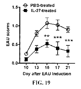
[0035] FIG. 19 is a graph showing the EAU scores of the retinas shown in
FIG. 18. The
EAU clinical scores and assessment of disease severity were based on changes
at the optic
nerve disc or retinal vessels as well as retinal and choroidal infiltrates.
[0036] FIG. 20 is a set of images of hematoxylin and eosin histological
sections of the
retinas of FIG. 18. Scale bar = 200 [tM; V = vitreous; GCL = ganglion cell
layer; INL =
inner nuclear layer; ONL = outer nuclear layer; RPE/CH = retinal pigmented
epithelial and
choroid.
[0037] FIG. 21 is a set of images showing the OCT analysis of the retinas
of FIG. 18
showing the layered structure of the retina. The white arrows indicate
inflammatory cells
(white arrows) in the vitreous or optic nerve.
[0038] FIG. 22 is a graph showing the ERG analysis of a retina of FIG. 18
on day 20 after
EAU induction. The averages of dark-adapted ERG a-wave amplitudes are plotted
as a
function of flash luminance, and the values are means SEM from 4 animals in
each group.
[0039] FIG. 23 is a graph showing the ERG analysis of a retina of FIG. 18
on day 20 after
EAU induction. The averages of dark-adapted ERG b-wave amplitudes are plotted
as a
function of flash luminance, and the values are means SEM from 4 animals in
each group.
[0040] FIG. 24 is a graph showing the ERG analysis of a retina of FIG. 18
on day 20 after
EAU induction. The averages of light-adapted ERG a-wave amplitudes are plotted
as a
function of flash luminance and values are means SEM from 4 animals in each
group.
[0041] FIG. 25 is a graph showing the ERG analysis of a retina of FIG. 18
on day 20 after
EAU induction. The averages of light-adapted ERG b-wave amplitudes are plotted
as a
function of flash luminance and values are means SEM from 4 animals in each
group.
[0042] FIG. 26 is a graph showing the analysis of cytokine IL-27 in the
serum of the mice
of FIG. 18.
[0043] FIG. 27 is a graph showing the analysis of cytokine IL-17 in the
serum of the mice
of FIG. 18.
[0044] FIG. 28 is a graph showing the analysis of cytokine IL-10 in the
serum of the mice
of FIG. 18.
CA 03143998 2021-12-16
WO 2020/257408
PCT/US2020/038368
7
[0045] FIG. 29 is a graph showing the analysis of cytokine IL-35 in the
serum of the mice
of FIG. 18.
[0046] FIG. 30 is a flow cytometry plot showing the percentage of IL-27-
expressing B
cells. The numbers in the quadrants indicate the percent of CD19+ or
CD19+CD5+CD1dh1 or
CD19+CD5+CD110"v cells in the spleen of control (PBS-treated) or IL-27-treated
EAU mice.
The gating strategies are as indicated.
[0047] FIG. 31 is a bar graph showing the percentage of IL-27-expressing B
cells of FIG.
30.
[0048] FIG. 32 is a flow cytometry plot showing the percentage of IL-27-
expressing B
cells in the spleen of control (PBS-treated) or IL-27-treated EAU mice. The
gating strategies
are as indicated. The numbers in the quadrants indicate the percent of CD19+or
CD19+CD5+CD1dhi or CD19+CD5+CD110w B cells expressing p28, Ebi3, or p28 and
Ebi3
(IL-27).
[0049] FIG. 33 is a bar graph showing the percentage of IL-27-expressing B-
10 cells in
the spleen of control (PBS-treated) or IL-27-treated EAU mice of FIG. 32.
[0050] FIG. 34 is a bar graph showing the percentage of IL-27-expressing B-
la cells in
the spleen of control (PBS-treated) or IL-27-treated EAU mice of FIG. 32.
[0051] FIG. 35 is a set of fundus images of retinas from mice 17 days after
adoptive
transfer by fundoscopy. Purified peritoneal cavity B-la cells (5 x10⁵
cells/mouse; >80% i27-
Bregs) from wild type donor CD45.2+ EAU mice were transferred to naive
syngeneic wild
type or IL-27RaK0 CD45.1+ mice and 24 h after the adaptive transfer, EAU was
induced in
recipient mice by immunization with IRBP651-670 (n = 12). Clinical disease was
monitored
until 17 days after adoptive transfer by fundoscopy.
[0052] FIG. 36 is a graph showing the EAU scores of the retinas shown in
FIG. 35.
[0053] FIG. 37 is set of flow cytometry plots from CD4+ T cells subjected
to FACS and
intracellular cytokine assays. The numbers in the quadrants indicate the
percentage of CD4+
T cells expressing IL-17. Data represents at least 3 independent experiments
(**P < 0.01;
***P <0.001; ****P < 0.0001).
[0054] FIG. 38A is a set of flow cytometry plots from CD4+ T cells
subjected to FACS
and intracellular cytokine assays. The numbers in the quadrants indicate the
percentage of
CD4+ T cells expressing IL-10.
CA 03143998 2021-12-16
WO 2020/257408
PCT/US2020/038368
8
[0055] FIG. 38B is a bar graph showing the percentage of CD4+ T cells
expressing IFN-y.
[0056] FIG. 38C is a bar graph showing the percentage of CD4+ T cells
expressing IL-17.
[0057] FIG. 38D is a bar graph showing the percentage of CD4+ T cells
expressing
IFN-y and IL-17.
[0058] FIG. 38E is a bar graph showing the percentage of CD4+ T cells
expressing IL-10.
[0059] FIG. 39 is a set of flow cytometry plots from CD19+ T cells (eye)
subjected to
FACS. The numbers in the quadrants indicate the percentage of CD19+CD5+CD23- B-
la
cells expressing p28, p35, Ebi3, p28 and Ebi3 (IL-27). Data represents at
least 3 independent
experiments (**P < 0.01; ***P < 0.001; ****P < 0.0001).
[0060] FIG. 40 is a set of flow cytometry plots from CD19+ T cells (eye)
subjected to
FACS. The numbers in the quadrants indicate the percentage of CD19+CD5-CD23+
B2
expressing p28 and Ebi3 (IL-27) or p35 and Ebi3 (IL-35). Data represents at
least 3
independent experiments (**P < 0.01; ***P < 0.001; ****P < 0.0001).
[0061] FIG. 41 is a bar graph showing the percentage of B-la cells in the
eye of FIG. 39
that express p28 and Ebi3.
[0062] FIG. 42 is a bar graph showing the percentage of B2 cells in the eye
of FIG. 40
that express p28 and Ebi3.
[0063] FIG. 43 is a bar graph showing the percentage of B2 cells in the eye
of FIG. 40
that express p35 and Ebi3.
[0064] FIG. 44 is a set of photomicrographs of hematoxylin and eosin
stained sections of
the brain (top row) and spinal cord (middle row) of mice on day 17 post-
immunization
(original magnification x200). Arrows show inflammatory cells in the brain or
spinal cord.
The extent of EAE-induced demyelination was assessed by Luxol fast blue
staining (bottom
row; Luxol fast blue is a copper phthalocyanine dye that is soluble in alcohol
and is attracted
to bases found in the lipoproteins of myelin sheaths). Arrows denote areas of
demyelination
in the spinal cord. EAE was induced by immunization of C57BL/6J mice with
M0G35-55-
peptide in CFA (n = 12). Mice were treated by intraperitoneal injection of IL-
27 (100
ng/mouse) or PBS on day 0 of immunization and every other day until day 12
post-
immunization.
CA 03143998 2021-12-16
WO 2020/257408
PCT/US2020/038368
9
[0065] FIG. 45 is a graph showing the EAU scores of the spinal cord
described in FIG.
44. The EAE clinical scores and disease assessment were ascertained by two
masked
investigators according to well established grading system.
[0066] FIG. 46 is a set of flow cytometry plots from inflammatory cells in
the brain and
spinal cord following intracellular cytokine analysis of untreated or IL-27-
treated mice that
were isolated day 17 post-immunization and then digested with collagenase. The
numbers in
quadrants indicate percentage of IL-17- or IFN-y-expressing CD4 T cells in the
spinal cord
and brain.
[0067] FIG. 47 is a set of flow cytometry plots from inflammatory cells in
the brain and
spinal cord following intracellular cytokine analysis of untreated or IL-27-
treated mice that
were isolated day 17 post-immunization and then digested with collagenase. The
numbers in
quadrants indicate percentage of IL-10- expressing and CD4 T cells in the
spinal cord and
brain.
[0068] FIG. 48 is a bar graph showing the percentage of CD4 T cells of FIG.
46 and 47
that express IFN-y.
[0069] FIG. 49 is a bar graph showing the percentage of CD4 T cells of FIG.
46 and 47
that express IL-17.
[0070] FIG. 50 is a bar graph showing the percentage of CD4 T cells of FIG.
46 and 47
that express IL-17 and IFN-y.
[0071] FIG. 51 is a bar graph showing the percentage of CD4 T cells of FIG.
46 and 47
that express IL-10.
[0072] FIG. 52 is a set of flow cytometry plots showing the percentage of
IL-27-
expressing B cells from the spinal cord and brain of unimmunized, PBS-treated
or IL-27-
treated EAE mice analyzed for IL-27 (p28 and Ebi3) expression by intracellular
cytokine
staining assay. The numbers in the quadrants indicate the percentage of
CD19+CD5+CD1e
or CD19+CD5+CD1ew B cells in the spinal cord or brain expressing p28, Ebi3 or
p28 and
Ebi3 (IL-27).
[0073] FIG. 53 is a bar graph showing the percentage of CD19 T cells of
FIG. 52 that
express CD19+CD5+CD1ew.
[0074] FIG. 54 is a set of flow cytometry plots showing the percentage of
IL-27-
expressing B cells from the spleen of unimmunized, PBS-treated or IL-27-
treated EAE mice
CA 03143998 2021-12-16
WO 2020/257408
PCT/US2020/038368
analyzed for IL-27 (p28 and Ebi3) expression by intracellular cytokine
staining assay. The
numbers in the quadrants indicate the percentage of total CD19+CD5+CD1dhi or
CD19+CD5+CD1d10w B cells in the spleen expressing p28, Ebi3 or p28 and Ebi3
(IL-27).
[0075] FIG. 55 is a bar graph showing the percentage of CD19 T cells of
FIG. 54 that
express p28 and Ebi3.
[0076] FIG. 56 is a set of flow cytometry plots showing analysis of spleen
cells of PBS-
treated or IL-27-treated EAE mice for IL-27 expansion. The numbers in the
quadrants
indicate the percentage of CD19+CD5+CD1d1 w B-la cells.
[0077] FIG. 57 is a bar graph showing the percentage of CD19 T cells of
FIG. 56 that
express CD19+CD5+CD1dhi.
[0078] FIG. 58 is a bar graph showing the percentage of CD19 T cells of
FIG. 56 that
express CD19+CD5+CD1d1 w.
[0079] FIG. 59 is a graph showing the EAU scores of spleen cells from M0G35-
55
immunized (PBS-treated EAE or IL-27-treated) CD45.2+ mice that were re-
stimulated ex-
vivo and transferred (1x10⁷ cells/mouse) to naïve CD45.1+ WT mice. The EAE
clinical
scores and disease assessment were ascertained by two masked investigators.
[0080] FIG. 60 is a set of flow cytometry plots showing the percentage of
CD4+ T cells.
Spinal cord, brain, lymph nodes (LN) or spleen of PBS-treated or IL-27-treated
mice were
isolated on day 20 post-adoptive transferred, digested with collagenase and
CD4+ T cells and
IL-27-producing B-la and analyzed by intracellular cytokine staining assay.
The numbers in
the quadrants indicate the percentage of CD4+ T cells expressing IL-17 or IFN-
y. Data
represents >3 independent experiments (**P < 0.01; ***P < 0.001; ****P <
0.0001).
[0081] FIG. 61 is a bar graph showing the percentage of the cells of FIG.
60 that express
IL-17.
[0082] FIG. 62 is a bar graph showing the percentage of the cells of FIG.
60 that express
IL-17 and IFN-y.
[0083] FIG. 63 is a set of flow cytometry plots showing the percentage of
IL-27-
producing B-la cells. Spinal cord, brain, lymph nodes (LN) or spleen of PBS-
treated or IL-
27-treated mice were isolated on day 20 post-adoptive transferred, digested
with collagenase
and CD4+ T cells and IL-27-producing B-la, and analyzed by intracellular
cytokine staining
assay. The numbers in the quadrants indicate the percentage of CD19+CD5+CD11b+
B-la
CA 03143998 2021-12-16
WO 2020/257408
PCT/US2020/038368
11
cells expressing p28, Ebi3 or p28 and Ebi3 (IL-27). Data represents >3
independent
experiments (**P < 0.01; ***P < 0.001; ****P < 0.0001).
[0084] FIG. 64 is a bar graph showing the percentage of the B-la cells of
FIG. 63 from
the spinal cord that express p28 and Ebi3 (IL-27).
[0085] FIG. 65 is a bar graph showing the percentage of the B-la cells of
FIG. 63 from
the brain that express p28 and Ebi3 (IL-27).
[0086] FIG. 66 is a bar graph showing the percentage of the B-la cells of
FIG. 63 from
the spleen that express p28 and Ebi3 (IL-27).
[0087] FIG. 67 is a set of flow cytometry plots (top) and a graph showing
EAE clinical
scores (bottom) from purified peritoneal cavity B-la cells (5x10⁵ cells/mouse;
>80% i27-
Bregs) from WT donor CD45.2+ mice that were transferred to naive syngeneic
wild type mice
and 24 h after the adaptive transfer, EAE was induced in recipient mice by
immunization
with M0G35-55 (n=12). The EAE clinical scores and disease assessment as
performed by two
masked investigators.
[0088] FIG. 68 is a set of flow cytometry plots showing the percentage of
CD4+ T cells
expressing IL-10, IL-17, or IFN-y. Spinal cords and brains of PBS-treated or B-
la-treated
mice were isolated on day 15 post-immunization, digested with collagenase and
analyzed by
an intracellular cytokine staining assay. Data represents >3 independent
experiments (**P <
0.01; ***P < 0.001; ****P <0.0001).
[0089] FIG. 69 is a bar graph showing the percentage of spinal cord and
brain cells of
FIG. 68 that express IFN-y.
[0090] FIG. 70 is a bar graph showing the percentage of spinal cord and
brain cells of
FIG. 68 that express IL-17.
[0091] FIG. 71 is a bar graph showing the percentage of spinal cord and
brain cells of
FIG. 68 that express IL-10.
[0092] FIG. 72 is a set of flow cytometry plots showing the percentage of
CD19+CD5+CD23- B-la or CD19+CD5-CD23+ B2 cells expressing p28, Ebi3 or p28 and
Ebi3 (IL-27) in spinal cords. Spinal cords of PBS-treated or B-la-treated mice
were isolated
on day 15 post-immunization, digested with collagenase and analyzed by an
intracellular
cytokine staining assay. Data represents >3 independent experiments (**P <
0.01; ***P <
0.001; ****P <0.0001).
CA 03143998 2021-12-16
WO 2020/257408
PCT/US2020/038368
12
[0093] FIG. 73 is a bar graph showing the percentage of spinal cord cells
of FIG. 72 that
express p28 and Ebi3 (IL-27).
[0094] FIG. 74 is a set of flow cytometry plots showing the percentage of
CD19+CD5+CD23- B-la or CD19+CD5-CD23+ B2 cells expressing p28, Ebi3 or p28 and
Ebi3 (IL-27) in brains. Brains of PBS-treated or B-la-treated mice were
isolated on day 15
post-immunization, digested with collagenase and analyzed by an intracellular
cytokine
staining assay. Data represents >3 independent experiments (**P < 0 .01;***P <
0.001;
****P <0.0001).
[0095] FIG. 75 is a bar graph showing the percentage of brain cells of FIG.
74 that
express p28 and Ebi3 (IL-27).
[0096] FIG. 76 is a set of flow cytometry plots showing the percentage of
CD19+CD5+CD23- B-la or CD19+CD5-CD23+ B2 cells expressing p28, Ebi3 or p28 and
Ebi3 (IL-27) in peritoneal cavities. Fluids from peritoneal cavities of PBS-
treated or B-la-
treated mice were isolated on day 15 post-immunization, digested with
collagenase and
analyzed by an intracellular cytokine staining assay. Data represents >3
independent
experiments (**P < 0.01; ***P < 0.001; ****P < 0.0001).
[0097] FIG. 77 is a bar graph showing the percentage of cells of FIG. 74
from the
peritoneal cavities that express p28 and Ebi3 (IL-27).
[0098] FIG. 78 is a depiction that illustrates macrophages from EAU mice
being cultured
in a trans-well system containing B-la cells from wild type EAU mice at the
bottom wells.
The effects of the macrophages on proliferation of B-la cells was assessed by
[31-11-thymidine
incorporation assays.
[0099] FIG. 79 is a set of flow cytometry plots showing the percentage of B-
la cells of
FIG. 78 that express p28, Ebi3 or p28 and Ebi3 (IL-27).
[0100] FIG. 80 is a bar graph showing the CPM mean values of the B-la cells
and
macrophages of FIG. 78. The proliferative responses were analyzed in 5
replicate cultures.
Data represents at least 3 independent experiments (**P < 0.01; ***P < 0.001;
****P <
0.0001).
[0101] FIG. 81 is a bar graph showing the percentage of B-la cells and
macrophages of
FIG. 78 that express p28 and Ebi3 (IL-27).
CA 03143998 2021-12-16
WO 2020/257408
PCT/US2020/038368
13
[0102] FIG. 82 is a graph showing the ELISA analysis of the secretion of IL-
27 of
primary mouse peritoneum macrophages that were activated with LPS in the
presence or
absence of lentivirus guide RNA that targets p28 (5gp28-1 or 5pg28-2).
[0103] FIG. 83 is a graph showing the ELISA analysis of the secretion of IL-
27 of
primary mouse peritoneum B-la cells that were activated with LPS in the
presence or
absence of lentivirus guide RNA that targets p28 (5gp28-1 or 5pg28-2).
[0104] FIG. 84 is a depiction that illustrates pathogenic (uveitogenic) T
cells from EAU
mice being cultured in a trans-well system containing B-la cells infected with
lentivirus
guide RNA that targets suppression of IL-27 (sgp28/Ebi3). The effects of the B-
la cells on
the proliferation of the uveitogenic T cells was assessed by [3H1-thymidine
incorporation
assays.
[0105] FIG. 85 is a bar graph showing the CPM mean values of the cells of
FIG. 84.
[0106] FIG. 86 is a set of flow cytometry plots showing the percentage of
uveitogenic
CD4+ T cells expressing IL-10, IL-17 and/or IFN-y as determined by an
intracellular cytokine
staining assay.
[0107] FIG. 87 is a bar graph showing the percentage of cells of FIG. 86
that express
IFN-y.
[0108] FIG. 88 is a bar graph showing the percentage of cells of FIG. 86
that express IL-
17.
[0109] FIG. 89 is a bar graph showing the percentage of cells of FIG. 86
that express
IFN-y and IL-17.
[0110] FIG. 90 is a bar graph showing the percentage of cells of FIG. 86
that express IL-
10.
[0111] FIG. 91 is a depiction that illustrates pathogenic (uveitogenic) T
cells from EAU
mice being cultured in a trans-well system containing B-la cells from wild
type EAU mice or
B-la cells infected with lentivirus guide RNA that targets suppression of IL-
27 (sgp28/Ebi3).
The effects of the B-la cells on the proliferation of the uveitogenic T cells
was assessed by
[3H1-thymidine incorporation assays.
[0112] FIG. 92 is a set of flow cytometry plots showing the percentage of
uveitogenic
CD4+ T cells expressing LAG-3 as determined by an intracellular cytokine
staining assay.
CA 03143998 2021-12-16
WO 2020/257408
PCT/US2020/038368
14
[0113] FIG. 93 is a bar graph showing the percentage of the cells of FIG.
92 that express
LAG-3.
[0114] FIG. 94 is a depiction that illustrates pathogenic (uveitogenic) T
cells from EAU
mice being cultured in a trans-well system containing B-la cells from wild
type EAU mice or
B-la cells infected with lentivirus guide RNA that targets suppression of IL-
27 (sgp28/Ebi3).
The effects of the B-la cells on the proliferation of the uveitogenic T cells
was assessed by
[3F11-thymidine incorporation assays.
[0115] FIG. 95 is a set of flow cytometry plots showing the percentage of
CD4+CD25+Foxp3+ and CD4+CD25+Foxp3- expressing p35, Ebi3, or IL-35 (p35/Ebi3).
[0116] FIG. 96 is a bar graph showing the percentage of the cells of FIG.
95 that express
p35 and Ebi3.
[0117] FIG. 97 is a set of flow cytometry plots showing the percentage of
CD4+CD25+Foxp3+ and CD4+CD25+Foxp3- expressing p35, Ebi3, or IL-35 (p35/Ebi3).
[0118] FIG. 98 is a bar graph showing the percentage of the cells of FIG.
97 that are
CD4+CD25+Foxp3+.
[0119] FIG. 99 is a bar graph showing the percentage of the cells of FIG.
97 that are
CD4+CD25+Foxp3-.
[0120] FIG. 100 is a flow cytometry plot and a set of graphs that show the
results of
sorted CD19+CD5+CD23- B-la cells from the peritoneal cavity of C57BL/6J mice
that were
activated in vitro for 48 h by stimulation with LPS analyzed by ELISA (left
flow cytometry
plot). The supernatants from the cultures in the peritoneal cavity were
analyzed by qPCR
(right).
[0121] FIG. 101 is a bar graph showing the results of qPCR analysis of
purified B-la
cells from the peritoneal cavities of C57BL/6J mice injected (i.v) 48 hours
prior with LPS.
[0122] FIG. 102 is a flow cytometry plot showing B-la or plasma cells (B2)
from
C57BL/6J mouse peritoneal cavity or spleen, respectively, after sorting using
magnetic beads
and then activated with anti-CD40/anti-IgM (BCR).
[0123] FIG. 103 is a graph of the qPCR analysis of RNA from the cells of
FIG. 102 that
was quantified for expression of Pd] mRNA transcript.
[0124] FIG. 104 is a graph of the qPCR analysis of RNA from the cells of
FIG. 102 that
was quantified for expression of Lag3 mRNA transcript.
CA 03143998 2021-12-16
WO 2020/257408
PCT/US2020/038368
[0125] FIG. 105 is a set of graphs showing RNA isolated at various time
points analyzed
by qRT-PCRC D19+ B cells from C57BL/6J mouse spleen that were activated with
anti-
CD40/anti-IgM in the presence or absence of IL-27.
[0126] FIG. 106 is the Volcano plot analysis of the genes differentially
induced by IL-27
24 h after they were detected using a NanoString transcription factor panel.
[0127] FIG. 107 is an image of a Western blot. CD19+ B cells were isolated
from the
spleen of C57BL/6J mouse 24 h after immunization with LPS in the presence or
absence of
IL-27 (in-vivo) or from mouse CD19+ B cells after activated with LPS in the
absence or
presence of IL-27 (in vitro). Nuclear extracts were prepared from the cells
and analyzed by
the electrophoretic mobility shift assay (EMSA) to detect IL-27-induced AICE
complexes.
Transcription factors recruited to the CTLA4-AICE or p28-AICE locus were
identified by
super-shift assay. Whole cell extracts prepared from CD19+ B cells of the
C57BL/6J mouse
immunized with LPS in the presence or absence of IL-27 were analyzed by
Western blotting.
[0128] FIG. 108 is a bar graph showing the relative gene expression of B-la
cells from
the peritoneal cavity.
[0129] FIG. 109 is a set of flow cytometry plots of the FACS analysis of
CD19+ B cells
showing the percentage of B-la or plasmablasts expressing p28, Ebi3, or p28
and Ebi3 (IL-
27). The CD19+ B cells from the spleen of C57BL/6J (wild type) or mice with
targeted
deletion of 1rf8 in B cells (CD19-IRF8K0) following activation for 3 days with
anti-
CD40/anti-IgM. The gating strategy is as indicated. Data represents >3
independent
experiments (**/3 < 0.01; ***P < 0.001; ****P < 0.0001).
[0130] FIG. 110 is a bar graph showing the amount of CD19+CD27+CD383+ cells
of FIG.
109.
[0131] FIG. 111 is a bar graph showing the amount of CD19+CD5+CD11b+ cells
of FIG.
109.
[0132] FIG. 112 is a bar graph showing a chromatin immunoprecipitation
(CHIP)
analysis that was performed with B-la cells stimulated with LPS or LPS+IL-27
for 24 h and
STAT1 binding to the p28 or ebi3 promoter region was analyzed. Cell lysates
were
immunoprecipitated with anti-STAT1 antibody or control IgG. Immunoprecipitated
and
input DNA were analyzed by qPCR using primers corresponding to p28 or ebi3
promoter
sites.
CA 03143998 2021-12-16
WO 2020/257408
PCT/US2020/038368
16
[0133] FIG. 113 is a bar graph showing a CHIP analysis that was performed
with B-la
cells stimulated with LPS or LPS+IL-27 for 24 h and STAT3 binding to the p28
or eb13
promoter region was analyzed. Cell lysates were immunoprecipitated with anti-
STAT3
antibody or control IgG. Immunoprecipitated and input DNA were analyzed by
qPCR using
primers corresponding to p28 or eb13 promoter sites.
[0134] FIG. 114 is a set of flow cytometry plots of the FACS analysis of
healthy human
PBMC that were cultured for 3 days with TLR9 agonist CpG and BCR (anti-CD40 or
anti-
IgM). Gating on human B-1 cells (CD19+CD20+CD27+CD43+) revealed that as high
as 19.9
% of BCR-activated B-cells in human PBMC produced IL-27.
[0135] FIG. 115 is a bar graph showing the amount of
CD19+CD2O+CD27+CD43+p28+Ebi3+ cells of FIG. 114.
[0136] FIG. 116 is a set of flow cytometry plots of the FACS analysis of
healthy human
PBMC that were cultured for 3 days with TLR9 agonist CpG and BCR (anti-CD40 or
anti-
IgM). Gating on CD19+CD20+CD27+CD43+CD1 lb + B-1 cells revealed as many as 35
% of
BCR-activated human B-1 cells.
[0137] FIG. 117 is a bar graph showing the amount of
CD19+CD2O+CD27+CD43+CD11b+p28+Ebi3+ cells of FIG. 116.
[0138] FIG. 118 is a bar graph showing the amount of
CD19+CD2O+CD27+CD43+CD11b-p28+Ebi3+ cells of FIG. 116.
[0139] FIG. 119 is a set of flow cytometry plots of the FACS analysis of
human umbilical
cord blood from healthy human donors. As many as 18.1 % of resting B-la cells
constitutively produced IL-27 and stimulation of BCR-activated cord blood B-
cells with IL-
27 increased the percentage of cord blood i27-Bregs to 73.9 %.
[0140] FIG. 120 is a bar graph showing the amount of cells of FIG. 119 (top
panel).
[0141] FIG. 121 is a bar graph showing the amount of cells of FIG. 119
(bottom panel).
[0142] FIG. 122 is a set of graphs showing the relative abundance of i27-
Bregs as
compared to other Breg subtypes (IL-10-producing Bregs and i35-Bregs).
Activated human
cord blood cells were propagated for 6 days. The majority of the Breg cells
were i27-Bregs
(largest slice of each pie graph, ranging from 61.2 +/- 5.3 to 87.1 +/- 3.1 %)
and much lower
levels of IL-10-producing Bregs (ranging from 2.6 +/- 0.3 to 6.7 +/- 1.1 %)
and i35-Bregs
(ranging from 10.2 +/- 2.7 to 32 +/- 6.8 5) were detected.
CA 03143998 2021-12-16
WO 2020/257408
PCT/US2020/038368
17
[0143] FIG. 123 is a set of graphs showing the relative abundance of B-2
cells. These
plots revealed that most i27-Bregs were either in the naïve or memory B-cell
pool.
[0144] FIG. 124 is a graph showing that similar to the mouse species, the
human i27-
Breg cells constitutively express inhibitory receptors PD-1 and LAG3.
[0145] FIG. 125 is a bar graph showing the amount of PD-1+ cells of FIG.
124.
[0146] FIG. 126 is a bar graph showing the amount of LAG3+ cells of FIG.
124.
[0147] FIG. 127 is a set of flow cytometry plots of the FACS analysis of
human
i27-Breg cells. The i27-Breg cells suppressed proliferative responses of TNF-a-
, IL-17-, and
IFN-y-producing pro-inflammatory CD4+ T-cells.
[0148] FIG. 128 is a bar graph showing the CPM mean values of the cells of
FIG. 127.
[0149] FIG. 129 is a bar graph showing the amount of TNF-a+CD4+ T cells of
FIG. 127.
[0150] FIG. 130 is a bar graph showing the amount of IFN-y +CD4+ T cells of
FIG. 127.
[0151] FIG. 131 is a bar graph showing the amount of IL-17A+CD4+ T cells of
FIG. 127.
[0152] FIG. 132 shows the results from a Proximity Ligation Assay (PLA)
which shows
the physical interaction between p28 and Ebi3.
[0153] FIG. 133 is a gel showing that B-cells produce the heterodimeric
(p28/Ebi3) IL-27
cytokine. C57BL/6J mice were injected (i.v) with LPS or LPS+IL-27 and after 24
h lysates
or supernatant from cultured B-la cells were subjected to reciprocal
immunoprecipitation/
Western blot analysis. The antibodies used for IP or Western blotting are
indicated.
[0154] FIG. 134 is a graph that shows the results of NanoString RNA
analysis showing
that IL-27 altered the pattern of chemokine receptor expression in activated B
cells.
[0155] FIG. 135A shows a flow cytometry plot depicting the differential
secretion of
natural IgM antibodies by unchallenged B-la and i27-Breg cells in the
peritoneal cavity.
[0156] FIG. 135B shows a set of graphs depicting the differential secretion
of natural
IgM antibodies by unchallenged B-la and i27-Breg cells in the peritoneal
cavity.
[0157] FIG. 136 shows the Principal Component Analysis (PCA) plot depicting
segregation of the cells into 4 distinct populations.
[0158] FIG. 137 shows Gene ontology (GO) analysis showing functional
pathway
enrichment for i27-Breg cells.
[0159] FIG. 138 is a heatmap of the i27-Breg cells gene signature in
comparison to
signature program of the unchallenged B-la cell.
CA 03143998 2021-12-16
WO 2020/257408
PCT/US2020/038368
18
[0160] FIG. 139 is set of heatmaps showing genes that encode transcription
factors,
signaling proteins, cytokines and chemokines or cell surface proteins which
are differentially
expressed genes in i27-Bregs and B-la cells.
[0161] FIG. 140 is a set of graphs from qPCR expression of genes encoding
inhibitory
receptors.
[0162] FIG. 141A is a set of representative flow cytometry plots showing
significant
expansion of IL-27 secreting CD19+CD20+CD27+CD43+ B-1 cells in response to
anti-CD40
or BCR.
[0163] FIG. 141B is a scatter plot showing significant expansion of IL-27
secreting
CD19+CD20+CD27+CD43+ B-1 cells in response to anti-CD40 or BCR.
[0164] FIG. 142A is a set of representative flow cytometry plots showing
significant
expansion of IL-27 secreting CD27+CD43+CD11+ or CD27+CD43+CD11- B-1 cells in
response to anti-CD40 or BCR.
[0165] FIG. 142B is a set of scatter plots showing significant expansion of
IL-27
secreting CD27+CD43+CD11+ or CD27+CD43+CD11- B-1 cells in response to anti-
CD40 or
BCR.
[0166] FIG. 143A is a set of representative flow cytometry plots of human
cord blood
CD19+ B cells (top) or sorted B-la cells in the blood (bottom) activated with
BCR or BCR
plus IL-27 showing significant expansion IL-27-producing CD27+CD43+ B-la
cells.
[0167] FIG. 143B is a set of scatter plots of human cord blood CD19+ B
cells (top) or
sorted B-la cells in the blood (bottom) activated with BCR or BCR plus IL-27
showing
significant expansion IL-27-producing CD27+CD43+ B-la cells.
[0168] FIG. 144 shows representative t-SNE clustering plots and flow
cytometry pie
charts showing the distribution and relative abundance of IL-27 (i27-Breg), IL-
35 (i35-Breg),
and IL-10-secreting Bregs in the B-1 compartment of activated human umbilical
cord blood.
[0169] FIG. 145 are graphs and pie charts showing the amount of various
Breg subsets
(e.g., i27-Bregs, i35-Bregs, and B10 cells) in cultures after human CD19+ B
cells in human
blood were activated for 6 days and analyzed by intracellular cytokine assay.
[0170] FIG. 146 shows the results of RNA-Seq analysis using RNA from the
conventional CD19+ B-2, i27-Breg, i35-Breg, or B10 cells.
CA 03143998 2021-12-16
WO 2020/257408
PCT/US2020/038368
19
[0171] FIG. 147 is a graph that depicts a heat map analysis showing genes
that are
differentially expressed between i27-Breg and i35-Breg cells.
[0172] FIG. 148 is a graph that depicts a heat map analysis showing genes
that are
differently expressed between conventional CD19+ B-2 and i27-Breg cells.
[0173] FIG. 149A is set of representative flow cytometry plots showing the
percentage of
IL-27 secreting CD1 lb B-la cells following co-culture of activated IL-27-
producing B-la
and plasmacytoid dendritic cells (1:1).
[0174] FIG. 149B is representative bar graph showing the percentage of IL-
27 secreting
CD11b+ B-la cells following co-culture of activated IL-27-producing B-la and
plasmacytoid
dendritic cells (1:1).
[0175] FIG. 150 is a scatter plot showing significant suppression of EAE
after purified
IL-27-secreting peritoneal B-la cells (>80% i27-Bregs) from WT donor CD45.2+
mice were
transferred (5 x10⁵ cells/mouse) to naïve syngeneic CD45.1+ mice and 24 h
later EAE was
induced in the recipient mice by immunization with M0G35-55(n=12).
[0176] FIG. 151A is a set of representative flow cytometry plots showing
reduced EAE
symptoms in mice treated with i27-Bregs as shown by percentage of CD4+ T cells
expressing
IL-10, IL-17 or IFN-y.
[0177] FIG. 151B is a set of scatter plots showing reduced EAE symptoms in
mice
treated with i27-Bregs as shown by percentage of CD4+ T cells expressing IL-
10, IL-17 or
[0178] FIG. 152A is a set of representative flow cytometry plots showing
CD19+CD5+CD23- B-la or CD19+CD5-CD23+ B2 cells secreting IL-27 in the spinal
cord.
[0179] FIG. 152B is a set of scatter plots showing CD19+CD5+CD23- B-la or
CD19+CD5-CD23+ B2 cells secreting IL-27 in the spinal cord.
[0180] FIG. 153A is a set of representative flow cytometry plots showing
CD19+CD5+CD23- B-la or CD19+CD5-CD23+ B2 cells secreting IL-27 in the brain.
[0181] FIG. 153B is a set of scatter plots showing CD19+CD5+CD23- B-la or
CD19+CD5-CD23+ B2 cells secreting IL-27 in the brain.
[0182] FIG. 154A is a set of representative flow cytometry plots showing
CD19+CD5+CD23- B-la or CD19+CD5-CD23+ B2 cells secreting IL-27 in the
peritoneal
cavity.
CA 03143998 2021-12-16
WO 2020/257408
PCT/US2020/038368
[0183] FIG. 154B is a set of scatter plots showing CD19+CD5+CD23- B-la or
CD19+CD5-CD23+ B2 cells secreting IL-27 in the peritoneal cavity.
DETAILED DESCRIPTION OF THE INVENTION
[0184] Regulatory B-cells (Bregs) suppress autoimmune diseases through
production of
IL-10 or IL-35 alone or in combination with inhibitory cell-surface receptors.
However,
Bregs described thus far (e.g., U.S. Patent 9,629,897) are antigen-specific
and derive from
B2-lymphocyte lineage. The invention provides an isolated population of human
cells
comprising a non-naturally occurring, concentrated population of regulatory B-
cells of B-la
lineage that produce and secrete interleukin-27 (i27-Bregs).
[0185] Interleukin-27 (IL-27) is a member of the IL-12 cytokine family. IL-
27 is a
heterodimeric cytokine that is composed of two distinct protein subunits
encoded by eb13
(Epstein-Barr virus-induced gene 3) and IL-27p28. IL-27 is expressed by cells
and interacts
with IL-27 receptor (IL-27R). IL-27R consists of two proteins, IL-27a (IL-27
alpha) and
gp130. IL-27 induces differentiation of the diverse populations of T cells in
the immune
system. Natural activation of B-la regulatory cells upon inflammatory stimuli
triggers IL-27
production and the coincident exodus of i27-Bregs to the spleen where they
reprogram
conventional lymphocytes to acquire immune-regulatory functions.
[0186] The population of cells of the invention can comprise about 25% or
more B-la
regulatory cells (e.g., about 30% or more, about 35% or more, about 40% or
more, about 45%
or more, about 55% or more, about 60% or more, about 65% or more, about 70% or
more,
about 75% or more, about 80% or more, about 81% or more, about 82% or more,
about 83%
or more, about 84% or more, about 85% or more, about 86% or more, about 87% or
more,
about 88% or more, about 89% or more, or about 90% or more B-la regulatory
cells).
Populations of B-la cells at such relatively high proportions compared to
other cell types
within the population of cells do not exist in the human body or in nature. B-
la cells within
the human body are detected at low numbers in peripheral lymphoid tissues
(<2%). Within
this minority population of less than 2%, i27-Bregs comprise less than 2% -
only up to about
4/10,000 of a naturally occurring human cell population (i.e., 0.02 x 0.02 =
0.0004).
[0187] The population of cells of the invention expresses the cell surface
inhibitory
receptors lymphocyte-activation gene 3 (LAG-3), programmed cell death protein
1 (PD-1),
CA 03143998 2021-12-16
WO 2020/257408
PCT/US2020/038368
21
and C-X-C chemokine receptor type 4 (CXCR4). The population of cells can have
the
receptors on their surfaces or be capable of having the receptors on their
surfaces.
[0188] LAG-3 (or cluster of differentiation 223 (CD223)) is a protein
encoded by the
LAG3 gene in humans. LAG3 is an immune checkpoint receptor.
[0189] PD-1 (or cluster of differentiation 279 (CD279)) is a protein
encoded by the
PDCD1 gene in humans. PD-1 is also an immune checkpoint receptor. PD-1
promotes
apoptosis of antigen-specific T-cells in lymph nodes and reduces apoptosis in
regulatory T
cells (anti-inflammatory, suppressive T cells).
[0190] CXCR4 (or fusin or cluster of differentiation 184 (CD184)) is a
protein encoded
by the CXCR4 gene in humans. CXCR4 is an alpha-chemokine receptor specific for
stromal-
derived-factor-1 (SDF-1 or CXCL12), a molecule with chemotactic activity for
lymphocytes.
[0191] The population of cells optionally also expresses the cell surface
inhibitory
receptor glucocorticoid-induced TNFR-related protein (GITR or tumor necrosis
factor
receptor superfamily member 18 (TNFRSF18) or activation-inducible TNFR family
receptor
(AITR)). GITR is a protein encoded by the TNFRSF18 gene in humans. GITR has
been
shown to have increased expression upon T-cell activation.
[0192] The population of cells of the invention optionally also expresses
the cell surface
inhibitory receptor 0X40 (or tumor necrosis factor receptor superfamily member
4
(TNFRSF4) or cluster of differentiation 134 (CD134)). 0X40 is a protein
encoded by the
TNFRSF4 gene in humans. 0X40 is not constitutively expressed on resting naïve
T cells.
[0193] The population of cells of the invention optionally also expresses
the cell surface
inhibitory receptor cytotoxic T-lymphocyte-associated protein 4 (CTLA4 or
cluster of
differentiation 152 (CD152)). CTLA4 is a protein encoded by the CTLA4 gene in
humans.
CTLA4 is an immune checkpoint and downregulates immune responses. CTLA4 is
constitutively expressed in regulatory T cells but only upregulated in
conventional T cells
after activation.
[0194] The population of cells of the invention can be from a mammal. The
term
"mammal" includes, but is not limited to, the order Rodentia, such as mice,
and the order
Logomorpha, such as rabbits, the order Carnivora, including Felines (cats) and
Canines
(dogs), the order Artiodactyla, including Bovines (cows) and Swines (pigs),
the order
Perssodactyla, including Equines (horses), Primates, Ceboids, or Simioids
(monkeys), and the
CA 03143998 2021-12-16
WO 2020/257408
PCT/US2020/038368
22
order Anthropoids (humans and apes). More preferably, the population of cell
are from a
human.
[0195] The invention provides methods of preparing the population of cells
(e.g., human
cells) comprising (a) isolating cluster of differentiation 5 positive (CD5+)
expressing cells
from a mammal tissue or fluid sample to provide isolated CD5+ expressing
cells, (b)
culturing the isolated CD5+ expressing cells in a cell culture media to
provide cultured cells,
(c) activating the cultured cells with a BCR (B cell receptor) or a TLR (Toll-
like receptor)
agonist to provide activated cells; and (d) exposing the activated cells to IL-
27. In this
regard, the isolating of the CD5+ (CD5 is expressed on the surface of T cells
and B-la cells)
expressing cells can be carried out by any suitable method, for example by
using
fluorescence-activated cell sorting (FACS), microfluidic cell sorting, or
magnetic cell sorting.
[0196] The mammal tissue or fluid sample can be from any suitable source,
such as
mammal peripheral lymphoid tissue, mammal cord blood, mammal peritoneal fluid,
mammal
bone marrow, induced pluripotent cells (iPSC), or any other sample containing
B-la cells. In
at least some embodiments, the use of peritoneal fluid or cord blood as the
sample may be
desirable because these sources typically have a higher percentage of B-la
cells than other
samples (e.g., peripheral lymphoid tissue). In some embodiments, the preferred
source of the
tissue or fluid may be from the donor subject that will be treated with the
population of cells
of the invention.
[0197] Any suitable cell culture media that can support the growth of B-la
cells can be
used. For example, Roswell Park Memorial Institute medium (RPMI 1640) culture
medium
can be used.
[0198] The cultured cells are exposed to a BCR agonist or a TLR agonist.
Any suitable
BCR agonist or a TLR agonist that can activate the cells can be used. Examples
of BCR
agonists include anti-CD40 and anti-IgM antibodies. Examples of TLR agonists
include
TLR9 and TLR4 agonists. As is the case for all lymphocytes, the B-la cells
have to be
activated to elicit biological activity and thus the CD5+ B-la cells activated
with a BCR
agonist or TLR agonist. CD40 is a costimulatory protein found on antigen
presenting cells
and is required for B cell activation following interaction of the B cell
receptor with antibody
to IgM. However, maximum secretion of IL-27 by the activated B-la cell
requires IL-27
CA 03143998 2021-12-16
WO 2020/257408
PCT/US2020/038368
23
signals provided by binding of IL-27 to its cognate receptor on the B-la cell
and further
upregulation of the IL-27 receptor.
[0199] As used herein, the terms "Toll-like receptor" and "TLR" refer to
any member of
a family of highly-conserved mammalian proteins which recognize pathogen-
associated
molecular patterns and act as key signaling elements in innate immunity. TLR
polypeptides
share a characteristic structure that includes an extracellular domain that
has leucine-rich
repeats, a transmembrane domain, and an intracellular domain that is involved
in TLR
signaling.
[0200] The terms "Toll-like receptor 4" and "TLR4" refer to nucleic acids
or
polypeptides sharing at least 70%, 80%, 90%, 95%, 96%, 97%, 98%, 99%, or more
sequence
identity to a publicly-available TLR4 sequence. A suitable TLR 4 agonist is
LPS.
[0201] The terms "Toll-like receptor 9" and "TLR9" refer to nucleic acids
or
polypeptides sharing at least 70%, 80%, 90%, 95%, 96%, 97%, 98%, 99%, or more
sequence
identity to a publicly-available TLR9 sequence. Suitable TLR9 agonists are
oligonucleotides
containing CpG motifs (CpG ODNs).
[0202] The activated cells are exposed to IL-27. The exposure to IL-27
facilitates
expansion of the i27-Bregs and creates an efficient ongoing increase in the
proportion and
amount of i27-Bregs.
[0203] The inventive methods are useful for the treatment of a disease in a
mammal. The
treatment may result in desirable suppression of the immune system.
[0204] The inventive methods are useful for the treatment, suppression, or
prevention of
GVHD. Patients can receive a solid organ or allogeneic bone marrow or
hematopoietic stem
cell transplant. In order to prevent or reduce the severity of GVHD, the
population of
mammal cells of the invention are administered to a mammal before the mammal
receives an
allogeneic transplant. Alternatively, GVHD can be prevented or suppressed by
mixing the
i27-Breg population of cells of the invention with a transplant material to
form a transplant
mixture, and then administering the transplant mixture to the mammal. In this
regard, the
transplant material can include allogeneic lymphocytes. In an embodiment, the
transplanted
cells are cells (e.g., heart cells, pancreatic cells, retinal cells) derived
from iPS cells.
[0205] The population of mammal cells of the invention can be mixed with
the transplant
material ex vivo. "Ex vivo" refers to methods conducted within or on cells or
tissue in an
CA 03143998 2021-12-16
WO 2020/257408
PCT/US2020/038368
24
artificial environment outside an organism with minimum alteration of natural
conditions. In
contrast, the term "in vivo" refers to a method that is conducted within
living organisms in
their normal, intact state, while an "in vitro" method is conducted using
components of an
organism that have been isolated from its usual biological context.
[0206] The population of mammal cells can be administered in the form of a
pharmaceutically acceptable (e.g., physiologically acceptable) composition.
The composition
may comprise a carrier, preferably a pharmaceutically (e.g., physiologically
acceptable)
carrier, and the population of mammal cells. Any suitable carrier can be used
within the
context of the invention, and many such carriers are known in the art. The
choice of carrier
will be determined, in part, by the particular site to which the composition
may be
administered and the particular method used to administer the composition. The
composition
optionally can be sterile. The composition can be frozen or lyophilized for
storage and
reconstituted in a suitable sterile carrier prior to use. The compositions can
be generated in
accordance with conventional techniques described in, e.g., Remington: The
Science and
Practice of Pharmacy, 21st Edition, Lippincott Williams & Wilkins,
Philadelphia, PA (2001).
[0207] The population of mammal cells can be administered to a mammal (as
earlier
defined herein). Preferably the mammal is a mouse or a human.
[0208] The invention provides a method of suppressing the immune system in
a mammal,
which method comprises administering the population of mammal cells of the
invention to a
mammal in need thereof, thereby suppressing the immune system in the mammal.
Thus, the
invention provides for a method of suppressing autoimmunity in a mammal
comprising
administering an isolated IL-27-producing B-la cell population to a mammal
whereupon the
in vivo IL-27 production in the mammal is increased to artificially high
levels and
autoimmunity is thereby suppressed in the mammal. IL-27 is rapidly cleared in
vivo,
however, the administration of the isolated IL-27-producing B-la cell
population allows for
proliferation of i27-Bregs and sustained IL-27 secretion in vivo. This
provides distinct
advantages over therapies that may rely upon direct administration of IL-27.
[0209] IL-27 and IL-35 are the two immune-suppressive members of the IL-12
family of
cytokines. Although IL-35 or IL-27 show substantial promise in suppressing
autoimmune
diseases, a major disadvantage of using cytokines as biologics, especially
heterodimeric
cytokines, is their relatively short half-life, transient biological
activities and unpredictable
CA 03143998 2021-12-16
WO 2020/257408
PCT/US2020/038368
pharmacokinetic characteristics. Another important impediment relates to the
issue of
dosing. Because association of the IL-35 or IL-27 subunit proteins is not
strong (non-
covalent), IL-35 and IL-27 subunit proteins readily dissociate making it
difficult to ascertain
the effective dose of bioactive p35:Ebi3 or p28:Ebi3 heterodimer administered
or required to
ameliorate disease. Therapeutic use of i27-Bregs provides several therapeutic
advantages
over the use of biologics such as IL-10, IL-27 or IL-35, which are the most
effective
cytokines produced by Breg or Treg cells: (i) the ex-vivo generated i27-Bregs
proliferated in-
vivo and thereby sustained production of IL-27 in recipient host tissues; (ii)
the ex-vivo
generated i27-Bregs proliferated in-vivo and reprogram recipient lymphocytes
into IL-b-,
IL-27, IL-35-producing Bregs and Tregs, and can thereby sustained production
of these
immune suppressive cytokines in recipient host tissues; (iii) disease
suppression by innate
i27-Bregs does not require prior activation by autoantigen that elicits
disease, providing
potential therapeutic advantage over disease-specific Breg/Treg therapies used
for
autoimmune diseases.
[0210] The term "autoimmunity," as used herein, refers to the failure of an
organism
(e.g., a mammal, such as a human or mouse) to recognize its own constituent
parts as self,
which results in an immune response against the organism's own cells and
tissues. In other
words, autoimmunity is an adaptive immune response directed against "self'
antigens and is
marked by the production of proinflammatory cytokines that mediate pathology
by damaging
host tissues or by production of "autoantibodies" that can cause complement
mediated
diseases.
[0211] "Autoimmune disease" refers to any one of a group of diseases or
disorders in
which tissue injury is associated with a humoral and/or cell-mediated immune
response to
body constituents or, in a broader sense, an immune response to self The
pathological
immune response may be systemic or organ specific. For example, the immune
response
directed against self may affect joints, skin, the brain, the myelin sheath
that protects neurons,
the kidneys, the liver, the pancreas, the thyroid, the adrenals, the eyes
(e.g., uveitis), and
ovaries. Immune complex formation plays a role in the etiology and progression
of
autoimmune disease. Increased immune complex formation correlates with the
presence of
antibodies directed to self (autoantibodies). The presence of autoantibodies
can contribute to
tissue inflammation either as part of an immune complex or unbound to antigen
(free
CA 03143998 2021-12-16
WO 2020/257408
PCT/US2020/038368
26
antibody). In some autoimmune diseases, the presence of free autoantibody
contributes
significantly to disease pathology. Another aspect of the etiology and
progression of
autoimmune disease is the role of proinflammatory cytokines. Under normal
circumstances,
proinflammatory cytokines such as tumor necrosis factor-a (TNF-a) and
interleukin-1 (IL-1)
play a protective role in the response to infection and cellular stress.
However, the
pathological consequences which result from chronic and/or excessive
production of TNF-a
and IL-1 are believed to underlie the progression of many autoimmune diseases
such as
rheumatoid arthritis, Crohn's disease, inflammatory bowel disease, uveitis,
and psoriasis.
Other proinflammatory cytokines involved in autoimmune disease include
interleukin-6,
interleukin-8, and granulocyte-macrophage colony stimulating factor (see,
e.g., U.S. Patent
8,080,555).
[0212] The inventive cell population and methods can be used to suppress
autoimmunity
associated with any autoimmune disease. There are more than 80 autoimmune
diseases
known in the art, examples of which include multiple sclerosis (MS), insulin-
dependent
diabetes mellitus, systemic lupus erythematosus (SLE), psoriasis, autoimmune
hepatitis,
thyroiditis, insulitis, uveitis, orchitis, myasthenia gravis, idiopathic
thrombocytopenic
purpura, inflammatory bowel diseases (e.g., Crohn's disease and ulcerative
colitis),
encephalomyelitis, systemic autoimmune diseases (e.g., rheumatoid arthritis
(RA),
scleroderma, and juvenile arthritis).
[0213] Autoimmunity is "suppressed" if one or more symptoms of an
autoimmune
disease is reduced or alleviated in a mammal (e.g., a human) affected by an
autoimmune
disease. Improvement, worsening, regression, or progression of a symptom may
be
determined by any objective or subjective measure, many of which are known in
the art. A
person of ordinary skill in the art will appreciate that the symptoms of
autoimmune diseases
vary based on the disease and location of the abnormal immune response.
Symptoms that are
common to several autoimmune diseases include, for example, fatigue, muscle
and/or joint
pain, muscle weakness, fever, swollen glands, inflammation, susceptibility to
infections,
weight loss or gain, allergies, digestive problems, blood pressure changes,
and vertigo.
[0214] The inventive cell population and methods can be used to decrease or
suppress
inflammation in the pancreas.
CA 03143998 2021-12-16
WO 2020/257408
PCT/US2020/038368
27
[0215] The inventive cell population and methods can be used to decrease or
suppress the
symptoms of AMD.
[0216] As used herein, the terms "treatment," "treating," and the like,
refer to obtaining a
desired pharmacologic and/or physiologic effect.
[0217] Preferably, the pharmacologic and/or physiologic effect is
therapeutic, i.e., the
effect partially or completely cures a disease and/or adverse symptom
attributable to the
disease. To this end, the inventive method comprises administering a
"therapeutically
effective amount" of the isolated IL-27-producing B-la cell population. A
"therapeutically
effective amount" refers to an amount effective, at dosages and for periods of
time necessary,
to achieve a desired therapeutic result. The therapeutically effective amount
may vary
according to factors such as the disease state, age, sex, and weight of the
individual, and the
ability of the IL-27-producing B-la cell population to elicit a desired
response in the
individual.
[0218] Alternatively, the pharmacologic and/or physiologic effect may be
prophylactic,
i.e., the effect completely or partially prevents an autoimmune disease or
symptom thereof
In this respect, the inventive method comprises administering a
"prophylactically effective
amount" of the isolated IL-27-producing B-la cell population to a mammal that
is
predisposed to, or otherwise at risk of developing, an autoimmune disease. A
"prophylactically effective amount" refers to an amount effective, at dosages
and for periods
of time necessary, to achieve a desired prophylactic result (e.g., prevention
of disease onset or
prevention of disease flare-ups).
[0219] The isolated IL-27-producing B-la cell population or composition
comprising an
isolated IL-27-producing B-la cell population of the invention can be
administered to a
mammal using any suitable administration techniques, many of which are known
in the art,
including oral, intravenous, intraperitoneal, subcutaneous, pulmonary,
transdermal,
intramuscular, intranasal, buccal, sublingual, or suppository administration.
The composition
preferably is suitable for parenteral administration. The term "parenteral,"
as used herein,
includes intravenous, intramuscular, subcutaneous, rectal, vaginal, and
intraperitoneal
administration. More preferably, the composition is administered to a mammal
using
peripheral systemic delivery by intravenous, intraperitoneal, or subcutaneous
injection.
CA 03143998 2021-12-16
WO 2020/257408
PCT/US2020/038368
28
102201 When the inventive method comprises administering an isolated IL-27-
producing
B-la cell population to a mammal, the isolated IL-27-producing B-la cell
population is
administered to the mammal at a dose sufficient to induce the generation of B-
cells that
produce IL-27 and suppress autoimmunity in the mammal. Therapeutic or
prophylactic
efficacy can be monitored by periodic assessment of treated patients. For
repeated
administrations over several days or longer, depending on the condition, the
treatment is
repeated until a desired suppression of disease symptoms occurs. However,
other dosage
regimens may be useful and are within the scope of the invention. The desired
dosage can be
delivered by a single bolus administration of the composition, by multiple
bolus
administrations of the composition, or by continuous infusion administration
of the
composition.
[0221] A typical amount of cells administered to a mammal (e.g., a human)
can be, for
example, in the range of 500,000 to 100 million cells, although amounts below
or above this
exemplary range can be suitable in the context of the invention. For example,
the daily dose
of cells can be about 500,000 to about 50 million cells (e.g., about 5 million
cells, about 15
million cells, about 25 million cells, about 35 million cells, about 45
million cells, or a range
defined by any two of the foregoing values), preferably about 10 million to
about 100 million
cells (e.g., about 20 million cells, about 30 million cells, about 40 million,
about 60 million
cells, about 70 million cells, about 80 million cells, about 90 million cells,
or a range defined
by any two of the foregoing values), more preferably about 10 million cells to
about 50
million cells (e.g., about 12 million cells, about 25 million cells, about 35
million cells, about
45 million cells, or a range defined by any two of the foregoing values).
[0222] The invention can be utilized in combination with other existing
therapies for
autoimmune diseases. For example, the cell population of the invention can be
administered
in combination with immunosuppressive or immunomodulating agents or other anti-
inflammatory agents for the treatment or prevention of an autoimmune disease,
such as the
autoimmune diseases disclosed herein. In this respect, the inventive method
can be used in
combination with disease-modifying anti-rheumatic drugs (DMARD) (e.g., gold
salts,
sulphasalazine, antimalarias, methotrexate, D-penicillamine, azathioprine,
mycophenolic
acid, cyclosporine A, tacrolimus, sirolimus, minocycline, leflunomide, and
glucocorticoids), a
calcineurin inhibitor (e.g., cyclosporin A or FK 506), a modulator of
lymphocyte
CA 03143998 2021-12-16
WO 2020/257408
PCT/US2020/038368
29
recirculation (e.g., FTY720 and FTY720 analogs), an mTOR inhibitor (e.g.,
rapamycin, 40-
0-(2-hydroxyethyl)-rapamycin, CCI779, ABT578, AP23573, or TAFA-93), an
ascomycin
having immuno-suppressive properties (e.g., ABT-281, ASM981, etc.),
corticosteroids,
cyclophosphamide, azathioprene, methotrexate, leflunomide, mizoribine,
mycophenolic acid,
mycophenolate mofetil, 15-deoxyspergualine, or an immunosuppressive homologue,
analogue or derivative thereof, immunosuppressive monoclonal antibodies (e.g.,
monoclonal
antibodies to leukocyte receptors such as MEIC, CD2, CD3, CD4, CD7, CD8, CD25,
CD28,
CD40. CD45, CD58, CD80, CD86, or their ligands), other immunomodulatory
compounds,
adhesion molecule inhibitors (e.g., LFA-1 antagonists, ICAM-1 or -3
antagonists, VCAM-4
antagonists, or VLA-4 antagonists), a chemotherapeutic agent (e.g.,
paclitaxel, gemcitabine,
cisplatinum, doxorubicin, or 5-fluorouracil), anti-TNF agents (e.g. monoclonal
antibodies to
TNF such as infliximab, adalimumab, CDP870, or receptor constructs to TNF-RI
or TNF-
RII, such as ENBRELTM (Etanercept) or PEG-TNF-RI), blockers of proinflammatory
cytokines, IL-1 blockers (e.g., KINERETTm (Anakinra) or IL-1 trap, AAL160, ACZ
885, and
IL-6 blockers), chemokine blockers (e.g., inhibitors or activators of
proteases), anti-IL-15
antibodies, anti-IL-6 antibodies, anti-CD20 antibodies, NSAIDs, and/or an anti-
infectious
agent.
[0223] The invention can be utilized in combination with administration of
B-cells that
produce interleukin-35 (IL-35). The B-cells that produce IL-35 (i35-Bregs) can
be
administered sequentially (before or after) or simultaneously with the cell
population of the
invention to a mammal.
[0224] Embodiments of the invention may be beneficial alone or in
combination, with
one or more other embodiments. Without limiting the foregoing description,
certain non-
limiting embodiments of the invention are provided below as embodiments
numbered 1-26.
As will be apparent to those of skill in the art upon reading this disclosure,
each of the
individually numbered embodiments may be used or combined with any of the
preceding or
following individually numbered embodiments. As such, the invention provides
for all
combinations of these embodiments and is not limited to combinations of
embodiments
explicitly provided below.
[0225] (1) An isolated population of mammal cells comprising about 75 % or
higher B-
la regulatory cells:
CA 03143998 2021-12-16
WO 2020/257408
PCT/US2020/038368
(a) expressing cell surface inhibitory receptors lymphocyte-activation gene
3
(LAG-3), programmed cell death protein 1 (PD-1), and C-X-C chemokine receptor
type 4
(CXCR4); and
(b) secreting interleukin-27 (IL-27).
[0226] (2) The population of mammal cells of embodiment (1), wherein the
regulatory
cells further express cell surface inhibitory receptor glucocorticoid-induced
TNFR-related
protein (GITR).
[0227] (3) The population of mammal cells of embodiment (1) or (2), wherein
the
regulatory cells further express cell surface inhibitory receptor 0X40.
[0228] (4) The population of mammal cells of any one of embodiments (1)-
(3), wherein
the regulatory cells further express cell surface inhibitory receptor
cytotoxic T-lymphocyte-
associated protein 4 (CTLA4).
[0229] (5) A method of preparing the population of mammal cells of any one
of
embodiments (1)-(4), comprising
(a) isolating cluster of differentiation 5 positive (CD5+) expressing cells
from a
sample of mammal peripheral lymphoid tissue, mammal cord blood, mammal
peritoneal
fluid, induced pluripotent cells (iPSC), or mammal bone marrow using
fluorescence-activated
cell sorting (FACS) to provide isolated CD5+ expressing cells;
(b) culturing the isolated CD5+ expressing cells in a cell culture media to
provide
cultured cells;
(c) activating the cultured cells with a BCR (B cell receptor) or a TLR
(Toll-like
receptor) agonists to provide activated cells; and
(d) exposing the activated cells to IL-27.
[0230] (6) A method of suppressing the immune system in a mammal, the
method
comprising administering to a mammal the population of mammal cells of any one
of
embodiments (1)-(4).
[0231] (7) The method of embodiment (6), further comprising sequentially or
simultaneously administering B-cells that produce interleukin-35 (IL-35) to
the mammal.
[0232] (8) The method of embodiment (6) or (7), wherein administration
treats a disease
in the mammal.
CA 03143998 2021-12-16
WO 2020/257408
PCT/US2020/038368
31
[0233] (9) The method of any of one of embodiments (6)-(8), wherein the
mammal has
an autoimmune disease.
[0234] (10) The method of embodiment (9), wherein the autoimmune disease
is a
disease of the eye.
[0235] (11) The method of embodiment (9), wherein the autoimmune disease
is a
disease of the central nervous system.
[0236] (12) The method of embodiment (9), wherein the autoimmune disease
is a
disease of the brain.
[0237] (13) The method of embodiment (9), wherein the autoimmune disease
is
uveitis.
[0238] (14) The method of embodiment (9), wherein the autoimmune disease
is
encephalomyelitis.
[0239] (15) The method of any of one of embodiments (6)-(8), wherein the
mammal has multiple sclerosis.
[0240] (16) The method of any of one of embodiments (6)-(8), wherein
administration suppresses inflammation of the pancreas.
[0241] (17) The method of embodiment (6) or (7), wherein the mammal has
received an allogeneic bone marrow or hematopoietic stem cell transplant.
[0242] (18) The method of embodiment (6) or (7), wherein the mammal has
received an allogeneic solid organ transplant.
[0243] (19) The method of embodiment (17) or (18), wherein the mammal
has
graft-versus-host disease (GVHD).
[0244] (20) The method of any of one of embodiments (6)-(8), wherein the
mammal has age-related macular degeneration (AMD).
[0245] (21) A method of treating a mammal with graft-versus-host
disease, the
method comprising administering the population of mammal cells of any one of
embodiments
(1)-(4) to a mammal with graft-versus-host disease.
[0246] (22) The method of embodiment (21), wherein the mammal received
an
allogeneic bone marrow or hematopoietic stem cell transplant prior to the
administration of
the population of mammal cells.
CA 03143998 2021-12-16
WO 2020/257408
PCT/US2020/038368
32
[0247] (23) The method of embodiment (21), wherein the mammal received
an
allogeneic solid organ transplant prior to the administration of the
population of mammal
cells.
[0248] (24) A method of preventing or reducing the severity of graft-
versus-host
disease in a mammal, the method comprising administering the population of
mammal cells
of any one of embodiments (1)-(4) to a mammal before the mammal receives an
allogeneic
transplant.
[0249] (25) The method of embodiment (24), wherein the allogeneic
transplant is
an allogeneic bone marrow or hematopoietic stem cell transplant.
[0250] (26) The method of embodiment (24), wherein the allogeneic
transplant is
an allogeneic solid organ transplant.
[0251] (27) A method of preventing or reducing the severity of graft-
versus-host
disease in a mammal, the method comprising
(a) mixing the population of mammal cells of any one of embodiments (1)-(4)
with a transplant material to form a transplant mixture; and
(b) administering the transplant mixture to a mammal.
[0252] (28) The method of embodiment (27), wherein the transplant
material
comprises allogeneic lymphocytes.
[0253] (29) The population of mammal cells of any one of embodiments (1)-
(4) or
the method of any one of embodiments (5)-(28), wherein the mammal is a human.
EXAMPLES
[0254] The following examples further illustrate the invention but, of
course, should not
be construed as in any way limiting its scope.
[0255] The following materials and procedures were used in Examples 1-5.
[0256] Mice and human PBMC and human cord blood CD1.9 B cells. Six- to 8-
week-
old C57BL/6J and IL-27RaK0 mice were purchased from Jackson Laboratory (Bar
Harbor,
Maine). Female mice were used, and the mice were randomized for all the
studies described.
Human peripheral blood mononuclear cells (PBMC) were obtained from the
National
Institutes of Health (NIH) Blood Bank administered by the NIH Department of
Transfusion
CA 03143998 2021-12-16
WO 2020/257408
PCT/US2020/038368
33
Medicine. Primary human umbilical cord blood CD19+B cells were purchased from
STEMCELL" Technologies (Vancouver, Canada).
[0257] Isolation of mouse and human B cells. PBMC of normal human subjects
were
isolated from buffy coats by density gradient centrifugation by using a
commercially
available lymphocyte separation medium (Mediatech Inc., Manassas, Virginia).
Human
CD19+ B cells were sorted using anti-CD19 antibody-conjugated magnetic beads
(Miltenyl
Biotec, Bergisch Gladbach, Germany). Mouse B2 cells were isolated from the
spleen using
B cell Isolation kit (130-090-862), CD19 MicroBeads (130-052-201), and Plasma
Cell
Isolation Kit (130-092-530) (all available from Miltenyl Biotec). B1 cells
were isolated from
the peritoneal cavity of C57BL/6J mice. Some of the mice were immunized with
LPS in the
presence or absence of IL-27. For the B-la cells, the isolation was performed
in a two-step
procedure using the B-la Cell Isolation Kit; Catalog # 130-097-413) as
recommended by the
manufacturer. Briefly, B-la cells from the peritoneal cavity were negatively
selected over a
MACS magnetic cell column consisting of magnetic beads labeled with a cocktail
of
biotin-conjugated non-B-la antibodies and the B-la cells. The B-la cells were
then
positively selected with magnetic beads conjugated with B-la-specific
antibodies.
[0258] Immunolluorescence Staining and Confocal Imaging Analysis. CD19+ B
cells
were activated in vitro for 48 h by stimulation with LPS or anti-CD40/anti-IgM
antibodies in
presence or absence of IL-27. The cells were fixed, blocked with 5% goat
serum, and then
incubated with fluorescence labelled anti-p28 (Invitrogen, Waltham,
Massachusetts) or anti-
Ebi3 antibody (Santa Cruz Biotechnology, Dallas, Texas). Cells were washed,
incubated in
ALEXA FLUOR" 568-, ALEXA FLUOR" 488-, or ALEXA FLUOR Tm 647-conjugated
secondary antibody (Invitrogen) containing 4',6-diamidino-2-phenylindole
(DAPI), and
examined on a laser scanning confocal microscope (FV1000, Olympus Corporation,
Tokyo,
JP, or LSM700, Carl s AG) (see Oh et al., I Biol. Chem., 287: 30436-30443
(2012)).
[0259] Experimental autoimmune uveitis (EAU). EAU was induced by active
immunization of C57BL/6J and IL-27RaK0 mice with interphotoreceptor retinoid
binding
protein (IRBP)651-670-peptide in a 0.2 ml emulsion (1:1 v/v with complete
Freund's adjuvant
(CFA) containing Mycobacterium tuberculosis strain H37RA (2.5 mg/ml). Mice
also
received Bordetella pertussis toxin (1 [ig/mouse) concurrently with
immunization. Mice
were treated by intraperitoneal injection of IL-27 (100 ng/mouse) or phosphate-
buffered
CA 03143998 2021-12-16
WO 2020/257408
PCT/US2020/038368
34
saline (PBS) on day -1 of immunization and every other day until day 12 post-
immunization.
For each study, 8 mice were used per group, and the mice were matched by age
and sex.
Clinical disease was established and scored by fundoscopy and histology (see
Wang et al.,
Nat. Med., 20: 633-641 (2014), and Oh et al., I Immunol., 187: 3338-3346
(2011)). Eyes
were examined for disease severity using a binocular microscope with coaxial
illumination.
Eyes for histology were enucleated 21 days post-immunization, fixed in 10%
buffered
formalin and serially sectioned in the vertical pupillary-optic nerve plane.
All sections were
stained with hematoxylin and eosin.
[0260] Fundoscopy. Funduscopic examinations were performed at day 10 to 21
after
EAU induction. Briefly, following systemic administration of systemic
anesthesia
(intraperitoneal injection of ketamine (1.4 mg/mouse) and xylazine (0.12
mg/mouse)), the
pupil was dilated by topical administration of 1% tropicamide ophthalmic
solution (Alcon
Inc., Fort Worth, Texas). The fundus image was captured using Micron III
retinal imaging
microscope (Phoenix Research Labs Pleasanton, California) for small rodent or
a modified
Karl Storz veterinary otoendoscope coupled with a Nikon D90 digital camera
(see Oh et al.,
(2012), supra, and Paques et al., Invest Ophthalmol. Vis. Sci., 48: 2769-2774
(2007)). To
avoid a subjective bias, evaluation of the fundus photographs was conducted
without
knowledge of the mouse identity by a masked observer. At least 6 images (2
posterior central
retinal view, 4 peripheral retinal views) were taken from each eye by
positioning the
endoscope and viewing from superior, inferior, lateral and medial fields and
each individual
lesion was identified, mapped, and recorded. The clinical grading system for
retinal
inflammation was used (see Xu et al., Exp. Eye Res., 87: 319-326 (2008), and
Chan et al., I
Autoimmun., 3: 247-255 (1990)).
[0261] Imaging mouse retina by Spectral-domain Optical Coherence Tomography
(SD-
OCT). Optical coherence tomography (OCT) is a noninvasive procedure that
allows
visualization of internal microstructure of various eye structures in living
animals. An SD-
OCT system with 820 nm center wavelength broadband light source (Bioptigen
Inc.,
Morrisville, North Carolina) was used for in vivo non-contact imaging of eyes
from control or
EAU mice. Mice were anesthetized, and the pupils were dilated as described
above. Mice
were then immobilized using adjustable holder that could be rotated easily
allowing for
horizontal or vertical scan scanning. Each scan was performed at least twice,
with
CA 03143998 2021-12-16
WO 2020/257408
PCT/US2020/038368
realignment each time. The dimension of the scan (in depth and transverse
extent) was
adjusted until the optimal signal intensity and contrast was achieved. Retinal
thickness was
measured from the central retinal area of all images obtained from both
horizontal and
vertical scans from the same eye, using the system software, and averaged. A
known method
was used to determine the retinal thicknesses in the system software (see
Gabriele et al.,
Invest. Ophthalmol. Vis. Sc., 52: 2250-2254 (2011)).
[0262] Electroretinogram (ERG). Before the ERG recordings, mice were dark-
adapted
overnight, and experiments were performed under dim red illumination. Mice
were
anesthetized with a single intraperitoneal injection of ketamine (1.4
mg/mouse) and xylazine
(0.12 mg/mouse) and pupils were dilated with MIDRINTm P containing of 0.5%
tropicamide
and 0.5% phenylephrine hydrochloride (Santen Pharmaceutical Co., Osaka,
Japan). ERGs
were recorded using an electroretinography console (Espion E2; Diagnosys LLC,
Lowell,
Massachusetts) that generated and controlled the light stimulus. Dark-adapted
ERG was
recorded with single-flash delivered in a ganzfeld dome with intensity of -4
to 1 log cd.s/m2
delivered in 6 steps. Light-adapted ERG was obtained with a 20 cd/m2
background, and light
stimuli started at 0.3 to 30 cd.s/m2 in 5 steps. Gonioscopic prism solution
(Alcon Labs, Fort
Worth, Texas) was used to provide good electrical contact and to maintain
corneal moisture.
A reference electrode (gold wire) was placed in the mouth, and a ground
electrode
(subcutaneous stainless steel needle) was positioned at the base of the tail.
Signals were
differentially amplified and digitized at a rate of 1 kHz. Amplitudes of the
major ERG
components (a- and b-wave) were measured (Espion software; Diagnosys LLC,
Lowell,
Massachusetts) using automated and manual methods. Immediately after ERG
recording,
imaging of the fundus was performed as previously described.
[0263] Retinal cells isolation. To characterize inflammatory cells that
cross the blood-
retina barrier during EAU, mice were anesthetized and perfused with lx PBS.
Enucleated
eyes were put in Petri dishes containing culture medium (Roswell Park Memorial
Institute
medium (RPMI 1640)) for immediate isolation of the retina under a dissecting
microscope.
The eye was cut along the limbus of the eye and the lens and cornea were
carefully removed.
Then, the retina was peeled off and the attached optic nerve was removed
before digesting the
freshly isolated retina with collagenase (1 mg/ml) in RPMI 1640 medium
containing 10
[tg/m1DNase (Sigma-Aldrich, St. Louis, Missouri) for 2 hours at 37 C. During
incubation,
CA 03143998 2021-12-16
WO 2020/257408
PCT/US2020/038368
36
the cells were pipetted intermittently every 30 minutes and the digestion
reactions was
quenched with 5-10 fold volumes of 10% fetal bovine serum (FBS) in RPMI 1640
medium.
The cells were washed twice in complete RPMI 1640 medium and the cells were
counted
using the VI-CELL XR cell viability analyzer (Beckman Coulter, Brea,
California).
[0264] Cell co-culture. Uveitogenic cells isolated from the lymph nodes and
spleen of
mice with EAU, B-la, macrophages, and dendritic cells were isolated from EAU
immunized
mice on day 17. B-la, macrophages, and dendritic cells were isolated by
magnetic column
beads (Miltenyi Biotech). Co-culture experiments were performed in a Trans-
well system
(Coming Incorporated, Corning, New York) in RPMI 1640 medium with 10% FBS.
After
seeding uveitogenic cells or B-la cells (5x10⁵) in the bottom well,
macrophages or dendritic
cells (5x10⁵) were seeded in the upper chamber (pore size: 0.4 p.m) and re-
stimulated with
IRBP651-670 (20 [tg/m1). Cells were collected for analysis with flow cytometry
and thymidine
incorporation assay after 72 h of the co-culture. For functional analysis of
human B-la cells,
CD19+CD20+CD27+CD43+ B1 cells from healthy controls were purified by cell
sorting and
stimulated with anti-CD40 (10 pg/m1) plus anti-IgM (5 pg/m1) in the presence
or absence of
rhIL-27 (100 ng/m1) for 72 h.
[0265] Experimental Autoimmune Encephalomyelitis (EAE). EAE was induced by
subcutaneous immunization with 200 pg myelin oligodendrocyte glycoprotein
peptide 35-55
(M0G35-55) (Sigma-Aldrich) in CFA emulsion, containing 2.5 mg/ml of heat
killed,
pulverized Mycobacterium tuberculosis strain H37RA. The mice also received two
doses of
0.3 pg Bordetella pertussis toxin (Sigma-Aldrich) on day 0, and day 2 post-
immunization by
intraperitoneal (i.p.) injection in 100 ill of RPMI 1640 medium containing
0.1% normal
mouse serum. Some mice received IL-27 (100 ng/mouse) concurrently with
immunization
with M0G35-55 and every other day until day 12 post-immunization. The control
or IL-27-
treated group (n = 12) was euthanized 17 days post-immunization. The mice were
monitored, and disease severity was assessed daily by a masked observer.
Clinical signs of
EAE were graded according to the following scale: 0, No clinical symptoms; 1,
clumsiness,
incontinence or atonic bladder, flaccid tail; 2, mild paraparesis (trouble
initiating movement);
3, moderate paraparesis (hind limb weakness); 4, complete front and hind limb
paralysis; 5,
moribund state (see Liu et al., I Immunol., 180: 6070-6076 (2008)). Spinal
cords and brains
were harvested 17 days post-immunization and stained with hematoxylin and
eosin (H&E).
CA 03143998 2021-12-16
WO 2020/257408
PCT/US2020/038368
37
For adoptive transfer studies, mice with EAE, treated with or without IL-27,
were sacrificed
on day 10 post-immunization and used as donors in passive induction of EAE by
adoptive
transfer of encephalitogenic cells. Spleen cells were isolated, stimulated
with M0G35-55
peptide (20 pg/ml) and anti-CD40 (10 pg/ml) antibody for 3 days in the
presence or absence
of IL-27 and transferred intravenously (i.v.) to naive syngeneic recipient
mice (10x106
cells/mouse; n=12). Twenty days after adoptive cell transfer, disease was
assessed and brain
or spinal cord tissue was collected from recipient mice, fixed in 10% buffered
formalin, and
sectioned for histopathological examination. Central Nervous System (CNS)
infiltrates were
collected from the brain and spinal cord and lymphocytes/mononuclear cells
were isolated by
collagenase digestion followed by percoll gradient for analysis.
[0266] Adoptive transfer of B- la cells. B-la cells were isolated from the
peritoneal
cavity of donor mice and sorted using magnetic beads. The B-la cells were
cultured in
complete RPMI 1640 with LPS (1 pg/ml) for 48 h, washed (2x) to remove residual
LPS and
adoptively transferred (5x10⁵) into C57BL/6J and IL-27RaK0 mice.
[0267] In vivo model of LPS-induced inflammation. C57BL/6J mice were
injected with
LPS (50p.g/mouse) and some mice received IL-27 (100 ng/mouse) 1 h before LPS
injection by i.v. route. The mice in the control and IL-27-treated group (n =
5) were
euthanized 24 h post-injection and spleen cells were subjected to fluorescence-
activated cell
sorter (FACS) analysis.
[0268] Proliferation assay. Uveitogenic cells or B-la cells were harvested
from IRBP
immunized C57BL/6J or IL-27RaK0 mice at day 17 post-immunization. The cells
were re-
stimulated in vitro with IRBP peptide for 72 h in the presence or absence of B-
la, dendritic
cell, and macrophages. For in vitro studies, CD19+ B cells were stimulated
with anti-CD40
antibodies (10 pg/ml) and anti-IgM antibodies (5 pg/ml) in the presence or
absence of IL-27.
Cells were pulsed with3H-thymidine (0.5 COO l/well) for the last 24 h in
culture. Presented
data are mean CPM S.E.M. of responses of 5 replicate cultures.
[0269] Detection of Cytokine-expressing Lymphocytes by FACS CD19+ B cells
(>98%)
were stimulated with LPS (2 g/m1) or activated with anti-CD40 antibodies (10
g/m1) and
anti-IgM antibodies (5 g/m1) as described above. For intracellular cytokine
detection, cells
were re-stimulated for 5 h with phorbol myristate acetate (PMA) (50
ng/m1)/ionomycin (500
ng/ml). GOLGIPLUarm (BD Pharmingen, San Diego, California) was added in the
last three
CA 03143998 2021-12-16
WO 2020/257408
PCT/US2020/038368
38
hours and intracellular cytokine staining was performed using BD
CYTOFIX/CYTOPERMTm
kit as recommended (BD Pharmingen). FACS analysis was performed on a
MACSQUANTim analyzer (Miltenyi Biotec) using protein-specific monoclonal
antibodies
and corresponding isotype control antibodies (BD Pharmingen) (see Amadi-Obi et
al., Nat.
Med., 13: 711-718 (2007), and Wang et al., Nat. Med, 20: 633-641 (2014)). FACS
analysis
was performed on samples stained with monoclonal antibodies conjugated with
fluorescent
dyes (including CD19, CD20, CD24, CD27, CD38, CD43, CD138, and CD11b). Cells
were
color compensated and quadrant gates were set using isotype controls with less
than 0.3%
background. Live cells were subjected to side-scatter (S SC) and forward
scatter (F SC)
analysis.
[0270] Characterization of Regulatory B (Breg) and T (Treg) cells. Primary
B cells
isolated from the brain, spinal cord, retina, peritoneal cavity, blood,
spleen, or draining lymph
node (LN) of unimmunized, EAE or EAU mice were sorted for CD19 + cells and
used for
surface and intracellular FACS analysis. Some cells were reactivated with LPS,
IRBP651-670-
peptide and anti-CD40 antibody, M0G35-55-peptide and anti-CD40 (see Wang et
al., Nat.
Med., 20: 633-641 (2014), and Choi et al., Front Immunol., 8: 1258 (2017)).
For intracellular
cytokine detection, cells were re-stimulated for 5 h with PMA (50 ng/ml) and
ionomycin (500
ng/ml). GOLGIPLUGTm (BD Pharmingen) was added in the last hour, and
intracellular
cytokine staining was performed using the BD BD CYTOFIX/CYTOPERMTm kit as
recommended (BD Pharmingen). FACS analysis was performed on a MACSQUANTTm
analyzer (Miltenyi Biotec) using protein-specific monoclonal antibodies and
corresponding
isotype control antibodies (BD Pharmingen) as previously described by using
protein-specific
monoclonal antibodies and corresponding isotype control antibodies (BD
Pharmingen).
Dead cells were stained with dead cell exclusion dye (Fixable Viability Dye
EFLUORTm 450,
Thermo Fisher Scientific), and live cells were subjected to side-scatter (S
SC) and forward-
scatter (FSC) analyses. Breg and Treg cells were characterized by analysis of
the expression
of CD4, CD19, CD5, CD27, CD38, CD138, B220, CD1d, IL-10, p28, p35 or Ebi3.
FACS
analysis was performed on cells stained with monoclonal antibodies conjugated
with
fluorescent dyes, dead cells were excluded, and each tube of cells was color-
compensated.
Quadrant gates were set using isotype controls with less than 0.5% background.
CA 03143998 2021-12-16
WO 2020/257408
PCT/US2020/038368
39
CRISPR/Cas9-mediated gene deletion. sgRNA was generated and cloned into
lentiCRISPR
v2, pMD2.G using a known technique (see Sanjana et al., Nat. Methods, 11: 783-
784 (2014)).
The sgRNAs were selected by CRISPRSCAN, an online tool racking sgRNA sites by
their
on-target binding efficiency and probabilities of off-target hits. For IL-27,
three sgRNAs
were selected and cloned into a Lentiviral vector carrying the SpCas9 sgRNA
scaffold driven
by the U6 promoter. The sgRNA sequences were: 5gp28 targeting site 1, 5'-
GCTTCCTCGCTACCACACT-3' (SEQ ID NO: 1), site 2; 5'-GGGCCATGAGGCTGGAT
CTC-3'(SEQ ID NO: 2); site 3 5'-GATGGTATCCCAGGGGCAGG-3'(SEQ ID NO: 3). For
Ebi3 targeting, the same Lentiviral vector was used for cloning of three
sgRNAs: site 1; 5'-
GTCGGGGATGGTGCATCGGG-3'(SEQ ID NO: 4); site 2 5'-
TCTCTGATGGGTCACTAACT-3'(SEQ ID NO: 5); site 3 5'-
CAGGAGCAGTCCACGGCCAC-3'(SEQ ID NO: 6). For deletion of IL-27, purified B-la
cells or macrophages were transduced with lentiviral clones expressing the
sgRNAs. Two
days after infection, cells were activated with LPS for 48 h and analyzed by
FACS or ELISA.
[0271] Detection of cytokine secretion by ELISA. CD19+ B cells or B-la
cells were
activated in vitro in presence or absence of LPS, anti-CD40 plus anti-IgM
and/or IL-27.
Supernatants were collected after 48 h in culture. IL-27 and IL-35 were
quantified using
mouse IL-27- or IL-35-specific heterodimeric ELISA kit (BioLegend, San Diego,
California).
IL-17 or IL-10 were quantified using kits from R&D systems as recommended by
manufacturer.
[0272] RNA extraction, NanoString analysis, and PCR. Total RNA was isolated
from the
peritoneal cavity or spleen using RNEASY plus mini kit (Qiagen, Hilden,
Germany).
cDNA synthesis, RT-PCR and qPCR analyses were performed according to known
techniques (see Amadi-Obi et al., Nat. Med., 13: 711-718 (2007)). Each gene-
specific primer
pair used for RT-PCR analysis spans at least an intron. The following primers
and probes
used for qPCR were purchased from Applied Biosystems (Foster City,
California): IRF8
(Mm 00492567), IRF4 (Mm 00516431), BCL6 (Mm 00477633), Blimpl (Mm 00476128),
Pax5 (Mm 00435501), Lag-3 (Mm 01185091), PD-1 (Mm 00435532), IL-
27 (Mm 004461162), IL-12a (Mm 00434169), IL-10 (Mm 00439614), IL-27Ra
(Mm 00497259), p21 (Mm 00817699), p27 (Mm 00438168), Cdkl (Mm 00772472), Cdk2
(Mm 00443947), Cdk4 (Mm 00726334), and mRNA expression was normalized to the
CA 03143998 2021-12-16
WO 2020/257408
PCT/US2020/038368
levels of GADPH (Mm 99999915) genes. For NanoString nCounter analysis, 100 ng
total of
RNA per sample was used. A custom nCounter Gene Expression CodeSet immunology
panel was used. Data was normalized using housekeeping genes and analyzed with
nSolver
Analysis software, version 3.
[0273] Immunoprecipitation and immunoblotting. Whole cell lysates were
prepared
according to a known technique (see Li et al., Invest. Ophthalmol. Vis. Sc.,
40: 976-982
(1999)). Cleared lysates or cellular supernatants were immunoprecipitated with
antibody that
was pre-coupled to protein G-sepharose beads according to a known technique
(see Oh et al.,
I Biol. Chem., 286: 30888-30897 (2014)). Immunoprecipitates were resolved by
sodium
dodecyl sulfate (SDS)-polyacrylamide gel electrophoresis (PAGE) and blots were
probed
with specific antibodies. The following antibodies were used for
immunoprecipitation and/or
Western blotting: p28 (Invitrogen), Ebi3, and 3-actin (Santa Cruz
Biotechnology). Pre-
immune serum was used in parallel as controls and signals were detected with
HRP
conjugated-secondary F(ab')2 (Zymed Labs, San Francisco, California) using ECL
system
(Amersham, Arlington Heights, Illinois).
[0274] Western blotting analysis. Preparation of whole cell lysates and
performance of
Western blot analysis were performed according to known techniques (see Wang
et al., Nat.
Med., 20: 633-641 (2014) and Egwuagu et al.,I Immunol., 168: 3181-3187
(2002)). Cell
extracts (20-40 ig/lane) were fractionated on 10% gradient SDS-PAGE in reduced
condition
and Western blot analysis was performed using antibodies specific to pSTAT1,
pSTAT3,
STAT1, STAT3, p28, p35, Ebi3, IL-27Ra, GP130, IRF8 or 3-Actin (Santa Cruz
Biotechnology and Cell Signaling Technology, Danvers, MA). Pre-immune serum
was used
in parallel as controls and signals were detected with HRP-conjugated
secondary F(ab')2 Ab
(Zymed Laboratories) using the ECL-PLUS system (Amersham). Each Western
blotting
analysis was repeated at least three times.
[0275] Chromatin Immunoprecipitation (ChIP) analyses. ChIP assays were
performed
using EZ-CHIPI'm chromatin immunoprecipitation kits (Millipore Sigma,
Darmstadt,
Germany). B cells were activated with LPS in presence or absence of IL-27 and
DNA-
protein complexes were cross-linked for 10 min by addition of fresh
formaldehyde (Sigma-
Aldrich) to the culture medium at a final concentration of 1%, followed by
quenching in 135
mM glycine. The cells were then washed in cold PBS (2x), lysed (EZ-CHIPTM
lysis buffer)
CA 03143998 2021-12-16
WO 2020/257408
PCT/US2020/038368
41
and sonicated (5x) in 15s bursts (output 5 on Sonic Dismembrator Model 1000,
Thermo
Fisher Scientific). Lysates were then cleared with Protein G-agarose for 1 h,
pelleted, and
incubated overnight with control IgG or anti-STAT1 or STAT3 antibody (Cell
Signaling
Technology). Prior to antibody incubation, input samples were removed from the
lysate and
stored at -80 C until extraction. Immunoprecipitation was performed according
to the
manufacturer's instructions (EZ-CHIP'). The immunoprecipitated and input DNA
were
subjected to PCR and qPCR using primers to detect STAT1 and STAT3 binding
activities.
The primers (5'-CTGAAACCCCAGCTTCCTGCCA-3' (SEQ ID NO: 7) and 5'-
CATCTCCTGGGTAGGGGGGTCTTATACT-3'(SEQ ID NO: 8)) for IL-27p28 gene
promoter are from -134 to -303 with STAT binding motif GGAAGGGAAATTACGTT (SEQ
ID NO: 9), while the primers (5'-CTGATTCTGTCTCTGTTTCTCTCAGTT-3' (SEQ ID
NO: 10) and 5'-GTGGGGAAAGGCCTTGAGGTAGA-3' (SEQ ID NO: 11)) for EBI3 gene
promoter region are from -1 to -150 with STAT binding motif CCTCAAGGCCTTICC
(SEQ
ID NO: 12).
[0276] Electrophoretic mobility shift assay (EMSA). EMSA was performed
according to
well-known procedures (see Yu et al., I Immunol., 157: 126-137 (1996)). The
double
stranded oligonucleotides containing motifs from the AP1-IRF-1 composite
elements (AICE)
5'TGAnTCA/GAAA-3' (SEQ ID NO: 13) were labeled by a fill-in reaction using
Klenow
polymerase (New England BioLabs, Beverly, Massachusetts) with [alpha-P32]dATP
or
(alpha-32P)dGTP (3000 Ci/mmol) (PerkinElmer Inc., Waltham, Massachusetts).
Sorted
CD19+ B cells were stimulated with LPS (1 pg/ml) in the presence or absence IL-
27 (20
pg/ml) for three days and nuclear extracts were prepared in buffer containing
the following
protease inhibitors: 2 p,M leupeptin, 2 p,M pepstatin, 0.1 p,M aprotinin, 1 mM
[4-(2-
aminoethyObenzenesulfonyl fluoride, hydrochloride], 0.5 mM phenylmethyl-
sulfonyl
fluoride, and 1 pM E-64 [N-(N-1-trans-carboxyoxiran-2-carbonyl)-1-
leucyflagmatine
according to known procedures (see Yu et al., I Immunol., 157: 126-137
(1996)). Protein
levels were determined by the BCA method as recommended, and extracts were
stored at ¨70
C until use. DNA-protein binding reaction was performed in a 20-p.1 mixture
containing 5
pg nuclear protein and 1 pg double-stranded poly(dl:C) (Boehringer Mannheim,
Barcelona,
Spain), 12 mM HEPES (pH 7.9), 60 mM KCI, 0.5 mM DTT, 12% glycerol, 2.5 mM
MgCl.
After a 15 min incubation on ice, samples were further incubated with 1 pi P32-
labeled probe
CA 03143998 2021-12-16
WO 2020/257408
PCT/US2020/038368
42
(15,000 cpm) at room temperature for 20 min and fractionated on 5% native
polyacrylamide
gel in 0.25xTris-borate-EDTA buffer. For super-shift analysis, before the
addition of 32P-
labeled probes, extracts were pre-incubated with 1 ul antibodies specific to
basic leucine
zipper transcription factor (BATF) (Cell Signaling Technology), Jun B, Jun D,
IRF-4, IRF-8
or IRF-1 (Santa Cruz Biotechnology).
[0277] Proximity ligation assay. The Proximity Ligation Assays (PLA) were
performed
with Duolink PLA kit (Sigma Aldrich, St. Louis, MO). Activated B cells were
attached to
slides, blocked for 1 h in blocking solution and then incubated overnight with
primary mouse
anti-p28 (rabbit) and anti-Ebi3 (mouse). A pair of oligonucleotide-labeled
secondary Abs
(PLA probes) that bind to the primary Abs were then added, incubated for 1 h,
and then
ligation solution containing hybridizing connector oligos was added. PLA
probes in close
proximity (within 40nm) then interacted and ligated to the connector oligos.
The resulting
closed, circular DNA template was amplified by DNA polymerase. Complementary
detection oligos coupled to fluorochromes hybridized to repeating sequences in
the amplicons
and p28:Ebi3 heterodimers are then detected as discrete fluorescent spots by
confocal
microscopy (LSM 700, Carl Zeiss AG, Oberkochen, Germany).
[0278] RNA -Seq and Analysis. For RNA-Seq, mRNA was isolated by oligo-dT
beads
and a library was prepared using the standard Illumina, Inc. library protocol
(kit RS-122-2101
TruSeq Stranded mRNA LT Sample prep kit, Illumina, Inc., San Diego, CA).
Libraries were
sequenced on the NovaSeq 6000 system (Illumina, Inc.). The relative abundances
of genes
were measured in Read Count using StringTie. The statistical analysis was
performed to find
differentially expressed genes using the estimates of abundances for each gene
in samples.
Genes with one more than zeroed Read Count values in the samples were
excluded. To
facilitate 10g2 transformation, 1 was added to each Read Count value of
filtered genes.
Filtered data were 10g2-transformed and subjected to the trimmed mean of M-
values (TMM)
normalization method. Statistical significance of the differential expression
data was
determined using exact t-test using edgeR and fold change in which the null
hypothesis was
that no difference exists among groups. P values were adjusted for multiple
testing using the
false discovery rate (FDR) correction of Benjamini and Hochberg. For heatmaps,
the R
package heatmap and Broad institute tool Morpheus was used to normalized
counts.
Hierarchical clustering analysis was performed using complete linkage and
Euclidean
CA 03143998 2021-12-16
WO 2020/257408
PCT/US2020/038368
43
distance as a measure of similarity to display the expression patterns of
differentially
expressed transcripts which are satisfied with Ifold changel>2 and independent
t-test raw p
<0.05.
[0279] Statistical analysis. Graphs were plotted and analyzed using
GraphPad Prism 7.0,
two-tailed unpaired Student's t test, non-parametric Mann-Whitney U-test or
One way
ANOVA depending on the experiments. Probability values of <0.05 were
considered
statistically significant. Some data are presented as mean + SEM. Asterisks
denote p value
as follows: *P < 0.05, **P <0.01,***P < 0.001, and ****P < 0.0001.
[0280] Sample sizes are indicated in figures or figure legends and refer to
number of
animals. In vitro assays using human cord blood or PBMC were repeated
independently
using cells from at least three unrelated donors. Results shown represent at
least three
independent experiments as noted in the legends. Optical coherence tomography,
ERG and
confocal image analyses were performed blindly. EAE and EAU scoring were
performed by
masked investigators. Essential immunotherapeutic effects of i27-Breg in EAU
was
validated and recapitulated in the EAE model. Mice were age/sex matched and
randomized,
consisting of equal numbers of males and females.
EXAMPLE 1
[0281] This example demonstrated that peritoneal B1 cells secrete IL-27
(i27-Bregs) and
activation of i27-Bregs during inflammation triggers their exodus into
secondary lymphoid
tissues.
[0282] Immunohistochemical/confocal microscopy co-localized p28 and Ebi3
expression
on activated mouse CD19+ B-cells (FIG. 1, white arrows), indicating that B-
lymphocytes
produce IL-27. B1-lymphocytes (B-la and B-1b) are innate B-cells localized
primarily in
peritoneal cavity while B2 are conventional Ag-specific B-cells in spleen.
Flow cytometric
intracellular cytokine staining of activated B-cells in the mouse peritoneal
cavity or spleen
revealed that both these developmentally and functionally distinct B-cell
lineages can
produce IL-27 (FIG 2). However, regardless of the activating stimulus or
source of the B-
cells, B-la cells are the major producers of IL-27 (FIG. 2A) and production of
IL-27 by B-la
cells was confirmed by ELISA (FIG. 2D). Reciprocal IP/Western analyses
detected
co-expression of p28 and Ebi3 in lysates and supernatant of activated B-la
cells, providing
CA 03143998 2021-12-16
WO 2020/257408
PCT/US2020/038368
44
further evidence that B-la cells do indeed secrete the heterodimeric IL-
27(p28/Ebi3). PLA
further demonstrated physical interaction between p28 and Ebi3 (Fig. 132),
providing direct
evidence that B cells secrete the heterodimeric IL-27 cytokine. Reciprocal
immuno-
precipitation and Western blot (IP/Western) analysis of whole cell extracts or
supernatant of
activated B-la cells detected co-expression of p28 and Ebi3 (Fig. 133),
further confirming
that B cells secrete heterodimeric IL-27.
[0283] FACS analysis of activated B-cells revealed a discrete population of
IL-27-
producing B-cells (about 7.73%) that increased (2.85-fold) in response to IL-
27 (FIG. 3),
suggesting that exposure to IL-27 can induce expansion of i27-Bregs.
NanoString RNA
analysis (FIG. 5) and Western blotting also showed that BCR/IL-27
synergistically
upregulated expression of IL-27 subunit p28, IL-27Ra and altered the pattern
of chemokine
receptors expression (FIG. 5). Immunohistochemical/confocal microscopy
analysis also
detected upregulated expression of IL-27 (white arrows) by B-cells in response
to IL-
27/BCR-signaling (FIG. 6), suggesting that BCR and IL-27 signals may be
required for
optimal expansion of i27-Bregs. Furthermore, chromatin immunoprecipitation
assay
demonstrated that IL-27 mediated its effects by inducing the binding of
activated STAT1 and
STAT3 to 1127a proximal promoter (FIGs. 112 and 113). While BCR/IL-27-induced
signals
promoted expansion of IL-27-producing cells, BCR/IL-27-induced signals could
not expand
these cells in cultures of B-cells lacking IL-27 receptor (IL-27RaK0) (FIG.
7), underscoring
the requirement for IL-27 signals for generation of the IL-27-producing B-
cells. Consistent
with the requirement of IL-27 for autocrine expansion of IL-27-producing B-la
cells is the
observation that IL-27 up-regulates IL-27Ra expression in B1 cells (FIG. 8).
Importantly, in
the context of applicability of IL-27-producing B-cells for immunotherapy,
innate-like human
B1 cells were found to also produce IL-27 (FIGs. 9-11).
[0284] To investigate whether B-cells can produce IL-27 in vivo, C57BL/6J
mice were
injected (i.v) with LPS, and the percentage of IL-27-producing B-la or B2
cells in the
peritoneal cavity or spleen was determined. As many as about 19.4% of B-la
cells in the
peritoneal cavity of PBS-treated mice were producing IL-27 at the 24 h time-
point while the
percentage of these cells increased to ¨55.6% in mice injected with LPS (FIGs.
13A-14B).
The rapid kinetics of this response indicates mobilization, rather than
proliferation.
Interestingly, the percentage of IL-27-secreting B-la cells progressively
declined with time in
CA 03143998 2021-12-16
WO 2020/257408
PCT/US2020/038368
the peritoneal cavity and eventually returned to basal level by day 4 of
inflammation (FIGs.
13A-14B). Similar analysis revealed a different pattern of recruitment of IL-
27-producing B-
la cells into the spleen. From day 1 after injection with LPS, the percentage
of B-la cells
recruited into the spleen progressively increased from 2.01%, reached a peak
of 8.8% by day
3, and then returned to basal level on day 4 of the inflammation (FIG. 14B).
Note that IL-27-
producing B2 cells in the spleen or peritoneal cavity never exceeded 2% (FIGs.
13B and
14B). These results indicate that injection of LPS induced rapid increase in
IL-27-producing
B-la cells followed by their egress from peritoneal cavity, and these events
correlated
temporarily with the subsequent recruitment of B-la cells into the spleen.
[0285] The data show a time-dependent increase of CXCR3- and CXCR5-
expressing B-
la cells in the spleen which coincided temporally with significant decrease of
CXCR4-
expressing B-la cells in the peritoneal cavity (FIGs. 15-17). These results
are in line with the
NanoString data (FIG. 5) which showed upregulation of Cxcr5 and downregulation
of Cxcr4
transcription by B-la cells in response to IL-27 (FIGs. 5 and 134).
[0286] Taken together, these observations suggest that differential
regulation of
chemokine receptors expression by B-la cells in response to IL-27 promotes
egress of B-la
cells from the peritoneal cavity and their subsequent trafficking to the
spleen.
EXAMPLE 2
[0287] This example demonstrated that IL-27-producing B-la cells (i27-
Bregs) confer
protection from severe uveitis.
[0288] EAU is an animal model of human uveitis and is a predominantly T
cell-mediated
intraocular inflammatory disease induced by immunization with retinal
proteins/peptides in
CFA. The EAU model was used to investigate whether i27-Bregs contribute to
regulating
immunity during uveitis EAU was induced in C57BL/6J mice by immunization with
a
peptide derived from interphotoreceptor-retinoid-binding protein (IRBP651-
670), and the mice
were treated with PBS (control) or IL-27 concurrent with immunization. Fundus
images of
PBS-treated mice revealed characteristic features of uveitis including blurred
optic disc
margins, enlarged juxta-papillary area, moderate to severe retinal vasculitis,
and cellular
infiltrate (FIG. 18). In contrast, IL-27-treated mice were protected from EAU,
exhibiting
mild EAU with few cells and lower disease scores (FIG. 19). Histological
analyses of PBS-
CA 03143998 2021-12-16
WO 2020/257408
PCT/US2020/038368
46
treated eyes show inflammatory cells in vitreous, choroiditis, photoreceptor
cell damage and
retinal folds, but these hallmark features of uveitis were not observed in
eyes of IL-27-treated
mice (FIG. 20). Optical coherence tomography (OCT) shows substantial
accumulation of
inflammatory cells in vitreous and optic nerve head of PBS-treated, but not IL-
27-treated,
mice (FIG. 21), and visual impairment of control mice was not detectable by
electroretinography (ERG) of IL-27-treated mice (FIGs. 22-25). Consistent with
amelioration of EAU, an increase of IL-27 and a reduction of IL-17 in serum of
IL-27-treated
mice was detected (FIGs. 26-29). Other immune-suppressive cytokines including
IL-10 and
IL-35 were also elevated in the serum of IL-27-treated mice (FIGs. 26-29).
Although
intracellular cytokine analysis shows that about 8.2% B-cells in spleen of PBS-
treated mice
secreted IL-27, the percentage of i27-Bregs increased to more than 15% in IL-
27-treated mice
(FIGs. 30 and 31), indicating correlation between increase in i27-Bregs and
amelioration of
EAU. As B10 (CD19+CD5+CD ldhi) and B-la (CD19+CD5+CDlew) cells are CD5+ and
exhibit innate-like Breg functions, whether i27-Bregs induced during EAU
derived from the
B-la or B10 pool was examined. Although PBS-treated mice contained modest
levels of IL-
27-producing B10 cells in their spleen (about 2.97%), the percentage of these
i27-Bregs did
not increase in IL-27-treated mice during EAU (FIGs. 32-34). In contrast, more
than about
6% of B-la cells in spleen of PBS-treated mice were i27-Bregs, which increased
to greater
than about 11% in IL-27-treated mice (FIGs. 32-34), suggesting that in vivo
exposure to IL-
27 further induced expansion of i27-producing B-la cells during EAU.
[0289] To investigate the potential therapeutic importance of i27-Bregs,
peritoneal cavity
B-la cells (>80% i27-Bregs) were purified from WT donor CD45.2+ mice with EAU,
transferred 5 x10⁵ cells/mouse to naive syngeneic WT or IL-27RaK0 CD45.1+
mice, and then
induced EAU 24 h after prophylactic administration of the B-la cells. Fundus
images on
day-17 post-immunization showed severe uveitis in IL-27Ra-deficient mice
(FIGs. 35 and
36) which correlated with an increase of Thl and Th17 cells in the eye (FIGs.
37-38E). The
PBS-injected group developed hallmark features of uveitis, albeit less severe
as compared to
IL-27RaK0 mice. In contrast, mice given prophylactic B-la cells developed only
mild EAU
(FIGs. 35 and 36) which correlated with a reduction of Thl/Th17 cells (FIG.
37) and
concomitant increase of IL-27-producing B-la cells (about 10.7%) in the eye
(FIG. 39). This
amelioration was not observed in IL-27Ra recipients, demonstrating that the
amelioration
CA 03143998 2021-12-16
WO 2020/257408
PCT/US2020/038368
47
was mediated by IL-27. Interestingly, B-la therapy induced an about 2.2-fold
expansion of
IL-35-producing Breg cells (i35-Bregs) (FIG. 40).
[0290] Further, it was found that plasmacytoid dendritic cells induce
expansion of i27-
Breg cells as confirmed by flow cytometry following co-culture of activated IL-
27-producing
B-la and plasmacytoid dendritic cells (1:1) (see FIGs. 149A-149B showing
percentage of IL-
27 secreting CD1 1 b+ B-la cells).
EXAMPLE 3
[0291] This example demonstrated that i27-Bregs in the brain and spinal
cord suppress
neuroinflammation and encephalomyelitis.
[0292] For these studies, the EAE model was used that shares essential
immunopathogenic features with multiple sclerosis (MS) and exhibits
progressive and
relapsing-remitting forms of the human disease. EAE was induced by
immunization of
C57BL/6J mice with M0G35-55-peptide/CFA. Control PBS-treated mice developed
EAE
characterized by infiltration of inflammatory cells into the brain and spinal
cord, flaccid tail,
paraparesis, front/hind limb paralysis, and moribund state (FIG. 44). However,
these
hallmark features of EAE were much reduced in IL-27-treated mice, as indicated
by histology
and lower EAE clinical scores (FIG. 45). Disease attenuation correlated with
significant
reduction of the frequency of Th17 or IFN-y/IL-17-expressing Th17 cells and
increase of IL-
10-expressing CD4+ T cells in the brain and spinal cord of IL-27-treated mice
(FIGs. 46-51).
More importantly, i27-Breg cells in the spinal cord and brain of EAE mice
(FIGs. 52 and 53)
and significant levels of IL-27-producing B-la cells in spinal cord (FIGs. 52
and 53) and
spleen of IL-27-treated mice were detected (FIGs. 54-58).
[0293] The role of i27-Breg cells in suppressing EAE was further
demonstrated in
adoptive transfer studies using CD45.1+ and CD45.2+ congenic mouse strains.
CD45.2+ mice
were immunized with M0G35-55-peptide/CFA and treated with PBS or IL-27.
Encephalitogenic cells were harvested from the spleen and LN 21 days after
immunization,
and 10x106 cells from PBS-treated or IL-27-treated CD45.2+ mice were
adoptively
transferred to unimmunized CD45.1+ mice and evaluated for EAE development and
severity.
Transfer of cells from PBS-treated mice induced disease with characteristic
features of EAE,
while CD45.1+ mice that received CD45.2+ cells from IL-27-treated mice
developed mild
CA 03143998 2021-12-16
WO 2020/257408
PCT/US2020/038368
48
EAE with delayed onset (FIG. 59). Reduced EAE in the recipient mice derived in
part from
suppression of Th17 responses (FIG. 60) and concomitant expansion of IL-27-
producing B-
la cells (FIGs. 63-66). It is notable that levels of CD45.2+ IL-27-producing B-
la cells
increased insignificantly in spinal cord, brain, and spleen of recipient IL-27-
treated mice
(FIGs. 63-66), indicating that the transferred CD45.2+ i27-Bregs cells might
have proliferated
in vivo. Most remarkable is that recruitment of CD45.2+ Bregs into CNS tissues
promoted
expansion of endogenous CD45.1+ Bregs (FIGs. 63-66). Expansion of transferred
i27-Bregs
and endogenous CD45.1+ Bregs in spinal cord and brain would therefore sustain
prolonged
production of IL-27 in host tissues. Thus, i27-Breg therapy could provide a
therapeutic
advantage over administering IL-27, which is rapidly cleared in vivo.
EXAMPLE 4
[0294] This example demonstrated that innate IL-27-producing B-la cells
suppress EAE
and EAU in antigen-independent manner.
[0295] Bregs are mostly antigen-specific and effective in suppressing
diseases mediated
by lymphocytes that recognize the same cognate autoantigen. Thus, it was
investigated
whether IL-27-producing B-la cells induced by an irrelevant stimulus like LPS
could
suppress encephalitogenic lymphocytes that mediate EAE. CD45.2+ C57BL/6J mice
were
injected with LPS, and after 2 days purified B-la cells were obtained from the
peritoneal
cavity (>80% B-la i27-Bregs). The i27-Bregs then were transferred into naïve
CD45.1+
congenic mice. EAE was induced in recipient CD45.1+ mice by immunization with
M0G35-
55 (n=7) 24 h after adoptive transfer. Transfer of the ex-vivo generated B-la
i27-Bregs (5 x10⁵
cells/mouse) suppressed EAE (FIG. 67), and disease amelioration was correlated
with a
reduction of IL-17-single positive and IL-17/IFN-y-double positive T-cells and
an increase in
IL-10-producing regulatory CD4+ T-cells in the brain and spinal cord (FIGs. 68-
71).
Suppression of EAE also correlated with an increase of i27-Breg cells in the
spinal cord
(FIGs. 72 and 73), brain (FIGs. 74 and 75), and the peritoneal cavity (FIGs.
76 and 77), and
the majority of the i27-Breg cells were observed to be B-la cells. Similar
results were
obtained in the EAU model. Thus, in line with its developmental origin,
suppression of CNS
autoimmune diseases by innate i27-Breg cells does not require prior activation
by the
autoantigen that elicited EAE or EAU. This result contrasts to B2 Breg therapy
that mediates
CA 03143998 2021-12-16
WO 2020/257408
PCT/US2020/038368
49
Ag-specific immune suppression and suggests that transfer of autologous innate
i27-Breg
cells can be exploited as a treatment for a wider array of autoimmune
diseases.
EXAMPLE 5
[0296] This example demonstrated that cross talk exists between IL-27-
producing B-la
and lymphoid or myeloid cells in the CNS.
[0297] This study examined whether i27-Bregs that enter the CNS during EAE
or EAU
might be a source of IL-27 that contributes to immune-suppressive environment
of the CNS.
B-la cells and macrophages from IRBP-immunized wild type were sorted, and it
was found
that co-culture of the cells for 3 days in a trans-well system significantly
increased of IL-27-
producing B-1 a cells (FIGs. 78-81), suggesting that soluble mediator(s)
produced by myeloid
cells might increase IL-27 levels in the retina during uveitis by promoting
the expansion of
i27-Breg cells. The data further shows that, like B-la cells, macrophages
respond to
inflammatory stimulus by producing IL-27 (FIGs. 82 and 83). However, infection
of either
cell type with Lentivirus expressing sgp28/sgpEbi3 guide RNA, that targets p28
and eb13
expression, suppressed capacity of the macrophages or B-la cells to produce IL-
27 (FIGs. 82
and 83), suggesting that i27-Bregs might synergize with myeloid cells to
increase IL-27
levels in the CNS during inflammation. The potential cross-talk between i27-
Bregs and
lymphocytes that mediate CNS autoimmune diseases was also examined. Co-culture
with B-
la cells suppressed the proliferation (FIGs. 84-90) of uveitogenic T-cells in
the spleen and
lymph nodes of EAU mice. The capacity to suppress Th17-induced inflammatory
responses
was curtailed if the B-la cells were defective in IL-27 expression (FIGs. 86-
90). These
results suggest that B-la cells that enter the retina can suppress Th17 cells
during EAU
through paracrine effects of the IL-27 they secrete.
[0298] The data show that co-culture of B-la cells and uveitogenic T-cells
induced the
expansion of CD4+ T-cells expressing the inhibitory receptor, LAG-3 (LAG-
3+CD4+ T-cells)
(FIGs. 91-93), and i35-Bregs (FIGs. 94-96) in an IL-27-dependent manner.
Interestingly, the
majority of the IL-35-producing cells induced by IL-27 were Foxp3-negative
(FIGs. 97-99).
These results suggest that i27-Bregs can suppress intraocular inflammation, at
least in part,
by inducing effector T-cells to acquire regulatory phenotype and functions.
CA 03143998 2021-12-16
WO 2020/257408
PCT/US2020/038368
EXAMPLE 6
[0299] This example demonstrated that IL-27 regulates B1 and B2 cells
differently.
[0300] This study examines whether the B-la cells that suppress EAU and EAE
through
production of IL-27 also suppress inflammation by expressing inhibitory
molecules. B-la
cells were isolated from mouse peritoneal cavity by sorting. The cells were
then stimulated
for 48 h with LPS and demonstrated that they were B-la cells by their capacity
to produce
IgM (FIG. 100). Analysis of cDNA prepared from the cells by qPCR revealed that
B-la cells
can indeed express Lag3 and Pd] (FIG. 100). The in vivo LPS model was used to
investigate
whether innate B-la cells also express these inhibitory receptors in response
to inflammatory
challenge, as occurs during EAE, EAU, or sepsis. C57BL/6J mice were injected
(i.v) with
LPS, and purified B-la cells were isolated from the peritoneal cavity by
magnetic bead
sorting. The results of the qPCR analysis of cDNAs derived from the cells 48 h
after LPS
administration confirmed that transcription of Lag3 and Pd] was upregulated by
B-la cells in
the peritoneal cavity (FIG. 101). As LAG-3+CD138+ natural regulatory plasma
cells develop
via an antigen-specific mechanism, B-la and B2 cells sorted from the mouse
peritoneal
cavity or spleen and cells stimulated with anti-IgM/anti-CD40 showed that both
B-la and
plasma cells upregulate transcription of Lag3 and Pd] in response to BCR
signaling, as
shown by qPCR analysis (FIGs. 102-104). Taken together, these observations
suggest that in
response to stimulation by a pathogen (e.g., a TLR agonist) or autoantigen, B-
la cells can
acquire capacity to express inhibitory molecules that enhance their immune-
regulatory
activities.
[0301] A previous report indicated that IL-35-producing B-cells are
exclusively B2
CD138+ plasma cells (Shen et al., Nature, 507: 366-370 (2014)). However, this
study shows
that IL-27-producing B-cells derive from the B1 compartment. To understand
mechanisms
that skew activated B-cells toward the i27-Breg developmental program, the
transcriptome of
activated CD19+ B-cells stimulated with IL-27 was profiled. qPCR (FIG. 105)
and
NanoString (FIG. 106) RNA analyses identified several genes differentially
activated by IL-
27 (Irf8, Irfl, Tbx21, Nfil3, Irf7, Xbpl, and Batf), some of which are known
to regulate
critical pathways in B-cells. Of particular interest was the differential
upregulation of IRF-8
and IRF-4, as these transcription factors are implicated in B-cell development
and effector
functions. In view of reports that mutual antagonism between IRF-4 and IRF-8
regulates B-
CA 03143998 2021-12-16
WO 2020/257408
PCT/US2020/038368
51
cell development, with an increase of IL-4 favoring plasma cell development
(see, for
example, Xu et al., Nat. Immunol., 16: 1274-1281 (2015)), preferential
upregulation of Irf8
by B-la cells might drive i27-Breg developmental program. As IL-27 induces
expansion of
i27-Bregs, whether IRF-8 activates transcription of /127a that codes for the
IL-27p28 subunit
protein was investigated. It is therefore of note that IRF-8 and IRF-4
activate transcription
through hetero-dimerization with ETS/PU-1 or BATF families of transcription
factors,
resulting in their recruitment to ETS-IRF (EICEs) or AP1-IRF (AICEs) composite
elements
of immune-regulatory genes. EMSA and Super-shift analyses using validated AICE
sites
relevant to expression of /127a or Ctla4 show IL-27 induces formation of AICE
complexes in
activated B-cells under in-vivo or in-vitro conditions. In addition, both IRF-
4 and IRF-8 were
recruited to AICE of Ctla4, while IRF-8 but not IRF4 was recruited to AICE
of1127a, thereby
suggesting that IRF-8 promotes expression of IL-27 in B-cells. Western blot
analysis
confirmed that IL-27 upregulates IRF-8 in B-cells (FIG. 107), and RNA analysis
showed up-
regulated transcription of Irf8 by B-la cells isolated from mice injected with
LPS (FIG. 108),
thereby suggesting an IRF-8/IL-27 axis that might orchestrate a reciprocal
autoregulatory
loop that promotes expression of IRF-8 and IL-27 in B-la cells. A significant
reduction of
IL-27-producing B-la cells of CD19-IRF8K0 mice (FIGs. 108-111) was also
observed,
which further underscores the role of IRF-8 in promoting expansion of i27-Breg
cells. These
results suggest that preferential activation of the IRF-8/IL-27 axis in the B1
compartment
may skew activated B-la cells toward the i27-Breg developmental program.
EXAMPLE 7
[0302] This example demonstrated that i27-Breg cells exist in humans and
can be
expanded in response to inflammatory stimuli.
[0303] This study examined whether i27-Breg cells existed in humans and
expanded in
response to inflammatory stimuli by culturing healthy human PBMC for 3 days
with TLR
agonist CpG and BCR (anti-CD40 or anti-IgM). Gating on human B-1 cells
(CD19+CD20+CD27+CD43+) revealed that as high as 19.9 % of BCR-activated B-
cells in
human PBMC produced IL-27 (FIGs. 114 and 115). CD19+CD2O+CD27+CD43+CD11+ B-1
cells represent a subset of B-la cells that are developmentally poised to
migrate to the spleen
and other sites of antibody production in response to appropriate stimuli and
gating on this
CA 03143998 2021-12-16
WO 2020/257408
PCT/US2020/038368
52
cell population revealed that as many as 35 % of BCR-activated human B-la
cells (FIGs.
116-118) can be recruited into the spleen and inflammatory sites during
inflammatory
diseases. Analysis of human umbilical cord blood from healthy human donors
revealed that
as much as 18.1 % of resting B-la cells constitutively produce IL-27 and
stimulation of BCR-
activated cord blood B-cells with IL-27 increased percentage of cord blood i27-
Bregs to 73.9
% (FIGs. 119-121). To determine the relative abundance of i27-Bregs viz-a-viz
other Breg
subtypes (IL-10-producing Bregs and i35-Bregs), activated cord blood cells
were propagated
for 6 days. While the majority of the Breg cells were i27-Bregs, low levels of
IL-10-
producing Bregs and i35-Bregs were detected, and their levels increased in a
time dependent
manner (FIG. 122). Similar analysis of B-2 cells revealed that most i27-Bregs
were either in
the naïve or memory B-cell pool (FIG. 123). Similar to the mouse species, the
human i27-
Breg cells constitutively express inhibitory receptors PD-1 and LAG3 (FIGs.
124-126) and
suppressed proliferative responses of TNF-a-, IL-1 7-, and/or IFN-y-producing
pro-
inflammatory CD4+ T-cells (FIGs. 127-131). The enrichment of i27-Bregs in cord
blood is
of clinical interest because cord blood is the preferred source of
hematopoietic stem cells for
allogeneic (non-self) transplantation for patients with significant miss-
matched human
leukocyte antigen (HLA; a gene complex encoding the major histocompatibility
complex
(MHC) proteins in humans), and cord blood i27-Bregs can therefore be exploited
to suppress
alloreactive responses and protect against GVHD after allogeneic hematopoietic
transplantation.
EXAMPLE 8
[0304] This example demonstrates that human i27-Breg cells can be used to
successfully
treat humans suffering from a disease or at risk for suffering from a disease.
[0305] Human i27-Breg cells are administered, by injection or i.v., to a
human suffering
from a disease, such as uveitis, MS, AMD, and/or GVHD, or a human in need of
the
prevention of a disease, such as GVHD. Following administration of the human
i27-Breg
cells, the severity and/or the symptoms of the disease will be decreased
and/or prevented.
EXAMPLE 9
[0306] This example demonstrates that i27-Breg have a unique transcriptome.
CA 03143998 2021-12-16
WO 2020/257408
PCT/US2020/038368
53
[0307]
Peritoneal cavity B-la cells enriched for i27-Breg cell by activation with BCR
and
IL-27 were used to determine the gene expression program required for the
development of
i27-Breg cells. Characterization of the highly enriched IL-27-producing B-la
cells (>83%
i27-Bregs) revealed that while B-la cells constitutively secrete natural IgM
antibodies, the
development into i27-Breg cell phenotype coincides with loss of capacity to
produce IgM
(FIGs. 135A-135B). Besides the unchallenged B-la cells, conventional B-2 and
IL-35-
producing B-2 cells (>57% i35-Breg) from mouse spleen were used as comparators
for RNA-
seq analysis. Principal component analysis (PCA) of differentially regulated
genes clearly
separated the B cells into 4 distinct populations (FIG. 136). Gene ontology
(GO) analysis
identified highly enriched genes encoding proteins that enhance molecular
processes and
pathways that further characterize the unique immune-suppressive activities of
i27-Breg cells
(FIG. 137). Heatmaps derived from global RNA-Seq analysis identified 1,998
genes that
were unregulated in i27-Breg and 1,179 genes that were downregulated (FIG.
137). Genes
differentially induced in i27-Breg (>2-fold higher expression) included those
that encode
cytokines, cytokine receptors and chemokine receptors (1127, Ebi3, 1110, I17r,
I121r, Cxcr3,
Cxcr5), inhibitory receptors (Pdcdl, Lag3), signaling molecules (Notch4,
Statl, Stat3, Stat5,
Aktl, Akt2), transcription factors (Irf8, Irfl, Batf, Bhlhe40, Xbpl, Arid3a,
Ikzfl, Ikzf2,
Ikzf4). Repressed genes included 1112a, Notch2 Cxcr4, Ccr2, Ccr7), genes that
encode
inhibitory receptors (Pdcd2, Cdldl, Ctla4) and transcription factors (Irf4,
Ikzf3, Bach2, Pax5,
Ebfl, Runxl, Foxol, Etsl) (FIG. 139). To further validate that IL-27 is
required for
maintaining the i27-Breg transcriptome we show that IL-27 deficient B-la cells
express IL-
35 (p35 and EBi3) but are defective in expressing inhibitory receptors genes
(Lag3, Pdl, as
well as Pd-11, Pd-12) (FIG. 140). Taken together, these results suggest that
i27-Breg
transcriptome exhibits significant increase of genes (Bhlhe40, Arid3a, and
Cd5) required for
B-la development, underscoring the developmental origin of i27-Breg from
innate B-1 cells.
However, the i27-Breg cell also exhibits transcription signature
characteristic of
differentiating germinal center B cells (Irf81, BatfT, Pax5T, Bach2T, Ebfl T)
but not of
terminally differentiated plasma cells (Prdml T, Bach2T, Pax5T, EbflT),
demonstrating that
i27-Breg has a unique transcriptome.
CA 03143998 2021-12-16
WO 2020/257408
PCT/US2020/038368
54
EXAMPLE 10
[0308] This example demonstrates that i27-Breg and i35-Breg in human cord
blood and
PBMC have distinct transcriptomic profiles.
[0309] Human PBMC and cord blood (CB) B cells produced IL-27 and in the
PBMC
¨19.9% of activated B-1-like cells (CD19+CD20+CD27+CD43+) are i27-bregs (FIG.
141A
and 141B). More than 40% of the i27-Breg cells exhibited the
CD19+CD2O+CD27+CD43+CD11b+ phenotype (FIG. 142A-142C), a B-la subset in body
cavities known to redistribute to regional lymph node in response to
inflammation. On the
other hand, ¨18.1% of resting B-la cells in CB constitutively secreted IL-27
and upon
activation in presence of IL-27, the percentage of CB i27-Bregs dramatically
increased to
73.9% (FIG. 143A-143C), suggesting that i27-Bregs may serve as natural Bregs
in human
CB, poised for rapid mobilization to regional lymph nodes in response to
inflammation. t-
SNE clustering analysis grouped Breg cells in the CB into 3 distinct spatially
segregated
subsets: B10, i27-Breg and i35-Breg; i27-Bregs were the most abundant,
comprising >85%
of the Bregs in day 3 cultures and declining to less than 61 % in the day 6
cultures (FIG.
144). Although B10 and i35-Breg cells were relatively sparse in day 3
cultures, i35-Bregs
increased substantially (32%) by Day 6 (FIG. 144). Interestingly, B cells at
all stages of
development were capable of producing IL-10, IL-27, or IL-35 although i27-
Bregs were most
abundant in immature and memory B cells (FIG. 145). Principal component and
RNA-seq
analyses revealed that i27-Breg and i35-Breg have distinct transcriptomic
profiles (FIG. 146);
of the 3,744 differentially expressed genes, 1,575 were elevated in i27-Bregs
while 2,169
were downregulated (FIG. 147). Similar comparison between CD19+ B cells and
i27-Bregs
found that of the 6,159 genes differentially expressed, 3,207 were unregulated
in i27-Breg
(FIG. 148). Results of analysis of human PBMC or CB thus suggest that
different Breg
subsets are induced during the course of an inflammatory response and the
relative
abundance of each subset fluctuates depending on the nature of the
inflammatory challenge.
EXAMPLE 11
[0310] This example demonstrates that innate i27-Bregs suppress CNS
autoimmune
disease through a BCR-independent mechanism.
CA 03143998 2021-12-16
WO 2020/257408
PCT/US2020/038368
[0311] Peritoneal cavity B-1 cells are to a large extent unresponsive to
BCR-induced
signals, but highly responsive to innate immune signals induced by pathogens
and TLR
agonists, suggesting that immune-suppressive activities of i27-Bregs. To
address whether
i27-Breg-mediated suppression of EAU or EAE requires prior activation by IRBP
or MOG
autoantigen, "sepsis" was induced in CD45.2+ C57BL/6J mice by injection of
LPS, sorted B-
la cells (>83.5% i27-Bregs) from the peritoneal cavity, and the i27-Breg-
enriched cells
(5x10⁵/mouse) were adoptively transferred into naïve CD45.1+ congenic mice. 24
h later the
mice were challenged by EAE induction. Clinical evaluation of the mice
revealed significant
suppression of EAE (FIG. 150) or EAU, compared to control mice that received
equivalent
number of B-la (<7% i27-Breg) cells. Disease amelioration correlated with
reduction of IL-
17-single positive and IL-17/IFN-y-double positive Th17 cells and expansion of
Tregs in
brain and spinal cord (FIGs. 151A-151B); expansion of B-la i27-Breg cells in
spinal cord
(FIG. 152A-152B), brain (FIGs. 153A-153B) and peritoneal cavity (FIGs. 154A-
154B).
These results support that adoptive i27-Breg therapy can be useful as a
treatment for
autoimmune diseases.
[0312] Collectively, the above examples show an innate IL-27-producing Breg
population exists in human cord blood, PBMC, as well as the brain, spinal
cord, retina, and
peritoneal cavity of mice suffering from experimental autoimmune
encephalomyelitis (EAE)
or experimental autoimmune uveitis (EAU), which are models of multiple
sclerosis and
uveitis, respectively. In vitro experimental systems including, confocal
microscopy, FACS-
based cell sorting, RNA-seq, Chip assay, and immunohistochemistry show that
the IL-27-
producing Breg has a unique transcriptome and is functionally distinct from
other Bregs.
Adoptive transfer of i27-Bregs ameliorated EAE and EAU by reprogramming
resting B cells
to i35-Breg cells that trafficked to the uvea, brain, and spinal cord and
suppressed pathogenic
T cells, thus demonstrating the efficacy of i27-Breg immunotherapy.
[0313] All references, including publications, patent applications, and
patents, cited
herein are hereby incorporated by reference to the same extent as if each
reference were
individually and specifically indicated to be incorporated by reference and
were set forth in
its entirety herein.
CA 03143998 2021-12-16
WO 2020/257408
PCT/US2020/038368
56
[0314] The use of the terms "a" and "an" and "the" and similar referents in
the context of
describing the invention (especially in the context of the following claims)
are to be
construed to cover both the singular and the plural, unless otherwise
indicated herein or
clearly contradicted by context. The terms "comprising," "having,"
"including," and
"containing" are to be construed as open-ended terms (i.e., meaning
"including, but not
limited to,") unless otherwise noted. Recitation of ranges of values herein
are merely
intended to serve as a shorthand method of referring individually to each
separate value
falling within the range, unless otherwise indicated herein, and each separate
value is
incorporated into the specification as if it were individually recited herein.
All methods
described herein can be performed in any suitable order unless otherwise
indicated herein or
otherwise clearly contradicted by context. The use of any and all examples, or
exemplary
language (e.g., "such as") provided herein, is intended merely to better
illuminate the
invention and does not pose a limitation on the scope of the invention unless
otherwise
claimed. No language in the specification should be construed as indicating
any non-claimed
element as essential to the practice of the invention.
[0315] Preferred embodiments of this invention are described herein,
including the best
mode known to the inventors for carrying out the invention. Variations of
those preferred
embodiments may become apparent to those of ordinary skill in the art upon
reading the
foregoing description. The inventors expect skilled artisans to employ such
variations as
appropriate, and the inventors intend for the invention to be practiced
otherwise than as
specifically described herein. Accordingly, this invention includes all
modifications and
equivalents of the subject matter recited in the claims appended hereto as
permitted by
applicable law. Moreover, any combination of the above-described elements in
all possible
variations thereof is encompassed by the invention unless otherwise indicated
herein or
otherwise clearly contradicted by context.