Note: Descriptions are shown in the official language in which they were submitted.
CA 03144252 2021-12-17
WO 2020/257281 PCT/US2020/038133
MITOCHONDRIAL TREATMENT OF ORGANS FOR
TRANSPLANTATION
CROSS-REFERENCE TO RELATED APPLICATIONS
[0001] This application claims priority to U.S. Provisional Application No.
62/863,034,
filed June 18, 2019.
FIELD OF THE INVENTION
[0002] This disclosure relates to the use of mitochondria, such as isolated
porcine
mitochondria or isolated human mitochondria, for improving cell, tissue, and
organ
function and to the therapeutic use of mitochondria.
BACKGROUND OF THE INVENTION
[0003] The mitochondrion is a double-membrane-bound organelle in eukaryotic
cells that
plays a key role in the maintenance and preservation of cellular homeostasis
and function.
For example, mitochondria supply cellular energy and play a key role in cell
signaling,
cellular differentiation, cellular apoptosis, cell cycle regulation, and cell
growth.
Typically, mitochondria supply more than 90% of a cell's ATP requirement.
[0004] The mitochondrion is composed of an outer mitochondrial membrane, an
inner
mitochondrial membrane, an intermembrane space between the outer and inner
membranes, the cristae space formed by infoldings of the inner membrane, the
matrix
space within the inner membrane, a mitochondria-associated ER membrane (MAM),
and
an independent genome within the matrix that shows substantial similarity to
bacterial
genomes. The outer mitochondrial membrane contains integral membrane proteins
called
porins, which allow low molecular weight molecules to freely diffuse across
the
membrane, as well as enzymes involved in a diverse array of activities such as
the
elongation of fatty acids, oxidation of epinephrine, and the degradation of
tryptophan.
Disruption of the outer mitochondrial membrane results in the leaking of
mitochondrial
1
CA 03144252 2021-12-17
WO 2020/257281 PCT/US2020/038133
proteins into the cytosol, which triggers cell death by apoptosis. The inner
mitochondrial
membrane is a highly impermeable, protein rich membrane that performs the
redox
reactions of oxidative phosphorylation and contains ATP synthase, which
generates ATP
in the matrix.
[0005] Mitochondrial injury and loss of function are deleterious to a cell,
tissue, or organ
and have been implicated in both acquired and hereditary human diseases,
including
cardiac dysfunction, heart failure, and autism. Mitochondrial dysfunction
occurs by a
variety of mechanisms, including genetic alterations in nuclear or
mitochondrial genomic
DNA, ischemia, environmental insult, proinflammatory cytokines, reactive
oxygen
species (ROS) generated by activated immune cells, and conditions associated
with
oxidative stress. See, e.g., Rossignol, D. and R. Frye, Mol Psychiatry 2012,
17:389-401;
Suematsu, N. et al., Circulation 2003, 107:1418-23; and Fernandez-Checa, J. et
al., Am J
Physiol. 1997, 273:G7-17, each of which is incorporated by reference herein in
its
entirety. For example, it has been shown that ischemia decreases mitochondrial
complex
activity, oxygen consumption, oxidoreductase activity, fatty acid and glucose
metabolism, and adenosine triphosphate (ATP) synthesis and increases calcium
accumulation. See, e.g., Faulk, E. et al., Circulation 1995, 92:405-12; Black,
K. et al.,
Physiol Genomics 2012, 44:1027-41; and Masuzawa, A. et at., Am J Physiol Heart
Circ
Physiol. 2013, 304:H966-82, each of which is incorporated by reference herein
in its
entirety. Diseases caused by mutations in mitochondrial DNA include Leber's
hereditary
optic neuropathy, MELAS syndrome, and Kearns-Sayre syndrome.
[0006] Because of the crucial role mitochondria play in cell metabolism,
improving
mitochondrial function could promote viability and function of cells, tissues,
and organs
under conditions of stress such as during cold exposure and ischemia. It has
previously
been shown by McCully et at. (Mitochondrion 2017, 34:127-34, which is
incorporated by
reference herein in its entirety) that transplantation of autologous
mitochondria (i.e.,
mitochondria isolated from a patient's own body) decreased myocardial injury
resulting
from transient ischemia. Currently, however, there are no known and approved
treatments
2
CA 03144252 2021-12-17
WO 2020/257281 PCT/US2020/038133
or therapies that involve the treatment of cells, tissue, or organs with
exogenous
mitochondria, such as porcine mitochondria or exogenous human mitochondria
(i.e.,
mitochondria isolated from a first human subject used to treat the cells,
tissue, or organs
of a second human subject).
[0007] Thus, there is a continuing need in the fields of cell therapy,
transplantation, and
organ/tissue engineering for exogenous mitochondria that can be obtained from
a readily
available source and are capable of improving the function and viability of
cells, tissues,
or organs. Such exogenous mitochondria would find utility in improving the
efficacy and
efficiency of organ transplantation and engineering, for example improving
lung function
during ex vivo lung perfusion (EVLP). Such exogenous mitochondria would also
find
utility in minimizing cell damage and inflammation associated with hypoxia and
cold
ischemia, for example cell damage and inflammation incurred during cold
storage or
shipment of harvested organs, tissues, or cells.
SUMMARY OF THE INVENTION
[0008] This present disclosure relates to the use of mitochondria for
improving cell,
tissue, or organ function and to the therapeutic use of mitochondria.
Mitochondria can be
isolated from any suitable source including, but not limited to, cells or
tissue obtained
from a mammalian donor. Non-limiting examples of mammalian donors are humans,
non-human primates, pigs, sheep, canines, rabbits, mice, and rats. The present
disclosure
frequently refers to the use of porcine mitochondria, but it should be
understood that any
suitable mitochondria can be used. Thus, when the disclosure, other than the
claims,
refers to "porcine mitochondria," it is to be understood that the mitochondria
can also be
mitochondria from human or other non-human sources.
[0009] In some embodiments, the mitochondria are exogenous mitochondria. In
some
embodiments, the exogenous mitochondria are xenogeneic with respect to the
target cell,
tissue, or organ. In some embodiments, the exogenous mitochondria are
allogeneic with
3
CA 03144252 2021-12-17
WO 2020/257281 PCT/US2020/038133
respect to the target cell, tissue, or organ. In some embodiments, the
mitochondria are
endogenous mitochondria. In some embodiments, the mitochondria are autologous
mitochondria. In preferred embodiments, porcine mitochondria are used to treat
a human
cell, tissue, or organ. In some embodiments, the porcine mitochondria are
isolated from a
porcine subject genetically engineered for use in organ transplantation in
humans. In
other preferred embodiments, mitochondria isolated from a first human subject
are used
to treat a human cell, tissue, or organ from a second human subject. In some
embodiments, the human mitochondria are isolated from the donor of a cell,
tissue, or
organ intended for transplantation. In some embodiments, the human
mitochondria are
isolated from a recipient of a cell, tissue, or organ transplant. In some
embodiments, the
human mitochondria are isolated from an intended recipient of a cell, tissue,
or organ
transplant. In some embodiments, the human mitochondria are allogeneic to the
intended
recipient of a cell, tissue, or organ transplant. In some embodiments, the
human
mitochondria are autologous to the intended recipient of a cell, tissue, or
organ transplant.
In some embodiments, the cell, tissue, or organ intended for transplantation
is treated
with mitochondria allogeneic to the cell, tissue, or organ intended for
transplantation. In
some embodiments, the cell, tissue, or organ intended for transplantation is
treated with
mitochondria autologous to the cell, tissue, or organ intended for
transplantation.
[0010] The present disclosure provides methods of organ transplantation
comprising
delivering isolated mitochondria to an organ intended for transplantation. In
another
embodiment, the disclosure provides methods of improving the performance of an
implanted tissue or transplanted organ in a subject comprising delivering
isolated
mitochondria to a tissue or organ before, during, or after implantation or
transplantation
of the tissue or organ, where the tissue or organ is a donor tissue, donor
organ, engineered
tissue, or engineered organ. In another embodiment, the disclosure provides
methods of
improving the function of a lung during ex vivo lung perfusion (EVLP)
comprising: (i)
delivering isolated mitochondria to a lung, and (ii) performing EVLP on the
lung in a
chamber or vessel by perfusing the lung with a perfusate solution from a
reservoir. In
4
CA 03144252 2021-12-17
WO 2020/257281 PCT/US2020/038133
another embodiment, the disclosure provides methods for minimizing damage to
an organ
ex vivo due to cold ischemia during transportation, shipment, or storage
comprising:
delivering isolated mitochondria to the organ 0-24 hours before cold ischemia,
during
cold ischemia, or 0-24 hours after cold ischemia, wherein cells of the organ
treated with
the isolated mitochondria have at least 5% improvement in mitochondrial
function in
comparison to cells of a corresponding organ not treated with the isolated
mitochondria,
and wherein the improved mitochondrial function is increased oxygen
consumption
and/or increased ATP synthesis.
[0011] In another embodiment, the disclosure provides methods for improving
the
function of an engineered organ or tissue comprising: (i) preparing an organ
or tissue
scaffold comprising one or more extracellular matrix components, (ii)
populating the
organ or tissue scaffold in a bioreactor, chamber, or vessel with populating
cells to
produce an engineered organ or tissue, and (iii) delivering isolated
mitochondria to the
engineered organ or tissue. In another embodiment, the disclosure provides
methods for
improving the function of an engineered organ or tissue comprising: (i)
preparing an
organ or tissue scaffold comprising one or more extracellular matrix
components, and (ii)
populating the organ or tissue scaffold in a bioreactor, chamber, or vessel
with the
populating cells treated with isolated mitochondria to produce an engineered
organ or
tissue. In another embodiment, the disclosure provides methods for improving
the
function of an engineered organ or tissue comprising: (i) preparing an organ
or tissue
scaffold comprising one or more extracellular matrix components, (ii) infusing
the organ
or tissue scaffold with the isolated mitochondria, and (iii) populating the
infused organ or
tissue scaffold in a bioreactor, chamber, or vessel with populating cells to
produce an
engineered organ or tissue.
[0012] In another embodiment, the disclosure provides methods for improving
the
function of an engineered lung comprising: (i) repopulating a decellularized
scaffold lung
in a bioreactor, chamber, or vessel with repopulating cells to produce an
engineered lung,
and (ii) delivering isolated mitochondria to the engineered lung. In another
embodiment,
CA 03144252 2021-12-17
WO 2020/257281 PCT/US2020/038133
the disclosure provides methods for improving the function of an engineered
lung
comprising: (i) delivering isolated mitochondria to repopulating cells, and
(ii)
repopulating a decellularized scaffold lung in a bioreactor, chamber, or
vessel with the
repopulating cells treated with the isolated mitochondria to produce an
engineered lung.
[0013] In another embodiment, the disclosure provides methods for improving
the
function of an engineered kidney comprising: (i) repopulating a decellularized
scaffold
kidney in a bioreactor, chamber, or vessel with repopulating cells to produce
an
engineered kidney, and (ii) delivering isolated mitochondria to the engineered
kidney. In
another embodiment, the disclosure provides methods for improving the function
of an
engineered kidney comprising: (i) delivering isolated mitochondria to
repopulating cells,
and (ii) repopulating a decellularized scaffold kidney in a bioreactor,
chamber, or vessel
with the repopulating cells treated with the isolated mitochondria to produce
an
engineered kidney.
[0014] In another embodiment, the disclosure provides methods for treating
a lung
disease or disorder in a subject in need thereof or for improving the function
of a donor
lung prior to or after transplantation, the method comprising administering to
the subject
or donor lung a pharmaceutical composition comprising a mesenchymal stem cell
or
endothelial progenitor cell that has been pre-treated with isolated
mitochondria, or
extracellular vesicles isolated from the mesenchymal stem cell or endothelial
progenitor
cell. In another embodiment, the disclosure provides methods for treating a
lung disease
or disorder in a subject in need thereof or for improving the function of a
donor lung prior
to or after transplantation, the method comprising administering to the
subject or donor
lung (A) a mesenchymal stem cell or endothelial progenitor cell, or
extracellular vesicles
isolated from the mesenchymal stem cell or endothelial progenitor cell, and
(B) isolated
mitochondria, wherein (A) and (B) are comprised in a single pharmaceutical
composition
or two separate pharmaceutical compositions. In another embodiment, the
disclosure
provides methods for treating a lung disease or disorder in a subject in need
thereof
comprising: (i) administering a therapeutically effective amount of a
composition
6
CA 03144252 2021-12-17
WO 2020/257281 PCT/US2020/038133
comprising isolated mitochondria to the subject, and (ii) administering a
therapeutically
effective amount of a medication for treating the lung disease or disorder,
wherein the
composition is administered to the subject before, concurrently with, or after
the
administration of the medication for treating the lung disease or disorder. In
another
embodiment, the disclosure provides methods for treating pulmonary
hypertension in a
subject in need thereof comprising: (i) administering a therapeutically
effective amount
of a composition comprising isolated mitochondria to the subject, and (ii)
administering a
therapeutically effective amount of treprostinil, wherein the composition is
administered
to the subject before, concurrently with, or after the administration of
treprostinil.
[0015] In another embodiment, the disclosure provides methods for treating
a lung
disease or disorder of a subject in need thereof or for improving the function
of a donor
lung prior to or after transplantation, the method comprising: (i)
administering a
therapeutically effective amount of a composition comprising isolated
mitochondria to
the subject or donor lung, and (ii) administering a therapeutically effective
amount of
UNEX-42 to the subject or donor lung, wherein the composition is administered
to the
subject or donor lung before, concurrently with, or after the administration
of UNEX-42.
In another embodiment, the disclosure provides methods for treating a lung
disease or
disorder in a subject in need thereof or for improving the function of a donor
lung prior to
or after transplantation, the method comprising: (i) administering a
therapeutically
effective amount of a composition comprising isolated mitochondria to the
subject or
donor lung, and (ii) administering a therapeutically effective amount of an
anti-oxidant to
the subject or donor lung, wherein the composition is administered to the
subject or donor
lung before, concurrently with, or after the administration of the anti-
oxidant. In another
embodiment, the disclosure provides methods for treating an acute exacerbation
of a lung
disease or disorder in a subject comprising administering an effective amount
of a
composition comprising isolated mitochondria to the subject for rescue
therapy. In
another embodiment, the disclosure provides methods for treating acute kidney
injury in
a subject in need thereof comprising administering a therapeutically effective
amount of a
7
CA 03144252 2021-12-17
WO 2020/257281 PCT/US2020/038133
composition comprising isolated mitochondria to the subject. In another
embodiment, the
disclosure provides methods for treating a subject in cardiac arrest or
undergoing
resuscitation comprising administering an effective amount of a composition
comprising
isolated mitochondria to the subject to facilitate transport thereof to a
medical facility or
medical treatment.
[0016] In another embodiment, the disclosure provides methods of preserving
a tissue or
organ for transportation and transplantation comprising delivering isolated
mitochondria
to a tissue or organ intended for transportation and transplantation, wherein
the tissue or
organ is procured from a deceased donor. In another embodiment, the disclosure
provides
methods of preserving a limb or other body part lost due to traumatic
amputation
comprising delivering isolated mitochondria to the limb or other body part
after the
traumatic amputation of the limb or other body part.
[0017] In another embodiment, the disclosure provides methods of reducing
inflammation in a subject in need thereof comprising: (i) delivering isolated
mitochondria
to isolated hematopoietic lineage cells from the subject, and (ii)
administering the
hematopoietic lineage cells treated with the isolated mitochondria to the
subject.
[0018] In another embodiment, the disclosure provides methods of improving
the cellular
function of isolated cells comprising delivering isolated mitochondria to the
isolated
cells.
[0019] In another embodiment, the disclosure provides methods of improving
cell
therapy in a subject in need thereof comprising: (i) delivering isolated
mitochondria to
isolated cells in vitro, and (ii) administering the cells treated with the
isolated
mitochondria to the subject.
[0020] In another embodiment, the disclosure provides methods for improving
the cold
transportation, cold shipment, or cold storage of isolated cells comprising
delivering
isolated mitochondria to the isolated cells before, during, or after cold
transportation, cold
8
CA 03144252 2021-12-17
WO 2020/257281 PCT/US2020/038133
shipment, or cold storage, wherein the cells treated with the isolated
mitochondria have at
least 5% improvement in viability in comparison to corresponding cells not
treated with
the isolated mitochondria. In another embodiment, the disclosure provides
methods for
cryopreservation of isolated mitochondria comprising freezing isolated
mitochondria in a
freezing buffer comprising a cryprotectant. In another embodiment, the
disclosure
provides methods for long-term storage of isolated mitochondria comprising (i)
isolating
mitochondria from cells or tissue, (ii) suspending the isolated mitochondria
in a cold
storage buffer, (iii) freezing the isolated mitochondria at a temperature from
about -70 C
to about -100 C, and (iv) maintaining the frozen isolated mitochondria at a
temperature
from about -70 C to about -100 C for 24 hours or longer. The storage period
can be at
least 24 hours, at least one week, at least four weeks, at least three months,
at least six
months, at least 9 months, or at least 1 year.
[0021] In another embodiment, the disclosure provides methods for detecting
porcine
mitochondria in a human cell, tissue, or organ sample comprising detecting in
vitro or ex
vivo the presence of a nucleic acid marker in the human cell, tissue, or organ
sample,
wherein the nucleic acid marker comprises a sequence of mitochondrial DNA or
RNA,
and wherein the nucleic acid marker is present in porcine mitochondria and
absent in
human mitochondria.
[0022] In another embodiment, the disclosure provides compositions
comprising human
cells, wherein the cytosol of the human cells comprises exogenous
mitochondria, wherein
the human cells of the composition have at least 5% improvement in
mitochondrial
function in comparison to corresponding human cells lacking exogenous
mitochondria,
and wherein the improved mitochondrial function is increased oxygen
consumption
and/or increased ATP synthesis.
[0023] Further objects and advantages of the present invention will be
clear from the
description that follows.
9
CA 03144252 2021-12-17
WO 2020/257281
PCT/US2020/038133
BRIEF DESCRIPTION OF THE DRAWINGS
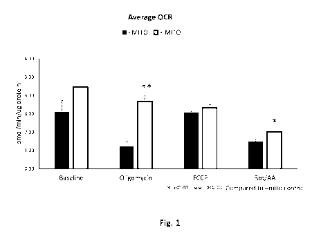
[0024] Figure
1 (Fig. 1) shows that treatment of human pulmonary artery endothelial
cells (HPAEC) with mitochondria isolated from pig hearts (i.e., porcine
mitochondria)
increases the oxygen consumption rate (OCR) after acute cold exposure. HPAEC
were
placed in 4 C for 6 hours. HPAEC recovered in normoxia for 1 hour at 37 C in
the
presence of either 20 tL of mitochondria suspension (respiration buffer
containing 29
particles per cell; "+ MITO") or 20 of respiration buffer only ("- MITO")
and
equilibrated in a non-0O2 incubator for 10 minutes. A "Mitochondrial Stress
Test" was
then performed using a Seahorse instrument with 10 i.tM oligomycin, 20 i.tM
FCCP, and
rotenone/antimycin A (Rot/AA). Porcine mitochondria treatment increased OCR at
baseline (43.6% increase), oligomycin-treated HPAEC (204.9% increase), FCCP-
treated
HPAEC (8.4% increase), and Rot/AA-treated HPAEC (34.1% increase) in comparison
to
the corresponding baseline, oligomycin-treated, FCCP-treated, or Rot/AA-
treated "-
MITO" HPAEC control. Statistical analysis performed was a two-tailed t-test (*
p <0.05;
** p < 0.01).
[0025]
Figure 2 (Fig. 2) shows that porcine mitochondria treatment of human pulmonary
artery endothelial cells (HPAEC) increases OCR after chronic cold exposure.
HPAEC
were placed in 4 C for 12 hours. HPAEC recovered in normoxia for 1 hour at 37
C in the
presence of 20 of
mitochondria suspension (respiration buffer containing 172 particles
per cell; "+ MITO") or 20 of
respiration buffer only ("- MITO") and equilibrated in a
non-0O2 incubator for 50 minutes. HPAEC were rested in the Seahorse instrument
at
37 C under non-0O2 conditions. A "Mitochondrial Stress Test" was then
performed with
the Seahorse instrument with 10 tM oligomycin, 20 i.tM FCCP, and 5 tM
rotenone/antimycin A (Rot/AA). Porcine mitochondria treatment increased OCR at
baseline (32.4% increase), oligomycin-treated HPAEC (51.9% increase), FCCP-
treated
HPAEC (9.5% increase), and Rot/AA-treated HPAEC (45.2% increase) in comparison
to
the corresponding baseline, oligomycin-treated, FCCP-treated, or Rot/AA-
treated "-
CA 03144252 2021-12-17
WO 2020/257281 PCT/US2020/038133
MITO" HPAEC control. Statistical analysis performed was a two-tailed t-test
(** p <
0.01).
[0026] Figure 3 (Fig. 3) shows that HPAEC exposed to cold stress take up
porcine
mitochondria. Porcine mitochondria were administered to HPAEC undergoing cold
stress. For the cold recovery group, HPAEC under cold stress take up the
porcine
mitochondria in a dose-dependent manner, and maximal expression of porcine
MtND5 is
achieved at 1,666 particles per cell. In the cold recovery condition, maximal
expression
of porcine MtND5 is achieved at 24 hours, where a 26,201% increase in porcine
MtND5
was observed compared to the untreated cold-recovery control. In the cold
exposure
condition, maximal expression of porcine MtND5 is achieved at 72 hours where a
301,932% increase in MtND5 was observed compared to the untreated cold-
exposure
control. Statistical analysis performed was a one-way ANOVA (* P<0.05 24 hour
compared to normoxia; # P<0.05 48 hour compared to normoxia; + P<0.05 72 hour
compared to normoxia; $ P<0.05 24 hour compared to cold control; A P<0.05 48
hour
compared to cold control; & P<0.05 72 hour compared to cold control).
[0027] Figure 4 (Fig. 4) shows that transcription of human mitochondrial
DNA in
HPAEC exposed to cold stress is largely unaffected by porcine mitochondria
treatment.
Untreated control HPAEC under cold recovery conditions demonstrated a 55%
increase
in human MtND5 expression compared to normothermic controls. This increase was
moderated by porcine mitochondria treatment, where 1 particle/cell
demonstrated a 3.8%
reduction in expression compared to untreated normothermic HPAEC and a 33%
reduction in expression compared to the untreated cold-recovery control. In
the cold
exposure group, maximal expression of human MtND5 was achieved at 72 hours,
but this
increase was not significantly impacted by porcine mitochondria treatment.
Statistical
analysis performed was a one-way ANOVA (* P<0.05 24 hour compared to normoxia;
#
P<0.05 48 hour compared to normoxia; + P<0.05 72 hour compared to normoxia; $
P<0.05 24 hour compared to cold control; A P<0.05 48 hour compared to cold
control; &
P<0.05 72 hour compared to cold control).
11
CA 03144252 2021-12-17
WO 2020/257281 PCT/US2020/038133
[0028] Figure 5 (Fig. 5) shows that porcine mitochondria treatment of HPAEC
reduces
NF-x13 expression in cold recovery at 24 hours. In the cold recovery
condition, untreated
control HPAEC demonstrated an 83% increase in NF--03 gene expression at 24
hours
compared to normothermic controls. Porcine mitochondria treatment trended to
decrease
NF-x13 expression compared to untreated cold-recovery control HPAEC, with 1
particle/cell demonstrating a 22% decrease compared to untreated cold-recovery
control
HPAEC. In the cold exposure condition, a slight increase in NF-x13 expression
occurs at
24 hours in HPAEC treated with porcine mitochondria, but this increase is not
statistically significant. Statistical analysis performed was a one-way ANOVA
(* P<0.05
24 hour compared to normoxia; # P<0.05 48 hour compared to normoxia; + P<0.05
72
hour compared to normoxia; $ P<0.05 24 hour compared to cold control; A P<0.05
48
hour compared to cold control; & P<0.05 72 hour compared to cold control).
[0029] Figure 6 (Fig. 6) shows that porcine mitochondria treatment of HPAEC
decreases
toll-like receptor-9 (TLR-9) expression in cold recovery after 24 hours. HPAEC
were
treated, cultured under cold recovery or cold exposure conditions, and
harvested at 24-
hour, 48-hour, or 72-hour time points. In the cold recovery condition,
untreated control
HPAEC demonstrated a 101% increase in TLR-9 expression at 24 hours compared to
normothermic controls. Porcine mitochondria treatment trended to decrease the
TLR-9
expression compared to untreated cold-recovery control HPAEC, with 166
particles/cell
demonstrating a 37% decrease compared to untreated cold-recovery control
HPAEC. In
cold exposure conditions, maximal expression of TLR-9 occurs in HPAEC treated
with 1
particle/cell, where a 60% increase in TLR-9 expression was observed compared
to the
untreated cold-exposure control HPAEC. Statistical analysis performed was a
one-way
ANOVA (* P<0.05 24 hour compared to normoxia; # P<0.05 48 hour compared to
normoxia; + P<0.05 72 hour compared to normoxia; $ P<0.05 24 hour compared to
cold
control; A P<0.05 48 hour compared to cold control; & P<0.05 72 hour compared
to cold
control).
12
CA 03144252 2021-12-17
WO 2020/257281 PCT/US2020/038133
[0030] Figure 7 (Fig. 7) shows that porcine mitochondria treatment of HPAEC
impacts
the expression of heme oxygenase-1 (H0-1) in cold exposure at 24 hours.
Porcine
mitochondria treatment increased HO-1 expression in the cold exposure
condition.
Porcine mitochondria treatment was maximally effective at 16 particles/cell,
where a
24% increase in HO-1 expression was seen compared to untreated cold-exposure
control
HPAEC (242% increase compared to untreated normothermic control HPAEC).
Statistical analysis performed was a one-way ANOVA (* P<0.05 24 hour compared
to
normoxia; # P<0.05 48 hour compared to normoxia; + P<0.05 72 hour compared to
normoxia; $ P<0.05 24 hour compared to cold control; A P<0.05 48 hour compared
to
cold control; & P<0.05 72 hour compared to cold control).
[0031] Figure 8 (Fig. 8) shows that porcine mitochondria treatment of HPAEC
decreases
macrophage-colony stimulating factor (M-CSF) secretion under hypoxic
conditions.
Porcine mitochondria treatment is maximally effective at 3 particles/cell,
where M-CSF
secretion was reduced by 65% compared to untreated hypoxia control HPAEC at 48
hours. Statistical analysis performed was a one-way ANOVA (* P<0.05 24 hour
compared to normoxia; # P<0.05 48 hour compared to normoxia; + P<0.05 72 hour
compared to normoxia; $ P<0.05 24 hour compared to hypoxia control; A P<0.05
48 hour
compared to hypoxia control; & P<0.05 72 hour compared to hypoxia control).
[0032] Figure 9 (Fig. 9) shows that porcine mitochondria treatment of HPAEC
decreases
macrophage inflammatory protein-13 (MIP-10) secretion under hypoxic
conditions.
Porcine mitochondria treatment was maximally effective in reducing MIP-10
secretion at
3 particles/cell, where MIP-10 secretion was reduced by 73% compared to
untreated
hypoxia control HPAEC at 48 hours. A decrease in potency is seen at 3,687
particles/cell.
Statistical analysis performed was a one-way ANOVA (* P<0.05 24 hour compared
to
normoxia; # P<0.05 48 hour compared to normoxia; + P<0.05 72 hour compared to
normoxia; $ P<0.05 24 hour compared to hypoxia control; A P<0.05 48 hour
compared to
hypoxia control; & P<0.05 72 hour compared to hypoxia control).
13
CA 03144252 2021-12-17
WO 2020/257281 PCT/US2020/038133
[0033] Figure 10 (Fig. 10) shows that porcine mitochondria treatment of
HPAEC
decreases platelet-derived growth factor-BB (PDGF-BB) secretion under hypoxic
conditions. Porcine mitochondria treatment was maximally effective in reducing
PDGF-
BB secretion at 36 particles/cell, where PDGF-BB secretion was reduced by 69%
compared to untreated hypoxia control HPAEC at 48 hours. A decrease in potency
is
seen at 3,687 particles/cell. Statistical analysis performed was a one-way
ANOVA (*
P<0.05 24 hour compared to normoxia; # P<0.05 48 hour compared to normoxia; +
P<0.05 72 hour compared to normoxia; $ P<0.05 24 hour compared to hypoxia
control; A
P<0.05 48 hour compared to hypoxia control; & P<0.05 72 hour compared to
hypoxia
control).
[0034] Figure 11 (Fig. 11) shows that porcine mitochondria treatment of
HPAEC
decreases RANTES (CCL5) secretion under hypoxic conditions. Porcine
mitochondria
treatment was maximally effective in reducing RANTES secretion at 0.3
particles/cell,
where RANTES secretion was reduced by 59% compared to untreated hypoxia
control
HPAEC at 48 hours. A decrease in potency is seen at 3,687 particles/cell.
Statistical
analysis performed was a one-way ANOVA (* P<0.05 24 hour compared to normoxia;
#
P<0.05 48 hour compared to normoxia; + P<0.05 72 hour compared to normoxia; $
P<0.05 24 hour compared to hypoxia control; A P<0.05 48 hour compared to
hypoxia
control; & P<0.05 72 hour compared to hypoxia control).
[0035] Figure 12 (Fig. 12) shows that porcine mitochondria treatment of
HPAEC
decreases intracellular adhesion molecule-1 (ICAM-1) secretion under hypoxic
conditions. Porcine mitochondria treatment was maximally effective in reducing
ICAM-1
secretion at 0.3 particles/cell, where ICAM-1 secretion was reduced by 82%
compared to
untreated hypoxia control HPAEC at 48 hours. A decrease in potency is seen at
3,687
particles/cell. Statistical analysis performed was a one-way ANOVA (* P<0.05
24 hour
compared to normoxia; # P<0.05 48 hour compared to normoxia; + P<0.05 72 hour
compared to normoxia; $ P<0.05 24 hour compared to hypoxia control; A P<0.05
48 hour
compared to hypoxia control; & P<0.05 72 hour compared to hypoxia control).
14
CA 03144252 2021-12-17
WO 2020/257281 PCT/US2020/038133
[0036] Figure 13 (Fig. 13) shows that porcine mitochondria treatment of
HPAEC
decreases brain-derived neurotrophic factor (BDNF) secretion under hypoxic
conditions.
Porcine mitochondria treatment was maximally effective in reducing BDNF
secretion at
3 particles/cell, where BDNF secretion was reduced by 85% compared to
untreated
hypoxia control HPAEC at 48 hours. Statistical analysis performed was a one-
way
ANOVA (* P<0.05 24 hour compared to normoxia; # P<0.05 48 hour compared to
normoxia; + P<0.05 72 hour compared to normoxia; $ P<0.05 24 hour compared to
hypoxia control; A P<0.05 48 hour compared to hypoxia control; & P<0.05 72
hour
compared to hypoxia control).
[0037] Figure 14 (Fig. 14) shows that porcine mitochondria treatment of
HPAEC
decreases interleukin-10 (IL-113) secretion under hypoxic conditions. Porcine
mitochondria treatment was maximally effective in reducing IL-113 secretion at
368
particles/cell, where IL-113 secretion was reduced by 70% compared to
untreated hypoxia
control HPAEC at 48 hours. Statistical analysis performed was a one-way ANOVA
(*
P<0.05 24 hour compared to normoxia; # P<0.05 48 hour compared to normoxia; +
P<0.05 72 hour compared to normoxia; $ P<0.05 24 hour compared to hypoxia
control; A
P<0.05 48 hour compared to hypoxia control; & P<0.05 72 hour compared to
hypoxia
control).
[0038] Figure 15 (Fig. 15) shows that porcine mitochondria treatment of
HPAEC
decreases growth/differentiation factor 15 (GDF15) secretion under hypoxic
conditions.
Porcine mitochondria treatment was maximally effective in reducing GDF15
secretion at
3 particles/cell, where GDF15 secretion was reduced by 70% compared to
untreated
hypoxia control HPAEC at 48 hours. Statistical analysis performed was a one-
way
ANOVA (* P<0.05 24 hour compared to normoxia; # P<0.05 48 hour compared to
normoxia; + P<0.05 72 hour compared to normoxia; $ P<0.05 24 hour compared to
hypoxia control; A P<0.05 48 hour compared to hypoxia control; & P<0.05 72
hour
compared to hypoxia control).
CA 03144252 2021-12-17
WO 2020/257281 PCT/US2020/038133
[0039] Figure 16 (Fig. 16) shows that porcine mitochondria treatment of
HPAEC
decreases interleukin-6 (IL-6) secretion under hypoxic conditions. Porcine
mitochondria
treatment was maximally effective in reducing IL-6 secretion at 368
particles/cell, where
IL-6 secretion was reduced by 70% compared to untreated hypoxia control HPAEC
at 48
hours. Statistical analysis performed was a one-way ANOVA (* P<0.05 24 hour
compared to normoxia; # P<0.05 48 hour compared to normoxia; + P<0.05 72 hour
compared to normoxia; $ P<0.05 24 hour compared to hypoxia control; A P<0.05
48 hour
compared to hypoxia control; & P<0.05 72 hour compared to hypoxia control).
[0040] Figure 17 (Fig. 17) shows that porcine mitochondria treatment of
HPAEC
decreases transforming growth factor-01 (TGF-01) secretion under hypoxic
conditions.
Porcine mitochondria treatment was maximally effective in reducing TGF-01
secretion at
36 particles/cell, where TGF-01 secretion was reduced by 95% compared to
untreated
hypoxia control HPAEC at 48 hours. Statistical analysis performed was a one-
way
ANOVA (* P<0.05 24 hour compared to normoxia; # P<0.05 48 hour compared to
normoxia; + P<0.05 72 hour compared to normoxia; $ P<0.05 24 hour compared to
hypoxia control; A P<0.05 48 hour compared to hypoxia control; & P<0.05 72
hour
compared to hypoxia control).
[0041] Figure 18 (Fig. 18) shows that HPAEC exposed to hypoxic stress take
up porcine
mitochondria. For the hypoxia recovery group, HPAEC were cultured in normoxia
for 24
hours and then in hypoxia (1% 02) for 24 hours prior to porcine mitochondria
treatment.
After porcine mitochondria treatment, the hypoxia recovery cells were placed
back in
normoxia. The hypoxia recovery HPAEC were harvested after 24, 28, or 72 hours
of
culture in normoxia. For the hypoxia exposure group, HPAEC were cultured in
normoxia
for 48 hours, treated with porcine mitochondria, and immediately placed in
hypoxia (1%
02). The hypoxia exposure HPAEC were harvested after 24, 28, or 72 hours of
hypoxia
exposure. As determined using a probe specific for porcine MtND5, HPAEC under
hypoxic stress take up the porcine mitochondria in a dose-dependent manner,
and
maximal expression of porcine MtND5 is achieved at 1,666 particles per cell.
In the
16
CA 03144252 2021-12-17
WO 2020/257281 PCT/US2020/038133
hypoxia recovery condition, maximal expression of porcine MtND5 is achieved at
48
hours, where a 4,655% increase in porcine mtND5 was observed compared to the
untreated hypoxia-recovery control. In the hypoxia exposure condition, maximal
expression is achieved at 24 hours, where a 26,680% increase in porcine mtND5
was
observed compared to the untreated hypoxia-exposure control. Statistical
analysis
performed was a one-way ANOVA (* P<0.05 24 hour compared to normoxia; # P<0.05
48 hour compared to normoxia; + P<0.05 72 hour compared to normoxia; $ P<0.05
24
hour compared to hypoxia control; A P<0.05 48 hour compared to hypoxia
control; &
P<0.05 72 hour compared to hypoxia control).
[0042] Figure 19 (Fig. 19) shows that transcription of human mitochondrial
DNA in
HPAEC exposed to hypoxic stress is largely unaffected by porcine mitochondria
treatment. As determined using a probe specific for human MtND5, maximal
expression
of human MtND5 for both the hypoxia recovery group and the hypoxia exposure
group
occurs at 72 hours. The time point that appears impacted by porcine
mitochondria
treatment occurs at 24 hours. In the hypoxia recovery group, there is a trend
for decreased
human MtND5 expression in HPAEC treated with porcine mitochondria, with 1
particle/cell demonstrating a 33% reduced expression compared to untreated
hypoxic
controls at 24 hours. In the hypoxia exposure group, there is a trend for
increased human
MtND5 expression in HPAEC treated with porcine mitochondria, with 1,666
particles/cell resulting in a 36% increase compared to untreated hypoxia-
exposure cells at
24 hours. Statistical analysis performed was a one-way ANOVA (* P<0.05 24 hour
compared to normoxia; # P<0.05 48 hour compared to normoxia; + P<0.05 72 hour
compared to normoxia; $ P<0.05 24 hour compared to hypoxia control; A P<0.05
48 hour
compared to hypoxia control; & P<0.05 72 hour compared to hypoxia control).
[0043] Figure 20 (Fig. 20) shows that porcine mitochondria treatment of
HPAEC
reduces TLR-9 expression in hypoxia recovery but increases TLR-9 expression in
hypoxia exposure at 24 hours. For both the hypoxia recovery group and the
hypoxia
exposure group, maximal expression of TLR-9 occurs at 24 hours. In the hypoxia
17
CA 03144252 2021-12-17
WO 2020/257281 PCT/US2020/038133
recovery group, there is a trend for decreased TLR-9 expression in HPAEC
treated with
porcine mitochondria, with 1 particle/cell demonstrating a 38% reduced
expression
compared to untreated hypoxic controls at 24 hours. In the hypoxia exposure
group, there
is a trend for increased TLR9 expression in HPAEC treated with porcine
mitochondria,
with 1,666 particles/cell resulting in a 32% increase compared to untreated
hypoxia-
exposure cells at 24 hours. Statistical analysis performed was a one-way ANOVA
(*
P<0.05 24 hour compared to normoxia; # P<0.05 48 hour compared to normoxia; +
P<0.05 72 hour compared to normoxia; $ P<0.05 24 hour compared to hypoxia
control; A
P<0.05 48 hour compared to hypoxia control; & P<0.05 72 hour compared to
hypoxia
control).
[0044] Figure 21 (Fig. 21) shows that porcine mitochondria treatment of
HPAEC
undergoing hypoxic stress reduces mRNA expression of interleukin-8 (IL-8;
CXCL8),
IL-6, BH3 interacting-domain death agonist (BID), human MtND1, and human
mitochondrial cytochrome B (Mt-CyB). Porcine mitochondria treatment of hypoxic
HPAEC is maximally effective for reducing IL-8 expression at 3,687
particles/cell, where
a 58% decrease in IL-8 expression was seen compared to untreated hypoxic
controls (Fig.
21A). Porcine mitochondria treatment of hypoxic HPAEC is maximally effective
for
reducing IL-6 expression at 3 particles/cell, where a 30% decrease in IL-6
expression was
seen compared to untreated hypoxic controls (Fig. 21B). Porcine mitochondria
treatment
of hypoxic HPAEC is maximally effective for reducing BID expression at 36
particles/cell, where a 30% decrease in BID expression was seen compared to
untreated
hypoxic controls (Fig. 21C). Porcine mitochondria treatment of hypoxic HPAEC
is
maximally effective for reducing human MtND1 expression at 3 particles/cell,
where a
57% decrease in MtND1 expression was seen compared to untreated hypoxic
controls
(Fig. 21D). Porcine mitochondria treatment of hypoxic HPAEC is maximally
effective
for reducing human Mt-CyB expression at 0.3 particles/cell, where a 57%
decrease in
MtCyB expression was seen compared to untreated hypoxic controls (Fig. 21E).
18
CA 03144252 2021-12-17
WO 2020/257281 PCT/US2020/038133
Statistical analysis performed was a one-way ANOVA (* P<0.05 24 hour compared
to
normoxia; $ P<0.05 24 hour compared to hypoxia control).
[0045] Figure 22 (Fig. 22) shows that treatment of human endothelial cells
with porcine
mitochondria decreases hypoxia-induced cell proliferation as indicated by a
decrease in
total cellular protein content of mitochondria treated HPAEC. HPAEC were
treated with
0, 5, 6, or 7 porcine mitochondria per cell and subjected to hypoxic
conditions for 24
hours. After the 24-hour exposure to hypoxia, total cellular protein content
was measured
for each sample via bicinchoninic acid (BCA) assay on HPAEC lysate.
Statistical
analysis performed was a one-way ANOVA (* P<0.05 compared to control HPAEC not
treated with porcine mitochondria).
[0046] Figure 23 (Fig. 23) shows that porcine mitochondria treatment of
human alveolar
epithelial type II (AT2) cells improved the nucleic acid content of the AT2
cells. AT2
cells were seeded directly from cryo-storage with and without porcine
mitochondria and
incubated overnight in a standard incubator. Following overnight incubation,
the nucleic
acid content of AT2 cells treated with porcine mitochondria increased by 23%
compared
to the untreated AT2 cell control.
[0047] Figure 24 (Fig. 24) shows the mitochondrial activity of isolated
porcine
mitochondria at various concentrations in respiration buffer containing
adenosine
diphosphate (ADP).
[0048] Figure 25 (Fig. 25) shows that porcine mitochondria retain
mitochondrial activity
after cold storage at -80 C. While mitochondria activity decreased at 4 C
over time,
storage at -80 C resulted in retention of approximately 40% OCR
(mitochondrial
activity). Storage in trehalose improved OCR, resulting in approximately 60%
retention
in original OCR rate.
[0049] Figure 26 (Fig. 26) shows that porcine mitochondria treatment
improves the
function of an isolated porcine cadaveric lung while on ex vivo lung perfusion
(EVLP). In
19
CA 03144252 2021-12-17
WO 2020/257281 PCT/US2020/038133
comparison to the right lung control, isolated porcine mitochondria injected
into the left
lung increased proliferating cell nuclear antigen (PCNA) positive cells in the
lower lung
(Fig. 26A), upper lung (Fig. 26B), and mid-lung (Fig. 26C) as measured by
histology
(Fig. 26A). Porcine mitochondria treatment was maximally effective at 24 hours
in the
lower lung (Fig. 26A), where a 50% improvement was seen in porcine
mitochondria-
treated cells compared to control (arrow).
[0050] Figure 27 (Fig. 27) shows that porcine mitochondria treatment
improves the
parameters of tidal volume (Fig. 27A) and dynamic compression (Fig. 27B) of an
isolated
porcine cadaveric lung while on EVLP. Isolated porcine mitochondria were
injected into
an isolated porcine cadaveric lung on EVLP, and perfusion was turned off for
10 minutes
while the lung continued inflation. Tidal volume (m1) and dynamic compression
(TV/(PIP-PEEP)) were determined at 10 minutes post-injection, 1 hour post-
injection,
and 4 hours post-injection (TV = tidal volume; PIP = peak inspiratory
pressure; PEEP =
positive end expiratory pressure). Baseline tidal volume and dynamic
compression
represent pre-injection tidal volume and dynamic compression, respectively. A
30%
improvement in tidal volume and a 40% increase in dynamic compression are seen
at 10
minutes post-injection in comparison to baseline.
[0051] Figure 28 (Fig. 28) shows that, following injection of isolated
porcine
mitochondria into an isolated porcine cadaveric lung on EVLP, there was an
immediate
and progressive drop in media glucose as well as a 17% decrease in circulating
ammonium at one hour post-injection. An isolated porcine cadaveric lung on
EVLP was
injected with isolated porcine mitochondria 24 minutes after commencement of
EVLP
and maintained on EVLP for approximately 20 hours. Glucose (g/L) in the
circulating
media was quantitated using BioPat (Fig. 28A) and Nova (Fig. 28B), and
circulating
ammonium (NH4+; mmol/L) was quantitated using Nova (Fig. 28C). Initial Nova
glucose
and ammonium levels represent Nova glucose and ammonium levels at time 0 post-
EVLP. Baseline Nova glucose and ammonium levels represent Nova glucose and
ammonium levels immediately prior to injection of the porcine mitochondria.
CA 03144252 2021-12-17
WO 2020/257281 PCT/US2020/038133
[0052] Figure 29 (Fig. 29) shows that injection of isolated porcine
mitochondria into a
porcine cadaveric lung on EVLP ("+Mito") increases tidal volume (mL/kg; Fig.
29A) and
gas exchange (AP02/Fi02; Fig. 29B) in comparison to a porcine cadaveric lung
on EVLP
injected with respiration buffer ("Control").
[0053] Figure 30 (Fig. 30) shows that injection of isolated porcine
mitochondria into a
porcine cadaveric lung on EVLP ("+MITO") decreases the amount of circulating
lactate
(mg/ml; Fig. 30A), leading to an increased glucose/lactate ratio (Fig. 30B) in
comparison
to a porcine cadaveric lung on EVLP injected with respiration buffer
("Control").
[0054] Figure 31 (Fig. 31) shows that injection of isolated porcine
mitochondria into a
porcine cadaveric lung on EVLP ("+MITO") decreases the percentage of apoptotic
cells
(% TUNEL; Fig. 31A) and increases expression of the cellular adhesion molecule
CD31
(Fig. 31B) in comparison to a porcine cadaveric lung injected with respiration
buffer
("Control"). The percentage of apoptotic cells was determined by TUNEL assay
on tissue
biopsies taken from the porcine cadaveric lungs during EVLP. CD31 expression
was
determined by immunofluorescence staining of tissue biopsies with an anti-CD31
antibody.
[0055] Figure 32 (Fig. 32) shows that the health and function of isolated
mitochondria
can be rapidly assessed by measuring changes in the size and complexity of
mitochondria, mitochondria membrane permeability transition pore (mPTP)
opening, or
mitochondria respiration. The size and complexity of healthy and damaged
mitochondria
were measured using flow cytometry. Compared to healthy mitochondria, the
damaged
mitochondria were larger and less complex, which is indicative of a
mitochondrial
swelling phenotype (Fig. 32A). mPTP opening was assessed using flow cytometry
to
measure green fluorescent (FITC) emission of calcein acetoxymethyl (AM)-
stained
mitochondria. Mitochondria were considered as having a regulated mPTP if they
retained
calcein-AM, resulting in FITC+ staining. Mitochondria were considered as
having
dysregulated, continuous mPTP opening if they were unable to retain calcein-
AM,
21
CA 03144252 2021-12-17
WO 2020/257281 PCT/US2020/038133
resulting in reduced FITC staining. Compared to healthy mitochondria, the
damaged
mitochondria had drastically reduced FITC emission due to their inability to
retain
calcein AM (Fig. 32B). To evaluate mitochondria respiration, respiratory
control ratios
(RCRs) were determined using the Seahorse instrument. RCRs were calculated
from the
oxygen consumption rate (OCR) during ADP-stimulated respiration (RCR) and
uncoupled respiration (RCRmax). The OCR during each of these two states was
divided
by the basal OCR to obtain the OCR ratio. Maximal respiration was achieved by
injecting
the mitochondrial protonophore uncoupler BAM15. Compared to healthy
mitochondria,
the damaged mitochondria had dramatically reduced ADP-stimulated respiration
rates
and uncoupled respiration rates (Fig. 32C).
[0056] Figure 33 (Fig. 33) shows that the health and function of isolated
mitochondria
can be rapidly assessed by measuring mitochondria membrane potential or
mitochondria
membrane permeability. Changes in mitochondria membrane potential were
assessed by
flow cytometry using a JC-1 assay. Mitochondria depolarization is indicated by
a
decrease in the red:green fluorescence intensity ratio or by a decrease in the
signal
intensity in the phycoerythrin (PE) channel. Compared to healthy mitochondria,
damaged
mitochondria had a decreased red:green ratio and a drastically reduced PE
emission (Fig.
33A). Mitochondria permeability was measured by flow cytometry using a SYTOX
green
nucleic acid stain, which easily permeates mitochondria with compromised
membranes.
Damaged mitochondria stained with SYTOX green will have higher FITC signal
intensity than non-damaged mitochondria stained with SYTOX green. Compared to
healthy mitochondria, the damaged mitochondria demonstrated increased FITC
emission
(Fig. 33B).
[0057] Figure 34 (Fig. 34) shows that mitochondria retain mitochondrial
function after
cold storage at -80 C, as measured by mitochondria size, complexity, mPTP
opening, and
respiration. The presence or absence of mitochondrial swelling was assessed
using flow
cytometry to measure size and complexity of mitochondria stored under non-
preserving
conditions (i.e., storage at 4 C) or preserving conditions (i.e., storage at -
80 C). While
22
CA 03144252 2021-12-17
WO 2020/257281 PCT/US2020/038133
mitochondria stored at 4 C almost immediately displayed a swelling phenotype
(i.e.,
increased size, decreased complexity), mitochondria stored at -80 C retained
a normal
phenotype comparable to freshly isolated mitochondria throughout the duration
of storage
(out to 7 months) (Fig. 34A). Mitochondria mPTP opening was assessed using
flow
cytometry to measure FITC emission of calcein AM-stained mitochondria stored
under
non-preserving conditions or preserving conditions. Mitochondria were
considered as
maintaining mPTP if they retained calcein-AM, resulting in FITC+ staining.
Mitochondria were considered as failing to maintain mPTP opening if they were
unable
to retain calcein-AM, resulting in reduced FITC staining. While mitochondria
stored at 4
C lost the ability to regulate their mPTP opening, mitochondria stored at -80
C
controlled mPTP opening comparable to freshly isolated mitochondria throughout
the
duration of storage (out to 7 months) (Fig. 34B). To evaluate mitochondria
respiration of
mitochondria stored under non-preserving conditions or preserving conditions,
RCRs
were determined using the Seahorse instrument. RCRs were calculated from the
OCR
during ADP-stimulated RCR and uncoupled respiration (RCRmax). The OCR during
each of these two states was divided by the basal OCR to obtain the OCR ratio.
Maximal
respiration was achieved by injecting the mitochondrial protonophore uncoupler
BAM15.
The ADP-stimulated respiration rates and uncoupled respiration rates of
mitochondria
stored at 4 C declined over time, while mitochondria stored at -80 C had ADP-
stimulated respiration rates (Fig. 34C) and uncoupled respiration rates (Fig.
34D)
comparable to freshly isolated mitochondria throughout the duration of storage
(out to 6
weeks).
[0058] Figure 35 (Fig. 35) shows that mitochondria retain mitochondrial
function after
cold storage at -80 C, as measured by mitochondria membrane potential and
mitochondria membrane permeability. Changes in mitochondria membrane potential
of
mitochondria stored under non-preserving conditions (i.e., storage at 4 C) or
preserving
conditions (i.e., storage at -80 C) were assessed by flow cytometry using the
JC-1 assay.
Mitochondria depolarization is indicated by a decrease in the red:green
fluorescence
23
CA 03144252 2021-12-17
WO 2020/257281 PCT/US2020/038133
intensity ratio or by a decrease in the signal intensity in the phycoerythrin
(PE) channel.
While mitochondria stored at 4 C showed a dramatic reduction in membrane
potential,
mitochondria stored at -80 C retained membrane potential comparable to
freshly isolated
mitochondria throughout the duration (out to 7 months) (Fig. 35A).
Permeability of
mitochondria stored under non-preserving conditions or preserving conditions
was
measured by flow cytometry using a SYTOX green nucleic acid stain, which
easily
permeates mitochondria with compromised membranes. Damaged mitochondria
stained
with SYTOX green will have higher FITC signal intensity than non-damaged
mitochondria stained with SYTOX green. While mitochondria stored at 4 C had
an
immediate increase in FITC emission, mitochondria stored at -80 C retained
membrane
potential comparable to freshly isolated mitochondria through the duration of
storage (out
to 7 months) (Fig. 35B).
[0059] Figure 36 (Fig. 36) shows that mitochondria retain mitochondrial
function after
cold storage at -80 C, as measured by their ability to reduce reactive oxygen
species
(ROS)-mediated chemokine secretion in HPAEC. HPAEC were cultured with 25 [tM
menadione with or without mitochondria treatment. Mitochondria used in these
experiments were stored under either non-preserving conditions (i.e., storage
at 4 C) or
preserving conditions (i.e., storage at -80 C). Chemokines in the culture
media of treated
HPAEC were measured using bead-based immunoassays. Mitochondria stored at 4 C
rapidly lost their ability to modulate secretion of IL-8/CXCL8 (Fig. 36A),
MIG/CXCL9
(Fig. 36B), MCP-1/CCL2 (Fig. 36C), and GROa/CXCL1 (Fig. 36D) compared to
mitochondria stored at -80 C, which retained the ability to reduce chemokine
secretion.
[0060] Figure 37 (Fig. 37) shows that mitochondria stored at -80 C have
the same gross
morphology (Fig. 37A) and average size (Fig. 37B) as freshly isolated
mitochondria.
Mitochondria scored as class I had a condensed, normal state (i.e., non-
damaged state)
represented by numerous narrow pleomorphic cristae in a contiguous electron-
dense
matrix space. Mitochondria scored as class II were in a state of remodeling
characterized
by reorganized cristae and matrix spaces. The appearance of the remodeling
state is
24
CA 03144252 2021-12-17
WO 2020/257281 PCT/US2020/038133
temporally correlated with the redistribution and availability of cytochrome c
from the
intermembrane space. Mitochondria scored as class III were swollen and
damaged. Class
III mitochondria had intact membranes, but the cristae of these mitochondria
have
deteriorated and gathered close to the perimeter of the mitochondria.
Mitochondria scored
as class IV were terminally swollen or ruptured. Class IV mitochondria showed
gross
morphological derangement, including asymmetric blebbing of matrix.
Mitochondria
scored as "condensed matrix (CM)" had a condensed matrix with no limiting
outer
membrane.
[0061] Figure 38 (Fig. 38) shows that intact mitochondria are the
functional component
in mitochondria treatment as opposed to a component released from the
mitochondria
after storage at -80 C or carried over from the isolation process.
Mitochondrial and non-
mitochondrial fractions were obtained by centrifugation from mitochondria
stored for two
weeks at -80 C. HPAEC were cultured with 25 [NI menadione and treated
volumetrically with either the mitochondria fraction or the non-mitochondria
fraction.
The volumes of 0.02%, 0.2%, 2%, and 20% correspond to 1 mitochondria/cell, 10
mitochondria/cell, 100 mitochondria/cell, and 1,000 mitochondria/cell,
respectively.
Parameters analyzed included secretion of the inflammatory chemokines IL-
8/CXCL8
(Fig. 38A), MCP-1/CCL-2 (Fig. 38B), and GROa/CXCL-1 (Fig, 38C), as well as
lactate
dehydrogenase (LDH) release (Fig. 38D), which is indicative of cell damage.
The
mitochondrial fraction alone retained the ability to reduce chemokine
secretion and LDH
release.
[0062] Figure 39 (Fig. 39) shows that porcine mitochondria treatment
improves kidney
function and recovery in vivo after acute kidney injury in an
ischemia/reperfusion (I/R)
mouse model. Acute FR injury was achieved in adult mice by clamping the renal
artery
for 45 minutes followed by reperfusion. Mice were injected with mitochondria
(0.01x or
0.1x) or the vehicle control upon reperfusion on day 1. Blood urea nitrogen
(BUN),
which is an indicator of kidney function, was increased after I/R injury and
trended to
decrease at day 2 and on day 4 after mitochondria injection (0.1x) (Fig. 39A).
Kidney
CA 03144252 2021-12-17
WO 2020/257281 PCT/US2020/038133
index, which is the percent mouse weight taken up by the kidney, was increased
after FR
injury and was reduced after mitochondria injection (0.01x) (Fig. 39B). Kidney
injury
molecule-1 (KIM1) is a marker of acute kidney injury. While FR injury
increased KIM1
serum levels, mitochondria treatment reduced these levels in a dose-responsive
manner
(Fig. 39C). Monocyte chemoattractant protein 1 (MCP1) is a proinflammatory
cytokine
associated with acute kidney injury. While I/R injury increased MCP1 serum
levels,
mitochondria treatment reduced these levels in a dose-responsive manner (Fig.
39D). The
C3a and C5a members of the compliment system induce inflammatory mediators
from
both renal tubular epithelial cells and macrophages after
hypoxia/reoxygenation. While
I/R injury increased serum levels of C3a (Fig. 39E) and C5a (Fig. 39F),
mitochondria
treatment reduced these levels in a dose-dependent manner (Fig. 39E-F). The
mitochondria used in these studies were stored for approximately one month at -
80 C
prior to injection. Statistical analysis performed was a one-way ANOVA (#
P<0.05
compared to sham; * P<0.05 compared to model + vehicle).
[0063] Figure 40 (Fig. 40) shows that porcine mitochondria treatment
improved the
expression of gap junction markers and reduced DNA oxidation in an isolated
porcine
cadaveric lung placed on EVLP following cold ischemic injury. EVLP was run on
isolated porcine cadaveric lungs after approximately 20 hours of cold ischemia
time.
Mitochondria treatment improved expression of gap junction markers junctional
adhesion
molecule 1 (JAM1) (Fig. 40A) and CD31 (Fig. 40B) in EVLP after 1 hour in the
superior
lobe and after 4 hours when measured in the distal segment of the caudal lobe,
the
proximal segment of the caudal lobe, and the superior lobe. 8-hydroxy-2"-
deoxyguanosine (8-0HdG) is a marker of ROS-induced DNA oxidation. Mitochondria
treatment decreased expression of 8-0HdG in lung tissue during EVLP after 1
hour in the
superior lobe and after 4 hours when measured in the distal segment of the
caudal lobe,
the proximal segment of the caudal lobe, the inferior lobe, and the superior
lobe (Fig.
40C). Protein expression was normalized to DAPI nuclear staining, and all data
was
26
CA 03144252 2021-12-17
WO 2020/257281 PCT/US2020/038133
normalized to baseline pre-EVLP tissue. Statistical analysis performed was a
two-tailed T
test.
[0064] Figure 41 (Fig. 41) shows that porcine mitochondria treatment
reduced IL-6, IL-
8, and interferon (IFN)-y expression or secretion in isolated porcine
cadaveric lungs
following cold ischemic injury. EVLP was run on isolated porcine cadaveric
lungs after
approximately 20 hours of cold ischemia time. Mitochondria treatment decreased
circulating IL-6 during EVLP (Fig. 41A) and decreased lung tissue lysate
levels of IL-8
after 1 hour EVLP in the superior lobe and after 4 hours EVLP in the distal
segment of
the caudal lobe, the proximal segment of the caudal lobe, and the superior
lobe (Fig.
41B).
[0065] Figure 42 (Fig. 42) shows the effect of mitochondria injection on
pulmonary
vascular resistance (PVR) during EVLP. PVR of isolated porcine cadaveric lungs
was
measured during EVLP. Six lungs ("Control") were treated with vehicle at the
EVLP
time of 3 hours, and five lungs were treated with mitochondria
("Mitochondria") at the
EVLP time of 3 hours were included in the analysis (Fig. 42A). A single
mitochondria-
treated lung is shown in Fig. 42B to demonstrate how mitochondria injection
can be
visually seen at the 3-hour injection. The dotted lines in Fig. 42A and Fig.
42B represent
the time of mitochondrial injection. The arrows in Fig. 42B represent the
times at which
gas exchange was assessed. Between each assessment was a recruitment event.
Statistical
analysis performed was a one-way ANOVA (#P<0.01 compared to control; *P<0.05
compared to control).
[0066] Figure 43 (Fig. 43) shows the pathways impacted by mitochondria
treatment of
isolated porcine cadaveric lungs placed on EVLP following cold ischemic
injury. Isolated
porcine cadaveric lungs were exposed to approximately 20 hours of cold
ischemia time,
after which EVLP was run on the lungs for 5 hours. Distal caudal and proximal
caudal
lung tissue was collected from control buffer injected or mitochondrial
injected lungs and
27
CA 03144252 2021-12-17
WO 2020/257281 PCT/US2020/038133
subjected to RNA sequencing. Relative to control samples, mitochondria
treatment
decreased inflammatory and apoptotic pathways.
[0067] Figure 44 (Fig. 44) shows that mitochondria treatment reduces ROS-
mediated
oxidative byproducts and ROS-mediated chemokine secretion. HPAEC were cultured
with 25 [tM of the ROS-inducer menadione with or without mitochondria
treatment for 5
hours. The oxidative stress markers 4-hydroxynonenal (4-HNE) and 8-0HdG were
measured in lysates of the treated cells by competitive ELISA. Mitochondria
treatment
effectively reduced levels of 4-HNE adducts (Fig. 44A) and 8-0HdG (Fig. 44B)
to
normal (no menadione treatment) levels. Cell culture supernatants of the
treated cells
were analyzed for the presence of secreted chemokines by flow cytometry.
Mitochondria
treatment effectively reduced secretion of IL-8/CXCL8 (Fig. 44C), MCP1/CCL2
(Fig.
44D), MIG/CXCL9 (Fig. 44E), and GROa/CXCL1 to normal (no menadione treatment)
levels. The mitochondria used for these experiments were stored at -80 C for
1 week
prior to use. Statistical analysis performed was a one-way ANOVA (***P<0.0001
compared to 25 tM menadione untreated; ****P<0.0001 compared to 25 tM
menadione
untreated).
[0068] Figure 45 (Fig. 45) shows that mitochondria treatment reduces ROS-
mediated
damage and improves viability of HPAEC subjected to cold/rewarming injury. To
replicate cold/rewarming injury in a two-dimensional (2D) culture model, HPAEC
were
cultured at 4 C for 24 hours (hypothermic conditions) and rewarmed at 37 C
for 4 hours
(normothermic conditions), as shown in Fig. 45A. The treatment groups included
HPAEC treated with mitochondria at the onset of hypothermia and HPAEC treated
with
mitochondria at rewarming. After the 4-hour rewarming period, ROS-mediated
damage
was measured using a 4-HNE adduct competitive ELISA for quantitation of 4-HNE
protein adducts in HPAEC lysates. 4-HNE adduct formation was very sensitive to
mitochondria treatment as very low doses of mitochondria were able to have an
impact
(Fig. 45B). Cellular viability was also measured after the 4-hour rewarming
period.
Results are shown in Fig. 45C as relative light units (RLU) normalized to
baseline (i.e.,
28
CA 03144252 2021-12-17
WO 2020/257281 PCT/US2020/038133
HPAEC exposed to cold/rewarming with no mitochondria treatment). Normal,
unstressed
HPAEC are represented by a dashed line (Fig. 45C). Mitochondria treatment
produced a
2-3 fold increase in cellular viability compared to untreated HPAEC (Fig.
45C).
[0069] Figure 46 (Fig. 46) shows that mitochondria treatment reduces
necrosis of
HPAEC subjected to cold/rewarming injury. Cold/rewarming injury was replicated
using
the 2D culture method shown in Fig. 45A. The treatment groups included HPAEC
treated with mitochondria at the onset of hypothermia and HPAEC treated with
mitochondria at rewarming. After the 4-hour rewarming period, necrotic cell
death was
measured using a cell-impermeant, profluorescent DNA dye. Results are shown in
Fig.
46A as relative light units (RLU) normalized to baseline (i.e., HPAEC exposed
to
cold/rewarming with no mitochondria treatment). HPAEC treated with
mitochondria
showed a dose-dependent decrease in necrosis (Fig. 46A). A hallmark of
necrotic cell
death is the phosphorylation of Mixed Lineage Kinase Domain Like Pseudokinase
(MLKL). HPAEC lysates collected after the 4-hour warming period were analyzed
using
a sandwich ELISA to measure phospho-MLKL (pMLKL) and total MLKL. Results are
shown in Fig. 46B as optical density measured at a wavelength of 450 nm
(01345o)
normalized to baseline (i.e., HPAEC exposed to cold/rewarming with no
mitochondria
treatment). HPAEC treated with mitochondria showed a dose-dependent decrease
in
pMLKL levels (Fig. 46B). Total MLKL levels were unchanged (data not shown).
High
Mobility Group Box 1 (HMGB-1) is a ubiquitous nuclear protein passively
released by
cells undergoing necrosis. Released HMGB-1 in HPAEC culture supernatants was
measured by sandwich ELISA. The results shown in Fig. 46C were normalized to
baseline (i.e., HPAEC exposed to cold/rewarming with no mitochondria
treatment).
Mitochondria treatment reduced HMGB-1 release compared to untreated cells
(Fig. 46C).
Lactate dehydrogenase (LDH) is a stable cytosolic enzyme that is released upon
cell
lysis. Released LDH in HPAEC culture supernatants was measured with a 30-
minute
coupled enzymatic assay, which results in conversion of a tetrazolium salt
(INT) into a
red formazan product. Results are shown in Fig. 46D as optical density
measured at a
29
CA 03144252 2021-12-17
WO 2020/257281 PCT/US2020/038133
wavelength of 490 nm (0D49o) normalized to baseline (i.e., HPAEC exposed to
cold/rewarming with no mitochondria treatment). Mitochondria treatment reduced
LDH
release compared to untreated cells (Fig. 46D). Normal, unstressed HPAEC
controls are
represented in Figs. 46A, 46B, and 46D by a dashed line.
[0070] Figure 47 (Fig. 47) shows that mitochondria treatment increases
total levels of
cellular ATP in HPAEC subjected to cold/rewarming injury, which correlates
with
improved cell viability. Cold/rewarming injury was replicated using the 2D
culture
method shown in Fig. 45A. The treatment groups included HPAEC treated with
mitochondria at the onset of hypothermia and HPAEC treated with mitochondria
at
rewarming. After the 4-hour rewarming period, total levels of cellular ATP
were
measured using a luminescent ATP detection assay. The results shown in Fig.
47A were
normalized to baseline (i.e., HPAEC exposed to cold/rewarming with no
mitochondria
treatment). Mitochondria treated HPAEC had increased ATP concentrations
compared to
untreated cells. There is a positive correlation between increased ATP
concentration and
cell viability (Fig. 47B) and a negative correlation between increased ATP
concentration
and necrosis (Fig. 47C). Statistical analysis performed was a one-way ANOVA.
[0071] Figure 48 (Fig. 48) shows that mitochondria treatment improves cell
viability and
reduces necrosis in lung homogenates. After 24 hours in cold storage, distal
pieces of
lung were collected, enzymatically digested, and placed into normothermic
(rewarming)
cell culture conditions. Mitochondria treatments (500 particles/mg or 1,000
particles/mg)
were based on wet tissue weight. Compared to untreated lung homogenates,
mitochondria
treatment significantly improved cell viability (Fig. 48A) and reduced
necrosis (Fig.
48B). Statistical analysis performed was a one-way ANOVA (****P<0.0001
compared
to untreated).
[0072] Figure 49 (Fig. 49) shows that mitochondria treatment reduces IL-6
and IFN-y
secretion by lung homogenates. After overnight storage at 4 C, lung tissue
was
homogenized, treated with increasing doses of mitochondria, and incubated at
standard
CA 03144252 2021-12-17
WO 2020/257281 PCT/US2020/038133
culture conditions (37 C) overnight. IL-6 and IFN-y were measured in the lung
homogenate lysates after the overnight culture under standard conditions.
Mitochondria
treatment decreased secretion of IL-6 and IFN-y compared to untreated control
lung
homogenates. Statistical analysis performed was a one-way ANOVA (*P<0.05
compared
to INF-y control; #P<0.05 compared to IL-6 control).
DETAILED DESCRIPTION OF THE INVENTION
[0073] The present invention will be now illustrated by the following
examples without
limiting the scope of said invention.
I. DEFINITIONS
[0074] To facilitate an understanding of the present invention, a number of
terms and
phrases are defined below. Unless otherwise noted, technical terms are used
according to
conventional usage.
[0075] As used herein, the terms "about" and "approximately," when used to
modify a
numeric value or numeric range, indicate the deviations of 5% to 10% above and
5% to
10% below the value or range remain within the intended meaning of the recited
value or
range.
[0076] "Administering" (or any form of administration such as
"administered") means
delivery of an effective amount of composition to a subject as described
herein.
Exemplary routes of administration include, but are not limited to, injection
(such as
subcutaneous, intramuscular, intradermal, and intravenous), oral, dermal, and
transdermal
routes.
[0077] The terms "anoxia," "anoxic," and "anoxic conditions" may refer to
conditions
under which the supply of oxygen to an organ, tissue, or cell is cut off The
terms
"anoxia," "anoxic," and "anoxic conditions" may also refer to a virtually
complete
31
CA 03144252 2021-12-17
WO 2020/257281 PCT/US2020/038133
absence of oxygen in the organ, tissue, or cell, which, if prolonged, may
result in death of
the organ, tissue, or cell.
[0078] The term "detection," as used herein, refers to quantitatively or
qualitatively
identifying a nucleotide, nucleic acid, or protein within a sample.
[0079] The term "differentiation" refers to any process by which an
unspecialized
("uncommitted") or less specialized cell acquires the features of a
specialized cell, such
as a nerve cell, muscle cell, or macrophage, for example. A differentiated
cell is one that
has taken on a more specialized ("committed") position within the lineage of a
cell. The
term committed, when applied to the process of differentiation, refers to a
cell that has
proceeded in the differentiation pathway to a point where, under normal
circumstances, it
will continue to differentiate into a specific cell type or subset of cell
types, and cannot,
under normal circumstances, differentiate into a different cell type or revert
to a less
differentiated cell type.
[0080] The terms "exogenous" and "heterologous" are used interchangeably
herein and
include a nucleic acid, protein, or organelle (e.g., porcine mitochondria)
that is not
normally present in a prokaryotic or eukaryotic cell. These terms, when used
with
reference to portions of a nucleic acid, indicate that the nucleic acid
comprises two or
more subsequences that are not found in the same relationship to each other in
nature. For
instance, the nucleic acid is typically recombinantly produced, having two or
more
sequences from unrelated genes arranged to make a new functional nucleic acid
(e.g., a
promoter from one source and a coding region from another source). Similarly,
a
heterologous protein indicates that the protein comprises two or more
subsequences that
are not found in the same relationship to each other in nature (e.g., a fusion
protein).
[0081] The term "ex vivo" refers to a condition applied to a cell, a
tissue, or other sample
obtained from an organism that takes place outside the organism.
32
CA 03144252 2021-12-17
WO 2020/257281 PCT/US2020/038133
[0082] As used herein, the terms "freeze-thaw" and "freeze-thaw cycle"
refer to
freezing of the mitochondria of the invention to a temperature below 0 C,
maintaining the
mitochondria in a temperature below 0 C for a defined period of time and
thawing the
mitochondria to room temperature or body temperature or any temperature above
0 C
which allows administering the mitochondria according to the methods of the
invention.
Each possibility represents a separate embodiment of the present invention.
The term
"room temperature," as used herein refers to a temperature of between 18 C and
25 C.
In another embodiment, the mitochondria that have undergone a freeze-thaw
cycle were
frozen at a temperature of at least -70 C. In another embodiment, the
mitochondria that
have undergone a freeze-thaw cycle were frozen at a temperature of at least -
20 C. In
another embodiment, the mitochondria that have undergone a freeze-thaw cycle
were
frozen at a temperature of at least -4 C. In another embodiment, the
mitochondria that
have undergone a freeze-thaw cycle were frozen at a temperature of at least 0
C.
According to another embodiment, freezing of the mitochondria is gradual.
According to
some embodiment, freezing of mitochondria is through flash-freezing. As used
herein,
the term "flash-freezing" refers to rapidly freezing the mitochondria by
subjecting them
to cryogenic temperatures.
[0083] According to another embodiment, the mitochondria are frozen in
freezing buffer
comprising a cryoprotectant. According to some embodiments, the cryoprotectant
is a
lipid, a protein, a saccharide, a disaccharide, an oligosaccharide a
polysaccharide, or any
combination thereof. In preferred embodiments, the cryoprotectant is
trehalose, sucrose,
glycerol, plasmaLyte, CryoStor, dimethyl sulfoxide (DMSO), glutamate, albumin,
polyethylene glycols (PEGs), poly(vinyl alcohols) (PVAs), or any combination
thereof.
Each possibility represents a separate embodiment of the present invention.
According to
another embodiment, the cryoprotectant concentration in the freezing buffer is
a
sufficient cryoprotectant concentration which acts to preserve mitochondrial
function.
Without wishing to be bound by any theory or mechanism, mitochondria that have
been
frozen within a freezing buffer comprising a saccharide, a disaccharide (e.g.,
sucrose,
33
CA 03144252 2021-12-17
WO 2020/257281 PCT/US2020/038133
trehalose), an oligosaccharide, or a polysaccharide demonstrate a comparable
or higher
oxygen consumption rate following thawing, as compared to control mitochondria
that
have not undergone a freeze-thaw cycle or that have been frozen within a
freezing buffer
or isolation buffer without a saccharide, a disaccharide (e.g., sucrose,
trehalose), an
oligosaccharide, or a polysaccharide.
[0084] According to some embodiments, the term "functional mitochondria"
refers to
mitochondria that consume oxygen. According to another embodiment, functional
mitochondria have an intact outer membrane. According to some embodiments,
functional mitochondria are intact mitochondria. In another embodiment,
functional
mitochondria consume oxygen at an increasing rate over time. In another
embodiment,
the functionality of mitochondria is measured by oxygen consumption. In
another
embodiment, oxygen consumption of mitochondria may be measured by any method
known in the art such as, but not limited to, the MitoXpress fluorescence
probe (Luxcel)
and Seahorse assay. According to some embodiments, functional mitochondria are
mitochondria which display an increase in the rate of oxygen consumption in
the
presence of ADP and a substrate such as, but not limited to, glutamate, malate
or
succinate. Each possibility represents a separate embodiment of the present
invention. In
another embodiment, functional mitochondria are mitochondria which produce
ATP. In
another embodiment, functional mitochondria are mitochondria capable of
manufacturing
their own RNAs and proteins and are self-reproducing structures. In another
embodiment,
functional mitochondria produce a mitochondrial ribosome and mitochondrial
tRNA
molecules.
[0085] The term "gene" refers to a nucleic acid that encodes an RNA, for
example,
nucleic acid sequences including, but not limited to, a structural gene
encoding a
polypeptide.
[0086] The terms "hypoxia," "hypoxic," and "hypoxic conditions" refer to a
condition
under which an organ, tissue, or cell receive an inadequate supply of oxygen.
34
CA 03144252 2021-12-17
WO 2020/257281 PCT/US2020/038133
[0087] As used herein, the term "intact mitochondria" refers to
mitochondria
comprising an outer and an inner membrane, an inter-membrane space, the
cristae
(formed by the inner membrane) and the matrix. In another embodiment, intact
mitochondria comprise mitochondrial DNA. In another embodiment, intact
mitochondria
contain active respiratory chain complexes I-V embedded in the inner membrane.
In
another embodiment, intact mitochondria consume oxygen. According to another
embodiment, intactness of a mitochondrial membrane may be determined by any
method
known in the art. In a non-limiting example, intactness of a mitochondrial
membrane is
measured using the tetramethylrhodamine methyl ester (TMRM) or the
tetramethylrhodamine ethyl ester (TMRE) fluorescent probes. Each possibility
represents
a separate embodiment of the present invention. Mitochondria that were
observed under a
microscope and show bright TMRM or TMRE staining have an intact mitochondrial
outer membrane.
[0088] The term "ischemia" is defined as an insufficient supply of blood to
a specific
organ, tissue, or cell. A consequence of decreased blood supply is an
inadequate supply
of oxygen to the organ, tissue, or cell (hypoxia). Prolonged hypoxia may
result in injury
to the affected organ, tissue, or cell.
[0089] A polypeptide, antibody, polynucleotide, vector, cell, or
composition which is
"isolated" is a polypeptide, polynucleotide, vector, cell, or composition
which is in a
form not found in nature. Isolated polypeptides, polynucleotides, vectors,
cells, or
compositions include those that have been purified to a degree that they are
no longer in a
form in which they are found in nature. In some embodiments, a polypeptide,
polynucleotide, vector, cell, or composition which is isolated is
substantially pure.
[0090] As used herein, the term "isolated mitochondria" refers to
mitochondria
separated from other cellular components, wherein the weight of the
mitochondria
constitutes more than 80% of the combined weight of the mitochondria and other
sub-
cellular fractions. Preparation of isolated mitochondria may involve changing
buffer
CA 03144252 2021-12-17
WO 2020/257281 PCT/US2020/038133
composition or additional washing steps, cleaning cycles, centrifugation
cycles and
sonication cycles which are not required in preparation of partially purified
mitochondria.
Without wishing to be bound by any theory or mechanism, such additional steps
and
cycles may harm the functionality of the isolated mitochondria. As used
herein,
mitochondria of a xenogeneic source refer to mitochondria derived from a
different
subject than the subject to be treated from a different species. As used
herein,
mitochondria of an autologous source refer to mitochondria derived from the
same
subject to be treated. As used herein, mitochondria of an allogeneic source
refer to
mitochondria derived from a different subject than the subject to be treated
from the same
species.
[0091] As used herein, the term "mitochondrial membrane" refers to a
mitochondrial
membrane selected from the mitochondrial inner membrane, the mitochondrial
outer
membrane or a combination thereof.
[0092] As used herein, the term "mitochondrial proteins" refers to proteins
which
originate from mitochondria, including mitochondrial proteins which are
encoded by
genomic DNA or mtDNA. As used herein, the term "cellular proteins" refers to
all
proteins which originate from the cells or tissue from which the mitochondria
are
produced.
[0093] The term "modulate" or "modulates" means that gene expression or
level of
RNA molecule or equivalent RNA molecules encoding one or more protein or
protein
subunits or peptides, or activity of one or more protein subunits or peptides,
is up-
regulated or down-regulated such that the expression, level, or activity is
greater than or
less than that observed in the absence of the modulator. The term "modulate"
includes
"inhibit."
[0094] As used herein, the terms "normoxic" and "normoxia" refer to a state
of normal
levels of oxygen.
36
CA 03144252 2021-12-17
WO 2020/257281 PCT/US2020/038133
[0095] The terms "nucleotide sequences" and "nucleic acid sequences" refer
to
deoxyribonucleic acid (DNA) or ribonucleic acid (RNA) sequences, including,
without
limitation, messenger RNA (mRNA), DNA/RNA hybrids, or synthetic nucleic acids.
The
nucleic acid may be single-stranded, or partially or completely double
stranded (duplex).
Duplex nucleic acids may be homoduplex or heteroduplex.
[0096] As used herein, the term "organ" refers to a part or structure of
the body, which is
adapted for a special function or functions. In a particular embodiment, the
organ is the
lungs, the liver, the kidneys, the heart, the pancreas and the bowel,
including the stomach
and intestines.
[0097] The term "pharmaceutically acceptable carrier or excipient", which
may be
used interchangeably with the term biologically compatible carrier or
excipient, refers to
reagents, cells, compounds, materials, compositions, and/or dosage forms that
are not
only compatible with the cells and other agents to be administered
therapeutically, but
also are, within the scope of sound medical judgment, suitable for use in
contact with the
tissues of human beings and animals without excessive toxicity, irritation,
allergic
response, or other complication commensurate with a reasonable benefit/risk
ratio.
Pharmaceutically acceptable carriers or excipients suitable for use in the
present
invention include liquids, semi-solid (e.g., gels) and solid materials (e.g.,
cell scaffolds
and matrices, tubes sheets and other such materials as known in the art and
described in
greater detail herein). These semi-solid and solid materials may be designed
to resist
degradation within the body (non-biodegradable) or they may be designed to
degrade
within the body (biodegradable, bioerodable). A biodegradable material may
further be
bioresorbable or bioabsorbable, i.e., it may be dissolved and absorbed into
bodily fluids
(water-soluble implants are one example), or degraded and ultimately
eliminated from the
body, either by conversion into other materials or breakdown and elimination
through
natural pathways.
37
CA 03144252 2021-12-17
WO 2020/257281 PCT/US2020/038133
[0098] As used herein, the term "polynucleotide" refers to a polymer of
ribonucleic acid
(RNA) or deoxyribonucleic acid (DNA). A polynucleotide is made up of four
bases:
adenine, cytosine, guanine, and thymine/uracil (uracil is used for RNA). A
coding
sequence from a nucleic acid is indicative of the sequence of the protein
encoded by the
nucleic acid. The term includes various modifications and analogues known in
the art.
[0099] The terms "protein," "peptide," "polypeptide," and "amino acid
sequence" are
used interchangeably herein to refer to polymers of amino acid residues of any
length.
The polymers may be linear or branched. The polymers may comprise modified
amino
acids or amino acid analogs and may be interrupted by chemical moieties other
than
amino acids. The terms also encompass an amino acid polymer that has been
modified
naturally or by intervention; for example, disulfide bond formation,
glycosylation,
lipidation, acetylation, phosphorylation, or any other manipulation or
modification, such
as conjugation with a labeling or bioactive component.
[0100] The term "recombinant" with reference to a nucleic acid or
polypeptide refers to
one that has a sequence that is not naturally occurring or has a sequence that
is made by
an artificial combination of two or more otherwise separated segments of
sequence. This
artificial combination is often accomplished by chemical synthesis or, more
commonly,
by the artificial manipulation of isolated segments of nucleic acids, e.g., by
genetic
engineering techniques. A recombinant polypeptide may also refer to a
polypeptide that
has been made using recombinant nucleic acids, including recombinant nucleic
acids
transferred to a host organism that is not the natural source of the
polypeptide. The term
"recombinant" when used with reference to a cell, virus, or vector indicates
that the cell,
virus, or vector has been modified by or is the result of laboratory methods.
A
recombinant cell, virus, or vector can include a cell, virus, or vector that
has been
modified by the introduction of a heterologous nucleic acid or protein or the
alteration of
a native nucleic acid or protein. Thus, for example, recombinant cells include
cells that
express genes that are not found within the native (non-recombinant) form of
the cell or
38
CA 03144252 2021-12-17
WO 2020/257281 PCT/US2020/038133
express native genes that are otherwise abnormally expressed, under expressed,
or not
expressed at all.
[0101] The term "reperfusion" refers to the resumption of blood flow in a
tissue or
organ following a period of ischemia.
[0102] The term "sample" is used in its broadest sense. A sample suspected
of
containing a nucleic acid can comprise a cell, chromosomes isolated from a
cell (e.g., a
spread of metaphase chromosomes), genomic DNA, RNA, cDNA and the like.
[0103] As used herein, the terms "stem cell" and "progenitor cells" refers
to a cell
capable of self-replication and pluripotency. Typically, stem cells and
progenitor cells
can regenerate an injured tissue. Stem cells and progenitor cells herein may
be, but are
not limited to, embryonic stem (ES) cells or tissue stem cells (also called
tissue-specific
stem cell, or somatic stem cell). Any artificially produced cell which can
have the above-
described abilities (e.g., fusion cells, reprogrammed cells, or the like used
herein) may be
a stem cell or progenitor cell. ES cells are pluripotent stem cells derived
from early
embryos.
[0104] As used herein, the term "subject" includes any human or nonhuman
animal. The
term "nonhuman animal" includes, but is not limited to, vertebrates such as
nonhuman
primates, sheep, dogs, cats, rabbits, ferrets, rodents (such as mice, rats and
guinea pigs),
avian species (such as chickens), amphibians, and reptiles. In preferred
embodiments, the
subject is a mammal such as a nonhuman primate, sheep, dog, cat, rabbit,
ferret, or
rodent. In more preferred embodiments, the subject is a human. The terms
"subject,"
"patient," and "individual" are used interchangeably herein.
[0105] The terms "transfection," "transduction," "transfecting," or
"transducing,"
can be used interchangeably and are defined as a process of introducing a
nucleic acid
molecule or a protein into a cell. Nucleic acids are introduced into a cell
using non-viral
or viral-based methods. The nucleic acid molecule can be a sequence encoding
complete
39
CA 03144252 2021-12-17
WO 2020/257281 PCT/US2020/038133
proteins or functional portions thereof. Typically, a nucleic acid vector,
comprising the
elements necessary for protein expression (e.g., a promoter, transcription
start site, etc.).
Non-viral methods of transfection include any appropriate method that does not
use viral
DNA or viral particles as a delivery system to introduce the nucleic acid
molecule into
the cell. Exemplary non-viral transfection methods include calcium phosphate
transfection, liposomal transfection, nucleofection, sonoporation,
transfection through
heat shock, magnetifection and electroporation. For viral-based methods, any
useful viral
vector can be used in the methods described herein. Examples of viral vectors
include,
but are not limited to retroviral, adenoviral, lentiviral, and adeno-
associated viral vectors.
In some aspects, the nucleic acid molecules are introduced into a cell using
an adenoviral
vector following standard procedures known in the art. The terms
"transfection" or
"transduction" also refer to introducing proteins into a cell from the
external
environment. Typically, transduction or transfection of a protein relies on
attachment of a
peptide or protein capable of crossing the cell membrane to the protein of
interest. See,
e.g., Ford, K.G., et at., Gene Ther. 2001 Jan;8(1): 1-4 and Prochiantz, A.,
Nat Methods.
2007 Feb;4(2): 119-20.
[0106] As used herein, terms such as "treating," "treatment," "treat," or
"to treat"
refer to an intervention or a therapeutic measure that ameliorates a sign or
symptom of
disease, pathological condition, or disorder. As used herein, the terms
"treating,"
"treatment," "treat," and "to treat," with reference to a disease, disorder,
pathological
condition or symptom, also refers to any observable beneficial effect of the
treatment.
The beneficial effect may be evidenced, for example, by: a delayed onset of
symptoms of
the disease, condition, or disorder; a slower progression of the disease,
condition, or
disorder; a reduction in the number of relapses of the disease, condition, or
disorder; an
improvement in the overall health or well-being of the subject; or by other
parameters
known in the art that are specific to the particular disease, condition, or
disorder. A
prophylactic treatment is a treatment administered to a subject who does not
exhibit signs
of a disease, condition, or disorder or exhibits only early signs, for the
purpose of
CA 03144252 2021-12-17
WO 2020/257281 PCT/US2020/038133
decreasing the risk of developing pathology. A therapeutic treatment is a
treatment
administered to a subject after signs and symptoms of the disease, condition,
or disorder
have developed.
[0107] The term "vector" means a construct which is capable of delivering
and
expressing one or more genes or sequences of interest in a host cell. Examples
of vectors
include, but are not limited to, viral vectors, naked DNA or RNA expression
vectors,
plasmid vectors, cosmid vectors, phage vectors, DNA or RNA expression vectors
associated with cationic condensing agents, DNA or RNA expression vectors
encapsulated in liposomes, and certain eukaryotic cells, such as producer
cells.
[0108] As used in the present disclosure and claims, the singular forms
"a," "an," and
"the" include the plural forms unless the context clearly dictates otherwise.
[0109] The terms "comprising," "including," "having," and the like, as used
with respect
to embodiments, are synonymous. It is understood that wherever embodiments
described
herein with the language "comprising," otherwise analogous embodiments
described in
terms of "consisting of' and/or "consisting essentially of' are also provided.
[0110] For the purpose of the description, a phrase in the form "A/B" or in
the form "A
and/or B" means (A), (B), or (A and B). For the purposes of the description, a
phrase in
the form "at least one of A, B, and C" means (A), (B), (C), (A and B), (A and
C), (B and
C), or (A, B, and C).
[0111] The description may use the terms "embodiment" or "embodiments,"
which may
each refer to one or more of the same or different embodiments.
II. Methods of organ transplantation
[0112] Disclosed herein is a method of organ transplantation, the method
comprising
delivering isolated mitochondria to an organ intended for transplantation. In
some
embodiments, the organ is from a human donor, allogeneic, heterogeneic, from a
non-
41
CA 03144252 2021-12-17
WO 2020/257281 PCT/US2020/038133
human donor (e.g., porcine), or engineered whether entirely or partially
(e.g., a
decellularized matrix from a porcine kidney recellularized for
transplantation). In some
embodiments, the method further comprises harvesting the organ from a donor.
In some
embodiments, the method further comprises transplanting the organ treated with
the
isolated mitochondria into a recipient. In some embodiments, the isolated
mitochondria
are isolated human mitochondria allogeneic to the recipient. In some
embodiments, the
isolated mitochondria are isolated mitochondria autologous to the recipient.
In preferred
embodiments, the organ intended for transplantation is harvested from a human
donor. In
some embodiments, the isolated mitochondria are isolated human mitochondria
allogeneic to the human donor. In some embodiments, the isolated mitochondria
are
isolated human mitochondria autologous to the human donor. In preferred
embodiments,
the organ intended for transplantation is engineered from a porcine organ
scaffold. In
some embodiments, the isolated mitochondria are isolated porcine mitochondria.
[0113] In preferred embodiments, the cells of the organ treated with the
isolated
mitochondria have at least 1%, or at least 2%, or at least 5%, or at least
10%, or at least
20%, or at least 50%, or at least 100% improvement in mitochondrial function
in
comparison to cells of a corresponding organ not treated with the isolated
mitochondria.
In some embodiments, the isolated mitochondria are delivered to the organ
prior to the
step of harvesting the organ from the donor. In other embodiments, the
isolated
mitochondria are delivered to the organ after the step of harvesting the organ
from the
donor. In preferred embodiments, the organ is a human organ. In other
embodiments, the
organ is a pig organ for xenotransplantation into the recipient.
[0114] In preferred embodiments, the organ is a lung. In particularly
preferred
embodiments, the lung treated with the isolated mitochondria is transplanted
into a
human recipient suffering from pulmonary hypertension. In particularly
preferred
embodiments, the lung is a human lung. In some embodiments, the isolated
mitochondria
are delivered to the lung through the airway, intravenously, or intra-
arterially.
42
CA 03144252 2021-12-17
WO 2020/257281 PCT/US2020/038133
[0115] In preferred embodiments, the organ is a kidney. In particularly
preferred
embodiments, the kidney treated with the isolated mitochondria is transplanted
into a
human recipient suffering from a kidney disease or disorder. In particularly
preferred
embodiments, the kidney is a human kidney. In some embodiments, the isolated
mitochondria are delivered to the kidney intravenously or intra-arterially.
[0116] In some embodiments, the organ, kidney, or lung treated with the
isolated
mitochondria has reduced inflammation and/or immune cell activation in
comparison to a
corresponding organ, kidney, or lung not treated with the isolated
mitochondria. In
preferred embodiments, the reduced inflammation and/or immune cell activation
is
associated with reduced expression of MAPK14, JNK, or p53, by at least 1%, or
at least
2%, or at least 5%, or at least 10%, or at least 20%, or at least 50%, or at
least 80%. In
preferred embodiments, the reduced inflammation and/or immune cell activation
is
associated with reduced expression of NF-KB, by at least 1%, or at least 2%,
or at least
5%, or at least 10%, or at least 20%, or at least 50%, or at least 80%. In
preferred
embodiments, the reduced inflammation and/or immune cell activation is
associated with
reduced secretion of pro-inflammatory cytokines and chemokines such as MIP-10
(CCL4), PDGF-BB, RANTES (CCL5), soluble ICAM-1 (sICAM-1), M-CSF (CSF-1),
IL-113, IL-6, IL-8 (CXCL8), GDF-15, TGF-(31, and any combination thereof, by
at least
1%, or at least 2%, or at least 5%, or at least 10%, or at least 20%, or at
least 50%, or at
least 80%. In preferred embodiments, the reduced inflammation and/or immune
cell
activation is associated with reduced expression of activation markers such as
CD69,
CD95, CD30, CD137, CD25 (IL2RA), CD38, CD154 (CD4OL), and any combination
thereof, by at least 1%, or at least 2%, or at least 5%, or at least 10%, or
at least 20%, or
at least 50%, or at least 80%. In preferred embodiments, the reduced
inflammation and/or
immune cell activation is associated with reduced expression or secretion of
IL-2, IL-4,
IL-5, IL-6, IL-9, IL-13, IL17, TNF-a, IFN-y, or any combination thereof, by at
least 1%,
or at least 2%, or at least 5%, or at least 10%, or at least 20%, or at least
50%, or at least
80%.
43
CA 03144252 2021-12-17
WO 2020/257281 PCT/US2020/038133
[0117] In some embodiments, the organ, kidney, or lung treated with the
isolated
mitochondria has reduced cellular apoptosis, increased cell viability, reduced
mitochondrial stress signaling, and/or reduced cell damage in comparison to a
corresponding organ, kidney, or lung not treated with the isolated
mitochondria. In
preferred embodiments, the reduced cell damage is associated with reduced TLR9
expression, altered heme oxygenase-1 (H0-1) expression, reduced cytosolic
mtDNA, or
any combination thereof, by at least 1%, or at least 2%, or at least 5%, or at
least 10%, or
at least 20%, or at least 50%, or at least 80%. In some embodiments, the
altered HO-1
expression is increased HO-1 expression after cold exposure. In preferred
embodiments,
the reduced cellular apoptosis, increased cell viability, reduced
mitochondrial stress
signaling, and/or reduced cell damage is associated with reduced NF-KB,
MAPK14, JNK,
p53 expression, or any combination thereof, by at least 1%, or at least 2%, or
at least 5%,
or at least 10%, or at least 20%, or at least 50%, or at least 80%. In
preferred
embodiments, the reduced cellular apoptosis is associated with reduced pro-
apoptotic
marker expression, by at least 1%, or at least 2%, or at least 5%, or at least
10%, or at
least 20%, or at least 50%, or at least 80%. In particularly preferred
embodiments, the
reduced cellular apoptosis is associated with reduced expression of Bax, Bid,
Bad, or any
combination thereof, by at least 1%, or at least 2%, or at least 5%, or at
least 10%, or at
least 20%, or at least 50%, or at least 80%. In preferred embodiments, the
reduced
cellular apoptosis is associated with increased anti-apoptotic marker
expression, by at
least 1%, or at least 2%, or at least 5%, or at least 10%, or at least 20%, or
at least 50%,
or at least 80%. In particularly preferred embodiments, the reduced cellular
apoptosis is
associated with increased expression of Bc1-2 and/or Mc-1 by at least 1%, or
at least 2%,
or at least 5%, or at least 10%, or at least 20%, or at least 50%, or at least
80%.
[0118] In some embodiments, the organ, kidney, or lung treated with the
isolated
mitochondria has increased glucose uptake and decreased lactate production in
comparison to a corresponding organ, kidney, or lung not treated with the
isolated
mitochondria. In preferred embodiments, the increased glucose uptake and
decreased
44
CA 03144252 2021-12-17
WO 2020/257281 PCT/US2020/038133
lactate production is associated with increased expression of HK, GLUT, VDAC1,
AKT1, or any combination thereof, by at least 1%, or at least 2%, or at least
5%, or at
least 10%, or at least 20%, or at least 50%, or at least 80%.
[0119] Also disclosed herein is a method of improving the performance of an
implanted
tissue or transplanted organ in a subject, the method comprising delivering
isolated
mitochondria to a tissue or organ before, during, or after implantation or
transplantation
of the tissue or organ, wherein the tissue or organ is a donor tissue, donor
organ,
engineered tissue, or engineered organ. In some embodiments, the isolated
mitochondria
are isolated porcine mitochondria. In some embodiments, the isolated
mitochondria are
isolated human mitochondria allogeneic to the tissue or organ. In some
embodiments, the
isolated mitochondria are isolated human mitochondria autologous to the tissue
or organ.
In preferred embodiments, the tissue or organ is a human tissue or organ. In
other
embodiments, the tissue or organ is a pig tissue or organ for
xenotransplantation into the
subject. In preferred embodiments, the organ is a kidney. In preferred
embodiments, the
organ is a lung. In particularly preferred embodiments, the lung is a human
lung. In some
embodiments, the isolated mitochondria are delivered to the lung through the
airway,
intravenously, or intra-arterially. In preferred embodiments, the tissue or
organ is selected
from the group consisting of: blood vessels, ureter, trachea, and skin patch.
In preferred
embodiments, the organ is a kidney. In particularly preferred embodiments, the
kidney is
a human kidney. In some embodiments, the isolated mitochondria are delivered
to the
kidney intravenously or intra-arterially.
[0120] In preferred embodiments, the cells of the tissue or organ treated
with the isolated
mitochondria have at least 1%, or at least 2%, or at least 5%, or at least
10%, or at least
20%, or at least 50%, or at least 100% improvement in mitochondrial function
in
comparison to cells of a corresponding organ or tissue not treated with the
isolated
mitochondria.
CA 03144252 2021-12-17
WO 2020/257281 PCT/US2020/038133
[0121] In some embodiments, the tissue or organ treated with the isolated
mitochondria
has reduced inflammation and/or immune cell activation in comparison to a
corresponding tissue or organ not treated with the isolated mitochondria. In
preferred
embodiments, the reduced inflammation and/or immune cell activation is
associated with
reduced expression of MAPK14, JNK, or p53, by at least 1%, or at least 2%, or
at least
5%, or at least 10%, or at least 20%, or at least 50%, or at least 80%. In
preferred
embodiments, the reduced inflammation and/or immune cell activation is
associated with
reduced expression of NF-KB, by at least 1%, or at least 2%, or at least 5%,
or at least
10%, or at least 20%, or at least 50%, or at least 80%. In preferred
embodiments, the
reduced inflammation and/or immune cell activation is associated with reduced
secretion
of pro-inflammatory cytokines and chemokines such as MIP-10 (CCL4), PDGF-BB,
RANTES (CCL5), soluble ICAM-1 (sICAM-1), M-CSF (CSF-1), IL-10, IL-6, IL-8
(CXCL8), GDF-15, TGF-01, and any combination thereof, by at least 1%, or at
least 2%,
or at least 5%, or at least 10%, or at least 20%, or at least 50%, or at least
80%. In
preferred embodiments, the reduced inflammation and/or immune cell activation
is
associated with reduced expression of activation markers such as CD69, CD95,
CD30,
CD137, CD25 (IL2RA), CD38, CD154 (CD4OL), and any combination thereof, by at
least 1%, or at least 2%, or at least 5%, or at least 10%, or at least 20%, or
at least 50%,
or at least 80%. In preferred embodiments, the reduced inflammation and/or
immune cell
activation is associated with reduced expression or secretion of IL-2, IL-4,
IL-5, IL-6, IL-
9, IL-13, IL17, TNF-a, IFN-y, or any combination thereof, by at least 1%, or
at least 2%,
or at least 5%, or at least 10%, or at least 20%, or at least 50%, or at least
80%.
[0122] In some embodiments, the tissue or organ treated with the isolated
mitochondria
has reduced cellular apoptosis, increased cell viability, reduced
mitochondrial stress
signaling, and/or reduced cell damage in comparison to a corresponding tissue
or organ
not treated with the isolated mitochondria. In preferred embodiments, the
reduced cell
damage is associated with reduced TLR9 expression, altered HO-1 expression,
reduced
cytosolic mtDNA, or any combination thereof, by at least 1%, or at least 2%,
or at least
46
CA 03144252 2021-12-17
WO 2020/257281 PCT/US2020/038133
5%, or at least 10%, or at least 20%, or at least 50%, or at least 80%. In
some
embodiments, the altered HO-1 expression is increased HO-1 expression after
cold
exposure. In preferred embodiments, the reduced cellular apoptosis, increased
cell
viability, reduced mitochondrial stress signaling, and/or reduced cell damage
is
associated with reduced NF-KB, MAPK14, JNK, p53 expression, or any combination
thereof, by at least 1%, or at least 2%, or at least 5%, or at least 10%, or
at least 20%, or
at least 50%, or at least 80%. In preferred embodiments, the reduced cellular
apoptosis is
associated with reduced pro-apoptotic marker expression, by at least 1%, or at
least 2%,
or at least 5%, or at least 10%, or at least 20%, or at least 50%, or at least
80%. In
particularly preferred embodiments, the reduced cellular apoptosis is
associated with
reduced expression of pro-apoptotic initiators (BIM, PUMA), pro-apoptotic
effectors
(BAX, BAK), apoptogenic factors (SMAC, DIABLO, BID, BAD, etc.) or any
combination thereof, by at least 1%, or at least 2%, or at least 5%, or at
least 10%, or at
least 20%, or at least 50%, or at least 80%. In preferred embodiments, the
reduced
cellular apoptosis is associated with increased anti-apoptotic marker
expression, by at
least 1%, or at least 2%, or at least 5%, or at least 10%, or at least 20%, or
at least 50%,
or at least 80%. In particularly preferred embodiments, the reduced cellular
apoptosis is
associated with increased expression of BCL-2, BCL-XL, BCL-W, Al/BFL-Lor MCL-1
by at least 1%, or at least 2%, or at least 5%, or at least 10%, or at least
20%, or at least
50%, or at least 80%.
[0123] In some embodiments, the tissue or organ treated with the isolated
mitochondria
has increased glucose uptake and decreased lactate production in comparison to
a
corresponding tissue or organ not treated with the isolated mitochondria. In
preferred
embodiments, the increased glucose uptake and decreased lactate production is
associated
with increased expression of HK, VDAC1, GLUT, AKT1, or any combination
thereof,
by at least 1%, or at least 2%, or at least 5%, or at least 10%, or at least
20%, or at least
50%, or at least 80%.
47
CA 03144252 2021-12-17
WO 2020/257281 PCT/US2020/038133
[0124] In some embodiments, the tissue or organ is generated by
bioprinting. See, e.g.,
Murphy, S.V. and Atala, A., Nat Biotechnol. 2004, 32(8):773-85.
[0125] Non-limiting examples of improved mitochondrial function are
increased oxygen
consumption and/or increased adenosine triphosphate (ATP) synthesis, by at
least 1%, or
at least 2%, or at least 5%, or at least 10%, or at least 20%, or at least
50%, or at least
100%.
[0126] Non-limiting examples of routes of delivery of isolated mitochondria
to organs or
tissues are delivery through the airway of the lung, intravenous delivery and
intra-arterial
delivery.
III. Methods of improving organ, tissue, or lung function
[0127] Disclosed herein is a method of improving the function of a lung
subjected to ex
vivo lung perfusion (EVLP), the method comprising: (i) delivering isolated
mitochondria
to a lung, and (ii) performing EVLP on the lung in a chamber or vessel by
perfusing the
lung with a perfusate solution from a reservoir. In some embodiments, the
isolated
mitochondria are isolated porcine mitochondria. In some embodiments, the
isolated
mitochondria are isolated human mitochondria allogeneic to the lung. In some
embodiments, the isolated mitochondria are isolated human mitochondria
autologous to
the lung. In preferred embodiments, cells of the lung treated with the
isolated
mitochondria have at least 1%, or at least 2%, or at least 5%, or at least
10%, or at least
20%, or at least 50%, or at least 100% improvement in mitochondrial function
in
comparison to cells of a corresponding lung not treated with the isolated
mitochondria. In
some embodiments, the lung treated with the isolated mitochondria has enhanced
stability
or maintenance of one or more EVLP parameters in comparison to a corresponding
lung
not treated with the isolated mitochondria. In preferred embodiments, the lung
treated
with the isolated mitochondria has at least 1%, or at least 2%, or at least
5%, or at least
10%, or at least 20%, or at least 50%, or at least 100% improvement in one or
more
48
CA 03144252 2021-12-17
WO 2020/257281 PCT/US2020/038133
EVLP parameters in comparison to a corresponding lung not treated with the
isolated
mitochondria. In preferred embodiments, the lung is a human lung.
[0128] In preferred embodiments, the lung treated with the isolated
mitochondria has
improved expression of gap junction markers, reduced reactive oxygen species
(ROS)-
induced DNA oxidation, reduced production of ROS-mediated oxidative
byproducts,
reduced ROS-mediated chemokine secretion, reduced levels of inflammatory
cytokines,
reduced apoptosis, or any combination thereof in comparison to a corresponding
lung not
treated with the isolated mitochondria. In some embodiments, the gap junction
markers
comprise junctional adhesion molecule 1 (JAM1) and CD31. In some embodiments,
the
inflammatory cytokines comprise IL-6, IL-8, and interferon-gamma (IFN-y). In
some
embodiments, the ROS-mediated oxidative byproducts comprise 4-hydroxynonenal
(4-
HNE) and 8-hydroxydeoxyguanosine (8-0HdG). In some embodiments, the ROS-
mediated chemokines comprise IL-8, CXCL9, MCP-1, and GROa.
[0129] In some embodiments, the method further comprises the step of
harvesting the
lung from a donor prior to performing EVLP. In other embodiments, the method
further
comprises the steps of harvesting the lung from a donor prior to performing
EVLP and
transplanting the lung into a recipient after performing EVLP.
[0130] In some embodiments, the recipient is a human recipient suffering
from lung
disease or disorder. In some embodiments, the lung disease or disorder is
pulmonary
hypertension, bronchopulmonary dysplasia (BPD), lung fibrosis, asthma, sleep-
disordered breathing, or chronic obstructive pulmonary disease (COPD). Non-
limiting
examples of pulmonary hypertension include pulmonary hypertension due to COPD,
chronic thromboembolic pulmonary hypertension (CTEPH), pulmonary arterial
hypertension (PAH), pulmonary veno-occlusive disease (PVOD), pulmonary
capillary
hemangiomatosis (PCH), persistent pulmonary hypertension of the newborn, BPD-
induced pulmonary hypertension, pulmonary hypertension secondary to left heart
disease,
49
CA 03144252 2021-12-17
WO 2020/257281 PCT/US2020/038133
pulmonary hypertension due to lung disease, chronic hypoxia, chronic arterial
obstruction, or pulmonary hypertension with unclear or multifactorial
mechanisms.
[0131] In some embodiments, the isolated mitochondria are delivered to the
lung prior to
performing EVLP. In other embodiments, the isolated mitochondria are delivered
to the
lung while performing EVLP. In some embodiments, the isolated mitochondria are
delivered to the lung after performing EVLP. In some embodiments, the isolated
mitochondria are delivered to the lung prior to the step of harvesting the
lung from the
donor. In other embodiments, the isolated mitochondria are delivered to the
lung after the
step of harvesting the lung from the donor. In some embodiments, the isolated
mitochondria are delivered to the lung through the airway, intravenously, or
intra-
arterially prior to the step of harvesting the lung from the donor. In other
embodiments,
the isolated mitochondria are delivered to the lung through the airway,
intravenously, or
intra-arterially after the step of harvesting the lung from the donor.
[0132] In preferred embodiments, the perfusate solution is introduced into
the lung
through a cannulated pulmonary artery. In preferred embodiments, the lung is
ventilated
in the chamber or vessel through a cannulated trachea.
[0133] In preferred embodiments, the lung treated with the isolated
mitochondria has
improved expression of gap junction markers, reduced ROS-induced DNA
oxidation,
reduced production of ROS-mediated oxidative byproducts, reduced ROS-mediated
chemokine secretion, reduced levels of inflammatory cytokines, reduced
apoptosis, or
any combination thereof in comparison to a corresponding lung not treated with
the
isolated mitochondria. In some embodiments, the gap junction markers comprise
JAM1
and CD31. In some embodiments, the inflammatory cytokines comprise IL-6, IL-8,
and
IFN-y. In some embodiments, the ROS-mediated oxidative byproducts comprise 4-
HNE
and 8-0HdG. In some embodiments, the ROS-mediated chemokines comprise IL-8,
CXCL9, MCP-1, and GROa.
CA 03144252 2021-12-17
WO 2020/257281 PCT/US2020/038133
[0134] In some embodiments, the lung treated with the isolated mitochondria
has reduced
inflammation and/or immune cell activation in comparison to a corresponding
lung not
treated with the isolated mitochondria. In preferred embodiments, the reduced
inflammation and/or immune cell activation is associated with reduced
expression of
MAPK14, JNK, or p53, by at least 1%, or at least 2%, or at least 5%, or at
least 10%, or
at least 20%, or at least 50%, or at least 80%. In preferred embodiments, the
reduced
inflammation and/or immune cell activation is associated with reduced
expression of NF-
-KB, by at least 1%, or at least 2%, or at least 5%, or at least 10%, or at
least 20%, or at
least 50%, or at least 80%. In preferred embodiments, the reduced inflammation
and/or
immune cell activation is associated with reduced secretion of pro-
inflammatory
cytokines and chemokines such as MIP-10 (CCL4), PDGF-BB, RANTES (CCL5),
soluble ICAM-1 (sICAM-1), M-CSF (CSF-1), IL-10, IL-6, IL-8 (CXCL8), GDF-15,
TGF-01, and any combination thereof, by at least 1%, or at least 2%, or at
least 5%, or at
least 10%, or at least 20%, or at least 50%, or at least 80%. In preferred
embodiments, the
reduced inflammation and/or immune cell activation is associated with reduced
expression of activation markers such as CD69, CD95, CD30, CD137, CD25
(IL2RA),
CD38, CD154 (CD4OL), and any combination thereof, by at least 1%, or at least
2%, or
at least 5%, or at least 10%, or at least 20%, or at least 50%, or at least
80%. In preferred
embodiments, the reduced inflammation and/or immune cell activation is
associated with
reduced expression or secretion of IL-2, IL-4, IL-5, IL-6, IL-9, IL-13, IL17,
TNF-a, IFN-
y, or any combination thereof, by at least 1%, or at least 2%, or at least 5%,
or at least
10%, or at least 20%, or at least 50%, or at least 80%.
[0135] In some embodiments, the lung treated with the isolated mitochondria
has reduced
cellular apoptosis, increased cell viability, reduced mitochondrial stress
signaling, and/or
reduced cell damage in comparison to a corresponding lung not treated with the
isolated
mitochondria. In preferred embodiments, the reduced cell damage is associated
with
reduced TLR9 expression, altered HO-1 expression, reduced cytosolic mtDNA, or
any
combination thereof, by at least 1%, or at least 2%, or at least 5%, or at
least 10%, or at
51
CA 03144252 2021-12-17
WO 2020/257281 PCT/US2020/038133
least 20%, or at least 50%, or at least 80%. In some embodiments, the altered
HO-1
expression is increased HO-1 expression after cold exposure. In preferred
embodiments,
the reduced cellular apoptosis, increased cell viability, reduced
mitochondrial stress
signaling, and/or reduced cell damage is associated with reduced NF-KB,
MAPK14, INK,
p53 expression, or any combination thereof, by at least 1%, or at least 2%, or
at least 5%,
or at least 10%, or at least 20%, or at least 50%, or at least 80%. In
preferred
embodiments, the reduced cellular apoptosis is associated with reduced pro-
apoptotic
marker expression, by at least 1%, or at least 2%, or at least 5%, or at least
10%, or at
least 20%, or at least 50%, or at least 80%. In particularly preferred
embodiments, the
reduced cellular apoptosis is associated with reduced expression of pro-
apoptotic
initiators (BIM, PUMA), pro-apoptotic effectors (BAX, BAK), apoptogenic
factors
(SMAC, DIABLO, BID, BAD, etc.), or any combination thereof, by at least 1%, or
at
least 2%, or at least 5%, or at least 10%, or at least 20%, or at least 50%,
or at least 80%.
In preferred embodiments, the reduced cellular apoptosis is associated with
increased
anti-apoptotic marker expression, by at least 1%, or at least 2%, or at least
5%, or at least
10%, or at least 20%, or at least 50%, or at least 80%. In particularly
preferred
embodiments, the reduced cellular apoptosis is associated with increased
expression of
BCL-2, BCL-XL, BCL-W, Al/BFL-Lor MCL -1 by at least 1%, or at least 2%, or at
least 5%, or at least 10%, or at least 20%, or at least 50%, or at least 80%.
[0136] In some embodiments, the lung treated with the isolated mitochondria
has
increased glucose uptake and decreased lactate production in comparison to a
corresponding lung not treated with the isolated mitochondria. In preferred
embodiments,
the increased glucose uptake and decreased in lactate production is associated
with
increased expression of HK, VDAC1, GLUT, AKT1, or any combination thereof, by
at
least 1%, or at least 2%, or at least 5%, or at least 10%, or at least 20%, or
at least 50%,
or at least 80%.
[0137] Non-limiting examples of stable, maintained, or improved EVLP
parameters are:
stable or improved pulmonary artery pressure (PAP); improved or maintained
tidal
52
CA 03144252 2021-12-17
WO 2020/257281 PCT/US2020/038133
volume (TV); improved or maintained dynamic compliance (TV/(peak inspiratory
pressure (PIP) ¨ positive end expiratory pressure (PEEP))); increased
glucose/lactose
ratio; decreased histological measures of cell death (e.g., decreased cell
death as
measured by TUNEL assay); increased angiogenesis and gap junction formation;
stable
or improved (i.e., decreased) pulmonary vascular resistance (PVR); reduced
lactate
production; reduced ammonium production; improved minute ventilation; improved
blood flow; reduced pulmonary edema; improved lung elastance; and stable or
improved
gas exchange. Increased CD31 expression is indicative of angiogenesis and gap
junction
formation.
[0138] Non-limiting examples of perfusate solutions are Steen solution,
Perfadex, low-
potassium dextran solution, whole blood, diluted blood, packed red blood cells
(RBCs), a
plasma substitute, one or more vasodilators, sodium bicarbonate, glucose, and
any
combination thereof.
[0139] Non-limiting examples of delivery of isolated mitochondria to lungs
are delivery
through the airway, delivery from the reservoir of the chamber or vessel,
intravenous
delivery, and intra-arterial delivery.
[0140] Also disclosed herein is a method for minimizing damage to an organ
ex vivo due
to cold ischemia during transportation, shipment, or storage, the method
comprising:
delivering isolated mitochondria to the organ 0-24 hours before cold ischemia,
during
cold ischemia, or 0-24 hours after cold ischemia, wherein cells of the organ
treated with
the isolated mitochondria have at least 1%, or at least 2%, or at least 5%, or
at least 10%,
or at least 20%, or at least 50%, or at least 100% improvement in
mitochondrial function
in comparison to cells of a corresponding organ not treated with the isolated
mitochondria, and wherein the improved mitochondrial function is increased
oxygen
consumption and/or increased ATP synthesis, by at least 1%, or at least 2%, or
at least
5%, or at least 10%, or at least 20%, or at least 50%, or at least 100%. In
some
embodiments, the isolated mitochondria are isolated porcine mitochondria. In
some
53
CA 03144252 2021-12-17
WO 2020/257281 PCT/US2020/038133
embodiments, the isolated mitochondria are isolated human mitochondria
allogeneic to
the organ. In some embodiments, the isolated mitochondria are isolated human
mitochondria autologous to the organ. In some embodiments, the method further
comprises the step of harvesting the organ from a donor. In some embodiments,
the
isolated mitochondria are delivered to the organ at 0, 1, 2, 3, 4, 5, 6, 7, 8,
9, 10, 11, 12,
13, 14, 15, 16, 17, 18, 19, 20, 21, 22, 23, or 24 hours before cold ischemia.
In other
embodiments, the isolated mitochondria are delivered to the organ during cold
ischemia.
In other embodiments, the isolated mitochondria are delivered to the organ at
0, 1, 2, 3, 4,
5, 6, 7, 8, 9, 10, 11, 12, 13, 14, 15, 16, 17, 18, 19, 20, 21, 22, 23, or 24
hours after cold
ischemia. In preferred embodiments, the organ is a human organ. In other
embodiments,
the organ is a pig organ for xenotransplantation into a human subject.
[0141] In preferred embodiments, the organ treated with the isolated
mitochondria has
reduced production of ROS-mediated oxidative byproducts, improved cell
viability,
reduced necrosis, reduced cell lysis, increased total levels of cellular ATP,
reduced
inflammatory cytokine secretion, or any combination thereof in comparison to a
corresponding organ not treated with the isolated mitochondria. In some
embodiments,
the inflammatory cytokines comprise IL-6, IL-8, and IFN-y. In some
embodiments, the
ROS-mediated oxidative byproducts comprise 4-HNE and 8-0HdG.
[0142] In preferred embodiments, the organ treated with the isolated
mitochondria is a
kidney. In other preferred embodiments, the organ is a kidney, and the method
further
comprises the step of transplanting the kidney treated with the isolated
mitochondria into
a human recipient suffering from a kidney disease or disorder. In other
preferred
embodiments, the organ is a kidney, and the method further comprises the step
of
harvesting the kidney from a donor. In other preferred embodiments, the organ
is a
kidney, and the method further comprises the steps of harvesting a kidney from
a donor
and transplanting the kidney treated with isolated mitochondria into a human
recipient
suffering from a kidney disease or disorder.
54
CA 03144252 2021-12-17
WO 2020/257281 PCT/US2020/038133
[0143] In preferred embodiments, the organ is a lung, and the method
further comprises
the step of performing EVLP on the lung in a chamber or vessel by perfusing
the lung
with a perfusate solution from a reservoir. In other preferred embodiments,
the organ is a
lung, and the method comprises the steps of harvesting the lung from a donor
and
performing EVLP on the lung in a chamber or vessel by perfusing the lung with
a
perfusate solution from a reservoir. In other preferred embodiments, the organ
is a lung,
and the method comprises the steps of harvesting the lung from a donor,
performing
EVLP on the lung in a chamber or vessel by perfusing the lung with a perfusate
solution
from a reservoir, and transplanting the lung into a human recipient suffering
from
pulmonary hypertension. In preferred embodiments, the lung treated with the
isolated
mitochondria has enhanced stability or maintenance of one or more EVLP
parameters in
comparison to a corresponding lung not treated with the isolated mitochondria.
In
particularly preferred embodiments, the lung treated with the isolated
mitochondria has at
least 1%, or at least 2%, or at least 5%, or at least 10%, or at least 20%, or
at least 50%,
or at least 100% improvement in one or more EVLP parameters in comparison to a
corresponding lung not treated with the isolated mitochondria. In particularly
preferred
embodiments, the lung is a human lung.
[0144] In some embodiments, the isolated mitochondria are delivered to the
lung prior to
performing EVLP. In other embodiments, the isolated mitochondria are delivered
to the
lung while performing EVLP. In other embodiments, the isolated mitochondria
are
delivered to the lung after performing EVLP. In some embodiments, the isolated
mitochondria are delivered to the lung prior to the step of harvesting the
lung from the
donor. In other embodiments, the isolated mitochondria are delivered to the
lung after the
step of harvesting the lung from the donor.
[0145] In preferred embodiments, the perfusate solution is introduced into
the lung
through a cannulated pulmonary artery. In preferred embodiments, the lung is
ventilated
in the chamber or vessel through a cannulated trachea.
CA 03144252 2021-12-17
WO 2020/257281 PCT/US2020/038133
[0146] In some embodiments, the organ, kidney, or lung treated with the
isolated
mitochondria has reduced inflammation and/or immune cell activation in
comparison to a
corresponding organ, kidney, or lung not treated with the isolated
mitochondria. In
preferred embodiments, the reduced inflammation and/or immune cell activation
is
associated with reduced expression of MAPK14, JNK, or p53, by at least 1%, or
at least
2%, or at least 5%, or at least 10%, or at least 20%, or at least 50%, or at
least 80%. In
preferred embodiments, the reduced inflammation and/or immune cell activation
is
associated with reduced expression of NF-KB, by at least 1%, or at least 2%,
or at least
5%, or at least 10%, or at least 20%, or at least 50%, or at least 80%. In
preferred
embodiments, the reduced inflammation and/or immune cell activation is
associated with
reduced secretion of pro-inflammatory cytokines and chemokines such as MIP-10
(CCL4), PDGF-BB, RANTES (CCL5), soluble ICAM-1 (sICAM-1), M-CSF (CSF-1),
IL-113, IL-6, IL-8 (CXCL8), GDF-15, TGF-(31, and any combination thereof, by
at least
1%, or at least 2%, or at least 5%, or at least 10%, or at least 20%, or at
least 50%, or at
least 80%. In preferred embodiments, the reduced inflammation and/or immune
cell
activation is associated with reduced expression of activation markers such as
CD69,
CD95, CD30, CD137, CD25 (IL2RA), CD38, CD154 (CD4OL), and any combination
thereof, by at least 1%, or at least 2%, or at least 5%, or at least 10%, or
at least 20%, or
at least 50%, or at least 80%. In preferred embodiments, the reduced
inflammation and/or
immune cell activation is associated with reduced expression or secretion of
IL-2, IL-4,
IL-5, IL-6, IL-9, IL-13, IL17, TNF-a, IFN-y, or any combination thereof, by at
least 1%,
or at least 2%, or at least 5%, or at least 10%, or at least 20%, or at least
50%, or at least
80%.
[0147] In some embodiments, the organ, kidney, or lung treated with the
isolated
mitochondria has reduced cellular apoptosis, increased cell viability, reduced
mitochondrial stress signaling, and/or reduced cell damage in comparison to a
corresponding organ, kidney, or lung not treated with the isolated
mitochondria. In
preferred embodiments, the reduced cell damage is associated with reduced TLR9
56
CA 03144252 2021-12-17
WO 2020/257281 PCT/US2020/038133
expression, altered HO-1 expression, reduced cytosolic mtDNA, or any
combination
thereof, by at least 1%, or at least 2%, or at least 5%, or at least 10%, or
at least 20%, or
at least 50%, or at least 80%. In some embodiments, the altered HO-1
expression is
increased HO-1 expression after cold exposure. In preferred embodiments, the
reduced
cellular apoptosis, increased cell viability, reduced mitochondrial stress
signaling, and/or
reduced cell damage is associated with reduced NF-KB, MAPK14, JNK, p53
expression,
or any combination thereof, by at least 1%, or at least 2%, or at least 5%, or
at least 10%,
or at least 20%, or at least 50%, or at least 80%. In preferred embodiments,
the reduced
cellular apoptosis is associated with reduced pro-apoptotic marker expression,
by at least
1%, or at least 2%, or at least 5%, or at least 10%, or at least 20%, or at
least 50%, or at
least 80%. In particularly preferred embodiments, the reduced cellular
apoptosis is
associated with reduced expression of pro-apoptotic initiators (BIM, PUMA),
pro-
apoptotic effectors (BAX, BAK), apoptogenic factors (SMAC, DIABLO, BID, BAD,
etc.), or any combination thereof, by at least 1%, or at least 2%, or at least
5%, or at least
10%, or at least 20%, or at least 50%, or at least 80%. In particularly
preferred
embodiments, the reduced cellular apoptosis is associated with increased anti-
apoptotic
marker expression, by at least 1%, or at least 2%, or at least 5%, or at least
10%, or at
least 20%, or at least 50%, or at least 80%. In particularly preferred
embodiments, the
reduced cellular apoptosis is associated with increased expression of BCL-2,
BCL-XL,
BCL-W, Al/BFL-1,or MCL -1 by at least 1%, or at least 2%, or at least 5%, or
at least
10%, or at least 20%, or at least 50%, or at least 80%.
[0148] In some embodiments, the organ, kidney, or lung treated with the
isolated
mitochondria has increased glucose uptake and decreased lactate production in
comparison to a corresponding organ, kidney, or lung not treated with the
isolated
mitochondria. In preferred embodiments, the increased glucose uptake and
decreased
lactate production is associated with increased expression of HK, VDAC1, GLUT,
AKT1, or any combination thereof, by at least 1%, or at least 2%, or at least
5%, or at
least 10%, or at least 20%, or at least 50%, or at least 80%.
57
CA 03144252 2021-12-17
WO 2020/257281
PCT/US2020/038133
[0149] Also
disclosed herein is a method for improving the function of an engineered
organ or tissue, the method comprising: (i) preparing an organ or tissue
scaffold
comprising one or more extracellular matrix components, (ii) populating the
organ or
tissue scaffold in a bioreactor, chamber, or vessel with populating cells to
produce an
engineered organ or tissue, and (iii) delivering isolated mitochondria to the
engineered
organ or tissue. In some embodiments, the isolated mitochondria are isolated
porcine
mitochondria. In some embodiments, the isolated mitochondria are isolated
human
mitochondria allogeneic to the engineered organ or tissue. In some
embodiments, the
isolated mitochondria are isolated human mitochondria autologous to the
engineered
organ or tissue. In preferred embodiments, cells of the engineered organ or
tissue treated
with the isolated mitochondria have at least 1%, or at least 2%, or at least
5%, or at least
10%, or at least 20%, or at least 50%, or at least 100% improvement in
mitochondrial
function in comparison to cells of a corresponding engineered organ not
treated with the
isolated mitochondria. In particularly preferred embodiments, the engineered
organ or
tissue treated with the isolated mitochondria has one or more improved
cellular, organ, or
tissue functions in comparison to a corresponding engineered organ or tissue
not treated
with the isolated mitochondria, wherein the one or more improved cellular,
organ or
tissue functions are increased cell adherence to the scaffold, increased cell
viability,
reduced apoptosis, reduced cell damage, increased cell proliferation,
increased cellular
barrier function, reduced DNA damage, increased angiogenesis, improved blood
vessel
maintenance, reduced mitochondrial stress signaling, reduced reactive oxygen
species
production, or any combination thereof In preferred embodiments, the
engineered organ
or tissue treated with the isolated mitochondria is an engineered human organ
or tissue.
[0150] In some
embodiments, the engineered organ or tissue treated with the isolated
mitochondria is an engineered human kidney. In some embodiments, the
engineered
human organ or tissue treated with the isolated mitochondria is an engineered
human
lung. In preferred embodiments, the engineered human lung treated with the
isolated
mitochondria has enhanced stability or maintenance of one or more EVLP
parameters in
58
CA 03144252 2021-12-17
WO 2020/257281 PCT/US2020/038133
comparison to a corresponding engineered lung not treated with the isolated
mitochondria. In particularly preferred embodiments, the engineered human lung
treated
with the isolated mitochondria has enhanced stability or maintenance of PAP;
TV;
dynamic compliance; PVR; gas exchange; or any combination thereof in
comparison to a
corresponding engineered human lung not treated with the isolated
mitochondria. In
preferred embodiments, the engineered human lung treated with the isolated
mitochondria has at least 1%, or at least 2%, or at least 5%, or at least 10%,
or at least
20%, or at least 50%, or at least 100% improvement in one or more EVLP
parameters in
comparison to a corresponding lung not treated with the isolated mitochondria.
In
particularly preferred embodiments, the improvement in one or more EVLP
parameters is
improved PAP; improved TV; improved dynamic compliance; increased
glucose/lactose
ratio; decreased histological measures of cell death; increased angiogenesis
and gap
junction formation; decreased PVR; reduced lactate production; reduced
ammonium
production; improved minute ventilation; improved blood flow; reduced
pulmonary
edema; improved lung elastance; improved gas exchange; or any combination
thereof.
[0151] In preferred embodiments, the engineered human lung treated with the
isolated
mitochondria has improved expression of gap junction markers, reduced ROS-
induced
DNA oxidation, reduced production of ROS-mediated oxidative byproducts,
reduced
ROS-mediated chemokine secretion, reduced levels of inflammatory cytokines,
reduced
apoptosis, or any combination thereof in comparison to a corresponding
engineered
human lung not treated with the isolated mitochondria. In some embodiments,
the gap
junction markers comprise JAM1 and CD31. In some embodiments, the inflammatory
cytokines comprise IL-6, IL-8, and IFN-y. In some embodiments, the ROS-
mediated
oxidative byproducts comprise 4-HNE and 8-0HdG. In some embodiments, the ROS-
mediated chemokines comprise IL-8, CXCL9, MCP-1, and GROa.
[0152] In some embodiments, the isolated mitochondria are delivered to the
engineered
organ or tissue after the step of populating the organ or tissue scaffold. In
other
embodiments, the isolated mitochondria are delivered to the engineered organ
or tissue
59
CA 03144252 2021-12-17
WO 2020/257281 PCT/US2020/038133
during the step of populating the organ or tissue scaffold. In preferred
embodiments, the
isolated mitochondria are delivered to the engineered organ or tissue together
with the
populating cells in the bioreactor, chamber, or vessel during the step of
populating the
organ or tissue scaffold.
[0153] In some embodiments, the organ or tissue scaffold is infused with
isolated
mitochondria prior to populating the organ or tissue scaffold in the
bioreactor, chamber,
or vessel.
[0154] In some embodiments, the organ or tissue scaffold is generated by
bioprinting. In
preferred embodiments, the populating cells and the artificial organ or tissue
matrix are
bioprinted concurrently to produce the engineered organ or tissue. See, e.g.,
Murphy, S.V.
and Atala, A., Nat Biotechnol. 2004, 32(8):773-85.
[0155] In some embodiments, the engineered organ or tissue treated with the
isolated
mitochondria has reduced inflammation and/or immune cell activation in
comparison to a
corresponding engineered organ or tissue not treated with the isolated
mitochondria. In
preferred embodiments, the reduced inflammation and/or immune cell activation
is
associated with reduced expression of MAPK14, JNK, or p53, by at least 1%, or
at least
2%, or at least 5%, or at least 10%, or at least 20%, or at least 50%, or at
least 80%. In
preferred embodiments, the reduced inflammation and/or immune cell activation
is
associated with reduced expression of NF-KB, by at least 1%, or at least 2%,
or at least
5%, or at least 10%, or at least 20%, or at least 50%, or at least 80%. In
preferred
embodiments, the reduced inflammation and/or immune cell activation is
associated with
reduced secretion of MIP-113 (CCL4), PDGF-BB, RANTES (CCL5), soluble ICAM-1
(sICAM-1), M-CSF (CSF-1), IL-113, IL-6, IL-8 (CXCL8), GDF-15, TGF-(31, and any
combination thereof, by at least 1%, or at least 2%, or at least 5%, or at
least 10%, or at
least 20%, or at least 50%, or at least 80%. In preferred embodiments, the
reduced
inflammation and/or immune cell activation is associated with reduced
expression of
activation markers such as CD69, CD95, CD30, CD137, CD25 (IL2RA), CD38, CD154
CA 03144252 2021-12-17
WO 2020/257281
PCT/US2020/038133
(CD4OL), and any combination thereof, by at least 1%, or at least 2%, or at
least 5%, or
at least 10%, or at least 20%, or at least 50%, or at least 80%. In preferred
embodiments,
the reduced inflammation and/or immune cell activation is associated with
reduced
expression or secretion of IL-2, IL-4, IL-5, IL-6, IL-9, IL-13, IL17, TNF-a,
IFN-y, or any
combination thereof, by at least 1%, or at least 2%, or at least 5%, or at
least 10%, or at
least 20%, or at least 50%, or at least 80%.
[0156] In some
embodiments, the engineered organ or tissue treated with the isolated
mitochondria has reduced cellular apoptosis, increased cell viability, reduced
mitochondrial stress signaling, and/or reduced cell damage in comparison to a
corresponding engineered organ or tissue not treated with the isolated
mitochondria. In
preferred embodiments, the reduced cell damage is associated with reduced TLR9
expression, altered HO-1 expression, reduced cytosolic mtDNA, or any
combination
thereof, by at least 1%, or at least 2%, or at least 5%, or at least 10%, or
at least 20%, or
at least 50%, or at least 80%. In some embodiments, the altered HO-1
expression is
increased HO-1 expression after cold exposure. In preferred embodiments, the
reduced
cellular apoptosis, increased cell viability, reduced mitochondrial stress
signaling, and/or
reduced cell damage is associated with reduced NF-xl3, MAPK14, JNK, p53
expression,
or any combination thereof, by at least 1%, or at least 2%, or at least 5%, or
at least 10%,
or at least 20%, or at least 50%, or at least 80%. In preferred embodiments,
the reduced
cellular apoptosis is associated with reduced pro-apoptotic marker expression,
by at least
1%, or at least 2%, or at least 5%, or at least 10%, or at least 20%, or at
least 50%, or at
least 80%. In particularly preferred embodiments, the reduced cellular
apoptosis is
associated with reduced expression of pro-apoptotic initiators (BIM, PUMA),
pro-
apoptotic effectors (BAX, BAK), apoptogenic factors (SMAC, DIABLO, BID, BAD,
etc.), or any combination thereof, by at least 1%, or at least 2%, or at least
5%, or at least
10%, or at least 20%, or at least 50%, or at least 80%. In preferred
embodiments, the
reduced cellular apoptosis is associated with increased anti-apoptotic marker
expression,
by at least 1%, or at least 2%, or at least 5%, or at least 10%, or at least
20%, or at least
61
CA 03144252 2021-12-17
WO 2020/257281 PCT/US2020/038133
50%, or at least 80%. In particularly preferred embodiments, the reduced
cellular
apoptosis is associated with increased expression of BCL-2, BCL-XL, BCL-W,
A1/BFL-
1,or MCL -1 by at least 1%, or at least 2%, or at least 5%, or at least 10%,
or at least
20%, or at least 50%, or at least 80%.
[0157] In some embodiments, the engineered organ or tissue treated with the
isolated
mitochondria has increased glucose uptake and decreased lactate production in
comparison to a corresponding engineered organ or tissue not treated with the
isolated
mitochondria. In preferred embodiments, the increased glucose uptake and
decreased
lactate production is associated with increased expression of HK, VDAC1, GLUT,
AKT1, or any combination thereof, by at least 1%, or at least 2%, or at least
5%, or at
least 10%, or at least 20%, or at least 50%, or at least 80%.
[0158] Non-limiting examples of populating cells are epithelial cells
(e.g., type I alveolar
cells, type II alveolar cells, small and large airway epithelial cells),
endothelial cells (e.g.,
human pulmonary artery endothelial cells (HPAEC)), fibroblasts, progenitor
cells (e.g.,
endothelial progenitor cells and mesenchymal stem cells), smooth muscle cells
(e.g.,
pulmonary artery smooth muscle cells), immune cells, mesenchymal cells,
pericytes, and
any combination thereof.
[0159] Non-limiting examples of delivery of the isolated mitochondria to
engineered
organs and tissues are intravenous delivery, intra-arterial delivery, intra-
tracheal delivery,
or delivery by perfusion, or delivery via the lymphatic system or the
bronchial
circulation.
[0160] Also disclosed herein is a method for improving the function of an
engineered
organ or tissue, the method comprising: (i) preparing an organ or tissue
scaffold
comprising one or more extracellular matrix components, and (ii) populating
the organ or
tissue scaffold in a bioreactor, chamber, or vessel with the populating cells
treated with
isolated mitochondria to produce an engineered organ or tissue. In some
embodiments,
62
CA 03144252 2021-12-17
WO 2020/257281 PCT/US2020/038133
the isolated mitochondria are isolated porcine mitochondria. In some
embodiments, the
isolated mitochondria are isolated human mitochondria allogeneic to the
engineered
organ or tissue. In some embodiments, the isolated mitochondria are isolated
human
mitochondria autologous to the engineered organ or tissue. In preferred
embodiments,
cells of the engineered organ or tissue treated with the isolated mitochondria
have at least
1%, or at least 2%, or at least 5%, or at least 10%, or at least 20%, or at
least 50%, or at
least 100% improvement in mitochondrial function in comparison to cells of a
corresponding engineered organ not treated with the isolated mitochondria. In
particularly preferred embodiments, the engineered organ or tissue treated
with the
isolated mitochondria has one or more improved cellular, organ, or tissue
functions in
comparison to a corresponding engineered organ or tissue not treated with the
isolated
mitochondria, wherein the one or more improved cellular, organ, or tissue
functions are
increased cell adherence to the scaffold, increased cell viability, reduced
apoptosis,
reduced cell damage, increased cell proliferation, increased cellular barrier
function,
reduced DNA damage, increased angiogenesis, improved blood vessel maintenance,
reduced mitochondrial stress signaling, reduced reactive oxygen species
production, or
any combination thereof In preferred embodiments, the engineered organ or
tissue
treated with the isolated mitochondria is an engineered human organ or tissue.
[0161] In some embodiments, the engineered human organ or tissue treated
with the
isolated mitochondria is an engineered human lung. In preferred embodiments,
the
engineered human lung treated with the isolated mitochondria has enhanced
stability or
maintenance of one or more EVLP parameters in comparison to a corresponding
engineered lung not treated with the isolated mitochondria. In particularly
preferred
embodiments, the engineered human lung treated with the isolated mitochondria
has
enhanced stability or maintenance of PAP; TV; dynamic compliance; PVR; gas
exchange; or any combination thereof in comparison to a corresponding
engineered
human lung not treated with the isolated mitochondria. In preferred
embodiments, the
engineered human lung treated with the isolated mitochondria has at least 1%,
or at least
63
CA 03144252 2021-12-17
WO 2020/257281 PCT/US2020/038133
2%, or at least 5%, or at least 10%, or at least 20%, or at least 50%, or at
least 100%
improvement in one or more EVLP parameters in comparison to a corresponding
lung
not treated with the isolated mitochondria. In particularly preferred
embodiments, the
improvement in one or more EVLP parameters is improved PAP; improved TV;
improved dynamic compliance; increased glucose/lactose ratio; decreased
histological
measures of cell death; increased angiogenesis and gap junction formation;
decreased
PVR; reduced lactate production; reduced ammonium production; improved minute
ventilation; improved blood flow; reduced pulmonary edema; improved lung
elastance;
improved gas exchange; or any combination thereof
[0162] In preferred embodiments, the engineered human lung treated with the
isolated
mitochondria has improved expression of gap junction markers, reduced ROS-
induced
DNA oxidation, reduced production of ROS-mediated oxidative byproducts,
reduced
ROS-mediated chemokine secretion, reduced levels of inflammatory cytokines,
reduced
apoptosis, or any combination thereof in comparison to a corresponding
engineered
human lung not treated with the isolated mitochondria. In some embodiments,
the gap
junction markers comprise JAM1 and CD31. In some embodiments, the inflammatory
cytokines comprise IL-6, IL-8, and IFN-y. In some embodiments, the ROS-
mediated
oxidative byproducts comprise 4-HNE and 8-0HdG. In some embodiments, the ROS-
mediated chemokines comprise IL-8, CXCL9, MCP-1, and GROa.
[0163] In some embodiments, the organ or tissue scaffold is infused with
isolated
mitochondria prior to populating the organ or tissue scaffold in the
bioreactor, chamber,
or vessel.
[0164] In some embodiments, the organ or tissue scaffold is generated by
bioprinting. In
preferred embodiments, the populating cells and the artificial organ or tissue
matrix are
bioprinted concurrently to produce the engineered organ or tissue. See, e.g.,
Murphy, S.V.
and Atala, A., Nat Biotechnol. 2004, 32(8):773-85.
64
CA 03144252 2021-12-17
WO 2020/257281
PCT/US2020/038133
[0165] In some
embodiments, the engineered organ or tissue treated with the isolated
mitochondria has reduced inflammation and/or immune cell activation in
comparison to a
corresponding engineered organ or tissue not treated with the isolated
mitochondria. In
preferred embodiments, the reduced inflammation and/or immune cell activation
is
associated with reduced expression of MAPK14, JNK, or p53, by at least 1%, or
at least
2%, or at least 5%, or at least 10%, or at least 20%, or at least 50%, or at
least 80%. In
preferred embodiments, the reduced inflammation and/or immune cell activation
is
associated with reduced expression of NF-KB, by at least 1%, or at least 2%,
or at least
5%, or at least 10%, or at least 20%, or at least 50%, or at least 80%. In
preferred
embodiments, the reduced inflammation and/or immune cell activation is
associated with
reduced secretion of pro-inflammatory cytokines and chemokines such as MIP-10
(CCL4), PDGF-BB, RANTES (CCL5), soluble ICAM-1 (sICAM-1), M-CSF (CSF-1),
IL-113, IL-6, IL-8 (CXCL8), GDF-15, TGF-(31, and any combination thereof, by
at least
1%, or at least 2%, or at least 5%, or at least 10%, or at least 20%, or at
least 50%, or at
least 80%. In preferred embodiments, the reduced inflammation and/or immune
cell
activation is associated with reduced expression of activation markers such as
CD69,
CD95, CD30, CD137, CD25 (IL2RA), CD38, CD154 (CD4OL), and any combination
thereof, by at least 1%, or at least 2%, or at least 5%, or at least 10%, or
at least 20%, or
at least 50%, or at least 80%. In preferred embodiments, the reduced
inflammation and/or
immune cell activation is associated with reduced expression or secretion of
IL-2, IL-4,
IL-5, IL-6, IL-9, IL-13, IL17, TNF-a, IFN-y, or any combination thereof, by at
least 1%,
or at least 2%, or at least 5%, or at least 10%, or at least 20%, or at least
50%, or at least
80%.
[0166] In some
embodiments, the engineered organ or tissue treated with the isolated
mitochondria has reduced cellular apoptosis, increased cell viability, reduced
mitochondrial stress signaling, and/or reduced cell damage in comparison to a
corresponding engineered organ or tissue not treated with the isolated
mitochondria. In
preferred embodiments, the reduced cell damage is associated with reduced TLR9
CA 03144252 2021-12-17
WO 2020/257281
PCT/US2020/038133
expression, altered HO-1 expression, reduced cytosolic mtDNA, or any
combination
thereof, by at least 1%, or at least 2%, or at least 5%, or at least 10%, or
at least 20%, or
at least 50%, or at least 80%. In some embodiments, the altered HO-1
expression is
increased HO-1 expression after cold exposure. In preferred embodiments, the
reduced
cellular apoptosis, increased cell viability, reduced mitochondrial stress
signaling, and/or
reduced cell damage is associated with reduced NF-KB, MAPK14, JNK, p53
expression,
or any combination thereof, by at least 1%, or at least 2%, or at least 5%, or
at least 10%,
or at least 20%, or at least 50%, or at least 80%. In preferred embodiments,
the reduced
cellular apoptosis is associated with reduced pro-apoptotic marker expression,
by at least
1%, or at least 2%, or at least 5%, or at least 10%, or at least 20%, or at
least 50%, or at
least 80%. In particularly preferred embodiments, the reduced cellular
apoptosis is
associated with reduced expression of pro-apoptotic initiators (BIM, PUMA),
pro-
apoptotic effectors (BAX, BAK), apoptogenic factors (SMAC, DIABLO, BID, BAD,
etc.), or any combination thereof, by at least 1%, or at least 2%, or at least
5%, or at least
10%, or at least 20%, or at least 50%, or at least 80%. In preferred
embodiments, the
reduced cellular apoptosis is associated with increased anti-apoptotic marker
expression,
by at least 1%, or at least 2%, or at least 5%, or at least 10%, or at least
20%, or at least
50%, or at least 80%. In particularly preferred embodiments, the reduced
cellular
apoptosis is associated with increased expression of BCL-2, BCL-XL, BCL-W,
Al/BFL-
1,or MCL -1 by at least 1%, or at least 2%, or at least 5%, or at least 10%,
or at least
20%, or at least 50%, or at least 80%.
[0167] In some
embodiments, the engineered organ or tissue treated with the isolated
mitochondria has increased glucose uptake and decreased lactate production in
comparison to a corresponding engineered organ or tissue not treated with the
isolated
mitochondria. In preferred embodiments, the increased glucose uptake and
decreased
lactate production is associated with increased expression of HK, VDAC1, GLUT,
AKT1, or any combination thereof, by at least 1%, or at least 2%, or at least
5%, or at
least 10%, or at least 20%, or at least 50%, or at least 80%.
66
CA 03144252 2021-12-17
WO 2020/257281
PCT/US2020/038133
[0168] Also
disclosed herein is a method for improving the function of an engineered
organ or tissue, the method comprising: (i) preparing an organ or tissue
scaffold
comprising one or more extracellular matrix components, (ii) infusing the
organ or tissue
scaffold with isolated mitochondria, and (iii) populating the infused organ or
tissue
scaffold in a bioreactor, chamber, or vessel with populating cells to produce
an
engineered organ or tissue. In some embodiments, the isolated mitochondria are
isolated
porcine mitochondria. In some embodiments, the isolated mitochondria are
isolated
human mitochondria allogeneic to the engineered organ or tissue. In some
embodiments,
the isolated mitochondria are isolated mitochondria autologous to the
engineered organ or
tissue. In preferred embodiments, cells of the engineered lung have at least
1%, or at least
2%, or at least 5%, or at least 10%, or at least 20%, or at least 50%, or at
least 100%,
improvement in mitochondrial function in comparison to cells of a
corresponding
engineered lung not treated with the isolated mitochondria. In particularly
preferred
embodiments, the engineered organ or tissue has one or more improved cellular,
organ, or
tissue functions in comparison to a corresponding engineered organ or tissue
not treated
with the isolated mitochondria, wherein the one or more improved cellular,
organ, or
tissue functions are increased cell adherence to the scaffold, increased cell
viability,
reduced apoptosis, reduced cell damage, increased cell proliferation,
increased cellular
barrier function, reduced DNA damage, increased angiogenesis, improved blood
vessel
maintenance, reduced mitochondrial stress signaling, reduced reactive oxygen
species
production, or any combination thereof In preferred embodiments, the
engineered organ
or tissue is an engineered human organ or tissue.
[0169] In some
embodiments, the engineered organ or tissue is an engineered human
kidney. In some embodiments, the engineered human organ or tissue is an
engineered
human lung. In preferred embodiments, the engineered human has enhanced
stability or
maintenance of one or more EVLP parameters in comparison to a corresponding
engineered lung not treated with the isolated mitochondria. In particularly
preferred
embodiments, the engineered human lung has enhanced stability or maintenance
of PAP;
67
CA 03144252 2021-12-17
WO 2020/257281 PCT/US2020/038133
TV; dynamic compliance; PVR; gas exchange; or any combination thereof in
comparison
to a corresponding engineered human lung not treated with the isolated
mitochondria. In
preferred embodiments, the engineered human lung has at least 1%, or at least
2%, or at
least 5%, or at least 10%, or at least 20%, or at least 50%, or at least 100%
improvement
in one or more EVLP parameters in comparison to a corresponding lung not
treated with
the isolated mitochondria. In particularly preferred embodiments, the
improvement in one
or more EVLP parameters is improved PAP; improved TV; improved dynamic
compliance; increased glucose/lactose ratio; decreased histological measures
of cell
death; increased angiogenesis and gap junction formation; decreased PVR;
reduced
lactate production; reduced ammonium production; improved minute ventilation;
improved blood flow; reduced pulmonary edema; improved lung elastance;
improved gas
exchange; or any combination thereof.
[0170] In preferred embodiments, the engineered human lung treated with the
isolated
mitochondria has improved expression of gap junction markers, reduced ROS-
induced
DNA oxidation, reduced production of ROS-mediated oxidative byproducts,
reduced
ROS-mediated chemokine secretion, reduced levels of inflammatory cytokines,
reduced
apoptosis, or any combination thereof in comparison to a corresponding human
engineered lung not treated with the isolated mitochondria. In some
embodiments, the
gap junction markers comprise JAM1 and CD31. In some embodiments, the
inflammatory cytokines comprise IL-6, IL-8, and IFN-y. In some embodiments,
the ROS-
mediated oxidative byproducts comprise 4-HNE and 8-0HdG. In some embodiments,
the
ROS-mediated chemokines comprise IL-8, CXCL9, MCP-1, and GROa.
[0171] In some embodiments, the organ or tissue scaffold is generated by
bioprinting. In
preferred embodiments, the populating cells and the artificial organ or tissue
matrix are
bioprinted concurrently to produce the engineered organ or tissue. See, e.g.,
Murphy, S.V.
and Atala, A., Nat Biotechnol. 2004, 32(8):773-85.
68
CA 03144252 2021-12-17
WO 2020/257281 PCT/US2020/038133
[0172] In some embodiments, the engineered organ or tissue has reduced
inflammation
and/or immune cell activation in comparison to a corresponding engineered
organ or
tissue not treated with the isolated mitochondria. In preferred embodiments,
the reduced
inflammation and/or immune cell activation is associated with reduced
expression of
MAPK14, JNK, or p53, by at least 1%, or at least 2%, or at least 5%, or at
least 10%, or
at least 20%, or at least 50%, or at least 80%. In preferred embodiments, the
reduced
inflammation and/or immune cell activation is associated with reduced
expression of NF-
-KB, by at least 1%, or at least 2%, or at least 5%, or at least 10%, or at
least 20%, or at
least 50%, or at least 80%. In preferred embodiments, the reduced inflammation
and/or
immune cell activation is associated with reduced secretion of pro-
inflammatory
cytokines and chemokines such as MIP-10 (CCL4), PDGF-BB, RANTES (CCL5),
soluble ICAM-1 (sICAM-1), M-CSF (CSF-1), IL-10, IL-6, IL-8 (CXCL8), GDF-15,
TGF-01, and any combination thereof, by at least 1%, or at least 2%, or at
least 5%, or at
least 10%, or at least 20%, or at least 50%, or at least 80%. In preferred
embodiments, the
reduced inflammation and/or immune cell activation is associated with reduced
expression of activation markers such as CD69, CD95, CD30, CD137, CD25
(IL2RA),
CD38, CD154 (CD4OL), and any combination thereof, by at least 1%, or at least
2%, or
at least 5%, or at least 10%, or at least 20%, or at least 50%, or at least
80%. In preferred
embodiments, the reduced inflammation and/or immune cell activation is
associated with
reduced expression or secretion of IL-2, IL-4, IL-5, IL-6, IL-9, IL-13, IL17,
TNF-a, IFN-
y, or any combination thereof, by at least 1%, or at least 2%, or at least 5%,
or at least
10%, or at least 20%, or at least 50%, or at least 80%.
[0173] In some embodiments, the engineered organ or tissue has reduced
cellular
apoptosis, increased cell viability, reduced mitochondrial stress signaling,
and/or reduced
cell damage in comparison to a corresponding engineered organ or tissue not
treated with
the isolated mitochondria. In preferred embodiments, the reduced cell damage
is
associated with reduced TLR9 expression, altered HO-1 expression, reduced
cytosolic
mtDNA, or any combination thereof, by at least 1%, or at least 2%, or at least
5%, or at
69
CA 03144252 2021-12-17
WO 2020/257281 PCT/US2020/038133
least 10%, or at least 20%, or at least 50%, or at least 80%. In some
embodiments, the
altered HO-1 expression is increased HO-1 expression after cold exposure. In
preferred
embodiments, the reduced cellular apoptosis, increased cell viability, reduced
mitochondrial stress signaling, and/or reduced cell damage is associated with
reduced
NF-KB, MAPK14, JNK, p53 expression, or any combination thereof, by at least
1%, or at
least 2%, or at least 5%, or at least 10%, or at least 20%, or at least 50%,
or at least 80%.
In preferred embodiments, the reduced cellular apoptosis is associated with
reduced pro-
apoptotic marker expression, by at least 1%, or at least 2%, or at least 5%,
or at least
10%, or at least 20%, or at least 50%, or at least 80%. In particularly
preferred
embodiments, the reduced cellular apoptosis is associated with reduced
expression of
pro-apoptotic initiators (BIM, PUMA), pro-apoptotic effectors (BAX, BAK),
apoptogenic factors (SMAC, DIABLO, BID, BAD, etc.), or any combination
thereof, by
at least 1%, or at least 2%, or at least 5%, or at least 10%, or at least 20%,
or at least 50%,
or at least 80%. In preferred embodiments, the reduced cellular apoptosis is
associated
with increased anti-apoptotic marker expression, by at least 1%, or at least
2%, or at least
5%, or at least 10%, or at least 20%, or at least 50%, or at least 80%. In
particularly
preferred embodiments, the reduced cellular apoptosis is associated with
increased
expression of BCL-2, BCL-XL, BCL-W, Al/BFL-1,or MCL -1 by at least 1%, or at
least
2%, or at least 5%, or at least 10%, or at least 20%, or at least 50%, or at
least 80%.
[0174] In some embodiments, the engineered organ or tissue has increased
glucose
uptake and decreased lactate production in comparison to a corresponding
engineered
organ or tissue not treated with the isolated mitochondria. In preferred
embodiments, the
increased glucose uptake and decreased lactate production is associated with
increased
expression of HK, VDAC1, GLUT, AKT1, or any combination thereof, by at least
1%, or
at least 2%, or at least 5%, or at least 10%, or at least 20%, or at least
50%, or at least
80%.
[0175] Also disclosed herein is a method for improving the function of an
engineered
lung, the method comprising: (i) repopulating a decellularized scaffold lung
in a
CA 03144252 2021-12-17
WO 2020/257281 PCT/US2020/038133
bioreactor, chamber, or vessel with repopulating cells to produce an
engineered lung, and
(ii) delivering isolated mitochondria to the engineered lung. In some
embodiments, the
isolated mitochondria are isolated porcine mitochondria. In some embodiments,
the
isolated mitochondria are isolated human mitochondria allogeneic to the
engineered lung.
In some embodiments, the isolated mitochondria are isolated human mitochondria
autologous to the engineered lung. In preferred embodiments, cells of the
engineered lung
treated with the isolated mitochondria have at least 1%, or at least 2%, or at
least 5%, or
at least 10%, or at least 20%, or at least 50%, or at least 100%, improvement
in
mitochondrial function in comparison to cells of a corresponding engineered
lung not
treated with the isolated mitochondria. In particularly preferred embodiments,
the
engineered lung treated with the isolated mitochondria has one or more
improved
cellular, organ, or tissue functions in comparison to a corresponding
engineered lung not
treated with the isolated mitochondria, wherein the one or more improved
cellular, organ,
or tissue functions are increased cell adherence to the scaffold, increased
cell viability,
reduced apoptosis, reduced cell damage, increased cell proliferation,
increased cellular
barrier function, reduced DNA damage, increased angiogenesis, improved blood
vessel
maintenance, reduced mitochondrial stress signaling, reduced reactive oxygen
species
production, or any combination thereof In preferred embodiments, the
engineered lung is
an engineered human lung.
[0176] In some embodiments, the isolated mitochondria are delivered to the
engineered
lung after the step of repopulating the decellularized scaffold lung. In other
embodiments,
the isolated mitochondria are delivered to the engineered lung during the step
of
repopulating the decellularized scaffold lung. In preferred embodiments, the
isolated
mitochondria are delivered to the engineered lung together with the
repopulating cells in
the bioreactor, chamber, or vessel during the step of repopulating the
decellularized
scaffold lung. In particularly preferred embodiments, the isolated
mitochondria are
delivered to the engineered lung through the airway, intravenously, or intra-
arterially.
71
CA 03144252 2021-12-17
WO 2020/257281 PCT/US2020/038133
[0177] In some embodiments, the method further comprises the step of
performing EVLP
on the engineered lung by perfusing the engineered lung with a perfusate
solution from a
reservoir. In preferred embodiments, the engineered lung treated with the
isolated
mitochondria has enhanced stability or maintenance of one or more EVLP
parameters in
comparison to a corresponding lung not treated with the isolated mitochondria.
In
particularly preferred embodiments, the engineered human lung treated with the
isolated
mitochondria has enhanced stability or maintenance of PAP; TV; dynamic
compliance;
PVR; gas exchange; or any combination thereof in comparison to a corresponding
engineered human lung not treated with the isolated mitochondria. In preferred
embodiments, the engineered lung treated with the isolated mitochondria has at
least 1%,
or at least 2%, or at least 5%, or at least 10%, or at least 20%, or at least
50%, or at least
100%, improvement in one or more EVLP parameters in comparison to a
corresponding
lung not treated with the isolated mitochondria. In particularly preferred
embodiments,
the improvement in one or more EVLP parameters is improved PAP; improved TV;
improved dynamic compliance; increased glucose/lactose ratio; decreased
histological
measures of cell death; increased angiogenesis and gap junction formation;
decreased
PVR; reduced lactate production; reduced ammonium production; improved minute
ventilation; improved blood flow; reduced pulmonary edema; improved lung
elastance;
improved gas exchange; or any combination thereof In some embodiments, the
isolated
mitochondria are delivered to the engineered lung prior to performing EVLP. In
other
embodiments, the isolated mitochondria are delivered to the engineered lung
while
performing EVLP. In some embodiments, the isolated mitochondria are delivered
to the
engineered lung through the airway, intravenously, or intra-arterially. In
other
embodiments, the isolated mitochondria are delivered to the engineered lung
from the
reservoir.
[0178] In some embodiments, the perfusate solution is introduced into the
engineered
lung through a cannulated pulmonary artery. Non-limiting examples of perfusate
solutions are Steen solution, Perfadex, low-potassium dextran solution, whole
blood,
72
CA 03144252 2021-12-17
WO 2020/257281 PCT/US2020/038133
diluted blood, packed RBCs, a plasma substitute, one or more vasodilators,
sodium
bicarbonate, glucose, and any combination thereof. In some embodiments, the
engineered
lung is ventilated in the chamber or vessel through a cannulated trachea.
[0179] In some embodiments, the engineered lung treated with the isolated
mitochondria
has reduced inflammation and/or immune cell activation in comparison to a
corresponding engineered lung not treated with the isolated mitochondria. In
preferred
embodiments, the reduced inflammation and/or immune cell activation is
associated with
reduced expression of MAPK14, JNK, or p53, by at least 1%, or at least 2%, or
at least
5%, or at least 10%, or at least 20%, or at least 50%, or at least 80%. In
preferred
embodiments, the reduced inflammation and/or immune cell activation is
associated with
reduced expression of NF-KB, by at least 1%, or at least 2%, or at least 5%,
or at least
10%, or at least 20%, or at least 50%, or at least 80%. In preferred
embodiments, the
reduced inflammation and/or immune cell activation is associated with reduced
secretion
of pro-inflammatory cytokines and chemokines such as MIP-10 (CCL4), PDGF-BB,
RANTES (CCL5), soluble ICAM-1 (sICAM-1), M-CSF (CSF-1), IL-10, IL-6, IL-8
(CXCL8), GDF-15, TGF-01, and any combination thereof, by at least 1%, or at
least 2%,
or at least 5%, or at least 10%, or at least 20%, or at least 50%, or at least
80%. In
preferred embodiments, the reduced inflammation and/or immune cell activation
is
associated with reduced expression of activation markers such as CD69, CD95,
CD30,
CD137, CD25 (IL2RA), CD38, CD154 (CD4OL), and any combination thereof, by at
least 1%, or at least 2%, or at least 5%, or at least 10%, or at least 20%, or
at least 50%,
or at least 80%. In preferred embodiments, the reduced inflammation and/or
immune cell
activation is associated with reduced expression or secretion of IL-2, IL-4,
IL-5, IL-6, IL-
9, IL-13, IL17, TNF-a, IFN-y, or any combination thereof, by at least 1%, or
at least 2%,
or at least 5%, or at least 10%, or at least 20%, or at least 50%, or at least
80%.
[0180] In some embodiments, the engineered lung treated with the isolated
mitochondria
has reduced cellular apoptosis, increased cell viability, reduced
mitochondrial stress
signaling, and/or reduced cell damage in comparison to a corresponding
engineered lung
73
CA 03144252 2021-12-17
WO 2020/257281 PCT/US2020/038133
not treated with the isolated mitochondria. In preferred embodiments, the
reduced cell
damage is associated with reduced TLR9 expression, altered HO-1 expression,
reduced
cytosolic mtDNA, or any combination thereof, by at least 1%, or at least 2%,
or at least
5%, or at least 10%, or at least 20%, or at least 50%, or at least 80%. In
some
embodiments, the altered HO-1 expression is increased HO-1 expression after
cold
exposure. In preferred embodiments, the reduced cellular apoptosis, increased
cell
viability, reduced mitochondrial stress signaling, and/or reduced cell damage
is
associated with reduced NF-KB, MAPK14, JNK, p53 expression, or any combination
thereof, by at least 1%, or at least 2%, or at least 5%, or at least 10%, or
at least 20%, or
at least 50%, or at least 80%. In preferred embodiments, the reduced cellular
apoptosis is
associated with reduced pro-apoptotic marker expression, by at least 1%, or at
least 2%,
or at least 5%, or at least 10%, or at least 20%, or at least 50%, or at least
80%. In
particularly preferred embodiments, the reduced cellular apoptosis is
associated with
reduced expression of pro-apoptotic initiators (BIM, PUMA), pro-apoptotic
effectors
(BAX, BAK), apoptogenic factors (SMAC, DIABLO, BID, BAD, etc.), or any
combination thereof, by at least 1%, or at least 2%, or at least 5%, or at
least 10%, or at
least 20%, or at least 50%, or at least 80%. In preferred embodiments, the
reduced
cellular apoptosis is associated with increased anti-apoptotic marker
expression, by at
least 1%, or at least 2%, or at least 5%, or at least 10%, or at least 20%, or
at least 50%,
or at least 80%. In particularly preferred embodiments, the reduced cellular
apoptosis is
associated with increased expression of BCL-2, BCL-XL, BCL-W, Al/BFL-1,or MCL -
1
by at least 1%, or at least 2%, or at least 5%, or at least 10%, or at least
20%, or at least
50%, or at least 80%.
[0181] In some embodiments, the engineered lung treated with the isolated
mitochondria
has increased glucose uptake and decreased lactate production in comparison to
a
corresponding engineered lung not treated with the isolated mitochondria. In
preferred
embodiments, the increased glucose uptake and decreased lactate production is
associated
with increased expression of HK, VDAC1, GLUT, AKT1, or any combination
thereof,
74
CA 03144252 2021-12-17
WO 2020/257281 PCT/US2020/038133
by at least 1%, or at least 2%, or at least 5%, or at least 10%, or at least
20%, or at least
50%, or at least 80%.
[0182] Non-limiting examples of repopulating cells are epithelial cells
(e.g., type I
alveolar cells, type II alveolar cells, small and large airway epithelial
cells), endothelial
cells (e.g., human pulmonary artery endothelial cells (HPAEC)), fibroblasts,
progenitor
cells (e.g., endothelial progenitor cells and mesenchymal stem cells), smooth
muscle cells
(e.g., pulmonary artery smooth muscle cells), immune cells, mesenchymal cells,
pericytes, and any combination thereof
[0183] Also disclosed herein is a method for improving the function of an
engineered
lung, the method comprising: (i) delivering isolated mitochondria to
repopulating cells,
and (ii) repopulating a decellularized scaffold lung in a bioreactor, chamber,
or vessel
with the repopulating cells treated with the isolated mitochondria to produce
an
engineered lung. Likewise, the method can comprise repopulating the
decellularized
scaffold lung using cells that have been treated with isolated mitochondria
before, during,
after, or combinations thereof the cells have been delivered to the
decellularized scaffold.
In some embodiments, the isolated mitochondria are isolated porcine
mitochondria. In
some embodiments, the isolated mitochondria are isolated human mitochondria
allogeneic to the engineered lung. In some embodiments, the isolated
mitochondria are
isolated human mitochondria autologous to the engineered lung. In preferred
embodiments, cells of the engineered lung treated with the isolated
mitochondria have at
least 1%, or at least 2%, or at least 5%, or at least 10%, or at least 20%, or
at least 50%,
or at least 100%, improvement in mitochondrial function in comparison to cells
of a
corresponding engineered lung not treated with the isolated mitochondria. In
particularly
preferred embodiments, the engineered lung treated with the isolated
mitochondria has
one or more improved cellular, organ, or tissue functions in comparison to a
corresponding engineered lung not treated with the isolated mitochondria,
wherein the
one or more improved cellular, organ, or tissue functions are increased cell
adherence to
the scaffold, increased cell viability, reduced apoptosis, reduced cell
damage, increased
CA 03144252 2021-12-17
WO 2020/257281 PCT/US2020/038133
cell proliferation, increased cellular barrier function, reduced DNA damage,
increased
angiogenesis, improved blood vessel maintenance, reduced mitochondrial stress
signaling, reduced reactive oxygen species production, or any combination
thereof. In
preferred embodiments, the engineered lung is an engineered human lung.
[0184] In some embodiments, the method further comprises the step of
performing EVLP
on the engineered lung by perfusing the engineered lung with a perfusate
solution from a
reservoir. In some embodiments, the perfusate solution is introduced into the
engineered
lung through a cannulated pulmonary artery. In some embodiments, the
engineered lung
is ventilated in the chamber or vessel through a cannulated trachea.
[0185] In some embodiments, the engineered lung treated with the isolated
mitochondria
has reduced inflammation and/or immune cell activation in comparison to a
corresponding engineered lung not treated with the isolated mitochondria. In
preferred
embodiments, the reduced inflammation and/or immune cell activation is
associated with
reduced expression of MAPK14, JNK, or p53, by at least 1%, or at least 2%, or
at least
5%, or at least 10%, or at least 20%, or at least 50%, or at least 80%. In
preferred
embodiments, the reduced inflammation and/or immune cell activation is
associated with
reduced expression of NF-KB, by at least 1%, or at least 2%, or at least 5%,
or at least
10%, or at least 20%, or at least 50%, or at least 80%. In preferred
embodiments, the
reduced inflammation and/or immune cell activation is associated with reduced
secretion
of pro-inflammatory cytokines and chemokines such as MIP-10 (CCL4), PDGF-BB,
RANTES (CCL5), soluble ICAM-1 (sICAM-1), M-CSF (CSF-1), IL-10, IL-6, IL-8
(CXCL8), GDF-15, TGF-01, and any combination thereof, by at least 1%, or at
least 2%,
or at least 5%, or at least 10%, or at least 20%, or at least 50%, or at least
80%. In
preferred embodiments, the reduced inflammation and/or immune cell activation
is
associated with reduced expression of activation markers such as CD69, CD95,
CD30,
CD137, CD25 (IL2RA), CD38, CD154 (CD4OL), and any combination thereof, by at
least 1%, or at least 2%, or at least 5%, or at least 10%, or at least 20%, or
at least 50%,
or at least 80%. In preferred embodiments, the reduced inflammation and/or
immune cell
76
CA 03144252 2021-12-17
WO 2020/257281 PCT/US2020/038133
activation is associated with reduced expression or secretion of IL-2, IL-4,
IL-5, IL-6, IL-
9, IL-13, IL17, TNF-a, IFN-y, or any combination thereof, by at least 1%, or
at least 2%,
or at least 5%, or at least 10%, or at least 20%, or at least 50%, or at least
80%.
[0186] In some embodiments, the engineered lung treated with the isolated
mitochondria
has reduced cellular apoptosis, increased cell viability, reduced
mitochondrial stress
signaling, and/or reduced cell damage in comparison to a corresponding
engineered lung
not treated with the isolated mitochondria. In preferred embodiments, the
reduced cell
damage is associated with reduced TLR9 expression, altered HO-1 expression,
reduced
cytosolic mtDNA, or any combination thereof, by at least 1%, or at least 2%,
or at least
5%, or at least 10%, or at least 20%, or at least 50%, or at least 80%. In
some
embodiments, the altered HO-1 expression is increased HO-1 expression after
cold
exposure. In preferred embodiments, the reduced cellular apoptosis, increased
cell
viability, reduced mitochondrial stress signaling, and/or reduced cell damage
is
associated with reduced NF-KB, MAPK14, JNK, p53 expression, or any combination
thereof, by at least 1%, or at least 2%, or at least 5%, or at least 10%, or
at least 20%, or
at least 50%, or at least 80%. In preferred embodiments, the reduced cellular
apoptosis is
associated with reduced pro-apoptotic marker expression, by at least 1%, or at
least 2%,
or at least 5%, or at least 10%, or at least 20%, or at least 50%, or at least
80%. In
particularly preferred embodiments, the reduced cellular apoptosis is
associated with
reduced expression of pro-apoptotic initiators (BIM, PUMA), pro-apoptotic
effectors
(BAX, BAK), apoptogenic factors (SMAC, DIABLO, BID, BAD, etc.), or any
combination thereof, by at least 1%, or at least 2%, or at least 5%, or at
least 10%, or at
least 20%, or at least 50%, or at least 80%. In preferred embodiments, the
reduced
cellular apoptosis is associated with increased anti-apoptotic marker
expression, by at
least 1%, or at least 2%, or at least 5%, or at least 10%, or at least 20%, or
at least 50%,
or at least 80%. In particularly preferred embodiments, the reduced cellular
apoptosis is
associated with increased expression of BCL-2, BCL-XL, BCL-W, Al/BFL-Lor MCL -
1
77
CA 03144252 2021-12-17
WO 2020/257281 PCT/US2020/038133
by at least 1%, or at least 2%, or at least 5%, or at least 10%, or at least
20%, or at least
50%, or at least 80%.
[0187] In some embodiments, the engineered lung treated with the isolated
mitochondria
has increased glucose uptake and decreased lactate production in comparison to
a
corresponding engineered lung not treated with the isolated mitochondria. In
preferred
embodiments, the increased glucose uptake and decreased lactate production is
associated
with increased expression of HK, VDAC1, GLUT, AKT1, or any combination
thereof,
by at least 1%, or at least 2%, or at least 5%, or at least 10%, or at least
20%, or at least
50%, or at least 80%.
[0188] Also disclosed herein is a method for improving the function of an
engineered
kidney, the method comprising: (i) repopulating a decellularized scaffold
kidney in a
bioreactor, chamber, or vessel with repopulating cells to produce an
engineered kidney,
and (ii) delivering isolated mitochondria to the engineered kidney. Likewise,
the method
can comprise repopulating the decellularized scaffold kidney using cells that
have been
treated with isolated mitochondria before, during, after, or combinations
thereof the cells
have been delivered to the decellularized scaffold. In some embodiments, the
isolated
mitochondria are isolated porcine mitochondria. In some embodiments, the
isolated
mitochondria are isolated human mitochondria allogeneic to the engineered
kidney. In
some embodiments, the isolated mitochondria are isolated human mitochondria
autologous to the engineered kidney. In preferred embodiments, cells of the
engineered
kidney treated with the isolated mitochondria have at least 1%, or at least
2%, or at least
5%, or at least 10%, or at least 20%, or at least 50%, or at least 100%,
improvement in
mitochondrial function in comparison to cells of a corresponding engineered
kidney not
treated with the isolated mitochondria. In particularly preferred embodiments,
the
engineered kidney treated with the isolated mitochondria has one or more
improved
cellular, organ, or tissue functions in comparison to a corresponding
engineered kidney
not treated with the isolated mitochondria, wherein the one or more improved
cellular,
organ, or tissue functions are increased cell adherence to the scaffold,
increased cell
78
CA 03144252 2021-12-17
WO 2020/257281 PCT/US2020/038133
viability, reduced apoptosis, reduced cell damage, increased cell
proliferation, increased
cellular barrier function, reduced DNA damage, increased angiogenesis,
improved blood
vessel maintenance, reduced mitochondrial stress signaling, reduced reactive
oxygen
species production, or any combination thereof In preferred embodiments, the
engineered kidney is an engineered human kidney.
[0189] In some embodiments, the isolated mitochondria are delivered to the
engineered
kidney after the step of repopulating the decellularized scaffold kidney. In
other
embodiments, the isolated mitochondria are delivered to the engineered kidney
during the
step of repopulating the decellularized scaffold kidney. In preferred
embodiments, the
isolated mitochondria are delivered to the engineered kidney together with the
repopulating cells in the bioreactor, chamber, or vessel during the step of
repopulating the
decellularized scaffold kidney. In particularly preferred embodiments, the
isolated
mitochondria are delivered to the engineered kidney intravenously or intra-
arterially.
[0190] In some embodiments, the engineered kidney treated with the isolated
mitochondria has reduced inflammation and/or immune cell activation in
comparison to a
corresponding engineered kidney not treated with the isolated mitochondria. In
preferred
embodiments, the reduced inflammation and/or immune cell activation is
associated with
reduced expression of MAPK14, JNK, or p53, by at least 1%, or at least 2%, or
at least
5%, or at least 10%, or at least 20%, or at least 50%, or at least 80%. In
preferred
embodiments, the reduced inflammation and/or immune cell activation is
associated with
reduced expression of NF-KB, by at least 1%, or at least 2%, or at least 5%,
or at least
10%, or at least 20%, or at least 50%, or at least 80%. In preferred
embodiments, the
reduced inflammation and/or immune cell activation is associated with reduced
secretion
of pro-inflammatory cytokines and chemokines such as MIP-10 (CCL4), PDGF-BB,
RANTES (CCL5), soluble ICAM-1 (sICAM-1), M-CSF (CSF-1), IL-10, IL-6, IL-8
(CXCL8), GDF-15, TGF-01, and any combination thereof, by at least 1%, or at
least 2%,
or at least 5%, or at least 10%, or at least 20%, or at least 50%, or at least
80%. In
preferred embodiments, the reduced inflammation and/or immune cell activation
is
79
CA 03144252 2021-12-17
WO 2020/257281 PCT/US2020/038133
associated with reduced expression of activation markers such as CD69, CD95,
CD30,
CD137, CD25 (IL2RA), CD38, CD154 (CD4OL), and any combination thereof, by at
least 1%, or at least 2%, or at least 5%, or at least 10%, or at least 20%, or
at least 50%,
or at least 80%. In preferred embodiments, the reduced inflammation and/or
immune cell
activation is associated with reduced expression or secretion of IL-2, IL-4,
IL-5, IL-6, IL-
9, IL-13, IL17, TNF-a, IFN-y, or any combination thereof, by at least 1%, or
at least 2%,
or at least 5%, or at least 10%, or at least 20%, or at least 50%, or at least
80%.
[0191] In some embodiments, the engineered kidney treated with the isolated
mitochondria has reduced cellular apoptosis, increased cell viability, reduced
mitochondrial stress signaling, and/or reduced cell damage in comparison to a
corresponding engineered kidney not treated with the isolated mitochondria. In
preferred
embodiments, the reduced cell damage is associated with reduced TLR9
expression,
altered HO-1 expression, reduced cytosolic mtDNA, or any combination thereof,
by at
least 1%, or at least 2%, or at least 5%, or at least 10%, or at least 20%, or
at least 50%,
or at least 80%. In some embodiments, the altered HO-1 expression is increased
HO-1
expression after cold exposure. In preferred embodiments, the reduced cellular
apoptosis,
increased cell viability, reduced mitochondrial stress signaling, and/or
reduced cell
damage is associated with reduced NF-KB, MAPK14, JNK, p53 expression, or any
combination thereof, by at least 1%, or at least 2%, or at least 5%, or at
least 10%, or at
least 20%, or at least 50%, or at least 80%. In preferred embodiments, the
reduced
cellular apoptosis is associated with reduced pro-apoptotic marker expression,
by at least
1%, or at least 2%, or at least 5%, or at least 10%, or at least 20%, or at
least 50%, or at
least 80%. In particularly preferred embodiments, the reduced cellular
apoptosis is
associated with reduced expression of pro-apoptotic initiators (BIM, PUMA),
pro-
apoptotic effectors (BAX, BAK), apoptogenic factors (SMAC, DIABLO, BID, BAD,
etc.), or any combination thereof, by at least 1%, or at least 2%, or at least
5%, or at least
10%, or at least 20%, or at least 50%, or at least 80%. In preferred
embodiments, the
reduced cellular apoptosis is associated with increased anti-apoptotic marker
expression,
CA 03144252 2021-12-17
WO 2020/257281 PCT/US2020/038133
by at least 1%, or at least 2%, or at least 5%, or at least 10%, or at least
20%, or at least
50%, or at least 80%. In particularly preferred embodiments, the reduced
cellular
apoptosis is associated with increased expression of BCL-2, BCL-XL, BCL-W,
A1/BFL-
1,or MCL -1 by at least 1%, or at least 2%, or at least 5%, or at least 10%,
or at least
20%, or at least 50%, or at least 80%.
[0192] In some embodiments, the engineered kidney treated with the isolated
mitochondria has increased glucose uptake and decreased lactate production in
comparison to a corresponding engineered kidney not treated with the isolated
mitochondria. In preferred embodiments, the increased glucose uptake and
decreased
lactate production is associated with increased expression of HK, VDAC1, GLUT,
AKT1, or any combination thereof, by at least 1%, or at least 2%, or at least
5%, or at
least 10%, or at least 20%, or at least 50%, or at least 80%.
[0193] Non-limiting examples of repopulating cells are epithelial cells
(e.g., type I
alveolar cells, type II alveolar cells, small and large airway epithelial
cells), endothelial
cells (e.g., human pulmonary artery endothelial cells (HPAEC)), fibroblasts,
progenitor
cells (e.g., endothelial progenitor cells and mesenchymal stem cells), smooth
muscle cells
(e.g., pulmonary artery smooth muscle cells), immune cells, mesenchymal cells,
pericytes, and any combination thereof
[0194] Also disclosed herein is a method for improving the function of an
engineered
kidney, the method comprising: (i) delivering isolated mitochondria to
repopulating cells,
and (ii) repopulating a decellularized scaffold kidney in a bioreactor,
chamber, or vessel
with the repopulating cells treated with the isolated mitochondria to produce
an
engineered kidney. In some embodiments, the isolated mitochondria are isolated
porcine
mitochondria. In some embodiments, the isolated mitochondria are isolated
human
mitochondria allogeneic to the engineered kidney. In some embodiments, the
isolated
mitochondria are isolated human mitochondria autologous to the engineered
kidney. In
preferred embodiments, cells of the engineered kidney treated with the
isolated
81
CA 03144252 2021-12-17
WO 2020/257281 PCT/US2020/038133
mitochondria have at least 1%, or at least 2%, or at least 5%, or at least
10%, or at least
20%, or at least 50%, or at least 100%, improvement in mitochondrial function
in
comparison to cells of a corresponding engineered kidney not treated with the
isolated
mitochondria. In particularly preferred embodiments, the engineered kidney
treated with
the isolated mitochondria has one or more improved cellular, organ, or tissue
functions in
comparison to a corresponding engineered kidney not treated with the isolated
mitochondria, wherein the one or more improved cellular, organ, or tissue
functions are
increased cell adherence to the scaffold, increased cell viability, reduced
apoptosis,
reduced cell damage, increased cell proliferation, increased cellular barrier
function,
reduced DNA damage, increased angiogenesis, improved blood vessel maintenance,
reduced mitochondrial stress signaling, reduced reactive oxygen species
production, or
any combination thereof In preferred embodiments, the engineered kidney is an
engineered human kidney.
[0195] In some embodiments, the engineered kidney treated with the isolated
mitochondria has reduced inflammation and/or immune cell activation in
comparison to a
corresponding engineered kidney not treated with the isolated mitochondria. In
preferred
embodiments, the reduced inflammation and/or immune cell activation is
associated with
reduced expression of MAPK14, JNK, or p53, by at least 1%, or at least 2%, or
at least
5%, or at least 10%, or at least 20%, or at least 50%, or at least 80%. In
preferred
embodiments, the reduced inflammation and/or immune cell activation is
associated with
reduced expression of NF-KB, by at least 1%, or at least 2%, or at least 5%,
or at least
10%, or at least 20%, or at least 50%, or at least 80%. In preferred
embodiments, the
reduced inflammation and/or immune cell activation is associated with reduced
secretion
of pro-inflammatory cytokines and chemokines such as MIP-10 (CCL4), PDGF-BB,
RANTES (CCL5), soluble ICAM-1 (sICAM-1), M-CSF (CSF-1), IL-10, IL-6, IL-8
(CXCL8), GDF-15, TGF-01, and any combination thereof, by at least 1%, or at
least 2%,
or at least 5%, or at least 10%, or at least 20%, or at least 50%, or at least
80%. In
preferred embodiments, the reduced inflammation and/or immune cell activation
is
82
CA 03144252 2021-12-17
WO 2020/257281 PCT/US2020/038133
associated with reduced expression of activation markers such as CD69, CD95,
CD30,
CD137, CD25 (IL2RA), CD38, CD154 (CD4OL), and any combination thereof, by at
least 1%, or at least 2%, or at least 5%, or at least 10%, or at least 20%, or
at least 50%,
or at least 80%. In preferred embodiments, the reduced inflammation and/or
immune cell
activation is associated with reduced expression or secretion of IL-2, IL-4,
IL-5, IL-6, IL-
9, IL-13, IL17, TNF-a, IFN-y, or any combination thereof, by at least 1%, or
at least 2%,
or at least 5%, or at least 10%, or at least 20%, or at least 50%, or at least
80%.
[0196] In some embodiments, the engineered kidney treated with the isolated
mitochondria has reduced cellular apoptosis, increased cell viability, reduced
mitochondrial stress signaling, and/or reduced cell damage in comparison to a
corresponding engineered kidney not treated with the isolated mitochondria. In
preferred
embodiments, the reduced cell damage is associated with reduced TLR9
expression,
altered HO-1 expression, reduced cytosolic mtDNA, or any combination thereof,
by at
least 1%, or at least 2%, or at least 5%, or at least 10%, or at least 20%, or
at least 50%,
or at least 80%. In some embodiments, the altered HO-1 expression is increased
HO-1
expression after cold exposure. In preferred embodiments, the reduced cellular
apoptosis,
increased cell viability, reduced mitochondrial stress signaling, and/or
reduced cell
damage is associated with reduced NF-x13, MAPK14, JNK, p53 expression, or any
combination thereof, by at least 1%, or at least 2%, or at least 5%, or at
least 10%, or at
least 20%, or at least 50%, or at least 80%. In preferred embodiments, the
reduced
cellular apoptosis is associated with reduced pro-apoptotic marker expression,
by at least
1%, or at least 2%, or at least 5%, or at least 10%, or at least 20%, or at
least 50%, or at
least 80%. In particularly preferred embodiments, the reduced cellular
apoptosis is
associated with reduced expression of pro-apoptotic initiators (BIM, PUMA),
pro-
apoptotic effectors (BAX, BAK), apoptogenic factors (SMAC, DIABLO, BID, BAD,
etc.), or any combination thereof, by at least 1%, or at least 2%, or at least
5%, or at least
10%, or at least 20%, or at least 50%, or at least 80%. In preferred
embodiments, the
reduced cellular apoptosis is associated with increased anti-apoptotic marker
expression,
83
CA 03144252 2021-12-17
WO 2020/257281 PCT/US2020/038133
by at least 1%, or at least 2%, or at least 5%, or at least 10%, or at least
20%, or at least
50%, or at least 80%. In particularly preferred embodiments, the reduced
cellular
apoptosis is associated with increased expression of BCL-2, BCL-XL, BCL-W,
A1/BFL-
1,or MCL-1 by at least 1%, or at least 2%, or at least 5%, or at least 10%, or
at least 20%,
or at least 50%, or at least 80%.
[0197] In some embodiments, the engineered kidney treated with the isolated
mitochondria has increased glucose uptake and decreased lactate production in
comparison to a corresponding engineered kidney not treated with the isolated
mitochondria. In preferred embodiments, the increased glucose uptake and
decreased
lactate production is associated with increased expression of HK, VDAC1, GLUT,
AKT1, or any combination thereof, by at least 1%, or at least 2%, or at least
5%, or at
least 10%, or at least 20%, or at least 50%, or at least 80%.
[0198] In some embodiments of the present methods, the engineered organ,
tissue,
kidney, or lung is generated using an artificial organ or tissue matrix.
Methods and
materials for a preparing an artificial organ or tissue matrix are known in
the art. Any
appropriate materials can be used to prepare such a matrix. In a preferred
embodiment, an
artificial organ or tissue matrix can be a scaffold developed from porous
materials such
as, for example, polyglycolic acid, Pluronic F-127 (PF-127), Gelfoam sponge,
collagen-
glycosaminoglycan (GAG), fibrinogen-fibronectin-vitronectin hydrogel (FFVH),
and
elastin. See, e.g., Ingenito et at., J Tissue Eng Regen Med. 2009 Dec 17;
Hoganson et at.,
Pediatric Research, 2008, 63(5):520-526; Chen et al., Tissue Eng. 2005 Sep-
Oct; 11(9-10):
1436-48. In some cases, an artificial organ or tissue matrix can have porous
structures
similar to alveolar units. See Andrade et at., Am J Physiol Lung Cell Mol
Physiol. 2007,
292(2):L510-8. In some cases, an implanted artificial organ or tissue matrix
can express
organ-specific markers (e.g., lung-specific markers for Clara cells (i.e.,
club cells),
pneumocytes, and respiratory epithelium). In some cases, an implanted
artificial organ or
tissue matrix can organize into identifiable structures (e.g., structures
similar to alveoli
and terminal bronchi in an artificial lung matrix). For example, an implanted
artificial
84
CA 03144252 2021-12-17
WO 2020/257281 PCT/US2020/038133
lung matrix made using FFVH can promote cell attachment, spreading and
extracellular
matrix expression in vitro and apparent engraftment in vivo, with evidence of
trophic
effects on the surrounding tissue. See Ingenito et at., supra. See also United
States Patent
Nos. 7,662,409 and 6,087,552; United States Patent Publication Nos.
2010/0034791;
2009/0075282; 2009/0035855; 2008/0292677; 2008/0131473; 2007/0059293;
2005/0196423; 2003/0166274; 2003/0129751; 2002/0182261; 2002/0182241; and
2002/0172705. In preferred embodiments, the artificial organ or tissue matrix
is infused
with isolated mitochondria prior to the seeding of populating cells to support
the
metabolism, attachment, and viability of the populating cells.
[0199] In some embodiments, the artificial organ or tissue matrix is
generated by
bioprinting. See, e.g., Murphy, S.V. and Atala, A., Nat Biotechnol. 2004,
32(8):773-85.
In preferred embodiments, the populating cells and the artificial organ or
tissue matrix are
printed concurrently to form a populated organ or tissue matrix. In preferred
embodiments, isolated mitochondria are delivered with the populating cells
and/or matrix
during printing in order to support cell viability during the initial period
of bioprinting. In
preferred embodiments, the bioprinted organ or tissue matrix is infused with
isolated
mitochondria prior to the seeding of populating cells to support the
metabolism,
attachment, and viability of the populating cells.
[0200] In some embodiments of the present methods, cadaveric organs are
prepared and
maintained for use in transplantation. Methods and materials to isolate donor
organs (e.g.,
lungs and kidneys) from human and animal donors are known in the art. For
example,
described in Pasque, M. et at., J Thorac Cardiovasc Surg. 2010, 139(1):13-7
and
Bribriesco A. et al., Front Biosci 2013, 5:266-72. Any appropriate method to
isolate these
can be used. These donor organs can be maintained using bioreactors, chambers,
or
vessels for a time sufficient to prepare a recipient for transplant, for a
time sufficient to
transport the organ to the recipient, or for a time sufficient to maintain the
organ under
conditions that facilitate the repair of the entire organ or portion thereof
so that it is
suitable for implantation.
CA 03144252 2021-12-17
WO 2020/257281 PCT/US2020/038133
[0201] In some embodiments, donor organs from organ donors can be modified
to
remove endothelial lining and subsequently reseeded with recipient-derived
endothelial
cells to minimize immunogenicity. For example, this can be accomplished by
osmotic
challenge via perfusion with deionized water, perfusion with low detergent
concentrations such as 0.05% Polidocanol, or perfusion with enzyme solutions
such as
DNase, or collagenase. Donor organs found unsuitable for immediate
transplantation due
to infection, physical damage such as trauma, or ischemic damage due to
prolonged
hypoperfusion, or damage due to donor conditions such as brain death can be
repaired
using the devices and methods described herein (e.g., by mounting, perfusing,
and
repairing using antibiotics, cells, growth factor stimulation, and anti-
inflammatory
treatment). Animal-derived organs can be rendered less immunogenic by genetic
and
cellular modification.
[0202] In some cases, donor lungs may exhibit evidence of damage resulting
from a
variety of factors, e.g., quality of the donor lung, the type of preservation
solution, length
of time between harvest and culture, and so forth. In order to reduce and/or
eliminate the
degree of damage the donor lungs and/or portions thereof can be mounted, e.g.,
on
devices described herein, and ventilated liquid and/or dry ventilation. In an
example, air
is perfused through the tracheal line, while the ventricular and/or arterial
lines are
perfused with a solution that mimics physiologic parameters, e.g., physiologic
saline
solution, blood containing solution, Steen solution, Perfadex and/or a
preservation
solution. The donor lungs may remain mounted until the donor lungs are needed
for
transplant and/or until the damaged donor lungs exhibit re-epithelialization
and exhibit
improved lung function (e.g., improved endothelial barrier function, improved
vascular
flow rate, decreased pulmonary edema, and/or improved ratio of arterial oxygen
partial
pressure to fractional inspired oxygen (Pa02/Fi02)). These perfusion methods
can be
combined with the cellular seeding methods, as described below.
[0203] In some of the methods described herein, a lung or kidney tissue
matrix, e.g.,
decellularized lung or kidney tissue matrix or artificial lung or kidney
matrix, is seeded
86
CA 03144252 2021-12-17
WO 2020/257281 PCT/US2020/038133
with cells, e.g., differentiated or regenerative cells. Any appropriate
regenerative cell
type, such as naive or undifferentiated cell types, can be used to seed the
lung or kidney
tissue matrix. The cells may be seeded at a variety of stages including, but
not limited to,
stem cell stage (e.g., after induction), progenitor cell stage, hemangioblast
stage, or
differentiated stage (e.g., CD 31+, CD144+). As used herein, regenerative
cells can
include, without limitation, progenitor cells, precursor cells, and "adult-
derived stem
cells including umbilical cord cells (e.g., human umbilical vein endothelial
cells) and
fetal stem cells. Regenerative cells also can include differentiated or
committed cell
types. Stem cells appropriate for the methods and materials provided herein
can include
human induced pluripotent stem cells (iPSC) (e.g., undifferentiated,
differentiated
endoderm, anteriolized endoderm, TTF-1 positive lung progenitors), human
mesenchymal stem cells, human umbilical vein endothelial cells, multipotent
adult
progenitor cells (MAPC), iPS derived mesenchymal cells, or embryonic stem
cells. In
some cases, regenerative cells derived from other tissues also can be used.
For example,
regenerative cells derived from skin, bone, muscle, heart, bone marrow,
synovium,
Wharton's jelly, placenta, foreskin, or adipose tissue can be used to develop
stem cell-
seeded tissue matrices.
[0204] In some cases, a lung or kidney tissue matrix provided herein can be
alternatively
or further seeded with differentiated cell types such as (preferably human)
epithelial cells
and endothelial cells. For example, a lung matrix can be seeded with
endothelial cells via
the vasculature (e.g., through the arterial line or the venous line), and
seeded with
epithelial cells via the airway (e.g., through the tracheal line). The lung or
kidney matrix
can also be seeded with one or more cell types (e.g., one or more types of
epithelial and
mesenchymal cells, adult peripheral blood derived epithelial cells, cord blood-
derived
epithelial cells, iPS derived epithelial cells, progenitor stage cells (e.g.,
smooth muscle),
adult lung derived cell mixture (e.g., rat human), commercially available
small airway
epithelial cells or alveolar epithelial cells, Embryonic Stem (ES) cell-
derived epithelial
cells, and/or human umbilical vein endothelial cells (HUVEC). Any type of
appropriate
87
CA 03144252 2021-12-17
WO 2020/257281 PCT/US2020/038133
commercially available media and/or media kits may be used for the seeding and
culture
of cells. For example, SAGM media may be used for small airway cells (e.g.,
SAGM
BulletKit by Lonza) and EGM-2 kits may be used for endothelial cells (e.g.,
EGM-2
BulletKit by Lonza). Media customized to the seeded endothelial cell type may
be used
(e.g., by increasing or decreasing growth factors such as VEGF) as described
in, for
example, Brudno, Y. et at., Biomaterials 2013, 34:9201-9. In the case of
endothelial cells,
a sequence of different media compositions may be used to induce different
phases of
seeding, expansion, engraftment, and maturation of cells. For example, in a
first phase, a
cell seeded constructs may be perfused with an 'angiogenic media' for 2-30
days to
increase endothelial cell expansion, migration, and metabolism. This media is
characterized by high concentration of cytokines, e.g., VEGF at 5-100 ng/ml
and bFGF at
5-100 ng/ml, and the presence of phorbol myristate acetate (PMA), e.g., 5-100
ng/ml
PMA, which activates the angiogenic pathway through activation of protein
kinase C, and
Ang-1, which stimulates endothelial cell sprouting. In a second phase, a cell
seeded
construct can then be perfused with 'tightening media' that supports
endothelial
maturation and the formation of tight junctions. Tightening media has lower
levels of
cytokines, with the same basic composition as the angiogenic media but with
decreased
levels of VEGF, bFGF and PMA (0.1-5 ng/ml VEGF, FGF, and PMA). Hydrocortisone,
which promotes tight junction formation and has been shown to reduce pulmonary
edema, can be further added to the tightening media to promote vascular
maturation.
Further promaturation factors such as PDGF and Ang-2 may be added to the
tightening
media to enhance vessel formation. Concentrations of these factors may be
titrated to
support different vessel sizes. Media changes can be performed gradually to
avoid
detrimental effects of sudden cytokine changes. Similar to endothelial cell
supporting
media, sequential media changes can be used to guide epithelial cell fate.
Initial media
may contain, for example, Activin A at 10-200 ng/ml and Pi3K inhibitors such
as ZSTK
474 at 0.01-1uM to induce definite endoderm, subsequently TGF-beta inhibitors
such as
A-8301 at 01-10 uM and BMP4 antagonists such as DMH-1 at 0.05-1 uM to induce
anteriorized endoderm, and finally BMP4 at 1-100 ug/ml, FGF2 at 10-500 ng/ml,
GSK-
88
CA 03144252 2021-12-17
WO 2020/257281 PCT/US2020/038133
3beta inhibitor such as CHIR 99021 at 10-500 nM, a PI3K inhibitor such as PIK-
75 at 1-
100 nM and methotrexate at 1-100 nM to induce the generation of lung
progenitor cells.
[0205] Any appropriate method for isolating and collecting cells for
seeding can be used.
For example, induced pluripotent stem cells generally can be obtained from
somatic cells
"reprogrammed" to a pluripotent state by the ectopic expression of
transcription factors
such as 0ct4, Sox2, Klf4, c-MYC, Nanog, and Lin28. See Takahashi et at., Cell
2007,
131:861-72 (2007); Park et al, Nature 451:141-146 (2008); Yu et al, Science
318: 1917-
20; Zhu et al., Cell Stem Cell 2010, 7:651-5; and Li et al., Cell Res. 2011,
21:196-204;
Malik and Rao, Methods Mol Biol. 2013;997:23-33; Okano et al, Circ Res. 2013
Feb 1;
112(3):523-33; Lin and Ying, Methods Mol Biol. 2013, 936:295-312. Peripheral
blood-
derived mononuclear cells can be isolated from patient blood samples and used
to
generate induced pluripotent stem cells. In other examples, induced
pluripotent stem cells
can be obtained by reprograming with constructs optimized for high co-
expression of
0ct4, 5ox2, Klf4, c-MYC in conjunction with small molecule such as
transforming
growth factor 0 (SB431542), MEK/ERK (PD0325901) and Rho-kinase signaling
(Thiazovivin). See GroB et al., Curr Mol Med. 2013, 13:765-76 and Hou et al.,
Science
2013, 341:651-4. Methods for generating endothelial cells from stem cells are
reviewed
in Reed et al., Br J Clin Pharmacol. 2013, 75(4):897-906. Cord blood stem
cells can be
isolated from fresh or frozen umbilical cord blood. Mesenchymal stem cells can
be
isolated from, for example, raw unpurified bone marrow or ficoll-purified bone
marrow.
Epithelial and endothelial cells can be isolated and collected from living or
cadaveric
donors, e.g., from the subject who will be receiving the bioartificial kidney
or lung,
according to methods known in the art. For example, epithelial cells can be
obtained from
a skin tissue sample (e.g., a punch biopsy), and endothelial cells can be
obtained from a
vascular tissue sample. In some embodiments, proteolytic enzymes are perfused
into the
tissue sample through a catheter placed in the vasculature. Portions of the
enzymatically
treated tissue can be subjected to further enzymatic and mechanical
disruption. The
mixture of cells obtained in this manner can be separated to purify epithelial
and
89
CA 03144252 2021-12-17
WO 2020/257281 PCT/US2020/038133
endothelial cells. In some cases, flow cytometry-based methods (e.g.,
fluorescence-
activated cell sorting) can be used to sort cells based on the presence or
absence of
specific cell surface markers. Furthermore, kidney or lung cells (e.g.,
epithelial,
mesenchymal, and endothelial) can be obtained from kidney or lung biopsies,
which can
be obtained, for example, via transbronchial and endobronchial biopsies or via
surgical
biopsies of kidney or lung tissue. In cases where non-autologous cells are
used, the
selection of immune type-matched cells should be considered, so that the organ
or tissue
will not be rejected when implanted into a subject.
[0206] In some cases, a decellularized or artificial kidney or lung tissue
matrix, as
provided herein, can be seeded with the cell types by perfusion seeding. For
example, a
flow perfusion system can be used to seed the decellularized kidney or lung
tissue matrix
via the vascular system preserved in the tissue matrix (e.g., through the
arterial line). In
some cases, automated flow perfusion systems can be used under the appropriate
conditions. Such perfusion seeding methods can improve seeding efficiencies
and
provide more uniform distribution of cells throughout the composition.
Quantitative
biochemical and image analysis techniques can be used to assess the
distribution of
seeded cells following either static or perfusion seeding methods.
[0207] In some cases, a tissue matrix can be impregnated with one or more
growth
factors to stimulate differentiation of the seeded regenerative cells. For
example, a tissue
matrix can be impregnated with growth factors appropriate for the methods and
materials
provided herein, for example, vascular endothelial growth factor (VEGF), TGF-0
growth
factors, bone morphogenetic proteins (e.g., BMP-1, BMP-4), platelet-derived
growth
factor (PDGF), basic fibroblast growth factor (b-FGF), e.g., FGF-10, insulin-
like growth
factor (IGF), epidermal growth factor (EGF), or growth differentiation factor-
5 (GDF-5).
See, e.g., Desai and Cardoso, Respire. Res. 2002, 3:2. These growth factors
can be
encapsulated to control temporal release. Different parts of the scaffold can
be enhanced
with different growth factors to add spatial control of growth factor
stimulation. In some
cases, the tissue matrix can be impregnated with extracellular matrix
components (e.g.,
CA 03144252 2021-12-17
WO 2020/257281 PCT/US2020/038133
laminin, fibronectin, collagen, elastin) prior to the seeding of regenerative
cells to support
the attachment and growth of regenerative cells. In some cases, the tissue
matrix can be
impregnated with isolated mitochondria prior to the seeding of regenerative
cells to
support the metabolism, attachment, and viability of the regenerative cells.
[0208] Seeded tissue matrices can be incubated for a period of time (e.g.,
from several
hours to about 14 days or more) post-seeding to improve fixation and
penetration of the
cells in the tissue matrix. The seeded tissue matrix can be maintained under
conditions in
which at least some of the regenerative cells can multiply and/or
differentiate within and
on the acellular tissue matrix. Such conditions can include, without
limitation, the
appropriate temperature (35-38 degree centigrade) and/or pressure (e.g.,
atmospheric),
electrical and/or mechanical activity (e.g., ventilation via positive or
negative pressure
with positive end expiratory pressure from 1-20 cmH20, mean airway pressure
from 5-
50 cmH20, and peak inspiratory pressure from 5-65cmH20), the appropriate
amounts of
fluid, e.g., 02 (1-100% Fi02) and/or CO2 (0-10% FiCO2), an appropriate amount
of
humidity (10-100%), and sterile or near-sterile conditions. Such conditions
can also
include wet ventilation, wet to dry ventilation and dry ventilation. In some
cases,
nutritional supplements (e.g., nutrients and/or a carbon source such as
glucose),
exogenous hormones, or growth factors can be added to the seeded tissue
matrix.
Histology and cell staining can be performed to assay for seeded cell
propagation. Any
appropriate method can be performed to assay for seeded cell differentiation.
[0209] Thus, the methods described herein can be used to generate a
transplantable
bioartificial organ or tissue, e.g., an artificial kidney or lung for
transplanting into a
human subject. A transplantable organ or tissue will preferably retain a
sufficiently intact
vasculature that can be connected to the patient's vascular system.
IV. Methods of treating a subject
91
CA 03144252 2021-12-17
WO 2020/257281 PCT/US2020/038133
[0210] Disclosed herein is a method for treating a lung disease or disorder
of a subject in
need thereof or for improving the function of a donor lung prior to or after
transplantation, the method comprising administering to the subject or donor
lung a
pharmaceutical composition comprising a mesenchymal stem cell or endothelial
progenitor cell that has been pre-treated with isolated mitochondria, or
extracellular
vesicles isolated from the mesenchymal stem cell or endothelial progenitor
cell. In some
embodiments, the isolated mitochondria are isolated porcine mitochondria. In
some
embodiments, the isolated mitochondria are isolated human mitochondria
allogeneic to
the subject or donor lung. In some embodiments, the isolated mitochondria are
isolated
human mitochondria autologous to the subject or donor lung. In some
embodiments, the
composition is administered to the subject by inhalation. In some embodiments,
the
composition is administered to the subject or donor lung through the lung
airway. In
other embodiments, the composition is administered to the subject or donor
lung by
injection (e.g., intravenous, subcutaneous, intraperitoneal, and
intramusclular injection).
In some embodiments, the composition further comprises at least one
pharmaceutically
acceptable carrier or excipient. In some embodiments, the composition further
comprises
at least one active ingredient. In preferred embodiments, the subject is a
human subject.
[0211] Non-limiting examples of pharmaceutically acceptable carriers or
excipients are
respiration buffers (e.g., a buffer containing sucrose, glutamate, malate,
succinate, and
ADP); extracellular matrix components (e.g., laminin, fibronectin, collage,
elastin); organ
or tissue preservation solutions (e.g., Euro-Collins solution); isotonic
saline; water;
balanced salt solutions; aqueous dextrose; polyols (e.g., glycerol, propylene
glycol, liquid
polyethylene glycol, and the like); and vegetable oils. One skilled in the art
may refer to
the reference handbook "Handbook of Pharmaceutical Excipients", American
Pharmaceutical Association, Pharmaceutical Press; 6th revised edition, 2009).
One
skilled in the art may moreover select the carrier or excipient from carriers
and excipients
for pharmaceutical use known for being adapted to the preparation of
compositions
intended for injection or inhalation. Pharmaceutical forms suitable for
injectable use
92
CA 03144252 2021-12-17
WO 2020/257281 PCT/US2020/038133
include sterile aqueous solutions or dispersions and sterile powders for the
extemporaneous preparation of sterile injectable solutions or dispersions. In
all cases the
form can be fluid to the extent that easy syringeability exists. If needed,
various
antibacterial and antifungal agents can be used, for example, parabens,
chlorobutanol,
phenol, sorbic acid, thimerosal, and the like. In many cases, it will be
preferable to
include isotonic agents, for example, sugars or sodium chloride.
[0212] Non-limiting examples of active ingredients are treprostinil, anti-
oxidants, anti-
histaminic agents, immuno-modulators, biological additives, analgesics,
anesthetic
agents, antibiotics, antifungal agents, UNEX-42, and anti-inflammatory agents.
In certain
embodiments, the active ingredient is a pharmaceutical active ingredient and
exerts a
therapeutic effect.
[0213] Also disclosed herein is a method for treating a lung disease or
disorder in a
subject in need thereof or for improving the function of a donor lung prior to
or after
transplantation, the method comprising administering to the subject or donor
lung (A) a
mesenchymal stem cell or endothelial progenitor cell, or extracellular
vesicles isolated
from the mesenchymal stem cell or endothelial progenitor cell, and (B)
isolated
mitochondria, wherein (A) and (B) are comprised in a single pharmaceutical
composition
or two separate pharmaceutical compositions. In some embodiments, the isolated
mitochondria are isolated porcine mitochondria. In some embodiments, the
isolated
mitochondria are isolated human mitochondria allogeneic to the subject or
donor lung. In
some embodiments, the isolated mitochondria are isolated human mitochondria
autologous to the subject or donor lung. In some embodiments, the composition
is
administered to the subject by inhalation. In other embodiments, the
composition is
administered to the subject or donor lung through the lung airway. In other
embodiments,
the composition is administered to the subject or donor lung by injection
(e.g.,
intravenous, subcutaneous, intraperitoneal, intramuscular). In some
embodiments, the
composition further comprises at least one pharmaceutically acceptable carrier
or
93
CA 03144252 2021-12-17
WO 2020/257281 PCT/US2020/038133
excipient. In some embodiments, the composition further comprises at least one
active
ingredient. In preferred embodiments, the subject is a human subject.
[0214] Also disclosed herein is a method for treating a lung disease or
disorder in a
subject in need thereof, the method comprising: (i) administering a
therapeutically
effective amount of a composition comprising isolated mitochondria to the
subject, and
(ii) administering a therapeutically effective amount of a medication for
treating the lung
disease or disorder, wherein the composition is administered to the subject
before,
concurrently with, or after the administration of the medication for treating
the lung
disease or disorder. In some embodiments, the isolated mitochondria are
isolated porcine
mitochondria. In some embodiments, the isolated mitochondria are isolated
human
mitochondria allogeneic to the subject. In some embodiments, the isolated
mitochondria
are isolated human mitochondria autologous to the subject. In some
embodiments, the
composition is administered to the subject by inhalation. In other
embodiments, the
composition is administered to the subject by injection. In some embodiments,
the
composition further comprises at least one pharmaceutically acceptable carrier
or
excipient. In some embodiments, the composition further comprises at least one
active
ingredient. In preferred embodiments, the subject is a human subject.
[0215] Non-limiting examples of pulmonary diseases and disorders are
pulmonary
hypertension, bronchopulmonary dysplasia (BPD), lung fibrosis, asthma, sleep-
disordered breathing, or chronic obstructive pulmonary disease (COPD).
[0216] Non-limiting examples of pulmonary hypertension are pulmonary
hypertension
due to COPD, chronic thromboembolic pulmonary hypertension (CTEPH), pulmonary
arterial hypertension (PAH), pulmonary veno-occlusive disease (PVOD),
pulmonary
capillary hemangiomatosis (PCH), persistent pulmonary hypertension of the
newborn,
BPD-induced pulmonary hypertension, pulmonary hypertension secondary to left
heart
disease, pulmonary hypertension due to lung disease, chronic hypoxia, chronic
arterial
obstruction, or pulmonary hypertension with unclear or multifactorial
mechanisms.
94
CA 03144252 2021-12-17
WO 2020/257281 PCT/US2020/038133
[0217] Non-limiting examples of medications for treating a lung disease or
disorder, such
as pulmonary hypertension, are treprostinil, epoprostenol, iloprost, bosentan,
ambrisentan, macitentan, and sildenafil.
[0218] Also disclosed herein is a method for treating pulmonary
hypertension in a subject
in need thereof, the method comprising: (i) administering a therapeutically
effective
amount of a composition comprising isolated mitochondria to the subject, and
(ii)
administering a therapeutically effective amount of treprostinil, wherein the
composition
is administered to the subject before, concurrently with, or after the
administration of
treprostinil. In some embodiments, the isolated mitochondria are isolated
porcine
mitochondria. In some embodiments, the isolated mitochondria are isolated
human
mitochondria allogeneic to the subject. In some embodiments, the isolated
mitochondria
are isolated human mitochondria autologous to the subject. In some
embodiments, the
composition is administered to the subject by inhalation. In other
embodiments, the
composition is administered to the subject by injection. In some embodiments,
the
composition further comprises at least one pharmaceutically acceptable carrier
or
excipient. In some embodiments, the composition further comprises at least one
active
ingredient. In preferred embodiments, the subject is a human subject.
[0219] UNEX-42 is a preparation of extracellular vesicles that are secreted
from human
mesenchymal stem cells. Also disclosed herein is a method for treating a lung
disease or
disorder of a subject in need thereof or for improving the function of a donor
lung prior to
or after transplantation, the method comprising: (i) administering a
therapeutically
effective amount of a composition comprising isolated mitochondria to the
subject or
donor lung, and (ii) administering a therapeutically effective amount of UNEX-
42 to the
subject or donor lung, wherein the composition is administered to the subject
or donor
lung before, concurrently with, or after the administration of UNEX-42. In
some
embodiments, the isolated mitochondria are isolated porcine mitochondria. In
some
embodiments, the isolated mitochondria are isolated human mitochondria
allogeneic to
the subject. In some embodiments, the isolated mitochondria are isolated human
CA 03144252 2021-12-17
WO 2020/257281 PCT/US2020/038133
mitochondria autologous to the subject. In some embodiments, the composition
is
administered to the subject by inhalation. In other embodiments, the
composition is
administered to the subject or donor lung through the lung airway. In other
embodiments,
the composition is administered to the subject or donor lung by injection. In
some
embodiments, the composition further comprises at least one pharmaceutically
acceptable
carrier or excipient. In some embodiments, the composition further comprises
at least one
active ingredient. In preferred embodiments, the subject is a human subject.
[0220] Also disclosed herein is a method for treating a lung disease or
disorder in a
subject in need thereof or for improving the function of a donor lung prior to
or after
transplantation, the method comprising: (i) administering a therapeutically
effective
amount of a composition comprising isolated mitochondria to the subject or
donor lung,
and (ii) administering a therapeutically effective amount of an anti-oxidant
to the subject
or donor lung, wherein the composition is administered to the subject or donor
lung
before, concurrently with, or after the administration of the anti-oxidant. In
some
embodiments, the isolated mitochondria are isolated porcine mitochondria. In
some
embodiments, the isolated mitochondria are isolated human mitochondria
allogeneic to
the subject. In some embodiments, the isolated mitochondria are isolated human
mitochondria autologous to the subject. In some embodiments, the anti-oxidant
is n-
acetylcysteine, tempol, or resveratrol. In some embodiments, the anti-oxidant
is
administered to the subject or donor lung concurrently with and as part of the
composition comprising isolated mitochondria. In some embodiments, the
composition is
administered to the subject by inhalation. In other embodiments, the
composition is
administered to the subject or donor lung through the lung airway. In other
embodiments,
the composition is administered to the subject or donor lung by injection. In
some
embodiments, the composition further comprises at least one pharmaceutically
acceptable
carrier or excipient. In some embodiments, the composition further comprises
at least one
active ingredient. In preferred embodiments, the subject is a human subject.
96
CA 03144252 2021-12-17
WO 2020/257281 PCT/US2020/038133
[0221] Also disclosed herein is a method for treating an acute exacerbation
of a lung
disease or disorder in a subject, the method comprising administering an
effective amount
of a composition comprising isolated mitochondria to the subject for rescue
therapy. In
some embodiments, the isolated mitochondria are isolated porcine mitochondria.
In some
embodiments, the isolated mitochondria are isolated human mitochondria
allogeneic to
the subject. In some embodiments, the isolated mitochondria are isolated human
mitochondria autologous to the subject. In preferred embodiments, the lung
disease or
disorder is pulmonary hypertension, asthma, sleep-disordered breathing, BPD,
COPD, or
lung fibrosis. In some embodiments, the pulmonary hypertension is pulmonary
hypertension of the newborn, BPD-induced pulmonary hypertension, pulmonary
hypertension secondary to left heart disease, pulmonary hypertension due to
lung disease,
chronic hypoxia, chronic arterial obstruction, or pulmonary hypertension with
unclear or
multifactorial mechanisms. In some embodiments, the composition is
administered to the
subject by inhalation. In other embodiments, the composition is administered
to the
subject by injection. In some embodiments, the composition further comprises
at least
one pharmaceutically acceptable carrier or excipient. In some embodiments, the
composition further comprises at least one active ingredient. In preferred
embodiments,
the subject is a human subject.
[0222] Also disclosed herein is a method for treating acute kidney injury
in a subject in
need thereof, the method comprising administering a therapeutically effective
amount of
a composition comprising isolated mitochondria to the subject. In some
embodiments, the
isolated mitochondria are isolated porcine mitochondria. In some embodiments,
the
isolated mitochondria are isolated human mitochondria allogeneic to the
subject. In some
embodiments, the isolated mitochondria are isolated human mitochondria
autologous to
the subject. In some embodiments, administering the therapeutically effective
amount of
the composition reduces serum levels of one or more proinflammatory cytokines
or
proinflammatory mediators in the subject. In some embodiments, the one or more
proinflammatory cytokines or proinflammatory mediators are selected from the
group
97
CA 03144252 2021-12-17
WO 2020/257281 PCT/US2020/038133
consisting of: monocyte chemoattractant protein 1 (MCP1), C3A, and C5a. In
some
embodiments, administering the therapeutically effective amount of the
composition
reduces kidney injury molecule-1 (KIM1) serum levels in the subject. In some
embodiments, administering the therapeutically effective amount of the
composition
reduces blood urea nitrogen (BUN) levels in the subject. In some embodiments,
administering the therapeutically effective amount of the composition reduces
kidney
weight in the subject.
[0223] Also disclosed herein is a method for treating a subject in cardiac
arrest or
undergoing resuscitation, the method comprising administering an effective
amount of a
composition comprising isolated mitochondria to the subject to facilitate
transport thereof
to a medical facility or medical treatment. In some embodiments, the isolated
mitochondria are isolated porcine mitochondria. In some embodiments, the
isolated
mitochondria are isolated human mitochondria allogeneic to the subject. In
some
embodiments, the composition is administered to the subject by inhalation. In
other
embodiments, the composition is administered to the subject by injection. In
some
embodiments, the composition further comprises at least one pharmaceutically
acceptable
carrier or excipient. In some embodiments, the composition further comprises
at least one
active ingredient. In preferred embodiments, the subject is a human subject.
[0224] Also disclosed herein is a method of reducing inflammation in a
subject in need
thereof, the method comprising: (i) delivering isolated mitochondria to
hematopoietic
lineage cells isolated from the subject, and (ii) administering the
hematopoietic lineage
cells treated with the isolated mitochondria to the subject. In some
embodiments, the
isolated mitochondria are isolated porcine mitochondria. In some embodiments,
the
isolated mitochondria are isolated human mitochondria allogeneic to the
subject. In some
embodiments, the isolated mitochondria are isolated human mitochondria
autologous to
the subject. In preferred embodiments, the hematopoietic lineage cells treated
with the
isolated mitochondria have at least 1%, or at least 2%, or at least 5%, or at
least 10%, or
at least 20%, or at least 50%, or at least 100% improvement in mitochondrial
function in
98
CA 03144252 2021-12-17
WO 2020/257281 PCT/US2020/038133
comparison to corresponding hematopoietic cells not treated with the isolated
mitochondria. In preferred embodiments, the subject is a human subject.
[0225] In some embodiments, the method further comprises the step of
introducing a
transgene encoding at least one heterologous protein into the isolated
hematopoietic
lineage cells prior to the step of delivering the isolated mitochondria to the
hematopoietic
lineage cells. In other embodiments, the method further comprises the step of
introducing
a transgene encoding at least one heterologous protein into the isolated
hematopoietic
lineage cells after the step of delivering the isolated mitochondria to the
hematopoietic
lineage cells.
[0226] In some embodiments, the isolated hematopoietic lineage cells are
myeloid cells,
myeloid precursor cells, or combinations thereof. In some embodiments, the
hematopoietic lineage cells are isolated from the peripheral blood of the
subject. In some
embodiments, the subject has been treated with a stem cell mobilizing agent
prior to
isolation of the hematopoietic lineage cells from the peripheral blood. In
preferred
embodiments, the stem cell mobilizing agent is granulocyte-colony stimulating
factor (G-
CSF). In other embodiments, the hematopoietic lineage cells are isolated from
the bone
marrow of the subject.
[0227] Techniques for isolating and enriching cell subsets from blood or
organ tissue of a
subject are known in the art and include techniques such as flow cytometry,
density
centrifugation, and magnetic isolation (see, e.g., Salvagno, C. and de Visser,
K.E.,
Methods Mol Biol. 2016; 1458:125-35, which is incorporated by reference in its
entirety).
[0228] Various assays for determining levels and activities of protein
(e.g., recombinant
protein) are available, such as amplification/expression methods,
immunohistochemistry
methods, FISH and shed antigen assays, southern blotting, western blotting, or
PCR
techniques. Moreover, the protein expression or amplification may be evaluated
using in
99
CA 03144252 2021-12-17
WO 2020/257281 PCT/US2020/038133
vivo diagnostic assays, e.g. by administering a molecule (such as an antibody)
which
binds the protein to be detected and is tagged with a detectable label (e.g.,
a radioactive
isotope) and externally scanning the patient for localization of the label.
Thus, methods of
measuring levels of protein levels in cells are generally known in the art and
may be used
to assess protein levels and/or activities in connection with the methods and
compositions
provided herein as applicable. These assays can be used to determine the
effect of
modifications to a recombinant protein encoded by a transgene. For example,
these
assays can be used to determine if the modifications result in a transgene not
capable of
producing normal levels or fully functional gene products or to confirm a
transgene
comprising a mutation of all or part of the recombinant protein.
[0229] In some embodiments, the method further comprises the step of
differentiating the
isolated hematopoietic lineage cells ex vivo prior to the step of delivering
the isolated
mitochondria to the isolated hematopoietic lineage cells. In other
embodiments, the
method further comprises the step of differentiating the isolated
hematopoietic lineage
cells ex vivo after the step of delivering the isolated mitochondria to the
isolated
hematopoietic lineage cells. In some embodiments, the isolated hematopoietic
lineage
cells are differentiated ex vivo into macrophages with a M1 or M2 phenotype.
[0230] In preferred embodiments, the hematopoietic lineage cells treated
with the
isolated mitochondria have reduced expression of NF-KB in comparison to
corresponding
hematopoietic lineage cells not treated with the isolated mitochondria. In
particularly
preferred embodiments, the hematopoietic lineage cells treated with the
isolated
mitochondria have reduced secretion of pro-inflammatory cytokines and
chemokines
such as MIP-10 (CCL4), PDGF-BB, RANTES (CCL5), soluble ICAM-1 (sICAM-1), M-
CSF (CSF-1), IL-10, IL-6, IL-8 (CXCL8), GDF-15, TGF-01, or any combination
thereof
in comparison to corresponding hematopoietic lineage cells not treated with
the isolated
mitochondria.
100
CA 03144252 2021-12-17
WO 2020/257281 PCT/US2020/038133
[0231] In some embodiments, the isolated hematopoietic lineage cells
treated with the
isolated mitochondria are administered to the subject by injection. In some
embodiments,
the isolated hematopoietic lineage cells treated with the isolated
mitochondria are
administered to the subject as part of a microcarrier. In some embodiments the
microcarriers are coated in a matrix, preferably having an extracellular
component. In
some embodiments the microcarriers are positively charged.
[0232] Non-limiting examples of myeloid cells or myeloid precursor cells
are monocytes,
macrophages, neutrophils, hematopoietic stem cells, and myeloid progenitor
cells.
V. Methods of preserving an organ, tissue, limb, or other body part
[0233] Disclosed herein is a method of preserving a tissue or organ for
transportation and
transplantation, the method comprising delivering isolated mitochondria to a
tissue or
organ intended for transportation and transplantation, wherein the tissue or
organ is
procured from a deceased donor. In some embodiments, the isolated mitochondria
are
isolated porcine mitochondria. In some embodiments, the isolated mitochondria
are
isolated human mitochondria allogeneic to the deceased donor. In some
embodiments,
the isolated mitochondria are isolated human mitochondria autologous to the
deceased
donor.
[0234] In some embodiments, the isolated mitochondria are delivered to the
tissue or
organ within 24 hours of after the death of the donor. In other embodiments,
the isolated
mitochondria are delivered to the tissue or organ within 12 hours after the
death of the
donor. In other embodiments, the isolated mitochondria are delivered to the
tissue or
organ within four hours after the death of the donor.
[0235] In some embodiments, the method further comprises the step of
procuring the
tissue or organ from the deceased donor by harvesting the tissue or organ from
the
deceased donor. In some embodiments, the isolated mitochondria are delivered
to the
tissue or organ prior to harvesting the tissue or organ from the deceased
donor. In other
101
CA 03144252 2021-12-17
WO 2020/257281 PCT/US2020/038133
embodiments, the isolated mitochondria are delivered to the tissue or organ
after
harvesting the tissue or organ from the deceased donor. In some embodiments,
the
isolated mitochondria are delivered to the tissue or organ by injection. In
some
embodiments, the tissue or organ is a heart, liver, lung, blood vessel,
ureter, trachea, skin
patch, or kidney. In preferred embodiments, the tissue or organ is a human
tissue or
organ.
[0236] In preferred embodiments, the tissue or organ is a lung. In some
embodiments, the
isolated mitochondria are delivered to the lung by through the airway,
intravenously, or
intra-arterially. In other embodiments, the isolated mitochondria are
delivered to the lung
during EVLP. In particularly preferred embodiments, the lung is a human lung.
In other
preferred embodiments, the tissue or organ is a kidney. In some embodiments
the isolated
mitochondria are delivered to the kidney intravenously or intra-arterially.
[0237] Also disclosed herein is a method of preserving a limb or other body
part lost due
to traumatic amputation, the method comprising delivering isolated
mitochondria to the
limb or other body part after the traumatic amputation of the limb or other
body part. In
some embodiments, the isolated mitochondria are isolated porcine mitochondria.
In some
embodiments, the isolated mitochondria are isolated human mitochondria
allogeneic to
the limb or other body part. In some embodiments, the isolated mitochondria
are isolated
human mitochondria autologous to the limb or other body part. In some
embodiments,
the isolated mitochondria are delivered to the amputated limb or other body
part no later
than 15 minutes, 30 minutes, 1 hour, 4 hours, 8 hours, 12 hours or 24 hours
after the
traumatic amputation. In some embodiments, the isolated mitochondria are
delivered to
the amputated limb or other body part by injection. In preferred embodiments,
the limb or
other body part is a human limb or other body part.
VI. Methods of improving cellular function and cell therapy
102
CA 03144252 2021-12-17
WO 2020/257281 PCT/US2020/038133
[0238] Disclosed herein is a method of improving the cellular function of
isolated cells,
the method comprising delivering isolated mitochondria to the isolated cells.
In some
embodiments, the isolated mitochondria are isolated porcine mitochondria. In
some
embodiments, the isolated mitochondria are isolated human mitochondria
allogeneic to
the isolated cells. In preferred embodiments, the cells treated with the
isolated
mitochondria have at least 1%, or at least 2%, or at least 5%, or at least
10%, or at least
20%, or at least 50%, or at least 100% improvement in mitochondrial function
in
comparison to corresponding cells not treated with the isolated mitochondria.
[0239] In preferred embodiments, the isolated cells are human cells. In
particularly
preferred embodiments, the isolated cells are epithelial cells (e.g., type I
alveolar cells,
type II alveolar cells, small and large airway epithelial cells), endothelial
cells (e.g.,
human pulmonary artery endothelial cells (HPAEC)), fibroblasts, progenitor
cells (e.g.,
endothelial progenitor cells and mesenchymal stem cells), smooth muscle cells
(e.g.,
pulmonary artery smooth muscle cells), immune cells (e.g., hematopoietic
lineage cells),
mesenchymal cells, pericytes, and any combination thereof.
[0240] In some embodiments, the cells treated with the isolated
mitochondria have
increased extracellular vesicle secretion in comparison to corresponding cells
not treated
with the isolated mitochondria. In some embodiments, the cells treated with
the isolated
mitochondria have an altered extracellular vesicle composition in comparison
to
corresponding cells not treated with the isolated mitochondria. In preferred
embodiments,
the altered extracellular vesicle composition is altered in terms of protein
content, nucleic
acid content, lipid content, or any combination thereof.
[0241] In some embodiments, the method further comprises the step of
introducing a
transgene encoding at least one heterologous protein into the isolated cells
prior to the
step of delivering the isolated mitochondria to the isolated cells. In other
embodiments,
the method comprises the step of introducing a transgene encoding at least one
heterologous protein into the isolated cells after the step of delivering the
isolated
103
CA 03144252 2021-12-17
WO 2020/257281 PCT/US2020/038133
mitochondria to the isolated cells. In preferred embodiments, the heterologous
protein is
secreted from the cells in extracellular vesicles.
[0242] In some embodiments, the cells treated with the isolated
mitochondria have
reduced cellular apoptosis, increased cell viability, reduced autophagy,
reduced
mitophagy, reduced senescence, reduced mitochondrial stress signaling, reduced
cell
damage, reduced cellular inflammation, reduced reactive oxygen species
production,
increased cellular barrier function, increased angiogenesis, increased
cellular adhesion,
increased growth kinetics, or any combination thereof in comparison to
corresponding
cells not treated with the isolated mitochondria. In preferred embodiments,
the reduced
cell damage is associated with reduced TLR9 expression, altered HO-1
expression,
reduced cytosolic mtDNA, or any combination thereof. In some embodiments, the
altered
HO-1 expression is increased HO-1 expression after cold exposure. In preferred
embodiments, the reduced cellular apoptosis, increased cell viability, reduced
mitochondrial stress signaling, and/or reduced cell damage is associated with
reduced
NF-KB, MAPK14, JNK, p53 expression, or any combination thereof. In preferred
embodiments, the reduced cellular apoptosis is associated with reduced pro-
apoptotic
marker expression. In particularly preferred embodiments, the reduced cellular
apoptosis
is associated with reduced expression of pro-apoptotic initiators (BIM, PUMA),
pro-
apoptotic effectors (BAX, BAK), apoptogenic factors (SMAC, DIABLO, BID, BAD,
etc.), or any combination thereof In preferred embodiments, the reduced
cellular
apoptosis is associated with increased anti-apoptotic marker expression. In
particularly
preferred embodiments, the reduced cellular apoptosis is associated with
increased
expression of BCL-2, BCL-XL, BCL-W, Al/BFL-1, MCL-1, or any combination
thereof
[0243] In some embodiments, the cells treated with the isolated
mitochondria have
increased glucose uptake and decreased lactate production in comparison to
corresponding cells not treated with the isolated mitochondria. In preferred
embodiments,
the increased glucose uptake and decreased lactate production is associated
with
increased expression of HK, VDAC1, GLUT, AKT1, or any combination thereof.
104
CA 03144252 2021-12-17
WO 2020/257281 PCT/US2020/038133
[0244] In preferred embodiments, the cells treated with the isolated
mitochondria have
improved cellular adhesion and growth kinetics on a two-dimensional or three-
dimensional cell support in comparison to corresponding cells not treated with
the
isolated mitochondria. In some embodiments, the two-dimensional or three-
dimensional
cell support is a microcarrier. In some embodiments, the two-dimensional or
three-
dimensional cell support comprises one or more extracellular matrix
components.
[0245] In preferred embodiments, the cells treated with the isolated
mitochondria
maintain viability in cold ischemia or cold storage longer than corresponding
cells not
treated with the isolated mitochondria.
[0246] Non-limiting examples of isolated cells epithelial cells (e.g., type
I alveolar cells,
type II alveolar cells, small and large airway epithelial cells), endothelial
cells (e.g.,
human pulmonary artery endothelial cells (HPAEC)), fibroblasts, progenitor
cells (e.g.,
endothelial progenitor cells and mesenchymal stem cells), smooth muscle cells
(e.g.,
pulmonary artery smooth muscle cells), skeletal muscle cells, cardiomyocytes,
hepatocytes, immune cells (e.g., hematopoietic lineage cells), mesenchymal
cells,
pericytes, neuronal cells, and any combination thereof.
[0247] Also disclosed herein is a method of improving cell therapy in a
subject in need
thereof, the method comprising: (i) delivering isolated mitochondria to
isolated cells in
vitro, and (ii) administering the cells treated with the isolated mitochondria
to the subject.
In some embodiments, the isolated mitochondria are isolated porcine
mitochondria. In
some embodiments, the isolated mitochondria are isolated human mitochondria
allogeneic to the subject. In some embodiments, the isolated mitochondria are
isolated
human mitochondria autologous to the subject. In some embodiments, the method
further
comprises the step of isolating the autologous cells from the subject prior to
the step of
delivering isolated mitochondria to the isolated cells in vitro. In preferred
embodiments,
the cells treated with the isolated mitochondria have at least 1%, or at least
2%, or at least
5%, or at least 10%, or at least 20%, or at least 50%, or at least 100%
improvement in
105
CA 03144252 2021-12-17
WO 2020/257281 PCT/US2020/038133
mitochondrial function in comparison to corresponding cells not treated with
the isolated
mitochondria. In preferred embodiments, the subject is a human subject.
[0248] In some embodiments, the isolated cells are allogeneic cells. In
other
embodiments, the isolated cells are autologous cells. In preferred
embodiments, the
isolated cells are human cells. In particularly preferred embodiments, the
isolated cells
are epithelial cells (e.g., type I alveolar cells, type II alveolar cells,
small and large airway
epithelial cells), endothelial cells (e.g., human pulmonary artery endothelial
cells
(HPAEC)), fibroblasts, progenitor cells (e.g., endothelial progenitor cells
and
mesenchymal stem cells), smooth muscle cells (e.g., pulmonary artery smooth
muscle
cells), skeletal muscle cells, cardiomyocytes, hepatocytes, immune cells
(e.g.,
hematopoietic lineage cells), mesenchymal cells, pericytes, neuronal cells, or
any
combination thereof.
[0249] In some embodiments, the cells treated with the isolated
mitochondria have
increased extracellular vesicle secretion in comparison to corresponding cells
not treated
with the isolated mitochondria. In some embodiments, the cells treated with
the isolated
mitochondria have an altered extracellular vesicle composition in comparison
to
corresponding cells not treated with the isolated mitochondria. In preferred
embodiments,
the altered extracellular vesicle composition is altered in terms of protein
content, nucleic
acid content, lipid content, or any combination thereof.
[0250] In some embodiments, the method further comprises the step of
introducing a
transgene encoding at least one heterologous protein into the isolated cells
prior to the
step of delivering the isolated mitochondria to the isolated cells. In other
embodiments,
the method further comprises the step of introducing a transgene encoding at
least one
heterologous protein into the isolated cells after the step of delivering the
isolated
mitochondria to the isolated cells. In preferred embodiments, the heterologous
protein is
secreted from the cells in extracellular vesicles.
106
CA 03144252 2021-12-17
WO 2020/257281 PCT/US2020/038133
[0251] In some embodiments, the cells treated with the isolated
mitochondria are
administered to the subject by injection. In other embodiments, the cells
treated with the
isolated mitochondria are administered to the subject through the airway. In
some
embodiments, cells treated with the isolated mitochondria are administered to
the subject
as part of a microcarrier.
[0252] In some embodiments, the treated cells have reduced cellular
apoptosis, increased
cell viability, reduced autophagy, reduced mitophagy, reduced senescence,
reduced
mitochondrial stress signaling, reduced reactive oxygen species production,
reduced cell
damage, reduced cellular inflammation, increased cellular barrier function,
increased
angiogenesis, increased cellular adhesion, increased growth kinetics, or any
combination
thereof in comparison to corresponding cells not treated with the isolated
mitochondria.
In preferred embodiments, the reduced cell damage is associated with reduced
TLR9
expression, altered HO-1 expression, reduced cytosolic mtDNA, or any
combination
thereof. In some embodiments, the altered HO-1 expression is increased HO-1
expression
after cold exposure. In preferred embodiments, the reduced cellular apoptosis,
increased
cell viability, reduced mitochondrial stress signaling, and/or reduced cell
damage is
associated with reduced NF-KB, MAPK14, JNK, p53 expression, or any combination
thereof. In preferred embodiments, the reduced cellular apoptosis is
associated with
reduced pro-apoptotic marker expression. In particularly preferred
embodiments, the
reduced cellular apoptosis is associated with reduced expression of pro-
apoptotic
initiators (BIM, PUMA), pro-apoptotic effectors (BAX, BAK), apoptogenic
factors
(SMAC, DIABLO, BID, BAD, etc.), or any combination thereof In preferred
embodiments, the reduced cellular apoptosis is associated with increased anti-
apoptotic
marker expression. In particularly preferred embodiments, the reduced cellular
apoptosis
is associated with increased expression of BCL-2, BCL-XL, BCL-W, Al/BFL-1, MCL-
1,
or any combination thereof.
[0253] In some embodiments, the treated cells have increased glucose uptake
and
decreased lactate production in comparison to corresponding cells not treated
with the
107
CA 03144252 2021-12-17
WO 2020/257281 PCT/US2020/038133
isolated mitochondria. In preferred embodiments, the increased glucose uptake
and
decreased lactate production is associated with increased expression of HK,
VDAC1,
GLUT, AKT1, or any combination thereof, by at least 1%, or at least 2%, or at
least 5%,
or at least 10%, or at least 20%, or at least 50%, or at least 80%.
[0254] In preferred embodiments, the cells treated with the isolated
mitochondria have
improved cellular adhesion and growth kinetics on a two-dimensional or three-
dimensional cell support in comparison to corresponding cells not treated with
the
isolated mitochondria. In some embodiments, the two-dimensional or three-
dimensional
cell support is a microcarrier. In some embodiments, the two-dimensional or
three-
dimensional cell support comprises one or more extracellular matrix
components.
[0255] In preferred embodiments, the cells treated with the isolated
mitochondria
maintain viability in cold ischemia longer than corresponding cells not
treated with the
isolated mitochondria.
VII. Methods for improving cold transportation, shipment, and storage of
isolated cells
[0256] Also disclosed herein is a method for improving the cold
transportation, cold
shipment, or cold storage of isolated cells, the method comprising delivering
isolated
mitochondria to the isolated cells before, during or after cold
transportation, cold
shipment, or cold storage, wherein the cells treated with the isolated
mitochondria have at
least 1%, or at least 2%, or at least 5%, or at least 10%, or at least 20%, or
at least 50%,
or at least 100% improvement in viability in comparison to cells of
corresponding cells
not treated with the isolated mitochondria. In some embodiments, the isolated
mitochondria are isolated porcine mitochondria. In some embodiments, the
isolated
mitochondria are isolated human mitochondria allogeneic to the cells. In some
embodiments, the isolated mitochondria are isolated human mitochondria
autologous to
the cells. In preferred embodiments, the cells treated with the isolated
mitochondria have
at least 1%, or at least 2%, or at least 5%, or at least 10%, or at least 20%,
or at least 50%,
108
CA 03144252 2021-12-17
WO 2020/257281 PCT/US2020/038133
or at least 100% improvement in mitochondrial function in comparison to cells
of
corresponding cells not treated with the isolated mitochondria. In preferred
embodiments,
the isolated cells are human cells. In particularly preferred embodiments, the
isolated
cells are epithelial cells (e.g., type I alveolar cells, type II alveolar
cells, small and large
airway epithelial cells), endothelial cells (e.g., human pulmonary artery
endothelial cells
(HPAEC)), fibroblasts, progenitor cells (e.g., endothelial progenitor cells
and
mesenchymal stem cells), smooth muscle cells (e.g., pulmonary artery smooth
muscle
cells), immune cells (e.g., hematopoietic lineage cells), mesenchymal cells,
or pericytes.
[0257] In preferred embodiments, the cells treated with the isolated
mitochondria have
reduced production of ROS-mediated oxidative byproducts, improved cell
viability,
reduced necrosis, reduced cell lysis, increased total levels of cellular ATP,
reduced
inflammatory cytokine secretion, or any combination thereof in comparison to
corresponding cells not treated with the isolated mitochondria. In some
embodiments, the
inflammatory cytokines comprise IL-6, IL-8, and IFN-y. In some embodiments,
the ROS-
mediated oxidative byproducts comprise 4-HNE and 8-0HdG.
[0258] In some embodiments, the method further comprises the step of
cryopreserving
the human cells treated with the isolated mitochondria. In preferred
embodiments, the
human cells treated with the isolated mitochondria are cryopreserved by step-
down liquid
N2 freezing. In some embodiments, the cells treated with the isolated
mitochondria are
maintained in a solution comprising a lipid, a protein, a saccharide, an
oligosaccharide a
polysaccharide, or any combination thereof. In preferred embodiments, the
cells treated
with theisolated mitochondria are maintained in a solution comprising
trehalose, sucrose,
glycerol, PlasmaLyte, CryoStor, dimethyl sulfoxide, lipid, glutamate, PEGs,
PVAs,
albumin, or any combination thereof. In particularly preferred embodiments,
the isolated
mitochondria are present in the solution. The isolated mitochondria may be
delivered to
the human cells prior to the step of cryopreserving the human cells, during
the step of
cryopreserving the human cells, upon thawing from cryopreservation, or any
combination
thereof.
109
CA 03144252 2021-12-17
WO 2020/257281 PCT/US2020/038133
[0259] In some embodiments, the isolated cells are allogeneic cells. In
other
embodiments, the isolated cells are autologous cells. In preferred
embodiments, the
isolated cells are human cells. In particularly preferred embodiments, the
isolated cells
are epithelial cells (e.g., type I alveolar cells, type II alveolar cells,
small and large airway
epithelial cells), endothelial cells (e.g., human pulmonary artery endothelial
cells
(HPAEC)), fibroblasts, progenitor cells (e.g., endothelial progenitor cells
and
mesenchymal stem cells), smooth muscle cells (e.g., pulmonary artery smooth
muscle
cells), skeletal muscle cells, cardiomyocytes, hepatocytes, immune cells
(e.g.,
hematopoietic lineage cells), mesenchymal cells, pericytes, neuronal cells, or
any
combination thereof.
VIII. Methods for preservation of isolated mitochondria
[0260] Disclosed herein is a method for cryopreservation of isolated
mitochondria, such
as porcine mitochondria, comprising freezing isolated mitochondria in a
freezing buffer
comprising a cryprotectant. In some embodiments, the isolated mitochondria are
isolated
porcine mitochondria. In some embodiments, the isolated mitochondria are
isolated
human mitochondria. In some embodiments, the method further comprises
isolating the
mitochondria from cells or tissue. In some embodiments, the cryoprotectant is
a lipid, a
protein, a saccharide, a disaccharide, an oligosaccharide a polysaccharide, or
any
combination thereof. In some embodiments, the isolated mitochondria are stored
at
physiologic pH using an isotonic buffer and may optionally include a
polypeptide,
protein, or other agent to preserve mitochondria membrane integrity. For
example, the
cold storage buffer can have a pH between 7.0 and 7.5, such as about 7.2,
7.35, or 7.4. In
preferred embodiments, the cryoprotectant is trehalose, sucrose, glycerol,
PlasmaLyte,
CryoStor, DMSO, glutamate, PEGs, PVAs, albumin, or any combination thereof. In
some
embodiments, the isolated mitochondria are cryopreserved by step-down liquid
N2
freezing. In some embodiments, the trehalose or other cryoprotectant can be
present in
amounts from 100-500 mM, 200-400 mM, 250-350 mM, or 275-325 mM. The
110
CA 03144252 2021-12-17
WO 2020/257281 PCT/US2020/038133
mitochondria can be held at a temperature of -20 C or below, -40 C or below, -
60 C or
below, -70 C or below, or -80 C or below.
[0261] In some embodiments, the method further comprises thawing the frozen
isolated
mitochondria and assessing the health and/or function of the thawed isolated
mitochondria by measuring one or more of: mitochondrial swelling, mitochondria
membrane transition pore (mPTP) opening, mitochondrial respiration,
mitochondria
membrane potential, complete mitochondria permeability, and mitochondrial
swelling. In
some preferred embodiments, the mitochondria are porcine mitochondria. In
other
embodiments, the method further comprises thawing the frozen isolated
mitochondria
and assessing the health and/or function of the thawed isolated mitochondria
by scoring
gross mitochondria morphology and/or measuring average mitochondria size. In
some
embodiments, the thawed isolated mitochondria can be sorted based on pre-
defined
criteria using techniques such as flow cytometry, such as to isolate only
healthy and/or
functional mitochondria.
[0262] Also disclosed herein is a method for long-term storage of isolated
mitochondria,
such as porcine mitochondria, the method comprising: (i) isolating
mitochondria from
cells or tissue, (ii) suspending the isolated mitochondria in a cold storage
buffer, (iii)
freezing the isolated mitochondria in the cold storage buffer at a temperature
from about -
70 C to about -100 C, and (iv) maintaining the frozen isolated mitochondria at
a
temperature from about -70 C to about -100 C for 24 hours or longer. In some
embodiments, the isolated mitochondria are isolated porcine mitochondria. In
some
embodiments, the isolated mitochondria are isolated human mitochondria.
[0263] In some embodiments, the method comprises freezing isolated
mitochondria in
the cold storage buffer at a temperature from about -70 C to about -100 C, and
maintaining the frozen isolated mitochondria at a temperature from about -70 C
to about -
100 C for 24 hours or longer. In preferred embodiments, the isolated
mitochondria in the
cold storage buffer are frozen at a temperature from about -75 C to about -95
C, and
111
CA 03144252 2021-12-17
WO 2020/257281 PCT/US2020/038133
wherein the frozen isolated mitochondria are maintained at a temperature from
about -
75 C to about -95 C. In particularly preferred embodiments, the isolated
mitochondria in
the cold storage buffer are frozen at a temperature from about -80 C to about -
90 C, and
wherein the frozen isolated mitochondria are maintained at a temperature from
about -
80 C to about -90 C. In some embodiments, the cold storage buffer comprises
trehalose,
sucrose, glycerol, CryoStor, or any combination thereof. In preferred
embodiments, the
cold storage buffer is isotonic and has a pH of about 7.0 to about 7.5. In
particularly
preferred embodiments, the cold storage buffer is isotonic and has a pH of
about 7.2. In
particularly preferred embodiments, the cold storage buffer comprises
trehalose. In
particularly preferred embodiments, the cold storage buffer comprises 300 mM
trehalose,
mM HEPES, 10 mM KC1, 1 mM EGTA, 0.1% fatty acid-free BSA. In some
embodiments, the frozen isolated mitochondria are maintained at the
temperature for 1
week or longer. In some embodiments, the frozen isolated mitochondria are
maintained at
the temperature for 1 month, 2 months, 3 months, 4 months, 5 months, 6 months,
7
months, 8 months, 9 months, 10 months, 11 months, 1 year, or longer. In some
embodiments, the method further comprises: (v) thawing the frozen isolated
mitochondria, and (vi) assessing the health and/or function of the thawed
isolated
mitochondria by measuring one or more of: mitochondrial swelling, mitochondria
membrane transition pore (mPTP) opening, mitochondrial respiration,
mitochondria
membrane potential, complete mitochondria permeability, and mitochondrial
swelling. In
some embodiments, the method further comprises: (v) thawing the frozen
isolated
mitochondria, (vi) assessing the health of the thawed isolated mitochondria by
measuring
mitochondrial swelling using flow cytometry, and (vii) isolating healthy
mitochondria
from mitochondria having a swelling phenotype using flow-cytometry-assisted
cell
sorting. In other embodiments, the method further comprises: (v) thawing the
frozen
isolated mitochondria, and (vi) assessing the health of the thawed isolated
mitochondria
by scoring gross mitochondria morphology and/or measuring average mitochondria
size.
In some preferred embodiments, the mitochondria are porcine mitochondria.
1 1 2
CA 03144252 2021-12-17
WO 2020/257281 PCT/US2020/038133
IX. Methods for detecting porcine mitochondria in human cells
[0264] Disclosed herein is a method for detecting porcine mitochondria in a
human cell,
tissue, or organ sample, the method comprising detecting in vitro or ex vivo
the presence
of a nucleic acid marker in the human cell, tissue, or organ sample, wherein
the nucleic
acid marker comprises a sequence of mitochondrial DNA or RNA, and wherein the
nucleic acid marker is present in porcine mitochondria and absent in human
mitochondria. In preferred embodiments, the method further comprises
quantitating the
amount of the nucleic acid marker in the human cell, tissue, or organ sample.
[0265] In some embodiments, the method further comprises the step of
amplifying the
nucleic acid marker by polymerase chain reaction (PCR). In some embodiments,
the
presence of the nucleic acid marker is detected by PCR using a primer pair,
wherein at
least one of the primers of the primer pair specifically hybridizes to the
nucleic acid
marker. In other embodiments, the presence of the nucleic acid marker is
detected using a
nucleic acid probe that specifically hybridizes to the nucleic acid marker.
X. Compositions comprising human cells with exogenous mitochondria
[0266] Disclosed herein is a composition comprising human cells, wherein
the cytosol of
the human cells comprises exogenous mitochondria, wherein the human cells of
the
composition have at least 1%, or at least 2%, or at least 5%, or at least 10%,
or at least
20%, or at least 50%, or at least 100% improvement in mitochondrial function
in
comparison to corresponding human cells lacking exogenous mitochondria, and
wherein
the improved mitochondrial function is increased oxygen consumption and/or
increased
ATP synthesis, by at least 1%, or at least 2%, or at least 5%, or at least
10%, or at least
20%, or at least 50%, or at least 100%. In some embodiments, the exogenous
mitochondria are porcine mitochondria. In some embodiments, the exogenous
mitochondria are human mitochondria allogeneic to the human cells. In some
embodiments, the exogenous mitochondria are derived from a porcine heart. In
some
113
CA 03144252 2021-12-17
WO 2020/257281 PCT/US2020/038133
embodiments, the human cells are epithelial cells (e.g., type I alveolar
cells, type II
alveolar cells, small and large airway epithelial cells), endothelial cells
(e.g., human
pulmonary artery endothelial cells (HPAEC)), fibroblasts, progenitor cells
(e.g.,
endothelial progenitor cells and mesenchymal stem cells), smooth muscle cells
(e.g.,
pulmonary artery smooth muscle cells), skeletal muscle cells, cardiomyocytes,
hepatocytes, immune cells (e.g., hematopoietic lineage cells), mesenchymal
cells,
pericytes, neuronal cells, or any combination thereof
[0267] In some embodiments, the human cells have increased extracellular
vesicle
secretion in comparison to corresponding human cells lacking exogenous
mitochondria.
In some embodiments, the human cells have an altered extracellular vesicle
composition
in comparison to corresponding human cells lacking exogenous mitochondria. In
preferred embodiments, the altered extracellular vesicle composition is
altered in terms of
protein content, nucleic acid content, lipid content, or any combination
thereof.
[0268] In some embodiments, the human cells further comprise a transgene
encoding at
least one heterologous protein. In some embodiments, transcription of the
transgene
occurs in the nucleus of the human cell. In some embodiments, the transgene is
stably
integrated in the nuclear DNA of the human cell. In preferred embodiments, the
heterologous protein is secreted from the human cells in extracellular
vesicles. In other
embodiments, transcription of the transgene occurs in the exogenous
mitochondria. In
some embodiments, the transgene is stably integrated in the mitochondrial DNA
(mtDNA) of the exogenous mitochondria.
[0269] In preferred embodiments, the human cells maintain viability in cold
ischemia or
cold storage longer than corresponding human cells lacking exogenous
mitochondria.
[0270] In preferred embodiments, the human cells have reduced cellular
apoptosis,
increased cell viability, reduced autophagy, reduced mitophagy, reduced
senescence,
reduced mitochondrial stress signaling, reduced reactive oxygen species
production,
1 1 4
CA 03144252 2021-12-17
WO 2020/257281 PCT/US2020/038133
reduced cellular inflammation, reduced cell damage, increased cellular
adhesion,
increased cellular barrier function, increased angiogenesis, increased growth
kinetics, or
any combination thereof in comparison to corresponding human cells lacking
exogenous
mitochondria. In particularly preferred embodiments, the reduced cell damage
is
associated with reduced TLR9 expression, altered HO-1 expression, reduced
cytosolic
mtDNA, or any combination thereof In some embodiments, the altered HO-1
expression
is increased HO-1 expression after cold exposure. In preferred embodiments,
the reduced
cellular apoptosis, increased cell viability, reduced mitochondrial stress
signaling, and/or
reduced cell damage is associated with reduced NF-KB, MAPK14, JNK, p53
expression.
In preferred embodiments, the reduced cellular apoptosis is associated with
reduced pro-
apoptotic marker expression. In particularly preferred embodiments, the
reduced cellular
apoptosis is associated with reduced expression of pro-apoptotic initiators
(BIM, PUMA),
pro-apoptotic effectors (BAX, BAK), apoptogenic factors (SMAC, DIABLO, BID,
BAD,
etc.), or any combination thereof In preferred embodiments, the reduced
cellular
apoptosis is associated with increased anti-apoptotic marker expression. In
particularly
preferred embodiments, the reduced cellular apoptosis is associated with
increased
expression of BCL-2, BCL-XL, BCL-W, Al/BFL-1, MCL-1, or any combination
thereof
[0271] In some embodiments, the human cells have increased glucose uptake
and
decreased lactate production in comparison to corresponding human cells not
treated with
exogenous mitochondria. In preferred embodiments, the increased glucose uptake
and
decreased lactate production is associated with increased expression of HK,
VDAC1,
GLUT, AKT1, or any combination thereof.
[0272] In preferred embodiments, the human cells have improved cellular
adhesion and
growth kinetics on a two-dimensional or three-dimensional cell support in
comparison to
corresponding human cells lacking exogenous mitochondria. In some embodiments,
the
composition further comprises a two-dimensional or three-dimensional cell
support. In
some embodiments, the two-dimensional or three-dimensional cell support is a
1 1 5
CA 03144252 2021-12-17
WO 2020/257281 PCT/US2020/038133
microcarrier. In some embodiments, the two-dimensional or three-dimensional
cell
support comprises one or more extracellular matrix components.
[0273] In some embodiments, the composition further comprises at least one
pharmaceutically acceptable carrier or excipient. In some embodiments, the
composition
further comprises at least one active ingredient.
[0274] The isolated polypeptides and recombinant proteins described herein
can be
produced by any suitable method known in the art. Such methods range from
direct
protein synthetic methods to constructing a DNA sequence encoding isolated
polypeptide
sequences and expressing those sequences in a suitable transformed host. In
some
embodiments, a DNA sequence is constructed using recombinant technology by
isolating
or synthesizing a DNA sequence encoding a wild-type protein of interest.
Optionally, the
sequence can be mutagenized by site-specific mutagenesis to provide functional
analogs
thereof. See, e.g. Mark, D.F., et al., Proc Natl Acad Sci USA. 1984
Sep;81(18):5662-6
and U.S. Pat. No. 4,588,585 (incorporated herein by reference in their
entireties).
[0275] A DNA sequence (e.g., a transgene) encoding one or more polypeptides
(e.g.,
recombinant proteins) of interest can be constructed by chemical synthesis
using an
oligonucleotide synthesizer. Such oligonucleotides can be designed based on
the amino
acid sequence of the desired polypeptide and selecting those codons that are
favored in
the host cell in which the polypeptide of interest will be produced. Standard
methods can
be applied to synthesize an isolated polynucleotide sequence encoding an
isolated
polypeptide of interest. For example, a complete amino acid sequence can be
used to
construct a back-translated gene. Further, a DNA oligomer containing a
nucleotide
sequence coding for the particular isolated polypeptide can be synthesized.
For example,
several small oligonucleotides coding for portions of the desired polypeptide
can be
synthesized and then ligated. The individual oligonucleotides typically
contain 5' or 3'
overhangs for complementary assembly.
116
CA 03144252 2021-12-17
WO 2020/257281 PCT/US2020/038133
[0276] Once assembled (by synthesis, site-directed mutagenesis or another
method), the
polynucleotide sequences encoding a particular isolated polypeptide of
interest will be
inserted into an expression vector and operatively linked to an expression
control
sequence appropriate for expression of the protein in a desired host. Proper
assembly can
be confirmed by nucleotide sequencing, restriction mapping, and expression of
a
biologically active polypeptide in a suitable host. As is known in the art, in
order to
obtain high expression levels of a transfected gene in a host, the gene can be
operatively
linked to transcriptional and translational expression control sequences that
are functional
in the chosen expression host.
[0277] In certain embodiments, recombinant expression vectors are used to
amplify and
express DNA (e.g., a transgene) encoding one or more polypeptides (e.g.,
recombinant
proteins) of interest. Recombinant expression vectors are replicable DNA
constructs
which have synthetic or cDNA-derived DNA fragments encoding a polypeptide of
interest, operatively linked to suitable transcriptional or translational
regulatory elements
derived from mammalian, microbial, viral or insect genes. A transcriptional
unit
generally comprises an assembly of (1) a genetic element or elements having a
regulatory
role in gene expression, for example, transcriptional promoters or enhancers,
(2) a
structural or coding sequence which is transcribed into mRNA and translated
into protein,
and (3) appropriate transcription and translation initiation and termination
sequences, as
described in detail below. Such regulatory elements can include an operator
sequence to
control transcription. The ability to replicate in a host, usually conferred
by an origin of
replication, and a selection gene to facilitate recognition of transformants
can additionally
be incorporated. DNA regions are operatively linked when they are functionally
related
to each other. For example, DNA for a signal peptide (secretory leader) is
operatively
linked to DNA for a polypeptide if it is expressed as a precursor which
participates in the
secretion of the polypeptide; a promoter is operatively linked to a coding
sequence if it
controls the transcription of the sequence; or a ribosome binding site is
operatively linked
to a coding sequence if it is positioned so as to permit translation.
Structural elements
1 1 7
CA 03144252 2021-12-17
WO 2020/257281 PCT/US2020/038133
intended for use in yeast expression systems include a leader sequence
allowing
extracellular secretion of translated protein by a host cell. Alternatively,
where
recombinant protein is expressed without a leader or transport sequence, it
can include an
N-terminal methionine residue. This residue can optionally be subsequently
cleaved from
the expressed recombinant protein to provide a final product.
[0278] The choice of expression control sequence and expression vector will
depend
upon the choice of host. A wide variety of expression host/vector combinations
can be
employed. Useful expression vectors for eukaryotic hosts, include, for
example, vectors
comprising expression control sequences from SV40, bovine papilloma virus,
adenovirus
and cytomegalovirus. Useful expression vectors for bacterial hosts include
known
bacterial plasmids, such as plasmids from Escherichia coli, including pCR1,
pBR322,
pMB9 and their derivatives, wider host range plasmids, such as M1 3 and
filamentous
single-stranded DNA phages.
[0279] Suitable host cells for expression one or more polypeptides of
interest include
prokaryotes, yeast, insect or higher eukaryotic cells under the control of
appropriate
promoters. Prokaryotes include gram negative or gram positive organisms, for
example
E. colt or bacilli. Higher eukaryotic cells include established cell lines of
mammalian
origin as described below. Cell-free translation systems could also be
employed.
Appropriate cloning and expression vectors for use with bacterial, fungal,
yeast, and
mammalian cellular hosts are described by Pouwels et at. (Cloning Vectors: A
Laboratory Manual, Elsevier, N.Y., 1985), the relevant disclosure of which is
hereby
incorporated by reference. Additional information regarding methods of protein
production can be found, e.g., in U.S. Patent Publication No. 2008/0187954,
U.S. Patent
Nos. 6,413,746 and 6,660,501, and International Patent Publication No. WO
04009823,
each of which is hereby incorporated by reference herein in its entirety.
[0280] The proteins produced by a transformed host can be purified
according to any
suitable method. Such standard methods include chromatography (e.g., ion
exchange,
1 1 8
CA 03144252 2021-12-17
WO 2020/257281 PCT/US2020/038133
affinity and sizing column chromatography), gradient, centrifugation,
differential
solubility, or by any other standard technique for protein purification.
Affinity tags such
as hexahistidine, maltose binding domain, influenza coat sequence and
glutathione-S-
transferase can be attached to the protein to allow purification by passage
over an
appropriate affinity column. Isolated proteins can also be physically
characterized using
such techniques as proteolysis, nuclear magnetic resonance and x-ray
crystallography.
[0281] In certain embodiments of the invention, cells harboring at least
one integrative or
non-integrative vector may be identified in vitro by including a reporter gene
in the
expression vector. Generally, a selectable reporter is one that confers a
property that
allows for selection. A positive selectable reporter is one in which the
presence of the
reporter gene allows for its selection, while a negative selectable reporter
is one in which
its presence prevents its selection. An example of a positive selectable
marker is a drug
resistance marker (genes that confer resistance to neomycin, puromycin,
hygromycin,
DHFR, GPT, zeocin and histidinol). Other types of reporters include screenable
reporters
such as GFP.
[0282] Various assays for determining levels and activities of protein
(e.g., recombinant
protein) are available, such as amplification/expression methods,
immunohistochemistry
methods, FISH and shed antigen assays, southern blotting, western blotting, or
PCR
techniques. Moreover, the protein expression or amplification may be evaluated
using in
vivo diagnostic assays, e.g. by administering a molecule (such as an antibody)
which
binds the protein to be detected and is tagged with a detectable label (e.g.,
a radioactive
isotope) and externally scanning the patient for localization of the label.
Thus, methods of
measuring levels of protein levels in cells are generally known in the art and
may be used
to assess protein levels and/or activities in connection with the methods and
compositions
provided herein as applicable. These assays can be used to determine the
effect of
modifications to a recombinant protein encoded by a transgene. For example,
these
assays can be used to determine if the modifications result in a transgene not
capable of
1 1 9
CA 03144252 2021-12-17
WO 2020/257281 PCT/US2020/038133
producing normal levels or fully functional gene products or to confirm a
transgene
comprising a mutation of all or part of the recombinant protein.
[0283] Upon formulation, aqueous solutions for parenteral administration
will be
administered in a manner compatible with the dosage formulation and in such
amount as
is therapeutically or prophylactically effective. The solution may be suitably
buffered if
necessary and the liquid diluent first rendered isotonic with sufficient
saline or glucose.
These particular aqueous solutions are especially suitable for intravascular
administration. In this connection, sterile aqueous media, which can be
employed will be
known to those of skill in the art in light of the present disclosure.
[0284] The appropriate dosage of the cells, mitochondria, or additional
active agents of
the compositions described herein depends on: the type of disease,
pathological
condition, or disorder to be treated; the severity and course of the disease,
pathological
condition, or disorder; the responsiveness of the disease, pathological
condition, or
disorder to previous therapy; the subject's clinical history; and so on. The
composition
can be administered one time or over a series of treatments lasting from
several days to
several months, or until a cure is effected or a diminution of the state of
the disease,
pathological condition, or disorder is achieved.
[0285] The stem cells according to certain aspects of the present invention
may be
cultured and maintained in an essentially undifferentiated state using
defined, feeder-
independent culture system, such as a TeSR medium (Ludwig et at., Nat.
Biotechnol.
2006, 24(2):185-7 and Ludwig et at., Nat. Methods 2006, 3(8):637-46). Feeder-
independent culture systems and media may be used to culture stem cells. These
approaches allow stem cells to grow in an essentially undifferentiated state
without the
need for mouse fibroblast "feeder layers."
[0286] The cell culture medium for culturing cells according to certain
aspects of the
present invention can be prepared using a medium used for culturing animal
cells as its
120
CA 03144252 2021-12-17
WO 2020/257281 PCT/US2020/038133
basal medium, such as any of TeSR, BME, BGJb, CMRL 1066, Glasgow MEM,
Improved MEM Zinc Option, IMDM, Medium 199, Eagle MEM, aMEM, DMEM, Ham,
RPMI 1640, and Fischer's media, as well as any combinations thereof, but the
medium is
not particularly limited thereto as far as it can be used for culturing animal
cells.
Particularly, the medium may be xeno-free or chemically defined.
[0287] The cell culture medium can be a serum-containing or serum-free
medium. The
serum-free medium refers to media with no unprocessed or unpurified serum, and
accordingly can include media with purified blood-derived components or animal
tissue-
derived components (such as growth factors). From the aspect of preventing
contamination with heterogeneous animal-derived components, serum can be
derived
from the same animal as that of the stem cell(s).
[0288] The cell culture medium may contain or may not contain any
alternatives to
serum. The alternatives to serum can include materials which appropriately
contain
albumin (such as lipid-rich albumin, albumin substitutes such as recombinant
albumin,
plant starch, dextrans and protein hydrolysates), transferrin (or other iron
transporters),
fatty acids, insulin, collagen precursors, trace elements, 2-mercaptoethanol,
3'-
thiolglycerol, human plasma lysate, or equivalents thereto. The alternatives
to serum can
be prepared by the method disclosed in International Publication No. 98/30679,
for
example. Alternatively, any commercially available materials can be used for
more
convenience. The commercially available materials include knockout Serum
Replacement (KSR), Chemically-defined Lipid concentrated (Gibco), and Glutamax
(Gibco).
[0289] The cell culture medium can also contain fatty acids or lipids,
glucose, amino
acids (such as non-essential amino acids), vitamin(s), growth factors,
cytokines,
antioxidant substances, 2-mercaptoethanol, pyruvic acid, buffering agents, and
inorganic
salts. The concentration of 2-mercaptoethanol can be, for example, about 0.05
to 1.0 mM,
121
CA 03144252 2021-12-17
WO 2020/257281 PCT/US2020/038133
and particularly about 0.1 to 0.5 mM, but the concentration is particularly
not limited
thereto as long as it is appropriate for culturing the stem cell(s).
[0290] A culture vessel used for culturing the cells according to certain
aspects of the
present invention can include, but is particularly not limited to: flask,
flask for tissue
culture, dish, petri dish, dish for tissue culture, multi dish, micro plate,
micro-well plate,
multi plate, multi-well plate, micro slide, chamber slide, tube, tray,
CellSTACK ,
chambers, culture bag, and roller bottle, as long as it is capable of
culturing the stem cells
therein. The cells may be cultured in a volume of at least or about 0.2, 0.5,
1, 2, 5, 10, 20,
30, 40, 50 ml, 100 ml, 150 ml, 200 ml, 250 ml, 300 ml, 350 ml, 400 ml, 450 ml,
500 ml,
550 ml, 600 ml, 800 ml, 1000 ml, 1500 ml, or any range derivable therein,
depending on
the needs of the culture. In a certain embodiment, the culture vessel may be a
bioreactor,
which may refer to any device or system that supports a biologically active
environment.
The bioreactor may have a volume of at least or about 2, 4, 5, 6, 8, 10, 15,
20, 25, 50, 75,
100, 150, 200, 500 liters, 1, 2, 4, 6, 8, 10, 15 cubic meters, or any range
derivable therein.
[0291] The culture vessel can be cellular adhesive or non-adhesive and
selected
depending on the purpose. The cellular adhesive culture vessel can be coated
with any of
substrates for cell adhesion such as extracellular matrix (ECM) to improve the
adhesiveness of the vessel surface to the cells. The substrate for cell
adhesion can be any
material intended to attach cells. The substrate for cell adhesion includes
collagen,
gelatin, poly-L-lysine, poly-D-lysine, laminin, and fibronectin, fragments or
mixtures
thereof.
[0292] The cells according to certain aspects of the present invention may
also be
cultured by suspension culture, including suspension culture on carriers
(Fernandes et at.,
Nature Cell Biology, 2004; 6:1082-93) or gel/biopolymer encapsulation (United
States
Publication 2007/0116680). The term suspension culture of the cells means that
the cells
are cultured under non-adherent condition with respect to the culture vessel
or feeder
cells (if used) in a medium.
122
CA 03144252 2021-12-17
WO 2020/257281 PCT/US2020/038133
[0293] Various approaches described herein may be used with the present
invention to
differentiate stem cells into cells or cell lineages including, but not
limited to,
keratinocytes, hematopoietic cells, myocytes, fibroblasts, epithelia cells,
and epidermal
cells, and tissues or organs derived therefrom.
EXAMPLES
[0294] It is understood that the examples and embodiments disclosed herein
are for
illustrative purposes only and that various modifications or changes in light
thereof will
be suggested to persons skilled in the art and are to be included within the
spirit and
purview of this application.
Example 1
Treatment of Cells with Porcine Mitochondria Improves Oxygen Consumption Rate
after
Acute and Chronic Cold Exposure
[0295] To isolate porcine mitochondria, the entire left ventricle was
removed from a
freshly excised pig heart and placed in ice cold washing media (300 mM
sucrose, 1 mM
EGTA, 10mM HEPES (pH 7.4)) for transport. A one inch square piece of tissue
was cut
from the left ventricle and transferred to a pre-chilled 50 ml conical tube
containing 20
mL ice cold Trehalose buffer. The tissue sample was minced to obtain sample
pieces of
approximately 1-2 mm in size. The sample was enzymatically digested in a
subtilisin A
solution (5 mg/ml subtilisin A in 250 .1 of Trehalose buffer) on ice for 10
minutes and
homogenized using a Potter-Elvehjem pattern tissue homogenizer (3-7 passes).
The
sample was then passed through gauze into a 50 ml conical tube. The sample was
centrifuged (10 minutes at 4 C at 500 g) and the supernatant was decanted into
a fresh 50
mL conical tube. The sample was centrifuged for 10 minutes at 4 C at 15,000 g.
The
supernatant was discarded. The sample pellet was resuspended in 500 11.1 of
Trehalose
buffer and transferred to a 1.5 ml Eppendorf tube. The 50 ml conical tube was
then rinsed
with 500 11.1 of Trehalose buffer, which was added to the sample in the 1.5 ml
Eppendorf
123
CA 03144252 2021-12-17
WO 2020/257281 PCT/US2020/038133
tube. The sample pellet was washed three times by centrifugation (10 minutes
at 4 C at
15,000 g) and resuspended in 1 mL Trehalose buffer.
[0296] It has previously been shown that the oxygen consumption rate (OCR),
which is
an indicator of mitochondrial respiration, can be determined in real-time in
live cells
using a Seahorse assay using a Seahorse Extracellular Flux (XF) Analyzer
(Seahorse
Bioscience, Inc., North Billerica, MA). See Rose, S., et al., PLOS One (2014),
9(1):e85436 ("Rose et al."), which is incorporated by reference herein in its
entirety.
Rose et at. showed that multiple measures of mitochondrial respiration, such
as basal
respiration, ATP-linked respiration, proton leak respiration, and reserve
capacity, can be
derived by treating cells with specific inhibitors. Id. In particular, cells
can be treated
with oligomycin, which is an inhibitor of complex V, to derive ATP-linked
respiration
and proton leak respiration. Carbonyl cyanide-p-trifluoromethoxyphenyl-
hydrazon
(FCCP), which is a protonophore, collapses the mitochondria inner membrane
gradient
and causes the electron transport chain (ETC) to function at its maximal rate.
Id.
Therefore, maximal respiratory capacity can be determined by treating cells
with FCCP.
Id. Non-mitochondrial respiration can be determined by treatment with a
combination of
rotenone, which is a complex I inhibitor, and antimycin A, which is a complex
III
inhibitor, to effectively shut down ETC function.
[0297] The effects of treatment of human pulmonary artery endothelial cells
(HPAEC)
with the isolated porcine mitochondria on oxygen consumption rate (OCR) after
acute
cold exposure were determined by Seahorse assay. HPAEC were placed in 4 C for
6
hours. HPAEC recovered in normoxia for 1 hour at 37 C in the presence of
either 20 uL
of mitochondria suspension (respiration buffer containing 29 particles per
cell; "+
MITO") or 20 1..t.L of respiration buffer only ("- MITO") and equilibrated in
a non-0O2
incubator for 10 minutes. A "Mitochondrial Stress Test" was then performed
using a
Seahorse instrument with 10 uM oligomycin, 20 uM FCCP, and 5 uM
rotenone/antimycin A (Rot/AA). As shown in Fig. 1, porcine mitochondria
treatment
increased OCR at baseline (43.6% increase), oligomycin-treated HPAEC (204.9%
124
CA 03144252 2021-12-17
WO 2020/257281 PCT/US2020/038133
increase), FCCP-treated HPAEC (8.4% increase), and Rot/AA-treated HPAEC (34.1%
increase) in comparison to the corresponding baseline, oligomycin-treated,
FCCP-treated,
or Rot/AA-treated "- MITO" HPAEC control.
[0298] The effect of porcine mitochondria treatment of HPAEC on OCR after
chronic
cold exposure was also examined. HPAEC were placed in 4 C for 12 hours. HPAEC
recovered in normoxia for 1 hour at 37 C in the presence of either 20 uL of
mitochondria
suspension (respiration buffer containing 172 particles per cell; "+ MITO") or
20 !IL of
respiration buffer only ("- MITO") and equilibrated in a non-0O2 incubator for
50
minutes. HPAEC were rested in the Seahorse instrument at 37 C under non-0O2
conditions. A "Mitochondrial Stress Test" was then performed with the Seahorse
instrument with 10 uM oligomycin, 20 uM FCCP, and 5 uM rotenone/antimycin A
(Rot/AA). As shown in Fig. 2, porcine mitochondria treatment increased OCR at
baseline
(32.4% increase), oligomycin-treated HPAEC (51.9% increase), FCCP-treated
HPAEC
(9.5% increase), and Rot/AA-treated HPAEC (45.2% increase) in comparison to
the
corresponding baseline, oligomycin-treated, FCCP-treated, or Rot/AA-treated "-
MITO"
HPAEC control.
[0299] The uptake of porcine mitochondria by HPAEC exposed to cold stress
was
evaluated using a probe specific for porcine mitochondria (Sus scrofa)ND5
(MtND5). In
particular, the effects of porcine mitochondria treatment during cold stress
and during
cold recovery were evaluated. Porcine mitochondria were administered to HPAEC
undergoing cold stress. For the cold recovery group, HPAEC were cultured for
24 hours
in normothermia and then for 24 hours at 4 C prior to porcine mitochondria
treatment.
After porcine mitochondria treatment, the cold recovery HPAEC were incubated
under
recovery conditions (normothermia at 37 C) for 24 hours, 48 hours, or 72 hours
prior to
harvest. For the cold exposure group, cells were cultured in normothermia for
48 hours,
treated with porcine mitochondria, and immediately placed in 4 C. The cold
exposure
HPAEC were harvested after 24, 48, or 72 hours of cold exposure. cDNA was
generated
for each sample, and a primer/probe mixture was used to interrogate the
expression levels
125
CA 03144252 2021-12-17
WO 2020/257281 PCT/US2020/038133
of porcine MtNND5 (forward primer sequence
CAGCACTATGTGCAATCACACAAAA; reverse primer sequence
TGGTTGATGCCGATTGTCACTATT; reporter sequence
TCGTAGCCTTCTCAACTTC; context sequence
CAGCACTATGTGCAATCACACAAAA) as compared to the reference gene PPIA. As
shown in Fig. 3, HPAEC under cold stress took up the porcine mitochondria in a
dose-
dependent manner, and maximal expression of porcine MtND5 was achieved at
1,666
particles per cell. In the cold recovery condition, maximal expression of
porcine MtND5
was achieved at 24 hours, where a 26,201% increase in porcine MtND5 was
observed
compared to the untreated cold-recovery control. In the cold exposure
condition, maximal
expression of porcine MtND5 was achieved at 72 hours where a 301,932% increase
in
MtND5 was observed compared to the untreated cold-exposure control.
[0300] As shown in Fig. 4, transcription of human mitochondrial DNA in
HPAEC
exposed to cold stress was largely unaffected by porcine mitochondria
treatment. HPAEC
were treated, cultured under cold recovery or cold exposure conditions, and
harvested at
24-hour, 48-hour, or 72-hour time points as described above. As determined
using a
probe specific for human MtND5, untreated control HPAEC under cold recovery
conditions demonstrated a 55% increase in human MtND5 expression compared to
normothermia controls. This increase was moderated by porcine mitochondria
treatment,
where 1 particle/cell demonstrated a 3.8% reduction in expression compared to
untreated
normothermia HPAEC and a 33% reduction in expression compared to the untreated
cold-recovery control. In the cold exposure group, maximal expression of human
MtND5
was achieved at 72 hours, but this increase was not significantly impacted by
porcine
mitochondria treatment.
[0301] Altogether, these findings show that human endothelial cells exposed
to cold
stress take up porcine mitochondria, which increases the rate of cellular
oxygen
consumption after cold injury and does not affect transcription of human
mitochondrial
RNA. In comparison to oligomycin-treated "- MITO" HPAEC controls, porcine
126
CA 03144252 2021-12-17
WO 2020/257281 PCT/US2020/038133
mitochondria treatment increased OCR in oligomycin-treated HPAEC after acute
and
chronic cold exposure (Figs. 1 and 2). These data indicate that mitochondria
treatment
increases proton leak respiration (i.e., the process wherein protons migrate
into the matrix
without producing ATP). Studies suggest that proton leak respiration decreases
mitochondrial reactive oxygen species (ROS) production and that this leak or
uncoupling
protects against ROS in various diseases. See, e.g., Ganote, C.E. and S.C.
Armstrong, J
Mol Cell Cardiol. 2003, 35(7):749-59; Speakman, J.R., et at., Aging Cell.
2004, 3(3):87-
95; and Green, K., et al., Diabetes. 2004, 53(Suppl. 1):S110-8. Therefore,
porcine
mitochondria treatment could protect against ROS in various diseases, such as
diabetes
and cardiovascular disease.
Example 2
Treatment of Cells with Porcine Mitochondria during Cold Recovery and Cold
Exposure
Alters the Expression of Genes Associated with Inflammation, the Innate Immune
Response,
and Cell Stress
[0302] NF-KB is a transcription factor known to upregulate pro-inflammatory
gene
expression. The effects of porcine mitochondria treatment on NF-KB gene
expression in
HPAEC under cold exposure and cold recovery conditions was evaluated by qRT-
PCR.
As shown in Fig. 5, porcine mitochondria treatment of HPAEC reduces NF--03
expression in cold recovery at 24 hours. HPAEC were treated, cultured under
cold
recovery or cold exposure conditions, and harvested at 24-hour, 48-hour, or 72-
hour time
points as described above for Fig. 3. In the cold recovery condition,
untreated control
HPAEC demonstrated an 83% increase in NF-KB expression at 24 hours compared to
normothermia controls. Porcine mitochondria treatment trended to decrease the
NF-KB
expression compared to untreated cold-recovery control HPAEC, with 1
particle/cell
demonstrating a 22% decrease compared to untreated cold-recovery control
HPAEC. In
the cold exposure condition, a slight increase in NF-KB expression occurred at
24 hours
in HPAEC treated with porcine mitochondria, but this increase is not
statistically
127
CA 03144252 2021-12-17
WO 2020/257281 PCT/US2020/038133
significant. These data suggest that porcine mitochondria treatment of human
endothelial
cells reduces a pro-inflammatory response associated with recovery from cold
exposure.
[0303] Toll-like receptor-9 (TLR-9) activates the innate immune response
upon
recognizing cytosolic mitochondrial DNA (mtDNA), which is a sign of cell
damage. As
shown in Fig. 6, porcine mitochondria treatment of HPAEC decreased TLR-9
expression
in cold recovery after 24 hours. HPAEC were treated, cultured under cold
recovery or
cold exposure conditions, and harvested at 24-hour, 48-hour, or 72-hour time
points as
described above for Fig. 3. In the cold recovery condition, untreated control
HPAEC
demonstrated a 101% increase in TLR-9 expression at 24 hours compared to
normothermia controls. Porcine mitochondria treatment trended to decrease the
TLR-9
expression compared to untreated cold-recovery control HPAEC, with 166
particles/cell
demonstrating a 37% decrease compared to untreated cold-recovery control
HPAEC. In
cold exposure conditions, maximal expression of TLR-9 occurs in HPAEC treated
with 1
particle/cell, where a 60% increase in TLR-9 expression was observed compared
to the
untreated cold-exposure control HPAEC. These data suggest that, during cold
recovery,
porcine mitochondria treatment of human endothelial cells reduces the innate
immune
response associated with cell damage from cold exposure.
[0304] Upregulation of heme oxygenase-1 (H0-1) reduces inflammation and
tissue
damage and is cryoprotective during cellular stress. As shown in Fig. 7,
porcine
mitochondria treatment of HPAEC impacts the expression of HO-1. HPAEC were
treated, cultured under cold recovery or cold exposure conditions, and
harvested at 24-
hour, 48-hour, or 72-hour time points as described above for Fig. 3. Porcine
mitochondria
treatment increased HO-1 expression in the cold exposure condition. Porcine
mitochondria treatment was maximally effective at 16 particles/cell, where a
24%
increase in HO-1 expression was seen compared to untreated cold-exposure
control
HPAEC (242% increase compared to untreated normothermia control HPAEC). These
data suggest that porcine mitochondria treatment of human endothelial cells
reduces
inflammation and cell damage during cold exposure by increasing HO-1
expression.
128
CA 03144252 2021-12-17
WO 2020/257281 PCT/US2020/038133
Example 3
Treatment of Cells with Porcine Mitochondria under Hypoxic Conditions
Decreases
Secretion of Pro-Inflammatory Gene Products
[0305] To evaluate the effects of porcine mitochondria treatment on human
endothelial
cells under hypoxic conditions, HPAEC were cultured at normoxia or hypoxia (1%
02)
for 24 hours prior to porcine mitochondria treatment. After porcine
mitochondria
treatment, HPAEC were placed back in their respective conditions, normoxia or
hypoxia.
300 tL cell culture media was then collected at 24 hours, 48 hours, or 72
hours and
placed in a sterile 1.5 mL Eppendorf tube at the appropriate time point (24
hours, 48
hours, or 72 hours). The tubes were spun down for 10 minutes at 4 C at 2,000
rpm. The
supernatant (270 ilL) was collected and placed in a fresh, sterile 1.5 mL
Eppendorf tube.
These samples were immediately stored at -80 C until analysis by inflammatory
cytokine
array. Secreted pro-inflammatory gene products were measured in the cell
culture media
using an inflammatory cytokine array (RayBiotech; Norcross, GA). A media-only
control
was utilized for background correction.
[0306] Macrophage-colony stimulating factor (M-CSF), also known as colony
stimulating factor-1 (CSF-1), promotes healing but also promotes macrophages
with an
M1 phenotype. In ischemia/transplantation models, M-CSF serum levels spike
during
acute rejection of a transplanted organ. As shown in Fig. 8, assay by pro-
inflammatory
cytokine array showed that porcine mitochondria treatment of HPAEC decreased M-
CSF
secretion under hypoxic conditions. Porcine mitochondria treatment was
maximally
effective at 3 particles/cell, where M-CSF secretion was reduced by 65%
compared to
untreated hypoxia control HPAEC at 48 hours.
[0307] Macrophage inflammatory protein-1(3 (MIP-10), also known as
chemokine (C-C
motif) ligand 4 (CCL4), is crucial for the immune response to infection and
inflammation. MIP-10 activates immune cells, leading to acute inflammation,
and can
induce the synthesis and release of pro-inflammatory cytokines, such as IL-
113, IL-6, and
129
CA 03144252 2021-12-17
WO 2020/257281 PCT/US2020/038133
TNF-a. As shown in Fig. 9, assay by pro-inflammatory cytokine array showed
that
porcine mitochondria treatment of HPAEC decreased MIP- 1 (3 secretion under
hypoxic
conditions. Porcine mitochondria treatment was maximally effective in reducing
MIP- 1 (3
secretion at 3 particles/cell, where MIP-113 secretion was reduced by 73%
compared to
untreated hypoxia control HPAEC at 48 hours. A decreased in potency was seen
at 3,687
particles/cell.
[0308] Platelet-derived growth factor-BB (PDGF-BB) is a potent inducer of
pro-
inflammatory cytokine production and stimulates the proliferation of cells. As
shown in
Fig. 10, assay by pro-inflammatory cytokine array showed that porcine
mitochondria
treatment of HPAEC decreased PDGF-BB secretion under hypoxic conditions.
Porcine
mitochondria treatment was maximally effective in reducing PDGF-BB secretion
at 36
particles/cell, where PDGF-BB secretion was reduced by 69% compared to
untreated
hypoxia control HPAEC at 48 hours. A decrease in potency was seen at 3,687
particles/cell.
[0309] RANTES, also known as chemokine (C-C motif) ligand 5 (CCL5), is a
pro-
inflammatory chemokine that is upregulated by the NF-KB pathway. RANTES plays
an
active role in recruiting leukocytes to sites of inflammation. As shown in
Fig. 11, assay
by pro-inflammatory cytokine array showed that porcine mitochondria treatment
of
HPAEC decreased RANTES secretion under hypoxic conditions. Porcine
mitochondria
treatment was maximally effective in reducing RANTES secretion at 0.3
particles/cell,
where RANTES secretion was reduced by 59% compared to untreated hypoxia
control
HPAEC at 48 hours. A decrease in potency was seen at 3,687 particles/cell.
[0310] In inflammatory states, intracellular adhesion molecule-1 (ICAM-1)
is
upregulated to allow for passage of immune cells to the site of injury. ICAM-1
expression maintains a pro-inflammatory environment to allow for
transmigration of
immune cells. As shown in Fig. 12, assay by pro-inflammatory cytokine array
showed
that porcine mitochondria treatment of HPAEC decreased ICAM-lsecretion under
130
CA 03144252 2021-12-17
WO 2020/257281 PCT/US2020/038133
hypoxic conditions. Porcine mitochondria treatment was maximally effective in
reducing
ICAM-1 secretion at 0.3 particles/cell, where ICAM-1 secretion was reduced by
82%
compared to untreated hypoxia control HPAEC at 48 hours. A decrease in potency
was
seen at 3,687 particles/cell.
[0311] Brain-derived neurotrophic factor (BDNF) is known to be secreted by
HPAEC
after hypoxia exposure and may play a role in PAH pathogenesis. As shown in
Fig. 13,
assay by pro-inflammatory cytokine array showed that porcine mitochondria
treatment of
HPAEC decreased BDNF secretion under hypoxic conditions. Porcine mitochondria
treatment was maximally effective in reducing BDNF secretion at 3
particles/cell, where
BDNF secretion was reduced by 85% compared to untreated hypoxia control HPAEC
at
48 hours.
[0312] Interleukin-113 (IL-113) is a pro-inflammatory cytokine implicated
in inflammation.
Expression of IL-113 is regulated by the inflammasome. As shown in Fig. 14,
assay by
pro-inflammatory cytokine array showed that porcine mitochondria treatment of
HPAEC
decreased IL-113 secretion under hypoxic conditions. Porcine mitochondria
treatment was
maximally effective in reducing IL-113 secretion at 368 particles/cell, where
IL-113
secretion was reduced by 70% compared to untreated hypoxia control HPAEC at 48
hours.
[0313] Growth/differentiation factor 15 (GDF15) is a member of the TGF-(3
superfamily.
In the lung, overexpression of GDF15 leads to an exaggerated immune response,
while
suppression of GDF15 expression attenuates the inflammatory response. As shown
in
Fig. 15, assay by pro-inflammatory cytokine array showed that porcine
mitochondria
treatment of HPAEC decreases GDF15 secretion under hypoxic conditions. Porcine
mitochondria treatment was maximally effective in reducing GDF15 secretion at
3
particles/cell, where GDF15 secretion was reduced by 70% compared to untreated
hypoxia control HPAEC at 48 hours.
131
CA 03144252 2021-12-17
WO 2020/257281 PCT/US2020/038133
[0314] Interleukin-6 (IL-6) is a pleiotropic cytokine produced in response
to tissue
damage. IL-6 plays a role in inflammation and immune cell activation. As shown
in Fig.
16, assay by pro-inflammatory cytokine array showed that porcine mitochondria
treatment of HPAEC decreased IL-6 secretion under hypoxic conditions. Porcine
mitochondria treatment was maximally effective in reducing IL-6 secretion at
368
particles/cell, where IL-6 secretion was reduced by 70% compared to untreated
hypoxia
control HPAEC at 48 hours.
[0315] Transforming growth factor-01 (TGF-01) is a pleiotropic cytokine
with potent
regulatory and inflammatory activity. In the presence of IL-6, TGF-01 is known
to drive
the differentiation of T-helper 17 (Th17) cells, which promote an inflammatory
environment. As shown in Fig. 17, assay by pro-inflammatory cytokine array
showed that
porcine mitochondria treatment of HPAEC decreased transforming growth factor-
01
(TGF-01) secretion under hypoxic conditions. Porcine mitochondria treatment
was
maximally effective in reducing TGF-01 secretion at 36 particles/cell, where
TGF-01
secretion was reduced by 95% compared to untreated hypoxia control HPAEC at 48
hours.
[0316] The uptake of porcine mitochondria by HPAEC exposed to hypoxic
stress was
evaluated using the probe specific for porcine MtND5. In particular, the
effects of
porcine mitochondria treatment during hypoxia exposure and during hypoxia
recovery
were evaluated. Porcine mitochondria were administered to HPAEC undergoing
hypoxic
stress. For the hypoxia recovery group, HPAEC were cultured for 24 hours in
normoxia
and then for 24 hours in hypoxia (1% 02) prior to porcine mitochondria
treatment. After
porcine mitochondria treatment, the hypoxia recovery HPAEC were placed back in
normoxia for 24 hours, 48 hours, or 72 hours prior to harvest. For the hypoxia
exposure
group, HPAEC were cultured in normoxia for 48 hours, treated with porcine
mitochondria, and immediately placed in hypoxia (1% 02). The hypoxia exposure
HPAEC were harvested after 24, 28, or 72 hours of hypoxia exposure. As
determined
using the probe specific for porcine MtND5 and shown in Fig. 18, HPAEC under
hypoxic
132
CA 03144252 2021-12-17
WO 2020/257281 PCT/US2020/038133
stress took up the porcine mitochondria in a dose-dependent manner, and
maximal
expression of porcine MtND5 was achieved at 1,666 particles per cell. In the
hypoxia
recovery condition, maximal expression of porcine MtND5 was achieved at 48
hours,
where a 4,655% increase in porcine mtND5 was observed compared to the
untreated
hypoxia-recovery control. In the hypoxia exposure condition, maximal
expression was
achieved at 24 hours, where a 26,680% increase in porcine mtND5 was observed
compared to the untreated hypoxia-exposure control.
[0317] As shown in Fig. 19, transcription of human mitochondrial DNA in
HPAEC
exposed to hypoxic stress was largely unaffected by porcine mitochondria
treatment.
HPAEC were treated, cultured under hypoxia recovery or hypoxia exposure
conditions,
and harvested at 34-hour, 48-hour, or 72-hour time points as described above
for Fig. 18.
As determined using the probe specific for human MtND5, maximal expression of
human
MtND5 for both the hypoxia recovery group and the hypoxia exposure group
occurred at
72 hours. The time point that appeared impacted by porcine mitochondria
treatment
occurred at 24 hours. In the hypoxia recovery group, there was a trend for
decreased
human MtND5 expression in HPAEC treated with porcine mitochondria, with 1
particle/cell demonstrating a 33% reduced expression compared to untreated
hypoxic
controls at 24 hours. In the hypoxia exposure group, there was a trend for
increased
human MtND5 expression in HPAEC treated with porcine mitochondria, with 1,666
particles/cell resulting in a 36% increase compared to untreated hypoxia-
exposure cells at
24 hours.
[0318] Altogether, these results show that treatment of human endothelial
cells exposed
to hypoxic conditions take up porcine mitochondria, which decreases secretion
of a wide
array of pro-inflammatory gene products and does not affect transcription of
human
mitochondrial RNA. These results suggest that porcine mitochondria treatment
is an
effective means to reduce the inflammation and cell damage associated with
hypoxia
during organ or tissue transplantation, as well as during cold storage or
transport of cells,
tissues, and organs.
133
CA 03144252 2021-12-17
WO 2020/257281 PCT/US2020/038133
Example 4
Treatment of Cells with Porcine Mitochondria Alters Gene Expression under
Hypoxic
Conditions
[0319] To evaluate the effects of porcine treatment on gene expression
under hypoxic
conditions, porcine mitochondria were administered to HPAEC undergoing hypoxic
stress. For the hypoxia recovery group, HPAEC were cultured for 24 hours in
normoxia
and then for 24 hours in hypoxia (1% 02) prior to porcine mitochondria
treatment. After
porcine mitochondria treatment, the hypoxia recovery HPAEC were placed back in
normoxia for 24 hours, 48 hours, or 72 hours prior to harvest. For the hypoxia
exposure
group, HPAEC were cultured in normoxia for 48 hours, treated with porcine
mitochondria, and immediately placed in hypoxia (1% 02). The hypoxia exposure
HPAEC were harvested after 24, 28, or 72 hours of hypoxia exposure. Gene
expression
was evaluated by qRT-PCR.
[0320] As discussed above, toll-like receptor-9 (TLR-9) activates the
innate immune
response upon recognizing cytosolic mtDNA, which is a sign of cell damage.
Porcine
mitochondria treatment of HPAEC reduced TLR-9 expression in hypoxia recovery
but
increased TLR-9 expression in hypoxia exposure at 24 hours, as shown in Fig.
20. In the
hypoxia recovery group, there was a trend for decreased TLR-9 expression in
HPAEC
treated with porcine mitochondria, with 1 particle/cell demonstrating a 38%
reduced
expression compared to untreated hypoxic controls at 24 hours post-treatment.
In the
hypoxia exposure group, there was a trend for increased TLR9 expression in
HPAEC
treated with porcine mitochondria, with 1,666 particles/cell resulting in a
32% increase
compared to untreated hypoxia-exposure cells at 24 hours post-treatment. These
data
suggest that porcine mitochondria treatment of human endothelial cells reduces
the innate
immune response associated with cell damage during hypoxia recovery.
[0321] Interleukin-8 (IL-8; CXCL8) attracts and activates neutrophils in
inflammatory
regions. Elevation of IL-8 is an indicator for graft failure and other
pathological
134
CA 03144252 2021-12-17
WO 2020/257281 PCT/US2020/038133
outcomes. As discussed above, IL-6 is a pleiotropic cytokine that is produced
in response
to tissue damage and that plays a role in inflammation and immune cell
activation. As
shown in Fig. 21, porcine mitochondria treatment of HPAEC undergoing hypoxic
stress
reduced mRNA expression of IL-8 and IL-6. Porcine mitochondria treatment of
hypoxic
HPAEC was maximally effective for reducing IL-8 expression at 3,687
particles/cell,
where a 58% decrease in IL-8 expression was seen compared to untreated hypoxic
controls (Fig. 21A). Porcine mitochondria treatment of hypoxic HPAEC is
maximally
effective for reducing IL-6 expression at 3 particles/cell, where a 30%
decrease in IL-6
expression was seen compared to untreated hypoxic controls (Fig. 21B). These
data
suggest that porcine mitochondria treatment of human endothelial cells reduces
an
inflammatory cytokine response associated with cells undergoing hypoxic stress
and
tissue damage.
[0322] BH3 interacting-domain death agonist (BID) is a pro-apoptotic
protein that plays
a role in disrupting the outer mitochondrial membrane in response to apoptosis
signaling.
As shown in Fig. 21, porcine mitochondria treatment of HPAEC undergoing
hypoxic
stress reduced mRNA expression of BID. Porcine mitochondria treatment of
hypoxic
HPAEC is maximally effective for reducing BID expression at 36 particles/cell,
where a
30% decrease in BID expression was seen compared to untreated hypoxic controls
(Fig.
21C). These data suggest that porcine mitochondria treatment of human
endothelial cells
reduces mitochondrial membrane disruption associated with apoptosis induced by
hypoxic stress.
[0323] Mitochondrial ND1 (MtND1) is a gene involved in the respiratory
complex 1,
which is the first step in the electron transport chain of mitochondrial
oxidative
phosphorylation. Mitochondrial cytochrome B (MtCyB) is the only mitochondrial
encoded subunit of respiratory complex III. As shown in Fig. 21, porcine
mitochondria
treatment of HPAEC undergoing hypoxic stress reduced mRNA expression of MtND1
and MtCyB. Porcine mitochondria treatment of hypoxic HPAEC is maximally
effective
for reducing human MtND1 expression at 3 particles/cell, where a 57% decrease
in
135
CA 03144252 2021-12-17
WO 2020/257281
PCT/US2020/038133
MtND1 expression was seen compared to untreated hypoxic controls (Fig. 21D).
Porcine
mitochondria treatment of hypoxic HPAEC is maximally effective for reducing
human
MtCyB expression at 0.3 particles/cell, where a 57% decrease in MtCyB
expression was
seen compared to untreated hypoxic controls (Fig. 21E).
[0324] A 24-
hour hypoxia exposure reduces host cell mitochondrial activity and thus
produces a compensatory stress response that increases mitochondrial gene
expression
(e.g., increased ND1 and CyB expression). Porcine mitochondrial transplant
protects the
host cell from hypoxia stress, eliminating the need for genetic compensation.
It was also
found that treatment of human endothelial cells with porcine mitochondria
decreased
hypoxia-induced cell proliferation as indicated by a decrease in total
cellular protein
content of mitochondria treated HPAEC (Fig. 22). Aberrant endothelial cell
proliferation
is implicated in the pathogenesis of pulmonary hypertensive disease, including
pulmonary arterial hypertension. See, e.g., Sakao, S. et at., Respir Res.
2009; 10(1):95.
Therefore, these data suggest that in vivo or ex vivo treatment of a patient's
cells with
porcine mitochondria could treat lung disease or alleviate symptoms associated
with lung
disease.
Example 5
Treatment of Epithelial Cells with Porcine Mitochondria Improves Nucleic Acid
Content
[0325] Human
alveolar epithelial type II (AT2) cells have low viability coming out of
cryo-storage and do not adhere well to cell culture plates. To determine
whether porcine
mitochondria treatment improves cellular viability and adherence of human lung
epithelial cells after cryopreservation, AT2 cells were seeded directly from
cryo-storage
with and without porcine mitochondria and incubated overnight in a standard
incubator.
Following overnight incubation, the nucleic acid content of AT2 cells treated
with
porcine mitochondria increased by 23% compared to the untreated AT2 cell
control (Fig.
23). These data show that porcine mitochondria treatment improves cell
viability,
adhesion or growth of human lung epithelial cells after cryopreservation.
Thus, porcine
136
CA 03144252 2021-12-17
WO 2020/257281 PCT/US2020/038133
mitochondria treatment can be implemented as part of a cellular therapy to
improve the
storage, viability or functionality of lungs or lung cells.
Example 6
Stability and Functionality of Porcine Mitochondria Are Retained Following
Cold Storage
[0326] Two porcine mitochondrial isolations ("Experiment 1" and "Experiment
2") were
used to test mitochondrial activity in a Seahorse instrument. A series of
mitochondrial
dilutions were created in ADP-containing respiration buffer. 50 [tL of each
mitochondrial
suspension was loaded into six wells of an 8-well Seahorse cell culture plate.
The plate
was centrifuged at 2000 xg for 20 minutes at 4 C. After centrifugation, 200
[tL of ADP-
containing respiration buffer (RB) was added to each well, and the plate was
equilibrated
in the non-0O2 incubator for 10 minutes. Baseline oxygen consumption was
recorded
with the Seahorse instrument. Fig. 24 shows the mitochondrial activity of
isolated porcine
mitochondria at various concentrations in respiration buffer containing
adenosine
diphosphate (ADP). A "maxing out" of OCR at ¨7e9 particles (particles were
counted
using the Zetaview) was observed.
[0327] To determine whether porcine mitochondria retain mitochondrial
activity after
cold storage, three 200 [tL aliquots of porcine mitochondria were centrifuged
at 15,000
xg for 10 minutes. The supernatant was removed, and two pellets were
resuspended in
200 [tL of ADP-containing respiration buffer (RB). One pellet was resuspended
in 200
[tL of trehalose (TH) storage buffer. One RB pellet was stored at 4 C
overnight, and the
remaining RB and TH pellets were stored at -80 C. Approximately 22 hours after
cold
storage, the tubes were thawed on ice, and a 1:10 dilution was performed using
respiration buffer. A 1:10 dilution was also used for the control group of
freshly thawed
mitochondria taken on the previous day (i.e., the porcine mitochondria of
"Experiment 2"
of Fig. 24). 50 [tL of mitochondrial suspension was loaded into six wells of
an 8-well
Seahorse cell culture plate. The plate was centrifuged at 2000 xg for 20
minutes at 4 C.
After centrifugation, 200 [tL of respiration buffer was added to each well,
and the plate
137
CA 03144252 2021-12-17
WO 2020/257281 PCT/US2020/038133
was equilibrated in the non-0O2 incubator for 10 minutes. Baseline oxygen
consumption
was recorded with the Seahorse instrument. As shown in Fig. 25, porcine
mitochondria
retain mitochondrial activity after cold storage at -80 C. While mitochondria
activity
decreased at 4 C over time, storage at -80 C resulted in retention of
approximately 40%
OCR (mitochondrial activity). Storage in trehalose improved OCR, resulting in
approximately 60% retention in original OCR rate.
Example 7
Porcine Mitochondria Treatment Improves Lung Function During EVLP
[0328] To prepare lungs for ex vivo lung perfusion (EVLP), porcine lungs
were inflated,
and the trachea clamped. Lungs were stored in ice cold saline for
approximately 1 hour
prior to initiation of procedure. The pulmonary artery (PA) and main bronchus
(trachea)
were cannulated at room temperature. The left atrium (LA) was kept open to
allow for
free efflux of perfusate from the lung. During the 4-hour EVLP, lungs were
ventilated
and perfused with 37 C Steen solution, which was buffered with bicarbonate and
deoxygenated with 5% CO2, 95% Nz. Pressure controlled ventilation was used
with
airway pressure capped at 17 cmH20. PA perfusion was started at 100 ml/min and
increased to 300 ml/min over approximately 30-45 minutes. Perfusion was held
at 300
ml/min during the EVLP. Before mitochondria injection, gas exchange was
assessed by
measuring PA and LA P02 at an Fi02 of 100%. Physiological parameters, such as
dynamic compliance, were also recorded at this time. Mitochondria or
respiration buffer
(control) were then injected into the PA line and perfusion was stopped for 10
minutes to
allow for mitochondrial uptake into the lung. Perfusion was resumed and EVLP
assessments, including gas exchange, were made at 15 minutes, 1 hour, 2 hour
and 4
hours post-injection.
[0329] As shown in Fig. 26, porcine mitochondria treatment improved the
function of an
isolated porcine cadaveric lung while on EVLP. In comparison to the right lung
control,
isolated porcine mitochondria injected into the left lung increased
proliferating cell
138
CA 03144252 2021-12-17
WO 2020/257281 PCT/US2020/038133
nuclear antigen (PCNA) positive cells in the lower lung (Fig. 26A), upper lung
(Fig.
26B), and mid-lung (Fig. 26C) as measured by histology (Fig. 26A). Porcine
mitochondria treatment was maximally effective at 24 hours in the lower lung
(Fig. 26A),
where a 50% improvement was seen in porcine mitochondria-treated cells
compared to
control (arrow). As further shown in Fig. 31, injection of isolated porcine
mitochondria
into a porcine cadaveric lung on EVLP ("+MITO") decreases the percentage of
apoptotic
cells (% TUNEL; Fig. 31A) and increases expression of the cellular adhesion
molecule
CD31 (Fig. 31B) in comparison to a porcine cadaveric lung injected with
respiration
buffer ("Control"). The percentage of apoptotic cells was determined by TUNEL
assay
on tissue biopsies taken from the porcine cadaveric lungs during EVLP. CD31
expression
was determined by immunofluorescence staining of tissue biopsies with an anti-
CD31
antibody.
[0330] As shown in Fig. 27, porcine mitochondria treatment improved the
parameters of
tidal volume (Fig. 27A) and dynamic compression (Fig. 27B) of an isolated
porcine
cadaveric lung while on EVLP. Isolated porcine mitochondria were injected into
an
isolated porcine cadaveric lung on EVLP, and perfusion was turned off for 10
minutes
while the lung continued inflation. Tidal volume (ml/kg) and dynamic
compression
(TV/(PIP-PEEP)) were determined at 10 minutes post-injection, 1 hour post-
injection,
and 4 hours post-injection (TV = tidal volume; PIP = peak inspiratory
pressure; PEEP =
positive end expiratory pressure). Baseline tidal volume and dynamic
compression
represent pre-injection tidal volume and dynamic compression, respectively. A
30%
improvement in tidal volume and a 40% increase in dynamic compression are seen
at 10
minutes post-injection in comparison to baseline. As further shown in Fig. 29,
injection
of isolated porcine mitochondria into a porcine cadaveric lung on EVLP
increased tidal
volume (mL/kg; Fig. 29A) and gas exchange (AP02/Fi02; Fig. 29B) in comparison
to a
porcine cadaveric lung on EVLP injected with respiration buffer.
[0331] Fig. 28 shows that, following injection of isolated porcine
mitochondria into an
isolated porcine cadaveric lung on EVLP, there was an immediate and
progressive drop
139
CA 03144252 2021-12-17
WO 2020/257281 PCT/US2020/038133
in media glucose as well as a 17% decrease in circulating ammonium at one hour
post-
injection. An isolated porcine cadaveric lung on EVLP was injected with
isolated porcine
mitochondria 24 minutes after commencement of EVLP and maintained on EVLP for
approximately 20 hours. Glucose (g/L) in the circulating media was quantitated
using
BioPat (Sartorius, Bohemia, NY) (Fig. 28A) and Nova (Nova Biomedical, Waltham,
MA) (Fig. 28B), and circulating ammonium (NH4+; mmol/L) was quantitated using
Nova
(Fig. 28C). Initial Nova glucose and ammonium levels represent Nova glucose
and
ammonium levels at time 0 post-EVLP. Baseline Nova glucose and ammonium levels
represent Nova glucose and ammonium levels immediately prior to injection of
the
porcine mitochondria. As further shown in Fig. 30, injection of isolated
porcine
mitochondria into a porcine cadaveric lung on EVLP ("+MITO") decreases the
amount of
circulating lactate (mg/ml; Fig. 30A), leading to an increased glucose/lactate
ratio (Fig.
30B) in comparison to a porcine cadaveric lung on EVLP injected with
respiration buffer.
[0332] Mitochondrial injection increased tidal volume and gas exchange
during EVLP
compared to respiration buffer control (Figure 29). Mitochondrial injection
decreased
circulating lactate during EVLP, leading to an increase in the glucose/lactate
ratio (Figure
30). Lastly, tissue biopsies taken during EVLP revealed a decrease in TUNEL
staining
(apoptotic cells), and increase in CD31 (marker of cellular adhesion), during
EVLP
(Figure 31).
[0333] Altogether, lungs treated with isolated porcine mitochondria during
EVLP
showed improved cellular function (e.g., increased cell viability, adhesion
and growth),
improved lung function (e.g., improved tidal volume, dynamic compression, and
gas
exchange), and improved metabolic activity (e.g., increased glucose/lactate
ratio). Thus,
porcine mitochondria treatment can be implemented during EVLP to improve lung
viability and function.
Example 8
Rapid Assessment of the Health and Function of Isolated Porcine Mitochondria
140
CA 03144252 2021-12-17
WO 2020/257281 PCT/US2020/038133
[0334] Phenotypic characteristics of damaged mitochondria (e.g., swelling,
dysregulated
mPTP opening, reduced respiration, reduced membrane potential, and complete
permeability) were used to rapidly assess the health of isolated porcine
mitochondria. In
particular, the phenotypic characteristics of isolations that yielded healthy
mitochondria
were compared to isolations that physically damaged the mitochondria (i.e.,
excess heat
generation, extended exposure to enzyme) or mitochondria that were isolate
from a
fibrotic heart. In each case, the mitochondria were stored at -80 C for 24
hours prior to
analysis.
[0335] As shown in Fig. 32, the health and function of isolated
mitochondria can be
rapidly assessed by measuring mitochondrial swelling, mPTP opening, and/or
mitochondria respiration. Mitochondria swelling was measured using flow
cytometry.
The mitochondria used in this study were stored at -80 C for 24 hours prior
to analysis.
Unstained mitochondria are collected up to 30,000 events. Parameters assessed
include
forward side scatter area (F SC-A; size) and side scatter height (S SC-H;
complexity).
Mitochondria were determined to have a swelling phenotype if they had
increased size
and decreased complexity. Compared to healthy mitochondria, the damaged
mitochondria were larger and less complex (Fig. 32A).
[0336] mPTP opening was measured using flow cytometry. Mitochondria were
stained
with 4 [tM calcein-AM. Mitochondria were collected up to 30,000 events,
excited with a
488 nm laser, and assessed on FITC emission. Mitochondria were determined to
have a
regulated mPTP if they were able to retain fluorescent calcein, resulting in
FITC+
staining. Mitochondria were determined to have dysregulated, continuous mPTP
opening
if they were unable retain fluorescent calcein, resulting in reduced FITC
staining.
Compared to healthy mitochondria, the damaged mitochondria had drastically
reduced
FITC emission due to their inability to retain calcein AM (Fig. 32B).
[0337] To evaluate mitochondria respiration, respiratory control ratios
(RCRs) were
determined using the Seahorse instrument. RCRs were calculated from the oxygen
141
CA 03144252 2021-12-17
WO 2020/257281 PCT/US2020/038133
consumption rate (OCR) during ADP-stimulated respiration (RCR) and uncoupled
respiration (RCRmax). The OCR during each of these two states was divided by
the basal
OCR to obtain the OCR ratio. Maximal respiration was achieved by injecting the
mitochondrial protonophore uncoupler BAM15. Compared to healthy mitochondria,
the
damaged mitochondria had dramatically reduced ADP-stimulated respiration rates
and
uncoupled respiration rates (Fig. 32C).
[0338] As shown in Fig. 33, the health and function of isolated
mitochondria can also be
rapidly assessed by measuring mitochondria membrane potential and/or
mitochondria
membrane permeability. Changes in mitochondria membrane potential were
assessed by
flow cytometry using a JC-1 assay. Mitochondria were stained with 2 [EIVI JC-
1, collected
up to 30,000 events excited with a 488 nm laser, and assessed on FITC and PE
emission.
JC-1 dye exhibits potential-dependent accumulation in mitochondria, indicated
by a
fluorescent emission shift from green/FITC (-529 nm) to red/PE (-590 nm). The
membrane potential-sensitive color shift is due to concentration-dependent
formation of
red fluorescent J-aggregates. Mitochondria depolarization is indicated by a
decrease in
the red:green fluorescence intensity ratio or by a decrease in the signal
intensity in the PE
(red) channel. Compared to healthy mitochondria, damaged mitochondria had a
decreased red:green ratio and a drastically reduced PE emission (Fig. 33A).
[0339] Complete mitochondria permeability was measured by flow cytometry
using a
SYTOX green nucleic acid stain, which easily permeates mitochondria with
comprised
membranes. Mitochondria were stained with 1 uM SYTOX, excited with a 488 nm
laser,
collected up to 30,000 events, and assessed on FITC emission. Damaged
mitochondria
stained with SYTOX green will have higher FITC signal intensity than non-
damaged
mitochondria stained with SYTOX green. Compared to healthy mitochondria, the
damaged mitochondria demonstrated increased FITC emission (Fig. 33B).
142
CA 03144252 2021-12-17
WO 2020/257281 PCT/US2020/038133
[0340] Altogether, these data show that the health and function of isolated
porcine
mitochondria can be rapidly assessed by measuring phenotypic characteristics
of
damaged mitochondria.
Example 9
Isolated Porcine Mitochondria Retain Mitochondrial Function After Cold Storage
at -80 C
[0341] One central tenant reported in the literature pertaining to isolated
mitochondria is
the inability to store mitochondria in a way to preserve function. To test the
ability to
store mitochondria long term, characterization parameters (mitochondria
swelling, mPTP
opening, respiration, membrane potential, and complete permeability) were
assessed in
mitochondria that had been suspended in trehalose buffer (300 mM trehalose, 10
mM
HEPES, 10 mM KC1, 1 mM EGTA, 0.1% fatty acid-free BSA, pH to 7.2) and stored
at
two conditions:
(1) mitochondria stored at 4 C, which was considered to be non-preserving to
mitochondria function; and
(2) mitochondria stored at -80 C, which was considered to be preserving to
mitochondria function.
As shown in Figs. 34-38 and described below, mitochondria surprisingly and
unexpectedly retained mitochondrial function after cold storage at -80 C, as
determined
by mitochondria size, complexity, mPTP opening, respiration, and gross
morphology and
the ability to reduce chemokine secretion in HPAEC.
[0342] Mitochondrial swelling was assessed using flow cytometry to measure
F SC-A
(size) and SSC-H (complexity) of mitochondria stored under non-preserving
conditions
(i.e., storage at 4 C) or preserving conditions (i.e., storage at -80 C).
Mitochondria were
determined to have a swelling phenotype if they had increased size and
decreased
complexity. While mitochondria stored at 4 C almost immediately displayed a
swelling
phenotype (i.e., increased size, decreased complexity), mitochondria stored at
-80 C
143
CA 03144252 2021-12-17
WO 2020/257281 PCT/US2020/038133
retained a normal phenotype comparable to freshly isolated mitochondria
throughout the
duration of storage (out to 7 months) (Fig. 34A).
[0343] Mitochondria mPTP opening was measured using flow cytometry.
Mitochondria
were stained with 41.1.M calcein-AM. The stained mitochondria were collected
up to
30,000 events, excited with a 488 nm laser, and assessed on FITC emission.
Mitochondria were determined to have a regulated mPTP if they were able to
retain
fluorescent calcein, resulting in FITC+ staining. Mitochondria were determined
to have
dysregulated, continuous opening if they were unable to retain fluorescent
calcein,
resulting in reduced FITC staining. While mitochondria stored at 4 C (non-
preserving
conditions) lost the ability to regulate their mPTP opening, mitochondria
stored at -80 C
(preserving conditions) controlled mPTP opening comparable to freshly isolated
mitochondria throughout the duration of storage (out to 7 months) (Fig. 34B).
[0344] To evaluate mitochondria respiration of mitochondria stored under
non-preserving
conditions or preserving conditions, RCRs were determined using the Seahorse
instrument. RCRs were calculated from the OCR during ADP-stimulated RCR and
uncoupled respiration (RCRmax). The OCR during each of these two states was
divided
by the basal OCR to obtain the OCR ratio. Maximal respiration was achieved by
injecting
the mitochondrial protonophore uncoupler BAM15. The ADP-stimulated respiration
rates
and uncoupled respiration rates of mitochondria stored at 4 C declined over
time, while
mitochondria stored at -80 C had ADP-stimulated respiration rates (Fig. 34C)
and
uncoupled respiration rates (Fig. 34D) comparable to freshly isolated
mitochondria
throughout the duration of storage (out to 6 weeks).
[0345] Changes in mitochondria membrane potential of mitochondria stored
under non-
preserving conditions (storage at 4 C) or preserving conditions (storage at -
80 C) were
assessed by flow cytometry using the JC-1 assay. Mitochondria were stained
with 21.1,M
JC-1, 2 collected up to 30,000 events excited with a 488 nm laser, and
assessed on FITC
and PE emission. JC-1 dye exhibits potential-dependent accumulation in
mitochondria,
144
CA 03144252 2021-12-17
WO 2020/257281 PCT/US2020/038133
indicated by a fluorescent emission shift from green/FITC (-529 nm) to red/PE
(-590
nm). The membrane potential-sensitive color shift is due to concentration-
dependent
formation of red fluorescent J-aggregates. Mitochondria depolarization is
indicated by a
decrease in the red:green fluorescence intensity ratio or by a decrease in the
signal
intensity in the PE (red) channel. While mitochondria stored at 4 C showed a
dramatic
reduction in membrane potential, mitochondria stored at -80 C retained
membrane
potential comparable to freshly isolated mitochondria throughout the duration
of storage
(out to 7 months) (Fig. 35A).
[0346] Permeability of mitochondria stored under non-preserving conditions
or
preserving conditions was measured by flow cytometry using a SYTOX green
nucleic
acid stain, which easily permeates mitochondria with comprised membranes.
Damaged
mitochondria stained with SYTOX green will have higher FITC signal intensity
than
non-damaged mitochondria stained with SYTOX green. While mitochondria stored
at 4
C had an immediate increase in FITC emission, mitochondria stored at -80 C
retained
membrane potential comparable to freshly isolated mitochondria through the
duration of
storage (out to 7 months) (Fig. 35B).
[0347] To determine whether the changes in the characterization parameters
translate to
changes in functional capabilities, the ability of stored mitochondria to
reduce chemokine
secretion in HPAEC was assessed using a menadione-induced ROS overproduction
model. HPAEC were cultured with 251.tM Menadione concurrently with or without
mitochondria treatment at 50 particles/cell for 5 hours prior to assessment on
all
parameters. Mitochondria used in these experiments were stored under either
non-
preserving conditions (storage at 4 C) or preserving conditions (storage at -
80 C) for 0
hours (fresh mitochondria), 24 hours, 48 hours, and 72 hours. Chemokines in
the culture
media of treated HPAEC were measured by flow cytometry using the LEGENIDplexTM
Human Proinflammatory Chemokine Panel (BioLegendg, San Diego, CA), which is a
bead-based immunoassay. Beads were differentiated by size and internal
fluorescent
intensities. Each bead set was conjugated with a specific antibody on its
surface and
145
CA 03144252 2021-12-17
WO 2020/257281 PCT/US2020/038133
served as the capture beads for a specific analyte (chemokine). When a
selected panel of
capture beads was mixed and incubated with a sample containing target analytes
specific
to the captured antibodies, each analyte would bind to its specific capture
beads. After
washing, a biotinylated detection antibody cocktail was added, and each
detection
antibody in the cocktail would bind to its specific analyte bound on the
capture beads,
thus forming capture bead-analyte-detection antibody sandwiches. Streptavidin-
phycoerythrin (SA-PE) was subsequently added, which would bind to the
biotinylated
detection antibodies, providing fluorescent signal intensities in proportion
to the amount
of bound analytes. Since the beads were differentiated by size and internal
fluorescent
intensity on a flow cytometer, analyte-specific populations could be
segregated, and PE
fluorescent signal could be quantified. The concentration of the analyte of
interest was
determined using a standard curve generated in the same assay.
[0348] The chemokines analyzed by the bead-based immunoassay include IL-
8/CXCL8,
MIG/CXCL9, MCP-1/CCL2, and GROa/CXCL1. IL-8 is a chemoattractant cytokine
(i.e., chemokine) with distinct specificity for the neutrophil. IL-8 attracts
neutrophils to
sites of inflammation where it then helps to activate them. MIG is a chemokine
that plays
an important role in recruiting activated T cells to sites of inflammation.
MIG participates
in Th1/Th2 polarization (attracting Thl cells and inhibiting Th2 migration).
MIG is
produced following an amplification of the IFN-y signal and may serve as a
useful
readout for activation. MCP-1 is a chemokine that controls recruitment of
monocytes and
macrophages to sites of inflammation. GROa is a chemokine that controls
recruitment of
neutrophils in the early stages of inflammation.
[0349] Results of the bead-based immunoassay are presented in Fig. 36 as
the percent
improvement over cells treated with 25 tM menadione + 0 mitochondria/cell.
Mitochondria stored at 4 C rapidly lost their ability to modulate secretion
of IL-
8/CXCL8 (Fig. 36A), MIG/CXCL9 (Fig. 36B), MCP-1/CCL2 (Fig. 36C), and
GROa/CXCL1 (Fig. 36D) compared to mitochondria stored at -80 C, which
retained the
146
CA 03144252 2021-12-17
WO 2020/257281 PCT/US2020/038133
ability to reduce chemokine secretion. These results show that isolated
porcine
mitochondria retain mitochondrial function after cold storage at -80 C.
[0350] As shown in Fig. 37, mitochondria stored at -80 C have the same
gross
morphology (Fig. 37A) and average size (Fig. 37B) as freshly isolated
mitochondria.
Mitochondria scored as class I had a condensed, normal state (i.e., non-
damaged state)
represented by numerous narrow pleomorphic cristae in a contiguous electron-
dense
matrix space. Mitochondria scored as class II were in a state of remodeling
characterized
by reorganized cristae and matrix spaces. The appearance of the remodeling
state is
temporally correlated with the redistribution and availability of cytochrome c
from the
intermembrane space. Mitochondria scored as class III were swollen and
damaged. Class
III mitochondria had intact membranes, but the cristae of these mitochondria
have
deteriorated and gathered close to the perimeter of the mitochondria.
Mitochondria scored
as class IV were terminally swollen or ruptured. Class IV mitochondria showed
gross
morphological derangement, including asymmetric blebbing of matrix.
Mitochondria
scored as "condensed matrix (CM)" had a condensed matrix with no limiting
outer
membrane.
[0351] To assess whether intact mitochondria are the functional component
in the
mitochondria treatment, mitochondrial and non-mitochondrial fractions were
obtained by
centrifugation from mitochondria stored for two weeks at -80 C. HPAEC were
cultured
with 25 [tM menadione and treated volumetrically with either the mitochondria
fraction
or the non-mitochondria fraction. The volumes of 0.02%, 0.2%, 2%, and 20%
correspond
to 1 mitochondria/cell, 10 mitochondria/cell, 100 mitochondria/cell, and 1,000
mitochondria/cell, respectively. Parameters analyzed included secretion of the
inflammatory chemokines IL-8/CXCL8 (Fig. 38A), MCP-1/CCL-2 (Fig. 38B), and
GROa/CXCL-1 (Fig, 38C), as well as lactate dehydrogenase (LDH) release (Fig.
38D),
which is indicative of cell damage. All results are presented in Fig. 38 as
the percent
improvement over HPAEC treated with 25 [iM menadione and 0 mitochondria/cell
(0%
volume). The mitochondrial fraction alone retained the ability to reduce
chemokine
147
CA 03144252 2021-12-17
WO 2020/257281 PCT/US2020/038133
secretion and LDH release. Therefore, mitochondria are the functional
component in
mitochondria treatment as opposed to a component released from the
mitochondria after
storage at -80 C or carried over from the isolation process.
Example 10
Isolated Porcine Mitochondria Stored Long Term Under Preserving Conditions
Improve
Kidney Function and Recovery In Vivo
[0352] An ischemia/reperfusion (I/R) mouse model was used to assess the
ability of
isolated porcine mitochondria stored under long-term preserving conditions to
improve
kidney function and recovery in vivo. The mitochondria used in this study were
stored for
approximately one month at -80 C (preserving conditions) prior to injection
into mice.
Acute I/R injury was achieved in adult mice by clamping the renal artery for
45 minutes
followed by reperfusion. Mice were injected with mitochondria (0.01x or 0.1x
dose) or
the vehicle control upon reperfusion on day 1. As shown in Fig. 39A, blood
urea nitrogen
(BUN), which is an indicator of kidney function, was increased after I/R
injury and
trended to decrease at day 2 and on day 4 after mitochondria injection (0.1x
dose).
Kidney index, which is the percent mouse weight taken up by the kidney, was
increased
after I/R injury and was reduced after mitochondria injection (0.01x dose), as
shown in
Fig. 39B. Kidney injury molecule-1 (KIM1) is a marker of acute kidney injury.
Fig. 39C
shows that while I/R injury increased KIM1 serum levels, mitochondria
treatment
reduced these levels in a dose-responsive manner. Monocyte chemoattractant
protein 1
(MCP1) is a proinflammatory cytokine associated with acute kidney injury. Fig.
39D
shows that while FR injury increased MCP1 serum levels, mitochondria treatment
reduced these levels in a dose-responsive manner. The C3a and C5a members of
the
compliment system induce inflammatory mediators from both renal tubular
epithelial
cells and macrophages after hypoxia/reoxygenation. While I/R injury increased
serum
levels of C3a (Fig. 39E) and C5a (Fig. 39F), mitochondria treatment reduced
these levels
in a dose-dependent manner (Fig. 39E-F).
148
CA 03144252 2021-12-17
WO 2020/257281 PCT/US2020/038133
[0353] Altogether, these data show that isolated porcine mitochondria
stored long-term
under preserving conditions can be administered to a subject to improve kidney
function
and recovery after injury.
Example 11
Treatment with Isolated Porcine Mitochondria Improves Lung Function after
Injury
[0354] To prepare lungs for ex vivo lung perfusion (EVLP), porcine lungs
were inflated,
and the trachea clamped. Lungs were stored at 4 degrees Celsius for
approximately 20
hours prior to the EVLP. The pulmonary artery (PA) and main bronchus (trachea)
were
cannulated on ice. The left atrium (LA) was kept open to allow for free efflux
of
perfusate from the lung. During the 5-hour EVLP, lungs were ventilated and
perfused
with 37 C Steen solution, which was buffered with bicarbonate and deoxygenated
with
5% CO2, 95% Nz. Volume controlled ventilation was used with a pressure cap of
25
cmH20. PA perfusion was started at 100 ml/min and increased to 30% cardiac
output
over approximately 30-45 minutes. Perfusion was held constant during the EVLP.
Before
mitochondria injection, gas exchange was assessed by measuring PA and LA P02
at an
Fi02 of 100%. Physiological parameters, such as dynamic compliance, were also
recorded at this time. Mitochondria or respiration buffer (control) were
injected into the
PA line immediately following baseline and after 3 hours of EVLP. EVLP
assessments,
including gas exchange, were made at 15 minutes, 1 hour, 2 hour, 3 hour, 4
hour and 5
hours post-baseline. Mitochondria used in these experiments were stored under
preserving conditions (storage at -80 C) prior to use (between 24 hours and 1
month in
storage).
[0355] EVLP was run on isolated porcine cadaveric lungs after approximately
20 hours
of cold ischemia time. As shown in Fig. 40, porcine mitochondria treatment
improved the
expression of gap junction markers and reduced DNA oxidation in an isolated
porcine
cadaveric lung placed on EVLP following cold ischemic injury. In particular,
mitochondria treatment improved expression of the gap junction markers JAM1
(Fig.
149
CA 03144252 2021-12-17
WO 2020/257281 PCT/US2020/038133
40A) and CD31 (Fig. 40B) in EVLP after 1 hour in the superior lobe and after 4
hours
when measured in the distal segment of the caudal lobe, the proximal segment
of the
caudal lobe, and the superior lobe. Mitochondria treatment also decreased
expression of
the ROS-induced DNA activation marker 8-0HdG in lung tissue during EVLP after
1
hour in the superior lobe and after 4 hours when measured in the distal
segment of the
caudal lobe, the proximal segment of the caudal lobe, the inferior lobe, and
the superior
lobe (Fig. 40C).
[0356] Lung tissue was stored overnight at 4 C, following which a lung
tissue
homogenate was made. Homogenate was treated with increasing doses of
mitochondria
and incubated at standard culture conditions overnight. As shown in Fig. 41A-
B, porcine
mitochondria treatment reduced inflammatory cytokine expression or secretion
in
isolated porcine cadaveric lungs following cold ischemic injury. In
particular,
mitochondria treatment decreased circulating IL-6 during EVLP (Fig. 41A) and
decreased lung tissue lysate levels of IL-8 after 1 hour EVLP in the superior
lobe and
after 4 hours EVLP in the distal segment of the caudal lobe, the proximal
segment of the
caudal lobe, and the superior lobe (Fig. 41B).
[0357] Pulmonary vascular resistance (PVR) of isolated porcine cadaveric
lungs was
measured during EVLP. Six lungs ("Control") were treated with vehicle at the
EVLP
time of 3 hours, and five lungs were treated with mitochondria
("Mitochondria") at the
EVLP time of 3 hours were included in the analysis (Fig. 42A). A single
mitochondria-
treated lung is shown in Fig. 42B to demonstrate how mitochondria injection
can be
visually seen at the 3-hour injection. The dotted lines in Fig. 42A and Fig.
42B represent
the time of mitochondrial injection. The arrows in Fig. 42B represent the
times at which
gas exchange was assessed. Between each assessment was a recruitment event.
These
results show that mitochondria injection during EVLP improved lung function by
decreasing PVR.
150
CA 03144252 2021-12-17
WO 2020/257281 PCT/US2020/038133
[0358] The impact of mitochondria treatment on signaling pathways was also
evaluated.
Isolated porcine cadaveric lungs were exposed to approximately 20 hours of
cold
ischemia time, after which EVLP was run on the lungs for 5 hours. Distal
caudal and
proximal caudal lung tissue was collected from control buffer injected or
mitochondrial
injected lungs and subjected to RNA sequencing. As shown in Fig. 43,
mitochondria
treatment decreased inflammatory and apoptotic signaling pathways in lungs
placed on
EVLP after cold ischemic injury.
[0359] Altogether, these results show that treatment of lungs with isolated
porcine
mitochondria improves lung function following injury by increasing expression
of gap
junction markers and by reducing DNA oxidation, inflammatory cytokine
production,
apoptosis, and PVR. As such, porcine mitochondria treatment can be implemented
during
EVLP to improve lung viability and function.
Example 12
Mitochondria Treatment Improves the Viability and Function of Cells or Organ
Tissue
Exposed to Damaging or Distressful Conditions
[0360] Healthy cells require tightly regulated amounts of ROS to function
normally.
When ROS generation is increased past a certain level, it becomes damaging to
the cells
and creates ROS-mediated damage to cellular components, including nucleic
acids,
lipids, and proteins. To evaluate the effects of mitochondria treatment on ROS
generation, HPAEC were cultured with 2511M of the ROS-inducer menadione with
or
without mitochondria treatment for 5 hours. The oxidative stress markers 4-HNE
and 8-
OHdG were measured in lysates of the treated cells by competitive ELISA. As
shown in
Fig. 44A-B, mitochondria treatment effectively reduced levels of 4-HNE adducts
and 8-
OHdG to normal (no menadione treatment) levels. Cell culture supernatants of
the treated
cells were analyzed for the presence of secreted chemokines by flow cytometry.
As
shown in Fig. 44C-E, mitochondria treatment effectively reduced secretion of
IL-
8/CXCL8, MCP1/CCL2, MIG/CXCL9, and GROa/CXCL1 to normal (no menadione
151
CA 03144252 2021-12-17
WO 2020/257281 PCT/US2020/038133
treatment) levels. The mitochondria used for these experiments were stored at -
80 C for
1 week prior to use.
[0361] In addition to ROS-mediated injury, organs slated for
transplantation often sustain
cold/rewarming injury. At a cellular level, when the temperature decreases,
the cells alter
their metabolic functions and deplete their ATP stores. As the cell or organ
is rewarmed,
ATP demand and consumption increases. At the mitochondrial level, there is a
prolonged
opening of the mPTP, with a subsequent loss of membrane potential and an
increase in
oxidative stress. As the cell has depleted its stores of ATP, it is not
prepared to jumpstart
regular cellular metabolism, which results in cellular injury, initiation and
activation of
the necrosome, and eventual death/rupture and leakage of cellular contents
resulting in a
strong inflammatory response. To replicate this mode of injury in a two-
dimensional (2D)
culture model, HPAEC were cultured at 4 C for 24 hours (hypothermic
conditions) and
rewarmed at 37 C for 4 hours (normothermic conditions), as shown in Fig. 45A.
The
treatment groups included HPAEC treated with mitochondria at the onset of
hypothermia
and HPAEC treated with mitochondria at rewarming. After the 4-hour rewarming
period,
ROS-mediated damage was measured using a 4-HNE adduct competitive ELISA for
quantitation of 4-HNE protein adducts in HPAEC lysates. 4-HNE adduct formation
was
very sensitive to mitochondria treatment as very low doses of mitochondria
were able to
have an impact (Fig. 45B). Cellular viability was also measured after the 4-
hour
rewarming period. Results are shown in Fig. 45C as relative light units (RLU)
normalized
to baseline (i.e., HPAEC exposed to cold/rewarming with no mitochondria
treatment).
Mitochondria treatment produced a 2-3 fold increase in cellular viability
compared to
untreated HPAEC (Fig. 45C).
[0362] The effect of mitochondria treatment on necrosis of HPAEC subjected
to
cold/rewarming injury was also assessed using the 2D culture model shown in
Fig. 45A.
The treatment groups included HPAEC treated with mitochondria at the onset of
hypothermia and HPAEC treated with mitochondria at rewarming. After the 4-hour
rewarming period, necrotic cell death was measured using a cell-impermeant,
152
CA 03144252 2021-12-17
WO 2020/257281 PCT/US2020/038133
profluorescent DNA dye. Live cells will exclude this dye, but necrotic cells
which have
compromised membrane integrity will allow entry of the dye. Results are shown
in Fig.
46A as relative light units (RLU) normalized to baseline (i.e., HPAEC exposed
to
cold/rewarming with no mitochondria treatment). Normal, unstressed HPAEC
controls
are represented in Fig. 46A by a dashed line. HPAEC treated with mitochondria
showed a
dose-dependent decrease in necrosis (Fig. 46A).
[0363] A hallmark of necrotic cell death is the phosphorylation of MLKL.
HPAEC
lysates collected after the 4-hour warming period in the 2D culture model
shown in Fig.
45A were analyzed using a sandwich ELISA to measure phospho-MLKL (pMLKL) and
total MLKL. An anti-pan MLKL antibody was coated onto a 96-well plate. In
select
wells, rabbit anti-phospho-MLKL (Ser358/345) antibody was added to detect
phosphorylated MLKL. In the remaining wells, rabbit anti-pan MLKL antibody was
used
to detect pan MLKL. Results are shown in Fig. 46B as optical density measured
at a
wavelength of 450 nm (01345o) normalized to baseline (i.e., HPAEC exposed to
cold/rewarming with no mitochondria treatment). Normal, unstressed HPAEC
controls
are represented in Fig. 46B by a dashed line. HPAEC treated with mitochondria
showed a
dose-dependent decrease in pMLKL levels (Fig. 46B). Total MLKL levels were
unchanged (data not shown).
[0364] High Mobility Group Box 1 (HMGB-1) is a ubiquitous nuclear protein
passively
released by cells undergoing necrosis. Released HMGB-1 in HPAEC culture
supernatants
in the 2D culture model shown in Fig. 45A was measured by sandwich ELISA. The
results shown in Fig. 46C were normalized to baseline (i.e., HPAEC exposed to
cold/rewarming with no mitochondria treatment). Mitochondria treatment reduced
HMGB-1 release compared to untreated cells (Fig. 46C). Lactate dehydrogenase
(LDH)
is a stable cytosolic enzyme that is released upon cell lysis. Released LDH in
HPAEC
culture supernatants was measured with a 30-minute coupled enzymatic assay,
which
results in conversion of a tetrazolium salt (INT) into a red formazan product.
Results are
shown in Fig. 46D as optical density measured at a wavelength of 490 nm
(0D49o)
153
CA 03144252 2021-12-17
WO 2020/257281 PCT/US2020/038133
normalized to baseline (i.e., HPAEC exposed to cold/rewarming with no
mitochondria
treatment). Normal, unstressed HPAEC controls are represented in Fig. 46C by a
dashed
line. Mitochondria treatment reduced LDH release compared to untreated cells
(Fig.
46D).
[0365] The effect of mitochondria treatment on total cellular levels of ATP
in HPAEC
subjected to cold/rewarming injury was also assessed by luminescent ATP
detection
assay using cell lysates obtained from the 2D culture model shown in Fig. 45A.
The
luminescent ATP detection assay allows the detection of total levels of
cellular ATP and
is based on the production of light caused by the reaction of ATP with added
firefly
luciferase and luciferin. The emitted light is proportional to the ATP
concentration inside
the cells. ATP degrading enzymes (i.e., ATPases) were irreversibly inactivated
during the
cell lysis step of this assay to ensure that the luminescent signal obtained
truly
corresponds to the endogenous levels of ATP. The treatment groups included
HPAEC
treated with mitochondria at the onset of hypothermia and HPAEC treated with
mitochondria at rewarming, and total levels of cellular ATP were measured
after the 4-
hour rewarming period. The results shown in Fig. 47A were normalized to
baseline (i.e.,
HPAEC exposed to cold/rewarming with no mitochondria treatment). Mitochondria
treated HPAEC had increased ATP concentrations compared to untreated cells
(Fig.
47A). There was a positive correlation between increased ATP concentration and
cell
viability (Fig. 47B) and a negative correlation between increased ATP
concentration and
necrosis (Fig. 47C). These results show that mitochondria treatment increases
total levels
of cellular ATP in cells subjected to cold/rewarming injury, which correlates
with
improved cell viability
[0366] The effects of mitochondria treatment on cell viability, necrosis,
and cytokine
secretion were also evaluated in lung homogenates. To evaluate cell viability
and
necrosis, distal pieces of lungs were collected after 24 hours in cold
storage,
enzymatically digested in a 0.1 mg/mL DNase1/0.01% collagenase solution,
treated with
mitochondria, and placed under normothermic (rewarming) cell culture
conditions for 4
154
CA 03144252 2021-12-17
WO 2020/257281 PCT/US2020/038133
hours prior to assessment. Mitochondria treatments (500-particles/mg or 100-
particles/mg) were based on wet tissue weight and were similar to the dosing
used in the
EVLP experiments described herein. Compared to untreated lung homogenates,
mitochondria treatment significantly improved cell viability (Fig. 48A) and
reduced
necrosis (Fig. 48B). To evaluate cytokine secretion, lung tissue was stored
overnight at
4 C, following which a lung tissue homogenate was made. Homogenate was treated
with
increasing doses of mitochondria and incubated at standard culture conditions
(37 C)
overnight. As shown in Fig. 49, mitochondria treatment decreased secretion of
IL-6 and
IFN-y after cold exposure and homogenization.
[0367] Altogether, the results show that mitochondria treatment reduces ROS-
mediated
oxidative byproduct production, ROS-mediated chemokine secretion, and
cold/rewarming injury of cells and organ tissue. As such, there is an
advantage to treating
cells or organ tissue with mitochondria prior to or after exposure of the
cells or organ
tissue to damaging or distressful conditions, such as hypothermia.
155