Note: Descriptions are shown in the official language in which they were submitted.
WO 2021/023804
PCT/EP2020/072088
Personalized treatment of ophthalmologic diseases
The current invention relates to antibodies, which bind to VEGF and ANG2 for
use
in the treatment of ocular vascular diseases such as neovascular AMD (nAMD)
(also
known as choroidal neovascularization [CNV] secondary to age-related macular
degeneration [AM)] or wet AMID), diabetic retinopathy in particular diabetic
5 macular edema (DME) or macular edema secondary to retinal vein
occlusion (RVO).
Background of the Inventiou
Ocular vascular diseases such as neovascular AMD (nAMD) (also known as
choroidal neovascularization [CNV] secondary to age-related macular
degeneration
[AMID] or wet AMID), diabetic retinopathy in particular diabetic macular edema
10 (DME) are severe diseases leading to often to visual loss and
blindness.
Neovascular age-related macular degeneration (nAMD) (also known as choroidal
neovascularization [CNV] secondary to age-related macular degeneration [AMID]
or
wet AMID) is a form of advanced AMID that causes rapid and severe visual loss
and
remains a leading cause of visual impairment in the elderly (Bourne et al.
Lancet
15 Glob Health 2013;1:e339-49; Wong et al. Lancet Glob Health
2014;2:e106-16).
Several biochemical and biological processes, such as angiogenesis,
inflammation,
and oxidative stress, are known to play a role in the pathogenesis of nAMD,
which
is characterized by the abnormal proliferation of choroidal capillaries that
penetrate
Bruch's membrane and migrate to or through the retinal pigment epithelium. CNV
20 leaks fluid, lipids, and blood into the outer retina causing severe,
irreversible loss of
central vision if left untreated.
Prior to anti-vascular endothelial growth factor (anti-VEGF) agents, laser
photocoagulation therapy and photodynamic therapy with verteporfin were the
standard of care and were shown to stabilize vision. Although such treatments
remain
25 a therapeutic option for selected patients, the treatment of nAMD has
been markedly
improved by the introduction of biological molecules that target an important
factor
in pathological angiogenesis, VEGF-A (Brown et al. N Engl J Med
2006;355:1432-44; Rosenfeld et al. N Engl J Med 2006;355A419-31; 1-Icier et
at.
Ophthalmology 2012;119:2537-48). The impressive benefit of anti-VEGF therapies
30 and their ability to restore vision has been widely recognized since
the first approval
of Lucentis (ranibizumab) in 2006 (American Academy of Ophthalmology 2015).
CA 03145239 2022-1-21
WO 2021/023804
PCT/EP2020/072088
- 2 -
A key challenge with currently available anti-VEGF treatments is the
requirement
for frequent and long-term administration to maintain vision gains (Heier et
at.
Ophthalmology 2012;119:2537-48; the Comparison of Age-Related Macular
Degeneration Treatment Trials [CATT] Research Group 2016 Ophthalmology
5 2016;123:1751-61). Real-world data suggest that many patients with
nAMD do not
receive treatment at the optimal frequency, and this under-treatment in
clinical
practice is associated with lower visual acuity (VA) gains compared with those
observed in controlled clinical trials (Cohen et al. Retina 2013;33:474-81;
Finger et
al. Acta Ophthalmol 2013;91 :540-6; Holz et al. Br J Ophthalmol 2015;99:220-6i
10 Rao et al. Ophthalmology 2018;125:522-28). Under-treatment of nAMD in
clinical
practice reflects the burden of frequent therapy on patients, caregivers, and
the
healthcare system (Gohil et al. PLoS One 2015;10:e0129361; Prenner et al. Am J
Ophthalmol 2015;160:725-31; Varano et at. Clin Ophthalmol 2015;9:2243-50;
CATT Research Group et at. Ophthalmology 2016;123:1751-61; Vukicevic et al_
15 Eye 2016;30: 413-21).
Diabetic macular edema (DME), a complication of diabetic retinopathy (DR), can
develop at any stage of the underlying disease of retinal microvasculature
(Fong et al. Diabetes Care 2004;27:2540-53). DME occurs with increasing
frequency as the underlying DR worsens (Henricsson et at Acta Ophthalmol.
Scand_
20 1999: 77: 218-223; Johnson Am J Ophthalmol 2009; 147:11-21) from non-
proliferative DR (NPDR) to proliferative DR (PDR). DME is the most common
cause of moderate and severe visual impairment in patients with DR (Ciulla et
at
Diabetes Care 2003;26:2653-64; Davidson et al. Endocrine 2007;32:107-16;
Leasher et at. Diabetes Care 2016;39:1643-9), and if left untreated can lead
to a loss
25 of 10 or more letters in visual acuity (VA) within 2 years in
approximately 50% of
patients (Ferris and Patz Sun' Ophthamol 1984; 28 Supp1:452-61; Diabetes Care
2003;26:2653-64et at. 2003). DME affects approximately 14% of patients with
diabetes and can be found in patients with both Type 1 and Type 2 diabetes
(Girach
and Lund-Andersen Int J Clin Practice 2007;61:88-97). In 2013, the worldwide
30 population of people with diabetes was approximately 382 million, and
it is
estimated to grow to 592 million by 2035 (International Diabetes Federation
2013).
With advances in imaging technology, DME is now often diagnosed by optical
coherence tomography (OCT) rather than the traditional Early Treatment
Diabetic
Retinopathy Study (ETDRS) ophthalmoscopy-based criteria. On a molecular level,
35 DME is a result of a vascular endothelial growth factor¨A (VEGF-
A)¨mediated
increase in vessel permeability and loss of pericytes, consequent to hypoxia-
CA 03145239 2022-1-21
WO 2021/023804
PCT/EP2020/072088
- 3 -
mediated release of pro-angiogenic, hyperpermeability, and pro-inflammatory
mediators (Antonetti et al. Semin Ophthalmol 1999;14:240-8). VEGF also
upregulates a homeostatic factor, angiopoietin-2 (Ang-2), which acts as an
antagonist
of the Tie2 receptor tyrosine kinase on endothelial cells, counteracting
vessel
5 stabilization maintained through Ang- 1¨dependent Tie2 activation.
Therefore, Ang-
2 acts as a vascular destabilization factor, rendering the vasculature more
elastic and
amenable to endothelial barrier breakdown and sprouting. The excess of Ang-2
and
VEGF in the retinal tissues promotes vessel destabilization, vascular leakage,
and
neovascularization. Ang-2 is also involved in inflammatory pathways such as
10 lymphocyte recruitment. In summary, both VEGF-A and Ang-2 are
recognized as
key factors mediating diabetic eye disease pathogenesis (Aiello et al. N Engl
J Med
1994;331:1480-7; Davis et al. Cell 1996;87:1161-9; Maisonpierre et al. Science
1997;277:55-60; Gardner et al. Sun' Ophthalmol 2002;47(Suppl 2):5253-62;
Joussen et al. Am J Path 2002;160:501-9; Fiedler et al. J Biol Chem
15 2003;278:1721-7).
Although macular laser used to be the standard of care (SOC) for treatment of
DME,
the development of anti-VEGF pharmacotherapy in the past 10 years has led to
dramatic improvements in visual outcomes for patients with DME Currently
available anti-VEGF therapies for DME include ranibizumab and aflibercept.
Other
20 available approved options for the treatment of DME include
periocular or
intravitreal (IVT) steroids and steroid implants.
Despite the strong efficacy achieved with anti-VEGF therapies in DME, a
significant
proportion of patients do not experience clinically meaningful improvements in
vision in the real world. Frequent IVT administration is required to achieve,
and in
25 some cases, to maintain the observed early benefits of DME treatment
over a long
period of time. The current SOC for administration of anti-VEGF injections
requires
patients to undergo frequent clinical examinations and IVT injections. This
imposes
a significant burden on patients, caregivers, treating physicians, and the
healthcare
system.
30 Large Phase HI trials of anti-VEGF agents in DME demonstrated that
after the first
year of treatment, the number of injections needed for maintenance of vision
gains
can be decreased (Diabetic Retinopathy Clinical Research Network et al.
Ophthalmology 2010:117:1064-77. Epub: 28 April 2010; Schmidt-Erfurth et al.
Ophthalmology 2014;121:193-201; Elman et al. Ophthalmology 2015;
35 122:375-81). However, to achieve optimal outcomes in the absence of
validated
CA 03145239 2022-1-21
WO 2021/023804
PCT/EP2020/072088
- 4 -
predictive biomarkers of treatment frequency, the standard anti-VEGF approach
in
DME still relies on frequent monitoring visits and places a substantial burden
on
patients and healthcare providers. In addition, anti-VEGF monotherapy does not
fully address other pathways, including inflammation and pericyte
destabilization,
5 that contribute to worsening of diabetic eye disease.
New treatments that target additional pathways and that lead to reduced burden
of
IVT injections are needed to address high unmet medical need in DME.
Summary of the Inventiou
According to one aspect of the present invention, methods, uses, bispecific
10 antibodies (for use), medicaments or pharmaceutical formulations are
provided for
the treatment of patients suffering from an ocular vascular disease selected
from
neovascular AMID (nAMD) and diabetic macular edema (DME), the method
comprising administering to the patient an effective amount of a bispecific
antibody
which binds to human vascular endothelial growth factor (VEGF) and to human
15 angiopoietin-2 (ANG-2) with personalized treatment interval (PTI)
regimen wherein
the treatment of patients suffering from an ocular vascular disease selected
from
nAIVID and DME includes a dosing schedule that extends the administration
interval
in stable absence of disease, or shortens the interval if there is disease
activity. In
such a way patients are optimally treated ensuring improvement and/or
maintenance
20 of their visual acuity and at the same time reducing unnecessary
treatment burden.
According to another aspect of the present invention, methods, uses,
bispecific
antibodies (for use), medicaments or pharmaceutical formulations are provided
for
the treatment of patients suffering from particular neovascular AMID (nAMD)
(also
called wet AMID (wAMD) ), the method comprising administering to the patient
an
25 effective amount of a bispecific antibody which binds to human
vascular endothelial
growth factor (VEGF) and to human angiopoietin-2 (ANG-2) with personalized
treatment interval (P11) regimen wherein the treatment of patients suffering
from
nAMD includes a dosing schedule that extends the administration interval in
stable
absence of disease, or shortens the interval if there is disease activity. In
such a way
30 patients are optimally treated ensuring improvement and/or
maintenance of their
visual acuity and at the same time reducing unnecessary treatment burden.
According to one aspect of the present invention, methods, uses, bispecific
antibodies (for use), medicaments or pharmaceutical formulations are provided
for
the treatment of patients suffering from the method comprising administering
to the
CA 03145239 2022-1-21
WO 2021/023804
PCT/EP2020/072088
- 5 -
patient an effective amount of a bispecific antibody which binds to human
vascular
endothelial growth factor (VEGF) and to human angiopoietin-2 (ANG-2),
wherein the treatment of patients suffering from AMID includes following
treatment
initiation a dosing schedule that extends the administration interval in
stable absence
5 of disease, or shortens the interval if there is disease activity.
One embodiment is such method, use, bispecific antibody (for use), medicament
or
pharmaceutical formulation for the treatment of patients suffering from
neovaseular AMID (nAMD) the method comprising administering to the
patient an effective amount of a bispecific antibody which binds to human
10 vascular endothelial growth factor (VEGF) and to human
angiopoietin-2
(ANG-2) with a personalized treatment interval, wherein
a) patients are treated first 4 times with the bispecific VEGF/ANG2 antibody
at an
every 4 weeks (Q4W) dosing interval;
b) at Weeks 20 and 24 the disease activity is assessed wherein the disease
activity
15 is determined if one of the following criteria are met:
i) increase of> 50 gm in central subfield thickness (CST) compared with the
average CST value over the previous two scheduled visits which are Weeks
12 and 16 for the Week 20 assessment, and Weeks 16 and 20 for the Week
24 assessment, or
20 ii) increase 75 pm in CST compared with the lowest CST value
recorded at
either of the previous two scheduled visits;
iii) decrease 5 letters in best-corrected visual acuity (BCVA) compared
with average BCVA value over the previous two scheduled visits, owing to
nAMD disease activity,
25 iv) decrease 10 letters in BCVA compared with the highest BCVA
value
recorded at either of the previous two scheduled visits, owing to nAMD
disease activity, or
v) presence of new macular hemorrhage, owing to nAMD activity
c) then patients
30 i) patients who meet the disease activity criteria at Week20
will be treated at
an every 8 weeks (Q8W) dosing interval from week 20 onward (with the first
Q8W dosing at Week20);
ii) patients who meet the disease activity criteria at Week24 will be treated
at
an 12 weeks (Q12W) dosing interval from week 24 onward (with the first
CA 03145239 2022-1-21
WO 2021/023804
PCT/EP2020/072088
- 6 -
Q12W dosing at Week24); and
iii) patients who do not meet disease activity criteria at Week20 and Week24
will be treated at an 16 weeks (Q16W) dosing interval from week 28 onward
(with the first Q16W dosing at Week28).
5 In one embodiment the personalized treatment interval will be
extended, reduced,
or maintained after week 60 wherein the
a) interval is extended by 4 weeks (to a maximum of Q16W) Wall of the
following criteria are met
10 1) stable CST compared with the average of the last 2
study drug dosing
visits where stability is defined as a change of CST of less than 30 gm
and no increase? 50 p.m in CST compared with the lowest on-study
drug dosing visit measurement,
ii) no decrease > 5 letters in BCVA compared with the average from
15 the last two study drug dosing visits, and no decrease
>10 letters in
BCVA compared with the highest on-study drug dosing visit
measurement,
iii) no new macular hemorrhage;
b) interval is reduced (to a minimum Q8W) by 4 weeks if one of the
20 following criteria is met,
or
is reduced to an 8-week interval if two or more of the following criteria
are met or one criterion includes new macular hemorrhage:
25 1) increase of? 50 pm in CST compared with the average
from the last
two dosing visits or of? 75 pm compared with the lowest dosing visit
measurement,
ii) decrease of? 5 letters in BCVA compared with average of last two
dosing visits or decrease? 10 letters in BCVA compared with the
30 highest dosing visit measurement,
iii) new macular hemorrhage.
According to another aspect of the present invention, methods, uses,
bispecific
antibodies (for use), medicaments or pharmaceutical formulations are
provided for the treatment of patients suffering from diabetic
CA 03145239 2022-1-21
WO 2021/023804
PCT/EP2020/072088
- 7 -
retinopathy, in particular from diabetic macular edema (DME) the
method comprising administering to the patient an effective amount of a
bispecific antibody which binds to human vascular endothelial growth
factor (VEGF) and to human angiopoietin-2 (ANG-2) with personalized
5
treatment interval (PTI) regimen wherein the
treatment of patients
suffering from DME includes a dosing schedule that extends the
administration interval in stable absence of disease, or shortens the
interval if there is disease activity. In such a way patients are optimally
treated ensuring improvement and/or maintenance of their visual acuity
10 and at the same time reducing unnecessary treatment
burden.
One embodiment is such method, use, bispecific antibody (for use), medicament
or
pharmaceutical formulation for the treatment of patients suffering from
diabetic macular edema (DME) the method comprising administering to the
patient an effective amount of a bispecific antibody which binds to human
15
vascular endothelial growth factor (VEGF) and to
human angiopoietin-2
(ANG-2) with a personalized treatment interval, wherein
a) patients are treated first with the bispecific VEGF/ANG2 antibody at an
every 4
weeks (Q4W) dosing interval until the central subfield thickness (CST) meets
a predefined reference CST threshold (of CST <325 gm for Spectralis spectral
20
domain - central subfield thickness SD-OCT, or
<315 gm for Cirrus SD-OCT
or Topcon SD-OCT) (as measured at week 12 or later);
b) then the dosing interval is increased by 4 weeks to an initial every 8
weeks (Q8W)
dosing interval;
c) from this point forward, the dosing interval is extended, reduced, or
maintained
25
based on assessments made at the dosing visits
which are based on the relative
change of the CST and best-corrected visual acuity (BCVA) compared with
the respective reference CST and BCVA;
wherein the
i) interval is extended by 4 weeks,
30 - if the CST value is increased or decreased by <10%
without an
associated >10-letter BCVA decrease;
CA 03145239 2022-1-21
WO 2021/023804
PCT/EP2020/072088
- 8 -
ii) interval will be maintained:
- if the CST is decreased by > 10%, or
- the CST value is increased or decreased by < 10% with an
associated >10-letter BCVA decrease, or
5 - the CST value is increased between > 10% and < 20%
without an
associated >5-letter BCVA decrease;
iii) interval is reduced by 4 weeks
-if the CST value is increased between > 10% and <20% with an
associated >5 to<10-letter BCVA decrease; or
10 - the CST value is increased by > 20% without an
associated >10-
letter BCVA decrease;
iv) interval is reduced by 8 weeks if the CST value is increased by > 10%
with an associated >10-letter BCVA decrease;
wherein the respective reference central subfield thickness (CST) is the CST
value
15
when the initial CST threshold criteria are met
and the reference CST is
adjusted if CST decreases by > 10% from the previous reference CST for two
consecutive dosing visits and the values obtained are within 30 pm so that the
CST value obtained at the latter visit will serve as the new reference CST;
and
wherein the reference best-corrected visual acuity (BCVA) is the mean of the
three
20 best BCVA scores obtained at any prior dosing visit.
In one embodiment such dosing interval can by adjusted by 4-week increments to
a
maximum of every 16 weeks (Q16W) and a minimum of Q4W.
According to another aspect of the present invention, methods, uses,
bispecific
antibodies (for use), medicaments or pharmaceutical formulations are provided
for
25
the treatment of patients suffering from macular
edema secondary to central retinal
vein occlusion, secondary to hemiretinal vein occlusion or secondary to branch
vein
occlusion the method comprising administering to the patient an effective
amount of
a bispecific antibody which binds to human vascular endothelial growth factor
(VEGF) and to human angiopoietin-2 (ANG-2) with personalized treatment
interval
30
(PTI) regimen wherein the treatment of patients
suffering from macular edema
secondary to central retinal vein occlusion, secondary to hemiretinal vein
occlusion
or secondary to branch vein occlusion includes a dosing schedule that extends
the
administration interval in stable absence of disease, or shortens the interval
if there
CA 03145239 2022-1-21
WO 2021/023804
PCT/EP2020/072088
- 9 -
is disease activity. In such a way patients are optimally treated ensuring
improvement
and/or maintenance of their visual acuity and at the same time reducing
unnecessary
treatment burden.
One embodiment is such method, use, bispecific antibody (for use), medicament
or
5
pharmaceutical formulation for the treatment of
patients suffering from
macular edema secondary to central retinal vein occlusion, secondary to
hemiretinal vein occlusion or secondary to branch vein occlusion the method
comprising administering to the patient an effective amount of a bispecific
antibody which binds to human vascular endothelial growth factor (VEGF) and
10
to human angiopoietin-2 (ANG-2) with a
personalized treatment interval,
wherein
a) patients are treated first with the bispecific VEGF/ANG2 antibody at an
every 4 weeks (Q4W) dosing interval from Day 1 through Week 20
b) from Week 24, patients receive the bispecific VEGF/ANG2 antibody at a
15
frequency of Q4W until the central subfield thickness
(CST) meets a
predefined reference CST threshold;
c) from this point forward, the dosing interval is extended, reduced, or
maintained based on assessments made at the dosing visits which are
based on the relative change of the CST and best-corrected visual acuity
20 (BCVA) compared with the respective reference CST and BCVA;
wherein the
i) interval is extended by 4 weeks
if the CST value is increased or decreased by < 10% without an
associated? 10-letter BCVA decrease; or
25
ii) interval is maintained if any of the following
criteria are met:
if the CST value is decreased by > 10%; or
if the CST value is decreased < 10% with an associated? 10-letter
BCVA decrease; or
CA 03145239 2022-1-21
WO 2021/023804
PCT/EP2020/072088
- 10 -
if the CST value is increased between > 10% and < 20% without an
associated? 5-letter BCVA decrease;
iii) interval is reduced by 4 weeks if any of the following criteria are
met:
5
if the CST value is increased between > 10% and
< 20% with an
associated > 5-to <10-letter BCVA decrease, or
if the CST value is increased by > 20% without an associated? 10-
letter BCVA decrease, or
if the CST value is increased by < 10% with an associated BCVA
10 decrease of? 10-letters;
iv) interval is reduced to Q4W
if the CST value is increased by > 10% with an associated? 10-letter
BCVA decrease,
wherein the respective reference central subfield thickness (CST)
15
is the CST value when the initial CST threshold
criteria are met
and the reference CST is adjusted if CST decreases by > 10%
from the previous reference CST for two consecutive dosing
visits and the values obtained are within 30 pm so that the CST
value obtained at the latter visit will serve as the new reference
20 CST; and
wherein the reference best-corrected visual acuity (BCVA) is the
mean of the three best BCVA scores obtained at any prior
dosing visit
In one embodiment such dosing interval can by adjusted by 4-week increments to
a
25
maximum of every 16 weeks (Q16W) and a minimum
of Q4W. In one
embodiment of the invention the bispecific antibody which binds to human
VEGF and to human ANG2 is a bispecific, bivalent anti-VEGF/ANG2 antibody
comprising a first antigen-binding site that specifically binds to human VEGF
and a second antigen-binding site that specifically binds to human ANG-2,
30 wherein
CA 03145239 2022-1-21
WO 2021/023804
PCT/EP2020/072088
- 11 -
Tr)
said first antigen-binding site
specifically binding to VEGF comprises in
the heavy chain variable domain a CDR3H region of SEQ ID NO: 1, a
CDR2H region of SEQ ID NO: 2, and a CDR1H region of SEQ ID NO:3,
and in the light chain variable domain a CDR3L region of SEQ ID NO: 4,
5
a CDR2L region of SEQ NO:5, and a CDR1L region
of SEQ ID
NO:6; and
ii) said second antigen-binding site specifically binding to ANG-2
comprises in the heavy chain variable domain a CDR3H region of SEQ
ID NO: 9, a CDR2H region of, SEQ ID NO: 10, and a CDR1H region of
10
SEQ ID NO: 11, and in the light chain variable
domain a CDR3L region
of SEQ ID NO: 12, a CDR2L region of SEQ ID NO: 13, and a CDR1L
region of SEQ
ID NO: 14,
and wherein
iii) the bispecific antibody comprises a constant heavy chain region of
15
human IgGil subclass comprising the mutations
I253A, H3 10A, and
H435A and the mutations L234A, L235A and P329G (numberings
according to EU Index of Kabat).
In one embodiment of the invention the patients suffering from an ocular
vascular
disease have not been previously treated with anti-VEGF treatment (e.g.
20 monotherapy) (are treatment naive).
In one embodiment of the invention the patients suffering from an ocular
vascular
disease have been previously treated with anti-VEGF treatment (e.g.
monotherapy).
In one embodiment of the present invention, the disclosed bispecific antibody
is
25 administered according to determinations of a software tool.
Description of the Figures
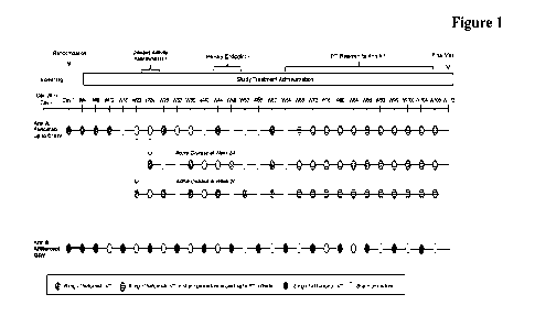
Figure 1: Figure 1 presents an overview of the
study design for nAMD
a
At Weeks 20 and 24, patients will
undergo a disease activity
assessment. Patients with anatomic or functional signs of disease
30
activity at these time points will receive Q8W
or Q12W dosing,
respectively, rather than Q1 6W dosing.
CA 03145239 2022-1-21
WO 2021/023804
PCT/EP2020/072088
- 12 -
b
The primary endpoint is the
change from baseline in BCVA
(as assessed on the ETDRS chart at a starting distance of 4 meters)
based on an average at Weeks 40, 44, and 48.
From Week 60 (when all patients in Ann A are scheduled to
5 receive faricimab) onward, patients in Arm A will be
treated
according to a PTI dosing regimen (between Q8W and Q16W).
BCVA=best-corrected visual acuity; ETDRS=Early Treatment
Diabetic Retinopathy Study; IVT=intravitreal; PTI = personalized
treatment interval; Q8W=every 8 weeks; Q12W=every 12 weeks;
10 Q1 6W=every 16 weeks; W=Week.
Figure 2: Figure 2 presents an overview of the
study design for MAE
Arm A (administered Q8W): Patients randomized to Arm A will
receive 6-mg IVT R06867461 (faricimab) injections Q4W to Week
20, followed by 6-mg IVT R06867461 (faricimab) injections Q8W
15 to Week 96, followed by the final study visit at
Week 100.
Arm B (personalized treatment interval PTO: Patients randomized to
Arm B will receive 6-mg IVT R06867461 (faricimab) injections
Q4W to at least Week 12, followed by PTI dosing (see the PTI dosing
criteria below) of 6-mg IVT R06867461 (faricimab) injections to
20 Week 96, followed by the final study visit at Week
100.
Ann C (comparator arm) (administered Q8W): Patients randomized
to Arm C will receive 2-mg IVT aflibercept injections Q4W to Week
16, followed by 2-mg IVT aflibercept injections Q8W to Week 96,
followed by the final study visit at Week 100.
25 Patients in all three treatment arms will complete
scheduled study
visits Q4W for the entire study duration (100 weeks). A sham
procedure will be administered to patients in all three treatment arms
at applicable visits to maintain masking among treatment arms
IVT=intravitreal; Q8W=every 8 weeks; PTI=personalized treatment
30 interval (see section 3.1.2 for additional details);
W=week.
CA 03145239 2022-1-21
WO 2021/023804
PCT/EP2020/072088
- 13 -
a The definition of 1 year
used for the primary efficacy
endpoint¨ defined as the change from baseline in BCVA, as
measured on the ETDRS chart at a starting distance of 4 meters at
1 year¨is the average of the Week 48, 52, and 56 visits.
5 Figure 3: Schematic Personalized treatment interval for DME-
Figure 3
outlines the algorithm for interval decision-making, which is based
on the relative change of the CST and BCVA compared with
reference CST and reference BCVA.
Significance of* and ** in Figure 3.
10 Reference central subfield thickness (CST):
the CST value
when the initial CST threshold criteria are met. Reference CST is
adjusted if CST decreases by > 10% from the previous reference
CST for two consecutive study drug dosing visits and the values
obtained are within 30 pm. The CST value obtained at the latter
15 visit will serve as the new reference CST, starting
immediately at
that visit.
** Reference best-corrected
visual acuity (BCVA): the mean of
the three best BCVA scores obtained at any prior study drug dosing
visit.
20 Figure 4: Schematic comparison of durability (time to
retreatment) in DME and
nAMD and efficacy (DME) to other treatment options of DME and
nAMD based on published results (Compared agents Lucentis
(ranibizumab), Eylea (aflibercept), brolucizumab and VA2
(R06867461/faricimab).
25 Figure 5: BCVA gains from baseline of patients with
neovascular age-related
macular degeneration (nAMD) comparing the bispecific anti-
VEGF/ANG2 antibody R06867461 (faricimab) at 12- and 16-week
intervals and ranibizumab (Lucentis0) at 4-week intervals.
Figure 6: Time to necessary retreatment of
diabetic macular edema (DME)
30 based on disease activity assessed by both: BCVA
decreased by?: 5
letters and CST increased by > 50 gm (after dosing has discontinued
(after 20 weeks or 6 monthly doses = Time post last intravitreal (PIT)
administration). The bi specific anti-VEGF/ANG2 antibody
CA 03145239 2022-1-21
WO 2021/023804
PCT/EP2020/072088
- 14 -
R06867461 (faricimab), was compared to ranibizumab (Lucentise)
and showed longer time to retreatment.
Figure 7: Figure 1 presents an overview of the
study design for the treatment
of macular edema secondary to retinal vein occlusion (RVO)
5 IVT = intravitreal; PTI = personalized treatment
interval; Q4W
=every 4 weeks; W = Week
Figure 8:
Schematic Personalized treatment
interval for the treatment of
macular edema secondary to retinal vein occlusion (RVO)-Figure 8
outlines the algorithm for interval decision-making, which is based on
10
the relative change of the CST and BCVA compared
with reference
CST and reference BCVA.
BCVA = best-corrected visual acuity; CST = central subfield
thickness; Q4W = every 4 weeks.
a Initial reference CST = CST value when the initial CST threshold
15
criteria are met, but no earlier than Week 20.
Reference CST is
adjusted if CST decreases by> 10% from the previous reference CST
for two consecutive faricimab dosing visits and the values obtained
are within 30 pm. The CST value obtained at the latter visit will serve
as the new reference CST, starting immediately at that visit.
20
b Reference BCVA =mean of the three best BCVA
scores obtained at
any prior dosing visit.
Detailed Description of the Invention
The method, use, bispecific antibody (for use), medicament or pharmaceutical
formulation for use in the treatment of ocular vascular disease selected from
nAMD
25
and DATE comprises sequentially administering
initial doses ("treatment initiation").
In some embodiments the initial doses may vary , e.g. from 3 to 7 monthly
administrations; in one embodiment the treatment initiation includes 3 to 4
monthly
administrations, in one embodiment the treatment initiation includes 4 to 5
monthly
administrations; in one embodiment the treatment initiation includes 4 to 6
monthly
30
administrations; in one embodiment the treatment
initiation includes at least 4
monthly administrations; in one embodiment the treatment initiation includes 5
to 7
CA 03145239 2022-1-21
WO 2021/023804
PCT/EP2020/072088
- 15 -
monthly administrations, in one embodiment the treatment initiation includes 6
monthly administrations.
In one embodiment of the invention the bispecific antibody, medicament or
pharmaceutical formulation is administered in a dose of about 5 to 7 mg (at
each
5 treatment). In one embodiment the bispecific antibody is administered
in a dose of
6 mg +1- 10 % (at each treatment). In one embodiment the bispecific antibody
is
administered in a dose of about 6 mg (at each treatment) (in one embodiment in
a
dose of 6 mg (at each treatment)).
In one embodiment of the invention the bispecific antibody, medicament or
10 pharmaceutical formulation is administered in a concentration of
about 120 mg/m1
(+1- 12 mg/ml), of the bispecific antibody.
Macular degeneration is a medical condition predominantly found in elderly
adults
in which the center of the inner lining of the eye, known as the macula area
of the
retina, suffers thinning, atrophy, and in some cases, bleeding. This can
result in loss
15 of central vision, which entails inability to see fine details, to
read, or to recognize
faces. According to the American Academy of Ophthalmology, it is the leading
cause
of central vision loss (blindness) in the United States today for those over
the age of
fifty years. Although some macular dystrophies that affect younger individuals
are
sometimes referred to as macular degeneration, the term generally refers to
age-
20 related macular degeneration (AMD or ARM])).
"Age-related macular degeneration (AMID)", as used herein, refers to a serious
eye
condition when the small central portion of the retina, known as the macula,
deteriorates. AMD includes wet AMID and neovascular AMID. The wet form of AMD
(wet AMID, wAMD or also called neovascular AMID, nAMD) is characterized by the
25 growth of abnormal blood vessels from the choroid underneath the
macula. This is
called choroidal neovascularization. These blood vessels leak blood and fluid
(below
and) into the retina, causing (elevation of the retina and) distortion of
vision that
makes straight lines look wavy, as well as blind spots and loss of central
vision.
These abnormal blood vessels eventually scar, leading to permanent loss of
central
30 vision. The symptoms of AMID include dark, blurry areas in the center
of vision; and
diminished or changed color perception. AMID can be detected in a routine eye
exam.
One of the most common early signs of macular degeneration is the presence of
drusen which are tiny yellow deposits under the retina and pigment clumping.
CA 03145239 2022-1-21
WO 2021/023804
PCT/EP2020/072088
- 16 -
Advanced AMD, which is responsible for profound vision loss, has two forms:
dry
and wet. Central geographic atrophy, the dry form of advanced AMID, results
from
atrophy to the retinal pigment epithelial layer below the retina, which causes
vision
loss through loss of photoreceptors (rods and cones) in the central part of
the eye.
5
While no treatment is available for this
condition, vitamin supplements with high
doses of antioxidants, lutein and zeaxanthin, have been demonstrated by the
National
Eye Institute and others to slow the progression of dry macular degeneration
and in
some patients, improve visual acuity.
"Diabetic Macular Edema" (DME), as used herein, refers to a serious eye
condition
10
that affects people with diabetes (type 1 or 2).
Macular edema occurs when blood
vessels in the retina leak into the macula and fluid and protein deposits
collect on or
under the macula of the eye and causes it to thicken and swell (edema) The
swelling
may distort a person's central vision, as the macula is near the center of the
retina at
the back of the eyeball. The primary symptoms of DME include, but are not
limited
15
to, blurry vision, floaters, loss of contrast,
double vision, and eventual loss of vision.
The pathology of DME is characterized by breakdown of inner the blood-retinal
bather, normally preventing fluid movement in the retina, thus allowing fluid
to
accumulate in the retinal tissue, and presence of retinal thickening. DME is
presently
diagnosed during an eye examination consisting of a visual acuity test, which
20
determines the smallest letters a person can
read on a standardized chart, a dilated
eye exam to check for signs of the disease, imaging tests such as optical
coherence
tomography (OCT) or fluorescein angiography (FA) and tonometry, an instrument
that measures pressure inside the eye. The following studies are also
performed to
determine treatment: optical coherence tomography (OCT), fluorescein
25
angiography, and color stereo fundus
photography. DME can be broadly
characterized into two main categories - Focal and Diffuse. Focal DME is
characterized by specific areas of separate and distinct leakage in the macula
with
sufficient macular blood flow. Diffuse DME results from leakage of the entire
capillary bed surrounding the macula, resulting from a breakdown of the inner
blood-
30
retina barrier of the eye. In addition to Focal
and Diffuse, DME is also categorized
based on clinical exam findings into clinically significant macular edema
(CSME),
non-CSME and CSME with central involvement (CSME-CI), which involves the
fovea. The present invention includes methods to treat the above-mentioned
categories of DME.
35
Retinal vein occlusion (RVO) is one of the most
common retinal vascular disorders
and is associated with varying degrees of visual loss (Hayreh and Zimmerman
1994).
CA 03145239 2022-1-21
WO 2021/023804
PCT/EP2020/072088
- 17 -
RVO has been reported as the second leading cause of blindness for patients
with
retinal vascular disease, following diabetic retinopathy (DR) (Cugati S, Wang
JJ,
Rochtchina E, et al_ Arch Ophthalmol 2006 ;124 :726-732; Klein R, Knudtson MD,
Lee KE, et at. Ophthalmology 2008 ;115 :1859-1868; Rogers 5, McIntosh RL,
5 Cheung N, et al. Ophthalmology 2010 Feb;117:313-9.el; Yasuda M,
Kiyohara Y,
Arakawa S, et at. Invest Ophtahlmol Vis Sci 2010;51:3205-3209).
The main types of RVO include branch retinal vein occlusion (BRVO),
hemiretinal
vein occlusion (HRVO), and central retinal vein occlusion (CRVO). The most
common presenting complaint of RVO is an abrupt, painless decrease of central
10 vision due to macular edema.
The main types of macular edema secondary to RVO include macular edema
secondary to branch retinal vein occlusion (BRVO), macular edema secondary to
hemiretinal vein occlusion (HRVO), and macular edema secondary to central
retinal
vein occlusion (CRVO).
15 Less frequently, patients may present with a history of transient
vision loss, lasting
a few seconds to minutes, with complete recovery of vision. These symptoms may
recur over several days to weeks, followed by a permanent decrease in vision.
Metamorphopsia and visual field defects have also been described (Achiron A,
Lagstein 0, Glick M, et al. Acta Ophthalmologica 2015;93:e649-53; Manabe K,
20 Osaka R, Nakano Y, et al_ PLoS One 2017;12 :e0186737).
The pathogenesis of macular edema in these patients starts with an increase in
intraluminal pressure due to vascular obstruction, which causes areas of
reduced
perfusion and ischemia. Ischemia leads to up-regulation and secretion of
vascular
endothelial growth factor (VEGF) (Boyd SR, Zachary I, Chakravarthy U, et al.
Arch
25 Ophthalmol 2002;12:1644-1650; Noma H, Minamoto A, Funatsu H, et al.
Graefes
Arch din Exp Ophthalmol 2006;244:309-315) and angiopoietin-2 (Ang-2), both
well-known proangiogenic and vessel hyperpermeability cytokines with Ang-2
contributing additional pro-inflammatory and vessel destabilization properties
(Maisonpierre PC, Suri C, Jones PF, et al. Science 1997;277:55-60; Hackett SF,
30 Ozaki H, Strauss RW, et al. J Cell Physiol 2000 ;184 :275-284;
Fiedler U, Reiss Y,
Scharpfenecker M, et al. Nat Med 2006;12:235-239. Epub: 5 February 2006).
Patients with RVO were found to have the highest vitreous levels of both Ang-2
and
VEGF among all retinal vascular diseases (Aiello LP, Avery RL, Arrigg PG, et
al. N
Engl J Med 1994,331;1480-1487; Regula IT, Lundh von Leithner P, Foxton R, et
al.
CA 03145239 2022-1-21
WO 2021/023804
PCT/EP2020/072088
- 18 -
EMT30 Mot Med 2016;8:1265-1288). Increased levels of Ang-2 and VEGF in retinal
tissue results in pathological changes in the retina and, in many patients,
also macular
edema accompanied with decrease in vision. A hallmark of RVO is the
characteristic
pattern of retinal hemorrhages, tortuous and dilated retinal veins across the
affected
5
area of retina (one quadrant in BRVO, two
quadrants in FIRVO and the entire retina
in CRVO). In more severe cases, patients can develop retinal ischemia with
subsequent retinal neovascularization, hemorrhages, neovascularization in the
anterior segment leading to rubeosis or neovascular glaucoma, and some
patients
may develop optic disc edema.
10
Although macular edema due to RVO and diabetic
macular edema (DME) have
different origins, they share a common pathophysiology. Both are characterized
by
a thickening of the macula due to fluid accumulation consequent to breakdown
of
the blood-retinal bather and a pathological increase of retinal vessel
permeability,
which can lead to irreversible vision loss in both diseases.
15
Anti-VEGF pharmacotherapy is the current
mainstay of treatment in macular edema
due to RVO and has demonstrated efficacy across several pivotal, randomized
clinical studies, although macular laser and intravitreal (PIT) steroids -
especially
steroid implants - are also used in some cases. Despite anti-VEGF being the
most
effective therapy for macular edema due to RVO, data from anti-VEGF clinical
trials
20
showed that many patients do not achieve optimal
best-corrected visual acuity
(BCVA) and anatomical outcomes, and many require frequent long-term injections
to maintain the gains achieved during initial intensive treatment. Moreover,
real-
world data analyses suggested that many patients with RVO do not achieve the
gains
reached in clinical trials due to suboptimal injection frequency (Vaz-Pereira,
S,
25
Marques IP, Matias J, et al. Eur J Ophthalmol
2017;27:756-761; Wecker T, Ehlken
C, Buhler A, et al. Br J Ophthalmol 2017;101:353-359; Jumper JM, Dugel PU,
Chen
S. et al. Clin Ophthalmol 2018;12:621-629). The data suggest that many
patients
with macular edema due to BRVO and the majority of patients with macular edema
due to CRVO require close monitoring and treatment for a longer period of time
and
30
that more durable and efficacious treatment
options are needed (Bhisitkul RB,
Campochiaro PA, Shapiro H, et al. Ophthalmology 2013;120:1057-1063; Scott
Neal NL, VanVeldhuisen, et al JAMA Ophthalmol 2019;El -E10).
Nonclinical studies have shown that Ang-2 and VEGF act in concert to regulate
the
vasculature and to increase retinal endothelial cell permeability in vitro.
35
Simultaneous inhibition of Ang-2 and VEGF with
the bispecific monoclonal
CA 03145239 2022-1-21
WO 2021/023804
PCT/EP2020/072088
- 19 -
antibody faricimab led to a greater reduction in the leakiness and severity of
choroidal neovascularization (CNV) lesions in a laser-induced CNV model in non-
human primates compared with the molar equivalent of anti-VEGF (ranibizumab)
or
anti-Ang-2 alone. Earlier experiments using a mouse model of spontaneous CNV
5
showed that dual inhibition of Ang-2 and VEGF
consistently outperformed
monotherapeutic inhibition of either target alone in terms of reduction in
vascular
growth, leakage, edema, leukocyte infiltration, and photoreceptor loss (Regula
JT,
Lundh von Leithner P, Foxton R, et al. EMBO Mol Med 2016;8:1265-1288).
In addition, aqueous and vitreous concentrations of both Ang-2 and VEGF were
10
shown to be upregulated in patients with
neovascular age-related macular
degeneration (nAMD), DR, and RVO (Tong JP, Chan WM, Liu DT, et al. Am J
Ophthalmol 2006;141 456-462; Penn JS, Madan A, Caldwell RB, et al. Prog Retin
Eye Res 2008;27:331-371.; Kinnunen K, PuustjArvi T, Terasvirta M, et al. Br J
Ophthalmol 2009;93:1109-1115; Tuuminen B Loukovaara S. Eye (Lond)
15
2014 ;28 :1095-1099; Regula JT, Lundh von
Leithner P, Foxton R, etal. EMBO Mol
Med 2016;8:1265-1288; Ng DS, Yip YW, Bakthavatsalam M, et al. Sci Rep
2017;7:45081). Therefore, simultaneous neutralization of both targets, Ang-2
and
VEGF, may further normalize the pathological ocular vasculature compared with
anti-VEGF therapy alone. Data from the completed Phase II studies in DME and
20
nAMD (see below) also support the hypothesis
that targeting Ang-2 has the potential
to extend the durability of effect beyond anti-VEGF therapy alone in diseases
affecting the retinal vasculature.
Faiicimab has been studied for the treatment of nAMD and DIVIE in two Phase I
studies (BP28936 in nAMD and 1P39844 in nAMD and DME) and in three Phase II
25
studies (BP29647 [AVENUE] and CR39521 [STAIRWAY]
for nAMD and
BP30099 [BOULEVARD] for DME). Four global Phase III studies are ongoing:
GR40349 (YOSEMITE) and GR40398 (RHINE) in DME and GR40306 (TENAYA)
and GR40844 (LUCERNE) in nAMD.
Based on the mechanism of action of faricimab, data from nonclinical and
clinical
30
trials, and the pathophysiology of macular edema
due to RVO, it is hypothesized that
faricimab may lead to stabilization of the pathological ocular vasculature and
to
improved visual and anatomical outcomes in RVO compared with anti-VEGF
monotherapies.
CA 03145239 2022-1-21
WO 2021/023804
PCT/EP2020/072088
- 20 -
Macular edema secondary to/due to RVO are among the highest in retinal
vascular
diseases (Aiello LP, Avery RL, Arrigg PG, et at. N Engl J Med1994;331:1480-
1487;
Regula JT, Lundh von Leithner P. Foxton R, et al. ElVIBO Mol Med 2016;8:1265-
1288). The effect of Ang-2 and VEGF inhibition in the nonclinical models of
5
angiogenesis and inflammation (Regula JT, Lundh
von Leithner P. Foxton R, et al.
EMBO Mol Med 2016;8:1265-1288) and the data from Phase I and Phase II
faricimab studies in patients with nAMD and DME provide the evidence of
efficacy
on pathological pathways that are common to all three retinal vascular
diseases:
nAMD, DME/DR, and macular edema due to RVO (Phase I study BP28936 in
10
nAMD; Phase II studies AVENUE in nAMD, STAIRWAY
in nAMD, and
BOULEVARD in DME).
Data from the Phase H BOULEVARD study are reported here due to parallels in
pathophysiology between DME and macular edema due to RVO. While the trigger
for macular edema in diabetic and RVO patients is different, the downstream
15
pathophysiology of hypoxia-driven macular edema
with subsequent vision loss is
similar and driven by the same proangiogenic, pro-inflammatory, vessel
destabilization and vessel permeability factors, including Ang-2, VEGF, and
interleukin-6 (IL-6). The BOULEVARD study provided preliminary evidence of a
positive benefit-risk profile for the use of 6-mg IVT injections of faricimab
for
20
patients with DME and supported further
evaluation of faricimab in the Phase III
DME studies. The study met its primary efficacy endpoint, demonstrating
statistically significant improvement in the mean change from baseline in BCVA
at
Week 24 in patients naive to anti-VEGF treatment who were treated with 6 mg
faricimab compared with 0.3 mg ranibizumab.Best Corrected Visual Acuity
25
(BCVA) is determined using methodology adapted
from the 4-meter Early
Treatment Diabetic Retinopathy Study [ETDRS] protocol (using Early Treatment
Diabetic Retinopathy Study (ETDRS) like charts) and resulting in the
respective
letter score. In one embodiment BCVA determination in such method, use,
bispecific
antibody (for use), medicament or pharmaceutical formulation is based on the
Early
30
Treatment of Diabetic Retinopathy Study (ETDRS)
Protocol adapted visual acuity
charts and is assessed at a starting distance of 4 meters.
Disease activity is determined e.g. via reduction of the BCVA/ETDRs letter
score
and/or e.g. via the macular thickening by spectral domain optical coherence
tomography (SD-OCT) involving the center of the macula as central subfield
35
thickness (CST) (also known as center subfoveal
thickness). In one preferred
embodiment Central Subfield Thickness (CST) is determined using spectral
CA 03145239 2022-1-21
WO 2021/023804
PCT/EP2020/072088
- 21 -
domain optical coherence tomography (SD-OCT): In one preferred embodiment
CST is measured by spectral domain optical coherence tomography (SD-OCT) with
a SpectralisTm device; in one preferred embodiment CST is measured by spectral
domain optical coherence tomography (SD-OCT) with a Cirrus device; in one
5 embodiment CST is measured by spectral domain optical coherence
tomography
(SD-OCT) with a TopconTm device; in one embodiment CST is measured by
spectral domain optical coherence tomography (SD-OCT) with a OptovueTm
device).
As used herein, the term "a patient suffering from" refers to a human that
exhibits
one or more symptoms or indications of, and/or who has been diagnosed with an
10 ocular vascular disease as described herein. The term "a patient
suffering from" may
also include, e.g., subjects who, prior to treatment, exhibit (or have
exhibited) one or
more indications of a vascular eye disease such as, e.g., retinal
angiogenesis,
neovascularization, vascular leak, retinal thickening of the center of the
fovea, hard,
yellow exudates of the center of the fovea with adjacent retinal thickening,
and at
15 least 1 disc area of retinal thickening, any part of which is within
1 disc diameter of
the center of the fovea, blurry vision, floaters, loss of contrast, double
vision, and
eventual loss of vision.
As used herein, the term "a patient suffering from" an ocular vascular disease
such
as nAMD or DME may include a subset of population which is more susceptible to
20 nAMD or DME or may show an elevated level of a nAMD-associated or DME
associated biomarker. For example, "a patient suffering from DME" may include
a
subject suffering from diabetes for more than 10 years, have frequent high
blood
sugar levels or high fasting blood glucose levels. In certain embodiments, the
term
"a patient suffering from DME" includes a subject who, prior to or at the time
of
25 administration of the bispecific anti-VEGF/ANG2 antibody, has or is
diagnosed with
diabetes. In certain embodiments, the term "a patient suffering from nAMD"
includes a subject who, prior to or at the time of administration of the anti-
VEGF/ANG2 antibody, is more than 50 years old. In some embodiments, the term
"a patient suffering from" includes subjects who are smokers, or subjects with
high
30 blood pressure or high cholesterol.
As used herein, the term "a patient suffering from" an ocular vascular disease
such
as macular edema secondary to branch retinal vein occlusion (BRVO), macular
edema secondary to hemiretinal vein occlusion (HRVO), or macular edema
secondary to central retinal vein occlusion (CRVO)may include a subset of
35 population which is more susceptible to macular edema secondary to
branch retinal
vein occlusion (BRVO), macular edema secondary to hemiretinal vein occlusion
CA 03145239 2022-1-21
WO 2021/023804
PCT/EP2020/072088
- 22 -
(1-11tV0), or macular edema secondary to central retinal vein occlusion (CRVO)
or
may show an elevated level of a RVO-associated biomarker. For example, "a
patient
suffering from RVO or macular edema secondary to RVO" may include a subject
with increased levels of VEGF, ANG2 or lL-6. In some embodiments, the term "a
5
patient suffering from" includes subjects who
are smokers, or subjects with high
blood pressure or high cholesterol. The present invention includes methods or
bispecific antibodies (for use), medicaments or pharmaceutical formulations
for
treating, preventing or reducing the severity of an ocular vascular disease
comprising
administering a therapeutically effective amount of a bispecific anti-
VEGF/ANG2
10
antibody (or a medicament or pharmaceutical
formulation comprising the bispecific
anti-VEGF/ANG2 antibody) to a subject in need thereof, wherein the bispecific
antibody, medicament or pharmaceutical formulation comprising such bispecific
anti-VEGF/ANG2 antibody is administered (intravitreally) to the subject in
multiple
doses, e.g., as part of a specific therapeutic dosing regimen.
15
One embodiment of the invention is the method of
treatment, use, bispecific antibody
(for use), medicament or pharmaceutical formulation as described herein
wherein patients suffering from an ocular vascular disease have not been
previously treated with anti-VEGF treatment (e.g. monotherapy) (are treatment
naive).
20
One embodiment of the invention is the method of
treatment, use, bispecific antibody
(for use), medicament or pharmaceutical formulation as described herein
wherein patients suffering from an ocular vascular disease have been
previously treated with anti-VEGF treatment (e.g. monotherapy, e.g., with
ranibizumab, aflibercept or brolocizumab ).
25
One embodiment of the invention is a method,
use, bispecific antibody (for use),
medicament or pharmaceutical formulation for use in the treatment of patients
suffering from neovascular AMID (nAMD) the method comprising
administering to the patient an effective amount of a bispecific antibody
which
binds to human vascular endothelial growth factor (VEGF) and to human
30 angiopoietin-2 (ANG-2) with a personalized treatment interval,
wherein
a) patients are treated first 4 times with the bispecific VEGF/ANG2 antibody
at an
every 4 weeks (Q4W) dosing interval;
b) at Weeks 20 and 24 the disease activity is assessed wherein the disease
activity
is determined if one of the following criteria are met:
CA 03145239 2022-1-21
WO 2021/023804
PCT/EP2020/072088
- 23 -
i) increase of> 50 p.m in central subfield thickness (CST) compared with the
average CST value over the previous two scheduled visits which are Weeks
12 and 16 for the Week 20 assessment, and Weeks 16 and 20 for the Week
24 assessment, or
5 ii) increase 75 pm in CST compared with the lowest CST value
recorded at
either of the previous two scheduled visits;
iii) decrease 5 letters in best-corrected visual acuity (BCVA) compared
with average BCVA value over the previous two scheduled visits, owing to
nAMD disease activity,
10 iv) decrease L 10 letters in BCVA compared with the highest
BCVA value
recorded at either of the previous two scheduled visits, owing to nAMD
disease activity, or
v) presence of new macular hemorrhage, owing to nAMD activity
c) then patients
15 i) patients who meet the disease activity criteria at Week20
will be treated at
a Q8W dosing interval from week 20 onward (with the first Q8W dosing at
Week20);
ii) patients who meet the disease activity criteria at Week24 will be treated
at
a Ql2W dosing interval from week 24 onward (with the first Q12W dosing at
20 Week24); and
iii) patients who do not meet disease activity criteria at Week20 and Week24
will be treated at a Q16W dosing interval from week 28 onward (with the
first Q16W dosing at Week28).
In one embodiment the personalized treatment interval will be extended,
reduced, or
25 maintained after week 60 wherein the
a) interval is extended by 4 weeks (to a maximum of Q16W) if all of the
following criteria are met:
1) stable CST compared with the average of the last 2 study drug dosing
30 visits where stability is defined as a change of CST of
less than 30 p.m
and no increase > 50 pin in CST compared with the lowest on-study
drug dosing visit measurement,
ii) no decrease? 5 letters in BCVA compared with the average from
the last two study drug dosing visits, and no decrease >10 letters in
35 BCVA compared with the highest on-study drug dosing
visit
CA 03145239 2022-1-21
WO 2021/023804
PCT/EP2020/072088
- 24 -
measurement,
iii) no new macular hemorrhage;
b) interval
is reduced (to a minimum Q8W) by 4 weeks if one of the following
5 criteria is met,
or
is reduced to an 8-week interval if two or more of the following criteria
are met or one criterion includes new macular hemorrhage:
10 i) increase of? 50 gm in CST compared with the average
from the last
two dosing visits or of? 75 pm compared with the lowest dosing visit
measurement,
ii) decrease of? 5 letters in BCVA compared with average of last two
dosing visits or decrease? 10 letters in BCVA compared with the
15 highest dosing visit measurement,
iii) new macular hemorrhage.
In one embodiment the disease activity assessment before the personalized
treatment
interval will be at Weeks 16 and Week 20, or at Weeks 24 and Week 28.
In one embodiment the personalized treatment interval with further extension,
20
reduction, or maintenance will start at a
different time point e.g. between after
week 50 and 70, e.g. after week 52 or after week 65 depending on the disease
activity. Another embodiment of the invention is a method, use, bispecific
antibody (for use), medicament or pharmaceutical formulation for use in the
treatment of patients suffering from diabetic macular edema (DME) the
25
method comprising administering to the patient
an effective amount of a
bispecific antibody which binds to human vascular endothelial growth factor
(VEGF) and to human angiopoietin-2 (ANG-2) with a personalized treatment
interval, wherein
a) patients are treated first with the bispecific VEGF/ANG2 antibody at an
every 4
30
weeks (Q4W) dosing interval until the central
subfield thickness (CST) meets
a predefined reference CST threshold (of CST <325 pm for Spectralis spectral
domain - central subfield thickness SD-OCT, or <315 gm for Cirrus SD-OCT
or Topcon SD-OCT) (as measured at week 12 or later);
b) then the dosing interval is increased by 4 weeks to an initial Q8W dosing
interval;
CA 03145239 2022-1-21
WO 2021/023804
PCT/EP2020/072088
- 25 -
c) from this point forward, the dosing interval is extended, reduced, or
maintained
based on assessments made at the dosing visits, which are based on the
relative
change of the CST and best-corrected visual acuity (BCVA) compared with
the respective reference CST and BCVA;
5 wherein the
i) interval is extended by 4 weeks,
- if the CST value is increased or decreased by <10% without an
associated >10-letter BCVA decrease;
ii) interval will be maintained:
10 - if the CST is decreased by > 10%, or
- the CST value is increased or decreased by < 10% with an
associated >10-letter BCVA decrease, or
- the CST value is increased between > 10% and < 20% without an
associated >5-letter BCVA decrease;
15 iii) interval is reduced by 4 weeks
-if the CST value is increased between > 10% and < 20% with an
associated >5 to<10-letter BCVA decrease; or
- the CST value is increased by > 20% without an associated >10-
letter BCVA decrease;
20 iv) interval is reduced by 8 weeks if the CST value is
increased by > 10%
with an associated >10-letter BCVA decrease;
wherein the respective reference central subfield thickness (CST) is the CST
value when the initial CST threshold criteria are met and the reference
CST is adjusted if CST decreases by > 10% from the previous reference
25
CST for two consecutive dosing visits and the
values obtained are within
30 Lun so that the CST value obtained at the latter visit will serve as the
new reference CST; and
wherein the reference best-corrected visual acuity (BCVA) is the mean of the
three best BCVA scores obtained at any prior dosing visit.
30
In one embodiment such dosing interval can by
adjusted by 4-week increments to a
maximum of every 16 weeks (Q1 6W) and a minimum of Q4W.
CA 03145239 2022-1-21
WO 2021/023804
PCT/EP2020/072088
- 26 -
Another embodiment of the invention is a method, use, bispecific antibody (for
use),
medicament or pharmaceutical formulation for use in the treatment of patients
suffering from an ocular vascular disease selected from macular edema
secondary to central retinal vein occlusion, secondary to hemiretinal vein
5
occlusion or secondary to branch vein occlusion,
or of patients suffering from
an ocular vascular disease selected from macular edema secondary to central
retinal vein occlusion, secondary to hemiretinal vein occlusion or secondary
to
branch vein occlusion, wherein the treatment includes a personalized treatment
interval (PIT), wherein
10
a) patients are treated first with the
bispecific VEGF/ANG2 antibody at an
every 4 weeks (Q4W) dosing interval from Day 1 through Week 20
b) from Week 24, patients receive the bispecific VEGF/ANG2 antibody at a
frequency of Q4W until the central subfield thickness (CST) meets a
predefined reference CST threshold (of CST <325 gm for Spectralis
15
spectral domain - central subfield thickness SD-
OCT, or <315 gm for
Cirrus SD-OCT or Topcon SD-OCT) (as measured at week 24 or later);
c) from this point forward, the dosing interval is extended, reduced, or
maintained based on assessments made at the dosing visits which are based
on the relative change of the CST and best-corrected visual acuity (BCVA)
20 compared with the respective reference CST and BCVA;
wherein the
i) interval is extended by 4 weeks
if the CST value is increased or decreased by < 10% without an
associated? 10-letter BCVA decrease; or
25 ii) interval is maintained if any of the following
criteria are met:
if the CST value is decreased by > 10%; or
if the CST value is decreased < 10% with an associated? 10-letter
BCVA decrease; or
if the CST value is increased between > 10% and < 20% without an
30 associated > 5-letter BCVA decrease;
CA 03145239 2022-1-21
WO 2021/023804
PCT/EP2020/072088
- 27 -
iii) interval is reduced by 4 weeks if any of the following criteria are
met:
if the CST value is increased between > 10% and < 20% with an
associated > 5-to <10-letter BCVA decrease, or
5
if the CST value is increased by > 20% without
an associated? 10-
letter BCVA decrease, or
if the CST value is increased by < 10% with an associated BCVA
decrease of? 10-letters;
iv) interval is reduced to Q4W
10
if the CST value is increased by > 10% with an
associated? 10-letter
BCVA decrease,
wherein the respective reference central subfield thickness (CST) is the CST
value when the initial CST threshold criteria are met and the reference CST
is adjusted if CST decreases by > 10% from the previous reference CST for
15
two consecutive dosing visits and the values
obtained are within 30 p.m so
that the CST value obtained at the latter visit will serve as the new
reference
CST, and
wherein the reference best-corrected visual acuity (BCVA) is the mean of the
three best BCVA scores obtained at any prior dosing visit.
20
In one embodiment such dosing interval can by
adjusted by 4-week increments to a
maximum of every 16 weeks (Q16W) and a minimum of Q4W. As used herein,
"antibody" refers to a binding protein that comprises antigen-binding sites.
The terms
"binding site" or "antigen-binding site" as used herein denotes the region(s)
of an
antibody molecule to which a ligand actually binds. The term "antigen-binding
site"
25
comprises an antibody heavy chain variable
domains (VH) and an antibody light
chain variable domains (VL) (pair of VHNL).).
Antibody specificity refers to selective recognition of the antibody for a
particular
epitope of an antigen. Natural antibodies, for example, are monospecific.
"Bispecific antibodies" according to the invention are antibodies which have
two
30
different antigen-binding specificities.
Antibodies of the present invention are
CA 03145239 2022-1-21
WO 2021/023804
PCT/EP2020/072088
- 28 -
specific for two different antigens, VEGF as first antigen and ANG-2 as second
antigen.
The term "monospecific" antibody as used herein denotes an antibody that has
one
or more binding sites each of which bind to the same epitope of the same
antigen.
5
The term "valent" as used within the current
application denotes the presence of a
specified number of binding sites in an antibody molecule As such, the terms
"bivalent", "tetravalent", and "hexavalent" denote the presence of two binding
site,
four binding sites, and six binding sites, respectively, in an antibody
molecule. The
bispecific antibodies according to the invention are preferably "bivalent".
10
The terms "bispecific antibody which binds to
human vascular endothelial growth
factor (VEGF) and to human angiopoietin-2 (ANG-2)", "bispecific anti-
VEGF/ANG2 antibody" and bispecific <VEGF/ANG2> antibody" as used herein
are interchangeable and refer to an antibody which has at least two different
antigen-
binding sites, a first one which binds to VEGF and a second one which binds to
15 ANG2.
Bispecific anti-VEGF/ANG2 antibodies are e.g. described in W02010040508,
W02011/117329, W02012/131078, W02015/083978, W02017/197199, and
W02014/009465. W02014/009465 describes bispecific anti-VEGF/ANG2
antibodies especially designed for treatment of ocular vascular diseases. The
20
bispecific anti-VEGF/ANG2 antibodies of
W02014/009465 (which is incorporated
herein in its entirety) are especially useful in the treatment and treatment
schedules
of ocular vascular diseases as described herein.
In one embodiment the bispecific antibody which binds to human vascular
endothelial growth factor (VEGF) and to human angiopoietin-2 (ANG-2) is a
25
bispecific anti-VEGF/ANG2 antibody comprising a
first antigen-binding site that
specifically binds to human VEGF and a second antigen-binding site that
specifically
binds to human ANG-2, wherein
i)
said first antigen-binding site specifically binding to VEGF
comprises in
the heavy chain variable domain a CDR3H region of SEQ ID NO: 1, a
30
CDR2H region of SEQ ID NO: 2, and a CDR1H region
of SEQ ID NO:3,
and in the light chain variable domain a CDR3L region of SEQ ID NO:
4, a CDR2L region of SEQ ID NO:5, and a CDR1L region of SEQ ID
NO:6; and
CA 03145239 2022-1-21
WO 2021/023804
PCT/EP2020/072088
- 29 -
ii) said second antigen-binding site specifically binding to ANG-2
comprises in the heavy chain variable domain a CDR3H region of SEQ
ID NO: 9, a CDR2H region of, SEQ ID NO: 10, and a CDR1H region of
SEQ ID NO: 11, and in the light chain variable domain a CDR3L region
5
of SEQ ID NO: 12, a CDR2L region of SEQ ID NO:
13, and a CDR1L
region of SEQ
ID NO: 14,
and wherein
iii) the bispecific antibody comprises a constant heavy chain region of human
IgGI subclass comprising the mutations I253A, H310A, and H435A and
10
the mutations L234A, L235A and P329G (numberings
according to EU
Index of Kabat).
In one embodiment such bispecific anti-VEGF/ANG2 antibody is bivalent.
In one embodiment such bispecific anti-VEGF/ANG2 antibody is characterized in
that
15 0
said first antigen-binding site specifically
binding to VEGF comprises as
heavy chain variable domain VH an amino acid sequence of SEQ ID
NO: 7, and as light chain variable domain VL an amino acid sequence of
SEQ ID NO: 8, and
ii) said second antigen-binding site specifically binding to ANG-2
20
comprises as heavy chain variable domain VH an
amino acid sequence
of SEQ ID NO: 15, and as light chain variable domain VL an amino acid
sequence of SEQ ID NO: 16.
In one aspect of the invention such bispecific, bivalent antibody according to
the
invention is characterized in comprising
25
a) the heavy chain and the light chain of a
first full length antibody that
specifically binds to VEGF;
b) the modified heavy chain and modified light chain of a second full length
antibody that specifically binds to ANG-2, wherein the constant domains
CL and CHI are replaced by each other.
30
This bispecific, bivalent antibody format for
the bispecific antibody specifically
binding to human vascular endothelial growth factor (VEGF) and human
angiopoietin-2 (ANG-2) is described in WO 2009/080253 (including Knobs-into-
CA 03145239 2022-1-21
WO 2021/023804
PCT/EP2020/072088
- 30 -
Holes modified CH3 domains). The antibodies based on this bispecific, bivalent
antibody format are named CrossMAbs.
In one embodiment such bispecific, bivalent anti-VEGF/ANG2 antibody is
characterized in comprising
5
a) as heavy chain of the first full length
antibody the amino acid sequence of
SEQ ID NO: 17, and as light chain of the first full length antibody the
amino acid sequence of SEQ ID NO: 18, and
b) as modified heavy chain of the second full length antibody the amino acid
sequence of SEQ ID NO: 19, and as modified light chain of the second full
10 length antibody the amino acid sequence of SEQ ID NO: 20.
In one embodiment such bispecific, bivalent anti-VEGF/ANG2 antibody is
characterized in comprising the amino acid sequences of SEQ ID NO: 17, of
SEQ ID NO: 18, of SEQ ID NO: 19, and of SEQ ID NO: 20. In one preferred
embodiment the bispecific, bivalent anti-VEGF/ANG2 antibody is faricimab.
15 Accordingly, one embodiment of the invention is a bispecific,
bivalent antibody
comprising a first antigen-binding site that specifically binds to human VEGF
and a
second antigen-binding site that specifically binds to human ANG-2,
characterized
in comprising the amino acid sequences of SEQ ID NO: 17, of SEQ ID NO: 18, of
SEQ ID NO: 19, and of SEQ ID NO: 20. In one preferred embodiment the
bispecific,
20 bivalent anti-VEGF/ANG2 antibody is faricimab.
In on embodiment the CH3 domains of the bispecific, bivalent antibody
according
to the invention is altered by the "knob-into-holes" technology which is
described in
detail with several examples in e.g. WO 96/027011, Ridgway J.B., et al.,
Protein Eng
9 (1996) 617-621; and Merchant, A.M., et al., Nat Biotechnol 16 (1998) 677-
681.
25 In this method the interaction surfaces of the two CH3 domains are
altered to increase
the heterodimerisation of both heavy chains containing these two CH3 domains.
Each of the two CH3 domains (of the two heavy chains) can be the "knob", while
the other is the "hole". The introduction of a disulfide bridge stabilizes the
heterodimers (Merchant, AM, et al., Nature Biotech 16 (1998) 677-681; Atwell,
S.,
30 et at. J. Mol. Biol. 270 (1997) 26-35) and increases the yield.
In a preferred aspect of the invention the bispecific anti-VEGF/ANG2
antibodies
according to the invention are characterized in that
CA 03145239 2022-1-21
WO 2021/023804
PCT/EP2020/072088
-31 -
the CH3 domain of one heavy chain and the CH3 domain of the other heavy chain
each meet at an interface which comprises an original interface between the
antibody
CH3 domains;
wherein said interface is altered to promote the formation of the bispecific
antibody,
5 wherein the alteration is characterized in that:
a) the CH3 domain of one heavy chain is altered,
so that within the original interface the CH3 domain of one heavy chain that
meets
the original interface of the CH3 domain of the other heavy chain within the
bispecific antibody,
10 an amino acid residue is replaced with an amino acid residue having a
larger side
chain volume, thereby generating a protuberance within the interface of the
CH3
domain of one heavy chain which is positionable in a cavity within the
interface of
the C1I3 domain of the other heavy chain
and
15 b) the CH3 domain of the other heavy chain is altered,
so that within the original interface of the second CH3 domain that meets the
original
interface of the first CH3 domain within the bispecific antibody
an amino acid residue is replaced with an amino acid residue having a smaller
side
chain volume, thereby generating a cavity within the interface of the second
CH3
20 domain within which a protuberance within the interface of the first
CH3 domain is
positionable.
Thus the bispecific anti-VEGF/ANG2 antibodies for use described herein are
preferably characterized in that
the CH3 domain of the heavy chain of the full length antibody of a) and the
25
CH3 domain of the heavy chain of the full length
antibody of b) each meet at
an interface which comprises an alteration in the original interface between
the antibody CH3 domains;
wherein i) in the CH3 domain of one heavy chain
CA 03145239 2022-1-21
WO 2021/023804
PCT/EP2020/072088
- 32 -
an amino acid residue is replaced with an amino acid residue having a larger
side chain volume, thereby generating a protuberance within the interface of
the CH3 domain of one heavy chain which is positionable in a cavity within
the interface of the CH3 domain of the other heavy chain
5 and wherein
ii) in the CH3 domain of the other heavy chain
an amino acid residue is replaced with an amino acid residue having a smaller
side chain volume, thereby generating a cavity within the interface of the
second CH3 domain within which a protuberance within the interface of the
10 first CH3 domain is positionable.
Preferably said amino acid residue having a larger side chain volume is
selected from
the group consisting of arginine (R), phenylalanine (F), tyrosine (Y),
tryptophan (W).
Preferably said amino acid residue having a smaller side chain volume is
selected
from the group consisting of alanine (A), serine (S), threonine (T), valine
(V).
15
In one aspect of the invention both C113 domains
are further altered by the
introduction of cysteine (C) as amino acid in the corresponding positions of
each
CH3 domain such that a disulfide bridge between both C113 domains can be
formed.
In one embodiment, the bispecific antibody comprises a T366W mutation in the
CH3
domain of the "knobs chain" and T366S, L368A, Y407V mutations in the CH3
20
domain of the "hole chain". An additional
interchain disulfide bridge between the
CH3 domains can also be used (Merchant, A.M, et al., Nature Biotech 16 (1998)
677-681) e.g. by introducing a S354C mutation into one CH3 domain and a Y349C
mutation into the other CH3 domain.
In a another preferred embodiment the bispecific antibody comprises S354C and
25
T366W mutations in one of the two CH3 domains
and Y349C, T366S, L368A,
Y407V mutations in the other of the two CH3 domains In a another preferred
embodiment the bispecific antibody comprises Y349C, T366W mutations in one of
the two CH3 domains and S354C, T366S, L368A, Y407V mutations in the other of
the two CH3 domains (the additional Y349C or S354C mutation in one CH3 domain
30
and the additional S354C or Y349C mutation in
the other CH3 domain forming a
interchain disulfide bridge) (numbering always according to EU index of Kabat
CA 03145239 2022-1-21
WO 2021/023804
PCT/EP2020/072088
- 33 -
(1Cabat, E A , et al., Sequences of Proteins of Immunological Interest, 5th ed
, Public
Health Service, National Institutes of Health, Bethesda, MD (1991)).
Other techniques for CH3-modifications to enforce the heterodimerization are
contemplated as alternatives of the invention and described e.g. in WO
96/27011,
5 W098/050431, EP 1870459, W02007/110205,
W02007/147901,
WO 2009/089004, WO 2010/129304, WO 2011/90754, WO 2011/143545,
WO 2012/058768, WO 2013/157954 and WO 2013/096291.
In one embodiment the heterodimerization approach described in EP 1 870 459A1
is used alternatively. This approach is based on the introduction of
10 substitutions/mutations of charged amino acids with the opposite
charge at specific
amino acid positions of the in the CH3/CH3 domain interface between both heavy
chains. One preferred embodiment for said multispecific antibodies are amino
acid
R409D and K370E mutations in the CH3 domain of one heavy chain and amino acid
D399K and E357K mutations in the CH3 domain of the other heavy chain of the
15 multispecific antibody (numberings according to Kabat EU index).
In another embodiment said multispecific antibody comprises an amino acid
T366W
mutation in the CH3 domain of the "knobs chain" and amino acid T366S, L368A
and Y407V mutations in the CH3 domain of the "hole chain"; and additionally
comprises amino acid R409D and K370E mutations in the C1I3 domain of the
"knobs
20 chain" and amino acid D399K and E357K mutations in the CH3 domain of
the "hole
chain".
In one embodiment the heterodimerization approach described in W02013/157953
is used alternatively. In one embodiment the CH3 domain of one heavy chain
comprises an amino acid T366K mutation and the CH3 domain of the other heavy
25 chain comprises an amino acid L351D mutation. In a further embodiment
the CH3
domain of the one heavy chain further comprises an amino acid L351K mutation.
In
a further embodiment the CH3 domain of the other heavy chain further comprises
an
amino acid mutation selected from Y349E, Y349D and L368E (in one embodiment
L3 68E).
30 In one embodiment the heterodimerization approach described in
W02012/058768
is used alternatively. In one embodiment the CH3 domain of one heavy chain
comprises amino acid L351Y and Y407A mutations and the CH3 domain of the other
heavy chain comprises amino acid T366A and K409F mutations. In a further
embodiment the CH3 domain of the other heavy chain further comprises an amino
CA 03145239 2022-1-21
WO 2021/023804
PCT/EP2020/072088
- 34 -
acid mutation at position T411, D399, S400, F405, N390 or K392. In one
embodiment said amino acid mutation is selected from the group consisting of
a) T411N, T411R, T41 IQ, T411K, T411D, T411E and T411W,
b) D399R, D399W, D399Y and D399K,
5 c) S400E, S400D, S400R and S400K,
d) F4051, F405M, F405T, F405S, F405V and F405W,
e) N390R, N390K and N390D,
0 K392V, K392M, K392R, K392L, K392F and K392E.
In a further embodiment the C113 domain of one heavy chain comprises amino
acid
10 L351Y and Y407A mutations and the CH3 domain of the other heavy chain
comprises amino acid T366V and K409F mutations. In a further embodiment the
CH3 domain of one heavy chain comprises an amino acid Y407A mutation and the
CH3 domain of the other heavy chain comprises amino acid T366A and K409F
mutations. In a further embodiment the C113 domain of the other heavy chain
further
15 comprises amino acid K392E, T411E, D399R and MOOR mutations.
In one embodiment the heterodimerization approach described in W02011/143545
is used alternatively. In one embodiment the amino acid modification according
to
W02011/143545 is introduced in the CH3 domain of the heavy chain at a position
selected from the group consisting of 368 and 409.
20 In one embodiment the heterodimerization approach described in
W02011/090762
which also uses the knob-into-hole technology described above is used
alternatively.
In one embodiment the CH3 domain of one heavy chain comprises an amino acid
T366W mutation and the CH3 domain of the other heavy chain comprises an amino
acid Y407A mutation. In one embodiment the CH3 domain of one heavy chain
25 comprises an amino acid T366Y mutation and the CH3 domain of the
other heavy
chain comprises an amino acid Y407T mutation.
In one embodiment the multispecific antibody is of IgG2 isotype and the
heterodimerization approach described in W02010/129304 is used alternatively.
In one embodiment the heterodimerization approach described in W02009/089004
30 is used alternatively. In one embodiment the CH3 domain of one heavy
chain
CA 03145239 2022-1-21
WO 2021/023804
PCT/EP2020/072088
- 35 -
comprises an amino acid substitution of K392 or N392 with a negatively-charged
amino acid (in one embodiment glutamic acid (E) or aspartic acid (D); in a
further
embodiment a K392D or N392D mutation) and the CH3 domain of the other heavy
chain comprises an amino acid substitution of D399, E356, D356, or E357 with a
5
positively-charged amino acid (in one embodiment
Lysine (K) or arginine (R), in a
further embodiment a D399K, E356K, D356K or E357K substitution; and in an even
further embodiment a D399K or E356K mutation). In a further embodiment the CH3
domain of the one heavy chain further comprises an amino acid substitution of
K409
or R409 with a negatively-charged amino acid (in one embodiment glutamic acid
(E)
10
or aspartic acid (D); in a further embodiment a
K409D or R409D mutation). In a
further embodiment the CH3 domain of the one heavy chain further or
alternatively
comprises an amino acid substitution of K439 and/or K370 with a negatively-
charged amino acid (in one embodiment glutamic acid (E) or aspartic acid (D)).
In one embodiment the heterodimerization approach described in W02007/147901
15
is used alternatively. In one embodiment the CH3
domain of one heavy chain
comprises amino acid K253E, D282K and K322D mutations and the CH3 domain
of the other heavy chain comprises amino acid D239K, E240K and K292D
mutations.
In one embodiment the heterodimerization approach described in W02007/110205
20 is used alternatively.
In one embodiment the bispecific antibody which binds to human vascular
endothelial growth factor (VEGF) and to human angiopoietin-2 (ANG-2) is a
bispecific anti-VEGF/ANG2 antibody comprising a first antigen-binding site
that specifically binds to human VEGF and a second antigen-binding site that
25 specifically binds to human ANG-2, wherein
i) said first antigen-binding site specifically binding to VEGF comprises
in
the heavy chain variable domain a CDR3H region of SEQ NO: I, a
CDR2H region of SEQ ID NO: 2, and a CDR1H region of SEQ ID NO:3,
and in the light chain variable domain a CDR3L region of SEQ ID NO:
30
4, a CDR2L region of SEQ ID NO:5, and a CDR1L
region of SEQ ID
NO:6; and
ii) said second antigen-binding site specifically binding to ANG-2
comprises in the heavy chain variable domain a CDR3H region of SEQ
ID NO: 9, a CDR2H region of, SEQ ID NO: 10, and a CDR1H region of
CA 03145239 2022-1-21
WO 2021/023804
PCT/EP2020/072088
- 36 -
SEQ ID NO: 11, and in the light chain variable domain a CDR3L region
of SEQ ID NO: 12, a CDR2L region of SEQ ID NO: 13, and a CDR1L
region of SEQ
ID NO: 14,
and wherein
5
iii) the bispecific antibody comprises a
constant heavy chain region of human
IgG1 subclass comprising the mutations I253A, H3 10A, and H435A and
the mutations L234A, L235A and P329G (numberings according to EU
Index of Kabat; and wherein
iv) in the constant heavy chain region a T366W mutation is comprised in one
10
CH3 domain and T3665, L368A, Y407V mutations are
comprised the
other C113 domain (numberings according to EU Index of Kabat).
In one embodiment the bispecific antibody which binds to human vascular
endothelial growth factor (VEGF) and to human angiopoietin-2 (ANG-2) is a
bispecific anti-VEGF/ANG2 antibody comprising a first antigen-binding site
15
that specifically binds to human VEGF and a
second antigen-binding site that
specifically binds to human ANG-2, wherein
0
said first antigen-binding site
specifically binding to VEGF comprises in
the heavy chain variable domain a CDR3H region of SEQ ID NO: I, a
CDR2H region of SEQ ID NO: 2, and a CDR1H region of SEQ ID NO:3,
20
and in the light chain variable domain a CDR3L
region of SEQ ID NO:
4, a CDR2L region of SEQ ID NO:5, and a CDR1L region of SEQ ID
NO:6; and
ii) said second antigen-binding site specifically binding to ANG-2
comprises in the heavy chain variable domain a CDR3H region of SEQ
25
ID NO: 9, a CDR2H region of, SEQ ID NO: 10, and
a CDR1H region of
SEQ ID NO: 11, and in the light chain variable domain a CDR3L region
of SEQ ID NO: 12, a CDR2L region of SEQ ID NO: 13, and a CDR1L
region of SEQ
ID NO: 14,
and wherein
30
iii) the bispecific antibody comprises a
constant heavy chain region of human
IgG1 subclass comprising the mutations I253A, H3 10A, and H435A and
the mutations L234A, L235A and P329G (numberings according to EU
Index of Kabat; and wherein
CA 03145239 2022-1-21
WO 2021/023804
PCT/EP2020/072088
- 37 -
iv) in the constant heavy chain region a 5354C and T366W mutations are
comprised in one CH3 domain and Y349C, T366S, L368A and Y407V
mutations are comprised the other CH3 domain (numberings according
to EU Index of Kabat).
5 In one embodiment such bispecific anti-VEGF/ANG2 antibody is
bivalent.
In one embodiment such bispecific anti-VEGF/ANG2 antibody is characterized in
that
i) said first antigen-binding site specifically binding to VEGF comprises
as
heavy chain variable domain VH an amino acid sequence of SEQ ID NO:
10
7, and as light chain variable domain VL an
amino acid sequence of SEQ
1D NO: 8, and
ii) said second antigen-binding site specifically binding to ANG-2
comprises as heavy chain variable domain VH an amino acid sequence
of SEQ 113 NO: 15, and as light chain variable domain VL an amino
15 acid sequence of SEQ ID NO: 16.
In one aspect of the invention such bispecific, bivalent antibody according to
the
invention is characterized in comprising
a) the heavy chain and the light chain of a first full length antibody that
specifically binds to VEGF;
20
b) the modified heavy chain and modified light
chain of a second full length
antibody that specifically binds to ANG-2, wherein the constant domains
CL and CH1 are replaced by each other.
The term "VEGF" as used herein refers to human vascular endothelial growth
factor
(VEGF/VEGF-A,) the 165-amino acid human vascular endothelial cell growth
factor
25
(amino acid 27-191 of precursor sequence of
human VEGF165: SEQ ID NO: 24;
amino acids 1-26 represent the signal peptide), and related 121, 189, and 206
vascular endothelial cell growth factor isoforms, as described by Leung, D.W.,
et al.,
Science 246 (1989) 1306-9; Houck et al., Mol. Endocrin. 5 (1991) 1806 -1814;
Keck,
P.J., et at., Science 246 (1989) 1309-12 and Connolly, D.T., et al., I Biol.
Chem.
30
264 (1989) 20017-24; together with the naturally
occurring allelic and processed
forms of those growth factors. VEGF is involved in the regulation of normal
and
abnormal angiogenesis and neovascularization associated with tumors and
CA 03145239 2022-1-21
WO 2021/023804
PCT/EP2020/072088
- 38 -
intraocular disorders (Ferrara, N., et al., Endocr. Rev. 18 (1997) 4-25;
Berkman,
RA., et al., J. Clin. Invest. 91 (1993) 153-159; Brown, L.F., et al., Human
Pathol.
26 (1995) 86-91; Brown, L.F., et al., Cancer Res. 53 (1993) 4727-4735; Mattem,
J.,
et al., Brit. J. Cancer. 73 (1996) 931-934; and Dvorak, H.F., et at, Am. I
Pathol. 146
5 (1995) 1029-1039). VEGF is a homodimeric glycoprotein that has been
isolated from
several sources and includes several isoforms. VEGF shows highly specific
mitogenic activity for endothelial cells. A VEGF antagonist/inhibitor inhibits
binding of VEGF to its receptor VEGFR. Known VEGF antagonist/inhibitors
include bispecific anti-VEGF/ANG2 antibodies as described in W02014/009465.
10 The term "ANG-2" as used herein refers to human angiopoietin-2 (ANG-
2)
(alternatively abbreviated with ANGPT2 or ANG2) (SEQ ID NO: 25) which is
described e.g. in Maisonpierre, P.C., et al, Science 277 (1997) 55-60 and
Cheungõ
A.H., et al., Genomics 48 (1998) 389-91. The angiopoietins-1 (SEQ ID NO: 26)
and
-2 were discovered as ligands for the Ties, a family of tyrosine kinases that
is
15 selectively expressed within the vascular endothelium (Yancopoulos,
G.D., et al.,
Nature 407 (2000) 242-48). There are now four definitive members of the
angiopoietin family. Angiopoietin-3 and -4 (Ang-3 and Ang-4) may represent
widely
diverged counterparts of the same gene locus in mouse and man (Kim, I., et
al., FEB S
Let, 443 (1999) 353-56; Kim, I., et al., J Biol Chem 274 (1999) 26523-28). ANG-
1
20 and ANG-2 were originally identified in tissue culture experiments as
agonist and
antagonist, respectively (see for ANG-1: Davis, S., et al., Cell 87 (1996)
1161-69;
and for ANG-2: Maisonpierre, P.C., et at, Science 277 (1997) 55-60). All of
the
known angiopoietins bind primarily to its receptor TIE2 (SEQ ID NO: 27), and
both
Ang-1 and -2 bind to TIE2 with an affinity of 3 nM (Kd) (Maisonpierre, P.C.,
et al.,
25 Science 277(1997) 55-60). An ANG2 antagonist/inhibitor inhibits
binding of ANG2
to its receptor TIE2. Known ANG2 antagonist/inhibitors include bispecific anti-
VEGF/ANG2 antibodies as described in W02014/009465.
An antigen-binding sites of the bispecific antibody of the invention contain
six
complementarily determining regions (CDRs) which contribute in varying degrees
30 to the affinity of the binding site for antigen. There are three
heavy chain variable
domain CDRs (CDRH1, CDRH2 and CDRH3) and three light chain variable domain
CDRs (CDRL I, CDRL2 and CDRL3). The extent of CDR and framework regions
(Fits) is determined by comparison to a compiled database of amino acid
sequences
in which those regions have been defined according to variability among the
35 sequences.
CA 03145239 2022-1-21
WO 2021/023804
PCT/EP2020/072088
- 39 -
The antibodies of the invention comprise immunoglobulin constant regions
derived
from human origin of one or more immunoglobulin classes, wherein such
immunoglobulin classes include IgG, IgM, IgA, IgD, and IgE classes and, in the
case
of IgG and IgA, their subclasses, especially IgG1 and IgG4.
5 The terms "monoclonal antibody" or "monoclonal antibody composition"
as used
herein refer to a preparation of antibody molecules of a single amino acid
composition.
The term "chimeric antibody" refers to an antibody comprising a variable
region, i.e.,
binding region, from one source or species and at least a portion of a
constant region
10 derived from a different source or species, usually prepared by
recombinant DNA
techniques. Chimeric antibodies comprising a murine variable region and a
human
constant region are preferred. Other preferred forms of "chimeric antibodies"
encompassed by the present invention are those in which the constant region
has
been modified or changed from that of the original antibody to generate the
15 properties according to the invention, especially in regard to Clq
binding and/or Fe
receptor (FcR) binding. Such chimeric antibodies are also referred to as
"class-
switched antibodies". Chimeric antibodies are the product of expressed
immunoglobulin genes comprising DNA segments encoding immunoglobulin
variable regions and DNA segments encoding immunoglobulin constant regions.
20 Methods for producing chimeric antibodies involve conventional
recombinant DNA
and gene transfection techniques are well known in the art. See, e.g.,
Morrison, St.,
et at, Proc. Natl. Acad Sol. USA 81 (1984) 6851-6855; US 5,202,238 and
US 5,204,244.
The term "humanized antibody" refers to antibodies in which the framework or
25 "complementarity determining regions" (CDR) have been modified to
comprise the
CDR of an immunoglobulin of different specificity as compared to that of the
parent
immunoglobulin. In a preferred embodiment, a murine CDR is grafted into the
framework region of a human antibody to prepare the "humanized antibody." See,
e.g., Riechmann, L., et al., Nature 332 (1988) 323-327; and Neuberger, M.S.,
et al.,
30 Nature 314 (1985) 268-270. Particularly preferred CDRs correspond to
those
representing sequences recognizing the antigens noted above for chimeric
antibodies. Other forms of "humanized antibodies" encompassed by the present
invention are those in which the constant region has been additionally
modified or
changed from that of the original antibody to generate the properties
according to the
35 invention, especially in regard to C1q binding and/or Fe receptor
(FcR) binding.
CA 03145239 2022-1-21
WO 2021/023804
PCT/EP2020/072088
- 40 -
The term "human antibody", as used herein, is intended to include antibodies
having
variable and constant regions derived from human germ line immunoglobulin
sequences. Human antibodies are well-known in the state of the art (van Dijk,
M.A.,
and van de Winkel, J.G., Curr. Opin. Chem. Biol. 5 (2001) 368-374). Human
5
antibodies can also be produced in transgenic
animals (e.g., mice) that are capable,
upon immunization, of producing a full repertoire or a selection of human
antibodies
in the absence of endogenous immunoglobulin production. Transfer of the human
germ-line immunoglobulin gene array in such germ-line mutant mice will result
in
the production of human antibodies upon antigen challenge (see, e.g.,
Jakobovits, A.,
10
et al., Proc. Natl. Acad, Sci. USA 90 (1993)
2551-2555; Jakobovits, A., et at., Nature
362 (1993) 255-258; Brueggemann, M., et al., Year Immunol. 7 (1993) 33-40).
Human antibodies can also be produced in phage display libraries (Hoogenboom,
H.R., and Winter, G., J. Mol. Biol. 227 (1992) 381-388; Marks, JD., et al., J.
Mal.
Biol. 222 (1991) 581-597). The techniques of Cole, A., et al. and Boerner, P.,
et at.
15
are also available for the preparation of human
monoclonal antibodies (Cole, A., et
al., Monoclonal Antibodies and Cancer Therapy, Liss, Ai., p. 77 (1985); and
Boemer, P., et al., J. Immunol. 147(1991) 86-95). As already mentioned for
chimeric
and humanized antibodies according to the invention the term "human antibody"
as
used herein also comprises such antibodies which are modified in the constant
region
20
to generate the properties according to the
invention, especially in regard to Cl q
binding and/or FcR binding, e.g. by "class switching" i.e. change or mutation
of Fc
parts (e.g. from IgG1 to IgG4 and/or IgG1/IgG4 mutation).
The term "recombinant antibody", as used herein, is intended to include all
human
antibodies that are prepared, expressed, created or isolated by recombinant
means,
25
such as antibodies isolated from a host cell
such as a NSO or CHO cell or from an
animal (e.g. a mouse) that is transgenic for human immunoglobulin genes or
antibodies expressed using a recombinant expression vector transfected into a
host
cell. Such recombinant antibodies have variable and constant regions in a
rearranged
form. The recombinant antibodies according to the invention have been
subjected to
30
in vivo somatic hypermutation. Thus, the amino
acid sequences of the VH and VL
regions of the recombinant antibodies are sequences that, while derived from
and
related to human germ line VH and VL sequences, may not naturally exist within
the
human antibody germ line repertoire in viva
The "variable domain" (variable domain of a light chain (VL), variable domain
of a
35
heavy chain (VH) as used herein denotes each of
the pair of light and heavy chains
which is involved directly in binding the antibody to the antigen. The domains
of
CA 03145239 2022-1-21
WO 2021/023804
PCT/EP2020/072088
- 41 -
variable human light and heavy chains have the same general structure and each
domain comprises four framework (FR) regions whose sequences are widely
conserved, connected by three "hypervariable regions" (or complementarity
determining regions, CDRs). The framework regions adopt a 13-sheet
conformation
5 and the CDRs may form loops connecting the 13-sheet structure. The
CDRs in each
chain are held in their three-dimensional structure by the framework regions
and
form together with the CDRs from the other chain the antigen binding site. The
antibody heavy and light chain CDR3 regions play a particularly important role
in
the binding specificity/affinity of the antibodies according to the invention
and
10 therefore provide a further object of the invention.
The terms "hypervariable region" or "antigen-binding portion of an antibody"
when
used herein refer to the amino acid residues of an antibody which are
responsible for
antigen-binding. The hypervariable region comprises amino acid residues from
the
"complementarity determining regions" or "CDRs". "Framework" or "FR" regions
15 are those variable domain regions other than the hypervariable region
residues as
herein defined. Therefore, the light and heavy chains of an antibody comprise
from
N- to C-terminus the domains FR1, CDR1, FR2, CDR2, FR3, CDR3, and FR4.
CDRs on each chain are separated by such framework amino acids Especially,
CDR3 of the heavy chain is the region which contributes most to antigen
binding.
20 CDR and FR regions are determined according to the standard
definition of Kabat,
E.A., et al., Sequences of Proteins of Immunological Interest, 5th S., Public
Health
Service, National Institutes of Health, Bethesda, MD (1991).
The term "full length antibody" denotes an antibody consisting of two "full
length
antibody heavy chains" and two "full length antibody light chains". A "full
length
25 antibody heavy chain" is a polypeptide consisting in N-terminal to C-
terminal
direction of an antibody heavy chain variable domain (VU), an antibody
constant
heavy chain domain 1 (CH1), an antibody hinge region (HR), an antibody heavy
chain constant domain 2 (CH2), and an antibody heavy chain constant domain 3
(CH3), abbreviated as VH-CH1-HR-CH2-CH3; and optionally an antibody heavy
30 chain constant domain 4 (CH4) in case of an antibody of the subclass
IgE. Preferably
the "full length antibody heavy chain" is a polypeptide consisting in N-
terminal to
C-terminal direction of VH, CH1, HR, CH2 and CH3. A "full length antibody
light
chain" is a polypeptide consisting in N-terminal to C-terminal direction of an
antibody light chain variable domain (VL), and an antibody light chain
constant
35 domain (CL), abbreviated as VL-CL. The antibody light chain constant
domain (CL)
can be K (kappa) or X (lambda). The two full length antibody chains are linked
CA 03145239 2022-1-21
WO 2021/023804
PCT/EP2020/072088
- 42 -
together via inter-polypeptide disulfide bonds between the CL domain and the
CHI
domain and between the hinge regions of the full length antibody heavy chains.
Examples of typical full length antibodies are natural antibodies like IgG
(e.g. IgG 1
and IgG2), IgM, IgA, IgD, and IgE. The full length antibodies according to the
5
invention can be from a single species e.g.
human, or they can be chimerized or
humanized antibodies. The full length antibodies according to the invention
comprise two antigen binding sites each formed by a pair of VU and VL, which
both
specifically bind to the same antigen. The C-terminus of the heavy or light
chain of
said full length antibody denotes the last amino acid at the C-terminus of
said heavy
10
or light chain. The N-terminus of the heavy or
light chain of said full length antibody
denotes the last amino acid at the N- terminus of said heavy or light chain.
The term "constant region" as used within the current applications denotes the
sum
of the domains of an antibody other than the variable region. The constant
region is
not involved directly in binding of an antigen, but exhibits various effector
functions.
15
Depending on the amino acid sequence of the
constant region of their heavy chains,
antibodies are divided in the classes: IgA, IgD, IgE, IgG and IgM, and several
of
these may be further divided into subclasses, such as IgGl, IgG2, IgG3, and
IgG4,
IgAl and IgA2. The heavy chain constant regions that correspond to the
different
classes of antibodies are called a, 8, e, y, and R. respectively. The light
chain constant
20
regions which can be found in all five antibody
classes are called K (kappa) and X,
(lambda).
The terms "constant region derived from human origin" or "human constant
region"
as used in the current application denotes a constant heavy chain region of a
human
antibody of the subclass IgGl, IgG2, IgG3, or IgG4 and/or a constant light
chain
25
kappa or lambda region. Such constant regions
are well known in the state of the art
and e.g. described by Kabat, E.A., et al., Sequences of Proteins of
Immunological
Interest, 5th ed., Public Health Service, National Institutes of Health,
Bethesda, MD
(1991) (see also e.g. Johnson, G., and Wu, T.T., Nucleic Acids Res. 28 (2000)
214-
218; Kabat, EA., et al., Proc. Natl. Acad. Sci. USA 72 (1975) 2785-2788).
Within
30
the application for the numbering of positions
and mutations the EU numbering
system (EU Index) according to Kabat, E.A., et al., Sequences of Proteins of
Immunological Interest, 5th ed., Public Health Service, National Institutes of
Health,
Bethesda, MD (1991) is used and referred to as "numbering according to EU
Index
of Kabat".
CA 03145239 2022-1-21
WO 2021/023804
PCT/EP2020/072088
- 43 -
In one embodiment the bispecific antibodies according to the invention have a
constant region of human IgG1 subclass (derived from human IgGI subclass).
However, the C-terminal lysine (Lys447), or the C-terminal glycine (Gly446)
and
the C-terminal lysine (Lys447), of the Fc region may or may not be present.
5
In one embodiment the bispecific antibody as
described herein is of IgG1
isotype/subclass and comprises a constant heavy chain domain of SEQ ID NO: 23
or
the constant parts of the heavy chain amino acid sequence of SEQ ID NO: 17 and
of
the heavy chain amino acid sequence of SEQ ID NO: 18. In one embodiment
additionally the C-terminal glycine (Gly446) is present. In one embodiment
10
additionally the C-terminal glycine (Gly446) and
the C-terminal lysine (Lys447) is
present.
Unless otherwise specified herein, numbering of amino acid residues in the
constant
region is according to the EU numbering system, also called the EU index of
Kabat,
as described in Kabat, E.A. et al., Sequences of Proteins of Immunological
Interest,
15
5th ed., Public Health Service, National
Institutes of Health, Bethesda, MD (1991),
NUT Publication 91-3242.
In one embodiment the bispecific antibody according to the invention is of
human
IgG1 subclass with mutations L234A (Leu235A1a), L235A (Leu234A1a) and P329G
(Pro329Gly). Such antibody has a reduced FcR binding (especially they show no
20
more binding to FcRgammaI, FcRgammall and
FeRgamma111). This especially
useful to reduce potential side effects like e.g. thrombosis (Meyer, T., et
al., J.
Thromb. Haemost. 7 (2009) 171-81).
While Pro329Ala mutation which was described already removes only two third of
the FcgammaRIIIa sandwich interaction, the Pro329Gly in the antibodies
according
25
to the invention fully imparts binding of the Pc
part to FcgammaRIII. This is
especially useful as the binding to FcgammaRIII is involved in ADCC (antibody
¨
dependent cellular toxicity) which leads to cell death, which may be helpful
in the
treatment of cancer diseases, but which can cause serious side effect in the
antibody
based treatment of other vascular or immunological diseases. So the antibodies
30
according to the invention of IgG1 subclass with
mutations L234A, L235A and
P329G and IgG4 subclass with mutations S228P, L235E and P329G are especially
useful, as they both show no more binding to FcRgammaI, FcRgamman and
FcRgammaIII.
CA 03145239 2022-1-21
WO 2021/023804
PCT/EP2020/072088
- 44 -
An "effective amount" of an agent, e.g., a pharmaceutical formulation or
bispecific
anti-VEGF/ANG2 antibody, refers to an amount effective, at dosages and for
periods
of time necessary, to achieve the desired therapeutic or prophylactic result.
In one embodiment of the invention the bispecific antibody, medicament or
5 pharmaceutical formulation as described herein is administered via
intravitreal
application, e.g. via intravitreal injection (is administered
"intravitreally"). This can
be performed in accordance with standard procedures known in the art. See,
e.g.,
Ritter et al., J. Clin. Invest. 116 (2006) 3266-76; Russelakis-Carneiro et
al.,
Neuropathol. Appl_ Neurobiol. 25 (1999) 196-206; and Wray et al., Arch.
Neurol. 33
10 (1976) 183-5.
In some embodiments, therapeutic kits of the invention can contain one or more
doses of the bispecific antibody described present in a medicament or
pharmaceutical
formulation, a suitable device for intravitreal injection of the medicament or
pharmaceutical formulation, and an instruction detailing suitable subjects and
15 protocols for carrying out the injection. In these embodiments, the
medicament or
pharmaceutical formulation are typically administered to the subject in need
of
treatment via intravitreal injection. This can be performed in accordance with
standard procedures known in the art. See, e.g., Ritter et al., J. Clin.
Invest. 116
(2006) 3266-76; Russel aki s-Carneiro et al., Neuropathol. Appl. Neurobiol.
25(1999)
20 196-206; and Wray et al., Arch. Neurol. 33 (1976) 183-5.
Regardless of the route of administration selected, the bispecific antibody as
described herein is formulated into pharmaceutically acceptable dosage forms
by
conventional methods known to those of skill in the art.
One embodiment is the method of treatment or the bispecific antibody
(medicament
25 or pharmaceutical formulation) for use in the treatment of
ocular vascular
diseases according to any one of the preceding claims wherein the antibody is
administered according to determinations of a software tool.
Another embodiment is method of providing a personalized dosing schedule
according to a personalized treatment interval (PT!) for the treatment of a
30 patient suffering from nAMD, the method comprising:
receiving, at a computing system, patient data comprising a patient's CST and
best-corrected visual acuity (BCVA); and optionally, the information on the
assessment of new macular hemorrhages;
CA 03145239 2022-1-21
WO 2021/023804
PCT/EP2020/072088
- 45 -
using the computing system, extending, reducing, or maintaining a dosing
interval based on the received patient data compared with respective
reference CST and BCVA; and
generating a PTI from the dosing interval based on the criteria as described
herein for the different ocular vascular diseases like nAMD, DME or macular
edema secondary to RVO.
Another embodiment is a computer device/computing system for use/for
implementation of such a method.
Description of the amino acid sequences
SEQ ID NO: 1 heavy chain CDR3H,
<VEGF>ranibizumab
SEQ ID NO: 2 heavy chain CDR2H, <VEGF>ranibizumab
SEQ ID NO: 3 heavy chain CDR1H,
<VEGF>ranibizumab
SEQ ID NO: 4 light chain CDR3L,
<VEGF>ranibizumab
SEQ ID NO: 5 light chain CDR2L,
<VEGF>ranibizumab
SEQ ID NO: 6 light chain CDR1L,
<VEGF>ranibizumab
SEQ ID NO: 7 heavy chain variable
domain VII, <VEGF>ranibizumab
SEQ ID NO: 8 light chain variable
domain VL, <VEGF>ranibizumab
SEQ ID NO: 9 heavy chain CDR3H, <ANG-
2> Ang2i LC10 variant
SEQ ID NO: 10 heavy chain CDR2H, <ANG-2> Ang2i LC10 variant
SEQ ID NO: 11 heavy chain CDR1H, <ANG-2> Ang2i LC10 variant
SEQ ID NO: 12 light chain CDR3L, <ANG-2>
Ang2i_LC10 variant
SEQ ID NO: 13 light chain CDR2L, <ANG-2>
Ang2i LC10 variant
SEQ ID NO: 14 light chain CDR1L, <ANG-2>
Ang2i_LC10 variant
CA 03145239 2022-1-21
WO 2021/023804
PCT/EP2020/072088
- 46 -
SEQ ID NO: 15 heavy chain variable domain VH, <ANG-2>
Ang2i_LC10 variant
SEQ ID NO: 16 light chain variable domain
VL, <ANG-2> Ang2i_LC10
variant
SEQ ID NO: 17 Heavy chain 1 of <VEGF-ANG-2> CrossMAb IgG1
with AAA mutations and P329G LALA mutations
(VEGFang2-0016)
SEQ ID NO: 18 Heavy chain 2 of <VEGF-ANG-2> CrossMAb IgG1
with AAA mutations and P329G LALA mutations
(VEGFang2-0016)
SEQ ID NO: 19 Light chain 1 of <VEGF-ANG-2> CrossMAb IgG1 with
AAA mutations and P329G LALA mutations
(VEGFang2-0016)
SEQ ID NO: 20 Light chain 2 of <VEGF-ANG-2> CrossMAb IgG1 with
AAA mutations and P329G LALA mutations
(VEGFang2-0016)
SEQ ID NO: 21 kappa light chain constant
region
SEQ TD NO: 22 lambda light chain constant
region
SEQ ID NO: 23 heavy chain constant region derived from human IgG1
SEQ ID NO: 24 Human vascular endothelial growth factor (VEGF);
precursor sequence of human VEGF165
SEQ ID NO: 25 Human angiopoietin-2 (ANG-2)
SEQ ID NO: 26 Human angiopoietin-1 (ANG-1)
SEQ TD NO: 27 Human Tie-2 receptor
lathelulloyinginthalinuattaliktiga.
.
A bi specific antibody which
binds to human vascular endothelial growth factor
(VEGF) and to human angiopoietin-2 (ANG-2) (or a medicament or
pharmaceutical formulation comprising the bispecific antibody, or the
bispecific antibody for use in the preparation of a medicament), for use in
the
treatment of an ocular vascular diseases selected from neovascular AMD
(nAMD) and diabetic macular edema (DME) or of patients suffering from an
ocular vascular diseases selected from neovascular AMID (nAMD) and diabetic
CA 03145239 2022-1-21
WO 2021/023804
PCT/EP2020/072088
- 47 -
macular edema (DME), wherein the treatment includes a personalized
treatment interval (PTI).
2. The bispecific antibody (for use) (medicament or pharmaceutical
formulation)
according to embodimentl, for use in the treatment of neovascular age-related
5 macular degeneration (nAMD) or of patients suffering from nAMD.
3. The bispecific antibody (for use) (medicament or pharmaceutical
formulation)
according to embodiment 2, wherein the treatment includes a personalized
treatment interval, wherein
a) patients are treated first 4 times with the bispecific VEGF/ANG2 antibody
10 at an every 4 weeks (Q4W) dosing interval;
b) at Weeks 20 and 24 the disease activity is assessed wherein the disease
activity is determined if one of the following criteria are met:
i) increase of> 50 pm in central subfield thickness (CST) compared
with the average CST value over the previous two scheduled visits
15 which are Weeks 12 and 16 for the Week 20 assessment,
and Weeks 16
and 20 for the Week 24 assessment, or
ii) increase 75 m in CST compared with the lowest CST value
recorded at either of the previous two scheduled visits;
iii) decrease 5 letters in best-corrected visual acuity (BCVA)
20 compared with average BCVA value over the previous two
scheduled
visits, owing to nAMD disease activity,
iv) decrease 10 letters in BCVA compared with the highest BCVA
value recorded at either of the previous two scheduled visits, owing to
nAMD disease activity, or
25 v) presence of new macular hemorrhage, owing to nAMD
activity
c) then patients
i) patients who meet the disease activity criteria at Week20 will be
treated at a Q8W dosing interval from week 20 onward (with the first
Q8W dosing at Week20);
30 ii) patients who meet the disease activity criteria at
Week24 will be
treated at a Q12W dosing interval from week 24 onward (with the first
Q12W dosing at Week24); and
iii) patients who do not meet disease activity criteria at Week20 and
CA 03145239 2022-1-21
WO 2021/023804
PCT/EP2020/072088
- 48 -
Week24 will be treated at a Q16W dosing interval from week 28
onward (with the first Q16W dosing at Week28).
4. The bispecific antibody (for use) (medicament or pharmaceutical
formulation)
according to embodiment 3, wherein the personalized treatment interval will
5 be extended, reduced, or maintained after week 60 wherein the
a) interval is extended by 4 weeks (to a maximum of Q16W) Wall of the
following criteria are met:
1) stable CST compared with the average of the last 2 study drug dosing
10 visits where stability is defined as a change of CST of
less than 30 pm
and no increase > 50 p.m in CST compared with the lowest on-study
drug dosing visit measurement,
ii) no decrease > 5 letters in BCVA compared with the average from
the last two study drug dosing visits, and no decrease >10 letters in
15 BCVA compared with the highest on-study drug dosing
visit
measurement,
iii) no new macular hemorrhage;
b) interval
is reduced (to a minimum Q8W) by 4 weeks if one of the following
20 criteria is met,
or
is reduced to an 8-week interval if two or more of the following criteria
are met or one criterion includes new macular hemorrhage:
25 i) increase of? 50 pm in CST compared with the average
from the last
two dosing visits or of? 75 gm compared with the lowest dosing visit
measurement,
ii) decrease of? 5 letters in BCVA compared with average of last two
dosing visits or decrease? 10 letters in BCVA compared with the
30 highest dosing visit measurement,
iii) new macular hemorrhage.
5. The bispecific antibody (for use) (medicament or pharmaceutical
formulation)
according to embodiment 1, for use in the treatment of diabetic macular edema
(DME) or of patients suffering from DME.
CA 03145239 2022-1-21
WO 2021/023804
PCT/EP2020/072088
-49-
6, The bispecific antibody (for use) (medicament or
pharmaceutical formulation)
according to embodiment 5, wherein the treatment includes a personalized
treatment interval (PT!), wherein
a) patients are treated first with the bispecific VEGF/ANG2 antibody at an
5 every 4 weeks (Q4W) dosing interval until the central
subfield thickness
(CST) meets a predefined reference CST threshold (of CST <325 gm for
Spectralis spectral domain - central subfield thickness SD-OCT, or <315
gm for Cirrus SD-OCT or Topcon SD-OCT) (as measured at week 12 or
later);
10 b) then the dosing interval is increased by 4 weeks to an
initial Q8W dosing
interval;
c) from this point forward, the dosing interval is extended, reduced, or
maintained based on assessments made at the dosing visits which are based
on the relative change of the CST and best-corrected visual acuity (BCVA)
15 compared with the respective reference CST and BCVA;
wherein the
i) interval is extended by 4 weeks,
- if the CST value is increased or decreased by <10% without an
associated >10-letter BCVA decrease;
20 ii) interval will be maintained:
- if the CST is decreased by > 10%, or
- the CST value is increased or decreased by < 10% with an associated
>10-letter BCVA decrease, or
- the CST value is increased between > 10% and < 20% without an
25 associated >5-letter BCVA decrease;
iii) interval is reduced by 4 weeks
-if the CST value is increased between > 10% and < 20% with an
associated >5 to<104etter BCVA decrease; or
- the CST value is increased by > 20% without an associated >10-letter
30 BCVA decrease;
iv) interval is reduced by 8 weeks if the CST value is increased by > 10%
with an associated >10-letter BCVA decrease;
CA 03145239 2022-1-21
WO 2021/023804
PCT/EP2020/072088
- 50 -
wherein the respective reference central subfield thickness (CST) is the CST
value when the initial CST threshold criteria are met and the reference CST
is adjusted if CST decreases by > 10% from the previous reference CST for
two consecutive dosing visits and the values obtained are within 30 pm so
5
that the CST value obtained at the latter visit
will serve as the new reference
CST; and
wherein the reference best-corrected visual acuity (BCVA) is the mean of the
three best BCVA scores obtained at any prior dosing visit.
7. The bispecific antibody (for use) (medicament or pharmaceutical
formulation)
10
according to the embodiment 6, wherein the
dosing interval can by adjusted by
4-week increments to a maximum of every 16 weeks (Q16W) and a minimum
of Q4W.
8. A bispecific antibody which binds to human vascular endothelial growth
factor
(VEGF) and to human angiopoietin-2 (ANG-2), for use in the treatment of an
15
ocular vascular disease selected from macular
edema secondary to central
retinal vein occlusion, secondary to hemiretinal vein occlusion or secondary
to
branch vein occlusion, or of patients suffering from an ocular vascular
disease
selected from macular edema secondary to central retinal vein occlusion,
secondary to hemiretinal vein occlusion or secondary to branch vein occlusion,
20
wherein the treatment includes a personalized
treatment interval (PTI),
wherein
a) patients are treated first with the bispecific VEGF/ANG2 antibody at an
every 4 weeks (Q4W) dosing interval from Day 1 through Week 20
b) from Week 24, patients receive the bispecific VEGF/ANG2 antibody at a
25
frequency of Q4W until the central subfield
thickness (CST) meets a
predefined reference CST threshold (of CST <325 gm for Spectralis
spectral domain - central subfield thickness SD-OCT, or <315 pm for Cirrus
SD-OCT or Topcon SD-OCT) (as measured at week 24 or later);
c) from this point forward, the dosing interval is extended, reduced, or
30
maintained based on assessments made at the
dosing visits which are based
on the relative change of the CST and best-corrected visual acuity (BCVA)
compared with the respective reference CST and BCVA;
CA 03145239 2022-1-21
WO 2021/023804
PCT/EP2020/072088
-51 -
wherein the
i) interval is extended by 4 weeks
if the CST value is increased or decreased by < 10% without an
associated?: 10-letter BCVA decrease; or
5 ii) interval is maintained if any of the following
criteria are met
if the CST value is decreased by > 10%; or
if the CST value is decreased < 10% with an associated? 10-letter
BCVA decrease; or
if the CST value is increased between > 10% and < 20% without an
10 associated? 5-letter BCVA decrease;
iii) interval is reduced by 4 weeks if any of the following criteria are
met:
if the CST value is increased between > 10% and < 20% with an
associated? 5-to <10-letter BCVA decrease, or
15 if the CST value is increased by > 20% without an
associated? 10-
letter BCVA decrease, or
if the CST value is increased by < 10% with an associated BCVA
decrease of? 10-letters;
iv) interval is reduced to Q4W
20 if the CST value is increased by > 10% with an
associated? 10-letter
BCVA decrease,
wherein the respective reference central subfield thickness (CST)
is the CST value when the initial CST threshold criteria are met
and the reference CST is adjusted if CST decreases by > 10%
25
from the previous reference CST for two
consecutive dosing
visits and the values obtained are within 30 pm so that the CST
CA 03145239 2022-1-21
WO 2021/023804
PCT/EP2020/072088
- 52 -
value obtained at the latter visit will serve as the new reference
CST; and
wherein the reference best-corrected visual acuity (BCVA) is the
mean of the three best BCVA scores obtained at any prior
5 dosing visit.
9. The bispecific antibody (for use) (medicament or pharmaceutical
formulation)
according to the embodiment 8, wherein the dosing interval can by adjusted to
a maximum of every 16 weeks (Q16W) and a minimum of Q4W.
10. The bispecific antibody (for use) (medicament or pharmaceutical
formulation)
10
according to any one of embodiments 1 to 9,
wherein the bispecific antibody
which binds to human VEGF and to human ANG2 is a bispecific, bivalent anti-
VEGF/ANG2 antibody comprising a first antigen-binding site that specifically
binds to human VEGF and a second antigen-binding site that specifically binds
to human ANG-2, wherein
15 i)
said first antigen-binding site specifically
binding to VEGF comprises in
the heavy chain variable domain a CDR3H region of SEQ ID NO: 1, a
CDR2H region of SEQ ID NO: 2, and a CDR1H region of SEQ ID NO:3,
and in the light chain variable domain a CDR3L region of SEQ ID NO: 4,
a CDR2L region of SEQ ID NO:5, and a CDR1L region of SEQ ID
20 NO:6; and
ii) said second antigen-binding site specifically binding to ANG-2
comprises in the heavy chain variable domain a CDR3H region of SEQ
ID NO: 9, a CDR2H region of, SEQ ID NO: 10, and a CDR1H region of
SEQ ID NO: 11, and in the light chain variable domain a CDR3L region
25
of SEQ ID NO: 12, a CDR2L region of SEQ ID NO:
13, and a CDR1L
region of SEQ ID NO: 14,
and wherein
iii) the bispecific antibody comprises a constant heavy chain region of
human IgG1 subclass comprising the mutations I253A, H3 10A, and
30
H435A and the mutations L234A, L235A and P329G
(numberings
according to EU Index of Kabat).
CA 03145239 2022-1-21
WO 2021/023804
PCT/EP2020/072088
-53-
11. The bispecific antibody (for use) (medicament or pharmaceutical
formulation)
according to embodiment 10, wherein
said first antigen-binding site specifically binding to VEGF comprises as
heavy chain variable domain VII an amino acid sequence of SEQ ID
5
NO: 7, and as light chain variable domain VL an
amino acid sequence of
SEQ ID NO: 8, and
ii) said second antigen-binding site specifically binding to ANG-2
comprises as heavy chain variable domain VH an amino acid sequence
of SEQ ID NO: 15, and as light chain variable domain VL an amino acid
10 sequence of SEQ ID NO: 16.
12. The bispecific antibody (for use) (medicament or pharmaceutical
formulation)
according to any one of embodiments 1 to 9, wherein the bispecific antibody
which binds to human VEGF and human ANG2 comprises the amino acid
sequences of SEQ ID NO: 17, of SEQ ID NO: 18, of SEQ ID NO: 19, and of
15 SEQ ID NO: 20.
13. The bispecific antibody (for use) (medicament or pharmaceutical
formulation)
according to any one of embodiments 1 to 9, wherein the bispecific antibody
is faricimab.
14. The bispecific antibody (for use) (medicament or pharmaceutical
formulation)
20
according to any one of embodiments 10 to 13,
wherein the bispecific antibody
is administered in a dose of about 5 to 7 mg (at each treatment).
15. The bispecific antibody (for use) (medicament or pharmaceutical
formulation)
according to any one of embodiments 8 to 13, wherein the bispecific antibody
is administered in a dose of about 6 mg (at each treatment).
25
16. The bispecific antibody (for use)
(medicament or pharmaceutical formulation)
according to any one of embodiments 14 to 15, wherein the bispecific antibody
is administered at a concentration of about 120 mg/ml.
17.
The bispecific antibody (for use)
(medicament or pharmaceutical formulation)
according to any one of the preceding embodiments wherein patients suffering
30
from an ocular vascular disease have not been
previously treated with
anti-VEGF treatment.
CA 03145239 2022-1-21
WO 2021/023804
PCT/EP2020/072088
-54-
18.
The bispecific antibody (for use)
(medicament or pharmaceutical formulation)
according to any one of the preceding embodiments wherein patients suffering
from an ocular vascular disease have been previously treated with anti-VEGF
treatment.
5
19. The bispecific antibody for use (medicament
or pharmaceutical formulation)
according to any one of the preceding embodiments wherein the antibody is
administered according to determinations of a software tool.
20. A method of providing a personalized dosing schedule according to a
personalized treatment interval (PTI) for the treatment of a patient suffering
10 from nAMD, the method comprising:
receiving, at a computing system, patient data comprising a patient's CST and
best-corrected visual acuity (BCVA) and optionally the information on the
assessment of new macular hemorrhages; and
using the computing system, extending, reducing, or maintaining a dosing
15
interval based on the received patient data
compared with respective reference
CST and BCVA; and
generating a PTI from the dosing interval, wherein the
a) interval is extended by 4 weeks (to a maximum of Q16W) if all of the
following criteria are met:
i) stable CST compared with the average of the last 2 study drug dosing
visits where stability is defined as a change of CST of less than 30 pm and
no increase? 50 p.m in CST compared with the lowest on-study drug
dosing visit measurement,
25 ii) no decrease? 5 letters in BCVA compared with the
average from the
last two study drug dosing visits, and no decrease >10 letters in BCVA
compared with the highest on-study drug dosing visit measurement,
iii) no new macular hemorrhage
b) interval
30 is reduced (to a minimum Q8W) by 4 weeks if one of the
following
criteria is met,
Or
CA 03145239 2022-1-21
WO 2021/023804
PCT/EP2020/072088
- 55 -
is reduced to an 8-week interval if two or more of the following criteria
are met or one criterion includes new macular hemorrhage:
0 increase of? 50 pm in CST compared with the average from the last
5 two dosing visits or of? 75 pm compared with the lowest
dosing visit
measurement;
ii) decrease of? 5 letters in BCVA compared with average of last two
dosing visits or decrease > 10 letters in BCVA compared with the highest
dosing visit measurement;
10 iii) new macular hemorrhage.
21. A method of providing a personalized dosing schedule according to a
personalized treatment interval (PTI) for the treatment of a patient suffering
from DIV1E, the method comprising:
receiving, at a computing system, patient data comprising a patient's CST and
15 best-corrected visual acuity (BCVA); and
using the computing system, extending, reducing, or maintaining a dosing
interval based on the received patient data compared with respective
reference CST and BCVA; and
generating a PTI from the dosing interval, wherein the
20 i) interval is extended by 4 weeks,
- if the CST value is increased or decreased by <10% without an
associated >10-letter BCVA decrease;
ii) interval will be maintained:
- if the CST is decreased by > 10%, or
25 - the CST value is increased or decreased by < 10% with an
associated
>10-letter BCVA decrease, or
- the CST value is increased between > 10% and < 20% without an
associated >5-letter BCVA decrease;
iii) interval is reduced by 4 weeks
30 -if the CST value is increased between > 10% and < 20% with
an
associated >5 to<104etter BCVA decrease; or
CA 03145239 2022-1-21
WO 2021/023804
PCT/EP2020/072088
-56-
- the CST value is increased by > 20% without an associated >10-letter
BCVA decrease;
iv) interval is reduced by 8 weeks if the CST value is increased by > 10%
with an associated >10-letter BCVA decrease.
5
22. A method of providing a personalized dosing
schedule according to a
personalized treatment interval (PTI) for the treatment of a patient suffering
from an ocular vascular disease selected from macular edema secondary to
central retinal vein occlusion, secondary to hemiretinal vein occlusion or
secondary to branch vein occlusion, the method comprising:
10
receiving, at a computing system, patient data
comprising a patient's CST and
best-corrected visual acuity (BCVA); and
using the computing system, extending, reducing, or maintaining a dosing
interval based on the received patient data compared with respective
reference CST and BCVA; and
15 generating a PTI from the dosing interval, wherein the
1) interval is extended by 4 weeks
if the CST value is increased or decreased by < 10% without an
associated? 10-letter BCVA decrease; or
ii) interval is maintained if any of the following criteria are met:
20 if the CST value is decreased by > 10%; or
if the CST value is decreased < 10% with an associated? 10-letter
BCVA decrease; or
if the CST value is increased between > 10% and < 20% without an
associated > 5-letter BCVA decrease;
25
iii) interval is reduced by 4 weeks if any of
the following criteria are
met:
CA 03145239 2022-1-21
WO 2021/023804
PCT/EP2020/072088
- 57 -
if the CST value is increased between > 10% and < 20% with an
associated > 5-to <10-letter BCVA decrease, or
if the CST value is increased by > 20% without an associated? 10-
letter BCVA decrease,or
5
if the CST value is increased by < 10% with an
associated BCVA
decrease of? 10-letters;
iv) interval is reduced to Q4W
if the CST value is increased by > 10% with an associated? 10-letter
BCVA decrease.
10 23. The method of any one of embodiments 20, 21 or 22, further
comprising:
receiving, at the computing system, updated patient data;
using the computing system, continually updating or maintaining the dosing
interval based on the updated patient data; and
generating a visualization, user interface, or notification based on the
updated
15 or maintained dosing interval.
24.
Use of a personalized dosing
schedule according to a personalized treatment
interval (PTI) (for the treatment of nAMD), wherein a computing system
generates the PTI by:
receiving, at a computing system, patient data comprising a patient's CST and
20
best-corrected visual acuity (BCVA) and
optionally the information on
the assessment of new macular hemorrhages; and
extending, reducing, or maintaining a dosing interval based on the received
patient data compared with respective reference CST and BCVA;
wherein the
25 a) interval is extended by 4 weeks (to a maximum of Q16W) if
all of the
following criteria are met:
i) stable CST compared with the average of the last 2 study drug dosing
CA 03145239 2022-1-21
WO 2021/023804
PCT/EP2020/072088
- 58 -
visits where stability is defined as a change of CST of less than 30 pm and
no increase? 50 pm in CST compared with the lowest on-study drug
dosing visit measurement,
ii) no decrease? 5 letters in BCVA compared with the average from the
5 last two study drug dosing visits, and no decrease >10
letters in BCVA
compared with the highest on-study drug dosing visit measurement,
iii) no new macular hemorrhage
b) interval
is reduced (to a minimum Q8W) by 4 weeks if one of the following
10 criteria is met,
or
is reduced to an 8-week interval if two or more of the following criteria
are met or one criterion includes new macular hemorrhage:
15 i) increase of? 50 pm in CST compared with the average from
the last
two dosing visits or of? 75 pm compared with the lowest dosing visit
measurement;
ii) decrease of? 5 letters in BCVA compared with average of last two
dosing visits or decrease > 10 letters in BCVA compared with the highest
20 dosing visit measurement;
iii) new macular hemorrhage.
25.
Use of a personalized dosing
schedule according to a personalized treatment
interval (PTI) (for the treatment of DME), wherein a computing system
generates the PTI by:
25
receiving patient data comprising a patient's
CST and best-corrected visual
acuity (BCVA); and
extending, reducing, or maintaining a dosing interval based on the received
patient data compared with respective reference CST and BCVA;
wherein the
30 i) interval is extended by 4 weeks,
- if the CST value is increased or decreased by <10% without an
associated >10-letter BCVA decrease;
CA 03145239 2022-1-21
WO 2021/023804
PCT/EP2020/072088
- 59 -
ii) interval will be maintained:
- if the CST is decreased by > 10%, or
- the CST value is increased or decreased by < 10% with an associated
>10-letter BCVA decrease, or
5 - the CST value is increased between > 10% and < 20%
without an
associated >5-letter BCVA decrease;
iii) interval is reduced by 4 weeks
-if the CST value is increased between > 10% and < 20% with an
associated >5 to<10-letter BCVA decrease; or
10 - the CST value is increased by > 20% without an associated
>10-letter
BCVA decrease;
iv) interval is reduced by 8 weeks if the CST value is increased by > 10%
with an associated >10-letter BCVA decrease
26.
Use of a personalized dosing
schedule according to a personalized treatment
15
interval (PTI) (for the treatment of macular
edema secondary to central retinal
vein occlusion, secondary to hemiretinal vein occlusion or secondary to branch
vein occlusion), wherein a computing system generates the PTI by:
receiving patient data comprising a patient's CST and best-corrected visual
acuity (BCVA); and
20
extending, reducing, or maintaining a dosing
interval based on the received
patient data compared with respective reference CST and BCVA;
wherein the
i) interval is extended by 4 weeks
if the CST value is increased or decreased by < 10% without an
25 associated? 10-letter BCVA decrease; or
ii) interval is maintained if any of the following criteria are met:
if the CST value is decreased by > 10%; or
if the CST value is decreased < 10% with an associated? 10-letter
BCVA decrease; or
CA 03145239 2022-1-21
WO 2021/023804
PCT/EP2020/072088
- 60 -
if the CST value is increased between > 10% and < 20% without an
associated? 5-letter BCVA decrease;
iii) interval is reduced by 4 weeks if any of the following criteria are
met:
5
if the CST value is increased between > 10% and
< 20% with an
associated > 5-to <10-letter BCVA decrease, or
if the CST value is increased by > 20% without an associated? 10-
letter BCVA decrease,or
if the CST value is increased by < 10% with an associated BCVA
10 decrease of? 10-
letters;
iv) interval is reduced to Q4W
if the CST value is increased by > 10% with an associated? 10-letter
BCVA decrease.
=
I I. I is=1. I 1. ,I=II ii=
15 1.
A method of treating patients suffering from an
ocular vascular disease
selected from neovascular Am (nAMD) and diabetic macular edema (DME)
the method comprising administering to the patient an effective amount of a
bispecific antibody which binds to human vascular endothelial growth factor
(VEGF) and to human angiopoietin-2 (ANG-2), wherein the treatment
20 includes a personalized treatment interval (PTI).
2. The method according to embodiment 1, wherein the ocular vascular
disease
is neovascular age-related macular degeneration (nAMD).
3. The method according to embodiment 2, wherein the treatment includes a
personalized treatment interval, wherein
25
a) patients are treated first 4 times with the
bispecific VEGF/ANG2 antibody
at an every 4 weeks (Q4W) dosing interval;
b) at Weeks 20 and 24 the disease activity is assessed wherein the disease
activity is determined if one of the following criteria are met:
CA 03145239 2022-1-21
WO 2021/023804
PCT/EP2020/072088
- 61 -
1) increase of> 50 gm in central subfield thickness (CST) compared
with the average CST value over the previous two scheduled visits
which are Weeks 12 and 16 for the Week 20 assessment, and Weeks 16
and 20 for the Week 24 assessment, or
5 ii) increase 75 p.m in CST compared with the lowest CST
value
recorded at either of the previous two scheduled visits;
iii) decrease 5 letters in best-corrected visual acuity (BCVA)
compared with average BCVA value over the previous two scheduled
visits, owing to nAMD disease activity,
10 iv) decrease 10 letters in BCVA compared with the
highest BCVA
value recorded at either of the previous two scheduled visits, owing to
nAMD disease activity, or
v) presence of new macular hemorrhage, owing to nAMD activity
c) then patients
15 1) patients who meet the disease activity criteria at
Week20 will be
treated at a Q8W dosing interval from week 20 onward (with the first
Q8W dosing at Week20);
ii) patients who meet the disease activity criteria at Week24 will be
treated at a Q12W dosing interval from week 24 onward (with the first
20 Ql2W dosing at Week24); and
iii) patients who do not meet disease activity criteria at Week20 and
Week24 will be treated at a Q16W dosing interval from week 28
onward (with the first Ql6W dosing at Week28).
4. The method according to embodiment 3, wherein
the personalized treatment
25 interval will be extended, reduced, or maintained after week 60
wherein the
a) interval is extended by 4 weeks (to a maximum of Q16W) if all of the
following criteria are met:
1) stable CST compared with the average of the last 2 study drug dosing
30 visits where stability is defined as a change of CST of
less than 30 gm
and no increase > 50 lam in CST compared with the lowest on-study
drug dosing visit measurement,
ii) no decrease? 5 letters in BCVA compared with the average from
the last two study drug dosing visits, and no decrease >10 letters in
35 BCVA compared with the highest on-study drug dosing
visit
CA 03145239 2022-1-21
WO 2021/023804
PCT/EP2020/072088
- 62 -
measurement,
iii) no new macular hemorrhage;
b) interval
is reduced (to a minimum Q8W) by 4 weeks if one of the following
5 criteria is met,
or
is reduced to an 8-week interval if two or more of the following criteria
are met or one criterion includes new macular hemorrhage:
10 i) increase of? 50 p.m in CST compared with the average
from the last
two dosing visits or of? 75 pm compared with the lowest dosing visit
measurement,
ii) decrease of? 5 letters in BCVA compared with average of last two
dosing visits or decrease? 10 letters in BCVA compared with the
15 highest dosing visit measurement,
iii) new macular hemorrhage.
5. The method according to embodiment 1, for use in the treatment of
diabetic
macular edema (DME) or of patients suffering from DME.
6. The method according to embodiment 5, wherein the treatment includes a
20 personalized treatment interval (PT!), wherein
a) patients are treated first with the bispecific VEGF/ANG2 antibody at an
every 4 weeks (Q4W) dosing interval until the central subfield thickness
(CST) meets a predefined reference CST threshold (of CST <325 gm for
Spectralis spectral domain - central subfield thickness SD-OCT, or <315
25
pm for Cirrus SD-OCT or Topcon SD-OCT) (as
measured at week 12 or
later);
b) then the dosing interval is increased by 4 weeks to an initial Q8W dosing
interval;
c) from this point forward, the dosing interval is extended, reduced, or
30
maintained based on assessments made at the
dosing visits, which are based
on the relative change of the CST and best-corrected visual acuity (BCVA)
compared with the respective reference CST and BCVA;
CA 03145239 2022-1-21
WO 2021/023804
PCT/EP2020/072088
- 63 -
wherein the
i) interval is extended by 4 weeks,
- if the CST value is increased or decreased by <10% without an
associated >10-letter BCVA decrease;
5 ii) interval will be maintained:
- if the CST is decreased by > 10%, or
- the CST value is increased or decreased by < 10% with an associated
>10-letter BCVA decrease, or
- the CST value is increased between > 10% and < 20% without an
10 associated >5-letter BCVA decrease;
iii) interval is reduced by 4 weeks
-if the CST value is increased between > 10% and < 20% with an
associated >5 to<104et1er BCVA decrease; or
- the CST value is increased by > 20% without an associated >10-letter
15 BCVA decrease;
iv) interval is reduced by 8 weeks if the CST value is increased by > 10% with
an associated >10-letter BCVA decrease;
wherein the respective reference central subfield thickness (CST) is the CST
value when the initial CST threshold criteria are met and the reference CST
20 is adjusted if CST decreases by > 10% from the previous
reference CST for
two consecutive dosing visits and the values obtained are within 30 m so
that the CST value obtained at the latter visit will serve as the new
reference
CST; and
wherein the reference best-corrected visual acuity (BCVA) is the mean of the
25 three best BCVA scores obtained at any prior dosing visit.
7. The method according to the embodiment 6, wherein the dosing interval
can
by adjusted by 4-week increments to a maximum of every 16 weeks (Q16W)
and a minimum of Q4W.
8. A method of treating patients suffering from an ocular vascular disease
30 selected from macular edema secondary to central retinal vein
occlusion,
secondary to hemirefinal vein occlusion or secondary to branch vein occlusion
the method comprising administering to the patient an effective amount of a
CA 03145239 2022-1-21
WO 2021/023804
PCT/EP2020/072088
- 64 -
bispecific antibody which binds to human vascular endothelial growth factor
(VEGF) and to human angiopoietin-2 (ANG-2), wherein the treatment
includes a personalized treatment interval (PT1), wherein
a) patients are treated first with the bispecific VEGF/ANG2 antibody at an
5 every 4 weeks (Q4W) dosing interval from Day 1 through
Week 20
b) from Week 24, patients receive the bispecific VEGF/ANG2 antibody at a
frequency of Q4W until the central subfield thickness (CST) meets a
predefined reference CST threshold (of CST <325 pm for Spectralis
spectral domain - central subfield thickness SD-OCT, or <315 pm for
10 Cirrus SD-OCT or Topcon SD-OCT) (as measured at week 24 or
later);
c) from this point forward, the dosing interval is extended, reduced, or
maintained based on assessments made at the dosing visits which are based
on the relative change of the CST and best-corrected visual acuity (BCVA)
compared with the respective reference CST and BCVA;
15 wherein the
i) interval is extended by 4 weeks
if the CST value is increased or decreased by < 10% without an
associated? 10-letter BCVA decrease; or
ii) interval is maintained if any of the following criteria are met:
20 if the CST value is decreased by > 10%; or
if the CST value is decreased < 10% with an associated? 10-letter
BCVA decrease; or
if the CST value is increased between > 10% and < 20% without an
associated? 5-letter BCVA decrease;
25 iii) interval is reduced by 4 weeks if any of the
following criteria are
met:
CA 03145239 2022-1-21
WO 2021/023804
PCT/EP2020/072088
- 65 -
if the CST value is increased between > 10% and < 20% with an
associated > 5-to <10-letter BCVA decrease, or
if the CST value is increased by > 20% without an associated? 10-
letter BCVA decrease, or
5
if the CST value is increased by < 10% with an
associated BCVA
decrease of? 10-letters;
iv) interval is reduced to Q4W
if the CST value is increased by > 10% with an associated? 10-letter
BCVA decrease,
10
wherein the respective reference central
subfield thickness (CST)
is the CST value when the initial CST threshold criteria are met
and the reference CST is adjusted if CST decreases by > 10%
from the previous reference CST for two consecutive dosing
visits and the values obtained are within 30 pm so that the CST
15
value obtained at the latter visit will serve as
the new reference
CST; and
wherein the reference best-corrected visual acuity (BCVA) is the
mean of the three best BCVA scores obtained at any prior
dosing visit
20 9.
The method according to the embodiment 8,
wherein the dosing interval can
by adjusted to a maximum of every 16 weeks (Q16W) and a minimum of Q4W.
10.
The method according to any one
of embodiments 1 to 9, wherein the bispecific
antibody which binds to human VEGF and to human ANG2 is a bispecific,
bivalent anti-VEGF/ANG2 antibody comprising a first antigen-binding site
25
that specifically binds to human VEGF and a
second antigen-binding site that
specifically binds to human ANG-2, wherein
i)
said first antigen-binding site
specifically binding to VEGF comprises in
the heavy chain variable domain a CDR3H region of SEQ ID NO: 1, a
CDR2H region of SEQ ID NO: 2, and a CDR1H region of SEQ ID NO:3,
30
and in the light chain variable domain a CDR3L
region of SEQ ID NO: 4,
CA 03145239 2022-1-21
WO 2021/023804
PCT/EP2020/072088
- 66 -
a CDR2L region of SEQ ID NO:5, and a CDR1L region of SEQ ID
NO:6; and
ii) said second antigen-binding site specifically binding to ANG-2
comprises in the heavy chain variable domain a CDR3H region of SEQ
5
ID NO: 9, a CDR2H region of, SEQ ID NO: 10, and
a CDR1H region of
SEQ ID NO: 11, and in the light chain variable domain a CDR3L region
of SEQ ID NO: 12, a CDR2L region of SEQ ID NO: 13, and a CDR1L
region of SEQ ID NO: 14,
and wherein
10
iii) the bispecific antibody comprises a
constant heavy chain region of
human IgG1 subclass comprising the mutations I253A, H310A, and
H435A and the mutations L234A, L235A and P329G (numberings
according to EU Index of Kabat).
11. The method according to embodiment 10, wherein
15 i)
said first antigen-binding site specifically
binding to VEGF comprises as
heavy chain variable domain VH an amino acid sequence of SEQ ID
NO: 7, and as light chain variable domain VL an amino acid sequence of
SEQ ID NO: 8, and
ii) said second antigen-binding site specifically binding to ANG-2
20
comprises as heavy chain variable domain VH an
amino acid sequence
of SEQ ID NO: 15, and as light chain variable domain VL an amino acid
sequence of SEQ ID NO: 16.
12. The method according to any one of embodiments 1 to 9, wherein the
bispecific
antibody which binds to human VEGF and human ANG2 comprises the amino
25
acid sequences of SEQ ID NO: 17, of SEQ ID NO:
18, of SEQ ID NO: 19, and
of SEQ ID NO: 20.
13. The method according to any one of embodiments 1 to 9, wherein the
bispecific
antibody is faiicimab.
CA 03145239 2022-1-21
WO 2021/023804
PCT/EP2020/072088
-67-
14. The method according to any one of embodiments 10 to 13, wherein the
bispecific antibody is administered in a dose of about 5 to 7 mg (at each
treatment).
15. The method according to any one of embodiments 10 to 13, wherein the
5 bispecific antibody is administered in a dose of about 6 mg (at
each treatment).
16. The method according to any one of embodiments 14 to 15, wherein the
bispecific antibody is administered at a concentration of about 120 ingiml.
17. The method according to any one of the preceding embodiments wherein
patients suffering from an ocular vascular disease have not been previously
10 treated with anti-VEGF treatment.
18. The method according to any one of the preceding embodiments wherein
patients suffering from an ocular vascular disease have been previously
treated
with anti-VEGF treatment.
19. The method according to any one of the preceding embodiments wherein
the
15 antibody is administered according to determinations of a
software tool.
20. A method of providing a personalized dosing schedule according to a
personalized treatment interval (PTI) for the treatment of a patient suffering
from nAMD, the method comprising:
receiving, at a computing system, patient data comprising a patient's CST
20
and best-corrected visual acuity (BCVA) and
optionally the information on
the assessment of new macular hemorrhages; and
using the computing system, extending, reducing, or maintaining a dosing
interval based on the received patient data compared with respective
reference CST and BCVA; and
25 generating a PTI from the dosing interval, wherein the
CA 03145239 2022-1-21
WO 2021/023804
PCT/EP2020/072088
- 68 -
a) interval is extended by 4 weeks (to a maximum of Q16W) Wall of the
following criteria are met:
0 stable CST compared with the average of the last 2 study drug dosing
5 visits where stability is defined as a change of CST of
less than 30 pm and
no increase? 50 pm in CST compared with the lowest on-study drug
dosing visit measurement,
ii) no decrease? 5 letters in BCVA compared with the average from the
last two study drug dosing visits, and no decrease >10 letters in BCVA
10 compared with the highest on-study drug dosing visit
measurement,
iii) no new macular hemorrhage
b) interval
is reduced (to a minimum Q8W) by 4 weeks if one of the following
criteria is met,
15 or
is reduced to an 8-week interval if two or more of the following criteria
are met or one criterion includes new macular hemorrhage:
i) increase of? 50 pm in CST compared with the average from the last
20 two dosing visits or of?: 75 pm compared with the lowest
dosing visit
measurement;
ii) decrease of?: 5 letters in BCVA compared with average of last two
dosing visits or decrease?: 10 letters in BCVA compared with the highest
dosing visit measurement;
25 iii) new macular hemorrhage.
21. A method of providing a personalized dosing schedule according to a
personalized treatment interval (PTI) for the treatment of a patient suffering
from DME, the method comprising:
receiving, at a computing system, patient data comprising a patient's CST and
30 best-corrected visual acuity (BCVA); and
using the computing system, extending, reducing, or maintaining a dosing
interval based on the received patient data compared with respective
reference CST and BCVA; and
generating a PTI from the dosing interval, wherein the
CA 03145239 2022-1-21
WO 2021/023804
PCT/EP2020/072088
- 69 -
i) interval is extended by 4 weeks,
- if the CST value is increased or decreased by 510% without an
associated >10-letter BCVA decrease;
ii) interval will be maintained:
5 - if the CST is decreased by > 10%, or
- the CST value is increased or decreased by < 10% with an associated
>10-letter BCVA decrease, or
- the CST value is increased between > 10% and < 20% without an
associated >5-letter BCVA decrease;
10 iii) interval is reduced by 4 weeks
-if the CST value is increased between > 100,6 and < 20% with an
associated >5 to<104etter BCVA decrease; or
- the CST value is increased by > 20% without an associated >10-letter
BCVA decrease;
15 iv) interval is reduced by 8 weeks if the CST value is
increased by > 10%
with an associated >10-letter BCVA decrease.
22, A method of providing a personalized dosing schedule according to a
personalized treatment interval (PTI) for the treatment of a patient suffering
from an ocular vascular disease selected from macular edema secondary to
20 central retinal vein occlusion, secondary to hemiretinal vein
occlusion or
secondary to branch vein occlusion, the method comprising:
receiving, at a computing system, patient data comprising a patient's CST and
best-corrected visual acuity (BCVA); and
using the computing system, extending, reducing, or maintaining a dosing
25 interval based on the received patient data compared with
respective
reference CST and BCVA; and
generating a PTI from the dosing interval, wherein the
i) interval is extended by 4 weeks
if the CST value is increased or decreased by < 10% without an
30 associated > 10-letter BCVA decrease; or
CA 03145239 2022-1-21
WO 2021/023804
PCT/EP2020/072088
- 70 -
ii) interval is maintained if any of the following criteria are met'
if the CST value is decreased by > 10%; or
if the CST value is decreased < 10% with an associated > 10-letter
BCVA decrease; or
5 if the CST value is increased between > 10% and <
20% without an
associated > 5-letter BCVA decrease;
iii) interval is reduced by 4 weeks if any of the following criteria are
met:
if the CST value is increased between > 10% and < 20% with an
10 associated > 5-to <10-letter BCVA decrease, or
if the CST value is increased by > 20% without an associated? 10-
letter BCVA decrease or
if the CST value is increased by < 10% with an associated BCVA
decrease of? 10-letters;
15 iv) interval is reduced to Q4W
if the CST value is increased by > 1004 with an associated? 10-letter
BCVA decrease.
23. The method of any one of embodiments 20, 21 or
22, further comprising:
receiving, at the computing system, updated patient data;
20 using the computing system, continually updating or maintaining
the dosing
interval based on the updated patient data; and
generating a visualization, user interface, or notification based on the
updated
or maintained dosing interval.
CA 03145239 2022-1-21
WO 2021/023804
PCT/EP2020/072088
-71-
24.
Use of a personalized dosing
schedule according to a personalized treatment
interval (PTI) (for the treatment of nAMD), wherein a computing system
generates the PTI by:
receiving, at a computing system, patient data comprising a patient's CST and
5
best-corrected visual acuity (BCVA) and
optionally the information on
the assessment of new macular hemorrhages; and
extending, reducing, or maintaining a dosing interval based on the received
patient data compared with respective reference CST and BCVA;
wherein the
10
a) interval is extended by 4 weeks (to a maximum of
Q16W) if all of the
following criteria are met:
i) stable CST compared with the average of the last 2 study drug dosing
visits where stability is defined as a change of CST of less than 30 pm and
15 no increase? 50
in CST compared with the lowest on-study drug
dosing visit measurement,
ii) no decrease > 5 letters in BCVA compared with the average from the
last two study drug dosing visits, and no decrease >10 letters in BCVA
compared with the highest on-study drug dosing visit measurement,
20 iii) no new macular hemorrhage
b) interval
is reduced (to a minimum Q8W) by 4 weeks if one of the following
criteria is met,
or
25 is reduced to an 8-week interval if two or more of the
following criteria
are met or one criterion includes new macular hemorrhage:
i) increase of > 50 gm in CST compared with the average from the last
two dosing visits or of 75 gm compared with the lowest dosing visit
30 measurement;
ii) decrease of> 5 letters in BCVA compared with average of last two
dosing visits or decrease > 10 letters in BCVA compared with the highest
dosing visit measurement;
iii) new macular hemorrhage.
CA 03145239 2022-1-21
WO 2021/023804
PCT/EP2020/072088
-72-
25.
Use of a personalized dosing
schedule according to a personalized treatment
interval (PT!) (for the treatment of DME), wherein a computing system
generates the PTI by:
receiving patient data comprising a patient's CST and best-corrected visual
5 acuity (BCVA); and
extending, reducing, or maintaining a dosing interval based on the received
patient data compared with respective reference CST and BCVA;
wherein the
i) interval is extended by 4 weeks,
10 - if the CST value is increased or decreased by <10%
without an
associated >10-letter BCVA decrease;
ii) interval will be maintained:
- if the CST is decreased by > 10%, or
- the CST value is increased or decreased by < 10% with an associated
15 >10-letter BCVA decrease, or
- the CST value is increased between > 10% and < 20% without an
associated >5-letter BCVA decrease;
iii) interval is reduced by 4 weeks
-if the CST value is increased between > 10% and < 20% with an
20 associated >5 to<104etter BCVA decrease; or
- the CST value is increased by > 20% without an associated >10-letter
BCVA decrease,
iv) interval is reduced by 8 weeks if the CST value is increased by > 10%
with an associated >10-letter BCVA decrease
25
26. Use of a personalized dosing schedule
according to a personalized treatment
interval (PTI) (for the treatment of macular edema secondary to central
retinal
vein occlusion, secondary to hemiretinal vein occlusion or secondary to branch
vein occlusion), wherein a computing system generates the PTI by:
receiving patient data comprising a patient's CST and best-corrected visual
30 acuity (BCVA); and
CA 03145239 2022-1-21
WO 2021/023804
PCT/EP2020/072088
- 73 -
extending, reducing, or maintaining a dosing interval based on the received
patient data compared with respective reference CST and BCVA;
wherein the
i) interval is extended by 4 weeks
5 if the CST value is increased or decreased by < 10%
without an
associated? 10-letter BCVA decrease; or
ii) interval is maintained if any of the following criteria are met:
if the CST value is decreased by > 10%; or
if the CST value is decreased < 10% with an associated? 10-letter
10 BCVA decrease; or
if the CST value is increased between > 10% and < 20% without an
associated? 5-letter BCVA decrease;
iii) interval is reduced by 4 weeks if any of the following criteria are
met:
15 if the CST value is increased between > 10% and <
20% with an
associated > 5-to <10-letter BCVA decrease, or
if the CST value is increased by > 20% without an associated? 10-
letter BCVA decrease, or
if the CST value is increased by < 10% with an associated BCVA
20 decrease of? 10-letters;
iv) interval is reduced to Q4W
if the CST value is increased by > 10% with an associated? 10-letter
BCVA decrease
CA 03145239 2022-1-21
WO 2021/023804
PCT/EP2020/072088
- 74 -
Examples
Treatment of patient suffering from vascular eve diseases with a bispecific
antibody that binds to human VEGF and human ANG2
Exam nle 1:
5 Efficacy and Durability of treatment of patients suffering from
neovascular
age-related macular degeneration (nAMD) using a personalized treatment
interval
In an earlier Phase 11, 52-week study to investigate, inter alia, the efficacy
of
R06867461 (faricimab) administered at 12- and 16-week intervals in treatment-
10 naive patients with nAMD some potential of longer durability
(potential longer time
to retreatment) over all patients involved could be seen. Three arms were
studied
-Arm A (Q12W): 6 mg R06867461 intravitreally (IVT) every 4 weeks up to Week
12 (4 injections), followed by 6 mg R06867461 IVT every 12 weeks up to Week 48
(injections at Weeks 24, 36, and 48; 3 injections)
.......................................................................
15 - Arm B (Q16W): 6 mg R06867461 IVT every 4 weeks up to Week 12(4
injections),
followed by 6 mg R06867461 IVT every 16 weeks up to Week 48 (injections at
Weeks 28 and 44; 2 injections)
...............................................................................
.............
-Arm C (comparator arm): 0.5 mg ranibizumab IVT every 4 weeks for 48 weeks (13
injections) Only one eye will be chosen as the study eye.
20 Results with respect to BCVA are shown in Figure 5. Figure 5 shows
the BCVA
gains from baseline of patients with neovascular age-related macular
degeneration
(nAMD) comparing the bispecific anti-VEGF/ANG2 antibody R06867461
(faricimab) at 12- and 16-week intervals and ranibizumab (Lucentis0)
((administered intravitreally with a 0.3 mg dose)) at 4-week intervals.
25 A follow-up Phase III study was initiated which will now evaluate the
efficacy,
safety, durability, and pharmacokinetics of the 6-mg dose of faricimab
administered
at up to 16-week intervals (with a specific personalized treatment interval
(PTI)
schedule) compared with affibercept monotherapy Q8W in patients with CNV
secondary to AM]), also known as nAMD. Faricimab will be administered at a
30 concentration of about 120 mg/ml.
Specific objectives and corresponding endpoints for the study are outlined in
Table 1.
CA 03145239 2022-1-21
WO 2021/023804
PCT/EP2020/072088
- 75 -
Table 1 Objectives and Corresponding Endpoints
Primary Efficacy Objective
Corresponding Endpoint
=
To evaluate the efficacy of IVT
= Change from baseline in BCVA (as measured
injections of the 6-mg dose of faricimab on
the ETDRS chart at a starting distance of
on BCVA outcomes compared with 4
meters) based on an average at Weeks 40,
aflibercept 44,
and 48
Secondary Efficacy Objectives
Corresponding Endpoints
= To evaluate the efficacy of faricimab on = Change from baseline in BCVA
over time
additional BCVA outcomes
= Proportion of patients
gaining L 15, L 10, L 5,
or L 0 letters in BCVA from baseline over
time
= Proportion of patients avoiding loss of L 15,
L 10, L 5, or > 0 letters in BCVA from
baseline over time
= Proportion of patients with BCVA Snellen
equivalent of 20/40 or better over time
= Proportion of patients gaining L 15 letters or
achieving BCVA of L 84 letters over time
= Proportion of patients with BCVA Snellen
equivalent of 20/200 or worse over time
= To evaluate the frequency of study drug = Proportion of patients on a
Q8W, Q I2W, and
administration
Q16W treatment interval at Weeks 48, 60,
and 112
= Number of study drug injections received
through Weeks 48, 60, and 112
= To evaluate the efficacy of faricimab on = Change from baseline in CST
based on an
anatomic outcome measures using OCT
average at Weeks 40, 44, and 48
= Change from baseline in CST over time
= Proportion of patients with absence of
intraretinal fluid over time
= Proportion of patients with absence of
subretinal fluid over time
= Proportion of patients with absence of
intraretinal and subretinal fluid over time
= Proportion of patients with absence of
intraretinal cysts over time
= Proportion of patients with absence of pigment
epithelial detachment over time
= To evaluate the efficacy of faricimab on = Change from baseline in total
area of CNV
anatomic outcome measures using FFA
lesion at Week 48 and Week 112
= Change from baseline in total area of leakage
at Week 48 and Week 112
CA 03145239 2022-1-21
WO 2021/023804
PCT/EP2020/072088
- 76 -
Table 1 Objectives and Corresponding Endpoints
(cont.)
Safety Objective
Corresponding Endpoints
=
To evaluate the ocular and non-ocular = Incidence and severity of ocular
adverse
safety and tolerability of faricimab
events
Incidence and severity of non-ocular
adverse events
Exploratory Efficacy Objectives
Corresponding Endpoint
=
To evaluate the efficacy of faricimab on = Change from baseline in NEI VFQ-
25
patient-reported vision-related functioning
composite score over time
and quality of life using the NEI VFQ-25
Pharmacokinetic Objectives
Corresponding Endpoints
= To characterize the systemic = Plasma concentration of faricimab
over time
pharmacokinetics of faricimab
Immunogenicity Objectives
Corresponding Endpoints
=
To evaluate the immune response to = Presence of ADAs during the study
relative
faricimab
to the presence of ADAs at baseline
=
To evaluate potential effects of ADAs = Relationship between ADA status and
efficacy, safety, or PK endpoints
Exploratory Pharmacokinetic,
Pharmacodynamic, and Biomarker
Objectives
Corresponding Endpoints
=
To evaluate potential relationships = Relationship between selected
covariates
between selected covariates and exposure
and plasma or aqueous humor (optional)
to faricimab
concentration or PK parameters for
faricimab
=
To evaluate the drug concentration = Relationship between pharmacokinetics
of
(exposure)-effect relationship for free
faricimab and concentration of free VEGF-
VEGF-A and Ang-2
A and Ang-2 in aqueous humor (optional),
=
To characterize the aqueous humor plasma, and/or vitreous (optional) over
time
(optional) and vitreous (optional)
= Aqueous humor (optional) and vitreous
pharmacokinetics of faricimab
(optional) concentration of faricimab
over time
=
To explore concentration-effect = Pharmacokinetics of faricimab and the
relationship for visual acuity and other
change in BCVA or other endpoints
endpoints (es., anatomical markers)
(e.g., anatomical markers) over time
CA 03145239 2022-1-21
WO 2021/023804
PCT/EP2020/072088
- 77 -
Table 1 Objectives and Corresponding Endpoints
(cont.)
Primary Efficacy Objective
Corresponding Endpoint
=
To evaluate the efficacy of IVT
= Change from baseline in BCVA (as measured
injections of the 6-mg dose of faricimab on
the ETDRS chart at a starting distance of
on BCVA outcomes compared with 4
meters) based on an average at Weeks 40,
aflibercept 44,
and 48
Secondary Efficacy Objectives
Corresponding Endpoints
= To evaluate the efficacy of faricimab on = Change from baseline in BCVA
over time
additional BCVA outcomes
= Proportion of patients
gaining L 15, L 10, L 5,
or L 0 letters in BCVA from baseline over
time
= Proportion of patients avoiding loss of L 15,
L 10, L 5, or > 0 letters in BCVA from
baseline over time
= Proportion of patients with BCVA Snellen
equivalent of 20/40 or better over time
= Proportion of patients gaining L 15 letters or
achieving BCVA of L 84 letters over time
= Proportion of patients with BCVA Snellen
equivalent of 20/200 or worse over time
= To evaluate the frequency of study drug = Proportion of patients on a
Q8W, Q I2W, and
administration
Q16W treatment interval at Weeks 48, 60,
and 112
= Number of study drug injections received
through Weeks 48, 60, and 112
= To evaluate the efficacy of faricimab on = Change from baseline in CST
based on an
anatomic outcome measures using OCT
average at Weeks 40, 44, and 48
= Change from baseline in CST over time
= Proportion of patients with absence of
intraretinal fluid over time
= Proportion of patients with absence of
subretinal fluid over time
= Proportion of patients with absence of
intraretinal and subretinal fluid over time
= Proportion of patients with absence of
intraretinal cysts over time
= Proportion of patients with absence of pigment
epithelial detachment over time
= To evaluate the efficacy of faricimab on = Change from baseline in total
area of CNV
anatomic outcome measures using FFA
lesion at Week 48 and Week 112
= Change from baseline in total area of leakage
at Week 48 and Week 112
Patients suffering from neovascular age-related macular degeneration (nAlVID)
(also
called wet age-related macular degeneration (wet AWED) are treated with the
bispecific antibody that binds to human VEGF and human ANG2 comprising the
CA 03145239 2022-1-21
WO 2021/023804
PCT/EP2020/072088
- 78 -
amino acid sequences of SEQ ID NO: 17, of SEQ ID NO: 18, of SEQ ID NO: 19,
and of SEQ ID NO: 20 (this antibody VEGFang2-0016 and its production is also
described in detail in W02014/009465 which is incorporated by reference).
Designations of this bispecific anti-VEGF/ANG2 antibody herein are R06867461
5 or RG7716 or VEGFang2-0016, or faricimab. As active comparator in
treatment
e.g. affibercept will be used. Patients include anti-VEGF treatment-naïve
patients (have not been previously treated with anti-VEGF treatment with e.g.
aflibercept and/ or ranibizumab and/or other anti-VEGF treatments)). Vials of
sterile, colorless to brownish, preservative-free solution of R06867461
(faricimab)
10 for intravitreal (IVT) administration of 6 mg dose are used.
Study Design
This is a multicenter, randomized, active comparator, double-masked, parallel-
group, 112-week study to investigate the efficacy, safety, durability, and
pharmacokinetics of faricimab administered at up to 16-week intervals to
treatment-
15 naive patients with nAMD.
Approximately 640 patients will be enrolled globally and randomized in a 1:1
ratio
to one of two treatment arms:
Arm A (faricimab up to Q16W) (n=320): Patients randomized to Arm A will
receive
6 mg of IVT faricimab Q4W up to Week 12(4 injections). At Week 20, a protocol-
20 defined assessment of disease activity requires patients in Arm A
with active disease
(for the criteria, see below) to be treated at that visit and to continue with
a Q8W
dosing regimen of faricimab. A second protocol-defined assessment of disease
activity at Week 24 requires patients in Arm A with active disease (excluding
those
with active disease at Week 20 and therefore receiving a Q8W dosing regimen of
25 faricimab) to be treated at that visit and to continue with a Q12W
dosing regimen of
faricimab. Patients receiving faricimab who do not have active disease
according to
the protocol-defined criteria at Week 20 and Week 24 will be treated with a Q
I6W
dosing regimen of faricimab. Patients will continue receiving faricimab on a
fixed
regimen every 8, 12, or 16 weeks until Week 60 according to the disease
activity
30 assessments made at Weeks 20 and 24. From Week 60 (when all patients
in Arm A
are scheduled to receive faricimab) onward, all patients in Arm A will be
treated
according to a personalized treatment interval (PT!) dosing regimen (see Table
2 for
the PTI dosing criteria) up to Week 108.
CA 03145239 2022-1-21
WO 2021/023804
PCT/EP2020/072088
- 79 -
Arm B (comparator arm) (Q8W): Patients randomized to Arm B will receive 2 mg
of IVT aflibercept Q4W up to Week 8 (3 injections), followed by 2 mg of IVT
aflibercept Q8W up to Week 108.
Patients in both treatment arms will complete scheduled study visits Q4W for
the
5
entire study duration (112 weeks). A sham
procedure will be administered to patients
in both treatment arms at study visits with no study treatment administration
to
maintain masking among treatment arms
Figure 1 presents an overview of the study design
a
At Weeks 20 and 24, patients will
undergo a disease activity assessment.
10
Patients with anatomic or functional signs of
disease activity at these time
points will receive Q8W or Q1 2W dosing, respectively, rather than Q16W
dosing.
The primary endpoint is the change from baseline in BCVA (as assessed on
the ETDRS chart at a starting distance of 4 meters) based on an average at
15 Weeks 40, 44, and 48.
c
From Week 60 (when all patients
in Arm A are scheduled to receive faricimab)
onward, patients in Arm A will be treated according to a PTI dosing regimen
(between Q8W and Q16W).
BCVA=best-corrected visual acuity; ETDRS=Early Treatment Diabetic Retinopathy
20
Study; IVT=intravitreal; PT! = personalized
treatment interval; Q8W=every 8
weeks; Q12W=every 12 weeks; Q16W=every 16 weeks; W=Week.
Only one eye will be assigned as the study eye. If both eyes are considered
eligible
(per the inclusion and exclusion criteria), the eye with the worse BCVA, as
assessed
at screening, will be selected as the study eye (unless based on medical
reasons, the
25 investigator deems the other eye to be more appropriate for treatment
in the study).
There will be a minimum of two investigators per site to fulfill the masking
requirements of the study. At least one investigator will be designated as the
assessor
physician who will be masked to each patient's treatment assignment and who
will
evaluate ocular assessments. At least one other investigator will be unmasked
and
30 will perform study treatments.
CA 03145239 2022-1-21
WO 2021/023804
PCT/EP2020/072088
- 80 -
The study will consist of a screening period of up to 28 days (Days ¨28 to ¨1)
in
length and an approximately 108-week treatment period, followed by the final
study
visit at Week 112 (at least 28 days after the last study treatment
administration).
Weeks 20 and 24 Disease Activity Criteria
5
Determination of active disease at Weeks 20 and
24 will be made if any of the
following criteria are met:
= Increase > 50 pm in CST compared with the average CST value over the
previous two scheduled visits (Weeks 12 and 16 for the Week 20 assessment
and Weeks 16 and 20 for the Week 24 assessment) ; or
10
= Increase > 75 pm in CST compared with the
lowest CST value recorded at
either of the previous two scheduled visits; or
= Decrease >5 letters in BCVA compared with average BCVA value over the
previous two scheduled visits, owing to nAMD disease activity (as
determined by the investigator), or
15
= Decrease > 10 letters in BCVA compared with
the highest BCVA value
recorded at either of the previous two scheduled visits, owing to nAMD
disease activity (as determined by the investigator); or
= Presence of new macular hemorrhage (as determined by the investigator),
owing to nAMD activity
20
Additional considerations at Week 24: If there
is significant nAMD disease
activity at Week 24 that does not meet the criteria above, but which in the
opinion of
the investigator would otherwise warrant treatment, patients randomized to Arm
A
will receive 6 mg of faricimab at Week 24 and will continue to receive
repeated
12 weekly treatments. Patients randomized to Arm A who meet the disease
activity
25
criteria at Week 20 will remain on their Q8W
dosing schedule and will not receive
treatment at Week 24. Patients randomized to Arm B will remain on their Q8W
dosing schedule and will receive aflibercept at Week 24.
Personalized Treatment Interval (PTO Disease Activity Criteria
Starting at Week 60, when all patients in Arm A are scheduled to receive
faricimab,
30
the study drug dosing interval for patients in
Ann A will be extended based on
assessments made at study drug dosing visits. Study drug dosing interval
decisions
during the PTI regimen phase for Arm A (and the respective algorithm) are
described
in Table 2. The decision will be made based on data from visits at which
patients
CA 03145239 2022-1-21
WO 2021/023804
PCT/EP2020/072088
- 81 -
received drug. Patients will receive a sham procedure at study visits when
they are
not receiving treatment with faticimab
Table 2 Personalized Treatment Interval
Algorithm
Dosing Interval
Criteria
Interval extended by 4 weeks = Stable CST' compared with the average of the
last
(to a maximum of Ql6W) 2 study drug
dosing visits, and no increase 50 pm
in CST compared with the lowest on-study drug
dosing visit measurement and
= No decrease 5 letters in BCVA U compared with
the average from the last two study drug dosing
visits, and no decrease .1.0 letters in BCVA
compared with the highest on-study drug dosing
visit measurement and
= No new macular hemorrhage
Interval reduced (to a = Increase 50
p.m in CST compared with the
minimum Q8W) average from
the last two study drug dosing visits
If one of the criteria is met, the or 75 pm
compared with the lowest on-study
interval will be reduced by drug dosing
visit measurement or
4 weeks. If two or more = Decrease 5
letters in BCVA 13 compared with
criteria are met or one criterion average of
last two study drug dosing visits or
includes new macular decrease 10
letters in BCVA b compared with the
hemorrhage, the interval will be highest on-
study drug dosing visit measurement or
reduced to an 8-week interval. = New macular
hemorrhage
Interval maintained If extension or
reduction criteria have not been met
BCVA = best-corrected visual acuity; CST=central subfield thickness;
IRF = intraretinal fluid; nAMD = neovascular age-related macular degeneration;
Q8W every 8 weeks; Q16W = every 16 weeks; SRF =
subretinal fluid.
a Where stability is defined as a change of CST of less than 30 gm
b Change in BCVA should be attributable to nAMD disease activity (as
determined by investigator).
= Refers to macular hemorrhage owing to nAMD activity (as determined by
investigator).
d Patients whose treatment interval is reduced by 8 weeks from Q16W to Q8W
will not be allowed to return to a Q16W interval during the study.
As outlined above in Table 2 the algorithm for the personalized drug treatment
interval decision making is based on the relative change of the CST and
absolute
change in BCVA compared with the reference CST and BCVA, respectively; and in
addition on the assessment/ finding of new macular hemorrhages.
CA 03145239 2022-1-21
WO 2021/023804
PCT/EP2020/072088
- 82 -
The algorithm may be implemented by a computing system or device. Such a
computing system or device may include a web interface, mobile app, software
program, or any clinical decision support tool. For example, patient CST and
BCVA
scores may be uploaded to a web interface of a personalized dosing interval
software
5 tool. Using the uploaded CST and BVCA, the tool may automatically
compute and
output the timing of a next dose. The tool may further provide dosing
schedules or
notifications, monitor and generate visualizations of dosing interval changes
for a
given patient, generate visualizations of dosing interval changes for groups
of
patients, aggregate received CST and BCVA data to determine trends, or a
10 combination thereof.
Dosing schedules or notifications may include displays of calendar dates of
scheduled dosing visit(s) and calendar alerts notifying clinicians or patients
of
upcoming dosing visits. Visualizations of dosing interval changes may include,
for
instance, displays of the schematics in Table 2. In one case, a patient's
dosing interval
15 adjustment may be shown in one color, and the patient's immediate
prior dosing
interval adjustment may be shown in another color. To illustrate, a patient
may first
have their interval extended by 4 weeks, and then have their personalized
treatment
interval maintained. The tool may generate a visualization of the patient's
personalized interval progression by showing the "interval maintained" area of
the
20 schematic in Table 2 in green, and the "interval extended by 4 weeks"
shown in
yellow. Green may reflect the patient's most recent interval computation and
yellow
may depict results of the patient's immediate prior interval computation. With
this
visualization, a user of the tool may quickly ascertain that a patient's
disease
progression is improving, but not so improved that their treatment interval
may be
25 extended more.
The tool may further aggregate patient and dosing schedule data and generate
visualizations of the aggregated data. Such data analyses may include
visualizations
of dosing changes for a single patient, similar to the color coding example
previously
described. Alternately, visualizations may show dosing adjustments across
groups of
30 patients. For example, one visualization may show which patients are
having interval
extensions, and which patients are having interval reductions. This
visualization may
be organized by various characteristic(s), e.g., patient age, prior treatment,
disease
state, administered antibody, clinical trial group, etc. The tool may also
aggregate
and create visualizations from patient CST and BCVA data. The visualizations
may
35 show trends in the data to facilitate or generate longitudinal
analyses. These
CA 03145239 2022-1-21
WO 2021/023804
PCT/EP2020/072088
- 83 -
visualizations may include alerts, plots, analysis workflow interfaces, or any
graphical interface.
The tool may generate dosing schedule outputs or visualizations in response
to, or
along with ocular assessments and images. In one embodiment, the tool may
directly
5
compute patient CST or BVCA. For CST, the tool
may receive or directly capture
ocular images. The tool may further employ image segmentation, image
recognition,
or machine learning techniques to compute CST from the ocular images. For
BCVA,
the tool may administer ocular assessments virtually, prompting and collecting
patient user inputs via a user interface or via eye tracking mechanisms.
Alternately,
10
the tool may receive, store, and track ocular
assessment data. In this way, the tool
may track each patient's disease progression and adjust dosing schedules
accordingly.
The present embodiments may include a method of providing a personalized
dosing
schedule according to a personalized treatment interval (PTI) for the
treatment of a
15
patient suffering from nAMD, the method
comprising: receiving, at a computing
system, patient data comprising a patient's CST and best-corrected visual
acuity
(BCVA); using the computing system, extending, reducing, or maintaining a
dosing
interval based on the received patient data compared with respective reference
CST
and BCVA; and generating a PTI from the dosing interval. The exemplary dosing
20
interval is extended by 4 weeks (to a maximum of
Q16W) if all of the following
criteria are met: i) stable CST compared with the average of the last 2 study
drug
dosing visits where stability is defined as a change of CST of less than 30 pm
and
no increase? 50 pm in CST compared with the lowest on-study drug dosing visit
measurement, ii) no decrease? 5 letters in BCVA compared with the average from
25
the last two study drug dosing visits, and no
decrease >10 letters in BCVA compared
with the highest on-study drug dosing visit measurement, iii) no new macular
hemorrhage. The exemplary dosing interval is reduced (to a minimum Q8W) by 4
weeks if one of the following criteria is met, or is reduced to an 8-week
interval if
two or more of the following criteria are met or one criterion includes new
macular
30
hemorrhage: i) increase of> 50 pm in CST
compared with the average from the last
two dosing visits or of? 75 um compared with the lowest dosing visit
measurement,
ii) decrease of? 5 letters in BCVA compared with average of last two dosing
visits
or decrease > 10 letters in BCVA compared with the highest dosing visit
measurement, iii) new macular hemorrhage.
CA 03145239 2022-1-21
WO 2021/023804
PCT/EP2020/072088
- 84 -
Such a method of providing a personalized dosing schedule according to a
personalized treatment interval (PTI) for the treatment of a patient suffering
from
nAMD, may further comprise receiving, at the computing system, updated patient
data; using the computing system, continually updating or maintaining the
dosing
5 interval based on the updated patient data; and generating a
visualization, user
interface, or notification based on the updated or maintained dosing interval.
The present embodiments also include use of a personalized dosing schedule
according to a personalized treatment interval (PTI) (for the treatment of
nAIVID),
wherein a computing system generates the PTI by receiving patient data
comprising
10 a patient's CST and best-corrected visual acuity (BCVA); and
extending, reducing,
or maintaining a dosing interval based on the received patient data compared
with
respective reference CST and BCVA The exemplary dosing interval is extended by
4 weeks (to a maximum of Q1 6W) if all of the following criteria are met: i)
stable
CST compared with the average of the last 2 study drug dosing visits where
stability
15 is defined as a change of CST of less than 30 pm and no increase? 50
pm in CST
compared with the lowest on-study drug dosing visit measurement, ii) no
decrease?
letters in BCVA compared with the average from the last two study drug dosing
visits, and no decrease >10 letters in BCVA compared with the highest on-study
drug
dosing visit measurement, iii) no new macular hemorrhage. The exemplary dosing
20 interval is reduced (to a minimum Q8W) by 4 weeks if one of the
following criteria
is met, or is reduced to an 8-week interval if two or more of the following
criteria are
met or one criterion includes new macular hemorrhage: i) increase of? 50 p.m
in
CST compared with the average from the last two dosing visits or of? 75 gm
compared with the lowest dosing visit measurement; ii) decrease of? 5 letters
in
25 BCVA compared with average of last two dosing visits or decrease? 10
letters in
BCVA compared with the highest dosing visit measurement; iii) new macular
hemorrhage.
Ocular Assessments
Ocular assessments include the following and will be performed at specified
time
30 points:
= BCVA is measured by using the set of three Precision VisionTM or
Lighthouse
distance acuity charts (modified ETDRS Charts 1,2, and R). A VA Manual was
provided to the investigators. VA examiner and VA examination room
certifications were obtained before any VA examinations were performed. The
CA 03145239 2022-1-21
WO 2021/023804
PCT/EP2020/072088
- 85 -
BCVA examiner is masked to study eye and treatment assignment and will only
perform the refraction and BCVA assessment (e.g. Visual Acuity Specification
Manual). The BCVA examiner is also masked to the BCVA letter scores of a
patient's previous visits and only knew the patient's refraction data from
5 previous visits. The BCVA examiner is not allowed to perform any
other tasks
involving direct patient care.
= Low-luminance BCVA, as assessed on the ETDRS chart at a starting distance
of 4 meters Low-Luminance Best-Corrected Visual Acuity Testing. There are
the same requirements as the best corrected visual acuity described in
Appendix
10 4; however, low-luminance best-corrected visual acuity will be
measured by
placing a 2.0 log-unit neutral density filter (Kodak Wratten 2.0 neutral
density
filter) over the best correction for that eye and having the participant read
the
normally illuminated Early Treatment Diabetic Retinopathy Study chart.
= Pre-treatment IOP (intraocular pressure) measurement of both eyes
(performed
15 prior to dilating eyes).
= Slitlamp examination (for grading scales for anterior and vitreous cells,
see
Foster CS, Kothaii S, Anesi SD, et al. The Ocular and Uveitis Foundation
preferred practice patterns of uveitis management. Sun' Optha1mol 61(2016)1-
17).
20 = Dilated binocular indirect high-magnification ophthalmoscopy.
= Finger counting test followed by hand motion and light perception tests
(when
necessary) performed within 15 minutes of post-study treatment in the study
eye
only by the unmasked treatment administrator.
= At study treatment visits, post-treatment IOP measurement in the study
eye only
25 within 30 ( 15) minutes by qualified personnel assigned to the
unmasked role.
If there are no safety concerns after 30 ( 15) minutes following the study
treatment, the patient will be permitted to leave the clinic. If the IOP value
is of
concern to the treatment administrator, the patient will remain in the clinic
and
will be managed in accordance with the treatment administrator's clinical
30 judgment. The adverse event will be recorded on the Adverse
Event electronic
Case Report Form (eCRF) as applicable.
CA 03145239 2022-1-21
WO 2021/023804
PCT/EP2020/072088
- 86 -
The method of TOP measurement used for a patient must remain consistent
throughout the study.
Ocular Imaging
The central reading center(s) (CRC(s)) will provide sites with the central
reading
5 center(s) manual and training materials for specified study ocular
images. Before
any study images are obtained, site personnel, test images, systems, and
software
(where applicable) will be certified and validated by the reading center(s) as
specified in the central reading center manual. All ocular images results will
be
obtained by trained site personnel at the study sites and forwarded to the
central
10 reading center(s) for independent analysis and/or storage.
After randomization, if a patient misses a study visit when ocular images are
scheduled, or the images are not taken at the scheduled visit (e.g., due to
broken equipment), the images should be obtained at the next scheduled visit
the
patient attends.
15 Ocular images include the following:
= Color Fundus Photography (CFP) of both eyes. Stereo color fundus
photographs will be obtained from both eyes by trained personnel at the study
sites. Fundus photography will be performed at the intervals specified in the
schedule of activities.
20 = Fundus Fluorescein Angiography (FFA of both eyes (performed after
laboratory
samples are obtained). Fundus fluorescein angiography will be performed on
both eyes at the study sites by trained personnel who are certified by the
central
reading center. The fundus fluorescein angiograms will be obtained at the
intervals specified in the protocol.
25 = Spectral-Domain Optical Coherence Tomography (SD-OCT) or swept-
source
OCT (SS-OCT) images of both eyes.
= Optional OCT-angiography (OCT-A) of both eyes at sites with agreed
OCT-A capabilities
= Optional Indocyanine Green Angiography (ICGA) of both eyes at selected
sites
30
with agreed ICGA capabilities (performed after
laboratory samples are
obtained). Indocyanine green angiography (ICGA) will be performed on both
CA 03145239 2022-1-21
WO 2021/023804
PCT/EP2020/072088
- 87 -
eyes by trained personnel who are certified by the central reading center at
the
intervals specified.
Results
The primary efficacy analyses included all randomized patients, with patients
5 grouped according to the treatment assigned at randomization.
The primary efficacy variable is the BCVA change. The primary efficacy
analysis
will be performed using e.g. a Mixed Model for Repeated Measurement (VIMRM)
model.
Best Corrected Visual Acuity
10 BCVA is measured as described. Primary Efficacy Outcome Measure is
shown in a
Figure which displays the primary efficacy endpoint: BCVA change from Baseline
over Time for patients. The bispecific anti-VEGF/ANG2 antibody R06867461
(faricimab) comprising the amino acid sequences of SEQ ID NO: 17, of SEQ ID
NO: 18, of SEQ ID NO: 19, and of SEQ ID NO: 20 (administered intravitreally
with
15 a 6.0 mg as described in Arm A using the personalized treatment
interval), is
compared e.g. to Arm B (affibercept (Eyleae) Q8W dosing) according to the
study
scheme described above.
Central Subfield Thickness (CST) Change from Baseline (Study Eye)
A key secondary endpoint is the change from baseline in CST, central subfield
20 thickness. CST (as well as retinal thickness) is measured via Optical
coherence
tomography (OCT). Results are shown in a Figure in which the change of CST is
shown over time for the bispecific anti-VEGF/ANG2 antibody R06867461
(faricimab) comprising the amino acid sequences of SEQ ID NO: 17, of SEQ ID
NO:
18, of SEQ ID NO: 19, and of SEQ ID NO: 20 (administered intravitreally with a
25 6.0 mg as described in Arm A using the personalized treatment
interval) is compared
e.g. to Arm B (aflibercept (Eyleae) Q8W dosing) according to the study scheme
described above
Further outcomes of the ocular assessment and imaging can be displayed
accordingly.
CA 03145239 2022-1-21
WO 2021/023804
PCT/EP2020/072088
- 88 -
Example 2:
Efficacy and Durability of bispecific anti-VEGF/ANG2 treatment of patients
suffering from diabetic macular edema (DME) using a personalized treatment
interval
5
In an earlier Phase II, 36-week study in
patients with diabetic macular edema (DME)
some potential of longer durability (potential longer time to retreatment)
over all
patients involved could be seen. The three study groups were treated as
follows: Arm
A: 0.3 mg ranibizumab intravitreal (IVT); Arm B: 1.5 mg R06867461 (faricimab)
IVT; Arm C: 6 mg R06867461 (faricimab) IVT.
10
Results with respect to the potential longer
time to retreatment for R06867461
(faricimab, VA2) are shown in Figure 6. Figure 6 shows the time to retreatment
in
DME patients after dosing has discontinued (after 20 weeks or 6 monthly doses
=
Time post last intravitreal (IVT) administration) based on disease activity
assessed
by both: BCVA decreased by? 5 letters and CST increased by > 50 gm (= patients
15
with an event). The bispecific anti-VEGF/ANG2
antibody R06867461 (faricimab)
(administered intravitreally with a 6.0 mg or 1.5 mg dose), was compared to
ranibizumab (Lucentise) (administered intravitreally with a 0.3 mg dose).
A follow-up Phase III study was initiated which will now evaluate the
efficacy,
safety, and pharmacokinetics of R06867461 (faricimab) when administered to
20
patients every 8 weeks (Q8W) and with a
personalized treatment interval (PTI)
regimen compared with aflibercept (Eylea0) monotherapy in patients with DME.
The effect on visual function will be assessed by measuring the change from
baseline
in best-corrected visual acuity (BCVA) (i.e., the number of ETDRS letters).
The
effect on retinal anatomy will be evaluated by retinal imaging (spectral-
domain
25
optical coherence tomography [SD-OCT], color
fundus photographs [CFPs], fundus
fluorescein angiography [FFA]), and other imaging modalities to assess both
DME
and DR outcomes. In addition, safety, patient-reported outcomes (PROs), and
the
pharmacokinetics of R06867461 will be assessed.
This study will evaluate the efficacy, safety, and pharmacokinetics of
R06867461
30
when dosed Q8W and with a PTI regimen compared
with aflibercept (Eylea0)
monotherapy in patients with DME. Specific objectives and corresponding
endpoints
for the study are outlined in Table 3.
CA 03145239 2022-1-21
WO 2021/023804
PCT/EP2020/072088
- 89 -
Table 3 Objectives and Corresponding Endpoints
Primary Efficacy Objective
Corresponding Endpoint
=
To evaluate the efficacy of
IVT = Change from baseline in BCVA (as measured on
injections of the 6-mg dose of the
ETDRS chart at a starting distance of
faricimab on BCVA outcomes 4
meters) at 1 year'
Key Secondary Efficacy Objective
Corresponding Endpoint
=
To evaluate the efficacy of
faricimab = Proportion of patients with a 2-step DRS
on DR severity outcomes
improvement from baseline on the
ETDRS
DRSS at Week 52
Secondary Efficacy Objectives
Corresponding Endpoints
= To evaluate the efficacy of faricimab = Change from baseline in BCVA (as
measured on
on additional BCVA outcomes the
ETDRS chart at a starting distance of
4 meters) over time
= Proportion of patients gaining 5, or
0 letters in BCVA from baseline over time
ei Proportion of patients avoiding a loss of 15,
10,
5, or > 0 letters in BCVA from
baseline
over time
= Proportion of patients gaining letters or
achieving BCVA of 84 letters over time
= Proportion of patients with BCVA Suellen
equivalent of 20/40 or better over time
= Proportion of patients with BCVA Snellen
equivalent of 20/200 or worse over time
= To evaluate the efficacy of faricimab = Proportion of patients with a 2-
step DRS
on additional DR outcomes
improvement from baseline on the ETDRS DRSS
over time
= Proportion of patients with a 3-step DRS
improvement from baseline on the ETDRS DRSS
over time
= Proportion of patients who develop new PDR
over time
=
To evaluate faricimab
treatment = Proportion of patients in the PT1 arm on a Q4W,
intervals in the PTI aim Q8W,
Q12W, or Q16W treatment interval at
1 year and 2 years
= Treatment intervals in the PTI arm over time
a The definition of 1 year is the average of the Week 48, 52, and 56 visits.
CA 03145239 2022-1-21
WO 2021/023804
PCT/EP2020/072088
- 90 -
Table 3 Objectives and Corresponding Endpoints (cont.)
Secondary Efficacy Objectives (cont.)
Corresponding Endpoints (cont.)
= To evaluate the efficacy of faricimab = Change from baseline in CST at 1
year a
on anatomical outcome measures = Change
from baseline in CST over time
using SD-OCT =
Proportion of patients with absence of DME (CST
<325 pm for Speetmlis SD-OCT, or <315 pm for
Cirrus SD-OCT or Topcon SD-OCT) over time
= Proportion of patients with absence of intraretinal
fluid over time
= Proportion of patients with absence of subretinal
fluid over time
= Proportion of patients with absence of intraretinal
fluid and subretinal fluid over time
= To evaluate the efficacy of faricimab = Change from baseline in NE! VFQ-
25 composite
on patient-reported vision-related score
over time
functioning and quality of life using
the NE! VFQ-25
Safety Objective
Corresponding Endpoints
= To
evaluate the ocular and systemic = Incidence and severity of ocular adverse
events
safety and tolerability of faricimab =
Incidence and severity of non-ocular
adverse events
Exploratory Efficacy Objectives
Corresponding Endpoints
= To
further evaluate the efficacy of = Proportion of patients with a 2-step or
3-step
faricimab on additional DR outcomes DRS
worsening from baseline on ETDRS DRSS
over time
= Proportion of patients who receive vitrectomy or
PRP over time during the study
= To
further evaluate the efficacy of = Change from baseline in the macular and
the
faricimab on anatomical outcome total
retinal area' of ischemic non-perfusion
measures using FFA and/or OCT-A C
(capillary loss) over time
= Change from baseline in vascular leakage in the
macula and in the total retinal area b over
time
= Proportion of patients with resolution of
vascular leakage in the macula and in the
total retinal area b over time
= To further evaluate the efficacy of = Change from baseline neurosensory
CST over
faricimab on anatomical outcome time
measures using SD-OCT = Change
from baseline in total macular volume
over time
a The definition of 1 year is the average of the Week 48, 52, and 56 visits.
b The total retinal area is defined as 7-modified fields or 4-wide fields or
ETDRS
7-field mask overlay on ultra-wide field (UWF; Optos ) images in all study
patients and as the entire UWF image, including peripheral areas in a subset
of
patients with Optos FFA_
= In a subset of patients with OCT-A.
CA 03145239 2022-1-21
WO 2021/023804
PCT/EP2020/072088
- 91 -
Table 3 Objectives and Corresponding Endpoints
(cont.)
Exploratory Efficacy Objectives
Corresponding Endpoints
(cont.)
(cont.)
= To
further evaluate the efficacy of = Change from baseline in the NE! VFQ-25
Near
faricimab on patient-reported vision-
Activities, Distance Activities, and Driving
related functioning and quality of life
subscales at I year
using the NE! VFQ-25 =
Proportion of patients with a 4-point
improvement from baseline in NE! VFQ-25
composite score at I year"
Pharmacokinetic Objective
Corresponding Endpoint
= To characterize the systemic = Plasma concentration of faricimab
over time
pharmacokinetics of faricimab
Immunogenicity Objectives
Corresponding Endpoints
= To evaluate the immune response to = Presence of ADAs during the study
relative to
faricimab the
presence of ADAs at baseline
= To
evaluate potential effects of = Relationship between ADA status and
efficacy,
ADAs
safety, or PK endpoints
Exploratory Pharmacokinetic,
Pharmacodynamic, and Biomarker
Objectives
Corresponding Endpoints
= To
identify biomarkers that are = Concentration of biomarkers of angiogenesis
predictive of response to faricimab, and
inflammation in aqueous humor (optional)
are associated with progression to a at
baseline and over time and their correlation
more severe disease state, are with
PK and/or primary and secondary
associated with susceptibility to
endpoints at baseline and over time
developing adverse events, can =
Relationship between efficacy, safety, PK,
provide evidence of faricimab activity,
inununogenicity, or other biomarker endpoints
or can increase the knowledge and and
genetic polymoiphisms at loci, including,
understanding of disease biology but
not limited to, VEGFA and ANGPT2
= Relationship between baseline anatomic
measures and the change in BCVA or other
endpoints (e.g., the frequency of study drug
administration) over time
= Relationship between anatomic measures and
visual acuity
a The definition of 1 year is the average of the Week 48, 52, and 56 visits.
CA 03145239 2022-1-21
WO 2021/023804
PCT/EP2020/072088
- 92 -
Table 3 Objectives and Corresponding Endpoints
(cont.)
Exploratory Pharmacokinetic,
Pharmacodynamic, and Biomarker
Objectives (cont.)
Corresponding Endpoints (cont.)
= To
evaluate potential relationships = Relationship between selected covariates
and
between selected covariates and plasma
or aqueous humor (optional)
exposure to faricimab
concentration or PK parameters for faricimab
= To
characterize the aqueous humor = Aqueous humor (optional) and vitreous
(optional) and vitreous (optional)
(optional) concentration of faricimab over time
phannacokinetics of faricimab
= To
evaluate the drug concentration = Relationship between phannacokinetics of
(exposure)¨effect relationship for free
faricimab and concentration of free VEGF-A and
VEGF-A andfree Ang-2 free
Ang-2 in aqueous humor (optional), plasma,
and/or vitreous (optional) over time
= To
explore the concentration¨effect = Phannacokinetics of faricimab and the
change in
relationship for visual acuity and other BCVA or
other endpoints (e.g., anatomical
endpoints (e.g., anatomical markers)
markers) over time
Abbreviations in the Table
ADAnti-drug antibody; Ang-2igiopoietin-2; ANGPT2 = angiopoietin-2 (gene);
BCVA=best-corrected visual acuity; CSThcentral subfield thickness; DR=diabetic
retinopathy; DRS=diabetic retinopathy severity; DRSS=Diabetic Retinopathy
Severity Scale;
ETDRS=Early Treatment Diabetic Retinopathy Study; FFA=finidus fluorescein
angiography;
IVT=intravitreal; NE! VFQ-25=National Eye Institute 25-Item Visual Function
Questionnaire;
OCT-A=optical coherence tomography¨angiography; PDR9:troliferative diabetic
retinopathy;
PK9:tharmacokinetic; PRP9mnretinal photocoagulation; P1I93ersonalized
treatment interval;
Q4W = every 4 weeks; Q8Wvery 8 weeks; Q12W = every 12 weeks; Q16W = every 16
weeks; SD-OCTpectral-domain optical coherence tomography; VEGFA=vascular
endothelial growth factor¨A (gene).
Patients suffering from DME (e.g. center-involving diabetic macular edema
(CI-DME)). are treated with the bispecific antibody that binds to human VEGF
and
5 human ANG2 comprising the amino acid sequences of SEQ 1D NO: 17, of
SEQ ID
NO: 18, of SEQ ID NO: 19, and of SEQ ID No: 20 (this antibody VEGFang2-0016
and its production is also described in detail in W02014/009465 which is
incorporated by reference). Designations of this bispecific anti-VEGF/ANG2
antibody herein are R06867461 or RG7716 or VEGFang2-0016, or faricimab. As
10 active comparator in treatment e.g. aflibercept will be used.
Patients include
anti-VEGF treatment-naive patients (have not been previously treated with anti-
VEGF treatment with e.g. aflibercept and/ or ranibizumab and/or other anti-
VEGF treatment)) and also a group of patients which have been previously
treated
with anti-VEGF treatment. Vials of sterile, colorless to brownish,
preservative-free
15 solution of R06867461(faricimab) for intravitreal (IVT)
administration of 6 mg dose
CA 03145239 2022-1-21
WO 2021/023804
PCT/EP2020/072088
- 93 -
are used R06867461 (faricimab) will be administered at a concentration of
about
120 mg/ml.
Approximately 900 patients will be randomized during the global enrollment
phase
of the study in a 1:1:1 ratio to one of three treatment arms (see Figure 2) at
5 approximately 240 investigational sites globally. The study will
randomize patients
with DME who are naive to anti-VEGF therapy in the study eye and patients who
have previously been treated with anti-VEGF therapy in the study eye, provided
that
the last treatment was at least 3 months prior to the Day 1 visit (the first
study
treatment). Site investigators will be retina specialists
10 The study treatment arms will be as follows (see also Figure 2):
Arm A (administered Q8W): Patients randomized to Arm A will receive 6-mg IVT
R06867461 (faricimab) injections Q4W to Week 20, followed by 6-mg IVT
R06867461 (faricimab) injections Q8W to Week 96, followed by the final study
visit at Week 100.
15 Arm B (personalized treatment interval PTI): Patients randomized to
Arm B will
receive 6-mg IVT R06867461 (faricimab) injections Q4W to at least Week 12,
followed by PT! dosing (see the PTI dosing criteria below) of 6-mg IVT
R06867461
(faricimab) injections to Week 96, followed by the final study visit at Week
100.
Arm C (comparator arm) (administered Q8W): Patients randomized to Arm C will
20 receive 2-mg IVT aflibercept injections Q4W to Week 16, followed by 2-
mg IVT
aflibercept injections Q8W to Week 96, followed by the final study visit at
Week
100.
Patients in all three treatment arms will complete scheduled study visits Q4W
for the
entire study duration (100 weeks). A sham procedure will be administered to
patients
25 in all three treatment arms at applicable visits to maintain masking
among treatment
arms (see Figure 2-Study Treatment Schema).
Only one eye will be assigned as the study eye. If both eyes are considered
eligible,
the eye with the worse BCVA, as assessed at screening, will be selected as the
study
eye unless the investigator deems the other eye to be more appropriate for
treatment
30 in the study.
There will be a minimum of two investigators per site to fulfill the masking
requirements of the study. At least one investigator will be designated as the
assessor
CA 03145239 2022-1-21
WO 2021/023804
PCT/EP2020/072088
- 94 -
physician who will be masked to each patient's treatment assignment and who
will
evaluate ocular assessments. At least one other investigator will be unmasked
and
will perform study treatments (see Section 4.2.2 for additional masking
details).
Treatment Schedule for Patients in the personalized treatment interval (PTI)
5 Arm (Arm B)
The dosing interval decisions in the PTI arm are described in this section.
Study drug
dosing visits are visits when a patient is assigned to receive faricimab
(R06867461).
Study Drug Dosing Interval Determination
Patients randomized to the PTI arm (Arm B) will be treated with faricimab on a
Q4W
10 dosing interval until the patient's Week 12 visit or later CST meets
the predefined
reference CST threshold (CST <325 pm for Spectralis SD-OCT, or <315 pm for
Cirrus SD-OCT or Topcon SD-OCT). The reference CST is used at study drug
dosing visits for interval decision-making.
After a patient's initial reference CST is established, their study drug
dosing interval
15 will be increased by 4 weeks to an initial Q8W dosing interval. From
this point
forward, the study drug dosing interval will be extended, reduced, or
maintained
based on assessments made at study drug dosing visits.
Figure 3 outlines the algorithm for interval decision-making, which is based
on the
relative change of the CST and BCVA compared with reference CST and reference
20 BCVA. In Figure 3 * and ** mean the following:
Reference central subfield thickness (CST): the CST value when the
initial CST threshold criteria are met. Reference CST is adjusted if CST
decreases
by > 10% from the previous reference CST for two consecutive study drug
dosing visits and the values obtained are within 30 gm. The CST value obtained
at
25 the latter visit will serve as the new reference CST, starting
immediately at that
visit.
** Reference best-corrected visual acuity (BCVA): the mean of the three best
BCVA scores obtained at any prior study drug dosing visit.
30 All comparisons are made relative to the reference CST* and reference
BCVA**.
Determination of the drug dosing interval based on CST and BCVA data obtained
from the drug dosing visits.
CA 03145239 2022-1-21
WO 2021/023804
PCT/EP2020/072088
- 95 -
Interval extended by 4 weeks:
= If the CST value is increased or decreased by .1_0% without an associated
104etter BCVA decrease
Interval maintained:
5 = If the CST is decreased by > 10% or
= CST value is increased or decreased by 10% with an associated 10-letter
BCVA decrease or
= CST value is increased between > 10% and 20% without an associated
5-letter BCVA decrease
10 Interval reduced by 4 weeks:
= If the CST value is increased between > 10% and 20% with an associated
to <10-letter BCVA decrease or
= CST value is increased by > 20% without an associated 10-letter BCVA
decrease
15 Interval reduced by 8 weeks:
= If the CST value is increased by > 10% with an associated 110-letter BCVA
decrease
= Reference central subfield thickness (CST): the CST value when the
initial
20 CST threshold criteria are met. Reference CST is adjusted if CST
decreases
by > 10% from the previous reference CST for two consecutive study drug
dosing visits and the values obtained are within 30 pm. The CST value
obtained at the latter visit will serve as the new reference CST, starting
immediately at that visit
25 ** Reference best-corrected visual acuity (BCVA): the mean of
the three best
BCVA scores obtained at any prior study drug dosing visit.
The personalized drug dosing interval can be adjusted by 4-week increments to
a
maximum of every 16 weeks (Q16W) and a minimum of Q4W. The algorithm for
the personalized drug treatment interval decision making is based on the
relative
30 change of the CST and absolute change in BCVA compared with the
reference CST
and BCVA, respectively.
The algorithm may be implemented by a computing system or device. Such a
computing system or device may include a web interface, mobile app, software
program, or any clinical decision support tool. For example, patient CST and
BCVA
35 scores may be uploaded to a web interface of a personalized dosing
interval software
CA 03145239 2022-1-21
WO 2021/023804
PCT/EP2020/072088
- 96 -
tool. Using the uploaded CST and BVCA, the tool may automatically compute and
output the timing of a next dose. The tool may further provide dosing
schedules or
notifications, monitor and generate visualizations of dosing interval changes
for a
given patient, generate visualizations of dosing interval changes for groups
of
5
patients, aggregate received CST and BCVA data
to determine trends, or a
combination thereof.
Dosing schedules or notifications may include displays of calendar dates of
scheduled dosing visit(s) and calendar alerts notifying clinicians or patients
of
upcoming dosing visits. Visualizations of dosing interval changes may include,
for
10
instance, displays of the schematics in Figure
3. In one case, a patient's dosing
interval adjustment may be shown in one color, and the patient's immediate
prior
dosing interval adjustment may be shown in another color. To illustrate, a
patient
may first have their interval extended by 4 weeks, and then have their
personalized
treatment interval maintained. The tool may generate a visualization of the
patient's
15
personalized interval progression by showing the
"interval maintained" area of the
schematic in Figure 3 in green, and the "interval extended by 4 weeks" shown
in
yellow. Green may reflect the patient's most recent interval computation and
yellow
may depict results of the patient's immediate prior interval computation. With
this
visualization, a user of the tool may quickly ascertain that a patient's
disease
20
progression is improving, but not so improved
that their treatment interval may be
extended more.
The tool may further aggregate patient and dosing schedule data and generate
visualizations of the aggregated data. Such data analyses may include
visualizations
of dosing changes for a single patient, similar to the color coding example
previously
25
described. Alternately, visualizations may show
dosing adjustments across groups of
patients. For example, one visualization may show which patients are having
interval
extensions, and which patients are having interval reductions. This
visualization may
be organized by various characteristic(s), e.g., patient age, prior treatment,
disease
state, administered antibody, clinical trial group, etc. The tool may also
aggregate
30
and create visualizations from patient CST and
BCVA data. The visualizations may
show trends in the data to facilitate or generate longitudinal analyses. These
visualizations may include alerts, plots, analysis workflow interfaces, or any
graphical interface.
The tool may generate dosing schedule outputs or visualizations in response
to, or
35
along with ocular assessments and images. In one
embodiment, the tool may directly
CA 03145239 2022-1-21
WO 2021/023804
PCT/EP2020/072088
- 97 -
compute patient CST or BVCA. For CST, the tool may receive or directly capture
ocular images. The tool may further employ image segmentation, image
recognition,
or machine learning techniques to compute CST from the ocular images. For
BCVA,
the tool may administer ocular assessments virtually, prompting and collecting
5 patient user inputs via a user interface or via eye tracking
mechanisms. Alternately,
the tool may receive, store, and track ocular assessment data. In this way,
the tool
may track each patient's disease progression and adjust dosing schedules
accordingly.
The present embodiments may include a method of providing a personalized
dosing
10 schedule according to a personalized treatment interval (PTO for the
treatment of a
patient suffering from DME, the method comprising: receiving, at a computing
system, patient data comprising a patient's CST and best-corrected visual
acuity
(BCVA); using the computing system, extending, reducing, or maintaining a
dosing
interval based on the received patient data compared with respective reference
CST
15 and BCVA; and generating a PTI from the dosing interval. The
exemplary dosing
interval is extended by 4 weeks, if the CST value is increased or decreased by
<10%
without an associated >10-letter BCVA decrease. The exemplary dosing interval
will
be maintained: if the CST is decreased by >10%, the CST value is increased or
decreased by < 10% with an associated >10-letter BCVA decrease, or the CST
value
20 is increased between > 10% and < 20% without an associated >54etter
BCVA
decrease. The exemplary dosing interval is reduced by 4 weeks if the CST value
is
increased between > 10% and < 20% with an associated >5 to<10-letter BCVA
decrease; or the CST value is increased by > 20% without an associated >10-
letter
BCVA decrease. The exemplary dosing interval is reduced by 8 weeks if the CST
25 value is increased by >10% with an associated >10-letter BCVA
decrease.
Such a method of providing a personalized dosing schedule according to a
personalized treatment interval (PTI) for the treatment of a patient suffering
from
DME, may further comprise receiving, at the computing system, updated patient
data; using the computing system, continually updating or maintaining the
dosing
30 interval based on the updated patient data; and generating a
visualization, user
interface, or notification based on the updated or maintained dosing interval.
The present embodiments also include use of a personalized dosing schedule
according to a personalized treatment interval (PTI) (for the treatment of
DME),
wherein a computing system generates the PTI by: receiving patient data
comprising
35 a patient's CST and best-corrected visual acuity (BCVA); and
extending, reducing,
CA 03145239 2022-1-21
WO 2021/023804
PCT/EP2020/072088
- 98 -
or maintaining a dosing interval based on the received patient data compared
with
respective reference CST and BCVA. The exemplary dosing interval is extended
by
4 weeks, if the CST value is increased or decreased by <10% without an
associated
>104etter BCVA decrease. The exemplary dosing interval will be maintained if
the
5
CST is decreased by > 10%, or the CST value is
increased or decreased by < 10%
with an associated >10-letter
BCVA decrease, or
the CST value is increased between > 10% and < 20% without an associated >5-
letter BCVA decrease. The exemplary dosing interval is reduced by 4 weeks
-if the CST value is increased between > 10% and < 20% with an associated >5
10
to<104etter BCVA decrease; or the CST value is
increased by > 20% without an
associated >10-letter BCVA decrease. The exemplary dosing interval is reduced
by
8 weeks if the CST value is increased by > 10% with an associated >10-letter
BCVA
decrease. Similar to Arms A and C, patients randomized to the PTI arm (Ann B)
will
receive a sham procedure at study visits when they are not receiving treatment
with
15 faricimab.
Ocular Assessments
Ocular assessments include the following and will be performed for both eyes
at
specified time points according to the schedule of activities:
= Refraction and BCVA assessed on ETDRS chart at a starting distance of 4
meters.
20
BCVA is measured by using the set of three
Precision VisionTM or Lighthouse
distance acuity charts (modified ETDRS Charts 1, 2, and R). A VA Manual was
provided to the investigators. VA examiner and VA examination room
certifications were obtained before any VA examinations were performed. The
BCVA examiner is masked to study eye and treatment assignment and will only
25
perform the refraction and BCVA assessment (e.g.
Visual Acuity Specification
Manual). The BCVA examiner is also masked to the BCVA letter scores of a
patient's previous visits and only knew the patient's refraction data from
previous
visits. The BCVA examiner is not allowed to perform any other tasks involving
direct patient care.
30
= Pre-treatment IOP (intraocular pressure)
measurement of both eyes (perform
prior to dilating eyes).
= Slitlamp examination (for grading scales for anterior and vitreous cells,
see
Foster CS, Kothari S. Anesi SD, et al. The Ocular and Uveitis Foundation
CA 03145239 2022-1-21
WO 2021/023804
PCT/EP2020/072088
- 99 -
preferred practice patterns of uveitis management. Sun' Opthalmol 61(2016)
1-17).
= Dilated binocular indirect high-magnification ophthalmoscopy.
= Finger-counting test followed by hand motion and light perception tests
(when
5
necessary) performed within approximately 15
minutes of post-study treatment
in the study eye only by the unmasked treatment administrator.
= At study treatment visits, post treatment IOP measurement in the study
eye only
at 30 ( 15) minutes by qualified personnel assigned to the unmasked role. If
there are no safety concerns after 30 ( 15) minutes following the study
treatment,
10
the patient will be permitted to leave the
clinic. If the LOP value is of concern to
the treatment administrator, the patient will remain in the clinic and will be
managed in accordance with this physician clinical judgment. The adverse event
will be recorded on the Adverse Event electronic Case Report Form (eCRF) as
applicable.
15
The method of LOP measurement used for a patient
must remain consistent
throughout the study.
Ocular Imaging
The central reading center(s) (CRC(s)) will provide sites with the CRC(s)
manual
and training materials for specified study ocular images. Before any study
images
20
are obtained, site personnel, test images,
systems and software (where applicable)
will be certified and validated by the CRC(s) as specified in the CRC manual.
All
ocular images results will be obtained by trained site personnel at the study
sites and
forwarded to the CRC(s) for independent analysis and/or storage.
After randomization, if a patient misses a study visit when ocular CFP and FFA
25
images are scheduled or the images are not taken
at the scheduled visit (e.g.,
due to broken equipment), they should be obtained at the next scheduled visit
the
patient attends.
Ocular images include the following:
= Mandatory Color Fundus Photography (CFP) (7- or 4-wide fields; perform
30
one of these methods for the patient
consistently throughout the trial
participation) of both eyes. Stereo color fundus photographs will be obtained
CA 03145239 2022-1-21
WO 2021/023804
PCT/EP2020/072088
- 100 -
from both eyes by trained personnel at the study sites. Fundus photography
will
be performed at the intervals specified in the schedule of activities.
= Optional ultra-wide field (UWF; Optos ) CFP of both eyes (at the sites
with UWF CFP capabilities and agreement to take these images in
5 addition to the mandatory CFP images)
= Fundus Fluorescein Angiography (FFA) (preferred method is UWF (Optos)
FFA if sites have capability; the sites without UWF (Optos) FFA to
capture 7 or 4-wide fields using the same method consistently throughout
the trial participation) of both eyes (if applicable, performed after blood
10
samples are obtained) will be performed on both
eyes at the study sites by trained
personnel. UWF (Optos) is the preferred method for fundus fluorescein
angiography (FFA) capture. The study sites without Optos equipment and
certification must use 7- or 4-wide field FFA capture.
= Spectral-Domain Optical Coherence Tomography (SD-OCT) or swept-source
15 OCT (SS-OCT) images of both eyes.
= Optional OCT-angiography (OCT-A) of both eyes at sites with OCT-A
capabilities and agreement by sites to take these images.
Results
The primary efficacy analyses included all randomized patients, with patients
20 grouped according to the treatment assigned at randomization.
The primary efficacy variable is the BCVA change as described herein. The
primary
efficacy analysis will be performed using e.g. a Mixed Model for Repeated
Measurement (MMRM) model.
Best Corrected Visual Acuity
25
BCVA is measured as described. Primary Efficacy
Outcome Measure is shown in a
Figure which displays the primary efficacy endpoint: BCVA change from Baseline
over Time for patients The bispecific anti-VEGF/ANG2 antibody R06867461
(faticimab) comprising the amino acid sequences of SEQ ID NO: 17, of SEQ ID
NO: 18, of SEQ ID NO: 19, and of SEQ ID NO: 20 (administered intravitreally
with
30
a 6.0 mg as described in Arm B using the
personalized treatment interval), is
CA 03145239 2022-1-21
WO 2021/023804
PCT/EP2020/072088
- 101 -
compared e.g. to Arm A (Faricimab with Q8W dosing) and/or Arm C (aflibercept
(Eylea0) Q8W dosing) according to the study scheme described above.
Central Subfield Thickness (CST) Change from Baseline (Study Eye)
A key secondary endpoint is the change from baseline in CST, central subfield
5 thickness. CST (as well as retinal thickness) is measured via Optical
coherence
tomography (OCT). Results are shown in a Figure in which the change of CST is
shown over time for the bispecific anti-VEGF/ANG2 antibody R06867461
(faricimab) comprising the amino acid sequences of SEQ ID NO: 17, of SEQ ID
NO:
18, of SEQ ID NO: 19, and of SEQ ID NO: 20 (administered intravitreally with a
10 6.0 mg as described in Arm B using the personalized treatment
interval), is compared
e.g. to Ann A (Faricimab with Q8W dosing) and/or Arm C (aflibercept (Eylea0)
Q8W dosing) according to the study scheme described above.
Further outcomes of the ocular assessment and imaging can be displayed
accordingly
Examnle 3:
15 Efficacy and Durability of bispecific anti-VEGF/ANG2 treatment of
patients
suffering from macular edema secondary to retinal vein occlusion (RVO)
(macular edema secondary to central retinal vein occlusion (CRVO),
secondary to hemiretinal vein occlusion (HRVO) or secondary to branch vein
occlusion(BRVO)) using a personalized treatment interval
20 Nonclinical studies have shown that Ang-2 and VEGF act in concert to
regulate the
vasculature and to increase retinal endothelial cell permeability in vitro.
Simultaneous inhibition of Ang-2 and VEGF with the bispecific monoclonal
antibody faricimab led to a greater reduction in the leakiness and severity of
choroidal neovascularization (CNV) lesions in a laser-induced CNV model in non-
25 human primates compared with the molar equivalent of anti-VEGF
(ranibizumab) or
anti-Ang-2 alone. Earlier experiments using a mouse model of spontaneous CNV
showed that dual inhibition of Ang-2 and VEGF consistently outperformed
monotherapeutic inhibition of either target alone in terms of reduction in
vascular
growth, leakage, edema, leukocyte infiltration, and photoreceptor loss (Regula
JT,
30 Lundh von Leithner P, Foxton R, et al. EMBO Mol Med 2016;8:1265-
1288).
In addition, aqueous and vitreous concentrations of both Ang-2 and VEGF were
shown to be upregulated in patients with neovascular age-related macular
degeneration (nAMD), DR, and RVO (Tong JP, Chan WM, Liu DT, et at. Am J
CA 03145239 2022-1-21
WO 2021/023804
PCT/EP2020/072088
- 102 -
Ophthalmol 2006;141:456-462; Penn JS, Madan A, Caldwell RB, et al. Prog Retin
Eye Res 2008;27:331-371.; Kinnunen K, Puustjarvi T, Tetasvirta M, et al. Br J
Ophthalmol 2009;93:1109-1115; Tuuminen B Loukovaara S. Eye (Lond)
2014 ;28 :1095-1099; Regula JT, Lundh von Leithner P, Foxton R, etal. EMBO Mol
5
Med 2016;8:1265-1288; Ng DS, Yip YVV,
Bakthavatsalam M, et al. Sci Rep
2017;7:45081). Therefore, simultaneous neutralization of both targets, Ang-2
and
VEGF, may further normalize the pathological ocular vasculature compared with
anti-VEGF therapy alone. Data from the completed Phase II studies in DME and
nAMD (see below) also support the hypothesis that targeting Ang-2 has the
potential
10
to extend the durability of effect beyond anti-
VEGF therapy alone in diseases
affecting the retinal vasculature.
Faiicimab has been studied for the treatment of nAMD and DME in two Phase I
studies (BP28936 in nAMD and JP39844 in nAMD and DATE) and in three Phase II
studies (BP29647 [AVENUE] and CR39521 [STAIRWAY] for nAMD and
15
8P30099 [BOULEVARD] for DME). Four global Phase
III studies are ongoing:
GR40349 (YOSEMITE) and GR40398 (RHINE) in DME and GR40306 (TENAYA)
and GR40844 (LUCERNE) in nAMD.
Based on the mechanism of action of faricimab, data from nonclinical and
clinical
trials, and the pathophysiology of macular edema due to RVO, it is
hypothesized that
20
faricimab may lead to stabilization of the
pathological ocular vasculature and to
improved visual and anatomical outcomes in RVO compared with anti-VEGF
monotherapies.
Macular edema secondary to/due to RVO are among the highest in retinal
vascular
diseases (Aiello LP, Avery RL, Arrigg PG, et al. N Engl J Med1994,331:1480-
1487;
25
Regula JT, Lundh von Leithner P. Foxton R, et
al. EMBO Mol Med 2016;8:1265-
1288). The effect of Ang-2 and VEGF inhibition in the nonclinical models of
angiogenesis and inflammation (Regula JT, Lundh von Leithner P. Foxton R, et
al.
EMBO Mol Med 2016;8:1265-1288) and the data from Phase I and Phase It
faricimab studies in patients with nAMD and DME provide the evidence of
efficacy
30
on pathological pathways that are common to all
three retinal vascular diseases:
nAMD, DME/DR, and macular edema due to RVO (Phase I study BP28936 in
nAMD; Phase II studies AVENUE in nAMD, STAIRWAY in nAMD, and
BOULEVARD in DME).
CA 03145239 2022-1-21
WO 2021/023804
PCT/EP2020/072088
- 103 -
Data from the Phase H BOULEVARD study are reported here due to parallels in
pathophysiology between DME and macular edema due to RVO. While the trigger
for macular edema in diabetic and RVO patients is different, the downstream
pathophysiology of hypoxia-driven macular edema with subsequent vision loss is
5 similar and driven by the same proangiogenic, pro-inflammatory,
vessel
destabilization and vessel permeability factors, including Ang-2, VEGF, and
interleukin-6 (IL-6) Results with respect to the potential longer time to
retreatment
for R06867461 (faricimab, VA2) are shown in Figure 6. Figure 6 shows the time
to retreatment in DME patients after dosing has discontinued (after 20 weeks
10 or 6 monthly doses = Time post last intravitreal (IVT)
administration) based
on disease activity assessed by both: BCVA decreased by > 5 letters and CST
increased by > 50 pm (= patients with an event). The bispecific anti-
VEGF/ANG2 antibody R06867461 (faricimab) (administered intravitreally with a
6.0 mg or 1.5 mg dose), was compared to ranibizumab (Lucentis0) (administered
15 intravitreally with a 0.3 mg dose).
The BOULEVARD study provided preliminary evidence of a positive benefit/risk
profile for the use of 6-mg IVT injections of faricimab for patients with DME
and
supported further evaluation of faricimab in the Phase III DME studies. The
study
met its primary efficacy endpoint, demonstrating statistically significant
20 improvement in the mean change from baseline in BCVA at Week 24 in
patients
naive to anti-VEGF treatment who were treated with 6 mg faricimab compared
with
0.3 mg ranibizumab.
The outcomes in the off-treatment study observation period provided evidence
of
prolonged duration of effect with faricimab compared with anti-VEGF
monotherapy.
25 Assessment of time to disease reactivation up to 16 weeks after the
last dose showed
an improvement in the duration of the effect of faricimab over ranibizumab, as
measured by the time to loss of > 5 Early Treatment Diabetic Retinopathy Study
(ETDRS) letters because of DME and an increase > 50 pm in central subfield
thickness (CST), in the treatment-naive patient population in a dose-dependent
30 manner, This improvement in the duration of effect of faricimab over
ranibizumab
was also seen in the previously treated group and the overall patient group.
Based on
the totality of this nonclinical and clinical evidence, treatment with
faricimab could
lead to improved efficacy over anti-VEGF standard of care in patients with
macular
edema due to RVO. Additionally, this study will investigate a less frequent
treatment
35 administration schedule tailored to individual need (up to every 16
weeks) that could
CA 03145239 2022-1-21
WO 2021/023804
PCT/EP2020/072088
- 104 -
provide BCVA outcomes comparable to those of more frequently administered anti-
VEGF monotherapy (e.g., every 4 to 8 weeks). Together, these would represent
an
important and meaningful advance relative to currently available therapies.
Study design
5 Phase III, multicenter, randomized, double-masked, active comparator-
controlled,
parallel-group study evaluating the efficacy, safety, and pharmacokinetics of
faricimab (bispecifie antibody That binds to human VEGF and human ANG2
comprising the amino acid sequences of SEQ ID NO: 17, of SEQ ID NO: 18, of SEQ
ID NO: 19, and of SEQ ID NO: 20 (VEGFang2-0016 W02014/009465 which is
10 incorporated by reference. Designations of this bispecific anti-
VEGF/ANG2
antibody herein are R06867461 or RG7716 or VEGFang2-0016, or faricimab).
administered by IVT injection at 4-week intervals until Week 24, followed by a
double-masked period of study without active control to evaluate faricimab
administered according to a PTI dosing regimen in patients with macular edema
15 secondary/due to CRVO or HRVO or BRVO was initiated.
Overview of Study Design
This study is comprised of two parts: Part 1 (Day 1 through Week 24) will
compare
faricimab Q4W versus aflibercept (active comparator) Q4W; Part 2 (Weeks 24-72)
will evaluate faricimab administered at masked treatment intervals of Q4W to
Q16W
20 based on PTI dosing criteria.
In Part 1 (Q4W Dosing), approximately 680 patients will be randomized during
the
global enrollment phase of the study in a 1:1 ratio to one of two treatment
arms, with
treatment defined as follows:
- Arm A (n = 340): Patients randomly assigned to Arm
A will receive faricimab 6
25 mg IVT Q4W from Day 1 through Week 20 (6 injections).
- Ann B (comparator arm, n = 340): Patients randomly assigned to Arm B will
receive aflibercept 2 mg 1VT Q4W from Day 1 through Week 20 (6 injections).
In Part 2 (PTI Regimen), patients in both Arms A and B will receive faricimab
6 mg
IVT according to a PTI dosing regimen from Week 24 through Week 68
30 All patients will complete scheduled study visits Q4W for the entire
study duration
(72 weeks). To preserve the masking of faricimab treatment intervals for Week
24
CA 03145239 2022-1-21
WO 2021/023804
PCT/EP2020/072088
- 105 -
through Week 68, a sham procedure will be administered during study visits at
which
(according to the PTI dosing regimen) no faricimab treatment is administered.
Figure 7 presents an overview of the study design.
Only one eye will be assigned as the study eye. If both eyes are considered
eligible,
5 the eye with the worse BCVA, as assessed at screening, will be
selected as the study
eye, unless the investigator deems the other eye to be more appropriate for
treatment
in the study. There will be a minimum of two investigators per site to fulfill
the
masking requirements of the study. At least one investigator will be
designated as
the assessor physician who will be masked to each patient's treatment
assignment
10 and who will evaluate ocular assessments. At least one other
investigator will be
unmasked and will perform study treatments.
The study will consist of a screening period of up to 28 days (Days -28 to -1)
and an
approximately 68-week treatment period, followed by the final study visit at
Week
72.
15 OBJECTIVES AND ENDPOINTS
This study will evaluate the efficacy, safety, and pharmacokinetics of
faricimab
compared with aflibercept in patients with macular edema secondary to (due to)
CRVO or HRVO or BRVO up to the primary endpoint at Week 24. Efficacy, safety,
and pharmacokinetics of faricimab administered according to the PTI dosing
20 regimen (i.e., from Q4W to Q16W) will be assessed during the study
period from
Week 24 to Week 72. Specific objectives and corresponding endpoints for the
study
are outlined below. In this protocol, "study drug" refers to faricimab or
aflibercept
and "study treatment" refers to faricimab, aflibercept, or the sham procedure.
EFFICACY OBJECTIVES
25 For efficacy endpoint evaluation, BCVA will be assessed on the ETDRS
visual
acuity chart at a starting test distance of 4 meters.
Primary Efficacy Objective
The primary efficacy objective for this study is to evaluate the efficacy of
faricimab
6 mg IVT Q4W compared with aflibercept 2 mg IVT Q4W on the basis of the
30 following endpoint:
CA 03145239 2022-1-21
WO 2021/023804
PCT/EP2020/072088
-106-
- Change from baseline in BCVA at Week 24
Secondary Efficacy Objectives
The secondary efficacy objective for Part 1 of this study (i.e.. through Week
24) is
to evaluate the efficacy of faricimab compared with aflibercept on the basis
of the
5 following endpoints:
- Change from baseline in BCVA at specified time points through Week 24
- Proportion of patients with an increase from
baseline of? 15 letters in BCVA at
Week 24
- Proportion of patients with an increase from baseline of? 15,? 10, > 5,
or > 0
10 letters in BCVA at specified time points through Week 24
- Proportion of patients avoiding a loss of? 15,? 10,? 5, or > 0 letters in
BCVA
from baseline at specified time points through Week 24
- Proportion of patients achieving? 84 letters (20/20 Snellen equivalent)
in BCVA
at specified time points through Week 24
15 - Proportion of patients with BCVA Snellen equivalent of 20/40 or
better at
specified time points through Week 24
- Proportion of patients with BCVA Snellen equivalent of 20/200 or worse at
specified time points through Week 24
- Change from baseline in CST at specified time points through Week 24
20 - Change from baseline in National Eye Institute 25-Item Visual
Functioning
Questionnaire (NFL VFQ-25) composite score at specified time points through
Week 24
CA 03145239 2022-1-21
WO 2021/023804
PCT/EP2020/072088
- 107 -
The secondary efficacy objective for Part 2 of this study (i.e., Week 24
through Week
72) is to evaluate and the efficacy of faricimab administered according to the
PTI
dosing regimen on the basis of the following endpoints:
- Change from baseline in BCVA at specified time points from Week 24 through
5 Week 72
- Proportion of patients with an increase from baseline of? 15 letters in
BCVA at
Week 24
- Proportion of patients with an increase from baseline of? 15,? 10, > 5,
or > 0
letters in BCVA at specified time points from Week 24 through Week 72
10 - Proportion of patients avoiding a loss of? 15,? 10,? 5, or > 0
letters in BCVA
from baseline at specified time points from Week 24 through Week 72
- Proportion of patients achieving > 84 letters
(20/20 Snellen equivalent) in BCVA
at specified time points from Week 24 through Week 72
- Proportion of patients with BCVA Snellen equivalent of 20/40 or better at
15 specified time points from Week 24 through Week 72
- Proportion of patients with BCVA Snellen equivalent of 20/200 or worse at
specified timepoints from Week 24 through Week 72
- Change from Week 24 in BCVA at specified timepoints through Week 72
- Proportion of patients avoiding a loss of? 15,> 10,? 5, or > 0 letters in
BCVA
20 from Week 24 through Week 72
- Proportion of patients on a Q4W, every 8 weeks (Q8W), every 12 weeks
(Q12W),
or Q16W treatment interval at Week 72
- Number of study drug injections received from Week
24 through Week 72
- Change from baseline in CST at specified
timepoints from Week 24 through
CA 03145239 2022-1-21
WO 2021/023804
PCT/EP2020/072088
- 108 -
Week 72
- Change from baseline in NEI VFQ-25 composite score at specified timepoints
from Week 24 through Week 72
Exploratory Efficacy Objective
5 The exploratory efficacy objective for this study is to evaluate the
efficacy of
faricimab on the basis of the following endpoints:
- Proportion of patients with absence of retinal ischemia on fiindus
fluorescein
angiography (FFA) and on optical coherence tomography angiography (OCT-
A) (optional) over time (at specified timepoints)
10 - Change from baseline in area of retinal ischemia on FFA and on OCT-
A
(optional) over time
- Proportion of patients with vascular leakage on FFA and on OCT-A
(optional)
over time
- Change from baseline in area of vascular leakage on FFA, and on OCT-A
15 (optional) over time
- Change from baseline in foveal avascular zone and other exploratory
outputs
defined in SAP (Statistical Analysis Plan) on OCT-A (optional) over time
- Proportion of patients with absence of retinal neovascularization (per
investigator assessment) over time
20 - Proportion of patients with absence of vitreal, preretinal, or
subretinal
hemorrhage over time (per investigator assessment)
- Proportion of patients with absence of anterior segment (iris and
anterior
chamber angle) neovascularization over time
- Proportion of patients requiring panretinal photocoagulation at any time
during
25 study
- Proportion of patients with absence of macular edema, defined as CST of <
325
pm for Spectralis SD-OCT, or 315 pm for Cirrus SD-OCT or Topcon
SD-OCT, over time
CA 03145239 2022-1-21
WO 2021/023804
PCT/EP2020/072088
-109-
- Proportion of patients with absence of intraretinal fluid over time
- Proportion of patients with absence of subretinal
fluid over time
- Proportion of patients with absence of both intraretinal fluid and
subretinal fluid
over time
5 - Proportion of patients with absence of intraretinal cysts over
time
- Change from baseline in NE! VFQ-25 near activities-subscale score and
distance activities-subscale scores over time
Treatment Schedule for Part 1 (Q4W Dosing)
In Part 1 of the study, patients will receive treatment as follows:
10 - Patients randomly assigned to Arm A will receive faricimab Q4W from
Day 1
through Week 20
- Patients randomly assigned to Arm B will receive aflibercept Q4W from Day 1
through Week 20
Treatment Schedule for Part 2 (Personalized Treatment interval (PTI)
15 Regimen)
In Part 2 of the study, all patients will visit the clinic Q4W from Week 24
through
Week 68 and receive either sham treatment or faricimab 6 mg !VT, depending on
their PTI dosing regimen.
Faricimab PTI decisions will be automatically calculated based on the PT!
criteria
20 described in this section.
Study drug dosing interval decisions in the PTI arm are based on the algorithm
described in this section. Faricimab dosing visits are defined as those visits
when the
patient receives faricimab 6 mg IVT.
Starting at Week 24, patients will receive faricimab at a frequency of Q4W
until CST
25 meets the predefined reference CST threshold (<325 min for Spectralis
SD-OCT or
<315 mm for Cirrus SD-OCT and Topcon SD-OCT), as determined by the CRC.
The reference CST (as defined in Figure 8 description and below) is used at
faricimab
dosing visits to determine the faricimab dosing interval. After a patient's
initial
CA 03145239 2022-1-21
WO 2021/023804
PCT/EP2020/072088
- 110 -
reference CST is established, the patient is eligible to have the faricimab
dosing
interval increased in 4-week increments if the CST value is stable (i.e., has
not
increased or decreased by > 10%) with no associated loss of vision of >10
letters
with respect to reference BCVA (as defined in Figure 8 description and below).
5 Reference CST and reference BCVA (in Figure 8 and figure description
see letters a
and ) mean the following:
a Reference central subfield thickness (CST):
the CST value when the
initial CST threshold criteria are met. Reference CST is adjusted if CST
decreases
by > 10% from the previous reference CST for two consecutive study drug
10 dosing visits and the values obtained are within 30 gm. The CST value
obtained at
the latter visit will serve as the new reference CST, starting immediately at
that
visit.
b Reference best-corrected visual acuity (BCVA): the
mean of the three best
BCVA scores obtained at any prior study drug dosing visit.
15 The maximum and minimum treatment intervals that may be assigned will
be Q16W
and Q4W, respectively. Patients whose dosing interval had been previously
extended
and who experience disease worsening that triggers interval reduction will not
be
allowed to extend the interval again, with the exception of patients whose
dosing
intervals were reduced to Q4W; their interval may be extended again but only
to an
20 interval that is 4 weeks less than their original maximum extension.
For example, if
a patient's interval is reduced from Q12W to Q8W, this patient's interval will
not be
extended beyond Q8W for the remainder of the treatment period. If a patient's
interval is reduced from Q16W to Q4W, this patient's interval can be extended
up to
Q12W, but cannot be extended back to Q16W.
25 Faricimab (R06867461/RG7716/ VEGFang2-0016) Interval Determination
The algorithm used for interval decision-making, which is based on the
relative
change of the CST and BCVA at faricimab dosing visits compared with the
reference
CST and reference BCVA, is outlined below and in Figure 8. The faricimab
dosing
interval will be extended, maintained, or reduced as follows.
30 - Interval extended by 4 weeks
If the CST value is increased or decreased by < 10% without an associated > 10-
letter BCVA decrease
CA 03145239 2022-1-21
WO 2021/023804
PCT/EP2020/072088
-111-
- Interval maintained if any of the following criteria are met:
If the CST value is decreased by > 10%
If the CST value is decreased < 10% with an associated > 10-letter BCVA
decrease
If the CST value is increased between > 10% and < 20% without an associated >
5-
5 letter BCVA decrease
-Interval reduced by 4 weeks if any of the following criteria are met:
If the CST value is increased between > 10% and <20% with an associated > 5-to
<10-letter BCVA decrease
If the CST value is increased by > 20% without an associated > 10-letter BCVA
10 decrease
If the CST value is increased by < 10% with an associated BCVA decrease of? 10-
letters
- Interval reduced to Q4W
If the CST value is increased by > 10% with an associated > 10-letter BCVA
15 Decrease
As outlined above the algorithm for the personalized drug treatment interval
decision
making is based on the relative change of the CST and absolute change in BCVA
compared with the reference CST and BCVA, respectively.
The algorithm may be implemented by a computing system or device. Such a
20 computing system or device may include a web interface, mobile app,
software
program, or any clinical decision support tool. For example, patient CST and
BCVA
scores may be uploaded to a web interface of a personalized dosing interval
software
tool. Using the uploaded CST and BVCA, the tool may automatically compute and
output the timing of a next dose. The tool may further provide dosing
schedules or
25 notifications, monitor and generate visualizations of dosing interval
changes for a
given patient, generate visualizations of dosing interval changes for groups
of
patients, aggregate received CST and BCVA data to determine trends, or a
combination thereof.
CA 03145239 2022-1-21
WO 2021/023804
PCT/EP2020/072088
- 112 -
Dosing schedules or notifications may include displays of calendar dates of
scheduled dosing visit(s) and calendar alerts notifying clinicians or patients
of
upcoming dosing visits. Visualizations of dosing interval changes may include,
for
instance, displays of the schematics in Figure 8. In one case, a patient's
dosing
5 interval adjustment may be shown in one color, and the patient's
immediate prior
dosing interval adjustment may be shown in another color. To illustrate, a
patient
may first have their interval extended by 4 weeks, and then have their
personalized
treatment interval maintained. The tool may generate a visualization of the
patient's
personalized interval progression by showing the "interval maintained" area of
the
10 schematic in Figure 8 in green, and the "interval extended by 4
weeks" shown in
yellow. Green may reflect the patient's most recent interval computation and
yellow
may depict results of the patient's immediate prior interval computation. With
this
visualization, a user of the tool may quickly ascertain that a patient's
disease
progression is improving, but not so improved that their treatment interval
may be
15 extended more.
The tool may further aggregate patient and dosing schedule data and generate
visualizations of the aggregated data. Such data analyses may include
visualizations
of dosing changes for a single patient, similar to the color coding example
previously
described. Alternately, visualizations may show dosing adjustments across
groups of
20 patients. For example, one visualization may show which patients are
having interval
extensions, and which patients are having interval reductions. This
visualization may
be organized by various characteristic(s), e.g., patient age, prior treatment,
disease
state, administered antibody, clinical trial group, etc. The tool may also
aggregate
and create visualizations from patient CST and BCVA data. The visualizations
may
25 show trends in the data to facilitate or generate longitudinal
analyses. These
visualizations may include alerts, plots, analysis workflow interfaces, or any
graphical interface.
The tool may generate dosing schedule outputs or visualizations in response
to, or
along with ocular assessments and images. In one embodiment, the tool may
directly
30 compute patient CST or BVCA. For CST, the tool may receive or
directly capture
ocular images. The tool may further employ image segmentation, image
recognition,
or machine learning techniques to compute CST from the ocular images. For
BCVA,
the tool may administer ocular assessments virtually, prompting and collecting
patient user inputs via a user interface or via eye tracking mechanisms.
Alternately,
35 the tool may receive, store, and track ocular assessment data. In
this way, the tool
CA 03145239 2022-1-21
WO 2021/023804
PCT/EP2020/072088
- 113 -
may track each patient's disease progression and adjust dosing schedules
accordingly.
The present embodiments may include a method of providing a personalized
dosing
schedule according to a personalized treatment interval (PT!) for the
treatment of a
5
patient suffering from an ocular vascular
disease selected from macular edema
secondary to central retinal vein occlusion, secondary to hemiretinal vein
occlusion
or secondary to branch vein occlusion, the method comprising: receiving, at a
computing system, patient data comprising a patient's CST and best-corrected
visual
acuity (BCVA); using the computing system, extending, reducing, or maintaining
a
10
dosing interval based on the received patient
data compared with respective
reference CST and BCVA; and generating a PTI from the dosing interval. The
exemplary dosing interval is extended by 4 weeks if the CST value is increased
or
decreased by < 10% without an associated > 10-letter BCVA decrease. The
exemplary dosing interval is maintained if any of the following criteria are
met: if
15
the CST value is decreased by > 10%; or if the
CST value is decreased < 10% with
an associated? 10-letter BCVA decrease; or if the CST value is increased
between
> 10% and < 20% without an associated? 5-letter BCVA decrease. The exemplary
dosing interval is reduced by 4 weeks if any of the following criteria are
met: if the
CST value is increased between > 10% and <2O% with an associated > 5-to <10-
20
letter BCVA decrease, or if the CST value is
increased by > 20% without an
associated? 10-letter BCVA decrease, or if the CST value is increased by < 10%
with an associated BCVA decrease of? 10-letters. The exemplary dosing interval
is
reduced to Q4W if the CST value is increased by > 10% with an associated? 10-
letter BCVA decrease.
25
Such a method of providing a personalized dosing
schedule according to a
personalized treatment interval (PTI) for the treatment of a patient suffering
from an
ocular vascular disease selected from macular edema secondary to central
retinal
vein occlusion, secondary to hemiretinal vein occlusion or secondary to branch
vein
occlusion may further comprise receiving, at the computing system, updated
patient
30
data; using the computing system, continually
updating or maintaining the dosing
interval based on the updated patient data; and generating a visualization,
user
interface, or notification based on the updated or maintained dosing interval.
The present embodiments also include use of a personalized dosing schedule
according to a personalized treatment interval (PTI) (for the treatment of
macular
35
edema secondary to central retinal vein
occlusion, secondary to hemiretinal vein
CA 03145239 2022-1-21
WO 2021/023804
PCT/EP2020/072088
- 1 14 -
occlusion or secondary to branch vein occlusion), wherein a computing system
generates the P11 by receiving patient data comprising a patient's CST and
best-
corrected visual acuity (BCVA) and extending, reducing, or maintaining a
dosing
interval based on the received patient data compared with respective reference
CST
5 and BCVA. The exemplary dosing interval is extended by 4 weeks if the
CST value
is increased or decreased by < 10% without an associated > 10-letter BCVA
decrease. The exemplary dosing interval is maintained if any of the following
criteria
are met: if the CST value is decreased by > 10%; or if the CST value is
decreased <
10% with an associated? 104etter BCVA decrease; or if the CST value is
increased
10 between > 10% and < 20% without an associated? 5-letter BCVA
decrease. The
exemplary dosing interval is reduced by 4 weeks if any of the following
criteria are
met: if the CST value is increased between > 10% and < 20% with an
associated?:
5-to <10-letter BCVA decrease, or if the CST value is increased by > 20%
without
an associated?: 10-letter BCVA decrease, or if the CST value is increased by <
10%
15 with an associated BCVA decrease of? 10-letters. The exemplary dosing
interval is
reduced to Q4W if the CST value is increased by > 10% with an associated? 10-
letter BCVA decrease.
Ocular Assessments
Ocular assessments will be performed for both eyes, unless otherwise
indicated, at
20 specified timepoints according to the schedule of activities.
Assessments include:
- Refraction and BCVA assessed on ETDRS visual acuity chart at a starting test
distance of 4 meters (perform prior to dilating eyes)
- Predose IOP measurement of both eyes (perform prior to dilating eyes)
- Slitlamp examination (for grading scales for anterior and vitreous cells)
25 - Dilated binocular indirect high-magnification ophthalmoscopy
- Finger-counting test followed by hand-motion and light-perception tests
(when
necessary) performed within approximately 15 minutes of study treatment in the
study eye only
- Postdose IOP (intraocular pressure) measurement only in the study eye
taken 30
30 ( 15) minutes after study treatment administration
CA 03145239 2022-1-21
WO 2021/023804
PCT/EP2020/072088
- 115 -
If there are no safety concerns after 30 ( 15) minutes following study
treatment
administration, the patient will be permitted to leave the clinic. If the IOP
value is of
concern to the treatment administrator/unmasked investigator, the patient will
remain
in the clinic and will be managed in accordance with the treatment
5 administrator/unmasked investigator's clinical judgment. The adverse
event will be
recorded on the Adverse Event electronic Case Report Form (eCRF) as
applicable.
- The method of TOP measurement used for a patient must remain consistent
throughout the study Ocular Imaging
After randomization, if a patient misses a study visit when Color Fundus
10 Photography (CFP) or Fundus Fluorescein Angiography (FFA) ocular
images are
scheduled or the images are not taken at the scheduled visit (e.g., due to
broken
equipment), they should be obtained at the next scheduled visit the patient
attends.
Ocular images include the following:
- EPA of study eye
15 - CFP of study eye
- Spectral-Domain Optical Coherence Tomography (SD-OCT) or swept-source
OCT (SS-OCT) images of study eye
- Optional OCT-A of study eye at sites with OCT-A capabilities (provided
sites
approve optional sampling)
20 For patients diagnosed at screening with bilateral RVO, CFP and OCT
images will
also be captured of the fellow eye and stored at the CRC.
Results
The primary efficacy analyses included all randomized patients, with patients
grouped according to the treatment assigned at randomization.
25 The primary efficacy variable is the BCVA change. The primary
efficacy analysis
will be performed using e.g. a Mixed Model for Repeated Measurement (MMR.M)
model.
CA 03145239 2022-1-21
WO 2021/023804
PCT/EP2020/072088
- 116 -
Best Corrected Visual Acuity
BCVA is measured as described. Primary Efficacy Outcome Measure is shown in a
Figure which displays the primary efficacy endpoint: BCVA change from Baseline
over Time for patients. The bispecific anti-VEGF/ANG2 antibody R06867461
5 (faricimab) comprising the amino acid sequences of SEQ lD NO: 17, of
SEQ ID
NO: 18, of SEQ ID NO: 19, and of SEQ ID NO: 20 (administered intravitreally
with
a 6.0 mg as described in Arm A using the personalized treatment interval), is
e.g.
compared to Arm B (aflibercept (Eyleae) in Part 1 of the study) according to
the
study scheme described above.
10 Central Subfield Thickness (CST) Change from Baseline (Study Eye)
A key secondary endpoint is the change from baseline in CST, central subfield
thickness. CST (as well as retinal thickness) is measured via Optical
coherence
tomography (OCT). Results are shown in a Figure in which the change of CST is
shown over time for the bispecific anti-VEGF/ANG2 antibody R06867461
15 (faricimab) comprising the amino acid sequences of SEQ ID NO: 17, of
SEQ ID
NO: 18, of SEQ ID NO: 19, and of SEQ ID NO: 20 (administered intravitreally
with
a 6.0 mg as described in Arm A using the personalized treatment interval), is
e.g.
compared to Arm B (aflibercept (Eyleae) in Part 1 of the study) according to
the
study scheme described above.
20 Further outcomes of the ocular assessment and imaging can be
displayed
accordingly.
Example 4
Binding to of the anti-VEGF/ANG2 antibody to VEGF, Ang2, FcgammaR and
FcRn
25 VEGF isoforms kinetic affinity including assessment of species-
crossreactivity
Around 12000 resonance units (RU) of the capturing system (10 pg/m1 goat anti
human F(ab)'2; Order Code: 28958325; GE Healthcare Bio-Sciences AB, Sweden)
were coupled on a CMS chip (GE Healthcare BR-1005-30) at pH 5.0 by using an
amine coupling kit supplied by the GE Healthcare. The sample and system buffer
30 was PBS-T (10 mM phosphate buffered saline including 0.05% Tween 20)
pH 7.4.
The flow cell was set to 25 C - and the sample block set to 12 C - and primed
with
running buffer twice. The bispecific antibody was captured by injecting a 50
n.M
CA 03145239 2022-1-21
WO 2021/023804
PCT/EP2020/072088
- 117 -
solution for 30 sec at a flow of 5 1/min. Association was measured by
injection of
human hVEGF121, mouse mVEGF120 or rat rVEGF164 in various concentrations
in solution for 300 sec at a flow of 30 itl/min starting with 300 nM in 1:3
dilutions.
The dissociation phase was monitored for up to 1200 sec and triggered by
switching
5 from the sample solution to running buffer. The surface was
regenerated by 60 sec
washing with a Glycine pH 2.1 solution at a flow rate of 30 pl/min. Bulk
refractive
index differences were corrected by subtracting the response obtained from a
goat
anti human F(ab')2 surface. Blank injections are also subtracted (= double
referencing). For calculation of apparent KD and other kinetic parameters the
10 Langmuir 1:1 model was used. Results are shown in Table 5.
Ang2 solution affinity including assessment of species-crossreactivity
Solution affinity measures the affinity of an interaction by determining the
concentration of free interaction partners in an equilibrium mixture. The
solution
affinity assay involves the mixing of an <VEGF-ANG-2> bispecific antibody,
kept
15 at a constant concentration, with a ligand
Ang2) at varying concentrations.
Maximum possible resonance units (e.g. 17000 resonance units (RU)) of an
antibody
was immobilized on the CMS chip (GE Healthcare BR-1005-30) surface at pH 5.0
using an amine coupling kit supplied by the GE Healthcare. The sample and
system
buffer was FIB S-P pH 7.4. Flow cell was set to 25 C and sample block to 12 C
and
20 primed with running buffer twice. To generate a calibration curve
increasing
concentrations of Ang2 were injected into a BIAcoreTM flowcell containing the
immobilized VEGF-ANG-2> bispecific antibody. The amount of bound Ang2 was
determined as resonance units (RU) and plotted against the concentration.
Solutions
of each ligand (11 concentrations from 0 to 200 n.M for the VEGF-ANG-2>
25 bispecific antibody) were incubated with 10 tiM Ang2 and allowed to
reach
equilibrium at room temperature. Free Ang2 concentrations were determined from
calibration curve generated before and after measuring the response of
solutions with
known amounts of Ang2. A 4-parameter fit was set with XLfit4 (IDBS Software)
using Model 201 using free Ang2 concentration as y-axis and used concentration
of
30 antibody for inhibition as x-axis. The affinity was calculated by
determining the
inflection point of this curve. The surface was regenerated by one time 30 sec
washing with a 0.85% H3PO4 solution at a flow rate of 30 p1/mm. Bulk
refractive
index differences were corrected by subtracting the response obtained from a
blank-
coupled surface. Results are shown in Tables below.
CA 03145239 2022-1-21
WO 2021/023804
PCT/EP2020/072088
- 1 1 8 -
FcRn steady state affinity
For FcRn measurement a steady state affinity was used to compare bispecific
antibodies against each other. Human FcRn was diluted into coupling buffer
(1011g/ml, Na-Acetate pH5.0) and immobilized on a Cl-Chip (GE Healthcare BR-
5
1005-35) by targeted immobilization procedure
using a BIAcoreTM wizard to a final
response of 200 RU. Flow cell was set to 25 C and sample block to 12 C and
primed
with running buffer twice. The sample and system buffer was PBS-T (10 tnM
phosphate buffered saline including 0.05% Tween 20) pH 6Ø To assess
different
IgG concentrations for each antibody, a concentration of 62.5 nM, 125 nM and
10
250 nM, 500 nM was prepared. Flow rate was set
to 30 p.1/min and the different
samples were injected consecutively onto the chip surface choosing 180 sec
association time. The surface was regenerated by injected PBS-T pH 8 for 60
sec at
a flow rate of 30 pl/min. Bulk refractive index differences were corrected by
subtracting the response obtained from a blank surface. Buffer injections are
also
15
subtracted (= double referencing). For
calculation of steady state affinity, the
method from the Bia-Evaluation software was used. Briefly, the RU values (RU
max) were plotted against the analysed concentrations, yielding a dose-
response
curve. Based on a 2-parametric fit, the upper asymptote is calculated,
allowing the
determination of the half-maximal RU value and hence the affinity. Results are
20
shown in the Tables below. Analogously the
affinity to cyno, mouse and rabbit FcRn
can be determined.
FcgammaRata measurement
For FcgammaRIIIa measurement a direct binding assay was used. Around 3000
resonance units (RU) of the capturing system (1 pg/tril Penta-His; Qiagen)
were
25
coupled on a CMS chip (GE Healthcare BR-1005-30)
at pH 5.0 by using an amine
coupling kit supplied by the GE Healthcare. The sample and system buffer was
HBS-
P+ pH 7.4. The flow cell was set to 25 C - and sample block to 12 "V - and
primed
with running buffer twice. The FcgammaRIIIa -His-receptor was captured by
injecting a 100 nM solution for 60 sec at a flow of 5 tl/min. Binding was
measured
30
by injection of 100 n/VI of bispecific antibody
or monospecific control antibodies
(anti-Dig for IgG1 subclass and an IgG4 subclass antibody) for 180 sec at a
flow of
30 pl/. The surface was regenerated by 120 sec washing with Glycine pH 2.5
solution
at a flow rate of 30 pl/min. Because FcgammaRIlla binding differs from the
Langmuir 1:1 model, only binding/no binding was determined with this assay. In
a
35
similar manner FcgammaRla, and FcgammaRlIa
binding can be determined. Results
CA 03145239 2022-1-21
WO 2021/023804
PCT/EP2020/072088
- 119 -
are shown in the tables below, where it follows that by introduction of the
mutations
P329G LALA no more binding to FcgammaRIlla could be detected.
Assessment of independent VEGF- and Ang2-binding to the <VEGF-ANG-2>
bispecific antibodies
5 Around 3500 resonance units (RU) of the capturing system (10 pg/m1
goat anti
human IgG; GE Healthcare Rio-Sciences AB, Sweden) were coupled on a CM4 chip
(GE Healthcare BR-1005-34) at pH 5.0 by using an amine coupling kit supplied
by
the GE Healthcare. The sample and system buffer was PBS-T (10 m.M phosphate
buffered saline including 0.05% Tween 20) pH 7.4. The temperature of the flow
10 cell was set to 25 C and of the sample block to 12 'C. Before
capturing, the flow
cell was primed with running buffer twice.
The bispecific antibody was captured by injecting a 10 nM solution for 60 sec
at a
flow of 5 jil/min. Independent binding of each ligand to the bispecific
antibody was
analysed by determining the active binding capacity for each ligand, either
added
15 sequentially or simultaneously (flow of 30 pl/min):
1. Injection of human VEGF with a concentration of 200 nM for 180 sec
(identifies the single binding of the antigen).
2. Injection of human Ang2 with a concentration of 100 tilvl for 180 sec
(identifies single binding of the antigen).
20 3. Injection of human VEGF with a concentration of 200 nM for 180
sec
followed by an additional injection of human Ang2 with a concentration of
100 n1Y1 for 180 sec (identifies binding of Ang2 in the presence of VEGF).
4. Injection of human Ang2 with a concentration of 100 nM for 180 sec
followed by an additional injection of human VEGF with a concentration of
25 200 nIVI (identifies binding of VEGF in the presence of
Ang2).
Co-Injection of human VEGF with a concentration of 200 TIM and of
human Ang2 with a concentration of 100 nM for 180 sec (identifies the
binding of VEGF and of Ang2 at the same time).
The surface was regenerated by 60 sec washing with a 3mM MgCl2 solution at a
30 flow rate of 30 p.1/min. Bulk refractive index differences were
corrected by
subtracting the response obtained from a goat anti human IgG surface.
The bispecific antibody is able to bind both antigens mutual independently if
the
resulting final signal of the approaches 3,4 & 5 equals or is similar to the
sum of the
CA 03145239 2022-1-21
WO 2021/023804
PCT/EP2020/072088
- 120 -
individual final signals of the approaches 1 and 2, Results are shown in the
Table
below, where VEGFang2-0016 (= R06867461), is shown to be able to bind mutual
independently to VEGF and ANG2
Assessment of simultaneous VEGF- and Ang2-binding to the <VEGF-ANC-2>
5 bispecific antibodies
First, around 1600 resonance units (RU) of VEGF (20 g/m1) were coupled on a
CM4
chip (GE Healthcare BR-1005-34) at pH 5.0 by using an amine coupling kit
supplied
by the GE Healthcare. The sample and system buffer was PBS-T (10 mM phosphate
buffered saline including 0.05% Tween 20) pH 7.4. Flow cell was set to 25 C
and
10 sample block to 12 C and primed with running buffer twice. Second,
50nM solution
of the bispecific antibody was injected for 180 sec at a flow of 30 til/min.
Third,
hAng-2 was injected for 180 sec at a flow of 30 pl/min. The binding response
of
hArtg-2 depends from the amount of the bispecific antibody bound to VEGF and
shows simultaneous binding. The surface was regenerated by 60 sec washing with
a
15 0.85% H3PO4 solution at a flow rate of 30 luil/min. Simultaneous
binding is shown
by an additional specific binding signal of hAng2 to the previous VEGF bound
<VEGF-ANG-2> bispecific antibodies.
Table: Results: Kinetic affinities to VEGF isoforms from different species
VEGFang2-0016 -apparent affinity
Human VEGF 121 pM
(out of Biacore specification)
mouseVEGF 120
no binding
Rat VEGF 164
14 nM
20 Table: Results: Solution affinities to Ang2
VEGFang2-0016 RD InM]
humanAng2
20
cynoAng2
13
mouseAng2
13
rabbitAng2
11
Table: Results: Affinity to FcRn of <VEGF-ANG-2> bispecific antibodies
VEGFang2-0016 [affinity]
Human FcRn
no binding
CA 03145239 2022-1-21
WO 2021/023804
PCT/EP2020/072088
- 121 -
Cyno FeRn
no binding
Mouse FcRn
no binding
Table: Results Binding to Fcgammallla
VEGFang2-0016
FcyRina No binding
Table: Results: Independent binding of VEGF- and Ang2 to <VEGF-ANG-2>
bispecific antibodies
1) Ang2 2) VEGF 3)
first 4) first 5) Coinjection
IRUmax] IRUmax] VEGF Ang2 Ang2-FVEGF
then
then Ultima/0
Ang2
VEGF
[RUmax] IRUmax]
VEGFang2-
174 50
211 211 211
0016
CA 03145239 2022-1-21