Note: Descriptions are shown in the official language in which they were submitted.
WO 2021/021668
PCT/US2020/043589
PEPTIDES FOR TREATING NON-EXUDATIVE MACULAR
DEGENERATION AND OTHER DISORDERS OF THE EYE
Related Application
[00011 This patent application claims priority to United States Provisional
Patent Application No. 62/879,281 entitled Peptides for Treating Dry Macular
Degeneration and Other Disorders of the Eye filed July 26, 20191 the entire
disclosure of which is expressly incorporated herein.
Field of the Invention
100021 The present disdosure relates generally to the fields of chemistry,
life sciences, pharmacy and medicine and more particularly to pharmaceutical
preparations and their use in the treatment of eye disorders.
Backaround
[0003] Pursuant to 37 CFR 1.71(e), this patent document contains material
which is subject to copyright protection and the owner of this patent document
reserves all copyright rights whatsoever.
[0004] Throughout this patent application, ranges may be specified as
"Value 1 to Value 2? Unless otherwise specified, the use of the word "to" in
this context is shall be interpreted as being inclusive of the stated upper
and
lower values defining the range. Thus, unless otherwise specified, a range
defined as extending from Value 1 'to" Value 2 shall be interpreted as being
inclusive of Value 1, Value 2 and all values therebetween.
[0005] Also, throughout this patent application amino adds may be referred
to interchangeably using the names, three letter codes and/or single letter
codes set forth in the following table:
1
CA 03145870 2022-1-26
SUBSTITUTE SHEET (RULE 26)
WO 2021/021668
PCT/US2020/043589
Amino Acid Three letter code Single Letter Code
Alanine Ala
A
Arginine Arg
Asparagine Pan
Aspartic Acid Asp
Cysteine Cys
Cysteic Acid Cys(Acid)
Glutamic Glu
Glutamine Gin
Glycine Gly
Histidine His
Isoleucine Ile
Leucine Leu
Lysine Lys
Methionine Met
Phenylalanine Phe
Proline Pro
Serine Ser
Threonine Thr
Tyrosine Tyr
Valine Val
V
[0006] Applicant is developing Risuteganib, a non-natural peptide having
the molecular formula C22-H39-N9-011-S and the following structural
formula:
N õ,õ.NFI2
HO3S NH
is!
HOOC N my"N H. tri0
oAx
N
0 0
HO
Compound 1
[0007] Risuteganib and preparations containing risuteganib have also been
referred to by other names, nomenclatures and designations, including:
risuteganib; Glycyl-L-arginylglycy1-3-sulfo-L-alanyl-L-threonyl-L-proline; Arg-
2
CA 03145870 2022-1-26
WO 2021/021668
PCT/US2020/043589
Gly-NH-CH(CH2-S03H)COOH; ALG-1001 and Luminate (Allegro
Ophthalmics, LLC, San Juan Capistrano, CA).
[0008] Risuteganib is an anti-integrin peptide,
which inhibits a number of
integrins upstream in the oxidative stress pathway. Risuteganib acts broadly
to downregulate multiple angiogenic and inflammatory processes, including
those associated with hypoxia and oxidative stress.
[0009] Additional description of and information relating to Risuteganib is
provided in United States Patent Nos. 9,018,352; 9,872,886; 9,896,480 and
10,307,460 and in United States Patent Application Publication Nos.
2018/0207227 and 2019/0062371, the entire disclosure of each such patent
and patent application being expressly incorporated herein by reference.
[0010] There are two basic types of age related macular degeneration: non-
exudative or "dry" and exudative or "wet." In contrast to the exudative or
"wet"
form of the disease, non-exudative age related macular degeneration
(referred to below as 'Dry AMD*) does not involve leakage of blood or serum
from small blood vessels of the retina. In some patients, Dry AMD may
progress to Wet AMD. Patients who suffer from Dry AMD typically experience
progressive loss of visual acuity due to thinning of the macula, which is a
central part of the retina.
[0011] In Dry AMD, deposits of amorphous yellow debris known as drusen
typically form adjacent to the basement membrane of the retinal pigment
epithelium. This leads to thinning and desiccation of the macula, which in
turn
results in loss of central visual acuity. Patients who suffer from Dry AMD
typically experience progressive loss of visual acuity due to thinning of the
macula, which is a central part of the retina.
[0012] In the past, there has been no known cure for Dry AMD. Treatments
for Dry AMD have typically include the use of nutritional supplements
recommended by the Age-Related Eye Disease Study 2 (AREDS2) as
well as controlling diet, weight, blood pressure and smoking, and
exposure to blue and ultraviolet light While these treatment
modalities may slow the progression of Dry AMD, they are not
recognized as being effective to actually reverse loss of vision that
has already occurred due to Dry AMD.
3
CA 03145870 2022-1-26
WO 2021/021668
PCT/US2020/043589
100131 Risuteganib was previously believed to have utility in treating
age related macular degeneration by reducing inflammation and
deterring the onset of pathological neovascularization, which is a
hallmark of the progression of Dry (non-exudative) AMD to Wet
(exudative) AMD.
[0014] As disclosed herein, Applicant has generated date indicating
that risuteganib administration to subjects suffering from Dry AMD,
which has not progressed to Wet AMD, may not only reduce
inflammation and delay potential onset of pathological
neovascularization, but also provide measurable improvements in
visual acuity and optical anatomy.
Summary of the Disclosure
[0015] The present disclosure describes methods and compositions for
treating disorders of the eye and for improving best corrected visual acuity
in
subjects suffering from Dry AMD and/or improving color vision in subjects
suffering from impaired color vision.
[0016] In accordance with one aspect of the present disclosure, there are
provided methods for a) improving best corrected visual acuity of an eye of a
subject suffering from non-exudative age related macular degeneration and/or
b) improving color vision in an eye of a subject suffering from impaired color
vision, said method comprising the step of administering to the subject an
anti-integrin peptide in an amount which is effective to improve best
corrected
visual acuity and/or color vision in said eye. Also provided are uses of an
anti-integrin peptide for a) improving best corrected visual acuity of an eye
of
a subject suffering from non-exudative age related macular degeneration
and/or b) improving color vision in an eye of a subject suffering from
impaired
color vision.
[0017] In some embodiments of the herein-disclosed methods and uses, the
peptide is linear or cyclic and comprises Glycinyl-Arginyl-Glycinyl-Cysteic
Acid-Threonyl-Proline-COOH or a fragment, congener, derivative,
pharmaceutically acceptable salt, hydrate, isomer, multimer, cyclic form,
linear
form, conjugate, derivative or other modified form thereof.
4
CA 03145870 2022-1-26
WO 2021/021668
PCT/US2020/043589
[0018] In some of the herein-disclosed methods and uses, the peptide
comprises risuteganib.
[0019] In some of the herein-disclosed methods and uses, the peptide may have
the
formula:
XI¨R-G-Cystelc Acld-X
where X and X1 are independently selected from: Phe-
Val-Ala, -Phe-Leu-Ala, -Phe-Val-Gly, -Phe-Leu-Gly, -Phe-
Pro-Gly, -Phe-Pro-Ala, -Phe-Val; or from Arg, Gly,
Cysteic, Phe, Val, Ala, Leu, Pro, Thr and salts,
combinations, 0-isomers and L-isomers thereof.
[0020] In some of the herein-disclosed methods and uses, the peptide may have
the
formula:
Y - ¨
wherein: Y = R, H, K, Cys(acid), G or D; X = G, A,
Cys(acid), R, G, D or E; and Z = Cys(acid), G, C, R, D,
N or E.
[0021] In some of the herein-disclosed methods and uses, the peptide may
comprise
or consist of an amino add sequence selected from: R-G-Cys(Acid), R-R-Cys, R-
CysAcid)-G, Cys(Acid)-R-G, Cys(Acid)-G-R, R-G-D, R-G-Cys(Acid). H-G-Cys(Acid),
R-G-N, D-G-R, R-D-G, R-A-E, K-G-D, R-G¨Cys(Acid)-G-G-G-D-G, Cyclo-(R-G-
Cys(acid)-F-N-Me-V), R-A-Cys (Acid), R-G-C, K-G-D, Cys(acid)-R-G, Cys(Acid)-G-
R,
Cydo-{R-G-D-D-F-NMe-V}, H ¨ G -Cys(acid) and salts thereof.
[0022] In some of the herein-disclosed methods and uses, the peptide is
administered intraviterally, or by any other effective route of administration
including
but not limited to topical and systemic routes (e.g., eye drops, oral,
intravenous,
intramuscular, subcutaneous, intranasal, buccal, transdermal, etc.) or by
release
from a suitable drug delivery implant substance or device.
[0023] In some of the herein-disclosed methods and uses, the peptide may
comprise
risuteganib administered intraviterally at a dose in the range of from 0.01mg
risuteganib to 10.0mg risuteganib; or at a dose in the range of from 0.05mg
risuteganib to 5.0mg risuteganib; or at a dose in the range of from 1.0mg
risuteganib
to 1.5mg risuteganib.
[0024] In some of the herein described methods and uses, the peptide may be
administered only once.
CA 03145870 2022-1-26
WO 2021/021668
PCT/US2020/043589
[0025] In some of the herein-disclosed methods and uses, the peptide may
be administered a plurality of limes.
[0026] In some of the herein-disclosed methods and uses, the peptide may be
administered a plurality of times with an interval of from 1 week to 20 weeks
between administrations of the peptide; or with an interval of from 12 weeks
to
16 weeks between administrations of the peptide.
[0027] In some of the herein-disclosed methods and uses, the peptide
comprises risuteganib administered intraviterally one or more times wherein
each intravitreal administration delivers a dose of 1mg. to 1.5mg risuteganib.
[0028] In some of the herein-disclosed methods and uses, the anti-integrin
peptide causes downregulafion of integrin aMp2.
[0029] In some of the herein-disclosed methods and uses, the anti-integrin
peptide reduces expression of a complement 3 receptor.
[0030] Further aspects and details of the present disclosure will be
understood
upon reading of the detailed description and examples set forth herebelow.
Brief Description of the Drawings
100311 The following figures are included in this patent application and
referenced in the following Detailed Description. These figures are intended
only to illustrate certain aspects or embodiments of the present disclosure
and
do not limit the scope of the present disclosure in any way:
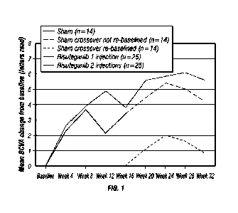
[0032] Figure 1 is a graph showing mean change in BCVA visit in a study of
human Subjects suffering from Dry AMD.
[0033] Figure 2A is a graph showing the change in Total Error Score Hue
Style by Change in Letters Read from Baseline at Week 12 in Dry AMD eyes
after intravitreal injection of 1mg risuteganib.
[0034] Figure 2B is a graph showing the change in Total Error Score Hue
Style by change from baseline in Letters Read at Week 12 in Dry AMD eyes
after sham injection.
[0035] Figure 3 is a graph showing change in Total Error Score Hue Style for
Risuteganib Responders (at 32 Weeks) Versus Sham Responders (at 12
Weeks).
[0036] Figure 4A is a graph showing change in Mean Retinal Sensitivity by
change from baseline in Letters Read in Dry AMD eyes at Week 12 after
intravitreal injection of 1mg risuteganib.
6
CA 03145870 2022-1-26
WO 2021/021668
PCT/US2020/043589
[0037] Figure 4B is a graph showing change in Mean Retinal Sensitivity by
change from baseline in Letters Read in Dry AMD eyes at Week 12 after
sham injection.
[0038] Figure 5 is a graph showing change in Mean Retinal Sensitivity for
Risuteganib Responders (at 32 Weeks) versus Sham Responders (at 12
Weeks).
[0039] Figure 6A is a graph showing change in microperimetry as measured
by Number of Loci Summed by Change from Baseline Number of Letters
Read in Dry AMD eyes at Week 12 at after intravitreal injection of lmg
risuteganib.
[0040] Figure 6B is a graph showing change in microperimetry as measured
by Number of Loci Summed by Change from Baseline Number of Letters
Read in Dry AMD eyes at Week 12 after sham injection.
[0041] Figure 7 is a graph showing change in microperimetry as measured by
Number of Loci Summed for Risuteganib Responders (at 32 Weeks) Versus
Sham Responders (at 12 Weeks).
[0042] Figure 8A shows locations and incidences of Geographic Atrophy (GA)
at baseline (pre-treatment) in Group 1 eyes.
[0043] Figure 8B shows locations and incidences of Geographic Atrophy (GA)
at baseline (pre-treatment) in Group 2 eyes.
[0044] Figure 9A shows an external limiting membrane map of the central l-
and 2-mm subfields exhibiting no disruption.
[0045] Figure 9B shows an external limiting membrane map of the central l-
and 2-mm subfields exhibiting segmental disruption.
[0046] Figure 9C shows an external limiting membrane map of the central l-
and 2-mm subfields exhibiting diffuse disruption affecting the fovea.
[0047] Figure 10A shows an OCT image (greyscale) taken from a risuteganib
responder eye.
[0048] Figure 10B shows an OCT image (greyscale) taken from a risuteganib
responder eye with an overlay of mapping of the individual retinal layers.
[0049] Figure 10C shows an I LM-RPE map of a risuteganib responder eye.
[0050] Figure 10D shows an EZ-RPE map of a risuteganib responder eye.
[0051] Figure 10E shows an RPE-BM map of a risuteganib responder eye.
[0052] Figure 11A shows an OCT image (greyscale) taken from a risuteganib
non-responder eye.
7
CA 03145870 2022-1-26
WO 2021/021668
PCT/US2020/043589
[0053] Figure 11B shows an OCT image (greyscale) taken from a risuteganib
non-responder eye with an overlay of mapping of the individual retinal layers.
[0054] Figure 11C shows an ILM-RPE map of a risuteganib non-responder
eye.
[0055] Figure 11D shows an EZ-RPE map of a risuteganib non-responder
eye.
[0056] Figure 11E shows an RPE-BM map of a risuteganib non-responder
eye.
[0057] Figure 12A is a bar graph comparing the effects of risuteganib vs.
control on gene expression under ITGAM and ITGB2 conditions in retinitis of
prematurity (ROP) mice.
[0058] Figure 12B is a bar graph showing the effects of risuteganib vs control
on expression of genes associated with complement, cell adhesion and
leukocyte migration, in ROP mice.
[0059] Figure 13A is a bar graph showing the effect of risuteganib vs. control
on retinal neuronal cell survival following exposure to kainic acid.
[0060] Figure 13B is a bar graph showing the effect of risuteganib vs. control
on retinal Muller cell survival following exposure to kainic acid.
[0061] Figure 13 C is a bar graph showing the effect of risuteganib vs.
control
on retinal pigment epithelium (RPE) cells following exposure to peroxide.
[0062] Figure 14 is a bar graph showing mouse Muller cell viability after
cytotoxic stress and risuteganib treatment.
[0063] Figure 15 is a bar graph showing mouse retinal neuron cell viability
after cytotoxic stress and risuteganib treatment
[0064] Figure 16 is a bar graph showing mouse RPE cell viability after
cytotoxic stress and risuteganib treatment.
[0065] Figure 17 is a bar graph showing human (M10-M1) Muller cell viability
after risuteganib treatment at three dosage levels vs control.
[0066] Figure 18 is a bar graph showing human (M10-M1) Muller cell viability
after treatment with anti-VEGF agents (Lucentis, Avastin and Eylea) and
risuteganib (Luminate) treatments.
[0067] Figure 19 (4-9) is a bar graph showing levels of reactive oxygen
species (ROS) in human (M10-M1) Muller cells after treatment with anti-VEGF
agents (Lucentis, Avastin and Eylea) and risuteganib (Luminate) treatments.
8
CA 03145870 2022-1-26
WO 2021/021668
PCT/US2020/043589
[0068] Figure 20 (4-10) is a bar graph showing mitochondrial membrane
potential in human (M10-M1) Muller cells after treatment with anti-VEGF
agents (Lucentis, Avastin and Eylea) and risuteganib (Luminate) treatments.
[0069] Figure 21A is a bar graph comparing the effects of control vs.
hydroquinone vs hydroquinone + risuteganib on mitochondrial membrane
potential in RPE cells.
[0070] Figure 21B is a bar graph comparing the effects of control vs.
hydroquinone vs hydroquinone + risuteganib on production of reactive oxygen
species (ROS) in RPE cells.
[0071] Figure 21C is a bar graph comparing the effects of control vs.
hydroquinone vs hydroquinone + risuteganib on viability of RPE cells.
Detailed Description
[0072] The following detailed description and the accompanying drawings
to which it refers are intended to describe some, but not necessarily all,
examples or embodiments of the invention. The described embodiments are
to be considered in all respects only as illustrative and not restrictive. The
contents of this detailed description and the accompanying drawings do not
limit the scope of the invention in any way.
[0073] As used herein, the term "patient or "subject" refers to either human
or non-human animals, such as humans, primates, mammals, and
vertebrates.
[0074] As used herein, the term "treat" or "treating" refers to preventing,
eliminating, curing, deterring, reducing the severity or reducing at least one
symptom of a condition, disease or disorder.
[0075] As used herein, the phrase "effective amount" or "amount effective
to" refers to an amount of an agent that produces some desired effect at a
reasonable benefit/risk ratio. In certain embodiments, the term refers to that
amount necessary or sufficient to treat Dry AMD or to cause return of
previously lost visual acuity in a subject who suffers from Dray AMD. The
effective amount may vary depending on such factors as the disease or
condition being treated, the particular composition being administered, or the
severity of the disease or condition. One of skill in the art may empirically
determine the effective amount of a particular agent without necessitating
undue experimentation.
9
CA 03145870 2022-1-26
WO 2021/021668
PCT/US2020/043589
[0076] This application discloses additional data, information and
therapeutic uses for Risuteganib. Risuteganib is shown to cause a number of
effects, including the following:
= Deterrence of angiogenesis and possible regression of
neovascularization by downregulating production of VEGF and other
proangiogenic growth factors including ANG-2; Suppression of retinal
angiogenesis in OIR, CNV and hVEGF mouse models; Inhibiting
endothelial adhesion and migration on matrix-coated surfaces and
suppression of endothelial cell proliferation
= Reduction of vascular leakage by inhibiting the production of VEGF
and inflammatory mediators;
= Reduction of inflammation, at least in part by targeting multiple
integrin subunits; Reducing expression of the Complement 3 Receptor
(also known as Integrin aM132); Reduction of leucocyte adhesion;
Reduction of trans-endothelial leucocyte migration; and Reductions of
TNF-a pathway gene expression in human immune ce11s2; Lowering
pro-inflammatory cytokine levels (e.g., in corneal tissue).
= Neuroprotection/Neuroregeration/Restoration of lost or impared
nerve function by decreasing apoptosis, increasing cell survival (e.g., in
a ROP Model); Reducing free radical oxygen production; Enhancing
mitochondria! health; Stabilizing and deterring leakage from
mitochondrial cell membranes; Improving retinal and/or optic nerve
function; Improving vision; Improving vision or restoring previously lost
visual acuity in subjects suffering from retinal and/or optic nerve
degeneration or damage (e.g., due to dry macular degeneration,
glaucoma, hereditary or familial retinal and/or macular disorders
including but not limited to Leber congenital amaurosis, choroideremia,
Stargardt's disease, Usher Syndrome and achromatopsia; Other
hereditary dystrophies affecting the central retina; Retinal and/or optic
nerve degeneration due to mutations in gene(s) responsible for
changes of the choroid (e.g., choroideremia) or retinal pigment
epithelium (RPE)(e.g., Best's disease)); Treating degeneration of
CA 03145870 2022-1-26
WO 2021/021668
PCT/US2020/043589
photoreceptor outer segments (e.g., Stargardts disease); Treating
impaired color vision; Treating degeneration of bipolar and/or Mueller
cells (e.g., x-linked refinoschisis); Increasing mitochondrial membrane
potential; Improving mitochondria!
bi energetics ; Reducing
mitochondrial reactive oxygen species (ROS) in tissues under
mechanical, oxidative, hypoxic, anoxic, chemical, chemo-toxic or other
stress (e.g., in retinal tissue following H202 and hydroquinone
exposure.
Flisuteoanib Treatment of Div AMD In Human Subjects
[0077] Eligible subjects who had been diagnosed with intermediate non-
exudative AMD that required treatment were enrolled and randomized to
either Group 1 or Group 2. Twenty-five subjects were assigned to Group 1
and fifteen (15) subjects were assigned to Group 2. Study treatments were
administered to the subjects in Groups 1 and 2, as follows:
= Each subject assigned to Group 1 received a first treatment
consisting of a sham injection in the study eye on day 1 of the study
and then crossed over to receive a second treatment consisting of an
intravitreal injection into the study eye of 1.0mg/50 pL risuteganib
during week 16 of the study.
= Each subject assigned to the Group 2 received a first treatment
consisting of an intravitreal injection into the study eye of 1.0mg/50 pL
risuteganib (Le., 1 .0mg in 50 pL of isotonic saline solution) on day 1 of
the study and a second treatment consisting of an intravitreal injection
into the study eye of 1.0mg/50 pL of risuteganib during week 16 of the
study.
[0078] The subjects in Groups 1 and 2 received the following treatments:
Thus, subjects in Group 1 received an initial sham injection in the study eye
followed by a single lmg dose of risuteganib in the study eye. The subjects in
Group 2 received a total of two (2) doses of risuteganib (lmg per dose) in the
study eye.
[0079] Numerous study assessments were conducted at various time points
throughout the study. Included among these study assessments were;
11
CA 03145870 2022-1-26
WO 2021/021668
PCT/US2020/043589
refractive eye examinations, determinations of BCVA AND low-luminance
BCVA, Lanthony D-15 color vision test, measurement of intraocular pressure
(10P), Indirect ophthalmoscopy/dilated fundus examinations and spectral-
domain optical coherence tomography (SD-OCT). Also, blood and saliva
samples were obtained from each subject for genetic analysis. The above-
listed study assessments were performed at the time points indicated in Table
1, below:
Table 1
Schedule of Visits and Assessments
EigigmEmmungomgg gonmEg-ES0Ø400itt !ftlk W0010 Wocig lik0IWOOk Wia WOW WO*
4
8 og gik:mmu24 2S 32h
manVisit flit Visit Visit mat Wit Wit 191dU
Visit
i. V sit Visit Visit Visit Visit
Visit I
Visit Visit
Visit Visit 2 3
6 7 8 9 10
(-28 to -2
4 ( 3 5 ( 3
(61 day) (th3
da s, da
days)
days)
Y 1 3's1 days) days) days) days) days)
Refraction and BCVA X X
X X X X X X X X
Low-luminasee BCVA X
X X
Lanthony D-15 color vision X
X X
test
JO? X
X X X X X X X X X
Indirect ophthalmoscopy/ X X X X X
X X X X X
dilated &Mos exam
SD-OCT X
X X
Blood or saliva sample for
X
genetic analysis [a]
Primary Efficacy Outcomes:
[0080] For this study, a primary efficacy endpoint was deemed to be the
percentage of population with an improvement in BCVA of at least 8 letters
(1.5 lines) BCVA. Table 2, below, summarizes the proportion of Group 2
subjects who exhibited this primary efficacy outcome at Week 12 and the
proportion of Group 1 subjects who exhibited this primary efficacy outcome at
Week 28 of the study:
Table 2
Proportion of Subjects With Gain of 8 or More BCVA Letters Read at
Primary Endpoint Week
12
CA 03145870 2022- 1-26
WO 2021/021668
PCT/US2020/043589
GROUP 1
GROUP 2
Week 12
Week 28
n=14
n=25
Gain of all letters read, n (%)
1(7.1) 12 (48.0)
95% exact CI
0.18,33.87 27.80,68.69
Baseline visit, letters read
14
25
Mean (SD)
67.1 (4.99) 64.4 (6.74)
95% Cl
64.26,70.02 61.62,67.18
Median
69.5 66.0
Min, Max
57,73 45,73
Primary endpoint week,* letters read
14
25
Mean (SD)
69.3 (8.64) 70.5 (8.03)
95% Cl
64.30,7428 67.20,73.84
Median
71.0 71.0
Mb, Max
51,83 57,87
Change in letters read
14
25
Mean (SD) 2.1
(5.04) 6.1 (7.60)
95% CI -
0.76, 5.05 2.98,9.26
Median
2.0 6.0
Mb, Max -
6, 10 -6,20
Abbreviations: Cl, confidence interval; max, maximum; min, minimum; SD,
standard
deviation.
Primary endpoint week was Week 12 for the sham group and Week 28 for the
risuteganib group.
[0081] It was determined that, at baseline, no anatomical measurements
showed a significant difference between risuteganib nonresponder eyes and
sham eyes.
[0082] Figure 1 is a graph showing mean change in BCVA visit in a study of
human Subjects suffering from Dry AMD. The proportion of subjects with a
gain of at least 8 BCVA letters read was 48% in Group 2 at Week 28
compared with 7.1% in Group 1 at Week 12. Although hypothesis testing was
not planned, post hoc analysis using a 2-sided Fishers exact test
demonstrated that this was a statistically significant difference between
groups (P= .013).
[0083] Additional post hoc analysis was performed to assess whether the
presence of foveal geographic atrophy (GA) in risuteganib-treated subjects
affected the degree of BCVA improvement The Group 2 subjects were
divided into 2 subgroups: those with eyes with no foveal geographic atrophy
(GA) in the central 6-mm subfield (the No GA Subgroup") and those with GA
13
CA 03145870 2022-1-26
WO 2021/021668
PCT/US2020/043589
in the central 6-mm subfieid (the 'GA Subgroup"). The proportion of
risuteganib-treated subjects with a gain of at least 8 BCVA letters read was
higher in the No GA Subgroup when compared to the GA Subgroup (80% vs
40%).
Secondary Efficacy Outcomes:
[0084] Secondary efficacy outcomes were deemed to be the following:
= Mean Observed Changes in BCVA Between the Group 1 at Week
12 and Group 2 at Week 28;
= Mean Observed Changes in BCVA Between Groups 1 and 2 at
Week 12;
= Maximum Observed Changes in BCVA Between Groups 1 and 2;
and
= Percentage of all subjects who exhibited an improvement in BCVA
of at least 8 letters (1.5 lines) BCVA.
[0085] Table 3, below, summarizes mean BCVA change over time in the
subset of subjects who met or exceeded the primary endpoint criteria:
Table 3
Mean BCVA Change Over Time in the Subset of Subjects With Gain of 8
or More BCVA Letters Read at Primary Endpoint Week
GROUP 1
GROUP 1 GROUP 2
Week 0 to Week 16 Week 16 to Week 32 Week 0 to Week n
n=1
n=2 n=12
Baseline visit, letters read
N 1
12
Mean (SD) 73.0 (NA)
62.9 (7.27)
95% CI
58.30, 67.53
Median 73.0
65.0
Min, Max 73,73
45,71
Week 4, letters read
N 1
12
Mean (SD) 72.0 (NA)
67.0 (10.07)
95% CI
60.60,73.40
Median 72.0
68.5
Min, Max 72,72
44, 81
(Table 3 continued on following pages)
14
CA 03145870 2022-1-26
WO 2021/021668
PCT/US2020/043589
Week 4 change in letters read
1
12
Mean (SD) -1.0 (NA)
4.1 (7.15)
95% CI
-0.46, 8.63
Median -1.0
3.0
Nim, Max -1,-i
-5,22
Week 8, letters read
1
12
Mean (SD) 85.0 (NA)
683 (10.80)
95% CI
61.64,75.36
Median 85.0
72.0
Min, Max 85,85
50,81
Week 8 change in letters read
1
12
Mean (SD) 12.0 (NA)
5.6 (6.92)
95% CI
1.19,9.98
Median 12.0
5.5
Min, Max 12,12
-6,18
Week 12, letters read
1
12
Mean (SD) 83.0 (NA)
70.6 (11.08)
95% CI
63.54,77.62
Median 83.0
72.0
Mb, Max 83,83
47,g7
Week 12 change in letters read
1
12
Mean (SD) 10.0 (NA)
7.7 (6.61)
95% CI
3.47, 11.87
Median 10.0
5.5
Mm, Max 10, 10
-1,21
Week 16, letters read
/4 1
2 12
Mean (SD) 81.0 (NA)
70.0 (7.07) 69.2 (9.45)
95% Cl
6.47, 133.53 63.16, 75.17
Median 81.0
70.0 703
IVfm, Max 81,81
65,75 52,80
Week 16 change in letters read
1
12
Mean (SD) 8.0 (NA)
63 (6.43)
95% Cl
2.17, 10.33
Median 8.0
8.0
Min, Max 8,8
-6,15
Week 20, letters read
2
12
Mean (SD)
70.0 (7.07) 74.2 (8.03)
95% CI
6.47, 133.53 69.06,79.27
Median
70.0 75.0
Mb, Max
65,75 58,90
Week 20 change in letters read
2
12
Mean (SD)
0.0 (0.00) 113 (4.56)
95% CI
8.36, 14.14
Median
0.0 10.0
CA 03145870 2022-1-26
WO 2021/021668
PCT/US2020/043589
Min, Max
0,0 5,20
Week 24, letters read
N
2 12
Mean (SD)
78.0 (2.83) 74.3 (7.88)
95% Cl
52.59, 103.41 69.33, 79.34
Median
78.0 75.5
Min, Max
76,80 56,85
Week 24 change in letters read
N
2 12
Mean (SD)
8.0 (4.24) 11.4 (4.34)
95% CI
-30.12, 46.12 8.66, 14.17
Median
8.0 10.0
Min, Max
5,11 6,21
Week 28, letters read
N
2 12
Mean (SD)
79.5 (7.78) 75.7 (7.66)
95% CI
9.62, 149.38 70.80, 80.53
Median
79.5 75.5
Min, Max
74,85 57,87
Week 28 change in letters read
N
2 12
Mean (SD)
95 (0.71) 12.8 (4.20)
95% Cl
3.15, 15.85 10.08, 15.42
Median
9.5 12.0
Min, Max
9,10 8,20
Week 32, letters read
N
2 12
Mean (SD)
76.5 (4.95) 72.4 (8.78)
95% Cl
32.03, 120.97 66.84,78.00
Median
76.5 74.0
Mitt, Max
73,80 57,85
Week 32 change in letters read
N
2 12
Mean (SD)
65 (2.12) 9.5 (5.00)
95% CI
-12.56, 25.56 6.32, 12.68
Median
6.5 10.5
MuTh, Max
5,8 -2,15
Abbreviations: CI, confidence interval; max, maximum; min, minimum; NA; not
applicable; SD, standard deviation.
[0086] Table 4, below, summarizes the change in BCVA over time at any
week in the study:
16
CA 03145870 2022-1-26
WO 2021/021668
PCT/US2020/043589
Table 4
Mean BCVA Change Over Time in the Subset of Subjects With Gain of 8
or More BCVA Letters Read at Any Week
GROUP 1
GROUP 1 GROUP 2
Week 0 to Week 16 Week 16 to Week 32 Week 0 to Week 32
r7
r3 r14
Baseline visit, letters read
N 7
14
Mean (SD) 69.9 (2.91)
62.5 (6.81)
95% CI 67.16,72.55
58.57, 66.43
Median 70.0
63.5
Min, Max 64,73
45,71
Week 4, letters read
N 7
14
Mean (SD) 72.1 (6.12)
66.5 (9.52)
95% CI 66.48,77.80
61.00, 72.00
Median 72.0
67.0
Min, Max 63, 83
44, 81
Week 4 change in letters read
N 7
14
Mean (SD) 2.3 (7.36)
4.0 (6.66)
95% CI -4.53, 9.10
0.16, 7.84
Median -1.0
3.0
Min, Max -7,14
-5,22
Week 8, letters read
N 7
14
Mean (SD) 75.7 (6.10)
68.3 (10.07)
Median 75.0
71.0
Min, Max 70, 85
50, 81
(Table 4 continued on following pages)
17
CA 03145870 2022-1-26
WO 2021/021668
PCT/US2020/043589
GROUP 1 GROUP 1 GROUP 2
Week 0 to Week 16 Week 16 to Week 32 Week 0 to Week 32
n=7
n=3 n=14
Week 8 change in letters read
7
14
Mean (SD) 5.9
(5.52) 5.8 (6.44)
95% CI 0.75,
10.96 2.07,9.50
Median 6.0
5.5
Min, Max -2,12
-6,18
Week 12, letters read
7
14
Mean (SD) 75.9
(4.14) 69.6 (10.54)
95% CI 72.03,79.69
63.56, 75.73
Median 76.0
69.5
Min, Max 70, 83
47, 87
Week 12 change in letters read
7
14
Mean (SD) 6.0
(3.00) 7.1 (6.53)
95% CI 3.23,
8.77 3.37, 10.91
Median 6.0
5.5
Min, Max 0,10
-1,21
Week 16, letters read
7
3 14
Mean (SD) 76.1
(4.41) 693 (5.03) 68.9 (8.73)
95% CI 72.06, 80.22
57.16,82.17 63.89, 73.97
Median 75.0
69.0 69.5
Min, Max 69,81
65,75 52,80
Week 16 change in letters read
7
14
Mean (SD) 6.3
(2.14) 6.4 (6.09)
95% Cl 4.31,
8.26 2.91, 9.94
Median 6.0
8.0
Min, Max 3,9
-6,15
Week 20, letters read
3
14
Mean (SD)
71.7 (5.77) 72.3 (9.22)
95% Cl
57.32, 86.01 66.96, 77.61
Median
75.0 74.5
Min, Max
65,75 54,90
Week 20 change in letters read
3
14
Mean (SD)
2.0 (3.46) 9.8 (6.62)
95% CI
-6.61, 10.61 5.96, 13.61
Median
0.0 10.0
Min, Max
0,6 -8,20
Week 24, letters read
3
14
Mean (SD)
78_3 (2_08) 72.7(834)
95% CI
73.16, 83.50 67.90, 77.53
Median
79.0 74.5
Min, Max
76,80 56, 85
Week 24 change in letters read
3
14
Mean (SD)
8.7 (3.21) 10.2 (5.16)
95% Cl
(LK 16.65 7_23, 13.19
Median
10.0 9.5
Min, Mai
5,11 0,21
18
CA 03145870 2022-1-26
WO 2021/021668
PCT/US2020/043589
GROUP 1
GROUP 1 GROUP 2
Week 0 to Week 16 Week 16 to Week 32 Week 0 to Week 32
n=7
n=3 n=14
Week 28, letters read
N
3 14
Mean (SD)
77.7 (6.35) 73.9 (8.42)
95% CI
61.89,93.44 69.00,78.72
Median
74.0 74.5
Min, Max
74,85 57,37
Week 28 change in letters read
N
3 14
Mean (SD)
8.0 (2.65) 11.4 (5.26)
95% 01
1.43, 14.57 8.32, 14.39
Median
9.0 11.5
Min, Max
5,10 Z20
Week 32, letters read
N
3 14
Mean (SD)
76.0 (3.61) 70.7 (9.19)
95% CI
67.04,84.96 65.41,76.02
Median
75.0 73.0
Min, Max
73,80 57, 85
Week 32 change in letters read
N
3 14
Mean (SD)
63 (1.53) 82 (5.65)
95% CI
2.54, 10.13 4.95, 11.47
Median
6.0 9.5
Min, Max
5,8 -2,15
Abbreviations: CI, confidence interval; max, maximum; min, minimum; SD,
standard
deviation.
Color Vision Test
[00871 The results of color vision testing of the study subjects are
summarized in Table 5, below.
Table 5
Color Vision as Measured by Total Error Score Hue Style
GROUP 1
GROUP 2
r14
n=25
Screening
N 14
25
Mean (SD)
50.52 (31.192) 43.27(28.678)
95% Cl
32.515, 68.534 31.429, 55.105
Median 47.59
44.67
Mb, Max
4.7, 101.0 0.0,993
Week 12
N 13
23
Mean (SD)
48.61 (33.835) 43.38 (30.099)
95% CI
28.168, 69.061 30.361, 56393
Median 48.00
39.67
Min, Max
1.3, 121.7 13,893
19
CA 03145870 2022-1-26
WO 2021/021668
PCT/US2020/043589
Week 12 change
13
23
Mean (SD)
1.97 (17.919) 2.41 (17.964)
95% CI -
8.855, 12.801 -5.363, 10.174
Median 533
2.67
Min, Max -28.2, 40.7
-44.2, 35.3
Week 32
14
24
Mean (SD)
48.76(34.018) 39.88 (33.181)
95% CI
29.121, 68.403 25.864, 53.887
Median 42.00
24.09
Mill, Max 6.7, 107.5
0.0, 104.0
Week 32 change
14
24
Mean (SD) -
1.76 (22.474) -4.36(20.808)
95% CI -14.738,
11.214 -13.147, 4.426
Median 134
-3.17
Min, Max -67.7, 26.5
-40.2, 42.7
Abbreviations: CI, confidence interval; max, maximum; min, minimum; SD,
standard deviation.
[0088] As shown in Table 5 above, the mean total color vision error score in
Group 1 subjects at screening (pre-treatrnent) was 50.52. At Week 12, the
mean color vision score of Group 1 subjects had increased (worsening of
color vision) by 1.97. Following crossover and administration of the single
dose of risuteganib, the mean total color vision error score in Group 1
subjects decreased (improved) by 1.76 at Week 32.
[0089] As shown in Table 5 above, the mean total error score on the color
vision test for Group 2 subjects was 43.27 at screening. This score increased
in the Group 2 subjects (worsening of color vision) by 2.41 at Week 12 and
then decreased (improvement in color vision) by 4.36 at Week 32.
[0090] Figures 2A and 2B show analysis of scatter plots of change in total
error score by change in BCVA letters read from baseline at Week 12. Figure
2A shows a negative correlation for Group 2 subjects at 12 weeks following
their initial risuteganib dose (decreased color vision scores correlate with
increased BCVA) and Figure 2B shows a slight positive correlation for Group
subjects at 12 weeks following their initial sham injection.
[0091] Examination of change in total error score by responder status
(subjects with or without letters BCVA
gain) shows that risuteganib
responders at Week 32 had a decrease (improvement) in color vision of 13.03
compared with an increase (worsening) of 2.98 for sham responders at Week
12n as seen in the bar graph of Figure 3.
CA 03145870 2022-1-26
WO 2021/021668
PCT/US2020/043589
Improvement in Perimetry Humphrey Visual Field Assessment
[0092] Table 6, below, shows mean deviation (MD) scores from the
Humphrey visual field assessment, which compares subject performance to
an age-matched normative database.
Table 6
Humphrey Visual Field as Measured by Mean Deviation
Sham or Crossover to
Risuteganth
Risuteganib
n=14
z2.5
Screening, dB
12
21
Mean (SD)
4.074 (4.6813) 4.557(4.0715)
95% CI -
7.0485, -1.0998 -6.4105, -23038
Median -2.455
-3.330
Min, Max -16.19, -0.44
-18.58, -0.48
Week 12, dB
8
21
Mean (SD)
4.665 (4.8504) -5.502(6.6203)
95% CI -
8.7201, -0.6099 -8.5154, -2.4884
Median -2.870
-3.500
Min, Max -
14.45, 0.02 -25.00, 0.66
Week 12 change, dB
7
17
Mean (SD)
0.561 (0.9252) 0.302(1.7590)
95% CI -
0.2942, 1.4171 -0.6026, 1.2061
Median 0.590
0.100
Min, Max -030,
1.74 -2.69, 3.21
Week 32, dB
11
21
Mean (SD) -
4.055 (5.1026) -5.211 (5.5763)
95% CI -
7.4834, -0.6275 -7.7493, -2.6727
Median -2.260
-3.470
Min, Max -
16.19, 0.54 -2533, -1.30
Week 32 change, dB
16
Mean (SD)
0.158 (0.7268) 0.191 (11383)
95% CI -
0.3619, 0.6779 -0.4153, 0.7978
Median -0.070
-0.040
Min, Max -
0.58, 1.73 -1.70, 1.94
Abbreviations: Cl, confidence interval; dB, decibels; max, maximum; min,
minimum;
SD, standard deviation.
NOTE: only measures of "acceptable" quality were included.
[0093] In the sham group, the mean MD score was -4.074 dB at screening.
This score increased (improved) by 0.561 dB at Week 12; after crossover to
1 risuteganib injection, this score increased by 0.158 dB at Week 32. In the
21
CA 03145870 2022-1-26
WO 2021/021668
PCT/US2020/043589
risuteganib group, the mean MD score was -4.557 dB at screening. This score
increased by 0.302 dB at Week 12 and by 0.191 dB at Week 32.
[00941 Table 7, below, shows pattern standard deviation (PSD) scores from
the Humphrey visual field assessment, which can identify focal defects.
Table 7
Humphrey Visual Field as Measured by Pattern Standard Deviation
Screening, dB
12
21
Mean (SD) 2.401 (1.5819)
3.352(3.2841)
95% CI 1.3957, 34060
1.8570, 4.8468
Median 2.150
1.660
Min, Max 1.18,
7.15 1.13, 13.27
Week 12, dB
8
21
Mean (SD) 2.914 (2.7491)
3.350(3.5796)
95% CI 0.6154, 5.2121
1.7201, 4.9789
Median 2.170
1.630
Min, Max 1.17,
9.45 1.10, 13.18
Week 12 change, dB
7
17
Mean (SD) 0.447(0.8439)
-0.340(0.8416)
95% CI -0.3333, 1.2276
-0.7727, 0.0927
Median 0.290
-0.090
Min, Max -
0.15, 2.30 -2.69, 0.43
Week 32, dB
11
21
Mean (SD) 2.790(1.7152)
3.113 (2.7424)
95% CI 1.6377, 3.9423
1.8650, 43616
Median 2.410
2.050
Min, Max 1.12,
7.15 1.17, 10.42
Week 32 change, dB
16
Mean (SD) 0.469(0.5951)
0.115 (0.9026)
95% CI 0.0433, 0.8947
-0.3660, 0.5960
Median 0.360
01)45
Min, Max -037,
1.72 -1.94, 1.38
Abbreviations: CI, confidence interval; dB, decibels; max, maximum; Mill,
minimum; SD, standard deviation
NOTE: only measures of "acceptable" quality were included.
[00951 In Group 1 subjects, the mean PSD score was 2.401 dB at
screening (pre-treatment). This score increased in Group 1 subjects by 0.447
dB at Week 12. After crossover and administration of the single risuteganib
injection, this score increased in the Group 1 subjects by 0.469 dB at Week
32.
22
CA 03145870 2022-1-26
WO 2021/021668
PCT/US2020/043589
[0096] In the Group 2 subjects, the mean PSD score was 3.352 dB at
screening (pre-treatment). This score decreased by 0.340 dB at Week 12 and
increased by 0.115 dB at Week 32.
Retinal Sensitivity
[0097] Table 8, below, shows mean retinal sensitivity as measured by
microperimetry.
Table 8
Microperimetry as Measured by Mean Sensitivity
GROUP 1
GROUP 2
n=14
e25
Screening
N
9 13
Mean (SD) 12.43 (5.199)
8.52(5.006)
95% CI
8.437, 16.430 5.490, 11.540
Median 15.10
10.40
Min, Max 3.1,
17.8 0.4, 16.7
eek 12
N
7 14
Mean (SD) 9.56(5.459)
7.52(4.969)
95% CI
4.509, 14.605 4.652, 10.390
Median 11.70
7.50
Min, Max 1.7,
16.0 0.0, 16.2
Week 12 change
N
7 11
Mean (SD) -1.49 (3.975)
-0.85(2.711)
95% CI -
5.162, 2.190 -2.676, 0.967
Median -0.60
-1.50
Min, Max -
6.4,4.9 -5.1,3.9
Week 32
N 8
12
Mean (SD) 11.44(6.655)
8.25 (4.601)
95% CI
5.873, 17.002 5.327, 11.173
Median 13.70
8.50
Min, Max 0.0,
17.3 0.0, 15.4
Week 32 change
N
8 9
Mean (SD) -2.16 (5.527)
-0.53 (4373)
95% CI -
6.783, 2.458 -3.895, 2.828
Median -0.20
-0.40
Min, Max -
12.9, 3.2 -7.8, 4.2
Abbreviations: CI, confidence interval; max, maximum; min, minimum; SD,
standard
deviation.
23
CA 03145870 2022-1-26
WO 2021/021668
PCT/US2020/043589
[0098] As seen in Table 8, above, mean retinal sensitivity in Group 1
subjects was 12.43 dB at screening (pre-treatment). This score decreased in
the Group 1 subjects (worsened) by 1.49 dB at Week 12. Following crossover
and administration of the single risuteganib injection to the Group 1
subjects,
the mean retinal sensitivity score in those subjects decreased by 2.16 dB at
Week 32.
[0099] In Group 2 subjects, mean retinal
sensitivity was 8.52 dB at
screening (pre-treatment). This score decreased by 0.85 dB in Group 2
subjects at Week 12 and further decreased by 0.53 dB at Week 32.
[00100] Figures 4A and 4B show scatter plots of change in mean sensitivity
by change in BCVA letters read from baseline at Week 12. Figure 4A shows
a positive correlation for Group 2 subjects following their initial dose of
risuteganib (increased mean sensitivity correlates with increased BCVA) and
Figure 4B shows a slight negative correlation for Group 1 subjects following
their initial sham injection.
[00101] Examination of change in mean sensitivity by responder status
showed that risuteganib responders at Week 32 had an increase
(improvement) of 2.2 dB compared with a decrease (worsening) of 1.9 dB for
sham responders at Week 12, as seen in the bar graph of Figure 5.
[00102] Table 9, below, summarizes number of loci with reduced retinal
sensitivity summed across assessments using a 20-dB threshold, an 11-dB
threshold, and by measuring absolute scotoma.
Table 9
Microperimetry as Measured by Number of Loci Summed
GROUP 1
GROUP 2
n=14 n=25
Screening
N 9
15
Mean (SD) 65A (2338)
81.4(24.23)
95% CI 47.47, 83.41
67.98,94.82
Median 56.0
74.0
Nun, Max 46, 1 1 1
48,123
Week 12
N 7
14
Mean (SD) 76.0 (26.98)
84.9(24.66)
95% CI 51.05, 100.95
70.69,99.17
Median 63.0
86.5
/vim, Max 48,122
47,135
(Table 9 continued on following page)
24
CA 03145870 2022-1-26
WO 2021/021668
PCT/US2020/043589
Week 12 change
7
13
Mean (SD) 5.1
(15.42) 6.1 (25.04)
95% CI -9.12, 19.40
-9.06, 21.21
Median 11.0
4.0
hilln, Max -26, 19
-29,69
Week 32
8
12
Mean (SD) 67.6 (33.30)
80.7 (23.78)
95% CI 39.78,95.47
65.56, 95.78
Median 60.0
79.5
Mtn, Max 28,127
53,135
Week 32 change
8
11
Mean (SD) 7.9
(27.46) 1.0(20.89)
95% CI -15.08, 30.83
-13.03, 15.03
Median -1.0
-3.0
Nlin, Max -22,58
-27,36
Abbreviations: CI, confidence interval; max, maximum; min, minimum; SD,
standard
deviation.
[00103] In the sham group, the mean number of summed loci with reduced
sensitivity was 65.4 at screening. This score increased (worsened) by 5.1 at
Week 12; after crossover to 1 risuteganib injection, this score increased by
7.9 at Week 32. In the risuteganib group, the mean number of summed loci
with reduced sensitivity was 81.4 at screening. This score increased by 6.1 at
Week 12 and by 1.0 at Week 32.
[00104] Figures 6A and 6B show scatter plots of change in number of loci
with reduced retinal sensitivity by change in BCVA letters read from baseline
at Week 12. Figure 6A shows a negative correlation for Group 2 subjects
following their initial risuteganib injection (decreased number of summed loci
with reduced sensitivity correlates with increased BCVA) and Figure 6B
shows a slight positive correlation for Group 1 subjects following their
initial
sham injection. Error! Reference source not found.
1001051 Examination of change in number of summed loci with reduced
retinal sensitivity by responder status showed that risuteganib responders had
a decrease (improvement) of 17.75 at Week 32 compared with an increase
(worsening) of 11.71 at Week 12 for sham responders, as seen in the bar
graph of Figure 7. (P = 0.014).
Low-Luminance Visual Acuity
CA 03145870 2022-1-26
WO 2021/021668
PCT/US2020/043589
[00106] Table 10, below, summarizes low-luminescence visual acuity in the
study subjects.
Table 10
improvement in Low-Luminance Visual Acuity by Visit
GROUP I.
GROUP 2
z14
n=25
Screening, letters read
14
25
Mean (SD) 48.1
(7A0) 47.4 (12.26)
95% CI 43.87,
52.41 42.30, 52.42
Median
50.5 50.0
Min, Max
35,56 6,68
Week 12, letters read
13
25
Mean (SD) 48.8
(9.91) 46.4 (12.51)
95% CI 42.86,
54.83 41.19, 51.53
Median
53.0 48.0
Min, Max
30,63 7,71
Week 12 change in letters read
13
25
Mean (SD) 0.9
(8.68) -1.0(6.95)
95% CI -432,
6.17 -3.87, 1.87
Median
0.0 -1.0
Min, Max -
10, 18 -19, 17
Week 32, letters read
14
25
Mean (SD)
50.7(17.58) 49.4 (12.50)
95% CI 40.57,
60.86 44.24, 54.56
Median
57.0 51.0
Min, Max
16,75 8,69
Week 32 change in letters read
14
25
Mean (SD)
2.6(16.59) 2.0 (7.95)
95% CI -7.01,
12.15 -1.24, 5.32
Median
3.0 0.0
Min, Max -
27,40 -7,24
Abbreviations: CI, confidence interval; max, maximum; min, minimum; SD,
standard deviation.
[00107] As shown in Table 10 above, the mean low-luminance visual acuity
in Group 1 subjects was 48.1 letters read at screening (pre-treatment). This
score increased (improved) in the Group 1 subjects by 0.9 letters at Week 12.
Following crossover and administration of the single risuteganib injection to
the Group 1 subjects, this score increased by an additional 2.6 letters at
Week
32.
[00108] Also, as shown in Table 10 above, the mean low-luminance visual
acuity in Group 2 subjects was 47.4 letters read at screening. This score
26
CA 03145870 2022-1-26
WO 2021/021668
PCT/US2020/043589
decreased (worsened) in Group 2 subjects by 1.0 letters at Week 12 and,
thereafter, increased by 2.0 letters at Week 32.
Retinal Examinations by Optical Coherence Tomography (OCT)
[00109] The OCT scans were analyzed by two (2) unrelated experts.
OCT Analysis 1:
[00110] The mean thickness and mean volume of retinal subfields and layer
segments were analyzed at screening (pre-treatment) and at Week 12 for
Group 1 subjects and at Week 32 for Group 2 subjects. The results of this
analysis are summarized in Table 11, below.
Table 11
Quantitative Anatomical Measurements at Baseline for Risuteganib
Nonresponder Eyes Versus Responder Eyes
Ftisuteganib
Risuteganlb T-test
Measurement
Nonresponder Responder p..
Layer, Sector
me12 n=10 value
Mean thickness, p.m
Inner retina, lineal center
27.833 42.500 0.305
Inner retina, central subfield
89.000 99.400 0.323
Outer retina, foveal center
12.4.417 143.200 0.210
Outer retina, central subfield
113.917 139.600 0.001
Photoreceptor, foveal center
46.833 48.500 0.784
Photoreceptor, central subfield
45.083 49.300 0.015
RPEDC, fovea! center
47.667 58.900 0.540
RPEDC, central subfield
46.500 54.800 0.611
Total volume, mm3
Inner retina, central subfield
0.070 0.078 0.319
Outer retina, central subfield
0.090 0.110 0.001
Photoreceptor, central subfield
0.035 0.039 0.011
RPEDC, central subfield
0.037 0.043 0.600
EZ defect area, nun3
0.308 0.111 0.012
Abbreviations: EZ, ellipsoid zone; RPEDC, retinal pigment epithelium-drusen
complex.
[00111] At baseline, those eyes that responded to risuteganib had
significantly greater mean thickness in the central subfield of the outer
retina
compared with eyes that did not respond to risuteganib (139.600 vs 113.917
pm; P=0.001); responder eyes also had significantly greater mean thickness
at baseline in the central subfield of the photoreceptor layer compared with
27
CA 03145870 2022-1-26
WO 2021/021668
PCT/US2020/043589
nonresponder eyes (49.300 vs 45.083 pm; P=0.015; Table 11). The same
anatomical locations also had significantly greater volume at baseline in the
responder eyes compared with nonresponder eyes (central subfield of the
outer retina, 0.110 vs 0.090 mm3; P=0.001 and central subfield of the
photoreceptor layer, 0.039 vs 0.035 mm3; P=0.011). In addition, the EZ defect
area of responder eyes was significantly smaller at baseline than that of
nonresponders (0.111 vs 0.308 mm2; P=0.012). No other anatomical
measurements showed a significant difference between risuteganib responder
and nonresponder eyes at baseline.
[00112] In addition to the quantitative analysis of OCT images, a qualitative
assessment of the OCT images at baseline (pre-treatment) was performed to
identify GA anywhere in the retina, in the fovea (1-mm central subfield), and
in
the fovea! center.
[00113] At baseline (pre-treatment), 7 of 25 (28%) of the eyes in Group 2
subjects had GA, 6 (24%) of which affected the fovea, and 2 (8%) of which
involved the foveal center, as indicated on Figure 8A. In addition, at
baseline
(pre-treatment), 5 of 14 (36%) Group 1 subject eyes had GA, 3 (26%) of
which involved the fovea, and 1 (7%) of which affected the foveal center, as
indicated on Figure 8B. The relationship between functional visual acuity
outcomes and the presence or absence of baseline GA is explored in the
following Tables 12 and 13, respectively:
Table 12
Visual Acuity Functional Outcome in Study Eyes With
Geographic Atrophy at Baseline
>3 Letter
>10 Letter .1.5 Letter
Improvement in Improvement in Improvement in
Treatment Visual
Acuity Visual Acuity Visual Acuity
Location of Geographic Atrophy n (%)
n (%) n (%)
Risuteganib
Geographic atrophy in retina (n=7) 2(29)
2(29) 1(14)
Geographic atrophy in fovea (n) 1(17)
1(17) 0(0)
Geographic atrophy in fovea' center
1 (50)
1 (50) 0(0)
(n=2)
Sham
Geographic atrophy in retina (n=5) 0(0)
0(0) 0(0)
Geographic atrophy in fovea (n=3) 0(0)
0(0) 0 (0)
Geographic atrophy in foveal center
0 (0)
0(0) 0 (0)
(n=1)
28
CA 03145870 2022-1-26
WO 2021/021668
PCT/US2020/043589
Table 13
Visual Acuity Functional Outcome in Study Eyes Without
Geographic Atrophy at Baseline
>8 Letter
>10 Letter .115 Letter
Treatment Improvement
in Improvement in Improvement in
Location of Absent Geographic Visual
Acuity Visual Acuity Visual Acuity
Atrophy n(%)
n(%) n(%)
Risuteganib
No geographic atrophy in retina (n=18) 10(56)
6(44) 4(22)
No geographic atrophy in fovea (r19) 11
(58) 7(37) 5(26)
No geographic atrophy in foveal center
11 (48)
7(30) 5(22)
(n=23)
Sham
No geographic atrophy in retina (n-9) 1(11)
1(11) 0(0)
No geographic atrophy in fovea (n=11) 1(9)
1(9) 0(0)
No geographic atrophy in foveal center
1 (8)
1 (8) 0(0)
(n=13)
[00114] Since only one sham-treated eye had at least an 8-letter
improvement in visual acuity, it is impossible to use the sham group to
determine the effect of presence or absence of GA on functional outcomes.
Therefore, the discussion below is focused on the risuteganib group.
1001151 Risuteganib-treated eyes without any GA at baseline (n=18) had a
56% responder rate when using an 8-letter improvement threshold compared
with a 29% responder rate among risuteganib-treated eyes with any GA at
baseline (n=7). The same pattern is maintained when using a 10-letter
improvement (44% vs 29%, respectively) or a 15-letter improvement (22% vs
14%, respectively) as the visual acuity threshold.
[00116] Risuteganib-treated eyes without GA in the fovea at baseline (n=19)
had a 58% responder rate (a8-letter improvement threshold) compared with a
17% responder rate among risuteganib eyes with GA in the fovea at baseline
(n=6). The same pattern is maintained when using a 10-letter improvement
(37% vs 17%, respectively) or a 15-letter improvement (26% vs 0%,
respectively) as the visual acuity threshold.
[00117] Risuteganib-treated eyes without GA in the foveal center at baseline
(n=23) had a 48% responder rate (a8-letter improvement threshold) compared
with a 50% responder rate among risuteganib eyes with GA in the foveal
center at baseline (n=2). However, because only 2 eyes had GA in the foveal
center, the 50% responder rate in these eyes is not informative, and no
conclusions can be drawn regarding the importance of GA under these
circumstances.
29
CA 03145870 2022-1-26
WO 2021/021668
PCT/US2020/043589
[00118] Overall, these results suggest that absence of GA anywhere in the
retina or at least in the central 1 mm (the area of the retina responsible for
BCVA) increases the likelihood of response to risuteganib.
[00119] Quantitative analysis of the OCT images was also performed to
measure changes in anatomical measurements over time. This analysis is
summarized in Table 14 below_
Table 14
Quantitative Anatomical Measurements Change From Baseline at Week
32 for Rlsuteganlb Nonresponder Eyes Versus Responder Eyes
T-
Risnteganili
Risnteganib test
Measurement Nonresponder
Responder P-
Layer, Sector n=12
n=10 Difference value
Mean change in mean thickness,
pm
Inner retina, foveal center 9.917
8.400 -1.517 0.904
Inner retina, central subfield -2.250
5.200 7.450 0.042
Outer retina, foveal center 17.833
-8.000 9.833 0.291
Outer retina, central subfield -5.417
-2.400 3.017 0.261
Photoreceptor, foveal center 3.833
3.100 -0_733 0.869
Photoreceptor, central subfield -1.333
-1.000 0.333 0.849
RPEDC, foveal center -2.250
-8.100 -5.850 0.425
RPEDC, central subfield 1.083
-9.800 -10.883 0.307
Mean change in total volume, mm?
Inner retina, central subfield -0.002
0.004 0.006 0.033
Outer retina, central subfield -0.004
-0.002 0.003 0223
Photoreceptor, central subfield -0.001
-0.001 0.000 0.934
RPEDC, central subfield 0.001
-0.008 -0.009 0.297
EZ defect area, mm2 0.014
0.020 0.006 0.834
Abbreviations: EZ, ellipsoid zone; RPEDC, retinal pigment epitlaelium-drusen
complex.
[00120] From baseline to Week 32, the central subfield of the inner retina in
the risuteganib responder eyes had significantly larger increases in thickness
(difference of 7.450 pm; P=0.042) and in volume (difference of 0.006 mm3;
P=0.033) from baseline compared with risuteganib nonresponder eyes No
other anatomical measurements showed a significant difference between
responder and nonresponder eyes over time.
[00121] Significant differences in mean change from baseline to Week 32 in
mean thickness for risuteganib eyes were observed compared with the mean
change from baseline to Week 12 for sham eyes in the foveal center of the
inner retina (difference of 15.404 pm; P-43.011), in the foveal center and
CA 03145870 2022-1-26
WO 2021/021668
PCT/US2020/043589
central subfield of the outer retina (difference of -14.794 pm; P-41007 and
difference of -3.812 pm; P=0.042, respectively), and in the central subfield
of
the photoreceptor layer (difference of -2.545 pm; P=0.007). This is
summarized in Table 15, below:
Table 15
Quantitative Anatomical Measurements Change From Baseline at Week
32 for Risuteganlb Arm Versus Change From Baseline at Week 12
for Sham Arrn
Measurement Risuteganib
Sham T-test
Layer, Sector
n=22 n=12 Difference P-value
Mean change in mean thickness, pm
Inner retina, foveal center
6.6% -8308 15.404 0.011
Inner retina, central subfield
1.565 0875 0.690 0.761
Inner retina, nasal subfield -
0.783 0.167 -0.949 0.609
Inner retina, superior subfield -
3.022 1.167 -4.188 0.059
Inner retina, temporal subfield
0.217 0.125 0.092 0.953
Inner retina, inferior subfield -
0.065 1.542 -1.607 0393
Outer retina, fovea! center -
9.543 5.250 -14.794 0.007
Outer retina, central subfield -
3.978 -0.167 -3.812 0,042
Outer retina, nasal subfield -
1.1% -2.583 13 88 0.470
Outer retina, superior subfield -
0.065 -0.833 0.768 0.723
Outer retina, temporal subfield -
2.283 -1.125 -1.158 0.484
Outer retina, inferior subfield -
2A57 -2.917 0.460 0.788
Photoreceptor, foveal center
1.717 0.375 1.342 0.608
Photoreceptor, central subfield -
1.087 1.458 -2.545 0.007
Photoreceptor, nasal subfield -
0.087 0.292 -0.379 0.407
Photoreceptor, superior subfield
0.130 0.292 -0.161 0.716
Photoreceptor, temporal subfield -
0.152 0.542 -0.694 0.109
Photoreceptor, inferior subfield -
0.239 0.542 -0.781 0.123
RPEDC, foveal center -
1.652 -0.250 -1.402 0136
RPEDC, central subfield -
1.500 -1.333 -0.167 0.969
RPEDC, nasal subfield
0.739 1.250 -0.511 0.649
RPEDC, superior subfield
1.739 -1.000 2.739 0.093
RPEDC, temporal subfield
0.870 -1.667 2.536 0.099
RPEDC, inferior subfield
0.457 -1.167 1.623 0.431
Mean change in total volume, mm3
Inner retina, central subfield
0.001 0.001 0.001 0.740
Inner retina, nasal subfield -
0.001 0.000 -0.002 0.594
Inner retina, superior subfield -
0.005 0.002 -0.007 0.054
Inner retina, temporal subfield
0.000 0.000 0.000 0.914
Inner retina, inferior subfield
0.000 0.002 -0.002 0.449
Outer retina, central subfield -
0.003 0.000 -0.003 0.035
Outer retina, nasal subfield -
0.002 -0.004 0.002 0.469
Outer retina, superior subfield -
0.000 -0.001 0.001 0.830
Outer retina, temporal subfield
41.004 -0.002 -0.002 0.508
Outer retina, inferior subfield -
0.004 -0.004 0.001 0.839
Photoreceptor, central subfield -
0.001 0.001 -0.002 0.009
Photoreceptor, nasal subfield -
0.000 0.000 -0.001 0.458
Photoreceptor, superior subfield
0.000 0.000 -0.000 0.562
31
CA 03145870 2022-1-26
WO 2021/021668
PCT/US2020/043589
Measurement Risuteganih
Sham T-test
Layer, Sector
n=22 n=12 Difference P-value
Photoreceptor, temporal subfield -
0.000 0.001 -0.001 0.128
Photoreceptor, inferior subfield -
0.001 0.001 -0.002 0.041
RPEDC, central subfield -
0.001 -0.001 -0.000 0,989
RPEDC, nasal subfield
0.001 0.002 -0.001 0.519
RPEDC, superior subfield
0.003 -0.002 0.005 0.073
RPEDC, temporal subfield
0.001 -0.003 0.004 0.084
RPEDC, inferior subfield
0.001 -0.002 0.002 0.481
EZ defect area, mm2
0.015 -0.010 0.025 0210
Abbreviations: EZ, ellipsoid zone; RPEDC, retinal pigment epithelium-drusen
complex.
[00122] As shown in the above Table 15, significant differences in mean
change in total volume from baseline to Week 32 for risuteganib eyes were
also observed compared with the mean change from baseline to Week 12 for
sham eyes in the central subfield of the outer retina (difference of -0.003
mm3;
P=0.035), and in the central and inferior subfield of the photoreceptor layer
(difference of -0.002 mm3; P=0.009 and difference of -0.002 mm3; P=0.041,
respectively). In most of these instances, the risuteganib eyes had the larger
decrease in thickness or volume over time, with the sham eyes showing a
smaller decrease or an increase in measurement; however, the sham eyes
had a larger decrease in mean thickness in the foveal center of the inner
retina.
[00123] No other anatomical measurements showed a significant difference
between risuteganib and sham eyes over time.
OCT ANALYSIS 2:
[00124] In Analysis #2, the OCT images of study eyes were analyzed to
determine mean thickness and mean volume of numerous retinal subfields
and layer segments at baseline and at Week 12 for sham eyes and at
baseline and at Week 32 for risuteganib eyes, to document any significant
differences between groups of eyes based on baseline measurements or
changes from baseline in those measurements.
[00125] Anatomical Measurements at Baseline by Risuteaanib Responder
Status. At baseline, those eyes that responded to risuteganib had
significantly greater mean thickness in 7 different retinal metrics compared
with eyes that did not respond to risuteganib: mean total retinal central
subfield thickness (256.11 vs 221.13 pm; P=0.011), mean total retinal mid
subfield (central 2 mm) thickness (294.80 vs 265.73 pm; P=0.004), mean
32
CA 03145870 2022-1-26
WO 2021/021668
PCT/US2020/043589
ONL-RPE fovea thickness (170.66 vs 136.07 pm; P=0.020), mean ONL-RPE
central subfield thickness (149.43 vs 123.33 pm; P=0.003), mean ONL-RPE
mid subfield thickness (130.07 vs 112.01 pm; P=0.023), mean ONL-EZ
central subfield thickness (116.17 vs 101.31 pm; P=0.021), and mean ONL-
EZ mid subfield thickness (95.43 vs 86.15 pm; P=0.032) These data are
summarized in Table 16, below:
Table 16
Quantitative Anatomical Measurements at Baseline for Risuteganib
Nonresponder Eyes Versus Responder Eyes
Risuteganib Risuteganib Two-Sample
Measurement
Nonresponder Responder T-test
Sector n=13
n=12 P-value
Mean (SD) thickness, gm
177.80
Total retinal foveal center
20431 (26.95) 0.087
(44.98)
221.13
Total retinal central subfield
256.11(30.19) 0.011
(32.68)
265.73
Total retinal mid subfield
294.80 (23.81) 0.004
(22.06)
19.95
EZ-RPE foveal center
38.67 (25.52)
(26.67) 0.086
22.02
EZ-RPE central subfield
33.26 (12.70)
(16.18) 0.065
25.86
EZ-RPE mid subfield
34.63 (10.94)
(14.79) 0.104
136.07
ONL-RPE foveal center
170.66 (23.56)
(42.38) 0.020
123.33
ONL-RPE central subfield
149.43 (17.71)
(21.74) 0.003
112.01
ONL-RPE mid subfield
130.07 (14.51)
(21.78) 0.023
34.21
RPE-BM foveal center
47.62 (51.59)
(33.57) 0.455
34.89
RPE-BM central subfield
42.64 (39.54)
(22.02) 0.557
29.53
RPE-BM mid subfield
36.52 (24.24)
(17.97) 0.425
42.76
ELM-RPE foveal center
56.87 (33.57)
(33.22) 0.302
41.24
ELM-RPE central subfield
57.56 (20.41)
(22.07) 0.067
43.73
ELM-RPE mid subfield
57.05 (17.35)
(21.14) 0.098
97.80
Inner retina central subfield
106.68 (19.28)
(21.56) 0.288
153.72
Inner retina mid subfield
164.73 (19.51)
(16.04) 0.139
ELM-EZ central subfield
19.22 (8.93) 24.30(8.28) 0.154
ELM-EZ mid subfield
17.88(7.14) 22.42(6.99) 0.122
ONL-EZ central subfield
101.31 116.17(13.26) 0.021
33
CA 03145870 2022-1-26
WO 2021/021668
PCT/US2020/043589
Risuteganib Risuteganib Two-Sample
Measurement
Nonresponder Responder T-test
Sector r13
r12 P-value
(16.52)
ONL-EZ mid subfield
9543 (9.57)
(10.786.151) 0.032
Volume, nun3
Total retinal
9.40 (0.51) 9.87(0.75) 0.081
Total retinal central subfield
0.17(0.03) 0.20(0.02) 0.010
Total retinal mid subfield
0.83(0.07) 0.93 (0.07) 0.004
EZ-RPE
1.28 (0.33) 1.33 (0.27) 0.636
EZ-RPE central subfield
0.02(0.01) 0.03 (0.01) 0.063
EZ-RPE mid subfield
0.08(0.05) 0.11(0.03) 0.102
ONL-RPE
3.78 (0.49) 4.09(0.30) 0.070
ONL-RPE central subfield
0.10 (0.02) 0.12(0.01) 0.003
ONL-RPE mid subfield
0.35 (0.07) 0.41 (0.05) 0.022
RPE-BM
0.55(0.15) 0.63 (0.14) 0.192
FtPE-BM central subfield
0.03 (0.02) 0.03 (0.03) 0.551
RPE-BM mid subfield
0.09(0.06) 0.11 (0.08) 0.421
ELM-RPE
3.07(0.46) 3.33 (0.30) 0.100
ELM-RPE central subfield
0.03 (0.02) 0.05 (0.02) 0.066
ELM-RPE mid subfield
0.14 (0.07) 0.18(0.05) 0.096
ELM-EZ central subfield
0.02(0.01) 0.02(0.01) 0.155
ELM-EZ mid subfield
0.06(0.02) 0.07(0.02) 0.121
ONL-EZ central subfield
0.08 (0.01) 0.09(0.01) 0.021
ONL-EZ mid subfield
0.27(0.03) 0.30(0.03) 0.030
Map coverage, %
250 pm RPE-BM
0.00(0.00) 0.01 (0.04) 0.339
150 pm RPE-BM
0.30 (0.64) 0.26(0.82) 0.888
50 pm RPE-BM
1.99 (3.63) 3.54 (3.60) 0.296
0 pm RPE-BM
9.06(14.78) 7.33 (14.43) 0.770
20 ism EZ
7.01 (12.39) 176 (1181) 0.806
pm EZ
6.71 (12.36) 5.49(12.47) 0.808
0 pm EZ
1.76(5.60) 1.29(3.64) 0.806
Abbreviations: ELM-EZ, external limiting membrane-ellipsoid zone; ELM-RPE,
external limiting membrane-
retinal pigment epithelium; EZ, ellipsoid zone; EZ-RPE, ellipsoid zone-retinal
pigment epithelium; ONL-EZ,
outer nuclear layer-ellipsoid zone; ONL-RPE outer nuclear layer-retinal
pigment epithelium; RPE-BM, retinal
pigment epithelium-Bruch's membrane.
[00126] Six of the same 7 metrics in risuteganib responder eyes also had
significantly greater volume at baseline compared with risuteganib
nonresponder eyes: total retinal central subfield volume (0.20 vs 0.17 mm3;
P=0.010), total retinal mid subfield volume (0.93 vs 0.83 mm3; P=0.004), ONL-
RPE central subfield volume (0.12 vs 0.10 mm3; P=0.003), ONL-RPE mid
subfield volume (0.41 vs 0.35 mm3; P=0.022), ONL-EZ central subfield
volume (0.09 vs 0.08 mm3; P=0.021), and ONL-EZ mid subfield volume (0.30
vs 0.27 mm3; P=0.030).
[00127] No other anatomical measurements showed a significant difference
between responder and nonresponder eyes at baseline.
34
CA 03145870 2022-1-26
WO 2021/021668
PCT/US2020/043589
[00128] In addition to the quantitative analysis of OCT images, OCT Analysis
#2 included qualitative assessment of the OCT images to identify GA,
pseudodrusen, and disruption of the ELM and EZ layers. Figures 9A, 9B and
9C illustrate the level of varying pathology within the ELM based on
quantitative mapping that were also assessed, with Figures 9A (left) showing
no ELM disruption, Figure 9B (center) showing segmental disruption, and
Figure 9C showing diffuse disruption.
[00129] Qualitative assessment revealed no significant differences in
anatomical features at baseline between risuteganib responder and
nonresponder eyes, with the exception of diffuse disruption of the central 1-
mm quadrant of the EZ layer (P=0.027).
[00130] Figures 10A through 10E and Figures 11A through 11E show OCT
and map images at baseline of a risuteganib responder eye and
nonresponder eye, respectively. Both ILM-RPE maps (Figures 10C and 11C)
evesl primarily normal images. However, the risuteganib responder eye
shows only small areas of attenuation/atrophy in the EZ-RPE map of Figure
10D and the RPE-BM map of Figure 10D while the non-responder eye shows
diffuse attenuation/atrophy in the EZ-RPE map of Figure 11D and the RPE-
BM map of Figure 11D.
[00131] Anatomical Measurements at Baseline by Risuteganib Responder
Status. At baseline, the eight (8) study eyes that responded to risuteganib
with an improvement of at least 11 letters (referred to below as "super-
responders") had significantly greater mean thickness in 7 different retinal
metrics compared with risuteganib nonresponder eyes: mean total retinal
central subfield thickness (255.74 vs 221.13 pm; P=0.046), mean total retinal
mid subfield thickness (293.59 vs 265.73 pm; P=0.021), mean ONL-RPE
fovea thickness (167.75 vs 136.07 pm; P41.044), mean ONL-RPE central
subfield thickness (150.31 vs 123.33 pm; P=0.014), mean ONL-RPE mid
subfield thickness (130.85 vs 112.01 pm; P=0.040), mean ONL-EZ central
subfield thickness (117.93 vs 101.31 pm; P=0.023), and mean ONL-EZ mid
subfield thickness (97.92 vs 86.15 pm; P4).010) These data are summarized
in Table 17, below:
CA 03145870 2022-1-26
WO 2021/021668
PCT/US2020/043589
Table 17
Quantitative Anatomical Measurements at Baseline for Risuteganib
Nonresponder Eyes Versus Super-Responder Eyes
Risuteganib Risuteganib Two-Sample
Measurement
Nonresponder Super-Responder T-test
Sector r13
ril P-value
Mean (SD) thickness, gm
177.80 204.09
Total retinal fovea' center
(44.98) (29.78) 0.124
221.13 255.74
Total retinal central subfield
(32.68) (36.57) 0.046
265.73 293.59
Total retinal mid subfield
(22.06) (24.86) 0.021
19.95 32.66
EZ-FtPE foveal center
(26.67) (24.75) 0.284
22.02 32.38
EZ-FtPE central subfield
(16.18) (15.50) 0.163
25.86 32.94
EZ-RPE mid subfield
(14.79) (13.16) 0.270
136.07 167.75
ONL-RPE foveal center
(42.38) (24.90) 0.044
123.33 150.31
ONL-RPE central subfield
(21.74) (2135) 0.014
112.01 130.85
ONL-RPE mid subfield
(21.78) (16.91) 0.040
34.21 52.90
RPE-RM foveal center
(33.57) (61.27) 0.447
34.89 42.44
RPE-BM central subfield
(22.02) (47.21) 0.681
29.53 36.07
RPE-BM mid subfield
(17.97) (28.87) 0.577
42.76 54.11
ELM-RPE fovea] center
(33.22) (36.01) 0.482
41.24 54.52
ELM-RPE central subfield
(22.07) (24.75) 0.235
43.73 53.96
ELM-RPE mid subfield
(21.14) (20.87) 0295
97.80 105.43
Inner retina central subfield
(21.56) (23.09) 0.463
153.72 162.73
Inner retina mid subfield
(16.04) (19.77) 0.297
19.22 22.14
ELM-EZ central subfield
(8.93) (9.52) 0.497
17.88 21.02
ELM-EZ mid subfield
(7.14) (8.36) 0.393
101.31 117.93
ONL-EZ central subfield
(16.52) (13.73) 0.023
86.15 97.92
ONL-EZ mid subfield
(10.71) (7.96) 0.010
Volume, me
9.40
Total retinal
(0.51) 9.88 (1160) 0.080
0.17
Total retinal central subfield
(0.03) 0.20(0.03) 0.045
Total retinal mid subfield
0.83 0.92(0.08) 0.021
36
CA 03145870 2022-1-26
WO 2021/021668
PCT/US2020/043589
Risnteganib Risuteganib Two-Sample
Measurement
Nonrespender Super-Responder T-test
Sector n=13
n=0 P-value
(0.07)
1.28
EZ-RPE
(033) 130(033) 0.888
0.02
EZ-RPE central subfield
(0_01) 0_03 (0.01) 0.160
0.08
EZ-RPE mid subfield
(0.05) 0.10(0.04) 0.268
3.78
ONL-RPE
(0_49) 4.13(032) 0.069
0.10
ONL-RPE central subfield
(0.02) 0.12(0.02) 0.013
0.35
ONL-RPE mid subfield
(0.07) 0.41(0.05) 0.039
0.55
RPE-BM
(0.15) 0.61 (0.16) 0.407
0.03
RPE-BM central subfield
(0.02) 0_03 (0.04) 0.675
0.09
RPE-BM mid subfield
(0.06) 0.11(0.09) 0374
3.07
ELM-RPE
(0.46) 333 (0.35) 0.152
0.03
ELM-RPE central subfield
(0_02) 0.04(0.02) 0.232
0.14
ELM-BYE mid subfield
(0.07) 0.17(0.07) 0.294
0.02
ELM-EZ central subfield
(0_01) 0.02(0.01) 0.499
0.06
ELM-EZ mid subfield
(0.02) 0.07(0.03) 0.392
0.08
ONL-EZ central subfield
(0.01) 0.09(0.01) 0.023
0.27
ONL-EZ mid subfield
(0.03) 0.31 (0.03) 0.010
Map coverage, %
0.00
250 um RPE-BM
(0.00) 0.02(0.05) 0.351
0.30
150 um RPE-BM
(0_64) 0.39(1.60) 0.829
1.99
50 um RPE-BM
(3.63) 3.29(4.18) 0.481
9.06 9.96
0 Fun RPE-BM
(14.78) (17.39) 0.905
7.01 8.08
20 um EZ
(12.39) (15.45) 0.871
6.71 7.77
um EZ
(12.36) (15.03) 0.870
1.76
0 inn EZ
(5.60) 1.93 (4.41) 0.940
Abbreviations: ELM-EZ, external limiting membrane-ellipsoid zone; ELM-BYE,
external limiting membrane-retinal pigment epithelium; EZ,, ellipsoid zone; EZ-
RPE,
ellipsoid zone-retinal pigment epithelium; ONL-EZ, outer nuclear layer-
ellipsoid
zone; ONL-RPE, outer nuclear layer-retinal pigment epithelium; RPE-BM, retinal
pigment epithelium-Bruch's membrane_
37
CA 03145870 2022-1-26
WO 2021/021668
PCT/US2020/043589
[00132] Six of the same 7 metrics in super-responder eyes also had
significantly greater volume at baseline compared with nonresponder eyes:
total retinal central subfield volume (0.20 vs 0.17 mm3; P=0.045), total
retinal
mid subfield volume (0.92 vs 0.83 mm3; P=0.021), ONL-RPE central subfield
volume (0.12 vs 0.10 mm3; P=0.013), ONL-RPE mid subfield volume (0.41 vs
0.35 mm3; P=0.039), ONL-EZ central subfield volume (0.09 vs 0.08 mm3;
P=0.023), and ONL-EZ mid subfield volume (0.31 vs 0.27 mm3; P=0.010).
Apart from these noted differences in volume, no significant differences in
anatomical features at baseline were observed between risuteganib super-
responder and nonresponder eyes, as shown in Table 17 above.
[00133] No other anatomical measurements, including map coverage,
showed a significant difference between super-responder and nonresponder
eyes at baseline.
[00134] . Anatomical Measurements at Baseline of Risuteganib Subgroups
vs Sham Arm. At baseline, no anatomical measurements showed a
significant difference between risuteganib nonresponder eyes and sham eyes.
This is summarized in Table 18, below:
Table 18
Quantitative Anatomical Measurements at Baseline for Risuteganib
Nonresponder Eyes Versus Sham Eyes
Risuteganib Two-Sample
Measurement
Nonresponder Sham T-test
Sector n=13
n=14 P-value
Mean (SD) thickness, gm
167.20
Total retinal fovea' center
177.80(44.98)
(5425) 0.585
235.46
Total retinal central subfield
221.13 (32.68)
(32.19) 0.262
276.31
Total retinal mid subfield
265.73 (22.06)
(29.47) 0.299
27.03
EZ-RPE foveal center
19.95 (26.67)
(22.25) 0.463
27.06
EZ-RPE central subfield
22.02 (16.18)
(15.74) 0.420
26.73
EZ-RPE mid subfield
25.86(14.79)
(M.62) 0.878
141.01
ONL-RPE foveal center
136.07 (42.38)
(49.01) 0.781
130.54
ONL-RPE central subfield
123.33 (21.74)
(32.27) 0.500
111.31
ONL-RPE mid subfield
112.01(21.78)
(35.19) 0.951
RPE-BM fovea! center
34.21 (33.57) 50.85 0.296
38
CA 03145870 2022-1-26
WO 2021/021668
PCT/US2020/043589
Risuteganib Two-Sample
Measurement
Nourespender Sham T-test
Sector r13
r14 P-value
(46.64)
40.17
RPE-BM central subfield
34.89 (22.02)
(30.47) 0.609
35.31
RPE-BM mid subfield
29.53 (17.97)
(35.03) 0.592
48.62
ELM-RPE fovea] center
42.76(33.22)
(34.85) 0.658
49.58
ELM-RPE central subfield
41.24 (22.07)
(21 A5) 0330
47.86
ELM-RPE mid subfield
43.73 (21.14)
(20.08) 0.608
104.92
Inner retina central subfield
97.80(21.56)
(23.12) 0.416
165.00
Inner retina mid subfield
153.72(16.04)
(19.67) 0.114
ELM-EZ central subfield
19.22 (8.93) 22.52(8.46) 0.336
ELM-EZ mid subfield
17.88(7.14) 21.13 (7.79) 0.268
103.48
ONL-EZ central subfield
101.31 (16.52)
(20.40) 0.764
35.48
ONL-EZ mid subfield
86.15 (10.71)
(22.53) 0.817
Volume, nun'
Total retinal
9.40 (0.51) 9.78(1.05) 0.240
Total retinal central subfield
0.17(0.03) 0.18(0.02) 0.283
Total retinal mid subfield
0.83 (0.07) 0.87(0.09) 0.293
EZ-RPE
1.28 (0.33) 1.18 (034) 0.489
EZ-RPE central subfield
0.02(0.01) 0.02(0.01) 0.422
EZ-RPE mid subfield
0.08(0.05) 0.08(0.05) 0178
ONL-RPE
3.78(0.49) 3.86(0.52) 0.705
ONL-RPE central subfield
0.10(0.02) 0.10(0,02) 0.519
ONL-RPE mid subfield
035 (0.07) 035 (0.11) 0.952
RPE-BM
0.55(0.15) 0.74(0.33) 0.062
RPE-BM central subfield
0.03 (0.02) 0.03 (0.02) 0.611
RPE-BM mid subfield
0.09(0.06) 0.11(0.11) 0.590
ELM-RPE
3.07(0.45) 3.14(0.42) 0.686
ELM-RPE central subfield
0.03(0.02) 0.04(0.02) 0.334
ELM-RPE mid subfield
0.14(0.07) 0.15 (0.06) 0.608
ELM-EZ central subfield
0.02(0.01) 0.02(0.01) 0.346
ELM-EZ mid subfield
0.06(0.02) 0.07(0.02) 0.269
ONL-EZ central subfield
0.08(0.01) 0.08(0.02) 0100
ONL-EZ mid subfield
0.27(0.03) 027 (0.07) 0.819
Map coverage, %
250 pm RPE-BM
0.00(0.00) 0.17(0.63) 0.336
150 p.m RPE-BM
030 (0.64) 1.77(3,47) 0.144
50 p.m RPE-BM
1.99 (3.63) 3.99(6.59) 0.337
14.40
0 pm RPE-BM
9.06(14.78) (20.12) 0.437
12.39
20 pm EZ
7.01 (12.39) (19.63) 0.400
11.99
p.m EZ
6.71 (12.36) (19.55) 0.408
0 pm EZ
1.76(5.60) 1.46 (3.45) 0.870
Abbreviations. ELM-EZ., external limiting membrane-ellipsoid zone; ELM-RPE,
external limiting membrane-
39
CA 03145870 2022-1-26
WO 2021/021668
PCT/US2020/043589
retinal pigment epithelium EZ, ellipsoid zone; EZ-RPE, ellipsoid zone-retinal
pigment epithehum; ONL-EZ,
outer nuclear layer-ellipsoid zone; ONL-RPE, outer nuclear layer-retinal
pigment epithelium RPE-BM,
retinal pigment epithelium-Bruch's membrane.
[00135] Compared with sham eyes, risuteganib responder eyes had
significantly greater mean thickness in the total retinal foveal center at
baseline (20431 vs 167_20 pm; P=0.036). This is summarized in the
following Table 19. No other anatomical measurements showed a significant
difference between risuteganib responder eyes and sham eyes at baseline.
Table 19
Quantitative Anatomical Measurements at Baseline for
Risuteganib Responder Eyes Versus Sham Eyes
Risutega nib Two-Sample
Measurement
Responder Sham T-test
Sector
n=12 n=14 P-value
Mean (SD) thickness, gm
Total retinal foveal center
204.31(26.95) 167.20(54.25) 0.036
Total retinal central subfield
256.11 (30.19) 235.46(32.19) 0.105
Total retinal mid subfield
294.80(23.81) 276.31 (29.47) 0.090
EZ-RPE foveal center
38.67 (25.52) 27.03 (22.25) 0.232
EZ-RPE central subfield
33.26 (12.70) 27.06(15.74) 0.278
EZ-RPE mid subfield
34.63(10.94) 26.73 (14.62) 0.129
ONL-RPE foveal center
170.66 (23.56) 141.01 (49.01) 0.059
ONL-RPE central subfield
149.43 (17.71) 130.54(32.27) 0.074
ONL-RPE mid subfield
130.07 (14.51) 111.31 (35.19) 0.085
RPE-BM foveal center
47.62 (51.59) 50.85(46.64) 0.869
RPE-BM central subfield
42.64 (39.54) 40.17(30.47) 0.862
RPE-BM mid subfield
36.52(24.24) 35.31 (35.03) 0.918
ELM-RPE fovea] center
56.87 (33.57) 48.62 (34.85) 0.545
ELM-BYE central subfield
57.56(20.41) 49.58(21.45) 0341
ELM-RYE mid subfield
57.05(17.35) 47.86(20.08) 0.223
Inner retina central subfield
106.68(19.28) 104.92 (23.12) 0.834
Inner retina mid subfield
164.73(19.51) 165.00(19.67) 0.973
ELM-EZ central subfield
2430 (8.28) 22.52(8.46) 0.592
ELM-EZ mid subfield
22.42 (6.99) 21.13(7.79) 0.661
ONL-EZ central subfield
116.17(13.26) 103.48(20.40) 0.070
ONL-EZ mid subfield
95.43(9.57) 35.48(22.53) 0.119
Volume, me
Total retinal
9.87(0.75) 9.78(1.05) 0.787
Total retinal central subfield
0.20 (0.02) 0.18(0.02) 0.089
Total retinal mid subfield
0.93 (0.07) 0.87(0.09) 0.084
EZ-RPE
1.33(0.27) 1.18(0.34) 0.228
EZ-RPE central subfield
0.03 (0.01) 0.02(0.01) 0.265
EZ-RPE mid subfield
0.11(0.03) 0.08(0.05) 0.127
ONL-RPE
4.09(0.30) 3.86(0.52) 0.166
ONL-RPE central subfield
0.12(0.01) 0.10(0.02) 0.064
ONL-RPE mid subfield
0.41 (0.05) 0.35 (0.11) 0.083
RPE-BM
0.63 (0.14) 0.74 (0.33) 0.249
CA 03145870 2022-1-26
WO 2021/021668
PCT/US2020/043589
Risnteganib Two-Sample
Measurement
Responder Sham T-test
Sector
n=12 n=14 P-value
RPE-BM central subfield
0.03 (0.03) 0.03 (0.02) 0.849
RPE-BM mid subfield
0.11 (0.08) 0.11 (0.11) 0.915
ELM-RPE
3.33 (0.30) 3.14(0.42) 0.186
ELM-RPE central subfield
0.05(0.02) 0.04(0.02) 0.327
ELM-RPE mid subfield
0.18 (0.05) 0.15(0.06) 0.219
ELM-EZ central subfield
0.02(0.01) 0.02(0.01) 0.579
ELM-EZ mid subfield
0.07(0.02) 0.07(0.02) 0.655
ONL-EZ central subfield
0.09(0.01) 0.08(0.02) 0.061
ONL-EZ mid subfield
030 (0.03) 0.27(0.07) 0.115
Map coverage, %
250 pm RPE-BM
0.01 (0.04) 0.17(0.63) 0.369
150 pm RPE-BM
0.26(0.82) 1.77(3.47) 0.137
50 p.m RPE-BM
3.54(3.60) 3.99(6.59) 0.829
ohm RPE-BM
7.33 (14.43) 14.40(20.12) 0.309
20 gm EZ
5.76(12.81) 12.39(19.63) 0312
pm EZ
5.49(12.47) 11.99(19.55) 0.317
O
pm EZ 1.29(3.64) 1.46(3.45) 0.906
Abbreviations: ELM-EZ, external limiting membrane-ellipsoid zone; ELM-RPE,
external limiting membrane-retinal pigment epithelium; EZ,, ellipsoid zone; EZ-
RPE,
ellipsoid zone-retinal pigment epithelium; ONL-EZ, outer nuclear layer-
ellipsoid
zone; ONL-RPE, outer nuclear layer-retinal pigment epithelium; RPE-BM, retinal
pigment epithelitnn-Bruch's membrane.
[00136] Anatomical Measurements at Baseline by Treatment Arm. At
baseline, no anatomical measurements showed a significant difference
between the risuteganib arm and the sham arm. This is summarized in Table
20, below.
Table 20
Quantitative Anatomical Measurements at Baseline for Risuteganib
Arm Versus Sham Arm
Two-Sample
Measurement
Risnteganib Sham T-test
Sector n=25
n=14 P-value
Mean (SD) thickness, p.m
Total retinal foveal center (fovea)
190.53 (39.08) 167.20(54.25) 0.172
Total retinal central subfield
237.92 (35.64) 235.46(32.19) 0.827
Total retinal mid subfield
279.68 (26.89) 276.31(29.47) 0.726
EZ-RPE foveal center (fovea)
28.94(27.30) 27.03(22.25) 0.814
EZ-RPE central subfield
27.41 (15.42) 27.06 (15.74) 0.947
EZ-RPE mid subfield
30.07(13.57) 26.73 (14.62) 0.489
ONL-RPE foveal center
152.67(38.26) 141.01(49.01) 0.450
ONL-RPE central subfield
135.86 (23.61) 130.54(32.27) 0.595
ONL-RPE mid subfield
120.68 (20.46) 111.31(35.19) 0.373
RPE-BM foveal center
40.65 (42.78) 50.85 (46.64) 0.506
RPE-BM central subfield
38.61 (31.22) 40.17 (30.47) 0.880
41
CA 03145870 2022-1-26
WO 2021/021668
PCT/US2020/043589
Two-Sample
Measurement
Risnteganib Sham T-test
Sector r25
r14 P-value
RPE-BM mid subfield
32.89(21.06) 35.31 (35.03) 0.816
ELM-RPE foveal center
49.54 (33.47) 48.62(34.85) 0.937
ELM-RPE central subfield
49.07(22.45) 49.58(21.45) 0.945
ELM-RPE mid subfield
50.13 (20.19) 47.86(20.08) 0.739
Irmer retina central subfield
102.06 (20.57) 104.92(23.12) 0.704
Inner retina mid subfield
159.01 (18.29) 165.00(19.67) 0358
ELM-EZ central subfield
21.66(8.83) 2152 (8.46) 0.768
ELM-EZ mid subfield
20.06(7.30) 21.13 (7.79) 0.676
ONL-EZ central subfield
108.44 (16.57) 103.48(20.40) 0.445
ONL-EZ mid subfield
90.61 (11.03) 35.48 (22.53) 0.361
Volume, mmi
Total retinal
9.63 (0.67) 9.78 (1.05) 0.632
Total retinal central subfield
0.19 (0.03) 0.18 (0.02) 0.771
Total retinal mid subfield
0.88 (0.08) 0.87 (0.09) 0.718
EZ-RPE
1.30(0.30) 1.18 (0.34) 0.286
EZ-RPE central subfield
0.02 (0.01) 0.02 (0.01) 0.932
EZ-RPE mid subfield
0.09 (0.04) 0.08 (0.05) 0.487
ONL-RPE
3.93 (0.43) 3.86 (0.52) 0.655
ONL-RPE central subfield
0.11 (0.02) 0.10 (0.02) 0.559
ONL-RPE mid subfield
0.38 (0.06) 0.35 (0.11) 0369
RPE-BM
0.59(0.15) 0.74 (0.33) 0.112
RPE-BM central subfield
0.03 (0.02) 0.03 (0.02) 0.892
RPE-BM mid subfield
0.10 (0.07) 0.11 (0.11) 0.817
ELM-RPE
3.20 (0.40) 3.14 (0.42) 0.685
ELM-RPE central subfield
0.04 (0.02) 0.04(0.02) 0.962
ELM-RPE mid subfield
0.16 (0.06) 0.15 (0.06) 0.735
ELM-EZ central subfield
0.02 (0.01) 0.02 (0.01) 0.784
ELM-EZ mid subfield
0.06(0.02) 0.07 (0.02) 0.680
ONL-EZ central subfield
0.09 (0.01) 0.08 (0.02) 0.409
ONL-EZ mid subfield
0.28 (0.03) 027 (0.07) 0.356
Map coverage, %
250 pm RPE-BM
0.01 (0.03) 0.17 (0.63) 0351
150 pm RPE-BM
0.28 (0.72) 1.77 (147) 0.137
50 pm RPE-BM
2.73 (3.63) 3.99 (6.59) 0.519
O
urn RPE-BM 8.23 (1433) 14.40(20.12) 0323
20 gm EZ
6.41 (1234) 12.39 (19.63) 0315
pm EZ
6.12 (1117) 11.99(19.55) 0.322
O
pm EZ 1.53 (4.67) 1.46 (3.45) 0.955
Abbreviations: ELM-EZ, external limiting membrane-ellipsoid zone; ELM-RPE,
external limiting membrane-retinal pigment epithelium; EZ,, ellipsoid zone; EZ-
RPE,
ellipsoid zone-retinal pigment epithelium; ONL-EZ, outer nuclear layer-
ellipsoid
zone; ONL-RPE, outer nuclear layer-retinal pigment epithelium; RPE-BM, retinal
pigment epithelium-Bruch's membrane.
1001371 No anatomical measurements showed a significant difference in the
change from baseline at Week 32 between risuteganib responder eyes and
nonresponder eyes, except for the change in RPE-BM volume (-0.049 vs
42
CA 03145870 2022-1-26
WO 2021/021668
PCT/US2020/043589
0.037 mm3; P=0.034), with the responder eyes showing a decline and the
nonresponder eyes showing an increase, as summarized in Table 21, below.
Table 21
Quantitative Anatomical Measurements Change From Baseline at Week
32 for Risuteganlb Nonresponder Eyes Versus Responder Eyes
Risnteganib Risuteganib Two-Sample
Measurement
Nonresponder Responder T-test
Sector n=12
n=12 P-value
Change in mean (SD) thickness, tun
-9.112 0.804
Total retinal fovea! center
(37.435) (32.231) 0.494
-5.981 -0.691
Total retinal central subfield
(10.604) (10370) 0.230
-4.046 -1.049
Total retinal mid subfield
(5.084) (6.183) 0.209
-1.789 0.975
EZ-RPE foveal center
(30.522) (21.174) 0.799
-1.390 -0.779
EZ-RPE central subfield
(6.069) (3.229) 0.762
-1.798 -1.174
EZ-RPE mid subfield
(3.956) (3.772) 0.696
-7.626 0.650
ONL-RPE fovea! center
(40.364) (30.970) 0.579
-7.877 -6.555
ONL-RPE central subfield
(14.446) (15.778) 0.832
-6.320 -6.561
ONL-RPE mid subfield
(9.478) (16.430) 0.965
-3.740 42.512
RPE-BM foveal center
(26.562) (33.585) 0.486
-0.118 -8238
RPE-BM central subfield
(9.162) (30.774) 0.397
1.114 -5287
RPE-BM mid subfield
(4.446) (17303) 0.237
-12.189 0.000
ELM-RPE foveal center
(38.267) (17.833) 0.333
-2.722 -3.102
ELM-RPE central subfield
(6.276) (3.866) 0.860
-1.044 -2.141
ELM-RPE mid subfield
(5.688) (4.001) 0.591
1.896 5.864
[ruler retina central subfield
(15.489) (9.780) 0.462
2.274 5.512
Inner retina mid subfield
(9.891) (12.749) 0.495
-1.332 -2322
ELM-EZ central subfield
(8.638) (3.909) 0.722
0.754 -0.967
ELM-EZ mid subfield
(6.292) (2.744) 0.399
-6.486 -5.775
ONL-EZ central subfield
(14.913) (15.038) 0.908
4.522 -5386
ONL-EZ mid subfield
(10.111) (15.685) 0.874
Change in volume, mm3
Total retinal
0.091 -0.188 0.125
43
CA 03145870 2022-1-26
WO 2021/021668
PCT/US2020/043589
Risuteganib Risuteganib Two-Sample
Measurement
Nonresponder Responder T-test
Sector r12
r12 P-value
(0.448) (0.406)
-0.004 0.000
Total retinal central subfield
(0.(09) (0.009) 0.255
-0.012 -0.004
Total retinal mid subfield
(0.017) (0.020) 0.266
0.005 -0.059
EZ-RPE
(0.136) (0.176) 0.331
-0.001 -0.001
EZ-RPE central subfield
(0.005) (0.003) 0.797
-0.006 -0.004
EZ-RPE mid subfield
(0.012) (0.012) 0.712
-0.026 0.009
ONL-RPE
(0.223) (0.547) 0.837
-0.006 -0.005
ONL-RPE central subfield
(0.011) (0.013) 0.849
-0.020 -0.021
ONL-RPE mid subfield
(0.030) (0.052) 0.948
0.037 -0.049
RPE-BM
(0.072) (0.110) 0.034
0.000 -0.007
RPE-BM central subfield
(0.007) (0.024) 0.393
0.004 -0.017
RPE-BM mid subfield
(0.014) (0_055) 0.236
0.009 0.086
ELM-RPE
(0.184) (0.516) 0.637
-0.002 -0.002
ELM-RPE central subfield
(0.005) (0_003) 0.832
-0.003 -0.007
ELM-RPE mid subfield
(0.018) (0.013) 0.582
-0.001 -0.002
ELM-EZ central subfield
(0.007) (0.003) 0330
0.002 -0.003
ELM-EZ mid subfield
(0.020) (0.009) 0.398
-0.005 -0.004
ONL-EZ central subfield
(0.012) (0_012) 0.912
-0.014 -0.017
ONL-EZ mid subfield
(0.032) (0.049) 0.861
Map coverage, %
0.000 -0.011
250 pm RPE-BM
(0.000) (0.040) 0.339
2.143 3.335
150 pm RPE-BM
(4.131) (3.091) 0.433
-1.794 -3.494
50 pm RPE-BM
(3.274) (3.545) 0.235
1.465 1.099
0 pm RPE-BM
(3.264) (2.468) 0.760
1.288 3.574
20 pm EZ
(1354) (9.082) 0.409
1.332 3.699
gm El
(2.027) (10.517) 0.459
1.469 3.679
0 pm EZ
(2.374) (10_682) 0.497
Abbreviations: ELM-EZ, external limiting membrane-ellipsoid zone; ELM-RPE,
external limiting membrane-retinal pigment epithelium; EZ, ellipsoid zone; EZ-
RPE,
44
CA 03145870 2022-1-26
WO 2021/021668
PCT/US2020/043589
ellipsoid zone-retinal pigment epithelium; ONL-EZ, outer nuclear layer-
ellipsoid
zone; ONL-RPE, outer nuclear layer-retinal pigment epithelium; RPE-BM, retinal
pigment epithelium-Bnich's membrane.
One subject in the risuteganib nonresponder group was excluded because of a
missing
endpoint image.
[00138] No anatomical measurements showed a significant difference in the
change from baseline at Week 32 between risuteganib super-responder eyes
and nonresponder eyes, as summarized in Table 22, below:
Table 22
Quantitative Anatomical Measurements Change From Baseline at Week
32 for Risuteganib Nonresponder Eyes Versus Super-Risuteganib Eyes
Risuteganib
Risuteganib Two-Sample
Measurement
Nonresponder Super-Responder T-test
Sector
n=12 n4i P-value
Change in mean (SD) thickness, run
Total retinal foveal center
-9.112 (37.435) -7.569(36.457) 0.928
Total retinal central subfield
-5.981 (1(1604) -2.202(12.024) 0.483
Total retinal mid subfield
4.046(5.084) -1.490(6.023) 0.341
EZ-RPE foveal center
-1.789 (30.522) 6.825(22.233) 0.475
EZ-RPE central subfield
-1.390(6.069) -1.803 (3.251) 0.846
EZ-RPE mid subfield
-1.798(3.956) -1445(4.583) 0.861
ONL-RPE foveal center
-7.626 (40.364) -6.825 (33.142) 0.962
ONL-RPE central subfield
-7.877(14.446) -10.961 (17.173) 0.682
ONL-RPE mid subfield
-6320(9.478) -10.001 (18.827) 0.621
RPE-BM fovea! center
-3.740 (26.562) -14.381 (39.152) 0515
RPE-BM central subfield
-0.118(9.162) -11.226(38.084) 0.443
RPE-BM mid subfield
1.114(4.446) -6.715 (21.395) 0.340
ELM-RPE foveal center
-12.189(38267) 2.925 (21.234) 0.273
ELM-RPE central subfield
-2.722(6.276) -3.461 (4.056) 0.753
ELM-RPE mid subfield
-1.044 (5.688) -2.493(4.363) 0.528
Inner retina central subfield
1.896(15.489) 8.759 (10.923) 0.261
Inner retina mid subfield
2.274 (9.891) 8.511(14.853) 0.319
ELM-EZ central subfield
-1.332 (8.638) -1.658(2.715) 0.905
ELM-EZ mid subfield
0.754(6.292) -1.049(2.460) 0.385
ONL-EZ central subfield
-6.486 (14.913) -9.158(16.526) 0.718
ONL-EZ mid subfield
4.522 (10.111) -8.556(17.980) 0.577
Change in volume, mm3
Total retinal
0.091 (0.448) -0.247(0.496) 0.144
Total retinal central subfield
-0.004(0.009) -0.001 (0.010) 0.511
Total retinal mid subfield
-0.012(0.017) -0.005(0.020) 0.436
EZ-RPE
0.005 (0.136) -0.051(0.190) 0.481
EZ-RPE central subfield
-0.001(0.005) -0.001 (0.003) 0.801
EZ-RPE mid subfield
-0.006(0.012) -0.005(0.014) 0.877
ONL-RPE
-0.026(0.223) 0.044 (0.680) 0.784
ONL-RPE central subfield
-0.006 (0.011) -0.008(0.014) 0.669
ONL-RPE mid subfield
-0.020(0.030) -0.032(0.059) 0.606
RPE-BM
0.037(0.072) -0.048(0.115) 0.091
RPE-BM central subfield
0.000(0.007) -0.009(0.030) 0.440
CA 03145870 2022-1-26
WO 2021/021668
PCT/US2020/043589
Risuteganib
Risuteganib Two-Sample
Measurement
Nonresponder Super-Responder T-test
Sector r12
r8 P-value
RPE-BM mid subfield
0.004 (0.014) -0.021 (0.068) 0.337
ELM-RPE
0.009(0.184) 0.171 (0.625) 0.497
ELM-RPE central subfield
-0.002(0005) -0.003 (0.003) 0.718
ELM-RPE mid subfield
-0.003 (0.018) -0.008(0.014) 0.520
ELM-EZ central subfield
-0.001(0.007) -0.001 (0.002) 0.907
ELM-EZ mid subfield
0.002 (0.020) -0.003 (0.008) 0.384
ONL-EZ central subfield
-0.005 (0.012) -0.007(0.013) 0.716
ONL-EZ mid subfield
-0.014(0.032) -0.027(0.056) 0.565
Map coverage, %
250 pm RPE-BM
0.000(0.000) -0.017(0.048) 0351
150 pm RPE-BM
2.143 (4.131) 2.943 (3.234) 0.634
50 pm RPE-BM
-1.794(3.274) -3.222(4.095) 0.424
0 pm RPE-BM
1.465 (3.264) 1.546(2.974) 0.955
20 p.m EZ
1.288 (1354) 4506 (11.065) 0.441
gm EZ
1332 (2.027) 5.037(12.899) 0.446
0 pm EZ
1.469 (2.374) 5.026 (13.116) 0.472
Abbreviations: ELM-EZ, external limiting membrane-ellipsoid zone; ELM-RPE,
external limiting membrane-retinal pigment epithelium; EZ, ellipsoid zone; EZ-
RPE,
ellipsoid zone-retinal pigment epithelium; ONL-EZ, outer nuclear layer-
ellipsoid
zone; ONL-RPE, outer nuclear layer-retinal pigment epithelium; RPE-BM, retinal
pigment epithelium-Much's membrane.
NOTE: One subject in the risuteganib nonresponder group was excluded because
of a
missing endpoint image.
[00139] Change in Anatomical Measurements Over Time of Risuteganib
Subgroups vs Sham Arm. Sham eyes had significantly greater change in
mean thickness from baseline at Week 12 in 3 different retinal metrics
compared with the change in risuteganib nonresponder eyes from baseline at
Week 32: mean total retinal central subfield thickness (1.659 vs -5.981 pm;
P=0.043), mean total retinal mid subfield thickness (1.281 vs -4.046 pm;
P=0.016), and mean ONL-RPE mid subfield thickness (0.778 vs -6.320 pm;
P=0.047). This is summarized in Table 23 below.
Table 23
Quantitative Anatomical Measurements Change From Baseline at Week
32 for Risuteganib Nonresponder Eyes Versus Change From Baseline at
Week 12 for Sham Eyes
Risuteganib Two-Sample
Measurement
Nonresponder Sham T-test
Sector n=12
n=13 P-value
Change in mean (SD) thickness, pin
Total retinal foveal center -
9.112(37435) 1.045 (28.248) 0.455
Total retinal central subfield -5.981
(10.604) 1.659(6.169) 0.043
46
CA 03145870 2022-1-26
WO 2021/021668
PCT/US2020/043589
Risuteganib
Two-Sample
Measurement
Nonresponder Sham T-test
Sector r12
r13 P-value
Total retinal mid subfield
-4.046(5.084) 1.281 (5.140) 0.016
EZ-RPE fovea1 center
-1.789(30.522) -3.900(15.437) 0.832
EZ-RPE central subfield
-1.390(6.069) 0.439 (5.330) 0.433
EZ-RPE mid subfield
-1.798(3.956) 0.412(4.151) 0.186
ONL-RPE foveal center
-7.626(40.364) -11.267(33.575) 0.809
ONL-RPE central subfield
-7.877(14.446) -1.441 (8.454) 0.196
ONL-RPE mid subfield
-6.320(9.478) 0.778(7.014) 0.047
RPE-BM foveal center
-3.740(26.562) -1.643 (17.883) 0.821
RPE-BM central subfield
-0.118(9.162) -4.036(9.785) 0312
RPE-BM mid subfield
1.114(4.446) -3.150(7.728) 0.104
ELM-RPE fovea] center
-12.189(38.267) -7.200(26.824) 0.712
ELM-RPE central subfield
-2.722(6.276) -1.959 (9.803) 0.817
ELM-RPE mid subfield
-1.044(5.688) -1.720(6.481) 0184
Inner retina central subfield
1.896(15.489) 3.100(7.421) 0.810
Inner retina mid subfield
2.274(9.891) 0.503(6.902) 0.612
ELM-EZ central subfield
-1.332 (8.638) -2398(6375) 0.728
ELM-EZ mid subfield
0.754 (6.292) -2.132(5.105) 0224
ONL-EZ central subfield
-6.486(14.913) -1.880(8.602) 0.362
ONL-EZ mid subfield
-4.522 (10.111) 0.365(6.790) 0.176
Change in volume, nun'
Total retinal
0.091 (0.448) -0.464(0.709) 0.028
Total retinal central subfield
-0.004 (0.009) 0.002(0.006) 0.047
Total retinal mid subfield
-0.012(0.017) 0.005(0.017) 0.020
EZ-RPE
0.005 (0.136) -0.043 (0.112) 0.347
EZ-RPE central subfield
-0.001 (0.005) 0.000(0.004) 0.432
EZ-RPE mid subfield
-0.006(0.012) 0.001 (0.013) 0.190
ONL-RPE
-0.026(0.223) -0.167 (0.317) 0.210
ONL-RPE central subfield
-0.006 (0.011) -0.001 (0.007) 0.192
ONL-RPE mid subfield
-0.020(0.030) 0.003(0.022) 0.046
RPE-BM
0.037(0.072) -0.071 (0.091) 0.003
FIFE-BM central subfield
0.000(0.007) -0.003 (0.008) 0.307
RPE-BM mid subfield
0.004(0.014) -0.010(0.024) 0.103
ELM-RPE
0.009(0.184) -0.103 (0.369) 0.344
ELM-RPE central subfield
-0002(0005) -0001 (0.008) 0.827
ELM-RPE mid subfield
-0.003 (0.018) -0.005(0.021) 0.784
ELM-EZ central subfield
-0.001 (0.007) -0.002(0.005) 0.724
ELM-EZ mid subfield
0.002(0.020) -0.007(0.016) 0.224
ONL-EZ central subfield
-0.005(0.012) -0.001 (0.007) 0.355
ONL-EZ mid subfield
-0.014(0.032) 0.001 (0.021) 0.172
Map coverage, %
250 pm RPE-BM
0.000(0.000) 0.058(0.167) 0.236
150 pm RPE-BM
2.143 (4.131) 3.376(5.205) 0.517
50 p.m RPE-BM
-1.794(3.274) -3.674(5.057) 0279
O
pm RPE-BM 1.465 (3.264) -0.144(0.828) 0.122
20 p.m EZ
1.288(1.754) 0.444(1.592) 0.222
p.m EZ
1.332(2.027) 0.476(1.226) 0.222
O
pm EZ 1.469(2.374) 0.530(1.302) 0.242
Abbreviations: ELM-EZ, external limiting membrane-ellipsoid zone; ELM-RPE,
external limiting membrane-retinal pigment epithelhun; EZ, ellipsoid zone; EZ-
RPE,
ellipsoid zone-retinal pigment epithelium; ONL-EZ, outer nuclear layer-
ellipsoid
47
CA 03145870 2022-1-26
WO 2021/021668
PCT/US2020/043589
zone; ONL-RPE, outer nuclear layer-retinal pigment epithelium; RPE-BM, retinal
pigment epithelium-Bruch's membrane_
NOTE: One subject in the risuteganib nonresponder group and one subject in the
sham group were excluded because of a missing endpoint image.
[00140] The same metrics in sham eyes also had significantly greater
change in volume from baseline at Week 12 compared with the change in
risuteganib non-responder eyes from baseline at Week 32: total retinal central
subfield volume (0.002 vs -0.004 mm3; P=0.047), total retinal mid subfield
volume (0.005 vs -0.012 mm3; P=0.020), and ONL-RPE mid subfield volume
(0.003 vs -0.020 mm3; P=0.046). In addition, the changes from baseline at
Week 12 in sham eyes in total retinal volume (-0.464 vs 0.091 mm3; P=0.028)
and RPE-BM volume (-0.071 vs 0.037 mm3; P=0.003) were significantly
smaller compared with the changes from baseline at Week 32 in non-
responder eyes.
[00141] No other anatomical measurements showed a significant difference
in the change from baseline at Week 32 between risuteganib non-responder
eyes and sham eyes.
[00142] No anatomical measurements showed a significant difference
between the change from baseline at Week 32 in risuteganib responder eyes
and the change from baseline at Week 12 in sham eyes, as summarized in
Table 24, below:
Table 24
Quantitative Anatomical Measurements Change From Baseline at Week
32 for Risuteganib Responder Eyes Versus Change From Baseline at
Week 12 for Sham Eyes
Risuteganib
Two-Sample
Measurement
Responder Sham T-test
Sector n=12
n=13 P-value
Change in mean (SD) thickness, run
Total retinal foveal center
0.804 (32.231) 1.045 (28.248) 0.984
Total retinal central subfield -
0.691 (10.370) 1.659(6.169) 0.504
Total retinal mid subfield -
1.049(6.183) 1.281 (5.140) 0319
EZ-RPE foveal center
0S75 (21.174) -3.900 (15.437) 0.521
EZ-RPE central subfield -
0.779(3.229) 0.439 (5.330) 0.494
EZ-RPE mid subfield -
1.174(3.772) 0.412(4.151) 0327
ONL-RPE foveal center
0.650(30.970) -11.267(33.575) 0.365
ONL-RPE central subfield -
6.555 (15.778) -1.441 (8.454) 0.333
ONL-RPE mid subfield -
6.561 (16.430) 0.778(7.014) 0.173
RPE-BM foveal center -
12.512 (33.585) -1.643 (17.883) 0333
RPE-BM central subfield -
8.238(30.774) 4.036(9.785) 0.658
48
CA 03145870 2022-1-26
WO 2021/021668
PCT/US2020/043589
Risuteganib
Two-Sample
Measurement
Responder Sham T-test
Sector r12
r13 P-value
RPE-BM mid subfield
-5.287 (17.303) -3.150(7.728) 0.700
ELM-RPE foveal center
-0.000 (17.833) -7.200(26.824) 0.435
ELM-RPE central subfield
-3.102(3.866) -1.959 (9.803) 0.702
ELM-RPE mid subfield
-2.141 (4.001) -1.720(6.481) 0.846
[ruler retina central subfield
5.864 (9.780) 3.100(7.421) 0.438
Inner retina mid subfield
5.512(12.749) 0.503(6.902) 0.244
ELM-EZ central subfield
-2.322(3.909) -2.398(6.175) 0.971
ELM-EZ mid subfield
-0.967(2.744) -2.132 (5.105) 0.481
ONL-EZ central subfield
-5/75 (15.038) -1.880(8.602) 0.442
ONL-EZ mid subfield
-5.386(15.685) 0.365(6.790) 0.259
Change in volume, mml
Total retinal
-0.188 (0.406) -0.464(0.709) 0.241
Total retinal central subfield
0.000(0.009) 0.002(0.006) 0.462
Total retinal mid subfield
-0.004 (0.020) 0.005(0.017) 0.287
EZ-RPE
-0.059(0.176) -0.043 (0.112) 0.795
EZ-RPE central subfield
-0.001 (0.003) 0.000(0.004) 0.463
EZ-RPE mid subfield
-0.004(0.012) 0.001 (0.013) 0322
ONL-RPE
0.009(0.547) -0.167(0.317) 0.342
ONL-RPE central subfield
-0.005(0.013) -0.001 (0.007) 0_316
ONL-RPE mid subfield
-0.021 (0.052) 0.003 (0.022) 0.166
RPE-BM
-0.049(0.110) -0.071 (0.091) 0.601
RPE-BM central subfield
-0.007(0.024) -0.003 (0.008) 0.654
RPE-BM mid subfield
-0.017(0.055) -0.010(0.024) 0.696
ELM-RPE
0.086 (0.516) -0.103 (0.369) 0.310
ELM-RPE central subfield
-0.002(0.003) -0.001(0.008) 0.689
ELM-RPE mid subfield
-0.007(0.013) -0.005(0.021) 0.835
ELM-EZ central subfield
-0.002(0.003) -0.002(0.005) 0.954
ELM-EZ mid subfield
-0.003(0.009) -0.007(0.016) 0.484
ONL-EZ central subfield
-0.004(0.012) -0.001 (0.007) 0.426
ONL-EZ mid subfield
-0.017(0.049) 0.001 (0.021) 0.250
Map coverage, %
250 pm RPE-BM
-0.011 (0.040) 0.058(0.167) 0.170
150 pm RPE-BM
3.335 (3.091) 3.376(5.205) 0.981
50 p.m RPE-BM
-3.494 (3.545) -3.674(5.057) 0.918
O
itrn RPE-BM 1.099(2.468) -0.144(0.828) 0.120
20 p.m EZ
3.574(9.082) 0.444(1.592) 0.263
pm EZ
3.699(10.517) 0.476(1.116) 0.314
O
pm EZ 3.679(10.682) 0.530(1.302) 0.332
Abbreviations: ELM-EZ, external limiting membrane-ellipsoid zone; ELM-RYE,
external limiting membrane-retinal pigment epithelium; EZ,, ellipsoid zone; EZ-
RPE,
ellipsoid zone-retinal pigment epithelium; ONL-EZ, outer nuclear layer-
ellipsoid
zone; ONL-RPE, outer nuclear layer-retinal pigment epithelium; RPE-BM, retinal
pigment epithelium-Bruch's membrane.
NOTE: One subject in the sham group was excluded because of a missing endpoint
image.
[00143] Change in Anatomical Measurements Over Time by Treatment Arm.
Eyes treated with sham had statistically significantly greater change in mean
thickness from baseline at Week 12 compared with the change from baseline
49
CA 03145870 2022-1-26
WO 2021/021668
PCT/US2020/043589
at Week 32 for eyes that were treated with risuteganib in mean total retinal
mid subfield thickness (1.281 vs -2.548 pm; P=0.048) and mean ONL-RPE
mid subfield thickness (0.778 vs -6.441 pm; P=0.036) This is summarized in
Table 25, below.
Table 25
Quantitative Anatomical Measurements Change From Baseline at Week
32 for Risuteganib Arm Versus Change From Baseline at Week 12
for Sham Arm
Two-Sample
Measurement
Risuteganib Sham T-test
Sector n=24
n=13 P-value
Change in mean (SD) thickness, pm
Total retinal foveal center
-4.154(34.536) 1.045 (28.248) 0.625
Total retinal central subfield
-3.336(10.607) 1.659(6.169) 0.079
Total retinal mid subfield
-2348 (1743) 1.281 (5.140) 0.048
EZ-RPE foveal center
-0.407(25.729) -3.900(15437) 0.609
EZ-RPE central subfield
-1.085 (4.765) 0.439 (5.330) 0.398
EZ-RPE mid subfield
-1.486 (3.793) 0.412 (4.151) 0.184
ONL-RPE fovea! center
-3.488 (35.437) -11.267(33.575) 0.515
ONL-RPE central subfield
-7.216(14.809) -1.441 (8.454) 0.140
ONL-RPE mid subfield
-6.441 (11118) 0.778(7.014) 0.036
FIFE-BM foveal center
4.126(29.949) -1.643 (17.883) 0.416
RPE-BM central subfield
-4.178 (22.590) 4.036 (9.785) 0.979
RPE-BM mid subfield
-2.086(12.730) -3.150(7.728) 0.755
ELM-RPE fovea' center
-6.094 (29.853) -7.200(26.824) 0.909
ELM-RPE central subfield
-2.912 (5.101) -1.959 (9.803) 0.748
ELM-RPE mid subfield
-1.593 (4.841) -1.720(6.481) 0.951
Inner retina central subfield
3.880(12.829) 3.100(7.421) 0.816
Inner retina mid subfield
3.893 (11281) 0303 (6.902) 0.265
ELM-EZ central subfield
-1.827(6.577) -2.398(6.175) 0.795
ELM-EZ mid subfield
-0.106(4.828) -2.132 (5.105) 0.252
ONL-EZ central subfield
-6.131 (14.651) -1.380(8.602) 0.274
ONL-EZ mid subfield
-4.954(12.913) 0.365(6.790) 0.110
Change in volume, tnrg
Total retinal
-0.048 (0.442) -0.464(0.709) 01171
Total retinal central subfield
-0.002 (0.009) 0.002(0.006) 0.080
Total retinal mid subfield
-0.008 (0.019) 0.005(0.017) 01049
EZ-RPE
-0.027(0.157) -0.043 (0.112) 0.718
EZ-RPE central subfield
-0.001 (0.004) 0.000(0.004) 0.385
EZ-RPE mid subfield
-0.005 (0.012) 0.001 (0.013) 0.186
ONL-RPE
-0.009 (0.409) -0.167(0.317) 0.200
ONL-RPE central subfield
-0.005 (0.012) -0.001 (0.007) 0.133
ONL-RPE mid subfield
-0.020 (0.041) 0.003 (0.022) 0.033
RPE-BM
-0.006 (0.101) -0.071 (0.091) 0.058
RPE-BM central subfield
-0.003 (0.018) -0.003 (0.008) 0.977
RPE-BM mid subfield
-0.007(0.040) -0.010(0.024) 0.757
ELM-RPE
0.048 (0381) -0.103 (0.369) 0.253
ELM-RPE central subfield
-0.002 (0.004) -0.001 (0.008) 0.745
CA 03145870 2022-1-26
WO 2021/021668
PCT/US2020/043589
Two-Sample
Measurement
Risuteganib Sham T-test
Sector r24
r13 P-value
ELM-RPE mid subfield -0.005
(0.015) -0.005(0.021) 0.957
ELM-EZ central subfield -0.001
(0.005) -0.002(0.005) 0.783
ELM-EZ mid subfield
0.000(0.015) -0.007(0.016) 0.252
ONL-EZ central subfield -0.005
(0.012) -0.001 (0.007) 0.262
ONL-EZ mid subfield -
0.016(0.041) 0.001 (0.021) 0.103
Map coverage, %
250 11111 RPE-BM -
0.006(0.028) 0.058(0.167) 0.198
150 gm RPE-BM
2.739(3.620) 3.376 (5205) 0.699
50 pm RPE-BM -
2.644(3.448) -3.674(5.057) 0.520
0 gm RPE-BM
1.282(2.836) -0.144(0.828) 0-029
20 gm EZ 2.431
(6302) 0.444(1.592) 0.167
gm EZ 2.515
(7505) 0.476(1.226) 0.205
0 gm EZ
2.574(7.651) 0.530(1.302) 0.214
Abbreviations: ELM-EZ, external limiting membrane-ellipsoid zone; ELM-RPE,
external limiting membrane-retinal pigment epithelium; EZ, ellipsoid zone; EZ-
RPE,
ellipsoid zone-retinal pigment epithelium; ONL-EZ, outer nuclear layer-
ellipsoid
zone; ONL-RPE, outer nuclear layer-retinal pigment epithelium; RPF.,-BM,
retinal
pigment epithelitnn-Bruch's membrane.
NOTE: One subject in the risuteganib arm and one in the sham arm was excluded
because of a missing endpoint image.
[00144] The same metrics in sham eyes also had significantly greater
change in volume from baseline at Week 12 compared with the change from
baseline at Week 32 for risuteganib eyes: total retinal mid subfield volume
(0.005 vs -0.008 mm3; P=0.049) and ONL-RPE mid subfield volume (0.003 vs
-0.020 mm3; P=0.033).
1001451 No other anatomical measurements showed a significant difference
between the change from baseline at Week 32 in risuteganib eyes and the
change from baseline at Week 12 in sham eyes.
1001461 Change in Anatomical Measurements Over Time Within Risuteganib
Responder Groups
[00147] Paired-eye analysis showed a significant decline in mean thickness
from baseline at Week 32 in risuteganib nonresponder eyes in mean total
retinal mid subfield thickness (-4.046 pm; P=0.019) and mean ONL-RPE mid
subfield thickness (-6.320 pm; P=0.041) and in risuteganib responder and
super-responder eyes in mean ELM-RPE central subfield thickness (-3.102
pm; P=0.018 and -3.461 pm; P=0.047, respectively, as summarized in Table
26, below.
51
CA 03145870 2022-1-26
WO 2021/021668
PCT/US2020/043589
Table 26
Quantitative Paired Anatomical Measurements at Baseline and at Week
32 for Risuteganib Responder Groups
Two-
Two. Risuteganib Two-
Risuteganib Sample Risuteganib Sample Super-
Sample T-
Measurement Nonresponder T-test Responder T-
test Responder test
Sector n=12 P-value
n=12 P-value n41 P-value
Change in mean (SD)
thickness, p.m
Total retinal fovea( -9.112
0.804 -7.569
center (37.435)
0.417 (32.231) 0.933 -- (36.457) -- 0.575
Total retinal central -5.981
-0.691 -2.202
subfield
(10.604) 0.077 (10370) 0.822
(12.024) 0.620
-4.046
-1.049 -1.490
Total retinal mid subfield
(5.084) 0.019 (6.183) 0.569 -- (6.023)
-- 0.507
-1.789
0.975 6.825
EZ-RPE foveal center
(30.522) 0.843 (21.174) 0.876 --
(22.233) -- 0.414
-1390
-0.779 -1.803
EZ-RPE central subfield
(6.069) 0.444 (3.229) 0.421 (3.251)
0.161
-1.798
-1.174 -1.445
EZ-RPE mid subfield
(3.956) 0.144 (3.772) 0.304 (4.583)
0.402
-7.626
0.650 -6.825
ONL-RPE foveal center
(40.364) 0.526 (30.970) 0.943 --
(33.142) -- 0.579
ONL-RPE central -7.877
-6.555 -10.961
subfield (14.446)
0.086 (15.778) 0.178 -- (17.173) -- 0.114
-6320
-6.561 -10.001
ONL-RPE mid subfield
(9.478) 0.041 (16.430) 0.194 --
(18.827) -- 0.177
-3.740
-12.512 -14.381
RPE-BM fovea( center
(26.562) 0.635 (33.585) 0.223 (39.152) 0333
-0.118
-8.238 -11.226
RPE-BM central subfield
(9.162) 0.965 (30.774) 0.374 (38.084) 0.432
-5.287 -6.715
RPE-BM mid subfield
1.114(4.446) 0.404 (17303) 0.313
(21.395) 0.404
-12.189 0.000 2.925
ELM-RPE foveal center
(38.267) 0.293 (17.833) 1.000
(21.234) 0.708
ELM-RPE central -2.722
-3.102 -3.461
subfield (6.276)
0.161 (3.866) 0.018 (4.056) 0.047
-1.044
-2.141 -2.493
ELM-RPE mid subfield (5.688)
0.538 (4.001) 0.091 -- (4.363) -- 0.150
Inner retina central 1.896
5.864 8.759
subfield (15.489)
0.680 (9.780) 0.062 (10.923) 0.058
5.512 8.511
Inner retina mid subfield 2.274 (9.891)
0.443 (12149) 0.162 (14.853) 0.149
-1332
-2.322 -1.658
ELM-EZ central subfield
(8.638) 0.604 (3.909) 0.064 (2.715)
0.128
-0.967 -1.049
ELM-EZ mid subfield
0.754 (6.292) 0.686 (2.744) 0.248 (2.460)
0.267
-6.486
-5.775 -9.158
ONL-EZ central subfield
(14.913) 0.160 (15.038) 0.210
(16.526) 0.161
-4.522
-5.386 -8.556
ONL-EZ mid subfield
(10.111) 0.150 (15.685) 0.259
(17.980) 0.220
Change in volume, mm?
-0.188 -0.247
Total retinal
0.091 (0.448) 0.496 (0.406) 0.138 (0.496)
0.203
Total retinal central -0.004
0.119 0.000 0.952 -0.001 0.700
52
CA 03145870 2022-1-26
WO 2021/021668
PCT/US2020/043589
Two-
Two- Risuteganib Two-
Risuteganib Sample Risuteganib Sample Super-
Sample T-
Measurement Nonresponder T-test Responder T-
test Responder test
Sector n=12 P-value
n=12 P-value n41 P-value
subfield (0.009)
(0.009) (0.010)
-0.012
-0.004 -0.005
Total retinal mid subfield
(0.017) 0.027 (0.020) 0.560 (0.020) 0.498
-0.059 -0.051
EZ-RPE
0.005 (0.136) 0.898 (0.176) 0.271 (0.190) 0.468
-0.001 -0.001 -0.001
EZ-RPE central subfield
(0.005) 0.493 (0.003) 0.460 (0.003) 0.171
-0.006
-0.004 -0.005
EZ-RPE mid subfield
(0.012) 0.150 (0.012) 0.303 (0.014) 0399
-0.026
0.009 0.044
ONL-RPE
(0.223) 0.690 (0.547) 0.954 (0.680) 0.860
ONL-RPE central -0.006
-0.005 -0.008
subfield (0.011)
0.105 (0.013) 0.202 (0.014) 0.123
-0.020
-0.021 -0.032
ONL-RPE mid subfield
(0.030) 0.044 (0.052) 0.192 (0.059) 0.174
-0.049 -0.048
RPE-BM
0.037 (0.072) 0.101 (0.110) 0.149 (0.115) 0.279
-0.007 -0.009
RPE-BM central subfield
0.000 (0.007) 0.996 (0.024) 0.377 (0.030) 0.434
-0.017 -0.021
RPE-BM mid subfield
0.004 (0.014) 0.397 (0.055) 0.312 (0.068) 0.403
0.086 0.171
ELM-RPE
0.009 (0.184) 0.864 (0.516) 0.577 (0.625) 0.464
ELM-RPE central -0.002
-0.002 -0.003
subfield
(0.005) 0.202 (0.003) 0.021 (0.003) 0.048
-0.003
-0.007 -0.008
ELM-RPE mid subfield
(0.018) 0.558 (0.013) 0.094 (0.014) 0.155
-0.001 -0.002 -0.001
ELM-EZ central subfield
(0.007) 0.620 (0.003) 0.070 (0.002) 0.134
-0.003 -0.003
ELM-EZ mid subfield
0.002 (0.020) 0.683 (0.009) 0.252 (0.008) 0.275
-0.005 -0.004 -0.007
ONL-EZ central subfield
(0.012) 0.183 (0.012) 0.231 (0.013) 0.171
-0.014
-0.017 -0.027
ONL-EZ mid subfield
(0.032) 0.155 (0.049) 0.257 (0.056) 0.217
Map coverage, %
-0.011 -0.017
250 gm RPE-BM
0.000 (0.000) - (0.040) 0.339 (0.048) 0.351
3.335 2.943
150 pm RPE-BM
2.143 (4.131) 0.100 (3.091) 0.003 (3.234) 0.037
-1.794
-3.494 -3.222
50 gm RPE-BM
(3.274) 0.084 (3.545) 0.006 (4.095) 0.061
1.099 1.546
0 pun RPE-BM
1.465 (3.264) 0.148 (2.468) 0.151 (2.974) 0.185
3.574 4.506
20 p.m EZ
1.288 (1.754) 0.027 (9.082) 0200 (11.065)
0.287
3.699 5.037
p.m EZ
1.332 (2.027) 0.044 (10.517) 0.249 (12.899)
0.306
3.679 5.026
0 gm EZ
1.469(2.374) 0.055 (10.682) 0.258 (13.116) 0314
Abbreviations: ELM-EZ, external limiting membrane-ellipsoid zone; ELM-RPE,
external limiting membrane-retinal pigment epithelium; EZ, ellipsoid zone; EZ-
RPE,
ellipsoid zone-retinal pigment epithelium; ONL-EZ, outer nuclear layer-
ellipsoid
53
CA 03145870 2022-1-26
WO 2021/021668
PCT/US2020/043589
zone; ONL-RPE, outer nuclear layer-retinal pigment epithelium; RPE-BM, retinal
pigment epithelium-Bruch's membrane_
NOTE: One subject in the risuteganib nonresponder group was excluded because
of a
missing endpoint image.
[00148] The same metrics also had a significant decline in volume from
baseline at Week 32 in the same groups of eyes: total retinal mid subfield
volume (-0.012 mm3; P=0.027) and ONL-RPE mid subfield volume (-0.020
mm3; P-41044) in nonresponder eyes and ELM-RPE central subfield volume
in responder and super-responder eyes (-0.002 mm3; P=0.021 and -0.003
mm3; P=0.048, respectively).
[00149] A significant difference in map coverage from baseline at Week 32
was observed in risuteganib nonresponder eyes in <20 pm EZ (+1.288%;
P=0.027) and <10 pm EZ (+1.332%; P=0.044), in responder eyes in 150 pm
RPE-BM (3.335%; P=0.003) and 50 pm RPE-BM (-3.494%; P=0.006), and in
super-responder eyes in 150 pm RPE-BM (+2.943%; P=0.037).
[00150] No other anatomical measurements in any risuteganib responder
group of eyes showed a significant difference from baseline at Week 32.
[00151] Change in Anatomical Measurements Over Time Within Treatment
Arms. Paired-eye analysis showed a significant decline in mean thickness
from baseline at Week 32 in the risuteganib arm in mean total retinal mid
subfield thickness (-2.548 pm; P=0.040), mean ONL-RPE central subfield
thickness (-7.216 pm; P=0.026), mean ONL-RPE mid subfield thickness (-
6.441 pm; P=0.025), and mean ELM-RPE central subfield thickness (-2.912
pm; P=0.010). This is summarized in Table 27, below.
Table 27
Quantitative Paired Anatomical Measurements at Baseline and Week 32
for Risuteganib Arm and at Baseline and Week 12 for Sham Arm
Two-Sample
Two-Sample
Measurement Risuteganib
T-test Sham T-test
Sector n=24
P-value n=13 P-value
Change in mean (SD) thickness,
Mm
Total retinal &weal center -4.154
(34.536) 0.561 1.045(28.248) 0.896
Total retinal central subfield -3.336
(10.607) 0.137 1.659 (6.169) 0.351
Total retinal mid subfield -2.548
(5.743) 0.040 1,281 (5.140) 0.387
EZ-RPE foveal center -0.407
(25.729) 0.939 -3.900 (15.437) 0.380
EZ-RPE central subfield -
1.085(4.765) 0.276 0.439 (5.330) 0.771
EZ-RPE mid subfield -
1.426(3.793) 0.067 0.412 (4.151) 0.726
ONL-RPE foveal center -
3.488(35.437) 0.634 -11.267 (33.575) 0.250
54
CA 03145870 2022-1-26
WO 2021/021668
PCT/US2020/043589
Two-Sample
Two-Sample
Measurement Rig' nteganib
T-test Sham T-test
Sector r24
P-value r13 P-value
ONL-RPE central subfield -7.216
(14.809) 0.026 -1.441 (8.454) 0.550
ONL-RPE mid subfield -6.441
(13.118) 0.025 0.778 (7.014) 0.6%
RPE-BM foveal center
4.126(29.949) 0.197 -1.643 (17.883) 0.746
RPE-BM central subfield 4.178
(22.590) 0.374 4.036(9.785) 0.163
RPE-BM mid subfield -2.086
(12.780) 0.432 -3.150 (7.728) 0.167
ELM-RPE foveal center -6.094
(29.853) 0.328 -7.200(26.824) 0352
ELM-RPE central subfield -
2.912(5.101) 0.010 -1.959 (9.803) 0.485
ELM-RPE mid subfield -1.593
(4.841) 0.121 -1.720(6.481) 0358
Inner retina central subfield
3.880(12.829) 0.152 3.100 (7.421) 0.158
Inner retina mid subfield 3.893
(11.281) 0.104 0.503 (6.902) 0.797
ELM-EZ central subfield -
1.827(6.577) 0.187 -2.398 (6.175) 0.187
ELM-EZ mid subfield -
0.106(4.828) 0.915 -2.132(5.105) 0.158
ONL-EZ central subfield -6.131
(14.651) 0.052 -1.880 (8.602) 0.446
ONL-EZ mid subfield 4.954
(12.913) 0.073 0.365 (6.790) 0.849
Change in volume, mm3
Total retinal -0.048
(0.442) 0.598 -0.464 (0.709) 0.036
Total retinal central subfield -
0.002(0.009) 0.226 0.002(0.006) 0.211
Total retinal mid subfield -
0.008(0.019) 0.051 0.005 (0.017) 0341
EZ-RPE -0.027
(0.157) 0.412 -0.043 (0.112) 0.192
EZ-RPE central subfield -0.001
(0.004) 0.324 0.000 (0.004) 0.694
EZ-RPE mid subfield -0.005
(0.012) 0.070 0.001 (0.013) 0.714
ONL-RPE -0.009
(0.409) 0.919 -0.167 (0.317) 0.081
ONL-RPE central subfield -
0.005(0.012) 0.035 -0.001 (0.007) 0.721
ONL-RPE mid subfield -0.020
(0.041) 0.025 0.003 (0.022) 0.656
RPE-BM -0.006
(0.101) 0.771 -0.071 (0.091) 0.016
RPE-BM central subfield -0.003
(0.018) 0.385 -0.003 (0.008) 0.168
RPE-BM mid subfield -
0.007(0.040) 0.433 -0.010 (0.024) 0.168
ELM-RPE 0.048
(0.381) 0.547 -0.103 (0.369) 0.335
ELM-RPE central subfield -
0.002(0.004) 0.016 -0.001 (0.008) 0.521
ELM-RPE mid subfield -
0.005(0.015) 0.130 -0.005 (0.021) 0.371
ELM-EZ central subfield -0.001
(0.005) 0.204 -0.002(0.005) 0.186
ELM-EZ mid subfield 0.000
(0.015) 0.919 -0.007(0.016) 0.158
ONL-EZ central subfield -
0.005(0.012) 0.064 -0.001 (0.007) 0354
ONL-EZ mid subfield -
0.016(0.041) 0.074 0.001 (0.021) 0.817
Map coverage, %
250 pm RPE-BM -
0.006(0.028) 0.328 0.058 (0.167) 0.236
150 pm RPE-BM 2.739
(3.620) 0.001 3.376(5.205) 0.037
50 p.m RPE-BM -
2.644(3.448) 0.001 -3.674 (5.057) 0.022
0 p.m RPE-BM 1.282
(2.836) 0.037 -0.144 (0.828) 0.543
20 p.m EZ 2.431
(6.502) 0.080 0.444 (1.592) 0.334
gm EZ 2.515 (7.505)
0.114 0.476(1.226) 0.187
0 gm EZ 2.574
(7.651) 0.113 0.530 (1.302) 0.168
Abbreviations: ELM-EZ, external limiting membrane-ellipsoid zone; ELM-RPE,
external limiting membrane-
retinal pigment epithehum; EZ, ellipsoid zone; EZ-RPE, ellipsoid zone-retinal
pigment epithelium; ONL-EZ,
outer nuclear layer-ellipsoid zone; ONL-RPE, outer nuclear layer-retinal
pigment epithelium; RPE-BM, retinal
pigment epitheliutn-Bruch's membrane.
NOTE: One subject in the sham group was excluded because of a missing endpoint
image.
[00152] A significant decline in volume from baseline at Week 32 was
observed in the risuteganib arm in ONL-RPE central subfield volume (-0.005
mm3; P=0.035), ONL-RPE mid subfield volume (-0.020 mm3; P=0.025), and
CA 03145870 2022-1-26
WO 2021/021668
PCT/US2020/043589
ELM-RPE mid subfield volume (-0.002 mm3; P=0.016), and in the sham arm
from baseline at Week 12 in total retinal volume (-0.464 mm3; P=0.036) and
RPE-BM volume (-0.071 mm3; P=0.016).
[00153] A significant difference in map coverage from baseline at Week 32
was observed in the risuteganib arm in 150 pm RPE-BM (2.739%; P=0.001),
50 pm RPE-BM (-2.644%; P=0.001), and 0 pm RPE-BM (1.282%; P=0.037),
and from baseline at Week 12 in the sham arm in 150 pm RPE-BM (3.376%;
P=0.037) and 50 pm RPE-BM (-3.674%; P=0.022).
[00154] Although these measurements are statistically significant, the
absolute values of these changes are quite small and not clear if they are
clinically meaningful. No other anatomical measurements showed a significant
difference from baseline at Week 32 in the risuteganib arm or from baseline at
Week 12 in the sham arm.
[00155] In this prospective, randomized, double-masked, US clinical trial, we
have demonstrated a statistically significantly higher percentage of subjects
that gained 8 letters or more after receiving 2 intravitreal injections with
risuteganib compared with sham. This is the first time that a therapeutic
agent
has shown reversal of vision loss in dry AMD. Supporting assessments such
as microperimetry and color vision show a trend of corroboration with the
BCVA results, although they were not statistically significant
[00156] A single injection of risuteganib demonstrated mild efficacy as seen
in the 2 cohorts, subjects who received risuteganib at Week 0 and subjects in
the sham group who crossed over and received risuteganib at Week 16. Two
injections of risuteganib demonstrated an additive effect with further
improvement in BCVA.
[00157] The peak effect of the drug is evident 12 weeks after treatment, with
a mild decrease in therapeutic effect at 16 weeks. Repeat dosing
demonstrated additive effect from the prior dose effect, peaking at 12 weeks
and again with mild decrease in therapeutic effect after 16 weeks. These
findings are similar to the 12-week peak effect observed with risuteganib in
the Phase 2 DME studies.
[00158] Baseline retinal anatomy seems to be an important predictor of
response. Subjects who had no GA in the central 6 mm and with intact
external limiting membrane in the fovea consistently demonstrated significant
improvement in vision with 2 risuteganib injections. Therefore, it is unknown
if
56
CA 03145870 2022-1-26
WO 2021/021668
PCT/US2020/043589
subjects with worse baseline anatomy would show improvement with more
than 2 injections of Luminate. However, this subject population will be
studied
in future clinical studies.
[00159] The drug was well tolerated with no drug-related serious adverse
events (SAEs). Floaters which recovered without sequelae were observed in
some subjects.
Suppression of Angiogenic and Inflammatory Gene Levels
in OM Mouse Retina
[00160] Purpose: This study used RNA-seq to identify the genes regulated
in the mouse retina following risuteganib intravitreal injection. Analysis of
the
specific genes regulated by risuteganib enables identification of biological
processes and pathways modulated by the oligopeptide. Results of this study
are summarized in Figures 12A and 12B. This study indicates that anti-
inflammatory effects of risuteganib are, at least in part, mediated by
downregulation of integrin am[32. Risuteganib causes reduced leukocyte
attachment, reduced leukocyte trans-endothelial migration and reduced
expression of complement 3 receptor.
[00161] Methods: OIR mouse pups received 5 days of hyperoxia (75% 02)
to obliterate developing retinal vessels. Following their return to room air,
retinal neovascularization develops due to an imbalance in oxygen supply and
demand. At the time of return to room air, both eyes of OIR pups received
either vehicle injection or a single intravitreal injection of risuteganib
solution
at concentration of 10 pg/1 pL. A separate group of pups raised at room air
served as control and received either vehicle or risuteganib solution
injection
consistent with the OIR mouse group. 5 days after injection, at the height of
retinal neovascularization in OIR mice, all mice are sacrificed, retina tissue
extracted for RNA isolation and RNA-seq. The generated reads were then
aligned to the mouse reference genome/transcriptome and gene expression
quantified for differential expression analysis and fold change calculation.
The
list of regulated genes was then submitted to identify biological processes
and
pathways that are regulated after risuteganib exposure compared to vehicle
control in OIR mice or control mice, and in OIR retina compared to control
retina that both received vehicle injections.
57
CA 03145870 2022-1-26
WO 2021/021668
PCT/US2020/043589
[00162] Results/Discussion: Risuteganib exposure regulated around 600
genes in the OIR retina with statistical significance, including 6 integrin
subunits that are down regulated: a6, a6, aM, 131, 132, and 135. These
integrins
are involved in diverse set of biological functions including cell
communication
and adhesion during ischemia-activated angiogenesis and inflammation in the
OIR retina. In particular, integrin aM and 132 subunits form the complement
receptor 3 protein, which is expressed on leukocytes and functions in
leukocytic adhesion, migration, and phagocytosis. Additionally, a5131, a6131,
and av135 integrins have all been implicated in regulating cell growth,
survival
and migration during angiogenesis.
[00163] When the entire list of regulated genes was considered, risuteganib
appeared to have a general effect in moderating hypoxia-activated gene
expression in angiogenesis and inflammation-related pathways. Among 11
biological pathways down-regulated by risuteganib, 10 are found to be up-
regulated in the OIR retina. Many of these pathways are associated with
angiogenesis and inflammation, such as PI3K-Aid signaling pathway and
ECM-receptor interaction. In addition, several immune relevant pathways are
suppressed by risuteganib, including complement and coagulation cascades
and leukocyte transendothelial migration pathways. Importantly, when the
specific regulated genes are considered, it was notable that many of the same
genes activated in the OIR retina are suppressed by risuteganib. Overall, this
unbiased transcriptome analysis suggest risuteganib solution injection was
able to moderate many of the genes and biological pathways activated in the
OIR retina, where ischemia generated an angiogenic and inflammatory
condition that resembles retinal diseases such as DR and AMD.
[00164] Conclusion: Unbiased transcriptome analysis shows risuteganib
solution injection moderated hypoxia-activated angiogenesis and
inflammation-related gene expression.
Neuroprotective Effects in Primary Mouse Muller Cells
[00165] Purpose: Investigate neuroprotective properties of risuteganib in
primary mouse Muller cells exposed to kainic add, a neuroexcitatory
compound that activates glutamate receptors, resulting in overstimulation and
cell death. Retinal Muller cells support normal functions of neurons and their
58
CA 03145870 2022-1-26
WO 2021/021668
PCT/US2020/043589
dysregulation can leads to loss of homeostasis and neuronal cell death.
Results of this study are summarized in Figures
[00166] Methods: Fresh retina were collected from CD1 mice and then
mechanically dissociated with sterile Pasteur pipette into small aggregates
and seeded into 35 mm culture dishes. All cultures were first left unchanged
for 5-6 days and then replenished every 3-4 days. When the cell growth had
reached around 80% confiuency, retinal aggregates and debris were removed
by media washes to form a purified cell monolayer. Cells were then exposed
to the experimental conditions: (1) untreated control, (2) 1.0 mg/mL
risuteganib, (3) 500 pM kainic acid (KA), and (4) 1.0 mg/mL risuteganib for 24
hours before 500 pM kainic acid exposure. 48 hours after kainic add
treatment, dead and live cell numbers were measured using Trypan blue
exclusion assay on a hemocytometer.
[00167] Results/Discussion: Risuteganib treatment alone did not induce
detectable change in cell viability. As shown graphically in Figure 14, kainic
acid treatment alone reduced Muller cell viability by 32%, thereby
establishing
its toxicity to Muller cells, but risuteganib pre-treatment demonstrated
protective effect by reducing the loss of Muller cell viability from 32% to
10%.
[00168] Conclusion: risuteganib alone did not alter cell viability, while pre-
treatment demonstrated measurable protection against kainic acid-based
cytotoxicity in primary mouse Muller cells.
Neuroprotective Effects in Primary Mouse Retinal Neuron Cells
[00169] Purpose: Investigate neuroprotective properties of risuteganib in
primary mouse retinal neuron cells exposed to kainic acid, a neuroexcitatory
compound that activates glutamate receptors, resulting in overstimulation and
cell death.
[00170] Methods: Fresh retina were collected from CD1 mice and then
mechanically dissociated with sterile Pasteur pipette. Cell suspensions were
then dispensed into petri dish and incubated for 6 hours. Cells were then
exposed to the experimental conditions: (1) untreated control, (2) 1.0 mg/mL
risuteganib, (3) 500 pM kainic acid (KA), and (4) 1.0 mg/mL risuteganib for 24
hours before 500 pM kainic add exposure. 8 hours after kainic add treatment,
dead and live cell numbers were measured using Trypan blue exclusion
assay on a hem ocytometer.
59
CA 03145870 2022-1-26
WO 2021/021668
PCT/US2020/043589
[00171] Results/Discussion: As shown graphically in Figure 15,
Risuteganib (Luminate) treatment alone did not induce detectable change in
cell viability. However, treatment with kainic acid alone reduced cell
viability
by 42%, establishing its toxicity to retinal neuron cells. Risuteganib pre-
treatment demonstrated protective effect by reducing the loss of cell
viability
from 42% to 18%.
[00172] Conclusion: Risuteganib alone did not alter cell viability, while pre-
treatment demonstrated measurable protection against kainic acid-based
cytotoxicity in primary mouse retinal neuron cells.
Cytoprotective Effects In Human RPE Cells (ARPE-19)
[00173] Purpose: Investigate cytoprotective properties of risuteganib in
human RPE cells (ARPE-19) exposed to hydrogen peroxide, which is a
reactive oxygen species that can induce cell death at elevated levels.
Methods: ARPE-19 cells were cultured in laminin-coated trans-wells for 2
weeks to induce differentiation. Cells were then exposed to the experimental
conditions: (1) untreated control, (2) 1.0 mg/mL risuteganib, (3) 100 pM
hydrogen peroxide (H202), and (4) 1.0 mg/mL risuteganib for 24 hours before
100 pM H202 exposure. 8 hours after H202 treatment, dead and live cell
numbers were measured using Trypan blue exclusion assay on a
hemocytometer.
[00174] Results/Discussion: As shown graphically in Figure 16, risuteganib
treatment alone did not induce detectable change in cell viability, while H202
treatment moderately reduced cell viability by 22%. Risuteganib pre-treatment
demonstrated protective effect by reducing the loss of cell viability from 22%
to 10%.
[00175] Conclusion: Risuteganib alone did not alter cell viability, while pre-
treatment demonstrated measurable protection against H202-based
cytotoxicity in human RPE cells.
Cytoprotective Effects of Risuteganib and Various Anti-VEGF Agents in
Human (MIO M1) Willer Cells
[00176] Purpose: To determine the effects of risuteganib and anti-VEGF
drugs on the cell viability of cultured human retinal Muller cells (M10-M1).
CA 03145870 2022-1-26
WO 2021/021668
PCT/US2020/043589
100177] Methods: The immortalized human retinal Muller cell line (M10-M1)
was obtained from the Department of Cell Biology of the University College,
London. Cells were cultured in Dulbecco's Modified Eagle's Medium (DMEM)
and plated in 96-well plates for 24 hours before treatment with 0.5x, lx or 2x
concentrations of 1 mg/50 4 risuteganib, or lx of ranibizumab, bevacizumab
or afliberoept. Dosage was based on clinical dose of each compound. The
experiments were repeated 3 times with 7-8 replicates each. After 24 hours of
drug treatment, MTT NAD(P)H-dependent colorimetric assay was used to
assess the number of viable cells present in the cultures. Absorbance ratios
were normalized to untreated control as 100%. Statistical analysis was
performed in GraphPad Prism software program.
[00178] Results/Discussion: As shown graphically in Figure 17, MIO-M1
cells treated with 0.5x risuteganib showed increased cell viability compared
to
the untreated cultures (111.3 2.189 versus 100 0.29, p = 0.0058). The
Mb-MI cultures treated with lx (113.5 13.5, p = 0.37) and 2x (100.3 7.8,
p = 0.92) risuteganib showed similar levels of cell viability to the untreated
MIO-M1 cultures. This is in contrast to experiments showing decreased cell
viability in MIO-M1 cells treated with lx concentration of ranibizumab
(Lucentisq, bevacizumab (Avastine), and aflibercept (Eylead)) as
summarized graphically in Figure 18.
[00179] Conclusion: Risuteganib treatments either significantly increased or
did not change MIO-M1 cell viability in comparison to untreated controls,
while
anti-VEGF drugs significantly reduced cell viability.
Effects of Risuteganib and Various Anti-VRGF Agents on Reactive
Oxygen Species Levels in Human (M10-M1) Muller Cells
[00180] Purpose: To determine the effects of risuteganib and anti-VEGF
drugs on reactive oxygen species (ROS) levels in cultured human retinal
Muller cells (M10-M1). Elevated ROS levels can disrupt normal cellular
functions, leading to reduced cell health and possible cell death.
[00181] Methods: The immortalized human retinal Miller cell line (M10-M1)
was obtained from the Department of Cell Biology of the University College,
London. Cells were cultured in Dulbecco's Modified Eagle's Medium (DMEM)
and plated in 24-well plates for 24 hours before treatment with lx
concentration of 1 mg/50 pL ALG-1001, ranibizumab, bevacizumab, or
61
CA 03145870 2022-1-26
WO 2021/021668
PCT/US2020/043589
aflibercept. Dosage was based on clinical dose of each compound. The
experiments were repeated 3 times with 6 replicates each. After 24 hours
drug treatment, ROS level was measured using the fluorescent dye 2',7'-
dichlorodihydrofluorescein diacetate. The signals were read using the Biotek
Synergy HT plate reader with EX filter in 482 nm and EM filter in 520 nm.
Results were normalized to untreated control as 100%. Statistical analysis
was performed in GraphPad Prism software program.
[00182] Results/Discussion: As shown graphically in Figure 19, MIO-M1 cells
treated with lx of risuteganib showed statistically significant reduced levels
of
ROS compared to the untreated control cultures (-19%, p = 0.016). In
comparison, lx of anti-VEGF drugs significantly increased ROS levels by 37%
(Lucentis0), 24% (Avastin.), and 29% (Eylea0).
[00183] Conclusion: Risutiganib treatment significantly reduced MIO-M1
ROS levels in comparison to untreated controls, while anti-VEGF drugs
significantly increased ROS levels.
Effects of Risuteganib and Various Anti-VRGF Agents on Mitochondria!
Membrane Potential (ALPm) in Human (M10-M1) Miller Cells
[00184] Purpose: To determine the effects of risuteganib on the
mitochondrial membrane potential (Mom) in cultured human retinal Muller
cells (M10-M1). Loss of ALlim is a marker for early cell death.
[00185] Methods: The immortalized human retinal Muller cell line (M10-M1)
was obtained from the Department of Cell Biology of the University College,
London. Cells were cultured in Dulbecco's Modified Eagle's Medium (DMEM)
and plated in 24-well plates for 24 hours before treatment with lx
concentration of 1 mg/50 pL risuteganib, ranibizumab, bevacizumab, or
aflibercept. Dosage was based on clinical dose of each compound. The
experiments were repeated 3 times with 6 replicates each. After 24 hours
drug treatment, the ALI-Pm was measured using the JC-1 kit, a cationic dye
that
fluoresces red within the mitochondria of healthy, live cells. In the stressed
or
apoptotic cells, the mitochondrial membrane potential collapses and the
cationic dye fluoresces green. First, cells were rinsed with fresh media and
then incubated with the JC-1 reagent for 15 minutes at 37 degrees C. The
dyes were then removed, and phosphate buffered saline was added to each
well. The Red fluorescence (live cells) was read at EX 550 nm and EM 600
62
CA 03145870 2022-1-26
WO 2021/021668
PCT/US2020/043589
nm. The Green fluorescence (apoptotic cells) was read at EX 483 nm and
EM 535 nm. The changes in AtPm were calculated by the ratio of red to
green fluorescence. Results were normalized to untreated control as 100%.
Statistical analysis was performed in GraphPad Prism software program.
[00186] Results/Discussion: As summarized graphically in Figure 20, MI0-
M1 cells treated with lx risuteganib (109.3 4.91, p = 0.038) showed
elevated mitochondrial membrane potential compared to the untreated control
cultures. This is in contrast to decreased ALPm in MIO-M1 cells treated with
lx
aflibercept (Eylea , p = 0.0093). The other anti-VEGF agents tested did not
cause significant changes mitochondrial membrane potential compared to the
untreated control cultures. An elevated mitochondrial membrane potential is
believed to correlate with improved cellular function of the mitochondria.
[00187] Conclusion: Risuteganib treatment significantly increased MIO-M1
mitochondria membrane potential in comparison to untreated controls, while
Eylea significantly reduced mitochondria membrane potential.
Regulation of Reactive Oxygen Species Levels, Mitochondria! Membrane
Potential, And Cell Viability in Primary Human RPE Cells
[00188] Purpose: To determine if risuteganib protects against hydroquinone
(HQ)-mediated cell injury, elevated ROS level and reduced mitochondrial
membrane potential (Atprn) in cultured human RPE cells. Elevated ROS levels
increase oxidative stress in the cells, leading to reduced cell health and
cell
death. Loss of Mom is a marker for early cell death.
[00189] Methods: Primary human RPE cells were seeded on collagen-
coated 96-well plates in triplicates at 8K, 10K and 17K cells/well,
respectively.
Cells reached 80% to 100% confluence 24 hours after plating, and confluent
cells were then grown for an additional 4 or 5 days until growth was density
arrested. On day 6 after plating, cells in the plate upper half were loaded
with
20 pM CM-H2DCFDA (measures ROS level) and in the plate lower half with
pM JC-1 (measures Atpm) for 30 minutes at 37 C. Cells were washed
twice with in media and treated with HQ at dosages between 125-180 uM in
the presence or absence of 0.4 mM risuteganib for 3-4 hours. For the ROS
and Atpm assays, a fluorescence plate reader was used to quantify ROS
production (490-nm excitation, 522-nm emission), and green monomer of JC-
1 (490-nm excitation, 522-nm emission) and red JC-1 aggregate (535-nm
63
CA 03145870 2022-1-26
WO 2021/021668
PCT/US2020/043589
excitation, 590-nm emission), respectively. For the WST-1 assay, 4 hours or 5
hours after treatment, the media were removed, and fresh media were added
into cells and incubated for 20 minutes at 37 C with WST-1 solution. The
WST reagent was quantified with a plate reader at 440 nm and a reference
wavelength at 690 nm. Data were normalized to untreated control as 100%
and were expressed as the mean SD_ Students t-test was used to
determine whether there were statistically significantly differences between
treatment groups.
[00190] Results/Discussion: The results of this study are summarized
graphically in Figures 21A, 21B and 21C. Compared to untreated cells, HQ
exposure significantly decreased ALM (-53%) (Fig. 21A) and cell viability (-
82%) (Fig. 21C) but increased ROS levels (78%) (Fig. 21B). Risuteganib co-
treatment significantly improved HQ-reduced Atpm (16% improvement) (Fig.
21A) and cell viability (16% improvement) (Fig. 21C), while suppressed HQ-
induced ROS production (61% reduction) (Fig.21B). The assays were
repeated in RPE cells from 3 different donor and similar results were
observed.
[00191] Conclusion: risuteganib moderated hydroquinone-induced ROS
level elevation, Alprrl reduction, and protected against hydroquinone-mediated
human RPE cell injury.
Other Peptides Expected to Have Effects Comparable to Risuteganib
[00192] The effects and mechanisms of action referred to in this patent
application are not necessarily limited to Risuteganib. Other peptides,
including those described in the above-incorporated United States Patent
Nos. 9,018,352; 9,872886; 9,896,480 and 10,307,460 and in United States
Patent Application Publication Nos. 2018/0207227 and 2019/0062371, which
may reasonably be expected to also exhibit the herein described effects
and/or mechanisms of action. Specific examples of other peptides believed to
exhibit some or all of these effects or mechanisms include, but are not
necessarily limited to, comprise peptides that consist of or include an amino
acid sequence having the formula:
64
CA 03145870 2022-1-26
WO 2021/021668
PCT/US2020/043589
Y - X - Z
wherein:
Y = R, H, K, Cys(acid), G or D;
X = G, A, Cys(acid), R, G, D or E; and
Z = Cys(acid), G, C, It, D, N or E.
[00193] Also, such peptides may comprise or consist of the amino acid
sequences; R-G-Cys(Acid), R-R-Cys, R-CysAcid)-G, Cys(Acid)-R-G,
Cys(Acid)-G-R, R-G-D, R-G-Cys(Acid). H-G-Cys(Acid), R-G-N, D-G-R, R-D-
G, R-A-E, K-G-D, R-G¨Cys(Acid)-G-G-G-D-G, Cyclo-{R-G-Cys(acid)-F-N-Me-
V}, R-A-Cys (Acid), R-G-C, K-G-D, Cys(acid)-R-G, Cys(Acid)-G-R, Cyclo-{R-
G-D-D-F-NMe-V}, H ¨ G -Cys(acid) and salts thereof. Possible salts include
but are not limited to acetate, trifluoroacetate (TFA) and hydrochloride
salts.
Such peptides are useful at least for inhibiting neovascularization of the
development of pathological or aberrant blood vessels in human or animal
subjects. Examples of such peptides, along with indications of their
respective levels of activity in suppressing retinal neovascularization in
mice,
are shown in Table 27 of the above-incorporated United States Patent
Application Publication No. 2019/0062371, which is reproduced below:
Table 27
ADDITIONAL PEPTIDES
Test Test Compound
Mean % Reduction Activity
Compound of Retinal At Dose
Number
Neov-ascularization Tested
In ROP Model
1 R ¨ G ¨ Cys(acid).TFA -
61 Active
ROP
1(CNV) R- G ¨ Cys(acid).TFA -
49- Fig 11 Active
CNV
2(CNV) R- G ¨ Cys(acid) Acetate -
56¨ Fig 11 Active
CNV
2 R- G ¨ Cys(acid).Acetate -
72 Active
ROP
3 R ¨ A ¨ Cys (acid).TFA
60 Active
4 R ¨ G ¨ Cystei ne.TFA
66 Active
R ¨ Cys(acid) ¨ G.TFA 33 Slightly
CA 03145870 2022-1-26
WO 2021/021668
PCT/US2020/043589
Active
6 K ¨ G ¨ Gys (acid).TFA
0 Not Active
7 R ¨ G ¨ Cys(acid)-G-G-G-
62 Active
D-G.TFA
8 Cys(acid) ¨ R ¨ G.TFA
21 Slightly
Active
9 Cys(acid) ¨ G ¨ R.TFA
63 Active
Cys(acid) ¨ A ¨ R.TFA 0 Not Active
11 G ¨ Cys(acid) ¨ R.TFA
0 Not Active
12 Gyclo-(R-G-Cys(acid)-F-
57 Active
N-Me-V) Acetate
13 Cyclo-(R-G-D-D-F-NMe-
75 Active
W.TFA
14 H ¨ G -Cys(acid).TFA
28 Slightly
Active
R ¨ G ¨ D.TFA 37 Slightly
Active
16 R ¨ G ¨ N.TFA
64 Active
17 D ¨ G ¨ R.TFA
56 Active
18 R ¨ D ¨ G.TFA
44 Active
19 R ¨ A ¨ E.TFA
63 Active
K ¨ G ¨ D.TFA ao Active
21 R ¨ G ¨ E.TFA
0 Not Active
22 R ¨ E ¨ G.TFA
0 Not Active
23 R ¨A ¨ D.TFA
0 Not Active
24 R-G-Cys(acid).TFA
58 Active
+Taurine
Taurine 33 Slightly
Active
Additional examples of other potentially useable peptides include, but are not
necessarily limited to, those described along with risuteganib (ALG-1001) in
the
above-incorporated United States Patent Not 9,018,352; 9,872,886; 9,896,480
and
10,307,460. These include peptides which comprise Glycinyl-Arginyl-Glycinyl-
Cysteic Acid-Threonyl-Proline-COOH or which have the formula:
XI¨Arg-Gly-Cysteic Acida
where X and X1 are independently selected from: Phe-Val-
Ala, -Phe-Leu-Ala, -Phe-Val-Gly, -Phe-Leu-Gly, -Phe-Pro-
Gly, -Phe-Pro-Ala, -Phe-Val; or from Arg, Gly, Cysteic, Phe,
Val, Ala, Leu, Pro, Thr and salts, combinations, D-isomers
and L-isomers thereof.
66
CA 03145870 2022-1-26
WO 2021/021668
PCT/US2020/043589
[00194] It is to be appreciated that, although this patent application
contains
specific examples of studies wherein the anti-integrin peptide is administered
by intravitreal injection, it is to be appreciated that any alternative
effective
route of administration including but not limited to topical and systemic
routes
(e.g., eye drops, oral, intravenous, intramuscular, subcutaneous, intranasal,
buccal, transdermal, etc.) or by release from a suitable drug delivery implant
substance or device. Additionally, although the above includes reference to
certain examples or embodiments, various additions, deletions, alterations
and modifications may be made to those described examples and
embodiments without departing from the intended spirit and scope of this
disclosure. For example, any elements, steps, members, components,
compositions, reactants, parts or portions of one embodiment or example may
be incorporated into or used with another embodiment or example, unless
otherwise specified or unless doing so would render that embodiment or
example unsuitable for its intended use. Also, where the steps of a method or
process have been described or listed in a particular order, the order of such
steps may be changed unless otherwise specified or unless doing so would
render the method or process unsuitable for its intended purpose.
Additionally, the elements, steps, members, components, compositions,
reactants, parts or portions of any invention or example described herein may
optionally exist or be utilized in the absence or substantial absence of any
other element, step, member, component, composition, reactant, part or
portion unless otherwise noted. All
reasonable additions, deletions,
modifications and alterations are to be considered equivalents of the
described examples and embodiments and are to be included within the
scope of the following claims.
67
CA 03145870 2022-1-26