Note: Descriptions are shown in the official language in which they were submitted.
ABC-044PCT
ANTIGEN SPECIFIC IMMUNOTHERAPY EMPLOYING COVID-19 FUSION
PROTEINS AND METHODS OF USE
100011 The present application claims the priority benefit of U.S. Provisional
Patent
Application Serial No. 63/008,497, filed April 10, 2020, Serial No.
63/008,503, filed April 10,
2020, Serial No. 63/008,509, filed April 10,2020, Serial No. 63/008,515, filed
April 10, 2020,
Serial No. 63/041,574, filed June 19, 2020, Serial No. 63/041,579, filed June
19, 2020, Serial
No. 63/041,582, filed June 19, 2020, Serial No. 63/041,584, filed June 19,
2020, Serial No.
63/048,939, filed July 7, 2020, Serial No. 63/068,775, filed August 21, 2020,
Serial No.
63/068,805, filed August 21, 2020, Serial No. 63/068,843, filed August 21,
2020, Serial No.
63/068,894, filed August 21, 2020, and Serial No. 63/068,911, filed August 21,
2020, each
entitled ANTIGEN SPECIFIC IMMLTNOTHERAPY FOR COVID-19 FUSION PROTEINS
AND METHODS OF USE.
SEQUENCE LISTING
[00021 The following application contains a sequence listing in computer
readable format
(CRF), submitted as a text file in ASCII format entitled
"SequenceListing_044," created on
April 9,2021, as 83 KB.
TECHNICAL FIELD
100031 The present technology relates to fusion proteins comprising a
truncation of the
SARS-CoV-2 surface glycoprotein comprising the SARS-CoV-2 Receptor Binding
Domain
(SARS-CoV-2-RBD) or an analog thereof linked to human Fc fragments and their
use in relation
to the 2019 Novel Coronavirus (COVID-19).
BACKGROUND
100041 The following description of the background is provided simply as an
aid in
understanding the present technology and is not admitted to describe or
constitute prior art to
the present technology.
Fc Fusion Proteins
100051 Fc fusion proteins are comprised of a species-specific immunoglobin
Fc domain that
is linked to another peptide such as a protein or peptide with therapeutic
potential. As used
herein, the terms "fusion protein" and "Fc fusion protein" refer to a protein
comprising more
1
Date Recue/Date Received 2023-01-05
WO 2021/207599
PCU1JS2021/026577
than one part, for example from different sources (e.g., different proteins,
polypeptides, cells,
etc.), that are covalently linked through peptide bonds. Fc fusion proteins
are preferably
oovalently linked by (i) connecting the genes that encode for each part into a
single nucleic acid
molecule and (ii) expressing in a host cell (e.g., HEK cell or CHO cell) the
protein for which
the nucleic acid molecule encodes. The fully recombinant synthesis approach is
preferred over
methods in which the therapeutic protein and Fc fragments are synthesized
separately and then
chemically conjugated. The chemical conjugation step and subsequent
purification process
increase the manufacturing complexity, reduce product yield, and increase
cost.
108061 The terms "Fe fragment," "Fc region," "Fc domain," or "Fe
polypeptide," are used
herein to define a C-terminal region of an inirnunoglobulin heavy chain. The
Fe fragment,
region, domain, or polypeptide may be a native sequence Fe region or a
variant/mutant Fe
region. Although the boundaries of the Fe region of an immunoglobulin heavy
chain may vary,
they generally comprise sonic or all of the hinge region of the heavy chain,
the C112 region of
the heavy chain, and the CH3 region of theheavy chain. The hinge region of a
Fc fragment (e.g.,
a canine or human Fe fragment) comprises amino acid sequences that connect the
CH1 domain
of the heavy chain to the CH2 region of the heavy chain and contains one or
more cysteines that
form one or more interheavy chain disulfide bridges to form a homodimer of an
Fc fusion protein
from two identical but separate monomers of the Fc fusion protein. The hinge
region may
comprise all or part of a naturally occurring amino add sequence or a non-
naturally occurring
amino acid sequence.
100071 The presence of the Fe domain. increases the plasma half-
life due to its interaction
with the neonatal Fe-receptor (FcRn) in addition to slower renal clearance of
the Fc fusion
protein due to the large molecule size, resulting in in vivo recycling of the
molecule achieving
prolonged activity of the linked peptide and improved solubility and stability
of the Fc fusion
protein molecule. The Fe domain also enables Fe fusion proteins to interact
with Fe receptors
on immune cells. In some examples, the therapeutic protein or peptide is
linked to the
immunoglobin Fe domain via a linker. The therapeutic protein or peptide and
linker effectively
replace the variable region of an antibody while keeping the Fe region intact.
100081 An Fe receptor (FcR) refers to a receptor that binds to an
Fe fragment or to the Fe
region of an antibody. In examples, the Fell. is a native sequence of the
canine Of human FcR,
and the FcR is one which binds an Fc fragment or the Fc region of an IgG
antibody (a gamma
receptor) and includes without limitation, receptors of the Fe(gamma) receptor
I, Fc(gamma)
receptor Ha, Fc(gamma) receptor lib, and Fc(gamma) receptor III subclasses,
including allelic
variants and alternatively spliced forms of these receptors. "FcR" also
includes the neonatal
receptor, FeRn, which is responsible for the transfer of maternal IgG
molecules to the fetus arid
CA 03146464 2022-1-31
is also responsible for the prolonged in vivo elimination half-lives of
antibodies and Fc-fusion
proteins in vivo. In examples, FcR of human origin are used in vitro (e.g., in
an assay) to measure
the binding of Fe fusion proteins comprising Fe fragments of any mammalian
origin so as to
assess their FcR binding properties. Those skilled in the art will understand
that mammalian
FcR from one species (e.g., FcR of human origin) are sometimes capable of in
vitro binding of
Fe fragments from a second species (e.g., FcR of canine origin).
SUMMARY OF THE PRESENT TECHNOLOGY
[0009] Described herein are fusion proteins, each comprising a respective
viral receptor
binding domain and an Fc fragment, wherein the viral receptor binding domain
and the Fe
fragment are connected by an optional linker, such as a peptide linker. In one
or more
embodiments, the viral receptor binding domain comprises an RBD fragment of
SARS-CoV-2
surface glycoprotein comprising SEQ ID NO: 1, or a functional fragment,
analog, or
variant/mutant thereof. In one or more embodiments, the viral receptor binding
domain
comprises a SP/RBD fragment or RBD fragment comprising SEQ ID NO: 2 or SEQ ID
NO: 8,
or a functional fragment, analog, or variant/mutant thereof. In one or more
embodiments, the
viral receptor binding domain comprises the following sequences or functional
fragment thereof,
SEQ ID NO: 9, SEQ ID NO: 10, SEQ ID NO: 13, SEQ ID NO: 14, or SEQ ID NO: 15.
In one
or more embodiments, the Fe fragment comprises a sequence or functional
fragment of SEQ ID
NO: 6, SEQ ID NO: 7, or SEQ ID NO: 33. In one or more embodiments, the linker,
if present,
comprises the following sequence: SEQ ID NO: 3, SEQ ID NO: 4, SEQ ID NO: 5, or
SEQ ID
NO: 27. In one or more embodiments, the fusion protein comprises (consists
essentially or even
consists of) a sequence of SEQ ID NO: 11, SEQ ID NO: 12, SEQ ID NO: 16, SEQ ID
NO: 17,
SEQ ID NO: 18, SEQ ID NO: 19, SEQ ID NO: 20, or SEQ ID NO: 21, or a functional
fragment
thereof. In one or more embodiments, the fusion protein is a homodimer. In one
or more
embodiments, the Fe fragment is glycosylatekl.
[0010] Also described herein are immunogenic compositions which comprise or
consist
essentially of a fusion protein(s) according to any embodiments or
combinations of
embodiments described herein, and a pharmaceutically-acceptable carrier. In
one or more
embodiments, the fusion protein is dispersed in the carrier. In one or more
embodiments, the
compositions further comprise an adjuvant. In one or more embodiments, the
adjuvant is
MontanideTM ISA-720. In one or more embodiments, the fusion protein is
emulsified with the
adjuvant. In one or more embodiments, the emulsification is prepared onsite
before
administration. In one or more embodiments, the prepared emulsification is
refrigeration (4 C)
or room temperature stable for at least 8 hours, preferably up to 24 hours. In
one or more
3
Date Recue/Date Received 2023-07-27
embodiments, the composition is an injectable foimulation. In one or more
embodiments, the
composition is adapted for subcutaneous administration. In one or more
embodiments, the
composition is adapted for prophylactic vaccination. In one or more
embodiments, the
composition is adapted for therapeutic vaccination.
100111 Also described herein are various methods for increasing antibody
production in a
subject against an antigenic agent. The methods generally comprise
administering a
therapeutically effective amount of a fusion protein(s) or immunogenic
composition(s)
according to any embodiments or combinations of embodiments described herein
to the subject.
In one or more embodiments, the subject has a measurable antibody titer
against the antigenic
agent prior to administration of the fusion protein or immunogenic
composition. In one or more
embodiments, the subject is antibody naïve prior to administration of the
fusion protein or
immunogenic composition. In one or more embodiments, the fusion protein or
immunogenic
composition is administered via injection. In one or more embodiments, the
fusion protein or
immunogenic composition is administered subcutaneously or intramuscularly. In
one or more
embodiments, the fusion protein or immunogenic composition is provided as a
unit dosage form.
In one or more embodiments, the fusion protein or immunogenic composition is
co-administered
with an adjuvant. In one or more embodiments, the methods further comprise
preparing the
fusion protein or immunogenic composition for administration, wherein the
preparation
comprises pre-mixing the fusion protein or immunogenic composition with an
adjuvant before
administration. In one or more embodiments, pre-mixing comprises emulsifying
the adjuvant
and fusion protein to yield an emulsion, and administering the emulsion to the
subject. In one
or more embodiments, the prepared emulsification is refrigeration (4 C) or
room temperature
stable for at least 8 hours, preferably up to 24 hours after preparation.
100121 Also described herein are methods of inducing an immune response in
a subject
against viral infection, preferably SARS-CoV-2 virus, more preferably COVID-
19. The
methods generally comprise administering a therapeutically effective amount of
a fusion
protein(s) or immunogenic composition(s) according to any embodiments or
combinations of
embodiments described herein to the subject. In one or more embodiments, the
subject has a
measurable antibody titer against the viral infection prior to administration
of the fusion protein
or immunogenic composition. In one or more embodiments, the subject is
antibody naive prior
to administration of the fusion protein or immunogenic composition. In one or
more
embodiments, the fusion protein or immunogenic composition is administered via
injection. In
one or more embodiments, the fusion protein or immunogenic composition is
administered
subcutaneously or intramuscularly. In one or more embodiments, the fusion
protein or
immunogenic composition is provided as a unit dosage form. In one or more
embodiments, the
4
Date Recue/Date Received 2023-07-27
WO 2021/207599
PCU1JS2021/026577
fusion protein or immunogenic composition is co-administered with an adjuvant.
In one or more
embodiments, the methods further comprise preparing the fusion protein or
immunogenic
composition for administration; wherein the preparation comprises pre-mixing
the fusion
protein or immunogenic composition with an adjuvant before administration. In
one or more
embodiments, pre-mixing comprises emulsifying the adjuvant and fusion protein
to yield an
emulsion, and administering the emulsion to the subject. In one or more
embodiments, the
prepared emulsification is refrigeration (4 C) or room temperature stable for
at least 8 hours,
preferably up to 24 hours after preparation.
100131 Also described herein are methods of producing a fusion
protein according to any
embodiments or combinations of embodiments described herein. The methods
generally
comprising transiently transfecting a nucleic acid encoding for thefusion
protein into a HEK293
or CHO-SE cell, wherein the transfected 11EK293 or CHO-SE cell expresses the
fusion protein,
or stably transfecting a nucleic acid encoding for the fusion protein into a
CHO cell, wherein
the recombinant CHO cell expresses the fusion protein. In one or more
embodiments, the fusion
protein is secreted by the cells into cell culture media, further comprising
purifying or isolating
the fusion protein from the media. Advantageously, the yield of the purified
or isolated fusion
protein is greater than 20 in any of the foregoing expression systems.
1001.41 Also described herein are cells engineered to express a
fusion protein a fusion protein
according to any embodiments or combinations of embodiments described herein.
In one or
more embodiments, the cell is transfected with a nucleic acid encoding the
fusion protein. in
one or more embodiments, the cell is a FIEK293 cell or a CHO cell.
100151 Also described herein are cDNA molecules encoding a fusion
protein according to
any embodiments or combinations of embodiments described herein. The present
disclosure
also concerns expression vectors and/or DNA expression constructs comprising
cDNA
encoding a fusion protein according to any embodiments or combinations of
embodiments
described herein.
100161 As described herein, the fusion protein(s) or immunogenic
composition(s) according
to any embodiments or combinations of embodiments described herein can be used
in therapy
andior as a medicament.
100171 As described herein, the fusion protein(s) or immunogenic
composition(s) according
to any embod iments or combinations of embodiments described herein can be
used in increasing
antibody production in a subject.
100181 As described herein, the fusion protein(s) or immunogenic
composition(s) according
to any embodiments or combinations of embodiments described herein can be used
in treatment
CA 03146464 2022-1-31
and/or prophylaxis of a viral infection, preferably SARS-CoV-2 virus, more
preferably COVID-
19.
100191 As described herein, the fusion protein(s) or immunogenic
composition(s) according
to any embodiments or combinations of embodiments described herein can be used
as a
prophylactic, therapeutic and/or booster vaccine.
100201 Particular embodiments concern fusion protein(s) selected from the
group consisting
of: SEQ ID NO: 16, SEQ ID NO: 18, SEQ ID NO: 19, SEQ ID NO: 20, and SEQ ID NO:
21,
or pharmaceutical composition thereof for use in treatment and/or prophylaxis
of a viral
infection, preferably SARS-CoV-2 virus, more preferably COVID-19.
100211 As described herein, the fusion protein(s) or immunogenic
composition(s) according
to any embodiments or combinations of embodiments described herein can be used
in the
manufacture of a medicament for the treatment and/or prophylaxis of a viral
infection.
100221 For example, SEQ ID NO: 19 emulsified in ISA 720 adjuvant was
initially evaluated
in BALB/c mice for immunogenicity and the capacity to induce production of
antibodies (Abs)
that bind and neutralize the SARS-CoV-2 virus' Spike Protein (SP). Upon
binding to the
Receptor Binding Domain of the SP (SP/RBD), these vaccine-induced Abs prevent
the virus
from attaching to the host target protein, ACE2, expressed on a variety of
cell types including
endothelial cells of the lung, blood vessels, and neurons. Even after a single
injection of! jig to
100 g of SEQ ID NO: 19 in the adjuvant MontanideTm ISA 720, substantial
neutralizing Abs
were induced in mice, NHPs, and rabbits that 1) bound to recombinant SP/RBD,
2) inhibited
recombinant ACE2 from binding recombinant SP/RBD, and 3) prevented the SARS-
CoV-2
virus from infecting live VERO-E6 cells that naturally express ACE2. Notably,
the potency of
the SEQ ID NO: 19 vaccine to induce neutralizing Abs in each of the above
animal models was
usually on par or above the neutralization capacity of human serum obtained
from convalescent
COV1D-19 subjects, setting the expectation that SEQ ID NO: 19 in Montanidem
ISA 720
adjuvant should induce sufficient protection in humans. The assays and animal
models described
herein were used to demonstrate that the optimal dose level of SEQ ID NO: 19
in Montanidem
ISA 720 adjuvant was in the 30 g to 100 g range, that two doses given either
subcutaneously
(s.c.) or intramuscularly (i.m.) induced maximum immunogenic responses, and
that 3 doses of
100 g of SEQ ID NO: 19 in MontanideTm ISA 720 given 14 days apart showed no
toxicities
or serious adverse effects in a GLP toxicology study in rabbits. As expected,
mild and transient
injection site reactions due to the Montanidem ISA 720 adjuvant were observed.
10022a1 According to one aspect, there is provided a fusion protein comprising
a viral receptor
binding domain and an Fc fragment, wherein the viral receptor binding domain
and the Pc
fragment are connected by a peptide linker, wherein the fusion protein
comprises the following
6
Date Recue/Date Received 2022-07-07
ABC-044PCT
sequence:
RVQPTESIVRFPNITNL CPFGEVFNATRFA SVYAWNRKRISNCVADYSVL YNSASFSTFKC
YGVSPTICLNDLCFTNVYADSFVIRGDEVRQIAPGQTGKIADYNYICLPDDFTGCVIAWNSN
NLDSKVGGNYNYLYRLFRKSNLKPFERDISTEIYQAGSTPCNGVEGFNCYFPLQSYGFQPT
NGVGYQPYRVVVLSFELLHAPATVCGPICKSTNLVICNKCVNFNFNGLTGTGVLTESNICKF
LPFQQFGRDIADTTDAVRDPQTLEILDITPCSGGGSGGGSDKTHTCPPCPAPELLGGPSVFL
FPPKPICDTLMISRTPEVTCVVVDVSHEDPEVICFNWYVDGVEVHNAKTKPREEQYNSTYR
VVSVLTVLHQDWLNGICEYKCKVSNICALPAPIEKTISICAKGQPREPQVYTLPPSRDELTICN
QVSLTCLVKGFYPSDIAVEWESNGQPENNYKTTPPVLDSDGSFFLYSICLTVDKSRWQQG
NVFSCSVMHEALHNHYTQKSLSLSPG (SEQ ID NO: 19).
[0022b] In some embodiments, the fusion protein is a homodimer.
10022e1 In some embodiments, the Fc fragment is glycosylated.
10022d1 According to another aspect, there is provided an immunogenic
composition
comprising a fusion protein as defined herein and a pharmaceutically-
acceptable carrier.
[0022e] In some embodiments, the immunogenic composition further comprises an
adjuvant.
[0022f] In some embodiments, the adjuvant is a water-in-oil emulsion.
[0022g] In some embodiments, said fusion protein is emulsified with said
adjuvant.
[0022h] According to another aspect, there is provided a use of a fusion
protein as defined
herein, for increasing antibody production in a subject against an antigenic
agent, wherein the
antigenic agent is a coronavirus surface glycoprotein, a coronavirus spike
protein, or an analog
thereof.
[00221] According to another aspect, there is provided a use of a fusion
protein as defined herein
for the manufacture of a medicament for increasing antibody production in a
subject against an
antigenic agent, wherein the antigenic agent is a coronavirus surface
glycoprotein, a coronavirus
spike protein, or an analog thereof.
10022j1 In some embodiments, the subject has a measurable antibody titer
against said antigenic
agent prior to said use.
10022k] In some embodiments, the subject is antibody naive against said
antigenic agent prior
to said use.
[00221] In some embodiments, the fusion protein is adapted for administration
via injection.
10022m] In some embodiments, the fusion protein is adapted for subcutaneously
or
intramuscularly administration.
10022n1 In some embodiments, the fusion protein is adapted for co-
administration with an
adjuvant.
1002201 In some embodiments, said fusion protein is adapted for pre-mixing
with said adjuvant
before said use.
6a
Date Recue/Date Received 2023-01-05
ABC-044PCT
[0022p] In some embodiments, said pre-mixing comprises emulsifying said
adjuvant and said
fusion protein to yield an emulsion.
10022q11 According to another aspect, there is provided a use of a fusion
protein as defined
herein, for inducing an immune response in a subject against a viral
infection, wherein the viral
infection is a coronavirus viral infection.
[0022r] According to another aspect, there is provided a use of a fusion
protein as defined herein
for the manufacture of a medicament for inducing an immune response in a
subject against a
viral infection, wherein the viral infection is a coronavirus viral infection.
[0022s] In some embodiments, the subject has a measurable antibody titer
against said viral
infection prior to said use.
[0022t] In some embodiments, the subject is antibody naïve against said
coronavirus prior to
said use.
[0022u] In some embodiments, the fusion protein is adapted for administration
via injection.
10022v1 In some embodiments, the fusion protein is adapted for subcutaneously
or
intramuscularly administration.
10022w] In some embodiments, the fusion protein is adapted for co-
administration with an
adjuvant.
[0022x] In some embodiments, said fusion protein is adapted for pre-mixing
with said adjuvant
before said co-administration.
[0022y] In some embodiments, said pre-mixing comprises emulsifying said
adjuvant and said
fusion protein to yield an emulsion, and said emulsion is adapted for
administration to said
subject.
10022z1 According to another aspect, there is provided a method of producing a
fusion protein
as defined herein, said method comprising transiently transfecting a nucleic
acid encoding for
the fusion protein into a HEK293 or CHO cell, wherein the transfected HEK293
or CHO cell
expresses the fusion protein, and wherein a yield of a purified or isolated
fusion protein is greater
than 20 mg/L in the transfected cells.
[0022aa] According to another aspect, there is provided a method of producing
a fusion protein
as defined herein, said method comprising stably transfecting a nucleic acid
encoding for the
fusion protein into a CHO cell, wherein the recombinant CHO cell expresses the
fusion protein,
and wherein a yield of a purified or isolated fusion protein is greater than
20 mg/L in the
transfected cells.
[0022bb] According to another aspect, there is provided a cell transfected
with a nucleic acid
encoding for a fusion protein as defined herein.
6b
Date Recue/Date Received 2023-01-05
10022cc] According to another aspect, there is provided a cDNA encoding a
fusion protein as
defined herein.
[0022dd] According to another aspect, there is provided a fusion protein
comprising the
sequence of SEQ ID NO: 19 for use in treatment of a viral infection from SARS-
CoV-2 virus.
10022ee] According to another aspect, there is provided a fusion protein
comprising the
sequence of SEQ ID NO: 19 for use in prophylaxis of a viral infection from
SARS-CoV-2 virus.
[0022ff] According to another aspect, there is provided a composition
comprising a fusion
protein comprising the sequence of SEQ ID NO: 19 and a pharmaceutically
acceptable carrier
for use in treatment of a viral infection from SARS-CoV-2 virus.
10022gg] According to another aspect, there is provided a composition
comprising a fusion
protein comprising the sequence of SEQ ID NO: 19 and a pharmaceutically
acceptable carrier
for use in prophylaxis of a viral infection from SARS-CoV-2 virus.
10022bh] According to another aspect, there is provided a use of a fusion
protein comprising
the sequence of SEQ ID NO: 19 for the manufacture of a medicament for the
treatment of a viral
infection from SARS-CoV-2 virus.
10022111 According to another aspect, there is provided a use of a fusion
protein comprising the
sequence of SEQ ID NO: 19 for the manufacture of a medicament for the
prophylaxis of a viral
infection from SARS-CoV-2 virus.
[0022j11 According to another aspect, there is provided a use of a composition
comprising a
fusion protein comprising the sequence of SEQ ID NO: 19 and a pharmaceutically
acceptable
carrier for the prophylaxis of a viral infection from SARS-CoV-2 virus.
[0022kk] According to another aspect, there is pluvidecl a use of a
composition comprising a
fusion protein comprising the sequence of SEQ ID NO: 19 and a pharmaceutically
acceptable
carrier for the treatment of a viral infection from SARS-CoV-2 virus.
[002211] According to another aspect, there is provided a fusion protein as
defined herein, for
increasing antibody production in a subject against an antigenic agent.
10022mm] According to another aspect, there is provided a fusion protein as
defined herein, for
inducing an immune response in a subject against a viral infection.
BRIEF DESCRIPTION OF THE DRAWINGS
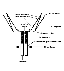
100231 FIG. 1
shows a schematic representation of an insulin-Fc fusion protein homodimer.
6c
Date Recue/Date Received 2022-07-07
WO 2021/207599
PCU1JS2021/026577
100241 FIG. 2 shows a schematic representation of an exemplary
SARS-CoV-2-RBD-hIgG-
Fe fusion protein homodimer.
[00251 FIG. 3 shows Fc(gaturna) receptor I binding for the insulin-
Fc fusion proteins of SEQ
ID NO: 29 and SEQ ID NO: 32.
100261 FIG, 4 shows titers of anti-insulin-antibodies (AIA)
against RI-II averaged over 200-
fold dilutions for 6 beagles with chemically induced diabetes over a series of
8 doses of the
insulin-Fe fusion protein of SEQ ID NO: 29,
[0027] FIG. 5 shows percentage change in the titers of anti-
insulin antibodies (Al A) against
RHI from Day 0 of the trial for 6 beagles with chemically induced diabetes
over a series of 8
weekly doses of the insulin-Fe fusion protein of SEQ ID NO: 29.
100281 FIG 6 shows the normalized ALA titers for 8 client dogs
treated for diabetes with
the insulin-Fe fusion protein of SEQ ID NO: 29 according to Protocol 1 or
Protocol 2 of Example
52.
100291 FIG. 7 shows the normalized ALA titer fora single dog
treated for diabetes with the
insulin-Fc fusion protein of SEQ ID NO: 29 with an interruption in treatment.
100301 FIG. 8 shows a graphical representation of the number of
dogs that showed AIA after
being treated for diabetes with the insulin-Fe fusion protein of SEQ ID NO: 29
manufactured in
either an HEK transient cell pool or a CHO stable cell pool,
100311 FIG. 9 shows the normalized AIA titers for 8 dogs treated
for diabetes with the
insulin-Fe fusion protein of SEQ ID NO: 32.
100321 FIG. 10 shows a graphical representation of the number of
dogs that showed AIA
after being treated for diabetes with the insulin-Fe fusion protein of SEQ ID
NO: 29 or SEQ ID
NO: 32.
[0033] FIG. 11 illustrates APC processing of Fe-fusion proteins
via the Fc(gamma)
receptors.
100341 FIG. 12 illustrates the promotion of B cell activation and
anti-SARS-CoV-2 SP/RBD
IgG production.
100351 FIG. 13 illustrates a side-by-side sequence comparison of
SEQ ID NO: 2 (extended
SP/RBD of SARS-CoV-2) and SEQ ID NO: 9 (novel truncation of the surface
glycoprotein of
SARS-CoV -2).
100361 FIG. 14 illustrates a side-by-side sequence comparison of
SEQ ID NO: 2 (extended
SP/RBD of SARS-CoV-2) and SEQ ID NO: 10 (novel truncation of the surface
glycoprotein of
SARS-CoV-2).
100371 FIG. 15 illustrates a side-by-side sequence comparison of
SEQ ID NO: 2 (extended
SIVRBD of SARS-CoV-2) and SEQ ID NO; 14 (novel truncation of the surface
glycoprotein of
7
CA 03146464 2022-1-31
WO 2021/207599
PCU1JS2021/026577
SARS-CoV-2).
100381 FIG. 16 illustrates a side-by-side sequence comparison of
SEQ ID NO: 2 (extended
SPIRBD of SARS-CoV-2) and SEQ ID NO: 15 (novel truncation of the surface
glycoprotein of
SARS-CoV-2).
100391 FIG, 17 illustrates a side-by-side sequence comparison of
SEQ ID NO: 2 (extended
SP/RBD of SARS-CoV-2) and SEQ ID NO: 13 (novel truncation of the sutface
glycoprotein of
SARS-CoV -2).
100401 FIG. 18 illustrates a general mechanism of action of a
vaccine adjuvant.
100411 FIG, 19 illustrates the EC50 of human Fc(gamma)RI binding
to the SARS-CoV-2-
RBD-hIgG-Fc fusion protein of SEQ ID NO: 19.
100421 FIG. 20 illustrates the EC50 of human Fc(gamma)Rna binding
to the SARS-CoV-
2-RBD-hEgG-Fc fusion protein of SEQ ID NO: 19.
100431 FIG. 21 illustrates the EC50 of human Fc(garrana)1111b
binding to the SARS-CoV-
2-RBD-hIgG-Fc fusion protein of SEQ ID NO: 19,
100441 FIG. 22 illustrates the EC50 of human Fc(garnma)R_III
binding to the SARS-CoV-2-
RBD-hIgG-Fc fusion protein of SEQ ID NO: 19.
100451 FIG. 23 illustrates the EC50 of human ACE2 binding to the
SARS-CoV-2-RBD-
hIgG-Fc fusion protein of SEQ ID NO: 19 and of human IgG.
100461 FIG. 24 illustrates the EC50 of human Ran receptor binding
to the SARS-CoV-2-
RBD-hIgG-Fc fusion protein of SEQ ID NO: 19_
100471 FIG. 25 illustrates the anti-SPIRBD IgG Ab titer response
in 6- to 8- week old female
BALBk mice 21 days after a single dose of the SARS-CoV-2 RBD-hIgG-Fc fusion
potein of'
SEQ ID NO: 17 across various dose levels.
10481 FIG. 26 illustrates the anti-SP/RBI) .1gG Ab titer response
in 6- to 8- week old female
BAI.,Bic mice on Day 35 after an injection on Day 0 and on Day 21 of the SARS-
CoV-2RBD-
hIgG-Fc fusion protein of SEQ ID NO: 17 across various dose levels.
100491 FIG. 27 illustrates the kinetic response to dose levels of
1 pg, 3 pg, 10 Fig, 30 pg and
100 lig after an injection on Day 0, Day 21 and Day 42 of the SARS-CoV-2 RBD-
hIgG-Fc
fusion protein of SEQ ID NO: 17.
100501 FIG. 28 illustrates the induced anti-SP/RBD IgG Ab titer
response after
administration of the SPIRED of SEQ ID NO: 2 or the SARS-CoV-2-RBD-hIgG-Fc
fusion
protein of SEQ ID NO: 17 to 6- to 8-week old mice without adjuvant at various
dose levels as
measured on Day 14 and Day 21 after one dose on Day O.
100511 FIG. 29 illustrates the anti-SP/11BD IgG Ab titer response
in 6- to 8-week old female
BALB/c mice in various adjuvanted formulations containing a 10 jig dose level
of the SARS-
CA 03146464 2022-1-31
WO 2021/207599
PCU1JS2021/026577
CoV-2-RBD-hIgG-Fc fusion protein of SEQ ID NO: 17 on Day 21 after one dose on
Day 0.
00521 FIG. 30 illustrates the anti-SP/RBD IgG Ab titer response
in 6- to 8- week old female
BALBIc mice in various adjuvanted formulations containing a 10 pg dose level
of the SARS-
CoV-2-RBD-hIgG-Fc fusion protein of SEQ ID NO: 17 on Day 35 after an injection
on Day 0
and on Day 21,
100531 FIG. 31 illustrates the anti-SPRBD IgG Ab titer response in
6- to 8- week old female
BAL13/c mice in various adjuvanted formulations containing a 10 lag dose level
of the SAR.S-
CoV-2-RBD-bIgG-Fc fusion protein of SEQ :ONO: 17 on Day 56 after an injection
on Day 0,
Day 21 and Day 42,
100541 FIG. 32 illustrates the anti-SP/RBD IgG Ab titer response
in 6- to 8-week old female
BALBIc mice in various adjuvanted formulations containing a 10 pg dose level
of the SARS-
CoV-2-RBD-hIgG-Fc fusion protein of SEQ :ED NO: 17 on Day 88 after an
injection on Day 0,
Day 21 and Day 42,
100551 FIG. 33 illustrates the ACE2-SPABD binding inhibition
potency 01350) calculated
at Day 21 after a single injection on Day 0 of the SARS-CoV-2-RBD-hIgG-Fc
fusion protein of
SEQ ID NO: 17 with and without adjuvants in 6- to 8-week old mice compared to
human
convalescent serum.
100561 FIG. 34 illustrates the ACE2-SP/RBD binding inhibition
potency (ID50) calculated
at Day 35 after an injection on Day 0 and on Day 21 of the SARS-CoV-2-RBD-h1gG-
Fc fusion
protein of SEQ ID NO: 17 with and without adjuvants in 6- to 8-week old mice
compared to
human convalescent serum.
100571 FIG. 35 illustrates the ACE2-SP/RBD binding inhibition
potency (ID50) calculated
at Day 56 after injections on Day 0, Day 21 and Day 42 of the SARS-CoV-2-RBD-
hIgG-Fc
fusion protein of SEQ ID NO: 17 with and without adjuvants in 6- to 8-week old
mice compared
to human convalescent serum.
100581 FIG. 36 illustrates the ACE2-SPRBD binding inhibition
potency (ID50) calculated
at Day 88 after injections on Day 0, Day 21 and Day 42 of the SARS-CoV-2-RBD-
hIgG-Fc
fusion protein of SEQ ID NO: 17 with and without adjuvants in 6- to 8-week old
mice compared
to human convalescent serum.
100591 FIG. 37 illustrates the induced anti-SPIRBD IgG Ab response
and the ACE2-
SIVRBD binding inhibition potency (ID50) after administration of the SARS-CoV-
2-RBD-
hIgG-Fc fusion protein of SEQ ID NO: 17 to 8- to 10-month old mice with
adjuvant at various
dose levels as measured after one dose on Day 21.
100601 FIG. 38 illustrates the induced anti-SIVRBD IgG Ab response
and the ACE2-
SPiRBD binding inhibition potency (ID50) after administration of the SARS-CoV-
2-RBD-
9
CA 03146464 2022-1-31
WO 2021/207599
PCU1JS2021/02,6577
hIgG-Fc fusion protein of SEQ ID NO: 17 to 8-to 10-month old mice with
adjuvant at various
dose levels as measured on Day 35 after an injection on Day 0 and Day 21.
[00611 FIG. 39 illustrates the induced anti-SPIRBD IgG Ab response
and the ACE2-
SP/RBD binding inhibition potency (1D50) after administration of the SARS-CoV-
2-RBD-
hlgO-Fc fusion protein of SEQ ID NO: 17 to 8-to 10-month old mice with
adjuvant at various
dose levels as measured on Day 56 after an injection on Day 0, Day 21 and Day
42.
100621 P16.40 illustrates the anti-SP/RBD IgG Ali titer response
in 6- to 8- week old female
BAL1Bic mice administered either a 1 jig or 10 pg dose level of the SARS-CoV-2-
R:BD-higG-
Fe fusion protein of SEQ ID NO; 19 with and without various adjuvants on Day
14 after one
dose,
10063] FIG 41 illustrates the anti-SPIRBD igG Ab titer response in
6-to 8- week old female
BALBk mice administered either a 1 jig or 10 fig dose level of the SARS-CoV-2-
RBD-hIgG-
Fe fusion protein of SEQ ID NO: 19 with and without various adjuvants on Day
35 after an
injection on Day 0 and Day 21.
100641 FI6.42 illustrates the anti-SPIRBD IgG Ab titer response in
6- to 8-week old female
BALB/c mice administered either a 1 jig or 10 pg dose level of the SARS-CoV-2-
RBD-hIgG-
Fe fusion protein of SEQ ID NO: 19 with and without various adjuvants on Day
56 after an
injection on Day 0, Day 21 and Day 42.
100651 FIG. 43 illustrates the ACE2-SPiRBD binding inhibition
potency (ipso) in 6- to 8-
week old female BALB/c mice administered either a 1 pg or 10 pg dose level of
the SARS-
CoV-2-RBD-hIgG-Fc fusion protein of SEQ ID NO: 19 with and without various
adjuvants on
Day 14 after one injection_
[00661 FIG. 44 illustrates the ACE2-SPIRBD binding inhibition
potency (ID50) in 6- to 8-
week old female BALB/c mice administered either a 1 jig or 10 pg dose level of
the SARS-
CoV-2-RBD-ItIgG-Fc fusion protein of SEQ ID NO: 19 with and without various
adjuvants on
Day 35 after an injection on Day 0 and Day 21, compared to human convalescent
serum.
100671 FIG. 45 illustrates the ACE2-SPIRBD binding inhibition
potency (11)50) in 6- to 8-
week old Female BALM mice administered either a 1 pg or 10 pg dose level of
the SARS-
CoV-2-RBD-hlgar-Fc fusion protein of SEQ ID NO: 19 with and without various
adjuvants on
Day 56 afteran injection on Day 0, Day 21 and Day 42, compared to human
convalescent serum.
100681 FIG. 46 illustrates the induction of anti-SP/RBD PRNT
neutralization potency in
serum samples from 6- to 8- week old female BALM mice administered either a 1
pg or 10 pg
dose level of SEQ ID NO: 19 on Day 0 and Day 21, as measured on Day 21 and Day
35_
100691 FIG. 47 illustrates the anti-SP/RBD IgG1 titer in 6-8 week
old BALB/C mice
administered a 10 pg dose level of the SARS-CoV-2-RBD-hIgG-Fe fusion protein
of SEQ
CA 03146464 2022-1-31
WO 2021/207599
PCU1JS2021/026577
NO: 19 with or without adjuvants (including MontanideTM ISA 720) as measured
on Day 14,
Day 35, and Day 56 after an injection on Day 0, Day 21 and Day 42.
[00701 FIG. 48 illustrates the anti-SP/RBD IgG2a titer in 6-8 week
old BALB/C mice
administered a 10 jig dose level of the SARS-CoV-2-RBD-hIgG-Fc fusion protein
of SEQ ID
NO: 19 with or without adjuvants (including Montaniderm ISA 720) as measured
on Day 14,
Day 35, and Day 56 after an injection on Day 0, Day 21 and Day 42.
[0071] FIG. 49 illustrates the anti-SP/RBD IgG2b titer in 6-8 week
old BALB/C
administered a 10 pg dose level of the SARS-CoV-2-RBD-hIgG-Fc fusion protein
of SEQ ID
NO: 19 with or without adjuvants (including Montaniderm ISA 720) as measured
on Day 14,
Day 35, and Day 56 after an injection on Day 0, Day 21 and Day 42.
[0072] FIG 50 illustrates the anti-SPIRED 1g63 titer in 6-8 week
old BALB/C mice
administered a 10 pg dose level of the SARS-CoV-2,-RBD-hIgG-Fc fusion protein
of SEQ ID
NO: 19 with or without adjuvants (including MontanideTm ISA 720) as measured
on Day 14,
Day 35, and Day 56 after an injection on Day 0, Day 21 and Day 42.
100731 FIG. 51 illustrates the anti-SP/RED IgG titers in 6- to 8-
week old female BALBk
mice administered a single dose on Day 0 of either a 1 pg or 10 pg dose level
of SEQ ID NO:
19 or SEQ ID NO: 23 (with mouse IgG2a-Fc) or a 0.5 pg or 5 pg dose level of
SEQ ID NO: 2
as measured on Day 14.
100741 FIG. 52 illustrates the anti-SPIRED IgG titers in 6- to 8-
week old female BALM
mice administered either a 1 pg or 10 jig dose level of SEQ ID NO: 19 or SEQ
ID NO: 23 (with
mouse IgG2a-Fc) or 0.5 pg or 5 jig dose level of SEQ ID NO: 2 as measured on
Day 35 after
an injection on Day 0 and Day 21.
[0075] FIG. 53 illustrates the induced ACE2-SPIRBD binding
inhibition potency (ID50) in
6- to 8-week old female BALR/c mice administered either a 1 pg or 10 pg dose
level of SEA)
ID NO: 19 or SEQ ID NO: 23 (with mouse I gG2a-Fc) or a 0.5 pg or 5 pg dose
level of SEQ ID
NO: 2 as measured on Day 14 compared to human convalescent serum.
[00761 FIG, 54 illustrates the induced ACE2-SP/RBD binding
inhibition potency (ID50)in
6- to 8- week old female BALB/c mice administered either a I pg or 10 pg dose
level of SEQ
ID NO: 19 or SEQ ID NO: 23 (with mouse IgG2a-Fc) or a 0.5 pg or 5 jig dose
level of SEQ. ID
NO: 2 as measured on Day 35 after an injection on Day 0 and Day 21 compared to
human
convalescent serum.
[0077] FIG. 55 illustrates the induced AC E2-SP/RBD binding
inhibition of the SARS-C2oV-
2-RBD-hIgG-Fc fusion protein of SEQ ID NO: 19 in mice as measured on Day 14
after injection
of freshly made emulsion versus the emulsion stored for 1 day and 7 days at
4cC and 25 C.
100781 FIG. 56 illustrates binding of the SARS-Colf-2-RBD-hIgG-Fe
fusion protein of SEQ
11
CA 03146464 2022-1-31
WO 2021/207599
PCU1JS2021/026577
ID NO: 19 to human Fc(ganuna)RI receptor as compared to the Fc(gamrna)RI
receptor of
Cynomolgus monkeys.
100791 FIG. 57 illustrates binding of the SARS-CoV-2-RBD-hIgG-Fc
fusion protein of SEQ
ID NO: 19 to human Fc(gamma)RlIa receptor as compared to the Fc(gamrna)RIla
receptor of
Cynomolgus monkeys,
100801 FIG. 58 illustrates binding of the SARS-CoV-2-RBD-hIgG-Fc
fusion protein of SEQ
ID NO: 19 to human Fc(garnma)RIII receptor as compared to the Fc(gamma)11111
receptor of
Cynomolgus monkeys.
100811 FIG, 59 illustrates binding of the SARS-CoV-2-RBD-hIgG-Fe
fusion protein of SEQ.
ID NO: 19 to human FeRn receptor as compared to the FcRn receptor of
Cynomolgus monkeys.
[0082] FIG. 60 illustrates the anti-SP/RBD igG Ab titer response
in male and female
Cynomolgus monkeys administered either a 10 lig or 30 lig dose level of the
SARS-CoV-2-
RBD-hIgG-Fc fusion protein of SEQ ID NO: 19 formulated with the MontanideTM
ISA 720
adjuvant at 30%10% Oily) on Day 0, Day 14, Day 21, Day 35 and Day 42 after an
injection on
Day 0 and Day 21_
[0083] FIG. 61 illustrates the induced ACE2-SPABD binding
inhibition potency (ID50)in
male and female Cynomolgus monkeys administered either a 10 jig or 30 lag dose
level of the
SARS-CoV-2-RBD-hIgG-Fc fusion protein of SEQ ID NO: 19 formulated with the
MontanideTM ISA 720 adjuvant at 30%170% (v/v) on Day 21 and Day 42 after an
injection on
Day 0 and Day 21.
100841 FIG. 62 illustrates the SARS-CoV-2-RBD-14G-Fc fusion
protein of SEQ ID NO:
19 induced SARS-CoV-2 virus neutralization potency in NHP serum samples as
measured on
Day 21 and Day 42, where SEQ ID NO: 19 formulated with MontanideT*4 ISA 720
adjuvant at
30%/70% (v/v) afteran injection on Day ()and Day 21, compared to human
convalescent serum.
100851 FIG. 63 illustrates the anti-SP/RBD response in inoculated
NHP after receiving a
booster injection.
100861 FIG, 64 illustrates that immune sera from NHP treated with
SEQ ID NO: 19 bound
the recombinant N501Y and E484K SP/RBD mutants as well as, or greater than,
the wild-type
SP/RBD molecule.
100871 FIG. 65 illustrates that immune sera from mice treated with
SEQ ID NO: 19 hound
the recombinant, N501Y and E484K RBD mutants as well as, or greater than, the
wild-type
RBD.
[0088] FIG. 66 illustrates a side-by-side sequence comparison of
the SP/RBD of SEQ ID
NO; 2, and the SPIRBD variants of SEQ ID NO: 24, and SEQ ID NO; 25.
100891 FIG. 67 illustrates the anti-SP/RBD IgG Ab titer in New
Zealand White Rabbits
12
CA 03146464 2022-1-31
WO 2021/207599
PCU1JS2021/026577
subcutaneously administered a vehicle control with Montanid ekt ISA 720
adjuvant on Day 1,
Day 15, and Day 29 measured before the first dose, second dose and third d ose
and after the
third dose.
[0090] FIG, 68 illustrates the substantial anti-SP/RBD IgG Ab
titer in New Zealand White
Rabbits administered a 30 pg dose level of the SARS-CoV-2-RBD-hIgG-Fe fusion
protein of
SEQ ID NO: 19 without adjuvant. on Day 1, Day 15, and Day 29 measured before
the first dose,
second dose and third dose and after the third dose.
[0091] FIG. 69 illustrates the substantial anti-SP/RBI) IgG Ab
titer in New Zealand White
Rabbits administered a 100 lag dose level of the SARS-CoV-2-RBD-hIgG-Ec fusion
preaein of
SEQ ID NO: 19 without adjuvant on Day 1, Day IS, and Day 29 measured before
the first dose,
second dose and third dose and after the third dose.
[0092] FIG. 70 illustrates the substantial anti-SP/RED IgG Ab
titer in New Zealand White
Rabbits administered a 30 pg dose level of the SARS-CoV-2-RBD-hIgG-Fc fusion
protein of
SEQ ID NO: 19 with Montaniderm ISA 720 adjuvant on Day 1, Day 15, and Day 29
measured
before the first dose, second dose and third dose and after the third dose.
100931 FIG. 71 illustrates the substantial anti-SP/RBD IgG Ab
titer in New Zealand White
Rabbits administered a 100 pg dose level of the SARS-CoV-2-RBD-hIgG-Fc fusion
protein of
SEQ ID NO: 19 with MontanideTM ISA 720 adjuvant on Day 1, Day 15, and Day 29
measured
before the first dose, second dose and third dose and after the third dose
[0094] FIG. 72 illustrates the induced ACE2-SP/RBD binding
inhibition potency (ID50) in
New Zealand White Rabbits administered a 30 pg or 100 ug dose level of the
SARS-C6V-2-
RBD-hIgG-Fc fusion protein of SEQ ID NO: 19 with MontanideTM ISA measured on
Day 15
and Day 29 after an injection on Day 0 and Day 21.
10095] FIG. 73 illustrates the SARS-CoV-2-RBD-hIgG-Fc fusion
protein of SEQ ID NO:
19 induced SAR S-CoV-2 virus neutralization potency in New Zealand White
Rabbit serum
samples measured on Day 21, and Day 35, where SEQ ID NO: 19 was emulsified
with
MontanideTM ISA 720 adjuvant and administered on Days 0 and 21, compared to
human
convalescent serum.
[0096] FIG. 74 illustrates the anti-SP/RBD IgG Ab titer in New
Zealand White Rabbits
administered a 100 pg dose level of the SARS-Co V -2-RBD-h I gG-Fc fusi on
protein of SEQ ID
NO: 19 with Montanidem ISA 720 adjuvant via subcutaneous (SC) injection or
intramuscular
injection (LM), measured on Day 15, Day 29, and Day 36 after an injection on
Day 0 and Day
21_
100971 FIG, 75 illustrates the induced ACE2-SP/RBD binding
inhibition potency (ID50) in
New Zealand White Rabbits administered a 100 pig dose level of the SARS-CoV-2-
RBD-hIgG-
1 3
CA 03146464 2022-1-31
WO 2021/207599
PCU1JS2021/026577
Fc fusion protein of SEQ ID NO: 19 with Montanid eTM ISA 720 adjuvant via
subcutaneous (SC)
injection or intramuscular injection (IM), measured on Day 15, Day 29, and Day
36 after an
injection on Day 0 and Day 2L
100981 FIG, 76 illustrates the anti-SPIRED IgG Ab titer in New
Zealand White Rabbits
administered a fresh emulsion and an emulsion stored for 24 hours at 2-8 C of
100 pg dose level
of the SARS--CoV.2-RBD-hIgG-Fc fusion protein of SEQ ID NO: 19 with
MontartideTM ISA
720 adjuvant via subcutaneous (SC) injection measured on Day 15, Day 29, and
Day 36 after
an injection on Day 0 and Day 21.
[00991 FIG, 77 illustrates the anti-SPIRED IgG Ab titer in New
Zealand White Rabbits
administered a fresh emulsion and an emulsion stored for 24 hours at 2-8 C of
100 pg dose level
of the SARS-CoV-2-RBD-hIgG-Fc fusion protein of SEQ ID NO: 19 with
Montan.ideTM ISA
720 adjuvant via intramuscular (IM) injection measured on Day 15, Day 29, and
Day 36 after
an injection on Day 0 and Day 21.
[01.00] FIG. 78 illustrates the genomic or subgenomic SARS-CoV-2
viral RNA copies per
tni, of nasal swabs taken from naive NHP and NHP that have been immunized with
the SARS-
CoV-2-RBD-higG-Fc fusion protein of SEQ ID NO: 19 with MontanideTm ISA 720
according
to Example 36.
101011 FIG. 79 illustrates the genornic or subgenornic SARS-CoV-2
viral RNA copies per
inL of bmnchoalveolar lavage (DAL) fluids collected from naïve NIIP and NIIP
that have been
immunized with the SARS-CoV-2-RBD-hIgG-Fc fusion protein of SEQ ID NO: 19 with
MontanideTm ISA 720 according to Example 36.
101021 FIG, 80 illustrates a side-by-side sequence comparison of
SEQ ID NO: 8 (RBD of
SARS-CoV-2) with SEQ ID NO: 9, SEQ ID NO: 10, SEQ ID NO: 14 and SEQ ID NO: 15
(all
RBD of SARS-CoV-2 with novel mutations).
101031 FIG. Si illustrates a 96 well microplate such as that which
may be used for a general
serology assay for evaluating existing SARS-CoV-2 antibody titer in semm.
DETAILED DESCRIPTION
Novel Coronavirus Disease 2019
101041 Novel Coronavims Disease 2019 (COVID-19) is a severe and
acute respiratory
illness caused by the SARS-CoV-2 virus. The first COVID-19 case was reported
in Wuhan,
China in December 2019 and as of 18 November 2020, there have been
approximately 56
million (M) cases worldwide to date (quantified as SARS-CoV-2 virus confirmed
and
unconfirmed "probable"), in which there are 18,5M active cases, 36M recovered
cases, and
1.3M fatal cases attributed to COVID- I 9 (University, J.H., COVID-19
Dashboard by the Center
14
CA 03146464 2022-1-31
ABC-044PCT
for Systems Science and Engineering (CSSE) at Johns Hopkins University). At
the end of 2020,
only the Pfizer-BioNTech COVID-19 vaccine BNT162b2 was approved under the
World Health
Organization's (WHO) Emergency Use Listing (EUL) procedure for emergency use
against
COVID-19. A second vaccine from Modema (mRNA-1273) is expected to be approved
under
the WHO EUL procedure for vaccine emergency use against COVID-19 by the end of
February
2021. The consensus among experts is that society cannot return to normal
unless and until there
is a sufficient level of immunity conferred on the population. Achieving
natural herd immunity
is estimated to require at least 70% of the population to have been infected
which would result
in millions of deaths worldwide, an ethically unacceptable outcome.
ACE2 Receptor
[0105] Angiotensin-Converting Enzyme 2 (ACE2) is the host cell receptor
responsible for
mediating infection by SARS-CoV-2 (i.e., to which SARS-CoV-2 binds in order to
infect cells).
ACE2 is a type 1 transmembrane metallocarboxypeptidase. Polymerase Chain
Reaction (PCR)
analysis shows that ACE2 is expressed on lung epithelium, blood vessel
endothelium, and
specific neuronal cells that appears to account for the dominant clinical
manifestations of
COVID-19, including pulmonary, cardiovascular, and neurological complications,
respectively.
Based on the sequence similarities of the receptor binding domain between SARS-
CoV-2 and
SARS-CoV, researchers have shown that SARS-CoV-2 can use ACE2 expressed on the
surface
of human cells to gain entry into ACE2 expressing HeLa cells.
Convalescent Sera for Treatment of Virus Patients
[0106] Clinicians and researchers around the world are working to develop
various solutions
to mitigate the pandemic caused by SARS-CoV-2. Scientists are working to
develop vaccines
that can prevent COVID-19 and antiviral treatments to reduce the severity and
symptoms of the
illness. Development of vaccines, monoclonal antibodies (mAbs), or drugs to
treat SARS-CoV-
2 is ongoing_ Many clinicians believed that human convalescent serum was a
viable option for
the prevention and treatment of COVID-19.
101071 Convalescent serum is a form of passive antibody therapy, through
which sera from
infected and recovered individuals containing anti-virus antibodies is
transfused to a susceptible
or infected individual, providing that individual with some level of immunity
to either prevent
or reduce the severity of the disease. This treatment is different from a
vaccine, which works by
inducing an immune response in an individual such that the individual produces
their own
antibodies against the virus. Experience from the SARS-CoV outbreak in 2002
and the 2009-
2010 H1N1 influenza outbreak has shown that sera from patients that have
contracted and
recovered from the virus (human convalescent sera) contains antibodies capable
of neutralizing
Date Recue/Date Received 2023-01-05
WO 2021/207599
PCU1JS2021/026577
the virus and is useful as an intervention for individuals with severe disease
symptoms or as a
prophylactic vaccine.
[01081 The use of human convalescent sera has risks and
limitations. Firstly, the transferof
blood substances from one person to another comes with it the risk of
inadvertent infection of
another infectious disease as well as the risk of reactions to other serum
constituents, Another
challenge in using convalescent sera is that some patients who recover from
viral diseases do
not have high titers of neutralising antibody. In one case with respect to
another human
coronayirus, Middle East respiratory syndrome (IvIERS-CoV), three patients in
South Korea
were treated with convalescent serum, but only two of the recipients had
neutralizing antibody
in their serum. Of those that do have neutralizing antibodies after recovering
from viral disease,
some may not have sufficiently high titers of neutralizing antibody to be a
viable donor_ A
further survey related to SARS-CoV found that of 99 samples of convalescent
sera from patients
with SARS, 87 had neutralizing antibody, with a geometric mean titer of 1 :61.
These and various
other studies suggest that few patients made high-titer responses and also
that neutralizing
antibody titer declines with time. There are a number of companies looking to
overcome this
challenge by producing recombinant antibodies instead of solely relying on
antibodies from
recovered patients; however, the scale ofproduction is insufficient, and
themedical intervention
required to administer effective doses to patients every few weeks to few
months, most likely
through intravenous injection or infusion, is highly burdensome_
[0109] A more significant limitation is that the proposed use of
convalescent sera in the
COV ID-19 epidemic would rely on preparations with high titers of SARS-CoV-2
neutralizing
antibodies, This requires a significant population of donors who have
recovered from the disease
and can donate convalescent serum. Determining who has already had the disease
and has
developed some immunity presents challenges. COVID-19 presents with a wide
variety of
severity of symptoms and many individuals with mild cases may not know that
they have had
the disease, A highly available and low-cost test kit to measure anti-SARS-CoV-
2 antibodies is
also required.
101101 However, even with the ability to identify recovered
patients with high titers of
neutralizing antibodies, it is unlikely that a single individual's plasma can
treat more than a few
patients. Therefore, while current approaches to convalescent sera treatment
may be able to
prevent or treat COVID-19 in a small number of patients, this solution does
not address the
greater need of humanity during and after this pandemic.
Overview and Challenges of Current Vaccines
(0111) Clinicians and researchers around the world are working to
develop various solutions
to mitigate the pandemic caused by the SARS-CoV-2 virus. These solutions
include vaccines
16
CA 03146464 2022-1-31
WO 2021/207599
PCU1JS2021/026577
that can prevent COVID-19 and antiviral treatments to reduce the severity and
symptoms of the
illness. The expectation of the foreseeable future is that natural and vaccine-
induced immunity
most likely will not be long-lived, and therefore a cost-effective and safe
vaccine administered
as frequently as every 6 months, if necessary, is required to maintain robust
immunity among
the population. Thus, the critical design features of an effective
prophylactic CO VID-19 vaccine
are: i) a potent capacity to induce SARS-CoV-2 viral neutralizing 1gG titers
and a significant T
helper type I (ml) cell response, preferably after a single dose; ii) an
acceptable safety and
tolerability profile, especially with respect to inflammation caused by
reactogenicity (systemic
effect) and injection site (local effect), a favorable cost-of-goods (COGs)
with respect to
manufacturability and vaccine potency which dictate dose-frequency and dose-
level, and a
suitable supply-chain path including a sufficient storage shelf-life and
robust test article
preparation and administration procedures.
101121 Live-attenuated or inactive whole virus vaccines represent
a classic strategy_ A major
advantage of whole virus vaccines is their inherent immunogeni city and
ability to stimulate toll-
like receptors (TLRs) including TLR 3, TL.R 7/8, and TLR 9_ However, live
virus vaccines often
require extensive additional testing to confirm their safety. This is
especially an issue for
coronavirus vaccines, given the findings of increased infectivity following
immunization with
live or killed whole virus SAR_S coronavirus vaccines. Johnson & Johnson is
employing
Janssen's AdVac ad erioviral vector manufactured in their PER_C60 cell line
technology to
generate their lead vaccine, INJ-78436735, which recently completed Phase 3
trials and has
been authorized for emergency use in the United States, This technology is an
attempt to
produce a viral vector to replace the whole virus with a purportedly benign
adenoniral vector
that carries a portion of the SARS-CoV-2 virus DNA. However, use of 3'M-
78436735
encountered significant serious adverse events (SAEs) that caused clinical
trial pauses.
101131 Two additional hurdles in the early development of SARS
corona-vials vaccines have
been the finding of 1) undesired immunopotentiation in the form of 'Fh2-
mediated eosinophilic
infiltration and 2) increased viral infectivity driven by ADE, which is noted
to occur following
challenge infections after immunizations with whole virus vaccines and
complete SP vaccines.
The risk of Th2-mediated eosinophilic infiltration and lung pathology is still
under investigation
in SA RS-CoV-2 infection but it has been found in infants and animals
challenged with
respiratory syncytial virus (RSV) or with immunization with whole RSV
vaccines.
101141 ADE is an adverse characteristic of other viral vaccines,
including those for the
original SARS-CoV, dengue virus, and Zika viral infections, in which vaccine-
induced Ab
concentrations or affinities are too low to neutralize virus infection, but
rather form immune
complexes with virus that tend to interact with Fey receptors on myeloid cell
surface through Fe
17
CA 03146464 2022-1-31
WO 2021/207599
PCU1JS2021/026577
domains of Abs. Such Abs do not neutralize viral infection or induce Fcy-med
iated viral
clearance (Li), but aid virus infection by directly increasing virus uptake
through Fey receptor
or boosting virus replication intracellularly via activating downstream
pathways to antagonize
the innate immunity (reviewed in Sun). In both ADE and Th2-
imrnunopotentiation, there is
evidence that feline IgG2a rnAbs (possibly of the Th2 isotype) can mediate
both adverse
conditions while IgG1 mAbs (known to have strong effector function, i.e., Thl
isotype) avoid
such effects.
101151 In addition to their risk of causing ADE and/of Th2-
imimmopotentiation, another
challenge with viral vector vaccines is the relatively low manufacturability
throughput and
therefore high cost of goods (COGs) due to either chicken egg-based production
or cell
expression systems (Ewer).
101.161 As an alternative, nucleic acid expression vector vaccine
platforms for C:OVID-19
encode the major cotonavims target antigen (Ag), the Spike Protein (SP), that
mediates the
virus' infective mechanism via its binding the host receptor, ACE2. Two
examples of such
vaccines that have advanced through Phase 3 trials are the mRNA vaccines
encoding the full-
length SP developed by BioNTech/Pfizer, BNT162b2 and Moderna, mRNA-1273. Both
vaccines have reported very positive Phase 3 results with efficacy in
protecting from
symptomatic SARS-CoV-2 viral infection of greater than 90% leading to recent
emergency use
authorizations (ELMO by the United States FDA. The concept of immunizing with
RNA or
DNA began with promising results in mice in 1993 showing protective immunity
against
influenza, but for decades these findings have not translated to similar
findings in humans.
Moreover, while non-replicative, many of these RNA and DNA expression vector
vaccines
continue to endogenously produce the target viral Ag well after induction of
the intended
immune response, an aspect that could ultimately create immune tolerance to
the virus which is
a growing concern and may become a practical risk with such current COVID-19
niRNA
vaccines. Other challenges of these nucleic acid vaccines are the low
durability of the response
that may require too frequent dosing, and an unfavorable COGs due to
cumbersome
manufacturability via chemical synthesis. Furthermore, dueto the inherent
instability of RNA,
the products must be kept and transported under frozen conditions, making them
very difficult
for most of the world to access_
101171 As an additional alternative, recombinant subunit vaccines
rely on eliciting an
immune response against the SP to prevent its docking with the host target
protein, A CE2, Such
vaccines comprise all or a portion of the SP, rather than the DNA or RNA
encoding for the
protein, which is then mixed with an adjuvant to enhance the immune response.
Due to the
inherent stability of proteins relative to RNA and DNA, the storage and
transportation
18
CA 03146464 2022-1-31
WO 2021/207599
PCU1JS2021/026577
requirements are less strict for subunit vaccines. Companies developing
recombinant subunit
vaccines include Novavax who has developed and produced immunogenic virus-like
nanoparticles based on recombinant expression of SP, NVX-Cov-2373, that are
formulated with
a saponin-based adjuvant system, Matrix-M, and Clover Biopharmaceuticals who
is
developing a subunit vaccine consisting of a trimerized SARS-CoV-2 SP using
their patented
Turner-Tag technology. However, the full-length SP target Ag is known to have
low
expression yields in cell-expression systems and when used in SARS vaccines is
known to
induce anti-SP IgG titers against non-neutralizing epitopes of SP that again
could mediate
increased viral infectivity (i,e, ADE) and inflammation caused by lung
eosinophilia (i.e., 112-
mediated immunopotentiation, discussed below), A subunit vaccine comprised of
only the
receptor-binding domain (RBD) of the SARS SP has the potential to mitigate
against these
safety challenges.
101181 A consortium led by Texas Children's Hospital Center for
Vaccine Development at
Baylor College of Medicine has developed and tested a subunit vaccine
comprised of only the
receptor-binding domain (REID) of the SARS SP, and when formulated with alum,
this RBD-
based vaccine can elicit high levels of protective immunity upon homologous
virus challenge,
in addition to avoid ing ADE and immuriopotentiation. Initial findings that
the SARS and SARS-
CoV-2 RBDs exhibit more than 80% amino acid similarity and bind the same ACE2
target offer
an opportunity to develop either protein Ag as a subunit vaccine. Indeed, such
a subunit vaccine
proof-of-concept has been successfully demonstrated with coronavirus SP/RBD
Ag's of MERS
and SARS infections.
101191 Some, but not all, of these features are being implemented
in over 170 SARS-00V-
2 vaccine candidates currently in development, including live viruses, nucleic
acids, and
recombinant protein subunits that may ultimately offer promise as preventive
vaccines against
COVID-19 However, each vaccine strategy has unique advantages and challenges
with respect
to manufacturing, safety, and efficacy that must be simultaneously managed in
an optimal
manner.
[01.20] The present disclosure is directed to methods for making
and using novel fusion
proteins which allow for the cost-effective production of large quantities of
a recombinant
subunit vaccine against the SARS-CoV-2 virus whict can he transported and
stored at mild
temperatures. The present disclosure is specifically directed to methods for
making and using
fusion proteins for use in a prophylactic, therapeutic or booster vaccine
which is efficacious for
causing patients to create anti-virus antibodies to the SARS-CoV-2 vinis.
Using a SARS-CoV-
2-RBD-hIgG-Fe fusion protein to cause a patient to create endogenous
antibodies targeted to
the receptor binding domain (RBD) portion of the SARS-CoV-2 virus is expected
to be
19
CA 03146464 2022-1-31
WO 2021/207599
PCU1JS2021/026577
significantly more cost effective than rmombinantly generating anti-SARS-CoV-2
therapeutic
antibodies to later be injected into a patient.
[01211 In examples, a booster vaccine comprising a SARS-CoV-2-RBD-
hIaG-Fe fusion
protein may be administered to patients that have recovered from COVID-19 as
an antibody
amplification treatment (AAT), to increase their anti-SARS-CoV-2 antibody
titers so that the
serum they donate for use as a convalescent serum treatment can be used to
treat more people.
Recovered patients can be administered this AA.T a few weeks before every new
serum
donation, significantly increasing the anti-SARS-CoV-2 antibody titer of the
extracted sera and
consequently significantly increasing the number of viral patients that can be
treated with each
donation.
[01.22] In examples, a SARS-CoV-21RBD.hIgG-Fc fusion protein may be
used for detecting
anti-SARS-CoV-2 antibodies in serum that is extracted from individuals. The
ability to create a
test kit using a SARS-CoV-2-RBD-hIgG-Fc fusion protein as a key reagent to
reliably determine
the presence and concentration of anti-SARS-CoV-2 antibodies in serum, permits
clinicians to
determine which individuals have had and recovered from the virus, which is
particularly
important in the case where patients may have experienced few or no symptoms_
In examples,
such a test kit could be used to evaluate performance of emerging vaccine
candidates for SARS-
CoV-2, by enabling the rapid and cost-effective ability to determine the
presence and
concentration of host-produced anti-SARS-CoV-2 antibodies in extracted serum
post-
vaccination. Broad deployment of such a test kit is expected to dramatically
increase the number
of potential donors of convalescent serum.
101231 In an example, a pharmaceutical composition of a SARS-CoV-2-
RBD-hIgG-Fc
fusion protein is administered to patients who have been infected by the SARS -
CoV-2 virus and
have contracted CON/D-19 to limit the scope of the infection and to ameliorate
the disease. In
examples, the SARS-CoV-2-RBD-hIgG-Fc fusion protein binds the ACE2 receptor,
blocking
the further uptake of the receptor binding domain (RBD) of the SARS-CoV-2
virus while also
generating antibodies to neutralize the SARS-CoV-2 virus, leaving fewer RBD
exposed to host
cells.
101241 in an example, a pharmaceutical composition of a SARS-CoV-2-
RBD-hIgCi--Fc
fusion protein is administered as a prophylactic COVID-19vaccine for lad
ividuals that have not
been infected by the SARS-00V-2 virus, resulting in the individual producing
their own pool
of anti-SARS-C oV-2 antibodies and immunity.
Equivalents and Definitions
101251 As used herein, the articles "a" and "an" refer to one or more than
one, e.g., to at least
CA 03146464 2022-1-31
WO 2021/207599
PCU1JS2021/026577
one, of the grammatical object of the article. The use of the words "a" or
"an" when used in
conjunction with the term "comprising" herein may mean "one," but it is also
consistent with
the meaning of "one or more," "at least one," and "one or more than one." As
used herein, the
phrase "and/or," when used in a list of two or more items, means that any one
of the listed items
can be employed by itself or any combination of two or more of the listed
items can be
employed. For example, if a composition is described as containing Of
excluding components
A, B, and/or C, the composition can contain or exclude A alone; B alone; C
alone; A and B in
combination; A and C in combination; B and C in combination; or A, B, and C in
combination.
101261 As used herein, "about" and "approximately" generally mean
an acceptable degree
of error for the quantity measured given the nature or precision of the
measurements.
[01271 As used herein, an amount of a molecule, compound,
conjugate, or substance
effective to treat a disorder (e.g., a disorder described herein),
"therapeutically effective
amount," or "effective amount" refers to an amount of the molecule, compound,
conjugate, or
substance which is effective, upon single or multiple dose administration(s)
to a subject, in
treating a subject, or in curing, alleviating, relieving or improving a
subject with a disorder (e.g.,
a disorder described herein) beyond that expected in the absence of such
treatment.
101281 As used herein, the term "analog- refers to a compound or
conjugate (e.g., a
compound or conjugate as described herein, e.g., RBD) having a chemical
structure similar to
that of another compound or conjugate but differing from it in at least one
aspect.
101291 As used herein, the term "antigen" refers to any substance
that causes a patient's
immune system to produce antibodies against it. An antigen may be a substance
from the
enviromnent, such as chemicals, bacteria, viruses, or pollen, or an antigen
may also form inside
the body. An example of an antigen is the SA RS-CoV-2 virus.
101.301 As used herein, the term "antibody" or "antibody molecule"
refers to an
immunoglobulin molecule (1g), or immunologically active portions of an
immunoglobulin (Ig)
molecule, Le., a molecule that contains an antigen binding site that
specifically binds, e.g.,
immunoreacts with, an antigen. As used herein, the term "antibody domain"
refers to a variable
or constant tegion of an immunoglobulin. it is documented in the art that
human antibodies
comprise several classes, for example IgA, IgIA, or IgG in the case of mammals
(e.g., humans
and dogs). Classes of mammalian IgG immtmogiobulins can be further classified
into different
isotypes, such as IgGA, IgGB, IgGC and IgGD for dogs and IgG I, IgG2, IgG3,
and IgG4 for
humans. Those skilled in the art will recognize that immunoglobulin isotypes
of a given
immunoglobulin class will comprise different amino acid sequences, structures,
and functional
properties from one another (e.g., different binding affinities to FOgamina)
receptors or ACM
receptor). "Specifically binds" or "imrnunoreacts with" means that the
antibody reacts with one
21
CA 03146464 2022-1-31
WO 2021/207599
PCU1JS2021/026577
or more antigenic determinants of the desired antigen and has a lower affinity
for other
polypeptides, e.g., does not react with other polypeptides.
101311 As used herein, the term "dimer" refers to a protein or a
fusion protein comprising
two polypeptides linked covalently. In embodiments, two identical polypeptides
are linked
covalently (e.g., via d stiff"' d e bond s) forming a "homodimer"
(diagrammatically represented in
FIG I, which is an illustration of an insulin-Fe fusion protein for reference,
and FIG. 2, which
is an illustration of a SARS-CoV-2-RBD-hIgG-Fe fusion protein). Referring to
FIG. 1 in more
detail, the insulin polypeptide (comprising an insulin B-chain analog
connected via a C-chain
peptide to an insulin A-chain analog) may hare one or more amino acid
mutations from native
insulin. The insulin peptide is connected via a linker to an Fe fragment.
Disulfide bonds (the
total number of disulfide bonds in actuality may be greater or less than the
number shown in
FIG. 1) create a homodimer from two identical Fe fusion proteins. Referring to
FIG. 2 in more
detail, a SARS-CoV-2 RBD fragment may comprise a portion of the full SARS-CoV-
2 surface
glyeoprotein. The RBI.) fragment may have one or more amino acid mutations
from the native
SARS-CoV-2 surface glycoprotein. The RBD fragment is connected to an Fe
fragment using an
optional linker (in some examples, the RED fragment is covalently linked to an
Fe fragment
directly with no linker). Disulfide bonds create a homodimer from two
identical SARS-CoV-2-
RBD-higG-Fe fusion proteins (the total number of disulfide bonds in actuality
may be greater
or less than the number shown in FIG. 2). The Fe fusion protein homodimer may
be encoded by
a single nucleic acid molecule, wherein the homodimer is made recombinantly
inside a cell by
first forming Fe fusion protein monomers and by then assembling twoidentical
Fe fusion protein
monomers into the homodimer upon further processing inside the cell.
101321 As used herein, the terms "multimer," "rmiltinterie," or
"multimeric state" refer to
non-covalent, associated forms of Fe fusion protein dimers that may be in
equilibrium with Fe
fusion protein dimers or may act as permanently aggregated versions ofFe
fusion protein (Inners
(e.g., dimers of Fe fusion protein homodimers, turners of Fe fusion protein
homodimers,
tetramers of Fe fusion protein homodimers, or higher order aggregates
containing five or more
Fe fusion pane-in homodimers). It may be expected that multimeric forms of Fe
fusion proteins
may have different physical, stability, or phartnacologic activities from that
of fusion protein
homodimers.
101331 As used herein, RBD-Fc fusion protein and SARS-00V-2-RBD-
higG-Fe fusion
protein (which tams may be interchangeably used) refers to a human
immunoglobin Fe domain
that is linked to a SARS-CoV-2 spike protein (SP) receptor binding domain
(SP/RBD or RBD)
or an analog thereof, which is useful in generating antibodies that
specifically bind the SARS-
CoV-2 antigen. For ease of reference, the terms "RBD" and/or "SP/RBD" may be
used
CA 03146464 2022-1-31
WO 2021/207599
PCU1JS2021/026577
interchangeably herein, and unless otherwise dictated by the context,
encompass protein
residues consisting of the receptor binding domain per se, smaller fragments
of the receptor
binding domain, or larger variants comprising the receptor binding domain and
adjacent residues
from the spike protein segment, provided that the fragments and/or larger
variants retain the
activity of the RBD (e.g., retain the ability to bind the spike protein
receptor). As used herein,
the general terms "fusion protein" and "Fc fusion protein" refer to a protein
comprising more
than one part, for example from different sources (e.g., different proteins,
polypeptides, cells,
etc.), that are covalently linked through peptide bonds. Fc fusion proteins
are covalently linked
by (i) connecting the genes that encode for each part into a single nucleic
acid molecule and (ii)
expressing in a host cell (e.g., HEK cell or CHO cell) the protein for which
the nucleic acid
molecule encodes The fully recombinant synthesis approach is preferred over
method sin which
the therapeutic protein and Fe fragments are synthesized separately and then
chemically
conjugated, The chemical conjugation step and subsequent purification process
increase the
manufacturing complexity, reduce product yield, and increase cost.
[01341 As used herein, the term "bioactivity," "activity,"
"biological activity," "potency,"
"bioactive potency," or "biological potency" refers to the extent to which an
Fe fusion protein
binds to or activates a cell receptor and/or exerts the production or
reduction of native or foreign
substances. As used herein, "in vitro activity" or "receptor activity" refers
to the affinity with
which an Fe fusion protein binds to the cell receptor and is typically
measured by the
concentration of an Fc fusion protein that causes the Fc fusion protein to
reach half of its
maximum binding (i.e.: EC50 value). For example, the "bioactivity" of a SARS-
CoV-2-RBD-
higG-Fc fusion protein refers to the extent to which the SARS-CoV-2-RBD-hIgG-
Fe fusion
protein induces the production of anti-SARS-CoV-2 antibodies in a cellular
assay or in a target
subject.
[0135] As used herein, the term "biosynthesis," "recombinant
synthesis," or "mcombinant13.,
made" refers to the process by which an Fe fusion protein is expressed within
a host cell by
transfecting the cell with a nucleic acid molecule (e.g., vector) encoding the
Fc fusion protein
(e.g., where the entire Fc fusion protein is encoded by a single nucleic acid
molecule).
Exemplary host cells include mammalian cells, e.g.. HEK293 cells or CHO cells.
The cells can
be cultured using standard methods in the art and the expressed Fe fusion
protein may be
harvested and purified from the cell culture using standard methods in the
art.
[0136] As used herein, theterm "cell surface receptor" refers to a
molecule such as a protein,
generally found on the external surface of the membrane of a cell and which
interacts with
soluble molecules, e.g., molecules that circulate in the blood supply. In some
embodiments, a
cell surface receptor may include a host cell receptor (e.g., an ACE2
receptor) or an Fe receptor
23
CA 03146464 2022-1-31
WO 2021/207599
PCU1JS2021/026577
which bind s to an Fc fragment or the Fe region of an antibody (e.g., an
Fc(gan-nna) receptor, for
example Fc(gamma) receptor I, or an Fc neonatal receptor, for example Ran). As
used herein,
"in vitro activity" or "Fc(gamma) receptor activity" or "Fc(gamma) receptor
binding" or "FcRn
receptor activity" or "FcRn binding" refers to the affinity with which an Fe
fusion protein binds
to the Fc receptor (e.g. Fc(gamma) receptor or FeRn receptor) and is typically
measured by the
concentration of an Fc fusion protein that causes the Fc fusion protein to
reach half of its
maximum binding (i.e., EC50 value) as measured on an assay (e.g., an enzyme-
linked
immunosorbent assay (EL1SA ) assay) using 01)450 nm values as measured on a
microplate
reader.
101371 As used herein, the term "immunogenic" or "immunogenicity"
refers to the capacity
fora given molecule (e.g., an Fe fusion protein of the present invention) to
provoke the immune
system of a target subject such that after administration of the molecule, the
subject develops
antibodies capable of binding all or specific portions of the molecule (i.e.,
anti-drug antibodies
or ADA). As used herein, the terms -neutralizing," -neutralizing antibodies",
or -neutralizing
anti-drug antibodies" refer to the capacity for antibodies to interfere with
all or a portion of the
Fc fusion protein's biological activity in the target subject. For example, in
the case of a SARS-
CoV-2-RBD-higG-Fc fusion molecule administered to humans, the immunogenicity
refers to
antibodies that bind to the SARS-CoV-2 RBD portion of the molecule since the
hIgG-Fe portion
of the molecule is endogenous to humans and therefore unlikely to elicit anti-
hIgG-Fc
antibodies. Likewise, antibodies generated by the administration of a SARS-CoV-
2-RBD-
h I gG-Fc fusion molecule are neutralizing when those anti -SAR S-C oV-2 RBD
antibodies inhibit
the binding between SARS-00V-2 RBD and the ACE2 receptor, which is directly
related to the
bioactivity of the SARS-CoV-2 RBD in the subject.
1013$1 As used herein, the term "monomer" refers to a protein or a
fusion protein
compiising a single polypeptide. In embodiments, the "monomer" is a protein or
a fusion
protein, e.g., a single polypeptide, comprising an RBD polypeptide and an Fc
fragment
polypeptide, wherein the RBD and Fc fragment polypeptides are joined by
peptide bonds to
form the single polypeptide. In embodiments, the monomer is encoded by a
single nucleic acid
molecule.
101391 As used herein and as illustrated in FIG. 1 and FIG. 2, ""N-
terminus" refers to the
start of a protein or polypeptide that is initiated by an amino acid
containing a free amine group
that is the alpha-amino group of the amino acid (e.g., the free amino that is
covaiently linked to
one carbon atom that is located adjacent to a second carbon atom, wherein the
second carbon
atom is part of the carbonyl group of the amino acid). As used herein and as
illustrated in FIG.
1 and FIG. 2, "C-terminus" refers to the end of a protein or polypeptide that
is terminated by an
24
CA 03146464 2022-1-31
WO 2021/207599
PCU1JS2021/026577
amino acid containing a carboxylic acid group, wherein the carbon atom of the
carboxylic acid
group is located adjacent to the alpha-amino group of the amino acid.
[01401 As used herein, the term "carrier' is used herein to refer
to diluents, excipients,
vehicles, and the like, in which the Fe fusion protein(s) may be dispersed,
emulsified, or
encapsulated for administration. Suitable caniers will be pharmaceutically
acceptable, As used
herein, the term "pharmaceutically acceptable" means not biologically or
otherwise undesirable.
in that it can be administered to a subject without excessive toxicity,
irritation, or allergic
response, and does not eanse unacceptable biological effects or interact in a
deleterious manner
with any of the other components of the composition in which it is contained.
A
pharmaceutically-acceptable catrier would naturally be selected to minimize
any degradation or
the compound or other agents and to minimize any adverse side effects in the
subject, as wonkl
be well known to one of skill in the art. Pharmaceutically-acceptable
ingredients include those
acceptable for veterinary use as well as human pharmaceutical use and will
depend on the route
of administration. Any carrier compatible with the excipient(s) and the Fc
fusion protein(s) can
be used.
[01411 As used herein, "pharmacodynamics" or "PD" generally refers
to the biological
effects of an Fe fusion protein in a subject. As an example, herein, the PD of
a SARS-CoV-2-
RBD-hIgG-Fcfusion protein refers to the measure of the anti-SARS-CoV-2
antibody titers over
time in a subject after the administration of the SARS-CoV-2-1U3D-higG-Fc
fusion protein.
[0142] As used herein, "phannacokinetics" or "PK" generally refers
to the characteristic
interactions of an Fe fusion protein and the body of the subject in terms of
its absorption,
distribution, metabolism, and excretion_ As an example, herein, the PK refers
to the
concentration of a SARS-CoV-2-RBD-hIgG-Fc fusion protein in the blood or serum
of a subject
at a given time after the administration of the SARS-CoV-2-RBD-hIgG-Fc fusion
protein. As
used herein, "half-life- refers to the time taken for the concentration of Fc
fusion protein in the
blood or serum of a subject to reach half of its original value as calculated
from a first order
exponential decay model for drug elimination. Fc fusion proteins with greater
"half-life" values
demonstrate greater duration of action in the target subject.
[0143j The terms "sequence identity," "sequence homology,"
"homology," or "identical" in
amino acid or nucleotide sequences as used herein describes that the same
nucleotides or amino
acid residues are found within the variant and reference sequences when a
specified, contiguous
segment of the nucleotide sequence or amino acid sequence of the variant is
aligned and
compared to the nucleotide sequence or amino acid sequence of the reference
sequence.
Methods for sequence alignment and for determining identity between sequences
are known in
the art, including the use of Clustal Omega, which organizes, aligns, and
compares sequences
CA 03146464 2022-1-31
WO 2021/207599
PCU1JS2021/026577
for similarity, wherein the software highlights each sequence position and
compares across all
sequences at that position and assigns one of the following scores: an "*"
(asterisk) for sequence
positions which have a single, fully conserved residue, a ":" (colon)
indicates conservation
between groups of strongly similar properties with scoring greater than 0.5 in
the Gannet PAM
250 matrix, and a "," (period) indicates conservation between groups of weakly
similar
properties with scoring less than or equal to 05 in the Goamet PAM 250 matrix,
a "-" (dash)
indicates a sequence gap, meaning that no local homology exists within a
particular set of
comparisons within a certain range of the sequences, and an empty space"
indicates little or
no sequence homology for that particular position across the compared
sequences.
101441 With respect to optimal alignment of two nucleotide
sequences, the contiguous
segment of the variant nucleotide sequence may have additional nucleotides or
deleted
nucleotides with respect to the reference nucleotide sequence. Likewise, for
purposes of optimal
alignment of two amino acid sequences, the contiguous segment of the variant
amino acid
sequence may have additional amino acid residues or deleted amino acid
residues with respect
to the reference amino acid sequence. In some embodiments, the contiguous
segment used for
comparison to the reference nucleotide sequence or reference amino acid
sequence will
comprise at least 6, 10, 15, or 20 contiguous nucleotides, or amino acid
residues, and may be
30, 40, 50, 100, or more nucleotides or amino acid residues. Corrections for
increased sequence
identity associated with inclusion of gaps in the variant's nucleotide
sequence or amino acid
sequence can be made by assigning gap penalties. Methods of sequence alignment
are known
in the art.
101451 In embodiments, the determination of percent identity or
"homology" between two
sequences is accomplished using a mathematical algorithm. For example, the
percent identity
of an amino acid sequence is determined using the Smith-Waterman homology
search algorithm
using an altine 6 gap search with a gap open penalty of 12 and a gap extension
penalty of 2,
BLOSUM matrix 62. In embodiments, the percent identity of a nucleotide
sequence is
determined using the Smith-Waterman homology search algorithm using a gap open
penalty of
25 and a gap extension penalty of 5. Such a determination of sequence identity
can be performed
using, for example, the DeCy pher Hardware Accelerator from TimeLogic.
101461 As used herein, the term "homology" is used to compare two
or more proteins by
locating common structural characteristics and common spatial distribution of,
forinstance, beta
strands, helices, and folds. Accordingly, homologous protein structures are
defined by spatial
analyse& Measuring structural homology involves computing the
geometric¨topological
features of a space. One approach used to generate and analyze three-
dimensional (3D) protein
structures is homology modeling (also called comparative modeling or knowledge-
based
26
CA 03146464 2022-1-31
WO 2021/207599
PCU1JS2021/026577
modeling) which works by finding similar sequences on the basis of the fact
that 3D similarity
reflects 2D similarity. Homologous structures do not imply sequence similarity
as a necessary
condition.
101471 M used herein, the terms "subject" and "patient" are
intended to include mice, non-
human primates (NIIP), rabbits, canines, and humans. Exemplary canine subjects
include dogs
having a disease or a disorder, e.g., diabetes or another disease or disorder
described herein, or
normal subjects. Exemplary human subjects include individuals that have a
disease, e.g.,
COVED-19, including variants of COVID-19 or SAKS-Co-V-2 infection, or another
virus, have
previously had a disease or disorder described herein, or normal subjects,
101481 As used herein, the term "titer or "yield" refers to the
amount of a fusion protein
product (e.g., an Fe fusion protein described herein) resulting from the
biosynthe-;s (e.g., in a
mammalian cell, e.g., in a HEIC293 cell or CHO cell) per volume of the cell
culture. The amount
of product may be determined at any step of the production process (e.g.,
before or after
purification), but the yield or titer is always stated per volume of the
original cell culture. As
used herein, the term "product yield" or "total protein yield" refers to the
total amount of Fc
fusion protein expressed by cells and purified via at least one affinity
chromatography step (e.g.,
Protein A or Protein G) and includes monomers of Fc fusion protein, homodimers
of Fe fusion
protein, and higher-order molecular aggregates of homodimers of Fc fusion
protein. As used
herein, the term "percent homed liner" or "%homodimer" refers to the
proportion of a fusion
protein product (e.g., an Fc fusion protein described herein) that is the
desired homodimer. As
used herein, the term "homodimer titer" refers to the product of the
%homodinier and the total
protein yield after Protein A purification step reported per volume of the
cell culture.
101491 As used herein, the terms "treat" or "treating" or
"treatment" of a subject having a
disease or a disorder refers to an intervention performed with the intention
of preventing the
development or altering the pathology of infection. Accordingly, "treatment"
refers to both
therapeutic treatment and prophylactic or preventative measures. A therapeutic
agent may
directly decrease the pathology of infection or render the infection more
susceptible to treatment
by other therapeutic agents or, for example the host's immune system.
Improvement after
treatment may be manifested as a decrease or elimination of such symptoms.
Thus, the
compositions are useful in treating an infection by preventing the development
of observable
clinical symptoms from infection, and/or reducing the incidence or severity of
clinical
symptoms and/or effects of the infection, and/or reducing the duration of the
infection/symptomsieffect_ Treating a subject having a disease or disorder may
refer to a subject
having a disease or a disorder refer to subjecting the subject to a regimen,
for example the
administration of a fusion protein such as an Fc fusion protein described
herein, or a
27
CA 03146464 2022-1-31
WO 2021/207599
PCU1JS2021/026577
pharmaceutical composition of a fusion protein such as an Fe fusion protein
described herein,
such that at least one symptom of the disease or disorder is cured, healed,
alleviated, relieved,
altered, remedied, ameliorated, or improved_ Treating includes administering
an amount
effective to alleviate, relieve, alter, remedy, ameliorate, improve, or affect
the disease or
disorder, or the symptoms of the disease or disorder. Treating includes
administering an amount
effective to generate antibodies to a disease or disorder in a normal subject
or a subject that has
previously had the disease or disorder. The treatment may inhibit
deterioration or worsening of
a symptom of a disease or disorder.
101501 As used herein, "prophylactic vaccine" refers to a
treatment that introduces an
antigen into a patient with the goal that the patient's immune system will
create antibodies for
the antigen and increase or improve the subject's immune response to the
associated illness or
virus. In other words, a vaccinated subject will have a higher degree of
resistance to illness or
disease from the associated virus as compared to a non-vaccinated subject This
resistance may
be evident by a decrease in severity or duration of symptoms of illness,
decrease or elimination
of viral shedding, and in some case the prevention of observable symptoms of
infection in the
vaccinated subject. In embodiments, a patient treated with a prophylactic
vaccine does not have
antibodies for the antigen prior to the treatment with the prophylactic
vaccine (otherwise stated,
the patient is "antibody naive").
[01511 As used herein, "therapeutic vaccine" refers to a treatment
that introduces an antigen
into a patient that already has the associated illness or virus, with the goal
that the patient's
immune system will create antibodies for the antigen enabling the patient's
body to fight harder
against the illness or virus that it already has.
101521 As used herein, "booster vaccine" refers to an extra
administration of a vaccine after
the patient has previously received an initial administration of a. vaccine,
or aftera patient has
acquired antibodies through having had and recovered from the associated
illness or virus. In
some examples, an additional dose of a vaccine is needed periodically to
"boost" the immunity
of a patient to an illness or virus causing antigen by increasing the
patient's antigen antibody
titer.
[0153] As used herein, when referring to an amino acid in some
portion of the SA.RS-CoV-
2 surface glycoprotein, including a receptor binding domain (RBD) of the SARS-
CoV-2 surface
glvcoprotein, a cited amino acid position is referenced as the position of the
amino acid in the
SARS-CoV-2 surface glycoprotein of SEQ ID NO: I. As an example, a reference to
a mutation
of an amino acid at position 331 of a SAR_S-CoV-2 RBD refers to the amino acid
at the 3314
position in SEQ ID NO: 1, even when the SARS-CoV-2 RBD comprises only a
portion of SEQ
ID NO: I.
28
CA 03146464 2022-1-31
WO 2021/207599
PCU1JS2021/026577
101541 As used herein, "RBD fragment" refers to a portion of a
SARS-CoV-2-hIgG-Fe
fusion protein that comprises some portion of the receptor binding domain
(RBD)of the SARS-
CoV-2 surface glycoprotein of SEQ ID NO: I, and which may also include
adjacent residues of
the spike protein (SP) unless otherwise indicated by context. In examples, the
RBD fragment
portion of the SARS-CoV-2 surface glyeoprotein is linked to an Fc fragment as
illustrated in
FIG. 2.
Rationale for Fe Fusion Protein Vaccines
101551 As discussed above, the recombinant protein-based subunit
vaccine approach has an
advantage of safety and multiple-booster dosing relative to inactivated or
live-attenuated virus
and nucleic acid vector-based vaccine formats, in addition to allowing for the
selective use of
the most dominant epitopes to generate potent neutralizing Ab titers.
Furthermore, such a
protein-based vaccine is more cost-effectively manufactured in large
quantities and is stable at
mild temperatures, allowing for easier transportation and storage. However,
given the
challenges of a recombinant SARS-CoV-2 SP subunit vaccine to induce a strong
protective
immune response in an immunologically naïve human population, the SP antigen
must be
modified and/or formulated with additional immune-enhancing features to
overcome the
activation thresholds of naive T and B cells.
Experimental Experience with Insulin-Fe Fusion Proteins in Canines
101561 One example of a fusion protein formed by linking a
therapeutic protein to an
immunogtobin Fe domain is an insulin-Fe fusion protein. This construct has
been used to
provide ultra-long acting basal insulin therapy for diabetic subjects. The
combination of an
insulin analog as the therapeutic protein with an Fe domain via a peptide
linker has been shown
to achieve significantly longer activity in vivo, in the order of days. An
example of ultra-long
acting insulin-Fe fusion pinteins for use in treating diabetes in cats and
dogs is described in
W02020006529A1. In an example from W02020006529A.1, an exemplary insulin
analog is:
F VNQHLCGSHLVEALELVCCWRGFHYGGGGGGSGGGGGI VE QC CISTC SLDQLE
NYC (SEQ ID NO: 28)
and an exemplary linker, used to link the therapeutic protein (i.e., the
insulin analog) to the Fe
domain is: GGGGGQGGGGQGGGGQGGGGG (SEQ ID NO: 27).
101571 Insulin-Fe fusion protein molecules for a given species
(e.g., dog, cat, or human)
suitable for uhra-long acting treatment for diabetes should be rnanufacturable
in mammalian
cells, for example human embryonic kidney (HEK, e.g. HEK293) cells, with an
acceptable titer
of the desired homodimer product (e.g., water than SO mg/L homodimer titer
from transiently
29
CA 03146464 2022-1-31
iransfected HEK cells, greater than 75 mg/L from transiently transfected HEK
cells, greater than
100 mg/L from transiently transfected HEK cells, etc.). Experience has
demonstrated that
homodimer titers less than 50 mg/L will not likely result in commercial
production homodimer
liters in Chinese hamster ovary (CHO) cells that meet the stringently low
manufacturing cost
requirements for veterinary products.
101581 The insulin-Fc fusion proteins for dogs described in W02020006529A1
and herein
were manufactured in HEK cells according to Example 45 or in CHO cells
according to Example
46. The insulin-Fc fusion proteins were purified according to Example 47.
Using the
conventional purification method, only the compounds comprising the canine
IgGA and the
canine IgGB immunoglobin Fc fragment showed any appreciable protein yields.
The structure
of the insulin-Fe fusion proteins was confirmed by non-reducing and reducing
CE-SDS
according to Example 48 and the sequence was confirmed by LC-MS with glycan
removal
according to Example 49. The purity (as assessed by the percent homodimer of
the fusion protein
yield) was measured according to Example 50. The canine IgGA version of the
insulin-Fc fusion
protein was highly aggregated with low levels of bioactivity, whereas the
canine IgGB version
of the insulin-Fe fusion protein exhibited a low degree of aggregation (i.e.,
high % homodimer),
a high titer of the desired homodimer (i.e., a homodimer titer greater than 50
mg/L), and
appreciable levels of long-duration glucose lowering bioactivity in dogs.
Therefore, the canine
IgGB (SEQ ID NO: 26) immtmoglobin Fc fragment is the preferred Fc fragment for
insulin-Fc
fusion proteins used in dogs.
DCPKCPAPEML GGP SVFIFPPKPICDTL L IARTPEVTCVVVDLDPEDP EVQISWFVDG
KQMQTAKTQPREEQFNGTYRVVSVLPIGHQDWLKGKQFTCKVNNICALPSPIERTI
SICARGQAHQPSVYVLPPSREEL SICNTVSLTCLIICDFFPPDIDVEWQSNGQQEPESK
YRTTPPQLDEDGSYFLYSICLSVDKSRWQRGDTFICAVMHEALHNHYTQESLSHSP
G (SEQ ID NO: 26)
101591 An exemplary canine ultra-long acting insulin-Fc fusion protein
comprising the
insulin analog of SEQ ID NO: 28 with the canine native IgGB fragment of SEQ ID
NO: 26 via
the peptide linker of SEQ ID NO: 27 is:
FVNQHLCGSHLVEALELVCGERGFHYGGGGGrGSGGGGGIVEQCCTSTCSLDQLE
NYC GGGGGQGGGGQ GGGGQGGGGGDCPKCPAP EML GGPSVFIFPPKPICDTLLIA
RTPEVTCVVVDLDPEDPEVQISWFVDGKQMQTAICTQPREEQFNGTYRVVSVLPIG
HQDWLKGKQF TOCVNNICALPSPIERTI SICARGQAHQP SVYVLPPSREEL SICNTVSL
TCLIKDHPPDIDVEWQSNGQQEPESKYRTTPPQLDEDGSYFLYSKLSVDKSRWQR
GDTFICAVMHEALHNHYTQESLSHSPG (SEQ ID NO: 29)
Date Recue/Date Received 2022-07-07
WO 2021/207599
PCU1JS2021/026577
101601 The binding of the insulin-Fe fusion protein of SEQ ID NO:
29 to the Fc(garnma)
Receptor I (RI) was assessed according to Example 51. Since canine receptor I
was not
commercially available, human Fc(ganuna) receptor I (i.e., rhFc(gamma)
receptor I) was used
as a surrogate mammalian receptor. The OD values proportional to the binding
of rhFc(garruna)
receptor I to SEQ ID NO: 29 were plotted against log concentrations of rliFe
(gamma) receptor
I added to each, to generate binding curves using GraphPad Prism software. The
results shown
in FIG. 3 illustrate that the 0D450 values increase with increased doses of
the insulin-Fc fusion
protein of SEQ ID NO: 29 for all Fc(gamma) receptors.
101611 The in vivo phannacodynamics (PD) after periodic
administrations of the insulin-Fe
fusion protein of SEQ ID NO: 29 manufactured in HEK cells according to Example
45 was
evaluated according to Example 52, The test population consisted of six beagle
dogs with
diabetes that was chemically induced using alloxan-streptozotocin, each
weighing
approximately 10 kg. The anti-insulin antibody (AIA)titers were measured
weekly over 8 weeks
for the six beagle dogs with chemically induced diabetes that were subjects in
the lab test for
SEQ ID NO: 29 according to Example 53. FIG.4 shows titers of AIA for the six
beagles with
chemically induced diabetes over a series of eight weekly doses of the insulin-
Fe fusion protein
of SEQ ID NO: 29. FIG. 5 shows percentage change in the titers of AIA from Day
0 of the trial
for the six beagles with chemically induced diabetes over a series of eight
weekly doses of the
insulin-Fc fusion protein of SEQ ID NO: 29. The data demonstrates that the
beagles' MA titas
did not substantially increase over the eight administered doses of the
insulin-Fc fusion protein
of SEQ ID NO: 29.
101621 Based on these positive lab test results with the
chemically induced diabetic beagles,
field trials with actual client-owned, naturally occurring diabetic dogs of
varying ages, breeds,
and extent of diabetes disease were initiated according to Example 52. The
client dogs in the
field trial had all been receiving insulin treatment with a known veterinary
or human insulin
product up to the point of the trial initiation and were given SEQ II.) NO: 29
according to
Protocol 1 or Protocol 2 as described in Example 52.
101631 The MA titer was again measured weekly over the course of
the treatment according
to Example 54. Unexpectedly, in contrast to the results obtained in the
chemically induced
diabetic beagles, several (8/20) client dogs in this "wild" patient population
demonstrated a
marked increase in anti-insulin antibodies. A normalized AIA titer of 0.15 was
considered the
minimum measurement for the client dog to be considered immunogenic to SEQ ID
NO: 29.
For client dogs that had a non-zero AIA titer at the start of the treatment,
if the AIA more than
doubled afterbeing treated with the insulin-Fe fusion protein of SEQ ID NO:
29, the insulin-Fe
fusion protein of SEQ ID NO: 29 was considered to be immunogenic in that
particular client
31
CA 03146464 2022-1-31
WO 2021/207599
PCU1JS2021/026577
dog. FIG. 6 is a plot of normalized AIA titer for each of the client dogs in
which the insulin-Fe
fusion protein of SEQ ID NO: 29 was considered immunogenic as measured weekly
during the
duration of their treatment (the specific protocol of Example 52 followed for
the treatment is
indicated for each dog). In each of the dogs shown in FIG, 6, the AIAs
neutralized the
therapeutic effect of the insulin-Fe fusion protein of SEQ ID NO: 29,
rendering it no longer
capable of controlling blood glucose levels in the client-owned diabetic dogs.
The observed
immunogenicity was additionally unexpected because the insulin analog portion
of the insulin-
Fc fusion protein is a near-native peptide for the dogs. Furthermore, the IgGB
Fe fragment
portion of the insulin-Fe fusion protein is a native canine Fe fragment. The
results indicate that
the specific activity of the dog IgGB Fe fragment was capable of inducing a
pronounced and
lasting increase in antibody titers specific to the therapeutic protein region
of the fusion protein
(i.e., the insulin). Other than demonstrating a significant increase in
neutralizing A IA titers, the
dogs otherwise remained healthy through repeated dosing and did not experience
any signs of
anaphylaxis or cytokine storms associated with the treatment.
[01641 In some cases, when the client dog began to show high
levels of AlAs, the dosing of
the insulin-Fe fusion protein of SEQ ID NO: 29 was discontinued, at which
point the AIA titer
began to decrease (see for example Dog 2011 FIG. 4). FIG. 7 is a plot of
normalized AIA titer
for example Dog 2 over a period of 12 weeks of once-weekly dosing. It can be
seen that the AIA
titer began to measurably increase after the 4th dose (Day 21) and the AIA
titer began to steeply
increase after the 6th dose (Day 35). The dosing was stopped afterthe 8th dose
(Day 49), and no
drug was administered on Day 56 or Day 63. FIG. 7 illustrates that the AIA
titer growth
immediately slowed and then the ALA titer began to fall after Day 56. The
dosing regime was
resumed on Day 70, with doses on Day 70 and again on Day 77. The ALA titer
growth after the
resumption of dosing on Day 70 matched or exceeded the maximum Al A titer
growth in the 7
weeks up to when the dosing was stopped, illustrating that the titer of anti-
drug antibodies was
unexpectedly restored in an appreciably shorter time than the build-up over
the initial series of
doses. This robust recall response may be indicative of a responsive memory
immune cell
population
[0165] Another observation from the field trial of client owned
diabetic dogs was that there
was an apparent difference in the dogs treated with the insulin-Fc fusion
protein of SEQ ID NO:
29 made in CHO cells according to Example 46, and the insulin-Fe fusion
protein of SEQ ID
NO: 29 made in HEK cells according to Example 45, with the CHO-made insulin-Fe
fusion
protein of SEQ ID NO: 29 showing a markedly higher prevalence of anti-drug
antibodies, as
shown in FIG. 8,
(0166) Each IgG fragment contains a conserved asparagine (N)-
glycosylation site in the
32
CA 03146464 2022-1-31
WO 2021/207599
PCU1JS2021/026577
CH2 domain of each heavy chain of the Fc region. Herein, the notation used to
refer to the
conserved N-glycosylation site is "cNg" (shown in FIG. 1 and in FIG. 2), In
therapeutic
monoclonal antibodies, the glycosylation is at the conserved amino acid N297
in the CI-12 region
(shown in FIG. 1 and FIG. 2). For an insulin-Fc fusion protein, the absolute
position of the cNg
site from the N-terminus of the B-chain of the insulin-Fe fusion protein
varies depending on the
length of the insulin polypeptide, the length of the linker, and any omitted
amino acids in the Fe
fragment prior to the eNg site. Herein, the notation used to refer to the
absolute position of the
cNg site in a given insulin-Fe fusion protein sequence (as measured counting
from the N-
terminus of the B-chain of the insulin-Fc fusion protein) is "N B(nu mber)",
For example, if the
cNg site is found at the 151$' amino acid position as counted from theN-
terminus of the B-chain,
the absolute position of this site is referred to as eNg-NB151. Ass further
example, if the cNg
site is found at the 1518' amino acid position as counted from the N-terminus
of the B-chain, and
the asparagine at this site is mutated to serine, this mutation is noted
as"eNg- NBI51-S".
101.671 One possible difference between fusion proteins
recombinantly manufactured in
HEK cells according to Example 45 and fusion proteins recombinantly
manufactured in CHO
cells according to Example 46 is the composition of the oligosaccharides that
attach at the oNg
site. Given that the canine IgGB isotypes interact with Fc(gamma) receptors,
there may be a risk
of unwanted inuriunosenieity after repeated injections. One method for
reducing the Fc(gamma)
interaction involves deglycosylating or preventing the glycosylation of the Fe
fragment during
synthesis in the host cell. Creation of antibodies is clearly undesirable for
treatment of a chronic
disease such as diabetes as the antibodies neutralize the therapeutic value of
the drug.
Accordingly, this led to attempting to create a non-glyeosylated canine
insulin-Fe fusion protein.
One way to remove the attached glycan from a synthesized insulin-Fe fusion
protein is to mutate
the eNg site to prevent the attachment of glyearis altogether during
production in the host cell.
Herein, the notation used to describe a cNg mutation is cNg-(substituted amino
acid). For
example, if the asparagine at the cNg site is mutated to serine, this mutation
is notated as "eNg-
S". A general representation of a canine IgGB Fe fragment with a eNg mutation
is shown in
SEQ ID NO: 30:
DCPKCPAPEMLOGPSVF1F PPI(PKIYILLIARTPEVI'CVVVIDLDPEDPEVQ1SWFVDG
KQMIQ'FAKTQPREEQFXIGTYRNIVSVLPIGHQDWLK CiKQFPCKVNNKA LP SP IER111
SK A RCiQ AH QPS V YV LPP SREEL WNW SLTC L IXDFFPPDID V EWQ SNGQQEPESK
YRT1PPQLDEDGSYFLYS KLSV OKSRWQRGDIT IC AVMHEALI-LNHY TQE SLSH SP
wherein Xi =5, D, K, Q, or A (SEQ ID NO: 30),
[01681 A canine insulin-Fe fusion protein was designed, comprising
the InGB Fe fragment
33
CA 03146464 2022-1-31
WO 2021/207599
PCU1JS2021/026577
of SEQ ID NO: 30 with X1=5 (bold residue below), the linker of SEQ ID NO: 27,
and the
following insulin analog:
FVNQHLCGSHLVEALALVCGERGFHYGGGGGGSGGGGGIVEQCCTSTC SLDQLE
NYC (SEQ ID NO: 31)
The resulting insulin-Fe fusion protein is shown below:
FVNQHLCGSHLVEALALVCGERGFH YGGGG GG SGG GGG I `vrE QC C TSTC SLDQLE
NYCGGGGGQGGGEIQGGIGGQGGGGGDCPKCPAPEMLGGPSVFIFPPKPICDTLIJA
RIPE VTICV V VDLDPED PEVQISWF VDGK QMQ TA K TQPREEQ FSGTYRV V VLNG
L
IIQDW LK GKQFTCK VNNK AIPSPIER SKARGQ AHQPSVYVL PPSREELSKNTV SI.
TCL IK, DFFP PD1:DVEW Q SNGQQ EPES K YRTIPPQLDEDCiSYFLYSKLSVDKSRWQR
G-DTFICAVMHEALHNHYTQESLSHSPG (SEQ ID NO: 32)
101691 The insulin-Fc fusion protein of SEQ ID NO: 32 was
manufactured in HEK cells
according to Example 45 or in CHO cells according to Example 46. The insulin-
Fe fusion
protein was purified accord ing to Example 47. The structure of the insulin-Fe
fusion protein was
confirmed by non-reducing and reducing CE-SDS according to Example 48 and the
sequence
was confirmed by LC-MS with glycan removal according to Example 49. The purity
(as
assessed by the percent homodimer of the fusion protein yield) was measured
according to
Example 50.
[0170] This fusion protein demonstrated desirable in vitro and in
vivo properties similar to
SEQ ID NO: 29. As illustrated in FIG. 3, the only difference is that the
Fe(gamma) RI binding
affinity for the insulin-Fe fusion protein of SEQ ID NO: 32 was significantly
reduced compared
to that of the insulin-Fe fusion protein of SEQ ID NO: 29. A field trial in
five diabetic client
dogs of varying ages, breeds, and extent of diabetes disease was initiated.
The five client dogs
in the field trial had all been receiving insulin treatment with a known
veterinary or human
insulin product to the point of the trial initiation and were given the
insulin-Fc fusion protein of
SEQ ID NO: 32 on a once-a-week basis.
101.71.1 The ALA titer was again measured weekly or as frequently as
possible over the course
of the treatment according to Example 54. As compared to the dogs receiving
the insulin-Fe
fusion protein of SEQ ID NO: 29, none of the client dogs in this "wild"
patient population
demonstrated insulin anti-drug antibodies when dosed with the non-glycosylated
insulin-Fe
fusion protein of SEQ ID NO: 32. A normalized ALA titer of 0.15 was considered
the minimum
measurement for the client dog to be considered immunogenic to SEQ ID NO: 32.
For client
dogs that had a non-zero AIA titer at the start of the treatment, if the AIA
more than doubled
after being treated with the insulin-Fe fusion protein of SEQ ID NO: 32, the
client doe was
considered to be immunogenic. FIG. 9 is a plot of the nortnalized AIA titers
for each of the
34
CA 03146464 2022-1-31
WO 2021/207599
PCU1JS2021/026577
client dogs as measured over the duration of their treatment, demonstrating
that none of the five
client dogs in this "wild" patient population demonstrated insulin anti-drug
antibodies when
dosed with the non-glycosy-lated insulin-Fe fusion protein of SEQ ID NO: 32.
As shown in FIG.
10, this is in contrast to the dogs receiving the insulin-Fe fusion protein of
SEQ ID NO: 29,
where twelve dogs tot al demonstrated insulin anti-d rug antibodies compared
to none of the dogs
receiving the non-glycosylated insulin-Fe fusion protein of SEQ ID NO: 32.
101721 Taken together, the results demonstrate that, unexpectedly,
outside a laboratory
animal population, certain Fe fusion proteins have the potential of inducing
high titers of
antibodies against the therapeutic peptide or protein component, and that this
response may be
re-induced rapidly and robustly upon subsequent presentation of the
therapeutic peptide or
protein component. Furthermore, these preliminary data indicate that the
induction of anti-
therapeutic, protein or peptide antibodies is more likely in individuals who
have already
developed an immune response to that particular therapeutic, protein or
peptide. These results
are in contrast to numerous published results that show the potential for Fe
fusion proteins to
induce immune tolerance against the fused therapeutic peptide or protein, and
that haptens like
DNP, nucleosides or penieiloyl groups, when chemically coupled to IgG
carriers, were highly
tolerogenic hapt en-carrier conjugates.
101731 For a therapeutic protein such as insulin which is used to
treat a chronic disease (i.e.,
diabetes), anti-insulin antibodies render the therapy useless_ Nevertheless,
the aforementioned
results led to the unique insight of how one might effectively design an Fe
fusion protein to
induce antibodies against, for example, a viral antigen to neutralize its
activity. Based on the
field experiments, the desired Fc fusion protein should be, at a minimum,
native to the target
subject (e.g., a human Fe or hFe for a human subject, a dog,Fe or dFc for a
dog subject), properly
glycosylated at the Fe-cNg site, and capable of binding the Fc(gam.ma) I
receptor. These
findings present an opportunity to develop a novel therapeutic Fc-fusion
protein against, for
example, the novel coronavirus SARS-CoV-2.
SARS-CoV-2-RBD-hIgG-Fc Fusion Proteins
101741 The outbreak of COVID-19 represents a serious threat to
public health. There is an
urgent need for safe and effective solutions to prevent against infection by
its causative agent,
the SARS-CoV-2 virus. The surface glycoprotein including the receptor-binding
domain (RBD)
of the SARS-CoV-2 spike protein (SP) has been identified, and it has been
found that the SARS-
CoV-2 SP/RBD binds strongly to human and bat angiotensin-converting enzyme 2
(ACE2)
receptors. The SARS-CoV-2 SP/RBD being a foreign antigen, a fusion protein
comprising this
antigen and a elyeosylated human imrnunoelobin Fe fragment (herein referred to
as a SARS-
3 5
CA 03146464 2022-1-31
WO 2021/207599
PCU1JS2021/026577
CoV-2-RBD-hIgG-Fc fusion protein or SP/RBD-Fc fusion protein) is a promising
approach to
create a fusion protein that can amplify existing antibody titers in a patient
or induce new
antibody titers in patients with no or low immune response against SARS-CoNT-
2. Specifically;
methods for making and using fusion proteins for use in a prophylactic or
booster vaccine which
is efficacious for causing patients to create anti-virus antibodies to the
SARS-C oV-2 virus meets
this urgent need and would have significant public health value.
101751 The goal therefore is to create an Fc fusion protein
comprising some portion of the
SARS-CoV-2 surface glycoprotein (or an analog thereof) and a human Fc fragment
(e.g., human
IgG I or hIgG I ) containing a site or residue with a tendency towards
glycosylation in order to
create a manufacturable conjugate that presents the antigen (SARS-CoV-2-
SP/RBD) in a novel
manner to cause a patient to produce anti-SARS-CoV-2 antibodies rapidly at
high titers_
10176] An SP/RBD-Fc fusion protein comprising a bivalent analog of
SP/RBD
recombinantly fused to a human IgG1 Fe moiety as shown in FIG. 2 would (i)
facilitate the
focused delivery of the SP/RBD Ag to local APCs that internalize SP/RBD-Fc via
Fc(gamma)
receptors, and then process and present SP/RBD fragments as illustrated in
FIG_ 12 to Cal+ Th
cells that in turn promote ("help") B cell activation and anti-SARS-CoV-2
SP/RBD IgG (i.e.,
Ab) production as illustrated in FIG. 12. In addition, a more direct and
unique mechanism might
be the direct binding of the SP/RBD-Fc fusion protein to existing SARS-CoV-2-
specific
memory B cells through their Ag-specific B cell receptors (BCRs). Such binding
triggers
activation signals upon BCR cross-linking via the SP/RBD bivalency feature of
SEQ ID NO:
19 that leads to enhanced proliferation and anti-SARS-Col/-2 IgG production in
the absence of
CD4+ Th cells as illustrated in FIG. 12. Furthemiore, the Fc fragment of the
SP/RBD-Fc fusion
protein should enhance the half-life and bio-exposure of SP/RBD to more APCs
due to binding
of the neonatal FcR (FcRn) receptor expressed on cells that enables long serum
half-lives of
most monoclonal Ah (mAb) therapeutics.
101771 The complete surface glycoprotein protein for SARS-CoV-2 is
shown below
(GenBank: Q11D43416.1):
NIFVFLVLLPLVSSQCVNLTIRTQLPPA YIN SFTRGVYYPDKVFRSSVLHSTQDLF
LPFFSNITTWF H AIHV SGTNGTKRFD NP V LA.NDO' V YFA STEK SNIIIkGW I F cam
DSKTQSLLIVNNATNVVIECNICEFQFCNDPFLGVYYTIKNNKSWMESEFIWYSSAN
NCIFEYV SQPFLMDLEGKQGNFICNILREFVFKNIDGYFKIYSICHTPINLVRDLPQG
F S A.LEPL DLPIONITRF QILL ALHRS Y LTPGDSSSGWTAGA AA Y Y VG YLQPRT
FLLKYNENGT1 FDA VDC ALDPLSETKCTLIC SFTVEKGIYQTSNFRVQPTESIVRFP
NITNLCPFGEVFN ATRFASV VAWNRICRISNCVAD YSVLYNSA SF STFKCYGVSPT
KLNDL C F TN VY ADSFN IRGDEVR QI APGQTGIC I AD YNYK LPDDF TGCVI AWN SN
36
CA 03146464 2022-1-31
WO 2021/207599
PCU1JS2021/026577
NLDSKVGGNYNYLYRLFRICSNLKPFERDISTEIYQAGSTPCNGVEGFNCYFPLQS
YGFQPTNGVGYQPYRVVVL SFR' LHAPATVCGPKKSTNLVKNKCVNFNFNGLT
GTGVLEESNICKFLPFQQFGRDIADIIDAVRDPQTLEILDIIPCSEGGVSVIIPGTN
TSNQVANTLYQDNINC IhVPVAIHADQLTP1WRVYSTGSNVFQTRAGCLIGAEHV
NNSYECDIPIGAGICASYQTQTNSPRRARSVASQSII AYTMSLGAENSVAYSNNSI
AIPTNFITSVITEILPVSMTKTSVDCTMYICGDSTECSNLLLQYGSFCTQLNR ALT
GIAVEQDKNTQEVFAQVKQI YK1PFIKDFGQFNFSQILPDPSKPSKR.SFIEDLLFN
KVILA D AGFIKQY GDCLGD1AARDL AQK FNGLTVLPP LEAD EMI AQYTSAILLA
GTITSGWEFGAGAALQIPFAMQMAYRFNGIG'VTQNVLYENQKLIANQFNSAIGK
IQDSLSsTASALGKLQDVVNQNAQALNILV1<QLSSNFGAISSVLNDILSRLDKVE
AEVQIDRLFTGRLQSLQTYVTQQLERAAEIRASANLAKIKMSECVLGQSKRVDFC
GY FILNISFPQ S A PHGVVFLH VTYVPAQEK. NFITAP A I CHDGKA EIFPREG VF-VS
NGTHWFVTQRNFYEPQIITIDNTFVSGNCDVVIGIVNNTVYDPLQPELDSFKEEL
DKYFXNHTSPDVDLGDISGINASVVNIQKEIDRLNEVAKNLNESLIDLQELGKYE
QYIKWPWYIWLGFIAGLIAIVMVTIMLCCMTSCCSCLICGCCSCGSCCKFDEDDS
EPVLKGVKLHYT (SEQ ID NO: 1)
101781 The spike protein receptor binding domain (SP/R[3D) of the
surface glycoprotein of
SARS-CoV-2 according to GenBank QHD43416.1 comprises the portion of the
surface
glycoprotein from arnino acid 330 to amino acid 583 as is shown below:
PNITNLCPFGEVFNATRFASVYAWNRKRISNCVADYSVLYNSASFSTFKCYGVSPT
KLNDLCFTNVYADSFVIRGDEVRQTAPGQTGKTADYNYKLPDDFTGCVIAWNSNN
LDSKNIGGNINYLYRLFRKSNLKPFERDISTEIYQAGSTPCNGVEGFNCYFPLQSYG
FQPTNGVGYQPYRVVVLSFELLI-EAPATVCGPICKSTNLVKNKCVNINFNGLTGTG
VLTESNKKFLPFQQFGIRDIADTIDAVRDPQTLE (SEQ ID NO: 2).
[0179] The RE!!) of the spike protein comprises amino acid 331
through 524 of the SAR.S-
CoV-2 surface glycoprotein as shown below:
NITNLCPFGEVFNA.TR.FASWAWNRKRISNCVADYSVLYNSA.SFSTFKCYGVSPIK
LNDLcFrN VY A DSF VI RGDEVRQ IAPGQTGK IADYNYKLPDDFTGC VIAWNSNNL
DSKVGGNYNYLYRLFRICSNLKPFERDISTEIYQAGSTPCNOVEGFNCYFPLQSYOF
QPTNGVGYQPYRVV'VLSFELLHAPA (SEQ ID NO: 8)
101801 Previous work with insulin-Fc fusion proteins, such as is
described in
W02018107117A1 and W02020006529A1 has demonstrated that the choices of the
protein
sequence, the linker sequence, and the composition of the Fe domain can all
potentially
influence protein yields, purity, and bioactivity.
(0181) In choosing the viral protein for the SARS-CoV-2-RBD-hIgG-
Fe fusion protein it is
37
CA 03146464 2022-1-31
WO 2021/207599
PCU1JS2021/026577
conceivable that one could choose a subset of the surface glycoprotein that
includes some
portion of the SP/RBD. The viral protein for the SARS-CoV-2-RBD-14G-Fc fusion
protein
may comprise all or a portion of the SPIRBD of the SARS-CoV-2 surface
glycoprotein. The
viral protein for the SARS-CoV-2-RBD-hIgG-Fe fusion protein may minimise all
or a portion
of the non-SP/RBD portions of the SARS-CoV-2 surface glycoprotein. In
examples, the viral
protein for the S ARS-CoV-2-RBD-hIgG-Fc fusion protein comprises all or a
portion of the RBD
of the SARS-CoV-2 surface glycoprotein and all or a portion of the non-SP/RBD
portions of
the SARS-CoV-2 surface glycoprotein. Some amino acids in the SPIRBD fragment
of the
SARS-CoV-2-RBD-hIgG-Fc fusion protein are mutated from their native state.
101821 Based on experience manufacturing insulin-Fe fusion
proteins, different viral protein
designs will result in different protein yields of the SAR S-CoV-2-RBD-hIgG-Fc
fusion protein.
For example, one could choose larger or shorter portions of the SP/RBI)
sequence of SEQ ID
NO: 8, and optionally mutate certain amino acids, to produce the desired viral
portion for the Fe
fusion protein. The resulting protein yield when the selected viral protein is
attached to an Fc
fragment can be experimentally determined. Furthemiore, the length and
composition of the
linker connecting the selected viral protein to the Fc fragment will similarly
have an impact on
the protein yield, as will the choice of the Fe fragment and the portion of
the Fe fragment hinge
region that is linked to the viral protein.
101831 FIG. 2 shows an illustration of an exemplary SARS-CoV-2-RBD-
hIgG-Fe fusion
protein according to the present disclosure. The SARS-CoV-2-RBD-hIgG-Fc fusion
protein
may comprise a linker, for example a peptide linker. The SPIRBD fragment of
the SA.RS-CoV-
2-RBD-hIgG-Fe fusion protein may be directly connected to the Fe fragment,
Le., no linker is
present. In examples, some amino acids in the SPIRBD fragment of the SARS-CoV-
2-RBD-
ItIgG-Fc fusion protein are mutated from their native state. The therapeutic
protein comprising
a portion of the SARS-CoV-2 surface glycoprotein is located on the N-terminal
side of the Fe
fragment. The fusion protein comprises domains in the following orientation
from N- to C-
termini: (N-tenninus)--therapeutic protein¨linker¨Fc fragment¨(C-terminus)
(e.g., (N-
temiintis)--SARS-CoV-2 peptide¨linker¨Fe fragment ¨(C-terminus)). The linker
may be
omitted, such that the fusion protein comprises domains in the following
orientation from N- to
C-termini: (NI-terminus)¨therapeutic protein--Fc fragment¨(C-terminus) (e.g.,
(N-terminus)¨
SARS-CoV-2 peptide¨Fc fragment ¨(C -terminus)). The SP/RBD fragment of the
fusion protein
shown in FIG. 2 may or may not express well as part of a fusion protein
recombinantly
manufactured in host cells (Le, manufactured in HEK cells according to Example
I, in CHO
cells transiently according to Example 2, or in stable CHO cells according to
Example 3),
101841 In all descriptions that follow, a cited amino acid
position is referenced to the
38
CA 03146464 2022-1-31
WO 2021/207599
PCU1JS2021/026577
position of the amino acid in the SARS-CoV-2 surface glycoprotein of SEQ ID
NO: 1. As a first
attempt in creating a SARS-CoV-2-RBD-hIgG-Fc fusion protein, the main viral
SARS-CoV-2
RBD of SEQ ID NO: 2 was shortened, eliminating the first amino acid (position
330). The
resulting SP/RBD fragment of the SARS-CoV-2 RBD of SEQ ID NO: 8 comprised
amino acids
331 to 524. Novel mutations were then made in the SP/RBD fragment. First, the
asparagine at
positions 331 and 343 were mutated to glutamine (general mutations are
diagrammatically
illustrated in FIG. 2). These mutations were made to reduce the number of
glycosylation sites
in the SP/RBD fragment, as having too many glycosylation sites was believed
likely to lead to
lower manufacturing yields when the Fe, fusion protein is recombinantly
manufactured
according to Example 1 (HEK cells), Example 2 (transient CHO-SET" cells) or
Example 3
(CHO cells)_ That glycan on the SPIRED fragment of the molecule may shield key
epitopes that
may be required to develop antibodies. In embodiments, the SP/RBD fragment was
located on
the N-terminal side of the Fe fragment as illustrated in FIG. 2.
101.851 A further novel mutation was made at position 391 in the
SP/RBD fragment, from
cysteine to methionine. This mutation was made to eliminate the unpaired
cysteine to prevent
undesirable protein folding and undesirable artifacts during recombinant
manufacturing in HEX
or CHO cells. The resulting analog SP/RBD fragment of the SARS-CoV-2 surface
glycoprotein
of SEQ ID NO: 1 is shown below:
Q I TNL CPF GEW Q ATRFAS V YAWNRKRI SNCV ADYS VLYNSASF S fF KC YGVSPIK
LNDLMF TN VYAD SF V1RGDEVRQIAPGQTGK I ADYN YK L PDD FTGC VIAWN SNNL
D SK VGGNYNYLYRLFRX SNIKPFERDISTEIYQ AG S TPCNGVEGFN CY FPI)Q S YGF
QPTNGVGYQPYRVVVLSFELLHAPATV (SEQ ID NO: 9),
101861 A side-by-side comparison of the SARS-Co'V-2 native SP/RBD
of SEQ ID NO: 2
and the analog SP/RBD fragment (SEQ ID NO: 9) was performed using Clustal
Omega and is
shown in FIG. 13. "*- represents complete homology across all sequences at a
given sequence
position. A ":" (colon) indicates conservation between groups of strongly
similar properties
with scoring greater than 0.5 in the Gonna PAM 250 matrix. A. "-" (dash)
indicates a sequence
p, meaning that no local homology exists within a particular set of
comparisons within a
certain range of the sequences
101871 The analog SP/RBD fragment (SEQ ID NO: 9) was linked using
the linker
GGGGGQGGGGQGGGGQGGGGG (SEQ ID NO: 4) to a native human IgG1 Fe fragment
comprising the following sequence:
DKTHTCPPCPAPELL GGPS VFL FPPKPKD TL MT SWIPE VTC TV VD V SHEDPEVKFN
WYVDGVEVHNAKTKPREEQYNSTYRWSVLTVLHQDWLNGICEYICCKVSNICALP
APIEKTISKAKGQPREPQVVILPPSR.DELTKNQVSLTCLVKGFYPSDIAVEWESNG
39
CA 03146464 2022-1-31
WO 2021/207599
PCU1JS2021/026577
QPENNYKriPPVLDSDGSFILY SKLTVDK SRW QQGNVF S CSV IViHEALHNHY TQK
SLSLSPG (SEQ ID NO: 6)
[01881 The N-terminal lysine on the human IeG I fragment was
eliminated as shown above
in SEQ ID NO: 6 in an attempt to improve manufacturing yield and purity. In
addition, the
asparagine at the cNg site on the human igG1 fragment was conserved to
preserve the glycan
attachment during fusion protein production in host cells. The resultant SARS-
CoV-2-RBD-
hIgG-Fc fusion protein is given below:
Q I TNLC PF GEVF QATR:FA S VYA WN RK S NCV AD YS V LYN SA SFSTFKC YGV SVIK
LNDLNIFINVYADS.FVERODEVRQIAPGQTGKEADYNYKLPDDFTGCVIAWNSNNL
D SK V GGN YNY LYRL FRK SNLKP FERDISIEIYQAGS TPCNGVEGFNCYFPLQSYGF
Q PTNGVGYQPYRVVVLSFELLH APATVGGGCiGQGGGGQGGGGQGC1GGGDKIIIT
C PPC P AP E L LGGPSVF LFP PKPK DILMISRTPE VICVV VD V SHEDPE VKFNW Y VDG
VEVIINAKTKPREEQYNSTYRVVSVLTVLHQDWLNGKEYKCKVSNKALPAPIEKT
I SK AKGQP REPQ VYMPPS RDELTKN QV SLTCLVKGFYP SDIA VEWE SNGQPENNY
K TI?P VLD SD GSFFL Y SK L TVDK SRWQQGNVF SC SVMHE ALIINH YTQK SLSLSPG
(SEQ ID NO: U)
101891 The SARS-CoV-2-RBD-hIgG-Fc fusion protein of SEQ ID NO: 11
was
manufactured in HEK293 cells according to Example I and purified according to
Example 4.
Unexpectedly, this gave rise to a highly aggregated SARS-CoV-2-RBD-hIgG-Fc
fusion protein
with extremely low homodimer titers of less than 5 mg/L.
[0199] in a second attempt to create a SARS-CoV-2-RBD-hIgG-Fc
fusion protein, the main
SARS-00V-2 RBD (SEQ H) NO: 2) sequence was shortened and the first 103 amino
acids
(amino acids at positions 330-432 of SEQ ID NO: I) were eliminated. The
resulting RBD
fragment of SEQ ID NO: I comprised amino acids 433 to 524 and was deemed tobe
structurally
continuous as per a 3D model.
101911 The resulting analog SP/RI3D fragment is shown below:
VIAWNSNNLDSK.VGGNYNYLYRITRKSNLKPFERMSTEIYQAGSTPCNGVEGFN
C YFPLQSYGFQVINGVGYQPYRVVVLSTELLHAPATV (SEQ ID NO: 10)
101921 A side-by-side comparison of the SARS-CoV-2 native SP/RBD
(SEQ ID NO: 2) and
the analog SP/RBD fragment (SEQ ID NO: 10) was performed using Clustal Omega
and is
shown in FIG. 14. "*" represents complete homology across all sequences at a
given sequence
position. A ":" (colon) indicates conservation between groups of strongly
similar properties
with scoring greater than 0.5 in the Gonnet PAM 250 matrix. A'-" (dash)
indicates a sequence
gap, meaning that no local homology exists within a particular set of
comparisons within a
certain range of the sequences.
CA 03146464 2022-1-31
WO 2021/207599
PCU1JS2021/026577
[0193]
The analog SP/R_BD fragment of SEQ ID NO: 10 was linked via the same
linker that
was used in the SARS-CoV-2-1U3D-hIgG-Fc fusion protein of SEQ ID NO: 11 ¨
GGGGGQGGGGQGGGGQGGGGG (SEQ ID NO: 4) - to a native human IgG I Fe fragment
comprising the following sequence:
D KIIIT'CFPC PAPELLGGP S VFLF PPKPK D TLMISRTPEVTC VVVDV STIED PEVK FN
W YVDGVE VIM AKTKPRE EQYN STY RVV S VLTVLIIQDWLNGKEYKCK VSNK ALP
A PIEK.11 SK. AKGQPR EPQVYTL PPSRDELTKN Q V SLTCLVIC GFY P SDI A V E WE SNG
QPENNYK TfPPVLDSDGSF:FLY SKLTVDK SRWQQGNVF SCSVMHEALIINHYTQK
SLSLSPG (SEQ ID NO: 6)
[0194]
The N-terminal lysine on the human IgGl. fragment was eliminated as
shown above
in SEQ ID NO: 6 in an attempt to improve manufacturing yield and purity_ 1:ri
addition, the
asparagine at the clqg site on the human IgG1 fragment was conserved to
preserve the glycan
attachment during fusion protein production in host cells. The resultant SARS-
CoV-2-RBD-
hIgG-Fc fusion protein is given below:
V1AWNSNNLDSKVGGNYNYLYRLFRKSNLKPFERD1STEIYQAGSTPCNGVEGFN
CYFPLQSYGFQPTNGVGYQPYRVVVLSFFT .LHAPATVGGGGGQGGGGQGGGGQ
GGGGGDK TIITC PPC PAP _____________ EI
______________________________________________ LGGP SWLFPPKPK DILIvIES RTPE
VTCV VVD V SIIEDP
EVKFNWYVDGVEVHNAKTKPREEQYNSTYRVVSVLTVLHQDWLNGKEYKCKVS
NKALPAPIEKTISKA KG QPREPQV Y TLPP SRDEL1KNQ V SLTCLVKGFYPSDIAVE
WE SNG QPENNY K.1-1
_________________________________________________________________ PPVL D SD G
SFFL YSICLTV DKS RWQ QGNVF SC SVMHEA LIIN
BYTQKSISLSPG (SEQ ID NO: 12)
101951
The SARS-CoV-2-RBD-hIgG-Fc fusion protein of SEQ ID NO: 12 was
manufactured in IIEK293 cells according to Example I and purified according to
Example 4.
Unexpectedly, this gave rise to a highly aggregated SARS-CoV-2-RBD-hIgG-Fc
fusion protein
with extremely low bornodimer titers of less than mg/L.
[0196]
In a further attempt to produce a SARS-CoV-2-RBD-hIgG-Fc fusion
protein, instead
of further shortening the SARS-C oV-2 RBD, the SP/RBD fragment of the SARS-CoV-
2-RBD-
hIgG-Fc fusion protein was selected by extending the N- and C- tennini of t he
SP/RBI) sequence
to comprise amino acids 319 through 541 of SEQ ID NO: I to create a sequence
which might
express better, fold better, and better enable pairing up of cysteines. The
resulting analog
SP/RBD fragment of SEQ ID NO: 14 is shown below:
R VQPTESI: VRFPNITN:LC PFGEVF NATRFA S VYAWNRKRISNC V AD Y SVL YNSA SF
STFKCYGVSPTKLNDLCFTNVYADSFVIRGDEVRQIAPGQTGKIADYNYKLPDDFT
GC V1AWN SNNL D SK VGGNYN YLYRLFRK. SNIK PF ERIK S TEI YQ A GSTPC NGVEG
FNCYFPLQ S YGFQPTNGVGYQPYRVVVL SFELL H AP ATVCGPKKS TNLVIGNKCVN
41
CA 03146464 2022-1-31
F (SEQ ID NO: 14).
101971 A side-by-side comparison of the SARS-CoV-2 native SP/RBD (SEQ ID
NO: 2) and
the SP/RBD fragment (SEQ ID NO: 14) was performed using Clustal Omega and is
shown in
FIG. 15. "s" represents complete homology across all sequences at a given
sequence position.
A ":" (colon) indicates conservation between groups of strongly similar
properties with scoring
greater than 0.5 in the Gonnet PAM 250 matrix. A (dash) indicates a sequence
gap, meaning
that no local homology exists within a particular set of comparisons within a
certain range of
the sequences, while ":", "." or spaces refer to conservative, moderate, or
very different amino
acid mutations across the sequences at a given sequence position respectively.
101981 In one attempt, the linker was also shortened and changed in
composition to
GGGSGGG (SEQ ID NO: 3), and the hinge region and the C-terminal lysine on the
human IgG1
Fc fragment was restored, producing the Fc fragment of SEQ ID NO: 7.
SPKS SDKTHTCPPCPAPELL GGPSVFLEPPKPICDTLMISRTPEVTCVVVDVSHEDPE
VICFNWYVDGVEVHNAICEICPREEQYNSTYRVVSVLTVLHQDWLNGICEYICCKVS
NICALPAPIEKTI SICAKGQPREPQVYTLPPSRDELTICNQVSLTCLVKGFYPSDIAVE
WESNGQPENNYKTTPPVLDSDGSFELYSICLTVDKSRWQQGNVESC SVMHEALHN
HYTQKSLSLSPGK (SEQ ID NO: 7)
101991 The asparagine at the cNg site on the human IgG1 fragment was
conserved to
preserve the gly can attachment during fusion protein production in host
cells. The resulting
SARS-CoV-2-RBD-hIgG-Fc fusion protein (SEQ ID NO: 17) is shown below.
RVQPTESIVRFPNITNLCPFGEVFNATRFASVYAWNRICRISNCVADYSVLYNSASF
STEKCYGVSPTKLNDLCFTNVYADSEVIRGDEVRQ1APGQTGKIADYNYICLPDDFT
GCVIAWNSNNLDSKVGGNYNYLYRLFRICSNLKPFERDISTEIYQAGSTPCNGVEG
FNCYFPLQ SYGF QPTNGVGYQPYRVVVLSFELLHAPATVCGPKICSTNLVICNKCVN
FGGGSGGGSPKSSDKTHTCPPCPAPELLGGP SVELFPPKPKDTLMISRTPEVTCVVV
DVSHEDPEVICFNWYVDGVEVHNAKTKPREEQYNSTYRVVSVLTVLHQDWLNGK
EYKCKVSNKALPAPIEKTISKAKGQPREPQVYTLPPSRDELTICNQVSLTCLVKGFY
PSDIAVEWESNGQPENNYKTTPPVLDSDGSFFLYSICLTVDKSRWQQGNVFSCSVM
HEALHNHYTQKSLSLSPGK (SEQ ID NO: 17)
[02001 The Fc(gamma) Receptor I binding of the SARS-CoV-2-REID-hIgG-Fc
fusion
protein of SEQ ID NO: 17 manufactured in 11EK293 cells according to Example 1
is measured
according to Example 9. It is expected that the 0D450 measurements for
Fc(gamma) Receptor
I, Fc(gamma) Receptor HA, Fc(gamma) Receptor JIB, Fc(gamma) Receptor HI, FcRn,
and
ACE2 receptor binding will increase as a function of the concentration of the
SARS-CoV-2-
RBD-hIgG-Fc fusion protein.
42
Date Recue/Date Received 2022-07-07
102011 In a further attempt, the SARS-CoV-2 RBD fragment of SEQ ID NO: 14
and the
linker of SEQ ID NO: 3 were retained, and the C-terminal lysine on the human
IgG1 Fe fragment
was removed again, producing the Fc fragment of SEQ ID NO: 33.
SPKS SDKTHTCPPCPAPELL GGP SVFLFPPICPICDTLMISRTPEVTCVVVDVSHEDPE
VICFNWYVDGVEVHNAKTKPREEQYNSTYRVVSVLTVLHQDWLNGKEYKCKVS
NKALPAP IEKTI SKAICGQPREPQVYTLPPSRDELTICNQV SLTCLVKGFYPSDIAVE
WESNGQPENNYKTTPPVLDSDGSFFLYSICLTVDKSRWQQGNVFSC SVMHEALHN
HYTQKSLSLSPG (SEQ ID NO: 33)
102021 The asparagine at the cNg site on the human IgG1 fragment was
conserved to
preserve the glycan attachment during fusion protein production in host cells.
The resulting
SARS-CoV-2-RBD-hIgG-Fc fusion putein is shown below.
RVQPTESIVRFPNITNLCPFGE'VFNATRFASVYAWNRICRISNCVADYSVLYNSASF
STEKCYGVSPTKLNDLCETNVYADSFVIRGDEVRQ1APGQTGKIADYNYICLPDDFT
GCVIAWN SNNLDSKVGGNYNYL YRLFRICSNLICPFERD IS l'ElYQAGSTPCNGVEG
FNCYFPLQ SYGFQPTNGVGYQPYRV'VVLSFELLHAPATVCGPICKSTNLVKNKCVN
FGGGSGGGSPKSSDKTHTCPPCPAPELLGGPSVELFPPKPICDTLMISRTPEVTCVVV
DVSHEDPEVICFNWYVDGVEVHNAKTKPREEQYNSTYRVVSVLTVLHQDWLNGK
EYKCKVSNICALPAPIEKTISKAKGQPREPQVYTLPPSRDELTKNQVSLTCLVKGFY
PSDIAVEWESNGQPENNYKTTPPVLDSDGSFFLYSICLTVDKSRWQQGNVFSCSVM
HEALHNH'YTQKSLSLSPG (SEQ ID NO: 16)
[0203] The Fc fusion protein of SEQ ID NO: 16 was recombinantly
manufactured in
transiently transfected CHO cells according to Example 2_ The Fc fusion
protein of SEQ ID NO:
16 was purified according to Example 5. The fusion protein structure of SEQ ID
NO: 16 was
confirmed according to Example 6, and the sequence identification was
performed according to
Example 7. To obtain the homodimer titer of the manufactured SARS-CoV-2-RBD-
hIgG-Fc
fusion protein, the %homodimer was measured according to Example 8 and the
homodimer titer
was calculated by multiplying the %homodimer by the protein titer of the
recombinantly
manufactured fusion protein. The homodimer titer of the SARS-CoV-2-RBD-hIgG-Fc
fusion
protein of SEQ ID NO: 16 was calculated to be 139 mg/L.
[02041 The Fc(gamma) Receptor I binding of the SARS-CoV-2-REID-hIgG-Fc
fusion
protein of SEQ ID NO: 16 manufactured in transiently transfected CHO cells
according to
Example 2, or in CHO cells according to Example 3 is measured according to
Example 9. It is
expected that the 0D450 measurements for Fc(gamma) Receptor I, Fc(gamma)
Receptor IIA,
Fc(gamma) Receptor JIB, Fc(gamma) Receptor III, FcRn, and ACE2 receptor
binding will
increase as a function of the concentration of the SARS-CoV-2-RBD-hIgG-Fc
fusion protein.
43
Date Recue/Date Received 2022-07-07
WO 2021/207599
PCU1JS2021/026577
102051 In a further attempt to produce a SARS-CoV-2-RBD-hIgG-Fc
fusion protein, the
SAR.S-CoV-2 SP/RED fragment of SEQ ID NO: 14 was linked via the same linker
that was
used in the SARS-CoV-2-RBD-hIgG-Fc fusion protein of SEQ ID NO: 11 ¨
GGGGGQGGGGQGGGGQGGGGG (SEQ ID NO: 4) - to a human IgG1 Fe fragment
comprising SEQ ID NO: 6, The resulting SARS-CoV-2-RBD-hIgG-Fc fusion protein
is shown
below.
R VQP TES I VRFPNITN LC PF GEVFNA TRF A S VY AWN RKRISNC V AD Y SV L YN S A SF
STFICC YG V SPTK LND LC F INV YADSF VIR GDE VRQ IA PGQTGK I AD YN YK LP DD FT
GCV 'AWN SNNLDSKVGGNYN YLYRLFRKSNI_XPF ERD1 STET YQAGSTPCNG VEG
FNC YF PLQ SYCiFQPT N GVGYQPYRVVVL SFELLH A P ATv CGPKKS'INL V K NKCVN
FGGGGGQGGGGQGGGGQGGGGGDICTHTICPPCPAPELLCiGPS VFLFPPKPK DT LMI
SR1PEVICV VVDV SHED PEVKFINIW Y VDGVE VHNAKTKPREEQYN STYR VV SVLT
VLHQDWLNGKEYKCKVSNKALPAPIEKTISKAKGQPREPQVYTLPPSRDELTKNQ
VSLTCLVK GF YPSD IA VEWESNGQ PENN YKTIPPVLDSDGSFFLY SK LTVDK SRW
QQGNVFSCSVMHEALINHYTQKSLSLSPG (SEQ ID NO: 18)
[02061 The Fc fusion protein of SEQ ID NO: 18 was mombinantly
manufactured in
transiently ttansfected CHO cells according to Example 2 and was purified
according to
Example 5. In examples, the fusion protein structure of SEQ ID NO: 18 was
confirmed
according to Example 6, and the sequence identification was performed
according to Example
7. To obtain the homodimer titer of the manufactured SARS-CoV-2-RBD-hIgG-Fc
fusion
protein of SEQ 1D NO: 18, the %hornodimer was measured according to Example 8
and the
homodimer titer was calculated by multiplying the %homodimer by the protein
titer of the
recornbinantly manufactured fusion protein. The hormxiimer titer of the SARS-
CoV-2-RBD-
ItIgG-Fc fusion protein of SEQ ID NO: 18 was calculated to be 124 mg&
102071 The Fc(gamma) Receptor I binding of the SARS-CoV-2 Fc Hu fusion protein
of SEQ
ID NO: 18 manufactured in transiently transfectecl CHO cells according to
Example 2, or in
CHO cells according to Example 3 is measured according to Example 9. It is
expected that the
0D450 measurements for Fc(gamma) Receptor I, Fc(gamma) Receptor 1.1A,
Fe(gattmta)
Receptor 1113, Fc(garnma) Receptor 11.11, FcRn, and ACE2 receptor binding will
increase as a
function of the concentration of the SA RS-CoV-2-RBD-hIgG-Fc fusion protein
and at a given
concentration the 0D450 measurements is greater than a reference standard.
102081 In a further attempt to produce a SARS-CoV-2-RBD-hIgG-Fc
fusion protein, in stead
of further shortening the SARS-CoV-2 SP/RED, the SP/RBD fragment of the SARS-
COV-2-
RBD-hIeG-Fc fusion protein VMS selected by extending the N- and C- termini of
the SP/RBD
sequence to comprise amino acids 319 through 541 of SEQ ID NO: 1 as shown
below in SEQ
44
CA 03146464 2022-1-31
WO 2021/207599
PCU1JS2021/026577
ID NO: 15, tocreate a sequence which might express better, fold better, and
better enable pairing
up of cysteines.
RVQPTESIVRFPNI1NLCPFGEVFNATRFASVI.TAWNRICRISNCVADYSVLYNSASF
SlFKCYGVSPTKLNDLCFTNVYADSFVIRGDEVRQIAPGQTGKIADYNYKLPDDFT
GCVIAWNSNNLDSKVGGNYNYLYRLFRKSNLKPFERDI S TEI Y QA GSTPCNG V EG
FNCYFPLQSYGFQPTNGVGYQPYRVVV1.,SFELI.,HAPATVCGPKKSTNLVK (SEQ
IDNO: 15)
102091 A side-by-side comparison of the S AR S-CoV-2 native SP/RBD
(SEQ I D NO: 2) and
the SP/RBD fragment of SEQ M NO: 15 was performed using Clustal Omega and is
shown in
FIG, 16. "*" represents complete homology across all sequences at a given
sequence position.
A ":" (colon) indicates conservation between groups of strongly 4mi1ar
properties with scoring
greater than 0.5 in the Gonnet PAM 250 matrix. A "-" (dash) indicates a
sequence gap, meaning
that no local homology exists within a particular set of comparisons within a
certain range of
the sequences, while ":", -." or spaces refer to conservative, moderate, or
very different amino
acid mutations across the sequences at a given sequence position respectively.
[0210] The linker was also shortened and changed in composition to
GGGSGGGS (SEQ ID
NO: 5) and was used to link the SP/RBD fragment to the human IgG1 Fe fragment
comprising
SEQ ID NO: 6. The resulting SARS-CoV-2-RBD-hIgG-Fc fusion protein is shown
below.
RVQPTESIVRFPNITNLCPFGEVFNAIRFASVYAWNRKRISNCVADYSVLYNSASF
STFKCYGITSPIKLNDLCFTNVYADSFVIRGDEVRQIAPGQTGKIADYNYKLPDDFT
G CV] A WN SNNLD SKVGGNYN YLYR LFR K SNIXPFERDI STEIYQ A GSTP CNG V EG
NC Y FPLQ SYGFQPINGVGYQPYRVVVLSFELLI-LAP ATV CGPKKS TNL VKGGGSG
GGSDKTHTCPPCPAPELLGGPSVFLFPPK P K DTI,MISR 1PEVTC VVVDV SHED PEV
KFNW YVDGVEVHNAKTKPREEQYN STYR VV SVI,TVLHQDWI.,NGKEYK CK V SN
K ALP APIEK TISK AK GQPRE PQ VYTI,PP SRDELTKN QV SI,TCINKGEYPSDIAVEAVE
SNGQPENNYKTIPPVLDSDGSFFLYSKLTVDKSIRWQQGNVFSCSVMHEALEINHY
TQKSLSLSPG (SEQ ID NO: 20)
[0211] The Fc fusion protein of SEQ ID NO: 20 was twombinantly
manufactured in
transiently transfected CHO cells according to Example 2 and was purified
according to
Example 4. In examples, the fusion protein structure of SEQ ID NO: 20 was
confirmed
according to Example 6, and the sequence identification was performed
according to Example
7. To obtain the homodimer titer of the manufactured SARS-CoV-2-RBD-higG-Fc
fusion
protein, the %homodimer of SEQ ID NO: 20 was measured according to Example 8
and the
homodimer titer was calculated by multiplying the %homodimer by the protein
titer of the
recombinantly manufactured fusion protein. The homoclimer titer of the SARS-
CoV-2-RBD-
CA 03146464 2022-1-31
WO 2021/207599
PCU1JS2021/026577
hIgG-Fc fusion protein of SEQ ID NO: 20 was calculated to be 134 mg/L.
1,02121 The Fc(gamma) Receptor I binding of the SAR.S-CoV-2-RBD-
hIgG-Fc fusion
protein of SEQ ID NO: 20 manufactured in transiently transfected CHO cells
according to
Example 2, or in CHO cells according to Example 3 is measured according to
Example 9. It is
expected that the 0D450 measurements for Fc(garrima) Receptor I, Fc(gamma)
Receptor IIA,
.Fc(gamma) Receptor IIB, Fc(gatnrna) Receptor III, FcRn, and ACE2 receptor
binding will
increase as a function of the concentration of the SARS-CoV-2-RBD-hIgG-Fc
fusion protein
and at a given concentration the 00450 measurements is greater than a
reference standard.
102131 In a further attempt to prod ucea S ARS-COV-2-RB D-hIgG-Fc
fusion protein, instead
of further shortening the SAR.S-CoV-2 SP/RBD, the SP/RBI) fragment of SEQ ID
NO: 15 was
retained. The linker was eliminated and the SPAM 0 fragment of SEQ ID NO: 15
was connected
directly to the human 1gG1 Fe fragment of SEQ ED NO: 6. The resulting SARS-CoV-
2-RBD-
hIgG-Fc fusion protein is shown below_
RVQPTESI VRFP N ITN LCPF GEV FNATR.FA S VY AWN KKR I SN C \IAD SV YN S A SF
STFKC YGV SPTK LNDLCF TN V YADSFVEIRGDE VRQ Let,PGQTGK IAD YN YK LPDDFT
GC VI AWN SNNL DSKVGGNYN YLYRLFRK SNLKPF ERDI STEIYQAGSTPCNGVEG
FNCYFPLQ SYGFQPTNGVGYQPYRVVVLSFELLHAPATVCGPKKSINLVKDKTHT
C PPC P APELLGG PSVF LFPP KPKUILMISRTPEV TC VV VD V SHEDPEVK FNWY VDG
VEVHNAKTKPREEQYNSTYRV VS VLTVL IIQDWLNGKEYKCKV SNKAL PAPIE KT
IS KAK GQPREPQ VYTLPPSRDELTKNQVSL TC L VKG F YP S DI AVE WE SNGQPENNY
K FIPP'VLDSDGSFTLYSKE.,TVDK SRWQQGNVF SC SVMHE ALTINHYTQK SLSLSPG
(SEQ ID NO: 21)
102141 The Fc fusion protein of SEQ ID NO: 21 was nacombinantly
manufactured in
transiently transfected CHO cells according to Example 2 and was purified
according to
Example 4. In examples, the fusion protein structure of SEQ ID NO: 21 was
confirmed
according to Example 6, and the sequence identification was performed
according to Example
7. To obtain the hotnodimer titer of the manufactured SARS-CoV-2-RBD-hIgG-Fc
fusion
protein of SEQ II) NO: 21, the %homodirner was measured according to Example 8
and the
homodimer titer was calculated by multiplying the %hornodimer by the protein
titer of the
recombinantly manufactured fusion protein. The protein titer was 156 IngIL and
the
%homodimer was 98.7%, resulting in a homodimer titer of the SARS-CoV-2-RBD-
hIgG-Fc
fusion protein of SEQ :ED NO: 21 of 154 mg/L.
102151 The Fc(garnma) Receptor I binding of the SARS-CoV-2 Fe Hu
fusion protein of
SEQ ID NO: 21 manufactured in transiently transfected CHO cells according to
Example 2, or
in CHO cells according to Example 3 is measured according to Example 9. It is
expected that
46
CA 03146464 2022-1-31
WO 2021/207599 PCU1JS2021/026577
the 0D450 measurements for Fc(ganuna) Receptor I, Fc(gamma) Receptor IIA,
Fc(gamma)
Receptor IIB, Fc(gamma) Receptor III, FcRI), and ACE2 receptor binding will
increase as a
function of the concentration of the SARS-CoV-2-RBD4higG-Fc fusion protein and
at a given
concentration the 0D450 measurements is greater than a reference standard.
102161
In a further attempt to produce a SARS-CoV-2-RBD-hIgG-Fc fusion
protein, the
SP/RBD fragment of the SARS-CoV-2-RBD-hIgG-Fc fusion protein was selected by
extending
the C-termini of the SP/RBD sequence such that the SP/RBD comprises amino
acids 319
through 591 of SEQ ID NO: 1 as shown below in SEQ ID NO: 13.
R VQPTESI VRFPNITNLCPFGEVF NATRFA SVY AWNRKR I SN C VAD Y SV.LYNSASF
S7I7K C YGV SPIK L NDLCF YADSFV IR GD E VR QI APGQTGK I AD YNYK L PDDFT
GCV EA WN SNNLDSKVGGNYN YLYRLFRK SNLK PF ERDI STEIYQAGSTPCNGVEG
FNCYFPLQ SYGFQ1PTNGVGYQPYR'VVVLSFELLHAP ATV CGP K KS TN LVKNK CVN
FNFNGLTGTGVLTESNKKFLPFQQFGRDIADTTDAVRDPQTLEILDITPCS (SEQ ID
NO: 13)
102171
The side-by-side comparison of the SARS-CoV-2 native SP/RBD (SEQ ID
NO: 2)
and the SP/RBD fragment (SEQ ID NO: 13) was performed using Clustal Omega and
is shown
in FIG. 17. "*" represents complete homology across all sequences at a given
sequence position.
A ":" (colon) indicates conservation between groups of strongly similar
properties with scoring
greater than (15 in the Gonnet PAM 250 matrix. A "-" (dash) indicates a
sequence gap, meaning
that no local homology exists within a particular set of comparisons within a
certain range of
the sequences, while ":", "." or spaces refer to conservative, moderate, or
very different amino
acid mutations across the sequences at a given sequence position respectively.
102181
The shortened linker of GGGSGGGS (SEQ ID NO: 5) was used to link the
SP/RBD
fragment of SEQ ID NO: 13 to the human IgG I Fe fragment of SEQ ID NO: 6. The
resulting
SARS-CoV-2-RBD-hIgG-Fc fusion protein, is shown below.
RVQPTESIVRFpNrINTLc PFGEVFNA TRFA SVYAWNRKRISNC VA DY SV L YN SASF
SITKCYGV SPTK I. ND LCF TNVYADSFV IR GD E VRQ 1APGQTGK AD YNYK LPDDFT
GCVIAWN SNNLDSKVGGNYNYLYRLFRK SNI,KPFERD1 STE IYQAGSTPCNGVEG
FNCY F PLQ S YtiFQPING V GY QP YR'VVVL SFEL LH AP ATVCGP K KS TNL VKNK CVN
FINIFNG LTGTGV L'EESNIKK FITE QQF GRD EADTTDAV RD PQTL EILDITPC S GGGSGG
GSDKTHTCPPCPAPELLGGPSVFLFPPKPKDTLMISRTPEVTCVVVDVSHEDPEVKF
N WY V DGVEVHNAKTKPREEQYNSTYR V VSVDVLHQDWLNGKEYKCK VSNKA
LPAP I EKTI SK AKGQ PREPQVY TLPP SRDE L TK NQV S LTC L VK GF YP SDIA VEWE SN
G QPENNYK ___________________________________________________ 1-11'PVLD
SDGSFFLYSKLTVDKSRWQQGNVFSC SVMHEALHNHYTQ
K SLSLSPG (SEQ ID NO: 19)
47
CA 03146464 2022-1-31
WO 2021/207599
PCU1JS2021/026577
[0219] The Fc fusion protein of SEQ ID NO: 19 was recombinantly
manufactured in
transiently transfected CHO cells according to Example 2 and was purified
according to
Example S. In examples, the fusion protein structure of SEQ ID NO: 19 was
confirmed
according to Example 6, and the sequence identification was performed
according to Example
7. To obtain the homodimer titer of the manufactured SARS-CoV-2-RBD-hIgG-Fc
fusion
protein, the %homodirner was measured according to Example 8 and the homodimer
titer was
calculated by multiplying the %hornoclimer by the protein titer of the
recombinantly
manufactured fusion protein. The homodimer titer of the SARS-CoV-2-RBD-higG-Fc
fusion
protein of SEQ ID NO: 19 was calculated to be 204 mg/L.
[0220] In an embodiment, the Fc fusion protein of SEQ ID NO: 19
was recombinantly
manufactured in stably transfected CHO mils according to Example 3. The Fc
fusion protein of
SEQ ID NO: 19 was purified according to Example 5. In examples, the fusion
protein structure
of SEQ ID NO: 19 was confirmed according to Example 6, and the sequence
identification was
performed according to Example 7. To obtain the homodimer titer of the
manufactured SARS-
CoV-2-RBD-higG-Fc fusion protein, the %homodimer was measured according to
Example 8
and the homodimer titer was calculated by multiplying the%hoinodimer by the
protein titer of
the recombinantly manufactured fusion protein. The homodimer titer of the SARS-
CoV-2-
RBD-hIgG-Fc fusion protein of SEQ ID NO: 19 was calculated to be greater than
750 mg/L.
102211 The Fc(gamma) Receptor I binding of the SARS-CoV-2-RBD-hIgG-
Fc fusion
protein of SEQ ID NO: 19 manufactured in CHO cells according to Example 3 is
measured
according to Example 9. As described in Example 16 and shown in FIG. 19, FIG.
20, FIG. 21,
FIG. 22,, and FIG, 23, the 0D450 measurements for Human Fc(gamma) Receptor I,
Fc(gamma)
Receptor HA, Fc(gamma) Receptor FIB, Fe(gamma) Receptor III, FcRn, and ACE2
receptor
binding increase as a function of the concentration of the SA.RS-CoV-2-RBD-
hIgG-Fc fusion
protein.
[0222] A side-by-side comparison of the SP/RBD domains of SEQ ID
NO: 8, SEQ ID NO:
9, SEQ ID NO: 10, SEQ ID NO: 14 and SEQ ID NO: 15 was performed using Clustal
Omega
and is shown in FIG. 80. "*" represents complete homology across all sequences
at a given
sequence position. A ":" (colon) indicates conservation between groups of
strongly similar
properties with scoring greater than 0.5 in the Gannet PAM 250 matrix. A "-"
(dash) indicates
a sequence gap, meaning that no local homology exists within a particular set
of comparisons
within a certain range of the sequences, while ":", "." or spaces refer to
conservative, moderate,
or very different amino acid mutations across the sequences at a given
sequence position
respectively.
48
CA 03146464 2022-1-31
WO 2021/207599
PCU1JS2021/026577
SARS-CoV-2-RBD-higG-Fc Fusion Proteins for Use as a Primary Vaccine
[82231 A SARS-CoV-2-RBD-hIgG-Fc fusion protein may be used as a
primary vaccine. In
one or more embodiments, the SARS-CoV-2-RBD-hIgG-Fc fusion protein is provided
in a
pharmaceutical composition. Injection of any protein can induce an immune
response, the
magnitude and type of which is highly dependent on the "status" of the
respective immune
system. For example, injection of a foreign antigen (Ag) relative to a self Ag
will induce a
greater immune response in an immune system that maintains central and
peripheral tolerance
mechanisms. Moreover, foreign Ag administration to an immune system that has
been primed
to previous exposure to the respective Ag (e.g., a viral infection) will lodge
a more rapid and
elevated immune response relative to that of an Ag-naive system. The
immunological basis of
this priming is two-fold: I) an Ag-naive immune system has naïve B and T
lymphocytes that
have a much higher threshold of activation than do the Ag-primed "memory"
cells of a Ag-
primed immune system, such that the antigen-presenting cells (APCs) that
present Ag require
much less Ag to activate primed memory T cells, and 2) due to expansion of
memory T cells
during the Ag priming exposure, there are inherently greater numbers of such
cells upon re-
exposure to an injected Ag. Note that dominant APCs are dendritic cells (DCs)
and macrophages
that present Ag in complex with Major Histocompatibility Complex (MIIC)
molecules on their
surface to T cell Ag receptors.
[02241 APCs can influence both the "magnitude" and "type" of
response to Ag. B cells
participate in the immune response directly by humoral immunity (antibody
production) and
also participate in the 1-cell immune response as specific APCs that
selectively capture and
present antigens to T cells. Both these functions are achieved through
activation of the surface
B cell receptor (BCR), which is essentially a membrane bound antibody that
binds specifically
to a particular antigen. Multivalent soluble antigens such as the Fe-fusion
homodimer
containing the specific antigen can be recognized by BCRs and activate them.
Thus, the SARS-
CoV -2-RBD-hIgG-Fc protein homodimers can activate B cells through antigen-
specific BCR
activation leading to an increase in antibody production, as well as an
increase in T cell activity
directed specifically against the RBD. Thus, these fusion proteins activate
both hurnoral and
cellular immunity after administration. More particularly, the induced 'lb 1-
type (cellular)
immunity activates the body's cell-killing machinery like cytotoxic T cells,
NK cells, and
macrophages which target cells that are already infected with the virus.
Concurrently, in Th2-
type immunity, the T helper cells stimulate B cells to proliferate and
differentiate into plasma
cells that secrete antigen-specific antibodies_ These antibodies help control
infection by
antibody directed cellular toxicity (ADC) which occurs when the antibodies
bind antigen
presented on the surface of infected cells and direct NK cells to destroy them
The antibodies
49
CA 03146464 2022-1-31
WO 2021/207599
PCU1JS2021/026577
also help by binding to and neutralizing the virus by preventing its inna
___________ action with cells and
ultimately clearing the virus via Pc-directed phagocytosis.
Adjuvants
102251
In some examples, the Thl cell response is required to clear most viral
and bacterial
infections, in which virus-like or bacterial-like substances (non-Ag in
nature) condition APCs
to express key cytokines and surface co-stimulatory molecules that, during Ag
presentation,
drive T cells tobecome the Th I type. In fact, this APC activation is the
conceptual basis of many
immune enhancing substances called adjuvants. The general mechanism of a
vaccine adjuvant
is illustrated in FIG. 18. Note that dominant APCs are d endri tic cells (DCs)
and macrophages
that present Ag in complex with Maior Histoc-ompatibility Complex OVIHC)
molecules on their
surface to 1 cell Ag receptors (FIG. 1). These APCs can influence both the
"magnitude" and
"type" of response to Ag. Some adjuvants are designed to trick the immune
system into reacting
to the injected vaccine Ag as if it were part of an on-going infection (i e.,
infectious agents
provide such natural viral or bacterial adjuvant substances). Therefore,
adjuvants activate APCs
for greater Ag-presentation capabilities necessary to overcome the high
activation threshold of
naive T cells, in addition to shaping their development into the Th I response
to effectively clear
the respective infection. Note that such T cells provide critical help to B
cells that specifically
bind the respective Ag to produce Ag-specific antibody (Ab) titers (FIG_ 12).
[0226]
The Fe fusion protein used as a primary vaccine may be co-administered
with an
adjuvant to enhance or otherwise alter the immune response in the target
subject. Adjuvants
activate APCs for greater Ag-presentation capabilities which are necessary to
overcome the high
activation threshold of naive T cells, in addition to shaping their
development into the Thl
response to effectively clear the respective infection. In examples, known
adjuvants may be used
in a pharmaceutical composition of the SARS-CoV-2-RBD-hIgG-Fc fusion protein
to enhance
the induction of anti-SARS-CoV-2 antibodies. Known adjuvants include adjuvants
used for
respiratory virus infections including trivalent or monovalent influenza
vaccines, pandemic
H1 N I, 115Ni , and SARS-CoV vaccines during the last decade in human clinical
studies.
[02271
Examples of adjuvants that may he employed in the phannaceutical
compositions
disclosed herein include but are not limited to oil-in-water, amorphous
aluminum
hydroxyphosphate sulfate (AAHS), aluminum hydroxide, aluminum phosphate,
potassium
aluminum sulfate (Alum), Freund's adjuvant (complete and/or incomplete),
squalene, AS02,
AS03, AS04, kW 59, A SO1B, QS-21, C pG 1018, I SCOMS, MontanideTM ISA-51,
MontanideTM
1SA-720, polylactide co-glycolide (PLG), monophosphoryl lipid A (MPL ), Detox,
AGF [RC-
5291, DC_Chol, OM-174 (lipid A derivative), CpG motifs (synthetic
oligonucleotides
CA 03146464 2022-1-31
WO 2021/207599
PCU1JS2021/026577
containing imrnunostimulatory CpG motifs), modified LT and CT, hGM-CSF, hIL-
12,
Irmnudaptin, Inert vehicles, such as gold particles as well as various
experimental adjuvants
from sources such as Advax (Australia) such as AddaVax(Invivogen) or other
Advax-based
vaccine adjuvants.
102281 In some examples, the selected adjuvant may be M17.59
(Novartis) and AS-03
(GlaxoStnithKline). A custom formulation of MF59 (Novartis) or an equivalent
such as
Add aVax (Invivogen) or other Ad vax-based vaccine adjuvants from Vaxine P'vt
Ltd. (Australia)
may be used in a pharmaceutical composition of the SARS-CoV-2-RBD-higG-Fc
fusion
protein. In preferred embodiments, the SARS-CoV-2-RBD-hIgG-Fc fusion protein
is co-
administered with the MontanideTM ISA-720 adjuvant to enhance or otherwise
alter the immune
response in the target subject. Many different adjuvants used for respiratoty
virus infections are
tested extensively in seasonal trivalent flu vaccines and with pandemic 1-11
NI and H5N1
vaccines (Protein Sciences) in Australia and recently in NIH supported human
influenza vaccine
trials conducted by Sanofi Pasteur in the United States and may be used in a
pharmaceutical
composition of the SARS-CoV-2-RID-hIgG-Fc fusion protein.
[02291 In one or more embodiments, the SARS-CoV-2-RBD-hIgG-Fc
fusion protein
formulation is prepared onsite for administration. In one aspect, the SARS-CoV-
2-RBD-hIgG-
Fc fusion protein is mixed with an adjuvant onsite under sterile mixing
conditions. In one
aspect, the SARS-C6V-2-FtBD-hIgG-Fc fusion protein and adjuvant are thoroughly
mixed
and/or emulsified to prepare a homogenous emulsion for administration to the
subject. The
adjuvanted formulation of the Fc fusion protein or a pharmaceutical
composition thereof is
administered to a patient by subcutaneous (s.c.) injection or intramuscular
(im.) injection, as
the sc. or i.m. injection sites are more likely to induce a strong antibody
response due to there
being more dendritic cells (DCs) in the subcutaneous and intramuscular spaces.
102301 After administering one or more than one treatment of an
exetnplaty SARS-C6V-2-
RBD-higG-Fc fusion protein of this disclosure or a pharmaceutical composition
thereof to an
antibody naive patient, anti-SARS-CoV-2 antibodies are measured =cording to
Example 11
and their neutralizing capacity assessed according to Example lilt is expected
that one or more
than one treatment with an exemplary SARS-CoV-2-RBD-hIgG-Fc fusion protein of
this
disclosure or a pharmaceutical composition thereof to previously antibody
naive patients will
induce measurable anti-SARS-COV-2 antibody titers in extracted serum post-
treatment.
10231.1 As described above, in some cases, it may be advantageous
to use an adjuvant in the
pharmaceutical composition in order to increase the quantity of anti-SARS-CoV-
2 antibody
titers as measured according to Example 11 and/or the virus-neutralizing
capacity of the anti-
SARS-CoV-2 antibody titers as measured according to Example 13. The use of an
adjuvant may
51
CA 03146464 2022-1-31
WO 2021/207599
PCU1JS2021/026577
be especially advantageous in antibody naïve patients who do not possess an
underlying immune
response to the virus or the SP/RBD. Furthermore, an adjuvant may be
advantageous in older
subjects who experience altered immune competence with increasing age, so-
called
immunosenescence, which is the result of changes at multiple levels of the
immune system over
time, Once a patient has measurable antibodies, upon re-challenge with the
SARS-CoV-2 virus,
the patient will exhibit very rapid development of anti-SARS-CoV-2 antibodies
to mount their
own defense agai nst C OVID-19,
Primary SARS-CoV-2-RBD-higG-Fe Fusion Vaccines Evaluated in Mice
[0232] The efficacy of an exemplary SARS-CoV-2-RBD-hIgG-Fc fusion
proteins of this
disclosure or a pharmaceutical composition thereof have been initially
evaluated in mice
immunization studies for their capacity to induce high-titer neutralizing AB
responses using
different dosing strategies according to the procedure in Example 12, BALBk
mice are a
relevant animal model that has been extensively used for preclinic,a1
immunogeni city assessment
of vaccines. This strain generates robust Ab responses when immunized with
adjuvanted and
non-adjuvanted vaccine candidates. Moreover, mouse-specific reagents are
widely available for
evaluating the kinetics and characteristics of a variety of immune responses
to vaccination,
including relevant Ab isotypes and T cell responses (e.g., Th 1 vs. Th2
responses). Therefore,
the BALB/c mouse model was selected to evaluate the immunogenicity of SPIRBD-
Fe vaccines
with respect to Ag dose, potentiation by adjuvants, routes of administration,
and dosing
frequency required to achieve optimal Ab responses.
102331 Briefly, target mice (e.g.. BALB/c mice) were injected up
to three times at
predetermined intervals (e.g., every three weeks) with SARS-CoV-2-RBD-hIgG-Fc
fusion
proteins (with or without adjuvant) or pharmaceutical compositions thereof,
and serum was
collected at regular intervals. The serum anti-SARS-CoV-2 antibody titers were
measured
according to the procedure in Example 10 or the procedure in Example 11 and
their potency to
inhibit AC E2¨SP/RB D binding was assessed according to Example 13.
Experimental variables
included the SARS-CoV-2-RBD-hIgG-Fc fusion protein composition and dose level,
number
of injections, and type of adjuvant.
102341 As described in detail in Example 17 it was shown that dose
levels between 1 and
100 pg of the SARS-CoV-2-RBD-hIgG-Fc fusion protein of SEQ ID NO: 17 induced
significant
anti-SP/RBD Ab titers 21 days after a single injection, and 14 days after a
second injection, as
shown in FIG. 25 and FIG. 26 respectively_ The kinetic response, that is the
durationof response,
to dose levels varying from 1 pg to 100 pg after 1, 2, and 3 doses
demonstrated increasing anti-
SP/RBD Ab titers at all dose levels up to at least 56 days post vaccination,
as shown in FIG. 27.
51
CA 03146464 2022-1-31
WO 2021/207599
PCU1JS2021/026577
The results highlight that a measurable increase in anti-SP/RBD IgG Ab titer
response can be
seen 14 days after each dose, and that the SP/RBD IgG Ab titer response
continued to increase
with each additional dose for all dose levels_
102351 In Example 18 it was importantly shown that the Fe fragment
of the non-adjuvanted
SARS-CoV-2-RBD-higG-Fc fusion protein of SEQ ID NO: 17 was necessary to induce
significant anti-SP/RBD IgG Ab titers, as demonstrated by the fact that the
SP/R.BD monomeric
Ag of SEQ ID NO: 2 (that lacks Fe) was not effective wit h respect to measured
immunogenicity
response at Day 21. The induced anti-SP/R.13D IgG Ab titer response of SEQ ID
NO: 17
including the Fc fragment reached near maximum titers even at 14 days after
the initial dose as
shown in FIG. 28, whereas the SP/RBD monomeric Ag of SEQ ID NO: 2 with no Fe
fragment
induced virtually no anti-SP/RBD IgG Ab titer response even after 21 days.
This Day 21 effect
of the Fe moiety is very robust in that it was maintained at several dose
levels. This difference
in immunogenicity between these two non-adjuvanted materials demonstrates the
significant
"built-in adjuvant" capability of the Fc moiety of the fusion protein Ag.
102361 As previously discussed, adjuvants activate APCs for
greater Ag-presentation
capabilities which are necessary to overcome the high activation threshold of
naïve T cells, in
addition to shaping their development into the ml response to effectively
clear the respective
infection, In Example 19 it was shown that some, but not all, adjuvanted
formulations containing
a 10 mg dose level of the SARS-CoV-2-RBD-higG-Fe fusion protein of SEQ ID NO:
17
enhanced immunogenicity after one (measured on Day 21- as shown on FIG. 29),
two (measured
on Day 35 ¨ as shown on FIG. 30), and three (measured on Day 56¨ as shown on
FIG. 31)
doses by approximately 3- to 5-fold, demonstrating a range of effectiveness
among the
adjuvants. Notably, 32 days after the third d ose(i.e., measured on Day 88 as
shown on FIG. 32),
titers induced by all formulations remained significantly elevated,
demonstrating the durability
of immunogenicity responses to the SARS-CoV-2-RBD-hIgG-Fc fusion protein of
SEQ ID NO:
17 even at three months after the first injection.
102371 The inhibition potency of ACE2-SPIRBD binding is calculated
as the 11)50 value
which represents the reciprocal of the dilution at which 50% of the ACE2
binding was achieved
by a serum sample. As described in Example 20, the induced inhibition potency
of ACE2-
SP/RBD binding was calculated at Day 21 after a single injection on Day 0, on
Day 42 after
injections at Day 0 and Day 21, and on Day 56 and Day 88 after injections on
Day 0, Day 21
and Day 42. The calculated 1050 values of immune serum from mice immunized
with 3 doses
of SEQ ID NO: 17 showed a response in which the inhibition potency was
maintained to 32
days after the last injection (FIG. 36). Moreover, the MontanideTm ISA 720
adjuvant
consistently induced the top potency at each timepoint. The Montanidelld ISA
720 result was
53
CA 03146464 2022-1-31
WO 2021/207599
PCU1JS2021/026577
particularly interesting and surprising given that the total IgG titers
obtained with this adjuvant
were not exceptionally potent relative to the other adjuvanted formulations,
thus suggesting
induction of greater intrinsic potency per IgG molecule with Montanidelim ISA
720_ These data
strongly supported the selection of MontanideTM ISA 720 as the lead adjuvant
candidate for this
vaccine development program,
102381 Individuals greater than 60 years of age found to have a
greater susceptibility to
severe illness with a higher mortality due to COVID-19. Therefore, it was
important to evaluate
the capacity of SARS-COV-2-RBD-h1gG-Fc fusion protein formulations to induce
immune
responses in older mice whose immune systems are a reliable model for the
senescent immune
systems of aging human adults.
[0239] Following the procedure of Example 21, BALM mice 8- to 10-
months of age were
immunized with the SARS-CoV-2-RBD-hl gG-Fc fusion protein of SEQ ID NO: 17.
Because
of the higher natural mortality in mice after 8-months of age, approximately
15 mice were
allocated for each study group. While 10,30, and 100 lig dose levels of the
S.,ARS-CoV-2-R.BD-
hIgG-Fc fusion protein of SEQ ID NO: 17 induced significant IgG titers after 1
dose (measured
on Day 21), 2 doses (measured on Day 35) and 3 doses (measured on Day 56)
without adjuvant,
the adjuvants strongly potentiated this imrnunogenicity in formulations with
10 Jig of the SARS-
CoV-2-RBD-hIgG-Fc fusion protein of SEQ ID NO: 17, as illustrated in FIG. 37,
FIG. 38 and
FIG. 39_ Similar to the immunogenic profile in younger mice, use of the
adjuvant IvIontanidelm
ISA 720 dramatically and unexpectedly enhanced the inhibition potency (ID50)
of the SARS-
CoV-2-RBD-hIgG-Fc fusion protein of SEQ ID NO: 17 by 17-fold relative to the
level obtained
without adjuvant measured at Day 56. Moreover, the inhibition potency (ID50)
obtained with
MontanideTM ISA 720 was again superior to the other adjuvants. Importantly,
this inhibition
potency induced by the SAR S-C oV-2-RBD-h I gG-Fc fusion protein of SEQ ID NO:
17 with and
without adjuvant was above that of human convalescent sennn (see FIG. 33, FIG.
34, FIG. 35
and FIG. 36), demonstrating the promising effectiveness of such a SARS-CoV-2-
RBD-hIgG-
Fc fusion protein vaccine.
102401 Collectively, the results in BALBic mice described above
demonstrated that the
SA.RS-CoV-2-RBD-hIgG-Fc fusion protein of SEQ ID NO: 17 substantially induces
I gG titers
that have a significant potential to inhibit SARS-CoV-2 viral SPIRBD binding
to ACE2. In
addition, because immunization with MontanideTm ISA 720 showed the greatest
inhibitory
potency, and because it is formulated for reduced reactogenicity in humans, it
was selected as a
lead development candidate adjuvant for further evaluation_
102411 Similar studies were performed using the higher yielding
SARS-CoV-2-RBD-hIaG-
Fc fusion protein of SEQ ID NO: 19. As described in more detail in Example 22,
the SARS-
54
CA 03146464 2022-1-31
WO 2021/207599
PCU1JS2021/026577
CoV-2-RBD-hIgG-Fc fusion protein of SEQ ID NO: 19 induced substantial IgG
titers in mice
at I pg and 10 ng dose levels in the absence of adjuvant as measured according
to the improved
ELISA format of Example 11, consistent with the immunogenicity profile of the
SARS-CoV-2-
RBD-hIgG-Fc fusion protein of SEQ ID NO: 17 described above, showing a
pronounced dose-
response between the two dose levels after the 1st (Day 14) and 2nd (Day 35)
doses (FIG, 40,
FIG. 41, and FIG. 42). As expected, the SARS-CoV-2-RBD-h I gG-Fc fusion
protein of SEQ ID
NO: 19 formulated with different adjuvants induced substantially higher titers
as measured
according to Example 22 relative to those in the absence of adjuvant after
each dose (¨ 3- to 10-
fold after the 1St and 2nd doses), and that Montaniderm ISA 720 was similar in
effectiveness to
that of Ad-Vax-2 after each dose whereas Montaniderm ISA 51 was less
effective, lrt addition,
the SARS-Co11-2-RBD-higG-Fe fusion protein of SEQ ID NO: 19 induced strong
ACE2-
SP/RBD inhibitoly potencies (ED 50 values) as measured according to Example 23
that were
significantly above those of human convalescent serum at after each dose (FIG.
43, FIG. 44 and
FIG. 45). Note that MontanideTm ISA 720 achieved the highest potencies after
the third dose.
102421 Example 24 verified that the SARS-CoV-2-RBD-higG-Fc fusion
protein of SEQ ID
NO: 19 with all adjuvants induced significant Thl-promoted IgG2a, IgG2b and
IgG3 isotype
titers in which MontanideTM ISA 720 consistently showed a strong enhancement
at all
timepoints (FIG, 48) FIG. 49 and FIG, 50), In addition, Th2-promoted isotype,
IgG1 titers were
approximately 10-fold less than those of the Thl -associated isotype (FIG.
47). These results
demonstrate that the MontanideTM ISA 720 adjuvant maintained a strong
enhancing effect when
delivered with SEQ ID NO: 19, especially by promoting the desired shift
towards a Th 1
response, supporting its dose-sparing characteristics and selection as the
clinical lead adjuvant_
102431 The role of the Fc fragment in its contribution to the
immunogenicity of the SPIRBD-
Fc fusion protein vaccine was further investigated in mice by attempting to
understand how the
use of a human IgG Fe construct in mice may affect the efficacy. Mouse IgG2a
is the functional
analog to human igGl. The experiments described in Example 25 were aimed at
determining i)
whether the Fe species mismatch might be the cause of the underlying
immunogenicity of the
SP/RBD-Fc fusion protein and ii) whether results from human IgG Fc constructs
in animal
models are appropriate for predicting performance in humans.
102441 Immunizations with SEQ ID NO: 19 (human Fc) and SEQ ID NO:
23 containing the
murine Fc comprising mouse IgG2a without adjuvant showed no significant
differences in
induction of anti-SP/11BD IgG Ab titers 14 days after the first or second
doses (i.e., Day 14 as
shown in FIG. 51 and Day 35 as shown in FIG_ 52), demonstrating that human Fc
antigenic
sequences do not perform any differently than native mouse Fc sequences in the
BALB/G mouse
immunogenicity model. Furthermore, immunizations with SEQ ID NO: 19 and SEQ ID
NO: 23
CA 03146464 2022-1-31
WO 2021/207599
PCU1JS2021/026577
without adjuvant showed no significant differences in ACE2-SP/RBD binding
inhibition
potency 14 days after the first or second doses (i.e., Day 14 as shown in FIG.
53 and Day 35 as
shown in FIG_ 54), demonstrating that human Fe antigenic sequences do not
perform any
differently than native mouse Fc sequences in the BALM mouse immunogenicity
model. Both
SEQ ID NO: 19 and SEQ ID NO: 23 exhibited ACE2-SP/RBD binding inhibition
potency
greater than human convalescent serum (HCS) even after just one dose. Given
that the mouse
version of the SARS-CoV-2-RBD-hIgG-Fc fusion protein (SEQ ID NO: 23)
demonstrates
enhanced immunogenicity in mice relative to the naked SPERBD protein (SEQ ID
NO: 2), it is
highly likely that the human version will demonstrate enhanced immunogenicity
in humans,
[0245] In furthering such translational research, this inhibitory
potency of disrupting the
biochemical interaction of recombinant ACE2 and SP/RBD was confirmed with the
neutralization of live SARS-CoV-2 virus from infecting live VERO-E6 cells in
the Plaque
Reduction Neutralization Test (PRNT) according to the procedure of Example 15
and shown in
FIG. 46.
[0246] The ability to store and/or transport a vaccine at
refrigerated temperature or room
temperature is very important to the effective distribution of the vaccine, in
particular in
countries that lack sophisticated cold chain transportation and storage
infrastructure. Example
26 describes in vivo studies that compared the effectiveness of generating
SP/RBD 1gG Ab titer
responses of freshly made SARS-CoV-2-RBD-hIgG-Fc fusion protein of SEQ ID NO:
19/ISA
720 emulsion with the same emulsion stored at 4 C and 25 C for one day and
seven days after
preparation.
10247] The results from the ACE2 binding inhibition assay
described in Example 13
demonstrated that the SAR.S-CoV-2-RBD-hIgG-Fc fusion protein of SEQ ID NO: 19
induced
similar inhibitory potency in serum (1050 values) in mice after injection of
freshly made
emulsion versus the emulsion stored for I day and 7 days at 4 C and 25C. As
shown in FIG.
55, there is no significant difference (p>0.05) in 1D50 values between the
animals injected with
freshly made emulsion versus any of the aged emulsions,
[0248] In an attempt to understand whether the SARS-CoV-2-RBD-higG-
Fc Fusion Protein
Vaccine of SEQ ID NO: 19 with the adjuvant, MontanideTM ISA 720 was effective
against
variants of the SARS-CoV-2 virus, the analog RBD fragment of the UK N*501Y
viral variant
(SEQ ID NO: 24) and the South African E484K viral variant (SEQ ID NO: 25) were
analyzed
using Day 56 serum samples from mice immunized according to Example 22 with 2
doses of
the SARS-CoV-2-RBD-hIgG-Fc Fusion Protein Vaccine of SEQ ID NO: 19 with the
adjuvant,
MontanideTM ISA 720. FIG. 66 illustrates aside by side comparison of the SARS-
CoV-2 native
RBD of SEQ ID NO: 2 and the analog RBD fragment of the UK N50IY vital variant
(SEQ
56
CA 03146464 2022-1-31
WO 2021/207599
PCU1JS2021/026577
NO: 24) and the South African E484K viral variant (SEQ ID NO: 25) performed
using Clustal
Omega where "*" represents complete homology across all sequences at a given
sequence
position_ A ":" (colon) indicates conservation between groups of strongly
similar properties
with scoring greater than 0.5 in the Gannet PAM 250 matrix. A "-" (dash)
indicates a sequence
gap, meaning that no local homology exists within a particular set of
comparisons within a
certain range of the sequences.
102491 The serum samples were added to plastic-bound recombinant
RBD wild-type (Lake
Phamia, Worcester, MA) or mutantsN'50IY (UK variant, provided by Sino
Biological, Wayne,
PA) or E484K (South. Africa variant, provided by AcroBiosystems, Newark, DE).
Bound RBD-
specific IgG was detected with labelled anti-mouse IgG secondary antibodies
and IgG pg/mL
titer values determined via a mouse EL] SA reference serum standard curve. The
results shown
in FIG. 65 illustrate that such immune sera from mice bound recombinant RBD
mutants, N50IY
and E484K, as well as, or greater than, the wild-type RBD molecule, indicating
that the UK
(N501Y) and South African (E484K) viral variants are not likely to escape SEQ
ID NO: 19
induced immunity_
Primary SARS-CoV-2-RBD-hIgG-Fc Fusion Vaccines Evaluated in Non-Human Primates
102501 To translate the SARS-CoV-2-RBD-hIgG-Fc fusion protein
immunogenicity results
from mice to human clinical studies, the SARS-CoV-2-RBD-hIgG-Fc fusion protein
of SEQ ID
NO: 19 formulated in MontanideTM ISA 720 was tested in the more genetically-
relevant NHP
species, Cynomolg,us monkeys, according to Example 27 and Example 28. The SARS-
CoV-2-
RBD-hIgG-Fc fusion protein of SEQ ID NO: 19 at 10 pg and 30 pg dose levels
formulated with
MontanjdeTM ISA 720 potently induced IgG titers as measured according to
Example 11 in
which the 30 jig dose level showed greater immunogenicity than the 10 pg dose
level at all
timepoints after the first and second doses (FIG. 60). Furthermore, this IgG
titer itnmunogenicity
profile was consistent with the potency of inhibiting AC.7E2-SP/R13D binding
that was well
above the potency of human convalescent serum (FIG. 61) for both the 10 pg and
30 pg dose
levels as measured according to Example 13. In furthering such translational
research, this
inhibitory potency of disrupting the biochemical interaction of recombinant A
CE2 and SP/RBD
was confirmed with the neutralization of live SARS-CoV-2 virus from infecting
live VERO-E6
cells in the Plaque Reduction Neutralization Test (PRNT) according to the
procedure of
Example 28 and shown in MG. 62_ These results demonstrate the value of testing
vaccination
in NIIPs and provide valuable guidance for translation to humans_
102511 To further confirm that the SAR S-CoV-2-RBD-hIgG-Fc Fusion
Protein Vaccine of
SEQ ID NO: 19 with the adjuvant, Montanideml ISA 720 was effective against
variants of the
57
CA 03146464 2022-1-31
WO 2021/207599
PCU1JS2021/026577
SARS-CoV-2 virus, serum samples from five NHPs irnmunized with 2 doses of the
SARS-CoV-
2-RBD-hIgG-Fc Fusion Protein Vaccine of SEQ ID NO: 19 with the adjuvant,
MontanideTM
ISA according to Example 27 were extracted on Day 42 and the serum samples
were added to
plastic-bound recombinant RBD wild-type or mutants N501Y or E484K. The results
shown in
FIG, 64 illustrate that such, similar to the mouse study, immune sera from NHP
and mice bound
recombinant RBD mutants, N50111 and E484K, as well as, or greater than, the
wild-type RBD
molecule.
[0252] Efficacy of the SEQ ID NO: 19 vaccine was evaluated in the
immunized NHP of
Example 27 using the rhesus macaque challenge model of SARS-CoV-2, Two groups
of NHP
(one vaccinated and administered a booster injection according to Example 27,
the other naïve)
were challenged with SARS-CoV-2 virus on Day 0. Blood was collected prior to
the challenge
and on Day 7. Nasal swabs, oral swabs, and collection of BAL fluid was
performed prior to the
challenge and on Days 2,4, and 7. Necroscopy post termination was performed on
Day 7.
102531 The NI-IPs immunized with SEQ ID NO: 19 with MontanideTM
ISA-720
demonstrated significant anti-SP/RBD 1gG titers and ACE2 inhibition ID50
values just prior to
the viral challenge as compared to the naïve control NHPs with negligible or
und etectable levels
for both assays. The post-challenge negligible levels of SARS-CoV-2 virus
genomic (Example
39) and subgenornic RNA titer (Example 40) in nasal swabs (FIG. 78) and BAL
samples (FIG.
79) demonstrate the efficacy of the vaccine in protecting against COVID-I9.
The low
subgenomic RNA (sgRNA) counts are particularly encouraging since these are
replicative
intermediates, and therefore, their abundance is related to viral replication
activity and severity
of host infection. Taken together, the data conclusively demonstrate that SEQ
ID NO: 19 with
MontanideTM ISA-720 protects against COVID-19 and that there is no risk of
aggravation of
COV ID-19 disease in the N HPs.
Primary SARS-C:oV-2-RBD-MgG-Fc Fusion Vaccines Evaluated in Rabbits
[0254] A preclinical GLP toxicology IND-enabling study using New
Zealand White Rabbits
was performed by Sinclair Research, LLC (Auxvasse, MO) as described in more
detail in
Example 30, Example 31, Example 32, Example 33, and Example 34. Rabbits were
used
because this species has been extensively used for pm-clinical studies of
vaccine-induced
immunogenicity that results in robust Ab responses. In addition, rabbits
express the SARS-CoN/-
2 viral receptor, ACE2, that allows the virus to bind and infect the animal
causing disease
svmptoms similar to those of human C OVID-19 disease.
[0255] As described in Example 31, substantial specific IgG titers
were induced in the
rabbits injected at a dose levels of 30 ng and 100 lig of the SARS-CoV-2-RBD-
hIgG-Fe fusion
58
CA 03146464 2022-1-31
WO 2021/207599
PCU1JS2021/026577
protein of SEQ ID NO: 19 with the Montanidem ISA 720 adjuvant after the first,
second, and
third injections (FIG. 70 arid FIG. 71). The serum samples were also evaluated
for functional
inhibitory potency as described in Example 13. In this ACE2 inhibition assay;
there was a clear
dose response in inhibitory potency between the 30 pg and 100 pg dose levels
of the adjuvanted
SARS-CoV-2-RBD-higG-Fc fusion protein of SEQ ID NO: 19 that were elevated and
became
similar 14 days after the second dose (FIG. 72). The potency values at both
timepoints were
substantially above those obtained for human convalescent serum. This
inhibitory potency was
further confirmed with the neutralization of live SARS-CoV-2 virus from
infecting live V:ERO-
E6 cells in the Plaque Reduction Neutralization Test (PAW!) according to the
procedure of
Example 15 and shown in FIG. 73.
[0256] The results obtained in rabbits demonstrate that the SAIRS-
CoV-2-RBD-14G-Fc
fusion protein of SEQ ID NO: 19 emulsified in ISA 720 induces functional
imrrmnogenicity at
levels found in humans that recovered from COVID-I9 infection and disease,
which puts these
toxicology results into a clinical context favoring the expectation of a clean
safety profile. No
test article-related mortality or organ weight changes were observed at the
main study
termination. Mild effects, such as increases in fibrinogen, were most likely
due to the
MontanideTM ISA 720 adjuvant during the Dosing Phase and subsided during the
recovery
phase. The expected macroscopic and microscopic transient injection site AEs
were also likely
due to the adjuvant. Considering the relevant clinical levels of
immunogenicity in this
toxicology study, it was concluded that there were no safety issues that would
raise concerns for
moving into clinical trials.
101571 Intramuscular (Lm.) as compared to subcutaneous (s-c.)
delivery routes were
evaluated according to Example 33 with a bridging study. Analysis at Day 15,
Day 29 and Day
36 demonstrated no statistically significant differences in anti-RBD Ab titer
(FIG. 74) or ACE2-
inhibi don potency (FIG. 75) between the s c. and i.m. administrations of the
SARS-CoV-2-
RBD-higG-Fc fusion protein of SEQ ID NO: 19 (100 pg dose level) in Montanidem
ISA 720
as measured according to Example 13, supporting the evaluation of either mute
of
administration in clinical trials.
[02581 Differences between freshly made emulsion and the same
emulsion after being
stored at refrigeration temperatures for 24 hours were analyzed using a sub-
group study on the
ACE2-SP/RBD binding inhibition potency (IC50) results (measured according to
Example 13)
measured on Day 15 (after one dose), Day 29 (after 2 doses) and Day 36 (after
three doses).
Again, as described above for the mouse immunogenicity studies (Example 26),
there was no
statistical difference in inhibitory potency between the fresh and stored
emulsion indicating that
the adjuvanted SEQ ID NO: 19 formulation performance is stable for at least 24
hours after
59
CA 03146464 2022-1-31
WO 2021/207599
PCU1JS2021/026577
preparation as shown in FIG. 76 (for subcutaneous administration) and FIG. 77
(for
intramuscular administration).
SARS-CoV-2-RBD-hIgG-Fc Fusion Proteins for Use as a Booster Vaccine
102591 In examples, a SARS-CoV-2-RBD-hIgG-Fc fusion protein may be
used as a booster
vaccine. Administration of the fusion protein to subjects that already have
low but measurable
antibody levels to the SARS-CoV-2 antigen to amplify their antibody titers
increases their
antiviral protection. SPIRBD fragment variants are synthesized to maximize
antigenicity while
the Fc region prolongs antigen residence time as compared to the native SARS-
CoV-2 SP/RBD
of SEQ ID NO: 2. Without wishing to be bound to any particular theory of
mechanism, it is
believed that during the longer in vivo residence time, the naturally
glycosylated human Fc
fragment will present the SARS-CoV-2 RBI) analog antigen to antigen producing
cells (APCs),
which is expected to produce a strong immune response to the SARS-CoV-2 RBD
antigen.
Specifically, the APCsinterrialize the SARS-COV-2 RBD antigen via Fc(gamma)
receptors, and
then process and present RBD fragments (FIG. 12) to CD4+ Th cells that in turn
promote
("help") B cell activation and anti-SARS-CoV-2 RBD IgG (i.e., Ab) production
(FIG. 12).
102601 Describing FIG. 12 in detail, antigen-presenting cells may
be, for example, dendritic
cells (DCs), monocytes or macrophages that can internalize the molecules of
the SARS-CoV-2-
RBD-hIgG-Fc fusion protein of SEQ ID NO: 19 via Fc-receptor mediated
phagocytosis (e.g.,
through the Fc region of the SARS-CoV-2-RBD-hIgG-Fc fusion protein binding to
the
Fc(gamma) receptors in immune cells). Fc-mediated uptake of the SARS-CoV-2-RBD-
hIgG-
Fe fusion protein by, for example, a subset of DCs (e.g,, cDC2s) promotes the
development of
anti-SARS-CoV-2 T helper 2 (Th2) cells through secretion of 1L-10 and 1L-33.
Anti-SARS-
CoV-2 Th2 cells activate anti-SARS-CoV-2 B-cells, for example by cross linking
their antigen
receptors to allow the B-cells to attract the Th2 cells. B-cell antigen
receptor (BCR) mediated
uptake binds the SA RS-CoV-2 RBD fragment of the SARS-CoV-2-RBD-hIgG-Fc fusion
protein molecules, then delivers the SARS-CoV-2 antigen to intracellular sites
where it is
degraded and returned to the B-cell surface as peptides bound to MHC class II
molecules. The
peptide MCH class II complex can be recognized by the SARS-CoV-2-specific
helper T cells
simulating them to make proteins that in turn ra Ilse the B-cell to
proliferate and us progeny to
differentiate into B cells that secrete anti-SARS-CoV-2 antibodies. The SARS-
CoV-2-RBD-
hIgG-Fe fusion protein of SEQ ID NO: 19 may directly expose the SARS-CoV-2
SP/RBD
fragment to antigen producing cells for a protracted period of time due to the
presence of the Fc
fragment, Furthermore, and as previously deserihed, the glycosylated Fe
fragment in the SARS-
CoV-2-RBD-hIoG-Fc fusion protein according to SEQ ID NO: 191s expected to
induce a vety
CA 03146464 2022-1-31
WO 2021/207599
PCU1JS2021/026577
strong immune response directed to the therapeutic or antigen portion of the
fusion protein.
These properties in combination significantly increase the amount of anti-
viral antibodies while
also decreasing the amount of antigen necessary to produce the required immune
response.
102611 In examples, a therapy comprising treatment of a patient
with the SARS-CoV-2-
RBD-hIgG-Fc fusion protein of SEQ ID NO: 19, SEQ ID NO: 16, SEQ ID NO: 18, SEQ
ID
NO: 20, or SEQ ID NO: 21 or a pharmaceutical composition thereof, may be
administered as a
booster vaccine to recovered patients of COVID-19 that are already antibody-
positive to SARS-
CoV -2, as a means toamplify their antibody titers and affinity so that when
these treated patients
are subsequently confronted with the virus, they will have sufficient immunity
to prevent
infection and/or serious symptoms related to infection with the SARS-CoV-2
virus.
Furthermore, a therapy comprising a treatment of a patient with the SARS-CoV-2-
RBD-hIgG-
Fc fusion protein of S:EQ ID NO: 19, SEQ ID NO: 16, SEQ ID NO: 18, SEQ ID NO:
20, or
SEQ ID NO: 21 or a pharmaceutical composition thereof, may be administered as
a booster
vaccine to subjects that have been previously immunized with a vaccine against
the SARS-CoV-
2 virus as a means to amplify their antibody titers and affinity specifically
against the SP/RBD.
Such a therapy is critical in cases where vaccines are not 100% effective
and/or the where
induced antibody titers wane over time. In examples, a SARS-CoV-2-RBD-hIgG-Fc
fusion
protein of SEQ ID NO: 19, SEQ ID NO: 16, SEQ ID NO: 18, SEQ ID NO: 20, or SEQ
ID NO:
21 or a pharmaceutical composition thereof may be administered to a patient by
subcutaneous
injection (sc.) or intramuscularly (i.m.), as the s.c. or i.m. injection sites
are more likely to
induce a strong antibody response due to there being more dendritic cells
(I)Cs) in the
subcutaneous and intramuscular spaces.
Booster SARS-CoV-2-RBD-MgG-Fc Fusion Vaccines Evaluated in Mice and NIIP
102621 The efficacy of the booster vaccine use was evaluated in
mice as described in more
detail in Example 12. BALBk mice were injected up to three times at
predetermined intervals
(e.g., every three weeks) with several SARS-CoV-2-RBD-hIgG-Fe fusion proteins
or
phamiaceutical compositions thereof, and serum was collected at regular
intervals. The serum
SARS-CoV-2 SP/RBD IgG antibody titers were measured according to the
procedures in
Example 10 or Example 11 and their neutralizing capacities were assessed
according to Example
13. Experimental variables included the SARS-CoV-2-RBD-hIgG-Fe fusion protein
composition and dose level, number of injections, and type of adjuvant. It is
expected that the
immunized mice may be left untreated for an additional 30-60 days until their
antibody levels
wane down to baseline/minimum levels and then given an additional series of
injections of
SARS-CoV-2-RBD-hIgG-Fc fusion proteins or pharmaceutical compositions thereof
to
61
CA 03146464 2022-1-31
WO 2021/207599
PCU1JS2021/026577
evaluate the recall (memory) immune response.
02631 In an analysis of the use of the SARS-CoV-2-RBD-14G-Fc
fusion protein of SEQ
ID NO: 19 with MontanideT" ISA 720 as a booster, NHP were first injected with
a 10 p.g or 30
pig dose of the fusion proteins and serum was collected at regular intervals
as described in
Example 27, After between tinve and four and a half months when their antibody
levels had
waned, the NI-IPs were injected with a booster vaccine (third injection) at
the 30 )tg dose level.
As shown in FIG. 63, in all cases the booster vaccine triggered a strong
memory immune
response.
Fc Fusion Protein Production
102641 In embodiments, a fusion protein can be expressed by a cell
as described in more
detail in the Examples section.
Expression and Purification
10265] An Fe fusion protein can be expressed recombinantly, e.g.,
in a eukaryotic cell, es.,
mammalian cell, or non-mammalian cell. Exemplary mammalian cells used for
expression
include HEK cells (e.g., 11EK293 cells) or CHO cells. CHO cells can be
subdivided into various
strains or subclasses, (e.g., CHO DG44, CHO-M, CHO-SET" and CHO-K1), and some
of these
cell strains may be genetically engineered for optimal use with a particular
type of nucleic acid
molecule (e.g., a vector comprising DNA) or a particular cell growth media
composition as
described in the Examples section. Cells may be transfected with a nucleic,
acid molecule (e.g.,
vector) encoding the Fc fusion protein (e.g., where the entire Fc fusion
protein is encoded by a
single nucleic acid molecule). IIEK293 cells may be transfected with a vector
that encodes for
the Fc fusion protein, but this process only results in temporary expression
of the Fc fusion
protein for a period of time (e.g., 3 days, 4 days, 5, days, 7 days, 10 days,
12 days, 14 days, or
more) before the host cell stops expressing appreciable levels of the Fe
fusion protein (i.e.,
transient transfeetion). HEK293 cells that are transiently transfected with
nuclei c acid sequences
encoding for Fe fusion proteins often allow for more rapid production of
recombinant proteins
which facilitates making and screening multiple Fe fusion protein candidates.
In examples,
CHOSETM (LakePharina, Belmont, CA) cells are transfected with a vector that
encodes for the
Fc fusion protein, but this process only results in temporary expression of
the Fe fusion protein
fora period of time (e.g., 3 days, 4 days, 5, days, 7 days, 10 days, 12 days,
14 days, or more)
before the host cell stops expressing appreciable levels of the Fc fusion
protein (i.e., transient
transfection). CHO-SET" (LakePharma, Belmont, CA) cells that are transiently
transfected with
nucleic acid sequences encoding for Fc fusion proteins often allow for more
rapid production of
recombinant proteins which facilitates making and screening multiple Fc fusion
protein
candidates. CHO cells may be transfected with a vector that is permanently
incorporated into
62
CA 03146464 2022-1-31
WO 2021/207599
PCU1JS2021/026577
the host cell DNA and leads to consistent and permanent impression (i.e.,
stable transfection) of
the Fc fusion protein as long as the cells are cultured appropriately. CHO
cells and CHO cell
lines that are stably transfected with nucleic acids encoding for Fc fusion
proteins often take
longer to develop, but they often produce higher protein yields and are more
amenable to
manufacturing low cost products (e.g., products for use in the veterinary
pharmaceutical
market). Cells and cell lines can be cultured using standard methods in
theart.
102661 In examples, the Fe fusion protein may be purified or
isolated from the cells (e.g., by
lysis of the cells). The Fc fusion protein is secreted by the cells and may be
purified or isolated
from the cell culture media in which the cells were grown. Purification of the
Fc fusion protein
can include using column chromatography (e.g., affinity chromatography) or
using other
separation methods based on differences in size, charge, and/or affinity for
certain molecules.
Purification of the Fc fusion protein involves selecting or enriching for
proteins containing an
Fc fragment, e.g., by using Protein A beads or a Protein A column that cause
proteins containing
an Fc fragment to become bound with high affinity at neutral solution pH to
the Protein A
covalently conjugated to the Protein A beads. The bound Fc fusion protein may
then be eluted
from the Protein A beads by a change in a solution variable (e.g., a decrease
in the solution pH).
Other separation methods such as ion exchange chromatography and/or gel
filtration
chromatography can also be employed alternatively or add itionally.
Purification of theFe fusion
protein may further comprise filtering or centrifuging the protein
preparation, diafiltration,
ultrafiltration, and filtration through porous membranes of various sizes, as
well as final
formulation with excipients.
102671 The purified Fc fusion protein can be characterized, e.g.,
for purity, protein yield,
structure, and/or activity, using a variety of methods, e.g., absorbance at
280 rim (e.g., to
determine protein yield), size exclusion or capillary electrophoresis (e.g.,
to determine the
molecular weight, percent aggregation, and/or purity), mass spectrometry (MS)
and/or liquid
chromatography (LC-MS) (e.g., to determine purity and/or glycosylation ),
and/or ELISA (e.g.,
to detemii rie extent of binding, e.g,, affinity, to a SARS-C o'V-2 antibody
or AC E.2). Exemplary
methods of characterization are also described in the Examples section.
102681 The protein yield of an Fc fusion protein after production
in transiently transfected
HEK cells and protein A purification may be greater than 5 mg/L, 10 mg/L, or
20 mg/L, or more
preferably greater than 50 mg/L (e.g., greater than 60 mg/L, greater than 70
mWL, greater than
80 mg/L, greater than 90 mg/L, greater than 100 mg/L). The %homodirner of an
Fe fusion
protein afterproduction in transiently transfected HEK cells and protein A
purification is greater
than 70% (e.g., greater than 80%, greater than 85%, greater than 90%, greater
than 95%, greater
than 96%, greater than 97%, greater than 98%, greater than 99%). The homodimer
titer of an
63
CA 03146464 2022-1-31
WO 2021/207599
PCU1JS2021/026577
Fc fusion protein after production in transiently transfected HEK cells and
protein A
purification, calculated as the product between the Fc fusion protein yield
and the %homodimer,
may be greater than 50 mg/L (es., greater than 60 mg/L, greater than 70 mg/I.,
greater than 80
mg/L, greater than 90 mg/L., greater than 100 mg/L).
102691 In examples, the protein yield of an Fc fusion protein
after production in transiently
transfected CHOSETM (LakePhanna, Belmont, CA) cells and protein A purification
is greater
than 5 mg/I, or 10 mg/I. In preferred embodiments, the protein yield of an Fe
fusion protein
after production in transiently transfected CHO-SETh (LalceFharma, Belmont,
CA) cells and
protein A purification is greater than 20 mg/L (e.g., greater than 30 mg/1.,
greater than 40 mg/1.,
greater than 50 mg/L, greater than 60 mg/L., greater than 70 mg/L, greater
than 80 mg/L, greater
than 90 mg,IL, greater than 100 mg/I). The %homodimer of an Fe fusion protein
after production
in transiently transfected CHO-SE nd (LakePhartna, Belmont, CA) cells and
protein A
purification may be greater than 70% (e.g., greater than 80%, greater than
85%, greater than
90%, greater than 95%, greater than 96%, greater than 97%, greater than 98%,
greater than
99%). In embodiments, the homodimer titer of an Fc fusion protein after
production in
transiently transfected CHO-SETm(LakePharma, Belmont, CA) cells and protein A
purification,
calculated as the product between the Fc fusion protein yield and the
%homodimer is greater
than 20 mg/L (e.g., greater than 30 mg/L, greater than 40 mg/L, greater than
50 mg/L, greater
than 60 mg/L, greater than 70 mg,t, greater than 80 mg/L, greater than 90
mg/L, greater than
100 mg/L).
102701 The protein yield of an Fe fusion protein after production
in stably transfected CHO
cells (e.g.. CHO cell lines or CHO cell clones) and protein A purification may
be greater than
100 mg of Fe fusion protein per L (e.g., 1110, of culture media). In
embodiments, the protein
yield of an Fe fusion protein after production in stably transfected CHO cells
(e.g., CHO cell
lines or CHO cell clones) and protein A purification is greater than 50 mg Fc
fusion protein/I,
of culture media (e.g., greater than 100 mg/L, greater than 200 mg/L, greater
than 300 mg/L,
greater than 400 mg/I, greater than 500 mg/I..õ greater than 600 ingIL or
more). The
%homodimer of an 1...c fusion protein after productionin stably transfected
CHO cells (e.g., CHO
cell lines or CHO cell clones) and protein A purification may be greater than
70% (e.g., greater
than 80%, greater than 85%, greater than 90%, greater than 95%, greater than
96%, greater than
97%, greater than 98%, greater than 99%). In embodiments, the homodimer titer
of an Fe fusion
protein after production in stably transfected CHO cells (e.g., CHO cell lines
or CHO cell
clones) and protein A purification, calculated as the product between the Fe
fusion protein yield
and the %homodimer is greater than 150 mg/L (e.g,, greater than 200 mg/L,
greater than 300
ing/L, greater than 400 rrig/L, greater than 500 mg/L, greater than 600 mg/1_,
or more),
64
CA 03146464 2022-1-31
WO 2021/207599
PCU1JS2021/026577
Pharmaceutical Compositions and Routes of Administration
[0271] The amount and concentration of the Fe fusion protein in the
pharmaceutical
compositions, as well as the quantity of the pharmaceutical composition
administered to a
subject, can be selected based on clinically relevant factors, such as
medically relevant
characteristics of the subject (e.g., age, weight, gender, other medical
conditions, and the like),
the solubility of compounds in the pharmaceutical compositions, the potency
and activity of the
compounds, and the manner of administration of the pharmaceutical
compositions.
[0272] Formulations of the present disclosure include those suitable for
parenteral
administration. The phrases "parenteral administration" and "administered
parenterally" as
used herein means modes of administration other than enteral and topical
administration, usually
by intravenous, intramuscular, or subcutaneous injection.
[0273] Examples of suitable aqueous and non-aqueous carriers that may be
employed in the
pharmaceutical compositions of the disclosure include water, saline, ethanol,
salts, pol3rols (such
as glycerol, propylene glycol, polyethylene glycol, and the like), and
suitable mixtures thereof,
vegetable oils, such as olive oil, and injectable organic esters, such as
ethyl oleate, buffering
agents, such as potassium and/or sodium phosphates, pH buffers, such as
hydrochloric acid
andlor sodium hydroxide, and the like. Proper fluidity can be maintained, for
example, by the
use of coating or emulsifier materials, such as lecithin, by the maintenance
of the required
particle size in the case of dispersions, and by the use of surfactants, e.g.,
Tween-like surfactant&
In some examples, the pharmaceutical composition (e.g., as described herein)
comprises a
Tween-like surfactant, e.g.: polysorbate-20, Tween-20 or Tween-80. In some
examples, the
pharmaceutical composition (e.g., as described herein) comprises a Tween-like
surfactant, e.g.,
Tween-80, at a concentration between about 0.001% and about 2%, or between
about 0.005%
and about 0.1%, or between about 0.01% and about 0.5%.
[0274] The Fe fusion protein may be administered as a bolus,
infusion, or an intravenous
push, or administered through syringe injection, pump, pen, needle, or
indwelling catheter. The
Fe fusion protein may be administered by a subcutaneous bolus injection. In
examples, the Fe
fusion protein or a pharmaceutical composition thereof is administered to a
patient by
subcutaneous injection (s.e.) or intramuscularly (i.m.), as the s.c. or i.m.
injection sites are more
likely to induce a strong antibody response due to there being more dendritic
cells (DCs) in the
subcutaneous and intramuscular spaces. Methods of introduction may also be
provided by
rechargeable or biodegradable devices, Various slow release polymeric devices
have been
developed and tested in vivo in recent years for the controlled delivery of
drugs, including
proteinaceous biopharmaceuticals. A variety of biocompatible polymers
(including hydrogels),
including both biodegradable and non-degradable polymers, can be used to form
an implant for
CA 03146464 2022-1-31
WO 2021/207599
PCU1JS2021/026577
the sustained release of a compound at a particular taro site. Additional
pharmaceutically-
acceptable ingredients for use in the compositions include buffering agents,
salts, stabilizing
agents, diluents, preservatives, antibiotics, isotonic agents, and the like.
Dosages
102751 In use, a therapeutically-effective amount of the Fc fusion
protein is administered to
a subject in need thereof. Administration of the Fc fusion protein elicits an
immune response in
the subject, and more specifically an immune response against coronavinis
infection, more
specifically SARS-CoV-2 or variants. The immune response will be demonstrated
by a lack of
observable clinical symptoms, or reduction of clinical symptoms nomially
displayed by an
infected subject, reduced viral shedding, faster recovery times from
infection, and/or reduced
duration of infection. In another embodiment, a method of activating an immune
cell at a site of
infection or disease is provided comprising administering a therapeutically-
effective amount of
the Fc fusion protein to a mammal. In another aspect, a method of increasing
antibody
production in a subject is provided comprising administering a therapeutically-
effective amount
of the Fc fusion protein to a mammal.
[0276] It will be appreciated that therapeutic and prophylactic
methods described herein are
applicable to humans as well as any suitable warm-blooded animal, including,
without
limitation, dogs, cats, and other companion animals, as well as, rodents,
primates, horses, cattle,
sheep, pigs, etc. The methods can be also applied for clinical research and/or
study.
[0277] As used herein, the phrase "effective amount" or
"therapeutically effective amount"
is meant to refer to a therapeutic or prophylactic amount of the Fc fusion
protein that would be
appropriate for an embodiment of the present disclosure, that will elicit the
desired therapeutic
or prophylactic effect or response, including alleviating some or all of such
symptoms of
infection or reducing the predisposition to the infection, when administered
in accordance with
the desired treatment regimen. One of skill in the art recognizes that an
amount may be
considered therapeutically "effective" even if the condition is not totally
eradicated or
prevented, but it or its symptoms and/or effects are improved or alleviated
partially in the
subject The therapeutically effective dosage of Fe fusion peptide may vary
depending on the
size and species of the subject, and according to the mode of administration.
[0278] Actual dosage levels of the Fe fusion protein can be varied
so as to obtain an amount
of the active ingredient that is effective to achieve the desired therapeutic
response for a
particular subject. The selected dosage level will depend upon a variety of
factors including the
activity of the particular fusion protein employed, or the ester, salt or
aniide thereof, the route
of administration, the time of administration, the rate of excretion of the
particular compound
being employed, the duration of the treatment, other drugs, compounds and/or
materials used in
66
CA 03146464 2022-1-31
WO 2021/207599
PCU1JS2021/026577
combination with the particular fusion protein employed, the age, sex, weight,
condition, general
health and prior medical history of the subject being treated, and like
factors well known in the
medical arts. In general, a suitable dose of an Fe fusion protein will be the
amount that is the
lowest dose effective to produce a therapeutic effect. Such an effective dose
will generally
depend upon the factors described above.
102791 The immunogenic formulation is provided, in various
aspects, in unit dosage form
for ease of administration and uniformity of dosage. "Unit dosage form" as
used herein refers
to physically discrete units suited as unitary dosages for the subject to be
treated, each unit
containing a predetermined quantity of the SARS-CoV-2-RBD-hIgG-Fc fusion
protein
calculated to produce the desired therapeutic effect in association with the
required
pharmaceutical carrier_ The specification for the dosage unit forms are
dictated by and are
directly dependent on the unique characteristics of the excipient(s) and
therapeutic agent(s) and
the particular biological effect to be achieved. In one or more embodiments,
the formulation is
provided in a kit of components for administration of the SARS-CoV-2-RBD-hIgG-
Fc fusion
protein to the subject. In one or more embodiments, a pharmaceutical
composition comprising
the SARS-CoV-2-RBD-hIgG-Fc fusion protein dispersed in a suitable carrier is
provided in a
unit dosage form (e.g., vial). In one or more embodiments, the kit further
comprises a discrete
unit dosage form (e.g., vial) containing an adjuvant and/or other carrier
system for onsite mixing
of the SARS-CoNT-2-RBD4hIgG-Fc fusion protein for administration_ In one or
more
embodiments, the kit comprises one or more emulsifying needles and syringes
foronsite mixing
of the immunogenic formulation for administration. In one or more embodiments,
the kit
comprises one or more dosing syringes for administering the prepared
immunological
composition to the subject. In one or more embodiments, the kit further
comprises instructions
for preparing the immunogenic composition and/or administering the immunogenic
composition.
102801 In examples following the procedures described above and in
the Examples that
follow, it was shown that dose levels between 1 jig and 100 lig of SARS-CoV-2-
RBD-hIgG-Fc
fusion protein induced significant anti-SP/RBI) Ab titers 21 days after a
single injection in mice
and rabbits. In examples, dose levels of 10 pg and 30 lig were effective in
non-human primates
(see FIG. 60, MG. 61, and FIG. 62). The kinetics of the response in rabbits
demonstrated that a
dose level of 100 jig induced the highest irmnunogenicity (as in IgG Ab
titers) after the Is1, 2md
and 3rd doses (both without adjuvant - see FIG. 69, and with MontanideTM ISA-
720 adjuvant ¨
see FIG. 71).
(0281) An initial evaluation of SEQ ID NO: 17 inu-nunogenicity was
performed according
to Example 17 in 6-to 8-week old female BALB/c mice administered 0.05, 0,1,
0.3, 1, 3, 10,
67
CA 03146464 2022-1-31
WO 2021/207599
PCU1JS2021/026577
30, or 100 pg of SEQ ID NO: 17 without adjuvant or 3 pg or 10 pg SEQ ID NO: 17
with
adjuvants as indicated in Table 1 (all SARS-CoV-2-RBD-hIgG-Fc fusion
protein:adjuvant
mixtures were 50%:50% iv*, except for SARS-CoV-2-RBD-hIgG-Fc fusion
protein:Montaniderm ISA 720 which was mixed at 30%70% Wit.). In addition, SEQ
ID NO: 2
lacking an Fc fusion moiety was evaluated to investigate the immunogenic
contribution of the
.Fc fragment.
Table 1: Test Parameters for Dose Level Finding Investigations Based on
Immunagenicity Study in BALM Mice According to Example 13 With the SP/RBD of
SEQ ID NO: 2 and With the SARS-CoV-2 SP/RBD-Fc Fusion Protein of SEQ ID NO:
17 With or Without Adjuvants.
Cohort
SARS-CoV-2 SPIRBD-Fc Test Article
ft
fusion protein
Dose Level
SEQ ro NO: Adjuvant
(pg/Dose)
1 Saline Control
SEQ ID NO: 17 0.05
3 SEQ ID NO: 17 0.1
4 SEQ ID NO: 17 0.3
SEQ ID NO: 17
6 SEQ ID NO: 17 3
7 SEQ ID NO: 17 10
8 SEQ ID NO: 17 30
9 SEQ ID NO: 17 100
SEQ ID NO: 17 3 Advax-1
11 SEQ ID NO: 17 10 Advax-1
12 - SEQ ID NO: 17 3 MontartideTm ISA
51
13 - SEQ ID NO: 17 10 " MontanideTM ISA 51
14 SEQ ID NO: 17 10 Advax-2
SEQ NO: 17 - 10 Advax -3
16 SEQ ID NO: 17 10 vac-ad x-10
17 SEQ ID NO: 17 10 Montanidirm ISA
720
18 SIVRBD ________________ 5
19 SP/RIID 15
SPIRBD 40
68
CA 03146464 2022-1-31
WO 2021/207599
PCU1JS2021/026577
11:12821 The present disclosure contemplates formulation of the Fc
fusion protein in any of
the aforementioned pharmaceutical compositions and preparations. Furthermore;
the present
disclosure contemplates administration via any of the foregoing routes of
administration. One
of skill in the art can select the appropriate formulation, dose level and
route of administration
based on the condition being treated and the overall health, age, and size of
the patient being
treated_
EX.A ISVLES
102831 The present technology is further illustrated by the
following Examples. It is to be
understood, however, that these examples are provided by way of illustration
and nothing
therein should be taken as a limitation upon the overall scope of the
technology.
General Examples for Synthesis, Purification and Validation of SARS-CoV-2-RBD-
hIgG-
Fc Fusion Proteins
Example J: Synthesis and Methods of Making of a SARS-CoV-2-RBD-higG-Fe Fusion
Protein
in HEK293 Cells,
102841 SARS-CoV-2-RBD-hIgG-Fc fusion proteins were synthesized as
follows. A gene
sequence of interest was constructed using proprietary software (LakePhanna,
Belmont, CA)
and was cloned into a high expression mammalian vector. HEK293 cells were
seeded in a shake
flask 24 hours before transfection and were grown using serum-free chemically
defined media.
A DNA expression construct that encodes the SARS-CoV-2-RBD-higG-Fc fusion
protein of
interest was transiently transfected into a suspension of HEK293 cells using
the (LakePharma,
Belmont, CA) standard operating procedure for transient transfection. After 20
hours, the cells
were counted to determine the viability and viable cell count, and the titer
was measured by
ForteBio Octet (Pall ForteBio LAX, Fremont, CA). Additional readings were
taken
throughout the transient transfection production run. The culture was
harvested on or after Day
5.
Example 2: Synthesis and Methods of Making of a SARS-CoV-2-RBD-hIgG-Fc Fusion
Protein
in Transiently Tramketed C110 Cells.
102851 SARS-CoV-2-RBD-hIgG-Fc fusion proteins were synthesized as
follows. A gene
sequence of interest was constructed using proprietary software (LakePharma,
Belmont, CA)
and was cloned into a human Fe fusion protein-specific high expression vector
CHO-SE"(
(LakePhanna, Belmont, CA) cells were seeded in a shake flask 24 hours before
transfection and
were grown using serum-free chemically defined media (ME,DNA Bio), A DNA
expression
69
CA 03146464 2022-1-31
WO 2021/207599
PCU1JS2021/026577
construct that encodes the SARS-CoV-2-RBD-hIgG-Fc fusion protein of interest
was
transiently transfected into a suspension of CHO-SE cells using standard
operating procedures
for transient transfection (LakePharma, Belmont, CA). After nearly 24 hours,
the cells were
counted to determine the viability and viable cell count, and the titer was
measured by
ForteBio Octet (Pall Fort6Bio LLC, Fremont, CA). Additional readings were
taken
throughout the transient transfection production run. The culture was
harvested on or after Day
7.
Example 3: Synthesis and Methods. of Making a SA RS-Co15-2-1?B1)-hIgG4ic
Fusion Pro/em in
Stably Transfected CHO Cells.
[0286] A CHO cell line was originally derived from CHO-
KI(LakePharma, Belmont, CA),
and the endogenous glutamine synthetase (GS) genes were knocked out by
recombinant
technology using methods known in the art. Stable expression DNA vectors were
designed and
optimized for CHO expression and GS selection and incorporated into a high
expression
mammalian vector (LakePharma, Belmont, CA). The sequence of each completed
construct was
confirmed prior to initiating scale up experiments. The suspension-adapted CHO
cells were
cultured in a humidified 5% CO2 incubator at 37 C in a chemically defined
media (CD
OptiCHQ,Invitrogen, Carlsbad, CA). No serum or other animal-derived products
were used in
culturing the CHO cells.
102071 Approximately 80 million suspension-adapted CHO cells,
growing in CD OptiCHO
media during the exponential growth phase, were transfected by electroporation
using
MaxCytee STX system (MaxCyte, Inc., Gaithersburg, MD) with 80 lag DNA to a
create a
stable C1-10 cell line for each SARS-CoV-2-RBD-hIgG-Fc fusion protein (DNA
construct
contains the full-length sequence of the SARS-CoV-2-RBD-hIgG-Fc fusion
protein). After
twenty-four hours, the transfected cells were counted and placed under
selection for stable
integration of the SARS-CoV-2-RBD-hIgG-Fc fusion genes. The transfected cells
were seeded
into CD OptiCHO selection media containing between 0-100 1.1.M methionine
sulfoximine
(MSX) at a cell density of 0.5i06 cell s/mL in a shaker flask and incubated at
37 C with 5%
CO2. During a selection process, the cells were spun down and resuspended in
fresh selection
media every 2-3 days until the CHO stable pool recovered its growth rate and
viability. The cell
culture was monitored for growth and titer.
102881 The cells were grown to 2.5 x 10⁶ cells per mL. At the time
of harvest for cell banking,
the viability was above 95%. The cells were then centrifuged, and the cell
pellet was
resuspended in the CD OptiCHO media with 7_5% dimethyl sulfoxide (DIVLSO) to a
cell count
of 15x 106 cells per rnL per vial. Vials were cryopreserved for storage in
liquid nitrogen.
CA 03146464 2022-1-31
WO 2021/207599
PCU1JS2021/026577
102891 A small-scale-up production was performed using the CHO
cells as follows. The
cells were scaled up for production in CD OptiCHO growth medium containing 100
p.M MSX
at 37 C and fed every 2-4 days as needed, with CD OptiCF10 growth medium
supplemented
with glucose and additional amino acids as necessary for approximately 14-21
days. The
conditioned media supernatant harvested from the stable pool production run
was clarified by
centrifuge spinning. The protein was run over a Protein A (MabSelect, GE
Healthcare, Little
Chalfont, United Kingdom) column pre-equilibrated with binding buffer. Washing
buffer was
then passed through the column until the 0D280 value (NanoDrop, Thermo
Scientific) was
measured to be at or near background levels, The SARS-CoV-2-RBD-higG-Fc fusion
protein
was eluted using a low pH buffer, elution fractions were collected, and the
OD280value of each
fraction was recorded. Fractions containing the target SARS-CoV-2-RBD-hIgG-Fc
fusion
protein were pooled and optionally further filtered using a 0.2 M membrane
filter.
102901 The cell line may be optionally further subcloned to
monoclonality and optionally
further selected for high titer SARS-Co`v`-2-RBD-higC.Fc-fusion protein-
expressing clones
using the method of limiting dilution, a method known to those skilled in the
art. After obtaining
a high titer, monoclonal SA.RS-CoV-2-RBD-hIgG-Fc fusion protein-expressing
cell line,
production of the SARS-CoV-2-RBD-hIgG-Fc fusion protein was accomplished as
described
above in growth medium without MSX, or optionally in growth medium containing
MSX, to
obtain a cell culture supernatant containing the recombinant, CHO-made, SARS-
CoV-2-RBD-
hIgG-Fc fusion protein. The MSX concentration may be optionally increased over
time to exert
additional selectivity for clones capable of yielding higher product titers.
Example 4: PurOcation of a 4RS-Col":2-RBD-higG-Fc FiLsion Protein
Manufactured fri
HEK293 Cells.
102911 Purification of a SARS-CoV-2-RBD-higG-Fc fusion protein was
performed as
follows. Conditioned media supernatants containing the secreted SARS-CoV-2-RBD-
hIgG-Fc
fusion protein were harvested from the transiently transfected HEIC293
production runs and
were clarified by oantrifugation. The supernatant containing the desired SARS-
CoV-2-1U3D-
hIgG-Fc fusion protein was nun over a Protein A column, washed and eluted
using a low p11
gradient. Afterwards, the eluted fractions containing the desired protein were
pooled and buffo'
exchanged into 200 mM IIFFES, 100 mM NaCl, 50 mM Na0Ac, pH 7.0 buffer. A final
filtration
step was performed using a 0.2 pm membrane filter. The final protein
concentration was
calculated from the solution optical density at 280 inn. Further optional
purification by ion-
exchange chromatography (e.g., using an anion exchange bead min or a cation
exchange bead
resin), gel filtration chromatography, or other methods was performed as
necessaty.
71
CA 03146464 2022-1-31
WO 2021/207599
PCU1JS2021/026577
Example 5: Purificatkoi ofa SARS-C'oV-2-RBD-h4G-1-;.'c Fusion Protein
Manufactured hi CHO
cells.
102921 Purification of a SARS-CoV-2-RBD-hIgG-Fc fusion protein was
performed as
follows. Conditioned media supernatants containing the secreted SARS-CoV-2-RBD-
hIgG-Fc
fusion protein were harvested from the transiently transfected CHO or stably
transfected C110
production runs and were clarified by centrifugation. The supernatant
containing the desired
SARS-CoV-2-RBD-hIgG-Fc fusion protein was run over a Protein A column, washed
and
eluted using a low pH' gradient. Aftenvards, the eluted fractions containing
the desired protein
were pooled and buffer exchanged into 200 ITIM HEPES, 100 rnM NaC1, 50 mM
Na0Ac, pH
7.0 buffer. A final filtration step was performed using a 0.2 pm membrane
filter. The final
protein concentration was calculated from the solution optical density at 280
nm. Further
optional purification by ion-exchange chromatography (e.g., using an anion
exchange bead resin
or a cation exchange bead resin), gel filtration chromatography, or other
methods was performed
as necessary.
Example 6: SARS-CoV-2-RBD-higG-Fc Fusion Protein Structure Confirmation by Non-
Reducing and Reducing CE-say.
102931 Capillary electrophoresis sodium dodecyl sulfate (CE-SDS)
analysis is performed in
a LabChipe XII (Perkin Elmer, Waltham, MA) on a solution of a purified SARS-
00V-2-
10EID-hIgG-Fc fusion protein dissolved in 200 mM HEPES, 100 mM NaCI, 50 mM
Na0Acõ pH
7.0 buffer, and the electropherogram is plotted_ Under non-reducing
conditions, the sample is
run against known molecular weight (MW) protein standards, and the eluting
peak represents
the 'apparent' MW of the SARS-CoV-2-RBD-hIgG-Fc fusion protein homodimer.
10294] Under reducing conditions (e.g., using beta-mercaptoethanol
to break disulfide
bonds of the SARS-CoV-2-RBD-hIgG-Fc fusion protein homodimer), the apparent MW
of the
resulting SARS-CoV-2-RI3D-hIgG-Fc fusion protein monomer is compared against
half the
molecular weight of the SARS-CoV-2-RBD-h1g6-Fc fusion protein homodimer as a
way of
determining that the structural purity of the SARS-CoV-2-RBD-hIgG-Fc fusion
protein is likely
to be correct.
Example 7: 4RS-CoV-2-RBD-hIgG-Fc Fusion Protein Sequence Identification by Le-
MS with
Glycan Removal.
102951 To obtain an accurate estimate of the SARS-CoV-2-RBD-hIgG-
Fc fusion protein
mass via mass spectroscopy (MS), the sample is first treated to remove
naturally occurring
glycan that might interfere with the MS analysis. 1000_, of a 2.5 mglini, SARS-
COV-2-RBD-
hIgG-Fc fusion protein dissolved in 200 mM.1-1EPES, 100 mM. NaC1, 50 triM
Na0Ac, p1-1 7.0
CA 03146464 2022-1-31
WO 2021/207599
PCU1JS2021/026577
buffer solution is first buffer exchanged into 0.1 M Tris, pH 8.0 buffer
containing 5 rnM EDTA
using a Zeba desalting column (Pierce, ThemxiFisher Scientific, Waltham, MA).
1.67 pL of
PNGase F enzyme (Prozyrne N-glycartase) is added to this solution to remove N-
linked glycan
present in the fusion protein (e.g., glycan linked to the side chain of the
asparagine located at
the eNg-N site), and the mixture is incubated at 37 C overnight in an
incubator. The sample is
then analyzed via LC-MS (NovaBioassays, Woburn, MA) resulting in a molecular
mass of the
molecule which corresponds to the desired hornodimer without the glycan. This
mass is then
further corrected since the enzymatic process used to cleave the glycan from
the oNg-asparagine
also deaminates the asparagine side chain to form an aspartic acid, and in
doing so the
enzymatically treated homodimer gains 2 Da overall, corresponding to a mass of
1 Da for each
chain present in the homodimer_ Therefore, the actual molecular mass is the
measured mass
minus 2 Da to correct for each of the enzymatic modifications of the SARS-CoV-
2-RBD-higG-
Fe fusion protein structure in the analytical sample.
Example 8: %Homodinter by Size-Exclusion Chromatography pr a SARS-CoV-2-RBD-
higG-
:Pc Fusion Protein.
102961
Size-exclusion chromatography (SEC-HPLC) of SARS-CoV-2-RBD-hIgG-Fc
fusion proteins was carried out using a Waters 2795HT HPLC (Waters
Corporation, Milford,
MA) connected to a 2998 Photodiode array at a wavelength of 280 nm_. 100 !AL
or less of a
sample containing a SARS-CoV-2-RBD-hIgG-Fc fusion protein of interest was
injected into a
MAbPac SEC-1, 5 pm, 4 x 300 mm column (ThermoFisher Scientific, Waltham, MA)
operating
at a flow rate of 0.2 mUmin and with a mobile phase comprising 50 m1,44 sodium
phosphate,
300 rnM NaCI, and 0.05% wfv sodium azide, pH 6.2_ The NLAkbPac SEC-1 column
operates on
the principle of molecular size separation. Therefore, larger soluble SARS-CoV-
2-RBD-hIgG-
Fc aggregates (e.g., multimers of SARS-CoV-2-RBD-hIgG-Fc fusion protein
homodimers)
eluted at earlier retention times, and the non-aggregated hormlimers eluted at
later retention
times. In separating the mixture of homodimers from aggregated multimeric
homodimers via
analytical
, the purity of the SAR.S-CoV-2-RBD-hIgG-Fc fusion protein solution in
terms of the percentage of non-aggregated horned imer was ascertained.
Example 9: In vitro Fc(Gamma), FcRn and ACE2 Receptors Binding (mu)'An br a
4RS-CoV-
2-RBD-higG-Fc Fusion Protein
[02971
The binding of a SARS-CoV-2-RBD-hIgG-Fc fusion protein to Fc(garnma)
receptors at pH 7.4 is conducted using an ELI SA assay as follows. Human
Fc(garnma) receptors
I, ha, 1lb, III and the FcRn receptor are used as mammalian receptors. A SAR S-
CoV-2-RBD-
73
CA 03146464 2022-1-31
WO 2021/207599
PCU1JS2021/026577
hIgG-Fc fusion protein is diluted to 10 pg/mL in sodium bicarbonate buffer at
pH 9.6 and coated
on Maxisorp (Nunc) microtiter plates overnight at 4 C, after which the
microplate strips are
washed 5 times with PBST (PBS10.05% Tween-20) buffer and blocked with
Superblock
blocking reagent (ThermoFisher). Serial dilutions of biotinylated rhFc(gamma)
receptors
(recombinant human Fc(gamma)RI, Fc(gatruna)Rlla, Fc(garnma)RIlb,
Fc(ganuna)R111, FeRn-,
R&D Systems) are prepared in PBST/I 0% Superblock buffer from 6000 ngimi_. to
8.2 nglmL
and loaded at 100 ItUwell onto the microplate strips coated with the SAR.S-CoV-
2-RBD-hIgG-
Fc fusion protein. Recombinant human ACE2 receptor is purchased from R&D
systems and
biotinylated using known conjugation procedures and then prepared in PBST/I0%
Superblock
buffer from 6000 ng/mL to 8.2 ng/mL and loaded at 100 nL/well onto the
microplate strips
coated with the SARS-Co'V-2-RBD-ItIgG-Fc fusion protein. The microtiter plate
is incubated
for 1 hour at mom temperature after which the microplate strips are washed 5
times with PBST
and then loaded with 100 pL/well of streptavidin-HRP diluted 1:10000 in
PBST/10%
Superblock buffer. Afterineuhating for 45 min, the microplate strips are
washed again 5 times
with PBST. TMB is added to reveal the bound Fe(gamrna), FeRn, or ACE2
receptors proteins
and stopped with ELI SA stop reagent (Boston Bioproducts). The plate is read
in an ELI SA plate
reader at 450 nm, and the OD values (proportional to the binding of each
rhFc(garnma), FeRn,
or ACE2 receptor to the SARS-CoV-2-RBD-hIgG-Fe fusion protein) are plotted
against log
concentrations of each rhre(gamma) receptor or Ran or ACE2 receptor added to
each well to
generate binding curves using GraphPad Prism software.
General Examples for In vivo Serology Assays for Evaluating SARS-CoV-2 RBD-
higG-
Fe Fusion Protein Action in Serum
Exampk 10: In vivo Quantitative .ELISA for Evaluating SARS-CoV-2 1?..81)1g7G
Antibody Tiler
in Human or Mouse Serum.
102981 A quantitative SARS-CoV-2 SP/R:BD-specific IgG ELISA is
used to measure anti-
SP/RBD IgG Ab titers in human or mouse serum and plasma samples, including
heat-inactivated
sesum or heat-inactivated plasma. The ELI SA method uses a recombinant SP/RBI)
immobilized
on plastic wells of a 96-well microtiter ELISA plate (as is shown in FIG. 81)
as the capture
antigen that binds anti-SP/RBD-specific antibodies (IgG) in serum samples when
incubated in
the microplate wells. A wash step removes all unbound proteins leaving the
anti-SP/RBD IgG
bound to the plate. Anti-SP/RBD antibodies are detected via addition of anti-
human or anti-
mouse IgG antibody (not cross-reactive to IgIVE) that is conjugated to
horseradish peroxidase
(IMP), depending on the species origin of the serum sample. After a second
wash to remove the
unbound HRP-antibody conjugate, 3,3 ',5,5'-trimethylbenzidine (11101B) is
added to each well
74
CA 03146464 2022-1-31
WO 2021/207599
PCU1JS2021/026577
that is catalyzed by the I-IRP enzyme causing a colonmetric change that is
proportional to the
amount of bound IIRP-antibody conjugate. The color development is stopped by
adding acid
and the color density of each well is measured using a spectrophotometric
microplate reader,
and SoftMax Pm or Gen 5 software are used for acquiring absorbance data and
for analyzing
titer values for each sample using the standard curve. The standard curve is
generated using
serial dilutions of a purified human or mouse 1gG sample of known quantity
i.e., pg/naL) bound
directly to the plastic well (i.e., the well does not contain bound-SP/RBD or
any serum sample)
and developed the same as described above for serum samples. The quantity of
SPIRB D-specific
antibodies (i.e., titervalue) for each serum sample is expressed as "wind-
units derived from the
standard curve using a 4-parameter curve fit model via an appropriate software
(e.g.,
SoffMaxPro or Gen 5).
Example 11: Improved In vivo Quantitative ELISA for Evaluating SA16-CoV-2 RBD
IgG
Antil3a...b/ Titer in Mouse and NH!' Serum.
102991 A quantitative SARS-CoV-2 SP/RBD-specific IgG ELI SA is
used to measure anti-
SP/RBI) IgG Ab titers in mouse or NIIP serum and plasma samples, including
heat-inactivated
serum or heat-inactivated plasma. The ELISA method uses a recombinant SPIRBD
(Lake
Phanna) immobilized on plastic wells of a 96-well rnicrotiter ELISA plate (as
is shown in FIG.
81) as the capture antigen that binds anti-SP/RBD-specific antibodies (IgO) in
serum samples
when incubated in the microplate wells.
10300j SPD/R8D protein (Lake Pharnia, Belmont CA) is diluted to
2.5 pg/triL in
Carb/Bicarb coating buffer pH 9.6, vortexing to mix. 100 p1./well of coating
solution is added
to all wells of a Maxisorb ELISA plate using a multichannel pipette. The plate
is covered with
a plate sealer and incubated overnight at 2-8 C. Plates are washed 5x with
PBST using a plate
washer (300 !IL/welt/wash) to remove all unbound protein. 300 pL of Superblock
blocking
solution is added to each well and the plate is incubated for greater than 1
hour at room
temperature.
103011 Standard mouse serum or NITP serum is diluted to create a
standard curve depending
on the species-specific assay format. To create the first standard, a mouse
serum or 'N1-IP serum
sample known to be positive with high anti-SP/R131) titers is diluted .1.10(20
iL of each sample
plus 180 pt of Sample Dilution Buffer) and mixed thoroughly by vortexing. The
remaining
standards are created by 2-fold serial dilutions of the 1:10 sample. Each
dilution of standard
mouse or NTIP sample is run in duplicate at 100 pt./well on each plate.
[0302] Serum or plasma samples are diluted 1:100 (4 pt. of each
sample plus 3% 1.1. of
Sample Dilution 131.rffer)and mixed thoroughly by vortexing. Blocking solution
is decanted fiom
CA 03146464 2022-1-31
WO 2021/207599
PCU1JS2021/026577
plate wells and the plates are washed 5 times with PBST using a plate washer
(100 pi,
PBST/welliwash). 100 pl.. of diluted standard samples are loaded to each well
in columns 1 and
2 of each plate. 100 pL of diluted test serum sample is added to the remaining
wells. Test plates
are covered with waling tape and incubated for 1 hour at room temperature.
[03031 10 pl., of anti-mouse or anti-NI-1P IgG-FcHRP plus 9,99
tril, of ix IIRP Conjugate
Stock Stabilizer are mixed well by vortexing to create a I:1000 stock
solution. The stock
solution is further diluted by vortexing 300 pi, of 1:1000 solution with 11.7
mi., of PBST/SB to
create a 1:40,000 dilution. The plates are washed 5 times with 300 tiL
PBS'Tiwell /wash with a
plate washer and the plates are patted dry with paper towel.
103041 100 tiL are added per well of 1:40,000 diluted anti-mouse
or anti-NHP lgG-Fc HRP
and the plates are incubated for 1 hour at room temperature in the dark. The
plates are then
washed five times with 300 liL PBST/welliwash with a plate washer and one time
with dF120
(300 p.L. di120/welliwash) manually. The plates are then patted dry on paper
towel. The 1M3
solution is added at 100 ttLiwell one column at a time in an orderly fashion
starting from column
1 towards column 12 at equal time intervals followed by incubation for 10-20
minutes in the
dark with monitoring of color development. The color development is stopped by
adding 100
ttLfwell stop reagent in the same order starting from column I towards column
12 and at the
same equal time intervals as done before for the TIV113 substrate solution.
The color density of
each well is measured within 30 minutes using a spectrophotometric microplate
reader at 450
nIVI, and SoftMax Pro or Gen 5 software is used for acquiring absorbance data
and for analyzing
titer values for each sample using the standard curve.
103051 A standard curve is generated using the concentration (log
of 1/dilution of standead
mouse or NTIP samples) versus OD values. Antibody titers in the samples are
analyzed by linear
regression through 10-12 points on the standard curve that resulted in the
highest R2 value.
Values are reported as RBD-specific IgG titer (Reference (Ref.) Titer Units).
Example 12: in vivo General Preclinical Evaluation of the Effrotiveness= (OARS-
OW-24WD-
hIgG-Fc Formula/ions in Inducing an Anti-SP/RBI) IgG Ab Ther I?eponse in Mice.
103061 In vivo studies to evaluate the effectiveness of different
SARS-CoV-2-RBD-hIgG-
Fe fusion protein formulations are canied out in mice as follows.
[03071 6-to 8-week old female B Mille mice or 8- to 10-month old
female BALBic mice
(Jackson Laboratories, Bar Harbor, ME) are acclimatized for at least 7 days
before being
weighed and then assigned into study groups for dosing. The older mice are
used to model the
effectiveness of the SARS-CoV-2-R13D-14G-Fc fusion protein formulations in
older people
(e.g., greater than 70 years old). N=5 mice are used per 6- to 8-week old
group and N=I 5 mice
76
CA 03146464 2022-1-31
WO 2021/207599
PCU1JS2021/026577
are used per 8- to 10-month old group, and mice are randomly assigned into
each study group.
The mice are kept 5 animals per cage in automatically ventilated racks and are
given standard
irradiated feed and filtered water ad libitum. Mice are ear tagged
individually foridentification
and the study cages are labeled with animal IDs, study name, and study group
identification,
and the study records including individual and group dosing sheets,
blood/serum collection
sheets, weight measurement, and health observation records per standard
operating procedures
(SOPs) and specific study protocols.
103081 Mice are administered up to three doses of a SARS-CoV-2-RBD-
higG4Fc fusion
protein synthesized in transiently transfected 11E1(293 cells according to
Example i,transiently
transfected CHO cells according to Example 2, or stably transfected CHO cells
according to
Example 3, or a SARS-CoV-2 Spike Protein RBD obtained from a commenial source
Doses
are given three weeks apart (Day 0, Day 21, and Day 42) via subcutaneous
(s.c.), intramuscular
(Lm), or intradertnal (i.d.) injection. Mice are observed for one to three
hours after dose
administration for any immediate reactions and then daily for general health.
All mice are non-
terminally bled via submandibular venipuncture 7 to 14 days prior to the first
immunization and
then 12 to 14 days after each dose administration to obtain serum samples for
SARS-CoV-2
RBD IgG Ab titer assessment. The collected blood is allowed to clot, and the
serum is separated
by centrifuging micro-vacutainer tubes and aliquoted and frozen for antibody
analysis by EL1SA
according to the methods described in Example 10 or Example 11.
[0309] After the last serum collection at 12 to 14 days after the
third immunization (i.e..
Days 54 to 56) groups of mice that showed substantial antibody responses and
the uni nununi zed
control group may be kept for furtherevaluation. For the kept mice, blood is
collected at 30 days
and 60 days after the third dose (Day 72 and Day 102 respectively) from each
mouse by
submandibular venipuncture method, allowed to clot, and the serum is separated
by centrifuging
micro-vacutainer tubes and aliquoted and frozen for antibody analysis by ELISA
according to
the methods described in Example 10, Example 11 and Example 13.
103101 The mice that are kept because they showed substantial
antibody responses are given
a booster dose of the SARS-CoV-2-RBD-hIgG-Fc fusion protein on Day 102 via
s.c., i.m., or
id. injection. Mice are observed for one to three hours after administration
for any immediate
reactions and then daily for general health. Blood is collected 12 to 14 days
after the final booster
dose from each mouse by submandibular venipuncture method, allowed to clot,
and the serum
is separated by centrifuging micro-vacutainer tubes and aliquoted and frozen
for antibody
analysis by ELI SA according to the method s described in Example 10, Example
I I and Example
13 to evaluate the recall (memory) immune response in those groups.
10311] Different adjuvants (e.g., Advax-2, Advax-3, VacAdx,
Montaniderm ISA-51,
77
CA 03146464 2022-1-31
WO 2021/207599
PCU1JS2021/026577
MontanideTM ISA-720 or substitute) may be tested in combination with the SARS-
CoV-2-RBD-
hIgG-Pc fusion proteins. Appropriate v/v ratios of SARS-CoV-2-RBD-hIgG-Fc
fusion protein
and adjuvants are mixed/emulsified immediately prior to being administered to
the mice.
Example 13: In vivo General Serology Assay for Eixtluating the Efficacy of
SARS-CoV-2
Antibodies. in Serum in Neutralizing 4C7E2 Binding to the SA1?S-CoV-2-Spike
Protein-RBD.
[0312] The ACE2 Inhibition Enzyme Linked Irnmunosorbent Assay
(ELISA)is designed to
measure the level of inhibitory anti-SP/RBD IgG titers present in serum that
inhibit the binding
of recombinant human ACE2 to recombinant SP/RBD. The serial dilution of a
serum sample in
the assay will yield the Inhibitory Dilution 50% (050), a potency value of
that sample, which
represents the dilution at which 50% of the total ACE2 binding signal
occurred. The assay can
be used to assess samples from multiple species, e.g., human,. NHP, mouse, and
rabbit, such that
any sample ID50 value can be compared to any other sample's value allowing for
such
comparisons across species and between separate experiments.
[0313] The assay is a competitive inhibition ELISA in which
serially diluted serum samples
and biotinylated -human ACE2 (biotin-huACE2, R&D Systems, Minneapolis, MN) are
added to
recombinant SP/RBD protein bound to plastic wells of a 96-well plate for
competitive binding
to SP/RBD. The level of inhibitory (i.e., neutralizing) IgG (i.e., antibodies)
present in the serum
sample will con-elate to the degree of inhibition of biotin-huACE2 binding to
SP/RBD. After
washing away serum and biotin-huACE2, streptavidin-HRP (Thermo Fisher,
Waltham, MA) is
added that binds to any biotin-huACE2 that bound to SP/RBD which is followed
by washes and
color development via addition of a 17MB substrate. The FIRP (enzyme)TMB
(substrate)
reaction is stopped by the addition of Stop Reagent (1% 1-12SO4) and the color
intensity (optical
density, OD) is measured via a rnicroplate reader at 450 rim wavelength. The
1D50 potency
value for each sample is calculated as the reciprocal dilution that
corresponds to the 0D450
value that exactly 50% of the total signal (i.e., maximum 0D450 value, 100%)
achieved by non-
competed biotin-ItuACE2, using GraphPad Prism software via a 4-parameter curve
fit (log-
inhibitor vs. response, variable slope) algorithm.
Example 14: In vivo General Quantiotive ELISA for Evaluating the Iglectiveness
of a SA RS-
CoV-2 RBI) Formulation in Inducing an Anil-SPIRED IgG Ab Titer Response for
GIP
Toxicology Studies in Rabbi/s.
[0314] In vivo studies to evaluate the toxicology of a SARS-CoV-2-
RBD-hIgG-Fc fusion
protein formulation of SEQ ID NO: 19 are carried out in rabbits as follows.
1_03151 The SEQ ED NO: 19 induced immunogenicity assay is an ELISA
in which Anti-
SPIRBD antibodies present in the rabbit nonclinical study serum samples are
captured using
78
CA 03146464 2022-1-31
WO 2021/207599
PCU1JS2021/026577
SARS-CoV-2 SP/RBD coated on microtiter wells and then detected by a BRP-enzyme
conjugated anti-rabbit IgG H+L-IERP secondary antibody, followed by TMB
substrate system.
A standard curve (1000 ng/mL to 137 ng/mL) is generated using SARS-CoV-2 (2019-
nCoV)
Spike RBD Antibody Rabbit pAb spiked into buffer containing a percentage of
normal rabbit
serum matching the sample dilution level. (For example, when testing samples
diluted 1:100,
1% normal serum buffer is used to prepare the standard curve.) Anti SP/RBD
antibodies
concentrations in the serum samples are analyzed by interpolating on the 4-
parameter standard
curve fitting using Soft:Max software. Three positive assay controls (High QC,
Mid QC, Low
QC) and a negative control (Negative QC) were prepared by spiking the Rabbit
pAb into buffer
and were stored frozen at -20 C as single-thaw use aliquots. (These QCs were
also used in the
validation assays to evaluate Accuracy and Precision.) Additional samples for
specific
validation tests (e.g., short-term stability, freeze/thaw stability) were
prepared by spiking rabbit
polyclonal Anti-SPIRBD antibodies directly into rabbit serum. For the purposes
of validation, a
minimum required dilution (MRD) of 1:100 in Sample Dilution Buffer was used
for serum
samples used during validation to match the MRD used for the actual study
serum samples.
Although most validation work requiring serum samples, or serum containing
buffer, were
conducted using reconstituted normal rabbit serum, selected parameters such as
linearity and
limits of quanritation also tested using pooled normal rabbit serum (BioIVT)
to better represent
the actual study serum samples_
[0316] As detemfined during validation, intra-and inter-assay
accuracy for this assay range
from 88 - 117% across all three non-zero QC levels, with inter- and intra-
assay precision
ranging from 6- 10% and 1 - 11% respectively across all QC levels. Using
pooled serum, the
LLOQ was determined to be 2 ng/mL, vs. I rig/mi., in normal serum. ULOQ was
found to be
500 ng/mL in pooled serum, versus 1000 ng/mL in normal serum. As the pooled
serum more
closely represented the study samples, the LLOQ and LTLOQ were set at 2 righnL
and 500
ng/mL, respectively. The assay was found to be linear between these limits of
quantitation,
although a significant hook effect was seen above -1000 nglinL. At very high
spiked antibody
concentrations, measured values were found to be as low as 192 ng/mL. (Based
on this result,
any sample measuring above 192 ng/mL during study sample analysis was re-
tested at a higher
dilution level.) Spike-recovery was accurate across 11 different serum
samples, with recoveries
ranging from 74 - 105% at a high spike level (400 ngtmL), 91 - 117% at a mid-
spike level (80
ng/mL), and 86 98% at a low spike level (10 ng/mL). Spiked serum samples were
found to be
stable through at least 3x freeze-thaw cycles (93 - 118% recovery), up to 1
week at 4":.=C (90 -
93% recovery), and 4 hours at room temperature (92 - 106% recovery). These
validation results
show that the SEQ ID NO: 19 inununogenicity ELISA is highly accurate and
reproducible, and
79
CA 03146464 2022-1-31
WO 2021/207599
PCU1JS2021/026577
fit for purpose.
Example 15: Plaque Reduction Neutralization Test (PRNT).
103171
Neutralization Ab (nAb) titers are determined by the Plaque Reduction
Neutralization Test (PRNT). Briefly, serum is heat-inactivated for 30 min at
56 degrees Celsius,
then serially diluted 2-fold in a microtiter plate. A diluted virus suspension
is added to thediluted
samples at a 1:1 (NA') ratio and incubated at 37 degrees Celsius for 1 h to
allow serum IgG
binding to virus. This serum-virus mixture is then added over a monolayer of
Vero E6 cells at
80% confluency on tissue-culture-treated plastic plates for I 11 to allow free
virus to infect cells
(these cells express high levels of SP target protein, huACE2). At this time,
an overlay of
complete DMEM medium, 2XMEIvi,, and 1.5% agarose is added in a 1:1:1 (\INN)
ratio. Cells
are incubated for 48 hours before fixating with 10% neutral buffered formalin
for at least 30
min, The agarose overlay plugs are removed and cells stained with 0.5% Crystal
Violet. After
washing the stain away, cells are dried overnight at ambient temperature and
plaques are
enumerated. Negative and -positive control nAbs (Sine Biological, Wayne, PA)
are used to
convert absorbance values of sample dilution curves into neutralization
percentage values from
which IDS neutralization potency values are derived for each sample.
Examples Illustrating in vitro and In vivo Performance of SARS-CoV-2-RBD-hIgG-
Fe
Fusion Proteins
Example 16: In vitro Human Fc(gainnta) and ACE2 Receptors Binding Affinity for
a 4RS-
CoV-2-RBD-hIgG-Fc Fusion Protein of SEQ. 11) NO: 19.
103181
The in vitro binding of the SARS-CoV-2-1tBD-higG-Fc fusion protein of
SEQ ID
NO: 19 to the human Fc(gamma) and ACE2 receptors at pH 7.4 was conducted
according to the
procedure of Example 9.The in vitro binding of the human IgG to the human FcRn
at pH 7A
was conducted according to the procedure of Example 9,
[0319]
Plates were read in an EI,ISA plate reader at 450 nm, and the OD values
(proportional to the binding of each rhFc(garnma) receptor to the SA.RS-CoV-2-
RED4rIgG-Fc
fusion protein of SEQ ID NO: 19) were plotted against log concentrations of
each rhFc(gamma)
receptor added to each well to generate binding curves using GraphPad Prism
software.
103201
As illustrated in FIG. 1.9, the ECSO of human Fc(garnma)RI binding to
the SARS-
CoV-2-RBD-higG-Fc fusion protein of SEQ ID NO: 19 is 33.86 ng/mL, As
illustrated in FIG.
20, the EC50 of human Fc(garnma)RIta binding to the SARS-CoV-2-RBD-hIgG-Fc
fusion
protein of SEQ ID NO: 19 is 2282
As illustrated in FIG. 21, the EC:50 of human
Fc(gamma)RIIb binding to the SARS-CoV-2-RED-hIgG-Fc fusion protein of SEQ ID
NO: 19
CA 03146464 2022-1-31
WO 2021/207599
PCU1JS2021/026577
is 2053 ng/mL. As illustrated in FIG. 22, the EC50 of human Fc(gamma)RIlla
F176 binding to
the SARS-CoV-2-RBD-hIgG-Fc fusion protein of SEQ ID NO: 19 is 95.54 ng/mL, the
EC50 of
human Fc(gamma)RIlla V176 binding to the SARS-CoV-2-RBD-hIgG-Fc fusion protein
of
SEQ ID NO: 19 is 24,61 ng/mL, and the EC50 of human Fc(gamma)RIIIb binding to
the SARS-
CoV-2-RBD-hIgG-Fc fusion protein of SEQ ID NO: 19 is 11674 ng/mL. As
illustrated in FIG.
23, the EC50 of human ACE2 binding to the SAR.S-CoV-2-RBD-hI8G-Fc fusion
protein of
SEQ ID NO: 19 is 87.00 ng/mL.
103211 As illustrated in FIG. 24, the EC50 of human FcRn binding
to the SARS-CoV-2--
RBD-hIgG-Fc fusion protein of SEQ ID NO: 19 is 39,46 ng/mL, while the EC50 of
human
FeRn binding to human IgG (used as a control) is 29.23 ng/mL, indicating that
the FcRri receptor
binding is preserved using SEQ ID NO- 19 compared to a human IgG control.
Example 17: In vivo Screening qf the Efteclivenesy of the 4RS'--CoV-24B1)-
hfgG-Fc Fusion
Protein of SEQ "ONO: 17 in Mice Without Adjuvant.
[0322] The SARS-CoN/-2-RBD-hIgG-Fc fusion protein of SEQ ID NO: 17
was synthesized
according to Example 1 and purified according to Example 4. The fusion protein
structure was
confirmed by non-reducing and reducing CE-SDS according to Example 6 and the
fusion
protein sequence identification was confirmed by LC-MS with glycan removal
according to
Example 7.
103231 According to the procedure of Example 12, eight groups of
N=5, 6- to 8-week old
female BALBic mice were administered via subcutaneous injection the SARS-CoV-2-
RBD-
hIgG-Fc fusion protein of SEQ ID NO: 17 at dose levels varying from 0.05
pgjdose to 100
Itg/d ose on Day 0, Day 21 and Day 42 without adjuvant in a 100 pL volume
according to Table
2. All mice were non-terminally bled via submandibular venipuncture at 14 and
21 days after
each injection to obtain serum samples for anti-SP/RBD Ab titer assessment.
The induced anti-
SP/RBD IgG Ab titer response (mean IgG pg/mL via an IgG ELISA standards curve)
after
administration of the SARS-CoV-2-RI3D-hIgG-Fc fusion protein of SEQ ID NO: 17
without
adjuvant was measured according to Example 10 and plotted against the
administered dose level
in jig immediately before the second dose on Day 21 as illustrated in FIG. 25,
and on Day 35 as
illustrated in FIG. 26.
Table 2: Test Groups and Dose Levels of SEQ ID NO: 17.
SA RS-CoV-2 SP/RBD-Fc fusion protein Dose Level
GROUP
SEQ ID NO: g/Dose)
1 SEQ ID NO: 17 0.05
2 SEQ ID NO: 17 0.1
81
CA 03146464 2022-1-31
WO 2021/207599
PCUUS2021/026577
3 SEQ ID NO: 17 0.3
SEQ ID NO: 17 1
SEQ ID NO: 17 3
6 SEQ ID NO: 17 10
SEQ ID NO: 17 30
=
8 SEQ ID NO: 17 100
[03241 The data show that dose levels between 1 lag and 100 pg
resulted in noticeable
increase in the induced anti-SP/RBD IgG Ab titer response, with the best
responses at Day 35
occurring for 1 lig, 10 gg, and 100 lig dose levels, The kinetic response to
dose levels of 1 pg,
3 pg, 10 pg, 30 pg and 100 fig of SEQ ID NO: 17 after 1, 2, and 3 doses is
illustrated in FIG.
27, highlighting that measurable increase in anti-SP/RBD igG Ab titer response
can be seen 14
Days after each dose, and that the SP/RBD :1gG Ab titer response continued to
increase with
each additional dose for all dose levels.
Example 18: hi Vivo Screening of the Effectiveness Qf the SP-RBI) of SEQ. 11)
NC): 2 in Mice
Without Adjuvant.
103251 The SARS-CoV-2 SP/RBD (without the Fc) of SEQ ID NO: 2 was
obtained from a
commemial source (item # 46438; Lake Phanna, Inc., San Mateo, CA) and was used
to compare
the enhancement of Ab responses by an Fe containing SPIRBD-Fe protein, SEQ ID
NO: 17,
which was synthesized according to Example 1, purified according to Example 4,
structurally
confirmed by non-reducing and reducing CE-SDS according to Example 6 and with
structure
identification confirmed by LC-MS with glycan removal according to Example 7.
The relative
dose levels were standardized based on equal molar amounts of the SP/RBD Ag
between the
SARS-CoV-2-RBD-hIgG-Fc fusion protein of SEQ ID NO: 17 and SPN,1313 of SEQ ID
NO: 2.
103261 According to the procedure of Example 12, groups of N=5, 6-
to 8-week old female
BALBk mice were administered via subcutaneous injection the SP/RBD of SEQ ID
NO: 2 at
dose levels of 5 pg/dose, 15 pg/doseõ and 40 pg/dose in a 100 pl., volume or
the SARS-CoN/-2-
RBD-hIgG-Fc fusion protein of SEQ ID NO: 17 without adjuvant at dose levels of
1 pg/dose, 3
pg/dose, 5 pg/dose, 30 pg/dose, and 100 pg/dose in a 100 ILL volume on Day 0,
Day 21 and
Day 42, without adjuvant according to Table 3. One group of mice was
administered a saline
control. All mice were non-terminally bled via submandibular venipuncture at
14 and 21 days
after each injection to obtain serum samples for anti-SP/RBD Al, titer
assessment. The induced
anti-SPIRBD 1gG Ab titer response (mean IgG pg/mL via an I gG IBA SA standards
curve) after
administration of the SP/RBD of SEQ ID NO: 2 or the SAR.S-CoV-2-RBD-hIgG-Fc
fusion
82
CA 03146464 2022-1-31
WO 2021/207599
PCU1JS2021/026577
protein of SEQ ID NO: 17 without adjuvant at each of the dose levels was
measured according
to Example 10 on Day 14 and Day 21 and is illustrated in FIG. 28.
[03271 Importantly, there is a relative "epitope dose" difference of
approximately (15
between the SP/RBD of SEQ ID NO: 2(27 kDa monomer) and SEQ ID NO: 17(104 kDa
dimer)
based on formula weight and consideration of SEQ ID NO: 17 bivalency; i,e.,
the bivalent
SARS-CoNT-2-RBD-hIgG-Fc fusion protein of SEQ ID NO: 17 contains two moieties
of the
SP/RBD epitope with a total formula weight that is approximately twice that of
2 SARS-CoV-
2-RBD-tilgG-Fausion protein monomers (i.e., 104 kDa vs 54 kDa, respectively).
For example,
tig of SEQ ID NO: 17 dimer is equivalent to 5 lag of SEQ ID NO: 17 monomer.
Table 3: Test Cohorts and Dose Levels of SEQ ID NO: 17 and SEQ ID NO: 2.
SARS-CoV-2 SP/RBD-Fc fusion protein
Cohort Dose Level (/Dose)
SEQ ID NO:
1 SEQ ID NO: 17 0.05
SEQ ID NO: 17 0.1
3 SEQ ID NO: 17 0.3
4 SE() ID NO: 17
5 SEQ ID NO: 17 3
6 SEQ ID NO: 17 10
7 SE() ID NO: 17 30
8 SEQ ID NO: 17 100
9 SEQ ID NO: 2 5
10 SEQ ID NO: 2 15
11 SEQ ID NO: 2 40
12 PBS
[03281 The effect of Fc moiety is demonstrated at both Day 14 and Day 21.
The induoed
anti-SPIRBD IgG Ab titer response of SEQ ID NO: 17 including the Fc fragment
reached near
maximum titers even at 14 days afterthe initial dose as shown in FIG. 28,
whereas the SP/RBD
with no Fc fragment induced virtually no anti-SP/RBD IgG Ab titer response
even after 21 days.
This difference in immunogenicity between these two non-adjuvanted materials
demonstrates
the significant "built-in adjuvant" capability of the Fe moiety of the fusion
protein Ag.
Example 19: In vivo Screening of the Effectiveness of the SARS-CoV-2-RBD-higG-
Fc Fusion
Protein of SEC) ID NO: 17 in Mice with Adjuvant.
N329:I The SARS-CoV-2-RBD-hIgG-Fc fusion protein of SEQ ID NO: 17 was
synthesized
83
CA 03146464 2022-1-31
WO 2021/207599
PCT/1JS2021/026577
according to Example 1 and purified according to Example 4. The fusion protein
structure was
confirmed by non-reducing and reducing CE-SDS according to Example 6 and the
fusion
protein sequence identification was confirmed by LC-MS with glycan removal
according to
Example 7. The SARS-CoV-2 SPABD (without the Fe) of SEQ ID NO: 2 was obtained
from a
commercial source (item # 46438; Lake Pharma, Inc., San Mateo, CA).
103301 According to the procedure of Example 12, groups of N-5, 6-
to 8-week old female
BALB/c mice were administered the SARS-CoV-2-RED-hIgG-Fc fusion protein of SEQ
ID
NO: 17 at a dose levels and adjuvants as given in Table 4 and 'fable 5 and the
SPIRBD of SEQ
ID NO: 2 at a dose level of 5 itgfclose at Day 0, Day 21 and Day 42, The
induced anti-SP/RBD
IgG Ab titer response (mean IgG itsimL via an IgG ELI SA standards curve)
after administration
of the SARS-CoV-2-RBD-hIgG-Fc fusion protein of SEQ ID NO: 17 and the SP:11B D
of SEQ
113 NO: 2 was measured according to Example 10 at Day 21 (illustrated in FIG.
29), at Day 35
(illustrated in FIG. 30), Day 56 (illustrated in FIG. 31) and Day 88
(illustrated in FIG. 32).
Table 4: Description of Adjuvants Screened in Preliminary SP/RBD-Fe
Immunizadon Studies
in BALB/C Mice.
Adjuvant Manufacturer Description
Advax-1 Va_xine Pty Ltd Based on delta in ulin that has been
previously shoi,vn in
Advax-2 (Bedford Park South, animal models to enhancethe
immunogenicity of abroad
Advax 3
Australia) range of viral and bacterial Ag's in
addition to being safe
-
and effective in preliminary human clinical trials,
Notably, Advaxmi adjuvant was recently shown to
enhance the immunosenicity and protection conferred by
both inactivated and recombinant SARS vaccines, without
the excess Th2 bias of alum adjuvants and hence without
the risk of inducing significant eosinophilic
immunopatholoff .
Vac-adx-10 InvivoGen (San A squalene-based oil-in-water nano-
emulsion with a
Diego, CA) formulation similar to that of MF59
that has been
licensed in Europe for adjuvanted flu vaccines.
Montanideirm Seppic Inc. (Paris, A water-in-oil (Wi0) emulsion
composed of a mineral oil
ISA 51 France) and a surfactant from the =inn ide
rnonooleate family that
is used as an adjuvant carrier with immune stimulatow
effect. When mixed with Ag's in a ratio of 50/50 v/v (111),
ISA-51 enhances Ag-specific Ab titers and cytotoxic
lymphocyte (C IL) responses.
84
CA 03146464 2022-1-31
WO 2021/207599
PCT/1J52021/026577
MontanideTM Seppic Inc. (Paris, An adjuvant which forms a stable
water-in-oil emulsion.
ISA 720 France) Its use with several recombinant
malaria proteins has
resulted in high Ab levels in mice, rabbits and sheep.
Montan d eTNI ISA 720 contains a n atural metabolizable oil
and a highly-refined emulsifier from the marmide
mono oleate family.
Table 5: Test Cohorts, Dose Levels and Adjuvants with SEQ ID NO: 17, SEQ ID
NO: 2 'and
PBS.
SARS-CoV-2 SP/RBD-Fe fusion .
Dose Level
Cohort protein Adjuvant
(pg,IDose)
SEQ ID NO:
.=
1 SEQ ID NO: 17 3 Advax-1. 50%50% v/v
2 SEQ ID NO: 17 10 Ad vax-1 50%50% viv
3 SEQ ID NO: 17 3 Montanide' ISA 51
50%:50% v/v
" 4 SEQ ID NO: 17 10 Montanidew ISA 51
50%:50% viv
SEQ ID NO: 17 10 Adva-x-2 50%:50% v/v
6 SEQ HI NO: 17 10 Adva,x -3 50%:50% Tv-
/v
7 SEQ ID NO: 17 10 vac-adx-10 50 4:50%
IA
8 SEQ ID NO: 17 10 Montanidew ISA 720
30%170%
9 SEQ ID NO: 2 . 5
PBS
[03311 Some, but not all, adjuvanted formulations containing a 10
pg dose level enhanced
irnmunogenicity after one (Day 21), two (Day 35), and three (Day 56) doses by
approximately
3- to 5-fold, demonstrating a range of effectiveness among the adjuvants_
Notably, 32 days after
the third dose (i.e., Day 88), titers induced by all formulations remained
significantly elevated,
demonstrating the durability of responses to SEQ ID NO: 17 even at three
months after the first
injection (FIG. 32). Also, the Fc portion of the SARS-CoV-2-RBD-hIgG-Fc fusion
protein of
SEQ ID NO: 17 was necessary to induce significant anti-SWR. BD IgG Ab titers
after the first 2
doses because the SEQ ID NO: 2 (that lacks an Fe fragment) was not effective
at Day 21 and
only modestly effective at Day 35 relative to SEQ ID NO: 17.
Example 20: hi vivo Evaluation ofthe Efficacy of the ACE2-Binding inhibition
Potency kith-feed
by the 41a-Co17-2-1?1-1D-higG-Fc Fusion Protein of SEQ ID NO: 17 in Mice With
and Without
Adjuvant
[03321 In vivo evaluation of the efficacy of the ACE2-binding
inhibition potency was
CA 03146464 2022-1-31
WO 2021/207599
PCU1JS2021/026577
carried out to determine whether vaccine-induced IgG titers measured by an Ag-
binding ELISA
are relevant to the viral neutralization capacity of immune serum, especially
with respect to
inhibiting the binding of SP/RBD of the SARS-COV-2 virus to the endogenous
human target
protein, ACE2. The potency of immune serum to inhibit the binding of
recombinant SP/RBD to
recombinant ACE2 in an EL1SA format with SPIRBD bound to the plate and
labelled-
recombinant human ACE2 added with or without serial dilutions of immune serum
was carded
Out.
103331 in vivo studies to further evaluate the effectiveness of
the SARS-CoV-2-R:BD-h igG
Fe fusion protein of SEQ ID NO; 17 were carried out in 6- to 8-week old Balbic
mice (Jackson
Laboratories, Bar Harbor, ME) according to the general methods described in
Example 12. The
SARS-CoNT-2-RBD-hIgG-Fc fusion protein of SEQ ID NO: 17 was synthesized
according to
Example 1. and purified according to Example 4. The fusion protein structure
was confirmed by
non-reducing and reducing CE-SDS according to Example 6 and the fusion protein
sequence
identification was confirmed by LC-MS with glycan removal according to Example
7. The
SARS-CoV-2-RI3D-hIgG-Fc fusion protein of SEQ ID NO: 17 at a dose level of 10
tagidose
with no adjuvant and the SARS-CoV-2-RBD-higG-Fc fusion protein of SEQ ID NO:
17 at a
dose level of 10 ttgidose with various adjuvants were administered
subcutaneously on Day 0,
Day 21, and Day 42 according to Table 6.
Table 6: Test Cohorts, Dose Levels and Adjuvants With SEQ ID NO: 17.
SA R S-CoV-2 S P/R B D-Fc
Dose Level
Cohort fusion protein Adjuvant
(lg/Dose)
SEQ ID NO:
SEQ ID NO: 17 10
2 SEQ ID NO: 17 10 MontartideTm ISA 51
50%:50% sr&
SEQ ID NO: 17 10 Advax -2 50%:50% viv
4 SEQ ID NO: 17 10 Advax -3 50%:50% viv
SEQ ID NO: 17 10 Vac-Adx-10 50%:50% vlv
6 SEQ ID NO: 17 10 Montanidenvi ISA 720
30%/70% NO/
[03341 Blood was collected 21 days after each dose from each mouse
(and prior to the next
dose) by submandibular venipuncture method, allowed to clot, and the serum was
separated by
centrifuging micro-vacutainer tubes and aliquoted and frozen for antibody
analysis by ELISA
according to the methods described in Example 10. The results from the
serology assay
described in Example 13 assessed the potency of inhibiting the binding of
recombinant SPIRBD
to bound recombinant human. ACE2. Serial dilutions of pooled serum samples
were performed
86
CA 03146464 2022-1-31
WO 2021/207599
PCU1JS2021/026577
from which inhibitory dilution 50% (ID 50) values were derived using GraphPad
Prism software
(i.e., equal volumes of serum from each mouse in a particular group were mixed
and this pooled
sample was evaluated). An 11350 value represents the reciprocal of the
dilution at which 50% of
the ACE2 binding was achieved by a serum sample. The inhibition potency of
ACE2 SP/RBD
binding measured as the ID50 calculated at Day 21 after a single injection on
Day 0 is illustrated
in FIG. 33. The ID50 calculated at Day 42 after injections at Day 0 and Day 21
is illustrated in
FIG. 34. The ID50calculated at Day 56 afterinjections at Day 0, Day 21 and Day
42 is illustrated
in FIG. 35, with a final 1D50 calculated at Day 88 illustrated in FIG. 36.
103351 Such IDS values of immune serum from mice immunized with 3
doses of SEQ ID
NO: 17 showed a response in which the inhibition potency was maintained to 32
days after the
last injection (Day 88) (FIG. 36). Moreover, the MontanideTM ISA 720 adjuvant
consistently
induced the top potency at each timepoint. The Montanidelm ISA 720 result was
particularly
interesting and surprising given that the total IgG titers obtained with this
adjuvant were not
exceptionally potent relative to the other adjuvanted formulations, thus
suggesting induction of
greater intrinsic potency per 1gG molecule with ISA 720. These data strongly
supported the
selection of MontanideT" ISA 720 as the lead adjuvant candidate for this
vaccine development
program.
Example 21: In vivo Screening of the Epctiveness and the Efficacy of the
Induced ACE2-
Binding Inhibition Potency of the SARS-- CoV-2-RBD-hIgG-Fc Fusion Protein of
SEQ ID NO:
17 With and Without Adjuvant in Older Mee.
103361 During the current COVID-19 outbreak, individuals greater
than 60 years of age
were found to have a greater susceptibility to severe illness with a higher
mortality rate.
Therefore, it is important to evaluate the capacity of vaccine formulations to
induce immune
responses in older mice whose immune systems are a reliable model for the
senescent immune
systems of aging human adults. Because of the higher natural mortality in mice
after 8 months
of age, approximately '15 mice were allocated for each study group.
13371 In vivo studies to evaluate the effectiveness of the SARS-
CoV-2-RBD-h1gG-Fc
fusion protein of SFQ ID NO: 17 with and without adjuvant were carried out in
8- to 10-month
old female BALM mice (Jackson Laboratories, Bar Harbor, ME) according to the
general
methods described in Example 12. The SARS-CoV-2-RBD-hIgG-Fc fusion protein of
SEQ11)
NO: 17 was synthesized according to Example 1 and purified according to
Example 4. The
fusion protein structure was confirmed by non-reducing and reducing CE-SDS
according to
Example 6 and the fusion protein sequence identification was confirmed by LC-
MS with glycan
removal according to Example 7.
87
CA 03146464 2022-1-31
WO 2021/207599
PCU1JS2021/026577
103381 Groups of N=15, 8- to 10-month old female BALM mice were
subcutaneously
administered the SARS-CoV-2-RBD-hIgG-Fc fusion protein of SEQ ID NO: 17 at a
dose levels
of 10 pgidose with and without adjuvants according to Table 7 in a 100 p.L
volume, at Day 0,
Day 21 and Day 42. Blood was collected 21 days after each dose from each mouse
(and prior to
the next dose) by submandibular venipuncture method, allowed to clot, and the
serum was
separated by centrifuging micro-vacutainer tubes and aliquoted and frozen for
antibody analysis
by ELISA according to the methods described in Example 10.
Table 7: Test Cohorts, Dose Levels and Adjuvants with SEQ ID NO: 17.
SARS-CoV-2 SP/RBD-Fc
Dose Level
Cohort fusion protein Adjuvant
(gig/Dose)
SEQ ID NO:
1 SEQ ID NO: 17 10
2 SEQ ID NO: 17 10 Montanidins4 ISA, 51
50%:50% v/v
3 SEQ ID NO: 17 10 isilontanidem ISA 720
30%/70% v/v
4 SEQ ID NO: 17 10 Advax -2 50%:50% v/v
103391 The induced anti-SPIRBD IgG Ab titer responses (mean IgG
pg/mL via an IgG
ELI SA standards curve) after administration of the SARS-CoV-2-RBD-hIgG-Fc
fusion protein
of SEQ ID NO: 17 according to Table 7 in a 100 ML volume was measured
according to Example
and plotted. The measured SP/RBD IgG Ab titer responses at Day 21 are
illustrated in FIG.
37. The measured SWIZBD IgG Ab titer responses at Day 35 are illustrated in
FIG. 38. The
measured SP/R.13D IgG Ab titer responses at Day 56 are illustrated in FIG. 39.
103401 The adjuvants strongly potentiated this immunogenidty in
formulations with 10
pg/dose of SEQ ID NO: 17. Similar to the immunogenic profile in younger mice,
Mon tanideTal
ISA 720 dramatically enhanced the inhibition potency 1D50 (as measured
according to Example
13) of the SARS-CoV-2-RBD-ItIgG-Fc fusion protein of SEQ ID NO: 17 by 17-fold
relative to
the level obtained without adjuvant at Day 56. Importantly, this inhibition
potency induced by
the SARS-CoV-2-RBD-hIgG-Fc fusion protein of SEQ ID NO: 17 with and without
adjuvant
was above that of human convalescent serum, demonstrating t he prom ising
effectiveness of such
a SP/RBD-Fc vaccine. Thus, the SARS-CoV-2-RBD-hIaG-Fc fusion protein of SEQ ID
NO:
17 maintains immunogenicity of anti-SP/RBD IgG titers with sufficient potency
in an aged
immune system in the presence and absence of adjuvant, thus providing a.
strong rationale for
testing such a vaccine in elderly subjects.
88
CA 03146464 2022-1-31
WO 2021/207599
PCU1JS2021/026577
Example 22: In Vivo Screening cl the Effectiveness of the SARS-CoV-2-RBD-hIgG-
Fc Fusion
Protein of SEQ II) NO: 19 in Mice With and Without Adjuvant
[03411 In vivo studies to evaluate the effectiveness of the SARS-
CoV-2-RBD-hleG-Fc
fusion protein of SEQ ID NO: 19 in generating SP/RBD 1gG Ab titer responses
were carried
out. The SAR.S-CoV-2-R130-hIgG-Fc fusion protein of SEQ ID NO: 19 was
synthesized
according to Example 3 and purified according to Example 5. The fusion protein
structure was
confirmed by non-reducing and reducing CE-SDS according to Example 6 and the
fusion
protein sequence identification was confirmed by LC-MS with glycan removal
according to
Example 7.
[0342] According to the procedure of Example 12, groups of N=5 6-
to 8-week old female
BALBk mice (Jackson Laboratories) were administered via subcutaneous injection
the SARS-
CoV-2-RBD-hIgG-Fc fusion protein of SEQ ID NO: 19 at dose levels of 1 pg and
10 g with
and without adjuvants according to Table 8 in a 100 pL volume on Day 0, Day 21
and Day 42.
Table 8: Test Cohorts, Dose Levels and Adjuvants with SEQ ID NO: 19.
SARS-CoV-2 SF/RED-Fe
Dose Level
Cohort fusion protein Adjuvant
(FigiDose)
SEQ ID NO:
1 SEQ ID NO: 19 1
2 SEQ ID NO: 19 10
3 SEQ ID NO: 19 1 MontanidemA ISA 720
30%70% v/v
4 SEQ ID NO: 19 10 MontanideTm ISA 720
30%/70% v/v
SEQ ID NO: 19 1 MontanideTm ISA Si 50%50% WA/
6 SEQ ELMO: 19 10 Montanideml ISA 51
50%:50% v/v
7 SEQ ID NO: 19 1 Advax-2 50 4:50% v/v
8 SEQ ID NO: 19 10 Advax-2 50%:50% vIv
103431 Blood was collected 14 days after each dose (i.e., on Day
14, Day 35, and Day 56)
from each mouse by submandibular venipuncture method, allowed to dot, and the
serum was
separated by centrifuging micro-vacutainer tubes and aliquoted and frozen for
analysis by
:ELISA.
103441 The induced SP/RBD IgG Ab titer responses (mean IgG jig/mL
via an IgG ELISA
standards curve) were measured according to Example 11 on Day 14 (FIG. 40),
Day 35 (FIG.
41), and Day 56 (FIG, 42),
103451 The SARS-CoV-2-RBD-higG-Fc fusion protein of SEQ ID NO: 19
inducxxl
substantial Igo titers at 1 and 10 ug dose levels in the absence of adjuvant,
consistent with the
89
CA 03146464 2022-1-31
WO 2021/207599
PCU1JS2021/026577
immunogenicity profile of SEQ ID NO: 17 in Example 17, showing a pronounced
dose-response
between the two dose levels after the 1'1 (Day 14) and 214 (Day 35) doses. As
expected, the
SARS-CoV-2-RBD-higG-Fc fusion protein of SEQ ID NO: 19 formulated with
diffetent
adjuvants induced substantially higher titers relative to those in the absence
of adjuvant after
each dose (¨ 3- to 10-fold after the Is' and 2" doses), and the adjuvant
Montaniderm ISA 720
was similar in effectiveness to Ad -Vax-2 after each dose whereas Montartidem1
I SA 51 was less
effective.
Example 23: In Vivo Evaluation of the Efficacy of the ACE2-Binding Inhibition
Potency and
Plaque Reduction Neutralization Test (PRNT) Induced by the 4RS-CoV-2-RBD-hier-
Fc
Fusion Protein of SEQ if) NO: 19 in Mice With and Without Adjuvant
103461
in vivo studies to evaluate the efficacy of the ACE2-binding
inhibition potency
induced by the SARS-CoV-2-RBD-hIgG-Fc fusion protein of SEQ ID NO: 19 in mice
with and
without adjuvant were carried out.
103471
The SARS-Coli-2-RBD-higG-Fc fusion protein of SEQ ID NO: 19 was
synthesized
according to Example 3 and purified according to Example 5. The fusion protein
structure was
confirmed by non-reducing and reducing CE-SDS according to Example 6 and the
fusion
protein sequence identification was confirmed by LC-MS with glycan removal
according to
Example 7.
103481
According to the procedure of Example 12, groups of N=5 6-to 8-week
old female
BALB/c mice (Jackson Laboratories) were administered via subcutaneous
injection the SARS-
CoV-2-RBD-hIgG-Fc fusion protein of SEQ ID NO: 19 at dose levels of 1 !Ag and
10 pg with
and without adjuvants according to Table 8 in a 100
volume on Day 0, Day 21 and Day 42.
103491
Blood was collected 14 days after each dose (i.e., on Day 14, Day 35,
and Day 56)
from each mouse by submandibular venipuncture method, allowed to clot, and the
serum was
separated by centrifuging micro-vacutainer tubes and aliquoted and frozen for
analysis by
ELIS A.
103501
The results from the serology assay described in Example 13 assessed
the potency
of inhibiting the binding of recombinant SP/1WD to bound recombinant human
ACE2. Serial
dilutions of pooled serum samples were performed from which inhibitory
dilution 50% (11)50)
values were derived using GraphPad Prism software (i.e., equal volumes of
serum from each
mouse in a particular group were mixed and this pooled sample was evaluated).
An 11)50 value
represents the reciprocal of the dilution at which 50% of the ACE2 binding was
achieved by a
serum sample. The inhibition potency of ACE2 SP/RBD binding measured as the
11350
calculated at Day 14 after a single injection on Day 0 is illustrated in FIG.
43. The 11)50
CA 03146464 2022-1-31
WO 2021/207599
PCU1JS2021/026577
calculated at Day 35 after injections at Day 0 and Day 21 is illustrated in
FIG. 44. The ID50
calculated at Day 56 after injections at Day 0, Day 21 and Day 42 is
illustrated in FIG. 45.
[03511 The SARS-CoV-2-RBD-hIgG-Fc fusion protein of SEQ ID NO: 19
induced strong
ACE2-SP/RBD inhibitory potencies (ID50 values) that were significantly above
those of human
convalescent serum at after each dose (FIG. 43, FIG. 44, and FIG. 45), Note
that the SARS-
CoV-2-RBD-hIgG-Fc fusion protein of SEQ 113 NO: 19 in formulation with the
MontanideTm
ISA.720 adjuvant achieved the highest potencies after the 3rd dose. In all
cases the higher doses
of the SARS-CoV-2-RBD-higG-Fc fusion protein of SEQ ID NO: 19 produce higher
affinity
antibodies which are more likely to inhibit the entry of SARS-CoV-2 virus into
a patient's cells,
Contrary to expectations, the results from the ACE2 binding inhibition assay
described in
Example 13 demonstrated that the SARS-CoV-2-RBD-higG-Fc fusion protein of SEQ
ID NO:
19 in formulation with the Montaniderm ISA 720 adjuvant produces the most
inhibitory
antibodies of all the adjuvant formulations despite inducing one of the lowest
levels of anti-RBD
IgG antibody titers when measured as described in Example 11. The MontanideTM
ISA 720
adjuvant may be highly advantageous to increase the subcutaneous residence
time and antigen
presentation potential of the SARS-CoV-2-RBD-hIgG-Fc fusion protein of SEQ ID
NO: 19 to
produce the highest affinity vials neutralizing antibodies.
[03521 Plaque Reduction Neutralization Test (PRNT) was conducted
according to Example
15 using Cohort 3 and Cohort 4 as described in Table 8 (administered the SARS-
CoV-2-RBD-
hIgG-Fc fusion protein of SEQ ID NO: 19 with Montanidem ISA 720 adjuvant at
dose levels
of 1 lig and 10 pg). The induced SARS-CoV-2 virus neutralization potency in
mouse serum
samples is shown in FIG. 46. Interestingly, immunization of mice with the SARS-
00V-2-RBD-
hIgG-Fc fusion protein of SEQ ID NO: 19 with MontanideTM ISA 720 adjuvant
according to
the procedure of Example 12 also showed strong PRNT neutralization potencies
that followed
an expected progressive elevation from one dose to two doses, such that
potencies after the first
dose were below those of human serum but those derived from serum after the
second dose were
much greater (-- 10-fold) than those of human convalescent serum. This is
illustrated in FIG, 46.
The neutralization potencies showed a similar profile as that of the ACE2-
inhibition potencies.
Example 24: in vivo Screening of the Effectiveness of the SARS-CoV-2-RBD-higG-
Fc Fusion
Protein of SR) ID NO: 19 With and Without Adjuvant to induce Anti-SP:1WD
IgG2a, 1gG2h
and 1gG3 IsoOpe fiery in Mice.
103531 In vivo studies to compare the effectiveness of generating
anti-SPIRBD IgG2a,
I gG2b and I gG3 isotype titers in mice of the SARS-C oV-2-RBD-hIgG-Fc fusion
protein of SEQ
ID NO: 19 with or without adjuvants were carried out. The SARS-CoV-2-RBD-hIgG-
Fc fusion
91
CA 03146464 2022-1-31
WO 2021/207599
PCU1JS2021/026577
protein of SEQ ID NO: 19 was synthesized according to Example 3 and purified
according to
Example 5. The fusion protein structure was confirmed by non-reducing and
reducing CE-SDS
according to Example 6 and the fusion protein sequence identification was
confirmed by LC-
MS with glycan removal according to Example 7.
103541 According to the procedure of Example 12, groups of N=5 6-
to 8-week old female
BALBic mice (Jackson Laboratories) were administered via subcutaneous
injection the SARS-
CoV-2-RBD-hIgG-Fc fusion protein of SEQ ID NO: 19 with or without adjuvant
according to
Table 9 on Day 0, Day 21, and Day 42.
Table 9: Test Cohorts, Dose Levels and Adjuvants with SEQ ID NO: 19.
SARS-CoV-2 SPIRED-Fe
Dose Level
Cohort fusion protein Adjuvant
(pg/Dose)
SEQ NO:
SEQ IDNO: 19 10
SEQ ID NO: 19 10 MontanideTm ISA 51
50%50% v/v
3 SEQ ID NO: 19 10 IvIontaniderm ISA 720
300/170% v/v
4 SE() ID NO: 19 10 Advax -2 50%:50% %/Iv
103551 Blood was collected 14 daysafter each injection from each
mouse by submandibular
venipunc-ture method, allowed to clot, and the serum was separated by
centrifuging micro-
vacutainer tubes and aliquoted and frozen for antibody analysis by ELISA
according to the
methods d escribed in Example 11. The SARS-CoV-2-RBD-hIgG-Fe fusion protein of
SEQ ID
NO: 19 with all adjuvants induced significant
-promoted IgG2aõ. IgG2b and IgG3 isotype
titers in which MontanideTht ISA 720 consistently showed a strong enhancement
at all
tirnepoints (see FIG. 48, FIG, 49 and FIG. 50). In addition, Th2-promotecl
isotype, IgG1 titers
were approximately I 0-fold less than those of the Thl-associated isotype
(FIG. 47). These
results demonstrate that the Montaniderm ISA 720 adjuvant maintained a strong
enhancing
effect when delivered with SEQ ID NO: 19, especially by promoting the desired
shift towards a
Thl response, supporting its dose-sparing characteristics and selection as the
clinical lead
adjuvant.
Example 25: In vivo Screening of the Effectiveness of the SARS-CoV-2-RBD-hIgG-
Fc Fusion
Protein of SID 11) NO: 23 With and Without Adjuvant to Induce Anti-SP,RBD IgG
isotype Titers
in Alice.
103561 The role of the Fc fragment in its contribution to the
immunogenicity of the SP/RBD-
Fc fusion protein vaccine was further investigated in mice by attempting to
understand how the
92
CA 03146464 2022-1-31
WO 2021/207599
PCU1JS2021/026577
use of a human IgG Fc construct in mice may affect the efficacy. Mouse IgG2a
(SEQ ID NO:
22) is the functional analog to human IgG 1.
EPRGPIIKPCPPCKCPAPNELGGPSVFIEPPKIKDVL.MISLSPIVTCVVVDVSEDDPD
VQISWEVNNVEVHTAQTQTFIREDYNSTLRVVSALPIQIIQDWMSGKEFKCKVNNK
DLPA.P1 ERTISK.PKGSVRAPQVYVLPPPEEEMIKKQVTLTC MVIDFMPEDIYVEWT
IhPVLDSDGSYFMYSKLRVEKKNWVERNSYSCSVVHEGLHNH
S VVIIEGLII
IITIKSFSRTPGK (SEQ ID NO: 22)
[03571 The experiments were aimed at determining 1) whether the Fc
species mismatch
might be the cause of the underlying immunogenicity of the SP/RBD-Fc fusion
protein and ii)
whether results from human IgG Fc constructs in animal models are appropriate
for predicting
performance in humans
103581 The SARS-CoV-2-RBD-hlgif:Fc fusion protein of SE() ID NO:
23 was therefore
constructed comprising the SP/RBD of SEQ 1E) NO: 14 with the linker of SEQ ID
NO: 5 and
the mouse IgG2a Fe fragment of SEQ ID NO: 22.
RVQPTESIVRFPNITNLCPFGEVFNATRFASVYAWNRKRISNCVADYSVLYNSASE
STFKC YGV SYIK LNDLCFINV YADSFATIR GDEVRQ IAPGQTGIC IAD YNYK LP DDFT
GCVI A WN SNNLDSKVGGNYNYL YRLFRK SNLKPF ERD I S TEI YQA GS TP CNGVEG
FNCYFPLQSYGFQPTNGVGYQPYRVVVLSFELLHAPATVCGPKKSTNLVKNKCVN
FGGGSGGGSEPRGPTIKPCPPCKCPAPNLLGGPSVFIFPPKIKDVLMI SL SP I VTC V
VDVSEDDPDVQISWFVNNVEVHEAQTQT'HREDYNSTLRVVSALPIQHQDVITMSGIC
EFKCK VNNKDLPAPIERTI SKPKGSVRAPQVYVL_PPPEEEMTK.KQVILTCNIVTDF
hiPED I Y VEWTNNGIC TELN YKNTEP VLD SDG S YFMY SKL RVE KICNIW V ERN SY SCS
VIIE SFSRTPGE. (SEQ ID NO: 23)
103591 In vivo studies to compare the effectiveness of generating
SP/R.BD IgG isotype titer
responses of the SARS-CoN1-2-RBD-hIg6--Fc fusion protein of SEQ ID NO: 23 with
the
effectiveness of generating SP/RBD IgG isotype titer responses of the SARS-CoV-
2-RBD-
hIgG-Fe fusion protein of SEQ ID NO: 19 and of the SP/RBI) of SEQ ID NO: 2
were carried
out.
[0360] The SARS-CoV-2-RBD-hIgG-Fc fusion protein of SEQ ID NO: 19
was synthesized
according to Example 2 and purified according to Example 5. The fusion protein
structure was
confirmed by non-reducing and reducing CE-SDS according to Example 6 and the
fusion
protein sequence identification was confirmed by LC-MS with glycan removal
according to
Example 7. The SARS-CoV-2-RBD-hIg,G-Fc fusion protein of SEQ ID NO: 23 was
obtained
from a commemial source (catalog # SPD-05259, ACRO Biosy, stems, Newark, DE).
The
SARS-CoV-2 Spike Protein Receptor Binding Domain (SP/RBD) (without the Fe) of
SEQ ID
93
CA 03146464 2022-1-31
WO 2021/207599
PCU1JS2021/026577
NO: 2 was obtained from a commercial source (item # 46438; Lake Pharma, Inc.,
San Mateo,
CA).
103611 According to the procedure of Example 12, groups of N--=5 6-
to 8-week old female
BALB/c mice (Jackson Laboratories, Bar Hail:tor, ME) were administered via
subcutaneous
injection the SARS-CoV-2-RBD-higG-Fc fusion protein of SEQ ID NO: 19, the SARS-
CoV-
2-RBD-14G-Fc fusion protein of SEQ ID NO: 23 or the SPIRBD of SEQ ID NO: 2
according
to the dose levels given in Table 10 on Day 0 and Day 21.
Table 10: Test Cohorts and Dose Levels of SEQ ID NO: 19, SEQ ID NO: 23 and SEQ
ID NO: 2.
SARS-CoV-2 SP/RBD-Fc
Dose level
Cohort fusion protein Adjuvant
(pg/Dose)
SEQ ID NO:
1 SEQ ID NO: 19 1
2 SEQ ID NO: 19 10
MontanideTm ISA 720 30%t70%
3 SEQ ID NO: 19 1
WV
Montanidemt ISA 720 30%110%
4 SEQ ID NO: 19 10
v/v
SEQ ID NO: 23
_
6 SEQ ID NO: 23 10
Montanidemi ISA 720 30W/0%
7 SEQ ID NO: 23 1
v/v
MontanideTm ISA 720 30 /0/70%
8 SEQ ID NO: 23 10
v/v
9 SEQ ID NO: 2 0.5
SEQ ID NO: 2 5
103621 Blood was collected 14 d ay s after each injection from
each mouse by submand ibular
venipuncture method, allowed to clot, and the serum was separated by
centrifuging micro-
vacutainer tubes and aliquoted and frozen for antibody analysis by El -ISA
according to the
methods described in Example 11 and ACE2-SP/RBD binding inhibition potency
according to
the methods described in Example 13.
103631 Immunizations with SEQ ID NO: 19 and SEQ ID NO: 23
containing the murine Fc
comprising mouse IgG2a without adjuvant (Cohorts 1, 2, 5, and 6 from Table 10)
showed no
significant differences in induction of IgG titer 14 days after the first or
second doses (i.e.. Day
94
CA 03146464 2022-1-31
WO 2021/207599
PCU1JS2021/026577
14 as shown in FIG. 51 and Day 35 as shown in FIG. 52), demonstrating that
human Fe antigenic
sequences do not perform any differently than native mouse Fe sequences in the
BALM mouse
immunogenicity model. Similar to results obtained using SEQ ID NO: 17, SEQ ID
NO: 2
without an Fc fragment (Cohorts 9 and 10 from Table 10) was not as immunogenic
after the
first and second doses relative to that of SEQ ID NO: 23 or SEQ ID NO: 19 in
the absence of
adjuvant. Given that the mouse version of the SAR S-CoV-2-RBD-h IgG-F c fusion
protein (SEQ
ID NO: 23) demonstrates enhanced immunogenicity in mice relative to the naked
SP/RBD
protein (SEQ ID NO: 2), it is highly likely that the human version wilt
demonstrate enhanced
immunogenicity in humans.
[0364]
Immunizations with SEQ ID NO: 19 and SEQ ID NO; 23 containing the
murine Fe
comprising mouse IgG2a with adjuvant (Cohort 3, 4, 7 and 8 from Table 10)
showed no
significant differences in ACE2-SP/RBD binding inhibition potency 14 days
after the first or
second doses (i.e., Day 14 as shown in FIG. 53 and Day 35 as shown in FIG.
54), demonstrating
that human Fe antigenic sequences do not perform any differently than native
mouse Fe
sequences in the BALB/c mouse immunogenicity model. Both SEQ ID NO: 19 and SEQ
NO: 23 exhibited ACE2-SP/RBD binding inhibition potency greater than human
convalescent
serum (HCS) even after just one dose. Similar to results obtained using SEQ ID
NO: 17, SD()
ID NO: 2 without an Fc fragment was not as potent in terms of ACE2-SP/RBD
binding
inhibition after the first and second doses relative to SEQ ID NO: 23 or SEQ
ID NO: 19 even in
the absence of adjuvant. Given that the mouse version of the SARS-CoV-2-RBD-
hIgG-Fe
fusion protein (SEQ ID NO: 23) demonstrates enhanced immunogenicity in mice
relative to the
naked SP/RBD protein (SEQ ID NO: 2), it is highly likely that the human
version wilt
demonstrate enhanced immunogenicity in humans.
Example 26: In vivo Screening of the Effectiveness of the 4RS-C7oV-2-RBD-higG-
Fc Fusion
Protein of SEQ ID NO: 19 in Mice with Monk:nide-cm IS4-720 Adjuvant After
Storage.
[0365]
In vivo studies to compare the effectiveness of generating SP/RBD IgG
Ab titer
responses of freshly male SARS-CoV-2-RBD-hIgG-Fc fusion protein of SEQ ID NO:
19 with
Moraanid 014 IS A 720 emulsion with the same emulsion stored at 4 C and 25 C
for one day and
seven days after preparation were carried out. The SARS-CoV-2-RBD-hIgG-Fc
fusion protein
of SEQ ID NO: 19 was synthesized according to Example 3 and purified according
to Example
5. The fusion protein structure was confirmed by non-reducing and reducing CE-
SDS according
to Example 6 and the fusion protein sequence identification was confirmed by
LC-MS with
glycan removal according to Example 7.
[0366]
According to the procedure of Example 12, groups of N=5 6-to 8-week
old female
CA 03146464 2022-1-31
WO 2021/207599
PCU1JS2021/026577
BALM mice (Jackson Laboratories, Bar Harbor, ME) were administered via
subcutaneous
injection the SARS-CoV-2-RBD-hIgG-Fc fusion protein of SEQ ID NO: 19 at a dose
level of
ttg mixed with Montanid erm ISA 720 adjuvant 30%/70% v/v fresh or after
storage for 1 day
or 7 days at 4 C and 2.5 C.
103671 Blood was collected at Day 14 after the injection from each
mouse by submandibular
venipuncture method, allowed to clot, and the serum was separated by
centrifuging micro-
vacutainer tubes and aliquoted and frozen for antibody analysis by ELISA
according to the
methods described in Example 13.
103681 The results from the ACE2 binding inhibition assay
described in Example 13
demonstrated that the SARS-CoV-2-RBD-hlgG-Fc fusion protein of SEQ ID NO: 19
induced
similar inhibitory potency in serum (ID 50 values) in mice after injection of
freshly made
emulsion versus the emulsion stored for 1 day and 7 days at 4 C and 25'C. As
shown in FIG.
55, there is no significant difference (p>0_05) in ID50 values between the
animals injected with
freshly made emulsion versus any of the aged emulsions.
Example 27: Preliminary Dosing Effectiveness and Efficacy of the ACE2-Binding
Inhibition
Potency Induced by SEQ NO: 19 With the Adjuvant, MonianideTM ISA 720, in Non-
Human
Primates (NHPs; Cynomolgus Monkeys).
103691 To translate this human IgG1 Fc fusion protein
immunagenicity knowledge from
mice to human clinical studies, the SARS-Colv"-2-RBD-hIgG-Fc fusion protein of
SEQ ID NO:
19 formulated with or without adjuvant (MontanideTm ISA 720) was tested in the
more
genetically-relevant NHP species, Cynomolgus monkeys. The Cynomolgus monkey
IgG 1-Fe
region sequence is highly similar to the human IgGl-Fc sequence and Cynomolgus
IgGeffector
functions and human IgG1 was indeed active in Cynomolgus monkeys but with a
somewhat
reduced potency. Thus, efficacy results obtained in Cynomolgus monkeys with a
human IgGl-
Fc fusion protein are likely to slightly underpredict the performance of the
human IgG 1-Fc
fusion protein in humans. Lastly, the translatability of Cynomolgus monkey
results was
confirmed by demonstrating that the human IgG Fc fragment of SEQ ID NO: 19 is
able to bind
to Cynomolgus FcR and FeRn hornologs with similar potency as obtained for the
human FcR
and FcRn receptors. Different concentrations of His-Tagged human and
Cynomolgus FcR
homolog molecules were added to plastic-bound SEQ ID NO: 19. After washing
away unbound
molecules, bound molecules were detected with labelled anti-His secondary
antibody and
developed calorimetrically with absorbance (OD 450) determined via a
spectrophotometer. As
shown in FIG. 56, FIG. 57, FIG. 58 and FIG. 59, the translatability of
Cynomolgus monkey
results was confirmed by demonstrating that the human IgG Fe fragment of SEQ
ID NO: 19 is
96
CA 03146464 2022-1-31
WO 2021/207599
PCU1JS2021/026577
able to bind to Cynomolgus FcR and FoRn homologs with similar potency as
obtained for the
human FcR and Fan receptors, thus establishing clinical relevancy of the NIIP
model.
[03701 The SARS-CoV-2-RBD-hIgG-Fc fusion protein of SEQ ID NO: 19
was synthesized
according to Example 2 or Example 3 and purified according to Example 5. The
fusion protein
structure was confirmed by non-reducing and reducing CE-SDS according to
Example 6 and
the fusion protein sequence identification was confirmed by LC-MS with glycan
removal
according to Example 7.
[0371] Groups of Is1=3 male and female Cynornolgus monkeys were
subcutaneously
administered the SARS-CoV-2-RBD-higG-Fc fusion protein of SEQ ID NO; 19 in
Montanidem ISA 720 adjuvant at 30%/70% (v/v) via a single subcutaneous
injection on Day 0
and Day 21 according to Table 11. Five ofthe six monkeys were administered a
booster injection
of the SARS-CoV-2-RBD-hIgG-Fc fusion protein of SEQ ID NO: 19 in MontanideTM
ISA 720
adjuvant at 30%70% (v/v) between approximately 3 and 4 I/2 months after the
second injection.
Table II: Cynomoigus Monkey Test Groups and Dose Levels for SEQ ID NO: 19 with
Montanideml ISA 720 3004n0% v/v.
SARS-CoV-2 SP/RBD-
Dose Level
Group Animal Fe fusion protein Sex Dosing
Schedule
(pgg/Dose)
SEQ ID NO:
- SEQ ID NO: 19 + ISA
=
1 6001 M 30 0, 21, 154
720
SEQ ID NO: 19 + ISA
1 6101 ,F 30 0,21
720
SEQ ID NO: 19 + ISA
6102 F 30 0, 21, 104
720
SEQ ID NO: 19 + ISA
2 7101 F 1.0 0, 21, 104
720
SEQ ID NO: 19 + ISA
2 7102 F 1.0 0, 21, 104
720
SEQ ID NO: 19 + ISA
7103 F 10 0, 21, 104
I 720
10372] Blood was collected from each NHP, allowed to clot, and the
serum was separated
by centrifuging micro-vacutainer tubes and aliquoted and frozen for antibody
analysis by MASA
according to the methods described in Example 11.
[0373] The induced anti-SP/RBD IgG A.b titer response (mean IgG
ug/mL via an IgG
97
CA 03146464 2022-1-31
WO 2021/207599
PCU1JS2021/026577
ELISA reference serum standard curve) of the SARS-CoV-2-RBD-hIgG-Fc fusion
protein of
SEQ ID NO: 19 formulated with MontanideTM ISA 720 adjuvant at dose levels of
10 Lig and 30
Fla was meacnred on Day 0 after one dose, Day 14 after one dose, Day 20 after
one dose. Day
35 after two doses and Day 42 after two doses according to Example 11,
revealing potently
induced IgG Al, titers, in which the higher dose level of 30 pg showed greater
immunogenicity
than the lower dose level of 10 mg at all tirnepoints after the Day 0 and Day
21 doses as shown
in FIG. 60.
103741 For the animals that received a booster (third) dose either
at Day 102 or Day 154
after the anti-SPIRB D 1g(3 Ab titer had dropped, the induced anti -S P/RBD
1gG Ab titer response
following a single booster dose was significant as shown in FIG. 61,
indicating a strong memory
recall.
103751 Furthermore, these EgG Ab titer immunogenicity profiles
were consistent the ACE2-
binding inhibition potency (I1)50) induced by the SARS-CoV-2-R_BD-hIgG-Fc
fusion protein
of SEQ ID NO: 19 and measured according to Example 13, which was well above
the potency
of human convalescent serum (HCS) as shown in FIG_ 61, where the 11350 value
represents the
reciprocal of the dilution at which 50% of the ACE2 binding was achieved by a
serum sample,
which was calculated on Day 21 after 1 dose and Day 42 after 2 doses for both
the 10 tig and
100 lag dose level.
Example 28: Plaque Reduction Neutralization Test to Confirm the Inhibitory
Potency of
Disrupting the Biochemical Interaction of Recombinant ACE2 and SP/RBD of SEQ
ID NO: 19
With the Adjuvant, Montanidirm ISA 720, in Non-Human Primates (NHPs; i.e.,
Cynoinolgus
Monkeys).
[0376] In furthering such translational research, this inhibitory
potency of disrupting the
biochemical interaction of recombinant ACE2 and SPIRBD was confinned with the
neutralization of live SARS-CoV-2 virus from infecting live VERO-E6 cells in
the Plaque
Reduction Neutralization Test (PRN1).
[0377] Groups of N=3 male and female Cynomolgus monkeys were
subcutaneously
administered the SARS-CoV-2-RBD-hIgG-Fc fusion protein of SEQ ID NO: 19 in
MontanideTM ISA 720 adjuvant at 30%170% (viv) on Day 0 and Day 21 according to
Table 11.
Blood was collected from each NEW, allowed to clot, and the serum was
separated by
centrifuging micro-vacutainer tubes, aliquoted, and frozen for analysis.
[0378] The Plaque Reduction Neutralization Test was conducted
according to Example 15.
The SARS-CoV-2-R13D-hIgG-Fc fusion protein of SEQ IDNO: 19 induced SARS-C oV-2
virus
neutralization potency in NEW serum samples is shown in FIG. 62. The
neutralization potencies
98
CA 03146464 2022-1-31
WO 2021/207599
PCU1JS2021/026577
showed a similar profile as that of the ACE2-inhibition potencies, although
such PRNT ID50
values were similar to those of human convalescent serum.
[03791
These results demonstrate the value of testing vaccination in NI-IPs
and provide
valuable guidance for designing the first-in-human clinical trials and set
expectations for the
translation to humans.
Example .29: 1%,:-Activeness of a SARS-CoV-2-I?BD-hIgG-I;C! Ftiskm Protein
Vaccine (04? ID
NO: /9 With the .44itvant, MontanideTM IM 720 Against the UK Variant (V50119
and the South
African Variant (E484K) of SARS-CoV-2 in Mice and Non-Human Primates (NHPs;
i.e.,
Cynomolgus Monkeys).
[0380]
Analysis was performed in an attempt to understand whether the SARS-
CoV-2-
RED-higG-Fc fusion protein of SEQ ID NO: 19 with the adjuvant, Montanid enli
SA 720 was
effective against variants of the SARS-CoV-2 virus, specifically a UK and
South African viral
variant.
103811 FIG. 66 illustrates a side by side comparison of the SARS-CoV-2 native
SP/RBD of
SEQ ID NO: 2 and the analog SP/RED fragment of the UK N501Y viral variant (SEQ
ID NO:
24) and the South African E484K viral variant (SEQ ID NO: 25) perfonned using
Clustal Omega
where "*" represents complete homology across all sequences at a given
sequence position. A
":" (colon) indicates conservation between groups of strongly similar
properties with scoring
greater than 0.5 in the Gonnet PAM 250 matrix. A "-" (dash) indicates a
sequence gap, meaning
that no local homology exists within a particular set of comparisons within a
certain range of
the sequences.
PNITNLCPFG-EVFNATRFASVYAWNRKRI SNC'VAD YSVLYNSA SFS
__________________________________ IT K C YGV SPT
KLNDLC FTN V Y ADSF VIRGDE VR QI APG QTGK AD YN YK LPDDFTGC VIAWN SNN
LDSK VGGNYNYLYRLFRKSNLKPFERDIS IEIYQAG S TPC NG V EGFNC YFPLQ SYG
QPTY VG Y Q P YRV VVL SF ELL H: APA TV C K KSINLVKNKC VNENFNGLTGTG
VLIESNKKFLPFQQFG.RDIADT.I.DAVRDPQME (SEQ ID NO: 24)
PNI INLCPFGE V FNATRFAS YAWNRKRI SNCVAD YSVLYNSA SFSTFK C YGV SPT
KLND:LCFTN VYADSFVIRGDEVRQiAPGQTGK IAD YNYK LPDDFTCiCVIAWN SNN
LDSK VGQNYNYLYRLFI(KSNLKPFERD[SThIYQAGSTPCNGVKGFNCYFPLQSYG
F QPT1\T GVGY QP YR-VW VL SFELLHAPATVCGPKKSTNLVKNK C VNFNFNGLT6TG
VLTESNKKFLPFQQ.FCi-RD1ADTIDAVRDPQ1LE (SEQ ID NO: 25)
[0382]
Serum samples from NHPs dosed according to Example 27 were extracted
on Day
42 from animals 1,2, 3,5 and 6. Serum samples from mice dosed according to
Example 22 were
extracted on Day 56, Both the NHF's and the mice had been immunized with 2
doses of the
99
CA 03146464 2022-1-31
WO 2021/207599
PCU1JS2021/026577
SARS-CoV-2-RBD-hIgG-Fc Fusion Protein Vaccine of SEQ ID NO: 19 with the
adjuvant,
Montanid eTM ISA 720.
[03831 The serum samples were added to plastic-bound recombinant
RBD wildtype (Lake
Pharma, San Mateo, CA) or mutants N501Y (UK variant, provided by SINO
Biological, Wayne
PA) or E484K (South At7rica variant, provided by AcroBiosystems, Newark, DE),
Bound RBD-
specific 1gG was detected with labelled anti-NHP or anti-mouse IgG secondary
antibodies and
developed colorimetrically (0D450 values). IgG ps/mL titer values were
determined via a NIIP
or mouse ELISA reference serum standard curve and the Mean (N=4) was reported
.
103841 The results shown in FIG, 64 and FIG, 65 illustrate that
such immune sera from
N:HP and mice bound recombinant RBD mutants, N501. Y. and E484K, as well as,
or greater than,
the wild-type RBD molecule, indicating that the UK (N501Y) and South ,African
(E484K) viral
variants are not likely to escape SEQ ID NO: 19 induced immunity.
Examples Illustrating Toxicity Study Results of the SARS-CoV-2-RBD-hIgG--Fc
Fusion
Protein of SEQ ID NO: 19
Example 30: Repeal Dose Toxicity Study of a S4RS-CoV-2-RBD-hIgG-Fc Fusion
Protein
Vaccine of SEO ID NO: 19 in New Zealand While Rabbits with a 2-Week Recovery
Period.
10385] A toxicology IND-enabling study using New Zealand White
Rabbits was performed.
Rabbits were chosen because this species has been extensively used for
preclinical studies of
vaccine-induced immunogenicity that results in robust A b responses. In
addition, rabbits express
the SARS-CoV-2 viral receptor, ACE2, that allows the virus to bind and infect
the animal
causing disease symptoms similar to those of human COVID-19. The vaccine
antigenic subunit
is the ACE2 receptor binding domain of the spike protein of SARS-CoV-2 virus
fused to a
human IgGlFc molecule that could potential directly bind ACE2 in rabbits
(expressed on lung,
epithelium, neurons) and cause pathology.
103861 The safety, toxicity, toxicokinetic and immunogenicity
profiles of the SARS-CoV-
2-RBD-hIgG-Fc fusion protein of SEQ ID NO: 19 with or without MontanideThr ISA
720
adjuvant was evaluated using vaccine formulations injected subcutaneously
every 14 days for a
total of 3 injections, and toevaluate reversibility of any toxicity signs
during a 14-day treatment-
free recovery period. The aggressive 14-day injection schedule is to ensure
that the intended
less-aggressive 28-day injection schedule in human clinical studies is
sufficiently supported by
animal toxicology. Study endpoints include clinical observations, body weight
gain/loss, food
consumption, clinical pathology, and terminal gross necropsy, organ weights,
and
histopathology. ,
103871 The SARS-CoV-2-RBD4hIgG-Fc fusion protein of SEQ II) NO: 19
was synthesized
100
CA 03146464 2022-1-31
WO 2021/207599
PCU1JS2021/026577
according to Example 3 and purified according to Example 5. The fusion protein
structure was
confirmed by non-reducing and reducing CE-SDS according to Example 6 and the
fusion
protein sequence identification was confirmed by LC-MS with glycan removal
according to
Example 7.
103881 The subjects of the study are male and female New Zealand
naïve, specific pathogen-
free white rabbits of 3 to 4 months of age and weighing 2 to 4 kg. The rabbits
are housed in
stainless steel mobile caging alone or in groups and undergo a 14 day
acclimation period prior
to dosing.
103891 5 male and 5 female rabbits per Main Study treatment groups
and 3 male and 3
female rabbits per Recovery treatment groups received either a 30 lig or a 100
lig dose of the
SARS-CoV-2-RBD-hIgG-Fc fusion protein of SEQ ID NO: 19 formulated with
Montanidem
ISA 720 adjuvant or without adjuvant, or a vehicle negative control (saline).
Recovery groups
were included to investigate the reversibility of any adverse effects observed
during the
treatment period, and to screen for any possible delayed adverse effects. The
specific details of
the repeat-dosing toxicology study in rabbits are given in Table 12.
Table 12: SEQ ID NO: 19 Repeat-Dosing Toxicology Study in Rabbits.
Dosing
Dose Number of animals
schedule
Group Test article Level
__________________________________________
F M F (3
doses total) :
(M)
Main Study Recovery
1 Vehicle 5 5 3 3 Day
1, 15, 29
SEQ ID NO: 19 30 5 5 3 3 Day
1, 15, 29
3 SEQ ID NO: 19 100 5 5 3 3 Day
1, 15,29
SEQ ID NO: 19 with
4 Montaniderm ISA 30 5 5 3 3 Day
1, 15, 29
720
SEQ ID NO: 19 with
Montani dem ISA 100 5 5 3 3 Day 1, 15, 29
720
103901 The target product profile of the SA RS-CoV-2-RBD-hIgG-Fc
fusion protein of SEQ
ID NO: 19 includes no more than 2 immunization injections in humans, therefore
a "clinical
dosing + 1" strategy was used wherein 3 vaccine doses were administered
subcutaneously on
Days 1, 15, and 29 (Day 15 and Day 29 toxicoldnetic injections are designed to
be in the
101
CA 03146464 2022-1-31
WO 2021/207599
PCU1JS2021/026577
presence of anti-SP/RBD titers induced from the Day 1 and Day 15 injections,
respectively).
Both s.c. and i.rn. mutes of administration were evaluated.
Example 31: Serum Levels of S4RS-CoV-2-RBD-hIgG-Fc Fusion Protein, Serum
Levels of
Anti-SP,RBD igG, and Observational and Quantitative Safety Assessments in GIP
Toxicology
Studies in Rabbits-.
103911 The P1CJTK assay is an enzyme linked inununosorbent assay
(ELISA) in which the
SARS-CoV-2-RBD-hIgG-Fc fusion protein of SEQ ID NO: 19 present in the rabbit
serum study
samples is captured by goat anti-Human 1gG (Fe) coated on micreaiter wells and
then is
quantitated by an anti-human IgG-Fc biotin conjugate which is then reacted
with high sensitivity
Streptavidin-HRP detection antibody followed by TMB substrate system. Purified
SEQ ID NO:
19 is used for preparing standard curves for the assay as well as for making
assay controls. Four
QCs (High QC, Mid QC, Low QC and Negative QC) were prepared at 10x by spiking
purified
SEQ ID NO: 19 stock solution into normal rabbit serum (Jackson Inununo), and
tested in
multiple runs to establish their nominal values and stored frozen at -20 C as
small-volume
aliquots for single-thaw use. These QCs were used as the spiked validation
samples in the
validation assays to evaluate accuracy, precision, limit of quantitation,
dilution linearity, spike
recovery, and short-term stability.
103921 During pre-validation, a minimum required dilution (MRD) of
1:10 was determined
as optimal for this ELI SA , and this dilution was used in th e val id ation
runs. Thus, 10% of normal
rabbit serum (Jackson Immuno; lyophilized normal rabbit serum reconstituted in
1120) was
added in the dilution buffer used for making standard curves during primary
validation runs.
When spike recovery was tested with actual normal rabbit sera (BiolVT),
significant under-
recovery of spiked values was observed due to matrix effects not seen with the
normal rabbit
103931 Based on these results, it was determined that pooled
rabbit serum, rather than
normal rabbit serum, should be used in preparation of the standard curve in
order to better
represent recovery of SEQ IDNO: 19 in non-clinical rabbit serum samples.
Selected parameters
such as accuracy and precision, linearity, and spike and recovety were re-
tested using 10%
pooled normal rabbit serum (Bid VT) in the standard curve buffer.
[03941 When using the pooled serum standard curve, intra-and inter-
assay accuracy ranged
from 93 ¨ 105% at all three non-zero QC levels. The assay was found to be
linear between 2
ngtinl, and 100 ng/mL, and those values were found to be the lower limit of
quantification
(LLOQ) and upper limit of quantification (IfLOQ) respectively. When using the
pooled serum
standard curve, spike-recovery was extremely accurate across 11 different
serum samples, with
102
CA 03146464 2022-1-31
WO 2021/207599
PCU1JS2021/026577
recoveries ranging from 100 ¨ 117% at a high spike level (70 ng/rnL), 107 ¨
116% at a mid-
spike level (40 nwinL), and 108 ¨ 123% at a low spike level (10 ng/mL). Spike
serum samples
were found to be stable through at least 3x freeze-thaw cycles (95 ¨ 103%
recovely), up to one
week at 4 C (97 ¨ 100% recovery), and 4 hours at room temperature (91 ¨ 98%
recovery).
Validation results show that the SEQ ID NO: 19 PK/1K ELISA was highly accurate
and
reproducible, and fit for purpose.
[0395] A 4-timepoint 1K study was conducted in which the same
animal was bled at pre-
dose, 6 h, 24 it, and 48 h after the 1st, 2nd, and 3rd injections in which an
appropriate volume
of blood was obtained foreach bleed forIK analyses in addition to
immunogenicity analyses.
103961 Systemic effects on the immune system were evaluated by
draining lymph nodes and
performing hematology and serum chemistry before the 1st dose and
approximately 2-3 days
following the administration of the 1st and last doses, at takedown for the
Main Study, and at
takedown of the Recovery groups, and included absolute and relative
differential white blood
cell (WBC) counts (lymphocytes, monocytes, granulocytes, abnormal cells),
albumin/globulin
ratio, enzymes, and electrolytes. Serum samples were evaluated for vaccine-
specific Ab titers
and TK levels.
[03971 Daily clinical observations included examination of local
inflammatory reactions at
the injection sites for up to three days after the injection and weekly
thereafter (such observations
included swelling, pain reaction to touch, impaired locomotion, and
granulomatous nodules).
Daily observation for health status (lethargic, lack of movement, difficulty
in walking, not eating
etc.) was also petfomted. Weekly body weights and food consumption (lack of
appetite,
anorexia) were monitored and assessed at study termination after an
approximately 12 hour
period of fasting.
103981 At study termination, blood samples were collected for
serum chemistry,
hematology, and immunological investigations, and at which time a complete
gross necropsy
was conducted with examination of gross lesions and organ weights.
Histopathological
examinations of tissues were performed, and special attention paid to immune
organs (both local
and distant lymph nodes, spleen, bone marrow, and Peyer's patches), pivotal
organs (lung, liver,
kidneys, brain, and reproductive organs), and the site of vaccine
administration. Local toxicity
(at the site of vaccine administration) assessment included evaluation for
inflammatory reactions
(e.g., soft granulomatous nodules, hard fibrous nodules, ulceration, etc.) and
sites excised, and
histopathology examination performed. Eesinophilia in lung tissue was
especially evaluated.
[0399] Doses were formulated and prepared according to Table 12.
Dosing sites were
rotated, clipped prior to dosing, and marked with indelible marker and rabbits
were dosed
subcutaneously on Days 1, 15 and 29, Rabbits were observed for detailed
clinical observations
103
CA 03146464 2022-1-31
WO 2021/207599
PCU1JS2021/026577
prior to and 1-hour following each dose as well as twice daily, daily and
weekly. Draize Scoring
to assess injection site inflammation was performed at 24, 48, & 72-hours
following each dose.
If a non-zero score was given at 72-hours post-dose, the site was scored every
seven days until
the finding resolved or the animals were sacrificed.
104001 Body temperature was measured prior to each dose, and 6 and
24-hours following
each dose. if body temperature exceeded 40 C, additional measurements were
taken every 24--
hours until values returned to normal, or animals were sacrificed. Body
weights were recorded
prior to the first dose, at 24 and 48-hours following the first dose, weekly,
and prior to each
scheduled necropsy. The rabbits were exampled once in the acclimation period
and then twice
daily for mortality. Food consumption was quantitively measured daily. An
ophthalmology
exam was performed once prior to the first dose and prior to each scheduled
necropsy.
104011 Clinical chemistry and hematology were performed once prior
to first dose, 48-hours
following the first and last doses, on all animals at necropsy of the main
study groups, and on
the remaining animals at end of recovery period. Coagulation was examined once
prior to the
first dose, 3 days following each dose, on all animals at necropsy of the main
study groups, and
on the remaining animals at end of recovery period,
104021 Blood was collected from all animals for C-Reactive Protein
once prior to 1st dose,
3 days following each dose, on all animals at necropsy of the main study
groups, and on
remaining animals at end of recovery period. Blood was collected from all
animals for ADA via
ELISA prior to the first dose and 14-days following each dose (with the
exception of the final
dose), on all animals at necropsy of the main study groups, and on remaining
animals at end of
recovery period. 4-point Toxicokinetics (7K) blood sampling for each animal in
the study were
collected on 1st, 2nd, and 3rd doses; i.e., blood was taken at Day 1 pre-dose,
and 6 h, 24 h, and
48 h after each dose, and then I additional sampling at the Day 36 takedown
for the Main Study
and at Day 43 for the Recovery animals; serum SEQ ID NO: 19 concentration
analysis was
performed via ELISA.
104031 Full necropsy on Day 36 (main study) and Day 43 (recovery)
was performed with
full tissue collection and standard organ weights on all animals at each
scheduled necropsy.
Histopathology examination was performed via tissues processed to slides and
was evaluated
on all main study and recovery animals as well as any found dead and those
euthanized in
extremis.
104041 There was no mortality as a result of the toxicology study.
During the dosing phase,
increases in fibrinogen in males given 100 pg SEQ ID NO: 19 with MontanideTM
ISA 720 from
dosing phase Day 4, and 30 or 100 pg SEQ ID NO: 19 with Montanidem ISA 720
from dosing
phase Day 32, and females given 100 pg 100 pg SEQ ID NO: 19 with MontanideTM
ISA 720
104
CA 03146464 2022-1-31
WO 2021/207599
PCU1JS2021/026577
from dosing phase Day 18, and 30 or 100 pg 100 lag SEQ ID NO: 19 with
Montaniden4 ISA
720 from dosing phase Day 32 were generally statistically significant and
likely related to the
test article and to the microscopic finding of chronic-active inflammation.
During the Recovery
Phase, differences in fibrinogen were considered recovered. Statistically
significant differences
in fibrinogen were present in females but were comparable to acclimation
values and were
considered Likely unrelated to the test article and due tobiological
variation. There were no other
test article-related clinical pathology changes at the scheduled collections,
1_04051 Statistically significant differences in Marline
aminotransferase, aspartate
aminotransferase, calcium, lactate dehydrogenase, potassium, chloride, percent
lymphocytes,
activated partial thromboplastin time, in males only, and sodium in females
only were
considered due to biologic variation and unrelated to the test article_
Statistically significant
differences in glucose, globulin, or albumin/globulin ratio, heterophils and
percent heterophils,
prothrombin time in males and females were comparable to acclimation values or
inconsistent
between sessions. These differenceswere small, not present in males, or values
were comparable
to values at acclimation and considered unrelated to the test article and due
to biological
variation. There were no organ weight differences at the main or recovery
terminations. Small
differences were not statistically significant, independent of dose, or
inconsistent between sexes
and were considered unrelated to test article administration,
1_041061 In Toxicokinetics (1K) studies, SEQ ID NO: 19 induced Anti-
Drug Antibody (ADA)
IgG titers specific for the SPIRED Ag in rabbits were measured. The rabbits
were divided into
N=10 animals/group that received 3 doses at 14 days apart of PBS Vehicle
(Group 1), 30 pg
SEQ ID NO: 19 (Group 2), 1001.1g SEQ ID NO: 19 (Group 3), 30 ug of SEQ ID NO:
19 with
MontanideTM ISA 720 (Group 4), or 100 pg of SEQ ID NO: 19 with Montanidem ISA
720
(Group 5). Serum samples were collected after designated times and evaluated
for anti-SP/RBD
IgG Ab titers (IgG pg/inl, Mean via ELBA using a rabbit IgG standard curve)
according to
Example 14.
[04071 The SARS-CoV-2-RBD-hIgG-Fc fusion protein of SEQ ID NO: 19
without adjuvant
(i.e., Groups 2 and 3 as shown in FIG. 68 and FIG. 69) showed negligible-to-
weak anti-SP/RBD-
specific :IgG titers at all timepoints that were in the range of those of the
vehicle control Group
[(as shown in FIG. 671: i.e., below 10 ItgimL. However, substantial specific
IgG titers were
induced in the presence of MontanideTM ISA 720 adjuvant with SEQ ID NO: 19
doses of 30 pg
and 100 pg (Groups 4 and 5, respectively) after the 1st (Day 15), 2nd (Day
29), and 3rd (Day
43) injections that were well above 10-fold ) those of human convalescent
serum after the lad
and 3rd doses, as shown in FIG. 70 and FIG, 71, There was a slight trend (not
statistically
significant) of a dose response between the 30 p.ig and 100 pe dose levels in
Group 4 and 5
105
CA 03146464 2022-1-31
WO 2021/207599
PCU1JS2021/026577
treated with SARS-CoV-2-RBD-hIgG-Fc fusion protein of SEQ ID NO: 19 with the
adjuvant
Montanid eTM ISA 720.
Example 32: Functional Inhibitory Potency Results:from Rabbit Serum Samples
Collected from
GLP Toxicology Studies in Rabbits.
104081 The serum samples used for the primary endpoints of TK and
ADA were collected
after designated times and evaluated for potency (ID 50 values) to inhibit
ACE2-SP/RBD
binding (according to the method of Example 13) and to inhibit live SARS-CoV-2
virus from
infecting VERO-E6 cells in the Plaque Reduction Neutralization Test (PRNT)
according to the
method of Example 15. As described in Example 31, N=10 animals/group received
two doses
at 14 days apart of 30 tig of SEQ ID NO: 19 with MontanideTM ISA 720 (Group 4)
or 100 pg of
SEQ. ID NO: 19 with ISA 720 (Group 5). :Because the desired SARS-CciV-2-RBD-
hIgG-Fc
fusion protein of SEQ ID NO: 19 vaccine includes formulation with the
Montanide" ISA 720
adjuvant, only Groups 4 and 5 were evaluated in the two functional assays. An
11.)50 value
represents the reciprocal of the dilution at which 50% of the assay signal was
achieved by a
serum sample.
[04091 In the ACE2 inhibition assay of Example 13, there was a
clear dose response in
inhibitory potency between the 30 pg and 100 pig dose levels that were
elevated and became
similar 14 claysafterthe 2nd dose as shown in FIG. 72_ These potencies values
at both timepoints
were substantially above those obtained for human convalescent serum.
Evaluation of the
capacity to inhibit live virus from infecting cells demonstrated a strong
potency at both 30 pg
and 100 pg dose levels as illustrated in FIG. 73, showing a trend toward
greater potency after
the second dose (i.e., Day 35) and that this potency was similar to that of
human convalescent
serum. These results demonstrate that the SARS-CoV-2-RBD-hIgG-Fc fusion
protein of SEQ
ID NO: 19 emulsified in Montani demi ISA 720 induces functional immunomnicity
in this CA .P
toxicology study at levels found in humans that recovered from COVID-19
infection and
disease, which puts these toxicology results into a clinical context favoring
the expectation of a
clean safety profile.
Example 33: Repeat Dose Toxicity Study of the SARS-CoV-2-RBD-hIgG-Fc Fusion
Protein of
SEQ ID NO: 19 in New Zealand White Rabbits to Bridge Toxicology Results: of
the
Subcutaneous Route with l'hose of Intramuscular Route.
104101 The majority of licensed vaccines are administered by
intramuscular (i.m.) injection,
but some are approved for subcutaneous (s.c.) or intradertnal (i.d.) use,
mainly because of the
theoretical rationale that s.c. and id. are sites that contain greater numbers
of APCs that can
deliver vaccine .Ag to draining lymph nodes in a more efficient manner than
could an i.m. route.
106
CA 03146464 2022-1-31
WO 2021/207599
PCU1JS2021/026577
Intramuscular administration is often preferred because it is easy to perform
and is generally
well tolerated, with a low risk for adverse reactions at the site of
injection. However, vaccine
delivery to the skin as a highly imrnunocompetent site compared with the
muscle has long been
considered a strategy to amplify vaccine responses. In some cases, such as
with yellow fever or
influenza virus vaccines, s.c. was superior to i.m, injections resulting in
enhanced
immunogenicity in healthy individuals, especially in non- Or low-responders.
In some cases,
vaccination in s.c. resulted in more efficient activation of APCs at the site
of injection (compared
with i.m.) that was accompanied by transiently higher levels of vaccine-
specific T cell responses
and Ab titers. However, in general, there appears to be little risk of
differences in
immunogenicity between ac, and i.m. routes demonstrated in comparator clinical
studies with
several approved vaccines such as those for hepatitis B and A viruses, herpes
zoster virus,
influenza, diphtheria toxin, measles, mumps, rubella, and varicella, and tick-
borne encephalitis
virus. Nevertheless, there appears to be a slight elevation in the incidence
of mild injection site
adverse events via s.c. relative to i.m. immunized individuals, which has
motivated i.m.
vaccination as the desired route of delivery.
104111 New Zealand White Rabbits am utilized for the study under
the same study design
and general protocol used in Example 32. The study design is described in
Table 13 and Table.
14. Induction of anti-SP/RBD IgG titers (measured according to Example 14)
that inhibit ACE2-
binding in male (14=5) and female (N=5) rabbits with a 100 lag dose of SEQ ID
NO: 19 (100)
emulsified in ISA 720 adjuvant administered either s.c. or i.m. on Day 1, Day
15, and Day 29
and serum was collected on Day 0, on Day 15 (before administration of dose 2),
Day 29 (before
administration of dose 3) and on Day 36 for binding inhibition analysis
measured according to
Example 13. An ID50 value represents the reciprocal of the dilution at which
50% of the A C'E2
binding was achieved by a serum sample.
104121 Analysis demonstrated no statistically significant
differences in IgG titer or ACE2
inhibition potency titers between the s.c. and i.m. administrations of a 100
mg dose of SEQ ID
NO: 19 in Montanidem ISA 720 at any of the measurement days (FIG. 74 and FIG.
75),
supporting the evaluation of either route of administration in clinical
trials.
107
CA 03146464 2022-1-31
WO 2021/207599
PCU1JS2021/026577
Table 13: Study Design for Repeat Dose Toxicity Study of SEQ 11) NO: 19 in New
Zealand White Rabbits Bridging Results Subcutaneous vs. Intramuscular routes.
SEQ ID NO: .Number of
Dosing
Route of animals
schedule
Group Test article 19 Dose Level
ad Min istration (3
doses
M
total)
Day 1, 15,
1 vehicle s.c. 5 5
29
SEQ ID NO: 19+
Day 1, 15,
2 MontanideTm ISA sc. 100 5 5
29
720
Day I, 15
3 vehicle i.m, 5 5
29
SEQ ID NO: 19
, Day 1, 15,
4 Montanid eT1.4 ISA i.m. 100 5 5
29
720
Table 14: Blootiffissue Sampling and all Analyses for SEQ ID NO: 19 1M vs SC
Bridging Study in Rabbits
Day of analysis and tissue sampling
(5/sex/group)
Test -7 1 4 15 18 29b 32 36
SEQ ID NO: 19 dosinga X X X
Hematology- X X X X
Blood Chemistry X X X X
Injection site examination (Draze
X X X XX X X
scoring)
Coagulation Parameters (PT, PIT) X X X X X
Physical Exams X
X
Necropsylflistopathology on i.m.
injection site muscle (Tissue
X
collection)
108
CA 03146464 2022-1-31
WO 2021/207599
PCU1JS2021/026577
Standard endpoints with daily (food
consumption, clinical observation,
X X X X XX X X
Mortality/Moribundity) and weekly
(body weight) observations
Toxicokinetics crigb X Xc Xc
Xc
Inununogenicity-ADA X Xc Xc
Take-Down
X
s.c. or i.m. injection dosing on Days 1, 15, and 29, a total of 3 injections;
take-down
of animals in main study on Day 36.
b 4-point TK sampling profile for each animal in the study on the lsr, 2, and
3r1 doses
(i.e., Day 1 pre-dose, 6, 24,48 h around each dose), then 1 addition sampling
at the Day
36 takedown; serum A KS-452 concentration analysis via ELISA. by Sponsor
cThe same blood samples will be used for both TIC and Immunogenicity.
Example 34: Subgroup A114761545* of the SAI?S-Coll-2-R8D-hIgG-Fc Fusion
Protein of SEQ ID
NO: 19 in New Zealand White Rabbits to Analyze the Effect 414-reshly Made vs.
Aged Emulsion
with Adjuvant.
[04131 Due to the logistics of having to dose so many animals in
the GLP Repeat Dose
Toxicity study in Rabbits (Example 30), half of the animals (male rabbits)
were dosed on. the
day the SEQ ID NO: 19 with MontanideTM ISA 720 emulsion was prepared (T-0),
and the other
half of the animals (female rabbits) were dosed 24 hours (T=24) after storing
the emulsion at 2-
8 C. 'therefore, it was possible to perform a sub-group analysis on the ACE2-
SP1RBD Binding
Inhibition Potency (IC:50) results (measured according to Example 13) measured
on Day 15
(after one dose), Day 29 (after 2 doses) and Day 36 (after three doses) to
determine whether
there was any difference in performance between the freshly made and stored
emulsion. Again,
as described above for the mouse immunogenicity studies (Example 26), there
was no statistical
difference in inhibitory potency between the fresh and stored emulsion
indicating that the
adjuvanted SEQ ID NO: 19 formulation performance is stable for at least 24
hours after
preparation as shown in FIG. 76 (for subcutaneous administration) and FIG. 77
(for
intramuscular administration).
Examples for Efficacy Testing in the NIIP SA RS-CoV-2 Viral Challenge Model
for Nur
Immunized with the SARS-CoV-2-RBD-higG-Fc Fusion Protein Vaccine of SEQ ID NO:
19 with the Adjuvant, MontanideTM ISA 720
109
CA 03146464 2022-1-31
WO 2021/207599
PCU1JS2021/026577
Example 35: Group Assignment for SEQ ID NO: 19 + ISA 720 Efficacy Testing in
the NHP
SAPS-Co V-2 Viral Challenge Model
[04141 Efficacy of the SEQ ID NO: 19 vaccine is evaluated in the
immunized NIIP of
Example 27 using the rhesus macaque challenge model of SARS-CoV-2. After
challenge,
SARS-CoV-2 burden is monitored through the experiment through collection and
analysis of
bronchoalveolar lavage (BAL) fluid, nasal wash fluid , as well as nasal and
oropharyngeal swabs.
Viral bads are assessed in tissues after necropsy.
104151 Two groups of N=5 Cynomolgus monkeys were challenged as
shown below in Table
15. One group consisted of 5 of the 6 Cynomolgus monkeys that had been
immunized SARS-
CoV-2-RBD-bIgG-Fc Fusion Protein Vaccine of SEQ ID NO: 19 with the adjuvant,
MontanideTM ISA 720 according to Example 27 and then injected with a booster
vaccine (third
injection) of SARS-CoV-2-RBD-hIgCi-fic Fusion Protein Vaccine of SEQ ID NO: 19
with the
adjuvant, MontanideTM ISA 720 at the 30 ug dose level between three and four
and a half months
after their first injection. The second group consisted of $ Cynomolgus
monkeys that had not
been treated.
Table 15: Group Assignment for SARS-CoV-2 Viral Challenge
Group Treatment Animal ID Sex DOB Body
No,
Weight (kg)
9071 Male Unknown
11.62
9067 Male Unknown 8.10
1 Untreated control 9068 Male Unknown 8,30
9084 Male Unknown 6.50
9029 Male Unknown 6.10
L938 Female 7/3/2018 2.54
30 tig
M287 Male 10/15/2018 3.13
SEQ IDNO: 19
2 K183 Female 1/26/2017 3.60
+ ASA 720
' 10 1.1g K265 Female 5/11/2017
3.82
1608006 Female 8/10/2016
2.76
104161 The two groups were challenged with SARS-001-2 virus on Day
0. Blood was
collected prior to the challenge and on Day 7. Nasal swabs, oral swabs, and
collection of BAJL
fluid was performed prior tothe challenge and on Days 2, 4, and 7. Necroscopy
post termination
was performed on Day 7. The study schedule is given in Table 16.
110
CA 03146464 2022-1-31
WO 2021/207599
PCU1JS2021/026577
Table 16: SARS-CoV-2 Viral Challenge Study Schedule
F.13TA Nasal
Monitoring BAL
Necropsy
bleed Swabs
Study Clinical SST
VCR/
Event VCR/ qPCR/
Day scores, body bleed
sgRNA
sgRNA sgRNA
weights and
Assays/
Assays Assays
temperatures
TCID50
Day - Pre-sampling / 8
X 5 rriL X X
6 inL triL
-
_______________________________________________________________________________
__
Day SARS-CoV-
2 Challenge
X
(IN/IT
Route)
Day
X
Day 2
X 2 mL X X
Day
X
3
Day 2
2 niL X
4 niL
Day
X
Day
X
6
Day 2 16 16
Termination X X X X
7 mi., ME, niL
Example 36: Viral inoculation fir SRO 1D NO: 19 + 1S'A 720 fficacy Testing in
the NI-IP S'ARS-
Co17-2 Viral Challenge Model.
104171 Virus inoculum was prepared by serial dilution in PBS to
reach the intended dose
level of 1x10⁵ irCID50 in 2 mL. Administration of virus was conducted under
Ketamine
sedation. Intranasal (IN) route administration was performed as follows. Using
a calibrated
P1000 pipettor, 0.5 niL of the viral inoculum was administered dropwise into
each nostril, 1.0
tni., per animal. The anesthetized NHP was held on its back on the procedure
table. A technician
111
CA 03146464 2022-1-31
WO 2021/207599
PCU1JS2021/026577
tilted the animal's head back so that the nostrils are pointing towards the
ceiling. The technician
placed the tip of the syringe into the first nostril, slowly depressed the
plunger, injected the
inoculum into the nasal passage, and then removed the syringe. This was
repeated for the second
nostril. The animal's head was tilted back for about 20 seconds and then
returned to its housing
unit and monitored until fully recovered.
104181 Intratracheal (IT) route administration was performed as
follows. One (I) int, of
diluted virus was delivered intratracheally using a French rubber
catheterifeeding tube, size 10,
sterile, (cut 4"-6" in length). The prescribed dose of inoculum was drawn into
a syringe. Before
inserting the syringe with the inoculum on the catheter, the technician pulled
back the syringe
allowing 1.5 cc of air into the syringe. This air pushes all the inocuhim
through the catheter. The
anesthetized animal was positioned for the procedure and its mouth opened by
an assistant. The
syringe containing the inoculum was attached to a sterile French catheter or
feeding tube. Once
the epiglottis was visualized and The glottis is opened, the small end of the
feeding tube was
inserted into the glottis. Once in place, the technician injected the inoculum
into the trachea and
then removed the catheter from the trachea. New or sterilized equipment was
used for each
animal. The study animal was returned to its housing unit and monitored until
fully recovered.
104191 The challenge inoculum was back-titered via a plaque or
TCID50 assay for
verification of proper dose level. Remaining inoculum was also aliquoted into
2 mL cryovials
and stored at temperatures below -70 C for use as a positive control in the
viral load assays.
Example 37: Sample Collection for S'EO ID NO: 19 4. NA 720 Efficacy Testing in
the NHP
SARS-CoV-2 Viral Challenge ModeL
194201 Collection of mucosal secretions was performed on the
sedated NI-IP using cotton
swabs (COPAN flocked swab). The swabs were inserted into the nasal cavity and
rotated gently.
Following collection, the swabs were placed into a collection vial
(2/specimen) containing
PBS for qPCR analysis. All vials were stored at temperatures below -70 C until
viral load
testing.
10421] The bronchoalreolar lavage (BAL) procedure was performed on
the anesthetized
animal by the "chair method". For this procedure one technician performed the
actual B At, wash
procedure, and another technician placed their hand on the animal's chest.
This was to decrease
or avoid any animal movement caused by possible coughing during the procedure
to minimize
any possible specimen contamination. The NEW was placed in dorsal recumbency
in the chair
channel. The handle at the chair base was slid up against the animal to keep
the animal from
sliding down the chair channel. Once the handle was into position supporting
the animal's
weight, the handle was then tightened into position. A red rubber feeding tube
was pretneasuml
112
CA 03146464 2022-1-31
WO 2021/207599
PCU1JS2021/026577
and marked before placement. The animal's head was tilted back and down below
the edge of
the chair channel. A red robber feeding tube was then inserted into the
animal's trachea via a
laryngoscope d uring inspiration_ The tube was placed into correct positioning
and the BAL wash
procedure was executed_ A total of 10 mL was flushed through the tube. Upon
sample
completion, the handle at the base of the chair was loosened and the animal
was removed from
the chair.
104221 The volume instilled and recovered from each animal, as
well as any presence of
blood in the BAL samples, was recorded. The collected 'HAL samples were placed
immediately
onto wet ice and processed for isolation of fluid (e.g,, for viral load
analysis). For this procedure,
the tube was centrifuged (8 min. 300-400 x g) at LIQC, the supernatant
removed. The following
BAL aliquots were prepared and civopreseived until viral load (VL) or other
testing: 3x. 0.2 mi.
for V L testing, 3x 1 mL for other assays and banking.
Example 38: BloodCollection for SW 1D NO: 19 ISA 720 Efficacy Testing in the
MIT SARS-
CoV-2 Viral Challenge Model
(04231 Blood was collected while under anesthesia in accordance
with Table 16, The blood
was withdrawn from anesthetized animals using femoral venipunctute using a
Vacutainer 21 g
x I" blood collection needle or Abbott Butterfly 23 g x 3/1" tubing attached
to Vacutainer blood
collection tubes, Blood VIM collected into BD Vacutainere tubes containing
EDTA
anticoagulant and analyzed using an in-house IDEXX BioResearch analyzer. The
following
routine panels were analyzed: CBC: WBC, RBC, HOD, HCT, MCV, MCH, MCHC, NRBC,
Neutrophils SEG, lymphocytes, monocy, tes, eosinophils, basophils, absolute
neutrophils SEG,
absolute lymphocytes, absolute eosinophils, absolute basophils.
11)4241 Scheduled necropsies were carried out for lung tissue
collection. The animal was
weighed, and body temperature recorded and followed by the removal of the
lungs from the
chest cavity. Gross evaluations of the animal and lungs was captured and
recorded along with
digital images of the lungs. Tissues were sampled for viral load assays by
collecting two small
pieces (0_ h0.2 gram each) from the left and right caudal and cranial lobes
(total of 8 pieces).
Following tissue collection for viral analysis, the lungs were insufflated
with and immersed in
10% neutral buffered formalin NBF. Collected lung and other issues will be
preserved in NW
for histologic processing and analysis.
11)4251 For viral load assays, tissues were weighed, placed into
pre-labeled Sarstedt
ciyovials (2/sample), and snap-frozen on dry ice (time of snap-freezing was
documented on the
weight harts). Prior to testing in the viral load assay, the tissues were
homogenized in 0.5 mL
cold medium (DME,M/10% FBS/gentamicin) or RNA-Stat (for the PCR-based assay)
for
113
CA 03146464 2022-1-31
WO 2021/207599
PCU1JS2021/026577
approximately 20 seconds using a hand-held tissue homogenizer (SOP BV-035;
Omni
International, Kennesaw, GA). The samples were then spun down to remove debris
and
supernatants isolated for viral load determination.
Example 39: Quantitative RT-PCR Assay for MRS-CoV-2 for SE0 11) NO: 19 + ISA
720
lyfleacy Testing in the AIIIP SARS-CoV-2 Viral Challenge Model.
104261 The amounts of RNA copies per mL bodily fluid or per gram
tissue was determined
using a qRT-PCR assay. The qRT-PCR. assay utilizes primers and a probe
specifically designed
to amplify and bind to a conserved legion of Nucleocapsid gene of coronavirus,
The signal was
compared to a known standard curve and calculated to give copies per mL. For
the qR.T-PCR
assay, viral RNA was first isolated from nasal wash using the Qiagen Min:Elute
virus spin kit
(cat. no. 57704). For tissues it was extracted with RNA -sTAT 60 (Tel-test "B
")/ chloroform,
precipitated and resuspended in RNAse-free water, To generate a control for
the amplification
reaction, RNA was isolated from the applicable virus stock using the same
procedure. The
amount of RNA was determined from an O.D. reading at 260, using the estimate
that 1.0 OD at
A260 equals 40 irgiml, of RNA. With the number of bases known and the average
base of RNA
weighing 340.5 gimole, the number of copies was then calculated, and the
control diluted
accordingly. A final dilution of 108 copies per 3 pL was then divided into
single use aliquots of
pi These were stored at -80QC until needed_ Several aliquots were chosen at
random and
compared to previous controls to verify consistency. For the master mix
preparation, 2.5 mL of
2X buffer containing Taq-polymerase, obtained from the TaqMan RT-PCR kit
(Bioline cat#
MO- 78005), was added to a 15 mL tube_ From the kit, 50 1.. of the RT and 100
!AL of R_NAse
inhibitor was also added. The primer pair at 2 p.M concentration was then
added in a volume of
1.5 mL. Lastly, 0.5 mL of water and 350 ilL of the probe at a concentration of
2 pM were added
and the tube vortexed. For the reactions, 45 pi, of the master mix and 5 pl.,
of the sample RNA
were added to the wells of a 96-well plate_ All samples were tested in
triplicate. The plates were
sealed with a plastic sheet.
[0427] For control curve preparation, samples of the control RNA
were obtained from the --
80"C freezer. The control RNA was prepared to contain 106 to 107 copies per 3
pL. Eight (8)
10-fold serial dilutions of control RNA was prepared using RNAse-free water by
adding 5 p.L
of the control to 45 pL of water and repeating this for 7 dilutions. This
gives a standard curve
with a range of I to 107 copies/reaction. Duplicate samples of each dilution
were prepared as
described above. If the copy number exceeded the upper detection limit, the
sample was diluted
as needed. For amplification, the plate was placed in an Applied Biosystems
7500 Sequence
detector and amplified using the following program: 48 C for 30 minutes, 95 C
for 10 minutes
114
CA 03146464 2022-1-31
WO 2021/207599
PCU1JS2021/026577
followed by 40 cycles of 95 C for 15 seconds, and 1 minute at 55 C. The number
of copies of
RNA per inL was calculated by extrapolation from the standard curve and
multiplying by the
reciprocal of 0.2 rriL extraction volume. This gave a practical range of 50 to
5 x 10⁸ RNA copies
per rriL for nasal washes, and for tissues the viral loads were given per
gram.
Example 40: Subgenomic niRNA Assay for SARS-CoV-2 for SEQ ID NO: 19 IS.4 720
Efficacy
Tesiing in the NIIP SARS-C7oV-2 Viral Challenge Model.
[04281 The RT-PCR assay for the sgRNA utilizes primers and a probe
specifically designed
to amplify and bind to a region of the E gene messenger RNA from the
Coronavints, This was
not packaged into the vizion. The signal was compared to a known standard
curve of plasmid
containing the sequence of part of the messenger RNA including part that is
not in the virus and
calculated to give copies per niL for the qRT-PCR assay. To generate a control
for the
amplification reaction, a plasmid containing a portion of the E gene messenger
RNA. A final
dilution of 106 copies per 3 [IL was then divided into single use aliquots of
104. These were
stored at -80 C until needed.
(0429) Several aliquots were chosen at random and compared to
previous controls to verify
consistency. The samples extracted for Viral RNA were then amplified to pick
up sgRNA. The
control DNA was prepared to contain 107 copies per 3 !IL. Seven (7) 10-fold
serial dilutions of
control RNA were prepared using AVE by adding 5 ;IL of the control to 45 4 of
water and
repeating this for 7 dilutions. This gives a standard curve with a range of 1
to 106
copies/reaction. Duplicate samples of each dilution were prepared as described
above. If the
copy number exceeded the upper detection limit, the sample was diluted as
needed. For
amplification, the plate was placed in an Applied Biosystern.s 7500 Sequence
detector and
amplified using the following program: 48 C for 30 minutes, 95 C for 10
minutes followed by
40 cycles of 95 C for 15 seconds, and 1 minute at 55 C. The number of copies
of RNA per inL
was calculated by extrapolation from the standard curve and multiplying by the
reciprocal of
0.2 rnL extraction volume. This gave a practical range of 50 to 5 x 10⁷ RNA
copies per mL for
nasal washes, and for tissues the viral loads were given per gram.
Example 41: Aniihody EELS A for SAPS-C(47-2 Splice Protein for SARS-CoV-2 for
SEQ ID NO:
19 With MonicinicleTm ISA 720 Efficacy Tes1ing in the NIIP SAR_S-CoV-2 Viral
Challenge Model.
104301 A standard indirect ELISA was performed to analyze serum
samples for binding
antibodies to the SARS-CoV-2 spike protein as pre-screening of the animals,
The assay used
the coating antigen and an IgG secondary antibody. For this assay, Nuric
MaxiSorp
plates (Thermo Scientific, Catft439454) were coated with 50 td., of SARS-CoV-2
spike protein
(Sino Biological, cat. no. 40589-VO8B1) diluted to 2 lig/mi., in ix Carbonate-
Bicarbonate Buffer
115
CA 03146464 2022-1-31
WO 2021/207599
PCUUS2021/026577
(CBB, Sigma, Cat4 C3041-50CAP). Plates were incubated statically overnight at
2-8 C.
Unbound coating antigen in each well was removed by washing 5 times with 200
Os with PBS
+ 0_05% Tween-20_ Plates were blocked with 100 pi_ of PBS + 1% BSA_ Test and
positive
control samples were diluted in assay diluent (PBS-Tween20-1% BSA) to starting
point dilution
of 1;20 followed by four folds serial dilution using U bottom dilution plates.
104311 Once blocking was completed, Blocking Buffer was removed by
inversion and each
sample was plated. Plates were incubated for 1 hour at room temperature
statically, followed by
washing 5 times with 200 Os PBS + 0.05% 'Fween-20 to remove unbound sera. 50
UL of the
secondary detection antibody (Goat anti-Monkey IgG (H+L) Secondary Antibody,
IIRP,
Invitr)gen, PA1-84631.) was added at a dilution of 1.:10,000 and plates were
incubated for 60
minutes at mom temperature. Unbound antibodies were subsequently removed by
washing 5
times with 200 RI, with PBS + 0.05% Tween-20 and 1 time with 200 tiL of PBS.
To develop,
100 L of 1-Step Ultra TMB substrate (SERA CARE, KPL Cati# 5120-0075) was
added to each
well and the plate was developed. The reaction was stopped after¨ 10 min. with
50 piL of TMB
stop solution (SERA CARE, Cat 4 5150-0020). The plates were read within 30
min. at 450 nm
with a Thermo Labsystems Multiskan spectrophotometer.
Example 42: Plaque Reduction Neutralization Test (PRATT) for SARS-CoV-2 for
S'EQ ID NO:
19 With kiontanidend 15,4 720 Efficacy Testing in the NEP SARS-CoV-2 Viral
Challenge
Model.
104321 The PRNT assay was conducted on serum samples for pre-
screening. For this assay,
Vero E6 cells (ATCC, cat# CRL-1586) were plated in 24-well plates at 175,000
cells/well in
DIvIRM + 10% FBS + Gentarnicin. The plates were incubated at 37'C, 5.0% CO2
until cells
reach 80- 100% confluency the following day. On the assay day, the serum
samples were heat
inactivated at 56 C for 30 minutes. The assay set-up was performed as follows:
In a 96 dent
well plate, 405 AL of d iluent (DNIEM + 2% FRS + gentarnicin) was added to
column 1 and 300
m,L of diluent was added to columns 3, 5, 7, 9 and 11. Forty-five (45) RI, of
heat inactivated
serum sample was added in the first column (1:10 dilution). When all samples
had been added,
the contents of the wells were mix up and down ¨5 times using a P200 pipettor,
and 150 RI, was
transferred to column 3 for a 1:3-fold dilution. This was repeated for the
next set of wells down
the plate to column 11. For the virus positive control, 300 AL of diluent was
added to columns
1,3, 5, 7, 9 and 11 while 600 uls of diluent was added to I row, representing
the negative control.
A 30 pfutwell concentration of virus was prepared and kept on ice until use.
104331 After the titration plate had been prepared as described
above, 300 MI., of 30 pfutwell
virus dilution was added to all samples and positive control wells. This
doubled the sample
116
CA 03146464 2022-1-31
WO 2021/207599
PCU1JS2021/026577
dilution factor (1st well begins at 1:20). The plate was then covered with a
plate sealer and
incubated at 37 C, 5.0% CO2 for 1 hour. After the incubation had been
completed, the media
from the 24-well plate was removed and 250iii . of titrated samples was added
in duplicate from
the titration plate using a multichannel pipette. Only one plate was prepared
at a time to avoid
drying out the cells. The 24-well plates were incubated at 37 C, 5.0% CO2 for
1 hour for virus
infection. During this time, the 0.5% inethylcellulose media was heated in a
37 C water bath.
After one-hour incubation, 1 mL of the 0.5% methylcellulose media was added to
each well and
the plates are incubated at 37 C, 5%CO2 for 3 days. The methyl cellul ose
medium was removed,
and the plates washed once with 1 rtiL PBS. The plates were fixed with 4001,t1-
ice cold methanol
per well at -20 C for30 minutes. Afterfixation, the methanol was discarded,
and the monolayers
stained with 250 pi, per well of 0.2% crystal violet (20% Me0F1, 80% d1120)
for 30 minutes at
room temperature. The plates were finally washed once with PBS or del-120 and
let dry for ¨15
min. The plaques in each well were recorded and the IC50 and IC90 titers were
calculated based
on the average number of plaques detected in the virus control wells.
Erample 43: infectious Viral Load Assay (ICID.50) for ..SARS-CoV-2 for SEQ ID
NO: 19 with
Monianiderm ISA 720 Efficacy Testing in the AWP 4RS-CoV-2 Viral Challenge
Model.
104341 Vero E6 cells (ATCC cat no. CRL-1586) were plated at 25,000
cells/well in [MEM
a- 10% FBS + Gentarnicin and the cultures were incubated at 37 C, 5.0% CO2.
Cells should be
80 -100% confluent the following day. Medium was aspirated and replaced with
180 pt. of
DIVIEM + 2% PBS + gentamicin. Twenty (20) pl. of sample was added to top row
in
quadruplicate and mixed using a P200 pipettor 5 times. Using the pipettor, 20
pL was transferred
to the next row, and repeated down the plate (columns A-H) representing 10-
fold dilutions. The
tips were disposed for each row and repeated until the last row. Positive
(virus stock of known
infectious titer in the assay) and negative (medium only) control wells were
included in each
assay set-up. The plates were incubated at 37 C, 5.0% CO2 for 4 days. The cell
monolayers
were visually inspected for CPE. Non-infected wells have a clear confluent
cell layer while
infected cells have cell rounding. The presence of CPE was marked on the lab
form as a + and
absence of CPE as -. The TCID50 value was calculated using the Read-Muench
formula.
Erample 44: Results ir SEC) ID NO: 19 with Montaniderm ISA 720 Efficacy
Testing in the
NHP SARS-CoV-2 Viral Challenge Mode!
104351 As shown in Table 17, the five NHPs immunized with SEQ II)
NO: 19 with ISA-
720 demonstrated significant anti-SPiRBD IgG titers and ACE2 inhibition 11350
values just prior
to the viral challenge as compared to the five naïve control NHPs with
negligible or undetectable
levels for both assays. The post-challenge results shown in FIG. 78 are a
stark demonstration of
117
CA 03146464 2022-1-31
WO 2021/207599
PCU1JS2021/026577
the efficacy of the SEQ ID NO: 19 with ISA-720 vaccine in protecting against
COVID-19
infection as evidenced by the negligible or undetectable levels of SARS-CoV-2
virus genomic
and virus subgenomic RNA titer (measured according to the procedure of Example
39)in nafaal
swabs obtained according to the procedure of Example 37. Similarly, FIG. 79
illustrates
negligible or undetectable levels of SARS-CoV-2 virus genomic and virus
subgenomic RNA
titers (measured according to the proced ure of Example 40) in B.AL samples
obtained according
to the procedure of Example 37. The low subgenomic RNA (sgRNA) counts are
particularly
encouraging since these are replicative intermediates, and therefore, their
abundance is related
to viral replication activity and severity of host infection. Taken together,
the data conclusively
demonstrate that SEQ ID NO: 19 with ISA-720 protects against COVID-19 and that
there is no
risk of aggravation of COVED-19 disease in the NIIPs..
Table 17: Summary of Anti-SP/RBD IgG Titers and ACE2 Inhibition 11)50 Values
Just
Prior to the Viral Challenge for SEQ ID NO: 19 with ISA-720 Vaccinated NHPs
Compared to the Naive Control Nfirs.
Animal ID Status IgG Titer ACE2-
Inhibition
(Reference Titer (I)50)
Units)
9071 Nave 0 0
9067 Naïve 0 0
=
9068 Naïve 0 0
9084 Naive 0 0
9029 Naive 310 0
6102 Immunized 19,140 419
6001 immunized 17,056 625
7101 Immunized 53,745 745
7103 Immunized 27,701 1229
7102 Immunized 13,784 239
104361
Histopathological examinations were performed on major organs/tissues,
particularly liver, lungs, respiratory tract, spleen, kidneys, brain, and
lymph nodes. SARS-00V-
2 has been shown to infect hepatocytes and cause hepatitis in humans, hence
histopathological
examinations were performed to assess the degree of hepatitis severity, and
its reduction
associated with the challenge in vaccinated and non-vaccinated controls. Lungs
and the
respiratory tract were examined for lesions associated with severe infection
and pneumonia.
118
CA 03146464 2022-1-31
WO 2021/207599
PCU1JS2021/026577
General Examples for Synthesis, Purification and Validation of Canine Insulin-
Fc Fusion
Proteins
Example 45: .S:vntheyis and Methods ofMaking an Insulin-Fe Fusion Protein in
BEK293 Cells.
104371 Insulin-Fe fusion proteins were synthesized as follows. A
gene sequence of interest
was constructed using proprietary software (LakePharma, Belmont, CA.) and was
cloned into a
high expression mammalian vector. H.EK293 cells were seeded in a shake flask
24 hours before
transfection and were grown using serum-free chemically defined media, A DNA
expression
construct that encodes the insulin-Fe fusion protein of interest was
transiently transfected into a
suspension of HEK.293 cells using the (LakePharma, Belmont, CA) standard
operating
procedure for transient tiansfeetion. After 20 hours, the cells were counted
to determine the
viability and viable cell count, and the titer was measured by ForteBio Octet
(Pall ForteBio
LLC, Fremont, CA). Additional readings were taken throughout the transient
transfection
production run. The culture was harvested on or after Day 5.
Example 46: Synthesis and Methods of Making an Insulin-Fe Fusion Protein in
CHO Cells.
(04381 A CHO cell line was originally derived from C:HO-K 1
(LakePhanna, Belmont, CA),
and the endogenous glutamine synthetase (GS) genes were knocked out by
recombinant
technology using methods known in the art. Stable expression DNA vectors were
designed and
optimized for CHO expression and GS selection and incorporated into a high
expression
mammalian vector (LakePharma, Belmont, CA). The sequence of each completed
construct was
confirmed prior to initiating scale up experiments. The suspension-adapted CHO
cells were
cultured in a humidified 5% CO2 incubator at 37 C in a chemically defined
media (CD
OptiCHO; Invitrogen, Carlsbad, CA).. No serum or other animal-derived products
were used in
culturing the CHO cells.
104391 Approximately 80 million suspension-adapted CHO cells,
growing in CD OptiC110
media during the exponential growth phase, were transfected by electroporation
using
MaxCytee SIX system (MaxCyte, Inc, (lraithersburg, MD) with 80 pg DNA to a
create a
stable CHO cell line foreach insulin-Fe fusion protein (DNA construct contains
the full-length
sequence of the insulin-Fe fusion protein). After twenty-four hours, the
transfected cells were
counted and placed under selection for stable integration of the insulin-Fe
fusion genes, The
transfected cells were seeded into CD OptiCHO selection media containing
between 0-100 tiM
methionine sulfoximine (MSX) at a cell density of 0.5x10⁶ cells/1Di, in a
shaker flask and
incubated at 37 C with 5% CO2. During a selection process, the cells were spun
down and
resuspended in fresh selection media every 2-3 days until the CHO stable pool
recovered its
growth rate and viability. The cell culture was monitored for growth and
titer.
119
CA 03146464 2022-1-31
WO 2021/207599
PCU1JS2021/026577
104401 The cells were grown to 2.5x10⁶ cells per mL. At the time
of harvest for cell
banking, the viability was above 95%. The cells were then centrifuged, and the
cell pellet was
resuspended in the CD OptiCHO media with 7.5% dimethyl snlfoxide (DIVISO) to a
cell count
of 15x106 cells per rriL per vial. Vials were cryopreserved for storage in
liquid nitrogen.
104411 A small-scale-up production was performed using the CH(..)
cells as follows, The
cells were scaled up for production in CD OptiCH 0 growth medium containing
100 1.11A MSX
at 37 C and fed every 2-4 days as needed, with CD OptiCHO growth medium
supplemented
with glucose and additional amino acids as necess-ary for approximately 14-21
days. The
conditioned media supernatant harvested from the stable pool production run
was clarified by
centrifuge spinning. The protein was run over a Protein A (MabSelect, GE
H:ealthcare, Little
Chalfont, United Kingdom) column pre-equilibrated with binding buffer, Washing
buffer was
then passed through the column until the 0D280 value (bianoDrop, Thermo
Scientific) was
measured to be at or near background levels_ The insulin-Fe fusion protein was
eluted using a
low pH buffer, elution fractions were collected, and the 0D280 value of each
fraction was
recorded_ Fractions containing the target insulin-Fe fusion protein were
pooled and optionally
further filtered using a 0.2 ttM membrane filter.
104421 The eel] line was optionally further subcloned to
monoclonality and optionally
further selected for high titer insulin-Fc-fusion protein-expressing clones
using the method of
limiting dilution, a method known to those skilled in the art_ After obtaining
a high titer,
monoclonal insulin-Fc fusion protein-expressing cell line, production of the
insulin-Fc fusion
protein was accomplished as described above in growth medium without MSX, or
optionally in
growth medium containing laISX, to obtain a cell culture supernatant
containing the
recombinant, CI-ED-made, insulin-Fc fusion protein. The MSX concentration was
optionally
increased over time to exert additional selectivity for clones capable of
yielding higher product
Liters.
Example 47: Pur!fication cyan insulin-Fe Fusion Protein,
[04431 Purification of an insulin-Fe fusion protein was performed
as follows. Conditioned
media supernatants containing the secreted insulin-Pc fusion protein were
harvested from the
transiently or stably transfected HEK production runs and were clarified by
centrifugation, The
supernatant containing the desired insulin-Fe fusion protein was run over a
Protein A. column
and eluted using a low pH gradient. Afterwards, the eluted fractions
containing the desired
protein were pooled and bufferexchanged into 200 /11M HEPES, 100 triM NaC I,
50 niM Na0Ac,
pH 7.0 buffer. A final filtration step was performed using a 0.2 tini membrane
filter. The final
protein concentration was calculated from the solution optical density at 280
nm. Further
120
CA 03146464 2022-1-31
WO 2021/207599
PCU1JS2021/026577
optional purification by ion-exchange chromatography (e.g., using an anion
exchange bead resin
or a cation exchange bead resin), gel filtration chromatography, or other
methods was performed
as necessary.
Example 48: Insulin-Fc Fusion Protein Structure Confirmation by Non-reducing
and Reducing
CE-SDS.
104441 Capillary electrophoresis sodium dodecyl sulfate (CE-SDS)
analysis was performed
in a Labe hipt) GXII (Perkin Elmer, Waltham, MA) on a solution of a purified
insulin-Fc fusion
protein dissolved in 200 mM HEPES., 100 mM NaC1, 50 mM Na0Ac, pH 7,0 buffer,
and the
electropherognun was plotted. Under non-reducing conditions, the sample was
run against
known molecular weight (MW) protein standards, and the eluting peak
represented the
'apparent' MW of the insulin-Fe fusion protein hotn.odimer,
[0445] Under reducing conditions (e.g,, using beta-
mercalatoethanol to break disulfide
bonds of the insulin-Fe fusion homodimer), the apparent MW of the resulting
insulin-Fc fusion
protein monomer is compared against half the molecular weight of the insulin-
To fusion protein
homodimer as a way of determining that the structural purity of the insulin-Pc
fusion protein is
likely to be correct.
Example 49: Insulin-Fc Fusion Protein Sequence klerrilfication by LC-MS with
Glycan
Removal
104461 To obtain an accurate estimate of the insulin-Fc fusion
protein mass via mass
spectroscopy (MS), the sample was first treated to remove naturally occurring
glycan that might
interfere with the MS analysis_ 100 pi, of a 2,5 niglml, insulin-Fe fusion
protein dissolved in
200 mM HEPES, 100 ml\if NaCI, 50 mM Na0Ac, pH 7.0 buffer solution was first
buffer
exchanged into 0,1 M Tris, pH 8.0 buffer containing 5 mM EDTA using a Zeba
desalting column
(Pierce, ThennoTisher Scientific, Waltham, MA). 1_67 ILL of PNGase F enzyme
(Prozyrne
glycanase) was added to this solution in order to remove N-linked glycan
present in the insulin-
Fc fusion protein (e_g., glycan linked to the side chain of the asparagine
located at the cNg-N
site), and the mixture was incubated at 37 C overnight in an incubator. The
sample was then
analyzed via LC-MS (NovaBioassays, Woburn, MA) resulting in a molecular mass
of the
molecule which corresponds to the desired homod inter without the glycan. This
mass was then
further corrected since the enzymatic process used to cleave the glycan from
the oNg-asparagine
also deaminates the asparagine side chain to form an aspartic acid, and in
doing so the
enzymatically treated homodimer gains 2 Da overall, corresponding to a mass of
I Da for each
chain present in the homodimer. Therefore, the actual molecular mass was the
measured mass
minus 2 Da to correct for the enzymatic modification of the insulin-Fc fusion
protein structure
121
CA 03146464 2022-1-31
WO 2021/207599
PCU1JS2021/026577
in the analytical sample.
Example 50: 915Homodinier by Size-Exclusion Chromatography for an Insulin-Fc
Fusion
Protein.
10447) Size-exclusion chromatography (SEC-HPLC) of insulin-Fc
fusion proteins was
carried out using a Waters 2795HT 11PLC (Waters Corporation, Milford, MA.)
connected to a
2998 Photodiode array at a wavelength of 280 nm. 100 gL or less of a sample
containing an
insulin-Fc fusion protein of interest was injected into a MAbPae SEC-1, 5 inn,
4 x 300 mm
column (Ffiermaisher Scientific, Waltham, MA) operating at a flow rate of 0.2
mIlmin and
with a mobile phase comprising 50 m.114 sodium phosphate, 300 mM NaCI, and
0.05% w/v
sodium azide, pH 6.2. The MAbPac SEC-1 column operates on the principle of
molecular size
separation. Therefore, larger soluble insulin-Fc aggregates (e.g., m.ultimers
of insulin-Fc fusion
protein hornoclimers) eluted at earlier retention times, and the non-
aggregated homodimers
eluted at later retention times. In separating the mixture of homodirriers
from aggregated
multimeric homodimers via analytical SEC-11PLC, the purity of the insulin-Fc
fusion protein
solution in terms of the percentage of non-aggregated homodimer was
ascertained.
Example 51: In vitro Fc(gamma) Receptor I Binding Affinity Assay Jr an Insulin-
Pc Fitsion
Protein.
[04481 The binding of insulin-Fe fusion proteins to the Fc(gamma)
receptor I at pH 7.4 was
conducted using an E1ASA assay as follows. Since canine Fc(gamma) receptor I
was not
commercially available, human Fc(garnma) receptor I (i.e., rhFc(gamrna)
receptor I) was used
as a surrogate mammalian receptor Insulin-Fe compounds were diluted to 10
ggithL in sodium
bicarbonate buffer at pH 9.6 and coated on Maxisorp (Nunc) microtiter plates
overnight at 4"C,
after which the microplate strips were washed 5 times with PB ST (PBS/0.05%
Tween-20) buffer
and blocked with Superblock blocking reagent (ThermoFisher). Serial dilutions
of biotinylated
rhFc(gamma) receptor I (recombinant human Fc(garnma)R-I; R&D Systems) were
prepared in
PBST/10% Superblock buffer from 6000 ng/mL to 8.2 nwInL and loaded at 100
plAvell onto
the microplate strips coated with insulin-Fe fusion protein. The microtiter
plate was incubated
for 1 hour at room temperature after which the microplate strips were washed 5
times with PBST
and then loaded with 100 pliwell of streptavidin-FIRP diluted 1:10000 in
MST/1094)
Superblock buffer. After incubating for 45 min, the microplate strips were
washed again 5 times
with PB ST. TMB was added to reveal the bound Fc(gamma) receptor I proteins
and stopped
with HASA stop reagent (Boston Bioproducts). The plate was read in an ELISA
plate reader at
450 run, and the OD values (proportional to the binding of rhFc(garnma)
receptor I to insulin-
Fc protein were plotted against log concentrations of rhFc(gamma) receptor I
added to each well
112
CA 03146464 2022-1-31
WO 2021/207599
PCU1JS2021/026577
to generate binding curves using GraphPad Prism software.
Example 52: In vivo Pharmacodynamics (PD) After Periodic Administrations of an
Insulin Fc-
Fusion Protein in Client Owned Canines.
104491 A bioactive insulin-Fe fusion homodimer construct was
synthesized according to
Example 45 or Example 46 and purified according to Example 47 was assessed for
its effects
on fasting blood glucose levels as follows.
104501 Protocol I is an unmasked, self-controlled, single arm,
pilot field efficacy study
treating client-owned dogs, diagnosed with diabetes mellitus, with an insulin-
Fe fusion protein.
The effect of the drug is assessed by comparing glycemic control (based on
clinical signs,
fructosamine levels, and interstitial glucose concentrations using a
continuous glucose
monitoring unit (cGms)) while on a standard insulin therapy (for one week.)
vs. treatment with
an escalating dose of an insulin-Fc fusion protein over 8 weeks. Doses are
administered
subcutaneously starting at 0.1 mg/kg and then increased each successive week
up toa maximum
of 0.5 mg/kg based on CGMS results and clinical signs. The Protocol I Study
Timeline used for
some dogs is given in Table 18.
Table 18: Protocol 1 Study Timeline
Visit 1 (Day -7) Screening (CBC/Chern/UA/PLIffructosarnine [6mI.:
of blood, 3 mL of
wine]/abdominal ultrasound/chest radiographs). Sedation might be
required for imaging studies.
Visit 2 (Day 0) Sensor placement (+I- sedation). Continue insulin
treatment as
previously prescribed at home for 6 days. Discontinue insulin on the
night of Day 6 (do not administer PM insulin on Day 6 and AM insulin
on Day 7)
Visit 3 (Day 7) Blood sample (1 mL for PK), 0.1 mg/kg insulin-Fc
fusion protein
injection (after BG>300mgidL), hospitalize for 2-3 days for
observation and treatment of hypoglycemia if BG <50
Visit 4 (Day 14) Sensor placement (+/- sedation). Blood sample (1
niL for PK), insulin-
Fc fusion protein injection. Consider dose increase based on CGMS.
Visit 5 (Day 21) Blood sample (1 mL for PK), insulin-Fe fusion
protein injection.
Consider dose increase based on CGMS.
Visit 6 (Day 28) Sensor placement (+/- sedation). Blood sample (1
mL for PK), insulin-
Fc fusion protein injection. Consider dose increase based on CGMS.
Visit 7 (Day 35) Insulin-Fe fusion protein injection. Consider dose
increase based on
123
CA 03146464 2022-1-31
WO 2021/207599
PCU1JS2021/026577
CGMS. Blood sample (fructosamine, CBC/Chem/UA, 6 mL of blood
total)
Visit 3 (Day 42) Sensor placement (-IV- sedation). Blood sample (1
rriL for PK), insulin-
Fe fusion protein injection. Consider dose increase based on CGMS.
Visit 9 (Day 49) Blood sample (I mL for PK), insulin-Fe fusion
protein injection.
Consider dose increase based on CGMS.
Visit 10 (Day 56) Sensor placement (+1- sedation). Blood sample (1
mL forPK), insulin-
Fe fusion protein injection. Consider dose increase based on CGMS. ,
Visit 11 (Day 63) Blood sample (fructosamine, CBC/Chein/LTA, 6 mL of blood
total), +/-
sedation. At the night of visit II, resume insulin therapy as before the
study (with commercially available insulin that the dog was on prior to
the study).
[0451] Protocol 2 is an unmasked, self-controlled, single ann,
pilot field efficacy study
treating client-owned dogs, diagnosed with diabetes mellitus, with an insulin-
Fc fusion protein.
The effect of the drug is assessed by comparing glyeemic control (based on
clinical signs,
fructosamine levels, and interstitial glucose concentrations using a
continuous glucose
monitoring unit (CGMS)) while on a standard insulin therapy (for one week) vs,
treatment with
an escalating dose of insulin-Fe fusion protein over 5 weeks. Doses are
administered
subcutaneously starting at 0,1 mg/kg and then increased each successive week
up to a maximum
of 0.5 mg/kg based on CGMS results and clinical signs. The Protocol 2 Study
Timeline used for
some dogs is given in Table 19,
Table 19: Protocol 2 Study Timeline
Visit 1 (Day -7) Health Screening (physical exam/thorough review of
diabetes
history/CBC/CherniTJA/PLI/fructosamine [6 triL of blood, 3 mL of
urine]
Visit 2 (Day 0) Sensor placement (+1- sedation). Baseline serum
sample and BG from
blood sample. Continue insulin treatment as previously prescribed at
home for 6 days. Discontinue insulin on the night of Day 6 (do not
administer PM insulin on Day 6 or AM insulin on Day 7)
Visit 3 (Day 7) Blood sample (1 in.L for PK), 0.1 mg/kg insulin-Fe
fusion protein
injection, hospitalize for 1-2 days for observation and treatment of
hypoglycemia (if BG <50 algid L)
Visit 4 (Day 14) Sensor placement (+1- sedation). Blood sample (1
mL for PK), insulin-
124
CA 03146464 2022-1-31
WO 2021/207599
PCU1JS2021/026577
Fe fusion protein injection. Consider dose increase based on FGMS.
Visit 5 (Day 21) Sensor placement (+/- sedation). Blood sample (1
niL for PK), insulin-
Fc fusion protein injection_ Consider dose increase based on FGMS.
Visit 6 (Day 28) Sensor placement (-F/- sedation). Blood sample
(PK), insulin-Fc fusion
protein injection. Consider dose increase based on FGMS.
Visit 7 (Day 35) insulin-Fe fusion protein injection_ Consider
dose increase based on
FGMS. Blood sample (fructosamine. CBC/Chem/UA, 6 nil- of blood
total)
[0452) After completion of either of the above protocols, there is
an optional "home use"
evaluation for up to one year. During this phase, the veterinarians treat each
dog on a case-by--
case basis as they would with any other patient on conventional insulin_
Clinical signs and
interstitial glucose concentrations using a continuous glucose monitoring unit
(CGMS) are
monitored throughout this phase of the study. In addition, blood samples are
collected as
frequently as possible to evaluate fructosamine levels, blood chemistry and
blood cell counts,
and test for the presence of anti-drug and anti-insulin antibodies.
Example 53: In vim Anti-Insulin Antibody (AIA) Titer Measurement after
Periodic
Administrations of an Insulin-1,1-: Fusion Protein in Canines - Protocol 1.
104531 1viaxisoip ELLS A Plates (Nunc) were coated with purified
KW diluted in coating
buffer (pH=9.6 Carbonate-Biocarbonate buffer) at 30 pg/mL overnight at 4 C.
Plates were then
washed 5x with PBST (PBS + 0.05% Tween 20) and blocked for 1 hour (or
overnight) with
Super-Block blocking solution (ThennoFisher). For calculating the ALA in dog
IgG units, strips
were directly coated with 1:2 serial dilutions of dog IgG (Jackson
Immunoresearch) in pH=9,6
Carb-Biocarb coating buffer at concentrations between 300-4.69 ng/mL overnight
at 4 C. and
were used to create a 7 point pseudo-standard curve. The standards strip
plates were also washed
and blocked with SuperBlock blocking solution for 1 hour (or overnight).
[04541 Test serum samples were diluted to
1:100 (typically tested as 1:200) in
PBST/S13/20%HS sample dilution buffer (PBS+0.1% Tween 20+10% SuperBlock+20%
horse
serum) and added toRI4I coated strips 100 ULIwell in duplicates. Duplicate
strips of dog IgG
coated standard strips were also added toeach plate and filled with PBSTISB
(PBS+).1%Tween
20+10% SuperBlock) buffer 1001.1.L/well. Plates were incubated for 1 hour at
room temperature.
Following incubation, plates were washed 5 times with PEST For detection of Al
As, IIRP
conjugated Goat anti-Dog IgG F(abl2 (Jackson Immunoresearch), which was cross-
reacted to
dog lgG, was diluted in PBST/SB to 1:10,000 and added 100 L/well to both
sample and
125
CA 03146464 2022-1-31
WO 2021/207599
PCU1JS2021/026577
standard wells and incubated for 45 minutes at room temperature in the dark.
Plates were washed
times with PB ST and developed by the adding 100 AL/well 1IVII1 substrate
(Invitrogen) for
15-20 minutes at mom temperature in the dark_ Color development is then
stopped by addition
of 100 pL/well of ELISA Stop Solution (Boston Bioproducts) and absorbance is
read at 450nm
using a SpectraMax plate reader within 30 minutes. Anti-drug antibody
concentration was
determined by interpolating the OD values in the 4-PL pseudo-standard
cuiveusing the SoftMax
Pro software.
Example 54: In vivo Anti-Insulin Antibody (MA) Titer Measurement after
Periodic
Administrationv of insulin-1'c Fusion Protein in Canines ¨ Protocol 2.
[0455] The general procedure is as follows: The serum with labeled
antigen is incubated
with and without cold insulin overnight. Antibody-hound labeled antigens are
precipitated with
protein-AK1 Sepharose in a 96-well plate format, with each serum tested in
duplicate. The 96-
well plates are washed to remove unbound labeled antigens. Each well is
counted with a 96-well
plate beta counter. The results are expressed as an index that adjusts the
delta counts per minute
(cpm) of the test serum for the delta cpm of positive and negative control
sera in a particular
assay.
[0456] The specific procedure involves the preparation of two
buffers as follows: Buffer 1
(150 mM NaC1, 20 mM Ttis-HC1, IcAB SA, 0.15%Tween-20, 0.1%Sodium Azide pH 7_4)
and
Buffer 2 (same as Buffer I except for 0.1%13SA instead of 1%BSA). Each serum
sample is
spun down to remove fibrin clots when necessary. Then the stock solution of
radiolabeled
insulin is prepared by dissolving 10 ti.Ci of 1251-insulin powder in 1 mL of
5%BSA in PBS.
The "hot" insulin antigen solution is prepared using 3040 id, of Buffer 1 and
160 AL of the stock
radiolabeled insulin solution. The "cold inhibited" insulin antigen solution
is prepared using
2784 AL, of Buffer 1, 160 ttL of the stock radiolabeled insulin solution, and
256 ILL offlumulin
solution (Eli Lilly, IN). All solutions are kept on ice prior to use. 6 pi, of
each serum sample
is mixed with 30 AL of the "hot" insulin antigen solution, and 6 1.tI. of each
serum sample is
mixed with 30 t_tL of the "cold inhibited" insulin antigen solution in a PCR
tube. The resulting
mixtures are incubated overnight at 4oC. The plate is then coated with BSA by
adding 150 oL
of Buffer 1 to each well followed by overnight incubation at room temperature
under an
aluminum foil cover followed by washing and removal of the wash buffer. The
Protein-MG
Sepharose mixture is prepared in two pans. The Protein-A Sepharose solution is
prepared in
Buffer I at a 62.5% concentration by volume. The Protein-G Sepharose solution
is prepared in
Buffer 1 at 40% concentration by volume. Finally, the Protein-A/G Sepharose
mixture is
prepared by mixing the Protein-A and Protein-G Sepharose solutions at a 4:1
ratio (final
concentration: 50% Protein-A/8% Protein-G Sepharose), To perform the assay, 50
14.L of the
126
CA 03146464 2022-1-31
WO 2021/207599
PCU1JS2021/026577
Protein AJG-Sephanase mixture and 30 f.i.L of the overnight incubated serum
solutions are added
to each well in duplicate. The plate is mixed on a plate shaker for 45 minutes
at 4oC and then
washed seven times using a Millipore plate washer device with 2004- of wash
buffer per well
and then placed in a 37*C incubator for 15 minutes to dry. 50 itL of
scintillation cocktail
(Microscint-20) is added to each well and the plate is counted using a 96-well
plate counter to
determine the cpm for each well.
EQUIVALENTS
104571 In the claims, articles such as "a," "an," and "the" may
mean one or more than one
unless indicated to the contrary or otherwise evident from the context. Claims
or descriptions
that include "or" between one or more members of a group are considered
satisfied if one, more
than one, or all of the group members are present in, employed in, or
otherwise relevant to a
given product or process unless indicated to the contrary or otherwise evident
from the context.
The disclosure includes embodiments in which exactly one member of the group
is present in,
employed in, or otherwise relevant to a given product or process. The
disclosure includes
embodiments in which more than one, or all of the group members are present
in, employed in,
or otherwise relevant to a given product or process.
104581 Furthermore, the disclosure encompasses all variations,
combinations, and
permutations in which one or more limitations, elements, clauses, and
descriptive terms from
one or more of the listed claims are introduced into another claim. For
example, any claim that
is dependent on another claim can be modified to include one or more
limitations found in any
other claim that is dependent on the same base claim. Where elements are
presented as lists,
e.g., in Maikush group format, each subgroup of the elements is also
disclosed, and any
element(s) can be removed from the group. It should be understood that, in
general, where the
disclosure, or aspects of the disclosure, is/are referred to as comprising
particular elements
and/or features, certain embodiments of the disclosure or aspects of the
disclosure consist, or
consist essentially of, such elements and/or features. For purposes of
simplicity, those
embodiments have not been specifically set forth in haec verba herein. It is
also noted that the
terms "comprise(s)," "comprising," "contain(s)," and "containing" are intended
to be open and
the use thereof permits the inclusion of additional elements or steps. Where
ranges are given,
endpoints are included. Furthermore, unless otherwise indicated or otherwise
evident from the
context and understanding of one of ordinary skill in the art, values that are
expressed as ranges
can assume any specific value or sub-range within the stated ranges in
different embodiments
of the disclosure, tothe tenth of the unit of the lower limit of the range,
unless the context clearly
dictates otherwise.
127
CA 03146464 2022-1-31
[0459] Additional advantages of the various embodiments of the technology will
be apparent to
those skilled in the art upon review of the disclosure herein and the working
examples below.
It will be appreciated that the various embodiments described herein are not
necessarily
mutually exclusive unless otherwise indicated herein. For example, a feature
described or
depicted in one embodiment may also be included in other embodiments, but is
not necessarily
included. Thus, the present invention encompasses a variety of combinations
and/or integrations
of the specific embodiments described herein.
[0460] The present description also uses numerical ranges to quantify certain
parameters
relating to various embodiments of the invention. It should be understood that
when numerical
ranges are provided, such ranges are to be construed as providing literal
support for claim
limitations that only recite the lower value of the range as well as claim
limitations that only
recite the upper value of the range. For example, a disclosed numerical range
of about 10 to
about 100 provides literal support for a claim reciting "greater than about
10" (with no upper
bounds) and a claim reciting "less than about 100" (with no lower bounds).
***
According to some embodiments, there is provided a number of items, as
follows:
1. A fusion protein comprising a viral receptor binding domain and an Fc
fragment,
wherein the viral receptor binding domain and the Fc fragment are connected by
a peptide linker,
wherein the fusion protein comprises the following sequence:
RVQPTESIVRFPNITNLCPFGEVFNATRFASVYAWNRKRISNCVADYSVLYNSASF
S1} KCYGVSPTICLNDLCFTNVYADSFVIRGDEVRQIAPGQTGKIADYNYKLPDDF
TGCVIAWNSNNLDSKVGGNYNYLYRLFRKSNLKPFERDISTEIYQAGSTPCNGVE
GFNC YFPL Q SY GFQPTNGVGYQPYRVVVL SF ELLHAPATVC GPICKSTNLVKNKC
VNFNFNGLTGTGVLTESNICKFLPFQQF GRDIADTTDAVRDPQTLEILDITPCSGGG
SGGGSDKTHTCPPCPAPELLGGPSVFLFPPKPKDTLMISRTPEVTCVVVDVSHEDP
EVICFNWYVDGVEVHNAKTKPREEQYNSTYRVVSVLTVLHQDWLNGKEYKCKV
SNKALPAPIEKTISKAKGQPREPQVYTLPP SRDELTKNQVSLTCLVKGFYPSDIAVE
WE SN GQP ENNYKTTPPVLD SDGSFFLY SKLTVDKSRWQQGNVF S C SVMHEALHN
HYTQKSLSLSPG (SEQ ID NO: 19).
2. The fusion protein of item 1, wherein the fusion protein is a homodimer.
3. The fusion protein of item 1 or 2, wherein the Fc fragment is
glycosylated.
128
Date Recue/Date Received 2023-07-27
4. An immunogenic composition comprising a fusion protein as defined in any
one of
items 1 to 3 and a pharmaceutically acceptable carrier.
5. The immunogenic composition of item 4, further comprising an adjuvant.
6. The immunogenic composition of item 5, wherein the adjuvant is a water-
in-oil
emulsion.
7. The immunogenic composition of item 5 or 6, wherein said fusion protein
is emulsified
with said adjuvant.
8. Use of a fusion protein as defined in any one of items 1 to 3, for
increasing antibody
production in a subject against an antigenic agent, wherein the antigenic
agent is a SARS-CoV-
2 surface glycoprotein.
9. Use of a fusion protein as defined in any one of items 1 to 3 for the
manufacture of a
medicament for increasing antibody production in a subject against an
antigenic agent, wherein
the antigenic agent is a SARS-CoV-2 surface glycoprotein.
10. The use of item 8 or 9, wherein the subject has a measurable antibody
titer against said
antigenic agent prior to said use.
11. The use of item 8 or 9, wherein the subject is antibody naïve against
said antigenic agent
prior to said use.
12. The use of any one of items 8 to 11, wherein the fusion protein is
adapted for
administration via injection.
13. The use of item 12, wherein the fusion protein is adapted for
subcutaneous or
intramuscular administration.
14. The use of any one of items 8 to 13, wherein the fusion protein is
adapted for co-
administration with an adjuvant.
129
Date Recue/Date Received 2023-07-27
15. The use of item 14, wherein said fusion protein is adapted for pre-
mixing with said
adjuvant before said use.
16. The use of item 15, wherein said pre-mixing comprises emulsifying said
adjuvant and
said fusion protein to yield an emulsion.
17. Use of a fusion protein as defined in any one of items 1 to 3, for
inducing an immune
response in a subject against a viral infection, wherein the viral infection
is a SARS-CoV-2
infection.
18. Use of a fusion protein as defined in any one of items 1 to 3 for the
manufacture of a
medicament for inducing an immune response in a subject against a viral
infection, wherein the
viral infection is a SARS-CoV-2 infection.
19. The use of item 17 or 18, wherein the subject has a measurable antibody
titer against
said viral infection prior to said use.
20. The use of item 17 or 18, wherein the subject is antibody naïve against
said viral infection
prior to said use.
21. The use of item 17 or 18, wherein the fusion protein is adapted for
administration via
injection.
22. The use of item 21, wherein the fusion protein is adapted for
subcutaneous or
intramuscular administration.
23. The use of any one of items 17 to 22, wherein the fusion protein is
adapted for co-
administration with an adjuvant.
24. The use of item 23, wherein said fusion protein is adapted for pre-
mixing with said
adjuvant before said co-administration.
25. The use of item 24, wherein said pre-mixing comprises emulsifying said
adjuvant and
said fusion protein to yield an emulsion, and said emulsion is adapted for
administration to said
subject.
130
Date Recue/Date Received 2023-07-27
26. A method of producing a fusion protein as defined in any one of items 1
to 3, said method
comprising transiently transfecting a nucleic acid encoding for the fusion
protein into a HEK293
or CHO cell, wherein the transfected HEK293 or CHO cell expresses the fusion
protein, and
wherein a yield of a purified or isolated fusion protein is greater than 20
mg/L in the transfected
cell.
27. A method of producing a fusion protein as defined in any one of items 1
to 3, said method
comprising stably transfecting a nucleic acid encoding for the fusion protein
into a CHO cell,
wherein the recombinant CHO cell expresses the fusion protein, and wherein a
yield of a purified
or isolated fusion protein is greater than 20 mg/L in the transfected cell.
28. A cell transfected with a nucleic acid encoding for a fusion protein as
defined in any one
of items 1 to 3.
29. A cDNA encoding a fusion protein as defined in any one of items 1 to 3.
30. A fusion protein comprising the sequence of SEQ ID NO: 19 for use in
treatment of a
viral infection from SARS-CoV-2 virus.
31. A fusion protein comprising the sequence of SEQ ID NO: 19 for use in
prophylaxis of a
viral infection from SARS-CoV-2 virus.
32. A composition comprising a fusion protein comprising the sequence of
SEQ ID NO: 19
and a pharmaceutically acceptable carrier for use in treatment of a viral
infection from SARS-
CoV-2 virus.
33. A composition comprising a fusion protein comprising the sequence of
SEQ ID NO: 19
and a pharmaceutically acceptable carrier for use in prophylaxis of a viral
infection from SARS-
CoV-2 virus.
34. Use of a fusion protein comprising the sequence of SEQ ID NO: 19 for
the manufacture
of a medicament for the treatment of a viral infection from SARS-CoV-2 virus.
131
Date Recue/Date Received 2023-07-27
35. Use of a fusion protein comprising the sequence of SEQ ID NO: 19 for
the manufacture
of a medicament for the prophylaxis of a viral infection from SARS-CoV-2
virus.
36. Use of a composition comprising a fusion protein comprising the
sequence of SEQ ID
NO: 19 and a pharmaceutically acceptable carrier for the prophylaxis of a
viral infection from
SARS-CoV-2 virus.
37. Use of a composition comprising a fusion protein comprising the
sequence of SEQ ID
NO: 19 and a pharmaceutically acceptable carrier for the treatment of a viral
infection from
SARS-CoV-2 virus.
38. A fusion protein as defined in any one of items 1 to 3, for increasing
antibody production
in a subject against a SARS-CoV-2 surface glycoprotein.
39. A fusion protein as defined in any one of items 1 to 3, for inducing an
immune response
in a subject against a SARS-CoV-2 infection.
132
Date Recue/Date Received 2023-07-27