Note: Descriptions are shown in the official language in which they were submitted.
CA 03147871 2022-01-18
WO 2021/015993
PCT/US2020/041934
MYOPIA PROGRESSION TREATMENT
CROSS REFERENCE TO RELATED APPLICATION DATA
[0001] The present application claims the benefit under 35 USC 119(e) of US
Provisional
Appin. No. 62/876,126 filed July 19, 2019; the full disclosure which is
incorporated herein by
reference in its entirety for all purposes.
BACKGROUND
[0002] Myopia (aka nearsightedness) is an optical condition where close
objects are seen
clearly and distant objects appear blurry. Myopia can be caused by the eyeball
being too long
and/or the cornea being too curved so that the light from a distant object is
focused in front of
the retina.
[0003] Myopia is the most common form of impaired vision under the age 40. The
prevalence of Myopia is growing at an alarming rate. It is estimated that
about 25 percent of
people in the world in the year 2000 were myopic. It is projected that about
50 percent of the
people in the world in the year 2050 will be myopic.
[0004]
Typically, myopia develops during childhood due, at least in part, to eye
growth
that occurs during childhood, and progresses until about age 20. Myopia may
also develop
after childhood due to visual stress or health conditions such as diabetes.
[0005] A person with myopia has increased risk of other optical maladies. For
example, a
myopic person has significantly increased risk of developing cataracts,
glaucoma, and retinal
detachment. Additionally, many people with high myopia are not well-suited for
LASIK or
other laser refractive surgery.
BRIEF SUMMARY
[0006] Embodiments described herein are directed to ophthalmic lenses, and
related
methods, that modify images formed on the peripheral retina so as to inhibit
progression of
myopia. In many embodiments, an ophthalmic lens includes an annular zone in
which
subsurface optical elements are formed via laser induced changes in refractive
index. The
subsurface optical elements modify distribution of light to the peripheral
retina of a user
associated with the ophthalmic lens so as to reduce stimulus on the peripheral
retina
associated with eye growth, which has been identified as exacerbating myopia
progression.
1
CA 03147871 2022-01-18
WO 2021/015993 PCT/US2020/041934
[0007] Thus, in one aspect, an ophthalmic lens includes a central zone and an
annular
zone. The annular zone includes subsurface optical elements formed via laser-
induced
changes in refractive index of a material forming the annular zone. The
subsurface optical
elements are configured to modify distribution of light to the peripheral
retina of a user
associated with the ophthalmic lens so as to inhibit progression of myopia.
[0008] The subsurface optical elements can be configured to provide any one or
more of
any suitable optical modification to distribution of light to the peripheral
retina of the wearer
of the contact lens so as to inhibit progression of myopia. For example, the
subsurface
optical elements can be configured to accomplish any one or more of the
following: (1)
reduce asymmetry of a radial versus azimuthal contrast in the peripheral
retina of the wearer
of the contact lens, (2) reduce hyperopia in the peripheral retina of the
wearer of the contact
lens, (3) increase depth of focus in the peripheral retina of the wearer of
the contact lens, (4)
decrease depth of focus in the peripheral retina of the wearer of the contact
lens, and/or (5)
increase asymmetry of a radial versus azimuthal contrast in the peripheral
retina of the wearer
of the contact lens
[0009] In some embodiments, the annular zone includes two or more annular
portions.
The subsurface optical elements in each of the two or more annular portions
can be
configured to provide any one or more of any suitable optical modification to
distribution of
light to the peripheral retina of the wearer of the contact lens so as to
inhibit progression of
myopia. For example, the subsurface optical elements in each of the two or
more annular
portions can be configured to accomplish any one or more of the following: (1)
reduce
asymmetry of a radial versus azimuthal contrast in the peripheral retina of
the wearer of the
contact lens, (2) reduce hyperopia in the peripheral retina of the wearer of
the contact lens,
(3) increase depth of focus in the peripheral retina of the wearer of the
contact lens, (4)
decrease depth of focus in the peripheral retina of the wearer of the contact
lens, and/or (5)
increase asymmetry of a radial versus azimuthal contrast in the peripheral
retina of the wearer
of the contact lens
[0010] In another aspect, a method of modifying an ophthalmic lens includes
inducing
subsurface changes in refractive index of a material forming an annular zone
of the
.. ophthalmic lens to form subsurface optical elements configured to modify
distribution of
light to the peripheral retina of a user associated with the ophthalmic lens
so as to inhibit
2
CA 03147871 2022-01-18
WO 2021/015993 PCT/US2020/041934
progression of myopia. In many embodiments, the subsurface changes in
refractive index are
induced by subjecting the material to pulses of laser light.
[0011] The subsurface changes in refractive index can be induced using
suitable pulses of
laser light. For example, each of the pulses of laser light can have a
duration in a range from
10 femtoseconds to 500 femtoseconds. In some embodiments, the laser light has
a
wavelength of about 405 nm. In some embodiments, the laser light has a
wavelength of
about 810 nm. In some embodiments, the laser light has a wavelength of about
1035 nm. In
some embodiments, each of the pulses of laser light have a duration in a range
from
femtoseconds to 50 femtoseconds.
10 [0012] In some embodiments, the method includes measuring a radial
versus azimuthal
contrast of light incident on a location of the peripheral retina. The
subsurface optical
elements can be configured to reduce asymmetry of the radial versus azimuthal
contrast of
the light incident on the location of the peripheral retina.
[0013] In some embodiments of the method, the subsurface optical elements can
be
configured to provide any one or more of any suitable optical modification to
distribution of
light to the peripheral retina of the user so as to inhibit progression of
myopia. For example,
the subsurface optical elements can be configured to accomplish any one or
more of the
following: (1) reduce asymmetry of a radial versus azimuthal contrast in the
peripheral retina
of the user, (2) reduce hyperopia in the peripheral retina of the user, (3)
increase depth of
focus in the peripheral retina of the user, (4) decrease depth of focus in the
peripheral retina
of the user, and/or (5) increase asymmetry of a radial versus azimuthal
contrast in the
peripheral retina of the user.
[0014] In some embodiments of the method, the annular zone includes two or
more
annular portions. The subsurface optical elements in each of the two or more
annular
portions can be configured to provide any one or more of any suitable optical
modification to
distribution of light to the peripheral retina of the user so as to inhibit
progression of myopia.
For example, the subsurface optical elements in each of the two or more
annular portions can
be configured to accomplish any one or more of the following: (1) reduce
asymmetry of a
radial versus azimuthal contrast in the peripheral retina of the user, (2)
reduce hyperopia in
the peripheral retina of the user, (3) increase depth of focus in the
peripheral retina of the
user, (4) decrease depth of focus in the peripheral retina of the user, and/or
(5) increase
asymmetry of a radial versus azimuthal contrast in the peripheral retina of
the user.
3
CA 03147871 2022-01-18
WO 2021/015993 PCT/US2020/041934
BRIEF DESCRIPTION OF THE DRAWINGS
[0015] FIG. 1 shows a cross-sectional view of an eye that illustrates
transmission of light
from an object located in the center of a field of view, through a central
zone of an
ophthalmic lens, to the fovea.
[0016] FIG. 2 shows a cross-sectional view of an eye that illustrates
transmission of light
from an object located in the periphery of a field of view, through an annular
zone of an
ophthalmic lens, to the perifovea.
[0017] FIG. 3 shows a cross-sectional view of an eye that illustrates
transmission of light
from an object located in the periphery of a field of view, through a central
zone and an
annular zone of an ophthalmic lens, to the perafovea.
[0018] FIG. 4 illustrates coexistence of central myopia and peripheral
hyperopia in an
example eye.
[0019] FIG. 5 illustrates point spread functions for one subject in the
retina at zero degree,
ten degree, and 20 degree eccentricities.
[0020] FIG. 6 illustrates wavefront aberrations in the retina of the subject
of FIG. 4 at
zero degree, ten degree, and 20 degree eccentricities.
[0021] FIG. 7 is a simplified schematic diagram of a system for measuring off-
axis and
on-axis optical aberrations for selected locations in the retina, in
accordance with
embodiments.
[0022] FIG. 8A is a simplified schematic drawing showing regions of a retina.
[0023] FIG. 8B illustrates an embodiment of an ophthalmic lens configured to
inhibit
progression of myopia and including four annular zones having subsurface
optical elements.
[0024] FIG. 9A is a simplified schematic drawing showing regions of a retina.
[0025] FIG. 9B illustrates an embodiment of an ophthalmic lens configured to
inhibit
progression of myopia and including eight annular zones having subsurface
optical elements.
[0026] FIG. 10A is a simplified schematic drawing showing regions of a retina.
[0027] FIG. 10B illustrates an embodiment of an ophthalmic lens configured to
inhibit
progression of myopia and including an annular zone having subsurface optical
elements.
4
CA 03147871 2022-01-18
WO 2021/015993 PCT/US2020/041934
[0028] FIG. 11 is a simplified schematic illustration of a method of forming
subsurface
optical elements, within an ophthalmic lens, that are configured to inhibit
progression of
myopia, in accordance with embodiments.
[0029] FIG. 12 is a schematic representation of a system that can be used to
form
subsurface optical elements, within an ophthalmic lens, that are configured to
inhibit
progression of myopia, in accordance with embodiments.
[0030] FIG. 13 and FIG. 14 schematically illustrate another system that can be
used to
form subsurface optical elements, within an ophthalmic lens, that are
configured to inhibit
progression of myopia, in accordance with embodiments.
[0031] FIG. 15 illustrates an example radial distribution of an optical
correction for
implementation via subsurface optical elements formed within an ophthalmic
lens, in
accordance with embodiments.
[0032] FIG. 16 illustrates a 1-wave phase wrapped distribution for the example
optical
correction of FIG. 15.
[0033] FIG. 17 illustrates a 1/3 wave ratio of the 1-wave phase wrapped
distribution of
FIG. 16.
[0034] FIG. 18 graphically illustrates diffraction efficiency for near
focus and far focus
versus phase height.
[0035] FIG. 19 graphically illustrates an example calibration curve for
resulting phase
change height as a function of laser pulse train optical power.
[0036] FIG. 20 is a plan view illustration of an ophthalmic lens that includes
subsurface
optical structures, in accordance with embodiments.
[0037] FIG. 21 is a plan view illustration of subsurface optical structures of
the ophthalmic
lens of FIG. 20.
[0038] FIG. 22 is a side view illustration of the subsurface optical
structures of the
ophthalmic lens of FIG. 20.
[0039] FIG. 23A, FIG. 23B, and FIG. 23C illustrate transmission of light onto
a portion
of the peripheral retina via central and peripheral zones of an ophthalmic
lens.
5
CA 03147871 2022-01-18
WO 2021/015993 PCT/US2020/041934
[0040] FIG. 24A and FIG. 24B illustrate relative coverage of an example pupil
by
example annular zones of an ophthalmic lens for different viewing angle
eccentricities.
[0041] FIG. 25 shows example average changes in optical aberrations from 0
degree to 20
degree retinal eccentricity for a group of 10 individuals.
.. [0042] FIG. 26 is a plot of peripheral retinal image symmetry over a range
of
accommodation levels for example contact lens induced optical corrections.
[0043] FIG. 27 is a plot of peripheral retinal image quality over a range of
accommodation
levels for example contact lens induced optical corrections.
[0044] FIG. 28 is a plot of horizontal and vertical peripheral retinal
image quality over a
range of accommodation levels for an example control case
[0045] FIG. 29 is a plot of horizontal and vertical peripheral retinal
image quality over a
range of accommodation levels for the example control case and an ophthalmic
lens having
subsurface refractive optical elements providing a cylindrical correction.
[0046] FIG. 30 is a plot of horizontal and vertical peripheral retinal
image quality over a
range of accommodation levels for the example control case and an ophthalmic
lens having
subsurface refractive optical elements providing a bifocal correction.
[0047] FIG. 31 is a plot of horizontal and vertical peripheral retinal
image quality over a
range of accommodation levels for the example control case and an ophthalmic
lens having
subsurface refractive optical elements providing a cylindrical and bifocal
correction.
DETAILED DESCRIPTION
[0048] In the description herein, various embodiments are described. For
purposes of
explanation, specific configurations and details are set forth in order to
provide a thorough
understanding of the embodiments. However, it will also be apparent to one
skilled in the art
that the embodiments may be practiced without the specific details.
Furthermore, well-known
features may be omitted or simplified in order not to obscure the embodiment
being
described.
[0049] Ophthalmic lenses described herein include subsurface optical elements
configured
to impart an optical correction to light focused on a peripheral retina so as
to reduce
progression of myopia. In many embodiments, the subsurface optical elements
are disposed
in an annular zone of the ophthalmic lens and are formed via laser-induced
changes in
6
CA 03147871 2022-01-18
WO 2021/015993 PCT/US2020/041934
refractive index of a material forming the annular zone. In many embodiments,
optical
aberrations are measured for one or more locations in a peripheral retina of a
subject. In
many embodiments, based on the measured optical aberrations, a myopia
progression
inhibiting optical correction is determined for each of the one or more
locations in the
peripheral retina of the subject. In many embodiments, surface refractive
index changes are
determined for forming the subsurface optical elements configured to provide
the myopia
progression inhibiting optical correction for each of the one or more
locations in the
peripheral retina. In many embodiments, the subsurface refractive index
changes are induced
by focusing laser light to corresponding subsurface locations in respective
one or more
annular zones of an ophthalmic lens. In many embodiments, each of the one or
more annular
zones of the ophthalmic lens is positioned opposite to the associated location
in the peripheral
retina with respect to the optical axis of an eye having the peripheral
retina. Ophthalmic lens
configured as described herein to inhibit progression of myopia can be any
suitable type of
ophthalmic lens including, for example, spectacles (aka glasses), contact
lenses, corneas,
native lenses, and intraocular lenses.
[0050] Turning now to the drawing figures in which the same or similar
reference numbers
refer to the same or similar elements in the drawing figures, FIG. 1 shows a
cross-sectional
view of an eye 10 that illustrates transmission of light 12 to the retina 16
of the eye 10 from a
first object 14 disposed at a first location so as to be in the center of a
field of view of the eye
10. The retina 16 includes the fovea 18, the perafovea 20, and the perifovea
22. The fovea
18 is the central portion of the retina 16. The perafovea 20 and the perifovea
22 form the
peripheral portion of the retina. Retinal cones are concentrated in the fovea
18. The light 12
is incident upon the fovea 18, thereby providing the highest visual acuity to
the center of field
of view. In the illustrated embodiment, the light 12 passes through a central
portion of a
contact lens 24 worn on the eye 10. The contact lens 24 is an example of a
type of
ophthalmic lens that can have subsurface optical elements configured to
inhibit progression
of myopia as described herein. In alternate embodiments, the cornea of the eye
10, the lens
of the eye 10, spectacles, and/or an intraocular lens can be configured to
have subsurface
optical elements configured to inhibit progression of myopia (of the eye 10)
as described
herein.
[0051] FIG. 2 illustrates transmission of light 26 to the retina 16 from a
second object 28
disposed at a second location so as to be in the periphery of the field of
view of the eye 10.
The eye 10 has an optical axis 30 that extends from the center of the fovea 18
through the
7
CA 03147871 2022-01-18
WO 2021/015993 PCT/US2020/041934
center of the pupil 32. Due to the peripheral location of the second object 28
with respect to
the optical axis 30, the light 26 passes through a peripheral portion of the
contact lens 24 and
is incident on the perifovea 22 portion of the retina 16. The light 26 also
passes through a
peripheral portion of the cornea of the eye 10 and through a peripheral
portion of the lens of
.. the eye 10. If the lens of the eye 10 is replaced by an intraocular lens,
light 26 would pass
through a peripheral portion of the intraocular lens.
[0052] FIG. 3 illustrates transmission of light 34 from a third object 36
disposed at a third
location so as to be in the periphery of a field of view to the retina 16. Due
to the peripheral
location of the third object 36 with respect to the optical axis 30, the light
34 passes through
.. both a central portion and a peripheral portion of the contact lens 24 and
is incident on the
perafovea 20 portion of the retina. Likewise, the light 34 also passes through
a central
portion and a peripheral portion of the cornea of the eye 10, and through a
central portion and
a peripheral portion of the cornea of the eye 10. If the lens of the eye 10 is
replaced by an
intraocular lens, light 34 would pass through a central portion and a
peripheral portion of the
intraocular lens.
[0053] Visual acuity for objects seen via the peripheral retina (i.e., the
perafovea 20 and/or
the perifovea 22) is less than for objects seen via the fovea 18. As
illustrated in FIG. 3, the
light incident on the peripheral retina can be a combination of light that
passes through a
peripheral portion and a central portion of the contact lens 24, a peripheral
portion and a
.. central portion of the cornea of the eye 10, and a peripheral portion and a
central portion of
the lens of the eye 10 or a peripheral portion and a central portion of an
intraocular lens that
replaces the lens of the eye 10. The eye 10 may also focus light better on the
fovea 18 than
on the peripheral retina 20, 22, thereby potentially further decreasing the
level of visual
acuity for objects seen via the peripheral retina 20, 22 relative to an object
seen via the fovea
18.
[0054] Myopia progression has been associated with excessive eye growth, which
can
increase the distance between the fovea 18 and lens 34 of the eye 10. The
increasing distance
between the fovea 18 and the lens 34 results in the image being focused
further forward of
the fovea 18, thereby increasing myopia.
[0055] Studies have suggested that eye growth is influenced by light incident
upon the
peripheral retina. For example, one study, Smith, Earl L., et al. "Peripheral
vision can
influence eye growth and refractive development in infant monkeys"
Investigative
8
CA 03147871 2022-01-18
WO 2021/015993 PCT/US2020/041934
ophthalmology & visual science 46.11(2005): 3965-3972, shows that eye growth
in infant
monkeys with no fovea (i.e., only the peripheral retina) is influenced by the
optics of the eye
with respect to the peripheral retina. As another example, in another study,
Hiraoka,
Takahiro, et al. "Relationship between higher-order wavefront aberrations and
natural
progression of myopia in schoolchildren" Scientific reports 7.1 (2017): 7876,
64 children
were studied over 2 years. Of the 64 children studied, those who naturally had
higher order
aberrations (which provide a longer depth of focus) had less myopic
progression over the 2
years.
[0056] The shape of the ocular globe can impact the nature of the light
incident upon the
peripheral retina. As illustrated in FIG. 4, for an ocular globe with a
prolate shape,
peripheral hyperopia can coexist with central myopia. Peripheral hyperopia has
been
identified as a potential stimulus for continued growth of the eye, which
exacerbates central
myopia.
[0057] It is believed by the inventor that anisotropy in peripheral vision may
be a potential
stimulus for continued growth of the eye, which exacerbates central myopia.
Studies have
shown that light incident on the peripheral retina often has some level of
anisotropy and/or
rotational asymmetry due to peripheral optical aberrations of the eye. For
example, FIG. 5
illustrates point spread functions for one subject at zero degrees, ten
degrees, and 20 degrees
in the temporal retina. As can be seen, the point spread function at 20
degrees exhibits a
substantial amount of anisotropy. FIG. 6 illustrates wavefront aberrations for
the subject of
FIG. 5 at zero degrees, ten degrees, and 20 degrees in the temporal retina. As
can be seen,
the wavefront aberrations for 20 degrees in the temporal retina exhibits a
substantial amount
of anisotropy.
[0058] FIG. 7 is a simplified schematic diagram illustrating a system 100 for
measuring
optical aberrations for selected locations of the retina, both off-axis and on-
axis. The system
100 includes a wavefront sensor 102, a visual stimulus 104, a deformable
mirror 106, a first
beam splitter 108, a fixation target 110, an artificial pupil 112, an
interference filter 114, a
second beam splitter 116, mirrors 118, 120, and lenses 122, 124, 126, 128, and
130. Light
emitted by the visual stimulus 104 is projected onto a targeted location on
the retina 16 of the
eye 10. The resulting light reflected from the targeted location on the retina
is then projected
by the eye 10 onto the beam splitter 108, which reflects to the projected
light thereby
directing the projected light onto the wavefront sensor 102. Any suitable
existing wavefront
9
CA 03147871 2022-01-18
WO 2021/015993 PCT/US2020/041934
sensor can be used as the wavefront sensor 102. For example, common wavefront
sensors
used today are based on the Schemer disk, the Shack Hartmann wavefront sensor,
the
Hartmann screen, and the Fizeau and Twymann-Green interferometers. The Shack-
Hartmann
wavefront measurement system is known in the art and is described in-part by
U.S. Patent
Nos.: 5,849,006; 6,261,220; 6,271,914 and 6,270,221. Such systems operate by
illuminating a
retina of the eye and measuring the reflected wavefront. In many embodiments,
the fixation
target 110 is selectively repositionable to provide for selective
reorientation of the eye 10 to
direct the light from the visual stimulus to selected locations in the fovea
18, the perafovea
20, and/or the perifovea 22, for measurement of optical aberrations associated
with each
selected locations of the retina via the wavefront sensor 102. The fixation
target 110 can also
be varied to reflect different viewing distances between the eye 10 and the
fixation target 110
so as to induce different accommodations of the eye 10 to enable measurement
of associated
optical aberrations of the eye 10 for any suitable range of accommodation of
the eye 10. The
deformable mirror 106 can be controlled to apply an optical correction (e.g.,
corresponding to
a candidate optical correction) to enable assessment of the optical correction
on an image
formed in the peripheral retina.
[0059] FIG. 8A is a simplified schematic drawing showing one approach for
defining
regions of the retina 16. In FIG. 8A, the perafovea 20 is subdivided into the
illustrated
regions, which include the perafovea nasal 20N, the perafovea tempo 20T, the
perafovea
superior 20S, and the perafovea inferior 201. The perifovea 22 is subdivided
into the
illustrated regions, which include the perifovea nasal 22N, the perifovea
tempo 22T, the
perifovea superior 22S, and the perifovea inferior 221.
[0060] In many embodiments, different annular regions of an ophthalmic lens
are
configured to provide a respective refractive optical correction for an image
formed on an
associated region of the retina. An optical correction provided by a
respective annular region
of the contact lens can be formulated based on an optical correction provide
by a central zone
of the contact lens. As described herein, light incident on some regions of
the peripheral
retina may be a combination of light that passes through a central portion of
an ophthalmic
lens (e.g., glasses, contact lens, cornea, native lens, or intraocular lens)
and light that passes
through a peripheral portion of the ophthalmic lens.
[0061] FIG. 8B illustrates an embodiment of an ophthalmic lens 150 (e.g.,
glasses, contact
lens, cornea, native lens, or intraocular lens) configured to inhibit
progression of myopia.
CA 03147871 2022-01-18
WO 2021/015993 PCT/US2020/041934
The ophthalmic lens 150 includes four annular zones having subsurface optical
elements.
The opthalmic lens 150 has a central zone 152, a nasal annular zone 154, a
tempo annular
zone 156, a superior annular zone 158, and an inferior annular zone 160.
[0062] In many embodiments, the central zone 152 is configured to provide a
suitable
optical correction for the central vision of a subject. For example, the
central zone 152 can
have subsurface optical elements formed therein that provide a suitable
optical correction for
the central visions of the subject. As another example, the central zone 152
can have an
external shape configured to provide a suitable optical correction for the
central vision of the
subject. As another example, the central zone 152 can have any suitable
combination of
subsurface optical elements formed therein and an external shape that combine
to provide a
suitable optical correction for the central vision of the subject.
[0063] The zones 152, 154, 156, 158, 160 can be configured to provide a
respective optical
correction to light incident on associated regions of the peripheral retina so
as to inhibit
progression of myopia. For example, the nasal annular zone 154 can be
configured to
provide an optical correction for light incident on the perifovea tempo region
22T so as to
inhibit progression of myopia. The nasal annular zone 154 can be configured to
provide an
optical correction, in combination with an optical correction provided by the
central
zone 152, to provide a combined optical correction to light incident on the
perafovea tempo
region 20T and/or the perifovea tempo region 22T so as to inhibit progression
of myopia.
The tempo annular zone 156 can be configured to provide an optical correction
for light
incident on the perifovea nasal region 22N so as to inhibit progression of
myopia. The tempo
annular zone 156 can be configured to provide an optical correction, in
combination with an
optical correction provided by the central zone 152, to provide a combined
optical correction
to light incident on the perafovea nasal region 20N and/or the perifovea nasal
region 22N so
as to inhibit progression of myopia. The superior annular zone 158 can be
configured to
provide an optical correction for light incident on the perifovea inferior
region 221 so as to
inhibit progression of myopia. The superior annular zone 158 can be configured
to provide
an optical correction, in combination with an optical correction provided by
the central
zone 152, to provide a combined optical correction to light incident on the
perafovea inferior
region 201 and/or the perifovea inferior region 221 so as to inhibit
progression of myopia.
The inferior annular zone 160 can be configured to provide an optical
correction for light
incident on the perifovea superior region 22S so as to inhibit progression of
myopia. The
inferior annular zone 160 can be configured to provide an optical correction,
in combination
11
CA 03147871 2022-01-18
WO 2021/015993 PCT/US2020/041934
with an optical correction provided by the central zone 152, to provide a
combined optical
correction to light incident on the perafovea superior region 20S and/or the
perifovea superior
region 22S so as to inhibit progression of myopia.
[0064] Other suitable approaches can be used for defining regions of the
retina 16 and
.. associated zones of an ophthalmic lens for providing optical corrections to
inhibit progression
of myopia. For example, FIG. 9A is a simplified schematic drawing showing
another
suitable approach for defining regions of the retina 16. In FIG. 9A, the
retina 16 is
subdivided into the fovea 18 and eight peripheral retinal zones (A through H).
FIG. 9B
illustrates an ophthalmic lens 170 with a central zone 152 and eight annular
zones (A through
H). Each of the eight annular zones illustrated in FIG. 9B can be configured
to provide a
respective optical correction to light incident on associated region of the
peripheral retina
illustrated in FIG. 9A so as to inhibit progression of myopia. For example,
the annular zone
(A) of the contact lens 170 can be configured to provide an optical correction
for light
incident on the peripheral retina zone (A) of FIG. 9A. The annular zone (A)
can be
configured to provide an optical correction, in combination with an optical
correction
provided by the central zone 152, to provide a combined optical correction to
light incident
on the peripheral retina zone (A) of FIG. 9A.
[0065] FIG. 10A and FIG. 10B illustrate another approach that can be used for
defining
regions of the retina 16 and associated zones of an ophthalmic lens for
providing optical
corrections to inhibit progression of myopia. In FIG. 10A, the retina 16 is
subdivided into
the fovea 18 and the peripheral retina 20, 22. FIG. 10B illustrates an
ophthalmic lens 180
with a central zone 152 and a single continuous annular zone 182. The annular
zone 182 can
be configured to provide a respective optical correction that to light
incident on the peripheral
retina 20, 22 so as to inhibit progression of myopia. The annular zone 182 can
be configured
to provide an optical correction, in combination with an optical correction
provided by the
central zone 152, to provide a combined optical correction to light incident
on the peripheral
retina 20, 22.
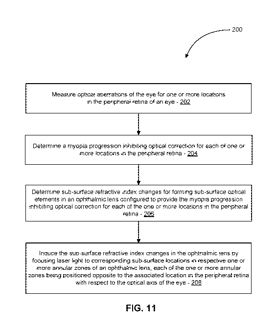
[0066] FIG. 11 is a simplified schematic illustration of a method 200 of
modifying an
ophthalmic lens so as to configure the ophthalmic lens to inhibit progression
of myopia in a
subject associated with the ophthalmic lens, in accordance with embodiments.
Any suitable
optical corrections, approaches, and/or systems, including those described
herein, can be used
to practice the method 200.
12
CA 03147871 2022-01-18
WO 2021/015993 PCT/US2020/041934
[0067] In act 202, an optical aberrations of an eye of the subject are
measured for each of
one or more locations in the peripheral retina of the eye. For example, the
system 100 can be
used to measure optical aberrations for selected locations in the peripheral
retina of the eye.
In some embodiments, optical aberrations are measured for each of the selected
locations for
a suitable range of accommodation levels of the eye. In some embodiments,
optical
aberrations of the eye are measured for one or more locations in the fovea 18
of the eye.
[0068] In act 204, a myopia progression inhibiting optical correction is
determined for
each of one or more locations in the peripheral retina of the eye. In many
embodiments, each
of the myopia progression inhibiting optical correction determined is based on
the optical
aberrations measured in act 202. In some embodiments, the myopia progression
inhibiting
optical correction determined for each location in the peripheral retina
corrects hyperopia at
the location. In some embodiments, the myopia progression inhibiting optical
correction
determined for each location in the peripheral retina reduces optical
anisotropy, which can be
defined as the ratio of the horizontal divided by vertical area under a mean
transfer function
(MTF) curve between zero and 60 cycles/degree. In some embodiments, the myopia
progression inhibiting optical correction determined for each location in the
peripheral retina
increases depth of focus at the respective location in the peripheral retina.
In some
embodiments, the myopia progression inhibiting optical correction determined
for each
location in the peripheral retina decreases depth of focus at the respective
location in the
peripheral retina.
[0069] In act 206, subsurface refractive index changes are determined for
forming
subsurface elements in an ophthalmic lens that are configured to provide the
myopia
progression inhibiting optical correction for each of the one or more
locations in the
peripheral retina. The subsurface refractive index changes can be formed using
any suitable
approaches, such as those described in U.S. Patent 8,932,352; U.S. Patent
9,939,558, and
U.S. Patent Application Publication 2018/0206979; the full disclosure of which
are
incorporated herein by reference. The subsurface optical elements can be
configured to
provide the entire myopia progression inhibiting optical correction for each
of the one or
more locations in the peripheral retina. Alternatively, the ophthalmic lens
can have an
external shape that provides a refractive correction that works in combination
with the
subsurface optical elements to provide the myopia progression inhibiting
optical correction
for each of the one or more locations in the peripheral retina.
13
CA 03147871 2022-01-18
WO 2021/015993 PCT/US2020/041934
[0070] In act 208, the subsurface refractive index changes are induced in the
ophthalmic
lens by focusing laser light to corresponding subsurface locations in
respective one or more
annular zones of the ophthalmic lens. Each of the one or more annular zones is
positioned
opposite to the associated location in the peripheral retina with respect to
the optical axis of
the eye.
[0071] Laser and optical systems for forming subsurface optical elements
[0072] FIG. 12 is a schematic representation of the laser and optical system
300 that can
be used to modify an ophthalmic lens to be configured to inhibit progression
of myopia, in
accordance with embodiments. The system 300 includes a laser source that
includes a Kerr-
lens mode-locked Ti:Sapphire laser 312 (Kapteyn-Mumane Labs, Boulder, Colo.)
pumped by
4 W of a frequency-doubled Nd:YV04 laser 314. The laser generates pulses of
300 mW
average power, 30 fs pulse width, and 93 MHz repetition rate at wavelength of
800 nm.
Because there is a reflective power loss from the mirrors and prisms in the
optical path, and
in particular, from the power loss of the objective 320, the measured average
laser power at
the objective focus on the material is about 120 mW, which indicates the pulse
energy for the
femtosecond laser is about 1.3 nJ.
[0073] Due to the limited laser pulse energy at the objective focus, the pulse
width can be
preserved so that the pulse peak power is strong enough to exceed the
nonlinear absorption
threshold of the ophthalmic lens. Because a large amount of glass inside the
focusing
objective significantly increases the pulse width due to the positive
dispersion inside of the
glass, an extra-cavity, compensation scheme can be used to provide the
negative dispersion
that compensates for the positive dispersion introduced by the focusing
objective. Two SF10
prisms 324 and 328 and one ending mirror 332 form a two-pass one-prism-pair
configuration.
A 37.5 cm separation distance between the prisms can be used to compensate the
dispersion
of the microscope objective and other optics within the optical path.
[0074] A collinear autocorrelator 340 using third-order harmonic generation is
used to
measure the pulse width at the objective focus. Both 2nd and 3rd harmonic
generation have
been used in autocorrelation measurements for low NA or high NA objectives.
Third order
surface harmonic generation (THG) autocorrelation was selected to characterize
the pulse
width at the focus of the high-numerical-aperture objectives because of its
simplicity, high
signal to noise ratio and Jack of material dispersion that second harmonic
generation (SHG)
crystals usually introduce. The THG signal is generated at the interface of
air and an ordinary
14
CA 03147871 2022-01-18
WO 2021/015993 PCT/US2020/041934
cover slip 342 (Corning No. 0211 Zinc Titania glass), and measured with a
photomultiplier
344 and a lock-in amplifier 346. After using a set of different high-numerical-
aperture
objectives and carefully adjusting the separation distance between the two
prisms and the
amount of glass inserted, a transform-limited 27-fs duration pulse was
selected. The pulse is
focused by a 60X 0.70NA Olympus LUCPlanFLN long-working-distance objective
348.
[0075] Because the laser beam will spatially diverge after it comes out
of the laser cavity,
a concave mirror pair 350 and 352 is added into the optical path in order to
adjust the
dimension of the laser beam so that the laser beam can optimally fills the
objective aperture.
A 3D 100 nm resolution DC servo motor stage 354 (Newport VP-25XA linear stage)
and a
2D 0.7 nm resolution piezo nanopositioning stage (P1 P-622.2CD piezo stage)
are controlled
and programmed by a computer 356 as a scanning platform to support and locate
an
ophthalmic lens 357. The servo stages have a DC servo-motor so they can move
smoothly
between adjacent steps. An optical shutter controlled by the computer with 1
ms time
resolution is installed in the system to precisely control the laser exposure
time. With
customized computer programs, the optical shutter could be operated with the
scanning
stages to form the subsurface optical elements in the ophthalmic lens 357 with
different
scanning speed at different position and depth and different laser exposure
time. In addition,
a CCD camera 358 along with a monitor 362 is used beside the objective 320 to
monitor the
process in real time. The system 300 can be used to modify the refractive
index of an
ophthalmic lens to form subsurface optical elements that provide a myopia
progression
inhibiting optical correction for each of one or more locations in the
peripheral retina.
[0076] FIG. 13 is a simplified schematic illustration of another system 430
for forming
one or more subsurface optical structures within an ophthalmic lens 410, in
accordance with
embodiments. The system 430 includes a laser beam source 432, a laser beam
intensity
control assembly 434, a laser beam pulse control assembly 436, a
scanning/interface
assembly 438, and a control unit 440.
[0077] The laser beam source 432 generates and emits a laser beam 446 having a
suitable
wavelength for inducing refractive index changes in target sub-volumes of the
ophthalmic
lens 410. In examples described herein, the laser beam 446 has a 1035 nm
wavelength. The
laser beam 446, however, can have any suitable wavelength (e.g., in a range
from 400 to
1100 nm) effective in inducing refractive index changes in the target sub-
volumes of the
ophthalmic lens 410.
CA 03147871 2022-01-18
WO 2021/015993 PCT/US2020/041934
[0078] The laser beam intensity control assembly 434 is controllable to
selectively vary
intensity of the laser beam 446 to produce a selected intensity laser beam 48
output to the
laser beam pulse control assembly 436. The laser beam intensity control
assembly 434 can
have any suitable configuration, including any suitable existing
configuration, to control the
intensity of the resulting laser beam 448.
[0079] The laser beam pulse control assembly 436 is controllable to
generate collimated
laser beam pulses 450 having suitable duration, intensity, size, and spatial
profile for inducing
refractive index changes in the target sub-volumes of the ophthalmic lens 410.
The laser
beam pulse control assembly 436 can have any suitable configuration, including
any suitable
existing configuration, to control the duration of the resulting laser beam
pulses 450.
[0080] The scanning/interface assembly 438 is controllable to selectively
scan the laser
beam pulses 450 to produce XYZ scanned laser pulses 474. The
scanning/interface assembly
438 can have any suitable configuration, including any suitable existing
configuration (for
example, the configuration illustrated in FIG. 14) to produce the XYZ scanned
laser pulses
474. The scanning/interface assembly 438 receives the laser beam pulses 450
and outputs the
XYZ scanned laser pulses 474 in a manner that minimizes vignetting. The
scanning/interface
assembly 438 can be controlled to selectively scan each of the laser beam
pulses 450 to
generate XYZ scanned laser pulses 474 focused onto targeted sub-volumes of the
ophthalmic
lens 410 to induce the respective refractive index changes in targeted sub-
volumes so as to
.. form the one or more subsurface optical structures within an ophthalmic
lens 410. In many
embodiments, the scanning/interface assembly 438 is configured to restrain the
position of
the ophthalmic lens 410 to a suitable degree to suitably control the location
of the targeted
sub-volumes of the ophthalmic lens 410 relative to the scanning/interface
assembly 438. In
many embodiments, such as the embodiment illustrated in FIG. 14, the
scanning/interface
assembly 438 includes a motorized Z-stage that is controlled to selectively
control the depth
within the ophthalmic lens 410 to which each of the XYZ scanned laser pulses
474 is
focused.
[0081] The control unit 440 is operatively coupled with each of the laser beam
source 432,
the laser beam intensity control assembly 434, the laser beam pulse control
assembly 436,
and the scanning/interface assembly 438. The control unit 440 provides
coordinated control
of each of the laser beam source 432, the laser beam intensity control
assembly 434, the laser
beam pulse control assembly 436, and the scanning/interface assembly 438 so
that each of the
16
CA 03147871 2022-01-18
WO 2021/015993 PCT/US2020/041934
XYZ scanned laser pulses 474 have a selected intensity and duration, and are
focused onto a
respective selected sub-volume of the ophthalmic lens 410 to form the one or
more
subsurface optical structures within an ophthalmic lens 410. The control unit
440 can have
any suitable configuration. For example, in some embodiments, the control unit
440
comprises one or more processors and a tangible memory device storing
instructions
executable by the one or more processors to cause the control unit 440 to
control and
coordinate operation of the of the laser beam source 432, the laser beam
intensity control
assembly 434, the laser beam pulse control assembly 436, and the
scanning/interface
assembly 438 to produce the XYZ scanned laser pulses 474, each of which is
synchronized
with the spatial position of the sub-volume optical structure.
[0082] FIG. 14 is a simplified schematic illustration of an embodiment of the
scanning/interface assembly 438. In the illustrated embodiment, the
scanning/interface
assembly 438 includes an XY galvo scanning unit 442, a relay optical assembly
444, a
Z stage 466, an XY stage 468, a focusing objective lens 470, and a patient
interface/ophthalmic lens holder 472. The XY galvo scanning unit 438 includes
XY galvo
scan mirrors 454, 456. The relay optical assembly 440 includes concave mirrors
460, 461
and plane mirrors 462, 464.
[0083] The XY galvo scanning unit 442 receives the laser pulses 450 (e.g.,
1035 nm
wavelength collimated laser pulses) from the laser beam pulse control assembly
436. In the
illustrated embodiment, the XY galvo scanning unit 442 includes a motorized X-
direction
scan mirror 454 and a motorized Y-direction scan mirror 456. The X-direction
scan mirror
454 is controlled to selectively vary orientation of the X-direction scan
mirror 454 to vary
direction/position of XY scanned laser pulses 458 in an X-direction transverse
to direction of
propagation of the XY scanned laser pulses 458. The Y-direction scan mirror
456 is
controlled to selectively vary orientation of the Y-direction scan mirror 456
to vary
direction/position of the XY scanned laser pulses 458 in an Y-direction
transverse to
direction of propagation of the XY scanned laser pulses 458. In many
embodiments, the Y-
direction is substantially perpendicular to the X-direction.
[0084] The relay optical assembly 440 receives the XY scanned laser pulses 458
from the
XY galvo scanning unit 442 and transfers the XY scanned laser pulses 458 to Z
stage 466 in a
manner that minimizes vignetting. Concave mirror 460 reflects each of the XY
scanned laser
pulse 458 to produce a converging laser pulses incident on plane mirror 462.
Plane mirror
17
CA 03147871 2022-01-18
WO 2021/015993 PCT/US2020/041934
462 reflects the converging XY scanned laser pulse 458 towards plane mirror
464. Between
the plane mirror 462 and the plane mirror 464, the XY scanned laser pulse 458
transitions
from being convergent to being divergent. The divergent laser pulse 458 is
reflected by plane
mirror 464 onto concave mirror 461. Concave mirror 461 reflects the laser
pulse 458 to
.. produce a collimated laser pulse that is directed to the Z stage 466.
[0085] The Z stage 466 receives the XY scanned laser pulses 458 from the relay
optical
assembly 442. In the illustrated embodiment, the Z stage 466 and the XY stage
468 are
coupled to the focusing objective lens 470 and controlled to selectively
position the focusing
objective lens 470 relative to the ophthalmic lens 410 for each of the XY
scanned laser pulses
474 so as to focus the XYZ scanned laser pulse 474 onto a respective targeted
sub-volume of
the ophthalmic lens 410. The Z stage 466 is controlled to selectively control
the depth within
the ophthalmic lens 410 to which the laser pulse is focused (i.e., the depth
of the sub-surface
volume of the ophthalmic lens 410 on which the laser pulse is focused to
induce a change in
refractive index of the targeted sub-surface volume). The XY stage 468 is
controlled in
conjunction with control of the XY galvo scanning unit 442 so that the
focusing objective
lens 470 is suitably positioned for the respective transverse position of each
of the XY
scanned laser pulses 458 received by the Z stage 466. The focusing objective
lens 470
converges the laser pulse onto the targeted sub-surface volume of the lens
410. The patient
interface/ophthalmic lens holder 472 restrains the ophthalmic lens 410 in a
fixed position to
support scanning of the laser pulses 474 by the scanning/interface assembly
438 to form the
subsurface optical structures within the ophthalmic lens 410.
[0086] Defining subsurface optical elements for a specified optical correction
[0087] FIG. 15 through FIG. 22 illustrate a process that can be used to define
subsurface
optical elements for a specified optical correction. While an optical
correction for inhibiting
progression of myopia in a subject using the approaches described herein may
be a
combination of any suitable number of low-order optical corrections and/or any
suitable
number of high-order optical corrections, a single, simple 2 diopter optical
correction is
illustrated. The same process, however, can be used to define subsurface
optical elements for
an ophthalmic lens to configure the ophthalmic lens to provide an optical
correction (such
any of the myopia inhibiting optical corrections described herein) that
inhibits myopia
progression.
18
CA 03147871 2022-01-18
WO 2021/015993 PCT/US2020/041934
[0088] FIG. 15 shows a radial variation in units of optical waves of a
2.0 diopter refractive
index distribution 510, in accordance with embodiments. The optical waves in
this curve
correspond to a design wavelength of 562.5 nm. In the illustrated embodiment,
the
2.0 diopter refractive index distribution 510 decreases from a maximum of 16.0
waves at the
optical axis of an ophthalmic lens down to 0.0 waves at 3.0 cm from the
optical axis.
[0089] FIG. 16 shows a 1.0 wave phase-wrapped refractive index distribution
512
corresponding to the 2.0 diopter refractive index distribution 510. Each
segment of the 1.0
wave phase-wrapped refractive index distribution 512 includes a sloped segment
(512a
through 512p). Each of all the segments, except the center segment, of the 1.0
wave phase-
wrapped refractive index distribution 512 includes a phase discontinuity (514b
through 514p)
with a height equal to 1.0 wave. Each of the sloped segments (512a through
512p) is shaped
to match the corresponding overlying segment (510a through 510p) of the 2.0
diopter
refractive index distribution 510. For example, sloped segment 512p matches
overlying
segment 510p; sloped segment 512o is equal to overlying segment 510o minus 1.0
wave;
sloped segment 512n is equal to overlying segment 510n minus 2.0 waves; sloped
segment
512a is equal to overlying segment 510a minus 15.0 waves. Each sloped segment
corresponds to a Fresnel zone.
[0090] The 1.0 wave height of each of the phase discontinuities (514b
through 514p) in the
distribution 512 results in diffraction at the design wavelength that provides
the same 2.0
diopter refractive correction as the 2.0 diopter refractive distribution 510
while limiting
maximum phase equal to 1.0 wave.
[0091] The 1.0 wave phase-wrapped refractive index distribution 512
requires
substantially lower total laser pulse energy to induce in comparison to the
2.0 diopter
refractive index distribution 510. The area under the 1.0 wave phase-wrapped
refractive
index distribution 512 is only about 5.2 percent of the area under the 2.0
diopter refractive
index distribution 510.
[0092] FIG. 17 shows the 1.0 wave phase-wrapped refractive index distribution
512 and
an example scaled phase-wrapped refractive index distribution (for a selected
maximum
wave value) corresponding to the 1.0 wave phase-wrapped refractive index
distribution 512.
In the illustrated embodiment, the example scaled phase-wrapped refractive
index
distribution has a maximum wave value of 1/3 wave. Similar scaled phase-
wrapped
refractive index distributions can be generated for other suitable maximum
wave values less
19
CA 03147871 2022-01-18
WO 2021/015993 PCT/US2020/041934
than 1.0 wave (e.g., 3/4 wave, 5/8 wave, 1/2 wave, 1/4 wave, 1/6 wave). The
1/3 optical
wave maximum scaled phase-wrapped refractive index distribution 516 is equal
to 1/3 of the
1.0 wave phase-wrapped refractive index distribution 512. The 1/3 optical wave
maximum
scaled phase-wrapped refractive index distribution 516 is one substitute for
the 1.0 wave
phase-wrapped refractive index distribution 512 and utilizes a maximum
refractive index
value that provides a corresponding maximum 1/3 wave optical correction.
[0093] The 1/3 optical wave maximum scaled phase-wrapped refractive index
distribution
516 requires less total laser pulse energy to induce in comparison with the
1.0 wave phase-
wrapped refractive index distribution 512. The area under the 1/3 optical wave
maximum
scaled phase-wrapped refractive index distribution 516 is 1/3 of the area
under the 1.0 wave
phase-wrapped refractive index distribution 512. Three stacked layers of the
1/3 wave
distribution 516 can be used to produce the same optical correction as the 1.0
wave
distribution 512.
[0094] FIG. 18 graphically illustrates diffraction efficiency for near
focus 574 and far
.. focus 576 versus phase change height. For phase change heights less than
0.25 waves, the
diffraction efficiency for near focus is only about 10 percent. Near focus
diffraction
efficiency of substantially greater than 10 percent, however, is desirable to
limit the number
of layers of the subsurface optical structures that are stacked to generate a
desired overall
optical correction. Greater phase change heights can be achieved by inducing
greater
refractive index changes in the targeted sub-volumes of the ophthalmic lens
410. Greater
refractive index changes in the targeted sub-volumes of the ophthalmic lens
410 can be
induced by increasing energy of the laser pulses focused onto the targeted sub-
volumes of the
ophthalmic lens 410.
[0095] FIG. 19 graphically illustrates an example calibration curve 578
for resulting phase
change height as a function of laser pulse optical power. The calibration
curve 578 shows
correspondence between resulting phase change height as a function of laser
average power
for a corresponding laser pulse duration, laser pulse wavelength, laser pulse
repetition rate,
numerical aperture, material of the ophthalmic lens 410, depth of the targeted
sub-volume,
spacing between the targeted sub-volumes, scanning speed, and line spacing.
The calibration
curve 578 shows that increasing laser pulse energy results in increased phase
change height.
[0096] Laser pulse energy, however, may be limited to avoid propagation of
damage
induced caused by laser pulse energy and/or heat accumulation with the
ophthalmic lens 410,
CA 03147871 2022-01-18
WO 2021/015993 PCT/US2020/041934
or even between the layers of the subsurface optical elements. In many
instances, there is no
observed damage during formation of the first two layers of subsurface optical
elements and
damage starts to occur during formation of the third layer of subsurface
optical elements. To
avoid such damage, the subsurface optical elements can be formed using laser
pulse energy
below a pulse energy threshold of the material of the ophthalmic lens 410.
Using lower pulse
energy, however, increases the number of layers of the subsurface optical
elements required
to provide the desired amount of resulting phase change height, thereby adding
to the time
required to form the total number of subsurface optical elements 412 employed.
[0097] FIG. 20 is a plan view illustration of an ophthalmic lens 410 that
includes one or
more subsurface optical elements 412 with refractive index spatial variations,
in accordance
with embodiments. The one or more subsurface elements 12 described herein can
be formed
in any suitable type of ophthalmic lens including, but not limited to, intra-
ocular lenses,
contact lenses, corneas, spectacle lenses, and native lenses (e.g., a human
native lens). The
one or more subsurface optical elements 412 with refractive index spatial
variations can be
configured to provide a suitable refractive correction configured to inhibit
progression of
myopia as described herein. Additionally, the one or more subsurface optical
elements 412
with refractive index spatial variations can be configured to provide a
suitable refractive
correction for each of many optical aberrations such as astigmatism, myopia,
hyperopia,
spherical aberrations, coma and trefoil, as well as any suitable combination
thereof.
[0098] FIG. 21 is a plan view illustration of one of the subsurface optical
elements 412 of
the ophthalmic lens 410. The illustrated subsurface optical elements 412
occupies a
respective volume of the lens 410, which includes associated sub-volumes of
the lens 410. In
many embodiments, the volume occupied by one of the optical elements 412
includes first,
second, and third portions 414. Each of the first, second, and third portions
414 can be
formed by focusing suitable laser pulses inside the respective portion 414 so
as to induce
changes in refractive index in sub-volumes of the lens 410 that make up the
respective
portion 414 so that each portion 414 has a respective refractive index
distribution.
[0099] In many embodiments, a refractive index distribution is defined for
each portion
414 that forms the subsurface optical structures 412 so that the resulting
subsurface optical
.. structures 412 provide a desired optical correction. The refractive index
distribution for each
portion 414 can be used to determine parameters (e.g., laser pulse power (mW),
laser pulse
21
CA 03147871 2022-01-18
WO 2021/015993 PCT/US2020/041934
width (fs)) of laser pulses that are focused onto the respective portions 414
to induce the
desired refractive index distributions in the portions 414.
[0100] While the portions 414 of the subsurface optical structures 412 have a
circular
shape in the illustrated embodiment, the portions 414 can have any suitable
shape and
distribution of refractive index variations. For example, a single portion 414
having an
overlapping spiral shape can be employed. In general, one or more portions 414
having any
suitable shapes can be distributed with intervening spaces so as to provide a
desired optical
correction for light incident on the subsurface optical structure 412.
[0101] FIG. 22 illustrates an embodiment in which the subsurface optical
elements 412 are
comprised of several stacked layers that are separated by intervening layer
spaces. In the
illustrated embodiment, the subsurface optical elements 412 have a spatial
distribution of
refractive index variations. FIG. 22 is a side view illustration of an example
distribution of
refractive index variations in the subsurface optical elements 412. In the
illustrated
embodiment, the subsurface optical elements 412 can be formed using a raster
scanning
.. approach in which each layer is sequentially formed starting with the
bottom layer and
working upward. For each layer, a raster scanning approach can sequentially
scan the focal
position of the laser pulses along planes of constant Z-dimension while
varying the Y-
dimension and the X-dimension so that the resulting layers have the flat cross-
sectional
shapes shown in FIG. 22, which shows a cross-sectional view of the ophthalmic
lens 410. In
.. the raster scanning approach, timing of the laser pulses can be controlled
to direct each laser
pulse onto a targeted sub-volume of the ophthalmic lens 410 and not direct
laser pulses onto
non-targeted sub-volumes of the ophthalmic lens 410, which include sub-volumes
of the
ophthalmic lens 10 that do not form any of the subsurface optical elements
412, such as the
intervening spaces between the adjacent stacked layers that can form the
subsurface optical
elements 412.
[0102] In the illustrated embodiment, there are three annular subsurface
optical elements
412 with distributions of refractive index spatial variations. Each of the
illustrated subsurface
optical elements 412 has a flat layer configuration and can be comprised of
one or more
layers. If the subsurface optical structures are comprised of more than one
layer, the layers
can be separated from each other by an intervening layer spacing. Each of the
layers,
however, can alternatively have any other suitable general shape including,
but not limited to,
any suitable non-planar or planar surface. In the illustrated embodiment, each
of the
22
CA 03147871 2022-01-18
WO 2021/015993 PCT/US2020/041934
subsurface optical elements 412 has a circular outer boundary. Each of the
subsurface optical
elements 412, however, can alternatively have any other suitable outer
boundary shape. Each
of the subsurface optical elements 412 can include two or more separate
portions 14 with
each covering a portion of the subsurface optical elements 412.
[0103] FIG. 23A, FIG. 23B, and FIG. 23C illustrate transmission of light onto
a portion
of the peripheral retina via central and peripheral zones of an ophthalmic
lens. FIG. 23A is a
simplified front view of an eye 10 showing the pupil 38 and the surrounding
iris 40. FIG.
23B is a simplified front view of an ophthalmic lens 190 that has a central
optical zone 192, a
peripheral optical zone 194, and an outer zone 196. FIG. 23C is a simplified
off-optical-axis
view illustrating relative contribution of the peripheral optical zone 194 to
a peripheral retinal
image and the central optical zone 194 to a peripheral retinal image. In view
of the
contribution of the central optical zone 194 to a peripheral retinal image, in
some
embodiments, the optical correction provided by the central optical zone 194
is accounted for
when determining a myopia mitigating optical correction for the peripheral
optical zone 194.
The optical correction provided by the central optical zone 194 can also be
based in part on a
desired correction to a peripheral retinal image provided by the central
optical zone 194.
[0104] FIG. 24A and FIG. 24B illustrate relative contribution of example
peripheral outer
zones of an ophthalmic lens to a resulting peripheral retina image. FIG. 24A
shows a plot of
the percentages of the peripheral annular zone 194 that is within a 4 mm
diameter pupil for a
4 mm diameter central optical zone 192. For peripheral viewing eccentricities
up to 15
degrees, a 6 mm diameter peripheral annular zone 194 is sufficient in size to
maximize the
percentage of the peripheral annular zone 194 within the pupil 38. For
peripheral viewing
eccentricities up to 20 degrees, a 7 mm diameter peripheral annular zone 194
is sufficient in
size to maximize the percentage of the peripheral annular zone 194 within the
pupil 38. For
peripheral viewing eccentricities up to 30 degrees, a 8 mm diameter peripheral
annular zone
194 is sufficient in size to maximize the percentage of the peripheral annular
zone 194 within
the pupil 38. The percentage of the peripheral annular zone 194 within the
pupil 38 can be
used to guide selection of the inner and outer diameter of the peripheral
annular zone 194 for
a particular user of the contact lens 24.
[0105] FIG. 25 shows example average change in aberrations from 0 degree to 20
degree
retinal eccentricity for a group of 10 normal individuals. Retinal image
quality was
computed, through-focus, in white light for the case of 20 degrees nasal
retinal eccentricity
23
CA 03147871 2022-01-18
WO 2021/015993 PCT/US2020/041934
(i.e. peripheral visual field). Overall image quality was defined as the
average of the
horizontal and vertical area under the modulation transfer function (MTF) from
0 to 60
cycles/degree. Optical anisotropy is a measure of the degree rotational
asymmetry in retina
blur. Optical anisotropy is defined herein as the ratio of horizontal divided
by vertical area
under the MTF, and was calculated for a 4 mm diameter circular pupil, which is
an
approximation. At 20 degree nasal retinal eccentricity, a 4 mm diameter pupil
is elliptical
with a 4 mm vertical axis and a 3.8 mm short (horizontal) axis. The through-
focus range
evaluated was -3 to +3 diopters in 0.125 diopter increments.
[0106] Example annular zone optical corrections for inhibiting myopia
progression
.. [0107] Through focus optical anisotropy and image quality for 20 degree
viewing
eccentricity plotted in FIG. 26, FIG. 27, FIG. 28, FIG. 29, FIG. 30, and FIG.
31 were
calculated for four cases using an annular optical zone that provided 100
percent coverage of
a 4 mm diameter pupil. FIG. 24A, however, shows that an annular optical zone
with a 4 mm
inner diameter covers only about 35-45% of the pupil at 20 degree viewing
eccentricity.
Accordingly, the through focus optical anisotropy and the image quality for 20
degree
viewing eccentricity plotted in FIG. 26, FIG. 27, FIG. 28, FIG. 29, FIG. 30,
and FIG. 31
somewhat overestimate the changes in the optical anisotropy and the image
quality provided.
The 100 percent coverage of the 4 mm diameter pupil by the annular optical
zone used was
employed for ease of computation. The four conditions calculated include: (1)
a control case
.. 402 of average 20 deg nasal wavefront aberration for 5 mm pupil taken from
10 normal
individuals, whose peripheral aberrations were published in Zheleznyak et al.,
Journal of
Vision, 20161; (2) cylinder correction 404 only applied to the control case;
(3) a multifocal
correction 406 applied to the control case with 1.5 diopters of add power with
0.4 waves of
optical phase change; (4) a cylinder correction and the multifocal correction
408 from #3
applied to the control case.
[0108] FIG. 26 is a plot of peripheral retinal image asymmetry (20 degree
viewing
eccentricity) over a range of accommodation levels for example contact lens
induced optical
corrections. The x-axis is the defocus or object distance in units of
diopters. A diopter is an
inverse meter. The y-axis is optical anisotropy, defined as the ratio of the
horizontal divided
.. by vertical area-under-the-MTF (between 0 and 60 cyc/deg). A y-axis value
of 1 indicates
rotational symmetry. A y-axis value of greater than 1 indicates horizontal
blur. A y-axis
24
CA 03147871 2022-01-18
WO 2021/015993 PCT/US2020/041934
value of less than 1 indicates vertical blur. The control case shows the
largest optical
anisotropy. The cylinder correction produces a large reduction in the optical
anisotropy.
[0109] FIG. 27 is a plot of peripheral retinal image quality (20 degree
viewing
eccentricity) over a range of accommodation levels for example contact lens
induced optical
corrections. The x-axis is the defocus or object distance in units of
diopters. The y-axis is
retinal image quality, defined as the average of the horizontal and vertical
area-under-the-
MTF (between 0 and 60 cyc/deg). The larger the y-axis value, the better the
image quality.
The cylinder correction provides the best peak image quality. The combination
of the
cylinder correction and the multifocal correction provides the largest depth
of focus and the
lowest anisotropy (as shown in FIG. 26).
[0110] FIG. 28, FIG. 29, FIG. 30, and FIG. 31 are plots of horizontal and
vertical
peripheral retinal image quality over a range of accommodation levels for the
four conditions.
FIG. 28 is a plot of horizontal and vertical peripheral retinal image quality
over a range of
accommodation levels for the control case 402. FIG. 29 is a plot of horizontal
and vertical
peripheral retinal image quality over a range of accommodation levels for the
control case
with an ophthalmic lens having subsurface refractive optical elements
providing the
cylindrical correction 404. FIG. 30 is a plot of horizontal and vertical
peripheral retinal
image quality over a range of accommodation levels for the control case with
an ophthalmic
lens having subsurface refractive optical elements providing the bifocal
correction 406. FIG.
31 is a plot of horizontal and vertical peripheral retinal image quality over
a range of
accommodation levels for the control case with an ophthalmic lens having
subsurface
refractive optical elements providing the cylindrical and bifocal correction
408. The cylinder
correction provides the best peak image quality. The combination of the
cylinder correction
and the multifocal correction provides the largest depth of focus and lowest
anisotropy (as
shown in FIG. 26).
[0111] Any of the ophthalmic lenses 24, 150, 170, 180, 190 described
herein can be
configured to ensure proper orientation so that each of the annular zones is
aligned with the
associated region in the peripheral retina. For example, a contact lens can
include any one or
more suitable design features that cause the contact lens to rotate to the
proper orientation on
the cornea. In some embodiments, a contact lens is weighted at the bottom to
cause the
contact lens to rotate to, and maintain, the proper orientation on the cornea
so that each of the
annular zones in the contact lens is aligned with the associated region in the
peripheral retina.
CA 03147871 2022-01-18
WO 2021/015993 PCT/US2020/041934
[0112] Other variations are within the spirit of the present disclosure.
Thus, while the
disclosed techniques are susceptible to various modifications and alternative
constructions,
certain illustrated embodiments thereof are shown in the drawings and have
been described
above in detail. It should be understood, however, that there is no intention
to limit the
disclosure to the specific form or forms disclosed, but on the contrary, the
intention is to
cover all modifications, alternative constructions, and equivalents falling
within the spirit and
scope of the disclosure, as defined in the appended claims.
[0113] The use of the terms "a" and "an" and "the" and similar referents in
the context of
describing the disclosed embodiments (especially in the context of the
following claims) are
.. to be construed to cover both the singular and the plural, unless otherwise
indicated herein or
clearly contradicted by context. The terms "comprising," "having,"
"including," and
"containing" are to be construed as open-ended terms (i.e., meaning
"including, but not
limited to,") unless otherwise noted. The term "connected" is to be construed
as partly or
wholly contained within, attached to, or joined together, even if there is
something
intervening. Recitation of ranges of values herein are merely intended to
serve as a shorthand
method of referring individually to each separate value falling within the
range, unless
otherwise indicated herein and each separate value is incorporated into the
specification as if
it were individually recited herein. All methods described herein can be
performed in any
suitable order unless otherwise indicated herein or otherwise clearly
contradicted by context.
The use of any and all examples, or exemplary language (e.g., "such as")
provided herein, is
intended merely to better illuminate embodiments of the disclosure and does
not pose a
limitation on the scope of the disclosure unless otherwise claimed. No
language in the
specification should be construed as indicating any non-claimed element as
essential to the
practice of the disclosure.
[0114] Disjunctive language such as the phrase "at least one of X, Y, or Z,"
unless
specifically stated otherwise, is intended to be understood within the context
as used in
general to present that an item, term, etc., may be either X, Y, or Z, or any
combination
thereof (e.g., X, Y, and/or Z). Thus, such disjunctive language is not
generally intended to,
and should not, imply that certain embodiments require at least one of X, at
least one of Y, or
at least one of Z to each be present.
[0115] Various embodiments of this disclosure are described herein, including
the best
mode known to the inventors for carrying out the disclosure. Variations of
those
26
CA 03147871 2022-01-18
WO 2021/015993 PCT/US2020/041934
embodiments may become apparent to those of ordinary skill in the art upon
reading the
foregoing description. The inventors expect skilled artisans to employ such
variations as
appropriate and the inventors intend for the disclosure to be practiced
otherwise than as
specifically described herein. Accordingly, this disclosure includes all
modifications and
equivalents of the subject matter recited in the claims appended hereto as
permitted by
applicable law. Moreover, any combination of the above-described elements in
all possible
variations thereof is encompassed by the disclosure unless otherwise indicated
herein or
otherwise clearly contradicted by context.
[0116] Examples of the embodiments of the present disclosure can be described
in view of
the following clauses:
[0117] Clause 1. An ophthalmic lens including a central zone and an annular
zone,
wherein the annular zone includes subsurface optical elements formed via laser-
induced
changes in refractive index of a material forming the annular zone, and
wherein the
subsurface optical elements are configured to modify distribution of light to
a peripheral
retina of a user so as to inhibit progression of myopia.
[0118] Clause 2. The ophthalmic lens of clause 1, wherein the subsurface
optical elements
are configured to reduce asymmetry of a radial versus azimuthal contrast in
the peripheral
retina of the user.
[0119] Clause 3. The ophthalmic lens of clause 1, wherein the subsurface
optical elements
are configured to reduce hyperopia in the peripheral retina of the user.
[0120] Clause 4. The ophthalmic lens of clause 1, wherein the subsurface
optical elements
are configured to increase depth of focus in the peripheral retina of the
user.
[0121] Clause 5. The ophthalmic lens of clause 1, wherein the subsurface
optical elements
are configured to decrease depth of focus in the peripheral retina of the
user.
[0122] Clause 6. The ophthalmic lens of clause 1, wherein the subsurface
optical elements
are configured to accomplish two or more of: 1) reduce asymmetry of a radial
versus
azimuthal contrast in the peripheral retina of the user, 2) reduce hyperopia
in the peripheral
retina of the user, and 3) increase depth of focus in the peripheral retina of
the user.
[0123] Clause 7. The ophthalmic lens of clause 1, wherein the subsurface
optical elements
are configured to accomplish two or more of: 1) reduce asymmetry of a radial
versus
27
CA 03147871 2022-01-18
WO 2021/015993 PCT/US2020/041934
azimuthal contrast in the peripheral retina of the user, 2) reduce hyperopia
in the peripheral
retina of the user, and 3) decrease depth of focus in the peripheral retina of
the user.
[0124] Clause 8. The ophthalmic lens of clause 1, wherein the subsurface
optical elements
are configured to accomplish two or more of: 1) increase asymmetry of a radial
versus
azimuthal contrast in the peripheral retina of the user, 2) reduce hyperopia
in the peripheral
retina of the user, and 3) increase depth of focus in the peripheral retina of
the user.
[0125] Clause 9. The ophthalmic lens of clause 1, wherein the subsurface
optical elements
are configured to accomplish two or more of: 1) increase asymmetry of a radial
versus
azimuthal contrast in the peripheral retina of the user, 2) reduce hyperopia
in the peripheral
retina of the user, and 3) decrease depth of focus in the peripheral retina of
the user.
[0126] Clause 10. The ophthalmic lens of clause 1, wherein the annular zone
comprises
two or more annular portions, and wherein the subsurface optical elements in
each of the two
or more annular portions are configured to accomplish one or both of: 1)
reduce asymmetry
of a radial versus azimuthal contrast in the peripheral retina of the user;
and/or 2) reduce
hyperopia in the peripheral retina of the user.
[0127] Clause 11. The ophthalmic lens of clause 10, wherein the subsurface
optical
elements in each of the two or more annular portions are configured to
increase depth of
focus in the peripheral retina of the user.
[0128] Clause 12. The ophthalmic lens of clause 10, wherein the subsurface
optical
elements in each of the two or more annular portions are configured to
decrease depth of
focus in the peripheral retina of the user.
[0129] Clause 13. The ophthalmic lens of any one of clause 1 through clause
12,
configured as a contact lens.
[0130] Clause 14. The ophthalmic lens of any one of clause 1 through clause
12,
configured as a spectacle lens.
[0131] Clause 15. The ophthalmic lens of any one of clause 1 through clause
12,
configured as a cornea.
[0132] Clause 16. The ophthalmic lens of any one of clause 1 through clause
12,
configured as a native lens of an eye.
28
CA 03147871 2022-01-18
WO 2021/015993 PCT/US2020/041934
[0133] Clause 17. The ophthalmic lens of any one of clause 1 through clause
12,
configured as an intraocular lens.
[0134] Clause 18. A method of modifying an ophthalmic lens, the method
including
inducing subsurface changes in refractive index of a material forming an
annular zone of an
ophthalmic lens to form subsurface optical elements configured to modify
distribution of
light to the peripheral retina of a user so as to inhibit progression of
myopia.
[0135] Clause 19. The method of clause 18, wherein the subsurface optical
elements are
configured to reduce asymmetry of a radial versus azimuthal contrast in the
peripheral retina
of the user.
[0136] Clause 20. The method of clause 18, wherein the subsurface optical
elements are
configured to reduce hyperopia in the peripheral retina of the user.
[0137] Clause 21. The method of clause 18, wherein the subsurface optical
elements are
configured to increase depth of focus in the peripheral retina of the user.
[0138] Clause 22. The method of clause 18, wherein the subsurface optical
elements are
configured to decrease depth of focus in the peripheral retina of the user.
[0139] Clause 23. The method of clause 18, wherein the subsurface optical
elements are
configured to accomplish two or more of: 1) reduce asymmetry of a radial
versus azimuthal
contrast in the peripheral retina of the user, 2) reduce hyperopia in the
peripheral retina of the
user, and 3) increase depth of focus in the peripheral retina of the user.
[0140] Clause 24. The method of clause 18, wherein the subsurface optical
elements are
configured to accomplish two or more of: 1) reduce asymmetry of a radial
versus azimuthal
contrast in the peripheral retina of the user, 2) reduce hyperopia in the
peripheral retina of the
user, and 3) decrease depth of focus in the peripheral retina of the user.
[0141] Clause 25. The method of clause 18, wherein the subsurface optical
elements are
configured to accomplish two or more of: 1) increase asymmetry of a radial
versus azimuthal
contrast in the peripheral retina of the user, 2) reduce hyperopia in the
peripheral retina of the
user, and 3) increase depth of focus in the peripheral retina of the user.
[0142] Clause 26. The method of clause 18, wherein the subsurface optical
elements are
configured to accomplish two or more of: 1) increase asymmetry of a radial
versus azimuthal
29
CA 03147871 2022-01-18
WO 2021/015993 PCT/US2020/041934
contrast in the peripheral retina of the user, 2) reduce hyperopia in the
peripheral retina of the
user, and 3) decrease depth of focus in the peripheral retina of the user.
[0143] Clause 27. The method of clause 18, wherein the changes in the
refractive index are
induced by subjecting the material to pulses of laser light.
[0144] Clause 28. The method of clause 27, wherein each of the pulses of laser
light have
a duration in a range from 10 femtoseconds to 500 femtoseconds.
[0145] Clause 29. The method of clause 28, wherein the laser light has a
wavelength of
about 405 nm.
[0146] Clause 30. The method of clause 28, wherein the laser light has a
wavelength of
about 810 nm.
[0147] Clause 31. The method of clause 28, wherein the laser light has a
wavelength of
about 1035 nm.
[0148] Clause 32. The method of clause 31, wherein each of the pulses of laser
light have
a duration in a range from 15 femtoseconds to 50 femtoseconds.
[0149] Clause 33. The method of clause 18, further comprising measuring a
radial versus
azimuthal contrast of light incident on a location of the peripheral retina,
and wherein the
subsurface optical elements are configured to reduce asymmetry of the radial
versus
azimuthal contrast of the light incident on the location of the peripheral
retina.
[0150] Clause 34. The method of clause 18, further comprising measuring
hyperopia for a
location of the peripheral retina, and wherein the subsurface optical elements
are configured
to reduce hyperopia at the location of the peripheral retina.
[0151] Clause 35. The method of clause 18, wherein the annular zone comprises
two or
more annular portions, and wherein the subsurface optical elements in each of
the two or
more annular portions are configured to: 1) reduce asymmetry of a radial
versus azimuthal
contrast in the peripheral retina of the user; and/or 2) reduce hyperopia in
the peripheral
retina of the user.
[0152] Clause 36. The method of clause 35, wherein the subsurface optical
elements in
each of the two or more annular portions are configured to increase depth of
focus in the
peripheral retina of the user.
CA 03147871 2022-01-18
WO 2021/015993
PCT/US2020/041934
[0153] Clause 37. The method of clause 35, wherein the subsurface optical
elements in
each of the two or more annular portions are configured to decrease depth of
focus in the
peripheral retina of the user.
[0154] Clause 38. The method of any one of clause 18 through clause 37,
wherein the
ophthalmic lens is a spectacle lens.
[0155] Clause 39. The method of any one of clause 18 through clause 37,
wherein the
ophthalmic lens is a cornea.
[0156] Clause 40. The method of any one of clause 18 through clause 37,
wherein the
ophthalmic lens is a native lens of an eye.
[0157] Clause 41. The method of any one of clause 18 through clause 37,
wherein the
ophthalmic lens is an intraocular lens.
31