Note: Descriptions are shown in the official language in which they were submitted.
CA 03148420 2022-01-21
WO 2021/019532
PCT/IL2020/050829
- 1 -
COMPONENTS OF OPEN LIQUID DRUG TRANSFER SYSTEMS AND A ROBOTIC SYSTEM
EMPLOYING THEM
Field of the Invention
The invention relates to the field of fluid transfer devices. Specifically the
invention relates to
components of open liquid drug transfer systems and their use in automated
robotic systems
for preparing drugs and medications for administration to patients.
Background of the Invention
US 8,196,614 to the applicant of the present invention describes closed system
liquid transfer
devices designed to provide contamination-free transfer of hazardous drugs.
Fig. la and Fig. lb
are schematic cross-sectional views of the apparatus 10 for transferring
hazardous drugs
without contaminating the surroundings, according to one embodiment of the
invention
described in US 8,196,614. The main features of this apparatus that are
relevant to the present
invention will be described herein. Additional details can be found in the
aforementioned
patent.
The proximal section of apparatus 10 is a syringe 12, which is adapted to draw
a desired
volume of a hazardous drug from a fluid transfer component, e.g. a vial 16 or
an intravenous
(IV) bag in which it is contained and to subsequently transfer the drug to
another fluid transfer
component. At the distal end of syringe 12 is connected a connector section
14, which is in
turn connected to vial 16 by means of vial adapter 15.
Syringe 12 of apparatus 10 is comprised of a cylindrical body having a tubular
throat that has a
considerably smaller diameter than body, an annular rubber gasket or stopper
assembly fitted
on the proximal end of cylindrical body, hollow piston rod which sealingly
passes through the
stopper, and proximal piston rod cap by which a user can push and pull piston
rod up and
down through stopper. A piston 28 made of an elastomeric material is securely
attached to the
distal end of the piston rod.
The piston, which sealingly engages the inner wall of, and is moveable with
respect to the
cylindrical body defines two chambers of variable volume: a distal liquid
chamber 30 between
CA 03148420 2022-01-21
WO 2021/019532
PCT/IL2020/050829
- 2 -
the distal face of piston and a connector section 14 and a proximal air
chamber 32 between
the proximal face of the piston and the stopper.
Connector section 14 comprises a cylindrical, hollow outer body; a distal
shoulder portion,
which radially protrudes from the body and terminates at the distal end with
an opening
through which the proximal end of a fluid transfer component is inserted for
coupling; a
double membrane seal actuator 34, which is reciprocally displaceable within
the interior of the
body; and one or more resilient arms 35 serving as connecting elements, which
are
connected at a proximal end thereof to an intermediate portion of a
cylindrical actuator casing
that contains double membrane seal actuator 34. Two hollow needles that
function as air
conduit 38 and liquid conduit 40 are fixedly retained in a needle holder,
which protrudes into
the interior of connector section 14 from a central portion of the top of
connector section 14.
Conduits 38 and 40 distally extend from the needle holder, piercing an upper
membrane of
actuator 34. The distal ends of conduits 38 and 40 have sharp pointed ends and
apertures
through which air and liquid can pass into and out of the interiors of the
conduits respectively
as required during a fluid transfer operation. The proximal end of air conduit
38 extends within
the interior of proximal air chamber 32 in syringe 12. In the embodiment
shown, air conduit 38
passes through piston 28 and extends inside of the hollow piston rod. Air
flowing through
conduit 38 enters/exits the interior of the piston rod and exits/enters to air
chamber 32
through an aperture formed at the distal end of the piston rod just above the
piston. The
proximal end of liquid conduit 40 terminates at the top of or slightly
proximally from the top of
the needle holder, so that the liquid conduit will be in fluid communication
with the distal
liquid chamber 30 via the interior of the throat of syringe 12.
Double membrane seal actuator 34 comprises a casing that holds a proximal disc
shaped
membrane 34a having a rectangular cross-section and a two level distal
membrane 34b. The
distal portion of the distal membrane 34b protrudes distally from actuator 34.
Two or more
equal length resilient elongated arms 35 are attached to the distal end of the
casing of
actuator 34. The arms terminate with distal enlarged elements. When actuator
34 is in a first
position, the pointed ends of conduits 38 and 40 are retained between the
proximal and distal
membranes, preventing a user from being exposed to, and injured by, the
pointed ends and
also isolating the ends of conduits 30 and 40 from the surroundings, thereby
preventing
CA 03148420 2022-01-21
WO 2021/019532
PCT/IL2020/050829
- 3 -
contamination of the interior of syringe 12 and leakage of a harmful drug
contained within its
interior to the surroundings.
Connector section 14 is adapted to be releasably coupled to another fluid
transfer component,
which can be any fluid container with a standard connector such as a drug
vial, intravenous
bag, or an intravenous line to produce a "fluid transfer assembly", through
which a fluid is
transferred from one fluid transfer component to another.
Drugs are commonly supplied in drug vials by pharmaceutical companies in
powdered or liquid
form. These drug vials have an elastomeric membrane at the top of the vial
that can be pierced
by a syringe needle to dilute (reconstitute) the powder with an appropriate
solvent and to
withdraw the dose of liquid drug required for administration to a patient from
the vial. If liquid
is injected into or withdrawn from a drug vial by piercing the membrane with a
syringe then
either overpressure or a vacuum will be created in the vial that can interfere
with the transfer
process. To enable equalization of pressure in the vial when liquid is
injected into it or
withdrawn from it an intermediate connection known as a vial adapter is used.
Fig. 2 and Fig. 3 show respectively a perspective view and a cross sectional
view of a prior art
vial adapter 15 that is designed to be a part of fluid transfer apparatus 10.
Vial adapter 15 is an
intermediate connection that is used to connect connector section 14 to a drug
vial 16 or any
other component having a suitably shaped and dimensioned port.
Vial adapter 15 comprises a collar portion 42 provided with an annular
proximal cap 44 and an
upwardly projecting structure 46 projecting proximally from cap 44. Upwardly
projecting
structure 46 is a second reason for using the vial adapter. It is much longer
than the neck on a
conventional drug vial and therefore fits into the opening at the distal end
of connector
section 14 to allow transfer of the drug as will be described herein below.
Collar portion 42
consists of a plurality of circumferential segments 48 formed with a convex
lip 50 on the inner
face thereof, for facilitating securement to a head portion of a vial 14.
Upwardly projecting
structure 46 terminates proximally with a membrane enclosure 52 having a
diameter larger
than that of extension 42. Membrane enclosure 52 has a proximal central
opening 54, by
which membrane 15a retained therein is made accessible.
CA 03148420 2022-01-21
WO 2021/019532
PCT/IL2020/050829
- 4 -
Two longitudinal channels 56 and 58, which are internally formed within the
upwardly
projecting structure and that extend distally from the membrane in the
membrane enclosure,
are adapted to receive conduits 38 and 40, respectively. A mechanical guidance
mechanism is
provided to insure that the conduits 38 and 40 will always enter their
designated channel
within the upwardly projecting structure when connector section 14 is mated
with vial adapter
15. Upwardly projecting structure 46 terminates distally with a spike element
15b which
protrudes distally from cap 44. Spike element 15b is formed with openings 60
and 62 in
communication with channels 56 and 58, respectively.
Vial 16 has an enlarged circular head portion 64 attached to the main body of
the vial with a
neck portion. In the center of the head portion 64 is a proximal membrane 16a,
which is
adapted to prevent the outward leakage of a drug contained therein. When the
head portion
of vial 16 is inserted into the collar portion of vial adapter 15 and a distal
force is applied to vial
adapter 15, the spike element 15b of the vial adapter 15 pierces the membrane
16a of vial 16,
to allow the internal channels in the vial adapter 15 to communicate with the
interior of drug
vial 16. When this occurs, the circumferential segments 48 at the distal end
of the collar
portion 42 of the connector section are securely engaged with the head portion
of vial 16.
After the membrane 16a of vial 16 is pierced it seals around the spike
preventing the outward
leakage of the drug from the vial. At the same time the tops of the internal
channels in vial
adapter 15 are sealed by the membrane 15a at the top of vial adapter 15,
preventing air or
drug from entering or exiting the interior of vial 16.
The procedure for assembling drug transfer apparatus 10 is carried out as
follows: Step 1 ¨
After the vial 16 and vial adapter 15 have been joined together, with spike
element 15b
penetrating proximal membrane 16a of the vial, the head portion of vial
adapter 15 is
positioned close to the distal opening of connector section 14. Step 2 - A
double membrane
engagement procedure is initiated by distally displacing the body of connector
section 14 with
an axial motion until the membrane enclosure and upwardly projecting structure
of vial
adapter 15 enters the opening at the distal end of the connector section 14.
Step 3 ¨ the distal
membrane 34b of actuator 34 is caused to contact and be pressed against the
stationary
membrane 15a of vial adapter 15 by additional distal displacement of the body
of the
connector section 14.
After the membranes are pressed tightly together the enlarged
elements at the ends of the arms of the connector section 14 are squeezed into
the more
narrow proximal section of connector section 14 thereby holding the membranes
pressed
CA 03148420 2022-01-21
WO 2021/019532
PCT/IL2020/050829
- 5 -
together and engaged around the upwardly projecting structure and under the
membrane
enclosure of vial adapter 15, thereby preventing disengagement of the double
membrane seal
actuator 34 from vial adapter 15. Step 4 - Additional distal displacement of
the body of
connector section 14 causes actuator 34 to move proximally relative to the
body of the
connector section 15 until the tips of conduits 38 and 40 pierce the distal
membrane of
actuator 34 and the membrane at the top of vial adapter 15 and are in fluid
communication
with the interior of vial 16.
After drug transfer assembly 10 shown in Fig. 1 is assembled as described
hereinabove, the
piston rod can be moved to withdraw liquid from vial 16 or to inject liquid
from the syringe
into the vial. The transfer of liquid between the distal liquid chamber 30 in
the syringe 12 and
liquid in the vial 16 and transfer of air between the proximal air chamber 32
in the syringe 12
and air in the vial 16 takes place by an internal pressure equalization
process in which the
same volumes of air and liquid are exchanged by moving through separate
channels. This is a
closed system which eliminates the possibility of exchange of air or liquid
drops or vapor
between the interior of assembly 10 and the surroundings.
Despite the care that was taken to separate air path through air channel 56
and air conduit 38
from the liquid path through liquid channel 58 and liquid conduit 40 there are
locations in the
prior art assembly described in US 8,196,614 in which these paths intersect
under certain
conditions allowing for the possibility of liquid to travel through the air
conduit from the distal
liquid chamber 30 or vial 16 to the proximal air chamber.
Solutions to this problem are described in US 9,510,997 to the applicant of
the present
invention. One of these solutions is to introduce a hydrophobic filter
membrane 66 at some
point in the air channel 38,58 between the vial 16 and the proximal air
chamber 32. Such a
filter, e.g. a 0.22 micron filter, will not only prevent passage of liquid
into the proximal air
chamber but also will improve the protection against microbial contamination
by additionally
filtering the air.
The location that has been determined to be the most effective and technically
simple one to
manufacture for introducing a filter into the air channel is to place it in
the vial adapter 15. Fig.
4 is a cross-sectional view of a vial adapter 15 modified to comprise a
hydrophobic filter
membrane 66. The filter is made of a very thin disc shaped piece of material.
A hole is cut
CA 03148420 2022-01-21
WO 2021/019532
PCT/IL2020/050829
- 6 -
through it to allow free passage of liquid through liquid channel 58 from
membrane 15a to
opening 62 at the tip of the spike element without passing through filter 66.
The filter 66 is
welded or glued or mechanically pressed to the vial adapter at its outer
circumference 67 and
inner circumference 67a. Air moves from opening 60 at the tip of spike element
15 via air
channel 56 into an open space formed by the ribs 56 below filter 66, passes
through filter 66
into an open space above the filter, and into a continuation of air channel 56
passing through
upwardly projecting structure 46 to membrane 15a.
Pressure exerted on filter 66 by air or liquid flowing through air channel 56
could be great
enough to tear the filter or to cause it to become crumpled or to clog the
filter 66 by the liquid
¨ even to the extent that air channel 56 becomes blocked. Therefore to provide
mechanical
support to withstand pressures, to prevent tearing, and to keep the filter
straight and flat,
filter 66 is placed between a plurality of closely spaced supporting ribs 68
from above and
below.
A problem that frequently arises with prior art vial adapters is that, due to
improper attaching
of the vial adapter, to the vial they are prone to leak liquid and vapor to
the surroundings and,
vice versa, the drug in the vial is prone to microbial contamination when air
from the
surroundings enters the vial. The cause of this problem is that when attaching
vial adapters
manually, the spike is often not properly centered and/or typically is
inserted into the stopper
of the vial at an angle. Such inaccuracy will cause tearing of the vial rubber
stopper when the
vial adapter fully settles on the vial and the locking wings enforce centered
position of the
spike and adapter.
US 9,510,997 describes a vial adapter designed to overcome the problem of
tearing of the
rubber stopper in the vial resulting from inaccurate insertion of the spike of
the vial adapter.
The vial adapter in this application is comprised of two parts ¨ a bottom part
adapted to be
attached to the head of a standard medicine vial and a top part that is
adapted to be coupled
to the bottom part and also to another component of a medical transfer system
such as the
connector section of the drug transfer apparatus described herein above, or a
syringe.
The method of operation of this vial adapter is to keep the spike enclosed and
at distance from
the rubber stopper of the vial until the vial adapter is properly placed and
locked on the head
portion of the vial. At this locked stage the spike has not yet contacted the
stopper. The proper
CA 03148420 2022-01-21
WO 2021/019532
PCT/IL2020/050829
- 7 -
positioning and locking achieved in this way insures that the spike is fixed
in a centered and
perpendicular position in relation to the rubber stopper. Only then is the
vial adapter ready to
be further advanced with an axial motion to guide the spike to precisely
pierce the stopper
until, in its final position, the vial adapter is irremovably locked to the
vial.
It is important to emphasize that the procedure is described herein as
comprising several
steps; however, this is for ease in describing the procedure only. It is to be
realized that in
actual practice the secured engagement procedure using the present invention
is carried out
using a single smooth axial movement.
Figs. 5a and 5b are perspective drawings showing different views of the bottom
part 202 of the
vial adapter of US 9,510,997. Bottom part 202 is a generally cylindrical
structure with a hollow
interior. The lower part of the structure has an inside diameter slightly
larger than that of the
cap of the vial to which it will be connected. On the inside of the lower part
of bottom part 202
are a plurality of inwardly facing teeth 206. Teeth 206 are on the end of
flexible arms that
allow teeth 206 to be pushed radially outward and then to snap back into their
original
position when the outward force on them is removed. Also seen on the inside of
the lower
part of bottom part 202 are a plurality of inwardly facing teeth 208
associated with teeth 206.
On the outside of the arms to which teeth 206 are attached there are
projections 210 for
locking together the two parts of the vial adapter.
Fig. 6 shows the top part 204 of the vial adapter 200. Top part 204 is a
generally cylindrical
structure. In the center of the structure is a downward projecting spike 218
that is in fluid
communication with an upwardly projecting structure 220 designed to connect in
a standard
way to another component of a drug transfer system. Projecting downward are at
least two
wings 216, some of which have windows 214 in them that play a role in
connecting the upper
part 204 to the lower part as will be explained herein below.
Not shown in the figures are air and liquid channels that pass through the
interior of vial
adapter 200 from a membrane at the upper end of structure 220 to the tip of
spike 218. The
membrane and channels are analogous to membrane 15a and channels 56 and 58
shown in
Fig. 4.
CA 03148420 2022-01-21
WO 2021/019532
PCT/IL2020/050829
- 8 -
Figs. 7a and 7b are perspective drawings showing different views of the vial
adapter 200. Top
part 204 has been slipped over and locked to bottom part 202 in a first locked
configuration. In
Fig. 7a it can be seen how the projections 210 on the bottom part 202 fit into
windows 214 on
the wings 216 of top part 204 to accomplish the locking together of the two
parts of vial
adapter 200, so they can't move with respect to each other even when pushed.
Also seen in
Fig. 7a are snaps 212 with inwardly facing teeth on the bottom edge of bottom
part 202 and an
outwardly facing ledge 222 around the circumference of top part 204. Snaps 212
and ledge
222 interact to lock top part 204 to bottom part 202 in a second locked
configuration to be
described herein below.
Fig. 8 to Fig. 11 show different stages in the telescopic attachment of vial
adapter 200 to a vial.
In the first stage, shown in Fig. 8, the cap of the vial has not yet entered
the interior of the
bottom part of vial adapter 200. In the enlarged detail A it is seen how the
projections 210 of
bottom part 202 fit into windows 214 on wings 216 of upper part 204 locking
the two parts
together.
In the second stage, shown in Fig. 9, the cap of the vial is beginning to
enter the interior of the
bottom part of vial adapter 200. In the enlarged detail A it is seen how the
how the teeth 206
and the teeth 208 are pushed radially outward by the cap of the vial while the
wings 216 are
pushed radially by the back side of the teeth 208. Projections 210 of bottom
part 202 are
pushed into the windows 214 on wings 216 of upper part 204 keeping the two
parts locked
together and not yet allowing the parts 104 and 202 to slide into each other.
In the third stage, shown in Fig. 10, the cap of the vial has entered the
interior of the bottom
part of vial adapter 200 to the end. In the enlarged detail A it is seen how
the teeth 208
continue to push wing 216 radially outward. At the same time, the cap of the
vial is no longer
pushing the teeth 206 outwards allowing the arm to which teeth 206 and
projections 210 are
attached to spring radially inwards. As a result, teeth 206 move under the
edge of the cap
firmly attaching vial to the vial adapter 200 and projections 210 of bottom
part 202 are pulled
out of the windows 214 on wings 216 of upper part 204 thereby breaking the
lock between the
two parts.
CA 03148420 2022-01-21
WO 2021/019532
PCT/IL2020/050829
- 9 -
It should be noticed that at this stage the spike has not yet contacted the
stopper in the top of
the vial; for this to happen all locks must open, which indicates that the
adapter is fully
attached and that the spike is in a centered and perpendicular position in
relation to the vial
rubber stopper and is ready to pierce precisely. If even one of the locks is
not open the parts
202 and 204 will not move until all are in position and unlocked. As a
consequence when in the
fourth stage, shown in Fig. 11, the top part 204 of vial adapter is pushed
downward towards
the vial, the spike is pushed through the vial stopper exactly in the center
and perpendicular to
the vial stopper. As the top part 204 slides over the bottom part 202, wings
216 slide over and
grip the sides of the vial adding more stability to the connection. Eventually
the teeth on the
top of snaps 212 slide over the top of ledge 222 locking both parts of vial
adapter 200
together, thus prohibiting reverse motion that could pull the spike out of the
vial. In
embodiments of the vial adapter snaps 212 are constructed so that both an
audible sound as
well as visual observation will confirm to the user that the attachment
process has been
completed.
Fig. 12 shows vial adapter 200 irremovably attached in its final position to a
medical vial.
An embodiment of vial adapter 200 designed to be coupled to transfer devices
such as those
described herein above can be provided with a filter located, for example, in
the top part 204
above the spike as described herein above for vial adapter 15 (see Fig. 4).
Fig. 13 is a cross sectional view showing a spike adapter 160 used in
conjunction with fluid
transfer apparatus 10 to transfer a drug to and from an intravenous (IV) bag.
Spike adaptor
160 comprises body 162 terminating in spike element 164 at the proximal end
and a standard
"twist off" end 166 to a spike port for connecting an infusion set at the
distal end. Substantially
at right angles to body 162 is a longitudinal extension 168. At the end of
longitudinal extension
168 are membrane enclosure 170 and membrane 172. The interior of spike adapter
160
comprises two separated channels 174 and 176 for fluid and air from the tip of
spike element
164 to membrane 172. A connector section 14 with attached syringe can connect
to
longitudinal extension 168 exactly as described hereinabove with respect to
vial adaptor 15 of
Fig. 3, thereby allowing insertion of a drug from the syringe into an IV bag
or withdrawal of
liquid from an IV bag into a syringe to be used for reconstitution of a drug.
CA 03148420 2022-01-21
WO 2021/019532
PCT/IL2020/050829
- 10 -
The vial adapters and other components described herein above are presented to
demonstrate the operating principles of Equashield closed drug transfer
systems. Over the
years many improvements of these components have been developed and produced.
For
example many of these improvements have been made in the connector section 14,
specifically in the actuator that holds the membrane that seals the connector
section to the
vial adapter. The double membrane seal actuator 34 shown in Fig. la is now
replaced with a
single membrane septum holder. The latest embodiment of which is described in
co-pending
Israeli patent application no. 261024 to the applicant of the present
application. An exploded
view of this septum holder, which comprises a moveable septum is shown in Fig.
14.
Septum holder 500 is comprised of a body part 560 and a septum support 561.
Body part 560
comprises a disk shaped upper surface and side elements 592 that project
downward from the
upper surface. The elements 592 can have other shapes and sizes than those
shown in the
figures. Two equal length resilient elongated arms 562 that terminate with
distal enlarged
elements 563 are attached at its sides projecting vertically downwards
parallel to each other as
shown in Fig. 14. Two pairs of projecting elements 577 project vertically
downwards from the
lower surface of body part 560. Each pair of projecting elements 577 defines a
slot 578
between the elements of the pair. Slots 578 pass vertically upward through the
disk shaped
upper surface of body part 560. Also seen in Fig. 14 are one of two windows
580 and one of
two slots 589 in the elements 592 of body part 560 and holes 579 that pass
through the upper
surface of body part 560.
In the embodiment shown in the figure septum support 561 is comprised of a
disk shaped
septum seat 582 from which two resilient elongated arms 586 projects upward
parallel to the
arms 562. At the lower end of each arm 586 is an outwardly projecting shoulder
590 and at the
upper end of each arm 586 is an outwardly projecting tooth-shaped element 588
having a
lower horizontal surface and an upper sloped surface. An insert 568, which in
this embodiment
comprises two bores 570 (in an embodiment not shown comprises only one bore),
forms the
seats of two needle valves. One or two holes 579 (depending on the embodiment)
are created
in body part 560 to allow the needles to pass through septum holder 500.
Insert 568 passes
through opening 584 in septum seat 582 and is held in place by small spikes
581 and 583. The
lower rim of the septum 572 is structured as an inwardly projecting edge that,
when pushed
over septum seat 582 holds septum 572 on septum seat 582.
CA 03148420 2022-01-21
WO 2021/019532
PCT/IL2020/050829
- 11 -
Because of the length of the arms 586 of septum support 561 and other features
of septum
holder 500, septum seat 582 and attached insert 568 and septum 572 can be
releasably held in
an unblocked configuration and moved relative to the body part 560 to be
locked in a blocked
configuration.
In co-pending Israeli patent application no. 257778 the applicant of the
present invention
describes a novel apparatus for securing a male-female connection. The
apparatus comprises:
a female connector comprising a securing actuator section; a male connector;
one or more
anchoring ledges; and at least one rotatable gear. The apparatus is
demonstrated for use in
connecting components of a system for transferring liquids between two
containers, e.g. a
medicine vial to a syringe or vice versa.
Fig. 23 is a perspective view of the body of an embodiment of the female
connector 1201 in
which the interior of receiving section 1202 is visible through an opening
1203 in the proximal
side of connector 1201. A ladder 1204 comprising a plurality of rungs (e.g.
1205), is formed on
the front or back side of each of the left and right sides of the interior of
receiving section
1202. A rail 1206 is formed on the opposite (i.e. back or front) side of each
of the left and right
sides of the interior of receiving section 1202. A track, generally indicated
by numeral 1207, is
defined between rail 1206 and ladder 1204, along which a gear may travel
longitudinally, given
that the gear comprises sprockets the size of which corresponds to the spaces
between rungs
1205.
Fig. 24 is a perspective view of a securing actuator 1401, according
comprising rotatable gears
1402, rotatably coupled to a guide 1403 on each side of a base 1407. Each gear
1402
comprises a plurality of sprockets (e.g. 1404) peripherally arranged around a
void portion
1405, whereas a gap 1406 is formed by removal of a portion of the periphery
thereby allowing
access from beyond the gears periphery to the void portion. Not shown in Fig.
24 is a
membrane (see Fig. 28 ¨ ref. no. 1706) that is attached to the bottom of base
1407.
Fig. 25 is a cutaway perspective view of female connector 1201 with securing
actuator 1401
present therein. Guides 1403 are located at tracks 1207 such that sprockets of
each gear 1402
are inserted between the rungs 1205 of the ladder 1204. Longitudinal motion of
actuator 1401
along the tracks 1207 causes gears 1402 to rotate due to the sprockets being
forced to rotate
CA 03148420 2022-01-21
WO 2021/019532
PCT/IL2020/050829
- 12 -
about their axis of rotation. Accordingly, the orientation of gap 1406,
relative to opening 1203,
changes with the longitudinal motion of actuator 1401.
Fig. 26 is a cross-section view of a protruding section 1222 of a male
connector 1221.
Protruding section 1222 can be for example the upwardly projecting structures
of the vial
adapters shown in Figs. 5a-12 or the spike adapter shown in Fig.13. On
opposite sides of the
recess surrounding membrane 1224 at the top of protruding section 1222 are two
anchoring
ledges 1223.
Figs. 27a-27c show perspective views of protruding section 1222 of a male
connector inserted
into receiving section 1202 of the female connector 1201 (shown in cutoff
view). The width of
anchoring ledges 1223 correspond to the size of gaps 1406 such that ledges
1223 may pass
through gaps 1406 and be housed into void portions 1405. The height and depth
of anchoring
ledges 1223 correspond to the diameter and depth of void portions 1405,
respectively, such
that gear 1402 may rotate freely while a ledge 1223 is present inside the void
portion 1405.
Fig. 27a shows an anchoring ledge 1223 being inserted through gap 1406 into
void portion
1405. In this position the rotation of gears 1402 is disabled because the
gear's gaps 1406 hit
the anchoring ledges 1223 from the side and subsequently any movement of the
entire
actuator 1401 is disabled. Upon further insertion of protruding section 1222
into receiving
section 1202, anchoring ledge 1223 completely passes through gap 1406 and is
accommodated within the void portion 1405, as shown in Fig. 27b. Upon yet
further insertion
of protruding section 1222 into receiving section 1202, gear 1402 rotates
according to the
direction dictated by ladder 1204 (i.e. clockwise in the embodiment show in
Fig. 27c, as
indicated by the circular arrow A). Upon initial rotation of gear 1402, the
anchoring ledges
1223 get trapped and locked inside void portion 1405 and remain locked
throughout the entire
connection and disconnection processes. For the abovementioned process of two
elastic
membranes compression, the moment of initial rotation of gears 1402 means a
precise locking
position of the membranes in a specific inseparable squeeze. A further
insertion of protruding
section 1222 into receiving section 1202 causes the locked membranes to be
pierced over
stationary needles of the female connector.
In the position of actuator 1401 shown in Fig. 27c it is impossible for the
anchoring ledges 1223
to leave void portions 1405, and therefore proximal displacement of the
protruding section
1222 of the male connector 1221 is prevented, unless gear 1402 is rotated and
anchoring
CA 03148420 2022-01-21
WO 2021/019532
PCT/IL2020/050829
- 13 -
ledges 1223 are released from the gears. Obviously, as will be apparent to the
skilled person,
in any position of the gear 1402 along ladder 204 in which gap 1406 is not
opposite opening
1203, the anchoring ledges 1223 are kept inside void portion 1405.
At disconnection of the female connector 1201 from the male connector 1221 the
process is
reversed, extraction of protruding section 1222 out of the receiving section
1202 causes the
gear 1402 to rotate counter clockwise along ladder 1204 until the anchoring
ledges 1223 come
opposite gap 1406 and are able to leave the void portion 1405. During
disconnection in the
above mentioned in parallel taking process, first the needles retract from the
membranes and
at the moment anchoring ledges 1223 come opposite gap 1406 and leave the void
portion
1405 the membranes separate safely leaving their surfaces clean of any
residuals of liquids).
Fig. 28 schematically illustrates a female connector 1201 and connected
syringe 1704 of a drug
transfer system viewed in cross-section. When actuator 1401 is at its lowest
position in female
connector 1201, needles 1703 and 1705 are located in a space above membrane
1706 and
their tips are isolated from the surroundings. When actuator 1401 is pushed
upwards (in Fig.
28 artificially without inserting a male connector) the needles 1703 and 1705,
which is in this
particular embodiment are part of connector 1201, perforate membrane 1706.
Fig. 29 shows a side cross-section of male 1221 and female 1201 connectors in
a position in
which the actuator 1401 with male connector 1221 attached by means of ledges
1223 locked
inside gears 1402 has been pushed up as far as possible inside receiving
section 1202 of female
connector 1201 until their relative membranes 1224 and 1706 press on one
another and the
needles have perforated both membranes and are located inside the vial.
All of the improved components described herein above comprise separate
internal channels
for air and liquid to enable equalization of pressure when liquid is
transferred from one
container to another without venting or introducing air into the atmosphere.
.. In order to obtain maximum advantage to users of the Equashield closed drug
transfer
systems the applicant has developed a fully automatic robotic system that is
designed to assist
a hospital pharmacy in the compounding of medications comprising hazardous
drugs and to
prepare syringes and IV bags comprising the required amount of liquid drug for
administration
to patients according to their individual prescriptions. The system is
described in detail in U.S.
CA 03148420 2022-01-21
WO 2021/019532
PCT/IL2020/050829
- 14 -
patent no. 10,181,186. The system comprises a biological safety cabinet and at
least two
robotic arm assemblies configured to simultaneously move vials and syringes
within the safety
cabinet. Each of the robotic arm assemblies comprises three mechanical
arrangements
configured to independently move either a vial gripper assembly or a syringe
gripper assembly
and syringe pump in three dimensions along three mutually orthogonal beams.
Within the
cabinet are a plurality of operational stations adapted to perform specific
tasks related to the
compounding process. The operating stations include: at least one
reconstitution module
configured to allow at least one vial to be connected to it and to inject a
predetermined
volume of liquid into the vial; at least one vial shaker module configured to
allow one or more
.. vials containing reconstituted drugs to be connected to it and shaken for a
predetermined
period of time and predetermined shaking method; at least one vial flipper
module configured
to allow at least one vial to be connected to it and to invert the vials; at
least one IV bag base
module to which the operator of the system can attach IV bags; a syringe
magazine; a plurality
of cameras each installed at a specific location in the safety cabinet or on
the robotic arm
assemblies, and a processor. Each of the cameras is dedicated to provide real
time digital
images of the stage of the preparation process carried out at its location.
Dedicated software
and algorithms in the system processor allow almost all steps in the
compounding process to
be carried out automatically by the robotic arm assemblies without
intervention by the
operator or a supervisor and the cameras and imaging process algorithms are
adapted to
provide real-time feedback control of all stages of the compounding process.
Fig. 22a is a schematic view of the safety cabinet with part of the external
walls and interior
partitions removed to show how the internal space is arranged to receive the
vials, syringes
and IV bags that are "loaded" into it by the operator. In Fig. 22 are shown
the working surface
816, the vial insertion area 842, two IV bag base modules 826(1) and 826(2),
two syringe pump
robotic arm assemblies 838, syringe magazine 840, and a vial robotic arm
assembly 828.
Fig. 22b schematically shows vial robotic arm assembly 828. Under direction of
the software of
the system vial robotic arm assembly 828 is configured to pick up vials from
vial insertion area
.. 842, move them to any location on working surface 816 behind an interior
partition; to
connect and disconnect them from a reconstitution module, shakers, and flip
mechanisms; and
to release them at a new location on working surface 816 or in a discard bin.
The degrees of
motion required to carry out these tasks are provided by a mechanical
arrangement, for
example, an x-axis motor and gear box 848 that turn a screw, a chain, or a
belt, to move y-axis
CA 03148420 2022-01-21
WO 2021/019532
PCT/IL2020/050829
- 15 -
motor and gear box 852 in the x-direction along x-axis beam 850. Y-axis motor
and gear box
852 turns a screw to move z-axis motor and gear box 856 in the y-direction
along y-axis beam
854. Z-axis motor and gear box 856 moves vial gripper assembly 860 up and down
in the z-
direction along z-axis beam 858. Motors 848, 852, and 856, as well as all
other motors in the
system, are reversible electrical motors.
Fig. 22c schematically shows the vial gripper assembly 860. The main
components of the vial
gripper assembly are a motor 868, a load cell 870 to give an estimate of the
amount of drug in
the vial, and a vial gripper 866, which is adapted to connect to a vial
adapter 864. In order to
pick up a vial, the control system activates motors 848 and 852, to position
vial gripper directly
above the vial adapter 864 that is attached to vial 862, then it activates
motor 856 to press the
vial gripper 866 on a vial adaptor 864.
Fig. 22d schematically shows the syringe pump robotic arm assembly 838. Under
direction of
the software of the system syringe pump robotic arm assembly is configured to
(1) move the
syringe pump in order to remove an empty syringe from the syringe magazine;
(2) to move the
syringe to the proper location under working surface 816 (3) to connect the
syringe to one of
the vials (through the vial adaptor) in the vial flip mechanisms (4) to
withdraw liquid from the
vial; (5) to disconnect the syringe; (6) to move the filled syringe and
connect it to an IV bag via
a spike adaptor connected to it; (7) to wait until the syringe pump 36 is
activated to inject the
contents of the syringe into the IV bag; and (8) to repeat the process until
the adequate dose
has been injected to the IV bag and finally to move the empty syringe to and
release it into a
disposal bin. The syringe pump robotic arm assembly executes steps (1) to (8)
mutatis
mutandis in the cases when the prescription is delivered to the patient by
infusion pump
cartridge. In the case the drug is delivered to the patient by injecting it
from a syringe, the
syringe pump robotic arm assembly executes steps (1) to (4) and then connects
the syringe to
a Protective Plug on the IV bag base 826 and leaves it there i.e. releases its
grip. The operator,
then, pulls the Protective Plug out from its mount, with the syringe attached
to it through a
slot in the work surface 16 and carries the syringe with attached plug out of
the safety cabinet
through the open front of the safety cabinet above surface 816.
Syringe pump robotic arm assembly 838 is configured to pick up syringes and to
move them to
different stations under the work surface 816. The degrees of motion required
to carry out
these tasks are provided by x-axis motor and gear box 124 that, for example,
turn a screw to
CA 03148420 2022-01-21
WO 2021/019532
PCT/IL2020/050829
- 16 -
move y-axis motor and gear box 128 in the x-direction along x-axis beam 130. Y-
axis motor
and gear box 128 turns a screw to move z-axis motor and gear box 132 in the y-
direction along
y-axis beam 130. Z-axis motor and gear box 132 moves syringe pump 36 up and
down in the z-
direction along z-axis beam 134.
Fig. 22e schematically shows the syringe pump 836. A syringe 122 is firmly
attached to the
housing 136 by means of syringe barrel gripper 144 and syringe bottom gripper
146. The
plunger cap is secured in syringe plunger gripper 140. Syringe plunger gripper
140 can be
moved up and down on pump rails 142 by means of a lead screw 138 that is
rotated by a
motor and gearbox inside housing 136; thereby drawing liquid into or ejecting
it from the
barrel of the syringe.
Much more commonly used in the art than closed transfer systems for hazardous
drugs are
open transfer systems for use with non-hazardous drugs. In open systems
pressure
equalization during a liquid transfer operation is accomplished by venting air
to the
surroundings if there is overpressure in the system or allowing atmospheric
air to be drawn
inwards by under-pressure in the system.
Safety considerations and regulations for handling hazardous drugs require
that the
Equashield system shall be of closed design with special components allowing
closed
operation, further, the components of the Equashield closed drug transfer
systems shall be
manufactured from relatively expensive and difficult to handle materials to
very strict
tolerances. Therefore, although components produced for hazardous drugs can
also be used
for non-hazardous drugs, for the latter applications it would be desirable to
provide
components for an open transfer system that retain the advantages of the
closed drug transfer
system, i.e. simple, rapid, and secure handling and connection ¨ both manually
and using a
robotic system.
It is a purpose of the present invention to provide components for an open
transfer system
that provide simple, rapid, and secure handling and connection.
It is another purpose of the present invention to provide components for an
open transfer
system that are configured to be used in a robotic system designed to assist a
hospital
CA 03148420 2022-01-21
WO 2021/019532
PCT/IL2020/050829
- 17 -
pharmacy in the compounding and preparation for administration of medications
comprising
non-hazardous drugs.
Further purposes and advantages of this invention will appear as the
description proceeds.
Summary of the Invention
Presented herein, in a first aspect, is a robotic system for compounding and
preparation of
medications comprising non-hazardous drugs. The system comprises: a laminar
flow cabinet;
at least one robotic arm; and, at least one vented drug vial adapter. The
vented drug vial
adapter comprises a hydrophobic venting filter. The drug vial adapter and
robotic system are
configured to allow liquid to be drawn out of a drug vial and inserted into a
drug vial.
Embodiments of the robotic system comprise: (i) at least two robotic arm
assemblies
configured to prepare syringes and intravenous (IV) bags comprising a
prescribed amount of
liquid drug for administration to patients according to their individual
prescriptions by moving
drug vials to which ventilated vial adapters have been connected and syringes
within the
laminar flow cabinet, (ii) cameras, and (iii) a system processor comprising
software comprising
imaging process algorithms that are adapted to provide real-time feedback
control of all stages
of the compounding process.
In embodiments of the robotic system the robotic arm assemblies are configured
to move in
three mutually orthogonal directions.
Embodiments of the robotic system comprise at least two robotic arm assemblies
configured
to move in three mutually orthogonal directions to prepare syringes and IV
bags comprising
the required amount of liquid drug for administration to patients according to
their individual
prescriptions by moving drug vials, to which ventilated vial adapters have
been connected, and
syringes, to which connector sections have been connected, within the laminar
flow cabinet
and cameras and a system processor comprising imaging process algorithms that
are adapted
to provide real-time feedback control of all stages of the compounding
process. These
embodiments are characterized in that:
a) the connector sections each comprise one of:
(i) a septum holder comprising two resilient elongated arms that project
vertically
downwards parallel to each other attached to the side of the body part, each
arm
CA 03148420 2022-01-21
WO 2021/019532
PCT/IL2020/050829
- 18 -
having distinctively shaped protrusions on the inner side of the distal ends
of the
arms; or
(ii) a securing actuator section comprising at least one rung formed on the
inside wall
of the connector section and at least one rotatable gear comprising sprockets
peripherally arranged around the gear, a void portion configured to house an
anchoring ledge, and a gap formed in the gear such that the void section is
provided with an opening the orientation of which changes with the rotation of
the gear;
b) the ventilated drug vial adapters each comprise one of:
(i) an upwardly projecting portion comprising a membrane at a proximal end and
sockets on an outside proximal end, the sockets having a shape and dimensions
configured to match those of the distinctively shaped protrusions on the
inside of
the arms of the septum holder; or
(ii) an upwardly projecting portion comprising a membrane at a proximal end
and
anchoring ledges on an outside proximal end, the anchoring ledges having a
shape
and dimensions configured to pass through the gap and fit into the void in the
gear
of the securing actuator section of the connector.
As a result of these characterizing features the connector sections can be
connected only to
drug vials connected to ventilated vial adapters comprising compatible sockets
or anchoring
ledges on the outside surface.
In embodiments of the robotic system the distinctively shaped protrusions are
on the outside
of the upwardly projecting structure of the vial adapter and the matching
sockets are on the
inner side of the arms of the septum holder in the connector section and
holder and on the
distal end of the gripper assembly.
Embodiments of the robotic system comprise a spike adapter configured for
connection to an
intravenous (IV) bag. The spike adapter comprises:
a) a body terminating in a spike element at the proximal end of the body, the
spike
element comprising separate liquid and air channels;
b) a standard port for connecting an infusion set at the distal end of the
body, the standard
port in fluid communication with the air channel in the spike; and
c) a longitudinal extension connected substantially at right angles to the
body, the
proximal end of the longitudinal extension comprising a membrane and
configured to
CA 03148420 2022-01-21
WO 2021/019532
PCT/IL2020/050829
- 19 -
be coupled with the connector section, and the longitudinal extension
comprising a
liquid channel in fluid communication with the liquid channel in the spike.
The spike adapter is characterized in that the longitudinal extension
comprises one of: (i) a
socket having a shape and dimensions configured to match those of the
distinctively shaped
protrusions on the arms of the septum holder; or (ii) anchoring ledges having
a shape and
dimensions configured to pass through the gap and fit into the void in the
gear of the securing
actuator section of the connector section; thereby allowing the spike adapter
to be connected
only to a connector section that comprises either a septum holder comprising
compatible
protrusions or a securing actuator section comprising a compatible gap and
void section.
In embodiments of the robotic system the cameras and software are configured
to recognize
the sockets, protrusions, the gaps, void portions and anchoring ledges and to
warn the user if
the wrong components are introduced into the cabinet; and, the robotic arm
assemblies
comprise mechanical features to insure that only the components compatible
with an open
transfer system are being used.
In embodiments of the robotic system the robotic arm assemblies configured to
pick up, move,
and release syringes comprise special mechanisms to grip the connector and the
syringe in
varying orientations and the system requires software configured to deal with
various syringes
and various orientations, identifying them and reading the right dosage;
thereby allowing the
system to use conventional syringes from various manufacturers and various
shapes and
dimensions.
Presented herein, in a second aspect, is an open liquid drug transfer system
assembly
comprising a first embodiment of a first embodiment of a ventilated vial
adapter and a
connector section; wherein,
A) the connector section comprises:
a) a hollow outer body having a proximal end configured for connection to a
conventional syringe and having an opening at its distal end configured to
allow
the proximal end of the ventilated vial adapter to be inserted for coupling;
b) one hollow needle that functions as a liquid conduit through the connector
section;
and
c) one of:
CA 03148420 2022-01-21
WO 2021/019532
PCT/IL2020/050829
- 20 -
(i) a septum holder comprising two resilient elongated arms that project
vertically
downwards parallel to each other attached to the side of the body part, each
arm having distinctively shaped protrusions on the inner side of the distal
ends
of the arms; or
(ii) a securing actuator section comprising at least one rung formed on the
inside
wall of the connector section and at least one rotatable gear comprising
sprockets peripherally arranged around the gear, a void portion configured to
house an anchoring ledge, and a gap formed in the gear such that the void
section is provided with an opening the orientation of which changes with the
rotation of the gear; and
B) the first embodiment of ventilated vial adapter comprises:
a) a distal structure configured for attaching the vial adapter to a drug
vial;
b) a spike element that projects downward inside the distal structure;
c) an upwardly projecting structure projecting upwards from the distal
structure, the
upwardly projecting portion comprising a membrane at its proximal end, the
proximal end of the upwardly projecting structure adapted to be coupled to the
connector section;
d) a liquid channel internally formed within the upwardly projecting structure
and
the spike element, the liquid channel configured to allow fluid communication
through the vial adapter from openings at the tip of the spike to the
proximally
located membrane;
e) a hydrophobic filter located in the distal structure beneath the upwardly
projecting
structure; and
f) an air channel internally formed within the vial adapter proximally of the
hydrophobic filter and the spike element, the air channel configured to allow
fluid
communication through the vial adapter from openings at the tip of the spike
to a
vent hole located proximally to the hydrophobic filter to allow fluid
communication between the air channel and the exterior of the vial adapter;
and
g) the upwardly projecting structure comprises one of:
(i) sockets on an outside proximal end, the sockets having a shape and
dimensions
configured to match those of the distinctively shaped protrusions on the
inside
of the arms of the septum holder; or
(ii) an upwardly projecting portion comprising a membrane at a proximal end
and
anchoring ledges on an outside proximal end, the anchoring ledges having a
CA 03148420 2022-01-21
WO 2021/019532
PCT/IL2020/050829
-21 -
shape and dimensions configured to pass through the gap and fit into the void
in the gear of the securing actuator section of the connector.
The features of protrusions, sockets, gaps, and anchoring ledges allow the
connector sections
to be connected only to drug vials connected to a first embodiment the
ventilated vial adapter
comprising compatible sockets or anchoring ledges.
In embodiments of the open liquid drug transfer system assembly comprising the
first
embodiment of a ventilated vial adapter the distinctively shaped protrusions
are on the
outside of the upwardly projecting structure of the vial adapter and the
matching sockets are
on the inner side of the arms of the septum holder in the connector section.
Embodiments of the open liquid drug transfer system assembly comprising the
first
embodiment of a ventilated vial adapter additionally comprise a spike adapter
configured for
connection to an intravenous (IV) bag. The spike adapter comprises:
a) a body terminating in a spike element at the proximal end of the body, the
spike
element comprising separate liquid and air channels;
b) a standard port for connecting an infusion set at the distal end of the
body, the standard
port in fluid communication with the air channel in the spike; and
c) a longitudinal extension connected substantially at right angles to the
body, the
proximal end of the longitudinal extension comprising a membrane and
configured to
be coupled with the connector section, and the longitudinal extension
comprising a
liquid channel in fluid communication with the liquid channel in the spike.
The spike adapter is characterized in that the longitudinal extension
comprises one of:
(i) a socket having a shape and dimensions configured to match those of the
distinctively
shaped protrusions on the arms of the septum holder; or
(ii) anchoring ledges having a shape and dimensions configured to pass through
the gap
and fit into the void in the gear of the securing actuator section of the
connector
section; thereby allowing the spike adapter to be connected only to a
connector
section that comprises either a septum holder comprising compatible
protrusions or a
securing actuator section comprising a compatible gap and void section.
In embodiments of the open liquid drug transfer system assembly the first
embodiment of
ventilated vial adapter is replaced with a second embodiment of ventilated
vial adapter that
comprises:
CA 03148420 2022-01-21
WO 2021/019532
PCT/IL2020/050829
- 22 -
(a) a bottom part adapted to be attached to the head section of a medical vial
or any type
of vessel or device that has a head section similar to that of the head of a
standard
medicine vial;
(b) a top part comprising:
(i) a disk shaped central piece and a plurality of wings adapted for
facilitating
securement of the top part to the bottom part, the wings attached to the
circumference of the disk shaped central piece and projecting distally away
from it;
(ii) an upwardly projecting structure projecting upwards from the disk shaped
central
piece, the upwardly projecting structure adapted to be coupled to the
connector
section;
(iii) a membrane that seals the proximal end of the upwardly projecting
structure;
(iv) a spike element which protrudes distally from the center of the disk
shaped central
piece;
(v) an air channel and a liquid channel both of which are internally formed
within the
vial adapter proximally the hydrophobic filter and the spike element, the
channels
adapted to allow fluid communication through the vial adapter from the
membrane that seals the proximal end of the upwardly projecting structure to
openings at the tip of the spike;
(c) a first locking mechanism; and
(d) a second locking mechanism;
(e) an annular shaped flat hydrophobic filter located in the disk shaped
central piece,
beneath the upwardly projecting structure, the vial adaptor and the filter
configured
to allow liquid flowing in the liquid channel to pass through the vial adapter
without
passing through the filter and the filter located to intersect the air channel
allowing air
flowing through the air channel to pass through the filter and preventing
liquid flowing
through the air channel from passing through the filter;
wherein:
(i) the first locking mechanism is adapted to lock the top part to the bottom
part such that
the tip of the spike cannot contact a stopper in the head section when the
head
section is being attached to the bottom part and to release the top part from
the
bottom part after the bottom part has been attached to the head section;
(ii) the second locking mechanism is adapted to allow, after the bottom part
has been
attached to the head section, the spike to penetrate the stopper in the head
section
and to irremovably lock the top part to the bottom part;
CA 03148420 2022-01-21
WO 2021/019532
PCT/IL2020/050829
- 23 -
(iii) the air channel above the filter comprises the entire interior volume of
the upwardly
projecting structure not occupied by the liquid conduit and a vent hole in the
side of
the upwardly projecting structure to allow fluid communication between the air
channel and the exterior of the vial adapter; and
(iv) the upwardly projecting structure comprises one of:
(a) a socket having a shape and dimensions configured to match those of the
distinctively shaped protrusions on the arms of the septum holder; or
(b) anchoring ledges having a shape and dimensions configured to pass through
the
gap and fit into the void in the gear of the securing actuator section of the
connector section; thereby allowing the spike adapter to be connected only to
a
connector section that comprises either a septum holder comprising compatible
protrusions or a securing actuator section comprising a compatible gap and
void
section.
In embodiments of the open liquid drug transfer system assembly comprising the
second
embodiment of ventilated vial adapter the distinctively shaped protrusions are
on the outside
of the upwardly projecting structure of the vial adapter and the matching
sockets are on the
inner side of the arms of the septum holder in the connector section.
Embodiments of the open liquid drug transfer system assembly comprising the
second
embodiment of ventilated vial adapter additionally comprise a spike adapter
configured for
connection to an intravenous (IV) bag. The spike adapter comprises:
a) a body terminating in a spike element at the proximal end of the body, the
spike
element comprising separate liquid and air channels;
b) a standard port for connecting an infusion set at the distal end of the
body, the standard
port in fluid communication with the air channel in the spike; and
c) a longitudinal extension connected substantially at right angles to the
body, the
proximal end of the longitudinal extension comprising a membrane and
configured to
be coupled with the connector section, and the longitudinal extension
comprising a
liquid channel in fluid communication with the liquid channel in the spike;
the spike adapter characterized in that the longitudinal extension comprises
one of:
(i) a socket having a shape and dimensions configured to match those of the
distinctively shaped protrusions on the arms of the septum holder; or
CA 03148420 2022-01-21
WO 2021/019532
PCT/IL2020/050829
- 24 -
(ii) anchoring ledges having a shape and dimensions configured to pass through
the
gap and fit into the void in the gear of the securing actuator section of the
connector section;
thereby allowing the spike adapter to be connected only to a connector section
that
comprises either a septum holder comprising compatible protrusions or a
securing
actuator section comprising a compatible gap and void section.
All the above and other characteristics and advantages of the invention will
be further
understood through the following illustrative and non-limitative description
of embodiments
thereof, with reference to the appended drawings.
Brief Description of the Drawings
¨ Fig. la and Fig. lb are schematic cross-sectional views of a prior art
apparatus for
transferring hazardous drugs without contaminating the surroundings;
¨ Fig. 2 and Fig. 3 show respectively a perspective view and a cross sectional
view of a prior
art vial adapter that is designed to be a part of an apparatus for
transferring hazardous
drugs without contaminating the surroundings;
¨ Fig. 4 is a cross-sectional view of the prior art vial adapter of Fig. 2
and Fig. 3 modified to
comprise a hydrophobic filter membrane;
¨ Fig. 5a to Fig. 12 are different views showing another embodiment of a prior
art vial
adapter that is designed to be a part of an apparatus for transferring
hazardous drugs
without contaminating the surroundings;
¨ Fig. 13 is a cross sectional view showing a prior art spike adapter used
in conjunction with
fluid transfer apparatus and connector section to transfer a drug to and from
an
intravenous (IV) bag;
¨ Fig. 14 schematically shows an exploded view of a septum holder for a
single membrane
seal actuator in a connector section;
¨ Fig. 15a is a cross-sectional view schematically showing a vial adapter
that is adapted for
use in an open transfer system;
¨ Fig. 15b schematically shows the paths of two-directional flows of liquid
and air through
the vial adapter of Fig. 15a;
¨ Fig. 16a and Fig. 16b show alternative locations for the vent hole in the
vial adapter of Fig.
15;
CA 03148420 2022-01-21
WO 2021/019532
PCT/IL2020/050829
- 25 -
¨ Fig. 17 shows another embodiment of vial adapter designed for use with an
open transfer
system;
¨ Fig. 18a shows an open transfer system partially assembled for use;
¨ Fig. 18b shows a cross-sectional view of the open transfer system of Fig.
18a in its blocked
configuration;
¨ Fig. 18c shows a connector section in the open transfer system of Fig.
18a;
¨ Fig. 19a shows the open transfer system of Fig. 18a in its fully
assembled configuration for
transfer of fluids;
¨ Fig. 19b is a cross-sectional view of the open transfer system of Fig.
19a;
¨ Fig. 19c is a zoom-in of section A in Fig. 19b focusing on the vial adaptor
and the
connected syringe connector;
¨ Fig. 20a and Fig. 20b schematically illustrate the elements that allow
connecting together
two components of an open transfer system and prevent an open transfer
component
from connecting with a closed transfer component;
¨ Fig. 21a schematically illustrate a spike adapter for connection to an IV
bag;
¨ Fig. 21b is the cross-sectional view of the spike adapter of Fig. 21a;
¨ Fig. 22a is a schematic view of the interior of the safety cabinet of a
robotic system for
preparing drugs and medications for administration to patients;
¨ Fig. 22b schematically shows vial robotic arm assembly;
¨ Fig. 22c schematically shows the vial gripper assembly;
¨ Fig. 22d schematically shows the syringe pump robotic arm assembly;
¨ Fig. 22e schematically shows the syringe pump;
¨ Fig. 23 schematically illustrates a perspective view of a prior art
female connector body;
¨ Fig. 24 is a perspective view of a prior art securing actuator;
¨ Fig. 25 is a cutaway perspective view of the female connector body of Fig.
23 with the
securing actuator of Fig. 24 present therein;
¨ Fig. 26 is a cross-section view of an upper part of a prior art male
connector;
¨ Figs. 27a-27c are cutaway perspective views of a prior art male section
inserted into the
female connector body of Fig. 23 in multiple sequential positions;
¨ Fig. 28 is a cross-section showing the female connector of Fig. 23 where the
actuator of
Fig. 24 has been pushed up artificially for clarity purposes without inserting
a male
connector, thus exposing the needles that have passed through the actuator's
membrane;
and
CA 03148420 2022-01-21
WO 2021/019532
PCT/IL2020/050829
- 26 -
¨ Fig. 29 shows a cross-section of the male and female connectors of Figs. 26
and 25, in a
position in which they have been brought into close proximity such that their
relative
membranes press on one another thus preventing liquid leakage, and the needles
have
perforated both membranes and are located inside the vial, viewed from the
front.
Detailed Description of Embodiments of the Invention
For more than a decade the applicant of the present application has been
engaged in
development, manufacture, and sales of components of closed system liquid
transfer devices
designed to provide contamination-free transfer of hazardous drugs. These
products are used
to reconstitute powdered drugs and to transfer hazardous drugs in liquid form
between drug
vials, syringes, and IV bags. Some of the products developed and a robotic
system that utilizes
them for automatic preparation of prescriptions are described in the
background section of
this application. The present invention relies on the work done to date on the
components for
closed systems to develop similar components for use in the preparation of
prescriptions
involving non-hazardous drugs.
Drugs are supplied by the manufacturers in vials as either liquids or powders.
If in powder form
then it must be reconstituted by addition of a measured amount of liquid
diluent to the
interior of the vial. In either case the preparation of a prescription
involves drawing a
measured amount of liquid drug from a vial into a syringe.
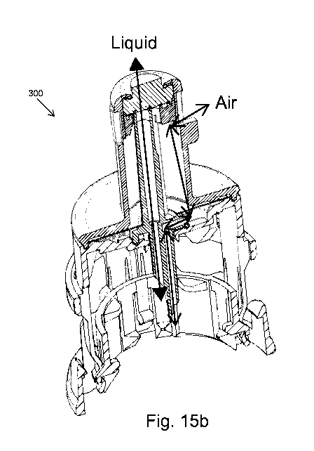
Fig. 15a is a cross-sectional view schematically showing a vial adapter 300
that is adapted for
use in an open transfer system. Vial adapter 300 comprises two parts a top
part 304 and a
bottom part 302. The structure of these two parts of vial adapter 300 and the
telescopic way in
which they lock together when connected to a drug vial is similar in most
respects to the
corresponding parts of vial adapter 200 described herein above with relation
to Fig. 5a to Fig.
12.
In contrast to the closed system vial adapter 200, the vial adapter 300
comprises only one
conduit ¨ liquid conduit 308 ¨ that passes through the entire vial adapter
from the bottom of
septum 322, which rests on septum seat 310 and seals the top of the vial
adapter, through
upwardly projecting structure 306, to the tip of spike 312.
CA 03148420 2022-01-21
WO 2021/019532
PCT/IL2020/050829
- 27 -
Vial adapter 300 comprises a hydrophobic filter 316. The filter is made of a
thin disc shaped
piece of hydrophobic material. A hole is cut through it to allow free passage
of liquid through
liquid conduit 308. The filter 316 is placed between a plurality of closely
spaced supporting ribs
from above and below and its outer and inner edges are welded, glued, or
mechanically
pressed to the top part 304 of vial adapter as described herein above with
respect to Fig. 4.
An air channel 314 through the spike terminates in an open space 324 beneath
filter 316. The
interior of the upwardly projecting structure 306 comprises a hollow air
chamber 318
surrounding liquid conduit 308. Air chamber 318 is sealed at the top by septum
322 and at the
bottom sealed to prevent the entrance of liquid by filter 316. A vent hole 320
near the top in
the side of upwardly projecting structure 306 above filter 316 allows fluid
communication
between the interior of air chamber 318 and the air outside of the vial
adapter.
Fig. 15b schematically shows the paths of two-directional flows of liquid and
air through the
vial adapter of Fig. 15a.
Fig. 16a and Fig. 16b show alternative locations in vial adapter 300 for the
vent hole 320, which
can be located at any place proximally, i.e. above or beyond, filter 316. A
person skilled in the
art can place and shape the venting feature in various places and ways.
Fig. 17 shows another embodiment of vial adapter designed for use with an open
transfer
system. It is identical to vial adapter 15 shown in Fig. 4 with the exception
that the air channel
56 has a vent hole 402 in its side that allows unhindered fluid communication
between the
interior of air channel 56 and the exterior of vial adapter 400. Vent hole 402
is located above
filter 66. Pressure equalization takes place in vial adapter 400 exactly as
described for vial
adapter 300 described with reference to Fig. 15a and Fig. 15b.
Fig. 18a shows an open transfer system partially assembled for use. The system
comprises a
vial adaptor 300 (see Fig. 15a) that is attached to a drug vial 16 and a
conventional syringe 450
that is attached to an open system connector 452.
Fig. 18b shows the cross-sectional view of the open transfer system of Fig.
18a. Shown in Fig.
18b are: conventional syringe 450, connector 452, and vial 16 with attached
vial adapter 300.
CA 03148420 2022-01-21
WO 2021/019532
PCT/IL2020/050829
- 28 -
Also shown are upwardly projecting structure 306, septum 322, liquid channel
308, and vent
hole 320 of vial adapter 300.
Fig. 18c shows connector 452, which is similar to the prior art connector
section 14 with the
following modifications: (a) the double membrane seal actuator 34 shown in
Fig. la is replaced
with a septum holder 500 (shown in Fig. 14) comprising septum 572 at its
bottom; and (b)
there is only one needle 454 acting as a liquid conduit within connector 452.
Connector 452 is
shown in its blocked configuration.
Fig. 19a shows the open transfer system of Fig. 18a in its fully assembled
configuration after
vial adapter 300 is connected to a drug vial and spike 312 has penetrated the
membrane at the
top of the vial as described herein above with reference to Figs. 8-11. The
vial adaptor 300
with attached drug vial 16 is connected to the conventional syringe 450 by
means of connector
452.
Fig. 19b is the cross-sectional view of the open transfer system of Fig. 19a.
The Fig. 19c is a
zoom-in of section A in Fig. 19b focusing on the vial adaptor with the
connected syringe
connector.
Using the open transfer system shown in Figs. 18a-19c, a drug in powdered form
can be
reconstituted by filling a conventional syringe 450 with the required amount
of diluent, the
syringe connector 452, which is connected to the syringe, is then pushed down
over the
upwardly projecting structure 306 of the open system vial adapter (Figs. 18a
and 18b) until the
connection is established as shown in Figs. 19a through 19c at which time the
needle 454 of
the connector 452 has penetrated through both the septum 572 of the septa
holder in
connector 452 and the septa 322 of the vial adaptor and has entered liquid
conduit 308 in the
vial adapter.
After the connection is established the piston of the syringe 450 can be
pushed downward
forcing the liquid diluent to flow through needle 454 in the connector and
liquid conduit 308 in
the vial adapter into the interior of the vial (arrow B). As liquid enters the
vial air is displaced
and pressure is equalized by air flowing out of the vial through air channel
314 through
hydrophobic filter 316 into air chamber 318 and out of the vial adapter
through vent hole 320
(arrow C).
CA 03148420 2022-01-21
WO 2021/019532
PCT/IL2020/050829
- 29 -
To draw liquid out of a drug vial the connected vial and syringe connected as
shown in Figs.
19a-19c are flipped and inverted upside down so that the vial is located above
the syringe.
Following the inversion the piston of the syringe can be pulled downward
drawing the liquid
out of the interior of the vial through liquid conduit 308. As the liquid is
drawn out of the vial a
partial vacuum is created in the vial, which is equalized by suction which
draws air into the vial
from outside of vial adapter 300 through vent hole 320, air chamber 318,
filter 316, and air
channel 314.
As mention above, the components of the closed systems can be used when
compounding and
filling prescriptions of hazardous and non-hazardous drugs; however the
components of the
open systems can be used only for non-hazardous drugs. In order to prevent
interchangeability
of the open and closed system components the applicant uses a different
configuration of
connecting elements to connect the components of each system.
Fig. 20a and Fig. 20b schematically illustrate the elements that allow
connecting together two
components of an open transfer system and prevent an open transfer component
from
connecting with a closed transfer component. For illustrative purposes an open
system septum
holder 600, which is a component of a connector section, is to be connected to
the upwardly
projecting structure 306 of an open system vial adapter (see Fig. 15a) and the
upwardly
projecting structure 220 of a closed system vial adapter (see Fig. 6).
Septum holder 600 is identical to septum holder 500 shown in Fig. 14 with the
exception of the
distal end of the inner facing side of the arms 662 that are connected to the
body part of the
septum holder. Septum 672 is shown fitted over the septum support. On the
outer side of
arms 662 are distal enlarged elements 668 and on the inner side of the arms
opposite the
enlarged elements 668 are distinctively shaped protrusions 602 comprising for
example as
shown, vertical and horizontal bars in the shape of an inverted letter L. As
shown in Fig. 20a,
the upwardly projecting structure 306 comprises a socket 604 in the shape of
an inverted
letter "L" on its side below septum 622. Socket 604 has a shape and dimensions
which match
those of the distinctively shaped protrusions 602 on the arms of the septum
holder allowing
protrusions distinctively shaped 602 to fit into sockets 604 connecting the
septum holder to
the vial adapter. On the other hand, the components of the closed system
comprise
protrusions and sockets having other shapes than those of the components of an
open system,
CA 03148420 2022-01-21
WO 2021/019532
PCT/IL2020/050829
- 30 -
for example for a closed system, the protrusion on the arms could be a
vertical bar and the
socket a vertical slot. In this case, as shown in Fig. 20b, the horizontal bar
at the top of
distinctively shaped protrusion 602 will prevent protrusion 602 from entering
vertical socket
606 on the upwardly projecting structure 220 of the closed system vial
adapter, thereby
preventing connecting the open system septum holder to the closed system vial
adapter. It is
noted that the shapes of the protrusions and sockets described are for
illustrative purposes
only and many other distinctive shapes could be used for the same purpose.
Fig. 21a schematically shows a spike adapter 700 used in conjunction with
fluid transfer
apparatus 10 to transfer a drug to and from an intravenous (IV) bag. Spike
adaptor 700
comprises body 762 terminating in a spike element 764 at the proximal end and
a standard
port 766 for connecting an infusion set at the distal end. Substantially at
right angles to body
762 is a longitudinal extension 768. At the end of longitudinal extension 768
are membrane
enclosure 770 and membrane 772. On the side of longitudinal extension 768
below membrane
.. enclosure 770 is a socket 604 configured to match with the distinctively
shaped projections on
the arms of a septum holder in a connector as shown in Fig. 20a. A connector
section, e.g.
connector 452 (see Fig. 18) with attached conventional syringe can connect to
longitudinal
extension 768 exactly as described herein above with respect to connection to
vial adaptor 300
in Figs. 19a-19c, thereby allowing insertion of a drug from the syringe into
an IV bag or
withdrawal of liquid from an IV bag into a syringe to be used for
reconstitution of a drug.
Fig. 21b is a cross-sectional view of the spike adapter. In this figure can be
seen that the
interior of spike adapter 700 comprises two separated channels 774 for liquid
and 776 for air.
In this open system spike adapter liquid channel 774 passes from the tip of
spike element 764
to membrane 772 for use if liquid is to be transferred into or out of the IV
bag from a syringe.
Channel 776 passes from the tip of the spike to port 766 to transfer liquid
from the IV bag to
the patient. In an open system for injection or withdrawal of liquid from a
syringe to an IV bag
there is no need for venting because, unlike a stiff glass vial, an IV bag is
flexible allowing it to
expand when pressurized or contract when it is evacuated.
The apparatus for securing a male-female connection described with respect to
figures 23-29
can easily be modified mutatis mutandis for use with an open drug transfer
system. For the
open system, the female connector, e.g. the connector section 452 in Figs. 18a-
18c would have
only one needle and septum holder 500 could be replaced with the ladders,
gears, and other
CA 03148420 2022-01-21
WO 2021/019532
PCT/IL2020/050829
- 31 -
features of female connector 1201. The vial adapters vial adapters of Figs.
15a and 17 and the
spike adapter of Fig. 21a would also be modified such that their upwardly
projecting structures
306, 46, and 768 would have smooth sides and two anchoring ledges 1223 on
opposite sides
near the top.
Referring to Fig. 27a, it can be seen how the components are configured to
prevent connection
of open and closed system components together. For example, for a closed
system the ledge
1223 can be wider than the gap 1406 in gear 1405 of the securing actuator 1401
for an open
system, thereby preventing connection of a closed system vial adapter to an
open system
connector 1201. Alternatively, for an open system the ledge 1223 can be wider
than the gap
1406 in gear 1405 of the securing actuator 1401 for a closed system, thereby
preventing
connection of an open system vial adapter to a closed system connector 1201.
The components of an open system described herein have been developed for use
in a robotic
system that can be installed in hospital pharmacies to assist in the
compounding of
medications comprising non-hazardous drugs and to prepare syringes and IV bags
comprising
the required amount of liquid drug for administration to patients according to
their individual
prescriptions. The robotic system is similar to the one described in the
background section for
use with hazardous drugs and shown in Fig. 22. In compliance with regulations
the two robotic
systems will be kept in separate rooms in the pharmacy.
For non-hazardous drugs the safety requirements are much less restrictive;
however, exactly
as in the case of the system for hazardous drugs, the system comprises at
least two robotic
arm assemblies configured to simultaneously move vials and syringes within the
cabinet. Each
of the robotic arm assemblies comprises three mechanical arrangements
configured to
independently move either a vial gripper assembly or a syringe gripper
assembly and syringe
pump in three dimensions along three mutually orthogonal beams. Within the
laminar flow
cabinet is a plurality of operational stations adapted to perform specific
tasks related to the
compounding process. The operating stations include: at least one
reconstitution module; at
least one vial shaker module; at least one vial flipper module; at least one
IV bag base module
to which the operator of the system can attach IV bags; a syringe magazine; a
plurality of
cameras each installed at a specific location in the cabinet or on the robotic
arm assemblies,
and a processor. Each of the cameras is dedicated to provide real time digital
images of the
stage of the preparation process carried out at its location. Dedicated
software and algorithms
CA 03148420 2022-01-21
WO 2021/019532
PCT/IL2020/050829
- 32 -
in the system processor allow almost all steps in the compounding process to
be carried out
automatically by the robotic arm assemblies without intervention by the
operator or a
supervisor and the cameras and imaging process algorithms are adapted to
provide real-time
feedback control of all stages of the compounding process.
One important difference between the robotic system developed for the closed
transfer
system and one for use in an open system is that the that closed transfer
system relies on the
use of Equashield syringes that have to be manufactured in perfect orientation
and alignment
with their connectors. This is important because the Equashield syringes will
be gripped and
placed when the connector extending shoulders and the extensions on the
syringe barrel are
always in same position relative to each other and due to this identical
orientation only simple
griping mechanisms are required and processes of placing and handling the
syringes is an easy
and fast task to accomplish. Unlike the well aligned Equashield syringes, the
open transfer
system uses conventional syringes from various manufacturers and various
shapes and
dimensions, and the connector shoulders on the arms and the extensions on the
syringe barrel
are seldom in same position relative to each other, a fact that requires
special mechanisms
integrated into the robot to grip the connector and the syringe in varying
orientations. This
also requires software that can deal with various syringes, various
orientations, identifying
them and reading the right dosage.
In using the robotic system, the prescriptions to be filled are entered into
the system
processor, which prompts the user to insert drug vials containing the required
medicines into
the cabinet, to load syringes of the required sizes into the syringe magazine,
and attach IV bags
to the IV bag base modules.
In order for the robotic arms to be able to grab the vials and syringes, the
user connects a vial
adapter to each vial and a connector section to each syringe before placing
them in the
cabinet. After the drug vials, syringes, and IV bags are placed in the
cabinet, all further
operations of compounding the drugs and preparing the required doses in
syringes or IV bags
for administration to a patient are carried out automatically by the robotic
arms as instructed
by the processor under supervision of the cameras.
In the open transfer robotic system the cameras and software are configured to
recognize the
sockets 604 and protrusions 602 on the vial adapter 220 and septum holder 600
in Figs. 20a
CA 03148420 2022-01-21
WO 2021/019532
PCT/IL2020/050829
- 33 -
and 20b and the gaps 1406 and void portions 1405 in the securing actuator and
ledges 1223 on
the male connector 1221 in Figs. 24 and 26 and to warn the user if the wrong
components are
introduced into the cabinet. Additionally, as a safety feature, the robotic
arm assemblies
comprise mechanical features, e.g. projecting pins that must fit matching
slots on the
components to be picked up, to insure that only the components compatible with
an open
transfer system are being used.
Open transfer components for use with the robotic system constitute two kits ¨
a basic kit will
contain a vial adapter and a connector section and an extended kit that
additionally contains
an IV spike adapter. The kits will come in several embodiments to include vial
adapters suitable
for different sized vials and connectors have different types of connections,
e.g. Luer lock or
bayonet connectors to mate with standard needless syringes.
Although embodiments of the invention have been described by way of
illustration, it will be
understood that the invention may be carried out with many variations,
modifications, and
adaptations, without exceeding the scope of the claims.