Note: Descriptions are shown in the official language in which they were submitted.
FUSED POLYPEPTIDE AND USE THEREOF
TECHNICAL FIELD
The present invention relates to the field of biopharmaceuticals, and in
particular, to a fused
polypeptide and use thereof
BACKGROUND
Fibrosis is a disease that causes a decrease in parenchymal cells of organs
and tissues and an
increase in fibrillar connective tissues increase. Continuous progression of
the disease may lead to
structural damage and hypofunction of organs, and eventually failure, which
seriously threatens
health of patients. Worldwide, fibrosis of tissues and organs is the main
cause of disability and
death in many diseases.
1. Pulmonary fibrosis
Pulmonary fibrosis is a lesion mainly caused by uncontrolled repair and
regulation and
abnormal reconstruction of damaged lung tissues. In this process, oxidative
stress caused by a
series of abnormal expression of cytokines and growth factors, inflammatory
response, vascular
proliferation and reconstruction, fibrinolysis disorder, matrix
metalloproteinases, external
environment, and other factors participates in the pathogenesis of pulmonary
fibrosis. This results
in major lesions such as epithelial cell deficiency, fibroblast proliferation,
and extracellular matrix
(ECM) accumulation. A final result is that fibroblasts replace alveolar
epithelial cells (AECs) that
perform normal functions, leading to the occurrence of fibrosis. The unclear
pathogenesis of IPF
causes great difficulties to the current treatment, but through experimental
research, it can be found
that many potential targets are worthy of attention. Because alveoli and AECs
are damaged, the
body needs to repair the damage, and inflammatory response is also involved.
Once the damage
repair is excessive or abnormal, the release of some cytokines for chemotaxis
and activation of
fibroblasts is caused, and the abnormal proliferation of fibroblasts is
accompanied by the
accumulation of a large number of ECMs, eventually leading to the occurrence
of lPF.
A plurality of types of cells, such as pulmonary epithelial cells, endothelial
cells, pulmonary
inflammatory cells (mainly macrophages), and pulmonary interstitial cells
(fibroblasts and
myofibroblasts), are involved in the occurrence of fibrosis, and the pulmonary
interstitial cells are
key effector cells for the occurrence of pulmonary fibrosis. In addition,
cytokines secreted by cells,
CA 03149590 2022-2-25
such as transforming growth factor-J3 (TGF-J3), a platelet-derived growth
factor (PDGF), a basic
fibroblast growth factor (BFGF), a connective tissue growth factor (CTGF), an
insulin-like growth
factor (IGF), a vascular endothelial growth factor (VEGF), integrin, matrix
metalloproteinase
(MMP), and an inhibitor (TIMP) thereof, also have a profound impact on the
occurrence of
pulmonary fibrosis.
The most critical cytokine is TGF-13, which is a multifunctional cell growth
factor that can
regulate cell proliferation and differentiation. The proliferation of a large
number of
myofibroblasts and the excessive accumulation of the ECM can be stimulated by
directly
stimulating the activation of in situ fibroblasts or through endothelial-
mesenchymal transition
(EnMT) and epithelial-mesenchymal transition (EMT) processes. When TGF-J3 is
continuously
activated due to damage, MAPK, EGF, and Wnt/J3-catenin signals are cross-
activated, leading to
the progression of fibrosis. The PDGF, the BFGF, and the VEGF as growth
factors can promote
the proliferation and differentiation of lung fibroblasts, and affect the
progression of pulmonary
fibrosis. The MMP/TIMP is a main regulator of the ECM, and the contents of the
two play a key
role in the balance of the ECM. These cytokines have a more or less influence
on the proliferation
and activation of lung fibroblasts and the formation of collagen, and
therefore reasonable
regulation of cytokine expression facilitates the treatment of pulmonary
fibrosis.
The polypeptide according to the present invention has a plurality of targets,
can inhibit the
release of TGF-13 I, the proliferation and activation of fibroblasts and the
expression of integrin,
further inhibit the activation of TGF-I31, inhibit angiogenesis and the
expression and release of the
VEGF, treat fibrosis in multiple ways, and slow down the process of fibrosis.
2. Hepatic fibrosis
Hepatic fibrosis is a common pathological change of chronic liver diseases
caused by a
plurality of causes, characterized by excessive synthesis and degradation
reduction of the ECM
that is mainly collagen in liver, and the joint control by a plurality of cell
signal transduction
pathways and a series of signal molecular networks. The activation and
proliferation of hepatic
stellate cells (HSCs) is an ultimate common way to cause hepatic fibrosis and
a central event of
hepatic fibrosis. However, a mechanism of occurrence and progression of
hepatic fibrosis is very
complicated. At present, the research mainly focuses on the activation and
transformation of
hepatic stellate cells into myofibroblasts and fibroblasts. Possible ways are
activation of a TGF-13
signal transduction pathway, a PDGF receptor-mediated signal transduction
pathway, a INF-a-
CA 03149590 2022-2-25
2
mediated signal transduction pathway, cyclooxygenase-2 (COX-2), diffuse ECM,
oxidative stress-
mediated hepatic fibrosis, or the like.
Hepatic fibrosis is a necessary pathological stage for all kinds of chronic
hepatitis to develop
into cirrhosis, and is the manifestation of liver injury self-repair.
According to a WHO report, there
are 20 million cases of hepatitis B virus infection in China, and hepatic
fibrosis has occurred to
most of these patients. Therefore, how to treat hepatic fibrosis has become an
urgent problem to
be resolved.
3. Renal fibrosis
Most chronic renal diseases, such as primary glomerular diseases, chronic
pyelonephritis,
renal damage caused by systemic diseases (such as lupus nephritis and diabetic
nephropathy), and
nephropathy (such as Alport syndrome) caused by genetic factors, may lead to
renal fibrosis. Renal
fibrosis is a pathological process driven by multiple factors, involving
inflammation, oxidative
stress, functions and signal cascade of a plurality of cytokines, cell
apoptosis, proliferation and
activation of fibroblasts, transformation of epithelial cells into
fibroblasts, and the like.
At present, most drugs for the treatment of renal fibrosis have problems such
as high toxicity,
low safety, and single pharmacological actions.
Polypeptide drugs have higher druggability than general chemical drugs, have
high biological
activity, high specificity and relatively weak toxic reaction, and do not
easily accumulate in the
body. A polypeptide may be designed according to its pathogenesis, is under a
multi-target design,
and can inhibit the occurrence of renal fibrosis in multiple ways.
4. Skin fibrosis
Skin fibrosis is excessive scar formation of skin and a result of pathological
wound healing
response. For many years, scholars at home and abroad have made in-depth
research on the
mechanism of scar occurrence, progression and regression from multiple angles
and levels, but up
to now, no clear conclusion is reached on its mechanism, and no effective way
for prevention and
treatment is available. Relatively consistent views are as follows: 0
Fibroblasts are main effector
cells of skin fibrosis, which are characterized by excessive cell
proliferation and excessive
deposition of the extracellular matrix.
Collagen metabolism
disorder is a main biological
manifestation of the skin fibrosis. 0 A TGF-I31/Smad signaling pathway is
closely related to a
plurality of physiological and pathological processes such as proliferation,
differentiation,
migration, apoptosis, and collagen metabolism of fibroblasts. Smads regulate
collagen metabolism
CA 03149590 2022-2-25
3
of fibroblasts bidirectionally according to different types.
The most common method used to treat skin fibrosis is immunosuppressive
therapy The basic
principle is that autoimmune causes inflammation of diseases and subsequent
tissue damage and
fibrosis. Commonly used drugs include methotrexate, cyclophosphamide, and
cyclosporine.
Although some improvements in immunosuppressive therapy have been observed,
concerns about
the safety of the drugs and the lack of confirmed clinical data and
demonstrable efficacy still exist.
Therefore, it is necessary to develop an effective pharmaceutical preparation
for the treatment of
skin fibrosis, fibrotic skin diseases and pathological scar formation of the
skin.
5. Myocardial fibrosis
Myocardial fibrosis refers to that under the action of various pathogenic
factors (such as
inflammation, ischemia, and hypoxia), collagen fibers in the normal tissue
structure of
myocardium are excessively accumulated, the collagen concentration in the
heart tissue
significantly increases or the collagen composition in the heart tissue
changes. Myocardial fibrosis
is an important pathological change in the progression of a plurality of
cardiovascular diseases,
and a final result is myocardial remodeling, stiffness of myocardium, decrease
of a ventricular
diastolic function, decrease of coronary artery reserves, or even sudden death
that may be directly
caused. Therefore, prevention and treatment of myocardial fibrosis is of great
significance.
SUMMARY
1. To-be-resolved Problem
In view of most of existing drugs for treating fibrosis are chemical drugs,
and the chemical
drugs have problems such as high toxicity, low safety, and single
pharmacological actions, the
present invention provides a fused polypeptide, which has a good therapeutic
effect on lung
fibrosis, hepatic fibrosis, renal fibrosis, myocardial fibrosis, and skin
fibrosis, and in inhibiting the
proliferation of various human tumor cells. The polypeptide according to the
present invention
contains a plurality of domains, which can target a plurality of targets, and
inhibit the occurrence
of fibrosis and the proliferation of tumors in multiple ways.
2. Technical Solutions
To resolve the foregoing problems, technical solutions adopted by the present
invention are
as follows:
A fused polypeptide with multifunctional activity, where the polypeptide
contains the
CA 03149590 2022-2-25
4
following domains:
N-Acetyl-S er-Asp -Lys-Pro, S er-Asp -Lys-Pro, Thr-Ser-Leu-Asp-Ala-S er-Ile-
Ile-Trp-Ala-
Met-Met-Gln-Asn, and Leu-Ser-Lys-Leu, or domains in which any amino acid in
the foregoing
domains is mutated.
The fused polypeptide is linked by a linker, and the linker is a flexible
linker composed of
Gly-Gly-Gly-Gly, Ser-Ser-Ser or other amino acids.
Preferably, an amino acid sequence of the polypeptide is as follows:
polypeptide I: Ser-Asp-Lys-Pro-linker-Leu-Ser-Lys-Leu-linker-Thr-Ser-Leu-Asp-
Ala-Ser-
Ile-Ile-Trp-Ala-Met-Met-Gln-Asn;
polypeptide II: Ser-Asp-Lys-Pro-linker-Thr-Ser-Leu-Asp-Ala-Ser-Ile-Ile-Trp-Ala-
Met-Met-
Gln-Asn-linker-Leu-Ser-Lys-Leu;
polypeptide III: Thr-S er-Leu-Asp-Ala-Ser-Ile-Ile-Trp-Ala-Met-Met-Gln-Asn-
linker-S er-
Asp-Lys -Pro-linker-Leu- Ser-Lys-Leu;
polypeptide IV: Thr-Ser-Leu-Asp-Ala-Ser-Ile-Ile-Trp-Ala-Met-Met-Gln-Asn-linker-
Leu-
S er-Lys-Leu-linker-S er-Asp -Lys -Pro ;
polypeptide V: Leu-Ser-Lys-Leu-linker-Ser-Asp-Lys-Pro-linker-Thr-Ser-Leu-Asp-
Ala-Ser-
Ile-Ile-Trp-Ala-Met-Met-Gln-Asn; and
polypeptide VI: Leu-S er-Lys-Leu-linker-Thr- S er-Leu-Asp -Ala- S er-Ile-Ile-
Trp -Ala-Met-
Met-Gln-Asn-linker- Ser-Asp-Lys-Pro ;
where the linker is Gly-Gly-Gly-Gly; and
use of the fused polypeptide in the preparation of anti-pulmonary fibrosis,
anti-hepatic
fibrosis, anti-renal fibrosis, anti-myocardial fibrosis, and anti-skin
fibrosis drugs and antitumor
drugs is provided.
The foregoing tumors include human head and neck cancer, brain cancer, thyroid
cancer,
esophageal cancer, pancreatic cancer, liver cancer, lung cancer, gastric
cancer, breast cancer,
CA 03149590 2022-2-25
kidney cancer, colon cancer or rectal cancer, ovarian cancer, cervical cancer,
uterine cancer,
prostate cancer, melanoma, hemangioma, and sarcoma.
Mechanism of action: The polypeptide according to the present invention has a
plurality of
targets, and can inhibit the release of TGF-I31, the expression of integrin
and angiogenesis, inhibit
the activation of fibroblasts in multiple ways, reduce the release of
cytokines and the deposition
of the extracellular matrix, slow down the foregoing fibrosis process, and
further inhibit the
proliferation of a plurality of types of human tumor cells.
3. Beneficial Effects
Compared with the prior art, the present invention has the following
beneficial effects:
(1) The fused polypeptide according to the present invention has excellent
anti-fibrosis
activity and can be used for treating a plurality of fibrosis diseases,
including pulmonary fibrosis,
hepatic fibrosis, renal fibrosis, myocardial fibrosis, and skin fibrosis.
Components of the fused
polypeptide are all natural amino acids, which are easy to synthesize, have no
obvious toxic or
side effects, and have high safety.
(2) The fused polypeptide according to the present invention can be used for
treating
pulmonary fibrosis, and in a pulmonary fibrosis model, the polypeptide can
significantly improve
the structure of the lung, lower a score of pulmonary fibrosis, and improve
the survival rate.
(3) The fused polypeptide according to the present invention can be used for
treating hepatic
fibrosis, and in an in vitro hepatic fibrosis model, the polypeptide can
inhibit the proliferation and
activation of hepatic stellate cells.
(4) The fused polypeptide according to the present invention can be used for
treating renal
fibrosis. In a renal fibrosis model, the polypeptide can significantly reduce
the expression content
of TGF-I31 in renal tissues and significantly improve a situation of renal
fibrosis.
(5) The fused polypeptide according to the present invention can be used for
treating
myocardial fibrosis, and in an in vitro myocardial fibrosis model, the
polypeptide can significantly
reduce the activation and proliferation of myocardial fibroblasts.
(6) The fused polypeptide according to the present invention can be used for
treating skin
fibrosis. In a skin fibrosis model, the polypeptide can significantly reduce
the expression content
of HYP in skin and significantly improve a situation of skin scar hyperplasia.
(7) The fused polypeptide according to the present invention can inhibit the
growth of a
plurality of types of tumor cells.
CA 03149590 2022-2-25
6
(8) The polypeptide according to the present invention is a multi-target drug,
and can inhibit
the process of fibrosis in multiple ways.
BRIEF DESCRIPTION OF DRAWINGS
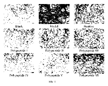
FIG. 1 is a diagram of HE staining of pulmonary fibrosis treated with fused
polypeptides I,
II, III, IV, V, and VI according to the present invention;
FIG. 2 is a diagram of Masson staining of pulmonary fibrosis treated with the
fused
polypeptides I, II, III, IV, V, and VI according to the present invention;
FIG. 3 shows that fused polypeptides I, II, III, IV, V and VI according to the
present invention
inhibit the expression content of TGF-131 in a renal fibrosis model;
FIG. 4 shows that fused polypeptides I, II, III, IV, V and VI according to the
present invention
inhibit the expression content of HYP in a skin fibrosis model; and
FIG. 5 shows inhibitory effects of the fused polypeptides I, II, III, IV, V,
and VI according to
the present invention on the growth of different types of tumors.
DETAILED DESCRIPTION
The polypeptides I, II, III, IV, V, and VI were synthesized by GenScript
(Nanjing) Co., Ltd.
Example 1 Pulmonary fibrosis animal model
Experimental animals and materials:
1. Experimental animals:
Source and strain: clean SD rats, provided by Comparative Medicine Center of
Yangzhou
University (laboratory animal production license: SCXK (Su) 2012-0004);
Laboratory Animal Use
License: SYXK (Su) 2012-0035).
Weight: 180-200 g at the time of purchase and 190-210 g at the beginning of
modeling.
Gender: Male.
2. Experimental materials:
Bleomycin
Manufacturer: Han Hui
Pharmaceutical Co.,
Ltd.
Normal saline
Manufacturer: Anhui Double-Crane
Pharmaceutical Co., Ltd.
Chloral hydrate
Manufacturer: Sinopharm
Chemical Reagent
Co., Ltd.
BIBF1120 (Nintedanib)
Manufacturer: Jinan
Synovel Chemical Co.,
CA 03149590 2022-2-25
7
Ltd.
Tissue fixative
Manufacturer: Wuhan servicebio
Co., Ltd.
3. Experimental method:
SD rats were anesthetized by intraperitoneal injection of 1 mL/100 g 4%
chloral hydrate.
After anesthesia, the rats were fixed and their necks were disinfected by
using cotton with 75%
alcohol. The skin of the rat neck was longitudinally cut with scissors, and
the fascia and muscle
were longitudinally bluntly torn with tweezers to expose the trachea. A
syringe was inserted into
the trachea to inject 5 mg/kg bleomycin, while a blank group was injected with
an equal amount
of normal saline. Then a rat plate was quickly erected and rotated, the rats'
breathing was observed,
the neck wound was sterilized after rotation and was sewn, and an amoxicillin
anti-inflammatory
drug was sprinkled on the suture. After the operation, the rats were put back
into a dry and clean
cage for resting, waiting was performed for awakening. The rats were awakened
after about 1-2
hours, and then fed normally. On the 7t11 day after modeling, modeling group
animals randomly
fell into a model group, a Nintedanib positive drug group, polypeptide I, II,
III, IV, V, VI dosage
groups, and a normal control group, and the groups were administered
separately for an
administration cycle of 14 days. Living situations of rats were observed every
day and their
weights were weighed. After administration for 14 days, the SD rats were
dissected, the lung tissue
was taken, and the right lung tissue was placed in a tissue fixative only for
fixation, and HE staining
and Masson staining and slice analysis were performed.
4. Experimental grouping and dosage setting
Table 1 Experimental grouping and dosage regimen
Administration
Administratio
Group Drug Dosage
Quantity
mode
n frequency
Subcutaneous
Blank group Normal saline 0.5 mL/200 g
Twice a day 10
injection
Subcutaneous
Model group Normal saline 0.5 mL/200 g
Twice a day 10
injection
Intragastric
Positive drug N intedanib 25 mg/kg
Once a day 10
administration
Test drug (1) Polypeptide 1 10 mg/kg
Subcutaneous Twice a day 10
ection
Test drug (2) Polypeptide 11 10 mg/kg
SubcutaneousTwice a day 10
inj ection
ub
Test drug (3) Polypeptide 111 10 mg/kg
ScutaneousTwice a day 10
inj ection
Test drug (4) Polypeptide IV 10 mg/kg
SubcutaneousTwice a day 10
inj ection
Test drug (5) Polypeptide V 10 mg/kg
Subcutaneous Twice a day 10
ection
Test drug (6) Polypeptide VI 10 mg/kg
Subcutaneous Twice a day 10
CA 03149590 2022-2-25
8
injection
4. Experimental results
(1) Impact of a polypeptide on the survival rate of SD rats induced by
bleomycin
As shown in Table 2, compared with the survival rate (50%) of SD rats in the
model group,
the survival rate of SD rats in each test drug group was higher than that of
the model group, and
each test drug could significantly increase the survival rate of SD rats, and
the survival rate of the
polypeptide I group was equivalent to that of the positive drug group.
Table 2 Impact of a polypeptide on survival rate (%) of SD rats with bleomycin-
induced pulmonary fibrosis
Group Dosage Number of
animals Number of animals Survival rate (%)
(mg/kg) at the
beginning at the end
Blank group 10
10 100
Model group 10
5 50
Positive drug 10 10
9 90
group
Polypeptide I 10 10
9 90
Polypeptide 11 10 10
8 80
Po lyp eptide 111 10 10
8 80
Polyp eptide IV 10 10
8 80
Polypeptide V 10 10
7 70
Polypeptide VI 10 10
7 70
2. Pathological analysis of a polypeptide on bleomycin-induced pulmonary
fibrosis in SD rats
Research results showed that a pulmonary fibrosis model in SD rats was
successfully
established in this study. Main manifestations of lung tissue lesions are
fibroblast proliferation and
collagen fiber formation in the alveolar wall and mesenchyme around
intrapulmonary bronchi and
vascular branches. Masson staining showed blue-green staining reaction, and
inflammatory cell
infiltration, congestion in the alveolar wall, cell degeneration disorder and
other lesions occurred.
After administration, the degree of pulmonary fibrosis and other lesions were
less than those in
the model group. See FIG. 1 and FIG. 2 for HE staining and Masson staining.
Example 2 In vitro hepatic fibrosis model
1. Experimental method
The inhibitory effect of a polypeptide on LX-2 hepatic stellate cells was
detected by MTT
assay Cells were cultured in a 1640 medium containing 10% of FBS, the
cytoplasm was made
into 4 x 10⁵/mL cell suspension, and 100 FL per well was inoculated into a 96-
well plate. After
the cells adhered to the wall, the medium was replaced with a serum-free 1640
medium, and the
serum-free medium was discarded after 24 hours. The cells were cultured with
different
polypeptides of 1 p.mol/L, and 5 multiple wells were set for each
concentration. After 12, 24 and
CA 03149590 2022-2-25
9
48 hours separately, 10 'IL of MTT was added to each well. After 4 hours, MTT
was sucked out,
and 150 'IL of DMSO was added to each well. After reaction for 5 min, an OD
value was measured
at 570 nm by a microplate reader.
2. Experimental results
At 24 hours and 48 hours, polypeptides I, H, III, IV, V, and VI could inhibit
the proliferation
of cardiac fibroblasts of rats at 1 ilmol/L. The results are shown in Table 3:
Table 3 Impact of a polypeptide on the proliferation of LX-2 hepatic stellate
cells
Optical density values at different time points
Group
12h
24h 48h
Blank group 0.456+0.012
0.548+0.01 0.812+0.016
Polypeptide 1 (1 innol/L) 0.452+0.008
0.542+0.03 0.68010.014"K
Polypeptide 11 (1 mon) 0.463+0.012
0.394+0.005K" 0.57810.005"K
Polypeptide 111(1 mon) 0.455+0.002
0.435+0.013" 0.642+0.018'
Polypeptide IV (1 union) 0.478+0.018
0.472+0.03 0.580+0.02***
Polypeptide V (1 mon) 0.462+0.004
0.477+0.015" 0.61810.015K
Polypeptide VI (1 innol/L) 0.453+0.021
0.502+0.013K 0.652+0.018'
***P < 0.001, **P < 0.01, *P < 0.05 VS control.
Example 3 Establishment of a renal fibrosis model
1. Experimental animals
Clean grade male SD rats, purchased from Nanjing Qinglong Mountain Animal
Farm, and
weighed 180-200 g at the time of purchase, 190-210 g at the beginning of
modeling, and 180-
200 g at the beginning of administration.
2. Experimental materials:
Normal saline Manufacturer: Anhui Double-Crane Pharmaceutical Co., Ltd.
Rat TGF-131 ELISA kit Manufacturer: Tianjin Annuo Ruikang Biotechnology Co.,
Ltd.
3. Experimental method
A renal fibrosis animal model was established. SD rats were anesthetized with
4% chloral
hydrate, injected with 1 mL/100 g intraperitoneally, fixed to an operation
board, and sterilized in
an operation area for later use. The abdominal cavity was cut open about 3-4
mm to the left of the
ventrimeson, left kidney ureter was separated in an operation group, the
ureter was ligated and
separated close to the ureter near the lower pole of the inferior pole of
kidney, and the ureter was
cut short between two ligations after the double ligations. Muscular layers
and abdominal walls
were sewed layer by layer, the suture was disinfected with alcohol. After SD
rats woke up, the rats
CA 03149590 2022-2-25
were put into a cage for feeding. In the blank group, ureter was not ligated,
and other steps were
the same.
Then, the animals fell into a blank group, a model group, and polypeptide
administration
groups, with 10 animals in each group, and the administration was started on
the second day after
the operation, twice a day for 14 days. After administration for 14 days,
blood was taken and
supernatant was taken to detect the content of TGF-I31 in serum.
4. Experimental grouping and dosage setting
Table 4 Experimental grouping and dosage regimen
Group Drug Dosage
Administration mode Administration Quantit
frequency
Blank group Normal saline 0.5 mL/200 g
Subcutaneous Once a day 10
injection
Model group Normal saline 0.5 mL/200 g
Subcutaneous Once a day 10
injection
Test drug (1) Polypeptide 1 7.5 mg/kg
Subcutaneous Twice a day 10
injection
Test drug (2) Polypeptide 11 7.5 mg/kg
Subcutaneous Twice a day 10
injection
Test drug (3) Polypeptide 111 7.5 mg/kg
Subcutaneous Twice a day 10
injection
Test drug (4) Polypeptide 7.5 mg/kg
Subcutaneous Twice a day 10
IV
injection
Test drug (5) Polypeptide V 7.5 mg/kg
Subcutaneous Twice a day 10
injection
Test drug (6) Polypeptide 7.5 mg/kg
Subcutaneous Twice a day 10
VI
injection
5. Experimental results
(1) Impact of a polypeptide on the content of TGF-I31 in serum of SD rats with
renal fibrosis
TGF-131 is the most important fibrogenic factor. In renal fibrosis, the
expression of TGF-131
was significantly increased. The result is shown in FIG. 3, and there was a
highly significant
difference between the model group and the blank group (***P < 0.001). After
administration, all
groups could significantly reduce the content of TGF-131 in serum, and the
polypeptide I group,
the polypeptide IT group and the polypeptide IV group were highly
significantly different from the
model group (***P < 0.001), and the polypeptide III group, the polypeptide V
group and the
polypeptide VI group were highly significantly different from the model group
(**P< 0.01).
Example 4 Establishment of a myocardial fibrosis model
1. Experimental method
The inhibitory effect of a polypeptide on cardiac fibroblasts of rats was
detected by MTT
assay Cells were cultured in a DMEM medium containing 10% of FBS, the
cytoplasm was made
CA 03149590 2022-2-25
11
into 1 x 10⁵/mL cell suspension, and 100 'IL per well was inoculated into a 96-
well plate. After the
cells adhered to the wall, the medium was replaced with a serum-free DMEM
medium, and the
serum-free medium was discarded after 24 hours. The cells were cultured with
different
polypeptides of 1 pmol/L, and 5 multiple wells were set for each
concentration. After 12, 24 and
48 hours separately, 10 pLL of MIT was added to each well. After 4 hours, MTT
was sucked out,
and 150 'IL of DMSO was added to each well. After reaction for 5 min, an OD
value was measured
at 570 nm by a microplate reader.
2. Experimental results
At 24 hours and 48 hours, polypeptides I, II, III, IV, V. and VI could inhibit
the proliferation
of cardiac fibroblasts of rats at 1 ilmol/L. The results are shown in Table 5.
Table 5 Impact of a polypeptide on the proliferation of cardiac fibroblasts of
rats
Group Optical
density values at different time points
2h
24h 48h
Blank group 0.353+0.001
0.464+0.018 0.896+0.001
Polypeptide 1 (1 gmol/L) 0.362+0.006
0.402+0.002' 0.678+0.002a
Polypeptide 11 (1 mon) 0.352+0.004
0.367+0.016' 0.568+0.013'
Polypeptide 111(1 mon) 0.349+0.012
0.413+0.003' 0.612+0.018a
Polypeptide IV (1 gmol/L) 0.362+0.015
0.392+0.008' 0.583+0.012'
Polypeptide V (1 mon) 0.357+0.024
0.397+0.015' 0.588+0.019'
Polypeptide VI (1 gmol/L) 0.340+0.012
0.412+0.005' 0.622+0.007'
***P < 0.001, **P < 0.01, *P < 0.05 VS control.
Example 6 Establishment of a skin fibrosis model
1. Experimental animals
Male C57/13L black mice aged 6-8 weeks, purchased from Nanjing Qinglong
Mountain
Animal Farm.
2. Experimental materials
Bleomycin
Manufacturer: Han Hui
Pharmaceutical Co.,
Ltd.
Normal saline
Manufacturer: Anhui Double-Crane
Pharmaceutical Co., Ltd.
Rat TGF-I31 ELISA kit
Manufacturer: Tianjin
Annuo Ruikang
Biotechnology Co., Ltd.
Alkaline HYP kit
Manufacturer: Nanjing Jiancheng
Bioengineering Institute
CA 03149590 2022-2-25
12
3. Modeling method
Bleomycin (10 p.g/mL) was injected subcutaneously every day for 28 days to
form skin
fibrosis. During the modeling period, the administration groups were given
polypeptide drugs
twice a day for treatment. After modeling, the mice were killed on the next
day, and the skin tissue
of the mouse back was taken to detect the content of HYP in the skin tissue.
4. Experimental grouping and dosage regimen
Table 6 Experimental grouping and dosage regimen
Administration Administration Quantity
Group Drug Dosage
mode
frequency
Subcutaneous
10
Blank group Normal saline 0.2 nit
Twice a day
injection
Subcutaneous
10
Model group Normal saline 0.2 nit
Twice a day
injection
Subcutaneous
10
Test drug (1) Polypeptide 1 10 mg/kg
inection Twice a day
j
Subcutaneous
10
Test drug (2) Polypeptide 11 10 mg/kg
injection Twice a day
Subcutaneous
10
injection
Test drug (3) Polypeptide III 10 mg/kg
Twice a day
Test drug (4) Polypeptide IV 10 mg/kg
Subcutaneous Twice a day 10
inj ection
Subcutaneous
10
injection
Test drug (5) Polypeptide V 10 mg/kg
Twice a day
Test drug (6) Polypeptide VI 10 mg/kg
Subcutaneous Twice a day 10
inj ection
5. Experimental results
(1) Expression of HYP content in the skin tissue of each group of mice
The content of hydroxyproline in the skin tissue of the mouse back was
detected. As the
characteristic protein of collagen, hydroxyproline can reflect the content of
collagen in the skin
tissue from the side. As shown in FIG. 4, each polypeptide group could reduce
the expression of
HYP in the skin tissue. The polypeptide II group, the polypeptide IV group and
the polypeptide VI
group could significantly reduce the expression of HYP in the lung tissue, and
were highly
significantly different from the model group (***P < 0.001). The polypeptide I
group, the
polypeptide III group and the polypeptide V group could reduce the content of
HYP in the lung
tissue of SD rats, and were highly significantly different from the model
group (*P < 0.05).
Example 7 Inhibitory effect of a polypeptide according to the present
invention on the
growth of tumor cells from a plurality of sources detected by using MTT assay
A plurality of types of human tumor cells were cultured in a 5% CO2 incubator
at 37 C and
digested with trypsin when the density was 90% or above. The cells were
resuspended in a culture
solution and counted, and the cell concentration was adjusted to 2 X 10⁴
cells/mL. The cell
CA 03149590 2022-2-25
13
suspension was inoculated into a 96-well plate with 100 pit per well, and then
cultured overnight
in a 5% CO2 incubator at 37 C. After the cells completely adhered to the wall,
each polypeptide
according to the present invention was added as an administration group, and
the culture solution
without any drug was used as a blank control group. The solutions were diluted
to 1 p.mol/L by
using a diluent. Each diluent was separately added to the 96-well plate with
100 RI, per well, and
the cells continued to be cultured in a 5% CO2 incubator for 48 hours at 37 C.
Then 20 RI, of MIT
was added, and the cells continued to be cultured for 4 hours. The medium was
sucked, and 100
!IL of DMSO was added to each well for dissolution. Absorbance was measured by
a microplate
reader at a detection wavelength of 570 nm and a reference wavelength of 630
nm, and the growth
inhibition rate was calculated. The formula was as follows: tumor growth
inhibition rate (%) = (1
¨ absorbance of the administration group/absorbance of the blank group) *
100%. The experiment
was repeated independently for 3 times. Experimental results were expressed by
mean standard
deviation, and the tumor growth inhibition rate of the blank group was 0.
Results in Table 8 showed
that the polypeptide according to the present invention had a significant
inhibitory effect on the
growth of a plurality of types of tumors (FIG. 5).
Table 7 Inhibitory effect (%) of a polypeptide according to the present
invention on the growth of a plurality of
types of tumors detected by MIT assay
Tumor type Polypeptide Polypeptide Polypeptide
Polypeptide Polypeptide Polypeptide Docetaxel
I TI III IV
V VI
Head and 54.48112.59 59.4812.98 61.4813.99
49.68113.16 67.68110.66 47.48+5.81 62.4812.12
neck cancer
Brain cancer 60.13+20.12 65.13119.36 67.13116.15
55.33123.49 73.33114.34 53.13116.94 68.13110.26
Esophageal 56.33110.53 61.3319.75 63.3316.54 51.53113.88 69.5314.75 49.3317.35
64.3918.06
cancer
Pancreatic 48.79111.54 53.79110.76 55.7917.55 43.99114.89 61.9915.76
41.7918.36 76.74110.09
cancer
Thyroid 65.26+20.71 70.26+19.93 72.26+16.72
60.46124.06 78.46+14.93 58.26117.53 73.21119.26
cancer
Liver cancer 73.42+18.21 78.42+17.43 80.42+14.22
68.62121.56 86.62+12.43 66.42115.03 74.22111.71
Breast 52.15+13.36 65.35+12.58 59.15+9.37 87.38116.71
65.38+7.58 85.18110.18 65.12110.66
cancer
Gastric 68.1419.86 73.14+9.08 75.14+5.87 63.34113.21
81.34+4.08 61.1416.68 74.1616.38
cancer
Kidney 87.48+22.39 92.48+21.61 94.48+18.4 82.68125.74
85.68+16.61 80.48119.21 75.48110.23
cancer
Colorectal 65.55111.54 70.55110.76 72.5517.55 60.75114.89 78.715.76 58.5518.36
53.55110.41
cancer
Ovarian 74.75124.12 79.75+23.34 81.75+20.13
69.95127.47 87.95118.34 67.75120.94 62.75120.23
cancer
Cervical 68.47115.31 73.47114.53 75.47111.32
63.67118.66 81.6719.53 61.47112.13 66.56111.31
cancer
Uterus 57.2+17.76 62.2+16.98 64.2113.77
52.4+21.11 70.4111.98 50.2+14.58 57.24112.28
cancer
CA 03149590 2022-2-25
14
Prostate 60.4+15.12 65.4+5.53 67.4+6.54
55.6+15.71 73.6+13.21 53.4+8.36 78.4+4.21
cancer
Melanoma 54.48+6.54 59.48+19.12 61.48+10.31 49.68+12.76 67.68+20.32
47.48+13.32 68.42+6.23
Hemangiom 58.98+16.59 63.98+6.98 65.98+7.99 54.18+17.16 72.18+14.66 51.98+9.81
78.76+6.16
Sarcoma 62.15+5.54 67.15+14.12 69.15+5.31 57.35+7.76
75.35+10.86 55.15+12.32 62.51+8.75
Lung cancer 68.15+12.21 68.15+12.21 63.42+3.51
64.57+6.77 76.45+8.06 60.87+3.12 73.32+7.03
CA 03149590 2022-2-25