Note: Descriptions are shown in the official language in which they were submitted.
CA 03149640 2022-02-02
WO 2021/026488 PCT/US2020/045477
USE OF PCBP1 TO GENERATE INDUCED PLURIPOTENT STEM CELLS WHILE
INHIBITING ONCOGENESIS
CROSS-REFERENCE TO RELATED APPLICATIONS
[0001] This Application claims the benefit of U.S. Provisional Application No.
62/883,815 filed
August 7, 2019, the contents of which are hereby incorporated by reference in
their entirety for
all purposes.
DESCRIPTION OF THE TEXT FILE SUBMITTED ELECTRONICALLY
[0002] The contents of the text file submitted electronically herewith are
incorporated herein by
reference in their entirety: A computer readable format copy of the Sequence
Listing (filename:
IBEX 002 06W0 SeqList ST25.txt, date recorded, August 7, 2020) file size 7kb.
FIELD
[0003] The present disclosure relates to compositions and methods to induce
pluripotent stem
cells while inhibiting oncogenesis. Specifically, the transduction of
differentiated cells with one
or more stem cell transcription factors and/or PCBP1 results in the formation
of iPSCs from
differentiated cells, but the presence of an anti-oncogene such as PCBP1
inhibits the formation
of tumors and the development of cancer that can be caused by use of such stem
cell
transcription factors. The compositions may be administered in vitro, in vivo,
or in situ.
BACKGROUND
[0004] Stem cells are defined by their capacity for self-renewal and ability
to differentiate into a
variety of somatic cell types. Cellular programs regarding proliferation,
potency, and cell fate
determination can be mediated by signal transduction events that modulate
transcription factor
expression and/or activation.
[0005] Stem cells have tremendous promise to understand and treat a range of
diseases, injuries
and other health-related conditions. Perhaps the most important potential
application of human
stem cells is the generation of cells and tissues that could be used for cell-
based therapies.
Today, donated organs and tissues are often used to replace ailing or
destroyed tissue, but the
need for transplantable tissues and organs far outweighs the available supply.
Stem cells,
directed to differentiate into specific cell types, offer the possibility of a
renewable source of
1
CA 03149640 2022-02-02
WO 2021/026488 PCT/US2020/045477
replacement cells and tissues to treat diseases including, but not limited to,
macular
degeneration, spinal cord injury, stroke, burns, heart disease, diabetes,
osteoarthritis, and
rheumatoid arthritis.
[0006] To realize the promise of novel cell-based therapies, the stem cells
must be manipulated
so that they possess the necessary characteristics for successful
differentiation, transplantation,
and engraftment. For example, to be useful for transplant purposes, stem cells
must be
reproducibly made to proliferate extensively and generate sufficient
quantities of cells for
making tissue, differentiate into the desired cell type(s), survive in the
recipient after transplant,
integrate into the surrounding tissue after transplant, function appropriately
for the duration of
the recipient's life, and avoid immune rejection.
SUMMARY OF THE INVENTION
[0007] One of the major hurdles in successfully using stem cell therapies is
that these cells, once
implanted, may become cancerous. Further, stem cell therapies known in the art
require ex vivo
modifications prior to infusion or implantation in order to make a successful
therapeutic product.
While this is typically done in the laboratory on isolated cells, the present
disclosure provides an
approach for generating induced pluripotent stem cells (iPSCs) in situ, e.g.
directly in the organ.
[0008] Here, gene therapy is used to introduce a PCBP1, a variant, or a mutant
thereof, alone or
with one or more transcription factors to the tissue in the body to induce the
formation of stem
cells.
[0009] The present disclosure provides methods of inducing pluripotent stem
cells from
differentiated cells comprising introducing a vector comprising the nucleic
acid sequence of a
PCBP1, a variant, or a mutant thereof, wherein induction of the pluripotent
stem cells is not
accompanied by tumorigenesis. The present disclosure also provides methods of
inducing
pluripotent stem cells from differentiated cells comprising introducing a
vector comprising the
nucleic acid sequences of a PCBP1, a variant, or a mutant thereof and one or
more other
transcription factors, wherein induction of the pluripotent stem cells is not
accompanied by
tumorigenesis.
[0010] In some embodiments, the methods are performed in vitro. In some
embodiments, the
methods are performed in vivo. In some embodiments, the methods are performed
ex vivo. In
some embodiments, the methods are performed in situ.
2
CA 03149640 2022-02-02
WO 2021/026488 PCT/US2020/045477
[0011] In some embodiments the differentiated cells are mammalian. In some
embodiments the
mammalian cells are human. In some embodiments the mammalian cells are organ
cells, tissue
cells, or blood cells. In some embodiments the tissue cells are retinal cells.
[0012] In some embodiments, the vector comprises the nucleic acid sequence of
PCBP1, or a
variant or mutant thereof In some embodiments the vector comprises the nucleic
acid sequences
of PCBP1, or a variant or mutant thereof, and at least one of the
transcription factors selected
from the group consisting of SOX2, OCT4, and KTL4. In some embodiments the
vector
comprises the nucleic acid sequences of PCBP1, or a variant or mutant thereof
and at least two
of the transcription factors selected from the group consisting of SOX2, OCT4,
and KTL4. In
some embodiments the vector comprises the nucleic acid sequences of PCBP1 or a
mutant or
variant thereof, SOX2, OCT4, and KTL4.
BRIEF DESCRIPTION OF THE FIGURES
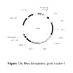
[0013] Figure 1A-C show examples of vectors useful in the present disclosure.
The therapeutic
gene vector may encode PCBP1 and stem cell inducing transcription factors
(Sox2, 0ct4, and
Klf4). This vector system uses the human elongation factor-1 alpha (EFP
promoter to drive
expression of genes fused with a green fluorescent protein (GFP) via a
cleavable protein
segment. This LVV vector system was then transduced into CFs. As demonstrated
in Example 1,
PCBP1 down-regulates and de-activates PRL3, a downstream protein involved in
tumorigenesis,
and thus provides a useful safe-guard for clinical applications in reducing
oncogenic side effects.
TRE: Tetracycline (SEQ ID NO: 5); rtTA: reverse tetracycline-controlled trans
activator; tTS:
transcription silencer is a fusion of the Tet Repressor Protein (TetR) with a
KRAB silencing
domain. tTS can be used to significantly lower the basal expression from l'
generation Tet-On
Inducible Expression Systems. In the absence of doxycycline, tTS binds to the
tet-operator
sequences within TRE promoters, silencing gene expression. Upon addition of
doxycycline, tTS
undergoes a conformational change that causes its release from the promoter,
to be replaced by
the Tet-On transactivator.
[0014] Figure 2A shows a comparison of GFP cell counts in retinal cell (ARPE-
19, ATCCTm)
cultures after transfection with therapeutic genes in vitro. All GFP positive
cell numbers are
estimated based on 103 cells.
[0015] Figure 2B shows retinal cells transfected by the therapeutic gene
vectors coexpressing
green fluorescent protein and monitored by a fluorescence microscopy (10x).
The results
3
CA 03149640 2022-02-02
WO 2021/026488 PCT/US2020/045477
(photos) indicate thatPCBP1-GFP, 0ct4, Sox2, and Klf4 were successfully
delivered by the
therapeutic vector system and co-expressed in the transfected cells.
[0016] Figure 3 shows real-time PCR results to compare the mRNA level of PCBP1
between
cells transfected by the gene therapy (GT) vector and control cells without
GT. PCBP1 mRNA
levels in GT transfected cells are more than 3 fold higher (ddCt = 3.37) than
that of the control
cells.
[0017] Figure 4A-B shows immunohistochemistry staining of a biomarker of
retinal stem cells
by antibody to human CRX-1 (see red fluorescent light). This indicates that
some of the cells
have been turned into retinal stem cell-like cells after differentiated
retinal cells were transduced
with a vector including PCBP1 and the SOX2, OCT4, KLF4 transcription factors.
[0018] Figure 5A-D shows immunohistochemistry staining of a biomarker of
retinal stem cells
by the antibody to human SOX2 (red fluorescent light). Multiple staining was
performed. First,
the retinal stem cell biomarker of CRX was detected. Then, the cells
transduced by the PCBP1-
vector and those transduced by vector SOX 2 were stained to show that these
cells overlap with
CRX expression. This indicates that some of the cells have been turned into
retinal stem cell-like
cells after differentiated cells were transduced with a vector containing
PCBP1 and the SOX2,
OCT4, KLF4 transcription factors.
[0019] Figure 6A-B shows skin cancer cells transfected with either the control
(GFP only) or
PCBP1-GFP (Figure 6A). Western-blot analysis on skin cancer cells treated with
PCBP1 and its
mutants that can down-regulate the protein level of the PRL-3, an oncogenic
factor in cancer
cells. Lane 1: GFP only; lane 2: PCBP1 with one mutation as mPCBP1 1; Lane 3:
PCBP1 wide-
type as wPCBP1 0; Lane 4: PCBP1 with two mutations as mPCBP1 2. The beta actin
was
served as a control set of house-keeping protein in this assay.
[0020] Figure 7 shows real-time qRT-PCR reactions to detect the expression
level of both
PCBP1 and PRL-3 in the cancer cells. The left panel indicates that PCBP1 was
highly expressed
in the Treatment group. The right panel shows that the PRL-3 was significantly
down-regulated
in the Treatment group of the cancer cells. The untreated groups were
transduced by a GFP
vector only.
[0021] Figure 8 shows representative schematics of experimental designs.
[0022] Figure 9 shows a representative delivery method to deliver drugs into
the retinal layer of
the eye (red square). See Park et al. (2015)
[0023] Figure 10 shows an example of the in vivo pre-clinical study to test
visual acuity after
gene therapy on macular degenerative disease. See Lu et at. (2009).
4
CA 03149640 2022-02-02
WO 2021/026488 PCT/US2020/045477
[0024] Figure 11A-B shows a representative experimental design to demonstrate
PCBP I
inhibits expression of biomarkers associated with metastasis and cancer (e.g.
PRL-3/STAT3).
[0025] Figure 12 shows a putative mechanism by which PCBPI inhibits PRL-
3/STAT3
expression.
[0026] Figure 13A-B shows results from transfection of cardiac fibroblasts
(CFs) with the
vectors of the present disclosure. Figure 13A shows CFs transduced with LVVs
expressing
PCBPI and GFP. GFP expression is detected by fluorescent microscopy (x10).
(a): PCBP1-
IRES-GFP (b): PCBP I -IRES-GFP and Sox2/0ct4/K1f4 (P+SOK) co-transduction
(c):LV vector
without GFP as control; (d) GFP vector control. GFP was detected under the
fluorescent
microscope to count the transduction efficiency with about more than 80% cells
transduced.
Figure 13B shows a comparison of the PCBP I mRNA levels between the CFs
transduced by the
PCBPI-IRES-GFP vector and control (no transfection) by qRT-PCR. PCBP I
expression is more
than 99-fold higher in the treatment group (ddCt=99.9); p<0.05.
[0027] Figure 14A-F shows immunohistochemistry staining for a cardiac stem
cell biomarker.
The CFs were transduced and labeled using antibody against human c-Kit and
detected with
NorthernLights-conjugated secondary antibody (A, B). No transduction control
group under red
fluorescent filter (A) and bright field (B); (C, D) PCBPI-IRES-GFP (c-Kit
staining) under red
fluorescent filter (C) and bright field (D); PCBPI-IRES-GFP and transcription
factors Sox2,
0ct4, Klf4 (P+SOK) co-transduction group (c-Kit staining) observed under red
fluorescent filter
(E) and bright field (F). Insets showing areas of interest enlarged for
details. Scale Bar: 10011.m.
[0028] Figure 15 shows real time RT-PCR reactions to detect mRNA expression of
cardiac
biomarkers after iPSCs induction. Top to bottom: COL lal, c-Kit, TNNT2. Left
column: LVV
PCBPI-GFP and 5ox2/0ct4/K1f4 (P + SOK) co-transduction; Right column: no
transduction
control. *p<0.05.
[0029] Figure 16 shows an exemplary animal model for testing the systems of
the present
disclosure in a murine model of myocardial infarction.
[0030] Figure 17A-B: Flow cytometry detection of CD44 and CD24 in MCF-7 and
MCF-7
(CSC) (BCSC). A: CD44 staining. B: CD24 staining.
[0031] Figure 18A-D: FACS analysis on biomarkers confirmed by dual-color
scatter plot
(CD44 vs CD24). Flow cytometry detection of CD24 and CD44 in MCF-7 and MCF-7
(CSC)
via dual-color scatter plot. A: isotype control of MCF-7 (CSC), B: CD44-PE &
CD24-APC
staining of MCF-7 (CSC), C: isotype control of MCF-7, D: CD44-PE & CD24-APC
staining of
CA 03149640 2022-02-02
WO 2021/026488 PCT/US2020/045477
MCF-7. These data are same from Figure 17 analyzed by dual-color scatter plot
for additional
clarity.
[0032] Figure 19A-D: Flow cytometry detection of CD133 in MCF-7 (CSC) and MDA-
MB-
231. A: isotype control, B: MCF-7 (CSC), C: MDA-MB-231, D: merge. Cells were
harvested
stained with Anti-Human CD133 APC-conjugated Monoclonal Antibody (10uL/106
cells). Non-
specific antibody used as Isotype control.
[0033] Figure 20: Western blot (WB) analysis of CD133, CD44 and CD24 in MCF-7
and U87-
MG treated with or without TGF-beta. Lane 1: MCF-7 (without TGF-beta), lane 2:
MCF-7 (with
TGF-beta); lane 3: U87-MG (without TGF-beta), lane 4: U87-MG (with TGF-beta).
[0034] Figure 21A-C: Normalized by GAPDH and visualized Western Blot data. A):
The
GAPDH-normalized signals from the western blot (WB) are plotted on a linear
scale. B) The
normalized CD24 and CD44 signals net change [TGF-beta (+) ¨ TGF-beta (-)]. C)
Summary
table of WB signal analysis. The table includes the data that were calculated
and converted from
the signals of WB analysis as the data-set to be compatible, when the cells
did not include TGF-
beta as base-line set of "1", and the higher numbers (bolded text) would be up-
regulation, and
the numbers below the "1" (base-line) were considered down-regulation (italics
texts).
[0035] Figure 22A-C: qRT-PCR analysis of PCBP1 transcripts in cancer stem
cells after
treatment with PCBP1-GT. Fold change in PCBP1 mRNA expression in U87-MG (CSC)
(A) ,
DU-145 (CSC) (B), and MCF-7 (CSC) (C) cells transfected with the indicated
constructs. Dotted
lines on y-axis mark 1.5 fold difference, typically indicating level
considered for statistical
significance. Statistical analysis of molecular assays quantification, was
analyzed using the two-
tailed Student's t test by RStudio or IMP. For all the significance were
defined asp < 0.05.
[0036] Figure 23A-C: Western blot analysis of CD133, CD44 and CD24 in DU-145,
MCF-7,
U87-MG transfected with indicated gene constructs. Panel A: U87-MG (CSC);
Panel B: DU-145
(CSC); Panel C: MCF-7 (CSC).
[0037] Figure 24A-C: Measurement of CD44 and CD133 by Western Blot. The
western blot
signals were normalized against beta actin. Panel A shows CD44 expression.
Panel B shows
CD133 expression. Summary table (C) shows data that with signals normalized
against cells
transfected with LV-GFP. Increased levels of expression are shown in bold, and
lower levels of
expression in italics.
[0038] Figure 25: Niclosamide targets NF-kB and elevates reactive oxygen
species (ROS)
levels. Niclosamide blocks the TNFa-induced D3 phosphorylation, translocation
of p65, and the
6
CA 03149640 2022-02-02
WO 2021/026488 PCT/US2020/045477
expression of NF-kB regulated genes. Conversely, niclosamide elevates the
intracellular ROS
content.
[0039] Figure 26: Western blot analysis of U87-MG (CSC) treated with
niclosamide (1 M,
2 M, 5 M and 10[tM). Cells were incubated with indicated concentrations of
niclosamide for
24 hours (equal volume of DMSO served as vehicle control).
[0040] Figure 27A-C: A) c-Myc signals, normalized against beta actin, are
plotted on linear
scale. B) Normalized c-Myc signals plotted as net change (i.e., with DMSO
signal subtracted).
C) Summary table of WB signal analysis, with upregulation shown in bold (>0),
and
downregulation shown in italics.
[0041] Figure 28A-B: qRT-PCR quantification of c-Myc and BCL-2 expression in
U87-
MG(CSC) cells treated with DMSA or 10 tM niclosamide. For calculations: DMSO
served as
base-line in panel A. Mock-transfection served as base-line in panel B. Dotted
line marks 1.5
fold change in y-axis, considered the benchmark for statistical significance.
[0042] Figure 29A-B: qRT-PCR quantification of c-Myc and BCL-2 expression in
DU-145
(CSC) cells treated with DMSA or 10 niclosamide. For calculations: DMSO
served as base-
line in panel A. Mock-transfection served as base-line in panel B. Dotted line
marks 1.5 fold
change in y-axis, considered the benchmark for statistical significance.
[0043] Figure 30A-B. qRT-PCR quantification of c-Myc and BCL-2 expression in
DU-145
(CSC) cells treated with DMSA or 10 niclosamide. For calculations: DMSO
served as base-
line in panel A. Mock-transfection served as base-line in panel B. Dotted line
marks 1.5 fold
change in y-axis, considered the benchmark for statistical significance.
DETAILED DESCRIPTION
[0044] Age-related macular degeneration (AMD) is one of the major causes of
irreversible
blindness in the elderly. Although management of neovascular AMD (wet AMD) has
dramatically progressed, there is still no effective treatment for diseases
such as non-neovascular
AMD (atrophic, or dry AMD) and autoimmune diseases such as Sjogren's syndrome
with
serious dry eye.
[0045] Clinical trials of stem cell therapies have shown promising prospects
for retinal pigment
epithelial (RPE) cell transplantation. For example, in 2011, Advanced Cell
Technology (Santa
Monica, CA, USA) performed Phase VII clinical trials to elucidate the efficacy
of hESC-derived
RPE cell transplantation in dry AMD and Stargardt's disease patients.
Subsequently, Schwartz et
7
CA 03149640 2022-02-02
WO 2021/026488 PCT/US2020/045477
at. (2016) published the results of two Phase I/II trials of 18 patients with
dry AMD or
Stargardt's disease. After four years, more than half of treated patients
experienced sustained
improvements in visual acuity and demonstrated evidence of possible cellular
engraftment.
However, key issues including implantation techniques, immune rejection, and
xeno-free
components still need to be further investigated and improved. Further, use of
human or animal-
derived stem cells components that may carry factors such as sialic acid or
Neu5Gc, which may
cause unwanted immunogenicity of the cells or even expose the patient to
certain pathogens,
needs to be considered. Therefore, stem cell therapy by iPSC technology and
retinal
microenvironmental regulation of gene expression represents potential new
approaches for dry
AMD treatments.
[0046] Techniques for generating iPSCs provide a new opportunity to directly
induce host (or
patient) differentiated tissue into stem cell-like cells without the exogenous
administration of
stem cell inducing proteins such as 0ct4, Sox2, cMyc, or Klf4. While these
transcription factors
are known to play a role in inducing iPSCs, they may also trigger oncogenesis.
Thus, the
compositions and methods disclosed herein include cell transduction with an
anti-oncogene such
as PCBP1 to reduce or prevent oncogenesis.
[0047] Without being bound by theory, PCBP1 may take the place of c-Myc in the
vectors of the
present disclosure as PCBP1 itself may play a role in transforming
differentiated cells into stem
cells. PCBP1 may transform cells into stem cells either in conjunction with
other transcription
factors, or on its own. Importantly, PCBP1 does not exhibit the same
oncogenesis observed with
c-Myc.
[0048] These two advanced features (e.g. stem cell-inducing agents or
transcription factors
involved in the maintenance of stem cell development and PCBP1) are
incorporated into a vector
system for gene therapy to directly induce conversion of cells into stem cell-
like cells for the
treatment of disease. These features provide a minimal potential for involving
tumorigenic
pathways that may be otherwise activated during stem cell development.
Polypyrimidine tract-binding protein 1 (PCBP1)
[0049] In some aspects, the present disclosure provides a vector containing
the transcription
factor Poly(rC)-binding protein 1 (PCBP1, also known as hnRNP-E1 and aCP1)
which can
inhibit or delay tumorigenesis. In some embodiments, the PCBP1 is a mammalian
PCBP1. In
some embodiments, the PCBP1 is human PCBP1, fragment, or variant thereof
8
CA 03149640 2022-02-02
WO 2021/026488 PCT/US2020/045477
[0050] In some embodiments, PCBP1 alters the expression of one or more cancer
biomarkers. In
some embodiments, the cancer biomarker is a breast cancer biomarker. In some
embodiments,
the cancer biomarker is a prostate cancer biomarker. In some embodiments, the
cancer
biomarker is a lung cancer biomarker. In some embodiments, the expression of
one or more
cancer biomarkers is increased. In some embodiments, the expression of one or
more cancer
biomarkers is decreased. In some embodiments, the cancer biomarker is
associated with
metastasis. In some embodiments, the cancer biomarker includes, but is not
limited to, PRL-3,
STAT3, CD44 variant, CD133, CD24, Estrogen receptor, progesterone receptor,
HER2, CA27,
CA29, CA25, CEA, CA125, Ki-67, ERa, Cyclin D1, Cyclin E, ERO, PSA, PCA3, AR-
V7, E-
cadherin, and vimentin. In some embodiments, transfection of a cancer cell
with the constructs
of the present disclosure decreases expression of CD44. In some embodiments,
transfection of a
cancer cell with the constructs of the present disclosure decreases expression
of CD133. In some
embodiments, the PCBP1 is a nucleic acid. In some embodiments, the nucleic
acid is DNA. In
some embodiments, the nucleic acid is RNA. In some embodiments, the nucleic
acid is mRNA.
In some embodiments, the PCBP1 contains the nucleic acid sequence of SEQ ID
NO: 1. In some
embodiments, the PCBP1 is the full-length nucleic acid sequence of SEQ ID NO:
1. In some
embodiments, the PCBP1 is a fragment of the nucleic acid sequence of SEQ ID
NO: 1. In some
embodiments, the PCBP1 is a variant of the nucleic acid sequence of SEQ ID NO:
1. Without
being bound by theory, each of the KH domains within PCBP1 has been shown to
bind to
mRNAs of different proteins, and use of one or more KH domain may selectively
inhibit the
translation of a given protein. In some embodiments, the PCBP1 nucleic acid
encodes all three
K-homologous (KH) domains. In some embodiments, the PCPB1 nucleic acid encodes
two KH
domains. In some embodiments, the PCPB1 nucleic acid encodes one KH domain.
[0051] Further, mutations in a nuclear localization signal may alter the
ability of PCBP1 to
translocate into the nucleus, and thus affect later gene expression. In some
embodiments, the
PCBP1 contains a mutation in one or both nuclear localization signals. In some
embodiments,
the change in one or both nuclear localization signals inhibits the ability of
PCBP1 to translocate
into the nucleus.
[0052] Phosphorylation also plays a role in the activity of PCBP1 (e.g.
unphosphorylated
PCBP1 may lack activity). In some embodiments, the PCBP1 nucleotide sequence
contains
mutations that affect the ability of the PCBP1 polypeptide to be
phosphorylated. In some
embodiments, the PCBP1 nucleotide sequence prevents PCBP1 polypeptide
phosphorylation. In
9
CA 03149640 2022-02-02
WO 2021/026488 PCT/US2020/045477
some embodiments, the unphosphorylated PCBP1 or not fully phosphorylated PCBP1
polypeptide is not active at wild type levels.
[0053] In some embodiments, the PCBP1 nucleotide sequence contains a point
mutation relative
to wild type PCBP1 (SEQ ID NO: 1). In some embodiments, the PCPB1 nucleotide
sequence
contains a point mutation that affects phosphorylation. In some embodiments,
the PCBP1
nucleotide sequence contains a mutation(s) that may affect nuclear membrane
translocation. In
some embodiments, the point mutation is selected from the group including, but
not limited to,
G13A, T299C, T299A, G326A, G527A, C652T, C688T, G781T, G814T, C947T, G1033C,
C1034T, or G1048C.
[0054] In some embodiments, the disclosure provides mimetics, analogs,
derivatives, variants,
or mutants of PCBP1 (SEQ ID NO: 1). In some embodiments, the mimetic, analog,
derivative,
variant, or mutant contains one or more nucleic acid substitutions compared to
the nucleic acid
sequence of the native PCBP1. In some embodiments, one to 20 nucleic acids are
substituted. In
some embodiments, the mimetic, analog, derivative, variant, or mutant contains
about 1, about 2,
about 3, about 4, about 5, about 6, about 7, about 8, about 9, or about 10
nucleic acid
substitutions compared to the nucleic acid sequence of the native PCBP1 (e.g.
SEQ ID NO: 1).
In some embodiments, the mimetic, analog, derivative, variant, or mutant
contains one or more
nucleic acid deletions compared to the amino acid sequence of the native PCBP1
(e.g. SEQ ID
NO: 1).
[0055] In some embodiments, one to 20 nucleic acids are deleted compared to
the nucleic acid
sequence of the native PCBP1 (e.g. SEQ ID NO: 1). In some embodiments, the
mimetic, analog,
derivative, variant, or mutant has about 1, about 2, about 3, about 4, about
5, about 6, about 7,
about 8, about 9, or about 10 nucleic acid deletions compared to the nucleic
acid sequence of the
native PCBP1 (e.g. SEQ ID NO: 1). In some embodiments, one to ten nucleic
acids are deleted
at either terminus compared to the nucleic acid sequence of the native PCBP1
(e.g. SEQ ID NO:
1). In some embodiments, one to ten nucleic acids are deleted from both
termini compared to the
nucleic acid sequence of the native PCBP1. In some embodiments, the nucleic
acid sequence of
the mimetic, analog, derivative, variant, or mutant is about 70% identical to
about 99.9%
identical to the nucleic acid sequence of the native PCBP1. In some
embodiments, the nucleic
acid sequence of the mimetic, analog, derivative, variant, or mutant is at
least about 70%
identical to the nucleic acid sequence of the native PCBP1. In some
embodiments, the nucleic
acid sequence of the mimetic, analog, derivative, variant, or mutant is about
70%, about 71%,
about 72%, about 73%, about 74%, about 75%, about 76%, about 77%, about 78%,
about 79%,
CA 03149640 2022-02-02
WO 2021/026488 PCT/US2020/045477
about 80%, about 81%, about 82%, about 83%, about 84%, about 85%, about 86%,
about 8'7%,
about 88%, about 89%, about 90% about 91%, about 92%, about 93%, about 94%,
about 95%,
about 96%, about 9700, about 98%, about 99%, about 99.100, about 99.2%, about
99.3%, about
99.40, about 99.50, about 99.6%, about 99.70, about 99.8%, or about 99.90
identical to the
nucleic acid sequence of the native PCBP1 (e.g. SEQ ID NO: 1). Percentage
identity can be
calculated using the alignment program EMBOSS Needle, available at
http ://www.ebi. ac. uk/Tool s/p sa/emb o s s needle/.
[0056] In some embodiments, the PCBP1 is a polypeptide. In some embodiments,
the PCBP1
contains the amino acid sequence of SEQ ID NO: 2. In some embodiments, the
PCBP1 is the
full-length amino acid sequence of SEQ ID NO: 2. In some embodiments, the
PCBP1 is a
fragment of the amino acid sequence of SEQ ID NO: 2. In some embodiments, the
PCB1 is a
variant of the amino acid sequence of SEQ ID NO: 2. In some embodiments, the
PCBP1 is a
functional fragment or variant of the amino acid sequence of SEQ ID NO: 2. In
some
embodiments, the PCBP1 polypeptide contains all three K-homologous (KH)
domains. In some
embodiments, the PCPB1 polypeptide contains two KH domains. In some
embodiments, the
PCPB1 polypeptide contains one KH domain.
[0057] In some embodiments, the PCBP1 polypeptide sequence contains an amino
acid mutation
relative to wild type PCBP1 (SEQ ID NO: 2). In some embodiments, the PCPB1
polypeptide
sequence contains an amino acid mutation that affects phosphorylation. In some
embodiments,
the amino acid mutation is selected from the group including, but not limited
to, V5M, LlOOP,
L100Q, C109Y, G176E, P218S, 5223L, D261Y, A2725, A316V, A345P, A345V, or
E350Q.
[0058] In some embodiments, the disclosure provides mimetics, analogs,
derivatives, variants,
or mutants of PCBP1 (SEQ ID NO:2). In some embodiments, the mimetic, analog,
derivative,
variant, or mutant contains one or more amino acid substitutions compared to
the amino acid
sequence of the native PCBP1. In some embodiments, one to 20 amino acids are
substituted. In
some embodiments, the mimetic, analog, derivative, variant, or mutant contains
about 1, about 2,
about 3, about 4, about 5, about 6, about 7, about 8, about 9, or about 10
amino acid substitutions
compared to the amino acid sequence of the native PCBP1 (SEQ ID NO:2).
[0059] In some embodiments, the mimetic, analog, derivative, variant, or
mutant contains one or
more amino acid deletions compared to the amino acid sequence of the native
therapeutic
peptide agent. In some embodiments, one to 20 amino acids are deleted compared
to the amino
acid sequence of the native protein agent. In some embodiments, the mimetic,
analog, derivative,
variant, or mutant has about 1, about 2, about 3, about 4, about 5, about 6,
about 7, about 8,
11
CA 03149640 2022-02-02
WO 2021/026488 PCT/US2020/045477
about 9, or about 10 amino acid deletions compared to the amino acid sequence
of the native
PCBP1 (SEQ ID NO:2). In some embodiments, one to ten amino acids are deleted
at either
terminus compared to the amino acid sequence of the native PCBP1 (SEQ ID
NO:2). In some
embodiments, one to ten amino acids are deleted from both termini compared to
the amino acid
sequence of the native PCBP1 (SEQ ID NO:2). In some embodiments, the amino
acid sequence
of the mimetic, analog, derivative, variant, or mutant is at least about 70%
identical to the amino
acid sequence of the native PCBP1 (SEQ ID NO:2). In some embodiments, the
amino acid
sequence of the mimetic, analog, derivative, variant, or mutant is about 70%
to about 99.9%
identical to the amino acid sequence of native PCBP1 (SEQ ID NO: 2). In some
embodiments,
the amino acid sequence of the mimetic, analog, derivative, variant, or mutant
is about 70%,
about 71%, about 72%, about 73%, about 74%, about 75%, about 76%, about 77%,
about 78%,
about 79% about 80%, about 81%, about 82%, about 83%, about 84%, about 85%,
about 86%,
about 87%, about 88%, about 89%, about 90%, about 91%, about 92%, about 93%,
about 94%,
about 95%, about 96%, about 97%, about 98%, about 99%, about 99.1%, about
99.2%, about
99.3%, about 99.4%, about 99.5%, about 99.6%, about 99.7%, about 99.8%, or
about
99.9%identical to the amino acid sequence of the native PCBP1 (SEQ ID NO:2).
In some
embodiments, the amino acid sequence of the mimetic, analog, derivative,
variant, or mutant is
about 70%, about 71%, about 72%, about 73%, about 74%, about 75%, about 76%,
about 77%,
about 78%, about 79% about 80%, about 81%, about 82%, about 83%, about 84%,
about 85%,
about 86%, about 87%, about 88%, about 89%, about 90%, about 91%, about 92%,
about 93%,
about 94%, about 95%, about 96%, about 97%, about 98%, about 99%, about 99.1%,
about
99.2%, about 99.3%, about 99.4%, about 99.5%, about 99.6%, about 99.7%, about
99.8%, or
about 99.9%identical to the amino acid sequence of the native PCBP1 (SEQ ID
NO:2) and
retains all or most of the biological activity of the native PCBP1. In some
embodiments, the
amino acid sequence of the mimetic, analog, derivative, variant, or mutant is
about 70%, about
71%, about 72%, about 73%, about 74%, about 75%, about 76%, about 77%, about
78%, about
79% about 80%, about 81%, about 82%, about 83%, about 84%, about 85%, about
86%, about
87%, about 88%, about 89%, about 90%, about 91%, about 92%, about 93%, about
94%, about
95%, about 96%, about 97%, about 98%, about 99%, about 99.1%, about 99.2%,
about 99.3%,
about 99.4%, about 99.5%, about 99.6%, about 99.7%, about 99.8%, or about
99.9%identical to
the amino acid sequence of the native PCBP1 (SEQ ID NO:2) and has reduced or
altered activity
compared with the native PCBP1.
12
CA 03149640 2022-02-02
WO 2021/026488 PCT/US2020/045477
[0060] In some embodiments, the amino acid sequence of the mimetic, analog,
derivative,
variant, or mutant is about 70% to about 99.9% identical to the amino acid
sequence or of one or
more domains of the native PCBP1 (SEQ ID NO: 2). In some embodiments, the
amino acid
sequence of the mimetic, analog, derivative, variant, or mutant is about 70%,
about 71%, about
72%, about 73%, about 74%, about 75%, about 76%, about 77%, about 78%, about
79% about
80%, about 81%, about 82%, about 83%, about 84%, about 85%, about 86%, about
87%, about
88%, about 89%, about 90%, about 91%, about 92%, about 93%, about 94%, about
95%, about
96%, about 97%, about 98%, about 99%, about 99.1%, about 99.2%, about 99.3%,
about
99.4%, about 99.5%, about 99.6%, about 99.7%, about 99.8%, or about
99.9%identical to the
amino acid sequence of one or more domains of the native PCBP1 (SEQ ID NO:2).
In some
embodiments, the amino acid sequence of the mimetic, analog, derivative,
variant, or mutant is
about 70% to about 99.9% identical to the amino acid sequence or of one or
more domains of the
native PCBP1 (SEQ ID NO: 2) and retains all or most of the biological activity
of the native
PCBP1. In some embodiments, the amino acid sequence of the mimetic, analog,
derivative,
variant, or mutant is about 70%, about 71%, about 72%, about 73%, about 74%,
about 75%,
about 76%, about 77%, about 78%, about 79% about 80%, about 81%, about 82%,
about 83%,
about 84%, about 85%, about 86%, about 87%, about 88%, about 89%, about 90%,
about 91%,
about 92%, about 93%, about 94%, about 95%, about 96%, about 97%, about 98%,
about 99%,
about 99.1%, about 99.2%, about 99.3%, about 99.4%, about 99.5%, about 99.6%,
about 99.7%,
about 99.8%, or about 99.9%identical to the amino acid sequence of one or more
domains of the
native PCBP1 (SEQ ID NO:2) and retains all or most of the biological activity
of the native
PCBP1. In some embodiments, the amino acid sequence of the mimetic, analog,
derivative,
variant, or mutant is about 70% to about 99.9% identical to the amino acid
sequence or of one or
more domains of the native PCBP1 (SEQ ID NO: 2) and has reduced or altered
activity
compared to the native PCBP1. In some embodiments, the amino acid sequence of
the mimetic,
analog, derivative, variant, or mutant is about 70%, about 71%, about 72%,
about 73%, about
74%, about 75%, about 76%, about 77%, about 78%, about 79% about 80%, about
81%, about
82%, about 83%, about 84%, about 85%, about 86%, about 87%, about 88%, about
89%, about
90%, about 91%, about 92%, about 93%, about 94%, about 95%, about 96%, about
97%, about
98%, about 99%, about 99.1%, about 99.2%, about 99.3%, about 99.4%, about
99.5%, about
99.6%, about 99.7%, about 99.8%, or about 99.9% identical to the amino acid
sequence of one
or more domains of the native PCBP1 (SEQ ID NO:2) and has reduced or altered
activity
compared with the native PCBP1. Percentage identity can be calculated using
the alignment
13
CA 03149640 2022-02-02
WO 2021/026488 PCT/US2020/045477
program EMBOSS Needle, available at http://www.ebi.ac.uk/Tools/psa/emboss
needle/. The
following default parameters may be used for Pairwise alignment: Protein
Weight Matrix =
BLOSUM62; Gap Open = 10; Gap Extension = 0.1.
Transcription Factors
[0061] Transcription factors promote the differentiation of embryonic stem
(ES) cells derived
from the inner cell mass of the blastocyst into stem or progenitor cells of
all three vertebrate
germ layers: ectoderm, mesoderm, and endoderm. In addition, the expression of
specific
transcription factors is sufficient to reprogram somatic cells back to a
pluripotent state. Induced
pluripotent stem (iPSC) cells, like ES cells, give rise to differentiated stem
cells such as neural
and epithelial stem cells. Differentiated stem cells function to replenish
cells of the tissue in
which they are found.
[0062] In some embodiments, the one or more additional transcription factors
are known to be
associated with stem cell induction. In some embodiments, the one or more
additional
transcription factors are known to be associated with stem cell renewal and/or
pluripotency. In
some embodiments, the one or more additional transcription factors associated
with stem cell
renewal and/or pluripotency are selected from, but not limited to, Brachyury,
FoxD3, GBX2,
Max, NFkB1, Pax6, c-Jun, c-Maf, c-Myc, FoxF1, Goosecoid, Mef2c, NFkB2, PRDM14,
FoxH1, HES-1, MIXL1, Oct-3/4, Rex-1/ZFP42, Fox01/FKHR, HNF-3 alpha/FoxAl,
MTF2,
OCT4, SALL1, C/EBP alpha, GATA-2, KLF2, Nanog, 0tx2, SALL4, EOMES, GATA-3,
KLF4, NFkB/IkB Activators, p53, Smadl, FoxC2, Gata4 , KLF5, NFkB/IkB
Inhibitors, Pax2,
Smad1/5, 5mad2, Smad 2/3, Smad 3, Smad 4, Smad 5, Smad 8, Snail, 50X15, SUZ12,
UTF1,
50X17, SEMA 6A-1, WDR5, 50X2, SFRP1, WT1, 50X7, Tbx5, ZNF206, STAT Activators,
TBX6, ZNF281, STAT Inhibitors, TCF-3/E2A, Snail, STAT3, and THAP11.=
[0063] In some embodiments, the transcription factor is involved in one or
more oncogenesis
pathways. In some embodiments, overexpression of the transcription factor is
involved in one or
more oncogenesis pathways (e.g. 50X2 overexpression has been described in lung
cancer
tissues). In some embodiments, the expression changes of the transcription
factor are biomarkers
of cancer. In some embodiments, the cancer is liver cancer or lung cancer.
[0064] Any appropriate transcription factors may be used, including but not
limited to
ASCL2/Mash2, ASCL1/Mashl, CDX2, DNMT1, ELF3, Ets-1, FoxMl, FoxN1, Hairless,
HNF-
4, alpha/NR2A1, IRF6, MITF, Miz-1/ZBTB17, MSX1, MSX2, MYB, Neurogenin-3,
NFATC1,
14
CA 03149640 2022-02-02
WO 2021/026488 PCT/US2020/045477
NKX3.1, Nrf2, p53, p63/TP73L, Pax2, Pax3, Pax4, Pax6, RUNX1/CBFA2,
RUNX2/CBFA1,
RUNX3/CBFA3, Smadl, Smad1/5, Smad2, Smad2/3, Smad3, Smad4, Smad5, Smad7,
Smad8,
Smad9, Snail, SOX9, STAT Activators, STAT Inhibitors, STAT1, STAT3, STAT4,
STAT5a,
STAT6, SUZ12, TCF-3/E2A, TCF7/TCF1, Brachyury, GATA-1, GATA-2, GATA-3, GATA-4,
GATA-6, LM02, LM04, SCL/Tall, AHR, Aiolos/IKZF3, CDX4, CREB, DNMT3A,
DNMT3B, EGR1, Fox03, Helios, HES-1, HHEX, HIF-1 alpha, HMGB1/HMG-1, HMGB3,
Ikaros, c-Jun, LM02, LM04, c-Maf, MafB, MEF2C, MYB, NFATC2, NFIL3/E4BP4,
PITX2,
PRDM16/MEL1, Proxl, PU.1/Spi-1, SALL4, SCL/Tal 1, Spi-B, T5C22, HNF-3
alpha/FoxAl,
HNF-3 beta/FoxA2, ONECUT2/0C-2, TBX3, TBX5, TBX18, TCF-2/HNF-1 beta, PDX-
1/IPF1, PTF1A, RFX6, EBF-1, EBF-2, EBF-3, ETV5, FoxC2, FoxF1, HMGA2, MYF-5,
Myocardin, MyoD, Myogenin, PLZF, Twist-1, Twist-2, ATF2, Brgl, CSL, EMX2,
FosB/G053,
GLI-1, GLI-2, HOXB1, MCM2, MCM7, NeuroD1, NeuroD2, Neurogenin-1, Neurogenin-2,
NKX2.2õ, NKX6.1, Oligl, 01ig2, 01ig3, RCOR1/CoREST, SOX1, 50X2, 50X3, 50X5,
50X6, TCF-12/HTF4, ZIC1, ZIC3, EOMES, FoxD3, FoxH1, Fox01/FKHR, GBX2,
Goosecoidõ KLF2, KLF4, KLF5, Max, MIXL1, MTF2, NFkB/IkB Activators, NFkB/IkB
Inhibitors, NFkB1, NFkB2, Oct-3/4, 0tx2, PRDM14, Rex-1/ZFP42, SALL1, 50X7,
SOX15,
SOX17, SOX18, TBX6, THAP11, UTF1, WDR5, WT1, ZNF206, ZNF281, ATBF1/ZFHX3,
HIPK1, HIPK2, PIAS2, PIAS3, TRIM21, TRIM32, WTX, ZMIZ1/ZimplO, Carml, CBP,
Cbx2,
CHD1, CHD7, CTCF, DEK, MBD3, NCOA2, NCOA3PIWIL1/HIWI, PIWIL2, PIWIL4,
RBBP4, SATB1, SIN3A, SMARCA5/SNF2H, TAF5L, ARA54, ATAD2, beta-Catenin, Bc1-9,
BTF3, CBFB, Cyclin Al, Cyclin A2, Cyclin Bl, Cyclin B2, Cyclin D3, DDX17,
DDX5, IKK
alpha, IKK beta, LEDGF, LITAF, LXR alpha/NR1H3, MED4, Park7/DJ-1, PGC1 alpha,
Pygopus-1, Pygopus-2, RNF4, TACC3, Tankyrase 1, Tankyrase Inhibitors,
TAZ/WWTR1,
TORC1, TORC2, TORC3, TRRAP ,YAP1, ACLP, ATN1, BASP1, Bc1-6, CDP/CUTL1, CIS-1,
Cited-2, COMMD1, CREG, CtBP1, DEP TOR/DEPDC 6, FHL1, FHL2, FIH-1/HIF- IAN, GFI-
1,
Host Cell Factor 1/HCFC1, Hexim 1, Histone Deacetylase 2/HDAC2, Histone
Deacetylase
8/HDAC8, ID1, ID2, IkB-alpha, IkB-beta, IkB-epsilon, IRF2BP1, KAP1, Keapl,
LCoR,
NCOR1, NC OR2, pl5INK4b/CDKN2B, pl6INK4a / CDKN2A, pl8INK4c/CDKN2C, PA2G4,
Prohibitin 2, SMURF2, SOCS-1, SOCS-2, SOCS-3, SOCS-4, SOCS-5, SOCS-6, SOCS-
7/Nck/NAP4, TANK, TLE1, TLE2, TLE3õ TMEM18, TMEM87A, ZBTB38,
ZBTB7A/Pokemon, ZFP900ct4, Sox 2, Nodal, FGF, TGF-f3, LIF, BMP4, KLF4, KLF2,
Myc,
LIN28A, TUBB, HAP90AB1, PELI1, SEMA6A, SLC7A5, RGMB, TUBB6, HSPA4, SFRP1,
LRRN1, PRDM14, C/EBP alpha, EOMES, and Nanog, or fragments thereof. In some
CA 03149640 2022-02-02
WO 2021/026488 PCT/US2020/045477
embodiments, the one or more additional transcription factors are SOX2, 0ct4,
and/or Klf4 or
fragments or variants thereof.
Vectors and Plasmids
[0065] Efficient delivery of the therapeutic gene to the target tissue or cell
is the most significant
hurdle for successful gene therapy. Since naked DNA is rapidly cleared or
degraded in vivo by
phagocytic immune cells or extracellular nucleases, a means of protecting the
transgene may be
desired. Furthermore, a vehicle for effecting tissue or cell entry is also
required, due to the poor
efficiency of spontaneous DNA uptake. Thus, DNA is normally combined with a
gene delivery
vehicle of some type, commonly known as a vector, to protect and mediate
effective tissue or
cell entry of the gene of interest.
[0066] Gene delivery systems can be grouped into non-biological (e.g. chemical
and physical
approaches of introducing plasmid DNA to mammalian cells) or biological (e.g.
viruses and
bacteria). Non-viral gene delivery systems normally involve the transfer genes
carried on
plasmid DNA. Plasmids employed do not generally replicate in mammalian cells.
[0067] Most commonly, recombinant viruses or naked DNA or DNA complexes are
used. For
example, viruses can be modified in the laboratory to provide vectors that
carry corrected,
therapeutic DNA into cells, where it can be integrated into the genome to
alter abnormal gene
expression and correct genetic disease. Alternatively, the vector may remain
extrachromosomal
and be expressed transiently.
[0068] In some aspects, the present disclosure provides methods of gene
therapy using viral
vectors. Viruses that may be used in gene therapy include, but are not limited
to, lentiviruses,
retroviruses, adenoviruses, adeno-associated viruses, replication-competent
vectors, vaccinia
virus, and the herpes simplex virus.
[0069] In some aspects, the present disclosure provides methods of gene
therapy using non-viral
vectors. In some aspects, the present disclosure provides methods of gene
therapy using bacterial
vectors. In some embodiments, the gene therapy method involves injection of
naked nucleic
acids (e.g. DNA or RNA). This may be performed using any appropriate means
known in the art.
[0070] In some aspects, the present disclosure provides methods of gene
therapy using non-viral
and non-bacterial vectors. In some embodiments, the non-viral and non-
bacterial vector is a
eukaryotic vector. In some embodiments the methods of gene therapy provide
CRISPR,
TALEN, or zinc finger proteins (ZFNs). In some embodiments, the eukaryotic
vector includes a
16
CA 03149640 2022-02-02
WO 2021/026488 PCT/US2020/045477
transposon system. In some embodiments, the transposon system is the Tn5,
CRISPR-
associated, Sleeping Beauty, or piggyBac transposon systems.
[0071] In some aspects, the present disclosure provides methods of gene
therapy using
nanoparticles. In some embodiments, the nanoparticle is a lipid-based
nanoparticle. In some
embodiments, the lipid-based nanoparticle is a solid lipid-nanoparticle (SLN).
In some
embodiments, the lipid-based nanoparticle is an unstructured lipid carrier
(NLC). In some
embodiments, the nanoparticle is a polymer-based nanoparticle. In some
embodiments, the
polymer-based nanoparticle is a nanosphere or a nanocapsule.
[0072] Plasmid DNA-based vectors are commonly used in gene therapy and can
accommodate
large segments of DNA and allows the manipulation of a variety of regulatory
elements that
impact gene transfer and expression. At its most basic, an expression plasmid
contains an
expression cassette and backbone. The expression cassette is a transcriptional
unit containing the
gene or genes of interest and any regulatory sequences required for expression
in the target cells.
The backbone may contain a selectable marker (e.g. an antibiotic resistance
gene or an
auxotrophic selection gene) and/or an origin of replication required for the
production of the
plasmid in bacteria. In some embodiments, the plasmid does not contain a
selectable marker or
traditional replication component (e.g. minicircles, self-replicating
minicircles, mini strings,
linear DNAs, etc.).
[0073] Any appropriate plasmid may be used in the methods disclosed herein. In
some
embodiments, representative plasmids are disclosed in Figure 1.
[0074] Any appropriate promoter may be used to drive expression of the genes
in the expression
cassette. Regulated and/or inducible promoters are important as they allow the
induction of stem
cells in situ according to different conditions that may be required in
clinical applications. These
promoters may be able to prevent stem cells from reproducing/being induced
indefinitely and
growing non-stop.
In some embodiments, the plasmid contains a retroviral promoter. In some
embodiments, the
plasmid contains a viral promoter. In some embodiments, the plasmid contains a
mammalian
promoter. In some embodiments, the mammalian promoter is a human promoter. In
some
embodiments, the promoter is tissue or cell specific. In some embodiments, the
tissue specific
promoter is selected from those listed in Table 1. In some embodiments, the
promoter is specific
for retinal tissue or cells. In some embodiments, the promoter is specific for
hematopoietic cells.
In some embodiments, the promoter is specific for liver tissue or cells. In
some embodiments,
the promoter is specific for lung tissue or cells. In some embodiments, the
promoter is specific
17
CA 03149640 2022-02-02
WO 2021/026488 PCT/US2020/045477
for muscle tissue or cells. In some embodiments, the promoter is specific for
HIV-infected cells.
Table 1. Tissue Specific Promoters
Promoter Note
GANT2 (Guanine nucleotide- Used with IRBP (interphotoreceptor retinoid
binding
binding protein G(t) subunit protein)
alpha-2)
VMD2 ( vitelliform macular 3.0kb, use VMD2 promoter direct Tetracycline
inducible
dystrophy) system, controlled Cre expression in transgenic
mice
Human IRBP (interphotoreceptor Sequence: ATTCTCATCGTAAATCAGGCTC
retinoid binding protein) ACTTCCATTG GCTGCATACG GTGGAGTGAT
GTGACCATAT GTCACTTGAGCATTACACAA
ATCCTAATGA GCTAAAAATA TGTTTGTTTT
AGCTAATTGA CCTCTTTGGCCTTCATAAAG
CAGTTGGTAA ACATCCTCAG ATAATGATTT
CCAAAGAGCA GATTGTGGGTCTCACCTGTG
CAGAGAAAGC CCACGTCCCT GAGACCACCT
TCTCCAG¨G CCTACTGAGGCACACAGGGG
CGCCTGCCTG CTGCCCGCTC AGCCAAGGCG
GTGTTGCTGGA (SEQ ID NO: 3)
Tetracycline Sequence:
GGTACCGAGCTCGACTTTCACTTTTCTCTATCAC
TGATAGGGAGTGGTAAACTCGACTTTCACTTTT
CTCTATCACTGATAGGGAGTGGTAAACTCGACT
TTCACTTTTCTCTATCACTGATAGGGAGTGGTAA
ACTCGACTTTCACTTTTCTCTATCACTGATAGGG
AGTGGTAAACTCGACTTTCACTTTTCTCTATCAC
TGATAGGGAGTGGTAAACTCGACTTTCACTTTT
CTCTATCACTGATAGGGAGTGGTAAACTCGACT
TTCACTTTTCTCTATCACTGATAGGGAGTGGTAA
ACTCGACCTATATAAGCAGAGCTCGTTTAGTGA
ACCGTCAGATCGCCTGGAGACGCCATCCACGCT
GTTTTGACCTCCATAGAAGACACCGGGACCGAT
CCAGCCTCCGCGGCCCCGAATTG (SEQ ID NO: 4)
[0075] In some embodiments, the promoter is an inducible promoter. In some
embodiments, the
inducible promoter is selected from those listed in Table 2. In some
embodiments, the promoter
is constitutive. In some embodiments, the promoter is a synthetic promoter or
contains enhancer
elements. In some embodiments, the promoter is a hybrid promoter. In some
embodiments, the
hybrid promoter contains regulatory regions of a gene. In some embodiments,
the promoter is
selected from the group including, but not limited to, Efl a, CAG, sv40, CMV,
RSV, 0ct4, Rexl,
18
CA 03149640 2022-02-02
WO 2021/026488 PCT/US2020/045477
Nanog, GANT2, VMD2, hIRBP, TET promoter, CAAT Box, CG Box, GT-1 motif, I-box,
AT-
rich sequence, RBCS1, TRE3G, GAL1, Lap267, Rapamycin, CD1 la, CD1 lb, CD18,
Beta-
globin promoter/LCR, Immunoglobulin promoter, PEPCK promoter, Albumin
promoter, hAAT,
SPC, SP-A, MCK, VLC1, HIV-LTR, tTS (tetracycline transcription silencer),
Tat/Rev-
responsive elements, Tat-inducible element, and FMR1.
Table 2. Inducible Promoters
Promoter Note
TRE3G Gene therapy use: will express high level of GOI (gene of
interest)
only when have doxycycline
Tetracycline A TRE is 7 repeats of a 19 nucleotide tetracycline operator
(tet0)
sequence, and is recognized by the tetracycline repressor (tetR). In the
endogenous bacterial system, if tetracycline, or one of its analogs like
doxycycline, are present, tetR will bind to tetracycline and not to the
TRE, permitting transcription.
Lac promoter Expressed in bacteria cell line, induced by IPTG
Ecdysone Can be expressed in mammalian cell line
Rapamycin AAV vectors were co-infused, one expressing the
transcription factors
and one encoding the hAADC transgene downstream from a
rapamycin-inducible promoter.
[0076] In some embodiments, the plasmid contains all of the genes to be
introduced via gene
therapy. In some embodiments, the plasmid contains an anti-oncogene. In some
embodiments,
the plasmid contains an anti-oncogene and one or more transcription factors.
[0077] In some embodiments, the methods disclosed herein introduce all the
desired genes on
one plasmid. In some embodiments, the methods disclosed herein employ one or
more plasmids.
In some embodiments, the anti-oncogene is on one plasmid and the one or more
transcription
factors are on another plasmid. In some embodiments, the anti-oncogene and one
transcription
factor are on one plasmid, and one or more transcription factors are on
another plasmid. In some
embodiments, the anti-oncogene and two or more transcription factors are on
one plasmid and
one or more transcription factors are on another plasmid. In some embodiments,
the anti-
oncogene and the other transcription factors are on one plasmid. In some
embodiments, the anti-
oncogene and three other transcription factors are on one plasmid.
19
CA 03149640 2022-02-02
WO 2021/026488 PCT/US2020/045477
[0078] In some embodiments, the anti-oncogene is PCBP1. In some embodiments,
the one or
more transcription factors are 0ct4, Sox2, and/or Klf4.
Diseases and Disorders
[0079] The methods disclosed herein may be employed on any appropriate cell or
tissue type. In
some embodiments, the methods disclosed herein are performed in vitro. In some
embodiments,
the methods disclosed herein are performed ex vivo. In some embodiments, the
methods
disclosed herein are performed on isolated cells. In some embodiments, the
methods disclosed
herein are performed on cell culture. In some embodiments, the isolated cells
or cell culture cells
are taken from the subject and then implanted after iPSC induction.
[0080] In some embodiments, the methods disclosed herein are employed in vivo.
In some
embodiments, the methods disclosed herein are employed in situ. In some
embodiments, the
methods disclosed herein are used to induce iPSCs in any appropriate part of
the body,
including, but not limited to, the eye, retina, heart, blood, white blood
cell, red blood cell,
platelet, vitreous humor, sclera, retina, iris, cornea, skeletal muscle,
cardiac muscle, smooth
muscle, cartilage, tendon, bone, epidermis, organ, liver, heart, kidney, lung,
stomach,
gastrointestinal tract, colon, bladder, ovary, testes, pancreas, bone marrow,
and/or gland.
[0081] The methods disclosed herein may be used to treat, prevent, ameliorate,
or delay any
appropriate disease. For example, diseases that may be treated, prevented,
ameliorated, or
delayed are characterized by injury and/or degeneration. In some embodiments,
the disease or
disorder includes, but is not limited to, macular degeneration, age-related
macular degeneration,
neovascular AMD (wet AMD), retinitis pigmentosa (RP), Stargardt's disease
retinal
degeneration, heart attack, myocardial infarction, autoimmune disorders,
Systemic Lupus
Erythematosus, type 1 diabetes, type 2 diabetes, heart disease,
cardiomyopathy, cystic fibrosis,
multiple sclerosis, graft-versus-host disease, stroke, Alzheimer's disease,
Parkinson's disease,
spinal cord injury, arthritis, emphysema, Crohn's disease, organ failure,
organ degeneration due
to aging and other pathological conditions.
[0082] In some aspects, the present disclosure does not comprise compositions
or methods
treating cancer or inhibiting the growth/migration of cancer cells by
introducing PCBP1 alone
into a cell. In some embodiments, the present disclosure does not comprise the
subject matter of
PCT/U52019/016688 filed February 5, 2019.
Administration
CA 03149640 2022-02-02
WO 2021/026488 PCT/US2020/045477
[0083] The compositions of the present disclosure may be administered via any
appropriate
means. In some embodiments, the vector is administered transdermally, via
injection,
intramuscularly, subcutaneously, orally, nasally, intra-vaginally, rectally,
transmucosally,
enterally, parenterally, topically, epidurally, intracerebraly,
intracerebroventricularly,
intraarterially, intraarticularly, intradermaly, intralesionaly intraocularly,
intraosseously
intraperitonealy, intrathecally, intrauterinely, intravenously, intravesical
infusion, or
intravitreally.
[0084] In some aspects, the disclosure provides one or more additional
transcription factors that
play a role in stem cell induction. In some embodiments, the anti-oncogene is
administered with
one additional transcription factor. In some embodiments, the anti-oncogene is
administered
with two additional transcription factors. In some embodiments, the anti-
oncogene is
administered with three or more additional transcription factors. In some
embodiments, the anti-
oncogene is PCBP1. In some embodiments, the one or more transcription factors
are Sox2, 0ct4,
and/or Klf4.
[0085] In some embodiments, iPSCs created using the methods disclosed herein
are
administered to a patient. In some embodiments, iPSCs created using the
methods disclosed
herein are administered to a patient to treat, prevent, ameliorate, or delay
the onset of a disease
or disorder. In some embodiments, administration of the vectors or plasmids
disclosed herein
induce iPSCs in the subject. In some embodiments, administration of the
vectors or plasmids
disclosed herein induce iPSCs in the subject and treat, prevent, ameliorate,
or delay the onset of
a disease or disorder. In some embodiments, the disease or disorder is a
degenerative disease or
disorder.
[0086] In some embodiments, the vectors, plasmids, or cells disclosed herein
are administered
once to a patient. In some embodiments, the vectors, plasmids, or cells
disclosed herein are
administered about 2 times, about 3 time, about 4 times, about 5 times, about
6 times, about 7
times, about 8 times, about 9 times, about 10 times, about 20 times, about 40
times, or more to a
patient. Vectors, plasmids, or cells disclosed herein are administered until
disease or disorder
symptoms improve.
[0087] In some embodiments, administration of the vectors or plasmids
disclosed herein induce
iPSCs in a treated patient compared to an untreated patient or the same
patient before treatment.
In some embodiments, the iPSCs are induced in a treated patient between week 1
and year 10. In
some embodiments, administration of the plasmids or vectors disclosed herein
induces iPSCs at
about week 1, about week 2, about week 3, about week 4, about week 5, about
week 6, about
21
CA 03149640 2022-02-02
WO 2021/026488 PCT/US2020/045477
week 7, about week 8, about week 9, about week 10, about week 20, about week
30, about week
40, about week 50, about week 60, about week 70, about week 80, about week 90,
about week
100, about year 1, about year 2, or about year 3 compared with iPSC induction
in an untreated
patient or the same patient before treatment. In some embodiments,
administration of the
plasmids or vectors disclosed herein induces iPSCs for about 1 week, about 1
month, about 2
months, about 3 months, about 4 months, about 5 months, about 6 months, about
1 year, about 2
years, about 5 years, or about 10 years, or more compared with iPSC induction
in an untreated
patient or the same patient before treatment.
[0088] In some embodiments, the vectors, nucleic acids, or cells disclosed
herein reduce cancer
biomarker expression in treated cells in vitro. In some embodiments,
administration of the
vectors, nucleic acids, or cells disclosed herein reduces cancer biomarker
expression in a treated
patient. In some embodiments, administration of the vectors, nucleic acids, or
cells disclosed
herein reduce cancer biomarker expression in a treated patient or in vitro
cells between day 1 and
year 10. In some embodiments, administration of the nucleic acids, vectors, or
cells disclosed
herein reduces cancer biomarker expression at about day 1, about day 2, about
day 3, about day
4, about day 5, about day. In some embodiments, iPSC induction is increased by
about 1%,
about 5%, about 10%, about 20%, about 30%, about 40%, about 50%, about 60%,
about 70%,
about 80%, about 90%, or about 100% compared with iPSC induction in an
untreated patient or
the same patient before treatment. In some embodiments, administration of the
plasmids or
vectors disclosed herein induces iPSCs by about 1%, about 5%, about 10%, about
20%, about
30%, about 40%, about 50%, about 60%, about 70%, about 80%, about 90%, or
about 100% at
about week 1, about week 2, about week 3, about week 4, about week 5, about
week 6, about
week 7, about week 8, about week 9, about week 10, about week 20, about week
30, about week
40, about week 50, about week 60, about week 70, about week 80, about week 90,
about week
100, about year 1, about year 2, or about year 3 compared with controls or
patients or in vitro
cells treated with other iPSC induction methods. iPSC induction in an
untreated patient or the
same patient before treatment. In some embodiments, administration of the
plasmids or vectors
induces iPSCs by about 1%, about 5%, about 10%, about 20%, about 30%, about
40%, about
50%, about 60%, about 70%, about 80%, about 90%, or about 100% for about 1
week, about 1
month, about 2 months, about 3 months, about 4 months, about 5 months, about 6
months, about
1 year, about 2 years, about 5 years, or about 10 years or more compared with
iPSC induction in
an untreated patient or the same patient before treatment.
22
CA 03149640 2022-02-02
WO 2021/026488 PCT/US2020/045477
[0089] In some embodiments, administration of the vectors, plasmids, or cells
disclosed herein
reduces cancer biomarker expression in a treated patient. In some embodiments,
administration
of the vectors, plasmids, or cells disclosed herein reduce cancer biomarker
expression in a
treated patient between week 1 and year 10. In some embodiments,
administration of the
plasmids, vectors, or cells disclosed herein reduces cancer biomarker
expression at about week
1, about week 2, about week 3, about week 4, about week 5, about week 6, about
week 7, about
week 8, about week 9, about week 10, about week 20, about week 30, about week
40, about
week 50, about week 60, about week 70, about week 80, about week 90, about
week 100, about
year 1, about year 2, or about year 3 compared with controls or patients
treated with other iPSC
induction methods. In some embodiments, administration of the plasmids,
vectors, or cells
disclosed herein reduces cancer biomarker expression for about 1 week, about 1
month, about 2
months, about 3 months, about 4 months, about 5 months, about 6 months, about
1 year, about 2
years, about 5 years, or about 10 years, or more compared with controls or
patients treated with
other iPSC induction methods.
[0090] In some embodiments, cancer biomarker expression is decreased by about
1%, about 5%,
about 10%, about 20%, about 30%, about 40%, about 50%, about 60%, about 70%,
about 80%,
about 90%, or about 100% compared with controls or patients treated with other
iPSC induction
methods. In some embodiments, administration of the plasmids, vectors, or
cells disclosed herein
reduces cancer biomarker expression by about 1%, about 5%, about 10%, about
20%, about
30%, about 40%, about 50%, about 60%, about 70%, about 80%, about 90%, or
about 100% at
about week 1, about week 2, about week 3, about week 4, about week 5, about
week 6, about
week 7, about week 8, about week 9, about week 10, about week 20, about week
30, about week
40, about week 50, about week 60, about week 70, about week 80, about week 90,
about week
100, about year 1, about year 2, or about year 3 compared with controls or
patients or in vitro
cells treated with other iPSC induction methods. In some embodiments,
administration of the
plasmids, vectors, or cells disclosed herein reduces cancer biomarker
expression by about 1%,
about 5%, about 10%, about 20%, about 30%, about 40%, about 50%, about 60%,
about 70%,
about 80%, about 90%, or about 100% for about 1 week, about 1 month, about 2
months, about
3 months, about 4 months, about 5 months, about 6 months, about 1 year, about
2 years, about 5
years, or about 10 years or more compared with controls or patients or in
vitro cells treated with
other iPSC induction methods. In some embodiments, the other iPSC induction
methods do not
include PCBP1, a fragment or variant thereof.
23
CA 03149640 2022-02-02
WO 2021/026488 PCT/US2020/045477
[0091] In some embodiments, administration of the vectors, plasmids, or cells
disclosed herein
reduces metastasis or tumorigenesis in a treated patient. In some embodiments,
administration of
the vectors, plasmids, or cells disclosed herein reduces metastasis or
tumorigenesis in a treated
patient between week 1 and year 10. In some embodiments, administration of the
plasmids,
vectors, or cells disclosed herein reduces metastasis or tumorigenesis at
about week 1, about
week 2, about week 3, about week 4, about week 5, about week 6, about week 7,
about week 8,
about week 9, about week 10, about week 20, about week 30, about week 40,
about week 50,
about week 60, about week 70, about week 80, about week 90, about week 100,
about year 1,
about year 2, or about year 3 compared with controls or patients treated with
other iPSC
induction methods. In some embodiments, administration of the plasmids,
vectors, or cells
disclosed herein reduces metastasis or tumorigenesis for about 1 week, about 1
month, about 2
months, about 3 months, about 4 months, about 5 months, about 6 months, about
1 year, about 2
years, about 5 years, or about 10 years, or more compared with controls or
patients treated with
other iPSC induction methods.
[0092] In some embodiments, metastasis or tumorigenesis is decreased by about
1%, about 5%,
about 10%, about 20%, about 30%, about 40%, about 50%, about 60%, about 70%,
about 80%,
about 90%, or about 100% compared with controls or patients treated with other
iPSC induction
methods. In some embodiments, administration of the plasmids, vectors, or
cells disclosed herein
reduces metastasis or tumorigenesis expression by about 1%, about 5%, about
10%, about 20%,
about 30%, about 40%, about 50%, about 60%, about 70%, about 80%, about 90%,
or about
100% at about week 1, about week 2, about week 3, about week 4, about week 5,
about week 6,
about week 7, about week 8, about week 9, about week 10, about week 20, about
week 30, about
week 40, about week 50, about week 60, about week 70, about week 80, about
week 90, about
week 100, about year 1, about year 2, or about year 3 compared with controls
or patients, or in
vitro or ex vivo organ or tissue treated with other iPSC induction methods. In
some
embodiments, administration of the plasmids, vectors, or cells disclosed
herein reduces
metastasis or tumorigenesis for about 1 day, about 2 days, about 3 days, about
4 days, about 5
days, about 6 days, about 1 week, about 2 weeks, about 3 weeks, about 4 weeks,
about 1 month,
about 2 months, about 3 months, about 4 months, about 5 months, about 6
months, about 1 year,
about 2 years, about 5 years, or about 10 years, or more compared with
controls or patients, or in
vitro or ex vivo organ or tissue treated with other iPSC induction methods.
[0093] In some embodiments, metastasis or tumorigenesis is decreased by about
1%, about 5%,
about 10%, about 20%, about 30%, about 40%, about 50%, about 60%, about 70%,
about 80%,
24
CA 03149640 2022-02-02
WO 2021/026488 PCT/US2020/045477
about 90%, or about 100% compared with controls or patients treated with other
iPSC induction
methods. In some embodiments, administration of the nucleic acids, vectors, or
cells disclosed
herein reduces metastasis or tumorigenesis expression by about 1%, about 5%,
about 10%, about
20%, about 30%, about 40%, about 50%, about 60%, about 70%, about 80%, about
90%, or
about 100% for about 1 week, about 1 month, about 2 months, about 3 months,
about 4 months,
about 5 months, about 6 months, about 1 year, about 2 years, about 5 years, or
about 10 years or
more compared controls or patients or in vitro or ex vivo organ or tissue
treated with other iPSC
induction methods. In some embodiments, the other iPSC induction methods do
not include
PCBP1, a fragment or variant thereof.
[0094] In some embodiments, administration of the plasmids, vectors, or cells
disclosed herein
reduce disease or disorder symptoms in a treated patient compared to an
untreated patient or the
same patient before treatment. In some embodiments, the disease or disorder
symptoms are
measured in a treated patient between week 1 and year 10. In some embodiments,
administration
of the plasmids, vectors, or cells disclosed herein reduces a disease or
disorder symptom at about
week 1, about week 2, about week 3, about week 4, about week 5, about week 6,
about week 7,
about week 8, about week 9, about week 10, about week 20, about week 30, about
week 40,
about week 50, about week 60, about week 70, about week 80, about week 90,
about week 100,
about year 1, about year 2, or about year 3 compared with the disease or
disorder symptom in an
untreated patient or the same patient before treatment. In some embodiments,
administration of
the plasmids, vectors, or cells disclosed herein reduces a disease or disorder
symptom for about
1 week, about 1 month, about 2 months, about 3 months, about 4 months, about 5
months, about
6 months, about 1 year, about 2 years, about 5 years, or about 10 years, or
more compared with
the disease or disorder symptom in an untreated patient or the same patient
before treatment.
[0095] In some embodiments, the disease or disorder symptom is reduced by
about 1%, about
5%, about 10%, about 20%, about 30%, about 40%, about 50%, about 60%, about
70%, about
80%, about 90%, or about 100% compared with the disease or disorder symptom in
an untreated
patient or the same patient before treatment. In some embodiments,
administration of the
plasmids, vectors, or cells disclosed herein reduces the disease or disorder
symptom by about
1%, about 5%, about 10%, about 20%, about 30%, about 40%, about 50%, about
60%, about
70%, about 80%, about 90%, or about 100% at about week 1, about week 2, about
week 3, about
week 4, about week 5, about week 6, about week 7, about week 8, about week 9,
about week 10,
about week 20, about week 30, about week 40, about week 50, about week 60,
about week 70,
about week 80, about week 90, about week 100, about year 1, about year 2, or
about year 3
CA 03149640 2022-02-02
WO 2021/026488 PCT/US2020/045477
compared with the disease or disorder symptom in an untreated patient or the
same patient
before treatment. In some embodiments, administration of the plasmids,
vectors, or cells
disclosed herein reduces the disease or disorder symptom by about 1%, about
5%, about 10%,
about 20%, about 30%, about 40%, about 50%, about 60%, about 70%, about 80%,
about 90%,
or about 100% for about 1 week, about 1 month, about 2 months, about 3 months,
about 4
months, about 5 months, about 6 months, about 1 year, about 2 years, about 5
years, or about 10
years or more compared with the disease or disorder symptom in an untreated
patient or the
same patient before treatment.
[0096] In some embodiments, administration of the vectors or plasmids
disclosed herein reduce
symptoms of AMD in a treated patient compared to an untreated patient or the
same patient
before treatment. In some embodiments, the symptom is blurred vision, decrease
in visual
acuity, partial loss of vision, and/or an inability to see in dim light. In
some embodiments, the
AMD symptoms are measured in a treated patient between week 1 and year 10. In
some
embodiments, administration of the plasmids, vectors, or cells disclosed
herein reduces an AMD
symptom at about week 1, about week 2, about week 3, about week 4, about week
5, about week
6, about week 7, about week 8, about week 9, about week 10, about week 20,
about week 30,
about week 40, about week 50, about week 60, about week 70, about week 80,
about week 90,
about week 100, about year 1, about year 2, or about year 3 compared with the
AMD symptom
in an untreated patient or the same patient before treatment. In some
embodiments,
administration of the plasmids, vectors, or cells disclosed herein reduces an
AMD symptom for
about 1 week, about 1 month, about 2 months, about 3 months, about 4 months,
about 5 months,
about 6 months, about 1 year, about 2 years, about 5 years, or about 10 years,
or more compared
with the AMD symptom in an untreated patient or the same patient before
treatment.
[0097] In some embodiments, the AMD symptom is reduced by about 1%, about 5%,
about
10%, about 20%, about 30%, about 40%, about 50%, about 60%, about 70%, about
80%, about
90%, or about 100% compared with the AMD symptom in an untreated patient or
the same
patient before treatment. In some embodiments, administration of the plasmids,
vectors, or cells
disclosed herein reduces the AMD symptom by about 1%, about 5%, about 10%,
about 20%,
about 30%, about 40%, about 50%, about 60%, about 70%, about 80%, about 90%,
or about
100% at about week 1, about week 2, about week 3, about week 4, about week 5,
about week 6,
about week 7, about week 8, about week 9, about week 10, about week 20, about
week 30, about
week 40, about week 50, about week 60, about week 70, about week 80, about
week 90, about
week 100, about year 1, about year 2, or about year 3 compared with the AMD
symptom in an
26
CA 03149640 2022-02-02
WO 2021/026488 PCT/US2020/045477
untreated patient or the same patient before treatment. In some embodiments,
administration of
the plasmids, vectors, or cells disclosed herein reduces the AMD symptom by
about 1%, about
5%, about 10%, about 20%, about 30%, about 40%, about 50%, about 60%, about
70%, about
80%, about 90%, or about 100% for about 1 week, about 1 month, about 2 months,
about 3
months, about 4 months, about 5 months, about 6 months, about 1 year, about 2
years, about 5
years, or about 10 years or more compared with the AMID symptom in an
untreated patient or the
same patient before treatment.
[0098] In some embodiments, administration of the vectors or plasmids
disclosed herein reduce
damage caused by a heart attack in a treated patient compared to an untreated
patient or the same
patient before treatment. In some embodiments, the damage is increased scar
tissue, cell death,
or inefficient heart activity. In some embodiments, the heart attack damages
are measured in a
treated patient between week 1 and year 10. In some embodiments,
administration of the
plasmids, vectors, or cells disclosed herein reduces heart attack damage at
about week 1, about
week 2, about week 3, about week 4, about week 5, about week 6, about week 7,
about week 8,
about week 9, about week 10, about week 20, about week 30, about week 40,
about week 50,
about week 60, about week 70, about week 80, about week 90, about week 100,
about year 1,
about year 2, or about year 3 compared with the heart attack damage in an
untreated patient or
the same patient before treatment. In some embodiments, administration of the
plasmids,
vectors, or cells disclosed herein reduces heart attack damage for about 1
week, about 1 month,
about 2 months, about 3 months, about 4 months, about 5 months, about 6
months, about 1 year,
about 2 years, about 5 years, or about 10 years, or more compared with the
heart attack damage
in an untreated patient or the same patient before treatment.
[0099] In some embodiments, the heart attack damage is reduced by about 1%,
about 5%, about
10%, about 20%, about 30%, about 40%, about 50%, about 60%, about 70%, about
80%, about
90%, or about 100% compared with the heart attack damage in an untreated
patient or the same
patient before treatment. In some embodiments, administration of the plasmids,
vectors, or cells
disclosed herein reduces the heart attack damage by about 1%, about 5%, about
10%, about
20%, about 30%, about 40%, about 50%, about 60%, about 70%, about 80%, about
90%, or
about 100% at about week 1, about week 2, about week 3, about week 4, about
week 5, about
week 6, about week 7, about week 8, about week 9, about week 10, about week
20, about week
30, about week 40, about week 50, about week 60, about week 70, about week 80,
about week
90, about week 100, about year 1, about year 2, or about year 3 compared with
the heart attack
damage in an untreated patient or the same patient before treatment. In some
embodiments,
27
CA 03149640 2022-02-02
WO 2021/026488 PCT/US2020/045477
administration of the plasmids, vectors, or cells disclosed herein reduces the
heart attack damage
by about 1%, about 5%, about 10%, about 20%, about 30%, about 40%, about 50%,
about 60%,
about 70%, about 80%, about 90%, or about 100% for about 1 week, about 1
month, about 2
months, about 3 months, about 4 months, about 5 months, about 6 months, about
1 year, about 2
years, about 5 years, or about 10 years or more compared with the heart attack
damage in an
untreated patient or the same patient before treatment.
Kits
[00100] The disclosure also provides kits for iPSC induction. In some
embodiments, the kits
include a vector or plasmid of the present disclosure. The kit can further
include a label or
printed instructions instructing the use of described reagents. The kit can
further include a
treatment to be tested. The kits are applied for in vitro and in vivo iPSC
induction.
[00101] Unless defined otherwise, all technical and scientific terms herein
have the same
meaning as commonly understood by one of ordinary skill in the art to which
this disclosure
belongs. Although any methods and materials, similar or equivalent to those
described herein,
can be used in the practice or testing of the present disclosure, the
preferred methods and
materials are described herein.
[00102] "Anti-oncogene" as used herein refers to any gene that prevents,
delays, prohibits, or
otherwise alters expression or activity of oncogenes. In some embodiments, the
anti-oncogene is
a tumor suppressing gene. In some embodiments the anti-oncogene encodes a
transcription
factor or element involved in stem cell development.
[00103] It should be understood that singular forms such as "a," "an," and
"the" are used
throughout this application for convenience, however, except where context or
an explicit
statement indicates otherwise, the singular forms are intended to include the
plural. All
numerical ranges should be understood to include each and every numerical
point within the
numerical range, and should be interpreted as reciting each and every
numerical point
individually. The endpoints of all ranges directed to the same component or
property are
inclusive, and intended to be independently combinable.
[00104] As used herein, the word "include," and its variants, is intended to
be non-limiting,
such that recitation of items in a list is not to the exclusion of other like
items that may also be
28
CA 03149640 2022-02-02
WO 2021/026488 PCT/US2020/045477
useful in the materials, compositions, devices, and methods of this
technology. Similarly, the
terms "can" and "may" and their variants are intended to be non-limiting, such
that recitation
that an embodiment can or may comprise certain elements or features does not
exclude other
embodiments of the present technology that do not contain those elements or
features. Although
the open-ended term "comprising," as a synonym of terms such as including,
containing, or
having, is used herein to describe and claim the disclosure, the present
technology, or
embodiments thereof, may alternatively be described using more limiting terms
such as
"consisting of' or "consisting essentially of' the recited ingredients.
[00105] The term "about", as used herein to refer to a numerical quantity,
includes "exactly"
plus or minus up to 10% of that referenced numeric indication. When the term
"about" is used in
reference to a range of values, the term "about" refers to both the minimum
and maximum value
of the range (e.g., "about 1-50 [im" means "about 1 [im to about 50 [im"). The
term "intimately
associated", as used herein to describe the spatial relationship between two
or more components
of a composition refers to components that are intimately mixed, such as, for
example, in
mixtures, coatings and matrices.
[00106] All publications, patents, and patent publications cited are
incorporated by reference
herein in their entirety for all purposes.
[00107] This disclosure is further illustrated by the following non-limiting
examples.
EXAMPLES
Example 1 ¨ Role of PCPBJ and transcription factors in inducing retinal stem
cells
[00108] Retinal degeneration is the deterioration of the retina, caused by
the progressive
and eventual death of the cells of the retina. These retinopathies affect
approximately one in
2000 individuals worldwide.
[00109] Currently, stem cell therapies and gene therapies are making
headlines for their
potential to cure diseases, including those that affect vision, however, these
stem cell therapies
require ex vivo modifications prior to infusion or implantation in order to
make a successful
therapeutic product. This is due to a number of reasons, principal among them,
the need to
deliver molecules that reprogram cell gene expression patterns and induce
pluripotent stem cells
(iPSCs). While this is typically done in the laboratory on isolated cells,
disclosed herein is an
approach for generating iPSCs in situ, e.g., directly in the eye.
29
CA 03149640 2022-02-02
WO 2021/026488 PCT/US2020/045477
[00110] Construction of gene therapy vector system for treatment on
retinal
degenerative disease: All genes involved in the generation of iPSCs were
inserted into one
vector and transduced into retinal cells. Figure 1 shows a vector system that
contains the strong
human elongation factor-1 alpha (EF) promoter to drive co-expression of genes
for PCBP1, GFP
(Fig 1A), as well as the genes for the transcription factors SOX2, 0ct4, and
KLF4 (Fig 1B). The
final product is a vector containing all cassettes for these transcriptional
factors in a single
delivery vector system (Fig 1C). Turn-on of each therapeutic gene is driven by
the promoter(s)
under the control or inducible manner.
[00111] Results: In vitro therapeutic efficacy of induction of
differentiated retinal
cells into stem cell-like cells: A differentiated retinal cell culture line
(ARPE-19) was obtained
from ATCCTm, and grown in medium without FBS. The cell culture was
subsequently split for
gene transfection and infection.
[00112] Expression of PCBP1 gene and transcription factor genes into
retinal cells:
The retinal cells were split and divided into three groups: The first group
was transfected with a
vector containing PCBP1-GFP (green fluorescent protein) for co-expression of
PCBP1 and GFP.
After 24-28 hours, expression of GFP was observed, indicating that the vector
system succeeded
in delivery of all the genes (transcription factors) into the retinal cells
(Figure 2A & 2B); the
second group was used to confirm the expression level of the PCBP1 in retinal
cells by
extraction of total RNA which was subjected to real-time RT-PCR reaction
(Figure 3); and the
third group was used for iPSC induction from the differentiated retinal cell,
demonstrated as
follows. In Figure 2A, the numbers show more than 90% GFP positive cells. This
figure shows
a comparison study from the same selected areas (top left (TL), bottom left
(BL), top right (TR),
bottom right (BR), and center (C)) as Figure 2B under the fluorescent
microscope. In each well,
five individual areas were selected and captured by the camera of a
fluorescent microscope (top
left (TL), bottom left (BL), top right (TR), bottom right (BR), and center
(C)). Fluorescent
microscopy was then used to observe/capture a photo in each of the 5 areas.
The GFP positive
cells were counted in each area, and the average calculated in the 5 areas.
This average of the 5
areas was multiplied by 103 (because the area from microscopy capture is
around 0.1% of the
whole cell area). Therefore, the formula for this method is:
Total GFP cell count per well = fTL+BL+TR+BR+C) * 1000
[00113] Twenty eight to seventy-two hours after retinal cell transfection
by gene vector,
the cells were harvested and their RNA extractions were performed by a Qiagen
extraction kit.
CA 03149640 2022-02-02
WO 2021/026488 PCT/US2020/045477
The isolated total RNA was stored at -80C for future use and a cDNA library
was generated for
RT-PCR using standard laboratory techniques.
[00114] Real-time PCR confirmed the expression level of PCBP1 in retinal
cells in vitro.
As shown in Figure 3, PCBP1 was successfully and highly expressed in the
target cells by the
vector encoding the PCBP1.
[00115] Stem Cell Culture: Retinal cells were cultured on cell culture
plate. PCBP1 (with
GFP) and other transcription factors (S0X2/OCT4/KLF4) were transfected into
cells by
lipofectamine. Transfected cells were cultured for 2-3 days, seeded on
Matrigel, and cultured
with stem cell growth media (StemRD Catalog No. SPGro-free).
[00116] Stem Cell Induced by Transcription Factors (PCBP1/OCT4/KLF4/S0X2):
Three commercialized retroviral transcription factors (OCT4/KLF4/S0X2) were
used. Two days
before viral transduction, 20 g of C/EBP alpha protein was added on retinal
culture cells at
37 C, 5% CO2 incubator. On the day of viral transduction, a suitable amount
(final
concentration is 10 [tM) of beta-estradiol was added on the cell to activate
C/EBP alpha-estrogen
receptor, and the cells were infected by retrovirus with transcription factors
(OCT4/KLF4/S0X2). The cells were incubated for 2-3 days, then the supernatant
was collected
and the culture was washed twice with 500 .1 PBS. The supernatant/PBS was then
centrifuged at
300g for 5 min, the pellet was resuspended in fresh reprogramming medium
(human iPS cell
growth medium) and plated on a new 24-well plate (well-prepared with
matrigel). The cells were
incubated in 37 C incubator for 4 days and stem cell medium was replaced on a
daily basis.
Daily observation of the cells started at day 4. At day 10-12, colonies should
have grown to an
appropriate size for analysis, isolation and reseeding. (Figure 4A).
[00117] Antibody Staining and Reorganization: The biomarker CRX-1 may be
used as
a marker for retinal stem cell reorganization/differentiation. Figure 4B shows
that iPSC cells
induced in this experiment express CRX-1. Human anti-CRX antibody (AF7085, R&D
system)
was applied to cultured cells and conjugated secondary antibody (NL010, R&D
system) was
added on human anti-CRX antibody as staining signal. Successful binding of
antibody resulted
in a red fluorescent signal.
[00118] The cells on slides were washed twice by PBS (0.145M NaCl, 0.0027M
KC1,
0.0081M Na2HPO4, 0.0015M KH2PO4, pH7.4), then fixed by fixative buffer (4%
paraformaldehyde in PBS). After fixation, the slides containing the fixed
cells were washed two
times in 400 tL of wash buffer (0.1% BSA ( bovine serum albumin) in PBS),
followed by
incubated in permeabilization buffer (0.5% Triton X-100 in PBS) for 5 minutes
in room
31
CA 03149640 2022-02-02
WO 2021/026488 PCT/US2020/045477
temperature and afterwards non-specific staining was blocked by adding 400 uL
of blocking
buffer (5% BSA in PBS). The slides were incubated for 45 minutes at room
temperature and the
blocking buffer was removed. The unconjugated primary antibody (or
fluorescence-conjugated
primary) was diluted in dilution buffer (1% BSA, 0.3% Triton X-100, and 0.01%
sodium azide
in PBS) according to the manufacturer's instructions. The secondary antibody
was similarly
diluted and added to illustrate the primary antibody bound to the target
cells. The slides were
visualized using a fluorescence microscope and filter sets appropriate for the
label used. Slides
were stored in a slide box at < -20 C for later examination. As shown in
Figure 4B, some of the
cells turned into retinal stem cell-like cells after transduction with a
vector containing PCBP1
and the 50X2, OCT4, KLF4 transcription factors. The antibody staining with one
of the retinal
stem cell markers shown in Figure 5 demonstrates that some of the cells have
been turned into
the retinal stem cell-like cells after transfection with a vector containing
PCBP1 and the 50X2,
OCT4, KLF4 transcription factors (control group: transfection without PCBP,
50X2, OCT4,
KLF4).
[00119] Moreover, the presence of PCBP1 on the vector prevents cancer
biomarker
expression. Figure 6 shows the gene vector product is capable to transduce
skin cancer cells in
vitro. Figure 7 shows the expression of both PCBP1 and PRL-3 in pancreatic
cancer cells.
Transduction of the pancreatic cancer cells with PCBP1 inhibits expression of
the metastatic
marker PRL-3. When compared with the control group, the PRL-3 was down-
regulated by
overexpression of the PCBP1 mRNA. This was also confirmed at the protein level
by Western
blot ( Figure 6B).
Example 2 - Injection of therapeutic vectors into the eyes of the animal
models and study on
therapeutic efficacy in vivo:
[00120] Transplanted RPE cells show limited adhesion and survival in human
eyes, and
aged Bruch's membrane are not likely to support adhesion, survival,
differentiation, and function
of grafted RPE cells. Therefore, the use of genetic engineering to overexpress
integrins or
integrin activators in the RPE cells or the use of RPE cells growing on
scaffolds might show
promising prospects. Further, although the subretinal space was once
considered to have immune
privilege, studies also have indicated that the long-term survival of the
transplanted stem cells in
the host eyes still require immune suppression. The course of
immunosuppression and the drugs
used for immunosuppression is being considered in the future clinical
applications.
32
CA 03149640 2022-02-02
WO 2021/026488 PCT/US2020/045477
[00121] Therefore, the in situ use of iPSC technology to promote retinal
cell renewal and
regeneration to repair damage will be a practical and improved strategy to
treat patients with
macular degenerative diseases with minimal or none of the side effects
mentioned above. The
efficacy of the systems disclosed herein for treating retinal disease will be
tested in an animal
model. A gene-trap vector will be used to study stem cells in vivo. For
example, both AAV and
lentiviral viruses may be used to deliver the plasmids of the disclosure. The
vectors may be
transfected with the plasmids of the disclosure and then purified using
standard laboratory
techniques.
[00122] Animal models: C57B1/6 and DBA/2J mouse will be used in this
investigation.
For C57B1/6 and DBA/2J mouse, animals will be placed into three major dose
groups that
include control (GFP only), vector with PCBP1 only, and the PCBP1 vector
combined with the
three transcription factors SOX2, OCT4 and KLF4 (see Table 3).
[00123] All animals will be maintained in the animal facility according to
the NIH
guidelines under a 12-hour light/dark cycle.
Table 3: Animal Model Experiment Design
Animal numbers and drug delivery
PCBP1,
Control PCBP1
Animal models Species +SOX2, Oct4, Note
GFP gene gene-
and KLF4
only GFP
genes
Triplicate of total
Mouse C57B1/6 5 5 5 animal number will
be 45
Triplicate of total
animal number will
Mouse DBA/2J 5 5 5 be 45
[00124] In vivo drug delivery into eyes of animals: Administration of the
vector constructs
into the retinal layer of the eyes in the rat and mouse models will be by
subretinal injection
(Figure 9).
[00125] Spatial Visual Acuity: To test whether repair of damaged retinal
cells by
delivery of the gene vector can restore the vision from the macular
generation, the animals will
be tested for visual acuity using an optometry testing apparatus (Cerebral
Mechanics,
Lethbridge, Canada). This test comprises four computer monitors arranged in a
square, which
project a virtual three-dimensional space of a rotating cylinder lined with a
vertical sine wave
grating. Unrestrained animals are placed on a platform in the center of the
square, where they
33
CA 03149640 2022-02-02
WO 2021/026488 PCT/US2020/045477
track the grating with reflexive head movements. The spatial frequency of the
grating is clamped
at the viewing position by recentering the "cylinder" on the animal's head.
The acuity threshold
is quantified by increasing the spatial frequency of the grating using a
psychophysics staircase
progression until the following response is lost, thus defining the acuity.
Rats are tested at
monthly intervals. Elov14 mice were also tested in this apparatus at 3, 5, 7,
and 11 weeks after
injections (see Figure 10). The version result can be adjusted/analyzed by
cycle/degree. Higher
cycle/degree indicates better version recovery (i.e., normal rat version is
0.6 cycle/degree).
[00126] Assessment of cancer development in treated animals: The long-term
risk of
teratoma formation is tested in the NIH III mouse model, chosen for its immune-
deficient status.
The nude mouse has three mutations rendering it devoid of T cells, NK cells,
and mature T-
independent B lymphocytes. However, the NIH III mouse retains eye
pigmentation, which
provides better visualization for subretinal injection. The surgical technique
is the same as
performed in the RCS study. The study compares the treatment to control groups
over three time
points: 1, 3, and 9 months (the approximate lifespan of the animal; n = 6 per
cohort. No teratoma
or tumor formation is found in any of the animals injected with the viral gene
vector in the safety
study by the following studies. Teratoma or tumor formation may be assessed
via histochemistry
or drug distribution studies as discussed below.
[00127] Histochemistry (cancer or tumor, tissue structure): At the end of
functional
studies, all animals are killed with an overdose of sodium pentobarbital and
perfused with
phosphate-buffered saline. The eyes are removed, immersed in 2%
paraformaldehyde for 1 hour,
infiltrated with sucrose, embedded in optical cutting temperature, and cut
into 15 p.m horizontal
sections on a cryostat. Four sections (50 p.m apart) are collected per slide,
providing five series
of every fourth section collected. One is stained with cresyl violet for
assessing the injection site
and integrity of retinal lamination. The remaining slides are used for
antibody staining, for
cancer/tumor detection, and tissue structure analysis.
[00128] Drug distribution and analysis of the pathways of drug actions in
retinal
tissues: Global gene expression analysis is performed using the human and
mouse or rat
Affymetrix HG- U133 Plus 2.0 microarray platform (Santa Clara, CA) on PCBP I
expression
from different organs/tissues that involve delivery of the drug. Additionally,
ARPE-19
(American Type Culture CollectionTM, Manassas, VA), and retinoblastoma
(American Type
Culture CollectionTM) cell lines and normal retinal cells are analyzed as
controls when compared
with treatment. Data analysis will be supported by statistical software.
34
CA 03149640 2022-02-02
WO 2021/026488 PCT/US2020/045477
[00129] Immunoblot analysis of PCBP1 and other transcription factors is
carried out using
standard SDS-PAGE methods using the Bio-Rad Mini-Protean and Mini-Transblot
Cell
(Hercules, CA). The protein bands are visualized using Western Lightning
Chemiluminescence
Reagent (PerkinElmer Life and Analytical Sciences, Boston) and a Kodak 4000MM
digital
imaging station (Rochester, NY).
Example 3 ¨ PCBP1 interacts with Sox2, 0ct4, KLF4 and others via PRL3/STAT3
Pathways:
[00130] To study the interaction of PCBP1 with other stem cell
transcription factors and
determine the pathway by which they interact, studies in retinal cells may be
performed as
follows. Four cell cultures are treated with various vectors of the present
disclosure. As shown in
Figure 11A, a culture of cancer cells is used as a control and not transfected
with any vector.
qRT-PCR of this culture will show expression of biomarkers associated with
cancer and
metastasis (PRL-3/STAT3). Three cultures containing retinal cells will be
transfected as shown
in Figure 11A. In the cell culture transfected with vectors containing PCBP1
and the
50X2/KLF4/OCT4 stem cell transcription factors, the expression of the PRL-
3/STAT3 is
expected to be inhibited compared with the expression levels in the cancer
cells or
nontransfected retinal cells. In the cell culture transfected with vectors
containing the
50X2/KLF4/OCT4 stem cell transcription factors, the expression of the PRL-
3/STAT3 is
expected to be increased compared with nontransfected retinal cells and those
additionally
transfected with PCBP1. Figure 11B shows a representative plate used in this
experiment.
[00131] Figure 12 shows a putative mechanism by which the stem cell
transcription
factors and the PRL-3/STAT3 pathway interact. During iPSC generation, PCBP1
works with
stem cell transcription factors (e.g. 50X2, OCT4, KLF4) to maintain stem cell
development.
However, PCBP1 also acts on pathways inhibiting PRL-3 linked to down-
regulation of the
STAT3 signal to promote progenitor cell differentiation into target cells or
tissues, without
increasing tumorigenesis.
Example 4 ¨ Cardiac fibroblast stem cell induction in vitro
[00132] There are more than 1.5 million cases of myocardial infarction in
the United
States each year, with the irreversible death of cardiomyocytes (CMCs)
secondary to insufficient
oxygen supply causing great injury. CMCs in the heart have limited
regenerative abilities, and as
a result, about five million patients who survive acute myocardial infarction
suffer from
CA 03149640 2022-02-02
WO 2021/026488 PCT/US2020/045477
ischemic cardiomyopathy, resulting in heart failure. Therefore, an effective
therapy for acute
myocardial infarction to repair the scarred tissue by replenishing the damaged
CMCs is required.
[00133] Direct fibroblast reprogramming was first attempted via viral
overexpression of
cardiomyogenic factors such as GATA4, Mef2C, and Tbx5 (GMT) (Chen JX et at.
(2012)).
Recently, Ma et at. (2017) further optimized the strategy by varying protein
stoichiometry to
achieve higher efficiency in the generation of mature CMCs. Subsequent
modification of the
therapeutic strategy increased the reprogramming efficiency up to 60% through
small molecule
inhibition of pro-fibrotic signaling. However, alterations in GATA4 gene
expression have been
associated with several cancer types, thus increasing tumorigenesis potential.
[00134] Stem cells that are induced in situ to specifically target the
damaged cells
facilitate tissue repair. Induction in situ can avoid the common problems
associated with ex situ
stem cell induction before transplantation, such as target area inaccuracy and
pathogen
contamination. Shown herein is direct conversion of cardiac fibroblasts (CFs)
into CMCs that
significantly lowers the risk of side effects, including tumorigenesis.
In vitro induction of cardiac fibroblast cells into stem cell-like cells
[00135] Individual lentiviral vectors were constructed expressing co-
expressing PCBP1
and GFP downstream of a target gene (PCBP1-IRES-GFP) and separately expressing
0ct4,
Klf4, 5ox2, and c-Myc. Human fetal cardiac fibroblast cells (FCF) were grown
in medium
without FBS and then split for lentiviral transduction. Subsequently, they
were divided into three
groups. Group 1 was transduced by LVV PCBP1-IRES-GFP with or without
5ox2/0ct4/K1f4 for
GFP visualization, or with a GFP-only vector used as a positive control
(Figure 13A). Group 2
was used to confirm the expression of the PCBP1 mRNA in cardiac fibroblast
cells by total
RNA extraction and qRT-PCR analysis (Figure 13B). Figure 13B shows a
comparison of the
PCBP1 mRNA levels of CFs transduced by the PCBP1-IRES-GFP vector and control
(no
transfection) by qRT-PCR. The transfected cells show more than a 99-fold
higher PCBP1
expression (ddCt=99.9; p<0.05).
[00136] The mRNA of the other transcription factors was also confirmed by
qRT-PCR
(data not shown). Group 3 was used for iPSC induction of the FCFs into cardiac
stem cell-like
cells.
[00137] The FCFs from Group 3 were continuously grown and then transduced
by
lentiviral vectors for 72 hours before being transferred onto new plates
containing matrigel as a
36
CA 03149640 2022-02-02
WO 2021/026488 PCT/US2020/045477
substrate, and grown in stem cell medium without FBS in the presence of c/EBP-
Alpha and beta
estradiol for 16 days.
[00138] The c-Kit receptor tyrosine kinase has been shown to enable heart
cells in culture
to differentiate into CMs, smooth muscle, and endothelial cells, and is used
as a cardiac stem cell
marker. An antibody specific for c-Kit was used to detect the emerging cardiac
stem cell
biomarker and identify cardiac stem cell-like cells as shown in Figure 14.
Sixteen days post-
transduction, the FCFs lost their typical spindle structure and displayed as
phenotypic cardiac
stem cell sphere-like structures. These FCFs were also positively detected by
the c-Kit
antibodies (Figure 14). Interestingly, transduction of CFs with LVV expressing
PCBP1 alone
effectively induced the cardiac fibroblasts to change phenotype from the
classic spindle-like
structure to a structure similar to that described for cardiospheres, a source
of cardiac stem cells
and proven to have regeneration capacity (Figure 14C and D).
[00139] As a CF becomes stem cell-like, it gradually loses the biomarkers
specific to
fibroblasts and expresses cardiac stem cell biomarkers on the cell surface.
For example, COLlal
is a cardiac fibroblast specific marker gene. TNNT2, which encodes the cardiac
muscle troponin
T, is a biomarker for CMC and is enriched in cardiac crescent-stage progenitor
cells. As shown
in Figure 15, transduction with LVVs containing PCBP1, 5ox2, 0ct4, and Klf4
resulted in more
than a 5-fold reduction of COL1 al mRNA levels. Moreover, transduction with
the LVVs
induced a more than 30-fold increase in c-Kit expression and an almost 8-fold
increase in TNN2
expression.
[00140] These results demonstrate that CFs can be induced by LVVs
containing PCBP1,
5ox2, 0ct4, and Klf4 in vitro to generate cardiac stem cell-like cells.
Example 5 ¨ Cardiac fibroblast stem cell induction in situ
[00141] Differentiated fibroblast cells can be reprogrammed to induce
pluripotency and
become stem cells. Current stem cell therapies require ex vivo modifications
prior to infusion or
implantation, primarily due to the need to deliver molecules that reprogram
gene expression
patterns and induce pluripotent stem cells (iPSCs). While this is typically
done in the laboratory
on isolated cells, it is difficult to maintain the stem cell state during ex
vivo manipulations. Here,
instead of inducing iPSCs ex vivo, induction will occur in situ, e.g. directly
in the heart using co-
expression of PCPB1 and one or more transcription factors. The combined
expression of PCBP1
and the iPSC transcription factors will directly induce conversion of the
cardiac fibroblasts into
stem cell-like cells and eventually CMC for the treatment of myocardial
infarction.
37
CA 03149640 2022-02-02
WO 2021/026488 PCT/US2020/045477
[00142] Using molecular cloning techniques, the four transcription factors
involved in
iPSCs were engineered to be expressed by a single vector system, facilitating
delivery of the
transcription factors and enabling reproducible results. The expression of
each individual gene
will be tested by qRT-PCR for mRNA and by Western blot for protein analyses.
[00143] Mouse heart tissue will be surgically treated to produce a
myocardial infarction.
The vector systems of the disclosure will then be transduced in differentiated
fibroblasts in situ
to trigger the transformation of the CFs into stem cells, which will be
detected by stem-cell
specific markers. Several in vitro functional assays will be conducted as
follows:
[00144] Immunocytochemistry: iPSCs will be induced in cultured CFs,
visualized by
bright filed and confirmed by detection of cardiac stem cell biomarkers. The
FCFs will be
transduced by the LVV vector system expressing pooled individual virus and
polycistronic
PCBP1, 5ox2, 0ct4, and Klf4 viruses in vitro. Additionally, a LVV vector
containing only
PCBP1 (i.e. with no other transcription factors) will be tested because of the
cardiosphere
structure observed from the study in Example 4. Four days post-transduction,
the cells will be
transferred onto a matrigel coated dish and cultured for 15 days to induce
cardiac stem cells.
After the cardiac stem cell clusters are visible (compared with a control
group,
immunohistochemistry with an antibody specific for c-Kit will be performed to
detect c-Kit
expression levels under fluorescence microscopy.
[00145] Evaluation of cardiac reprogramming in vitro - CMC beating assay:
iPSCs will be
induced as described above. Fluorescence imaging of intracellular calcium
levels will be
analyzed by automated signaling analysis software. The CMC are expected to
demonstrate the
phenotypic response of spontaneously contracting cells, with analysis of the
parameters
indicative of cardiac physiology.
[00146] Evaluation of tumorigenesis potential in vitro ¨ Signaling pathway
analysis:
Phosphatase of Regenerating Liver 3 (PRL3) is an oncogenic factor, the
activation of which is
often associated with tumorigenesis. Likewise, 5tat3 is involved in
carcinogenesis of a variety of
cancers. Evaluation of these oncoproteins is important in mapping out the
signaling pathway
regulated by the current LVV system. The total RNA and proteins will be
extracted and then
subjected to analysis for specific biomarkers such as c-Kit, COLlal, TNNT2,
PRL3, and 5tat3
using qRT-PCR and Western blot for total proteins and phosphorylated/activated
forms. Down
regulation of PRL3 total and activated protein levels and decreased activation
of downstream
target STAT3 indicate reduced oncogenesis potential.
38
CA 03149640 2022-02-02
WO 2021/026488 PCT/US2020/045477
[00147] Efficacy in myocardial infarction murine model: Animal testing
will be
conducted to confirm the applicability of the system in vivo (see Figure 16).
Lentiviral vectors
will be injected into the heart tissue of an MI animal model to induce CMC
regeneration.
Successful heart function recovery will be monitored by behavioral study, and
physiologic and
histological analysis. Assessments of signaling pathway components at the
molecular level will
also be performed.
[00148] In vivo models: the left anterior descending (LAD) artery-ligation
murine model
for MI is used to reproduce the local ischemic myocardial tissue damage that
is typical in human
heart attacks. Previous studies using a mouse MI model delivered Gata4, Mcf2c,
and Tbx5 in a
single mRNA via a retroviral vector. Here, a lentiviral vector is used which
allows for delivery
of a larger gene payload. There will be four cohorts for the animal study
using LAD-ligation in
Balb/C mice: 1) PCBP1; 2) PCBP1/0ct4/K1f4/5ox2; 3) c-Myc/0ct4/K1f4/50x2; and
4) control
vector expressing GFP as a negative control. Five animals will be included in
each cohort, and
experiments will be repeated three times. Twenty days after the injection of
virus, the behavior
of the animals will be recorded for recovery from heart attack. The animals
will then be
anesthetized and sacrificed. The explanted hearts will be analyzed by
immunohistochemistry for
recovery in the ischemic area. Global microarray gene expression profiling
will be monitored for
signaling pathway components downstream of the PCBP1 and 5ox2, 0ct4, Klf4
transcription
factors.
[00149] Evaluation of direct reprogramming in vivo - Determination of scar
size: Heart
tissue will be stained with standard Masson-Trichrome staining 8 weeks after
MI. The infarcted
area shows in blue, while viable myocardium is red. The scar area on a serial
of transverse
sections with the first level right below the ligation will be analyzed using
Image J software.
[00150] Evaluation of direct reprogramming in vivo ¨ Cardiac function: The
echocardiography, hemodynamics, and MRI of the hearts of experimental animals
will be
analyzed to evaluate the cardiac function at 4, 8, and 12 weeks after MI with
or without LVV
inj ecti on.
[00151] Evaluation of direct reprogramming in vivo ¨ Gene regulation: CMC
will be
isolated from different heart regions and time points after MI and treated by
digestion and
FACS. Total RNA and protein will be harvested and analyzed for signaling
pathway components
such as 5tat3 and/or PRL3. The group injected with LVV containing PCBP1, 5ox2,
0ct4, and
Klf4 will show comparable or even better recovery of heart functions compared
with the c-Myc,
5ox2, 0ct4, and Klf4 group. Both the LVV transcription factor groups are
expected to recover
39
CA 03149640 2022-02-02
WO 2021/026488 PCT/US2020/045477
better than the control groups, and there should be no significant difference
between the GFP
and the saline control group.
[00152] Alternative vector construction strategy: Because of the
accumulative large size
of the four transcription factor proteins, they may need to be delivered
individually due to
payload capacity limitations. Therefore, the following alternative
construction options may be
used. (1) Re-engineering the construct, by generating two fusion proteins of
"PCBP1-2A-Sox2"
and "0ct4-2A-KLF4", the expression of these two fusion proteins will be driven
by two separate
EF promoters, and each protein will be equally released within cell after
delivery; (2)
Constructing four individual vectors expressing each transcription factor,
respectively.
[00153] If stem cell-like cells in the in vivo model cannot be induced
using a single vector,
the following alternative plans may be applied: (1) Use different constructs ¨
Pool the virus
containing four individual vectors together for delivery into the targeting
tissue since that
individual vector has been proven to express transcription factors in the
previously made
constructs; (2) Selection of another delivery system such as Adeno-associated
Virus vector
(AAV) is another alternative. Four AAV vectors may be constructed, each
encoding one
transcription factor. The advantage of the AAV vector is that they can deliver
much higher virus
titers and local protein concentration, which might lead to higher efficacy on
the target cells.
Furthermore, AAV vector has been approved by FDA for clinical trial on
therapeutics for
patients with advanced heart failure; (4) Add inhibitors of pro-fibrotic
signaling such as TGF
beta and ROCK signaling to increase the efficiency.
Example 6 ¨ FACS detection of CD44 and CD24 in MCF-7 (CSC) breast cancer stem
cells
(BCSC) vs. MCF-7 cancer cells
[00154] TGF-beta culturing: MCF-7 breast cancer cells stem cells and MCF-7
cancer cells
were treated using complete medium supplemented with TGF-beta (5 ng/mL) for 7
days after
incubation to enhance cancer stem cell production and purification by
selection for CD44+ and
CD24- sub-populations. Selected cells were cultured (e.g. with TGF-beta+
medium) until the
cell numbers were sufficient for experiments (typically about 4 to 5 days post-
isolation). Control
cells (i.e. those not administered TGF=beta) were treated as normal with the
recommended
medium. All transfections were conducted with Lipofectamine 3000. Mock
transfections were
conducted using the Lipofectamine treatment without vector DNA.
CA 03149640 2022-02-02
WO 2021/026488 PCT/US2020/045477
[00155] Harvested cells were co-stained with anti-human CD44 PE-conjugated
monoclonal antibody (10 L/106 cells) and anti-human CD24 APC-conjugated
monoclonal
antibody (10 L/106 cells). A non-specific antibody was used as an isotype
control.
[00156] Figure 17 shows the MCF-7 (CSC) (BCSC) have higher CD44
expression, but
decreased CD24 expression, thus validating the enrichment of breast cancer
stem cell
populations (CD24-; CD44+) using this method.
[00157] The cells were also analyzed for expression of CD24 and CD44 via
dual-color
scatter plots. Cells were harvested and co-stained with anti-human CD44 PE-
conjugated
monoclonal antibody (10 L/106 cells) and anti-human CD24 APC-conjugated
monoclonal
antibody (10 L/106 cells). Non-specific antibody was used as a control. Figure
18A-D. Further,
Figure 19A-D shows that the MCF7 cell line expresses less CD133 than the MDA-
231 breast
cancer cell line.
[00158] To determine the effect of TGF-beta on the various cell lines, MCF-
7 and U87-
MG (human glioblastoma) cells were lysed by RIPA lysis buffer and 35 mcg
protein
(determined by BCA assay) of cell lysate was loaded per well. Rabbit anti-
CD133 antibody
(1:1000), murine anti-CD44 monoclonal antibody (1:1000), and sheep anti-CD24
antibody
(1 g/mL) were used as primary antibodies. Secondary reagents were goat-anti-
rabbit HRP-
conjugated antibody (1:1000), goat-anti-mouse HRP-conjugated antibody
(1:1000); and donkey-
anti-sheep HRP-conjugated antibody (1:1000). For normalization and
quantification, after
primary probing the blot was stripped and incubated with rabbit anti-GAPDH
antibody (as
internal control) (1:3000).The second antibody for GAPDH was goat-anti-rabbit
HRP-
conjugated antibody (1:1000). Figure 20 shows this western blot analysis of
MCF7/U87MG
cancer cell lines after treatment with TGF-beta. These Western Blot data were
normalized by
GAPDH. See Figure 21A-C.
Example 7 ¨ Effects of PCBP1 overexpression in Cancer Stem Cells
[00159] Various cancer cell lines were transfected with PCBP1 constructs,
LV-GFP,
wPCBP1 0, m PCBP1 1, mPCBP1 2, or mock-transfected, as described herein. The
amount of
PCBP1 transcripts in the transfected cells was analyzed by qRT-PCR. Figure 22.
Total RNA
was extracted from cells 48 hours post-transfection, followed by conversion to
cDNA using
reverse-transcription PCR. PCBP1 and variants were measured by qPCR with a
PCBP1-specific
Taqman probe. GAPDH served as an internal control. There is no significant
difference of
41
CA 03149640 2022-02-02
WO 2021/026488 PCT/US2020/045477
PCBP1 mRNA expression among LV-wPCBP1-GFP (wild type), LV-PCBP1 mutant 1-GFP
and LV-PCBP1 mutant 2-GFP in all three cell lines. Mock-transfection served as
base-line for
calculation.
[00160] The biomarker expression in the cancer stem cells after
transfection with the
PBCBP1 constructs of the disclosure is shown in Figure 23. Cells were lysed
with RIPA lysis
buffer and 35 mcg protein (determined by BCA assay) of the cell lysate loaded
per well. Rabbit
anti-CD133 antibody, (1:500) and murine anti-CD44 monoclonal antibody (1:1000)
were used
as the primary antibodies. Goat-anti-rabbit HRP-conjugated antibody (1:1000)
and goat-anti-
mouse HRP-conjugated antibody (1:1000) were used as the secondary reagents.
For
normalization and quantification, after primary probing the blot was stripped
and probed with
mouse anti-Beta actin monoclonal antibody (0.01 pg/mL). The second antibody
for beta actin
was goat-anti-mouse HRP-conjugated antibody (1:1000). Figure 23A-C shows that
both CD44
and CD133 are significantly down-regulated after treatment. Figure 24A-C shows
the
normalized (against beta-actin) expression of CD44 and CD133 in the
transfected cells.
[00161] Summary: Cancer stem cells were generated and isolated using TGF-
beta
treatment and sorting for a CD24- and CD44+ populations (confirmed by FACS and
Western
blot). After transfection of those isolated cancer stem cells with either wild
type or mutant forms
of PCBP1, CD44 and CD133 were significantly decreased in cancer stem cells
after expression
of either the wild type or mutant forms of PCBP1. CD44 can be used as a cancer
stem cell
biomarker and CD133 can be used as a cancer cell biomarker. However, as
demonstrated here,
expression of wild type or mutant PCBP1 can block the expression of CD44 and
CD133 to
prevent the tumorigenesis potential and stemness of the cancer stem cells.
This indicates that
PCBP1 may play an important role as a regulator during the generation of iPCS.
Example 8 ¨ Niclosamide or PCBP1 expression downregulates BCL-2 and c-Myc
[00162] Niclosamide has been found to have inhibitory potential on cancer
stem cells,
involving various pathways. We therefore studied niclosamide as a cancer stem
cell inhibitor.
Many relevant pathways were shared between cancer stem cells and stem cells
(e.g., maintaining
an undifferentiated status, instead of inducement to the generation of PSCs).
Here, the effect of
niclosamide on the NF-kB pathway was studied. Figure 25.
[00163] U87-MG (CSC) were used to validate the NF-kB pathway. Cells were
lysed using
RIPA lysis buffer and 35 mcg cell lysate protein (determined by BCA assay) was
loaded per
well. Murine anti-c-Myc monoclonal antibody (2ug/mL) and rabbit anti-beta
actin antibody
42
CA 03149640 2022-02-02
WO 2021/026488 PCT/US2020/045477
(0.5ug/mL) were used as the primary antibodies. Goat-anti-mouse HRP-conjugated
antibody
(1:1000) and goat-anti-rabbit HRP-conjugated antibody (1:1000) were used as
the secondary
antibodies. The expression of c-Myc in the cells as measured by Western Blot
was normalized.
Figure 27A. U87-MG (CSC) cells were treated with either DMSA as a control or
10[tM
niclosamide. The total RNA was extracted from the cells 48 hours post-
transfection, followed by
conversion of the RNA to cDNA via RT-PCR. RNA levels were measured by qPRC
using gene-
specific Taqman probes for c-Myc and BCL-2. GAPDH served as an internal
control. Figure
28A-B shows the c-Myc and BCL-2 expression in U87-MG (CSC) cells treated with
either
niclosamide or PCBP1 transfection. The expression of c-Myc and BCL-2 in DU-145
(CSC) and
MCF-7 (CSC) cells similarly treated is shown in Figure 29A-B and Figure 30A-B.
[00164] Summary: Both BCL-2 and c-Myc were significantly down-regulated by
treatment with PCBP1 (and mutants) or niclosamide. Since niclosamide has been
used as an
inhibitor of cancer stem cells, these data support comparable effects of PCBP1
gene therapy.
These data thus indicate that PCBP1 also acts as a cancer stem cell inhibitor
because it can block
the expression of CD44 and CD133, possibly via the down-regulation of both the
BCL-2 and c-
Myc expression, similar to the effects of niclosamide on the NF-kB pathway.
These data support
use of PCBP1 and mutants in inhibiting and preventing the tumorigenesis during
the generation
of iPSCs.
EXAMPLES OF NON-LIMITING EMBODIMENTS OF THE DISCLOSURE
[00165] Embodiments, of the present subject matter disclosed herein may be
beneficial
alone or in combination, with one or more other embodiments. Without limiting
the foregoing
description, certain non-limiting embodiments of the disclosure, numbered 1 to
24 are provided
below. As will be apparent to those of skill in the art upon reading this
disclosure, each of the
individually numbered embodiments may be used or combined with any of the
preceding or
following individually numbered embodiments. This is intended to provide
support for all such
combinations of embodiments and is not limited to combinations of embodiments
explicitly
provided below.
[00166] Embodiment 1. A method of inducing pluripotent stem cells from
differentiated cells comprising introducing a vector comprising the nucleic
acid sequences of
PCBP1 or a mutant or variant thereof or PCBP1 or a mutant or variant thereof
and one or more
43
CA 03149640 2022-02-02
WO 2021/026488 PCT/US2020/045477
other transcription factors, wherein induction of the pluripotent stem cells
is not accompanied by
tumorigenesi s.
[00167] Embodiment 2. The method of embodiment 1, wherein the method
is
performed in vitro.
[00168] Embodiment 3. The method of embodiment 1, wherein the method
is
performed ex vivo.
[00169] Embodiment 4. The method of embodiment 1, wherein the method
is
performed in vivo.
[00170] Embodiment 5. The method of any one of embodiments 1-4,
wherein the
differentiated cells are mammalian.
[00171] Embodiment 6. The method of embodiment 5, wherein the
mammalian
cells are human.
[00172] Embodiment 7. The method of embodiment 5 or 6, wherein the
mammalian
cells are organ cells, tissue cells, or blood cells.
[00173] Embodiment 8. The method of embodiment 7, wherein the tissue
cells are
retinal or cardiac cells.
[00174] Embodiment 9. The method of any of the preceding embodiments,
wherein
the vector comprises the nucleic acid sequences of PCBP1 or a mutant or
variant thereof and at
least one of the transcription factors selected from the group consisting of
SOX2, OCT4, and
KTL4.
[00175] Embodiment 10. The method of any of the preceding embodiments,
wherein
the vector comprises the nucleic acid sequences of PCBP1 or a mutant or
variant thereof and at
least two of the transcription factors selected from the group consisting of
SOX2, OCT4, and
KTL4.
[00176] Embodiment 11. The method of any of the preceding embodiments,
wherein
the vector comprises the nucleic acid sequences of PCBP1 or a mutant or
variant thereof, SOX2,
OCT4, and KTL4.
[00177] Embodiment 12. A method of inducing pluripotent stem cells from
differentiated cells comprising introducing a vector comprising PCBP1 or a
mutant or variant
thereof, wherein induction of the pluripotent stem cells is not accompanied by
tumorigenesis.
[00178] Embodiment 13. The method of embodiment 12, wherein the method
is
performed in vitro.
44
CA 03149640 2022-02-02
WO 2021/026488 PCT/US2020/045477
[00179] Embodiment 14. The method of embodiment 12, wherein the method
is
performed ex vivo.
[00180] Embodiment 15. The method of embodiment 12, wherein the method
is
performed in vivo.
[00181] Embodiment 16. The method of any one of embodiments 12-15,
wherein the
differentiated cells are mammalian.
[00182] Embodiment 17. The method of embodiment 16, wherein the
mammalian
cells are human.
[00183] Embodiment 18. The method of embodiment 16 or 17, wherein the
mammalian cells are organ cells, tissue cells, or blood cells.
[00184] Embodiment 19. The method of embodiment 18 wherein the tissue
cells are
retinal or cardiac cells.
[00185] Embodiment 20. The method of any one of embodiments 12-19,
wherein the
vector comprises the nucleic acid sequences of PCBP1 or a mutant or variant
thereof and one or
more other transcription factors.
[00186] Embodiment 21. The method of any one of embodiments 12-20,
wherein the
vector comprises the nucleic acid sequences of PCBP1 or a mutant or variant
thereof and at least
one of the transcription factors selected from the group consisting of SOX2,
OCT4, and KTL4.
[00187] Embodiment 22. The method of any one of embodiments 12-21,
wherein the
vector comprises the nucleic acid sequences of PCBP1 and at least two of the
transcription
factors selected from the group consisting of SOX2, OCT4, and KTL4.
[00188] Embodiment 23. The method of any one of embodiments 12-22,
wherein the
vector comprises the nucleic acid sequences of PCBP1, SOX2, OCT4, and KTL4.
[00189] Embodiment 24. The method of any one of embodiments 12-23,
wherein the
vector does not comprise an additional transcription factor.
CA 03149640 2022-02-02
WO 2021/026488 PCT/US2020/045477
INCORPORATION BY REFERENCE
[00190] All publications, patents, and patent publications cited are
incorporated by
reference herein in their entirety for all purposes. The contents of
PCT/US2019/016688 file
February 5. 2019 are incorporated by reference in their entireties for all
purposes.
REFERENCES
1. J. Krause William (2005-07-01). Krause's Essential Human Histology for
Medical Students.
Boca Raton: Universal Publishers. ISBN 9781581124682.
2. Optigene, Glossary. http://www.optigen.com/opt9 glossary html
3. Sohocki, M. M.; Daiger, S. P.; Bowne, S. J.; Rodriquez, J. A.; Northrup, H;
Heckenlively, J.
R.; Birch, D. G.; Mintz-Hittner, H; Ruiz, R. S.; Lewis, R. A.; Saperstein, D.
A.; Sullivan, L. S.
(2001). "Prevalence of Mutations Causing Retinitis Pigmentosa and Other
Inherited
Retinopathies". Human Mutation. 17 (1): 42-51. doi:10.1002/1098-
1004(2001)17:1<42::AID-
HUMU5>3Ø00;2-K. PMC 2585107Freely accessible. PMID 11139241.
4. Elena Ezhkova and Elaine Fuchs. (2010). An eye to treating blindness.
Nature. 2010 Jul 29;
466(7306): 567-568. doi: 10.1038/466567a
5. Schmidt, R., & Plath, K. (2012). The roles of the reprogramming factors
0ct4, 5ox2 and Klf4
in resetting the somatic cell epigenome during induced pluripotent stem cell
generation. Genome
Biology, 13(10), 251. http://doi.org/10.1186/gb-2012-13-10-251
6. Yori, J. L., Seachrist, D. D., Johnson, E., Lozada, K. L., Abdul-Karim, F.
W., Chodosh, L. A.,
Ken, R. A. (2011). Krappel-like Factor 4 Inhibits Tumorigenic Progression and
Metastasis in a
Mouse Model of Breast Cancer. Neoplasia (New York, N.Y.), 13(7), 601-610.
7. Yu, F., Li, J., Chen, H., Fu, J., Ray, S., Huang, S., ... Ai, W. (2011).
Kruppel-like factor 4
(KLF4) is required for maintenance of breast cancer stem cells and for cell
migration and
invasion. Oncogene, 30(18), 2161-2172. http://doi.org/10.1038/onc.2010.591
8. Ghanem, L. R., Kromer, A., Silverman, I. M., Chatterji, P., Traxler, E.,
Penzo-Mendez, A.,
Liebhaber, S. A. (2016). The Poly(C) Binding Protein Pcbp2 and Its
Retrotransposed Derivative
Pcbpl Are Independently Essential to Mouse Development. Molecular and Cellular
Biology,
36(2), 304-319. http://doi.org/10.1128/MCB.00936-15
9. M.J. Evans & M.H Kaufman. Establishment in culture of pluripotential cells
from mouse
embryos. Nature. Vol. 292. 9 July 1981.
10. Wilbur, Beth, editor. The World of the Cell, Becker, W.M., et al., 7th ed.
San Francisco, CA;
2009.
46
CA 03149640 2022-02-02
WO 2021/026488 PCT/US2020/045477
11. Jomary C et al. Induction of functional photoreceptor phemotype by
exogenous CRX
expression in mouse tentinal stem cells. Invest Ophthalmol Vis Sci. 2008 Jan;
49 (1): 429-37.
Doi:10.1167/iovs. 07-0812
12. Basak et al., The Metastasis-Associated gene PRL-3 is a p53 Target
involved in cell-cycle
regulation. Mol Cell. 2008 May 9.30(3):303-314. Doi:
10.1016/j.molce1.2008.04.002
13. Park, S. et al. Limbal Approach-Subretinal Injection of Viral Vector for
Gene Therapy in
Mice Retinal Pigment Epithelium
14. Lai, Z., Han, I., Zirzow, G., Brady, RO and Reiser, J. Intercellular
protein delivery by
lentivirus vectors using a herpes simplex virus vp22 fusion protein Proc.
Natl. Acad. Sci. USA.
2000; 97: 11297-11302.
15. Chaudhury A, Chander P, Howe PH (2010) Heterogeneous nuclear
ribonucleoproteins
(hnRNPs) in cellular processes: Focus on hnRNP El's multifunctional regulatory
roles. RNA
16(8): 1449-1462.
16. Friedman DS, O'Colmain BJ, Munoz B, et al; Eye Diseases Prevalence
Research Group.
Prevalence of age-related macular degeneration in the United States. Arch
Ophthalmol.
2004;122(4):564-572.
17. Vingerling JR, Dielemans I, Hofman A, et al. The prevalence of age-related
maculopathy in
the Rotterdam Study. Ophthalmology. 1995; 102(2): 205-210.
18. Klein R, Knudtson MD, Lee KE, et al. Age-period-cohort effect on the
incidence of age-
related macular degeneration: the Beaver Dam Eye Study. Ophthalmology.
2008;115(9):1460-
1467.
19. Klein R, Klein BE, Lee KE, et al. Changes in visual acuity in a population
over a 15-year
period: the Beaver Dam Eye Study. Am J Ophthalmol. 2006;142(4):539-549.
20. Lai, Z., et al. "Aquaporin gene therapy corrects Sjogren's syndrome
phenotype in mice".
Proceedings of the National Academy of Sciences, May 17,2016. vol. 113 no.
20.5694-5699,
doi: 10.1073/pnas.1601992113.
21. Lu B, Malcuit C, Wang S, et al. Long-term safety and function of RPE from
human
embryonic stem cells in preclinical models of macular degeneration. Stem
Cells. 2009;27:2126-
2135.
22. Schwartz et al. (2016) Subretinal Transplantation of Embryonic Stem Cell-
Derived Retinal
Pigment Epithelium for the Treatment of Macular Degeneration: An Assessment at
4 Years.
Invest Ophthalmol Vis Sci. Apr 1;57(5):ORSFc1-9. doi: 10.1167/iovs.15-18681
23. Martin MJ, Muotri A, Gage F, Varki A. Human embryonic stem cells express
an
immunogenic nonhuman sialic acid. Nat Med. 2005;11(2): 228-232.
24. Sakamoto N, Tsuji K, Muul LM, et al. Bovine apolipoprotein B-100 is a
dominant
immunogen in therapeutic cell populations cultured in fetal calf serum in mice
and humans.
Blood. 2007;110(2): 501-508.
47
CA 03149640 2022-02-02
WO 2021/026488 PCT/US2020/045477
25. Tezel TH, Kaplan HJ, Del Priore LV. Fate of human retinal pigment
epithelial cells seeded
onto layers of human Bruch's membrane. Invest Ophthalmol Vis Sci. 1999;40:467-
476.
26. Del Priore LV, Tezel TH. Reattachment rate of human retinal pigment
epithelium to layers
of human Bruch's membrane. Arch Ophthalmol. 1998;116:335-341.
27. Gullapalli VK, Sugino IK, Van Patten Y, et al. Retinal pigment epithelium
resurfacing of
aged submacular human Bruch's membrane. Trans Am Ophthalmol Soc. 2004;102:123-
137.
discussion 137-138.
28. Sun K, Cai H, Tezel TH, et al. Bruch's membrane aging decreases
phagocytosis of outer
segments by retinal pigment epithelium. Mol Vis. 2007;13:2310-2319.
29. Afshari FT, Kwok JC, Andrews MR, et al. Integrin activation or alpha 9
expression allows
retinal pigmented epithelial cell adhesion on Bruch's membrane in wet age-
related macular
degeneration. Brain. 2010;133: 448-464.
30. Afshari FT, Fawcett JW. Improving RPE adhesion to Bruch's membrane. Eye.
2009;23(10):1890-1893.
31. Fang IM, Yang CH, Yang CM, Chen MS. Overexpression of integrin a1pha6 and
beta4
enhances adhesion and proliferation of human retinal pigment epithelial cells
on layers of
porcine Bruch's membrane. Exp Eye Res. 2009;88:12-21.
32. Del Priore LV, Ishida 0, Johnson EW, et al. Triple immune suppression
increases short-term
survival of porcine fetal retinal pigment epithelium xenografts. Invest
Ophthalmol Vis Sci.
2003;44(9):4044-4053.
33. Lai CC, Gouras P, Dio K, et al. Local immunosuppression prolongs survival
of RPE
xenografts labeled by retroviral gene transfer. Invest Ophthalmol Vis Sci.
2000;41(10):3134-
3141.
34. He S, Wang HM, Ogden TE, Ryan SJ. Transplantation of cultured human
retinal pigment
epithelium into rabbit subretina. Graefes Arch Clin Exp Ophthalmol.
1993;231(12):737-742.
35. Lai, Z., eta. "Aquaporin gene therapy corrects Sjogren's syndrome
phenotype in mice".
Proceedings of the National Academy of Sciences, May 17,2016. vol. 113 no.
20.5694-5699.
36. Lai, Z, and Brady, RO. Gene transfer into the central nervous system in
vivo using a
recombinant lentivirus vector. Journal of Neuroscience Research. 2002; 67: 363-
371
(correspondent author).
37. Lai Z., 2002: Design of an HIV-1 Lentiviral-Based Gene-Trap Vector to
Detect
Developmentally Regulated Genes in Mammalian Cells. Proceedings of the
National Academy
of Sciences of the United States of America Vol. 99, No. 6 (Mar. 19,2002), pp.
3651-3656.
48
CA 03149640 2022-02-02
WO 2021/026488 PCT/US2020/045477
38. Yin H, Cabrera J, Lai Z, et al., Association of bone morphogenetic protein
6 with exocrine
gland dysfunction in patients with Sjogren's syndrome and in mice. Arthritis
Rheum. 2013 Dec;
65 (12):3228-38.
39. Lai Z., et al., PNAS, 2002: Design of an HIV-1 Lentiviral-Based Gene-Trap
Vector to Detect
Developmentally Regulated Genes in Mammalian Cells. Proceedings of the
National Academy
of Sciences of the United States of America Vol. 99, No. 6 (Mar. 19, 2002),
pp. 3651-3656.
40. Chen JX, Krane M, Deutsch MA, et al., Inefficient reprogramming of
fibroblasts into
cardiomyocytes using Gata4, Mef2c, and Tbx5. Circ Res. 2012 Jun 22;111(1):50-
5.
41. Ma H, Wang L, Liu J, and Qian L. Direct Cardiac Reprogramming as a Novel
Therapeutic
Strategy for Treatment of Myocardial Infarction Methods Mol Biol. 2017; 1521:
69-88.
42. Heart Disease and Stroke Statistics-2017 Update
43. Takahashi K, Tanabe K, Ohnuki M, Narita M, Ichisaka T, Tomoda K, Yamanaka
S.
Induction of pluripotent stem cells from adult human fibroblasts by defined
factors. Cell. 2007
Nov 30;131(5):861-72.
44. Zhao Y, Londono P, Cao Y, et al., High-efficiency reprogramming of
fibroblasts into
cardiomyocytes requires suppression of profibrotic signalling. Nat Commun.
2015 Sep
10;6:8243
45. Wang H. et al. PCBP1 suppresses the translation of metastasis-associated
PRL-3
phosphatase. Cancer Cell. 2010 Jul 13;18(1):52-62. doi: 10.1016/j
.ccr.2010.04.028.
46. Lai, Z., Han, I., Zirzow, G., Brady, R. 0. & Reiser, J. Intercellular
delivery of a herpes
simplex virus VP22 fusion protein from cells infected with lentiviral vectors.
Proc. Natl. Acad.
Sci. U. S. A. 97, 11297-302 (2000).
47. Beltrami AP, et al. Adult cardiac stem cells are multipotent and support
myocardial
regeneration. Cell. 2003;114(6):763-76. Epub 2003/09/25. pmid:1450557514.
48. Bearzi, C. et al. Human cardiac stem cells. Proceedings of the National
Academy of
Sciences. 14068-14073, doi: 10.1073/pnas.0706760104
49. Barile, L. et al. Cardiac stem cells: isolation, expansion and
experimental use for myocardial
regeneration. Nature Clinical Practice Cardiovascular Medicine (2007) 4, S9-
S14
50. Barile, L. et al. Human Cardiospheres as a Source of Multipotent Stem and
Progenitor Cells.
Stem Cells International. Volume 2013 (2013), Article ID 916837.
51. Malliaras, K. Intracoronary Cardiosphere-Derived Cells After Myocardial
Infarction-
Evidence of Therapeutic Regeneration in the Final 1-Year Results of the
CADUCEUS Trial
(CArdiosphere-Derived aUtologous stem CElls to reverse ventricUlar
dySfunction). Journal of
the American College of Cardiology. Volume 63, Issue 2, January 2014. DOI:
10.1016/j.jacc.2013.08.724
49
CA 03149640 2022-02-02
WO 2021/026488 PCT/US2020/045477
52. Moore-Morris, T. et al. Resident fibroblast lineages mediate pressure
overload-induced
cardiac fibrosis. The Journal of Clinical Investigation.
2014; 124(7): 2921-2934.
doi:10.1172/JCI74783.
53. Masino A et al. Transcriptional Regulation of Cardiac Progenitor Cell
Populations.
Circulation Research. August 20, 2004. DOT: 10.1161/01.RES.0000138302.02691.be
54. Reiser, J., Lai, Z., Zhang, X. Y. & Brady, R. 0. Development of multigene
and regulated
lentivirus vectors. J. Virol. 74, 10589-99 (2000).
55. Lai, Z. & Brady, R. 0. Gene transfer into the central nervous system in
vivo using a
recombinanat lentivirus vector. J. Neurosci. Res. 67, 363-371 (2002).
56. Lai, Z., Han, I., Park, M. & Brady, R. 0. Design of an HIV-1 lentiviral-
based gene-trap
vector to detect developmentally regulated genes in mammalian cells. Proc.
Natl. Acad. Sci. U.
S. A. 99, 3651-6 (2002).
57. Kolk, MV. et al. LAD-ligation: a murine model of myocardial infarction. J
Vis Exp. 2009
Oct 14;(32). pii: 1438. doi: 10.3791/1438.
58. Jaski BE et al., Calcium upregulation by percutaneous administration of
gene therapy in
cardiac disease (CUPID Trial), a first-inhuman phase 1/2 clinical trial. J
Card Fail. 2009
Apr;15(3):171-81.
59. Merkle, FT, et al. (2017) Human pluripotent stem cells recurrently acquire
and expand
dominant negative P53 mutations. Nature 545: 229-233.
60. Di Stefano B. et at. (2016). C/EBPa creates elite cells for iPSC
reprogramming by
upregulating Klf4 and increasing the levels of Lsdl and Brd4. Apr;18(4):371-
81. doi:
10.1038/ncb3326. Epub 2016 Mar 14.
61. Collin, J. et at. (2019) CRX Expression in Pluripotent Stem Cell-Derived
Photoreceptors
Marks a Transplantable Subpopulation of Early Cones. Stem Cells. 2019
May;37(5):609-622.
doi: 10.1002/stem.2974. Epub 2019 Jan 30.
62. Samuel et al 2019, Appropriately differentiated ARPE-19 cells regain
phenotype and gene
expression profiles similar to those of native RPE cells. Mol. Vis. 23:60-89.
63. Hazim et al 2019, Rapid differentiation of the human RPE cell line, ARPE-
19, induced by
nicotinamide. Exp Eye Res, 179:18-24.
64. Warburton et al 2004, Proteomic Comparison of Dedifferentiated and
Differentiated ARPE-
19 Cells with Human Retinal Pigment Epithelial Cells. Invest Ophthal Vis Sci
45:634.
65. Ahmado et al 2011. Induction of Differentiation by Pyruvate and DMEM in
the Human
Retinal Pigment Epithelium Cell Line ARPE-19. Invest Ophthal Vis Sci 52:7148-
7159.
66. Bouchlaka, Myriam N et al. (2010) Immunotherapy following hematopoietic
stem cell
transplantation: potential for synergistic effects." Immunotherapy vol. 2,3:
399-418.
doi :10.2217/imt.10.20
(littps://www.ricbi.nlm.Mh.gov/pmclarticles/PMC3492055/).
CA 03149640 2022-02-02
WO 2021/026488 PCT/US2020/045477
67. Bricker-Anthony C & Rex TS (2015). Neurodegeneration and Vision Loss after
Mild Blunt
Trauma in the C57B1/6 and DBA/ 2J Mouse" PLoS ONE 10(7): e0131921.
doi:10.1371/journal.
pone.0131921.
68. Hines-Beard, Jessica et al. (2012) A mouse model of ocular blast injury
that induces closed
globe anterior and posterior pole damage." Experimental eye research vol. 99:
63-70.
doi:10.1016/j.exer.2012.03.013
69. Chowers, G. et at. (2017) Course of Sodium Iodate¨Induced Retinal
Degeneration in Albino
and Pigmented Mice, Retina, IOVS:April Vol. 58.No. 4; 2239 -2249.
70.Pennesi et at. (2012) Animal models of age related macular degeneration,
Mol Aspects Med .
August; 33(4): 487-509. doi:10.1016/j.mam.2012.06.003.
71. Turgut et at. (2017). Experimental Animal Models for Retinal and Choroidal
Diseases; Adv
Ophthalmol Vis Syst., 7(4): 00232
72. Wang L, Liu Z, Yin C, et al. (2015) Stoichiometry of Gata4, Mef2c, and
Tbx5 influences the
efficiency and quality of induced cardiac myocyte reprogramming. Circ Res.
2015;116(2)237-
244. doi:10.1161/CIRCRESAHA.116.305547.
73. Liu et al. (2013) Oncogene Jan 31;32(5):544-53. doi: 10.1038/onc.2012.85.
Epub 2012 Apr
2.
74. Pan, Jing-Xuan et al. "Niclosamide, an old antihelminthic agent,
demonstrates antitumor
activity by blocking multiple signaling pathways of cancer stem cells."
Chinese journal of
cancer vol. 31,4 (2012): 178-84. doi:10.5732/cjc.011.1029
51