Note: Descriptions are shown in the official language in which they were submitted.
WO 2021/046299
PCT/US2020/049330
METHODS FOR IDENTIFICATION OF ANTIGEN BINDING
SPECIFICITY OF ANTIBODIES
5 CROSS REFERENCE TO RELATED APPLICATIONS
This application claims the benefit of U.S. Provisional Patent Application
Serial No.
62/895,687 filed September 4, 2019 and U.S. Provisional Patent Application
Serial No.
62/913,432 filed October 10, 2019, the disclosures of which are expressly
incorporated herein by
reference.
STATEMENT REGARDING FEDERALLY SPONSORED RESEARCH
This invention was made with government support under Grant No. ROI AI131722
awarded by the National Institutes of Health. The government has certain
rights in the invention.
15 REFERENCE TO SEQUENCE LISTING
The Sequence Listing submitted September 4, 2020, as a text file named
"10644_104W0 l_Sequence_Listing," created on September 4, 2020, and having a
size of 676342
bytes, is hereby incorporated by reference.
HELD
The present disclosure relates to methods for identification of antigen
binding signal from
a sequencing-based readout and determination of antibody sequence-antigen
specificity
associations.
BACKGROUND
The antibody repertoire - the collection of antibodies present in an
individual - responds
efficiently to invading pathogens due to its exceptional diversity and ability
to fine-tune antigen
specificity via somatic hypermutation (Briney et al., 2019; Rajewsky, 1996;
Soto et al., 2019).
This antibody repertoire is a rich source of potential therapeutics, but its
size makes it difficult to
examine more than a small cross-section of the total repertoire (Brekke and
Sandlie, 2003;
(ieorgiou et al., 2014; Wang et al., 2018; Wilson and Andrews, 2012).
Historically, a variety of
approaches have been developed to characterize antigen-specific B cells in
human infection and
vaccination samples. The methods most frequently used include single-cell
sorting with
1
CA 03150030 2022-3-3
WO 2021/046299
PCT/US2020/049330
fluorescent antigen baits (Scheid et al., 2009; Wu et al., 2010), screens of
inunortalized B cells
(Buchacher et al., 1994; Stiegler et al., 2001), and B cell culture
(Bonsignori et al., 2018; Huang
et at., 2014; Walker et al., 2009, 2011). However, these methods to couple
functional screens with
sequences of the variable heavy (VH) and variable light (VI) immunoglobulin
genes are low
throughput; generally, individual B cells can only be screened against a few
antigens
simultaneously. What is needed are high-throughput systems and methods for the
simultaneous
detection of antigens and antigen specific antibodies.
SUMMARY
In some aspects, disclosed herein is a method for simultaneous detection of an
antigen and
an antibody that specifically binds said antigen, comprising:
labeling a plurality of antigens with unique antigen barcodes;
providing a plurality of barcode-labeled antigens to a population of B-cells;
allowing the plurality of barcode-labeled antigens to bind to the population
of B-cells;
washing unbound antigens from the population of B-cells;
separating the B-cells into single cell emulsions;
introducing into each single cell emulsion a unique cell barcode-labeled bead;
preparing a single cell cDNA library from the single cell emulsions;
performing PCR amplification reactions to produce a plurality of amplicons,
wherein the
amplicons comprise: 1) the cell barcode and the antigen barcode, 2) the cell
barcode
and an antibody sequence, and 3) a unique molecular identifier (UMI);
sequencing the plurality of amplicons;
removing a sequence lacking the cell barcode, the UMI, or the antigen barcode;
aligning the antibody sequence to a reference library of immunoglobulin V, D,
J and C
sequences;
constructing a UMI count matrix comprising the cell barcode, the antigen
barcode, and the
antibody sequence;
determining a LIBRA-seq score; and
determining that the antibody specifically binds an antigen if the LIBRA-seq
score of the
antibody for the antigen is increased in comparison to a control sample.
In some embodiments, the barcode-labeled antigens are labeled with a first
barcode
comprising a DNA sequence or an RNA sequence. In some embodiments, the cell
barcode-labeled
beads are labeled with a second barcode comprising a DNA sequence or an RNA
sequence.
2
CA 03150030 2022-3-3
WO 2021/046299
PCT/US2020/049330
In some embodiments, the antibody sequence comprises an inununoglobulin heavy
chain
(VDJ) sequence, or an immunoglobulin light chain (Vi) sequence.
In some embodiments, the barcode-labeled antigens comprise an antigen from a
pathogen
or an animal. In some embodiments, the antigen from a pathogen comprises an
antigen from a
virus. In some embodiments, the antigen from a virus comprises an antigen from
human
immunodeficiency virus (HIV), an antigen from influenza virus, or an antigen
from respiratory
syncytial virus (RSV).
In some embodiments, the method of any preceding aspect further comprises
determining
a level of somatic hypermutaiion of the antibody specifically binding to the
antigen
ci In some embodiments, the method of any preceding aspect further
comprises determining
a length of a complementarity-determining region (CDR) of the antibody
specifically binding to
the antigen.
In some embodiments, the method of any preceding aspect further comprises
determining
a motif of a CDR of the antibody specifically binding to the antigen. In some
embodiments, the
15 CDR is selected from the group consisting of CDRH1, CDRH2, CDRH3, CDRL1,
CDRL2, and
CDRL3.
In another aspect, disclosed herein is a method of determining a broadly
neutralizing
antibody to a pathogen, said method comprising:
labeling a plurality of antigens derived from the pathogen with unique antigen
barcodes;
20 providing a plurality of barcode-labeled antigens to a population
of B-cells;
allowing the plurality of barcode-labeled antigens to bind to the population
of B-cells;
washing unbound antigens from the population of B-cells;
separating the B-cells into single cell emulsions;
introducing into each single cell emulsion a unique cell barcode-labeled bead;
25 preparing a single cell cDNA library from the single cell
emulsions;
performing PCR amplification reactions to produce a plurality of amplicons,
wherein the
amplicons comprise: 1) the cell barcode and the antigen barcode, 2) the cell
barcode
and an antibody sequence, and 3) a unique molecular identifier (UMI);
sequencing the plurality of amplicons;
30 removing a sequence lacking a cell barcode, unique molecular
identifier (UMI), or an
antigen barcode;
aligning the antibody sequence to a reference library of immunoglobulin V, D,
J and C
sequences;
3
CA 03150030 2022-3-3
WO 2021/046299
PCT/US2020/049330
constructing a UMI count matrix comprising the cell barcode, the antigen
barcode, and the
antibody sequence;
determining a LIBRA-seq score; and
determining that the antibody is a broadly neutralizing antibody if the LIBRA-
seq scores
5 of the antibody for two or more antigens are increased in
comparison to a control.
In some aspects, disclosed herein is a polynucleotide comprising a sequence
set forth in
the specification.
In some aspects, disclosed herein is a polypeptide, wherein the polypeptide is
encoded by
a polynucleotide sequence set forth in the specification_
10 In some aspects, disclosed herein is a polypeptide comprising a
sequence set forth in FIG.
2 or FIG. 3.
In some aspects, disclosed herein is a therapeutic antibody comprising the
polypeptide of
any preceding aspect.
15 DESCRIPTION OF DRAWINGS
The accompanying figures, which are incorporated in and constitute a part of
this
specification, illustrate aspects described below.
FIG. 1. LIBRA-seq assay schematic and validation. (A.) Schematic of LIBRA-seq
assay.
Fluorescently-lahelled, DNA-barcoded antigens are used to sort antigen-
positive B cells before
20 co-encapsulation of single B cells with bead-delivered oligos using
droplet tnicrofluidics. Bead-
delivered oligos index both cellular BCR transcripts and antigen barcodes
during reverse
transcription, enabling direct mapping of BCR sequence to antigen specificity
following
sequencing. Note: elements of the depiction are not shown to scale, and the
number and placement
of oligonucleotides on each antigen can vary. (B.) The assay was initially
validated on Ramos B
25 cell lines expressing BCR sequences of known neutralizing antibodies
VRCO1 and Fe53 with a
three-antigen screening library: BG505, CZA97 and 111 A/New Caledonia/20/99.
(C.) Between
the minimum (y-axis, top) and maximum (y-axis, bottom) LIBRA-seq score for
each antigen, the
ability of each of 100 cutoffs was tested for its ability to classify each
VRCO1 cell and FE53 cell
as antigen positive or negative, where antigen positive is defined as having a
LIBRA-seq score
30 greater than or equal to the cutoff being evaluated and antigen negative
is defined as having a
LIBRA-seq score below the cutoff. At each cutoff, the percent of total VRCO1
cells (left column
of each antigen subpanel) and percent of total FE53 (right columns) that are
classified as positive
is represented on a white (0%) to dark purple (100%) color scale. (D.) The
LIBRA-seq score for
4
CA 03150030 2022-3-3
WO 2021/046299
PCT/US2020/049330
each pair of antigens for each B cell was plotted. Each axis represents the
range of LIBRA-seq
scores for each antigen. Density of total cells is shown, with purple to
yellow indicating lowest to
highest number of cells, respectively. (E.) The LIBRA-seq score for BG505 (y-
axis) and CZA97
(x-axis) for each VRCO1 B cell was plotted. Each axis represents the range of
LIBRA-seq scores
5 for each antigen. Density of total cells is shown, with purple to yellow
indicating lowest to highest
number of cells, respectively.
FIG. 2. LIBRA-seq applied to a human B cell sample from HIV-infected donor
NIAID 45.
(A.) LIBRA-seq experiment setup consisted of three antigens in the screening
library: BG505,
CZA97, and H1 A/New Caledonia/20/99, and the cellular input was donor NIAID45
PBMCs. (B.)
10 After bioinformatic processing and filtering of cells recovered from
single-cell sequencing, the
LIBRA-seq scorn for each antigen was plotted (total = 866). Each axis
represents the range of
LIBRA-seq scores for each antigen. Density of total cells is shown, with
purple to yellow
indicating lowest to highest number of cells, respectively. (C.) 29 VRCO1
lineage B cells were
identified and examined for phylogenetic relatedness to known lineage members
and for sequence
15 features, with phylogenetic tree showing relatedness of previously
identified VRCO1 lineage
members (black) and members newly identified using LIBRA-seq (red). Each row
represents an
antibody. Sequences were aligned using clustalW and a maximum likelihood tree
was inferred
using maximum likelihood inference. The resulting tree was visualized using an
inferred VRCO1
unmutated common ancestor (UCA) (accession MK032222) as the root. For each
antibody isolated
20 from L1BRA-seq, a heat map of the LIBRA-seq scores for each antigen
(BG505, CZA97, and H1
A/New Caledonia/20/99) is shown; blue-white-red represents low to high scores,
respectively.
Levels of somatic hypermutation (SHIM) at the nucleotide level for the heavy
and light chain
variable genes as reported by the international ImMunoGeneTics information
system (IMGT) are
displayed as bars, with the numerical percentage value listed to the right of
the bar; length of the
25 bar corresponds to level of SHM. Amino acid sequences of the
complementarily determining
region 3 for the heavy chain (CDRH3) and the light chain (CDRL3) for each
antibody are
displayed. The tree was visualized and annotated using iTol (Letunic and Bork,
2019). CDRH3
Sequences in FIG. 2C: AMRDYCRDDNCNKWDLRH (SEQ ID NO: 770);
AMRDYCRDDNCNRNVDLRH (SEQ ID NO: 771); AMRDYCRDDSCNIWDLRH (SEQ ID
30 NO: 917); AMRDYCRDDNCNIWDLRH (SEQ ID NO: 918); VRTAYCERDPCKGWVFPH
(SEQ ID NO: 919); VRRFVCDHCSDYTFGH (SEQ ID NO: 920); VRRGHCDHCYEWTLQH
(SEQ ID NO: 921); VRRGSCDYCGDFFWQY (SEQ ID NO: 922); VRRGSCGYCGDFPWQY
(SEQ ID NO: 923); VRGSSCCGGRRHCNGADCFNWDFQY (SEQ ID NO: 924);
CA 03150030 2022-3-3
WO 2021/046299
PCT/US2020/049330
VRGRSCCGGRRHCNGADCFNWDFQY
(SEQ ID NO: 925);
VRGKSCCGGRRYCNGADCFNVVDFEH
(SEQ ID NO: 926);
VRGRSCCDGRRYCNGADCFNWDFEH (SEQ ID NO: 927); TRGKYCTARDYYNWDFEH
(SEQ ID NO: 928); TRGKYCTARDYYNWDFEY (SEQ ID NO: 929); TRGICNCDDNWDFEH
5 (SEQ ID NO: 930); TRGKNCNYNWDFEH (SEQ ID NO: 931). CDRL3 sequences in
FIG. 2C:
QHRET (SEQ ID NO: 907); QFLEN (SEQ ID NO: 906); QDQEF (SEQ ID NO: 904); QDRQS
(SEQ ID NO: 905); QQFEF (SEQ ID NO: 908); QCLEA (SEQ ID NO: 903); QSFEG (SEQ
ID
NO: 915); QCFEG (SEQ ID NO: 902); QQYEF (SEQ ID NO: 911). (D.) Antigen
specificity as
predicted by LU3RA-seq was validated by ELISA for a subset of monoclonal
antibodies belonging
10 to the VRCO1 lineage. ELISA data are representative from at least two
independent experiment&
(E.) Neutralization of Tier 1, Tier 2, and control viruses by VRCO1 and newly
identified VRC01
lineage members, 2723-3131, 2723-4186, and 2723-3055. (F.) Sequence
characteristics and
antigen specificity of newly identified antibodies from donor NIAID 45.
Percent identity is
calculated at the nucleotide level, and CDR length and sequences are noted at
the amino acid level.
15 LIBRA-seq scores for each antigen are displayed as a heat map with the
overall minimum LIBRA-
seq score for each antigen displayed as light yellow, 0 as white, and the
overall maximum LIBRA-
seq score for each antigen as purple. ELISA binding data against BG505, CZA97,
and 111 A/New
Caledonia/20/99 is displayed as a heat map of the AUC analysis with AUC of 0
displayed as light
yellow, 50% max as white, and maximum AUC as purple. ELISA data are
representative from at
20 least two independent experiments_ VDJ junction sequences in FIG_ 2F:
ARHRADYDFWNGNNLRGYFDP (SEQ ID NO: 939); ARHRANYDFWGGSNLRGYFDP
(SEQ ID NO: 940); ARHRADYDFWGGSNLRGYFDP (SEQ ID NO: 941);
ARDEVLRGSASWFLGPNEVRHYGMDV (SEQ ID NO: 942); VGRQKYISGNVGDFDF
(SEQ ID NO: 943); ATGRIAASGFYFQH (SEQ ID NO: 944); AREHTMIFGVAEGFVVFDP
25 (SEQ ID NO: 775); VTMSGYHVSNTYLDA (SEQ ID NO: 945); ARGRVYSDY (SEQ ID
NO:
946); VJ junction sequences in FIG. 2F: QQYGSSPTT (SEQ ID NO: 912); QQYGTSPTT
(SEQ
ID NO: 913); MQSLQLRS (SEQ ID NO: 899); QQYTNLPPALN (SEQ ID NO: 914);
HHYNSFSHT (SEQ ID NO: 892); SSRDTDDISVI (SEQ ID NO: 916); QQYANSPLT (SEQ ID
NO: 910); QQSGTSPPNVT (SEQ ID NO: 909). Sequences in FIG. 2 can also be found
in Table 3
30 and Table 4.
FIG. 3. LIBRA-seq applied to a sample from NIAID donor N90. (A.) LIBRA-seq
experiment setup consisted of nine antigens in the screening library: 5 HIV-1
Env (KNH1144,
BG505, ZM197, ZNI106.9, 841), and 4 influenza HA (H1 A/New Caledonia/20/99, HI
6
CA 03150030 2022-3-3
WO 2021/046299
PCT/US2020/049330
A/Michigan/45/2015, H5 Indonesia/5/2005, H7 Anhui/1/2013), and the cellular
input was donor
N90 PBMCs. (B.) 18 VRC38 lineage B cells were identified and examined for
phylogenetic
relatedness to known lineage members as well as for sequence features, with
phylogenetic tree
showing relatedness of previously identified VRC38 lineage members (black) and
members newly
5 identified using LIBRA-seq (red). Each row represents an antibody.
Sequences were aligned using
clustalW and a maximum likelihood tree was inferred using maximum likelihood
inference. The
resulting tree was visualized using the germline IGHV3-23t01 gene as the root.
For each antibody
isolated from LIBRA-seq, a heat map of the LIBRA-seq scores for each HIV
antigen (BG505,
B41, KN111144, ZM106.9 and ZM197) is shown; blue-white-red represents low to
high scores,
10 respectively. Levels of somatic hypermutation (SHM) at the nucleotide
level for the heavy and
light chain variable genes as reported by IMGT are displayed as bars, with the
numerical
percentage value listed to the right of the bar; length of the bar corresponds
to level of SHM.
Amino acid sequences of the complementarily determining region 3 for the heavy
chain (CDRH3)
and the light chain (CDRL3) for each antibody are displayed. The tree was
visualized and
15 annotated using iTol (Letunic and Bork, 2019). CDRH3 sequences in FIG. 3B:
VRGPSSGWWYHEYSGLDV (SEQ ID NO: 932); IRGPESGWFYHYYFGLGV (SEQ ID NO:
933); ARGPSSGWHLHYYFGMGL (SEQ ID NO: 934); VRGPSSGWIILHYYFGMDL (SEQ ID
NO: 935); VRGASSGWHLHYYFGMDL (SEQ ID NO: 936). CDRL3 sequences in FIG. 3B:
MQARQTPRLS (SEQ ID NO: 897); MQSLETPRLS (SEQ ID NO: 937); MQSLQTPRLS (SEQ
20 ID NO: 938); MEALQTPRLT (SEQ ID NO: 894); METLQTPRLT (SEQ ID NO: 896);
MESLQTPRLT (SEQ ID NO: 895). (C.) Sequence characteristics and antigen
specificity of newly
identified antibodies from donor N90. Percent identity is calculated at the
nucleotide level, and
CDR length and sequences are noted at the amino acid level. LIBRA-seq scores
for each antigen
are displayed as a heat map with the overall minimum LIBRA-seq score for each
antigen displayed
25 as light yellow, 0 as white, and the overall maximum LIBRA-seq score for
each antigen as purple
and ELISA binding data is displayed as a heat map of the AUC analysis
calculated from the data
with AUC of 0 displayed as light yellow, 50% max as white, and maximum AUC as
purple. ELISA
data are representative from at least two independent experiments. VDJ
junction sequences in HG.
3C: ARDAGERGLRGYSVGFFDS
(SEQ ID NO: 947);
30 AKVVAGGQLRYFDWQEGHYYGMDV (SEQ ID NO: 948). VJ junction sequences in FIG.
3C:
HQYGTTPYT (SEQ ID NO: 893); MQSLQTPHS (SEQ ID NO:900). (D.) Neutralization of
Tier
2, and control viruses by newly identified antibody 3602-870. (E.) BG505 DS-
SOS1P binding to
3602-870 IgG alone or in presence of PGT145 Fab (green), PGT122 Fab (blue) and
VRC01 Fab
7
CA 03150030 2022-3-3
WO 2021/046299
PCT/US2020/049330
(black). (F.) For each combination of HIV SOSIPs (left) or influenza
hemagglutinins (right), the
number of B cells with high LIBRA-seq scores (>= 1) is displayed as a bar
graph. The
combinations of antigens are displayed by filled in dots indicating a given
antigen is part of the
indicated combination. Each combination is mutually exclusive. The total
number of B cells with
5 high LIBRA-seq scores for each antigen is indicated as a horizontal bar
on the bottom left of each
subpanel. Sequences in FIG. 3 can also be found in Table 5 and Table 6.
FIG. 4. Sequence properties of the antigen-specific B cell repertoire. (A.) V
gene usage of
broadly HIV-reactive B cells. For each IGHV gene, the number of B cells with
high LIBRA-seq
scores for 3 or more HIV SOSIP variants is displayed as a bar, including B
cells with high scores
10 to any 3, 4 or 5 SOSIPs. (13.) Each dot represents a IGHV germline gene,
plotted based on the
number of B cells reactive to only 1 SOSIP (x axis) and the number of B cells
reactive to 3 or
more SOSIPs (y axis) that are assigned to that respective IGHV germline gene.
IGHV genes above
the dotted line (y=x) could indicate enrichment for broad SOSIP antigen
reactivity, and IGHV
genes below the dotted line ¨ enrichment for strain-specific SOSIP
recognition. (C.) IGHV gene
15 identity (y-axis) is plotted for cells with high (>=1) LIBRA-seq scores
for each of 1 through 5
HIV-1 SOSIP antigens (x-axis). Each distribution is displayed as a kernel
density estimation,
where wider sections of a given distribution represent a higher probability
that B cells possess a
given germline identity percentage. The median of each distribution is
displayed as a white dot,
the interquartile range is displayed as a thick bar, and a thin line extends
to 1.5x the interquartile
20 range_
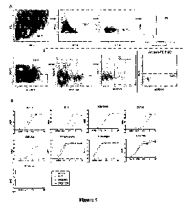
FIG. 5. Purification of DNA-barcoded antigens. (A.) After barcoding each
antigen with a
unique oligonucleotide, antigen-oligo complexes are run on size exclusion
chromatography to
remove excess, unconjugated oligonucleotide from the reaction mixture. DNA-
barcoded BG505
was run on the Superose 6 Increase 10/300 GL column and all other DNA-barcoded
antigens were
25 run on the Superdex 200 Increase 10/300 GL on the AKTA FPLC system. For
size exclusion
chromatography, dotted lines indicate DNA-barcoded antigens and fractions
taken. The second
peak indicates excess oligonucleotide from the conjugation reaction. (B.)
Binding of VRCO1 or
Fe53 Ramos B-cell lines to DNA-barcoded, fluorescently labeled antigens via
flow cytometiy.
VRCO1 cells bound to DNA-barcocled BG505-PE, DNA-barcocled CZA97-PE, and not
DNA-
30 barcoded H1 A/New Caledonia/20/99-PE. Fe53 cells bound to DNA-barcoded H1
A/New
Caledonia/20/99-PE.
FIG. 6. Ramos B-cell line sorting scheme. (A.) Gating scheme for fluorescence
activated
cell sorting of Ramos B-cell lines. VRC01 and Fe53 Ramos B cells were mixed in
a 1:1 ratio and
8
CA 03150030 2022-3-3
WO 2021/046299
PCT/US2020/049330
then stained with LiveDead-V500 and a DNA-barcoded antigen screening library
consisting of
BG505-PE, CZA97-PE, and 111 A/New Caledonia/20/99-PE. Gates as drawn are based
on gates
used during the sort, and percentages from the sort are listed. (B.) For each
experiment, the
categorization of the number of Cellranger-identified (10X Genomics) cells
after sequencing is
5 shown. Each category (row) is a subset of cells of the previous category
(row).
FIG. 7. Identification of antigen-specific B cells from donor NIAID 45 PBMCs.
(A.)
Gating scheme for fluorescence activated cell sorting of donor NIAID 45 PBMCs.
Cells were
stained with LiveDead-V500, CD14-V500, CD3-APCC y7, CD19-BV711, IgG-FITC, and
a DNA-
barcoded antigen screening library consisting of BG505-PE, CZA97-PE, and H1
A/New
10 Caledonia/20/99-PE. Gates as drawn are based on gates used during the
sort, and percentages from
the sort are listed. These plots show a starting number of 50,187 total
events. Due to the
visualization parameters, 18 IgG-positive, antigen-positive cells are
displayed, but 3400 IgG were
sorted and supplemented with 13,000 antigen positive B cells for single cell
sequencing. A small
aliquot of donor NIAID45 PBMCs were used for fluorescence minus one (FMO)
staining, and
15 were stained with the same antibody panel as listed above with the
exception of the HIV-1 and
influenza antigens. (B.) L1BRA-seq scores for BG505 (x-axis) and CZA97 (y-
axis) are shown.
Each axis represents the range of LIBRA-seq scores for each antigen. Density
of total cells is
shown. Overlaid on the density plot are the 29 VRCO1 lineage members (dots)
indicated in light
green. (C.) Antigen specificity as predicted by LIBRA-seq was validated by
ELISA for a variety
20 of antibodies isolated from donor NIAID 45. Antibodies were tested for
binding to BG505,
CZA97, and H1 A/New Caledonia/20/99. ELISA data are representative from at
least two
independent experiments.
FIG. 8. Characterization of antibody lineage 2121. (A.) Binding of BG505 DS-
SOSIP
trimer to (a) PGT145 IgG, (b) VRCO1 IgG, (c) 17b IgG, and (d) 2723-2121 IgG.
(B.) Inhibition
25 of BG505 DS-SOSIP binding to 2723-2121 IgG in presence of VRC34 Fab
(diamond), PGT145
Fab (square) and VRCO1 Fab (triangle). (C.) Neutralization of Tier 1, Tier 2,
and control viruses
by antibody 2723-2121 and VRCO1. Results are shown as the concentration of
antibody (in CI g/m1)
needed for 50% inhibition (IC50). (D.) Levels of ADCP, ADCD, ADCT-PICH26 and
ADCC
displayed by antibody 2723-2121 compared to VRCO1. HIVIG was used as a
positive control and
30 the anti-RSV InAb Palivisumab as a negative control.
FIG. 9. Identification of antigen-specific B cells from donor N90 PBMCs. (A.)
Gating
scheme for fluorescence activated cell sorting of donor N90 PBMCs. Cells were
stained LiveDead-
APCCy7, CD14-APCCy7, CD3-FITC, CD19-BV711, and IgG-PECy5 with and a DNA-
barcoded
9
CA 03150030 2022-3-3
WO 2021/046299
PCT/US2020/049330
antigen screening library consisting of BG505-PE, KNH1144-PE, ZM197-PE,
ZM106.9-PE, B41-
PE, 111 A/New Caledonia/20/99-PE, H1 A/Michigan/45/2015-PE, H5
Indonesia/5/2005-PE, 117
Anhui/1/2013-PE. Gates as drawn are based on gates used during the sort, and
percentages from
the sort are listed. 5450 IgG positive, antigen positive cells were sorted and
supplemented with
5
1480 IgG negative, antigen positive B cells
for single cell sequencing. A small aliquot of donor
N90 PBMCs were used for fluorescence minus one (FM0) staining, and were
stained with the
same antibody panel as listed above without the antigen screening library.
(B.) Antigen specificity
as predicted by LIBRA-seq was validated by ELISA for two antibodies isolated
from donor N90.
Antibodies were tested for binding to all antigens from the screening library:
5 HIV-1 SOSIP
10
(BG505, KNH1144, ZM197, ZM106.9, B41), and 4
influenza HA (H1 A/New Caledonia/20/99,
H1 A/Michigan/45/2015, 115 Indonesia/5/2005, 117 Anhui/1/2013). ELISA data are
representative
from at least two independent experiments.
FIG_ 10. Each graph shows the LIBRA-seq score for an HIV antigen (y-axes) vs.
an
influenza antigen (x-axes) in the screening library. The 901 cells that had a
LIBRA-seq scorn
15
above one for at least one antigen are
displayed as individual dots. IgG cells (591 of 901) are
colored orange and cells of all other isotypes are colored blue. Red lines on
each axis indicate a
LIBRA-seq score of one. Only 9 of the 591 IgG cells displayed high LIBRA-seq
scores for at least
one HIV-1 antigen and one influenza antigen, confirming the ability of the
technology to
successfully discriminate between diverse antigen specificities.
20
FIG_ 11. Sequencing preprocessing and quality
statistics_ (A.) Quality filtering of the
antigen barcode FASTQ files. Fastp (Chen et al., 2018) was used to trim
adapters and remove low-
quality reads using default parameters. Shown are read and base statistics
generated from the
output html report from each of the Ramos B cell experiment (left), primary B
cell experiment
from donor NIAID45 (middle), and primary B cell experiment from donor N90
(right). (B.) Shown
25
is a distribution of insert sizes of the
antigen barcode reads from the Ramos B cell line experiment,
as output from the fastp html report. (C.) Shown is a distribution of insert
sizes of the antigen
barcode reads from the donor NIAID45 experiment, as output from the fastp
halal report. (D.)
Shown is a distribution of insert sizes of the antigen barcode reads from the
donor NIH90
experiment, as output from the fastp html report
30
FIG_ 12. Architecture of antigen barcode
library. The antigen barcode library is composed
of the cell barcode, unique molecular identifier, a capture sequences (the
template switch oligo
sequence), and an antigen barcode.
CA 03150030 2022-3-3
WO 2021/046299
PCT/US2020/049330
FIG. 13. Schematic of cell barcode - antigen barcode UMI count matrix. This is
created
from the sequencing of antigen barcode libraries and used in subsequent
analysis to determine
antigen specificity.
5 DETAILED DESCRIPTION
Recent advances in next-generation sequencing (NGS) enable high-throughput
interrogation of antibody repertoires at the sequence level, including paired
heavy and light chains
(Busse et al., 2014; Dekosky et al., 2013; Tan et al., 2014). However,
annotation of NGS antibody
sequences for their cognate antigen partner(s) generally requires synthesis,
production and
10 characterization of individual recombinant monoclonal antibodies
(DeFalco et al., 2018; Setliff et
al., 2018). Recent efforts to develop new antibody screening technologies have
sought to overcome
throughput limitations while still uniting antibody sequence and functional
information. For
example, natively-paired human BCR heavy and light chain amplicons can be
expressed and
screened as Fab (Wang et al., 2018) or scFV (Adler et al., 2017b, 2017a) in a
yeast display system.
15 Although these various antibody discovery technologies have led to the
identification of a number
of potently neutralizing antibodies, they remain limited by the number of
antigens against which
single cells can simultaneously be screened efficiently.
L,IBRA-seq (jjnking B Cell Receptor to Antigen specificity through sequencing)
is
developed to simultaneously recover both antigen specificity and paired heavy
and light chain
20 BCR sequence. LIBRA-seq is a next-generation sequencing-based readout for
BCR-antigen
binding interactions that utilizes oligonucleotides (oligos) conjugated to
recombinant antigens.
Antigen barcocles are recovered during paired-chain BCR sequencing experiments
and
bioinformatically mapped to single cells. The LIBRA-seq method was applied to
PBMC samples
from two HIV-infected subjects, and from these, HIV- and influenza-specific
antibodies were
25 successfully identified, including both known and novel broadly
neutralizing antibody (bNAb)
lineages. LIBRA-seq is high-throughput, scalable, and applicable to many
targets. This single,
integrated assay enables the mapping of monoclonal antibody sequences to
panels of diverse
antigens theoretically unlimited in number and facilitates the rapid
identification of cross-reactive
antibodies that serves as therapeutics or vaccine templates.
30 Disclosed herein are systems and methods for simultaneous
detection of antigens and
antigen specific antibodies.
11
CA 03150030 2022-3-3
WO 2021/046299
PCT/US2020/049330
Reference will now be made in detail to the embodiments of the invention,
examples of
which are illustrated in the drawings and the examples. This invention may,
however, be embodied
in many different forms and should not be construed as limited to the
embodiments set forth herein.
Unless defined otherwise, all technical and scientific terms used herein have
the same
5 meaning as commonly understood to one of ordinary skill in the art to
which this disclosure
belongs. The term "comprising" and variations thereof as used herein is used
synonymously with
the term "including" and variations thereof and are open, non-limiting terms.
Although the terms
"comprising" and "including" have been used herein to describe various
embodiments, the terms
"consisting essentially of' and "consisting of' can be used in place of
"comprising" and
-h) "including" to provide for more specific embodiments and are also
disclosed. As used in this
disclosure and in the appended claims, the singular forms "a", "an", "the",
include plural referents
unless the context clearly dictates otherwise.
The following definitions are provided for the full understanding of terms
used in this
specification.
Terminology
As used herein, the terms "may," "optionally," and "may optionally" are used
interchangeably and are meant to include cases in which the condition occurs
as well as cases in
which the condition does not occur. Thus, for example, the statement that a
formulation "may
20 include an excipient" is meant to include cases in which the formulation
includes an excipient as
well as cases in which the formulation does not include an excipient.
As used herein, the term "subject" or "host" can refer to living organisms
such as
mammals, including, but not limited to humans, livestock, dogs, cats, and
other mammals.
Administration of the therapeutic agents can be carried out at dosages and for
periods of time
25 effective for treatment of a subject. In some embodiments, the subject
is a human.
"Nucleotide," "nucleoside," "nucleotide residue," and "nucleoside residue," as
used
herein, can mean a deoxyribonucleotide or ribonucleotide residue, or other
similar nucleoside
analogue. A nucleotide is a molecule that contains a base moiety, a sugar
moiety and a phosphate
moiety. Nucleotides can be linked together through their phosphate moieties
and sugar moieties
30 creating an internucleoside linkage. The base moiety of a nucleotide can
be adenin-9-y1 (A),
cytosin- 1 -y1 (C), guanin-9-y1 (G), uracil-1-y1 (U), and thymin- 1-y1 (T).
The sugar moiety of a
nucleotide is a ribose or a deoxyribose. The phosphate moiety of a nucleotide
is pentavalent
phosphate. A non-limiting example of a nucleotide would be 3'-AMP (3'-
adenosine
12
CA 03150030 2022-3-3
WO 2021/046299
PCT/US2020/049330
monophosphate) or 5'-GMP (5'-guanosine monophosphate). There are many
varieties of these
types of molecules available in the art and available herein.
The term "polynucleotide" refers to a single or double stranded polymer
composed of
nucleotide monomers.
5 The method and the system disclosed here including the use of
primers, which are capable
of interacting with the disclosed nucleic acids, such as the antigen barcode
as disclosed herein. In
certain embodiments the primers are used to support DNA amplification
reactions. Typically, the
primers will be capable of being extended in a sequence specific manner.
Extension of a primer in
a sequence specific manner includes any methods wherein the sequence and/or
composition of the
ri nucleic acid molecule to which the primer is hybridized or otherwise
associated directs or
influences the composition or sequence of the product produced by the
extension of the primer.
Extension of the primer in a sequence specific manner therefore includes, but
is not limited to,
PCR, DNA sequencing, DNA extension, DNA polymerization, RNA transcription, or
reverse
transcription. Techniques and conditions that amplify the primer in a sequence
specific manner
15 are preferred. In certain embodiments the primers are used for the DNA
amplification reactions,
such as PCR or direct sequencing. It is understood that in certain embodiments
the primers can
also be extended using non-enzymatic techniques, where for example, the
nucleotides or
oligonucleotides used to extend the primer are modified such that they will
chemically react to
extend the primer in a sequence specific manner. Typically, the disclosed
primers hybridize with
20 the disclosed nucleic acids or region of the nucleic acids or they
hybridize with the complement
of the nucleic acids or complement of a region of the nucleic acids.
The term "amplification" refers to the production of one or more copies of a
genetic
fragment or target sequence, specifically the "amplicon". As it refers to the
product of an
amplification reaction, amplicon is used interchangeably with conunon
laboratory terms, such as
25 "PCR product."
The term "polypeptide" refers to a compound made up of a single chain of D- or
L-amino
acids or a mixture of D- and L-amino acids joined by peptide bonds.
As used herein, the term "antigen" refers to a molecule that is capable of
stimulating an
immune response such as by production of antibodies specific for the antigen.
Antigens of the
30 present invention can be, for example, an antigen from human
immunodeficiency virus (HIV), an
antigen from influenza virus, or an antigen from respiratory syncytial virus
(RSV). Antigens of
the present invention can also be, for example, a human antigen (e.g. an
oncogene-encoded
protein).
13
CA 03150030 2022-3-3
WO 2021/046299
PCT/US2020/049330
In the present invention, "specific for" and "specificity" means a condition
where one of
the molecules involved in selective binding. Accordingly, an antibody that is
specific for one
antigen selectively binds that antigen and not other antigens.
The term "antibodies" is used herein in a broad sense and includes both
polyclonal and
5 monoclonal antibodies. In addition to intact inamunoglobulin molecules,
also included in the term
"antibodies" are fragments or polymers of those immunoglobufin molecules, and
human or
humanized versions of immunoglobulin molecules or fragments thereof, as long
as they are chosen
for their ability to specifically interact with the HIV virus, such that the
HIV viral infection is
prevented, inhibited, reduced, or delayed_ The antibodies can be tested for
their desired activity
10 using the in vitro assays described herein, or by analogous methods,
after which their in vivo
therapeutic and/or prophylactic activities are tested according to known
clinical testing methods.
There are five major classes of human inununoglobulins: IgA, IgD, IgE, IgG and
IgM, and several
of these may be further divided into subclasses (isotypes), e.g., IgG-1, IgG-
2, IgG-3, and IgG-4;
IgA-1 and IgA-2. One skilled in the art would recognize the comparable classes
for mouse. The
15 heavy chain constant domains that correspond to the different classes of
immunoglobulins are
called alpha, delta, epsilon, gamma, and mu, respectively.
Each antibody molecule is made up of the protein products of two genes, heavy-
chain gene
and light-chain gene. The heavy-chain gene is constructed through somatic
recombination of V,
D, and 3 gene segments. In humans, there are 51 VII, 27 DH, 6311, 9 CH gene
segments on human
20 chromosome 14_ The light-chain gene is constructed through somatic
recombination of V and J
gene segments. Them are 40 Vic, 31 VA., 53K , 41k gene segments on human
chromosome 14(80
VJ). The heavy-chain constant domains that correspond to the different classes
of
inununoglobulins are called a, 3, a, y, and it, respectively. The "fight
chains" of antibodies from
any vertebrate species can be assigned to one of two clearly distinct types,
called kappa (K) and
25 lambda Q,), based on the amino acid sequences of their constant domains.
The term "monoclonal antibody" as used herein refers to an antibody obtained
from a
substantially homogeneous population of antibodies, i.e., the individual
antibodies within the
population are identical except for possible naturally occurring mutations
that may be present in a
small subset of the antibody molecules. The monoclonal antibodies herein
specifically include
30 "chimeric" antibodies in which a portion of the heavy and/or light chain
is identical with or
homologous to corresponding sequences in antibodies derived from a particular
species or
belonging to a particular antibody class or subclass, while the remainder of
the chain(s) is identical
with or homologous to corresponding sequences in antibodies derived from
another species or
14
CA 03150030 2022-3-3
WO 2021/046299
PCT/US2020/049330
belonging to another antibody class or subclass, as well as fragments of such
antibodies, as long
as they exhibit the desired antagonistic activity.
The disclosed monoclonal antibodies can be made using any procedure which
produces
monoclonal antibodies. For example, disclosed monoclonal antibodies can be
prepared using
5
hybridoma methods, such as those described by
Kohler and Milstein, Nature, 256:495 (1975). In
a hybridoma method, a mouse or other appropriate host animal is typically
immunized with an
immunizing agent to elicit lymphocytes that produce or are capable of
producing antibodies that
will specifically bind to the immunizing agent. Alternatively, the lymphocytes
may be immunized
in vitro.
10
The monoclonal antibodies may also be made by
recombinant DNA methods. DNA
encoding the disclosed monoclonal antibodies can be readily isolated and
sequenced using
conventional procedures (e.g., by using oligonucleotide probes that are
capable of binding
specifically to genes encoding the heavy and light chains of murine
antibodies). Libraries of
antibodies or active antibody fragments can also be generated and screened
using phage display
15
techniques, e.g., as described in U.S. Patent
No. 5,804,440 to Burton et al. and U.S. Patent No.
6,096,441 to Barbas et at.
In vitro methods are also suitable for preparing monovalent antibodies.
Digestion of
antibodies to produce fragments thereof, particularly, Fab fragments, can be
accomplished using
routine techniques known in the art. For instance, digestion can be performed
using papain.
20
Examples of papain digestion are described in
WO 94/29348 published Dec_ 22, 1994 and U.S.
Pat. No. 4,342,566. Papain digestion of antibodies typically produces two
identical antigen binding
fragments, called Fab fragments, each with a single antigen binding site, and
a residual Fc
fragment. Pepsin treatment yields a fragment that has two antigen combining
sites and is still
capable of cross linking antigen.
25
As used herein, the term "antibody or antigen
binding fragment thereof' or "antibody or
fragments thereof" encompasses chimeric antibodks and hybrid antibodks, with
dual or multiple
antigen or epitope specificities, and fragments, such as F(ab')2, Fab', Fab,
Fv, sFy, scFy and the
like, including hybrid fragments. Thus, fragments of the antibodies that
retain the ability to bind
their specific antigens are provided. For example, fragments of antibodies
which maintain HIV
30
virus binding activity are included within the
meaning of the term "antibody or antigen binding
fragment thereof" Such antibodies and fragments can be made by techniques
known in the art
and can be screened for specificity and activity according to the methods set
forth in the Examples
and in genet-al methods for producing antibodies and screening antibodies for
specificity and
CA 03150030 2022-3-3
WO 2021/046299
PCT/US2020/049330
activity (See Harlow and Lane. Antibodies, A Laboratory Manual. Cold Spring
Harbor
Publications, New York, (1988)).
Also included within the meaning of "antibody or antigen binding fragment
thereof' are
conjugates of antibody fragments and antigen binding proteins (single chain
antibodies). Also
included within the meaning of "antibody or antigen binding fragment thereof'
are
immunoglobulin single variable domains, such as for example a nanobody.
The fragments, whether attached to other sequences or not, can also include
insertions,
deletions, substitutions, or other selected modifications of particular
regions or specific amino
acids residues, provided the activity of the antibody or antibody fragment is
not significantly
altered or impaired compared to the non-modified antibody or antibody
fragment. These
modifications can provide for some additional property, such as to remove/add
amino acids
capable of disulfide bonding, to increase its bio-longevity, to alter its
secretory characteristics, etc.
In any case, the antibody or antibody fragment must possess a bioactive
property, such as specific
binding to its cognate antigen. Functional or active regions of the antibody
or antibody fragment
may be identified by mutagenesis of a specific region of the protein, followed
by expression and
testing of the expressed polypeptide. Such methods are readily apparent to a
skilled practitioner
in the art and can include site-specific mutagenesis of the nucleic acid
encoding the antibody or
antibody fragment. (Zoller, M.J. Curr. Opin. Biotechnol. 3:348-354, 1992).
As used herein, the term "antibody" or "antibodies" can also refer to a human
antibody
and/or a humanized antibody. Many non-human antibodies (e.g., those derived
from mice, rats, or
rabbits) are naturally antigenic in humans, and thus can give rise to
undesirable immune responses
when administered to humans. Therefore, the use of human or humanized
antibodies in the
methods serves to lessen the chance that an antibody administered to a human
will evoke an
undesirable immune response.
"Pharmaceutically acceptable" component can refer to a component that is not
biologically
or otherwise undesirable, i.e., the component may be incorporated into a
pharmaceutical
formulation of the invention and administered to a subject as described herein
without causing
significant undesirable biological effects or interacting in a deleterious
manner with any of the
other components of the formulation in which it is contained_ When used in
reference to
administration to a human, the term generally implies the component has met
the required
standards of toxicological and manufacturing testing or that it is included on
the Inactive
Ingredient Guide prepared by the U.S. Food and Drug Administration.
16
CA 03150030 2022-3-3
WO 2021/046299
PCT/US2020/049330
"Pharmaceutically acceptable carrier" (sometimes referred to as a "carrier")
means a carrier
or excipient that is useful in preparing a pharmaceutical or therapeutic
composition that is
generally safe and non-toxic, and includes a carrier that is acceptable for
veterinary and/or human
pharmaceutical or therapeutic use. The terms "carrier" or "pharmaceutically
acceptable carrier"
5 can include, but are not limited to, phosphate buffered saline solution,
water, emulsions (such as
an oil/water or water/oil emulsion) and/or various types of wetting agents.
As used herein, the terms "treating" or "treatment" of a subject includes the
administration
of a drug to a subject with the purpose of curing, healing, alleviating,
relieving, altering,
remedying, ameliorating, improving, stabilizing or affecting a disease or
disorder, or a symptom
10 of a disease or disorder_ The terms "treating" and "treatment" can also
refer to reduction in severity
and/or frequency of symptoms, elimination of symptoms and/or underlying cause,
and
improvement or remediation of damage.
"Therapeutically effective amount" or "therapeutically effective dose" of a
composition
refers to an amount that is effective to achieve a desired therapeutic result.
Therapeutically
15 effective amounts of a given therapeutic agent will typically vary with
respect to factors such as
the type and severity of the disorder or disease being treated and the age,
gender, and weight of
the subject. The term can also refer to an amount of a therapeutic agent, or a
rate of delivery of a
therapeutic agent (e.g., amount over time), effective to facilitate a desired
therapeutic effect, such
as coughing relief. The precise desired therapeutic effect will vary according
to the condition to
20 be treated, the tolerance of the subject, the agent and/or agent
formulation to be administered (e.g.,
the potency of the therapeutic agent, the concentration of agent in the
formulation, and the like),
and a variety of other factors that are appreciated by those of ordinary skill
in the art In some
instances, a desired biological or medical response is achieved following
administration of
multiple dosages of the composition to the subject over a period of days,
weeks, or years.
Methods
In some aspects, disclosed herein is a method for simultaneous detection of an
antigen and
an antibody that specifically binds said antigen, comprising:
labeling a plurality of antigens with unique antigen barcodes;
30 providing a plurality of barcode-labeled antigens to a population
of B-cells;
allowing the plurality of barcode-labeled antigens to bind to the population
of B-cells;
washing unbound antigens from the population of B-cells;
separating the B-cells into single cell emulsions;
17
CA 03150030 2022-3-3
WO 2021/046299
PCT/US2020/049330
introducing into each single cell emulsion a unique cell barcode-labeled bead;
preparing a single cell cDNA library from the single cell emulsions;
performing PCR amplification reactions to produce a plurality of amplicons,
wherein the
amplicons comprise: 1) the cell barcode and the antigen barcode, 2) the cell
barcode
5 and an antibody sequence, and 3) a unique molecular identifier
(UMI);
sequencing the plurality of amplicons;
removing a sequence lacking the cell barcode, the UMI, or the antigen barcode;
aligning the antibody sequence to a reference library of immunoglobulin V. D,
J and C
sequences;
10 constructing a UMI count matrix comprising the cell barcode, the
antigen barcode, and
the antibody sequence;
determining a LIBRA-seq score; and
determining that the antibody specifically binds an antigen if the LIBRA-seq
score of the
antibody for the antigen is increased in comparison to a control sample.
15
Following a LIBRA-seq experiment, there are 2
resulting pairs of FASTQ files: (1) B cell
receptor libraries (containing heavy and light chain contigs), and (2) antigen
barcode libraries
(containing antigen-identifying DNA barcode sequences from the antigen
screening library). In
some embodiments, it should be understood that the methods described herein
are for uniting the
information from these two sequencing libraries. Accordingly, in some
embodiments, the above
20
noted step of removing a sequence lacking the
cell barcode, the UMI, or the antigen barcode is for
removing a sequence from the antigen barcode library lacking the cell barcode,
the UMI, or the
antigen barcode. The general structure of the antigen barcode should be look
like, for example.
FIG. 1 disclosed herein. The methods describe here are for processing the
antigen barcodes. The
processing serves two purposes: (1) quality control and annotation of
sequenced reads, and (2)
25
identification of binding signal from the
annotated sequenced reads. Before the following steps
are carried out, the BCR libraries are processed in order to determine the
list of cell barcotles that
have a VDJ sequence.
Processing of antigen barcode reads and BCR sequence contigs. A pipeline shown
herein
takes paired-end fastq files of oligo libraries as input, processes and
annotates reads for cell
30
barcode, UMI, and antigen barcode, and
generates a cell barcode - antigen barcode UMI count
matrix. BCR contigs are processed using cellranger (10X Genomics) using GRCh38
as reference.
For the antigen barcode libraries, initial quality and length filtering is
carded out by fastp (Chen
et at., 2018) using default parameters for filtering. This results in only
high-quality reads being
18
CA 03150030 2022-3-3
WO 2021/046299
PCT/US2020/049330
retained in the antigen barcode library (FIG. 11). In a histogram of insert
lengths, this results in a
sharp peak of the expected insert size of 52-54 (FIG. 9B-9C). Fastx_collapser
is then used to group
identical sequences and convert the output to deduplicated fasta files. Then,
having removed low-
quality reads, just the R2 sequences were processed, as the entire insert is
present in both R1 and
5 R2. Each unique R2 sequence (or R1, or the consensus of R1 and R2) was
processed one by one
using the following steps:
(1) The reverse complement of the R2 sequence is determined (Skip step 1 if
using R1).
(2) The sequence is screened for possessing an exact match to any of the valid
10X cell
barcodes present in the filtered_contiglasta file output by cell ranger during
processing of BCR
10 V(D)J fastq files. Sequences without a BCR-associated cell barcode are
discarded.
(3) The 10 bases immediate 3* to the cell barcode are annotated as the read's
UMI.
(4) The remainder of the sequence 3' to the UM' is screened for a 13 or 15 bp
sequence
with a hamming distance of 0, 1, or 2 to any of the antigen barcodes used in
the screening library.
Following this processing, only sequences around the expected lengths are
retained (the lengths
15 of sequences can be from more than 1, 2, 3, 4, 5, 6, 7, 8, 9, 10, 11,
12, 13, 14, 15, 16, 17, 18, 19,
20, 21, 22, 23, 24, 25, 26, 27, 28, 29, or 30 bases shorter to more than 1, 2,
3, 4, 5, 6, 7, 8, 9, 10,
11, 12, 13, 14, 15, 16, 17, 18, 19, 20, 21, 22, 23, 24, 25, 26, 27, 28, 29, or
30 bases longer than the
expected lengths), thus allowing for a deletion, an insertion outside the cell
barcode, or bases
flanking the cell barcode.
20 This general process requires that sequences possess all elements
needed for analysis (cell
barcode, UMI, and antigen barcode), but is permissive to insertions or
deletions in the TS0 region
between the UMI and antigen barcode. After processing each sequence one-by-
one, cell barcode
- UMI - antigen barcode collisions are screened. Any cell barcode - UMI
combination (indicative
of a unique oligo molecule) that has multiple antigen barcthies associated
with it is removed. A
25 cell barcode - antigen barcode UMI count matrix is then constructed,
which served as the basis of
subsequent analysis. Additionally, the BCR contigs are aligned
(filtered_contigs.fasta file output
by Celhanger, 10X Genomics) to IMGT reference genes using HighV-Quest (Alamyar
et al.,
2012). The output of High V-Quest is parsed using Change() (Gupta et al.,
2015), and merged with
the UMI count matrix.
30 The above stated procedure can be summarized as the following
steps:
1) Remove low quality reads;
2) Remove reads too long or too short to be a valid antigen barcode read
containing a cell
barcode, UMI, and antigen barcode;
19
CA 03150030 2022-3-3
WO 2021/046299
PCT/US2020/049330
3) For each quality read, annotate:
a. Cell barcode,
b. UMI
c. Antigen barcocle, allowing for sequencing/PCR errors by using a hamming
distance
5 threshold.
Determination of LIBRA -seq Score. Starting with the UMI count matrix, all
counts of more
than one UMIs (for example, more than 1, 2, 3,4, 5, 6, 7, 8, 9, 10, 11, 12,
13, 14, 15, 16, 17, 18,
19, 20 UMIs) were set to 0, with the idea that these low counts can be
attributed to noise. After
this, the UMI count matrix was subset to contain only cells with a count of
one or more UMIs than
10
the minimum value in the above noted step of
noise filtering for at least 1 antigen. The centered-
log ratios (CLR) of each antigen UMI count for each cell were then calculated
(Mimitou et al.,
2019; Stoeckius et al., 2017,2018). Because UMI counts were on different
scales for each antigen,
due to differential oligo loading during oligo-antigen conjugation, the CLRs
UMI counts were
resealed using the StandardSealer method in scikit learn (Pedregosa and
Varoquaux, 2011). Lastly,
15
A correction procedure was performed to the z-
score-normalized CLRs from UMI counts of 0,
setting them to the minimum for each antigen for donor NIAID 45 and N90
experiments, and to -
1 for the Ramos B cell line experiment. These CLR-transformed, Z-score-
normalized, corrected
values served as the final LIBRA-seq scores. LIBRA-seq scores were visualized
using Cytobank
(Kotecha et al., 2010).
20
Identification of sequence feature ¨ antigen
specificity associations. Following
determination of LIBRA-seq scores (above), and because antibody sequence is
united with antigen
specificity (in the form of a LIBRA-seq score), sequence-specificity
associations can be made.
Accordingly, in some embodiments, the method of any preceding aspect further
comprises
determining a level of somatic hypermutation of the antibody specifically
binding to the antigen
25
In some embodiments, the method of any
preceding aspect further comprises determining
a length of a complementarity-determining region (CDR) of the antibody
specifically binding to
the antigen. The term "complementarity determining region (CDR)" used herein
refers to an amino
acid sequence of an antibody variable region of a heavy chain or light chain.
CDRs are necessary
for antigen binding and determine the specificity of an antibody. Each
variable region typically
30
has three CDRs identified as CDR1 (CDRH1 or
CDRL1, where "H" indicates the heavy chain
CDR1 and "L" indicates the light chain CDR1), CDR2 (CDRH2 or CDRL2), and CDR3
(CDRH3
or CDRL3). The CDRs may provide contact residues that play a major role in the
binding of
CA 03150030 2022-3-3
WO 2021/046299
PCT/US2020/049330
antibodies to antigens or epitopes. Four framework regions, which have more
highly conserved
amino acid sequences than the CDRs, separate the CDR regions in the VII or VL.
Accordingly, in some embodiments, the method of any preceding aspect further
comprises
determining a motif of a CDR of the antibody specifically binding to the
antigen. In some
embodiments, the CDR is selected from the group consisting of CDRH1, CDRH2,
CDRH3,
CDRL1, CDRL2, and CDRL3.
In some embodiments, the method of any preceding aspect further comprises
identification
of IGHV, IGHD, IGHI, IGKV, IGKJ, IGLV, or IGLI genes, or combinations thereof,
associated
with any particular combination of antigen specificities.
In some embodiments, the method of any preceding aspect further comprises
identification
of mutations in heavy or light FW I, FW2, FW 3 or FW4 associated with any
particular combination
of antigen specificities.
In some embodiments, the method of any preceding aspect further comprises
identification
of overall gene expression profiles or select up- or down-regulated genes
associated with any
particular combination of antigen specificities.
In some embodiments, the method of any preceding aspect further comprises
identification
of surface markers, via, for example, fluorescence-activated cell sorting, or
oligo-conjugated
antibodies associated with any particular combination of antigen specificities
In some embodiments, the method of any preceding aspect further comprises
identification
of any combination of BCR sequence feature (for example, immunoglobulin gene,
sequence motif,
or CDR length), gene expression profile, or surface marker profile associated
with any particular
combination of antigen specificities.
In some embodiments, the method of any preceding aspect further comprises
training a
machine learning algorithm on sequence features, sequence motifs, or encoded
sequence
properties (such as via Kidera factors), associated with any particular
combination of antigen
specificities for subsequent application to sequenced antibodies lacking
antigen specificity
information due to not using LIBRA-seq or otherwise.
In some aspects, disclosed herein is a method for simultaneous detection of an
antigen and
an antibody that specifically binds said antigen, comprising:
labeling a plurality of antigens with unique antigen barcodes;
providing a plurality of barcocle-labeled antigens to a population of B-cells;
allowing the plurality of barcode-labeled antigens to bind to the population
of B-cells;
washing unbound antigens from the population of B-cells;
21
CA 03150030 2022-3-3
WO 2021/046299
PCT/US2020/049330
separating the B-cells into single cell emulsions;
introducing into each single cell emulsion a unique cell barcode-labeled bead;
preparing a single cell eDNA library from the single cell emulsions;
performing PCR amplification reactions to produce a plurality of amplicons,
wherein the
5
amplicons comprise: 1) the cell barcode and
the antigen barcode, 2) the cell barcode
and an antibody sequence, and 3) a unique molecular identifier (UMI);
sequencing the plurality of amplicons;
removing a sequence lacking the cell barcode, the UMI, or the antigen barcode;
aligning the antibody sequence to a reference library of immunoglobulin V. D,
J and C
10 sequences;
constructing a UMI count matrix comprising the cell barcode, the antigen
barcode, and the
antibody sequence;
determining a LIBRA-seq score; and
determining that the antibody specifically binds an antigen if the LIBRA-seq
score of the
15 antibody for the antigen is increased in comparison to a
control sample.
In some embodiments, the barcode-labeled antigens are labeled with a fast
barcode
comprising a DNA sequence or an RNA sequence. In some embodiments, the cell
barcode-labeled
beads are labeled with a second barcode comprising a DNA sequence or an RNA
sequence.
It should be understood that the barcode described above is conjugated to the
barcode-
20
labeled antigen in a way that are known to one
of ordinary skill in the art. Conjugates can be
chemically linked to the nucleotide or nucleotide analogs. Such conjugates
include but are not
limited to lipid moieties such as a cholesterol moiety (Letsinger et al.,
Proc. Natl. Acad. Sci. USA,
1989, 86, 6553-6556), cholic acid (Manoharan et at., Bioorg. Med. Chem. Let.,
1994, 4,
1053-1060), a thioether, e.g., hexyl-S-tritylthiol (Manoharan et al., Ann.
N.Y. Acad. Sci., 1992,
25
660, 306-309; Manoharan et al., Bioorg. Med.
Chem. Let., 1993, 3, 2765-2770), a thiocholesterol
(Oberhauser et al., Nucl. Acids Res., 1992, 20, 533-538), an aliphatic chain,
e.g., dodecandiol or
undecyl residues (Saison-Behmoaras et al., EMBO J., 1991, 10, 1111-1118;
Kabanov et al., FEBS
Lett., 1990, 259, 327-330; Svinarchuk et al., Biochimie, 1993, 75, 49-54), a
phospholipid, e.g., di-
hexadecyl-rac-glycerol or triethylammonium 1,2-di-O-hexadecyl-rac-glycero-3-H-
phosphonate
30
(Manoharan et al., Tetrahedron Let, 1995, 36,
3651-3654; Shea et at., Nucl. Acids Res., 1990, 18,
3777-3783), a polyamine or a polyethylene glycol chain (Manoharan et al.,
Nucleosides &
Nucleotides, 1995, 14, 969-973), or adamantane acetic acid (Manoharan et al,
Tetrahedron Lett.,
1995, 36, 3651-3654), a palmityl moiety (Mishra et at., Biochim. Biophys.
Acta, 1995, 1264,
22
CA 03150030 2022-3-3
WO 2021/046299
PCT/US2020/049330
229-237), or an octadecylamine or hexylatnino-carbonyl-oxycholesterol moiety
(Crooke et al., I
Pharmacy!. Exp. Ther., 1996, 277, 923-937. An oligonucleotide barcode can also
be conjugated
to an antigen using the Solulink Protein-Oligonucleotide Conjugation Kit
(TriLink cat no. 5-9011)
according to manufacturer's instructions. Briefly, the oligo and protein are
desalted, and then the
5
amino-oligo is modified with the 4FB
crosslinker, and the biotinylated antigen protein is modified
with S-HyNic. Then, the 4FB-oligo and the HyNic-antigen are mixed together.
This causes a stable
bond to form between the protein and the oligonucleotide. In some embodiments,
the cell barcode-
labeled beads are labeled with a second barcode comprising a DNA sequence or
an RNA sequence_
In some embodiments, the cell barcode-labeled beads are labeled with a second
barcode
10
comprising a DNA sequence. In some
embodiments, the cell barcode-labeled beads are labeled
with a second barcode comprising an RNA sequence. In some embodiments, the
cell barcode-
labeled beads are labeled with a barcode on the inside of the bead. In some
embodiments, the cell
barcode-labeled beads are labeled with a barcode encapsulated within the bead.
In some
embodiments, the cell barcode-labeled beads are labeled with a barcode on the
outside of the bead.
15
As used herein, "beads" is not limited to a
specific type of bead. Rather, a large number of
beads are available and are known to one of ordinary skill in the art. A
suitable bead may be
selected on the basis of the desired end use and suitability for various
protocols. In some
embodiments, the bead is or comprises a particle or a bead. In some
embodiments, the solid support
bead is magnetic_ Beads comprise particles have been described in the prior
art in, for example,
20 U.S. Pat No. 5,084,169, US. Pat. No. 5,079,155, US. Pat. No. 473,231, and
U.S. Pat. No.
8,110,351. The particle or bead size can be optimized for binding B cell in a
single cell emulsion
and optimized for the subsequent PCR reaction.
These oligos, which contain the cell barcode, both: (1) enable amplification
of cellular
inRNA transcripts through the template switch oligo that is part of the oligo
containing the cell
25
barcode, and (2) directly anneal to the
antigen barcode-containing oligos from the antigen. In some
embodiments, the oligos delivered from the beads have the general structure:
P5_PCR_handle ¨
Cell_barcode ¨ UM! ¨ Template_switch_oligo.
It is noted above that the antibody is determined as specifically binding an
antigen if the
LIB RA-seq score of the antibody for the antigen is increased in comparison to
a control sample_
30
It should be understood herein that, as taught
by FIG. 1C, between the minimum (y-axis, top) and
maximum (y-axis, bottom) LIBRA-seq score for each antigen, the ability of each
of 100 cutoffs
was tested for its ability to classify each antibody as antigen positive or
negative, where antigen
23
CA 03150030 2022-3-3
WO 2021/046299
PCT/US2020/049330
positive is defined as having a LIBRA-seq score greater than or equal to the
cutoff being evaluated
and antigen negative is defined as having a LIBRA-seq score below the cutoff.
In some embodiments, the antibody sequence comprises an immunoglobulin heavy
chain
(VDJ) sequence, or an immunoglobulin light chain (VJ) sequence. In some
embodiments, the
antibody sequence comprises an immunoglobulin heavy chain (VDJ) sequence. In
some
embodiments, the antibody sequence comprises an immunoglobulin light chain
(VJ) sequence.
In some embodiments, the barcork-labeled antigens comprise an antigen from a
pathogen
or an animal. In some embodiments, the barcode-labeled antigens comprise an
antigen from a
pathogen. In some embodiments, the barcode-labeled antigens comprise an
antigen from an
animal_ In some embodiments, the animal is a mammal, including, but not
limited to, primates
(e.g., humans and nonhuman primates), cows, sheep, goats, horses, dogs, cats,
rabbits, rats, mice
and the like. In some embodiments, the subject is a human.
In some embodiments, the antigen from a pathogen comprises an antigen from a
virus. In
some embodiments, the antigen from a virus comprises an antigen from human
immunodeficiency
virus (HIV), an antigen from influenza virus, or an antigen from respiratoiy
syncytial virus (RSV).
In some embodiments, the antigen from a virus comprises an antigen from human
immunodeficiency virus (HIV). In some embodiments, the antigen from a virus
comprises an
antigen from influenza virus. In some embodiments, the antigen from a virus
comprises an antigen
from respiratory syncytial virus (RSV).
In some embodiments, the antigen from HIV comprises an antigen from HIV-1. In
some
embodiments, the antigen from HIV comprises an antigen from HIV-2. In some
embodiments, the
antigen from HIV comprises HIV-1 Env. In some embodiments, the antigen from
influenza virus
comprises heinagglutinin (HA). In some embodiments, the antigen from RSV
comprises an RSV
F protein. In some embodiments, the antigen is selected from the antigens
listed in Table 1.
24
CA 03150030 2022-3-3
WO 2021/046299
PCT/US2020/049330
Table 1. Antigen screening library for human B-cell sample analysis. For a set
of pathogens,
shown are selected protein targets, number of strains, and resulting total
number of antigens in the
screening library.
# Antgehe
Pathogen Protein-targets =# Strains in Wary
CMV 2 25
Dengue E. prM 5 10
I-Feria-fills fl laBsAg 2 2
Hepetihs C E2. El E2 2
HIV./ gp140. gpl 23. MPER 3 9
HPV Li 3 3
HSV-1 913 1
Entusrzs HA NA 12
Malaria 12?C9F 1
Measies H. F 1 2
Mumps HN, NP 1 2 10
Nocovir,:sP 10 10
fthinovinas VPi 6 6
ReteµArus VP7. VP.4 a
FtSY F. G 4 a
Rubsila El 1
Staphy1ocomisatireus HtsA, Se*, lsd8, EstD 1 4
UPEC Hrna. MA. FyuA, issA 1
Zika pilyl 1 2
15 *influenza: A (6 HA, 4 NA) and B (2 HA);
Arotavirus: 6G. 2 P variants)
In some embodiments, the population of B-cells comprise a memory B-cell, a
plasma cell,
a naïve B cell, an activated B-cell, or a B-cell line. In some embodiments,
the population of B-
cells comprise a memory B-cell, a plasma cell, a naïve B cell, an activated B-
cell, or a B-cell line.
20 In some embodiments, the population of B-cells comprise a
plasma cell. In some embodiments,
the population of B-cells comprise a naïve B cell. In some embodiments, the
population of B-cells
comprise an activated B-cell. In some embodiments, the population of B-cells
comprise a B-cell
line.
In another aspect, disclosed herein is a method of determining a broadly
neutralizing
25 antibody to a pathogen, said method comprising:
labeling a plurality of antigens derived from the pathogen with unique antigen
barcodes;
providing a plurality of barcode-labeled antigens to a population of B-cells;
allowing the plurality of barcode-labeled antigens to bind to the population
of B-cells;
washing unbound antigens from the population of B-cells;
30 separating the B-cells into single cell emulsions;
introducing into each single cell emulsion a unique cell barcode-labeled bead;
preparing a single cell cDNA library from the single cell emulsions;
CA 03150030 2022-3-3
WO 2021/046299
PCT/US2020/049330
performing PCR amplification reactions to produce a plurality of amplicons,
wherein the
amplicons comprise: 1) the cell barcode and the antigen barcode, 2) the cell
barcode
and an antibody sequence, and 3) a unique molecular identifier (UMI);
sequencing the plurality of amplicons;
5 removing a sequence lacking a cell barcode, unique molecular
identifier (UMI), or an
antigen barcode;
aligning the antibody sequence to a reference library of immunoglobulin V, D,
J and C
sequences;
constructing a UMI count matrix comprising the cell barcode, the antigen
barcode, and the
10 antibody sequence;
determining a LIBRA-seq score; and
determining that the antibody is a broadly neutralizing antibody if the LIBRA-
seq scores of the
antibody for two or more antigens are increased in comparison to a control.
15 Polypeptides and polynucleotides
In some aspects, disclosed herein is a polynucleotide comprising a sequence
set forth in
the specification.
In some aspects, disclosed herein is a polypeptide, wherein the polypeptide is
encoded by
a polynucleotide sequence set forth in the specification.
20 In some aspects, disclosed herein is a recombinant antibody, said
antibody comprising a
light chain variable region (VL) and a heavy chain variable region (VI-1),
wherein
the VH comprises an amino acid sequence at least 60% (for example, at least
60%, at least
65%, at least 70%, at least 75%, at least 80%, at least 85%, at least 90%, at
least
95%, at least 96%, at least 97%, at least 98%, at least 99%) identical to SEQ
ID
25 NOs: 667-711; and/or
the VL comprises an amino acid sequence at least 60% (for example, at least
60%, at least
65%, at least 70%, at least 75%, at least 80%, at least 85%, at least 90%, at
least
95%, at least 96%, at least 97%, at least 98%, at least 99%) identical to SEQ
ID
NOs: 802-845.
30 In some embodiments, the VH comprises at least one amino acid
substitution (including,
for example, at least 1, 2, 3, 4, 5, 6, 7, 8, 9, 10, 11, 12, 13, 14, 15, or 16
substitutions) when
compared to SEQ ID NOs: 667-711. In some embodiments, the VL comprises at
least one amino
26
CA 03150030 2022-3-3
WO 2021/046299
PCT/US2020/049330
acid substitution (including, for example, at least 1, 2, 3, 4, 5, 6, 7, 8, 9,
10, 11, 12, 13, 14, 15, or
16 substitutions) when compared to SEQ ID NOs: 802-845.
In some aspects, disclosed herein is a recombinant antibody, said antibody
comprising a
light chain variable region (VL) that comprises a light chain complementarity
determining region
5
(CDRL)1, CDRL2, and CDRL3 and a heavy chain
variable region (VH) that comprises a heavy
chain complementarity determining region (CDRH)1, CDRH2, and CDRH3, wherein
the CDRH1 comprises an amino acid sequence at least 60% (for example, at least
60%, at
least 65%, at least 70%, at least 75%, at least 80%, at least 85%, at least
90%, at
least 95%, at least 96%, at least 97%, at least 98%, at least 99%) identical
to SEQ
10 ID NOs: 712-740; and/or
the CDRL1 comprises an amino acid sequence at least 60% (for example, at least
60%, at
least 65%, at least 70%, at least 75%, at least 80%, at least 85%, at least
90%, at
least 95%, at least 96%, at least 97%, at least 98%, at least 99%) identical
to SEQ
ID NOs: 846-876.
15
In some aspects, disclosed herein is a
recombinant antibody, said antibody comprising a
light chain variable region (VL) that comprises a light chain complementarity
determining region
(CDRL)1, CDRL2, and CDRL3 and a heavy chain variable region (VH) that
comprises a heavy
chain complementarity determining region (CDRH)1, CDRH2, and CDRH3, wherein
the CDRH2 comprises an amino acid sequence at least 60% (for example, at least
60%, at
20
least 65%, at least 70%, at least 75%, at
least 80%, at least 85%, at least 90%, at
least 95%, at least 96%, at least 97%, at least 98%, at least 99%) identical
to SEQ
ID NOs: 741-767; and/or
the CDRL2 comprises an amino acid sequence at least 60% (for example, at least
60%, at
least 65%, at least 70%, at least 75%, at least 80%, at least 85%, at least
90%, at
25
least 95%, at least 96%, at least 97%, at
least 98%, at least 99%) identical to SEQ
ID NOs: 877-891.
In some aspects, disclosed herein is a recombinant antibody, said antibody
comprising a
light chain variable region (VL) that comprises a light chain complementarity
determining region
(CDRL)1, CDRL2, and CDRL3 and a heavy chain variable region (VH) that
comprises a heavy
30 chain complementarity determining region (CDRH)1, CDRH2, and CDRH3,
wherein
the CDRH3 comprises an amino acid sequence at least 60% (for example, at least
60%, at
least 65%, at least 70%, at least 75%, at least 80%, at least 85%, at least
90%, at
27
CA 03150030 2022-3-3
WO 2021/046299
PCT/US2020/049330
least 95%, at least 96%, at least 97%, at least 98%, at least 99%) identical
to SEQ
ID NOs: 768-801 or 917-936; and/or
the CDRL3 comprises an amino acid sequence at least 60% (for example, at least
60%, at
least 65%, at least 70%, at least 75%, at least 80%, at least 85%, at least
90%, at
5 least 95%, at least 96%, at least 97%, at least 98%, at
least 99%) identical to SEQ
ID NOs: 892-916 or 937-938.
In some aspects, disclosed herein is a recombinant antibody, said antibody
comprising a
light chain variable region (VL) that comprises a light chain complementarity
determining region
(CDRL)1, CDRL2, and CDRL3 and a heavy chain variable region (VH) that
comprises a heavy
10 chain complementarity determining region (CDRH)1, CDRH2, and CDRH3,
wherein
the CDRH1 comprises an amino acid sequence at least 60% (for example, at least
60%, at
least 65%, at least 70%, at least 75%, at least 80%, at least 85%, at least
90%, at
least 95%, at least 96%, at least 97%, at least 98%, at least 99%) identical
to SEQ
ID NOs: 712-740;
15 the CDRL1 comprises an amino acid sequence at least 60% (for
example, at least 60%, at
least 65%, at least 70%, at least 75%, at least 80%, at least 85%, at least
90%, at
least 95%, at least 96%, at least 97%, at least 98%, at least 99%) identical
to SEQ
ID NOs: 846-876;
the CDRH2 comprises an amino acid sequence at least 60% (for example, at least
60%, at
20 least 65%, at least 70%, at least 75%, at least 80%, at
least 85%, at least 90%, at
least 95%, at least 96%, at least 97%, at least 98%, at least 99%) identical
to SEQ
ID NOs: 741-767;
the CDRL2 comprises an amino acid sequence at least 60% (for example, at least
60%, at
least 65%, at least 70%, at least 75%, at least 80%, at least 85%, at least
90%, at
25 least 95%, at least 96%, at least 97%, at least 98%, at
least 99%) identical to SEQ
ID NOs: 877-891;
the CDRH3 comprises an amino acid sequence at least 60% (for example, at least
60%, at
least 65%, at least 70%, at least 75%, at least 80%, at least 85%, at least
90%, at
least 95%, at least 96%, at least 97%, at least 98%, at least 99%) identical
to SEQ
30 ID NOs: 768-801 or 917-936; and/or
the CDRL3 comprises an amino acid sequence at least 60% (for example, at least
60%, at
least 65%, at least 70%, at least 75%, at least 80%, at least 85%, at least
90%, at
28
CA 03150030 2022-3-3
WO 2021/046299
PCT/US2020/049330
least 95%, at least 96%, at least 97%, at least 98%, at least 99%) identical
to SEQ
ID NOs: 892-916 or 937-938.
In some embodiments, the CDRH1 comprises at least one amino acid substitution
(including, for example, at least 1, 2, 3, 4, 5, or 6 substitutions) when
compared to SEQ ID NOs:
712-740. In some embodiments, the CDRH2 comprises at least one amino acid
substitution
(including, for example, at least 1, 2, 3, 4, 5, or 6 substitutions) when
compared to SEQ ID NOs:
741-767. In some embodiments, the CDRH3 comprises at least one amino acid
substitution
(including, for example, at least 1, 2, 3, 4, 5, or 6 substitutions) when
compared to SEQ ID Nos:
768-801 or 917-936. In some embodiments, the CDRH3 comprises at least one
amino acid
substitution (including, for example, at least 1, 2, 3, 4, 5, or 6
substitutions) when compared to
SEQ ID NO: 770. In some embodiments, the CDRH3 comprises at least one amino
acid
substitution (including, for example, at least 1, 2, 3, 4, 5, or 6
substitutions) when compared to
SEQ ID NO: 771. In some embodiments, the CDRH3 comprises at least one amino
acid
substitution (including, for example, at least 1, 2, 3, 4, 5, or 6
substitutions) when compared to
SEQ ID NO: 917. In some embodiments, the CDRH3 comprises at least one amino
acid
substitution (including, for example, at least 1, 2, 3, 4, 5, or 6
substitutions) when compared to
SEQ ID NO: 918. In some embodiments, the CDRH3 comprises at least one amino
acid
substitution (including, for example, at least 1, 2, 3, 4, 5, or 6
substitutions) when compared to
SEQ ID NO: 919. In some embodiments, the CDRH3 comprises at least one amino
acid
substitution (including, for example, at least 1, 2, 3, 4, 5, or 6
substitutions) when compared to
SEQ ID NO: 920. In some embodiments, the CDRH3 comprises at least one amino
acid
substitution (including, for example, at least 1, 2, 3, 4, 5, or 6
substitutions) when compared to
SEQ ID NO: 921. In some embodiments, the CDRH3 comprises at least one amino
acid
substitution (including, for example, at least 1, 2, 3, 4, 5, or 6
substitutions) when compared to
SEQ ID NO: 922. In some embodiments, the CDRH3 comprises at least one amino
acid
substitution (including, for example, at least 1, 2, 3, 4, 5, or 6
substitutions) when compared to
SEQ ID NO: 923. In some embodiments, the CDRH3 comprises at least one amino
acid
substitution (including, for example, at least 1, 2, 3, 4, 5, or 6
substitutions) when compared to
SEQ ID NO: 924_ In some embodiments, the CDRH3 comprises at least one amino
acid
substitution (including, for example, at least 1, 2, 3, 4, 5, or 6
substitutions) when compared to
SEQ ID NO: 925. In some embodiments, the CDRH3 comprises at least one amino
acid
substitution (including, for example, at least 1, 2, 3, 4, 5, or 6
substitutions) when compared to
SEQ ID NO: 926. In some embodiments, the CDRH3 comprises at least one amino
acid
29
CA 03150030 2022-3-3
WO 2021/046299
PCT/US2020/049330
substitution (including, for example, at least 1, 2, 3, 4, 5, or 6
substitutions) when compared to
SEQ ID NO: 927. In some embodiments, the CDRH3 comprises at least one amino
acid
substitution (including, for example, at least 1, 2, 3, 4, 5, or 6
substitutions) when compared to
SEQ ID NO: 928. In some embodiments, the CDRH3 comprises at least one amino
acid
5 substitution (including, for example, at least 1, 2, 3, 4, 5, or 6
substitutions) when compared to
SEQ ID NO: 929. In some embodiments, the CDRH3 comprises at least one amino
acid
substitution (including, for example, at least 1, 2, 3, 4, 5, or 6
substitutions) when compared to
SEQ ID NO: 930. In some embodiments, the CDRH3 comprises at least one amino
acid
substitution (including, for example, at least 1, 2, 3, 4, 5, or 6
substitutions) when compared to
SEQ ID NO: 931. In some embodiments, the CDRH3 comprises at least one amino
acid
substitution (including, for example, at least 1, 2, 3, 4, 5, or 6
substitutions) when compared to
SEQ ID NO: 932. In some embodiments, the CDRH3 comprises at least one amino
acid
substitution (including, for example, at least 1, 2, 3, 4, 5, or 6
substitutions) when compared to
SEQ ID NO: 933. In some embodiments, the CDR1rI3 comprises at least one amino
acid
15 substitution (including, for example, at least 1, 2, 3, 4, 5, or 6
substitutions) when compared to
SEQ ID NO: 934. In some embodiments, the CDR1rI3 comprises at least one amino
acid
substitution (including, for example, at least 1, 2, 3, 4, 5, or 6
substitutions) when compared to
SEQ ID NO: 935. In some embodiments, the CDRH3 comprises at least one amino
acid
substitution (including, for example, at least 1, 2, 3, 4, 5, or 6
substitutions) when compared to
20 SEQ ID NO: 936. In some embodiments, the CDRH3 comprises a polypeptide
sequence selected
from SEQ ID NOs: 770-771 or 917-936.
In some embodiments, the CDRL1 comprises at least one amino acid substitution
(including, for example, at least 1, 2, 3, 4, 5, or 6 substitutions) when
compared to SEQ ID NOs:
846-876. In some embodiments, the CDRL2 comprises at least one amino acid
substitution
25 (including, for example, at least 1, 2, 3, 4, 5, or 6 substitutions)
when compared to SEQ ID NOs:
877-891. In some embodiments, the CDRL3 comprises at least one amino acid
substitution
(including, for example, at least 1, 2, 3, 4, 5, or 6 substitutions) when
compared to SEQ ID NOs:
892-916 or 937-938. In some embodiments, the CDRL3 comprises at least one
amino acid
substitution (including, for example, at least 1, 2, 3, 4, 5, or 6
substitutions) when compared to
30 SEQ ID NO: 894. In some embodiments, the CDRL3 comprises at least one amino
acid
substitution (including, for example, at least 1, 2, 3, 4, 5, or 6
substitutions) when compared to
SEQ ID NO: 895. In some embodiments, the CDRL3 comprises at least one amino
acid
substitution (including, for example, at least 1, 2, 3, 4, 5, or 6
substitutions) when compared to
CA 03150030 2022-3-3
WO 2021/046299
PCT/US2020/049330
SEQ ID NO: 896_ In some embodiments, the CDRL3 comprises at least one amino
acid
substitution (including, for example, at least 1, 2, 3, 4, 5, or 6
substitutions) when compared to
SEQ ID NO: 897. In some embodiments, the CDRL3 comprises at least one amino
acid
substitution (including, for example, at least 1, 2, 3, 4, 5, or 6
substitutions) when compared to
SEQ ID NO: 902_ In some embodiments, the CDRL3 comprises at least one amino
acid
substitution (including, for example, at least 1, 2, 3, 4, 5, or 6
substitutions) when compared to
SEQ ID NO: 903. In some embodiments, the CDRL3 comprises at least one amino
acid
substitution (including, for example, at least 1, 2, 3, 4, 5, or 6
substitutions) when compared to
SEQ ID NO: 904_ In some embodiments, the CDRL3 comprises at least one amino
acid
substitution (including, for example, at least 1, 2, 3, 4, 5, or 6
substitutions) when compared to
SEQ ID NO: 905. In some embodiments, the CDRL3 comprises at least one amino
acid
substitution (including, for example, at least 1, 2, 3, 4, 5, or 6
substitutions) when compared to
SEQ ID NO: 906. In some embodiments, the CDRL3 comprises at least one amino
acid
substitution (including, for example, at least 1, 2, 3, 4, 5, or 6
substitutions) when compared to
SEQ ID NO: 907. In some embodiments, the CDRL3 comprises at least one amino
acid
substitution (including, for example, at least 1, 2, 3, 4, 5, or 6
substitutions) when compared to
SEQ ID NO: 908. In some embodiments, the CDRL3 comprises at least one amino
acid
substitution (including, for example, at least 1, 2, 3, 4, 5, or 6
substitutions) when compared to
SEQ ID NO: 911. In some embodiments, the CDRL3 comprises at least one amino
acid
substitution (including, for example, at least 1, 2, 3, 4, 5, or 6
substitutions) when compared to
SEQ ID NO: 915. In some embodiments, the CDRL3 comprises at least one amino
acid
substitution (including, for example, at least 1, 2, 3, 4, 5, or 6
substitutions) when compared to
SEQ ID NO: 937. In some embodiments, the CDRL3 comprises at least one amino
acid
substitution (including, for example, at least 1, 2, 3, 4, 5, or 6
substitutions) when compared to
SEQ ID NO: 938. In some embodiments, the CDRL3 comprises a polypeptide
sequence selected
from the group consisting of SEQ ID NOs: 894-897, 902-908, 911, 915, 937, or
938.
In some aspects, disclosed herein is a recombinant antibody, said antibody
comprising a
heavy chain variable region (VH) that comprises a VDJ junction, wherein
the VDJ junction comprises an amino acid sequence at least 60% (for example,
at least
60%, at least 65%, at least 70%, at least 75%, at least 80%, at least 85%, at
least
90%, at least 95%, at least 96%, at least 97%, at least 98%, at least 99%)
identical
to SEQ ID NOs: 775 or 939-948.
31
CA 03150030 2022-3-3
WO 2021/046299
PCT/US2020/049330
In some aspects, disclosed herein is a recombinant antibody, said antibody
comprising a
light chain variable region (VL) that comprises a VJ junction, wherein
the VJ junction comprises an amino acid sequence at least 60% (for example, at
least 60%,
at least 65%, at least 70%, at least 75%, at least 80%, at least 85%, at least
90%, at
5 least 95%, at least 96%, at least 97%, at least 98%, at
least 99%) identical to SEQ
ID NOs: 892, 893, 899, 900, 909, 910, 912, 913, 914, or 916.
In some aspects, disclosed herein is a recombinant antibody, said antibody
comprising a
heavy chain variable region (VH) and a light chain variable region (VL),
wherein the VH
comprises a VDJ junction comprising an amino acid sequence at least 60% (for
example, at least
10 60%, at least 65%, at least 70%, at least 75%, at least 80%, at least
85%, at least 90%, at least
95%, at least 96%, at least 97%, at least 98%, at least 99%) identical to SEQ
ID NOs: 775 or 939-
948, and wherein the VL comprises a Vflunction comprising an amino acid
sequence at least 60%
(for example, at least 60%, at least 65%, at least 70%, at least 75%, at least
80%, at least 85%, at
least 90%, at least 95%, at least 96%, at least 97%, at least 98%, at least
99%) identical to SEQ ID
15 NOs: 892, 893, 899, 900, 909, 910, 912, 913, 914, or 916.
In some aspects, disclosed herein is a polypeptide comprising a sequence set
forth in
Figure. 2 or Figure. 3. In some aspects, disclosed herein is a recombinant
antibody comprising a
sequence set forth in Figure. 201 Figure. 3.
In some aspects, disclosed herein is a recombinant antibody, said antibody
comprising a
20 heavy chain variable region (VH) that is encoded by a polynucleotide at
least 60% (for example,
at least 60%, at least 65%, at least 70%, at least 75%, at least 80%, at least
85%, at least 90%, at
least 95%, at least 96%, at least 97%, at least 98%, at least 99%) identical
to SEQ ID NOs: 223-
444.
In some aspects, disclosed herein is a recombinant antibody, said antibody
comprising a
25 light chain variable region (VL) that is encoded by a polynucleotide at
least 60% (for example, at
least 60%, at least 65%, at least 70%, at least 75%, at least 80%, at least
85%, at least 90%, at least
95%, at least 96%, at least 97%, at least 98%, at least 99%) identical to SEQ
ID NOs: 445-666.
In some aspects, disclosed herein is a recombinant antibody, said antibody
comprising a
heavy chain variable region (VH) and a light chain variable region (VL),
wherein the VH is
30 encoded by a polynucleotide at least 60% (for example, at least 60%, at
least 65%, at least 70%,
at least 75%, at least 80%, at least 85%, at least 90%, at least 95%, at least
96%, at least 97%, at
least 98%, at least 99%) identical to SEQ ID NOs: 223-444, and wherein the VL
is encoded by a
polynucleotide at least 60% (for example, at least 60%, at least 65%, at least
70%, at least 75%, at
32
CA 03150030 2022-3-3
WO 2021/046299
PCT/US2020/049330
least 80%, at least 85%, at least 90%, at least 95%, at least 96%, at least
97%, at least 98%, at least
99%) identical to SEQ ID NOs: 445-666.
In some aspects, disclosed herein is a therapeutic antibody comprising the
polypeptide of
any preceding aspect. The term "neutralizing antibody" is any antibody or
antigen-binding
5 fragment thereof that binds to a pathogen and interferes with the ability
of the pathogen to infect
a cell and/or cause disease in a subject. Typically, the neutralizing
antibodies used in the method
of the present disclosure bind to the surface of the pathogen and inhibit or
reduce infection by the
pathogen by at least 99 percent, at least 95 percent, at least 90 percent, at
least 85 percent, at least
80 percent, at least 75 percent, at least 70 percent, at least 60 percent, at
least 50 percent, at least
10 45 percent, at least 40 percent, at least 35 percent, at least 30
percent, at least 25 percent, at least
20 percent, or at least 10 percent relative to infection by the pathogen
(e.g., HIV or influenza) in
the absence of said antibody(ies) or in the presence of a negative control.
In some embodiments, the neutralizing antibody comprises a polypeptide
sequence set
forth in the specification. In some embodiments, the neutralizing antibody
comprises 3602-870,
15 or a polypeptide sequence having at or greater than about 80%, about
85%, about 90%, about 95%,
or about 98% homology with the sequence of 3602-870, or a polypeptide
comprising a portion of
3602-870. As used herein, "broadly neutralizing antibody" or "BNAb" is
understood as an
antibody obtained by any method that when delivered at an effective dose can
be used as a
therapeutic agent for the prevention or treatment of HIV or influenza
infection or an infection-
20 related disease against a broad array of different HIV or influenza
strains (for example, more than
3 strains of HIV/influenza, preferably more than 4, 5, 6, 7, 8, 9, 10, 11, 12,
13, 14, 15, 16, 17, 18,
19, 20, or more strains of HIV/influenza). In some embodiments, the broadly
neutralizing antibody
comprises a polypeptide sequence set forth in the specification. In some
embodiments, the
neutralizing antibody comprises 3602-870, or a polypeptide sequence having at
or greater than
25 about 80%, about 85%, about 90%, about 95%, or about 98% homology with
the sequence of
3602-870, or a polypeptide comprising a portion of 3602-870.
Accordingly, in some embodiments, the neutralizing antibody comprises a VH and
a VL,
wherein the VH comprises a polypeptide sequence at least 60% (for example, at
least 60%, at least
65%, at least 70%, at least 75%, at least 80%, at least 85%, at least 90%, at
least 95%, at least
30 96%, at least 97%, at least 98%, at least 99%) to SEQ ID NO: 685, and
wherein the VL comprises
a polypeptide sequence at least 60% (for example, at least 60%, at least 65%,
at least 70%, at least
75%, at least 80%, at least 85%, at least 90%, at least 95%, at least 96%, at
least 97%, at least
98%, at least 99%) to SEQ ID NO: 813. In some embodiments, the neutralizing
antibody
33
CA 03150030 2022-3-3
WO 2021/046299
PCT/US2020/049330
comprises a VH comprising a CDRH1, CDRH2, and CDRH3, wherein the CDRH1
comprises a
polypeptide sequence at least 60% (for example, at least 60%, at least 65%, at
least 70%, at least
75%, at least 80%, at least 85%, at least 90%, at least 95%, at least 96%, at
least 97%, at least
98%, at least 99%) to SEQ ID NO: 713, wherein the CDRH2 comprises a
polypeptide sequence
5 at least 60% (for example, at least 60%, at least 65%, at least 70%, at
least 75%, at least 80%, at
least 85%, at least 90%, at least 95%, at least 96%, at least 97%, at least
98%, at least 99%) to
SEQ ID NO: 749, and wherein the CDRH3 comprises a polypeptide sequence at
least 60% (for
example, at least 60%, at least 65%, at least 70%, at least 75%, at least 80%,
at least 85%, at least
90%, at least 95%, at least 96%, at least 97%, at least 98%, at least 99%) to
SEQ ID NO: 773. In
10 some embodiments, the neutralizing antibody comprises a VL comprising a
CDRL I, CDRL2, and
CDRL3, wherein the CDRL I comprises a polypeptide sequence at least 60% (for
example, at least
60%, at least 65%, at least 70%, at least 75%, at least 80%, at least 85%, at
least 90%, at least
95%, at least 96%, at least 97%, at least 98%, at least 99%) to SEQ ID NO:
851, wherein the
CDRL2 comprises a polypeptide sequence at least 60% (for example, at least
60%, at least 65%,
15 at least 70%, at least 75%, at least 80%, at least 85%, at least 90%, at
least 95%, at least 96%, at
least 97%, at least 98%, at least 99%) to SEQ ID NO: 879, and wherein the
CDRL3 comprises a
polypeptide sequence at least 60% (for example, at least 60%, at least 65%, at
least 70%, at least
75%, at least 80%, at least 85%, at least 90%, at least 95%, at least 96%, at
least 97%, at least
98%, at least 99%) to SEQ ID NO: 893.
20 In some aspect, disclosed herein is a method of treating HIV
infection in a subject,
comprising administering to the subject a therapeutically effective amount of
the recombinant
polypeptide and/or neutralizing antibody of any preceding aspect.
In some aspect, disclosed herein is a method of treating flu infection in a
subject,
comprising administering to the subject a therapeutically effective amount of
the recombinant
25 polypeptide and/or neutralizing antibody of any preceding aspect.
EXAMPLES
The following examples are set forth below to illustrate the systems, methods,
and results
according to the disclosed subject matter. These examples are not intended to
be inclusive of all
30 aspects of the subject matter disclosed herein, but rather to illustrate
representative methods and
results_ These examples are not intended to exclude equivalents and variations
of the present
invention which are apparent to one skilled in the art.
34
CA 03150030 2022-3-3
WO 2021/046299
PCT/US2020/049330
Example 1. LIBRA-seq method
LIBRA-seq transforms antibody-antigen interactions into sequencing-detectable
events by
conjugating DNA-barcod.ed oligos to each antigen in a screening library. All
antigens are labeled
with the same fluorophore, which enables sorting of antigen-positive B cells
by fluorescence-
5 activated cell sorting (FACS) before encapsulation of single B cells via
droplet microfluidics.
Antigen barcocies and BCR transcripts are tagged with a common cell barcode
from bead-delivered
oligos, enabling direct mapping of BCR sequence to antigen specificity (FIG.
1A).
To investigate the ability of LIBRA-seq to accurately unite BCR sequence and
antigen
specificity, a mapping experiment was devised using two Ramos B-cell lines
with differing BCR
10 sequences and antigen specificities (Weaver et al., 2016). These
engineered B-cell lines do not
display endogenous BCR and instead express specific, user-defined surface IgM
BCR sequences
(Weaver et al., 2016). To that end, two well-characterized BCRs were selected:
VRC01, a C134-
binding site-directed HIV-1 bNAb (Wu et al., 2010), and Fe53, a bNAb
recognizing the stem of
group 1 influenza hemagglutinins (HA) (Lingwood et al., 2012). These two
populations of B-cell
15 lines were mixed at a 1:1 ratio and incubated with three unique DNA-
barcoded antigens: two
variants of the trimeric HIV-1 Env protein from strains BG505 and CZA97
(Georgiev et al., 2015;
van Gils et al., 2013; Ringe et al., 2017), and trimeric hemagglutinin from
strain 111 A/New
Caledonia/20/1999 (Whittle et al., 2014) (FIG. 1B; FIGS. 5A-B and 6A).
2321 cells with BCR sequence and antigen mapping information were recovered,
20 highlighting the high throughput capacity of LIBRA-seq (FIG. 6B). For
each cell, the LIBRA-seq
scores for each antigen in the screening library were computed as a function
of the number of
unique molecular identifiers (UMIs) for the respective antigen barcode;
therefore, scores serve as
a proxy for the relative amount of bound antigen (Methods). The LIBRA-seq
scores of each
individual antigen reliably categorized Ramos B cells by their specificity
(FIG. 1C). Overall, cells
25 fell into two major populations based on their LIBRA-seq scores, and no
cell was observed with
cross-reactivity for influenza HA and 11IV-1 Env (FIG. 1D). Further, VRCO1
Ramos B cells bound
both BG505 and CZA97 with a high correlation between the scores for these two
antigens
(Pearson's 1=0.84), showing that L1BRA-seq readily identifies B cells that
bind to multiple HIV-
1 antigens (FIG. 1E).
Example 2. Isolation of antibodies from a known HIV bNAb lineage.
LIBRA-seq was next used to analyze the antibody repertoire of donor NIAID 45,
who had
been living with HIV-1 without antiretroviral therapy for approximately 17
years at the time of
CA 03150030 2022-3-3
WO 2021/046299
PCT/US2020/049330
sample collection. This sample was selected as an appropriate target for LIBRA-
seq analysis
because a large lineage of HIV-1 bNAbs had been identified previously from
this donor
(Bonsignori et al., 2018; Wu et al., 2010, 2015). This lineage consists of the
prototypical bNAb
VRCO1, as well as multiple clades of clonally related bNAbs with diverse
neutralization
5 phenotypes (Wu et al., 2015). The same BG505, CZA97, and H1 A/New
Caledonia/20/99 antigen
screening library was used in the Ramos B-cell line experiments, recovering
paired VH:Vi.
antibody sequences with antigen mapping for 866 cells (FIG. 2A; FIGS. 6B and
7A). These B
cells exhibited a variety of LIBRA-seq scores among the three antigens (HG.
2B), as these were
from a polyclonal sample possessing a wide variety of B cell specificities and
antigen affinities.
10 The cells displayed a few discrete patterns based on their LIBRA-seq
scores; generally, cells were
either (1) HAffighEnvi" or (2) HAI"Envhigh (FIG. 213). Additionally, cells
that were double positive
for both HIV Env variants, BG505 and CZA97 were observed, indicating HIV-1
strain cross-
reactivity of these B cells (HG. 2B).
To further investigate LIBRA-seq in monoclonal antibody isolation, new members
of the
15 VRCO1 antibody lineage were identified from the LIBRA-seq-identified
antigen-specific B cells.
29 BCRs that were clonally related to previously-identified members of the
VRCO1 lineage (FIG.
2C) were observed. All newly identified BCRs had high levels of somatic
hypermutation and
utilized IGHV1-2*02 along with the characteristic five-residue CDRL3 paired
with IGVK3-20
(HG. 2D). These 13 cells came from multiple known clades of the VRCO1 lineage,
with sequences
20 with high identity and phylogenetic relatedness to lineage members
VRCO1, VRCO2, VRC03,
VRC07, VRC08, NIH45-46, and others (FIG. 2C). Of these, 25 (87%) had a high
LIBRA-seq
score for at least 1 HIV-1 antigen, three (10%) had mid-range scores (between
0 and 1) for at least
1 HIV-1 antigen, and only one of the VRCO1 lineage B cells had negative scores
for both HIV-1
antigens (FIG. 2C, FIG. 7B). Three of the newly identified lineage members,
named 2723-3055,
25 2723-4186 and 2723-3131, were recombinantly expressed to confirm the
ability of these
antibodies to bind the screening probes. 2723-3131 bound to CZA97 and had
somewhat lower
binding to BG505 by enzyme linked immunosorbent assay (ELISA) (FIG. 2D). 2723-
3131 did not
neutralize any viruses on the global panel (deCamp et al., 2014) but did
neutralize two Tier one
viruses (FIG. 2E). Both 2723-3055 and 2723-4186 bound to BG505 and CZA97, and
potently
30 neutralized 11/12 and 12/12 viruses on a global panel, respectively
(FIG. 2D-2E). Together, the
results from the donor 45 analysis show that the LB3RA-seq platform can be
successfully used to
down-select cross-reactive bNAbs in prospective antibody discovery efforts.
36
CA 03150030 2022-3-3
WO 2021/046299
PCT/US2020/049330
Example 3. Identification of additional broadly-reactive anti-H1V and anti-
influenza
antibodies.
To further assess the ability of LIBRA-seq to accurately identify antigen-
specific B cells,
a number of putative HIV-specific and influenza-specific monoclonal antibodies
were produced
5 from donor 45 that did not belong to the VRC01 lineage. In particular,
seven additional anti-HIV
antibodies were recombinantly produced, three of which were clonally related
(2723-2121, 2723-
422, and 2723-2304) (HG. 2F). These seven antibodies were selected because all
had high
LIBRA-seq scores for at least one HIV-1 antigen. All seven antibodies bound
the antigens by
ELISA based on the respective LIBRA-seq scores, with high similarity between
the patterns of
10 LIBRA-seq scores and ELBA area under the curve (AUC) values (Fl(1 2F,
FIG. 7C, Methods).
One of these antibodies, 2723-2121, were characterized, determining that it
bound to a stabilized
BG505 trimer (Do Kwon et al., 2015) by surface plasmon resonance (SPR) (Ha
8A), was
indicated to have a CD4 binding site epitope specificity (HG. 8B), neutralized
three Tier 1
pseudoviruses and 2/11 Tier 2 pseudoviruses from the global panel (FIG. 8C),
and mediated
15 trogocytosis and antibody-dependent cellular phagocytosis (FIG. 8D). In
addition to the HIV-
specific antibodies, assessment was performed to characterize two antibodies
predicted of having
influenza specificity based on their LIBRA-seq scores for Ill A/New
Caledonia/20/99 (HG. 2F).
In agreement with the LIBRA-seq scores, antibodies 2723-2859 and 2723-3415
bound H1 A/New
Caledonia/20/99 but not 136505 or CZA97 by ELISA, confirming the ability of
LIBRA-seq to
20 simultaneously isolate antibodies to multiple diverse antigens (FIG. 2F,
FIG. 7C).
Example 4. Discovery of an HINT laiNAh using a nine-antigen screening library.
Having validated LIBRA-seq with three antigens on both Ramos B cell lines and
primary
B cells from a patient sample, experiment was performed to increase the number
of antigens in the
25 screening library. To that end, the B cell repertoire of NIAID donor N90
was screened against nine
antigens (FIG. 3A). This sample was selected because a single broadly
neutralizing antibody
lineage (VRC38) targeting the V1/V2 epitope was isolated previously from this
donor; however,
the neutralization breadth of the VRC38 lineage could not account for the full
serum neutralization
breadth (Cale et al., 2017; Wu et al., 2012). This suggests that there could
be additional bNAb
30 lineages present in the B cell repertoire of N90 and that utilizing
multiple SOS1P probes could
help accelerate identification of such antibodies. Thus, whether L1BRA-seq can
accomplish two
goals was determined: (1) to recover antigen-specific B cells from the VRC38
lineage, and (2) to
37
CA 03150030 2022-3-3
WO 2021/046299
PCT/US2020/049330
identify new bNAbs that can neutralize viruses that are resistant to the VRC38
lineage but sensitive
to the serum.
To increase the number of antigens in the screening library, a panel consisted
of five HIV-
1 Env trimers from a variety of clades, 13G505 (clade A), B41 (clade B),
ZM106.9 (clade C),
5 ZM197 (clade C) and KNH1144 (clade A) was utilized (van (ills et al.,
2013; Harris et al., 2011;
Joyce et at., 2017; Julien et at., 2015; Pugach et al., 2015; Ringe et at.,
2017), along with four
diverse hemagglutinin trimers (H1 A/New Caledonia/20/99, H1
A/Michigan/45/2015, 115
A/Indonesia/5/2005, and H7 A/Anhui/1/2013) (HG. 3A, FIG. SA). After applying
LIBRA-seq to
donor N90 PBMCs, paired Vui:VL antibody sequences with antigen mapping for
1465 cells (FIG.
10 6B, 9A) were recovered. Within this set of cells, eighteen B cells were
identified as members of
the VRC38 lineage (FIG. 313). Of these, seventeen had high LIBRA-seq scores
for at least one
HIV antigen, and one had no high LIBRA-seq scores but had a mid-range score
for two SOSIPs
(HG. 3B), indicating that LIBRA-seq can successfully identify HIV-1 reactivity
for virtually all
B cells from the VRC38 lineage.
15 The B cells with the highest LIBRA-seq scores in the N90 sample
were analyzed,
especially those cells that had LIBFtA-seq scores for any antigen above one
(901 cells) (FIG. 10).
32 cells were observed with high LIBRA-seq scores for three of the four
influenza antigens (HG.
3P; one of these, 3602-1707, was recombinantly produced and confirmed with
broad influenza
recognition, with high correlation between LIBRA-seq scores and ELISA AUC
(Spearman
20 correlation 037, 13=0.015) (Ha 3C, FIG. 9B).
Cells that had high LIBRA-seq scores for each of multiple HIV-1 antigens were
also
observed, including 124 cells that had high scores for four or more SOSIPs
(HG. 3F). SOS IP-high
B cells were then down selected based on two requirements: (1) high LIBRA-seq
scores to at least
3 SOW' variants, and (2) one of these SOSIP variants must be ZM106.9, since
the serum of N90
25 neutralized ZM106.9 but the VRC38 lineage did not (Cale et al., 2017).
In particular, two members
from the same antibody lineage were identified with high LIBRA-seq scores for
13G505,
KNH1144, ZM106.9 and ZM197. This lineage utilized the germline genes IGHV1-46
and IGK3-
20, was highly mutated in both the heavy- and light-chain V genes, and had a
19 amino acid
CDRH3 and nine amino acid CDRL3. One of the lineage members, 3602-870, that
was 28.5%
30 mutated in its heavy chain V gene and 17.0% mutated in its light chain V
gene (FIG. 3C) was
recombinandy expressed. 3602-870 bound all SOW probes by ELISA (Spearman
correlation of
0.97, p4).001 between LIBRA-seq scores and ELISA AUC) and neutralized 79% of
tested Tier 2
viruses (11/14), including four viruses that were not neutralized by VRC38.01
(TRO.11,
38
CA 03150030 2022-3-3
WO 2021/046299
PCT/US2020/049330
CH119.10, 25710.243, and CE1176.A3) (Cale et al., 2017) (FIG. 3D, FIG. 9B). Of
note, 3602-
870 neutralized BG505 and Z11,1197, both of which were used as probes in the
antigen screening
library (FIG. 3D). 3602-870 bound BG505 DS-SOSIP by SPR and competed for BG505
DS-
SOS1P binding to the greatest extent with VRC01 Fab (FIG. 3E). In summary,
LIBRA-seq enabled
5 the high-throughput, highly multiplexed screening of single B cells
against many HIV antigen
variants. This resulted in the identification of hundreds of antigen-specific
monoclonal antibody
leads from donor N90, with high-resolution antigen specificity mapping helping
to facilitate rapid
lead prioritization to identify a novel bNAb lineage.
10 Example 5. Discussion.
Disclosed herein is a method to interrogate antibody-antigen interactions via
a sequencing-
based readout were disclosed. New members of two known HIV-specific bNAb
lineages were
identified from previously characterized human infection samples and a novel
bNAb lineage.
Additionally, many other broadly-reactive HIV-specific antibodies were
identified and
15 investigated regarding their specificity for a subset of them. Within
both HI V-1 infection samples,
influenza-specific antibodies were also isolated using hemagglutinin screening
probes,
highlighting LIBRA-seq for use in methods of simultaneously screening B cell
repertoires against
multiple, diverse antigen targets. The NGS-based coupling of antibody sequence
and specificity
enables screening of potentially millions of single B cells for reactivity to
a larger repertoire of
20 epitopes than purely fluorescence-based methods, since sequence space is
not hindered by spectral
overlap. Using LIBRA-seq therefore helps to maximize lead discovery per
experiment, an
important consideration when preserving limited sample.
Beyond LIBRA-sea importance in antibody discovery, the high-throughput
coupling of
antibody sequence and specificity can enable high-resolution immune profiling.
For example, in
25 donor N90, the use of specific germline genes (e.g., IGHV1-69, IGHV4-39,
and IGHV1-18) was
enriched in B cells that exhibited broad, as opposed to strain-specific, HIV-1
antigen reactivity
(FIG. 4A-4B). In addition, an increase in somatic hypermutation levels was
observed between B
cells that bind a single SOSIP probe versus those that bind multiple probes
(HG. 4C). The
elucidation of such relationships, enabled by the LIBRA-seq technology, can
allow germline-
30 targeting vaccine design efforts (Dosenovic et al., 2019; Jardine et
al., 2013, 2016; Statnatatos et
al., 2017) and can also allow the determination of the requirements for the
acquisition of HIV-1
antigen cross-reactivity.
39
CA 03150030 2022-3-3
WO 2021/046299
PCT/US2020/049330
Example 6. Methods and materials.
Antigen expression and purification. For the different LIBRA-seq experiments,
a total
of six HIV-1 gp140 SOSIP variants from strains BG505 (clade A), CZA97 (clade
C), B41 (clade
B), ZM197 (clade C), ZM106.9 (clade C), I<NH1144 (clade A) and four influenza
hemagglutinin
variants from strains A/New Caledonia/20/99 (H1N1) (GenBank ACF41878),
A/Michigan/45/2015 (H1N1) (GenBank AMA11475), A/Indonesia/5/2005 (H5N1)
(GenBank
ABF51969), and A/Anhui/1/2013 (117N9) (GISAID H31439507) were expressed as
recombinant
soluble antigens.
The single-chain variants (Georgiev et al., 2015) of BG505, CZA97, B41, ZM197,
ZM106.9, and KNH1144 each containing an Avi tag, were expressed in 293F
mammalian cells
using polyethylenirnine (PEI) transfection reagent and cultured for 5-7 days.
Next, cultures were
centrifuged at 6000 rpm for 20 minute& Supernatant was 0.45 pm filtered with
Nalgene Rapid
Flow Disposable Filter Units with PES membrane, and then run slowly over an
affinity column of
agarose bound Galanthus nivalis lectin (Vector Laboratories cat no. AL-1243-5)
at 4 C. The
column was washed with PBS, and proteins were eluted with 30 mL of 1 M methyl-
a-D-
mannopyranoside. The protein elution was buffer exchanged 3X into PBS and
concentrated using
30kDa Amicon Ultra centrifugal filter units. Concentrated protein was run on a
Superdex 200
Increase 10/300 GL sizing column on the AKTA FPLC system, and fractions were
collected on
an F9-R fraction collector. Fractions corresponding to conectly folded antigen
were analyzed by
SDS-PAGE, and antigenicity by ELISA was characterized with known monoclonal
antibodies
specific for that antigen.
Recombinant HA proteins all contained the HA ectodomain with a point mutation
at the
sialic acid-binding site (Y98F), T4 fibritin foldon trimerization domain, Avi
tag, and hexahistidine
tag, and were expressed in Expi 293F mammalian cells using Expifectamine 293
tansfection
reagent (Thermo Fisher Scientific) cultured for 4-5 days. Culture supernatant
was harvested and
cleaved as above, and then adjusted pH and Naar concentration by adding 1M
Tris-HC1 (pH 7.5)
and 5M NaCl to 50 mM and 500 inM, respectively. Ni Sepharose excel resin (GE
Healthcare) was
added to the supernatant to capture hexahistidine tag. Resin was separated on
a column by gravity
and captured HA protein was eluted by a Tris-NaCl (pH 7.5) buffer containing
300 mM imidazole.
The eluate was further purified by a size exclusion chromatography with a
HiLoad 16/60 Superdex
200 column (GE Healthcare). Fractions containing HA were concentrated,
analyzed by SDS-
PAGE and tested for antigenicity by ELISA with known antibodies. Proteins were
frozen in LN2
and stored at -80C until use.
CA 03150030 2022-3-3
WO 2021/046299
PCT/US2020/049330
All antigens included an AviTag modification at the C-terminus of their
sequence, and
after purification, each AviTag labeled antigen was biotinylated using the
BirA-500: BirA biotin-
protein ligase standard reaction kit (Avidity LLC, cat no. BirA500).
Oligonucleotide barcode design. Oligo used herein possess a 13-15 bp antigen
barcode,
5
a sequence capable of annealing to the
template switch oligo that is part of the 10X bead-delivered
oligos, and contain truncated TruSeq small RNA read 1 sequences in the
following structure: 5%
CCITGGCACCCGAGAATTCCANNNNNNNNNNNNNCCCATATAAGA*A*A -3' (SEQ ID
NO: 949), where Ns represent the antigen barcode. For the cell line and
NIAID45 experiments,
we used the following antigen barcodes: CATGATTGGCTCA (SEQ ID NO: 950)
(BG505),
10
TGTCCGGCAATAA (SEQ ID NO: 951) (CZA97),
GATCGTAATACCA (SEQ ID NO: 952)
(H1 A/New Caledonia/20/99). For the 494) experiment, we used longer antigen
barcodes (15 bp),
as follows: TCCTTTCCTGATAGG (SEQ ID NO: 953) (ZM106.9), TAACTCAGGGCCTAT
(SEQ ID NO: 954) (KNH1144), GCTCCTTTACACGTA (SEQ ID NO: 955) (ZM197),
GCAGCGTATAAGTCA (SEQ ID NO: 956) (B41), ATCGTCGAGAGCTAG (SEQ ID NO: 957)
15 (BG505), CAGGTCCCTTATTIC (SEQ ID NO: 958) (A/Indonesia/5/2005),
ACAATTTGTCTGCGA (SEQ ID NO: 959) (A/Anhui/1/2013), TGACCTTCCTCTCCT (SEQ
ID NO: 960) (A/Michigan/45/2015), AATCACGGTCCTTGT (SEQ ID NO: 961) (A/New
Caledonia/20/99). Oligos were ordered from Sigma-Aldrich and IDT with a 5'
amino modification
and HPLC purified.
20
Conjugation of oligonucleotide barcodes to
antigens. For each antigen, a unique DNA
"barcode" was directly conjugated to the antigen itself. In particular, 5'
arnino-oligonucleotides
were conjugated directly to each antigen using the Soklink Protein-
Oligonucleotide Conjugation
Kit (TriLink cat no. S-9011) according to manufacturer's instructions.
Briefly, the oligo and
protein were desalted, and then the amino-oligo was modified with the 4FB
crosslinker, and the
25
biotinylated antigen protein was modified with
S-HyNic. Then, the 4FB-oligo and the HyNic-
antigen were mixed together. This causes a stable bond to form between the
protein and the
oligonucleotide. The concentration of the antigen-oligo conjugates was
determined by a BCA
assay, and the HyNic molar substitution ratio of the antigen-oligo conjugates
was analyzed using
the NanoDrop according to the Solulink protocol guidelines. AKTA FPLC was used
to remove
30 excess oligonucleotide from the protein-oligo conjugates. Additionally, the
antigen-oligo
conjugates were analyzed via SDS-PAGE with a silver stain.
Fluorescent labeling of antigens. After attaching DNA barcodes directly to a
biotinylated
antigen, the barcoded antigens were mixed with streptavidin labeled with
fluorophore
41
CA 03150030 2022-3-3
WO 2021/046299
PCT/US2020/049330
phycoerythrin (PE). The streptavidin-PE was mixed with biotinylated antigen at
a 5X molar excess
of antigen to streptavidin. 1/5 of the streptavidin-oligo conjugate was added
to the antigen every
20 minutes with constant rotation at 4 C.
B cell lines production and identification by sequencing. B cell lines were
engineered
5
from a clone of Ramos Burkitt's lymphoma that
do not display endogenous antibody, and they
ectopically express specific surface IgM B cell receptor sequences. The B cell
lines used expressed
B cell receptor sequences for HIV-1 specific antibody VRC01 and influenza
specific antibody
Fe53. The cells are cultured at 37 C with 5% CO2 saturation in complete RPM!,
made up of RPMI
supplemented with 15% fetal bovine serum, 1% L-Glutamine, and 1%
Penicillin/Streptomycin.
10
Although endogenous heavy chains are
scrambled, endogenous light chain transcripts remain and
are detectable by sequencing. We thus identified and classified single Ramos
Burkites B cells as
either VRCO1 or FE53 based on their heavy chain sequences. These Ramos B cell
lines were
validated for binding to our antigen probes by FACS.
Donor PBMCs. Donor NIA1045 Peripheral blood mononuclear cells were collected
from
15 donor NIAID45 on July 12, 2007. Donor NIAID45, from whom antibodies VRCO1,
VRCO2,
VRC03, VRC06, VRC07, VRC08, N1H45-46, and others from the VRCO1 bNAb lineage
had been
previously isolated, was enrolled in investigational review board approved
clinical protocols at the
National Institute of Allergy and Infectious Diseases and had been living with
HIV without
antiretroviral treatment for approximately 17 years at the time of sample
collection. Donor N90
20
Peripheral blood mononuclear cells were
collected from donor N90 on May 29, 2008. Donor N90,
from whom antibody lineage VRC38 had been previously isolated, was enrolled in
investigational
review board approved clinical protocols at the National Institute of Allergy
and Infectious
Diseases and had been living with HIV without antiretroviral treatment through
the timepoint of
sample collection since diagnosis in 1985 (Wu et al., 2012).
25
Enrichment of antigen-specific IgG+ B cells.
For the given sample, cells were stained
and mixed with fluorescently labeled DNA-barcoded antigens and other
antibodies, and then
sorted using fluorescence activated cell sorting (FACS). First, cells were
counted and viability was
assessed using Trypan Blue. Then, cells were washed with DPBS supplemented
with 1 % Bovine
serum albumin (BSA) through centrifugation at 300 g for 7 minutes. Cells were
resuspended in
30
PBS-BSA and stained with a variety of cell
markers. For donor NIAID 45 PB MCs, these markers
included CD3-APCCy7, IgG-FITC, CD19-BV711, CD14-V500, and LiveDead-V500.
Additionally, fluorescently labeled antigen-oligo conjugates (described above)
were added to the
stain, so antigen-specific sorting could occur. For donor N90 PBMCs, these
markers included
42
CA 03150030 2022-3-3
WO 2021/046299
PCT/US2020/049330
LiveDead-APCCy7, CD14-APCCy7, CD3-FITC, CD19-BV711, and IgG-PECy5.
Additionally,
fluorescently labeled antigen-oligo conjugates were added to the stain, so
antigen-specific sorting
could occur. After staining in the dark for 30 minutes at room temperature,
cells were washed 3
times with PBS-BSA at 300 g for 7 minutes. Then, cells were resuspended in PBS-
BSA and sorted
5
on the cell sorter. Antigen positive cells
were bulk sorted and then they were delivered to the
Vanderbilt VANTAGE sequencing core at an appropriate target concentration for
10X Genomics
library preparation and NGS analysis. FACS data were analyzed using Cytobank
(Kotecha et al.,
2010).
lox single cell processing and next generation sequencing. Single-cell
suspensions
10
were loaded onto the Chromium microfluidics
device (10X Genomics) and processed using the B-
een VDJ solution according to manufacturer's suggestions for a target capture
of 10,000 B cells
per 1/8 10X cassette for B cell lines, 9,000 cells for B cells from donor
NIA1D45, and 4,000 for
donor N90, with minor modifications in order to intercept, amplify and purify
the antigen barcode
libraries. The library preparation follows the CITE-seq protocol (available at
cite-seq.com), with
15 the exception of an increase in the number of PCR cycles of the antigen
barcodes. Briefly,
following cDNA amplification using an additive primer (5'-
CCTTG4JCACCCGAGAATT*C*C-
3') (SEQ ID NO: 962) to increase the yield of antigen barcode libraries
(Stoeckius et al., 2017),
SPRI separation was used to size separate antigen barcode libraries from
cellular mRNA libraries,
PCR amplified for 10-12 cycles, and purified using 1.6X purification. Sample
preparation for the
20
cellular tnRNA library continued according to
10X Genomics-suggested protocols, resulting in
Illumina-ready libraries. Following library construction, we sequenced both
BCR and antigen
barcode libraries on a NovaSeq 6000 at the VANTAGE sequencing core, dedicating
-25% of a
flow cell to each experiment, with a target 10% of this fraction dedicated to
antigen barcode
libraries. This resulted in -334.5 million reads for the cell line V(D)J
libraries (-96,500 reads/cell),
25
-376.3 million reads for donor NIAID45 V(D)J
libraries (-79,300 reads/cell), and -272.4 million
reads for the N90 V(D)J libraries (-151,400 reads/cell). Additionally, this
sequencing depth
resulted in -463 million total reads for antigen barcode library of the cell
lines, -39.6 million
reads for the antigen barcode library of donor NIAID45, and -82.9 million
reads for the antigen
barcode library for N90.
30
Processing of antigen barcode reads and SCR
sequence contigs. A pipeline shown
herein takes paired-end fastq files of oligo libraries as input, processes and
annotates reads for cell
barcode, UMI, and antigen barcode, and generates a cell barcode - antigen
barcode UMI count
matrix. BCR contigs are processed using cellranger (10X Genomics) using GRCh38
as reference.
43
CA 03150030 2022-3-3
WO 2021/046299
PCT/US2020/049330
For the antigen barcode libraries, initial quality and length filtering is
carried out by fastp (Chen
et al., 2018) using default parameters for filtering. This results in only
high-quality reads being
retained in the antigen barcode library (FIG. 11). In a histogram of insert
lengths, this results in a
sharp peak of the expected insert size of 52-54 (FIG. 9B-9C). Fastx_collapser
is then used to group
5 identical sequences and convert the output to deduplicated fasta files.
Then, having removed low-
quality reads, just the R2 sequences were processed, as the entire insert is
present in both RI and
R2. Each unique R2 sequence (or RE or the consensus of RI and R2) was
processed one by one
using the following steps: (1) The reverse complement of the R2 sequence was
determined (Skip
step 1 if using R1). (2) The sequence was screened for possessing an exact
match to any of the
valid 10X cell barcodes present in the fdtered_contig.fasta file output by
cell ranger during
processing of BCR V(D),I fastq files. Sequences without a BCR-associated cell
barcode were
discarded. (3) The 10 bases immediate 3' to the cell barcode were annotated as
the read's UMI.
(4) The remainder of the sequence 3' to the UMI is screened for a 13 or 15 bp
sequence with a
hamming distance of 0, 1, or 2 to any of the antigen barcodes used in the
screening library.
15 Following this processing, only sequences with lengths of 51 to 58 were
retained, thus allowing
for a deletion, an insertion outside the cell barcode, or bases flanking the
cell barcode. This general
process requires that sequences possess all elements needed for analysis (cell
barcode, UMI, and
antigen barcode), but is permissive to insertions or deletions in the TS0
region between the UMI
and antigen barcode. After processing each sequence one-by-one, we screened
for cell barcode -
20 UMI - antigen barcode collisions. Any cell barcode - UMI combination
(indicative of a unique
oligo molecule) that had multiple antigen barcodes associated with it was
removed. A cell barcode
- antigen barcode UMI count matrix was then constructed, which served as the
basis of subsequent
analysis. Additionally, the BCR contigs were aligned (filtered_contigs.fasta
file output by
Ce&anger, 10X Genomics) to 11VIGT reference genes using HighV-Quest (Alamyar
et al., 2012).
25 The output of HighV-Quest is parsed using Change (Gupta et al., 2015),
and merged with the
UMI count matrix.
Determination of LIBRA-seq Score. Starting with the UMI count matrix, all
counts of 1,
2, or 3 UMIs were set to 0, with the idea that these low counts can be
attributed to noise. After
this, the UMI count matrix was subset to contain only cells with a count of at
least 4 UMIs for at
30 least 1 antigen. The centered-log ratios (CLR) of each antigen UMI count
for each cell were then
calculated (Mimitou et al., 2019; Stoeckius et al., 2017, 2018). Because UMI
counts were on
different scales for each antigen, due to differential oligo loading during
oligo-antigen conjugation,
the CLRs UMI counts were resealed using the StandardScaler method in scikit
learn (Pedregosa
44
CA 03150030 2022-3-3
WO 2021/046299
PCT/US2020/049330
and Varoquaux, 2011). Lastly, A correction procedure was performed to the z-
score-normalized
CLRs from UMI counts of 0, setting them to the minimum for each antigen for
donor NIAID 45
and N90 experiments, and to -1 for the Ramos B cell line experiment. These CLR-
transfornied, Z-
score-normalized, corrected values served as the final LIBRA-seq scores. LIBRA-
seq scores were
5 visualized using Cytobank (Kotecha et al., 2010).
Phylogenefic frees. Phylogenetic trees of antibody heavy chain sequences were
constructed in order to assess the relative relatedness of antibodies within a
given lineage. For the
VRCO1 lineage, the 29 sequences identified by LIBRA-seq and 52 sequences
identified from the
literature were aligned using clustal within Geneious. We then used the PhyML
maximum
likelihood (Guindon et al., 2009) plugin in Geneious (available at
www.geneious.com/plugins/phyml-plugin/) to infer a phylogenetic tree. The
resulting tree was
then rooted to the inferred unmutated common ancestor (Bonsignori et at.,
2018) (accession
MK032222). A similar process was used to build a phylogenetic tree for lineage
2121, with one
exception. Rather than using an inferred germline precursor, the IGHV and IGHJ
genes were
15 germline-reverted and the CDRH3 nucleotide sequence of the lineage
member was used with the
least IGHV somatic mutation. Trees were annotated and visualized in iTol
(Letunic and Boric
2019).
Antibody expression and purification. For each antibody, variable genes were
inserted
into plasmids encoding the constant region for the heavy chain (pFUSE-CHIg,
Invivogen) and
light chain (pFUSE2-CLIg, Invivogen) and synthesized from GenScript. In cases
where the
IgBLAST-aligned sequence was missing any residues at the beginning of
framework 1 or end of
framework 4, sequences were completed with germline residues. mAbs were
expressed in Expi
293F matrunalian cells by co-transfecting heavy chain and light chain
expressing plasmids using
polyethylenimine (PEI) transfection reagent and cultured for 5-7 days. Next,
cultures were
25 centrifuged at 6000 rpm for 20 minutes. Supernatant was 0.45 pm filtered
with Nalgene Rapid
Flow Disposable Filter Units with PES membrane. Filtered supernatant was run
over a column
containing Protein A agarose resin that had been equilibrated with PBS. The
column was washed
with PBS, and then antibodies were eluted with 100 mNI Glycine HCl at pH 2.7
directly into a
1:10 volume of I M Tris-HCL pH 8. Eluted antibodies were buffer exchanged into
PBS 3 times
30 using 10kDa Amicon Ultra centrifugal filter units.
Enzyme linked inimunasorbent assay (ELISA). For ELISAs, soluble hemagglutinin
protein was plated at 2 pg/m1 overnight at 4 C. The next day, plates were
washed three times with
PBS supplemented with 0.05% Tween20 (PBS-T) and coated with 5% milk powder in
PBS-T.
CA 03150030 2022-3-3
WO 2021/046299
PCT/US2020/049330
Plates were incubated for one hour at room temperature and then washed three
times with PBS-T.
Primary antibodies were diluted in 1% milk in PBS-T, starting at 10 pg/m1 with
a serial 1:5 dilution
and then added to the plate. The plates were incubated at room temperature for
one hour and then
washed three times in PBS-T. The secondary antibody, goat anti-human IgG
conjugated to
peroxida.se, was added at 1:20,000 dilution in 1% milk in PBS-T to the plates,
which were
incubated for one hour at room temperature. Plates were washed three times
with PBS-T and then
developed by adding TMB substrate to each well. The plates were incubated at
room temperature
for ten minutes, and then 1 N sulfuric acid was added to stop the reaction.
Plates were read at 450
nm.
For recombinant trimer capture for single-chain SOSIPs, 2 lg/m1 of a mouse
anti-AviTag
antibody (GenScript) was coated overnight at 4C in phosphate-buffered saline
(PBS) (pH 7.5).
The next day plates were washed three times with PBS-T, and blocked with 5%
milk in PBS-T.
After an hour incubation at room temperature and three washes with PBS-T, 2
pg,/m1 of
recombinant Ulmer proteins diluted in 1% milk PBS-T were added to the plate
and incubated for
one hour at room temperature. Primary and secondary antibodies, along with
substrate and sulfuric
acid, were added as described above. ELISAs were performed in at least two
experimental
replicates and data were graphed using GraphPad Prism 8Ø0. Data shown is
representative of one
replicate, with error bars representing standard error of the mean for
technical duplicates within
that experiment. The area under the curve (AUC) was calculated using GraphPad
Prism 8Ø0.
TZM-bl Neutralization Assays. Antibody neutralization was assessed using the
TZM-b1
assay as described (Sarzotti-Kelsoe et al., 2014). This standardized assay
measures antibody-
mediated inhibition of infection of JC53BL-13 cells (also known as TZM-131
cells) by molecularly
cloned Env-pseudoviruses. Viruses that are highly sensitive to neutralization
(Tier 1) and those
representing circulating strains that are moderately sensitive (Tier 2) were
included. Antibodies
were tested against a variety of Tier 1 viruses and the Tier 2 Global panel
plus additional viruses,
including a subset of the antigens used for LIBRA-seq. Murine leukemia virus
(MLV) was
included as an HIV-specificity control and VRCO1 was used as a positive
control. Results are
presented as the concentration of monoclonal antibody (in jig/ml) required to
inhibit 50% of virus
infection (IC50_
Surface Plasmon Resonance and Fab competition. The binding of antibody 2723-
2121
to BG505 DS-SOSIP (Do Kwon et al., 2015) was assessed by surface plasmon
resonance on
Biacore T-200 (GE-Healthcare) at 25 C with HBS-EP+ (10 rnIv1HEPES, pH 7.4, 150
niM NaCl,
3 inM EDTA, and 0.05% surfactant P-20) as the running buffer. Antibodies VRCO1
and PGT145
46
CA 03150030 2022-3-3
WO 2021/046299
PCT/US2020/049330
were tested as positive control, and antibody 17b was tested as negative
control to confirm that the
trimer was in the closed conformation. Antibody 2723-2121 was captured on a
flow cell of CM5
chip immobilized with -7500 RU of anti-human Fe antibody, and binding was
measured by
flowing over a 200 nM solution BG505-DS SOSIP in running buffer. Similar runs
were performed
with VRC01, P6T145 and 17b IgGs. To determine the epitope of antibody 2723-
2121, we
captured 2723-2121 IgG on a single flow cell of CMS chip immobilized with -
7500 RU of anti-
human Fc antibody. Next 200 nM B6505 DS-SOW, either alone or with different
concentrations
of antigen binding fragments (Fab) of VRCO1 or PGT145 or VRC34 was flowed over
the captured
2723-2121 flow cell for 60s at a rate of 10 pl/nain. The surface was
regenerated between injections
by flowing over 3M MgC12 solution for 10 s with flow rate of 100 pl/min. Blank
sensorgrams were
obtained by injection of same volume of HBS-EP-i- buffer in place of trimer
with Fabs solutions.
Sensorgrams of the concentration series were corrected with corresponding
blank curves. The
binding of antibody 3602-870 to BG505 DS-SOSIP was assessed by surface plasmon
resonance
in the same way as described for 2723-2121. For 3602-870, competition
experiments were
performed with PGT145 Fab, PGT122 Fab, and VRCO1 Fab.
ADCP, ADCD, Trogocytosis, ADCC Assays. Antibody-dependent cellular
phagocytosis
(ADCP) was performed using gp120 ConC coated neutravidin beads as previously
described
(Ackerman et al., 2011). Phagocytosis score was determined as the percentage
of cells that took
up beads multiplied by the fluorescent intensity of the beads. Antibody-
dependent complement
deposition (ADCD) was performed as in (Richardson et al., 2018a) where
CEM.NKR.CCR5
gp120 ConC coated target cells were opsonized with mAb and incubated with
complement from
a healthy donor. C3b deposition was then determined by flow cytometry with
complement
deposition score determined as the percentage of C3b positive cells multiplied
by the fluorescence
intensity. Antibody dependent cellular trogocytosis (ADCT) was measured as the
percentage
transfer of PICH26 dye of the surface of CEM.NKR.CCR5 target cells to CSFE
stained monocytic
cell line THP-1 cells in the presence of HIV specific mAbs as described
elsewhere (Richardson et
al., 2018b). Antibody-dependent cellular cytotoxicity (ADCC) was done using a
GranToxiLux
based assay (Pollara et al., 2011) with gp120 ConC coated CEM.NKR.CCR5 target
cells and
PBMCs from a healthy donor. The percentage of granzyme B present in target
cells was measured
by flow cytomeny.
Statistics. ELISA error bars (standard error) were calculated using GraphPad
Prism
version 8Ø0. The Pearson's r value comparing BG505 and CZA97 LIBRA-seq
scores for Ramos
47
CA 03150030 2022-3-3
WO 2021/046299
PCT/US2020/049330
B-cell lines was calculated using Cytobank. Spearman correlations and
associated p values were
calculated using SeiPy in Python.
Table 1. Nucleic acid sequences encoding heavy and light chains of antibodies
and the cell
barcodes thereof.
SE ID NO f SEQ ID NO for
SEQ ID NO for
Q or
Donor Index Cell Barcode Heavy Chain
Light Chain Selection logic
Contig
Contig
N90 585 1 223
445 Cross-reactive HIV
N90 1758 2 224
446 Cross-reactive HIV
N90 3086 3 225
447 Cross-reactive HIV
N90 2163 4 226
448 Cross-reactive 1-1IV
N90 627 5 227
449 Cross-reactive HIV
N90 3218 6 228
450 Cross-reactive HIV
N90 490 7 229
451 Cross-reactive HIV
N90 84 8 230
452 Cross-reactive HIV
N90 3023 9 231
453 Cross-reactive HIV
N90 370 10 232
454 Cross-reactive HIV
N90 2064 11 233
455 Cross-reactive HIV
N90 2673 12 234
456 Cross-reactive HIV
N90 3279 13 235
457 Cross-reactive HIV
N90 2394 14 236
458 Cross-reactive HIV
N90 2429 15 237
459 Cross-reactive HIV
N90 1582 16 238
460 Cross-reactive HIV
N90 2808 17 239
461 Cross-reactive HIV
N90 2320 18 240
462 Cross-reactive HIV
N90 2052 19 241
463 Cross-reactive HIV
N90 1057 20 242
464 Cross-reactive HIV
N90 1140 21 243
465 Cross-reactive HIV
N90 2538 22 244
466 Cross-reactive HIV
N90 2212 23 245
467 Cross-reactive HIV
N90 1925 24 246
468 Cross-reactive HIV
N90 528 25 247
469 Cross-reactive 1-1IV
N90 3353 26 248
470 Cross-reactive HIV
N90 2302 27 249
471 Cross-reactive HIV
N90 318 28 250
472 Cross-reactive HIV
N90 3258 29 251
473 Cross-reactive HIV
N90 2664 30 252
474 Cross-reactive HIV
N90 2548 31 253
475 Cross-reactive HIV
N90 1762 32 254
476 Cross-reactive HIV
N90 1062 33 255
477 Cross-reactive HIV
N90 1284 34 256
478 Cross-reactive HIV
48
CA 03150030 2022-3-3
WO 2021/046299
PCT/US2020/049330
SEQ ID NO for SEQ ID NO for
SEQ ID NO for
Donor Index Cell B Heavy Chain
Light Chain Selection logic
arcode
Contig
Contig
N90 592 35 257
479 Cross-reactive HIV
N90 2876 36 258
480 Cross-reactive HIV
N90 1887 37 259
481 Cross-reactive HIV
N90 1178 38 260
482 Cross-reactive HIV
N90 2507 39 261
483 Cross-reactive HIV
N90 957 40 262
484 Cross-reactive HIV
N90 3359 41 263
485 Cross-reactive HIV
N90 1904 42 264
486 Cross-reactive HIV
N90 1692 43 265
487 Cross-reactive HIV
N90 1661 44 266
488 Cross-reactive HIV
N90 1407 45 267
489 Cross-reactive HIV
N90 1042 46 268
490 Cross-reactive HIV
N90 1954 47 269
491 Cross-reactive HIV
N90 1442 48 270
492 Cross-reactive HIV
N90 2211 49 271
493 Cross-reactive HIV
N90 451 50 272
494 Cross-reactive HIV
N90 3544 51 273
495 Cross-reactive HIV
N90 3232 52 274
496 Cross-reactive HIV
N90 3226 53 275
497 Cross-reactive HIV
N90 2985 54 276
498 Cross-reactive HIV
N90 180 55 277
499 Cross-reactive HIV
N90 2427 56 278
500 Cross-reactive HIV
N90 1433 57 279
501 Cross-reactive HIV
N90 979 58 280
502 Cross-reactive HIV
N90 889 59 281
503 Cross-reactive HIV
N90 442 60 282
504 Cross-reactive HIV
N90 389 61 283
505 Cross-reactive HIV
N90 3494 62 284
506 Cross-reactive HIV
N90 3093 63 285
507 Cross-reactive HIV
N90 2420 64 286
508 Cross-reactive HIV
N90 2232 65 287
509 Cross-reactive HIV
N90 1884 66 288
510 Cross-reactive HIV
N90 463 67 289
511 Cross-reactive HIV
N90 334 68 290
512 Cross-reactive HIV
N90 223 69 291
513 Cross-reactive HIV
N90 3415 70 292
514 Cross-reactive HIV
N90 1992 71 293
515 Cross-reactive HIV
N90 1987 72 294
516 Cross-reactive HIV
N90 1977 73 295
517 Cross-reactive HIV
49
CA 03150030 2022-3-3
WO 2021/046299
PCT/US2020/049330
SEQ ID NO for SEQ ID NO for
SEQ ID NO for
Donor Index Cell B Heavy Chain
Light Chain Selection logic
arcode
Contig
Contig
N90 1848 74 296
518 Cross-reactive HIV
N90 1728 75 297
519 Cross-reactive HIV
N90 1567 76 298
520 Cross-reactive HIV
N90 1506 77 299
521 Cross-reactive HIV
N90 1416 78 300
522 Cross-reactive HIV
N90 1027 79 301
523 Cross-reactive HIV
N90 934 80 302
524 Cross-reactive HIV
N90 652 81 303
525 Cross-reactive HIV
N90 624 82 304
526 Cross-reactive HIV
N90 431 83 305
527 Cross-reactive HIV
N90 350 84 306
528 Cross-reactive HIV
N90 3345 85 307
529 Cross-reactive HIV
N90 2504 86 308
530 Cross-reactive HIV
N90 1753 87 309
531 Cross-reactive HIV
N90 1690 88 310
532 Cross-reactive HIV
N90 1324 89 311
533 Cross-reactive HIV
N90 1314 90 312
534 Cross-reactive HIV
N90 155 91 313
535 Cross-reactive HIV
N90 1866 92 314
536 Cross-reactive HIV
N90 654 93 315
537 Cross-reactive HIV
N90 1487 94 316
538 Cross-reactive HIV
N90 842 95 317
539 Cross-reactive HIV
N90 523 96 318
540 Cross-reactive HIV
N90 284 97 319
541 Cross-reactive HIV
N90 208 98 320
542 Cross-reactive HIV
N90 1149 99 321
543 Cross-reactive HIV
N90 1882 100 322
544 Cross-reactive HIV
N90 1662 101 323
545 Cross-reactive HIV
N90 1572 102 324
546 Cross-reactive HIV
N90 404 103 325
547 Cross-reactive HIV
N90 2978 104 326
548 Cross-reactive HIV
N90 1261 105 327
549 Cross-reactive HIV
N90 845 106 328
550 Cross-reactive HIV
N90 1125 107 329
551 Cross-reactive HIV
N90 3035 108 330
552 Cross-reactive HIV
N90 3272 109 331
553 Cross-reactive HIV
N90 2759 110 332
554 Cross-reactive HIV
N90 2638 111 333
555 Cross-reactive HIV
N90 2014 112 334
556 Cross-reactive HIV
CA 03150030 2022-3-3
WO 2021/046299
PCT/US2020/049330
SEQ ID NO for SEQ ID NO for
SEQ ID NO for
Donor Index Cell B Heavy Chain
Light Chain Selection logic
arcode
Contig
Contig
N90 1824 113 335
557 Cross-reactive HIV
N90 1612 114 336
558 Cross-reactive HIV
N90 1478 115 337
559 Cross-reactive HIV
N90 1422 116 338
560 Cross-reactive HIV
N90 942 117 339
561 Cross-reactive HIV
N90 818 118 340
562 Cross-reactive HIV
N90 445 119 341
563 Cross-reactive HIV
N90 183 120 342
564 Cross-reactive HIV
N90 30 121 343
565 Cross-reactive HIV
N90 29 122 344
566 Cross-reactive HIV
N90 3477 123 345
567 Cross-reactive HIV
N90 2845 124 346
568 Cross-reactive HIV
N90 587 125 347
569 Cross-reactive HIV
N90 3330 126 348
570 Cross-reactive HIV
N90 3047 127 349
571 Cross-reactive HIV
N90 2612 128 350
572 Cross-reactive HIV
N90 2148 129 351
573 Cross-reactive HIV
N90 1657 130 352
574 Cross-reactive HIV
N90 1016 131 353
575 Cross-reactive HIV
N90 968 132 354
576 Cross-reactive HIV
N90 277 133 355
577 Cross-reactive HIV
N90 2309 134 356
578 Cross-reactive HIV
N90 3140 135 357
579 Cross-reactive HIV
N90 2790 136 358
580 Cross-reactive HIV
N90 2726 137 359
581 Cross-reactive HIV
N90 1308 138 360
582 Cross-reactive HIV
N90 991 139 361
583 Cross-reactive HIV
N90 406 140 362
584 Cross-reactive HIV
N90 137 141 363
585 Cross-reactive HIV
N90 3005 142 364
586 Cross-reactive HIV
N90 2745 143 365
587 Cross-reactive HIV
N90 3439 144 366
588 Cross-reactive HIV
N90 3400 145 367
589 Cross-reactive HIV
N90 1921 146 368
590 Cross-reactive HIV
N90 1126 147 369
591 Cross-reactive HIV
N90 256 148 370
592 Cross-reactive HIV
N90 3109 149 371
593 Cross-reactive HIV
N90 2967 150 372
594 Cross-reactive HIV
N90 2337 151 373
595 Cross-reactive HIV
51
CA 03150030 2022-3-3
WO 2021/046299
PCT/US2020/049330
SEQ ID NO for SEQ ID NO for
SEQ ID NO for
Donor Index Cell B Heavy Chain
Light Chain Selection logic
arcode
Contig
Contig
N90 1705 152 374
596 Cross-reactive HIV
N90 492 153 375
597 Cross-reactive HIV
N90 1479 154 376
598 Cross-reactive HIV
N90 2002 155 377
599 Cross-reactive HIV
N90 1813 156 378
600 Cross-reactive HIV
N90 1048 157 379
601 Cross-reactive HIV
N90 931 158 380
602 Cross-reactive HIV
N90 460 159 381
603 Cross-reactive HIV
N90 245 160 382
604 Cross-reactive HIV
N90 3543 161 383
605 Cross-reactive HIV
N90 2495 162 384
606 Cross-reactive HIV
N90 2294 163 385
607 Cross-reactive HIV
N90 91 164 386
608 Cross-reactive HIV
N90 2379 165 387
609 Cross-reactive HIV
N90 1851 166 388
610 Cross-reactive HIV
N90 1357 167 389
611 Cross-reactive HIV
N90 129 168 390
612 Cross-reactive HIV
N90 48 169 391
613 Cross-reactive HIV
N90 1287 170 392
614 Cross-reactive HIV
N90 505 171 393
615 Cross-reactive HIV
N90 3434 172 394
616 Cross-reactive HIV
N90 3260 173 395
617 Cross-reactive HIV
N90 51 174 396
618 Cross-reactive HIV
N90 3441 175 397
619 Cross-reactive HIV
N90 2535 176 398
620 Cross-reactive HIV
N90 510 177 399
621 Cross-reactive HIV
N90 328 178 400
622 Cross-reactive HIV
N90 3497 179 401
623 Cross-reactive HIV
N90 1549 180 402
624 Cross-reactive HIV
N90 884 181 403
625 Cross-reactive HIV
N90 2943 182 404
626 Cross-reactive HIV
N90 2487 183 405
627 Cross-reactive HIV
N90 1733 184 406
628 Cross-reactive HIV
N90 3333 185 407
629 Cross-reactive HIV
N90 3087 186 408
630 Cross-reactive Flu
N90 1282 187 409
631 Cross-reactive Flu
N90 2363 188 410
632 Cross-reactive Flu
N90 251 189 411
633 Cross-reactive Flu
N90 1849 190 412
634 Cross-reactive Flu
52
CA 03150030 2022-3-3
WO 2021/046299
PCT/US2020/049330
SEQ ID NO for SEQ ID NO for
SEQ ID NO for
Donor Index Cell Barcode Heavy Chain
Light Chain Selection logic
Contig
Contig
N90 3139 191 413
635 Cross-reactive Flu
N90 3455 192 414
636 Cross-reactive Flu
N90 3180 193 415
637 Cross-reactive Flu
N90 1993 194 416
638 Cross-reactive Flu
N90 206 195 417
639 Cross-reactive Flu
N90 2361 196 418
640 Cross-reactive Flu
N90 218 197 419
641 Cross-reactive Flu
N90 833 198 420
642 Cross-reactive Flu
N90 2976 199 421
643 Cross-reactive Flu
N90 2883 200 422
644 Cross-reactive Flu
N90 1910 201 423
645 Cross-reactive Flu
N90 1724 202 424
646 Cross-reactive Flu
N90 377 203 425
647 Cross-reactive Flu
N90 1757 204 426
648 Cross-reactive Flu
N90 3326 205 427
649 Cross-reactive Flu
N90 1864 206 428
650 Cross-reactive Flu
N90 2822 207 429
651 Cross-reactive Flu
N90 1373 208 430
652 Cross-reactive Flu
N90 2709 209 431
653 Cross-reactive Flu
N90 2496 210 432
654 Cross-reactive Flu
N90 2018 211 433
655 Cross-reactive Flu
N90 3505 212 434
656 Cross-reactive Flu
N90 2115 213 435
657 Cross-reactive Flu
N90 2724 214 436
658 Cross-reactive Flu
N90 3436 215 437
659 Cross-reactive Flu
N90 2678 216 438
660 Cross-reactive Flu
N90 645 217 439
661 Cross-reactive Flu
N90 3007 218 440
662 Cross-reactive Flu
N90 2539 219 441
663 Cross-reactive Flu
N90 1900 220 442
664 Cross-reactive Flu
N90 1499 221 443
665 Cross-reactive Flu
N90 1367 222 444
666 Cross-reactive Flu
53
CA 03150030 2022-3-3
WO 2021/046299
PCT/US2020/049330
Table 2. Amino add sequences for heavy and light chains and the CDRs thereof.
SEQ ID SEQ
ID
mAb NO for SEQ ID SEQ ID SEQ ID NO for SEQ ID
SEQ ID SEQ ID
name Heavy NO for NO for NO for Light NO for NO
for NO for Specificity
chain CDRH1 CDRH2 CDRH3 chain CDRL1 CDRL2 CDRL3
aa aa
2723-44372 667 734 761 796 844 852 878 903 HIV
3602-2648 668 721 746 784 830 863 888 897 HIV
3602-3278 668 721 746 784 830 863 888 897 HIV
3602-520 668 721 746 784 830 863 888 897 HIV
2723-432 669 720 766 774
810 864 882 899 11 IV
3602-1483 670 714 744 794 829 862 891 897 HIV
3602-1075 671 719 745 776 815 872 889 898 HIV
3602-2137 672 719 745 776 816 869 889 898 HIV
3602-2199 673 719 745 776 814 866 889 901 HIV
3602-3420 674 722 742 793 831 867 889 894 HIV
3602-1337 675 717 743 793 812 865 889 896 HIV
3602-1494 675 717 743 793 812 865 889 896 HIV
3602-1735 675 717 743 793 812 865 889 896 HIV
3602-2848 675 717 743 793 812 865 889 896 HIV
3602-392 675 717 743 793 812 865 889 896 HIV
3602-964 675 717 743 793 812 865 889 896 HIV
3602-1544 676 717 743 791 811 865 889 895 HIV
3602-1841 676 715 743 791 811 865 889 895 HIV
3602-1737 677 718 743 793 811 865 889 895 HIV
3602-819 677 718 743 785 811 865 889 895 HIV
2723-3862 678 738 751 798 832 855 877
906 H IV
2723-5847 678 738 751 798 833 855 877 906 HIV
2723-483 679 736 747 783 827 848 881 908 HIV
2723-7033 680 736 747 783 828 847 880 908 HIV
2723-6307 681 736 747 783 828 847 880 908 HIV
2723-4196 682 736 747 782 825 848 880 908 HIV
2723-1241 683 736 747 783 826 848 881 908 HIV
2723-4559 684 735 748 800 822 850 880 904 HIV
3602-870 685 713 749 773 813 851 879 893 HIV
3602-1707 686 723 752 768 809 868 889 900 flu
2723-2304 687 725 762 778 818 859 885 913 HIV
2723-422 688 726 763 780 817 859 885 913 HIV
2723-3415 689 739 753 777 819 860 878 909 flu
2723-2120 690 727 741 775 834 873 884 916 HIV
2723-2121 691 728 767 779 821 871 883 912 HIV
2723-1952 692 740 756 781 808 870 887 892 HIV
2723-3196 693 716 764 790 807 857 879
914 H IV
54
CA 03150030 2022-3-3
WO 2021/046299
PCT/US2020/049330
SEQ ID SEQ
ID
NO for SEQ ID SEQ ID SEQ ID NO for SEQ ID SEQ ID SEQ ID
mAb
Heavy NO for NO for NO for Light NO for NO for NO for Specificity
name
chain CDRH1 CDRH2 CDRH3 chain CDRL1 CDRL2 CDRL3
aa aa
2723-2859 694 724 757 799 820 861 883 910 flu
2723-5469 695 730 758 787 839 876 890 911 HIV
2723-293 696 731 760 788 835 874 890 911 HIV
2723-4186 696 731 760 788 840 858 890 911 HIV
2723-2540 697 733 765 786 838 876 890 911 HIV
2723-3244 698 732 758 788 837 875 890 911 HIV
2723-6220 699 732 758 789 837 875 890 911 HIV
2723-5655 700 732 758 788 837 875 890 911 HIV
2723-6684 701 731 760 788 836 874 890 911 HIV
2723-2624 702 729 750 792 841 853 886 915 HIV
2723-5479 703 729 750 792 842 853 886 915 HIV
2723-3069 704 737 759 801 824 849 880 905 HIV
2723-4975 704 737 759 801 823 846 880 905 HIV
2723-6609 704 737 759 801 823 846 880 905 HIV
2723-3055 705 729 761 795 843 853 886 902 HIV
2723-3131 706 712 754 772 806 856 883 907 HIV
2723-4886 707 712 754 769 802 856 883 907 HIV
27234509 708 712 755 770 804 856 883 907 HIV
2723-1879 709 712 755 771 803 856 883 907 HIV
2723-229 710 712 755 770 805 856 883 907 HIV
2723-6245 711 734 761 797 845 854 885 903 HIV
Table 3. Sequences in FIG. 2.
SEQ ID CDRH3 SEQ
ID CDRL3
NO NO
770 AM RDYCRDDNCNKWDLRH 907
QH R ET
771 AM RDYCRDDNCNRWDLRH 907
QH R ET
917 AM RDYCRDDSCNIWDLRH 907
QI-IR ET
918 AM RDYCRDDNCNIWDLRH 907
QH R ET
919 VRTAYCERDPCKGWVEPH 906
QF L EN
920 VRRFVCDHCSDYTFGH 904
QDQE F
921 VRRG HCDHCYEWTLQH 905
QDRQS
922 VRRGSCDYCGDFPWQY 908
QQFEF
923 VRRGSCGYCGDFPWQY 908
QQFEF
924 VRGSSCCGG RRHCNGADCFNWDFQY 903
QC LEA
925 VRG RSCCGG RRHCNGADCFNWDFQY 903
QC LEA
926 VRG KSCCGG RRYCNGADCFNWDFE H 915
QSF EG
927 VRG RSCCDG RRYCNGADCFNW DE EH 902
QC FEG
CA 03150030 2022-3-3
WO 2021/046299
PCT/US2020/049330
928 TRGKYCTARDYY NWD FEH 911
QQYEF
929 TRGKYCTARDYY NWD F EY 911
QQYEF
930 TRGKNCD DNWDFE H 911
QQYEF
931 TRGKNCNYNWDF EH 911
QQYEF
Table 4. Additional sequences in FIG. 2.
SEQ ID NO VDJ Junction
SEQ ID NO Vi junction
939 ARH RADYDFWNG NN LRGYFDP
912 QQYGSSPTT
940 ARH RANYDFWGGSNLRGYFDP
913 QQYGTSPTT
941 ARH RADYDFWGGSNLRGYFDP
913 QQYGTSPTT
942 ARD EVLRGSASWFLG PNEVRHYG M DV 899
MQSLQL RS
943 VG RQKYISG NVG DFDF
914 QQYTN LP PA LN
944 ATG RIAASG FYFQH
892 HHYNSFSHT
775 ARE HTM I FGVAEG FWFDP
916 SSRDTDDISVI
945 VTMSGYHVSNTYLDA
910 QQYANS P LT
946 A RG RVYSDY
909 QQSGTSPPWT
Table S. Sequences in FIG. 3.
SEQ ID NO CDRH3 SEQ ID NO
CDRL3
932 VRG PSSGWWYHEYSG LDV 897
MQARQTPRLS
933 IRG PESGWFYHYYFGLGV 897
MQARQTPRLS
934 ARG PSSGWHLHYYFGMG L 937
M QSLETP R LS
934 ARG PSSGWHLHYYFGMG L 938
MQSLQTPRLS
935 VRG PSSGWHLHYYFGMDL 894
M EALQTPRLT
935 VRG PSSGWHLHYYFGMDL 896
M ETLQTP R LT
935 VRG PSSGWHLHYYFGMDL 895
M ESLQTP R LT
936 VRGASSGWHLHYYFGM DL 895
M ESLQTP R LT
Table 6. Additional sequences in FIG. 3.
SEQ ID NO VDJ Junction
SEQ ID NO VI junction
947 AR DAG ERG LRGYSVG FFDS
893 HQYGTTPYT
948 AKVVAGGQLRYF DWQEG HYYG M DV 900
MQSLQTPHS
56
CA 03150030 2022-3-3
WO 2021/046299
PCT/US2020/049330
Unless defined otherwise, all technical and scientific terms used herein have
the same
meanings as commonly understood by one of skill in the art to which the
disclosed invention
belongs. Publications cited herein and the materials for which they are cited
are specifically
incorporated by reference.
Those skilled in the art will appreciate that numerous changes and
modifications can be
made to the preferred embodiments of the invention and that such changes and
modifications can
be made without departing from the spirit of the invention. It is, therefore,
intended that the
appended claims cover all such equivalent variations as fall within the true
spirit and scope of the
invention.
57
CA 03150030 2022-3-3