Note: Descriptions are shown in the official language in which they were submitted.
WO 2021/046502
PCT/US2020/049629
TITLE
KITS AND METHODS FOR TESTING FOR LUNG CANCER RISKS
CROSS-REFERENCE TO RELATED APPLICATIONS
[0001] This application is a national stage application
filed under 35 USC 371 of
international application PCT/U52020/xxxxxx tiled September 8, 2020 which
claims the benefit of
US provisional application Ser. No. 62/897,343 filed September 8, 2019, the
entire disclosures of
which are expressly incorporated herein by reference.
STATEMENT REGARDING FEDERALLY SPONSORED RESEARCH
[0002] This invention was made with government support under Grant Number
CA086368
awarded by the National Institutes of Health and Early Detection Research
Network Sub-Award
0000921356 awarded by the National Cancer Institute. The government has
certain rights in the
invention.
FIELD OF THE INVENTION
[0003] The present invention relates to kits and
methods for testing lung cancer risks.
BACKGROUND
[0004] Lung cancer is the leading cause of cancer-related death in men and
women, and
cigarette smoking is the most significant preventable risk factor. Despite
widespread smoking
cessation initiatives, due to past and continued cigarette use, as well as the
lack of effective
treatment for advanced disease, lung cancer will continue to be the deadliest
cancer for decades to
come.
[0005] The primary strategies to reduce lung cancer
death are prevention through reduction in
exposure to tobacco products and screening of high-risk subjects by annual low-
dose CT (LDCT)
scan to diagnose lung cancer when it is in early stage and curable. Annual
LDCT screening
significantly reduces lung cancer mortality. However, there is large inter-
individual variation in
lung cancer risk among those currently recommended for screening according to
demographic
criteria. Overall, lung cancer incidence is low (i.e., <10%) among those who
currently meet
screening criteria, and this is associated with low positive predictive value
and specificity.
[0006] However, one challenge is that cancers contain
many unique population sub-clones.
Mutations providing resistance are selected for survival when sensitive clones
are killed.
[0007] The current strategy is to re-sample when
resistance develops and identify new
dominant clone. However, identifying resistant sub-clones and potential
drivers is dependent on
assay level of detail. Also, traditional NGS methods create signal artifacts
due to multiple sources
of imprecision making identification of mutations with variant allele fraction
(VAF) <2.5%
difficult.
1
CA 03150250 2022-3-4
WO 2021/046502
PCT/US2020/049629
[0008] In addition, some non-limiting examples of
sources of imprecision in clinical NUS
include technical errors due to library preparation (amplicon and hybrid
capture) that involves
PCR amplification, which introduces errors at a rate that corresponds to
polymerase infidelity
(-104); and, sequencing where each Next Generation Sequencing (NGS) platform
has a nucleotide
substitution error rate associated with it that limits its ability to
accurately sequence a strand of
DNA.
[0009] Other sources of imprecision in clinical NOS
include variation in sample quantity
resulting in stochastic sampling errors. Diagnostic samples may be limiting
because, for example,
fine-needle aspirate (FNA) yields little material beyond that necessary for
cytologic analysis;
and/or core biopsies yield little beyond that necessary for histologic
analysis. In addition,
circulating tumor DNA (ctDNA) is highly variable and dependent on disease
progression such that
measurable genome copies is often limiting in a plasma sample.
[0010] Other sources of imprecision in clinical NGS
include sample quality errors where
DNA may be damaged during processing and result in a higher rate of technical
error not
representative of true biological variation. For example, sources of DNA
damage occur during
processing including the Formalin-Fixed Paraffin-Embedded (FFPE) method of
preservation of
cell tissues, and during DNA extraction and sequencing protocols. Much
evidence indicates FFPE
damage is systematic and time-dependent.
[0011] Therefore, both standardization and quality
control is needed to provide inter-lab
harmonization for low-frequency variant calling.
[0012] For example, in a recent study, targeted NGS
capable of measuring mutations with
variant allele frequency (VAF) >1.0% was used to assess driver gene somatic
mutations in lung
cancer tissue and adjacent matched normal tissue from a group of subjects. A
large number of
mutations known to be drivers for lung cancer were identified in non-cancer
lung tissues in close
proximity to each cancer. As such, measurement of mutations with VAF >I% may
support
development of biornarkers for early diagnosis and/or genetic characterization
of a prevalent lung
cancer. However, the clone prevalence diminished proportional to the distance
from the cancer
site, with very few mutants in the normal airway of the lung not affected by
the cancer or in nasal
epithelium. As such, this approach did not support development of a non-
invasive test for future
incidental lung cancer risk. (Kadara H, Sivakurnar 5, Jakubek Y, San Lucas FA,
Lang W,
McDowell T, et al. , Mutations in Normal Airway Epithelium Elucidate
Spatiotemporal Resolution
of Lung Cancer, Am J Respir Crit Care Med., 2019).
[0013] Thus, there is need for methods and kits that
will enable NOS measurement a
combination of test features that are highly associated with lung cancer risk,
and also better control
for quantitative and qualitative technical errors associated with NGS. Meeting
these needs will
allow more accurate stratification of individuals according to lung cancer
risk and thereby reduce
cost and harms related to LCDT screening.
2
CA 03150250 2022-3-4
WO 2021/046502
PCT/US2020/049629
SUMMARY OF THE INVENTION
[0014] In a first aspect, described herein are lung
cancer risk test kits that include reagents for
measurement of multiple low VAF (defined as VAF <1%) mutants in a set of lung
cancer driver
genes; and, instructions therefor.
[0015] In certain embodiments, the kit comprises
reagents for measurement of expression
and/or somatic mutations in multiple genes in normal airway epithelial cells
by next generation
sequencing, the kit including: PCR primers for each target gene, synthetic
internal standard for
each target gene, and reagents to prepare PCR products as a library for next
generation sequencing_
[0016] In certain embodiments, the kit comprises
reagents for measurement of expression
and/or somatic mutations in multiple genes in normal airway epithelial cells
by next generation
sequencing, the kit including: DNA capture probes for each target gene,
synthetic internal standard
for each target gene, and reagents to prepare bait-capture products as a
library for next generation
sequencing.
[0017] In certain embodiments, VAF < 0.01%.
[0018] In certain embodiments, the VAF is about 5 x 10-
4 (0_05%).
[0019] In certain embodiments, inclusion of the
internal standards reliably measures
mutations at a variant frequency as low as 0.05%, and 5% without the inclusion
of the internal
standards.
[0020] In certain embodiments, inclusion of the
internal standards reliably measures
mutations at a variant frequency as low as 0.05%.
[0021] In certain embodiments, the kit or method
enables measurement of VAF as low as
0.05% without any qualifications (i.e., 5% without inclusion).
[0022] In certain embodiments, synthetic internal
standards are included.
[0023] In certain embodiments, the lung cancer risk
associated driver genes comprise one or
more of: TP53, PIK3CA, BRAF, ICRAS, NRAS, NOTCHI, EGFR, and ERBB2.
[0024] In certain embodiments, the lung cancer driver
risk associated genes comprise one or
more of: CDICN1 A, E2F1, ERCC1, ERCC4, ERCC5, GPX1, GSTP1, ICEAP1, RBI, TP63,
and
XRCC1.
[0025] In certain embodiments, the analytes are measured in RNA or DNA from
airway
epithelial cells.
[0026] In certain embodiments, the analytes are
measured in non-invasively obtained
specimens, including exhaled breath condensate and/or airway epithelial cells
obtained by nasal
brushings.
[0027] In certain embodiments, the each kit or method
provides reagents and instructions
necessary for measurement of multiple analytes comprised by one or more lung
cancer risk tests.
[0028] In certain embodiments, each kit or method is
used to measure each analyte comprised
3
CA 03150250 2022-3-4
WO 2021/046502
PCT/US2020/049629
by each test in multiple patient specimens.
[0029] hi another aspect, described herein are methods
of diagnosing whether a subject is at
risk of developing lung cancer. In one embodiment, the method comprises:
[0030] obtaining a biological sample from the subject;
[0031] measuring the levels of set of lung cancer
driver genes in the biological sample using
any one of the kits of any one of the claims herein so as to obtain physical
data to determine
whether the levels in the biological sample is higher than the levels in a
control;
[0032] comparing the levels in the biological sample
with the levels in the control;
[0033] distinguishing between true mutations and
artifacts by controlling for sources of
imprecision, false positives, and false negatives; and,
[0034] identifying the subject is at risk of developing
lung cancer if the physical data indicate
that the levels in the biological sample are significantly different from the
levels in the control.
[0035] In another aspect, there is described herein are
methods to determine an actionable
treatment recommendation for a subject diagnosed with lung cancer, comprising:
[0036] obtaining a biological sample from the subject
detecting at least one feature that meets
the threshold criteria for a positive value using a set of probes that
hybridize to and amplify EGFR,
ALK, ROS1, KRAS, BRAF, ERBB2, ERRBB4, MET, RET, FGFR1, FGFR2, FGFR3, 0DR2,
NRAS, PTEN, MAP2K1, TP53, STK1, CTNNI31, SMAD4, FI3XW7, NOTCH 1, ICTT/PGDFRA,
P1K3CA, AKT1, and HRAS genes to detect the at least one feature that meets the
threshold criteria
for a positive value; and,
[0037] determining, based on the at least one feature
with positive value detected, an
actionable treatment recommendation for the subject.
[0038] In another aspect, there is described herein are
methods of treatment for patients at risk
of developing lung cancer wherein before medical management (e.g., screening
for lung cancer
and/or preventive treatment), risk of developing lung cancer is assessed by
using any one of the
kits as claimed herein; and,
[0039] the patients at low risk for developing lung
cancer are subject to routine long term
evaluation; and subsequently administering the medical treatment; and,
[0040] the patients at high risk of developing lung
cancer or affected by lung cancer are
subjected to preventive medical management or surgery for removing the
lesions; and,
subsequently administering the medical treatment..
[0041] In certain embodiments, measurement of low VAF
mutants, comprises:
[0042] calculation of limit of detection/limit of
quantification for measurement of each
analyte in each specimen, based on measurement of specimen analyte relative to
a known number
of synthetic internal standard molecules.
[0043] In certain embodiments, the method comprises
conducting the following steps:
[0044] step 1) multiplex gradient PCR to enable primers
with varying melting temperatures
4
CA 03150250 2022-3-4
WO 2021/046502
PCT/US2020/049629
to anneal to specific target;
[0045] step 2) single-plex PCR followed by
quantification and equimolar mixing enables
equal loading onto sequencer; and,
[0046] step 3) PCR targets chosen based on high
occurrence in lung cancer and lung
premalignant lesions.
[0047] In certain embodiments, the diagnosis or
evaluation comprises one or more of a
diagnosis of a lung cancer, a diagnosis of a stage of lung cancer, a diagnosis
of a type or
classification of a lung cancer, a diagnosis or detection of a recurrence of a
lung cancer, a
diagnosis or detection of a regression of a lung cancer, a prognosis of a lung
cancer, or an
evaluation of the response of a lung cancer to a surgical or non-surgical
therapy.
[0048] In certain embodiments, the lung cancer is a non-
small cell lung cancer.
[0049] In certain embodiments, the test subject has
undergone surgery for solid tumor
resection and/or chemotherapy, and/or radiation treatment.
[0050] In certain embodiments, the method further
comprises a step where the patients are
subjected to ongoing short-term evaluation_
[0051] In certain embodiments, the method further
comprises a step where the patients are
subjected to therapy with anti-cancer drugs.
[0052] In another aspect, there is described herein are
uses of the kits and methods to
facilitate approval by FDA and other regulatory agencies of lung cancer risk
testing in kit or
method form in regional laboratories.
[0053] In another aspect, there is described herein are
uses of the kits and methods to
facilitate approval by FDA and other regulatory agencies of testing for
measurement of mutations
in cancer cells that will then guide targeted therapy of the cancer in kit or
method form in regional
laboratories.
[0054] In another aspect, there is described herein are
uses of the kits and methods to
facilitate approval by FDA and other regulatory agencies of testing for
measurement without
unique molecular indices (UM[) of very low VAF (as low as 0.01%) mutations in
cancer cells that
will then guide targeted therapy of the cancer in kit or method form in
regional laboratories.
[0055] In another aspect, there is described herein are
uses of the kits and methods to enable
measurement of lung cancer risk in non-invasively obtained specimens, such as
exhaled breath
condensate, bronchial brush and/or nasal brush specimens.
[0056] In another aspect, there is described herein are
uses of the kits and methods to enable
measurement of very low VAF mutations in airway epithelial cells.
[0057] In another aspect, there is described herein are
uses of the kits and methods to measure
mutations in cancer cells that will then guide targeted therapy of the cancer.
[0058] In another aspect, there is described herein are
uses of the kits and methods to measure
mutations in these genes in normal airway cells to determine risk for cancer.
CA 03150250 2022-3-4
WO 2021/046502
PCT/US2020/049629
[0059] Other systems, methods, features, and advantages
of the present invention will be or
will become apparent to one with skill in the art upon examination of the
following drawings and
detailed description. It is intended that all such additional systems,
methods, features, and
advantages be included within this description, be within the scope of the
present invention, and be
protected by the accompanying claims.
BRIEF DESCRIPTION OF THE DRAWINGS
[0060] The patent or application file may contain one
or more drawings executed in color
and/or one or more photographs. Copies of this patent or patent application
publication with color
drawing(s) and/or photograph(s) will be provided by the Patent Office upon
request and payment
of the necessary fee.
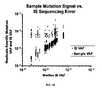
[0061] FIG. 1A. Mutations identified in patient
specimens. Sample mutation signal versus
IS sequencing error. Variant allele frequency (VAF) of sample mutations (red
triangle) relative to
VAF of corresponding nucleotide-specific error variants in 19 IS replicates
(black circle). VAF =
site specific variant allele reads/total allele reads.
[0062] FIG. 1B. Showing how as the VAF% rises, there is
a diminished difference between
CA and NC subject, highlighting the importance of detecting variants with
ultra-low VAR It is
likely that once a clone increases it's VAF to a significant size, the inunune
system takes it out.
Thus, being able to identify low VAF clones allows for distinction between
those at high risk for
lung cancer and those at lower risk.
[0063] FIGS. 2A-2B. Inter-cohort comparison of TP53 mutation mean prevalence.
FIG.2A -
Mean mutation prevalence among subjects within each cohort in each separate
TP53 exon 5, 6, or
7 (mutations/target base/subject). FIG. 2B - Cohort- and substitution-specific
mean mutation
prevalence for the combined three TP53 exon targets. FIG. 2C - Number of
mutations at TP53
hotspot sites. Inset: number of mutations according to mutation type.
Mutations were defined as
those with VAF (variant allele reads/total allele reads) >0.05% and
significantly above IS
background VAF based on contingency table analysis. TP53 mutations in CA-SMK
subjects were
enriched significantly at "hotspot" lung cancer driver mutation sites. (p =
0.002).
[0064] FIGS. 3A-3B. Inter-cohort comparison of subject-
specific mutation prevalence. Inter-
cohort comparison of subject-specific mutation prevalence in (FIG. 3A) TP53
exons only or (FIG.
313) TP53 exons, PIIC3CA, and BRAE'.
[0065] FIGS. 4A-4C. Inter-cohort comparison of EGFR mutation mean prevalence.
FIG.
4A - Mean mutation prevalence among subjects within each cohort in each EGFR
exon (18, 19,
20, or 21) (mutations/target base/subject). FIG. 4B - Cohort- and substitution-
specific mean
mutation prevalence for the combined four EGFR exon targets. FIG. 4C - Number
of mutations at
EGFR hotspot sites. Inset: number of mutations according to mutation type.
Mutations were
defined as those with VAF (variant allele reads/total allele reads) >5 x 10⁴
(0.05%) and
6
CA 03150250 2022-3-4
WO 2021/046502
PCT/US2020/049629
significantly above IS background VAF based on contingency table analysis.
[0066] FIG. S. Qiagen CLC Genomics Workbench Settings.
[0067] FIG. 6. Schematic illustration of how to design
internal standard (IS) spike-in
molecules for NGS.
[0068] FIG. 7. Frequency of observed sequence
variations for native template group and
internal standards group for different types of sequence variations.
[0069] FIG. 8. Internal standard error for four
replicates, showing the individual replicate
error and mean error.
[0070] FIG. 9A. Hybrid capture panel for exons EGFR_18 (red), EGFR_20 (blue)
and
EGFR_21 (green), showing IS frequency (%).
[0071] FIG. 9B. NT frequency (%) showing replicate
measurement, limit of blank (LOB),
and variant allele frequency for exons EGFR 18 (red), EGFR_20 (blue) and
EGFR_21 (green).
Without internal standards, Limit of Blank (LOB) calculations are based on
average error
frequency across all variant types at all nucleotide positions. This
effectively raises the Limit of
Detection (LOD) and prevents statistical determination of variants with VAF
<5%.
[0072] FIG. 9C Internal standards enable calculation of
Limit of Blank (LOB) for each
variant type at each nucleotide position providing site-specific determination
of the Limit of
Detection (LOD). This allows for identification of variants with VAF <1% at
locations where the
LOB is sufficient low.
[0073] FIG. 9D. Comparison of expected, NT, reported NT and reported IS for
exons
EGFR_18 (red), EGFR_20 (blue) and EGFR_21 (green).
[0074] FIG. 10. Applying Internal Standards to
fragmented FDA Samples.
[0075] FIG. 11. Transition Sequencing Error at TP53
(exon 6) Across 19 Internal Standard
Replicates, showing the Variant Allele Frequency for TP53 transactivation
domain, TP53 DNA
binding domain, and TP53 tetramerization domain.
[0076] FIG. 12. TP53 (exon 6) Transition Variants in
Sample 7.
[0077] FIG. 13. Mutations in 19 Patient Specimens
Relative to IS.
DETAILED DESCRIPTION
[0078] Throughout this disclosure, various
publications, patents and published patent
specifications are referenced by an identifying citation. The disclosures of
these publications,
patents and published patent specifications are hereby incorporated by
reference into the present
disclosure to more fully describe the state of the art to which this invention
pertains.
Definitions and Abbreviations
[0079] AEC - Airway Epithelial Cells
[0080] CA-SMK - Cancer subjects, smokers
[0081] COSMIC - Catalog of Somatic Mutations in Cancer
7
CA 03150250 2022-3-4
WO 2021/046502
PCT/US2020/049629
[0082] FASMIC - Functional Annotation of Somatic Mutations in Cancer
[0083] FDA - Food and Drug Administration
[0084] HUGO - Human Genome Organization
[0085] IS - Internal Standard, synthetic DNA
[0086] .. ISM - Internal Standard Mixture
[0087] LCRT - Lung Cancer Risk Test
[0088] LDCT - Low Dose Computed Tomography
[0089] NC-NON - Non-cancer subjects, non-smokers
[0090] NC-SMK - Non-cancer subjects, smokers
[0091] NC-TOT - Non-cancer subjects, non-smokers + smokers (all non-cancer
subjects)
[0092] NGS - Next Generation Sequencing
[0093] NT - Native Template, from targeted region of specimen DNA
[0094] PCR - Polymerase Chain Reaction
[0095] SNP - Single Nucleotide Polymorphism
[0096] VAF - Variant Allele Frequency
[0097] TCGA - The Cancer Genome Atlas
[0098] A "gene" is one or more sequence(s) of nucleotides in a genome that
together encode
one or more expressed molecules, e.g., an RNA, or polypeptide. The gene can
include coding
sequences that are transcribed into RNA which may then be translated into a
polypeptide
sequence, and can include associated structural or regulatory sequences that
aid in replication or
expression of the gene.
[0099] .. A "set" of markers, probes or primers refers to a collection or
group of markers
probes, primers, or the data derived therefrom, used for a common purpose
(e.g., assessing an
individual's risk of developing cancer). Frequently, data corresponding to the
markers, probes or
primers, or derived from their use, is stored in an electronic medium. While
each of the members
of a set possess utility with respect to the specified purpose, individual
markers selected from the
set as well as subsets including some, but not all of the markers, are also
effective in achieving the
specified purpose.
[00100] "Specimen" as used herein can refer to material collected for
analysis, e.g., a swab of
culture, a pinch of tissue, a biopsy extraction, a vial of a bodily fluid
e.g., saliva, blood and/or
urine, etc. that is taken for research, diagnostic or other purposes from any
biological entity.
[00101] Specimen can also refer to amounts typically collected in biopsies,
e.g., endoscopic
biopsies (using brush and/or forceps), needle aspirate biopsies (including
fine needle aspirate
biopsies), as well as amounts provided in sorted cell populations (e.g., flow-
sorted cell
populations) and/or micro-dissected materials (e.g., laser captured micro-
dissected tissues). For
example, biopsies of suspected cancerous lesions, commonly are done by fine
needle aspirate
(FNA) biopsy, bone marrow is also obtained by biopsy, and tissues of the
brain, developing
8
CA 03150250 2022-3-4
WO 2021/046502
PCT/US2020/049629
embryo, and animal models may be obtained by laser captured micro-dissected
samples.
[00102] "Biological entity" as used herein can refer to
any entity capable of harboring a nucleic
acid, including any species, e.g., a virus, a cell, a tissue, an in vitro
culture, a plant, an animal, a
subject participating in a clinical trial, and/or a subject being diagnosed or
treated for a disease or
condition.
[00103] "Sample" as used herein can refer to specimen
material used for a given assay,
reaction, run, trial and/or experiment. For example, a sample may comprise an
aliquot of the
specimen material collected, up to and including all of the specimen.. As used
herein the terms
assay, reaction, run, trial and/or experiment can be used interchangeably
[00104] In some embodiments, the specimen collected may
comprise less than about 100,000
cells, less than about 10,000 cells, less than about 5,000 cells, less than
about 1,000 cells, less than
about 500 cells, less than about 100 cells, less than about 50 cells, or less
than about 10 cells.
[00105] In some embodiments, assessing, evaluating
and/or measuring a nucleic acid can refer
to providing a measure of the amount of a nucleic acid in a specimen and/or
sample, e.g., to
determine the level of expression of a gene_ In some embodiments, providing a
measure of an
amount refers to detecting a presence or absence of the nucleic acid of
interest_ In some
embodiments, providing a measure of an amount can refer to quantifying an
amount of a nucleic
acid can, e.g., providing a measure of concentration or degree of the amount
of the nucleic acid
present. In some embodiments, providing a measure of the amount of nucleic
acid refer to
enumerating the amount of the nucleic acid, e.g., indicating a number of
molecules of the nucleic
acid present in a sample. The "nucleic acid of interest" may be referred to as
a "target" nucleic
acid, and/or a "gene of interest," e.g., a gene being evaluated, may be
referred to as a target gene.
The number of molecules of a nucleic acid can also be referred to as the
number of copies of the
nucleic acid found in a sample and/or specimen.
[00106] As used herein, "nucleic acid" can refer to a
polymeric form of nucleotides and/or
nucleotide-like molecules of any length. In certain embodiments, the nucleic
acid can serve as a
template for synthesis of a complementary nucleic acid, e.g., by base-
complementary
incorporation of nucleotide units. For example, a nucleic acid can comprise
naturally occurring
DNA, e.g., genomic DNA; RNA, e.g., mRNA, and/or can comprise a synthetic
molecule,
including but not limited to cDNA and recombinant molecules generated in any
manner. For
example the nucleic acid can be generated from chemical synthesis, reverse
transcription, DNA
replication or a combination of these generating methods. The linkage between
the subunits can
be provided by phosphates, phosphonates, phosphoramidates, phosphorothioates,
or the like, or by
nonphosphate groups, such as, but not limited to peptide-type linkages
utilized in peptide nucleic
acids (FNAs). The linking groups can be chiral or achiral. The polynucleotides
can have any
three-dimensional structure, encompassing single-stranded, double-stranded,
and triple helical
molecules that can be, e.g., DNA, RNA, or hybrid DNA/RNA molecules_
9
CA 03150250 2022-3-4
WO 2021/046502
PCT/US2020/049629
1001071 A nucleotide-like molecule can refer to a
structural moiety that can act substantially
like a nucleotide, for example exhibiting base complementarity with one or
more of the bases that
occur in DNA or RNA and/or being capable of base-complementary incorporation.
The terms
"polynucleotide," "polynucleotide molecule," "nucleic acid molecule,"
"polynucleotide sequence"
and "nucleic acid sequence," can be used interchangeably with "nucleic acid"
herein. In some
specific embodiments, the nucleic acid to be measured may comprise a sequence
corresponding to
a specific gene.
1001081 In some embodiments the specimen collected
comprises RNA to be measured, e.g.,
mRNA expressed in a tissue culture. In some embodiments the specimen collected
comprises
DNA to be measured, e.g., cDNA reverse transcribed from transcripts. In some
embodiments, the
nucleic acid to be measured is provided in a heterogeneous mixture of other
nucleic acid
molecules.
[00109] The term "native template" as used herein can
refer to nucleic acid obtained directly or
indirectly from a specimen that can serve as a template for amplification. For
example, it may
refer to cDNA molecules, corresponding to a gene whose expression is to be
measured, where the
cDNA is amplified and quantified.
[00110] The term "primer" generally refers to a nucleic
acid capable of acting as a point of
initiation of synthesis along a complementary strand when conditions are
suitable for synthesis of a
primer extension product.
General Description
[00111] Described herein are kits and methods for
assessing amounts of a nucleic acid in a
sample. In some embodiments, the method allows measurement of small amounts of
a nucleic
acid, for example, where the nucleic acid is expressed in low amounts in a
specimen, where small
amounts of the nucleic acid remain intact and/or where small amounts of a
specimen are provided.
Design of Internal Standard (IS) Spike-In Molecules for NGS
[00112] Referring first 10 FIG. 6, a schematic
illustration of how to design internal standard
(IS) spike-in molecules for NGS is shown.
[00113] IS are synthetic DNA molecule homologous with
target analyte except for known one
or more nucleotide changes.
[00114] IS Design goal: To behave the same as, but be
distinguishable from target analyte
DNA native template (NT)
[00115] IS Uses: 1) quantify measurable gencpme copies
of each target analyte NT in library
prep, and 2 quantify and characterize nucleotide site-specific technical error
[00116] IS Implementation: 1) mix sample DNA with known
number of IS molecules at 1:1
genome copy ratio prior to NGS library preparation; 2) co-amplify IS + NT
mixture; 3) prepare
sequencing library; and, 4) sequence sample.
[00117] Internal Standard "Spike-In Molecules" are
custom perl script which separates IS
CA 03150250 2022-3-4
WO 2021/046502
PCT/US2020/049629
reads from sample reads using one or more nucleotide changes. The error
profile in native
template (NT) nearly identical in internal standard (IS).
[00118] Thus, IS controls for library-specific error
profiles, as shown in FIG. 7, which shows
the frequency of observed sequence variations for native template group and
internal standards
group for different types of sequence variations.
[00119] Additionally, as shown in FIG. 8, the nucleotide-
specific technical error is
reproducible. FIG. 8 shows the internal standard error for four replicates,
showing the individual
replicate error and mean error_ The nucleotide-specific technical error at
each NT base position
matches corresponding IS position. Also, DNA landscape affects sequencing
error on a region-to-
region and nucleotide-to-nucleotide basis 4 IS and NT behave the same way.
[00120] Spiking IS into each reaction thus controls for
variation within library prep (e.g.,
interfering substances, intra- and inter-panel hybridization efficiency,
ligation efficiency,
amplification).
[00121] Internal standards also control for sources of
imprecision enabling narrow confidence
interval at each nucleotide: nucleotide-specific error frequency; platform-
specific errors, and
polymerase- specific errors.
[00122] FIGS. 9A-9D show that internal standards enable
site-specific LOD (logarithm of the
odds). FIG. 9A shows a hybrid capture panel for exons EGFR_18 (red), EGFR_20
(blue) and
EGFR 21 (green), showing IS frequency (%). FIGS. 9B-9C shows NT frequency (%),
showing
replicate measurement, LOB, and variant allele frequency for exons EGFR_18
(red), EGFR_20
(blue) and EGFR_21 (green). FIG. 9D shows a comparison of expected, NT,
reported NT and
reported IS for exons EGFR_18 (red), EGFR_20 (blue) and EGFR_21 (green). Thus,
FIGS. 9A-
9D show that traditional methods based on external process performance
estimates do not support
VAF measurements <5%. Also, alternative correction methods are complex and
require 10- to 20-
fold more sequencing reads.
[00123] FIG. 10 shows applying Internal Standards (IS)
to fragmented FDA samples. The
known mutations identified with LOD based on site-specific LOB determined by
internal
standards (IS).
[00124] Multiplex gradient PCR enables primers with
varying melting temperatures to anneal
to specific target Single-plex PCR followed by quantification and equimolar
mixing enables
equal loading onto sequencer. PCR targets chosen based on high occurrence in
lung cancer and
lung premalignant lesions.
[00125] Synthetic DNA internal standards (IS) were
prepared for each of various lung cancer
driver genes and mixed with each AEC genomic (gDNA) specimen prior to
competitive multiplex
PCR amplicon NGS library preparation. A custom Pal script was developed to
separate IS reads
and respective specimen gDNA reads from each target into separate files for
parallel variant
frequency analysis. This approach enabled reliable detection of mutations with
VAF as low as 5 x
11
CA 03150250 2022-3-4
WO 2021/046502
PCT/US2020/049629
104 (0.05%). This method was then applied in a retrospective case-control
study. Specifically,
AEC specimens were collected by bronchoscopic brush biopsy from the normal
airways of 19
subjects, including eleven lung cancer cases and eight non-cancer controls,
and the association of
lung cancer risk with AEC driver gene mutations was tested.
[00126] FIG. 11 is an example of transition sequencing
error at TP53 (exon 6) across 19
Internal Standard (S) replicates, showing the variant allele frequency (VAF)
for TP53
transactivation domain, TP53 DNA binding domain, and TP53 tctramerization
domain.
[00127] FIG. 12 is an example of transition variants in
a sample at TP53 (exon 6), showing the
variant allele frequency (VAF) for TP53 transactivation domain, TP53 DNA
binding domain, and
TP53 tetramerization domain.
[00128] FIG. 13 shows mutations in 19 patient specimens
relative to IS. 129 significant
variants identified in 19 patient specimens. The VAF for these variants range
from 0.05% to
0.46%. 99 variants found in 11 cancer specimens. 30 variants found in 8 non-
cancer specimens.
Also, there were significant increase in variants of smokers with cancer
compared to smokers
without cancer_
[00129] Described herein is a kit or method that
includes reagents and instructions for
measuring analytes in a lung cancer risk test.
[00130] This lit or method incorporates reagents for
measurement of analytes that have not
been previously described for inclusion in a test for lung cancer risk.
[00131] Specifically, the lung cancer risk test (LCRT)
kit or method includes reagents for
measurement of multiple low variant allele frequency (VAF) (i.e. VAF < 0.01 11-
.0%1) mutants in
lung cancer driver genes, including TP53, PIK3CA, BRAF, KRAS, NRAS, NOTCHI,
EGFR, and
ERBB2.
[00132] Other reagents can be included for such genes as CDKN1A, E2F1, ERCC1,
ERCC4,
ERCC5, GPX1, GSTP I, KEAP1, RBI, TP53, TP63, and XRCC I.
[00133] These analytes may be measured in RNA or DNA from airway epithelial
cells, and
may be measured in non-invasively obtained specimens, including exhaled breath
condensate and
airway epithelial cells obtained by nasal brushings.
[00134] Also described herein are methods for measurement of low VAF mutants
with
calculation of limit of detection/limit of quantification for measurement of
each analyte in each
specimen, based on measurement of specimen analyte relative to a known number
of synthetic
internal standard molecules.
[00135] In certain embodiments, these kits and methods
are useful to facilitate approval by
FDA and other regulatory agencies of lung cancer risk testing in kit or method
form in regional
laboratories.
[00136] In certain embodiments, these kits and methods
are useful to enable measurement of
lung cancer risk in non-invasively obtained specimens, such as exhaled breath
condensate, nasal
12
CA 03150250 2022-3-4
WO 2021/046502
PCT/US2020/049629
brush specimens, sputum, oral epithelium, blood, and the like.
[00137] hi certain embodiments, these kits and methods
are useful to enable measurement of
very low VAF mutations in airway epithelial cells.
EXAMPLES
[00138] The methods and embodiments described herein are
further defined in the following
Examples, in which all parts and percentages are by weight and degrees are
Celsius, unless
otherwise stated. Certain embodiments of the present invention are defined in
the Examples
herein. It should be understood that these Examples, while indicating
preferred embodiments of
the invention, are given by way of illustration only. From the discussion
herein and these
Examples, one skilled in the art can ascertain the essential characteristics
of this invention and
without departing from the spirit and scope thereof, can make various changes
and modifications
of the invention to adapt it to various usages and conditions.
[00139] The measurement of mutations in the 0.05-LO% VAF range enables more
informative
analysis of AEC somatic mutations associated with cancer risk_ Among lung
cancer subjects,
TP53 mutations were more prevalent (p<0.05) and significantly more enriched
for tobacco smoke
and age signatures compared to non-cancer subjects matched for smoking and
age_
METHODS
Study Cohort Enrollment and Characterization.
[00140] For this retrospective case-control study, AEC
specimens collected from nineteen
subjects were used, including eleven smokers with lung cancer (CA-SMK), five
smokers without
cancer (NC-SMK) matched for age and smoking history, and three non-smokers
without cancer
(NC-NON) (Table 1).
[00141] Subjects were enrolled into research trials at
the University of Toledo Medical Center
(UTMC) between 2000 and 2018. Each subject included in this research study
provided written
informed consent under protocols approved by the University of Toledo
Institutional Review
Board. Clinical characteristics, including lung cancer diagnosis, smoking
history, and demographic
information were obtained from the medical record. Lung cancer histology was
reviewed and
confirmed by an independent pathologist certified in anatomical and clinical
pathology.
[00142]
Table 1. Patient Demographics.
Sample Cancer Pack Smoking
Sex Age Race
Diagnosis
Status Years Status
946 CA 45 F 55 Black Former NSCLC-SQ
167 CA 50 F 60 Unknown Unknown NSCLC
947 CA 45 M 61 White Former SCLC
146 CA 46.5 F 64
White Former NSCLC
887 CA 28 F 70 White Current NSCLC-AD
885 CA 90 M 73 White Current SCLC
13
CA 03150250 2022-3-4
WO 2021/046502
PCT/US2020/049629
940 CA 60 M 74 White Former NSCLC-AD
191 CA NA* M 75 White Current NSCLC-SQ
147 CA 75 M 76 White Former SCLC
128 CA 40 F 50
Black Current NSCLC
923 CA 15 M 79 White Former NSCLC
210 NC 34 M 40 White Current Noncancer
886 NC 0 F 46
White Never Noncancer
952 NC 30 M 52 White Former Noncancer
157 NC 100 M 60 White Unknown Noncancer
943 NC 0 F 65
Black Never Noncancer
956 NC 20 M 69 Black Current Noncancer
884 NC 54 M 77 White Former Noncancer
883 NC 0 M 81 White Never Noncancer
*Not available: The exact pack year smoking history for this patient was not
recorded. However, it was
recorded that the patient was an active 2 PPD smoker at time of lung cancer
diagnosis at age 75 and had
advanced stage COPD, thus there is compelling circumstantial evidence for
large smoking history.
Specimen Acquisition
[00143] AEC were obtained via bronchoscopic brush biopsy
of normal appearing airway
epithelium at the time of a diagnostic procedure done according to standard of
care indication. For
patients with a lung cancer diagnosis, sampling of AEC was from the main
bronchus of the lung
not involved with cancer. Specimens were immediately placed in cold saline and
processed within
one hour of collection.
DNA Extraction and Quantification
[00144] Genomic DNA (gDNA) was extracted from approximately 500,000 AEC per
subject
using a FlexiGene DNA kit (Qiagen, Hilden, (Jermany) according to manufacturer
protocol and
quantified using competitive polymerase chain reaction (PCR) amplification of
a well-
characterized genomic locus in the Secretoglobin, family 1A, member 1 gene.
Target Selection
[00145] Twelve loci in seven gene regions recently
reported by The Cancer Genome Atlas
(TCGA) project to be the most commonly mutated in non-small cell lung cancer
were selected as
targets. The targeted regions, specified according to Human Genome
Organization (HUGO)
names with exon numbers and abbreviations provided in parentheses, included B-
Raf proto-
oncogene exon 15 (BRAF 15), epidermal growth factor receptor exons 18-21
(EGFR_18,
EGFR_19, EGFR_20, EGFR_21), erb-b2 receptor tyrosine kinase 2 (ERBB2), ICRAS
proto-
oncogene exon 2 (KRAS_2), notch receptor 1 exon 26 (NOTCH1_26),
phosphatidylinosito1-4,5-
hisphosphate 3-kinase catalytic subunit alpha exon 10 (P1K3CA_10), and tumor
protein p53 exons
5-7 (TP53_5, TP53 6, TP53_7). Primers were developed for each of these
targets.
[00146] Primers for all targets except for NOTCH1 26
performed efficiently in multiplex and
downstream library preparation. As such, data are reported for the remaining
11 targets.
14
CA 03150250 2022-3-4
WO 2021/046502
PCT/US2020/049629
Synthetic Internal Standard Mixture Preparation
[00147] Competitive synthetic DNA internal standard (IS)
molecules for TCGA targets
described above were designed with known dinucleotide substitution mutations
relative to target
analyte native template (NT) every 50 bases. This enabled separation of NT and
IS reads during
post-sequencing data processing of either PCR arnplicon libraries used in this
study, or of random
fragment hybrid capture libraries in other ongoing studies not reported here.
IS were cloned into
plasmids and selected as pure clonal isolates using Sanger sequencing
confirmation to verify the
final sequence. This additional purification step was taken to select clones
free of any potential
errors introduced by synthesis.. Due to the high fidelity of endogenous E.coli
polymerase, the
frequency of variants in the cloned IS can be expected to be between 10-7 to
104¨well below the
desired limit of detection for this study. Each cloned plasrnid was
linearized, quantified by digital
droplet PCR, then combined in an equal genome copy balance. An internal
standard mixture
(ISM) containing equal concentrations (per genome copy) of each linearized
target analyte IS
molecule was prepared by Accugenornics, Inc. (Wilmington, NC).
[00148] Technically-derived base substitution errors
occur at the same rate in synthetic IS as in
the respective target sequence within gDNA test samples during the combined
library preparation
and sequencing steps. Therefore, each IS controls for target-specific site and
regional differences
in base substitution error rate.
Multiplex Competitive PCR Amplicorz Libraries
[00149] In order to amplify each target in a sample and
maximize opportunity to detect low
frequency variants, a multiplex competitive PCR amplicon library was prepared
for each AEC
DNA sample. Conditions were optimized to minimize technical error during PCR,
including use
of Q5 HotStart High Fidelity DNA Polymerase with a reported error frequency of
10-6 (New
England Biolabs, Ipswich, MA) and minimization of PCR cycles in each round.
Round 1: Competitive Multiplex PCR
[00150] Twelve target-specific primers with universal
tails were synthesized by Life
Technologies (Carlsbad, CA). Individual primer solutions for each target were
created by adding
TE buffer (10 mIVI Tris-C1, pH 7.4, 0.1 naM EDTA) to the lyophilized primers
to make a 100 pM
stock. A 25 LiM multiplex primer mixture was prepared by mixing 5 pL of each
100 LIM forward
and reverse primer stock solution and bringing the final volume to 200 pL with
TE buffer.
[00151] For each subject, an aliquot of AEC DNA was combined with equal genome
copies of
ISM to control for nucleotide-specific substitution error occurring during
library preparation
and/or sequencing. Reactions containing at least 50,000 genome equivalents of
both sample and
IS in a mixture, 6 FiL 5X Q5 Buffer (New England Biolabs, Ipswich, MA), 0.6
piL 10 mIVI dNTP
(Promega, Madison, WI), 3 pi, 25 pM multiplex primer mixture, 1.5 fa, 2% w/v
bovine serum
albumin (New England Biolabs, Ipswich, MA), 0.3 piL Q5 HotStart High Fidelity
DNA
CA 03150250 2022-3-4
WO 2021/046502
PCT/US2020/049629
Polymerase (New England Biolabs, Ipswich, MA, Ipswich, MA), and molecular-
grade water to a
final reaction volume of 30 pit were prepared.
[00152] Each competitive multiplex reaction mixture was
amplified in a 7500 Fast Real-Time
PCR System (Applied Biosystems, Foster City, CA) for a total of 20 cycles
under modified
gradient PCR conditions: 95 C/2 min (Q5 HotStart DNA Polymerase activation);
20 cycles of
94 C/10 sec (denaturation), 70t/10 sec, 68 C/10 sec, 66 C/10 sec, 64 C/10 sec,
62 C/10 sec,
(annealing), and 72 C/30 sec (extension); a final extension 72 C12 min
extension to ensure
complete extension of all products. PCR products were column-purified using
QIAquick PCR
Purification Kit (Qiagen, Hilden, Germany) according to manufacturer protocol.
Round 2: Singleplex PCR
[00153] Following multiplex amplification, a second
round of 12 parallel singleplex PCR
reactions using primers for each individual target at a final concentration of
500 nM were
performed to ensure robust amplification of product for primers with lower
efficiency in multiplex.
High fidelity Q5 Hot Start Polymerase and other PCR reagents were used as
described above.
[00154] Singleplex reactions were amplified in a 7500
Fast Real-Time PCR System (Applied
Biosystems, Foster City, CA) for 15 cycles using the following conditions: 95
C/2 min (Q5
polymerase activation); 15 cycles of 94t/10 sec (denaturation), 65t/20 sec,
(annealing), and
72 C/30 sec (extension); a final extension 72 C/2 min extension was performed
to ensure
complete extension of all products. Each singleplex PCR product was checked
for quality and
quantity with an Agilent 2100 Bioanalyzer using DNA Chips with DNA 1000 Kit
reagents
according to manufacturer protocol (Agilent Technologies, Deutschland (imbH,
Waldbronn,
Germany). Sample-specific singleplex reactions then were (a) mixed in
equimolar amounts to
ensure an equal balance of target reads among sequencing read counts and (b)
column-purified
using QIAquick PCR Purification Kit (Qiagen, Hilden, Germany) according to
manufacturer
protocol_
Round 3: Addition of Sample-Spectfic Barcodes
[00155] The column-purified mixture of singleplex
reactions from each patient sample was
labeled using a unique set of dual-indexed barcode primers to reduce
likelihood of false-
indexing/barcoding a sequencing read_ A pair of fusion primers containing the
barcode sequences
and Illurnina priming sites were designed with: 1) their 3'-end complementary
to the universal
sequence tails added during the initial multiplex and singleplex reactions, 2)
5' to that a 10-
nucleotide index/barcode sequence, and (3) 5' to that, an Illumina Read 1 or
Read 2 priming site.
The fmal concentration of the barcode primers in each reaction was 500 nM. PCR
conditions were
identical to those described for singleplex reactions except the cycle number
was reduced to 10.
[00156] PCR products were checked for quality and
quantity with an Agilent 2100 Bioanalyzer
using DNA Chips with DNA 1000 Kit reagents according to manufacturer protocol
and diluted
100-fold with molecular grade water for input into final sequencing adapter
PCR.
16
CA 03150250 2022-3-4
WO 2021/046502
PCT/US2020/049629
Round 4: Addition of Sequencing Adapters
[00157] Individual diluted barcoded samples were labeled
with an Illumina platform-specific
adapter using a second set of fusion primers designed with their 3'-end
complimentary to the
Illumina Read 1 or Read 2 priming sites and 5' Illurnina sequencing adapter
using the same PCR
conditions used in Round 3.
Sample Pooling
[00158] Following Round 4, each uniquely barcoded sample
was quantified on an Agilent
2100 Bioanalyzer as described above. The samples then were mixed in equimolar
ratios to
optimize the percentage of sequencing reads that each library would eventually
receive; in most
cases 1:1 was used.
Product Purification and Sequencing
[00159] The combined sequencing library was purified
using gel electrophoresis on a 2% w/v
agarose gel. The resultant product band was then cut out, separating it from
unwanted
heterodimers, extracted using a QIAquick Gel Extraction Kit (Qiagen, Bilden,
Germany), and
eluted in 50 pl. elution buffer. The purified sequencing library was sent to
the University of
Michigan Genotnics core facility for Next Generation Sequencing on an
Illurnina NextSeq 550
sequencing
Analysis of NOS Data
[00160] FASTQ data files generated by the University of
Michigan Genornics core facility
were processed using a custom Perl script to separate the internal standard
(IS) and native template
(NT) reads into separate NT and IS files, followed by parallel analysis using
the Qiagen CLC
Genomks Workbench 12 software suite for quality-trimming, alignment, and
variant calling, as
shown in FIG. 5.
[00161] Primer sequences, internal standard dinucleotide
positions plus their 5' and 3' bases,
and known single nucleotide polymorphism (SNP) positions were excluded from
variant analysis.
[00162] Variant Calling
[00163] Variants were called based on NT signal
significantly above the background error
measured in IS for the respective mutation type at each respective position.
Significance was
determined using contingency table chi-square analysis of each individual
variant type at each
nucleotide position, for identifying rare variants in pooled samples. To
maximize stringency of
test for signal above noise, a variant was called if the proportion of variant
reads to wild-type reads
in the specimen was significantly higher than the proportion of variant reads
to wild-type reads at
the same site in the IS mixed with the respective specimen, and also higher
than the proportion
observed in IS mixed with each of the other 18 specimens. Thus, each variant
in a specimen was
considered a true positive (p<0.05) only if the proportion of variant reads to
wild-type reads was
significantly higher in the specimen than each of the 19 IS replicates. A
Bonferroni correction for
false discovery was used based on the number of nucleotides assessed (760 bp)
and the number of
17
CA 03150250 2022-3-4
WO 2021/046502
PCT/US2020/049629
substitution mutations possible at each nucleotide position. Further, to avoid
potential analytical
variation from stochastic sampling, only mutations with significant signal
above IS noise, and with
VAF >0.05% were called.
Variant Annotation and Hotspot Analysis
[00164] Called variants were characterized for
pathogenicity using publicly-available
databases including dbSNP, COSMIC, and FASMIC. Identification of known
oncogenic hotspots
and generation of corresponding figures were assessed using the cBioPortal for
Cancer Genomics
developed at Memorial Sloan Kettering (MSK) Cancer Center.
Statistical Analysis
[00165] Calling of variants based on contingency table
chi-square analysis of each individual
variant type at each nucleotide position was performed using R: A Language and
Environment for
Statistical Computing (www.R-projectord). Assessment of hotspot enrichment for
called variants
was performed using Kruskal-Wallis test using a chi-square distribution.
Mutation prevalence
based on type of mutation and target was assessed using ICruskal-Wallis test
with Nemenyi test for
multiple comparisons..
RESULTS
Measurement of low frequency mutations in non-cancer airway epithelium
[00166] In this study of 11 driver gene target regions
in AEC specimens from normal airways
of 19 subjects, there were 129 called variants with VAF ranging from 5 x 10⁴
(0.05%) to 4.6 x 10⁻³
(0.46%). As described in the Methods section, a VAF minimum threshold of 0.05%
was used to
minimize risk of false discovery due to stochastic sampling. Among the 129
called variants, the
relationship between sample mutation signal (Mutation VAF) and background
technical error
(noise) (IS VAF) for the respective variant at the same site is presented in
FIG. IA.
[00167] For each sample mutation VAF, there is displayed
the IS VAF for 19 IS. These
represent the VAF for the IS mixed with the sample that contained the mutation
as well as the
VAF for each of the IS mixed with the other 18 samples. These 19 independent
IS replicate values
display the variation around the IS VAF (error) measurement within an
experiment. The inter-
replicate variation in IS VAF values increases with decreasing IS VAF,
consistent with effects of
the Poisson distribution on stochastic sampling.
[00168] Further, due to very low technical error at some
of these sites there was no IS VAF
value (FIG. lii).
[00169] These effects of Poisson distribution present
challenges for statistical analysis of
significance for observed sample mutations. A simple Z-score analysis is
appropriate if there are
at least 10 sequencing reads for all four components: sample reference and
variant allele, and IS
reference and variant (error) allele. Using a minimum sample mutation VAF of
0.05% ensured at
least 10 variant allele reads for each called sample mutation. However, when
the corresponding IS
error was very low, the IS variant allele read count was below 10, and
sometimes zero_
18
CA 03150250 2022-3-4
WO 2021/046502
PCT/US2020/049629
[00170] If there were at least one variant allele read
for each IS replicate, it would be
appropriate to use Poisson exact test. In this study, because the IS error in
the targeted hotspot
regions was so low that for some measurements there were zero IS variant reads
corresponding to
observed sample variants, even with the deep sequencing employed it was
advantageous to use the
contingency table approach to determine significance of each sample mutation
in this study.
[00171] FIG. 1B shows TP53 mutations detectable in AEC
depends on lower limit of
detection for VAF (%) detection.
[00172] A key reason that a TP53 mutation test for lung
cancer risk measured in airway
epithelial cells was not discovered previously, in spite of efforts to do so
is that commonly used
methods are not able to reliably measure mutations at VAF <19k
Characteristics of sequencing error in the targeted regions
[00173] As shown in FIG. 1A, the maximum sequencing error (Median IS VAF
across
replicates) at sites within the targeted regions for which a sample variant
was called was 0.06%.
This error rate is much lower than that observed for whole exome sequencing on
111urnina
platform. In addition, as reported by others, this is a key factor that
enables meaningful calling of
low frequency variants without need for methods that employ unique molecular
indices (UMI)
with attendant cost and computational requirements.
Prevalence of low frequency mutations in AEC
[00174] Mutation prevalence was calculated as called
mutations per nucleotide positions
assessed for each target. The number of nucleotides assessed for each target
varied somewhat
based on region spanned by primers and number of dinucleotide sites blocked
from analysis due to
modification in IS to enable separation of IS reads from NT reads. Among all
19 subjects, the
average mutation prevalence, across the targeted DNA region (760 bp) in each
subject
(mutations/bp/subject) was 8.9 x 10⁻³. (Table 2).
Table 2. Target- and cohort-specific mutation prevalence.
Target CA-SMK
NC-SMK NC-NON NC-TOT Average
(All Subjects)
BRAF 15 6.7 x 10⁻³ 0
0 0 3.9 x 10⁻³
EGFR_18 0 0
0 0 0
EGFR_19 0 0
0 0 0
EGFR_20 3.9 x 10⁻²
3.4 x 10⁻² 45 x 10a 3.8 x 10⁻² 3.8 x 10⁻²
EGFR_21 1.7 x 10⁻³ 0
0 0 9.9 x 10⁻⁴
ERBB2 1.1 x 10⁻² 1.4 x 10⁻²
1.4 x 10⁻² 1.4x 10⁻² 1.2x 10⁻²
ICRAS_2 0 0
0 0 0
PIK3CA_10 4.2 x 10⁻³ 0
0 0 2.4 x 10⁻³
TP53_5 2.2 x 10⁻² 4.7 x 10⁻³
0 2_9 x 10-3 1 A x 10-2
TP53_6 22x 10-2 0
3.1 x 10⁻³ 1.2x 10⁻³ 1.3x 10⁻²
TP53_7 1.3 x 10⁻² 2.9 x Kr
0 12 x 10-3 85 x 10-3
Average
1.2 x 10⁻² 4.7 x 10⁻³
5.3 x 10⁻³ 4.9 . le 8.9 x 10⁻³
(All Targets)
19
CA 03150250 2022-3-4
WO 2021/046502
PCT/US2020/049629
[00175] This AEC mutation prevalence value is much
higher than reported for methods that
only detect mutants with relatively high variant frequency (VAF >1 %) (14), or
that are n-tore
sensitive but non-targeted. However, it is consistent with other analysis of
AEC using a highly
sensitive PCR-based method.
Association of Low Frequency Substitution Mutations in TP53, PIK3CA, and BRAE
with
Lung Cancer
[00176] Among the three measured exons of TP53, the
prevalence (mutations/bp/subject) of
substitution mutations was 10.4-fold higher (pc0.)5) in AEC from CA-SMK
subjects relative to
NC-SMK subjects matched for smoking and age (FIG. 24, Table 3).
Table 3. Statistical analysis of target specific inter-cohort differences in
mutation
prevalence.
T CA-SMK vs. CA-SMK vs.
CA-SMK vs. NC-SMK vs.
arget
NC-TOT NC-SMK
NC-NON NC-NON
BRAF 15 0.12 OA
054 1
E,GFR 18 N/A N/A
N/A N/A
EGFR_19 N/A N/A
N/A N/A
EGFR_20 0/2
0.78 0.96 0.74
EGFR_21 0.39
0.76 0.83 1
ERBB2 0.35
0.73 0.8 1
ICRAS 2 N/A N/A
N/A N/A
PHOCA_10 0.062
0.27 041 1
TP53_5 0.022
0.27 0.1 0.77
TP53_6 0.0083 0.037
0.333 0.849
TP53_7 0.028
0.25 0.16 0.9
TP53_Total 0.0019 0.047
0.043 0.92
[00177] In addition, PIK3CA or BRAF mutations were
observed in seven cancer subjects and
no non-cancer subjects (Table 3).
[00178] Notably, the majority of mutations in TP53 (FIG.
2C), all of the mutations in
PIK3CA, and one of three mutations in BRAF occurred in previously identified
"hotspots"
associated with biological changes that drive carcinogenesis.
[00179] Toward the goal of developing a biornarker that
might contribute to improved
determination of lung cancer risk, we assessed subject-specific inter-cohort
differences in
prevalence of these low frequency mutations. Based on data obtained in this
small retrospective
case-control study, a TP53 exon mutation prevalence cut-off of 0.02
mutations/bp would have
100% specificity and 55% sensitivity (FIG. 3A). Similar discrimination was
observed when TP53
exon mutations were combined with PIK3CA, and BRAE* mutations (FIG. 3B).
[00180] Nearly all of the TP53 mutations in CA-SMK
subjects were tobacco signature or age-
related mutations (C>A, C>T, and DC substitutions) (FIG. 2B, Table 4), closely
approximating
CA 03150250 2022-3-4
WO 2021/046502
PCT/US2020/049629
the spectrum of TP53 mutations reported for lung cancer tissues. The
prevalence of each type of
tobacco or age signature TP53 mutation was significantly higher in cancer
subjects than in non-
cancer subjects, including C>A (p = 0.002), C>T (p = 0.003), and 'NC (p =
0.001) (Table 4).
[00181] For example, while C to A mutations comprised
29.8% (17/57) of TP53 mutations
observed in AEC from CA-SMK subjects, there was only one C to A TP53 mutation
observed in
all non-cancer subjects (NC-TOT) (Table 4). C>T transitions comprised 47% of
TP53 mutations
in lung cancer subjects in this study. Further, TP53 mutations in CA-SMK
subjects were enriched
significantly (p =01)02) at "hotspot" lung cancer driver mutation sites (Fig.
2C).
[00182]
Table 4. Inter-cohort comparison of type-specific substitution mutations
across
all TP53 exons.
Mutation CA-SMICI NC-SMK2 NC-NON3 NC-
TOT4
C>A 17 (2.0 x 10-3)* 1 (L2 x 104) 0 1
(1.2 x 10⁴)
C>G 1 (1.2 x 10⁴) 1 (1.2 x 10⁴) 0 1
(1.2 x 10⁴)
C>T
27 (3.2 x 10⁻³)*** 1(1.2 x 10⁴) 1 (1.2 x 10⁴) 2 (2.4 x 10⁴)
T>A 3 (3.6 x 10⁴) 0 0 0
'NC 9 (1.1 x 1(I-4)* 0 0 0
T>G 0 0 0 0
'CA-SMK; Cancer subject, present or past smoker. 2NC-SMK; Non-Cancer subject,
present
or past smoker. 3NC-NON; Non-Cancer subject, never smoker. 4NC-TOT; All Non-
Cancer
subjects, smokers and non-smokers_
*pc0.05; "pc0.01; ***pc0.005
Lack of association of TP53 mutations with smoking history
[00183] Notably, among non-cancer subjects, smoking was
not associated with higher TP53
mutation prevalence (Table 3). Specifically, only half of NC-SMK subjects had
a TP53 mutation
with VAF >0.05% and in each case, only one variant was observed. (Table 3).
Due to the small
number of P1K3CA and BRAF mutations it was not possible to address a smoking
association.
Characteristics of Low Frequency AEC Mutations Not Associated with Lung Cancer
[00184] In contrast to TP53, at non-TP53 targets the
mutation prevalence was not significantly
different in cancer compared to non-cancer subjects (Table 3). Among the 11
targets measured,
mutation count was highest in the EGFR_20 target region with a total of 43
mutations observed
across all subjects (Table 3). There was no difference in EGFR_20 mutation
prevalence between
cancer and non-cancer (3.9 x 10⁻² vs 18 x 10-2, respectively; p = 0.72) (FIG.
4A, Table 3), and no
association between smoking and non-smoking (3.4 x 10⁻² vs 4.5 x 10⁻²
respectively; p = 0.74).
ERBB2 mutations (N=17) displayed a similar spectrum to that of EGFR_20 with no
age or
tobacco signature mutation pattern and no difference among the cohorts.
Notably, in contrast to
the high fraction of C>T transitions among TP53 (29/61; 48%), only 1/43 (2.3%)
EGFR_20
mutations, and 1 ERBB2 mutation was C>T (Fig. 3B). Further, the majority of
the EGFR_20
21
CA 03150250 2022-3-4
WO 2021/046502
PCT/US2020/049629
mutations were synonymous and not predicted to be pathogenic (FIG. 3C).
DISCUSSION
Measurement of low frequency mutations in AEC
[00185] The ability to measure low frequency mutations
in AEC in this study was due to a
combination of low technical error in the regions targeted (FIG. 1), and the
use of synthetic
internal standards to control for technical error on a site- and variant-
specific basis (FIG. 1). The
range of prevalence for low frequency TP53 mutations in AEC among subjects in
this study was
similar to previously reported. The enrichment for TP53 mutations in driver
mutation sites and for
tobacco-smoke signatures provides another source of validation that the
observed mutations are
true positives.
Identification of a TP53 mutation field effect associated with lung cancer
risk
[00186] The higher prevalence of low frequency TP53 hot-spot pathogenic
tobacco smoke and
age signature mutations in AEC of CA subjects compared with NC subjects
matched for smoking
and age represents a field of injury strongly associated with lung cancer risk
(FIG. 2A, FIG. 211,
FIG. 3A, Table 3, Table 4).
[00187] Thus, the results of low frequency (La, VAF < 1%) show that TP53
hotspot mutations
in AEC are a lung cancer risk biotnarker. Moreover, inclusion of low frequency
actionable
mutations in BRAF and P1K3CA can further enhance accuracy of this biomarker
(FIG. 3B).
[00188] Lung cancer predisposition is due, in part, to
sub-optimal protection from DNA
damage associated with cigarette smoking and age-related DNA replication
errors. There is
evidence for both hereditary and acquired causes of sub-optimal AEC protection
from DNA
damage. For example, there is a large inter-individual variation in regulation
of key DNA repair,
antioxidant, and cell-cycle control genes in AEC, and the lung cancer risk
test (LCRT) based on
this variation, has high accuracy to identify lung cancer subjects.
[00189] One of the variables in the LCRT biomarker is
TP53 transcript abundance, and there is
a 100-fold variation in TP53 expression in AEC. TP53 plays a key role in
upregulating DNA
repair genes in response to DNA damage, and the TP53 protein directly
regulates the key
nucleotide excision repair (NER) gene, ERCC5, in AEC.
[00190] The germ line allelic variation at rs2296147, a
TP53 recognition site in the
regulatory region of ERCC5, is associated with variation in allele-specific
expression of ERCC5 in
AEC. Hereditary inter-individual variation in ERCC5 transcription regulation
by TP53 is
significant because ERCC5 is the rate-limiting enzyme in transcription-coupled
NER, and
mutations associated with tobacco smoke result from inefficient NER of DNA
adducts arising
from the binding of cigarette smoke carcinogen metabolites to the exocycfic N2-
positions of
guanines on the transcribed strand.
[00191] Thus, sub-optimal ERCC5 regulation by TP53,
determined by inherited germ line
variants, is an important factor responsible for higher prevalence of tobacco
smoke induced
22
CA 03150250 2022-3-4
WO 2021/046502
PCT/US2020/049629
hotspot mutations in the transcribed strand of TP53 among cancer subjects.
Interpretation of non-pathogenic EGFR mutations
[00192] There was no difference in prevalence between
cancer and non-cancer subjects or
smokers and non-smokers for EGFR total mutations or cigarette- or age-
signature mutations (FIG.
4A, FIG. 4B; Table 2, Table 3). The substitution pattern (evenly distributed
between C>A and
C>G) is most consistent with previously described Signature 3, associated with
sub-optimal
homologous-recombination DNA double-strand break repair. In addition, evidence
presented here
supports the conclusion that the observed EGFR exon 20 mutations do not confer
growth
advantage.
[00193] Specifically, in contrast to the observed non-
synonymous pathogenic TP53 smoke-
and age-related mutations, only 1/43 EGFR 20 mutations was synonymous and
present at a known
pathogenic hotspot (FIG. 4C).
[00194] It is now believed that clonal populations with
this type of mutation likely occurred as
stochastic DNA replication errors in stem cell proliferation to generate the
airway epithelium
during the fetal-juvenile period.
[00195] A highly sensitive mismatch PCR assay capable of
detecting VAF as low as 5 x 10'
(0.005%) was used to test for the effect of cigarette smoke on prevalence of
low VAF somatic
mutations in AEC of non-cancer patients, including mutations in TP53, ICRAS,
and HPRT1 genes.
Surprisingly, among these non-cancer subjects, there was no effect of smoking
on the prevalence
of TP53 or ICRAS mutations in AEC.
[00196] It is also now believed that in individuals
without lung cancer, either smoker or non-
smoker, most low frequency mutations in airway epithelium are the consequence
of cell
replication-related stochastic mutation events that occur during tissue
development in the
fetal/neonatal period.
Biomarkers for targeted chemoprevetztion
[00197] Currently, there is no targeted therapy for lung
cancer-associated TP53 mutations.
However, mutations at lung cancer-associated P1K3CA or BRAF hotspots were
detected in the
AEC of six of the 11 lung cancer subjects and none of the non-cancer subjects
(Table 3). For each
subject in this study, DNA was extracted from approximately 500,000 AEC, and
for each of the
six subjects positive for PIK3CA or BRAF mutations, the average mutation VAF
was about 10-3.
Thus, if clones were evenly distributed at a similar prevalence, using a prior
estimation of 5 x10⁸
AEC throughout bronchial trees of both lungs, it would be expected that a
total of 105 mutations in
1,000 colonies per subject. Relatively non-toxic gene targeted therapies for
P1K3CA and BRAF
are FDA-approved or in advanced trials for some cancers. For example,
alpelisib is currently in
Phase B1 trials for treatment of PLIC3CA driver mutations in cancers of the
lung and other tissues,
and a combination of dabrafenib and tratnetinib has clear efficacy in
treatment of BRAF:V600E
mutated non-small cell lung cancers.
23
CA 03150250 2022-3-4
WO 2021/046502
PCT/US2020/049629
[00198] Thus, the presently described test of PIK3CA/BRAF prevalence in AEC is
useful
where the AEC mutation spectrum is measured before and after treatment of lung
cancer subjects
bearing cancers. Thus, well-tolerated gene targeted therapy could reduce the
burden of AEC field
of injury mutations that contribute to development of lung cancer. Then,
individuals with elevated
FIK3CA/BRAF mutation prevalence in AEC could be considered for chemoprevention
trials.
Use of internal standards for nucleotide site-specific and variant-specific
error
characterization and control in targeted NGS analysis of cancer driver
mutations
[00199] As shown in FIG. I, for the targeted driver gene
regions spanned in this study, the
median technical error VAF measured in IS for corresponding true positive
sample variants was
0.014%. This error rate is similar to that reported from other studies that
employed targeted NGS
on an Illurnina platform to assess cancer driver gene hotspot regions.
[00200] A key advantage of the presently describe
approach is that inclusion of synthetic
internal standards with confirmed reference sequence in each library sample
preparation enabled
qualitative and quantitative characterization of technical error for each
variant at each nucleotide
site in each library. This approach enabled determination of significance
relative to background
error for each detected variant in each measurement as is desirable for all
diagnostic applications,
including those that employ NGS.
[00201] Use of synthetic IS as described here for
targeted NGS diagnostics is analogous to IS
applications that are now standard in liquid and gas chromatography, and mass
spectrometry
diagnostic application.
[00202] As such, use of the low cost, low complexity
approach presented here for error control
is highly suited to analysis of somatic mutations with VAF >0.05% in driver
gene regions. Due to
practical limits on size of clinical specimens available for NGS analysis, it
is reasonable to
consider the specimen-determined lower limit for mutation VAF to be >0.05%.
Non-limiting Examples of Applications
[00203] In some embodiments, a method for obtaining a
numerical index that indicates a
biological state comprises providing 2 samples corresponding to each of a
first biological state and
a second biological state; measuring and/or enumerating an amount of each of 2
nucleic acids in
each of the 2 samples; providing the amounts as numerical values that are
directly comparable
between a number of samples; mathematically computing the numerical values
corresponding to
each of the first and second biological states; and determining a mathematical
computation that
discriminates the two biological states. First and second biological states as
used herein
correspond to two biological states of to be compared, such as two phenotypic
states to be
distinguished. Non-limiting examples include, e.g., non-disease (normal)
tissue vs. disease tissue;
a culture showing a therapeutic drug response vs. a culture showing less of
the therapeutic drug
response; a subject showing an adverse drug response vs. a subject showing a
less adverse
response; a treated group of subjects vs. a non-treated group of subjects,
etc.
24
CA 03150250 2022-3-4
WO 2021/046502
PCT/US2020/049629
1002041 A "biological state" as used herein can refer to
a phenotypic state, for e.g., a clinically
relevant phenotype or other metabolic condition of interest. Biological states
can include, e.g., a
disease phenotype, a predisposition to a disease state or a non-disease state;
a therapeutic drug
response or predisposition to such a response, an adverse drug response (e.g.
drug toxicity) or a
predisposition to such a response, a resistance to a drug, or a predisposition
to showing such a
resistance, etc. In preferred embodiments, the numerical index obtained can
act as a biomarker,
e.g., by correlating with a phenotype of interest. In some embodiments, the
drug may be and anti-
tumor drug. In certain embodiments, the use of the method described herein can
provide
personalized medicine.
[00205] In certain embodiments, the biological state
corresponds to a normal expression level
of a gene. Where the biological state does not correspond to normal levels,
for example falling
outside of a desired range, a non-normal, e.g., disease condition may be
indicated.
[00206] A numerical index that discriminates a
particular biological state, e.g., a disease or
metabolic condition, can he used as a biornarker for the given condition
and/or conditions related
thereto.
[00207] In some embodiments, one or more of the nucleic
acids to be measured are associated
with one of the biological states to a greater degree than the other(s). For
example, in some
embodiments, one or more of the nucleic acids to be evaluated is associated
with a first biological
state and not with a second biological state.
[00208] A nucleic acid may be said to be "associated
with" a particular biological state where
the nucleic acid is either positively or negatively associated with the
biological state. For example,
a nucleic acid may be said to be "positively associated" with a first
biological state where the
nucleic acid occurs in higher amounts in a first biological state compared to
a second biological
state. As an illustration, genes highly expressed in cancer cells compared to
non-cancer cells can
be said to be positively associated with cancer. On the other hand, a nucleic
acid present in lower
amounts in a first biological state compared to a second biological state can
be said to be
negatively associated with the first biological state.
[00209] The nucleic acid to be measured and/or enumerated may correspond to a
gene
associated with a particular phenotype. The sequence of the nucleic acid may
correspond to the
transcribed, expresetni and/or regulatory regions of the gene (e.g., a
regulatory region of a
transcription factor, e.g., a transcription factor for co-regulation).
[00210] In some embodiments, expressed amounts of more than 2 genes are
measured and
used in to provide a numerical index indicative of a biological state. For
example, in some cases,
expression patterns of multiple genes are used to characterize a given
phenotypic state, e.g., a
clinically relevant phenotype. In some embodiments, expressed amounts of at
least about 5 genes,
at least about 10 genes, at least about 20 genes, at least about 50 genes, or
at least about 70 genes
may be measured and used to provide a numerical index indicative of a
biological state. In some
CA 03150250 2022-3-4
WO 2021/046502
PCT/US2020/049629
embodiments of the instant invention, expressed amounts of less than about 90
genes, less than
about 100 genes, less than about 120 genes, less than about 150 genes, or less
than about 200
genes may be measured and used to provide a numerical index indicative of a
biological state.
[00211] Determining which mathematic computation to use
to provide a numerical index
indicative of a biological state may be achieved by any methods known in the
arts, e.g., in the
mathematical, statistical, and/or computational arts. In some embodiments,
determining the
mathematical computation involves a use of software. For example, in some
embodiments, a
machine learning software can be used.
1002121 Mathematically computing numerical values can
refer to using any equation,
operation, formula and/or rule for interacting numerical values, e.g., a sum,
difference, product,
quotient, log power and/or other mathematical computation. In some
embodiments, a numerical
index is calculated by dividing a numerator by a denominator, where the
numerator corresponds to
an amount of one nucleic acid and the denominator corresponds to an amount the
another nucleic
acid. In certain embodiments, the numerator corresponds to a gene positively
associated with a
given biological state and the denominator corresponds to a gene negatively
associated with the
biological state. In some embodiments, more than one gene positively
associated with the
biological state being evaluated and more than one gene negatively associated
with the biological
state being evaluated can be used. For example, in some embodiments, a
numerical index can be
derived comprising numerical values for the positively associated genes in the
numerator and
numerical values for an equivalent number of the negatively associated genes
in the denominator.
In such balanced numerical indices, the reference nucleic acid numerical
values cancel out. In
some embodiments, balanced numerical values can neutralize effects of
variation in the expression
of the gene(s) providing the reference nucleic acid(s). In some embodiments, a
numerical index is
calculated by a series of one or more mathematical functions.
[002131 In some embodiments, more than 2 biological
states can be compared, e.g.,
distinguished. For example, in some embodiments, samples may be provided from
a range of
biological states, e.g., corresponding to different stages of disease
progression, e.g., different
stages of cancer. Cells in different stages of cancer, for example, include a
non-cancerous cell vs.
a non-metastasizing cancerous cell vs. a metastasizing cell from a given
patient at various times
over the disease course. In preferred embodiments, biomarkers can be developed
to predict which
chemotherapeutic agent can work best for a given type of cancer, e.g., in a
particular patient.
[00214] A non-cancerous cell may include a cell of
hematoma and/or scar tissue, as well as
morphologically normal parenchyma from non-cancer patients, e.g., non-cancer
patients related or
not related to a cancer patient. Non-cancerous cells may also include
morphologically normal
parenchyma from cancer patients, e.g., from a site close to the site of the
cancer in the same tissue
and/or same organ; from a site further away from the site of the cancer, e.g.,
in a different tissue
and/or organ in the same organ-system, or from a site still further away e.g.,
in a different organ
26
CA 03150250 2022-3-4
WO 2021/046502
PCT/US2020/049629
and/or a different organ-system.
[00215] Numerical indices obtained can be provided as a
database. Numerical indices and/or
databases thereof can find use in diagnoses, e.g. in the development and
application of clinical
tests.
Diagnostic Applications
[00216] In some embodiments, a method of identifying a
biological state is provided. In some
embodiments, the method comprises measuring and/or enumerating an amount of
each of 2 nucleic
acids in a sample, providing the amounts as numerical values; and using the
numerical values to
provide a numerical index, whereby the numerical index indicates the
biological state.
[00217] A numerical index that indicates a biological
state can be determined as described
above in accordance with various embodiments. The sample may be obtained from
a specimen,
e.g., a specimen collected from a subject to be ireated. The subject may be in
a clinical setting,
including, e.g., a hospital, office of a health care provider, clinic, and/or
other health care and/or
research facility. Amounts of nucleic acid(s) of interests in the sample can
then be measured
and/or enumerated.
[00218] In certain embodiments, where a given number of
genes are to be evaluated,
expression data for that given number of genes can be obtained simultaneously.
By comparing the
expression pattern of certain genes to those in a database, a chemotherapeutic
agent that a tumor
with that gene expression pattern would most likely respond to can be
determined.
[00219] In some embodiments, the methods can be used to quantify exogenous
normal gene in
the presence of mutated endogenous gene. Using primers that span the deleted
region, one can
selectively amplify and quantitate expression from a transfected normal gene
and/or a constitutive
abnormal gene.
[00220] In some embodiments, methods described herein can be used to determine
normal
expression levels, e.g., providing numerical values corresponding to normal
gene transcript
expression levels. Such embodiments may be used to indicate a normal
biological state, at least
with respect to expression of the evaluated gene.
[00221] Normal expression levels can refer to the
expression level of a transcript under
conditions not normally associated with a disease, trauma, and/or other
cellular insult. In some
embodiments, normal expression levels may be provided as a number, or
preferably as a range of
numerical values corresponding to a range of normal expression of a particular
gene, e.g., within
+/-a percentage for experimental error. Comparison of a numerical value
obtained for a given
nucleic acid in a sample, e.g., a nucleic acid corresponding to a particular
gene, can be compared
to established-normal numerical values, e.g., by comparison to data in a
database provided herein.
As numerical values can indicate numbers of molecules of the nucleic acid in
the sample, this
comparison can indicate whether the gene is being expressed within normal
levels or not.
[00222] In some embodiments, the method can be used for
identifying a biological state
27
CA 03150250 2022-3-4
WO 2021/046502
PCT/US2020/049629
comprising assessing an amount a nucleic acid in a first sample, and providing
said amount as a
numerical value wherein said numerical value is directly comparable between a
number of other
samples. In some embodiments, the numerical value is potentially directly
comparable to an
unlimited number of other samples_ Samples may be evaluated at different
times, e.g., on different
days; in the same or different experiments in the same laboratory; and/or in
different experiments
in different laboratories_
Therapeutic Applications
1002231 Some embodiments provide a method of improving drug development. For
example,
use of a standardized mixture of internal standards, a database of numerical
values and/or a
database of numerical indices may be used to improve drug development.
[00224] In some embodiments, modulation of gene expression is measured and/or
enumerated
at one or more of these stages, e.g., to determine effect a candidate drug.
For example, a candidate
drug (e.g., identified at a given stage) can be administered to a biological
entity. The biological
entity can be any entity capable of harboring a nucleic acid, as described
above, and can be
selected appropriately based on the stage of drug development. For example, at
the lead
identification stage, the biological entity may be an in vitro culture_ At the
stage of a clinical trial,
the biological entity can be a human patient.
[00225] The effect of the candidate drug on gene
expression may then be evaluated, e.g., using
various embodiments of the instant invention. For example, a nucleic acid
sample may be
collected from the biological entity and amounts of nucleic acids of interest
can be measured
and/or enumerated. For example, amounts can be provided as numerical value
and/or numerical
indices. An amount then may be compared to another amount of that nucleic acid
at a different
stage of drug development; and/or to a numerical values and/or indices in a
database. This
comparison can provide information for altering the drug development process
in one or more
ways.
[00226] Altering a step of drug development may refer to
making one or more changes in the
process of developing a drug, preferably so as to reduce the time and/or
expense for drug
development. For example, altering may comprise stratifying a clinical trial.
Stratification of a
clinical trial can refer to, e.g., segmenting a patient population within a
clinical trial and/or
determining whether or not a particular individual may enter into the clinical
trial and/or continue
to a subsequent phase of the clinical trial. For example, patients may be
segmented based on one
or more features of their genetic makeup determined using various embodiments
of the instant
invention. For example, consider a numerical value obtained at a pre-clinical
stage, e.g., from an
in vitro culture that is found to correspond to a lack of a response to a
candidate drug. At the
clinical trial stage, subjects showing the same or similar numerical value can
be exempted from
participation in the trial. The drug development process has accordingly be
altered, saving time,
and costs.
28
CA 03150250 2022-3-4
WO 2021/046502
PCT/US2020/049629
Kits
[00227] The internal amplification control
(IAC)/competitive internal standards (IS) described
herein may be assembled and provided in the form of kits. In some embodiments,
the kit provides
the IAC and reagents necessary to perform a PCR, including Multiplex-PCR and
next-generation
sequencing (NGS). The IAC may be provided in a single, concentrated form where
the
concentration is known, or serially diluted in solution to at least one of
several known working
concentrations.
[00228] The kits may include IS of 150 identified
endogenous targets, as described herein, or
IS of 28 ERCC (External RNA Control Consortium) targets, as described herein,
or both.
[00229] These IS may be provided in solution allowing
the IS to remain stable for up to several
years.
[00230] The kits may also provide primers designed
specifically to amplify the IS of 150
endogenous targets, the IS of 28 ERCC targets, and their corresponding native
targets.
[00231] The kits may also provide one or more containers
filled with one or more necessary
PCR reagents, including but not limited to difTPs, reaction buffer, Taq
polymerase, and RNAse-
free water. Optionally associated with such container(s) is a notice in the
form prescribed by a
governmental agency regulating the manufacture, use or sale of [AC and
associated reagents,
which notice reflects approval by the agency of manufacture, use or sale for
research use.
[00232] The kits may include appropriate instructions
for preparing, executing, and analyzing
PCR, including Multiplex-PCR and NGS, using the IS included in the kit. The
instructions may be
in any suitable format, including, but not limited to, printed matter,
videotape, computer readable
disk, or optical disc.
[00233] All publications, including patents and non-
patent literature, referred to in this
specification are expressly incorporated by reference herein. Citation of the
any of the documents
recited herein is not intended as an admission that any of the foregoing is
pertinent prior art. All
statements as to the date or representation as to the contents of these
documents is based on the
information available to the applicant and does not constitute any admission
as to the correctness
of the dates or contents of these documents.
[00234] While the invention has been described with
reference to various and preferred
embodiments, it should be understood by those skilled in the art that various
changes may be made
and equivalents may be substituted for elements thereof without departing from
the essential scope
of the invention. In addition, many modifications may be made to adapt a
particular situation or
material to the teachings of the invention without departing from the
essential scope thereof.
[00235] Therefore, it is intended that the invention not
be limited to the particular embodiment
disclosed herein contemplated for carrying out this invention, but that the
invention will include all
embodiments falling within the scope of the claims.
29
CA 03150250 2022-3-4