Note: Descriptions are shown in the official language in which they were submitted.
CA 03150769 2022-02-09
1
AGENT FOR TREATING OR PREVENTING CEREBRO VASCULAR
DEMENTIA
TECHNICAL FIELD
[0001]
The present invention relates to a cell product for regenerative therapy.
More specifically, the present invention relates to a cell product comprising
a
pluripotent stem cell that is effective in treatment or prevention of vascular
dementia.
BACKGROUND ART
[0002]
There are global issues of how to prevent dementia increased by the advent of
an aging society. Dementia puts large emotional, physical, and economic
strains on
not only patients themselves, but also their families, and causes serious
social
problems.
[0003]
Dementia is roughly classified to Alzheimer's disease and vascular dementia.
Alzheimer's disease causes senile plaques, neurofibrillary tangle, loss of
neurons,
brain shrinkage, and/or the like, and the cause thereof is still unclear. On
the other
hand, vascular dementia is caused by no supply of oxygen and nutrients to
neurons
within the brain. This can happen as a result of cerebrovascular disorders,
cerebral
infarction, and/or brain hemorrhage.
[0004]
Agents for treating Alzheimer's disease, used in Japan, are Aricept, Memary,
Reminyl, and Exelon. However, there are no agents for treating vascular
dementia
itself, and a brain blood flow improving drug, a brain blood vessel dilator, a
cerebral
metabolic activator, and/or the like is used for treatment of cerebrovascular
disorders,
cerebral infarction, and/or the like.
Date Recue/Date Received 2022-02-09
CA 03150769 2022-02-09
2
[0005]
Thus, there are currently no agents for fundamentally treating vascular
dementia, and there is an urgent need for providing a drug effective for
treating
and/or preventing vascular dementia.
[0006]
On the other hand, treatment of vascular dementia by, for example,
transplantation of bone marrow stem cells has been increasingly studied
according to
advance of recent studies of regenerative therapy.
For example, Patent Document 1 discloses a synapse formation agent
comprising a bone marrow-derived mesenchymal stem cell as an active
ingredient,
and describes its effect observed in a vascular dementia model.
However, there is still not currently found any treating method for completely
curing vascular dementia, which is demonstrated to be safe and effective, and
a
definite curative is expected to be realized.
[0007]
It has been found in researches by Dezawa et al., that pluripotent stem cells,
which are present in mesenchymal cell fractions, can be obtained without gene
introduction or induction by cytokines or the like, and express SSEA-3 (Stage-
Specific Embryonic Antigen-3) as a surface antigen (Multilineage-
differentiating
Stress Enduringcells; Muse cells), can be responsible for the pluripotency
possessed
by the mesenchymal cell fractions. They also found that such cells can be
applied
to disease treatment aimed at tissue regeneration (e.g., Patent Document 2;
Non-
patent Documents 1 to 3). It is known that Muse cells can be obtained from,
for
example, bone marrow aspirates, adipose tissues (Ogura, F., et al., Stem Cells
Dev.,
Nov 20, 2013 (Epub) (published on Jan 17, 2014)) and dermal connective tissues
of
skin, and are also broadly present in tissues and connective tissues in
organs.
Patent Document 3 discloses that the Muse cell is effective for treating
Date Recue/Date Received 2022-02-09
CA 03150769 2022-02-09
3
cerebral infarction, but the effect on vascular dementia is not clear.
PRIOR ART DOCUMENTS
PATENT DOCUMENTS
[0008]
Patent Document 1: W02017/188457
Patent Document 2: Japanese Patent No. 5185443
Patent Document 3: Japanese Patent Application Publication No. 2018-
111722
NON-PATENT DOCUMENTS
[0009]
Non-patent Document 1: Kuroda Y et al. Proc Natl Acad Sci USA 2010; 107:
8639-8643.
Non-patent Document 2: Wakao S et al. Proc Natl Acad Sci USA 2011; 108:
9875-9880.
Non-patent Document 3: Kuroda Y et al. Nat Protc 2013; 8: 1391-1415.
SUMMARY OF THE INVENTION
PROBLEMS TO BE SOLVED BY THE INVENTION
[0010]
An object of the present invention is to provide a cell product for treating
and/or preventing vascular dementia.
MEANS FOR SOLVING THE PROBLEMS
[0011]
The present inventors have found that administration of human Muse cells to
a model rat of vascular dementia that is derived by experimentally causing a
chronic
cerebral hypoperfusion condition can allow for protection of neurons and an
enhancement in cognitive function, and thus have found that Muse cells can be
used
in treatment and/or prevention of vascular dementia, thereby completed the
present
Date Recue/Date Received 2022-02-09
CA 03150769 2022-02-09
4
invention.
[0012]
Accordingly, the present invention provides the following Items.
[1] A cell product for treating or preventing vascular dementia, comprising a
SSEA-3-positive pluripotent stem cell derived from a mesenchymal tissue in a
living
body or a SSEA-3-positive pluripotent stem cell derived from a cultured
mesenchymal cell.
[2] The cell product of Item [1], wherein the vascular dementia is vascular
dementia without cerebral infarction.
[3] The cell product of Item [1] or [2], wherein the vascular dementia is
vascular dementia with white matter lesion.
[4] The cell product of any one of Items [1] to [3], wherein the pluripotent
stem cell has all of the following characteristics:
(i) having low or no telomerase activity;
(ii) capable of differentiating into any of tridermic cells;
(iii) showing no neoplastic proliferation; and
(iv) having self-renewal capacities.
[5] The cell product of any one of Items [1] to [4], wherein the pluripotent
stem cell has all of the following characteristics:
(i) SSEA-3-positive;
(ii) CD105-positive;
(iii) having low or no telomerase activity;
(iv) capable of differentiating into any of tridermic cells;
(v) showing no neoplastic proliferation; and
(vi) having self-renewal capacities.
[6] A SSEA-3-positive pluripotent stem cell derived from a mesenchymal
tissue in a living body or a SSEA-3-positive pluripotent stem cell derived
from a
Date Recue/Date Received 2022-02-09
CA 03150769 2022-02-09
cultured mesenchymal cell, for use in manufacture of a cell product for
treating or
preventing vascular dementia.
[7] A method of treating vascular dementia, comprising administering an
effective amount of a cell product comprising a SSEA-3-positive pluripotent
stem
5 cell derived from a mesenchymal tissue in a living body or a SSEA-3-
positive
pluripotent stem cell derived from a cultured mesenchymal cell, to a vascular
dementia patient in need thereof.
EFFECT OF THE INVENTION
[0013]
According to the present invention, Muse cells are administered to a patient
having or suspected to have vascular dementia via a blood vessel or the like,
or
administered directly into the brain, to thereby enable an impaired site of
the brain to
be repaired, resulting in prevention of the onset of dementia and/or
improvement or
reverse of a condition. Therefore, the cell product comprising Muse cells of
the
present invention can be used in treatment or prevention of vascular dementia.
[0014]
Since it is considered that Muse cells can efficiently migrate and engraft to
an
impaired site of the brain, in which white matter lesion or the like occurs,
and
spontaneously differentiate into pyramidal cells or the like at the
engraftment site,
they do not require induction of differentiation to cells to be treated prior
to
transplantation. In addition, Muse cells are non-tumorigenic and excellent in
safety.
Furthermore, since Muse cells do not induce any immune rejection, treatment
with
allogenic preparations produced from donors is also possible. Therefore, Muse
cells having the excellent characteristics as described above can provide
readily
feasible means for treatment and/or prevention of vascular dementia.
BRIEF DESCRIPTION OF THE DRAWINGS
[0015]
Date Recue/Date Received 2022-02-09
CA 03150769 2022-02-09
6
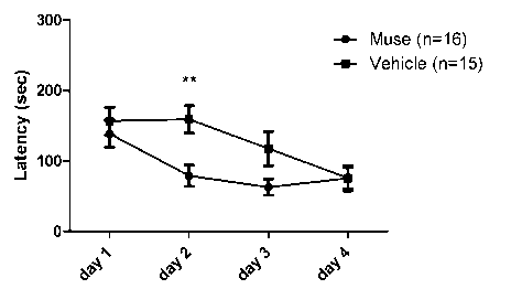
FIG. 1 shows a graph representing the arrival times in the Barnes maze in the
Muse cell administration group and the vehicle administration group. Muse
cells or
vehicle were administered 1 week after the induction of cerebral
hypoperfusion. **
represents P < 0.01. (FIG. 1 and FIG. 2 are obtained by statistically
processing with
two-way ANOVA and the Bonferroni post hoc test, and other drawings are each
obtained by statistically processing with the t-test.)
FIG. 2 shows a graph representing the proportions of Direct + Serial in the
Barnes maze in the Muse cell administration group and the vehicle
administration
group. Muse cells or vehicle were administered 1 week after the induction of
cerebral hypoperfusion. * represents P < 0.05.
FIG. 3 shows micrographs illustrating the results of Kluver-Barrera staining
of the hippocampal CA1 subregion and graphs representing the pyramidal cell
counts
and the neuropathology scores in the Muse cell administration group and the
vehicle
administration group. Muse cells or vehicle were administered 1 week after the
induction of cerebral hypoperfusion.
FIG. 4 shows micrographs illustrating the results of Kluver-Barrera staining
of the hippocampal CA2-3 subregion and graphs representing the pyramidal cell
counts and the neuropathology scores in the Muse cell administration group and
the
vehicle administration group. Muse cells or vehicle were administered 1 week
after
the induction of cerebral hypoperfusion.
FIG. 5 shows micrographs illustrating the results of Kluver-Barrera staining
of the hippocampal CA4 subregion and graphs representing the pyramidal cell
counts
and the neuropathology scores in the Muse cell administration group and the
vehicle
administration group. Muse cells or vehicle were administered 1 week after the
induction of cerebral hypoperfusion.
FIG. 6 shows graphs representing the pyramidal cell counts and the
neuropathology scores in the entire hippocampi in the Muse cell administration
Date Recue/Date Received 2022-02-09
CA 03150769 2022-02-09
7
group and the vehicle administration group. Muse cells or vehicle were
administered 1 week after the induction of cerebral hypoperfusion.
FIG. 7 shows photographs and a graph each representing the western blot
analysis results of the expressions of Bc1-2 in the hippocampi in the Muse
cell
administration group and the vehicle administration group. Muse cells or
vehicle
were administered 1 week after the induction of cerebral hypoperfusion.
FIG. 8 shows a graph representing the arrival times in the Barnes mazes in the
Muse cell administration group and the vehicle administration group. Muse
cells or
vehicle were administered 6 weeks after the induction of cerebral
hypoperfusion.
FIG. 9 shows a graph representing the proportions of Direct + Serial in the
Barnes mazes in the Muse cell administration group and the vehicle
administration
group. Muse cells or vehicle were administered 6 weeks after the induction of
cerebral hypoperfusion. * represents P < 0.05.
FIG. 10 shows graphs representing the neuropathology scores of the
hippocampal CA1, CA2-3 and CA4 subregions, and DG (dentate gyms) in the Muse
cell administration group and the vehicle administration group. Muse cells or
vehicle were administered 6 weeks after the induction of cerebral
hypoperfusion. *
and ** respectively represent P < 0.05 and P < 0.01.
FIG. 11 shows a graph representing the neuropathology scores in the entire
hippocampi in the Muse cell administration group and the vehicle
administration
group. Muse cells or vehicle were administered 6 weeks after the induction of
cerebral hypoperfusion. * represents P < 0.05.
FIG. 12 shows a graph representing the Myelin densities of corpus callosum
in the Muse cell administration group and the vehicle administration group.
Muse
cells or vehicle were administered 6 weeks after the induction of cerebral
hypoperfusion. * represents P < 0.05.
FIG. 13 shows graphs representing the numbers of CD34-positive cells in the
Date Recue/Date Received 2022-02-09
CA 03150769 2022-02-09
8
hippocampal CAL CA2-3and CA4 subregions, and DG (dentate gyms) in the Muse
cell administration group and the vehicle administration group. Muse cells or
vehicle were administered 6 weeks after the induction of cerebral
hypoperfusion. *
and ** respectively represent P < 0.05 and P < 0.01.
FIG. 14 shows a graph representing the numbers of CD34-positive cells in the
entire hippocampi in the Muse cell administration group and the vehicle
administration group. Muse cells or vehicle were administered 6 weeks after
the
induction of cerebral hypoperfusion. ** represents P < 0.01.
FIG. 15 shows the expression analysis results of pro-apoptosis markers (Bid
and Bim) and anti-apoptosis markers (Bc1-2 and Bc1-xL) in the hippocampi in
the
Muse cell administration group and the vehicle administration group. Muse
cells or
vehicle were administered 6 weeks after the induction of cerebral
hypoperfusion. *
and *** respectively represent P < 0.05 and P < 0.001.
FIG. 16 shows a graph representing the GFAP luminance per each area of the
hippocampal CAL CA2-3 and CA4 subregions, and DG (dentate gyms) in the Muse
cell administration group and the non-Muse cell administration group. Muse
cells
or non-Muse cells were administered one week after the induction of cerebral
hypoperfusion.
FIG. 17 shows a graph representing the GFAP luminance per each area of the
hippocampal CAL CA2-3 and CA4 subregions, and DG (dentate gyms) in the Muse
cell administration group and the MSC administration group. Muse cells or MSCs
were administered one week after the induction of cerebral hypoperfusion.
FIG. 18 shows a graph representing the Ibal luminance per each area of the
hippocampal CAL CA2-3 and CA4 subregions, and DG (dentate gyms) in the Muse
cell administration group and the non-Muse cell administration group. Muse
cells
or non-Muse cells were administered one week after the induction of cerebral
hypoperfusion.
Date Recue/Date Received 2022-02-09
CA 03150769 2022-02-09
9
FIG. 19 shows a graph representing the Ibal luminance per each area of the
hippocampal CAL CA2-3 and CA4 subregions, and DG (dentate gyms) in the Muse
cell administration group and the MSC administration group. Muse cells or MSCs
were administered one week after the induction of cerebral hypoperfusion.
DETAILED DESCRIPTION OF THE INVENTION
[0016]
<1> Cell product comprising Muse cell
The present invention relates to a cell product for treating or preventing
vascular dementia, comprising a SSEA-3-positive pluripotent stem cell (Muse
cell).
The treating herein encompasses curing, alleviation, recurrence prevention,
and the
like of a condition. The preventing herein encompasses preventing the onset of
dementia and preventing the progression of white matter lesion. The present
invention will be described in detail below.
[0017]
1. Indications
The cell product comprising a SSEA-3-positive pluripotent stem cell (Muse
cell) of the present invention is used for treatment or prevention of vascular
dementia.
[0018]
Vascular dementia is diagnosed by 1) the presence of dementia, 2) the
presence of a brain blood vessel disorder, and 3) relationship between both
the
presences (causal correlation). Examples of vascular dementia include multiple
infarct dementia, small vessel diseases with dementia, strategic single-
infarct
dementia, hypoperfusion dementia, and brain vascular dementia, and dementia
with
white matter lesion is preferable. The dementia in the present invention is
preferably dementia without cerebral infarction.
[0019]
Date Recue/Date Received 2022-02-09
CA 03150769 2022-02-09
2. Cell product
(1) Pluripotent stem cell (Muse cell)
The pluripotent stem cell used in the cell product of the present invention is
a
cell that was found in human living body and named "Muse (Multilineage-
5 differentiating Stress Enduring) cell" by Dezawa et al. It is known that
Muse cells
can be obtained from, for example, bone marrow aspirates, adipose tissues
(Ogura,
F., et al., Stem Cells Dev., Nov 20, 2013 (Epub) (published on Jan 17, 2014))
and
dermal connective tissues of skin, and are also broadly present in tissues and
connective tissues in organs. This cell also has both characteristics of
pluripotent
10 stem cell and mesenchymal stem cell and is identified as, for example, a
cell positive
for "SSEA-3 (Stage-specific embryonic antigen-3)," a cell surface marker,
preferably
as a double-positive cell that is SSEA-3-positive and CD105-positive.
Therefore,
Muse cells or a cell population containing Muse cells can be isolated from
living
tissues using, for example, expression of SSEA-3 only or a combination of SSEA-
3
and CD105 as an index. Methods for separation and identification of, and
characteristics of Muse cells have been disclosed in W02011/007900 in detail.
Taking advantage of the high resistance of Muse cells to various external
stresses,
Muse cells can be selectively enriched by culturing the cells under various
external
stress conditions, such as under protease treatment, under hypoxic conditions,
under
low phosphate conditions, in a low serum concentration, under undernutrition
conditions, under heat shock exposure, in the presence of toxic substances, in
the
presence of reactive oxygen species, under mechanical stimulation, and under
pressure treatment. As used herein, pluripotent stem cells prepared from
mesenchymal tissues in a living body or those derived from cultured
mesenchymal
tissues using SSEA-3 as an index (Muse cells), or a cell population comprising
Muse
cells, as a cell product for treating vascular dementia, may be simply
referred to as
"SSEA-3-positive cells." As used herein, the term "non-Muse cell" refers to a
cell
Date Recue/Date Received 2022-02-09
CA 03150769 2022-02-09
11
contained in a mesenchymal tissue in a living body or a cell contained in
cultured
mesenchymal cells, and may refer to a cell other than "SSEA-3-positive cell."
[0020]
Muse cells or a cell population comprising Muse cells can be prepared from
living tissues (e.g., mesenchymal tissues) using cell surface markers, SSEA-3,
or
SSEA-3 and CD105. As used herein, the term "living" body means mammal living
body. In the present invention, living bodies exclude fertilized egg and
embryos in
developmental stages before blastula stage, but include embryos in
developmental
stages of blastula stage or later, including fetus and blastula. Examples of
the
mammal include, but not limited to, primates such as human and monkey; rodents
such as mouse, rat, rabbit, and guinea pig; and cat, dog, sheep, pig, cattle,
horse,
donkey, goat, and ferret. Muse cells to be used in the cell product of the
present
invention are directly isolated from living tissues using markers, and thus
are clearly
distinguished from embryonic stem cells (ES cells) and induced pluripotent
stem
(iPS) cells. The term "mesenchymal tissue" refers to tissues such as bone,
synovial
membrane, fat, blood, bone marrow, skeletal muscle, dermis, ligament, tendon,
dental pulp, umbilical cord, cord blood, and amnion, as well as tissues
present in
various organs. For example, Muse cells can be obtained from bone marrow,
skin,
adipose tissues, blood, dental pulp, umbilical cord, cord blood, or amnion.
For
example, and preferably, a mesenchymal tissue in a living body is collected,
and then
Muse cells are prepared from the tissue and used. Alternatively, using the
preparation method described above, Muse cells may be prepared from cultured
mesenchymal cells such as fibroblasts or bone marrow mesenchymal stem cells.
[0021]
The cell population comprising Muse cells to be used in the cell product of
the present invention can also be prepared by a method comprising stimulating
a
mesenchymal tissue in a living body or cultured mesenchymal cells with an
external
Date Recue/Date Received 2022-02-09
CA 03150769 2022-02-09
12
stress to selectively increase cells that are resistant to the external
stress, and
collecting the cells with an increased abundance ratio.
The external stress may be any one of or a combination of the following:
protease treatment, culturing under low oxygen concentration, culturing under
low
phosphate conditions, culturing under low serum concentration, culturing
undernutrition conditions, culturing under heat shock exposure, culturing at
low
temperatures, freezing treatment, culturing in the presence of toxic
substances,
culturing in the presence of reactive oxygen species, culturing under
mechanical
stimulation, culturing under shaking, culturing under pressure treatment or
physical
shocks.
The protease treatment is preferably carried out for 0.5 to 36 hours in total
to
exert an external stress on cells. The concentration of the protease is
preferably
used when cells adhered to a culture vessel are peeled off, when cell
aggregates are
separated into single cells, or when single cells are collected from a tissue.
Preferably, the protease is a serine protease, an aspartic protease, a
cysteine
protease, a metalloprotease, a glutamic protease, or an N-terminal threonine
protease.
More preferably, the protease is trypsin, collagenase, or Dispase.
[0022]
Muse cells to be used in the cell product of the present invention may be
autologous or allogeneic to a recipient who will receive the cells.
[0023]
As described above, Muse cells or a cell population comprising Muse cells
can be prepared from living tissues, for example, by using SSEA-3 positivity
or
SSEA-3 and CD105 double positivity as an index. Human adult skin is known to
comprise various types of stem cells and progenitor cells. However, Muse cells
are
different from these cells. These stem cells and progenitor cells include skin-
derived progenitor cells (SKI)), neural crest stem cells (NCSC), melanoblasts
(MB),
Date Recue/Date Received 2022-02-09
CA 03150769 2022-02-09
13
pericytes (PC), endothelial progenitor cells (EP), and adipose-derived stem
cells
(ADSC). Muse cells can be prepared using "non-expression" of markers unique to
these cells as an index. More specifically, Muse cells can be isolated using
as an
index non-expression of at least one, e.g., 2, 3, 4, 5, 6, 7, 8,9, 10, or 11,
of 11
markers selected from the group consisting of CD34 (a marker for EP and ADSC),
CD117 (c-kit) (a marker for MB), CD146 (a marker for PC and ADSC), CD271
(NGFR) (a marker for NC SC), NG2 (a marker for PC), vWF factor (von Willebrand
factor) (a marker for EP), Sox10 (a marker for NCSC), Snail (a marker for
SKI)),
Slug (a marker for SKI)), Tyrpl (a marker for MB), and Dct (a marker for MB).
Muse cells can be prepared by using as an index non-expression of, for
example, but
not limited to, CD117 and CD146; CD117, CD146, NG2, CD34, vWF, and CD271;
or the above-described 11 markers.
[0024]
Muse cells having the above-described characteristics and used in the cell
product of the present invention may also have at least one selected from the
group
consisting of the following characteristics:
(i) having low or no telomerase activity;
(ii) capable of differentiating into any of tfidermic cells;
(iii) showing no neoplastic proliferation; and
(iv) having self-renewal capacities
Preferably, Muse cells to be used in the cell product of the present invention
have all
of the characteristics described above.
[0025]
With respect to (i) above, the phrase "having low or no telomerase activity"
means that the telomerase activity is low or undetectable when detected using,
for
example, TRAPEZE XL telomerase detection kit (Millipore Corporation). Having
"low" telomerase activity means, for example, having a telomerase activity
Date Recue/Date Received 2022-02-09
CA 03150769 2022-02-09
14
comparable to somatic human fibroblast, or having 1/5 or less telomerase
activity,
preferably 1/10 or less telomerase activity, as compared with that of Hela
cell.
[0026]
With respect to (ii) above, Muse cells are capable of being differentiated
into
tridermic cells (endodermal, mesodermal, and ectodermal cells) in vitro and in
vivo,
and can be differentiated into, for example, hepatocytes (including cells
expressing
markers of hepatoblast or hepatocyte), neurons, skeletal muscle cells, smooth
muscle
cells, osteocytes, or adipocytes by in vitro inductive culturing. Muse cells
may also
show the ability to be differentiated into tridermic cells when transplanted
in testis in
vivo. Further, Muse cells are capable of migrating and engrafting to injured
organs
(such as heart, skin, spinal cord, liver, and muscle) when transplanted into a
living
body via intravenous injection and being differentiated into cells depending
on the
tissues.
[0027]
With respect to (iii) above, Muse cells are characterized in that they
proliferate at a growth rate of about 1.3 days and proliferate from a single
cell in
suspension culture to form embryoid body-like cell aggregates, and then arrest
their
proliferation after about 14 days when the aggregates reach a certain size.
When
these embryoid body-like cell aggregates are transferred to adherent culture,
the cells
restart proliferation and cells proliferated from the cell aggregates expand
at a growth
rate of about 1.3 days. Further, Muse cells are characterized in that, when
transplanted into testis, they do not become cancerous for at least half a
year.
[0028]
With respect to (iv) above, Muse cells have self-renewal (self-replication)
capacities. The term "self-renewal," as used herein, means that the followings
can
be observed: differentiation into tridermic cells from cells contained in
first embryoid
body-like cell aggregates obtained by culturing single Muse cells in a
suspension
Date Recue/Date Received 2022-02-09
CA 03150769 2022-02-09
culture; as well as formation of next-generation second embryoid body-like
cell
aggregates by again culturing single cells in the first embryoid body-like
cell
aggregates in a suspension culture; and further differentiation into tridermic
cells and
formation of third embryoid body-like cell aggregates in a suspension culture
from
5 the second embryoid body-like cell aggregates. Self renewal may be
repeated for
one or more cycles.
[0029]
(2) Preparation and Use of Cell Product Comprising Muse Cell
The cell product comprising Muse cells of the present invention can be
10 obtained by, but not limited to, suspending Muse cells or a cell
population
comprising Muse cells obtained in (1) above in a physiological saline or a
suitable
buffer solution (e.g., a phosphate buffered saline). In this case, when only
small
numbers of Muse cells are isolated from an autologous or allogeneic tissue,
these
cells may be cultured before cell transplantation until the predetermined
number of
15 cells is attained. As previously reported (W02011/007900), since Muse
cells are
non-tumorigenic, they are less likely to be cancerous and thus are safe, even
if cells
collected from a living tissue are contained in undifferentiated states. The
collected
Muse cells can be cultured in any normal growth medium (e.g., alpha-minimum
essential medium (a-MEM) supplemented with 10% calf serum). More
specifically, with reference to the above-described W02011/007900, Muse cells
can
be cultured and proliferated using an appropriately selected culture medium,
additives (e.g., antibiotics, and serum) and the like, to prepare a solution
containing
Muse cells at a predetermined concentration. When the cell product comprising
Muse cells of the present invention is administered to a human subject, bone
marrow
aspirates are collected from a human ilium. Then, for example, bone marrow
mesenchymal stem cells are cultured as adherent cells obtained from the bone
marrow aspirate and proliferated until reaching the cell amount where a
Date Recue/Date Received 2022-02-09
CA 03150769 2022-02-09
16
therapeutically effective amount of Muse cells can be obtained. Thereafter,
Muse
cells are isolated using an antigenic marker SSEA-3 as an index to prepare a
cell
product containing autologous or allogeneic Muse cells. Alternatively, for
example,
bone marrow mesenchymal stem cells obtained from the bone marrow aspirates can
be cultured under external stress conditions, so that Muse cells can be grown
and
enriched until they reach a therapeutically effective amount, thereby
preparing a cell
product comprising autologous or allogeneic Muse cells.
[0030]
When Muse cells are used in a cell product, the cell product may also
comprise dimethyl sulfoxide (DMSO), serum albumin and the like for protection
of
the cells and antibiotics and the like for prevention of contamination and
proliferation of bacteria. The cell product may further comprise other
pharmaceutically acceptable components (e.g., carriers, excipients,
disintegrants,
buffer agents, emulsifiers, suspending agents, soothing agents, stabilizers,
preservatives, antiseptics, physiological saline). These agents and drugs can
be
added to the cell product at appropriate concentrations by the skilled person.
Thus,
Muse cells can also be used as a pharmaceutical composition comprising various
additives.
[0031]
The number of Muse cells contained in the cell product prepared above can
be appropriately adjusted to achieve desired effects in treatment and/or
prevention of
vascular dementia, in consideration of, for example, sex, age, and weight of
the
subject, the condition of the affected area, and the condition of the cells to
be used.
Individuals as the subject includes, but not limited to, mammals such as
human.
The cell product comprising Muse cells of the present invention may be
administered
multiple times at appropriate intervals (e.g., twice a day, once a day, twice
a week,
once a week, once every two weeks, once a month, once every two months, once
Date Recue/Date Received 2022-02-09
CA 03150769 2022-02-09
17
every three months, or once every six months) until the desired therapeutic
effect is
obtained. Thus, the therapeutically effective amount is preferably, for
example, 1 to
doses of 1 x10³ to 1 x10¹⁹ cells/individual/dose for one year, depending on
the
state of the subject. The total amount administered to an individual is, but
not
5 limited to, lx 103 to lx 1011 cells, preferably 1 x 10⁴ to 1x10' cells,
more preferably
1x10⁵ to 1x10⁹ cells.
[0032]
Muse cells to be used in the cell product of the present invention are
characterized in that they migrate and engraft to an impaired site of the
brain. Thus,
10 the site and method of administration of the cell product in
administration of the cell
product are not limited, and examples include intravascular administration
(intravenous, intraarterial), intrathecal administration, and intraparenchymal
administration.
[0033]
The cell product comprising Muse cells of the present invention allows repair
and regeneration of the impaired site in a patient having vascular dementia to
be
realized.
[0034]
The present invention will be described in more detail with reference to
examples below, but is not limited to the examples in any way.
EXAMPLES
[0035]
<Preparation of human Muse cell>
Muse cells were obtained according to the method for isolation and
identification of human Muse cells described in W02011/007900. Muse cells were
cultured by expansive enrichment with culturing of mesenchymal stem cells
under
stress conditions.
Date Recue/Date Received 2022-02-09
CA 03150769 2022-02-09
18
[0036]
<Production of rat model of vascular dementia>
The experimental protocols using mice in this Example complied with
"Regulations on Animal Experiments and Related Activities in Tohoku
University,"
and the experimental animals were prepared in accordance with the regulations
under
the supervision of the Animal Experiment Center of Tohoku University. A model
of chronic cerebral hypoperfusion was used as a rat model of vascular
dementia.
Specifically, SD rats (eight to ten-week male rats, weight 250 to 300 g) were
subjected to ligation of both carotid arteries as described in Journal of
Cerebral
Blood Flow & Metabolism 2016, vol. 36(9) 1592-1602, and thus were under
chronic
cerebral hypoperfusion conditions. The brain blood flows of the models were
reduced to about 30 to 50% of normal values, and caused the onset of a
cognitive
disorder along with white matter lesion and neurodegeneration of the
hippocampus.
[0037]
<Administration of Muse cells>
The above model rats of chronic cerebral hypoperfusion were divided to two
groups, and Muse cells (3 x10⁵ cells/PBS) or HBSS (vehicle) was administered
by
injection thereof into the cervical vein of each of the rats in each of the
groups after
one week (corresponding to a acute phase of vascular dementia) of the
induction of
cerebral hypoperfusion. Since human Muse cells heterologous for rats were
transplanted, an immunosuppressant (FK506) was administered to each cerebral
infarction rat before transplantation.
[0038]
<Cognitive function evaluation using Barnes maze>
The cognitive function was evaluated using the Barnes maze after three
weeks of Muse cell or vehicle administration. The Barnes maze is used to
measure
spatial learning and memory. The Barnes mase consists of a circular disk
platform
Date Recue/Date Received 2022-02-09
CA 03150769 2022-02-09
19
with eighteen circular holes unifomily placed along the outer periphery
thereof. An
escaping box was located under one of the holes. After the rats were
familiarized
with the maze for one day, the arrival times until the rats entered the
escaping box
and the search strategy were recorded for four days (Day 1 to Day 4) (the test
was
performed three times a day).
The strategy to find the escaping box was categorized into three classes, 1)
Direct, 2) Serial, and 3) Random.
1) Direct: direct arriving at the escaping box, or arriving at a box next to
the
escaping box and then arriving at the escaping box
2) Serial: arriving at the escaping box while following the peripheral part of
the maze
3) Random: searching the holes while reciprocating between the center and
the periphery of the maze many times
[0039]
The results are shown in FIG. 1 and 2. The arrival time (Latency) was
significantly shortened on Day 2 in the Muse cell administration group. The
ratio
between Direct and Serial in the search strategy was significantly enhanced
also on
Day 2 in the Muse cell administration group. It was thus found that Muse cell
administration could enhance a cognitive function deteriorated due to a
chronic
cerebral hypoperfusion condition.
[0040]
<Histological evaluation>
After the above behavioral test, the hippocampus region of the brain of each
of the rats was isolated to produce tissue sections, and the sections were
histologicaly
evaluated by Kluver-Barrera staining. Apoptosis was analyzed by examining the
expression of Bc1-2 by the western blot. Engraftment of human Muse cells was
confirmed by human mitochondria staining in the Muse cell administration
group.
Date Recue/Date Received 2022-02-09
CA 03150769 2022-02-09
The hippocampus CA1, CA2-3 and CA4 subregions were each stained, and
the pyramidal cell count and neuropathology score described below were
calculated
and shown graphically.
[0041]
5 Pyramidal cell count (%) = Number of living pyramidal cells/Total
number of
pyramidal cells
[0042]
Neuropathology score
0: no lesion, 1: dead cell 1 to 10%, 2: dead cell 11 to 25%, 3: dead cell 26
to 45%, 4:
10 dead cell 46% or more
[0043]
The results in the CA1 (FIG. 3), CA2-3 region (FIG. 4), CA4 (FIG. 5)
subregions,
and the entire hippocampus (FIG. 6) are summarized in the figures. While no
significant difference between the Muse cell administration group and the
vehicle
15 administration group was observed in the CA1 subregion. A significant
increase
in pyramidal cell count in the CA2-3 and CA4 subregions were observed.
Significant improvement in neuropathology score in the CA4 subregion was also
exhibited in the Muse cell administration group. Significant increase in
pyramidal
cell count and a significant improvement in neuropathology score were also
20 exhibited in the entire hippocampus in the Muse cell administration
group.
These results suggest that Muse cells administered were engrafted to the
hippocampus region of the brain and protected neurons and thus maintain a
cognitive
function.
[0044]
The expression of Bc1-2 in the hippocampus was examined by the western
blot. The expression of Bc1-2 was upregulated in the Muse cell administration
group (FIG. 6), suggesting apoptosis was suppressed by Muse cell
administration.
Date Recue/Date Received 2022-02-09
CA 03150769 2022-02-09
21
The same results were also observed in Tunnel staining (no data shown). From
the
results of Ki67 staining, a tendency was observed where cell proliferation in
the
hippocampus was increased by Muse cell administration.
[0045]
<Chronic phase administration evaluation>
Muse cells (3x 10⁵ cells/PBS) or HBSS (vehicle) was administered to each of
the above model rats of chronic cerebral hypoperfusion after six weeks
(corresponding to a chronic phase of vascular dementia) of the induction of
the
cerebral hypoperfusion. The cognitive function evaluation (started after nine
weeks
of the cerebral hypoperfusion) using the Barnes maze and the histological
staining
(ten weeks after cerebral hypoperfusion) were performed in the same manner as
described above. The histological evaluation was performed by Myelin staining
and CD34 staining, in addition to the neuropathology score. Apoptotic pathways
were analyzed with the expressions of Bid and Bim as pro-apoptotic markers and
the
expressions of Bc1-2 and Bc1-xL as anti-apoptotic markers by the western blot.
Engraftment of human Muse cells was confirmed by human mitochondria staining
in
the Muse cell administration group.
[0046]
The results of the cognitive function test are shown in FIG. 8 and FIG. 9.
The arrival time (Latency) was significantly shortened on Day 2 and Day 3 in
the
Muse cell administration group. The ratio between Direct and Serial in the
search
strategy was significantly increased also on Day 2 in the Muse cell
administration
group. These results suggest that Muse cell administration could
improvecognitive
function even in the chronic phase administration.
[0047]
The results of the CAL CA2-3 and CA4 subregions, and DG (dentate gyms)
are shown in FIG. 10, with respect to analysis of the neuropathology score.
The
Date Recue/Date Received 2022-02-09
CA 03150769 2022-02-09
22
results of the entire hippocampus are summarized in FIG. 11. As a result, a
significant improvement in neuropathology score was observed in the CA1, CA2-
3,
and CA4 subregions in the Muse cell administration group. A significant
improvement in neuropathology score was observed also in the entire
hippocampus
in the Muse cell administration group. These results suggest that Muse cells
administered were engrafted to the hippocampal region of the brain and
protected
neurons to maintain a cognitive function.
[0048]
The results of Myelin staining are shown in FIG. 12. As a result, Myelin
density in the corpus callosum was significantly increased in the Muse cell
administration group, which implies that Muse cells have the effect of
improving the
white matter damage of the brain.
[0049]
The results of CD34 staining are shown in FIG. 13 and FIG. 14. CD34-
positive cells were significantly increased in the CA1, CA2-3, and CA4
subregions
in the Muse cell administration group. CD34-positive cells were also
significantly
increased in the entire hippocampus, in the Muse cell administration group.
These
results suggest that Muse cells administered were engrafted to the hippocampus
region of the brain and promoted blood vessel growth to thereby protect
neurons and
thus maintain a cognitive function.
[0050]
The expressions of pro- and anti-apoptotic markers in the hippocampus were
examined by the western blot, and thus the expressions of pro-apoptotic
markers
were reduced and the expressions of anti-apoptotic markers were increased in
the
Muse cell administration group as shown in FIG. 15. These results suggest that
apoptosis is suppressed by Muse cell administration. The same results were
also
observed in Tunel staining (no data shown).
Date Recue/Date Received 2022-02-09
CA 03150769 2022-02-09
23
[0051]
<Preparation of human Muse cell, non-Muse cell, and MSC>
Muse cells were obtained according to the method for isolation and
identification of human Muse cells described in W02011/007900. A commercially
available mesenchymal stem cell (MSC, Lonza) was used as a source of Muse
cells.
Muse cells used for transplantation were made to express green fluorescent
protein
(GFP) to determine whether the cells were engrafted into each tissue. For cell
labeling with GFP, Muse cells had been previously transduced with a lentivirus-
GFP
gene. GFP-labeled Muse cells are isolated as GFP- and SSEA-3-double positive
cells by FACS and used as a Muse cell group. GFP-positive MSCs were also
isolated by FACS and used as a MSC group, and the remaining cells obtained by
isolating Muse cells from GFP-positive MSCs were used as a non-Muse cell
group.
[0052]
<Administration of Muse cells and the like>
GFP-Muse cells (lx 105 cells/individual), GFP-non-Muse cells (1 x10⁵
cells/individual), or GFP-MSCs (1 x10⁵ cells/individual) were administered to
each
of the above model rats of chronic cerebral hypoperfusion by injection thereof
into
the left cervical vein of each of the rats after one week (corresponding to an
acute
phase of vascular dementia) of procedure.
[0053]
<Histological evaluation>
The brains of each of the rats were measured by Nissle staining after one
week of the above cell administration. Any specimen where cerebral infarction
occurred was excluded. The brain tissues were fixed, and histological
evaluation
was performed by staining of glial fibrillary acidic protein (GFAP), a brain
astrocyte
marker, and Ibal, a microglia marker and also serving as an index of nerve
inflammation.
Date Recue/Date Received 2022-02-09
CA 03150769 2022-02-09
24
[0054]
The results of GFAP staining are shown in FIG. 16 and FIG. 17. The
GFAP-positive cells were significantly decreased in the CA1, CA2-3 and CA4
subregions, and DG (dentate gyms) in the Muse cell administration group
compared
with the non-Muse cell group. GFAP-positive cells were also likely decreased
in
the CA1, CA2-3 and CA4 subregions and DG (dentate gyms) in the Muse cell
administration group compared with the MSC administration group.
[0055]
The results of Ibal staining are shown in FIG. 18 and FIG. 19. Ibal-positive
cells were significantly decreased in the CA1 and CA4 subregions, and DG
(dentate
gyms). Ibal-positive cells were likely decreased in the CA2-3 subregion in the
Muse cell administration group compared with the non-Muse cell group. Ibal-
positive cells were significantly decreased in the CA1 and CA4 subregions, and
DG
(dentate gyms) in the Muse cell administration group. Ibal-positive cells also
likely
decreased in the CA2-3 subregion in the Muse cell administration group
compared
with the MSC administration group.
[0056]
In summary, the expressions of astrocyte and microglia markers were reduced
in the Muse cell administration group compared with the non-Muse cell and MSC
administration groups. These results suggest that Muse cells repair the
damaged
hippocampus region of the brain.
Industrial Applicability
[0057]
The cell product of the present invention can be administered to a patient
having or suspected to have vascular dementia, resulting in repair of an
impaired site
of the brain, in which white matter lesion or the like occurs, and
ameliorating or
treating cognitive function disorders, and can be applied to treatment and/or
Date Recue/Date Received 2022-02-09
CA 03150769 2022-02-09
prevention of vascular dementia.
Date Recue/Date Received 2022-02-09