Note: Descriptions are shown in the official language in which they were submitted.
WO 2021/055517
PCT/US2020/051130
METHODS OF TREATMENTS BASED UPON MOLECULAR CHARACTERIZATION
OF BREAST CANCER
CROSS REFERENCE TO RELATED APPLICATIONS
[0001] This application claims priority to U.S.
Provisional Application Ser. No.
62/901,175, entitled "Methods of Treatments Based Upon Molecular
Characterization of
Breast Cancer" by Christina Curtis et al., filed September 16, 20191 which is
incorporated
herein by reference in its entirety.
TECHNICAL FIELD
[0002] The invention is generally directed to methods of diagnostics and
treatments
based upon a molecular characterization of an individual's breast cancer, and
more
specifically to treatments based upon molecular diagnostics indicative of
aggressiveness,
relapse risk of breast cancer, or molecular subtype.
BACKGROUND
[0003] Breast cancer is the most frequent cancer diagnosis and cause of cancer
death
in women worldwide with 1.4 million diagnoses and 500,000 deaths annually.
Survival
rates have dramatically improved due to new treatments but a sizable minority
of patients
suffer from an aggressive form of cancer and/or experience a relapse, which
may be
incurable. Most cancer registries do not record recurrence information and the
rates of
relapse are poorly characterized. Analysis of retrospective cohorts and
clinical trials have
provided some insights into patterns of recurrence. For example, some estrogen
receptor-
positive (ER+) tumors continue to recur well past five years with a higher
rate of bone
metastasis, while estrogen receptor-negative (ER-) tumors recur more quickly
and have
higher rates of visceral metastases. However, methods to reliably stratify
risk of relapse
are lacking as are therapeutic approaches for early stage breast cancer
patients who are
at high risk of relapse or who have already recurred on the basis of their
tumor molecular
profile.
-1-
CA 03151330 2022-3-15
WO 2021/055517
PCT/US2020/051130
SUMMARY
[0004] Various embodiments are directed towards methods treatments for breast
cancer based on its molecular characterization. In various embodiments, the
molecular
subtype of a breast cancer is determined based on its genetics. In various
embodiments,
a molecular subtype is indicative breast cancer aggressiveness and risk of
relapse. In
various embodiments, a molecular subtype is indicative of the molecular
pathology of a
breast cancer. In various embodiments, a breast cancer is treated based upon
aggressiveness, risk of relapse, and molecular drivers as determined by its
molecular
subtype.
[0005] In an embodiment, an individual having breast
cancer is treated. A breast
cancer of an individual is stratified utilizing a risk stratification model
into a high risk of
recurrence subgroup. The risk stratification model is a statistical model that
incorporates
features derived from integrative subtype clusters that are delineated by a
molecular
pathology. The individual is treated to reduce the risk of recurrence by
administering a
prolonged treatment regimen that includes chemotherapy, endocrine therapy,
targeted
therapy, or health professional surveillance.
[0006] In another embodiment, the risk stratification
model utilizes a multi-state semi-
markov Model, a Cox Proportional Hazards model, a shrinkage based method, a
tree
based method, a Bayesian method, a kernel based method, or a neural network.
[0007] In yet another embodiment, the integrated subtype
cluster features are
membership to a given cluster or the posterior probability of membership to a
given
cluster.
[0008] In a further embodiment, the integrative subtype
clusters are determined by the
IntClust classification model that incorporates molecular data as features.
[0009] In still yet another embodiment, the molecular data
is obtained by microarray
based gene expression, microarray/SNP array based copy number inference, RNA-
sequencing, targeted (capture) RNA-sequencing, exome sequencing, whole genome
sequencing (VVES/VVGS), targeted (panel) sequencing, Nanostring nCounter for
gene
expression, Nanostring nCounter for copy number inference, Nanostring digital
spatial
-2-
CA 03151330 2022-3-15
WO 2021/055517
PCT/US2020/051130
profiler measurement of protein, Nanostring digital spatial profiler
measurement of
protein gene expression in situ, DNA-ISH, RNA-ISH, RNAScope, DNA Methylation
assays, or ATAC-seq.
[0010] In yet a further embodiment, the molecular data is
derived utilizing a gene
panel.
[0011] In an even further embodiment, the gene panel is
one of: Foundation Medicine
CDx, Memorial Sloan Kettering Cancer Center Integrated Mutation Profiling of
Actionable
Cancer Targets (MSK-IM PACT), Stanford Tumor Actionable Mutation Panel
(STAMP), or
UCSF500 Cancer Gene Panel.
[0012] In yet an even further embodiment, the risk
stratification model utilizes clinical
data, such as age, cancer stage, number of tumor positive lymph nodes, size of
tumor,
grade of tumor, surgery performed, treatment performed, or basic molecular
identities.
[0013] In still yet an even further embodiment, the risk
stratification model utilizes the
CTS5 algorithm.
[0014] In still yet an even further embodiment, the risk
stratification model incorporates
Oncotype DX, Prosigna PAM50, Prosigna ROR, MammaPrint, EndoPredict or Breast
Cancer Index (BC).
[0015] In still yet an even further embodiment, the
prolonged treatment regimen
includes adjuvant chemotherapy.
[0016] In still yet an even further embodiment, the
prolonged treatment regimen
includes treatment beyond the standard course of treatment.
[0017] In an embodiment, an individual having breast
cancer is treated. A breast
cancer of an individual is stratified utilizing a risk stratification model
into a lower risk of
recurrence subgroup. The risk stratification model is a statistical model that
incorporates
features derived from integrative subtype clusters that are delineated by a
molecular
pathology. The individual is treated to reduce the harmful effects of
chemotherapy by
administering a treatment regimen that includes surgery or endocrine therapy,
but not
chemotherapy.
[0018] In another embodiment, the risk stratification
model utilizes a multi-state semi-
markov Model, a Cox Proportional Hazards model, a shrinkage based method, a
tree
based method, a Bayesian method, a kernel based method, or a neural network.
-3-
CA 03151330 2022-3-15
WO 2021/055517
PCT/US2020/051130
[0019] In yet another embodiment, the integrated subtype
cluster features are
membership to a given cluster or the posterior probability of membership to a
given
cluster.
[0020] In a further embodiment, the integrative subtype
clusters are determined by the
IntClust classification model that incorporates molecular data as features.
[0021] In still yet another embodiment, the molecular data
is obtained by microarray
based gene expression, microarray/SNP array based copy number inference, RNA-
sequencing, targeted (capture) RNA-sequencing, exome sequencing, whole genome
sequencing (WES/WGS), targeted (panel) sequencing, Nanostring nCounter for
gene
expression, Nanostring nCounter for copy number inference, Nanostring digital
spatial
profiler measurement of protein, Nanostring digital spatial profiler
measurement of
protein gene expression in situ, DNA-ISH, RNA-ISH, RNAScope, DNA Methylation
assays, or ATAC-seq.
[0022] In yet a further embodiment, the molecular data is
derived utilizing a gene
panel.
[0023] In an even further embodiment, the gene panel is one of: Foundation
Medicine
CDx, Memorial Sloan Kettering Cancer Center Integrated Mutation Profiling of
Actionable
Cancer Targets (MSK-IM PACT), Stanford Tumor Actionable Mutation Panel
(STAMP), or
UCSF500 Cancer Gene Panel.
[0024] In yet an even further embodiment, the risk
stratification model utilizes clinical
data, such as age, cancer stage, number of tumor positive lymph nodes, size of
tumor,
grade of tumor, surgery performed, treatment performed, or basic molecular
identities.
[0025] In still yet an even further embodiment, the risk
stratification model utilizes the
CTS5 algorithm.
[0026] In still yet an even further embodiment, the risk
stratification model incorporates
Oncotype DX, Prosigna PAM50, Prosigna ROR, MammaPrint, EndoPredict or Breast
Cancer Index (BC).
[0027] In still yet an even further embodiment, the
treatment regimen includes
adjuvant endocrine therapy.
-4-
CA 03151330 2022-3-15
WO 2021/055517
PCT/US2020/051130
[0028] In an embodiment, an individual having breast
cancer is treated. The results an
assay is determined, classifying an individual's breast cancer into an
integrated cluster
(IntClust) subgroup. The results indicate that the breast cancer is classified
into one of:
IntClustal , IntClust2, IntClust6, or IntClust9. The individual is treated
with a prolonged
treatment regimen that includes chemotherapy, endocrine therapy, targeted
therapy, and
health professional surveillance.
[0029] In another embodiment, the classification of the
individual's breast cancer is
performed utilizing a molecular class prediction tool.
[0030] In yet another embodiment, the molecular class
prediction tool utilizes a
shrinkage based method, logistic regression, a support vector machine with a
linear
kernel, a support vector machine with a gaussian kernel, or a neural network.
[0031] In a further embodiment, the molecular class
prediction tool incorporates
molecular data as features.
[0032] In still yet another embodiment, the molecular data
features are copy number
features, gene expression features, genomic methylation features, or occupancy
features
derived from DNA or RNA analysis of the individual's breast cancer.
[0033] In yet a further embodiment, the molecular data is
obtained by microarray
based gene expression, micxoarray/SNP array based copy number inference, RNA-
sequencing, targeted (capture) RNA-sequencing, exome sequencing, whole genome
sequencing (VVES/VVGS), targeted (panel) sequencing, Nanostring nCounter for
gene
expression, Nanostring nCounter for copy number inference, Nanostring digital
spatial
profiler measurement of protein, Nanostring digital spatial profiler
measurement of
protein gene expression in situ, DNA-ISH, RNA-ISH, RNAScope, DNA Methylation
assays, or ATAC-seq.
[0034] In an even further embodiment, the molecular data
is derived utilizing a gene
panel.
[0035] In yet an even further embodiment, the gene panel is Foundation
Medicine
CDx, Memorial Sloan Kettering Cancer Center Integrated Mutation Profiling of
Actionable
Cancer Targets (MSK-IM PACT), Stanford Tumor Actionable Mutation Panel
(STAMP), or
UCSF500 Cancer Gene Panel.
-5-
CA 03151330 2022-3-15
WO 2021/055517
PCT/US2020/051130
[0036] In still yet an even further embodiment, the breast
cancer the individual is
administered adjuvant chemotherapy.
[0037] In still yet an even further embodiment, the breast
cancer the individual is
administered extended endocrine therapy.
[0038] In still yet an even further embodiment, the
endocrine therapy comprises
administering a selective estrogen receptor modulator, a selective estrogen
receptor
degrader, an aromatase inhibitor, or PROTAC ARV-471.
[0039] In still yet an even further embodiment, the
selective estrogen receptor
modulator is tamoxifen, toremifene, raloxifene, ospennifene, or bazedoxifene.
[0040] In still yet an even further embodiment, the
selective estrogen receptor
degrader is fulvestrant, brilanestrant (GDC-0810), elacestrant, GDC-9545,
SAR439859
(SERD '859), RG6171, or AZD9833.
[0041] In still yet an even further embodiment, the
aromatase inhibitor is anastrozole,
exemestane, letrozole, vorozole, formestane, or fadrozole.
[0042] In still yet an even further embodiment, the breast
cancer is classified into
IntClust1 and the individual is administered an mTOR pathway antagonist, an
AKT1
antagonist, an AKT1JRPS6KB1 antagonist, an RPS6KB1 antagonist, a PI3K
antagonist,
an elF4A antagonist, or an elF4E antagonist.
[0043] In still yet an even further embodiment, the breast
cancer is classified into
IntClust2 and the individual is administered a CDK4/6 antagonist, an FGFR
pathway
antagonist, a PARP antagonist, a homologous recombination deficiency (HRD)
targeted
therapy, a PAK1 antagonist, an elF4A antagonist, or elF4E antagonist.
[0044] In still yet an even further embodiment, the breast
cancer is classified into
IntClust6 and the individual is administered an FGFR pathway antagonist, an
elF4A
antagonists, or an elF4E antagonist.
[0045] In still yet an even further embodiment, the breast
cancer is classified into
IntClust9 and the individual is administered a selective estrogen receptor
degrader, an
SRC3 antagonist, a MYC antagonist, a BET bromodomain antagonist, an elF4A
antagonist, or an elF4E antagonist.
-6-
CA 03151330 2022-3-15
WO 2021/055517
PCT/US2020/051130
[0046] In an embodiment, an individual having breast
cancer is treated. An oncogenic
pathology of an individual's cancer is classified. The oncogenic pathology
indicates
mTOR pathway. The individual is administered an mTOR antagonist.
[0047] In another embodiment, the oncogenic pathology is
classified utilizing a
molecular class prediction tool that utilizes a shrinkage based method,
logistic regression,
a support vector machine with a linear kernel, a support vector machine with a
gaussian
kernel, or a neural network. The molecular prediction tool also utilizes copy
number
features, gene expression features, genomic methylation features, or
nucleosome
occupancy features derived from DNA or RNA analysis of the individual's breast
cancer.
[0048] In yet another embodiment, the mTOR antagonist is
everolimus, tennsirolinnus,
sirolimus, or rapamycin.
[0049] In an embodiment, an individual having breast
cancer is treated. An oncogenic
pathology of an individual's cancer is classified. The oncogenic pathology
indicates AKT1.
The individual is administered an AKT1 antagonist.
[0050] In another embodiment, the oncogenic pathology is
classified utilizing a
molecular class prediction tool that utilizes a shrinkage based method,
logistic regression,
a support vector machine with a linear kernel, a support vector machine with a
gaussian
kernel, or a neural network. The molecular prediction tool also utilizes copy
number
features, gene expression features, genomic methylation features, or
nucleosome
occupancy features derived from DNA or RNA analysis of the individual's breast
cancer.
[0051] In yet another embodiment, the Alai antagonist is
ipatasertib, or capivasertib
(AZD5363).
[0052] In an embodiment, an individual having breast
cancer is treated. An oncogenic
pathology of an individual's cancer is classified. The oncogenic pathology
indicates
AKT1/RPS6KB1. The individual is administered an AKT1 /RPS6KB1 antagonist.
[0053] In another embodiment, the oncogenic pathology is
classified utilizing a
molecular class prediction tool that utilizes a shrinkage based method,
logistic regression,
a support vector machine with a linear kernel, a support vector machine with a
gaussian
kernel, or a neural network. The molecular prediction tool also utilizes copy
number
features, gene expression features, genomic methylation features, or
nucleosome
occupancy features derived from DNA or RNA analysis of the individual's breast
cancer.
-7-
CA 03151330 2022-3-15
WO 2021/055517
PCT/US2020/051130
[0054] In yet another embodiment, the AKT1/RPS6KB1antagonist is M2698.
[0055] In an embodiment, an individual having breast
cancer is treated. An oncogenic
pathology of an individual's cancer is classified. The oncogenic pathology
indicates
RPS6KB1. The individual is administered an RPS6KB1 antagonist.
[0056] In another embodiment, the oncogenic pathology is
classified utilizing a
molecular class prediction tool that utilizes a shrinkage based method,
logistic regression,
a support vector machine with a linear kernel, a support vector machine with a
gaussian
kernel, or a neural network. The molecular prediction tool also utilizes copy
number
features, gene expression features, genomic methylation features, or
nucleosome
occupancy features derived from DNA or RNA analysis of the individual's breast
cancer.
[0057] In yet another embodiment, the RPS6KB1 antagonist is LY2584702.
[0058] In an embodiment, an individual having breast
cancer is treated. An oncogenic
pathology of an individual's cancer is classified. The oncogenic pathology
indicates P I3K.
The individual is administered an PI3K antagonist.
[0059] In another embodiment, the oncogenic pathology is
classified utilizing a
molecular class prediction tool that utilizes a shrinkage based method,
logistic regression,
a support vector machine with a linear kernel, a support vector machine with a
gaussian
kernel, or a neural network. The molecular prediction tool also utilizes copy
number
features, gene expression features, genomic methylation features, or
nucleosome
occupancy features derived from DNA or RNA analysis of the individual's breast
cancer.
[0060] In yet another embodiment, the PI3K antagonist is
alpelisib, buparlisib
(BKM120), or pictilisib (GDC-0941)_
[0061] In an embodiment, an individual having breast
cancer is treated. An oncogenic
pathology of an individual's cancer is classified. The oncogenic pathology
indicates
CDK4/6. The individual is administered an CDK4/6 antagonist.
[0062] In another embodiment, the oncogenic pathology is
classified utilizing a
molecular class prediction tool that utilizes a shrinkage based method,
logistic regression,
a support vector machine with a linear kernel, a support vector machine with a
gaussian
kernel, or a neural network. The molecular prediction tool also utilizes copy
number
features, gene expression features, genomic methylation features, or
nucleosome
occupancy features derived from DNA or RNA analysis of the individual's breast
cancer.
-8-
CA 03151330 2022-3-15
WO 2021/055517
PCT/US2020/051130
[0063] In yet another embodiment, the CDK4/6 antagonist is
palbociclib, ribociclib, or
abemaciclib.
[0064] In an embodiment, an individual having breast
cancer is treated. An oncogenic
pathology of an individual's cancer is classified. The oncogenic pathology
indicates FGFR
pathway. The individual is administered an FGFR pathway antagonist.
[0065] In another embodiment, the oncogenic pathology is
classified utilizing a
molecular class prediction tool that utilizes a shrinkage based method,
logistic regression,
a support vector machine with a linear kernel, a support vector machine with a
gaussian
kernel, or a neural network. The molecular prediction tool also utilizes copy
number
features, gene expression features, genomic methylation features, or
nucleosome
occupancy features derived from DNA or RNA analysis of the individual's breast
cancer.
[0066] In yet another embodiment, the FGFR pathway
antagonist is lucitanib, dovitinib,
A7D4547, erdafitinib, infigratinib (BGJ398), BAY-1163877, or ponatinib.
[0067] In an embodiment, an individual having breast
cancer is treated. An oncogenic
pathology of an individual's cancer is classified. The oncogenic pathology
indicates
SRC3. The individual is administered an SRC3 antagonist.
[0068] In another embodiment, the oncogenic pathology is
classified utilizing a
molecular class prediction tool that utilizes a shrinkage based method,
logistic regression,
a support vector machine with a linear kernel, a support vector machine with a
gaussian
kernel, or a neural network. The molecular prediction tool also utilizes copy
number
features, gene expression features, genomic methylation features, or
nucleosome
occupancy features derived from DNA or RNA analysis of the individual's breast
cancer.
[0069] In yet another embodiment, the SRC3 antagonist is
81-2.
[0070] In an embodiment, an individual having breast
cancer is treated. An oncogenic
pathology of an individual's cancer is classified. The oncogenic pathology
indicates MYC.
The individual is administered a MYC antagonist.
[0071] In another embodiment, the oncogenic pathology is
classified utilizing a
molecular class prediction tool that utilizes a shrinkage based method,
logistic regression,
a support vector machine with a linear kernel, a support vector machine with a
gaussian
-9-
CA 03151330 2022-3-15
WO 2021/055517
PCT/US2020/051130
kernel, or a neural network. The molecular prediction tool also utilizes copy
number
features, gene expression features, genomic methylation features, or
nucleosome
occupancy features derived from DNA or RNA analysis of the individual's breast
cancer.
[0072] In yet another embodiment, the MYC antagonist is omomyc.
[0073] In an embodiment, an individual having breast
cancer is treated. An oncogenic
pathology of an individual's cancer is classified. The oncogenic pathology
indicates BET
bromodomain. The individual is administered an BET bromodomain antagonist.
[0074] In another embodiment, the oncogenic pathology is
classified utilizing a
molecular class prediction tool that utilizes a shrinkage based method,
logistic regression,
a support vector machine with a linear kernel, a support vector machine with a
gaussian
kernel, or a neural network. The molecular prediction tool also utilizes copy
number
features, gene expression features, genomic methylation features, or
nucleosome
occupancy features derived from DNA or RNA analysis of the individual's breast
cancer.
[0075] In yet another embodiment, the BET bromodomain antagonist is JQ1 or
PROTAC ARV-771.
[0076] In an embodiment, an individual having breast
cancer is treated. An oncogenic
pathology of an individual's cancer is classified. The oncogenic pathology
indicates
elF4A. The individual is administered an elF4A antagonist.
[0077] In another embodiment, the oncogenic pathology is
classified utilizing a
molecular class prediction tool that utilizes a shrinkage based method,
logistic regression,
a support vector machine with a linear kernel, a support vector machine with a
gaussian
kernel, or a neural network. The molecular prediction tool also utilizes copy
number
features, gene expression features, genomic methylation features, or
nucleosome
occupancy features derived from DNA or RNA analysis of the individual's breast
cancer.
[0078] In yet another embodiment, the elF4A antagonist is
zotatifin.
[0079] In an embodiment, an individual having breast
cancer is treated. An oncogenic
pathology of an individual's cancer is classified. The oncogenic pathology
indicates v. The
individual is administered an elF4E antagonist.
[0080] In another embodiment, the oncogenic pathology is
classified utilizing a
molecular class prediction tool that utilizes a shrinkage based method,
logistic regression,
a support vector machine with a linear kernel, a support vector machine with a
gaussian
-10-
CA 03151330 2022-3-15
WO 2021/055517
PCT/US2020/051130
kernel, or a neural network. The molecular prediction tool also utilizes copy
number
features, gene expression features, genomic methylation features, or
nucleosome
occupancy features derived from DNA or RNA analysis of the individual's breast
cancer.
[0081] In yet another embodiment, the elF4E antagonist is
rapamycin, a rapamycin
analogue, ribavirin, or AZD8055.
[0082] In an embodiment, an individual having breast
cancer is treated. An oncogenic
pathology of an individual's cancer is classified. The oncogenic pathology
indicates
PARP. The individual is administered a PARP antagonist.
[0083] In another embodiment, the oncogenic pathology is
classified utilizing a
molecular class prediction tool that utilizes a shrinkage based method,
logistic regression,
a support vector machine with a linear kernel, a support vector machine with a
gaussian
kernel, or a neural network. The molecular prediction tool also utilizes copy
number
features, gene expression features, genomic methylation features, or
nucleosonne
occupancy features derived from DNA or RNA analysis of the individual's breast
cancer.
[0084] In yet another embodiment, the PARP antagonist is
niraparib or olaparib_
[0085] In an embodiment, an individual having breast
cancer is treated. An oncogenic
pathology of an individual's cancer is classified. The oncogenic pathology
indicates PAK1 .
The individual is administered a PAK1 antagonist.
[0086] In another embodiment, the oncogenic pathology is
classified utilizing a
molecular class prediction tool that utilizes a shrinkage based method,
logistic regression,
a support vector machine with a linear kernel, a support vector machine with a
gaussian
kernel, or a neural network. The molecular prediction tool also utilizes copy
number
features, gene expression features, genomic methylation features, or
nucleosonne
occupancy features derived from DNA or RNA analysis of the individual's breast
cancer.
[0087] In yet another embodiment, the PAK1 antagonist is
IPA3.
[0088] In an embodiment, drug compounds are assessed utilizing breast cancer
patient derived organoids. Cancer cells are extracted from one or more
patients. The
oncogenic pathology of each patient's cancer is classified into a molecular
pathology
subgroup. A panel of patient derived organoid lines is developed utilizing the
extracted
-11-
CA 03151330 2022-3-15
WO 2021/055517
PCT/US2020/051130
cancer cells. Each patient derived organoid line of the panel is within the
same molecular
pathology subgroup. A plurality of drug compounds is administered on the panel
of patient
derived organoid lines to assess the toxicity of each drug compound.
[0089] In another embodiment, the oncogenic pathology is
classified utilizing a
molecular class prediction tool that utilizes a shrinkage based method,
logistic regression,
a support vector machine with a linear kernel, a support vector machine with a
gaussian
kernel, or a neural network. The molecular class prediction tool also utilizes
copy number
features, gene expression features, genonnic methylation features, or
nucleosome
occupancy features derived from DNA or RNA analysis of the patient's breast
cancer or
of the patient derived organoid line.
[0090] In yet another embodiment, the molecular pathology
subgroup is an integrated
cluster subgroup.
[0091] In a further embodiment, compound concentration is assessed.
[0092] In still yet another embodiment, compound toxicity
on healthy cells is assessed.
[0093] In an embodiment, drug compounds are assessed for a personalized
treatment
utilizing breast cancer patient derived organoids. Cancer cells are extracted
from a
patient. The oncogenic pathology the patient's cancer is classified into a
molecular
pathology subgroup. One or more patient derived organoid lines is developed
using the
extracted cancer cells. A plurality of drug compounds is administered on the
one or more
patient derived organoid lines to assess the toxicity of each drug compound.
The drug
compounds to be administered are candidate compounds associated with the
molecular
pathology subgroup.
[0094] In another embodiment, the oncogenic pathology is
classified utilizing a
molecular class prediction tool that utilizes a shrinkage based method,
logistic regression,
a support vector machine with a linear kernel, a support vector machine with a
gaussian
kernel, or a neural network. The molecular class prediction tool also utilizes
copy number
features, gene expression features, genonnic methylation features, or
nucleosome
occupancy features derived from DNA or RNA analysis of the patient's breast
cancer or
of the patient derived organoid line.
[0095] In yet another embodiment, the molecular pathology
subgroup is an integrated
cluster subgroup.
-12-
CA 03151330 2022-3-15
WO 2021/055517
PCT/US2020/051130
[0096] In a further embodiment, compound concentration is assessed.
[0097] In still yet another embodiment, compound toxicity
on healthy cells is assessed.
[0098] In yet a further embodiment, at least one combination of the drug
compounds
is assessed.
[0099] In an even further embodiment, the patient is administered a drug
compound
of the plurality of drug compounds based on the drug compound's toxicity on
the one or
more patient derived organoid lines.
[0100] In yet an even further embodiment, the drug compound is administered as
an
adjuvant therapy.
BRIEF DESCRIPTION OF THE DRAWINGS
[0101] The description and claims will be more fully understood with reference
to the
following figures and data graphs, which are presented as exemplary
embodiments of the
invention and should not be construed as a complete recitation of the scope of
the
invention.
[0102] Figs. 1A to IF provides a list of genomic assays for breast cancer
characterization in accordance with the prior art.
[0103] Figs. 2A and 2B provide a map of chromosomal copy number aberrations
and
their prevalence across Integrative Clusters, generated in the prior art and
utilized as
reference.
[0104] Figs. 3A and 3B provide bar graphs indicating the percent of breast
cancers
within a high risk integrative cluster experiencing a copy number gain or
amplification in
the genes listed, generated in the prior art and utilized as reference.
[0105] Fig. 4 provides probabilities of relapse for the
subgroups of the Integrative
Cluster system, generated in the prior art and utilized as reference.
[0106] Fig. 5 provides probabilities of relapse over time
for the ER+ subgroups of the
Integrative Cluster system, utilized in accordance with various embodiments of
the
invention.
[0107] Fig. 6 provides bar graphs indicating the percent
of breast cancers divided into
integrative cluster subgroups experiencing a copy number gain of particular
genes,
utilized in accordance with various embodiments of the invention.
-13-
CA 03151330 2022-3-15
WO 2021/055517
PCT/US2020/051130
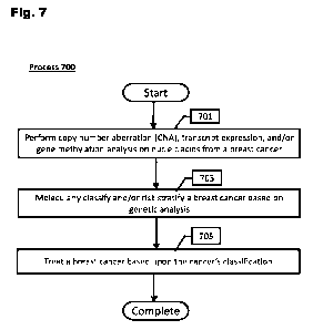
[0108] Fig. 7 provides a flow diagram of a method to treat a breast cancer
based upon
classification into a molecular subgroup in accordance with various
embodiments of the
invention.
[0109] Fig. 8 provides a flow chart of the METABRIC cohort
clinical characteristics
and inclusion analysis, generated in the prior art and utilized as reference.
[0110] Fig. 9 provides a flow chart of the external
validation metacohort clinical
characteristics and inclusion analysis, generated in the prior art and
utilized as reference.
[0111] Figs. 10 and 11 provide data graphs depicting
cumulative incidence of death
for ER+ and ER- patients, generated in the prior art and utilized as
reference.
[0112] Fig. 12 provides a data chart detailing the average
age at onset of breast
cancer in ER+ and ER- patients, generated in the prior art and utilized as
reference.
[0113] Fig. 13 provides a graphical representation of a
multistate Markov model of
breast cancer progression, generated in the prior art and utilized as
reference.
[0114] Fig. 14 provides a data chart depicting prognostic
values of clinical covariates
at different disease states, generated in the prior art and utilized as
reference.
[0115] Fig. 15 provides data charts depicting the internal
validation of the global
prediction of the models on all transitions using bootstrap, generated in the
prior art and
utilized as reference.
[0116] Fig. 16 provides a scatterplot of predictions of
disease-specific death risk
computed by two computational models based on ER status at ten years,
demonstrating
strong concordance for a simple model, generated in the prior art and utilized
as
reference.
[0117] Fig. 17 provides concordance c-indexes of
prediction of risks of distant relapse
(dr), disease-specific death (ds), death (as) and relapse (r), generated in
the prior art and
utilized as reference.
[0118] Figs. 18 and 19 provide data charts depicting
probability of relapse of various
subgroups over time, generated in the prior art and utilized as reference.
[0119] Fig. 20 provides data charts depicting associations
between probabilities of
distant relapse after 10 year of loco-regional relapse and several clinic-
pathological and
molecular features, generated in the prior art and utilized as reference.
-14-
CA 03151330 2022-3-15
WO 2021/055517
PCT/US2020/051130
[0120] Figs. 21 to 26 provide data charts depicting
average probability of relapse or
cancer-related death after surgery in various subgroups over time, generated
in the prior
art and utilized as reference.
[0121] Fig. 27 provides a data graph depicting the
evaluation of predictive utility of a
standard clinical model relative to a model incorporating the integrative
cluster subtypes,
generated in the prior art and utilized as reference.
[0122] Fig. 28 provides a data graph depicting
probabilities of distant relapse or breast
cancer death among ER+/Her2- patients who were relapse free at 5 years post
diagnosis,
generated in the prior art and utilized as reference.
[0123] Fig. 29 provides a data graph depicting
probabilities of distant relapse or
breast-specific death for individual average ER+/HER2- patients in the four
late-relapsing
subgroups relative to IntClust3 for patients who were relapse free five years
post
diagnosis, generated in the prior art and utilized as reference.
[0124] Fig. 30 provides receiver operating characteristic
and precision recall curves
of various computational models utilizing whole genome copy number data,
utilized in
accordance with various embodiments of the invention.
[0125] Figs. 31A and 316 each provide results of
stratifying risk of breast cancers
utilizing various sequencing panels, utilized in accordance with various
embodiments of
the invention.
[0126] Fig. 32A provides sensitivity and specificity
results of a classifier to predict high
risk IntClust subgroups using the Foundation Medicine targeted sequencing gene
panel,
generated in accordance with various embodiments of the invention.
[0127] Fig. 32B provides sensitivity and specificity
results of a classifier to predict high
risk IntClust subgroups using the MSK-IMPACT targeted sequencing gene panel,
generated in accordance with various embodiments of the invention.
[0128] Fig. 32C provides distribution of IntClust
subgroups predicted using the MSK-
IMPACT targeted sequencing gene panel, generated in accordance with various
embodiments of the invention.
[0129] Fig. 33 provides C-index scores of various
diagnostic tests at predicting
recurrence of breast cancer, utilized in accordance with various embodiments
of the
invention.
-15-
CA 03151330 2022-3-15
WO 2021/055517
PCT/US2020/051130
[0130] Figs. 34 to 37 each provide hazard ratio scores of various diagnostic
tests at
predicting recurrence of breast cancer, utilized in accordance with various
embodiments
of the invention.
[0131] Fig. 38 provides results of stratifying breast
cancer risk of recurrence by various
diagnostic tests, utilized in accordance with various embodiments of the
invention.
[0132] Figs. 39 to 43 each provide results of stratifying
breast cancer risk of
recurrence utilizing the In1Clust classification system in combination with
various
diagnostic tests, utilized in accordance with various embodiments of the
invention.
[0133] Figs. 44 to 51 each provide probabilities of
progression free survival of various
high-risk oncogenic molecular subgroups in various forms treatments, including
chemotherapy, targeted (molecular) therapy, or endocrine therapy, utilized in
accordance
with various embodiments of the invention.
[0134] Figs. 52A and 52B provide viability curves of
patient derived organoids derived
from patient 19006, generated in accordance with various embodiments of the
invention.
[0135] Figs. 53A and 53B provide viability curves of
patient derived organoids derived
from patient 19004, generated in accordance with various embodiments of the
invention.
DETAILED DESCRIPTION
[0136] Turning now to the drawings and data, systems, kits, and methods of
determining breast cancer aggressiveness and potential for relapse and
treating breast
cancer based upon the cancers molecular pathology are provided. Many
embodiments
are directed to determining a breast cancer's aggressiveness and potential for
relapse
utilizing a diagnostic assay. Many embodiments are directed to determining a
breast
cancer's molecular pathology utilizing a diagnostic assay. In a number of
embodiments,
a determination of a breast cancer's aggressiveness and potential for relapse
and/or
molecular pathology is then used to determine a treatment option, and to treat
that
neoplasm accordingly. In various embodiments, somatic copy-number or
transcript-
expression data provide an indication of breast cancer molecular subtype and
thus
provide a means of determining appropriate treatment. In some embodiments,
gene copy
number changes or aberrant expression of molecular drivers of cancer
progression are
determined as basis of a cancers pathology. In accordance with multiple
embodiments,
-16-
CA 03151330 2022-3-15
WO 2021/055517
PCT/US2020/051130
breast cancers exhibiting particular molecular pathologies indicating high
aggression and
high potential for relapse are treated aggressively with an appropriate
therapy, such as
adjuvant chemotherapy, targeted therapy, and/or prolonged hormone/endocrine
therapy.
Furthermore, in several embodiments, individuals with cancer that have high
potential for
relapse are closely and repeatedly monitored for an extended period of time
after a
surgical and/or chemotherapy treatment, including treatments that reduce the
cancer to
undetectable levels. In various embodiments, cancers having a particular
molecular
pathology are treated with therapies that are directed at the genes that
classify the
molecular pathology by targeting the gene, the gene product, and/or the
molecular
pathway involving the gene. In accordance with many embodiments, breast cancer
exhibiting a molecular pathology indicative of low aggression and recurrence
are treated
appropriately, which may be only endocrine therapy or less aggressive
chemotherapy.
[0137] A number of embodiments are directed to determining an individual's
molecular
pathology. In many embodiments, copy number aberrations (CNAs) are assessed
from
an individual's DNA and/or RNA, which can be used to classify an individual's
cancer.
CNAs are to be understood as amplification (e.g., duplication) and/or
reduction (e.g.,
deletion) of a set of genomic loci within the genome of a cancer. In some
embodiments,
a cancer is classified by copy number aberrations that include a set of one or
more
molecular drivers (i.e., genes classified to be at least partially pathogenic
in
tumorigenesis). Various embodiments utilize the integrative cluster (IntClust)
classification to determine a set of molecular drivers that describe the
pathogenesis of a
breast cancer. For more on the Intelust classification system, see C. Curtis,
et at, Nature
486, 346-52(2012) and H. R. Ali, et at, Genome Blot 15, 431 (2014), the
disclosures of
which are each herein incorporated by reference. In many embodiments, the risk
of
relapse is determined by a risk classifier.
[0138] Based on recent discoveries, the connection between the molecular
pathology
and cancer progression, including the potential for reoccurrence, is now
appreciated,
indicating courses of treatment and surveillance. Accordingly, various
embodiments are
directed to classifying breast cancer into an IntClust subgroup and/or risk
subgroup to
-17-
CA 03151330 2022-3-15
WO 2021/055517
PCT/US2020/051130
determine a treatment regimen that is tailored for a particular breast cancer.
In addition,
a number of tools and kits are described to classify a breast cancer into an
IntClust and/or
risk subgroup.
[0139] Several diagnostic tests are currently available in
order to guide clinicians on
the approach to monitoring and treating patients with breast cancer (Figs. 1A
to 1F). Most
of these tests utilize molecular and genomic techniques in order to gain
insight on the
genetic aberrations within a neoplasm and potential associated risks, such as
recurrence.
In addition, the tests can inform personalized treatment options, for
instance, the decision
to utilize chemotherapy (including neoadjuvant or adjuvant chemotherapy), the
strength,
dose, and duration of a chemotherapeutic, to utilize endocrine therapy, and to
utilize other
treatment options (e.g., targeted therapy, immunotherapy). For a detailed
discussion on
the various diagnostic tests available for breast cancer, see 0.M Fayanju,
K.U. Park, and
A Lucci Ann. Surg, Oncol. 25, 512-19 (2018), the disclosure of which is herein
incorporated by reference.
[0140] Diagnostic tests include the Oncotype Dx (Genomic Health, Redwood City,
CA), Prosigna (NanoString Technologies, Seattle WA), MammaPrint (Agendia,
Irvine,
CA), EndoPredict (Myriad Genetics, Salt Lake City, UT) and Breast Cancer Index
(BC!)
(Biotheranostics, Inc., San Diego, CA) (See Figs. IA to 1F).
[0141] Oncotype Dx is the most commonly used diagnostic test used for breast
cancer
in the United States. The test examines the expression of 21 genes, which is
used to
determine whether chemotherapy is indicated, especially in individuals with
early-stage
ER+, HER2-, lymph node negative (LN-) breast cancer. Oncotype Dx quantifies
the
likelihood of distant recurrence within 10 years, providing a score that
indicates a high
(31), intermediate (18-30), or low (0-17) likelihood of recurrence. It is
noted that results
indicating intermediate recurrence scores present a clinical conundrum for
clinicians with
respect to the indication of which treatment to perform.
[0142] Prosigna, which is based on the PAM50 classifier,
is a diagnostic test that
determines expression of 50 genes. The Prosigna test generates a risk of
recurrence
score (ROR) and assigns a tumor to one of four intrinsic subtypes: Lumina! A,
Luminal B,
HER2+, and Basal-like. Based on ROR score and other clinical factors
(including lymph
node status), risk status is determined.
-18-
CA 03151330 2022-3-15
WO 2021/055517
PCT/US2020/051130
[0143] MammaPrint is a 70 gene expression assay profiled on a microarray to
predict
distant metastasis within 5 years in ER+/HER2- patients. MammaPrint can be
utilized for
patients with positive or negative lymph node status. Based on expression
profile results,
the molecular prognosis profile of low risk or high risk is determined.
[0144] EndoPredict is a 12-gene test to predict risk of
distance recurrence 10 years
post diagnosis in ER+/HER2- patients with a negative lymph node status or
positive lymph
node (1-3) status. Based on expression profile results, the molecular
prognosis profile of
low risk or high risk is determined_
[0145] Breast Cancer Index (BC!) combines proliferative
and estrogen-signaling gene-
expression signatures to predict distant recurrence 5 to 10 years post
diagnosis in ER+
patients with a negative lymph node status or positive lymph node (1-3)
status. BC! is
intended to be utilized to determine whether a patient can benefit from
extended (>5
year) adjuvant endocrine therapy.
[0146] Some individuals have an aggressive form of cancer, which may also
include
a persistent risk of recurrence and breast cancer death up to and beyond
twenty years
later. Often, from the current diagnostic tests available, it can be difficult
to discern who
is at risk of recurrence, especially late recurrence (e.g., > 5 years). For
instance, a subset
of individuals with early stage ER+ breast cancer have a persistent risk of
recurrence and
death up to 20 years after diagnosis, but the current diagnostics have a
difficult time
identifying this subset In fact, most current diagnostic assays fail to
reliably predict
beyond five years and, as time passes, clinical covariates continue to lose
prediction
power. Accordingly, there is a critical need to identify tumor characteristics
that are more
predictive of aggressiveness and risk of recurrence than the current available
tests and
standard clinical covariates (nodal status, tumor size and grade) in order to
define subsets
of patients with high-risk and low-risk cancers, including risk of recurrence_
Having a
better understanding of risk and relapse potential can help delineate which
individuals
would benefit from various therapies, such as extended endocrine therapy or
higher
dosage of a chemotherapeutic or molecularly targeted therapies.
[0147] Here, several embodiments are based on molecular tests that classify
breast
cancer into a reoccurrence risk subgroup (e.g., high, intermediate, low)
and/or an
integrative cluster (IntClust) (see C. Curtis, et at, (2012), cited supra).
Classification into
a risk subgroup can be performed by a number of statistical techniques,
including (but not
-19-
CA 03151330 2022-3-15
WO 2021/055517
PCT/US2020/051130
limited to) multi-state sem i-markov Models, Cox Proportional Hazards models,
shrinkage
based methods, tree based methods, Bayesian methods, kernel based methods and
neural networks.
[0148] For clustering into an IntClust subgroup, a total
of 11 IntC lust subgroups are
currently described, which were developed utilizing an unsupervised joint
latent variable
clustering of gene expression and copy number profiles that each breast cancer
within
the study harbored. A total of -1000 early stage breast cancers were used to
develop the
clusters, which were validated in another -1000 early stage breast cancers,
and the
results are shown in Figs. 2A and 2B. CNA amplifications are depicted in red
while CNA
losses are depicted in blue. Note that 10 IntClust subgroups are depicted,
each
determined by the computational modeling, however, IntClust4 can be further
divided into
ER+ and ER- to yield 11 IntClust subgroups.
[0149] The IntClust subgroups are each characterized by the copy number
aberrations
(CNAs) and relative gene expression levels that are harbored within the cancer
and are
likely to be involved with the progression of cancer (La, molecular drivers of
breast
cancer). For example, IntClust subgroups 1, 2, 6, and 9 were found to account
for
approximately 25% of all ER+ tumors and each subgroup is enriched for
characteristic
copy number amplification events in various regions of the genome (see Figs.
2A and
2B). Regarding IntGiusti, it now known that the genes near 17q23 (e.g.,
RPS61031) are
amplified and over-expressed. Likewise, IntClust2 has amplifications of genes
CCA1D1,
FGF3 (11q13.3) and 11q13.2 am plicon genes (e.g., EMSY, RSF1, PA/CT), and
these
regions of the genome are frequently co-amplified with concomitant gene
expression
upregulation, suggesting oncogenic cooperation between these loci. Of note,
the
recurrent amplification of chromosome 8p12 and 11q13 suggests that these loci
may
cooperate to promote tumor development and progression. As such, they may need
to
be co-targeted in some patients. IntClust6 exhibits amplifications of the
genes near 8p12
(e.g., FGFR1, ZNF703, ElF4EBP1). In addition, IntClust9 has amplification and
over-
expression of genes near 8q24 (e.g., MYC) and 20q13 (e.g., SRC3, NCOA3). In
similar
analysis, Intc1ust5 is characterized by amplification and over-expression in
HER2fERBB2,
an oncogene that is well-understood to be a molecular driver of breast cancer.
Shown in
Figs. 3A and 3B are the percentage of tumors in the cohort having CNA gain or
-20-
CA 03151330 2022-3-15
WO 2021/055517
PCT/US2020/051130
amplification of genes that define IntClust subgroup that it has been assigned
(note: Figs.
3A and 3B include oncogenic drivers for each integrated cluster, which are
asterisked,
based on predinical data).
[0150] It is now known that particular IntClust subgroups
confer aggressiveness and
potential for relapse (Fig. 4). In other words, when a breast cancer is
classified into a
particular IntClust subgroup, the likelihood of the cancer to be aggressive
and to relapse
can be determined. This knowledge can also be used to determine courses of
treatments
and/or the necessity of continued monitoring. For example, subtyping into
IntClust
subgroups can inform whether to extend endocrine therapy in high-risk
populations, avoid
endocrine therapy in patents that are intrinsically endocrine resistant,
applying targeted
therapy based on molecular drivers of the IntClust subgroup, and the
appropriate choice
and treatment regimen of chemotherapeutics.
[0151] The use of these integrated clusters was found to improve prediction of
late
distant relapse (especially relapse after 5 years) better than standard
clinical covariates
and current diagnostic methods, which is corroborated in an external
validation cohort. It
was also found that a subgroup of triple-negative breast cancer patients
rarely recur after
years while others remain at risk. After distant recurrence, tumor subtype
continues to
dictate the rate of subsequent metastases, underscoring the importance of
classifying
tumors accordingly. Based on these findings, several embodiments are directed
to
identifying individuals having a particular risk of aggressive cancer and
relapse, as
determined by a diagnostic method. Various embodiments treat and/or monitor an
individual based on their cancers aggressiveness and risk of relapse.
[0152] Figure 4 shows the results of a study to investigate aggressiveness and
relapse
of breast cancers within each classification. Here a non-homogenous (semi)
Markov
chain model was utilized to delineate the spatio-temporal dynamics of breast
cancer
relapse across the IntClust subgroups (see Exemplary Embodiments). The results
from
this model illustrate that various subgroups have a much higher likelihood of
relapse,
especially beyond the 5 or even 10 or 15 year marks.
[0153] Shown in Fig. 4 is each of the 11 IntClust
subgroups and the probability of
relapse from three timepoints: surgery, 5 years after surgery and disease
free, and 10
years after surgery and disease free. The results are ordered by the risk of
relapse, with
the subgroups having the least risk of relapse on the left on the most risk of
relapse on
-21-
CA 03151330 2022-3-15
WO 2021/055517
PCT/US2020/051130
the right. Based on these results, groups can be split into high risk groups
and lower risk
groups. Lower risk groups include IntClust3, IntClust7, IntClust8,
IntClust4ER+, and
IntGiusti . High risk groups include IntClust4ER-, IntGiusti, IntClust6,
IntClust9,
IntClust2, and IntClust5.
[0154] Provided in Fig. 5 are cumulative incidence plots
(i.e., 1 ¨ Kaplan Meier
estimates) displaying the risk of distant relapse among ER+/HER2- breast
cancer patients
over time, based on clinical outcome data. As can be seen in the top panel of
Fig. 5,
IntClust subgroups 2, 9, 6 and 1 have an increased probability of distant
relapse. The
lower panel of Fig. 5 compares high risk subgroups (IntClust subgroups 1, 2, 6
and 9)
compared to lower risk subgroups (IntClust subgroups 3, 4ER+, 7, and 8). The
results
show a clear distinction of risk between the two subgroups.
[0155] IntClust10 and IntClust4ER- have a clinical
classification of being triple
negative breast cancer (TNBC), which means they are ER-, HER2-, and PR-. TNBC
occurs in 10% to 20% of breast cancers and is more likely to affect younger
people. TNBC
can be difficult to treat, due to its aggressiveness and potential for
recurrence. However,
the results of the IntClust study show that those in IntClust10 have a very
low likelihood
of recurrence after 5 years disease free. On the contrary, IntClust4ER- has a
relatively
high likelihood of recurrence, even after 5 years or even after 10 years of
being disease
free. Accordingly, in a number of embodiments, an individual having TNBC is
assessed
to determine which IntClust subgroup the cancer is classified into, and thus
performing a
treatment based on the result.
[0156] IntClust3, IntClust7, IntClust8, and IntClust4ER+
are all ER+/HER2- and have
a modest risk of recurrence. IntClust1, IntClust6, IntClust9, and IntClust2,
on the other
hand, are ER+/HER2- and have a high and persistent risk of recurrence.
Accordingly, in
various embodiments, when a cancer is classified as a high risk ER+/HER2-, a
more
aggressive treatment regimen may be beneficial (e.g., adjuvant chemotherapy in
addition
to endocrine therapy). In addition, the oncogenic genomic drivers of the high
risk of
recurrence groups can targeted directly by specific targeted treatments. For
instance, in
some embodiments, IntClust1 cancers are treated with mTOR pathway antagonists
(e.g.,
everolimus, temsirolimus, sirolimus, rapamycin), AKT1 antagonists (e.g.,
ipatasertib,
capivasertib (AZD5363)), AKT1/RPS6KB1 antagonists (e.g., M2698), RPS6KB1
antagonists (e.g., LY2584702), PI3K antagonists (e.g., alpelisib, buparlisib
(BKM120),
-22-
CA 03151330 2022-3-15
WO 2021/055517
PCT/US2020/051130
pictilisib (GDC-0941)), elF4A antagonists (e.g., zotatifin), elF4E antagonists
(e.g.,
rapamycin, rapamycin analogues, ribavirin, A7D8055),or a combination thereof.
In
various embodiments, IntClust2 cancers are treated with epigenetically
targeted
therapies, CDK4/6 antagonists (e_g_, palbociclib, ribociclib, abemaciclib),
FGFR pathway
antagonists (e.g., lucitanib, dovitinib, A7D4547, erdafitinib, infigratinib
(BGJ398), BAY-
1163877, ponatinib), PARP antagonist (e.g., niraparib, olaparib), homologous
recombination deficiency (HRD)-targeted therapies, PAK1 antagonist (e.g.,
IPA3), elF4A
antagonists (e.g., zotatifin), elF4E antagonists (e.g., rapamycin, rapamycin
analogues,
ribavirin, A7D8055), or a combination thereof. In some embodiments, IntClust6
cancers
are treated with FGFR pathway antagonists (e.g., lucitanib, dovitinib,
AZD4547,
erdafitinib, Infigratinib (BGJ398), BAY-1163877, Ponatinib), elF4A antagonists
(e.g.,
zotatifin), elF4E antagonists (e.g., rapamycin, rapamycin analogues,
ribavirin, A7D8055),
or a combination thereof. And in various embodiments, IntClust9 cancers are
treated with
selective estrogen receptor degraders (SERDs) (e.g., fulvestrant, GDC-9545,
SAR439859 (SERD '859), RG6171, AZD9833), the proteolysis targeting chimera
(PROTAC) ARV-471, SRC3 antagonists (e.g., SI-2), MYC antagonists (e.g.,
ornomyc),
BET bronnodomain antagonists (e.g., JQ1, PROTAC ARV-771), elF4A antagonists
(e.g.,
zotatifin), elF4E antagonists (e.g., rapamycin, rapamycin analogues,
ribavirin, AZD8055),
or a combination thereof.
Methods to Classify and Stratify Breast Cancers
[0157] Several embodiments are directed to classifying
and/or stratifying risk of a
breast cancer for diagnostic purposes. In some embodiments, a breast cancer is
classified into a particular IntClust subgroup. In some embodiments, a breast
cancer is
stratified by risk potential (e.g., low, intermediate or high risk).
[0158] In a number of embodiments, a breast cancer is
classified into an integrated
cluster (IntClust), as those described in C. Curtis, et al. (2012), cited
supra. Each of the
eleven IntClust subgroups have a relatively defined set of CNAs as determined
by
clustering analysis (Fig. 2). It is noted that Intelust 4 can be further
divided into ER+ and
ER- to round out the eleven subgroups. By using the IntClust classification,
in various
-23-
CA 03151330 2022-3-15
WO 2021/055517
PCT/US2020/051130
embodiments, a breast cancer is classified into one of the eleven subgroups.
Although
IntClust classification is described, other genomic driver classification
methods of breast
cancer can be used in accordance with some embodiments.
[0159] It is now understood that ER+/HER2- breast cancers
that fall within various
IntClust subgroups are highly aggressive with high risk of relapse, including
subgroups 1,
2, 6, and 9. Likewise, cancers that fall within IntClust subgroups 3, 7, 8,
and 4ER+ are
less aggressive and have lower risk of relapse. Accordingly, various
embodiments
classify a breast cancer into an IntClust subgroup to determine the
aggressiveness and
risk relapse of the cancer. In a similar manner, TNBC can be classified into
high risk
subgroup IntClust4ER- or lower risk subgroup IntGiusti 0.
[0160] To classify an individual into an IntClust, gene expression and/or CNA
data is
obtained from a breast cancer. CNAs can be detected by a number of methods_ In
some
embodiments, DNA of a cancer is extracted from an individual and processed to
detect
CNA levels. In various embodiments, RNA of a cancer is extracted and processed
to
detect expression levels of a number of genes, which can be utilized to
determine
aberrations in copy number. It should be further understood that various
embodiments
can utilize both DNA and RNA extractions to determine molecular subtypes.
Additionally,
since DNA methylation is highly correlated with gene expression as is
chromatin
accessibility (or state), DNA methylation or chromatin accessibility profiling
(ATAC-seq)
is used in a number of embodiments to determine Integrative Cluster membership
or
Integrative Subtype.
[0161] In a number of embodiments, features used to determine a breast
cancer's
integrative subtype include CNA and/or expression data. Accordingly, a
computational
classifier can utilize copy number features and/or gene expression features
but may also
use DNA (gene/CpG) methylation features and/or accessible DNA peaks derived
from
DNA methylation or chromatin (DNA) accessibility analysis of a breast cancer.
In some
embodiments, copy number features are matched by either genomic position or
gene
name. In various embodiments, expression features or matched to a probe that
detects
expression. After features are matched, various embodiments scale each feature
to a z-
score and may include other normalization methods. In numerous embodiments,
the
matched features are entered into the computational classifier such that the
classifier
-24-
CA 03151330 2022-3-15
WO 2021/055517
PCT/US2020/051130
determines which subgroup the breast cancer falls within. In some embodiments,
the
previously described unsupervised joint latent variable clustering approach is
used
(described in the publication of C. Curtis, etal., (2012) or the integrative
subtype (iC10)
classifier as described in the publication of H. R. Ali, et al., (2014), which
can be found as
a CRAN R package labeled
iC10 (https:// cran. r-
project. org/web/packages/iC10/index. htnn I), cited supra.
[0162]
In a various embodiments,
molecular class prediction models include (but not
limited to) shrinkage based methods, logistic regression, support vector
machines with a
linear kernel, support vector machines with a gaussian kernel, and neural
networks, each
of which can independently be used to classify a breast cancer into the 11
integrative
subtypes. Class prediction models can be based on various molecular features
including
copy number features and/or gene expression features, DNA (gene/CpG)
methylation
features ancVor accessible DNA peaks derived from chromatin accessibility
analysis of a
breast cancer. In some embodiments, a top scoring pairs (TSP) classification
approach
(or variations thereof) is used, in which a pair of variables whose relative
ordering can be
used for accurately predicting the class label of a sample. An example of this
approach
is implemented in the Rgtsp package (V. Popovici, E. Budinska, and M.
Delorenzi,
Bioinformatics 27, 1729-30 (2011), the disclosure of which is herein
incorporated by
reference). Further, in some embodiments, molecular class prediction is
extended to
perform absolute subtype assignments, such as utilizing the AIMS algorithm
described
by Paquet et al. (E. R. Paquet and M. T. Hallet, J. Natl. Cancer Inst. 107,
357 (2014), the
disclosure of which is herein incorporated by reference).
[0163]
Nucleic acids or protein can
be extracted or examined within a tissue biopsy of
the tumor and/or from an individual's bodily fluids (e.g., blood, plasma,
urine) by a number
of methodologies, as understood by practitioners in the field. Once extracted,
nucleic
acids can be processed and prepared for detection. Methods of detection
include (but are
not limited to) hybridization techniques (e.g., in situ hybridization (ISH),
nucleic acid
proliferation techniques, and sequencing. Various molecular techniques can be
used,
including (but not limited to) microarray based gene expression,
microarray/SNP array
based copy number inference, RNA-sequencing, targeted (capture) RNA-
sequencing,
exome sequencing, whole genome sequencing (WESNVGS), targeted (panel)
-25-
CA 03151330 2022-3-15
WO 2021/055517
PCT/US2020/051130
sequencing, NanoString nCounter for gene expression, NanoString nCounter for
copy
number inference, Nanostring Digital Spatial Profiling (for in situ protein
expression/RNA
expression), DNA-ISH, RNA-ISH, RNAScope, DNA Methylation assays, and ATAC-seq,
and immunohistochemistry (NC).
[0164] In several embodiments, CNA and/or expression
levels are defined relative to
a known result. In some instances, CNA and/or expression levels of a test
sample is
determined relative to a control sample or molecular signature (i.e., a
sample/signature
with a known classification). A control sample/signature can either be healthy
tissue (i.e.,
null control), a known positive control, or any other control that is desired.
Accordingly,
when the CNA and/or expression levels of a test sample is compared to one or
more
controls, the relative CNA and/or expression levels can determine which
genomic driver
subgroup the test sample falls within. In some instances, gene expression
levels are
determined relative to a stably expressed biomarker (i.e., endogenous
control). In some
instances, when gene expression levels exceed a certain threshold relative to
a stably
expressed biomarker, the level of expression is indicative of a particular
genomic driver
subgroup. In some instances, CNA and/or expression levels are determined
absolutely.
In some instances, various CNA and/or expression level thresholds and ranges
can be
set to classify genomic driver subgroups and thus used to indicate which
subgroup a test
sample falls within. It should be understood that methods to define CNA and/or
expression levels can be combined, as necessary for the applicable assessment.
Utilizing
transcript expression levels, CNA levels, DNA methylation levels, chromatin
(DNA)
accessibility peaks, or any combination thereof, a breast cancer can be
classified.
[0165] Genomic loci and/or genes are detected in accordance with various
embodiments. In some embodiments, detection of a particular set of genomic
CNAs
and/or transcript expression classifies a breast cancer into a particular
IntClust subgroup.
Referring back to Figs. 3A and 3B, CNAs in various loci are demonstrative of a
number
of IntClust subgroups. For example, IntClust subgroups 1, 2, 6, and 9 were
found to
account for approximately 25% of all ER+ tumors and each is enriched for a
characteristic
copy number amplification events of various sections of the genome. Regarding
IntClustl,
it now known that the genes near 17q23 including (but not limited to) RPS6KB1,
HASF5,
PPM1E, PRR11, DHX40, TUBD1, CA4, C17otf64, BCAS3, TBX2, BRIP1, and
-26-
CA 03151330 2022-3-15
WO 2021/055517
PCT/US2020/051130
TBC1D3P2 are amplified. Likewise, IntClust2 has amplifications of genes CCND1,
FGF3
(at 11q13.3) and 11q13.2 annplicon genes including (but not limited to) EMSY,
RSF1,
PAM, CTTN, CLPB, P2RY2, UCP2, CHRDL2, MAP6, OMP, and ARS2. Int01ust6
exhibits amplifications of the genes near 8p12 including (but not limited to)
FGFR1,
ZNF703, ElFzIEBPI, LETM2, and STAR. In addition, IntClust9 has amplification
of genes
near 8q24 including but limited to MYC, F8X032, LINC00861, PCAT1, LINC00977,
MIR5192, and ADCY8 and near 20q13 including (but not limited to) SRC3, NCOA3.
Accordingly, detection of an amplification (CNA or expression) of a locus or
gene, or a
combination of loci and/or genes, can be utilized to indicate a particular
IntClust
classification.
[0166] In a number of embodiments, classification of
breast cancer is performed
utilizing a computational model based on multiple genomic copy number
aberrations,
multiple gene expression profiles, DNA methylation levels, chromatin (DNA)
accessibility
peaks, or any combination thereof, which may provide a more accurate
classification than
copy number state/gene expression at a single chromosomal locus. For instance,
amplifications of the genes RPS6KB1, FGFR1, and FGF3 occur within a variety
breast
cancer IntClust subgroups, including those that have low aggressiveness and
risk of
relapse. As can be seen in Fig. 6, approximately 50% of breast cancers having
an
RPS6KB1 gain or amplification are classified into IntClusti , however RPS6KB1
copy
number alteration is also detected within several more IntClust subgroups.
Likewise,
approximately 50% of breast cancers having an FGFR1 amplification are
classified into
IntClust6 and the amplification can be detected within all the other
subgroups. FGF3
amplification is fairly evenly distributed between the IntClust subgroups.
Thus, it may be
beneficial to utilize a trained computational model such that a breast cancer
can be more
accurately classified into the appropriate subtype (e.g., IntClust
classifier).
[0167] A number of embodiments utilize statistical
computation to stratify breast
cancer recurrence risk (e.g., high, intermediate, low). In various
embodiments, statistical
computation models include (but not limited to) multi-state semi-markov
Models, Cox
Proportional Hazards models, shrinkage based methods, tree based methods,
Bayesian
methods, kernel based methods and neural networks. In some embodiments,
thresholds
are utilized to separate higher risk scores from lower risk scores. In several
embodiments,
-27-
CA 03151330 2022-3-15
WO 2021/055517
PCT/US2020/051130
features used to train statistical models and/or to predict risk of recurrence
in breast
cancer include (but not limited to) clinical data, age, cancer stage, number
of tumor
positive lymph nodes, size of tumor, grade of tumor, surgery performed,
treatment
performed, basic molecular identities, and integrative subtype
classification/membership.
Age of the patient can be coded as a continuous value (and potentially trimmed
to avoid
excessively high values (e.g., age > 80). Clinical stage (values ranging from
1-4), can be
included as a continuous value or as a factor or can be grouped as high (3-4)
vs low (1-
2) stage. Positive lymph nodes can be included as a continuous value
(potentially trimmed
to avoid excessively high values). The number of positive lymph nodes this can
also be
categorized as lymph node negative versus positive or amongst positive, graded
as low
(1 positive node), medium (2-3 positive nodes), high (4-9 positive nodes),
very high (>=10
positive nodes) or variations thereof. Size of tumor can be used as a
continuous value,
which can be trimmed to avoid excessive high values). Size of tumor can also
be
categorized (e.g., staging system: Tl<20mm, T2 (20-50), T3 (>50)). The grade
of tumor
can be used as a continuous value or as a category (1-3) or high (3) vs low
(1,2). In some
embodiments, classifiers include the CTS5 algorithm, which is based encoding
of lymph
node, size, grade may be incorporated as follows:
0.438 x nodes + 0.988 x (0.093 x size ¨ 0.001 x s1ze2 + 0.37S x grade + 0.017
x age)
(for more on CTS5 algorithm, see M. Dowsett, et al., J. Clio. Oncol. 36, 1941-
48 (2018),
the disclosure of which is herein incorporated by reference). Basic molecular
identities
include status of estrogen receptor (ESR1), Progesterone receptor (PGR), human
epidermal growth factor receptor 2 (HER2/ERBB2) and MKI67 based on clinical
pathology reports and/or inferred from gene expression data. Surgery types can
include
breast conserving or mastectomy. Treatment type can include hormonal,
chemotherapy,
targeted therapy, where agents may be specified or grouped more broadly and
treatment
duration included. Various embodiments also utilize germ line genetic
variants, ethnicity,
general health data, and/or treatment regimes. In some embodiments, the
Predict Tool
(https://breast.predict.nhs.uk) or components thereof can be utilized in the
model.
[0168] In some embodiments, features can be derived from integrated subtype
clusters (e.g., IntClust classification) and included in the model. These
features can be
integrative subtype membership or the posterior probability of membership to a
given
-28-
CA 03151330 2022-3-15
WO 2021/055517
PCT/US2020/051130
cluster. An integrative subtype is coded individually as a logical feature.
Distance to the
centroid of each subgroup can be utilized. Any score derived from the IC
classifier can
also be utilized. In some embodiments, risk of relapse prediction on specific
subpopulations is utilized, such as ER+/HER2- patients or triple negative
breast cancer
patients. Amongst ER+/HER2- patients, high risk (IntClust1 , IntClust2,
Intclust6 or
IntClust10) versus lower risk (IntClust3, IntClust4, Intc1ust7 or IntClust8)
categories may
be considered. Likewise, TNBG classified into IntClust4ER- are determined to
be
aggressive and have high risk, whereas TNBC classified into IntClust10 are
determined
to have lower risk
[0169] In a number of embodiments, a multi-state Cox reset
model is utilized, which is
a statistical model that accounts for different disease states (loco-regional
recurrence and
distal recurrence), different timescales (time from diagnosis and time from
relapse),
competing causes of death (cancer death or other causes), clinical covariates
or age
effects, and distinct baseline hazards for different molecular subgroups (see
H. Putter, M.
Fiocco, & R. B. Geskus, Stat. Med. 26,2389-430 (2007); 0. Aalen, 0. Borgan, &
H.
Gjessing, Survival and Event History Analysis ¨ A Process Point of View.
(Springer-
Verlag New York, 2008); and T. M. Therneau & P. M. Grambsh, Modeling Survival
Data:
Extending the Cox Model. (Springer-Verlag New York, 2000); the disclosures of
which
are each herein incorporated by reference). In many embodiments, a multistate
statistical
model is fit to the dataset, such that the chronology of breast cancer,
starting with surgical
excision of the primary tumor, followed by the development of loco-regional
and/or distant
recurrence and accounting by competing risks of death due to cancer or other
causes are
accounted. In some embodiments, the hazards of occurrence of each of these
states are
modeled with a non-homogenous semi-Markov Chain with two absorbent states
(Death/Cancer and Death/Other). For more on multi-state Cox models, see the
description in the Exemplary Embodiments.
[0170] Cox proportion hazard models are statistical
survival models that relate the time
that passes to an event and the covariates associated with that quantity in
time (See D.
R. Cox, J. R. Stat. Soc. B 34, 187-220 (1972), the disclosure of which is
herein
incorporated by reference). To utilize Cox proportional hazards models, in
some
embodiments, clinical, molecular, and integrative subtype features are
included. In some
-29-
CA 03151330 2022-3-15
WO 2021/055517
PCT/US2020/051130
embodiments, features can be linear and/or polynomial transformed and
interaction can
include variable selection_ In some embodiments, to further simplify the
model, stepwise
variable selection can be incorporated into the cross validation scheme. Any
appropriate
computational package can be utilized and/or adapted, such as (for example),
the RMS
package (https://www. rdocumentation.org/packages/rms).
[0171] Shrinkage based methods include (but not limited
to) regularized lasso (R.
Tibshirani Stat. Med. 16, 385-95 (1997), the disclosure of which is herein
incorporated by
reference), lassoed principal components (D. M. Witten and R. Tibshirani Ann.
App!. Stat.
2, 986-1012 (2008), the disclosure of which is herein incorporated by
reference), and
shrunken centroids (R. Tibshirani, et at, Proc. Natl. Acad. Sci. U S A 99,
6567-72 (2002),
the disclosure of which is herein incorporated by reference). Any appropriate
computation
package can be utilized and/or adapted, such as (for example), the PAMR
package for
shrunken centroid (https://www.rdocumentation.org/packages/pam
r/versions/1.56.1).
[0172] Tree based models include (but not limited to) survival random forest
(H.
Ishwaran, et at, Ann. App. Stat. 2, 841-60 (2008), the disclosure of which is
herein
incorporated by reference) and random rotation survival forest (L. Zhou, H.
Wang, and Q.
Xu, Springerplus 5, 1425 (2016), the disclosure of which is herein
incorporated by
reference). In some embodiments, the hyperparameter corresponds to the number
of
features selected for each tree. My appropriate setting for the number of
trees can be
utilized, such as (for example) 1000 trees. Any appropriate computation
package can be
utilized and/or adapted, such as (for example), the RRotSF package for random
rotation
survival forest (https://github.com/whcsuiRRotSF).
[0173] Bayesian methods include (but not limited to)
Bayesian survival regression (J.
G. Ibrahim, M. H. Chen, and D. Sinha, BAYESIAN SURVIVAL ANALYSIS, Springer
(2001), the
disclosure of which is herein incorporated by reference) and Bayes mixture
survival
models (A. Kottas I Stat Pan. Inference 3, 578-96 (2006), the disclosure of
which is
herein incorporated by reference). In some embodiments, sampling is performed
with a
multivariate normal distribution or a linear combination of monotone splines
(See B. Cai,
X. Lin, and L. Wang, Comput Stat Data Anal. 55, 2644-51 (2011), the disclosure
of which
-30-
CA 03151330 2022-3-15
WO 2021/055517
PCT/US2020/051130
is herein incorporated by reference). Any appropriate computation package can
be
utilized and/or adapted, such as (for example), the ICBayes package
(https://www. rdocu mentation.org/packages/ICBayes/versions/1.0/topics/IC B
ayes).
[0174] Kernel based methods include (but not limited to)
survival support vector
machines (L. Evers and C. M. Messow, Bioinformatics 24, 1632-38 (2008), the
disclosure
of which is herein incorporated by reference), kernel Cox regression (H. Li
and Y. Luan,
Pac. Symp. Biuocomp. 65-76 (2003), the disclosure of which is herein
incorporated by
reference), and multiple kernel learning (0. Dereli, C. Oguz, and M. Gonen
Bioinformatics
(2019), the disclosure of which is herein incorporated by reference). It is to
be understood
that kernel based methods can include support vector machines (SVM) and
survival
support vector machines with polynomial and Gaussian kernel, where
hyperparameter C
specifies regularization (See L. Evers and C. M. Messow, cited supra). In some
embodiments, multiple kernel learning (MKL) approaches combine features in
kernels,
including kernels embed clinical information, molecular information and
integrative
subtype. Any appropriate computation package can be utilized and/or adapted,
such as
(for example), the path2sury package
(https://github.com/mehmetgonen/path2surv).
[0175] Neural network methods include (but not limited to)
DeepSury (J. L. Katzman,
et at, BMC Med. Res_ Methodot 18, 24 (2018), the disclosure of which is herein
incorporated by reference), and SuvivalNet (S. Yousefi, et at, Sc!. Rep. 7,
11707 (2017),
the disclosure of which is herein incorporated by reference). Any appropriate
computation
package can be utilized and/or adapted, such as (for example), the Optunity
package
(pypi.org/project/Optunity/).
[0176] In several embodiments, in order to ensure that a
model is not overfitted,
models are trained using an X-times, and cross validated X-fold scheme (e.g.,
10-fold
training, 10-fold cross validation). Sample data can be split into subsets,
and some data
is used to train the model and some data is used to evaluate the model. By
using this
method, it can be assured that all data are validated at least once and no
sample is used
for both training and validation at the same time, all while the X-fold cross
validation
minimized sampling bias. A training/cross-validation approach also enables
evaluation of
-31-
CA 03151330 2022-3-15
WO 2021/055517
PCT/US2020/051130
the stability of the predictions by calculating confidence intervals, which
facilitates model
comparisons. Additionally, an internal cross validation scheme can be employed
for
hyperparameter specification.
[0177] While specific examples of processes for molecularly classifying and
stratifying
risk of a breast cancer are described above, one of ordinary skill in the art
can appreciate
that various steps of the process can be performed in different orders and
that certain
steps may be optional according to some embodiments. As such, it should be
clear that
the various steps of the process could be used as appropriate to the
requirements of
specific applications. Furthermore, any of a variety of processes for
molecular
classification and risk stratification appropriate to the requirements of a
given application
can be utilized in accordance with various embodiments.
[0178] Numerous embodiments are directed towards combining risk prediction
models
that incorporate integrative subtype information with other multigene
signatures, including
(but not limited to) Oncotype Ox (Genomic Health, Redwood City, CA), Prosigna
(NanoString Technologies, Seattle WA), MammaPrint (Agendia, Irvine, CA),
EndoPredict
(Myriad Genetics, Salt Lake City, UT), Breast Cancer Index (Bel)
(Biotheranostics, Inc.,
San Diego, CA). Of particular interest is the combination of Oncotype DX with
the
Integrative Subtype (IntClust). As stated previously, Oncotype DX yields a
result
indicating one of: high, intermediate or low likelihood of recurrence and the
treatment
choice for an intermediate likelihood can be a conundrum for clinicians.
However, when
Oncotype DX is combined with an integrative clustering technique, breast
cancers that
would normally fall within the intermediate risk group can be better
stratified resulting in
clear results of high and lower risk. Details of combining Oncotype DX with an
integrative
clustering technique is described within the exemplary embodiments section.
Combinations with Prosigna, MammaPrint, BCI, and EndoPredict have also shown
improvements in diagnostic stratification, as detailed in the Exemplary
Embodiments.
Methods of Detecting Copy Number Aberrations & Gene Expression
[0179] Aberrations in copy number can be detected by a number of methods in
accordance with various embodiments, as would be understood by those skilled
in the
art. In several embodiments, CNAs are detected directly from genomic DNA
and/or
inferred from RNA transcript expression. Accordingly, in some embodiments CNA
-32-
CA 03151330 2022-3-15
WO 2021/055517
PCT/US2020/051130
analysis is used to classify breast cancers. In some embodiments, RNA
expression
analysis is used to classify breast cancer. And in some embodiments, analysis
of both
CNA and RNA expression is used to classify a breast cancer.
[0180] The source of nucleic acids (e.g., DNA and RNA) to determine expression
can
be derived de novo (i.e., from a biological source). Several methods are well
known to
extract nucleic acids from biological sources. Generally, nucleic acids are
extracted from
cells or tissue, then prepped for further analysis. Alternatively, DNA and/or
RNA can be
observed within cells, which are typically fixed and prepped for further
analysis. The
decision to extract nucleic acids or fix tissue (via fomnalin fixation and
paraffin embedding
(FFPE)) for direct examination depends on the assay to be performed, as would
be
understood by those skilled in the art. In some embodiments, DNA and/or RNA is
extracted from tissue that is fixed.
[0181] In several embodiments, nucleic acids are extracted
and/or examined in the
type of cells and tissues to be treated. In many cases, the cells to be
treated are neoplastic
cells of a breast cancer of an individual, which can be extracted in a biopsy.
In some
embodiments, nucleic acids are extracted from blood or serum, which can
include
circulating tumor DNA, for analysis. The precise source to extract and/or
examine nucleic
acids can depend on the assay to be performed, the availability of a biopsy,
and
preference of the practitioner.
[0182] A number of assays are known to measure and quantify genomic loci copy
numbers and transcript expression. CNAs and RNA expression levels can be
determined
by a number of methods, including (but are not limited to) hybridization
techniques (e.g.,
in situ hybridization (ISH), nucleic acid proliferation techniques, and
sequencing. Various
molecular techniques can be used, including (but not limited to) microarray
based gene
expression, microarray/SNP array based copy number inference, RNA-sequencing,
targeted (capture) RNA-sequencing, exome sequencing, whole genome sequencing
(VVESNVGS), targeted (panel) DNA sequencing (including Memorial Sloan
Kettering
Cancer Center Integrated Mutation Profiling of Actionable Cancer Targets (MSK-
IMPACT), Foundation Medicine CDx, Stanford Tumor Actionable Mutation Panel
(STAMP) (see moleculargenetics.stanford.edu/solid_tumors.htm), nanoString
nCounter
-33-
CA 03151330 2022-3-15
WO 2021/055517
PCT/US2020/051130
for gene expression, nanoString nCounter for copy number inference, nanoString
Digital
Spatial Profiler forcludes protein and RNA expression, DNA-ISH, RNA-ISH,
RNAScope,
DNA Methylation assays, and ATAC-seq.
[0183] Several embodiments are directed towards
classifying integrative subtype from
targeted sequencing data derived from a gene panel, such as those built by
academic
centers (e.g., UCSF500 Cancer Gene Panel (San Francisco, CA)) or companion
diagnostic assays intended for other uses such as Foundation One CDx
(Foundation
Medicine, Cambridge, MA), and MS K-IMPACT (Memorial Sloan Kettering Cancer
Center,
New York, NY) or Stanford Tumor Actionable Mutation Panel (STAMP) (Stanford,
Stanford, CA). Provided sufficient gene coverage is included within the panel,
the various
embodiments of algorithms described herein can be utilized. In some
embodiments, a
gene panel designed for breast cancer assessment is utilized. In some
embodiments, a
gene panel designed for chromatin regulatory gene assessment is utilized.
[0184] Several embodiments are directed to targeted detection of CNAs or gene
transcripts. Accordingly, in many embodiments probes and/or primers are
utilized to
detect specific genes and/or genomic loci that are indicative of IntClust
subgroups either
directly or via a computational model as described herein.
[0185] As understood in the art, only a portion of a genomic locus or gene may
need
to be detected in order to have a positive detection. In some instances, genes
can be
detected with identification of as few as ten nucleotides. In many
hybridization techniques,
detection probes are typically between ten and fifty bases, however, the
precise length
will depend on assay conditions and preferences of the assay developer. In
many
amplification techniques, arriplicons are often between fifty and one-thousand
bases,
which will also depend on assay conditions and preferences of the assay
developer. In
many sequencing techniques, genomic loci and transcripts are identified with
sequence
reads between ten and several hundred bases, which again will depend on assay
conditions and preferences of the assay developer.
[0186] It should be understood that minor variations in
gene sequence and/or assay
tools (e.g., hybridization probes, amplification primers) may exist but would
be expected
to provide similar results in a detection assay. These minor variations are to
include (but
not limited to) insertions, deletions, single nucleotide polymorphisms, and
other variations
-34-
CA 03151330 2022-3-15
WO 2021/055517
PCT/US2020/051130
due to assay design. In some embodiments, detection assays are able to detect
genomic
loci and transcripts having high homology but not perfect homology (e_g., 70%,
80%, 90%,
or 95% homology). As understood in the art, the longer the nucleic acid
polymers used
for hybridization, less homology is needed for the hybridization to occur.
[0187] It should also be understood that several gene
transcripts have a number
isoforms that are expressed. As understood in the art, many alternative
isoforms would
be understood to confer similar indication of molecular classification, and
thus cancer
aggressiveness and risk of relapse. Accordingly, alternative isoforms of gene
transcripts
are also covered in some embodiments.
[0188] In many embodiments, an assay is used to measure and quantify CNAs and
transcript expression. The results of the assay can be used to determine
relative CNA
and transcript expression of a tissue of interest. For example, the nanoString
nCounter,
which can quantify up to several hundred nucleic acid molecule sequences in
one
microtube utilizing a set of complement nucleic acids and probes, which can be
used to
determine CNA and transcript expression of a set of genomic loci and/or gene
transcripts.
The resulting copy number and expression can be used to classify the sample
either
directly or utilizing a computational model as described herein, thus
determining the
cancer's aggressiveness and risk of relapse. Based on the cancer's
aggressiveness and
risk of relapse, the cancer can be treated accordingly.
Kits for Detection Copy Number Aberrations and Gene Expression
[0189] In several embodiments, kits are utilized for
evaluating individuals for breast
cancer risk, wherein the kits can be used to detect genetic aberrations in
biomarkers
and/or prepare for a sequencing reaction as described herein. For example, the
kits can
be used to detect any one or more of the gene biomarkers described herein,
which can
be used to determine aggressiveness and metastatic potential. The kit may
include one
or more agents for determining genetic aberrations and/or preparing
sequencing, a
container for holding a biological sample (e.g., tumor or liquid biopsy)
obtained from a
subject; and printed instructions for reacting agents with the biological
sample to detect
the presence or amount of one or more genetic aberrations within biomarker
genes
derived from the sample. The agents may be packaged in separate containers.
The kit
-35-
CA 03151330 2022-3-15
WO 2021/055517
PCT/US2020/051130
may further comprise one or more control reference samples and reagents for
performing
a biochemical assay, enzymatic assay, immunoassay, hybridization assay, or
sequencing
assay.
[0190] A kit can include one or more containers for compositions contained in
the kit.
Compositions can be in liquid form or can be lyophilized. Suitable containers
for the
compositions include, for example, bottles, vials, syringes, and test tubes.
Containers can
be formed from a variety of materials, including glass or plastic. The kit can
also comprise
a package insert containing written instructions for methods of detecting
aberrations from
tumor and/or liquid biopsies.
[0191] In several embodiments, kits are used to measure and quantify CNAs and
transcript expression. A nucleic acid detection kit, in accordance with
various
embodiments, includes a set of hybridization-capable complement sequences
and/or
amplification primers specific for a set of genomic loci and/or expressed
transcripts. In
some instances, a kit will include further reagents sufficient to facilitate
detection and/or
quantitation of a set of genomic loci and/or expressed transcripts. In some
instances, a
nucleic acid detection kit will be able to detect and/or quantify for at least
5, 10, 15, 20,
25, 30, 40 or 50 loci and/or genes. In some instances, a nucleic acid
detection kit will
include an array to detect and/or quantify for at least 100, 200, 300, 400,
500 or 1000 loci
and/or genes. In some instances, a kit will be able to detect and/or quantify
thousands or
more genes via an array or sequencing technique.
[0192] In a number of embodiments, a set of hybridization-capable complement
sequences are immobilized on an array, such as those designed by Affymetrix or
IIlumina.
In many embodiments, a set of hybridization-capable complement sequences are
linked
to a "barcode" to promote detection of hybridized species and provided such
that
hybridization can be performed in solution, such as those designed by
nanoString. In
several embodiments, a set of primers (and, in some cases probes) to promote
amplification and detection of amplified species are provided such that a PCR
can be
performed in solution, such as those designed by Applied Biosystems of
ThermoScientific
(Foster City, CA).
-36-
CA 03151330 2022-3-15
WO 2021/055517
PCT/US2020/051130
[0193] Many embodiments are directed to a kit being utilized as a companion
diagnostic. Accordingly, in various embodiments, a kit is utilized to classify
a breast
cancer, which is then used to determine a particular treatment. For instance,
a kit can be
utilized to determine aggressiveness and risk of relapse of a breast cancer to
determine
the appropriate treatment. In some embodiments, a kit determines whether a
breast
cancer is a high risk, intermediate risk, or low risk, which then infers a
more aggressive
or less aggressive treatment, respectively. In some embodiments, a kit
determines the
molecular pathology of the breast cancer, which then infers whether to use a
treatment
that directly targets one or more oncogenic drivers.
Treatment of Breast Cancer Determined by Molecular Characterization
[0194] A number of embodiments are directed to classifying and treating breast
cancer. In several embodiments, a breast cancer is molecularly classified
based and/or
risk stratified based on its DNA and/or transcript expression. In some
embodiments, a
breast cancer is stratified based on risk utilizing a statistical model.
Molecular
classifications, in accordance with some embodiments, indicate the
aggressiveness and
risk of relapse. In some embodiments, integrative cluster (IntClust) subtype
is used to
molecularly classify a breast cancer. In various embodiments, copy number
and/or
transcript expression analysis of a set of one or more genes are used to
classify a breast
cancer into molecular pathology subgroups. Based on molecular pathology and/or
risk
stratification, a number of embodiments determine a course of treatment for a
breast
cancer, which may include measures to mitigate cancer recurrence and/or
promote tumor
shrinkage.
[0195] Provided in Fig. 7 is an embodiment of a method to
molecularly classify and/or
risk stratify a breast cancer. Process 700 begins with performing (701) copy
number
aberration (CNA) transcript expression and/or gene methylation analysis on
nucleic acids
from a breast cancer. In several embodiments, DNA and/or RNA transcripts are
extracted
from an individual having breast cancer and processed for analysis. DNA can be
used to
detect CNAs and/or methylation analysis at various genomic loci and RNA can be
used
to determine expression levels of various genes.
-37-
CA 03151330 2022-3-15
WO 2021/055517
PCT/US2020/051130
[0196] CNAs can be detected by a number of methods as described herein. In
some
embodiments, DNA of a cancer is extracted from an individual and processed to
detect
CNA levels. In various embodiments, RNA of a cancer is extracted and processed
to
detect expression levels of a number of genes. In some instances, gene
expression is
used directly for further analysis. In some instances, gene expression is
utilized to
determine whether aberrations in copy number impact expression and/or to
delineate
driver genes in a given patient's tumor. In some instances, CNA levels can be
inferred
from RNA sequencing data. Methylation of genes and/or determination of
chromatin
availability can be performed, which can be used for further analysis.
pim Nucleic acids can be extracted from a cancer biopsy
and/or from an individual's
bodily fluids (e.g., blood, plasma), including circulating tumor DNA (ctDNA),
by a number
of methodologies, as understood by practitioners in the field. Once extracted,
nucleic
acids can be processed and prepared for detection, as described herein.
Methods of
detection include (but are not limited to) hybridization techniques (e.g., in
situ
hybridization (ISH)), nucleic acid amplification techniques (e.g., PCR), and
sequencing
(e.g., exome, genome sequencing).
[0198] Genomic loci and/or genes are detected in accordance with various
embodiments as described herein. In some embodiments, a set of probes and/or
primers
are used to identify a particular set of genomic CNAs and/or expressed
transcripts. In
various embodiments, whole or partial genomes, exomes, and/or transcriptomes
are
sequenced and analyzed to identify a particular set of genomic CNAs and/or
expressed
transcripts. In many embodiments, a particular set of genomic CNAs and/or
expressed
transcripts represent a particular molecular classification. In some
embodiments, a
molecular classification signifies a cancer's aggressiveness and risk of
relapse. In some
embodiments, a molecular classification signifies a cancer's molecular
pathology. In
some embodiments, a particular set of genomic CNAs and/or expression of
transcripts
represent a particular IntClust subgroup. In some embodiments, molecular
classification
is further used to stratify risk of recurrence.
[0199] Process 700 molecularly classifies and/or risk
stratifies (703) a breast cancer
based on genetic analysis (e.g., CNA, transcript expression, methylation
analysis). In
various embodiments, molecular class prediction models include (but are not
limited to)
-38-
CA 03151330 2022-3-15
WO 2021/055517
PCT/US2020/051130
shrinkage based methods, logistic regression, support vector machines with a
linear
kernel, support vector machines with a gaussian kernel, and neural networks.
In various
embodiments, statistical computation models include (but are not limited to)
multi-state
semi-markov Models, Cox Proportional Hazards models, shrinkage based methods,
tree
based methods, Bayesian methods, kernel based methods and neural networks.
[0200] The copy number amplifications described for the various IntClust
subgroups,
in accordance with various embodiments, are used as biomarkers for classifying
a cancer
into a particular subgroup as described herein. A number of embodiments
utilize a
previously trained computational classifier to assign a breast cancer into a
particular
molecular pathology subgroup (e.g., IntClust) as described herein. Various
embodiments
can utilize a previously trained risk stratification model to determine the
risk of recurrence
of a breast cancer. Accordingly, a computational classifier can utilize copy
number
features, gene expression features, genomic methylation features, and/or
nucleosorne
occupancy features derived from DNA and RNA analysis of an individual having
breast
cancer. In some embodiments, copy number features are matched by either
genomic
position or gene name. In various embodiments, expression features are matched
to a
probe that detects expression and/or sequencing results. After features are
matched,
various embodiments scale each feature to a z-score and may include other
normalization
methods. In numerous embodiments, the matched features are entered into a
molecular
classifier and/ or risk stratification model to reveal how to treat an
individual based on the
molecular classification and/or risk of recurrence.
[0201] Process 700 also treats (705) a breast cancer based upon the cancer's
molecular classification and/or risk stratification. In some embodiments,
cancers
classified into aggressive and/or late relapsing (e.g. IntClust subgroups 1,
2, 6 and 9)
and/or high risk subgroups, a prolonged hormone/endocrine therapy (e.g.,
fulvestrant,
anastrozole, exemestane, letrozole, tamoxifen, GDC9545) may be applied. In
various
embodiments, cancers classified into aggressive and/or late relapsing and/or
high risk
subgroups are treated with chemotherapy.
[0202] As previously noted, various IntClust subgroups are characterized by
specific
molecular aberrations and genomic drivers some of which can readily be
therapeutically
targeted. In some embodiments, IntClust1 cancers are treated with mTOR pathway
-39-
CA 03151330 2022-3-15
WO 2021/055517
PCT/US2020/051130
antagonists (e.g., everolimus, temsirolimus, sirolimus, rapamycin), AKT1
antagonists
(e.g., ipatasertib, capivasertib (A7D5363)), AKT1/RPS6KB1 antagonists (e.g.,
M2698),
RPS6KB1 antagonists (e.g., LY2584702), PI3K antagonists (e.g., alpelisib,
buparlisib
(BKM120), pictilisib (GDC-0941)), elF4A antagonists (e.g., zotatifin), elF4E
antagonists
(e.g., rapamycin, rapamycin analogues, ribavirin, AZD8055),or a combination
thereof. In
various embodiments, IntClust2 cancers are treated with epigenetically
targeted
therapies, CDK4/6 antagonists (e.g., palbociclib, ribociclib, abemaciclib),
FGFR pathway
antagonists (e.g., lucitanib, dovitinib, AZD4547, erdafitinib, lnfigratinib
(BGJ398), BAY-
1163877, Ponatinib), PARP-inhibitors (e.g., niraparib, olaparib), homologous
recombination deficiency (HRD)-targeted therapies, PAK1 inhibitors (e.g.,
IPM), elF4A
antagonists (e.g., zotatifin), elF4E antagonists (e.g., rapamycin, rapamycin
analogues,
ribavirin, AZD8055), or a combination thereof. In some embodiments, IntClust6
cancers
are treated with FGFR pathway antagonists (e.g., lucitanib, dovitinib,
A7D4547,
erdafitinib, Infigratinib (BGJ398), BAY-1163877, Ponatinib), elF4A antagonists
(e.g.,
zotatifin), elF4E antagonists (e.g., rapamycin, rapamycin analogues,
ribavirin, AZD8055),
or a combination thereof. And in various embodiments, IntClust9 cancers are
treated with
selective estrogen receptor degraders (SERDs) (e.g., fulvestrant, GDC-9545,
SAR439859 (SERD '859), RG61711 AZD9833), the proteolysis targeting chimera
(PROTAC) ARV-471, SRC3 antagonists (e.g., SI-2), MYC antagonists (e.g.,
omomyc),
BET bronnodomain antagonists (e.g., JQ1, PROTAC ARV-771), elF4A antagonists
(e.g.,
zotatifin), elF4E antagonists (e.g., rapamycin, rapamycin analogues,
ribavirin, AZD8055),
or a combination thereof.
[0203] While specific examples of processes for treating a breast cancer based
upon
molecular classification and/or risk stratification are described above, one
of ordinary skill
in the art can appreciate that various steps of the process can be performed
in different
orders and that certain steps may be optional according to some embodiments of
the
invention. As such, it should be clear that the various steps of the process
could be used
as appropriate to the requirements of specific applications. Furthermore, any
of a variety
of processes for treating a breast cancer to the requirements of a given
application can
be utilized in accordance with various embodiments of the invention.
-40-
CA 03151330 2022-3-15
WO 2021/055517
PCT/US2020/051130
Methods of Treatment
[0204] Various embodiments are directed to treatments of breast cancer based
on
molecular characterization and/or risk stratification of the cancer. As
described herein,
classification of a breast cancer by the molecular pathology and/or the
aggressiveness
and risk of relapse of the cancer. Based on the classification, a breast
cancer (or
individuals having breast cancer) can be treated accordingly.
[0205] Several embodiments are directed to the use of medications to treat a
breast
cancer based on molecular classification and/or risk stratification of the
cancer. In some
embodiments, medications are administered in a therapeutically effective
amount as part
of a course of treatment. As used in this context, to "treat" means to
ameliorate at least
one symptom of the disorder to be treated or to provide a beneficial
physiological effect.
For example, one such amelioration of a symptom could be reduction of tumor
size and/or
risk of relapse.
[0206] A therapeutically effective amount can be an amount sufficient to
prevent
reduce, ameliorate or eliminate the symptoms of breast cancer. In some
embodiments, a
therapeutically effective amount is an amount sufficient to reduce cancer
growth in a
breast cancer growth, which can be determined by a number of ways including
(but not
limited to) measuring tumor size and measuring proliferation levels (e.g.,
Ki67+
expression).
[0207] A number of treatments and medications are available to treat breast
cancer
including (but not limited to) radiotherapy, chemotherapy, targeted
(molecular) therapy,
endocrine therapy, and immunotherapy. Accordingly, an individual may be
treated, in
accordance with various embodiments, by a single medication or a combination
of
medications described herein.
[0208] Classes of anti-cancer or chemotherapeutic agents can include
alkylating
agents, platinum agents, taxanes, vinca agents, anti-estrogen drugs, aromatase
inhibitors, ovarian suppression agents, endocrine/hormonal agents,
bisphosphonate
therapy agents and targeted biological therapy agents. Medications include
(but are not
limited to) cyclophosphamide, fluorouracil (or 5-fluorouracil or 5-FU),
methotrexate,
thiotepa, carboplatin, cisplatin, taxanes, paclitaxel, protein-bound
paclitaxel, docetaxel,
vinorelbine, tamoxifen, raloxifene, toremifene, fulvestrant, gemcitabine,
irinotecan,
-41-
CA 03151330 2022-3-15
WO 2021/055517
PCT/US2020/051130
ixabepilone, temozolomide, topotecan, vincristine, vinblastine, eribulin,
mutamycin,
capecitabine, capecitabine, anastrozole, exennestane, letrozole, leuprolide,
abarelix,
buserelin, goserelin, megestrol acetate, risedronate, pamidronate,
ibandronate,
alendronate, zoledronate, and tykerb. Anthracyclines include (but are not
limited to)
daunorubicin, doxorubicin, epirubicin, idarubicin, valrubicin and
mitoxantrone.
[0209] Endocrine therapy includes (but is not limited to)
selective estrogen receptor
modulators (SERMs), selective estrogen receptor degraders (SERDs), aromatase
inhibitors, and PROTAC ARV-471. SERMs include (but are not limited to)
tamoxifen,
torennifene, raloxifene, ospemifene, and bazedoxifene. SERDs include (but are
not limited
to) fulvestrant, brilanestrant (GDC-0810), elacestrant, GDC-9545, SAR439859
(SERD
'859), RG6171, and AZD9833. Aromatase inhibitors include (but are not limited
to)
anastrozole, exemestane, letrozole, vorozole, formestane, and fadrozole.
Endocrine
therapy for premenopausal women includes (but is not limited to)
administration of
tamoxifen, a SERD or an aromatase inhibitor. Ovarian ablation and/or ovarian
suppression can also be performed. Endocrine therapy for postmenopausal women
includes (but is not limited to) administration of SERM or an aromatase
inhibitor
[0210] . Dosing and therapeutic regimes can be administered appropriate to the
breast
cancer to be treated, as understood by those skilled in the art. For example,
anthracyclines can be administered intravenously at dosages from 10 mg/m2 to
300
mg/rn2 per week. Likewise, 5-FU can be administered intravenously at dosages
between
25 mg/m2 and 1000 mg/m2. Methotrexate can be administered intravenously at
dosages
between 1 mg/m2 and 500 mg/m2.
[0211] Any appropriate breast cancer can be treated,
including Stage I, II, Ill, and IV
breast cancer. Breast cancer with positive and/or negative status for estrogen
receptor
(ER), progesterone receptor (PR) and human epidermal growth factor 2 (Her2)
can also
be treated in accordance with various embodiments of the invention.
Targeted Therapy Based Upon Oncogenic Pathology
[0212] Several embodiments are directed towards targeted (molecular) therapy
to
treat a breast cancer_ In many of these embodiments, a targeted therapy is a
therapy that
specifically targets the molecular pathology or oncogenic driver of a breast
cancer, which
-42-
CA 03151330 2022-3-15
WO 2021/055517
PCT/US2020/051130
is determined based upon molecular classification (e.g., classification into
an IntClust
subgroup). Accordingly, a targeted therapy is one that mitigates the function
of the
oncogenic drivers, such as (for example) antagonists that inhibit the activity
of the
oncogenic driver. In some embodiments, a targeted therapy targets the pathway
of the
oncogenic driver. In some embodiments, a companion diagnostic is utilized to
determine
whether to utilize a targeted therapy in which the companion diagnostic
identifies an
oncogenic driver of the breast cancer.
[0213] It is now appreciated that ER+/HER2- breast cancers
that classify within
IntClust subgroups 1, 2, 6 and 9 are a more aggressive cancer with a high
likelihood to
relapse. It is further appreciated that the oncogenic drivers of the high risk
subgroups can
be targeted such to improve therapies to this hard to treat group. As shown in
Figs. 3A
and 3B, some oncogenic drivers of IntClust1 are RPS6KB1, PRR11, and/or BCAS3
some
oncogenic drivers of IntClust2 are FGF3/FGF41FGF19, CCND1 likely in
combination with
EMSY, PAK1 and/or RSF1, some oncogenic drivers of IntClust6 are FGFR1,
ElF4EBP1,
and/or ZNF703, and an oncogenic driver of IntClust9 is MYC and/or NCOA3.
[0214] In several embodiments, oncogenic pathologies are
targeted directly. In some
embodiments, IntGiusti cancers are treated with mTOR pathway antagonists
(e.g.,
everolimus, temsirolimus, sirolimus, rapamycin), AKT1 antagonists (e.g.,
ipatasertib,
capivasertib (AZD5363)), AKT1/RPS6KB1 antagonists (e.g., M2698), RPS6KB1
antagonists (e.g., LY2584702), PI3K antagonists (e.g., alpelisib, buparlisib
(BKM120),
pictilisib (GDC-0941)), elF4A antagonists (e.g., zotatifin), elF4E antagonists
(e.g.,
rapamycin, rapamycin analogues, ribavirin, AZD8055),or a combination thereof.
In
various embodiments, IntClust2 cancers are treated with epigenetically
targeted
therapies, CDK4/6 antagonists (e.g., palbociclib, ribociclib, abemaciclib),
FGFR pathway
antagonists (e.g., lucitanib, dovitinib, AZ134547, erdafitinib, Infigratinib
(BGJ398), BAY-
1163877, Ponatinib), PARP-inhibitors (e.g., niraparib, olaparib), homologous
recombination deficiency (HRD)-targeted therapies, PAK1 inhibitors (e.g.,
IPA3), elF4A
antagonists (e.g., zotatifin), elF4E antagonists (e.g., rapamycin, rapamycin
analogues,
ribavirin, A7D8055), or a combination thereof. In some embodiments, IntClust6
cancers
are treated with FGFR pathway antagonists (e.g., lucitanib, dovitinib,
AZD4547,
erdafitinib, Infigratinib (BGJ398), BAY-1163877, Ponatinib), elF4A antagonists
(e.g.,
-43-
CA 03151330 2022-3-15
WO 2021/055517
PCT/US2020/051130
zotatifin), elF4E antagonists (e.g., rapamycin, rapamycin analogues,
ribavirin, AZD8055),
or a combination thereof. And in various embodiments, IntClust9 cancers are
treated with
selective estrogen receptor degraders (SERDs) (e.g., fulvestrant, GDC-9545,
8AR439859 (SERD '859), RG6171, AZD9833), the proteolysis targeting chimera
(PROTAC) ARV-471, SRC3 antagonists (e.g., 8I-2), MYC antagonists (e.g.,
omomyc),
BET bronnodomain antagonists (e.g., JQ1, PROTAC ARV-771), elF4A antagonists
(e.g_,
zotatifin), elF4E antagonists (e.g., rapamycin, rapamycin analogues,
ribavirin, AZDB055),
or a combination thereof.
Stratification and Treatments for Early Stage ER+MER2- Breast Cancer
[0215] A number of embodiments are directed towards methods of treatments of
early
stage breast cancer in which IntClust classification and/or risk
stratification is utilized to
stratify treatment. In current protocol standards, a breast cancer screening
provides some
preliminary determinations on how to proceed. Typically, basic histology and
tumor
assessment, and imaging is performed, including determining cancer stage
(i.e., Stages
I, II, Ill, and IV tumor type (ie., ductal, lobular, mixed, metaplastic),
tumor size, presence
of cancer within lymph nodes, and basic genetic analysis (Le., status of
progesterone
receptor (PR), estrogen receptor (ER), and human epidermal growth factor
receptor 2
(HER2). Based on these factors, particular treatments are performed, as
currently
practiced in the field.
[0216] When an ER+/HER2- breast cancer is Stage I to III and node negative, it
is
considered an early stage breast cancer. In accordance with current standards
of care,
early stage ER+/HER2- breast cancer that has a tumor less than 0.5 cm is
treated with
surgery and adjuvant endocrine therapy. When an early stage ER+/HER2- breast
cancer
has a tumor greater than 0.5 cm, in accordance with current standards of care,
molecular
testing is often performed, such as Oncotype DX, to determine risk of
recurrence. When
risk of recurrence is low (e.g., Oncotype score < 18), treatment entails
surgery and
adjuvant endocrine therapy. When risk of recurrence is high (e.g., Oncotype
score 31),
treatment entails surgery, adjuvant endocrine therapy, and adjuvant
chemotherapy.
When risk of recurrence is intermediate (e.g., Oncotype score 18-30),
treatment entails
44-
CA 03151330 2022-3-15
WO 2021/055517
PCT/US2020/051130
surgery and adjuvant endocrine therapy with the possibility to also perform
adjuvant
chemotherapy. The benefit of adjuvant chemotherapy in the intermediate risk is
not clear,
due to lack of stratification of risk within this group.
[0217]
In a number of embodiments,
IntClust classification is to be used as a
molecular test on an early stage ER+/FIER2- breast cancer, whether or not it
is node
positive or negative. Accordingly, in some embodiments, early stage ER+/HER2-
breast
cancer is classified into a high risk IntClust subgroup (i.e., IntClust
subgroups 1, 2, 6 or
9) is treated with surgery, adjuvant endocrine therapy, and adjuvant
chemotherapy. In
some embodiments, IntClust classification is used as a feature within a
statistical model
to determine risk of recurrence. In some embodiments, a cancer stratified as
high risk or
classified into a high risk IntClust subgroup, receives targeted therapy
directed at the
molecular drivers of an IntClust subgroup. And in some embodiments, early
stage
ER+/HER2- breast cancer stratified as lower risk or classified into a lower
risk IntClust
subgroup (i.e., IntClust subgroups 3, 4ER+, 7 or 8) is treated with surgery
and adjuvant
endocrine therapy, but not chemotherapy to reduce the harmful effects
associated with
chemotherapy.
[0218]
In a number of embodiments,
risk stratification and/or IntClust classification is
used in addition to a classical molecular test on an early stage ER+/HER2-
breast cancer.
In some embodiments, risk stratification and/or IntClust classification is
used when risk of
recurrence is determined to be intermediate by another model (e.g., Oncotype
score 18-
30) to further stratify these patients. Accordingly, in some embodiments, when
an early
stage ER+/HER2- breast cancer is classified into an intermediate risk group by
classical
methods (e.g., Oncotype score 18-30) and high risk by methods described
herein, (e.g.,
molecular classification into a high risk IntClust subgroup), the cancer is
treated with
surgery, adjuvant endocrine therapy, and adjuvant chemotherapy. In some
embodiments,
a cancer stratified as high risk also receives targeted therapy directed at
the molecular
drivers of an IntClust subgroup. And in some embodiments, when an early stage
ER+/HER2- breast cancer is classified into an intermediate risk group by
classical
methods (e.g., Oncotype score 18-30) and a lower risk by methods described
herein,
(e.g., molecular classification into a lower risk IntClust subgroup) is
treated with surgery
and adjuvant endocrine therapy, but not chemotherapy.
-45-
CA 03151330 2022-3-15
WO 2021/055517
PCT/US2020/051130
[0219] It is noted that the classification of molecular
test scores such as Oncotype into
low, intermediate and high may change (See, e.g., J. A. Sparano, etal., N.
Engl_ ,1_ Med.
379, 111-121(2018), the disclosure of which is herein incorporated by
reference). Despite
the changes that may occur, the proposition of using a molecular driver
classification
(e.g., IntClust classification) to better comprehend scores still applies. As
detailed in the
Exemplary Embodiments, the utilization of a molecular driver classification in
combination
with Oncotype yields a better comprehension of risk of relapse than Oncotype
alone.
[0220] Several other molecular classification assessments can be performed on
early
stage breast cancer, including Prosigna, MarnrnaPrint, EndoPredict, BCI.
Accordingly, in
several various embodiments, IntClust classification is used in addition to
Prosigna,
MammaPrint, EndoPredict, BC I, or a combination thereof. In many embodiments,
IntClust
classification can be combined with another molecular classification to
confirm a
diagnosis and/or better stratify patients to determine an appropriate
treatment strategy.
[0221] Menopausal status of women can also be helpful in determining
appropriate
treatment, as the regulation of estrogen is important. For pre-menopausal
women with
ER-1-/HER2- breast cancer and higher risk of recurrence (young age, high-grade
tumor,
lymph node involvement or based on molecular predictors of risk of
recurrence),
tamoxifen or an aromatase inhibitor (plus ovarian suppression or ablation) for
5 years is
administered in accordance with some embodiments. Aromatase inhibitors include
(but
are not limited to) anastrozole, exemestane, and letrozole.
[0222] In a number of embodiments, for postmenopausal women, tamoxifen is
administered for 4.5-6 years and up to 10 years. In some embodiments,
aromatase
inhibitors are administered to postmenopausal women. As part of their
treatment plan,
some post-menopausal women will use aromatase inhibitors alone in accordance
with
various embodiments. Others will use tamoxifen for 1-5 years and then begin
using
aromatase inhibitors in accordance with various embodiments. Aromatase
inhibitors
include (but are not limited to) anastrozole, exemestane, and letrozole.
[0223] A number of embodiments utilize a targeted treatment for early stage
breast
cancer. For instance, in some embodiments, early stage breast cancers having
RPS6KB1
oncogenic pathologies (e.g., Intelust1), capivasertib (AZD5363) or M2698 can
be
administered. In one treatment regimen, capivasertib is administered at 400 mg
twice
-46-
CA 03151330 2022-3-15
WO 2021/055517
PCT/US2020/051130
daily (2 oral tablets) given on an intermittent weekly dosing schedule with 4
days on and
3 days off (Le., dosed on Days 2 to 5 of Weeks 1, 2, and 3 followed by 1 week
off-
treatment within each 28-day treatment cycle). It may be given in combination
with
endocrine therapy such as fulvestrant (500 mg) and potentially with tamoxifen.
M2698
can be administered at 240 mg daily alone or at 160 mg daily in combination
with
tamoxifen. In cancers that have the FGFR pathway oncogenic pathologies (e.g.,
FGFR
and FGF oncogenes) (e.g., IntClust2, IntClust6), infigratinib can be
administered at 75-
125 mg daily, 3 weeks on, 1 week off. In cancers that have the CDK416
oncogenic
pathologies (e.g., Intelust2, IntClust6), palbociclib can be administered at
125 mg daily,
3 weeks on, 1 week off.
[0224] While specific treatment regimens are described, these are provided as
exemplary treatment options. It should be understood that alterations of
dosing amount,
and/or schedule are to be included within various embodiments. It should also
be
understood that various treatment combinations can be altered, substituted,
and/or
combined with other treatment combinations, as would be appreciated by those
skilled in
the art. For example, various treatment regimens including fulvestrant can be
altered to
include other SERDs, tamoxifen or an aromatase inhibitor. Because fulvestrant
has low
oral availability, in some embodiments, PROTAC ARV-471 or an orally available
SERD
such as GDC-9545, 5AR439859 (SERD '89), RG6171, or AZD9833, may be utilized.
Treatments for Metastatic ER+MER2- Breast Cancer
[0225] A number of embodiments are directed towards methods of treatments of
metastatic breast cancer in which IntClust classification is utilized. In
current protocol
standards, a breast cancer screening provides some preliminary determinations
on how
to proceed. Typically, basic histology and tumor assessment is performed,
including
determining cancer stage (Le., Stages I, II, Ill, and IV tumor type (Le.,
ductal, lobular,
mixed, metaplastic), tumor size, presence of cancer within lymph nodes, and
basic
genetic analysis (Le., status of progesterone receptor (PR), estrogen receptor
(ER), and
human epidermal growth factor receptor 2 (HER2). Based on these factors,
particular
treatments are performed, as currently practiced in the field.
-47-
CA 03151330 2022-3-15
WO 2021/055517
PCT/US2020/051130
[0226] When an ERAIHER2- breast cancer is Stage IV and/or node positive, it is
considered a metastatic breast cancer. Treatment determination depends on
whether the
woman is premenopausal or postmenopausal. For premenopausal women, treatment
includes (but is not limited to) administration of tamoxifen, toremifene, or
fulvestrant.
Ovarian ablation and/or ovarian suppression can also be performed. For
postmenopausal
women, treatment includes (but is not limited to) administration of tamoxifen
and/or an
aromatase inhibitor. These treatments can be performed from 5 years, up to 10
years.
[0227] In a number of embodiments, metastatic cancer is administered a
targeted
treatment. For instance, in some embodiments, cancers having RPS6KB1 oncogenic
pathologies (e.g., IntClust 1), capivasertib (AZD5363) or ipatasertib can be
administered
and can be combined with an aromatase inhibitor and/or other endocrine
therapy. A
number of treatment regimens are contemplated. In one regimen, treatment
includes
capivasertib and an aromatase inhibitor, and capivasertib is administered 4
days on 3
days off at 400 mg/day, while aromatase inhibitors will be administered on a
daily basis.
In one regimen, treatment includes capivasertib and fulvestrant, and
capivasertib is
administered 4 days on 3 days off at 400 mg/day, while 500mg of fulvestrant
will be
administered on day 1 and 15 of a 28-day cycle and again on day 1 each
subsequent
cycle. In one regimen, treatment includes capivasertib and fulvestrant and
palbociclib,
and capivasertib is administered 4 days on 3 days off at 400 mg/day, while
500mg of
fulvestrant will be administered on day 1 and 15 of a 28-day cycle and again
on day 1
each subsequent cycle and palbociclib will be administered orally on a 3 week
on and 1
week off schedule. In one regimen, treatment includes ipatasertib and an
aromatase
inhibitor, and ipatasertib is administered daily at 400 mg/day along with
aromatase
inhibitors that will also be administered on a daily basis. In one regimen,
treatment
includes ipatasertib and fulvestrant, and ipatasertib is administered at 400
mg/day daily,
while 500mg of fulvestrant will be administered on day 1 and 15 of a 28-day
cycle and
again on day 1 each subsequent cycle. In one regimen, treatment includes
ipatasertib
and fulvestrant and palbociclib, and ipatasertib is administered at 400 mg/day
daily, while
500mg of fulvestrant is administered on day 1 and 15 of a 28-day cycle and
again on day
1 each subsequent cycle and palbociclib will be administered orally on a 3
week on and
1 week off schedule.
-48-
CA 03151330 2022-3-15
WO 2021/055517
PCT/US2020/051130
[0228] In a number of embodiments, metastatic cancer is administered a
targeted
treatment, which can be determined by IntClust classification. For instance,
in some
embodiments, cancers having FGFR pathway (e.g., FGFR and/or FGF oncogenes)
oncogenic pathologies (e.g., IntClust2, IntClust6), infigratinib (BaJ398) can
be
administered and can be combined with an aromatase inhibitor and/or other
endocrine
therapy or potentially chemotherapies. A number of treatment regimens are
contemplated. In one regimen, treatment includes infigratinib and an aromatase
inhibitor,
and infigratinib is administered daily at 125 ring/day for 3 weeks on and 1
week off, while
Als that will be administered on a daily basis. In one regimen, treatment
includes
infigratinib and fulvestrant, and infigratinib is administered at 125 mg/day
daily for 3 weeks
on and 1 week off, while 500mg of fulvestrant will be administered on day 1
and 15 of a
28-day cycle and again on day 1 each subsequent cycle. In one regimen,
treatment
includes infigratinib and fulvestrant and palbociclib, and infigratinib
administered at 125
mg/day daily 3 weeks on and 1 week off, while 500mg of fulvestrant is
administered on
day 1 and 15 of a 28-day cycle and again on day 1 each subsequent cycle and
palbociclib
will be administered orally on a 3 week on and 1 week off schedule.
[0229] While specific treatment regimens are described, these are provided as
exemplary treatment options_ It should be understood that alterations of
dosing amount,
and/or schedule are to be included within various embodiments. It should also
be
understood that various treatment combinations can be altered, substituted,
and/or
combined with other treatment combinations, as would be appreciated by those
skilled in
the art. For example, treatment regimens inclusive of palbociclib can be
altered to include
ribociclib and/or abemaciclib.
Treatments for Triple Negative Breast Cancer
[0230] A number of embodiments are directed towards methods of treatments of
triple
negative cancer in which IntClust classification is utilized. In current
protocol standards,
a breast cancer screening provides some preliminary determinations on how to
proceed.
Typically, basic histology and tumor assessment is performed, including
determining
cancer stage (i.e., Stages I, II, III, and IV tumor type (i.e., ductal,
lobular, mixed,
metaplastic), tumor size, presence of cancer within lymph nodes, and basic
genetic
-49-
CA 03151330 2022-3-15
WO 2021/055517
PCT/US2020/051130
analysis (Le., status of progesterone receptor (PR), estrogen receptor (ER),
and human
epidermal growth factor receptor 2 (HER2). Based on these factors, particular
treatments
are performed, as currently practiced in the field.
[0231] When a breast cancer lacks amplification of PR, ER, or HER2 (i.e., PR-,
ER-,
and HER2-), it is considered a triple negative breast cancer. For triple
negative breast
cancer (TNBC), therapies that target hormones or HER2 do not work. Instead, in
accordance with current standards of care, TNBC is treated with a combination
of surgery,
radiation therapy and/or chemotherapy. An emerging option for TNBC is
treatment with
checkpoint inhibitors such as pembrolizunnab or nivolunnab and/or
innnnunotherapies that
target the protein PD-Ll or PD1 such as atezolizumab (Tecentriq). In some
embodiments,
TNBCs that classify within IntClust4ER- are treated with atezolizumab, as
cancers within
this classification have a high degree of immune infiltration and a persistent
risk of
recurrence. In some embodiments, TNBCs that classify within IntClustal 0 are
treated with
atezolizumab after or potentially in combination with radiation or
chemotherapy to better
stimulate the immune system and thus more sensitive to the atezolizumab
treatment.
Patient Derived Organoid Development and Use
[0232] Several embodiments are directed to the development and use of patient
derived organoids (PD0s), which are three-dimensional tissue of cancer cell
derived from
a patient's cancer tissue and cultured in vitro, where oncogenic signaling in
three-
dimensional cultures better mimic the in vivo setting. PDOs can also be
xenotransplanted
in viva PDOs recapitulate the biological features of a patient's cancer and
thus are well-
suited models to investigate the ability of drug compounds to treat a cancer.
In addition,
PDOs can be developed for the high risk breast cancers, which are not well
represented
amongst existing cancer cell lines.
[0233] In various embodiments, PDO lines are developed for general and/or
personal
drug compound treatment investigation. Accordingly, in some embodiments, a PDO
line
is characterized into a molecular subgroup (e.g., an IntClust subgroup) and
utilized as
model to infer candidate drug compounds to treat patients that fall within
that subgroup.
In some embodiments, a panel of PDO lines with a molecular subgroup are
investigated
-50-
CA 03151330 2022-3-15
WO 2021/055517
PCT/US2020/051130
to infer candidate drug compounds to treat patients that fall within that
subgroup. And in
some embodiments for personalized assessment, PDO lines are derived from a
particular
patient and then assessed to infer which drug compounds to treat that patient.
[0234] For general drug compound treatment investigation, an embodiment of a
method to infer candidate drug compounds can be performed as follows:
= Extract cancer cells from one or more patients
= Classify the oncogenic pathology of the tissue into a molecular subgroup
= Develop a panel of one or more PDO lines from one or more patients; each
PDO
line within the panel sharing a similar molecular pathology (e.g., a panel of
PDO
lines within an IntClust subgroup)
= Administer drug compounds on the panel to identify candidate drug
compounds
for treatment of patients sharing the similar molecular pathology
[0235] In some embodiments, results of a general drug compound treatment
investigation are utilized as pre-clinical data or to develop a clinical trial
on patients. In
some embodiments, compound concentration is assessed (e.g., IC50). In some
embodiments, compound toxicity on cancer cells is assessed. In some
embodiments,
compound toxicity on healthy cells is assessed to determine potential off-
target and/or
side effects.
[0236] For personal drug compound treatment investigation, an embodiment of a
method to infer candidate drug compounds can be performed as follows:
= Extract cancer cells from a patients
= Optional: characterize the patient's cancer or derivative PDO into a
molecular
subgroup
= Develop a panel of one or more PDO lines from the patient
= Test drug compounds in the panel to identify drug compounds for a
particular
treatment regimen for the patient
o Optional: drug compounds to be tested are candidate compounds for a
particular molecular subgroup
o Optional: test combinations of drug compounds to determine a more optimal
combination of drugs for the treatment regimen
-51-
CA 03151330 2022-3-15
WO 2021/055517
PCT/US2020/051130
[0237] In some embodiments, results of a personal drug compound treatment
investigation are utilized to administer a personal treatment on a patient. In
some
embodiments, compound concentration is assessed. In some embodiments, compound
toxicity on a patient's cancer cells is assessed. In some embodiments,
compound toxicity
on a patient's healthy cells is assessed to determine potential off-target
andior side
effects.
EXEMPLARY EMBODIMENTS
[0238] The embodiments of the invention will be better understood with the
several
examples provided within. Many exemplary results of processes that identify
molecular
indicators of breast cancer relapse are described. Validation results are also
provided.
Example 1: Dynamics of breast cancer relapse
[0239] Breast cancer has multiple stages of progression
(Le., a multistate disease),
with clinically relevant intermediate endpoints such as recurrence in loco-
regional or
distant locations. These recurrence events are correlated, and individual
survival
analyses of one endpoint cannot fully capture patterns of recurrence that may
be
associated with differential prognosis. A patient's prognosis can differ
dramatically
depending on when and where a relapse occurs, time since surgery, and time
since loco-
regional or distant relapse. These distinct states and timescales are
generally not
accounted for and motivate the development of a unified statistical framework,
as
proposed here.
[0240] To overcome these limitations, various embodiments incorporate a
computational model that accounts for different clinical endpoints and
timescales, as well
as competing risks of mortality, enabling a description of an individual's
risk, including risk
of relapse. In some of these embodiments, a non-homogenous (semi) Markov chain
model is used. Application of these models to cohorts of breast cancer
patients with years
of clinical follow-up, including many patients with accompanying molecular
data, can
delineates the spatio-temporal dynamics of breast cancer relapse across
distinct
molecular subgroups. In particular, the patterns of relapse across the
clinical subgroups,
PAM50 subgroups (C. M. Perou, et al. Nature 406, 747-52 (2000), J. S. Parker
J. Cfin.
-52-
CA 03151330 2022-3-15
WO 2021/055517
PCT/US2020/051130
Oncot 27, 1160-67 (2009), the disclosures of which are each incorporated by
reference
in their entirety) and integrative clusters (IntClust) defined based on
integration of
genomic copy number alterations and transcriptional profiles (C. Curtis, et
at, 2012, cited
supra), were evaluated to identify molecular subgroups of patients having
aggressive
cancer and high risk of recurrence. Of note, in several embodiments, four
Integrative
Subgroups harboring specific genomic drivers have high risk of recurrence up
to twenty
years post initial diagnosis. These four subgroups were found to account for
approximately 25% of all ER+ tumors. In addition, each of these four subgroups
maps to
one of the integrative clusters, and is enriched for a characteristic copy
number
amplification events of various sections of the genome, including 11q13 (FGF3,
CCNDI,
RSFI), 8p12 (FGFRI, ZNF703), 17q23 (RPS6K131), and 8q24 (MYC). The use of
these
integrated clusters was found to improve prediction of late distant relapse
beyond
standard clinical covariates, which is corroborated in an external validation
cohort. It was
also found that a subgroup of triple-negative breast cancer patients rarely
recur after 5
years while others remain at risk. After distant recurrence, tumor subtype
continues to
dictate the rate of subsequent metastases, underscoring the importance of
classifying
tumors accordingly. Based on these findings, several embodiments are directed
to
identifying individuals having a particular risk of aggressive cancer and
relapse, as
determined by a diagnostic method. Various embodiments treat and/or monitor an
individual based on their cancer aggressiveness and risk of relapse.
[0241] Data from 3,240 patients derived from five tumor banks in the UK and
Canada
was employed for studies described herein, referred to herein as the Full
Dataset [FD]
(median follow-up of 9.75 years). The [FD] included clinical and pathological
variables
and was used to define the clinical subtypes (ER+/HER2+, ER+/HER2-, ER-/HER2+,
ER-
/HER2-). For a subset of 1,980 patients an integrated genomic analysis based
on gene
expression and copy number data was previously described and referred to
herein as
the molecular dataset or METABRIC [MD] For this cohort, tumors are stratified
based on
clinical subtypes, intrinsic subtypes (PAM50) (C. M. Perou, etal., (2000)1 and
J. S. Parker,
etal., (2009) cited supra) and integrative cluster (IntClust) membership (C.
Curtis, et S.,
(2012), and H. R. Ali, et at, (2014), cited supra). Finally, for a subset of
patients who
experienced distant metastasis (618 out of the 1079 who relapsed), full
information on
-53-
CA 03151330 2022-3-15
WO 2021/055517
PCT/US2020/051130
the dates of each recurrence (rather than only the first) is available,
enabling analysis of
spatio-temporal dynamics. This data is referred herein as the recurrent events
dataset
[RD]. These three datasets are summarized in Table 1 with clinical details
provided in
Tables 2-4, Fig. 8. An independent cohort composed of 1380 breast cancer
patients was
used to externally validate the findings (Fig. 9).
[0242] From the [FD] several basic parameters that naively describe the two
key
intermediate endpoints in breast cancer were derived: loco-regional relapse
(LR) and
distant relapse (DR). For this example, loco-regional relapse is a local or
regional
recurrence, including lesions in the same breast, skin of chest, axilla,
internal mammary,
axillary, or supraclavicular lymph nodes. A distant relapse is defined as a
distant
metastasis.
[0243] Among the 2297 ER+ patients, 312(14%) and 718(31%) patients experienced
a LR or DR, respectively, and 176 (8%) had both LR and DR, whereas among the
850
ER- patients, 140 (16%) experienced LR, 335 (39%) experienced a DR and 111
(13%)
had both. Amongst patients who recurred, the average time to relapse differed
with ER+
patients averaging 5.7 years to LR and 5.4 years to DR, while ER- patients
averaged 2_8
years to LR and 2.8 years to DR. Finally, among those patients who experienced
a LR,
56% of ER+ and 79% of ER- patients went on to have a DR or breast cancer
death_ The
average time to DR or breast cancer death after LR was 2.1 years for patients
with ER+
tumors and 0.9 years for those with ER- disease.
[0244] Basic quality control was performed on the data. Observations that had
relapse
times equal to zero or relapse times equal to the last observed time were
shifted 0.1 days.
Local relapses that occurred after distant relapses were omitted. Eleven cases
with stage
IV cancer were also omitted from analysis. Benign and phylloid tumors were
removed
from analysis. Last follow-up time or time of death was the final endpoint for
all patients.
Special care was taken to remove second primary tumors from the dataset. The
total
number of cases used in each model can differ due to different missing values
in clinical
variables, molecular classification, etc.
-54-
CA 03151330 2022-3-15
WO 2021/055517
PCT/US2020/051130
A multistate model for breast cancer recurrence
[0245] An analysis of survival incorporating the intermediate events of LR and
DR was
also examined. While most studies examine disease-free survival or overall
survival,
there are significant limitations to this approach. Importantly, ER+ patients
experience
higher mortality from non-malignant causes than ER- patients because they tend
to be
older at the time of diagnosis.
[0246] Most survival analyses employ disease-specific
death as the primary endpoint
and censor natural deaths, however, this strategy produces a censoring
mechanism that
is not independent of the variables studied in situations where several
competing risks
are present and resulting in a Kaplan-Meier estimate of survival that is
biased. The extent
of the bias in the cohort is evident by comparing the naïve cumulative
incidence for
cancer-related deaths (computed as 1 ¨ the survival probability) for ER- and
ER+ patients
taking into account only cancer-related deaths (Fig. 10) relative to the
estimates with the
proper cumulative incidence functions for different causes of death (Fig. 11).
As described
in this example, a cancer-related death is any death that has been labeled as
cancer-
related in the death certificate. If the cause of death was labeled as for
another reason,
unknown, or missing, the death was considered an "other" cause death. These
comparisons indicate that the incidence of disease-specific death is
overestimated for
ER-'- tumors (0.46 at 20 years vs 0.37). This is because the age of diagnosis
is higher
for ER+ than ER- tumors (median 63.9 vs 53.0 years; p-value <2.2e-16), and
therefore
patients have greater risk of non-malignancy related death (Fig. 12). Using
overall survival
as an endpoint does not resolve this issue, as it merges two different causes
of death and
inflates the risk in ER+ patients. Furthermore, since the baseline survival
functions for
pathological subgroups are distinct (Fig. 13), their differences cannot be
adequately
summarized with a single parameter in a Cox proportional hazards model.
[0247] To overcome these challenges, a statistical model was developed that
accounts for different disease states (LR and DR), different timescales (time
from
diagnosis and time from relapse), competing causes of death (cancer death or
other
causes), clinical covariates or age effects, and distinct baseline hazards for
different
molecular subgroups (see H. Putter, M. Fiocco, & R. B. Geskus, Stat. Med_
2612389-430
(2007); 0. Aalen, 0. Borgan, & H. Gjessing, SURVIVAL AND EVENT HISTORY
ANALYSIS ¨ A
-55-
CA 03151330 2022-3-15
WO 2021/055517
PCT/US2020/051130
PROCESS POINT OF VIEW. (Springer-Verlag New York, 2008); and T. M. Therneau &
P. M.
Grambsh, MODELING SURVIVAL DATA: EXTENDING THE COX MODEL. (Springer-Verlag New
York, 2000); the disclosures of which are each cited supra). The multistate
statistical
model (Fig. 13) was fit to the [FD], thereby accounting for the chronology of
breast cancer,
starting with surgical excision of the primary tumor, followed by the
development of loco-
regional and/or distant recurrence and accounting by competing risks of death
due to
cancer or other causes. The hazards of occurrence of each of these states are
modeled
with a non-homogenous sem i-Markov Chain with two absorbent states
(Death/Cancer
and Death/Other), and the number of transitions between each pair of states
was
recorded (Tables 5 - 7).
[0248] The model was stratified by molecular subtype and used a clock-reset
time
scale, in which the clock stops when the patient enters a new state. Although
there were
a small number of transitions from distant to local relapse (15 ER+ cases and
7 ER-), the
local relapse was omitted in these instances as it was considered redundant
and only
allowed transitions from local to distant relapse in our model. The
possibility of cancer
death without a recurrence was included to account for cases where metastasis
was not
detected. The R packages instate and survival were used to fit the data. For
more on
mstate and survival, see L. C. de Wreede, M. Fiocco, and H. Putter J. Stet
Softw. 38, 1-
30(2011), the disclosure of which is herein incorporated by reference; and
T.M. Therneau
and P.M Grambsch, 2000, cited supra.
[0249] Several covariates were included in the model: age
at diagnosis, tumor grade,
tumor size, and the number of positive lymph nodes. Lymph nodes, which were
entered
as a continuous variable but capped at 10 lymph nodes to avoid influential
observations
from extreme cases. The time from diagnosis was also included as continuous.
[0250] The model employs independent baseline hazards for ER+ and ER- disease,
in accordance with their distinct profiles. For dataset [FD], a Cox model was
fated stratified
on ER status. Age had the same coefficient for all transitions into
death/other causes for
both ER values. Grade, Size and Lymph Nodes had different coefficients from
the starting
state to states of recurrence/death for each ER status. Time since diagnosis
had different
-56-
CA 03151330 2022-3-15
WO 2021/055517
PCT/US2020/051130
coefficients from the starting state of relapse to states of recurrence/death
for each ER
status and time since loco-regional relapse had different coefficients from
distant relapse
state to cancer related death for each ER status.
[0251] The majority of cancer related deaths (83% in ER+ and 87% in ER-
tumors)
occurred subsequent to distant metastasis (Table 5). The remainder of cases
reflect
either undetected recurrences or situations where patients succumbed to
another
malignancy.
[0252] Age was significantly associated with the transition to death by other
causes
(p-value < 0.01). Examination of the log hazard ratios and 95% confidence
intervals for
all other variables indicates that the effect of each variable decreased with
disease
progression (Fig. 14). This implies that clinical variables related to the
primary tumor were
more prognostic for earlier transitions (e.g., from a disease-free state to
recurrence) than
for later transitions (e.g., from DR to death). Several tumor characteristics,
however,
informed the risk of progression from LR to DR and from DR to death. In ER+
cancer,
tumor grade, tumor size, and number of positive lymph nodes all increased the
risk of
progression to a "worse" state. A longer time between surgery and LR or
between surgery
and DR, however, decreased the risk of transition to a "worse" state, and this
decreased
risk was more prevalent in ER- cancer. The amount of time after LR was not
predictive of
the onset of DR. Hence, this variable was not included in the remainder of
analyses.
[0253] Extensive validation indicates that these models
are well calibrated and not
prone to overfilling (Fig. 15). Moreover, strong concordance is shown for a
basic model
stratified by ER status relative to the established tool Predict (Fig. 16)
with comparable
model performance in an external metacohort (Fig. 17) (for more on Predict,
see G. C.
VVishart, et at, Breast Cancer Res. 12, R1 (2010), the disclosure of which is
herein
incorporated by reference).
Differential patterns of recurrence across breast cancer molecular subtypes
[0254] A relevant end point is the probability of experiencing a LR or DR,
computed
as the average probabilities of relapse among all patients. In general, the
risk of LR
remains relatively small, while the risk of DR changes through the course of
the disease,
as evident in the Intelust groups (Fig. 4), as well as the clinical (Fig. 18)
and PAM50 (Fig.
-57-
CA 03151330 2022-3-15
WO 2021/055517
PCT/US2020/051130
19) subgroups. These comparisons further illuminate the elevated risk of LR
and DR after
years for IntClust4ER- patients relative to IntClust10 patients. Collectively,
these data
indicate that amongst triple negative patients, those belonging to IntClust10
and who are
relapse-free after 5 years have negligible risk of relapse, whereas the PAM50
Basal
subtype and ER-/HER2- subgroups are less discriminatory.
[0255] Comparisons of the probability of LR or DR also
reveal dramatic differences in
relapse trajectories amongst the ER-'- patients with IntClust3, IntClust7,
IntCluste, and
IntClust4ER+ corresponding to better prognosis subgroups while IntGiusti,
IntClust2,
IntClust6, and IntClust9 correspond to late-recurring poor prognosis patients
(Figs. 18
and 22). These four subgroups account for 26% of all ER+ cases and are at
particularly
high-risk of late relapse after surgery with mean probabilities of DR ranging
from 0.42 to
0.55 up to 20 years after surgery. The trends are similar when restricted to
ER+/HER2-
cases. These high-risk ER+ subgroups thus define a sizeable minority of women
who
may benefit from extended monitoring and treatment given the chronic nature of
their
disease.
[0256] Importantly, each of the four high-risk of
recurrence subgroups are each
enriched for characteristic genomic copy number alterations spanning putative
driver
genes, corresponding to potential biomarkers (Figs. 3A and 3B). For example,
IntClust2
tumors are defined by amplification of chromosome 11q13 spanning multiple
putative
oncogenes, including FGF3, CCND1, EMSY, PAK1, and RSF1. IntClust2 accounts for
4.5% of [RI- cases, 96% of which have RSF1 amplification, compared to 0-22% of
other
subgroups. IntClust6 tumors are characterized by focal amplification of 8p12
centered at
FGFR1 and ZNF703 (100% of Intelust6 cases vs. 2-21% of others) and accounts
for
5.5% of ER+ tumors. IntClustl accounts for 8% of ER+ tumors and exhibits
amplification
of chromosome 17g23 spanning the mTOR effector, RPS6KB1 (S6K1), which is
gained
or amplified in 96% and 70% of cases, respectively, whereas amplification
occurs in 0-
25% of other groups. Intelust9 accounts for another 8% of ER+ cases, and is
characterized by amplification of chromosome 8q24 spanning the MYC oncogene
with
amplification occurring in 89% of IntClust9 tumors (3-42% of other groups).
Collectively,
-58-
CA 03151330 2022-3-15
WO 2021/055517
PCT/US2020/051130
these findings highlight late-recurring ER+ patient subgroups and accompanying
genomic
bionnarkers that can be used to stratify patients and determine appropriate
therapeutic
strategies.
Identification of molecularly defined late-recurring patient subtypes
[0257] The trajectory of patient outcomes was further evaluated by comparing
the
average probability of progressing to DR or death for patients that had a LR
(Fig. 20),
which is further detailed by stratifying into the IntClust subgroups (Fig.
21), clinical
classification subgroups, (Fig. 22) and PAM50 subgroups (Fig. 23). According
to
molecular subtype and pathological features of the primary tumor at diagnosis,
the risk of
DR following LR varied significantly. For example, across the IntClust
subgroups,
differences in risk exceed 0.6 at 10 years and this separation was more
extreme than for
the PAM50 subgroups. Similarly, the median time to progression varied by more
than 5
years across the IntClust and PAM50 subgroups.
[0258] The average probability of progressing to death after DR was also
evaluated
and detailed by stratifying into the IntClust subgroups (Fig. 24), clinical
classification
subgroups, (Fig. 25) and PAM50 subgroups (Fig. 26). While the prognosis was
poor for
all subtypes, there were notable differences in the median time to death.
These data
suggest that both the pathological and molecular subtypes are still prognostic
after distant
relapse, as detailed further below.
Clinical prognostic value of integrative subtyping
[0259] It was next assessed whether IntClust membership provided information
about
a patient's risk of late distant relapse above and beyond what could be
inferred optimally
from standard clinical information. As has been shown in other cohorts,
clinical variables
defined at diagnosis continued to dictate distant relapse outcomes even after
a long
disease-free interval. It was found that the IHC model, that included clinical
variables
(age, tumor size, grade, number of positive lymph nodes, time since surgery)
combined
with IHC subtype provided substantial information about the probability of
distant relapse
in patients who were relapse-free at 5 years: C-index of 0.63 (Cl 0.58-0.68)
at 10 years,
0.62 (Cl 0.58-0.67) at 15 years, and 0.61 (Cl 0.57-0.66) at 20 years. However,
including
-59-
CA 03151330 2022-3-15
WO 2021/055517
PCT/US2020/051130
the integrative subtypes significantly improved its predictive value: C-index
of 0.70 (CI
0.64-0.75; improvement over the clinical model P = 0.00011) at 10 years, 0.67
(Cl 0.63-
0.72, P = 0.0016) at 15 years, and 0.66 (Cl 0.62-0.711 P = 0.0017) at 20
years. In other
words, information about the dynamics of late relapse provided by integrative
subtype
could not be inferred from standard clinical variables, including IHC subtype.
These trends
were recapitulated in an external validation cohort despite the shorter follow-
up times
(prohibiting analyses at 20 years) and smaller sample size. Moreover, similar
patterns
were seen in the subset of patients whose tumors were ER-positive/HER2-
negative (Figs.
27-29), a group in which late relapse and strategies to target this, such as
extended
endocrine therapy.
[0260] The appreciable risk of relapse associated with ER+/Her2- patients in
each of
these four subgroups following surgery (relative to Intelust3) varies over
time and is not
captured by the standard clinical model (Fig. 28). Moreover, the probabilities
of DR or
breast cancer death amongst individual ER+/Her2- patients who were relapse
free at 5
years post diagnosis, varies considerably amongst each the four late relapsing
IntClust
subgroups (Fig. 29), further highlighting the importance of individualized
monitoring
strategies.
Goodness of fit testing
[0261] Goodness of fit tests were performed for all models. Proportional
hazards
assumption was tested using the Schoenfeld Residuals vs. time using the
survival
function cox.zpha None of the models showed covariates that violated the
assumption,
except the model for sites of metastasis (ER+), where the number of metastases
and
"other metastasis" were significant and the model for sites of metastasis (ER-
) where
grade and the number of metastases were significant. Visual inspection of the
plots
showed that the trend was roughly flat and thus the violation was not
critical. In the model
that includes ER, as previously shown ER violates the proportional hazard
assumption.
However, this model was only used to test differences in the hazard ratios of
the other
covariates according to ER.
-60-
CA 03151330 2022-3-15
WO 2021/055517
PCT/US2020/051130
Comparison of probabilities of relapse in ER+ high risk Integrative Clusters
[0262] To test the model that stratifies risk based on the Integrative
Clusters predicts
different probabilities of relapse amongst the ER+ high-risk groups. The
probability of
having a distant relapse was computed when the patient is disease-free after
surgery
(defined as the probability of having distant relapse, no matter what happened
next) and
the probability of distant relapse/cancer death following a loco-regional
relapse for
ER+/HER2- patients in IntClust 1, 2, 6 and 9. A linear model with IntClust
membership as
an independent variable was fitted and Tukey's post hoc tests for pairwise
comparisons
was performed.
Example 2: Models for risk stratification of breast cancer
[0263] A number of statistical models can be used to stratify risk of breast
cancers. In
many embodiments, risk stratification incorporates molecular classification
and/or
predictors derived from a molecular classifier (e.g., IntClust classification)
as features.
Molecular features can be based on gene expression and/or copy number levels,
as well
as DNA nnethylation or chromatin accessibility which reflect transcriptional
levels/states.
[0264] In an assessment of model performance for
determining risk stratification from
genome-wide copy number data, the following types of models were built and
tested:
logistic regression, SVM with linear kernel, SVM with Gaussian kernel, and
neural
network (Fig. 30).
[0265] To perform the analysis, genomic copy number from a SNP6 array
consisting
of 1,191,855 segments spanning the entire genome was utilized. Each segment
denoted
the average copy number in that region. In order to both reduce the
dimensionality and
obtain useful features, the CNRegions function from the iClusterPlus R package
were
used to merge adjacent regions and obtain a final set of 4794 consistent copy
number
regions for each sample (of the 1285 patients in the dataset), with adjusted
mean copy
number values for each region. These were used as features, alongside the
clinical
covariates such as age at diagnosis, tumor grade, tumor size, and number of
tumor-
positive lymph nodes in machine learning methods to predict integrative
subtype or binary
high [IC 1, 2, 6, 9] versus low [IC 3, 4, 7, 8] risk of relapse labels. The
performance of
various models including logistic regression, support vector machines with a
linear kernel,
-61-
CA 03151330 2022-3-15
WO 2021/055517
PCT/US2020/051130
support vector machines with a gaussian kernel, and neural networks were
evaluated to
determine their ability to accurately predict integrative subtype risk labels
from genome-
wide copy number data (Fig. 30). While multiple models performed well, the
neural
network has the strongest performance among the different models, with both
the highest
AUROC and the highest AUPRC.
Example 3: Predicting integrative subtype and risk labels from targeted panel
sequencing
[0266] Targeted panel sequencing data (such as from MSK-Innpact, Foundation
Medicine or STAMP) can be utilized to predict integrative subtype and the
performance
of such methods can be evaluated using cohorts with genome-wide copy number
(and
expression data). In particular, the METABRIC and TCGA cohorts had been
utilized
previously for integrative subtype assignments based on the IntClust
classifier (based on
both gene expression and genomic copy number data). Genes in the IntClust
classifier
that overlap with the panel of interest were used to create a matrix
consisting of Genes x
Samples, where for each tumor, segmented copy number values based on the
circular
binary segmentation (CBS) algorithm are used. Alternatively, all genes on the
panel can
be utilized, again resulting in a matrix consisting of Genes x Samples, for
each tumor,
where for each tumor, segmented copy number values based on the circular
binary
segmentation (CBS) algorithm are used. The PAM algorithm from the pamR package
was used to train the classifier in the METABRIC (or TCGA training set) using
cross-
validation to select the proper shrinkage parameter (i.e., optimizing F1).
Breast tumors
were classified into the Integrative Subtypes and the class labels for the
training and
withheld test set compared with the well validated IC10 assignments (based on
genomic
copy number and gene expression data). Measures of performance, including
balanced
accuracy were evaluated for assignments to each of the 10 groups and for the
binary risk
categories amongst ER-F/Her2- tumors, namely high risk (IntClust subgroups 1,
2, 6, 9)
vs lower risk (IntClust subgroups 3, 4, 7, 8) or relapse (Figs. 31A and 31B)
and
demonstrate the robust classification of integrative subtype from targeted
(panel)
sequencing data which is available through several companion diagnostic
assays.
-62-
CA 03151330 2022-3-15
WO 2021/055517
PCT/US2020/051130
[0267] An alternative approach for predicting integrative subtype from panel
sequencing data involves step-wise binning. In this approach, copy number
estimates for
METABRIC generated using ASCAT were used (for more on ASCAT, see P. Van Loo,
et
al, Proc Nat! Acad Sci U S A. 2010;107(39):16910-16915, the disclosure of
which is
incorporated herein by reference). These copy number calls were subsetted to
genes
within the FoundationOne panel_ Fraction of genome altered (FGA) was computed
for the
genes and the METABRIC data was filtered to include samples with FGA > 0. This
resulted in 510 samples to train a classifier. The copy number estimates was
then
transformed using a binning approach to avoid over-fitting to specific copy
number
profiles. For this, the following bins - 0-6, 6-10, 10-14, 14-20, 20-60 and >
60 were used.
Additionally, arm level copy number estimates for the chromosomal arms
relevant to the
high risk subgroups were incorporated (i.e. 8p11, 8q24, 11q13 and 17q23).
[0268] IntGiusti, IntClust2, Intelust4, IntClust6,
IntClust8 and IntClust9 were used for
training, maximizing the accuracy for the four high risk categories, namely
Intelustl
IntClust2, IntClust6 and IntClusta The model uses a voting based approach
incorporating
elastic net regression, random forest and gradient boosted tree to infer the
IntClust type
for a given sample. While the overall accuracy was 69% across all subtypes,
reasonably
high test accuracy for the high risk groups was achieved as shown below.
Group !Precision I Recall
F-score
IntClust1 76% 187%
I 81% I:
IntClust2
IntClust6 187% I 87%
87%
IntClust9 75%
I 94% 183%
[0269] The overall train+test accuracy for all METABRIC samples is shown in
Fig. 32A.
For the Foundation Medicine data, copy number estimates from the clinical
reports
provided by Foundation Medicine Inc. were used. These include amplifications
of 6 copies
or higher. Starting with the reported CN calls, the binning was performed as
described
above and computed arm level copy number estimates for the chromosomal arms of
interest. This was then used as input to the classifier above to make
predictions on the
Foundation Medicine data.
-63-
CA 03151330 2022-3-15
WO 2021/055517
PCT/US2020/051130
[0270] The MSK cohort comprises of 1918 samples from 1756 patients, of which
1345
ER-positive and HEFt2-negative samples were analyzed. In order to identify
integrated
subtypes from the MSK data, a classifier-based approach was developed using
the genes
present in the MSK-IMPACT panel. For this, the original METABRIC cohort was
used to
first identify the 10 integrative subtypes. Among the METABRIC samples, 1363
were ER-
positive HER2-negative and these were the samples used to develop the IMPACT-
IC
classifier.
[0271] Copy number estimates for METABRIC generated using ASCAT were used (P.
Van Loo et al., cited supra). These copy number calls were subsetted to the
genes of the
MSK-IMPACT panel. Fraction of genonne altered (FGA) was computed for the genes
and
the METABRIC data was filtered to include samples with FGA > 0. This resulted
in 611
samples to train the classifier. The copy number estimates were then
transformed using
a binning approach to avoid over-fitting to specific copy number profiles. For
this, the
following bins - 0-6, 6-9, 9-12, 12-15, 15-20, 20-60 and > 60 were used. For
genes that
are most important for IntClust1 prediction (as determined from feature
importance values
from elastic net regression), the first two bins were lowered to 0-4, 4-9.
Additionally, arm
level copy number estimates were incorporated for the chromosomal arms
relevant to the
high risk subgroups (i.e. 8p11, 8q24, 11q13 and 17q23).
[0272] Although all 10 IntClust subtypes were used for training, maximizing
accuracy
for the four high risk categories, namely IntClust1, IntClust2, IntClust6 and
IntClust9. The
model uses a voting based approach incorporating elastic net regression,
random forest
and gradient boosted tree to infer the IntClust type for a given sample. While
the overall
accuracy was 68% across all subtypes, reasonably high test accuracy was
achieved for
the high risk groups as shown below.
Group Precision Recall F-
score
IntClust1 57%
181% 67%
IntClust2 71%
92% 80%
IntClust6 83%
94% 88%
IntClust9 100%
94% 97%
-64-
CA 03151330 2022-3-15
WO 2021/055517
PCT/US2020/051130
[0273] The overall train-Ftest accuracy for all METABRIC samples is shown in
Fig. 32B
The precision for IntClust1 is relatively lower due to this group being
characterized by low
level gains of 17q23 arm as opposed to high level amplifications.
[0274] For the MSK dataset, allele specific copy number estimates were
generated
utilizing FACETS (see R. Shen and V. E. Seshan, Nucleic Acids Res.
2016;44(16):e131,
the disclosure of which is incorporated herein by reference). FACETS results
were
provided by Memorial Sloan Kettering Cancer Center. An initial quality control
of the copy
number profiles was performed and in cases where there were multiple possible
fits, the
best fit was chosen based on several metrics including rate of homozygous
deletions,
rate of loss of heterozygosity and balanced chromosomal segments. Although the
two
methods used for copy number calling were different, they are both allele-
specific in
nature and correct for tumor purity in the copy number estimates. Starting
with the
FACETs calls, the binning was performed as described above and computed arm
level
copy number estimates for the chromosomal arms of interest. This was then used
as input
to the classifier above to make predictions on the MSK-IMPACT data.
[0275] There are 3 versions of the panel in use among these patients, the IM3
with
341 genes, the IM5 with 410 genes and the IM6 with 468 genes. In order to
account for
the difference in the content of these panels, some parameters were slightly
modified to
optimize performance in the versions of the panel with fewer genes.
[0276] Of the 1345 samples that were subtypes, 385 fell
into high risk categories. This
was not significantly different from the proportion of high risk subtypes
within METABRIC
(Fishers exact p-value = 0.26). The overall distribution of integrative
clusters is shown in
Fig. 32C. This result suggests that the classifier captures the key groupings.
[0277] Among the 1344 samples from the MSK-cohort, 728 samples were from
primary tumors and the remaining 616 were from metastatic lesions. When
comparing
the distribution of primary and metastatic tumors, it can be seen that the
proportion of
high risk integrative clusters in the metastatic samples is significantly
higher than that
seen in the samples from primary tumors (odds ratio 1.76, fishers exact p-
value = 3.98e-
06), reflecting the fact that the high-risk IntClust groups indeed confer
increased risk of
relapse.
-65-
CA 03151330 2022-3-15
WO 2021/055517
PCT/US2020/051130
Example 4: Benchmarking the performance and clinical utility of integrative
subtyping for ER+/HER- breast cancer
[0278] Utilization of the IntClust classification system
results in better performance in
predicting distance relapse than the currently marketed diagnostic tests,
especially in
ER+/HER2- breast cancer. In this example, Integrative subtyping is compared to
Oncotype Dx (Genonnic Health, Redwood City, CA), Prosigna (NanoString
Technologies,
Seattle WA), MammaPrint (Agendia, Irvine, CA), and Breast Cancer Index (BC!)
(Biotheranostics, Inc., San Diego, CA).
[0279] Score and risk were generated for each test per their protocol and
using the
genefu Gene Expression Based Signatures in Breast Cancer (D.M. Gendoo, et al.,
http://www.pmgenomics.ca/bhklab/software/genefu). In regards to the IntClust
classification, high risk is classification into IntClust subgroups 1, 2, 6 or
9 and lower risk
is classification into IntClust subgroups 3, 4, 7 or 8. IntClust scores were
calculated as
distance to the closest high risk centroid. Prosigna's PAM50 was used to
compute an
RoR score and further used the subgroups to categorize risk and score: high
risk is
classification into LumB, lower risk is classification into LumA and score was
determined
by probability of LumB. For BC!, score was calculated by [0.44*(first PC
prolit)+0.4972*(hoxb12/IL17RB ratio) ¨ 0.09 (hoxb12/IL17RB ratio)A3]*2+5; and
risk is
high if score was greater than 6.4 and risk is low if score was less than 5.
[0280] The METABRIC dataset was used to generate signatures from gene
expression data as detailed in Curtis, et at, (2012), cited supra. Outcome
associations,
including late relapse, of the METABRIC cohort were also calculated as
detailed in
Example 1. In this example, the data was limited to ER+/HER2- samples (n =
1398). Late
relapse is defined as relapse that occurs after 5 years without any previous
incidents of
relapse after surgery (i.e., relapse free at year 5). Two outcomes were
considered, distant
relapse free survival and relapse free survival. Distant relapse free survival
is defined as
time to distance relapse. Relapse free survival is defined as time to distant
relapse or
disease specific death.
[0281] To perform outcome analyses, Kaplan Meier plots were generated using
the
survival packages (model using survfit function) and survminer (pit, using
ggsurvplot
function. P-values were generated using Logrank test. Hazard ratio was
calculated with
-66-
CA 03151330 2022-3-15
WO 2021/055517
PCT/US2020/051130
hazard.ratio function from survcomp package, which was used to measure the
effect size
of the signature. Concordance Index (C-Index) was calculated using
concordanceindex
from survcomp package. Area under the curve was used to evaluate the
prediction
performance of the signatures in different time points. Uno's AUROC from
AUC.uno
function of survAUC package was used to calculate AUROC. To better compare the
improvement in prediction with respect to clinical covariates, for each
timepoint, the AUC
was calculated using a Cox Proportional Hazard model using the risk or the
scores along
with adjusted clinical covariates. A 20X 10-fold cross validation was
performed to avoid
overfilling in the overestimation of the AUC.
[0282] Provided in Fig. 33 are C-index scores for BC I, Prosigna's ROR,
Oncotype DX,
Prosigna's PAM50 and the IntClust classification (IC10). The C-index scores
were
calculated for the ability to predict a late relapse at 10 years, 15 years,
and 20 years. As
can be seen, the IntClust classification outperforms the other diagnostic
tests at each
timepoint.
[0283] Provided in Figs. 34 to 37 are hazard ratio (HR)
plots of late distant relapse.
Fig. 34 provides HR of late distant relapse amongst ER+/HER2- patients (in
some cases
stratified by lymph node status) who were relapse-free at 5 years for
different multigene
signatures and corresponding risk categories. Whereas the confidence intervals
for most
signatures overlap the equality line (one), indicating that they are not
significantly
associated with differential risk of late distant relapse, high versus lower
risk IntClust
stratification (ICI 0) exhibits a significantly elevated HR. Further, the
error bars for
Oncotype Dx are particularly wide. This is due to the fact that the Oncotype
Dx resulting
low risk group is extremely low risk and includes very few patients. Many more
patients
are stratified into the intermediate risk group (for which treatment issues
are less clear).
Indeed, the use of arbitrary thresholds for binning individuals into risk
categories when
comparing the hazard ratios for different multigene signatures can create
artifacts,
complicating the interpretation of the results (Fig. 34-36). For this reason,
it is preferable
to compare scores for each signature as shown in Fig. 37. This effect is also
mitigated
when comparing C-indices (Fig. 33).
-67-
CA 03151330 2022-3-15
WO 2021/055517
PCT/US2020/051130
[0284] Fig. 35 provides HR of late distant relapse amongst ER+/HER2-, lymph
node
negative patients who were relapse-free at 5 years for different nnultigene
signatures and
corresponding risk categories. High versus lower risk IntClust stratification
(IC10) exhibits
the highest HR amongst all signatures.
[0285] Fig. 36 provides HR of late distant relapse amongst ER+/HER2-, lymph
node
positive patients who were relapse-free at 5 years for different nnultigene
signatures and
corresponding risk categories. Whereas the confidence intervals for most
signatures
overlap the equality line (one), indicating that they are not significantly
associated with
differential risk of late distant relapse, high versus lower risk IntClust
stratification (IC10)
exhibits a significantly elevated HR. Note that Oncotype Dx is not shown due
to the low
number of events in the low risk group.
[0286] Fig. 37 provides HR of late distant relapse amongst ER+/HER2- patients
who
were relapse-free at 5 years for different multi-gene signatures. Here a score
was
computed to facilitate comparisons between high versus lower risk categories
for each
multigene signature. Whereas the confidence intervals for most signatures
overlap the
equality line (one), indicating that they are not significantly associated
with differential risk
of late distant relapse, high versus lower risk IntClust stratification (IC10)
exhibits a
significantly elevated HR, as particularly evident in all cases and lymph node
positive
cases (right panel).
Example 5: Combining integrative subtyping with other diagnostic tests
[0287] Provided in Fig. 38 are survival probability curves
for late distant relapse of a
number of diagnostic tests, including IntClust stratification (IC10),
OncotypeDX, PAM50,
ROR, BCI, EndoPredict and MammaPrint. To obtain these curves, the METABRIC
data
set that included late relapse data of a cohort of ER+/HER2- patients was
utilized to
predict risk by each diagnostic test The patients within METABRIC cohort were
assigned
to the risk group as determined by each diagnostic test, according to their
methods. The
late distant relapse survival probability (i.e., relapse beyond 5 years of
diagnosis) of each
risk group was plotted.
-68-
CA 03151330 2022-3-15
WO 2021/055517
PCT/US2020/051130
[0288] The signatures for each diagnostic test were computed as follows:
= IC10: IC10 assignments from Curtis et al. 2012; Rueda et al. 2019 (cited
supra)
were used. Samples assigned to IntClust subgroups 1, 2,6 and 9 were considered
high risk, whilst samples assigned to IntClust subgroups 3, 4, 7 and 8 were
considered lower risk Samples assigned to IntClust subgroups 10 and 5 were
discarded when predicting risk of relapse in ER+/HER2- disease. The ICI 0
score
is calculated by measuring the maximum posterior probability of belonging to
the
high risk groups where posterior probabilities are calculated from the predict
function of the pamR package.
= PAM50: The genefu package molecular subtyping function was used to
calculate
the PAM50 assignments for the METABRIC dataset. Luminal B/LumB were
assigned to the high risk group, and Lumina! A/LurnA and Normal like to the
lower
risk group. The pam50 score is defined as the posterior probability of LumB
assignment.
= OncotypeDX: A modified version of the oncotypedx function in the genefu
package was used to call OncotypeDX score and risks and leveraged an external
cohort with actual oncotypeDX values and expression data available to
recalibrate
the model. Values higher than 31 were considered high risk, lower than 18 low
risk, and those in between are intermediate risk.
= Prosigna ROR (ROR): The genefu package rorS function was used to compute
the Prosigna (PAM50) risk of relapse (ROR) score, which is scaled from 1:100.
Values lower than 29 were consider low risk, those higher than 52 were
considered high risk, and the remainder intermediate risk.
= BC!: The BCI score was calculated by combining a proliferation signature
with the
ratio between HOXB13 and iL17RB (hiratio) such that BCI=0.4431*prolif +
0.4972*hiratio ¨ 0.09hiratioA3). The proliferation signature is the first
principal
component of the expression of the following genes: BUI318, CENPA, NEK2,
R4CGAP1 and RRM2. BCI was scaled by multiplying by 2 and adding 5. Values
higher than 6.4 were considered high risk, those lower than 5 were considered
low risk, and the remainder intermediate risk.
-69-
CA 03151330 2022-3-15
WO 2021/055517
PCT/US2020/051130
= Endopredict: The endopredict function in the genefu package was used to
calculate the Endopredict score and risk. Values higher than 5 were considered
high risk and the remainder were considered low risk.
= Mammaprint: The mammaprint function in the genefu package was used to
calculate the Mammaprint score and risk, where values higher than 0.3 were
considered high risk, and the remainder were considered low risk.
To standardize comparisons, all scores were scaled to mean 0, standard
deviation of 1.
[0289] As can be seen in Fig. 38, integrative subtype (IC10) provides much
better
stratification between high and lower risk groups in terms of survival from
late distant
relapse. In fact, ICI 0 is the only signature to robustly stratify high versus
lower risk of late
distant relapse. In other words, utilization of an IC10 diagnostic provides a
better indicator
of the risk that an ER+/HER2- patient has of experiencing a relapse beyond 5
years.
MammaPrint provided the second best stratification, followed by OncotypeDX and
ROR,
but these were far more modest than that achieved by ICI 0.
[0290] Provided in Figs. 39 to 43 are survival probability
curves for late distant relapse
of a number of diagnostic tests, including OncotypeDX, PAM50, ROR, BC!, and
MammaPrint, and their combination with ICI 0. To obtain these curves, the
METABRIC
data set that included late relapse data of a cohort of ER+/HER2- patients was
utilized to
predict risk by each diagnostic test. The patients within METABRIC cohort were
assigned
to the risk group as determined by each diagnostic test and in combination
with integrative
subtype IC10, according to their methods. The distant relapse within 10 years
and late
distant relapse survival probability (i.e., relapse beyond 5 years) of each
risk group were
plotted.
[0291] As can be seen in Figs. 39 to 43, combining IC10 with each diagnostic
test
improved the stratification of patients for prediction of risk of late distant
relapse. These
results provide the combination of an integrated cluster system with these
genetic tests
improves their diagnostic ability, especially for late distant relapse.
[0292] Of particular interest in the combination of
integrative cluster testing with
Oncotype DX, which is a popular diagnostic test to determine treatment for
ER+/HER-
breast cancer. The test examines expression of 21 genes, which is used to
tailor
treatments, especially in individuals with early-stage ER+, HER2- breast
cancer.
-70-
CA 03151330 2022-3-15
WO 2021/055517
PCT/US2020/051130
Oncotype Dx quantifies the likelihood of distant recurrence within 10 years,
providing a
score that indicates a high, intermediate, or low likelihood of recurrence. It
is noted that
results indicating intermediate likelihood of recurrence can often present a
clinical
conundrum for clinicians and thus does not provide a good indication of which
treatment
to perform.
[0293] Combining the ICI 0 classification system and Oncotype DX resulted in a
better
stratification than Oncotype DX alone and much more clearly stratified the
Oncotype DX
intermediate risk group in both distant relapse within 10 years and late
distant relapse
(Fig. 39). The combined Oncotype DX intermediate risk and IntClust high risk
group is
clearly much more likely to have a relapse as compared to the combined
Oncotype DX
intermediate risk and IntC lust lower risk group. This result indicates that
combining
Oncotype DX with an IntClust classification can provide better prediction of
relapse risk
than Oncotype DX alone, especially for the intermediate group risk group.
[0294] Combining the IC10 classification with PAM50 also
improved stratification of
the LunnA and LumB groups in both distant relapse within 10 years and late
distant
relapse (Fig. 40). Combining the IC10 classification with ROR also improved
stratification
of the intermediate risk group in both distant relapse within 10 years and
late distant
relapse (Fig. 41). Combining the IC10 classification with BC! also improved
stratification
of the intermediate risk group in both distant relapse within 10 years and
late distant
relapse (Fig. 42). Combining the IC10 classification with MammaPrint also
improved
stratification of the lower risk group beyond 5 years and especially for late
distant relapse
(Fig. 43).
Example 6: Treatment results on particular molecular subgroups
[0295] The ability of chemotherapy, targeted therapies, and endocrine
therapies on
patients within particular molecular subgroups was examined in a prospective
cohort of
812 patients with metastatic ER-positive breast cancer. Provided in Fig. 44 is
a
comparison of progression free survival after chemotherapy administered to
high-risk
integrative cluster groups (IntClustl , IntClust2, IntClust6, and IntClust9)
and to lower risk
-71-
CA 03151330 2022-3-15
WO 2021/055517
PCT/US2020/051130
groups (averaged together). The data suggests that IntClust2 molecular
subgroups
benefit greatly from chemotherapy as their progression free survival
probability is higher
(adj. P = 0.045), as compared to lower risk groups and the other high-risk
groups.
[0296] Figure 45 provides a comparison of progression free
survival in the molecular
subgroup IntClust1 with and without an mTOR antagonist treatment.
Specifically, patients
receiving the mTOR inhibitor everolimus had profoundly greater survival
probability than
patients that did not receive an mTOR antagonist (adj. P value = 0.023). This
result
suggests that utilizing an mTOR antagonist to specifically target the
oncogenic driver
RPS6KB1 of this subgroup can increase the probability progression free
survival.
[0297] Figure 46 provides a comparison of progression free
survival in the molecular
subgroup IntClust2 with and without an CDK4/6 antagonist treatment.
Specifically,
patients receiving a CDK4/6 inhibitor (palbociclib, ribocidib, or abemaciclib)
had
profoundly greater survival probability than patients that did not receive an
0DK4/6
antagonist (adj. P value= 0.016). This result suggests that utilizing an
CDK4/6 antagonist
to specifically target the oncogenic driver CDK4/6 of this subgroup can
increase the
probability progression free survival.
[0298] Figure 47 provides a comparison of progression free survival after
endocrine
therapy (fulvestrant or tamoxifen) administered to high-risk integrative
cluster groups
(averaged together) and to lower risk groups (averaged together). The data
suggests that
lower risk groups have higher probability of progression free survival than
high-risk groups
(adj. P = 0.0075).
[0299] Figure 48 provides a comparison of progression free
survival in the molecular
subgroups IntClust1 InClust2, and IntClust6 (averaged together) with aromatase
inhibitor
treatment and with selective estrogen receptor degrader (SERD) fulvestrant
treatment.
IntClust1, InClust2, and IntClust6 patients receiving an aromatase inhibitor
had greater
survival probability than patients receiving fulvestrant (adj. P value =
0.004). This result
suggests that an endocrine therapy utilizing an aromatase inhibitor can
increase the
probability progression free survival in patients within IntClust1, InClust2,
and IntClust6.
[0300] Figure 49 provides a comparison of progression free
survival in the molecular
subgroup IntClust9 with aromatase inhibitor treatment and with selective
estrogen
receptor degrader (SERD) fulvestrant treatment IntClust9 patients receiving
fulvestrant
-72-
CA 03151330 2022-3-15
WO 2021/055517
PCT/US2020/051130
had slightly, yet insignificantly, greater survival probability than patients
receiving an
aromatase inhibitor (adj. P value = 0.361). This result suggests that an
endocrine therapy
utilizing an aromatase inhibitor does not increase progression free survival
in IntClust9,
unlike IntClustl , InClust2, and IntClust6. Thus, endocrine treatments to
various high-risk
molecular subgroups should be tailored accordingly.
[0301] Figure 50 provides a comparison of progression free
survival after endocrine
therapy utilizing an aromatase inhibitor administered to patients of the high-
risk molecular
group IntClust9 and to lower risk groups (averaged together). IntClust9
patients receiving
an aromatase inhibitor had significantly less survival probability than lower
risk patients
receiving an aromatase inhibitor (adj. P value = 0.0019). This result suggests
that an
endocrine therapy utilizing an aromatase inhibitor does not increase survival
probability
in the IntClust9 molecular group, but perhaps, instead a SERD or PROTAC ARV-
471
might provide better results as these compounds mitigate estrogen-receptor
signaling
crosstalk.
[0302] Figure 51 provides a comparison of progression free
survival after endocrine
therapy utilizing the SERD fulvestrant administered to patients of the high-
risk molecular
group IntClust9 and to lower risk groups (averaged together). IntClust9
patients receiving
an fulvestrant had similar survival probability than lower risk patients
receiving an
fulvestrant (adj. P value = 0.784). This result, combined with aromatase
inhibitor result,
suggests that an endocrine therapy utilizing a SERD provides a better survival
probability
in the IntClust9 molecular group than an endocrine therapy utilizing an
aromatase
inhibitor.
Example 7: Patient derived organoids
[0303] Cancer patient derived organoids (PDOs) provide an
ability to test various
drugs on cancer cells in a preclinical setting. Within this example, breast
cancer PDOs
were developed, each patient PDO having a molecular pathology that falls
within an
integrated cluster molecular subgroup. The various developed PDOs were
administered
various drug compounds to determine their responsiveness. The results identify
various
candidate compounds to be evaluated in clinical trials for patients falling
within a particular
molecular subgroup. Or, alternatively, PDOs can be to clinical setting to
identify particular
-73-
CA 03151330 2022-3-15
WO 2021/055517
PCT/US2020/051130
drugs for a patient. In this scenario, cancer cells are extracted from the
patient to yield
PDOs to be treated with various drug compounds. Compounds with the best
results can
be utilized in a personalized therapy for the patient
[0304] To assess breast cancer PDOs, the organoids were digested into single
cells
with TrypLE (Gibco). Cells are strained with a 100pm filter then seeded as
101000 cells
per well with 10p1 beta-mercaptoethanol (BME) (Cultrex) in a black, clear
bottom 96-well
plate and covered with 100p1 breast organoid media. Cells are grown for 4 days
to form
small spheroids. Cells were treated with 6 concentrations of different
targeted Therapies
(including but not limited to capivasertib, ipatasertib, PF4706871, M2698,
alpelisib), as
well as negative control (DMSO) and positive control (Triton X-100) in
duplicate for 8
days, with drug media refreshed on day 5. On day 8, the plates are manually
checked
under the microscope to ensure the positive control drug(s) had effectively
killed
organoids, and that organoids present in the negative control wells were
healthy. Cell
viability is assessed using AlamarBlue (Therrnofisher) by adding the dye to
the media in
final concentration of 1:10, followed by incubation for 4 hours at 37 C, and
luminescence
measurement using a microplate reader (Molecular Devices). 1050 values are
computed
using R package drc. Averages of IC5os from two to three independent
experiments were
calculated and visualized using R.
[0305] Exemplary results of ER-positive PDOs categorized in to IntClust4 are
provided
in Figs. 52A to 53B. As can be seen, capivasertib, ipatasertib, M22698, and
alpelisib, but
not PF4706871, each provide ICso on the order of 100 nM to 10 pM for PDOs
derived
from the 19006 patient (Figs. 52A and 52B). Likewise, capivasertib,
ipatasertib, and
M22698õ but not alpelisib and PF4706871, each provide IC5oon the order of 100
nM to
pM for PDOs derived from the 19006 patient (Figs. 53A and 53B).
DOCTRINE OF EQUIVALENTS
[0306] While the above description contains many specific embodiments of the
invention, these should not be construed as limitations on the scope of the
invention, but
rather as an example of one embodiment thereof Accordingly, the scope of the
invention
should be determined not by the embodiments illustrated, but by the appended
claims
and their equivalents.
-74-
CA 03151330 2022-3-15