Note: Descriptions are shown in the official language in which they were submitted.
I
WO 2021/058782
PCT/EP2020/076978
BIOLOGICAL SAMPLE PROCESSING SYSTEM AND MICROFLUIDIC CARTRIDGE
THEREFOR
The present invention relates to a biological sample processing system for
analyzing tissue
samples fixed on a support using an imaging system including a microscope.
Conventional supports for tissue sample analysis typically include glass
slides, or coverslips,
which may uncoated or may be coated, such as poly-lysine coated slides or gel
coated
slides, to fix the tissue samples on the support. Supports may however also be
made of other
materials.
Samples include whole tissue samples, surgical biopsies or needle biopsies of
tissue types,
blood samples or cell smears. Tissue samples may be provided as tissue cut
into thin
sections and subsequently applied to a support, tissue samples smeared on a
support, tissue
samples provided as fluids dropped or otherwise applied on to the support.
Tissue samples
may for instance be samples of breast tissue, lung tissue, tonsil tissue,
colon tissue, lymph
node tissue, prostate tissue, gut tissue, liver tissue or kidney tissue.
Samples for analysis
may be tumor samples, including biopsies from cancers, for example breast
cancer, lung
cancer, prostate cancer, ovarian cancer, colorectal cancer and melanoma. The
present
invention may also be applied to samples of microbial nature such as bacteria,
or samples of
living tissue such as tissue cultures.
A common form of fixing tissue samples for analysis is formalin fixed paraffin
embedded
(FFPE) samples.
Analysis of biological tissue samples include lmmunohistochemistry (IHC) and
I mmunofluorescence.
IHC is a technique involving the use of specific probe molecules such as
antibodies to detect
the existence of specific biomarkers (e.g. antigens) that may be expressed by
cells in a
tissue sample. IHC is widely used in both clinical and research settings, for
example to
diagnose particular diseases such as a type of cancer or to investigate the
correlation
between disease prognosis and the expression of novel biomarkers. The dominant
application area of IHC is cancer diagnosis; but it has other application
areas including the
detection of infectious agents such as viruses and aiding the diagnosis of
other diseases
such as Alzheimer's.
CA 03152364 2022-3-23
2
WO 2021/058782
PCT/EP2020/076978
Immunofluorescence is an alternative technique to classical
immunohistochemistiy,
especially for applications where it is desired to observe multiple molecular
measurements
on a single sample. However, it has several limitations which result in low
multiplexity (La
the number of simultaneous molecular readouts). Its major limitation is the
cross-talk
between fluorophore signals. An overlap between the emission spectra of
detection
molecules decreases the specificity of each signal, thus making feasible a
maximum of only
4-5 simultaneous readouts_ Another limitation comes from the fact that each
molecular target
requires a primary antibody derived from a different species, severely
limiting the multiplexity.
This can be overcome if direct labeling of antibodies is used instead of
sandwich assays; but
this would result in much lower output signals due to a lack of amplification,
leading to
decreased sensitivity.
Multi-cycle multiplexing is a technique which can overcome certain limitations
of classical
multiplexing methods. The technique involves the elution of the target
antibody or the
inactivation of labeling molecules after each staining and imaging cycle.
There are however
several disadvantages associated with conventional multi-cycle staining and
imaging
technologies for tissue sections. A first drawback is the extremely long
turnaround times,
resulting from long incubation and washing cycles (usually up to several
hours), which limit
the throughput and may cause sample degradation over time. Further, repeated
mounting/demounting of imaging coverslips steps further deteriorate tissue
integrity. Manual
sample handling during cycles also decreases reproducibility and reliability.
Another
consideration is the sample area to be imaged and accuracy of whole slide
scanning. When
whole slides or large areas of interest are imaged with high magnification
objectives,
overlaying/stitching software solutions are used to obtain the image. Removing
and re-
inserting the sample under the objective after each staining cycle can result
in alignment
errors between images corresponding to different markers and decreases the
accuracy of
multiplexing.
It is an object of this invention to provide a biological sample processing
system for imaging
and analyzing tissues samples fixed on a support, that is rapid and efficient,
and allows
accurate imaging of tissue samples over a large area.
It is advantageous to provide a biological sample processing system that is
versatile and can
be used or adapted for different applications.
CA 03152364 2022-3-23
3
WO 2021/058782
PCT/EP2020/076978
It is advantageous to provide a biological sample processing system that can
perform
sequential multiplex processing of a biological sample with a sequence of
reagents that
generates rapid, accurate and reliable results.
It is another object of this invention to provide a microfluidic cartridge for
a biological sample
processing system for imaging and analyzing tissues samples fixed on a
support, which
enables rapid, efficient, and accurate imaging of tissue samples over a large
area.
It is advantageous to provide a microfluidic cartridge that is versatile and
can be used or
adapted for different applications.
It is advantageous to provide a microfluidic cartridge that is compact,
economical and easy to
install and replace.
Objects of the invention have been achieved by providing a biological sample
processing
system according to claim 1.
Objects of the invention have been achieved by providing a microfluidic
cartridge according
to claim 10.
Disclosed herein is a biological sample processing system comprising an
imaging unit
comprising a digital image processing system and at least one microscope
including at least
one lens, a sample processing station comprising a handling platform including
a support
and a displacement mechanism for moving the support, and a sample processing
unit
mounted on the handling platform. The sample processing unit comprises a
tissue slide
holder for mounting thereon a tissue slide with a biological sample fixed
thereon and a
microfluidic cartridge holder for mounting a microfluidic cartridge thereon.
The tissue slide
holder is coupled to the microfluidic cartridge holder via a coupling allowing
the microfluidic
cartridge and the tissue support to be mounted and removed from the sample
processing
unit in an opened position, and in a closed position for the tissue support to
be in sealing
contact with the microfluidic cartridge.
The sample processing station comprises a plurality of said sample processing
units
mounted on the handling platform and moveable from a position allowing
mounting of the
tissue slide, respectively microfluidic cartridge, or removal thereof, to a
position in which the
viewing window in the microfluidic cartridge holder is positioned in alignment
with the lens of
said at least one microscope.
CA 03152364 2022-3-23
4
WO 2021/058782
PCT/EP2020/076978
In an advantageous embodiment, the microfluidic cartridge holder window
comprises a
recess within which the lens is partially inserted in the imaging position.
In an advantageous embodiment, the sample processing station comprises at
least three,
preferably four or more sample processing units.
In an advantageous embodiment, the handling platform comprises a rotating
displacement
mechanism for rotating the support between positions.
In an advantageous embodiment, each sample processing unit is coupled to at
least one
reagent supply tube and at least one reagent outlet tube.
In an advantageous embodiment, each sample processing unit comprises a damping
mechanism including a locking mechanism and a pressure actuator configured to
apply
pressure on the tissue support against the microfluidic cartridge in a closed
position, the
damping mechanism comprising a compressed gas piston.
In an advantageous embodiment, each sample processing unit comprises a
temperature
control system including a cooling and heating system coupled to the tissue
slide holder.
In an advantageous embodiment, the microfluidic cartridge holder and tissue
holder are
pivotally coupled together via a hinge coupling.
In an advantageous embodiment, the microfluidic cartridge holder is in a form
of a moveable
lid and the tissue slide holder in a form of a base statically fixed to the
support of a handling
platform.
In an advantageous embodiment, the microfluidic cartridge holder viewing
window comprises
a chamfered recess.
Also disclosed herein, is a microfluidic cartridge for a biological sample
processing system
comprising a substrate, a fluid flow network formed within the substrate, a
seal mounted on
the substrate, a cavity of a reaction chamber formed in the substrate, and a
viewing window,
the microfluidic cartridge configured to be placed against a tissue support to
cover said cavity
and constitute a side of the reaction chamber, the reaction chamber thus being
formed
between the tissue support and microfluidic cartridge. The fluid flow network
comprises an
inlet, inlet channel network and a plurality of chamber entry orifices. The
fluid flow network
further comprises an outlet, outlet channel network, and a plurality of
chamber exit orifices.
The chamber entry orifices and chamber exit orifices are arranged on opposed
sides of the
CA 03152364 2022-3-23
5
WO 2021/058782
PCT/EP2020/076978
cavity of the reaction chamber for flow of reagents through the reaction
chamber. The seal
surrounds the cavity of the reaction chamber and chamber entry and exit
orifices.
The viewing window comprises a transparent cover less than 1 mm thick and
having an outer
surface within a recess formed in the substrate of the viewing window relative
to an outer
surface of the substrate, configured to enable a lens of a microscope to be
partially inserted
in said viewing window recess.
In an advantageous embodiment, the transparent cover is made of glass or
sapphire.
In an advantageous embodiment, the transparent cover has a thickness of less
than 0.5 mm,
preferably of less than 0.3 mm thickness.
In an advantageous embodiment, the cartridge further comprises spacer elements
defining a
height of the reaction chamber when a tissue support is placed and pressed the
reagainst.
The spacer element may be in the form of a continuous or partially continuous
protuberance,
or preferably of discrete spaced apart protuberances.
In an advantageous embodiment, the spacer elements are arranged on an outer
side of the
seal with respect to the reaction chamber.
In an advantageous embodiment, the seal is mounted in a groove in the
substrate
Further objects and advantageous features of the invention will be apparent
from the claims,
from the detailed description, and annexed drawings, in which:
Figure 1 is a schematic perspective view of a biological sample processing
system according
to an embodiment of the invention;
Figure 2 is a schematic perspective view of main components of a sample
processing station
of a biological sample processing system according to an embodiment of the
invention;
Figure 3a and 3b are perspective views of a sample processing unit of sample
processing
station according to embodiments of the invention, in the open position
(figure 3a) and closed
position (figure 3b);
Figure 3c is a perspective view in cross-section of a sample processing unit
of sample
processing station according to embodiments of the invention, in the closed
position;
CA 03152364 2022-3-23
6
WO 2021/058782
PCT/EP2020/076978
Figures 4a and 4b are top and bottom side perspective views of a microfluidic
cartridge of a
biological sample processing system according to embodiments of the invention;
Figure 5 is a schematic enlarged cross sectional view of a microfluidic
cartridge mounted on
a sample slide in a biological sample processing system according to an
embodiment of the
invention.
Referring to the figures, a biological sample processing system according to
embodiments of
the invention comprises an imaging unit 2, a sample processing station 3, and
a plurality of
microfluidic cartridges 4 mounted in the sample processing station 3. The
biological sample
processing system 1 is for analyzing biological tissue samples 36 that may be
fixed to a
support 34.
The support 34 may be in form of a conventional microscope slide, for instance
made of
glass and having typical dimensions of 3 x 2 cm surface area and about 1mm
thickness.
Such microscope slides are widely used for fixing tissue samples for placement
under a
microscope objective to analyze the samples manually or by an automated
imaging system.
Other supports, whether conventional or not, may however also be used for
fixing a tissue
sample for analysis with an imaging system according to embodiments of the
invention.
Preferably the support is transparent in order to provide a light source
beneath the sample,
although within scope of the invention the support may be opaque and a light
for imaging
may be provided from the viewing side of the sample.
Various tissue samples may be analysed, examples having been provided in the
introductory
section herein above.
An application that benefits from the advantageous features of the present
invention indudes
analysis of tissue samples from biopsies that are taken just preceding the
analysis and
where rapid analysis of the tissue is required. This may for instance occur
during biopsies of
a possible cancer. In particular, an application in which rapid generation of
results is very
advantageous is during surgical removal of cancerous tissue, in order to
verify that all of the
cancer cell bearing tissue has been completely removed. The analysis may thus
be
performed during surgery and prior to completing the surgical operation. The
present
invention enables tissue sample processing to be performed in less than one
hour, preferably
less than forty five minutes, possibly less than thirty minutes.
Biological processing system according to embodiments of the invention may
however be
used in other applications that do not require such rapid output of results,
however that
CA 03152364 2022-3-23
7
WO 2021/058782
PCT/EP2020/076978
benefit from rapid, reliable and efficient analysis of tissue samples. One of
the sought after
advantages is to reduce the amount of tissue required for analysis in order to
ensure that the
biopsy process is as minimally invasive as possible.
The imaging unit 2 comprises one or more microscopes, each having at least one
lens 14,
and an image processing system (internal details not explicitly illustrated)
comprising an
image capture sensor and associated electronic circuit and software for
capturing and
processing images viewed through the microscope lens. Imaging systems for
capture,
processing and storage of images are per se well known and do not need to be
further
described herein.
The biological sample processing system may further comprise a reagent storage
and
delivery module (not shown) for supplying reagents, buffer solutions, and
washing solutions
to the sample processing station, in particular for flow through a reaction
chamber 29 of the
microtluidic cartridge 4 for analysis of the sample.
The sample processing station 3 comprises a handling platform 5 and a
plurality of sample
processing units 7 mounted on the support 17 of the handling platform 5. The
handling
platform 5 further comprises a displacement mechanism (not shown) for moving
the support
17 and/or the sample processing unit 7 on the support in order to move the
sample
processing unit 7 between a position below the microscope lens 14 to at least
a position for
loading and unloading of a tissue slide 34 from a sample processing unit 7.
In an embodiment, the displacement mechanism may comprise a rotating coupling,
for
instance arranged below the support 17 for rotation of the support about a
central axis. In the
illustrated embodiment, the plurality (here four are shown) of sample
processing units 7 are
rotated about the centre axis A between loading and viewing positions and any
other
positions (e.g. waiting positions).
In another embodiment (not shown), the sample processing station may comprise
for
instance only two sample processing units that are mounted on a slide of the
displacement
mechanism for translation between viewing and loading positions.
Various combinations of rotational and/or translational axes of displacement
may however be
implemented in displacement mechanisms within the scope of the invention.
CA 03152364 2022-3-23
8
WO 2021/058782
PCT/EP2020/076978
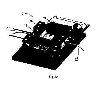
Each sample processing unit 7 comprises a microfluidic cartridge holder 9, a
tissue holder
11, and a coupling 13 therebetween to allow movement of the cartridge holder 9
relative to
the tissue slide holder 11 for mounting and dismounting of the tissue slide
34. In the
illustrated embodiment, the microfluidic cartridge holder is provided in a
form of a lid rotatably
coupled via a hinge forming the coupling 13 to a base forming the tissue slide
holder 11.
Within the scope of the invention, it may however also be envisaged to have
the microfluidic
cartridge holder as the base and the tissue slide holder as the lid moveably
mounted to the
base. This configuration can for example be used in combination with inverted
microscopy.
The base is fixedly mounted to the handling platform 5 of the sample
processing station 3.
The coupling 13 may be provided in other forms instead of a pivot hinge, for
instance by
means of link arms or a slide allowing the microfluidic cartridge holder to be
moved away
from the base holding the tissue slide holder 11, in a translation movement or
a combined
translational and rotational movement. The coupling 13 in a form of a pivot
hinge is however
simple and robust and corresponds to a preferred embodiment.
The microfluidic cartridge holder 9 advantageously comprises a viewing window
19 with a
recess 43 configured to receive at least partially therein a lens 14 of the
microscope such
that the microscope lens may be positioned very close to a viewing window 12
of the
microfluidic cartridge. A lens with a very large numerical aperture may thus
be used to
improve the quality of image capture of the sample under observation.
A sample processing unit 7 further advantageously comprises a clamping
mechanism 15
including a locking mechanism 16 and a pressure actuator 18. The pressure
actuator 18 may
comprise a piston driven by a compressed fluid, for instance a compressed air
piston 37, that
applies pressure on the tissue slide 34 against the microfluidic cartridge 4.
The pressure
ensures that a seal 10 arranged between a substrate 6 of a microfluidic
cartridge 4 and the
tissue slide 34 is hermetically closed to withstand a pressure in the reaction
chamber 29
during injection of reagent and other fluids in the reaction chamber. The
pressure applied by
the pressure actuator ensures that the maximum pressure attained in the
reaction chamber
does not cause the seal 10 to leak_
The locking mechanism 16 may for instance be in the form of a one or more
locking pins
inserted into corresponding orifices in a locking flange or tab on the other
of the lid or base.
Within the scope of the invention, the locking mechanism may however have
other
CA 03152364 2022-3-23
9
WO 2021/058782
PCT/EP2020/076978
configurations, for instance a pivotable arm with a catch shoulder engaging a
corresponding
catch shoulder on the other of the lid or base parts.
The moveable part of the microfluidic cartridge holder or tissue slide holder
may be actuated
manually or may include a motorized actuation mechanism (not shown) and
similarly the
locking mechanism may be manually operated or may include a motorized
actuation system
for automatic opening and closing of the moveable and static parts.
The sample processing unit 7 further comprises a reagent fluid flow system for
directing the
flow of reagents and other fluids from the external reagent source to the
microfluidic cartridge
4. The reagent fluid flow system thus comprises inlet couplings for reagent
conduits such as
reagent tubes for the inlet and outlet of reagents, and an interface
surrounded by a sealing
element that couples to the fluid flow network 8 on the microfluidic cartridge
4.
The clamping mechanism 15, when pressing the tissue slide 34 against the
microfluidic
cartridge 4 may also serve to push the microfluidic cartridge against the
tissue slide holder
11 to ensure tight sealing at the interface between the inlet and outlet on
the microfluidic
cartridge and the corresponding outlets and inlets on the reagent fluid flow
system within the
microfluidic cartridge holder 9.
The sample processing unit may further comprise a temperature control system
24 for
cooling and/or heating of the tissue slide 34 in view of heating or cooling
the reagents within
the reaction chamber 29 during tissue sample processing, in particular for the
purposes of
multiplexing. The temperature control system 24 may advantageously comprise a
Peltier chip
31 positioned in or under the base forming the tissue slide holder 11. In a
variant, the
temperature control system may further comprise heating and/or cooling
elements positioned
for heating and/or cooling around the reagent fluid flow system within the
sample processing
unit, in particular to pre-heat or pre-cool reagents entering into the
reaction chamber 29.
The microfluidic cartridge according to the embodiment of the invention
comprises a
substrate 6, a fluid flow network 8 formed within the substrate 6, a seal 10
and a viewing
window 12. The fluid flow network 8 comprises an inlet 26 for coupling to the
reagent fluid
flow system in the base of the sample processing unit 7, an outlet 32 for
outflow of reagents
from the reaction chamber 29, and an inlet channel network 27 and outlet
channel network
31 connected respectively to chamber entry orifices 28 and chamber exit
orifices 30. The
fluid flow network is configured to provide a substantially uniform flow of
reagents through the
CA 03152364 2022-3-23
10
WO 2021/058782
PCT/EP2020/076978
reaction chamber 29, intended to ensure substantially advective transport of
reagents into
the biological sample 36 fixed on the tissue support 34.
The seal 10 is mounted in a groove in a substrate 6 that surrounds the
reaction chamber 29
as well as the chamber entry orifices and exit orifices 28, 30. The reaction
chamber 29 is
formed between the tissue support 34 and the viewing window 12 enclosed by the
seal 10
sandwiched between the substrate 6 and tissue support 34.
The microfluidic cartridge 4 may advantageously further comprise spacer
elements 40, for
instance advantageously in a form of a continuous rim or a plurality of
discreet protuberences
arranged preferably on an outer side of the seal 10. The spacer elements
ensure that the
height of the reaction chamber 29 is maintained at a defined constant height
that is not
dependent on the compression force on the seal 10 supplied by the pressure
actuator 18.
The force of the pressure actuator and clamping mechanism 15 is arranged to be
sufficient to
compress the seal 10 until the spacer elements 40 are in contact with the
tissue support 34,
whereby excess pressure does not further compress the seal or change the
reaction
chamber height due to the rigid spacer elements. The spacer elements also
advantageously
ensure that the viewing window 12 remains in a parallel relationship with the
tissue support
34 and does not tilt with respect thereto.
The viewing window 12 comprises a transparent cover 33 having a thickness of
less than
1 mm, preferably less than 0.5 mm, for instance around 0.2 mm (e.g. 0.17 mm).
The
transparent cover 33 may advantageously be made of glass or of sapphire. The
transparent
cover 33 may be separately formed from the substrate 6 and assembled thereto
by adhesive
bonding, by welding, or by overmolding with a material of the substrate 6. The
viewing
window 12 comprises a recess relative to an outer surface of the substrate 6,
configured to
enable a lens of a microscope to be partially inserted in said viewing window
recess so as to
be very close to the surface of the transparent cover 33 and to the tissue
sample thereunder,
as further discussed below.
The substrate 6 may advantageously be formed of a molded polymer, for instance
an
injection molded polymer such as COP, COC, PC, PSU and PEEK that may be
transparent
or opaque.
The thin transparent cover 33 and recess of the viewing window 12 allows a
viewing face 41
of a microscope lens 14 to be placed at a distance from the reaction chamber
29 of less than
1 mm, in particular of less than 0.5 mm, such that the distance from the
tissue sample to the
CA 03152364 2022-3-23
11
WO 2021/058782
PCT/EP2020/076978
microscope lens is typically less than 1 mm considering that the reaction
chamber height is in
a range of 0.05 to 0.5 mm. The height of the spacer elements is advantageously
in a range
of 0.05 to 0.3 mm preferably in a range of 0.05 to 0.2 mm in order to have an
optimal flow of
reagents through the reaction chamber and advective transport of reagents to
the tissue
support.
A high numerical aperture microscope lens may thus be used to capture a large
surface area
of the tissue sample through successive imaging steps, for instance in a range
of 80nnnn2 to
120 mm2, typically in a range of 80mm2 to 100 mm2, thus allowing good image
capture and
analysis of a section of tissue sample exceeding 50 mm2. The very thin
transparent cover
which may advantageously be made of a material such as glass reduces artefacts
and
aberrations on the image captured by the microscope lens 14 for high
performance sample
analysis.
A plurality of sample processing units mounted on the handling platform
advantageously
allows processing of tissue samples with reagents while simultaneously
performing image
capture and analysis of other samples positioned under the microscope in order
to increase
rapidity of analysis of samples, especially during multiplexing.
For instance each of the plurality of sample processing units 7 may be at a
different stage of
a multiplex process, in other words with different reagents, the sample
processing units being
sequentially advanced to the lens of the imaging unit. Also loading and
unloading of tissue
samples 36 may be performed on certain sample processing units 7 while others
are being
analyzed by the imaging unit 2 or having reagents being injected in reaction
chamber for
subsequent analysis.
The plurality of sample processing units comprises preferably three or more
sample
processing units, preferably four or more sample processing units on the
common handling
platform 5.
It may be noted that for analysis of a biopsy tissue sample, the sample of
tissue from a same
patient may be distributed on a plurality of a tissue slides placed in the
various corresponding
sample processing units 7 such that various different reagents and analysis
can be
performed on the tissue samples simultaneously. Alternatively, the same
reagents and
analysis may be performed in order to provide a plurality of test results that
may be
compared for increasing the reliability of the diagnosis. Alternatively, the
plurality of sample
CA 03152364 2022-3-23
12
WO 2021/058782
PCT/EP2020/076978
stations may also be used to perform analysis of different tissue samples from
a same
patient or from different patients.
CA 03152364 2022-3-23
13
WO 2021/058782
PCT/EP2020/076978
List of references used
biological sample processing system 1
imaging unit 2
microscope
lens 14
viewing face 41
image processing system
sample processing station 3
handling platform 5
support 17
displacement mechanism (not shown)
sample processing unit 7
microfluidic cartridge holder 9
(lid)
viewing window 19
chamfered recess 43
tissue slide holder 11
base
coupling 13
hinge
clamping mechanism 15
locking mechanism 16
locking pin
pressure actuator 18
piston
compressed air piston
reagent fluid flow system
inlet conduits 20
outlet conduits 22
temperature control system 24
cooling / heating system
peltier chip 41
temperature sensor (not shown)
microfluidic cartridge 4
substrate 6
fluid flow network 8
cartridge inlet 26
inlet channels 27
chamber entry orifices 28
reaction chamber 29
chamber exit orifices 30
outlet channels 31
cartridge outlet 32
seal 10
viewing window 12
transparent cover 33
glass layer
spacer elements 40
tissue support 34
CA 03152364 2022-3-23
14
WO 2021/058782
PCT/EP2020/076978
tissue sample 36
external reagent sources
reagent tubes
Thickness of transparent cover T
CA 03152364 2022-3-23