Note: Descriptions are shown in the official language in which they were submitted.
WO 2021/072021
PCT/US2020/054702
MEDICAL DEVICE FOR APPLYING FORCE ON BIOLOGICAL TISSUE, OR THE LIKE
CROSS-REFERENCE TO PRIORITY APPLICATION
[0001] The present application claims the benefit of U.S. Provisional Patent
Application
No. 62/913,754, which is incorporated herein by reference in its entirety.
FIELD OF THE INVENTION
[0002] The present invention generally relates to medical devices for at least
partially covering wounds
and/or scars, and, more particularly, to wound closure and/or reducing tension
in wounds and/or scats.
BACKGROUND
[0003] Traditional methods of wound closure, wound support, wound dressings,
and bandages typically
do not adequately control wound tension, which is well known to be a primary
stimulus of excess scar
formation. In addition, tension reduction is known to decrease the size,
discoloration, and poor
appearance of scars when applied during the wound healing period.
[0004] Force modulating tissue bridges (see, e.g., International Publication
Nos. WO 2012/135735 and
WO 2018/075879) seek to allow wounds to be closed accurately, and further seek
to provide
simultaneous reduction of tension on closed wounds and scars in the healing
phases. There is a desire
for force modulating tissue bridges, and related devices, systems, and
methods, that provide a new
balance of properties.
SUMMARY
[0005] An aspect of this disclosure is the provision of a medical device for
at least partially covering
and applying force on tissue, wherein the medical device includes a body and a
flexible web (e.g., sheet)
connected to the body, and at least a portion of the web is configured to
engage and apply force (e.g.,
everting forces) on the tissue. The body can include a spanning structure and
struts respectively
connected to lateral portions of the spanning structure. Inner ends of the
struts can extend into an area
over which the medial portion of the spanning structure extends. At least a
portion of the web can span
between the inner ends of the struts. The medical device typically is
reconfigurable between extended
and retracted configurations_ The inner ends of the struts are typically
closer to one another in the
retracted configuration than in the extended configuration. The inner ends of
the struts are typically
closer to the medial portion of the spanning structure in the retracted
configuration than in the extended
configuration. Each strut typically includes an engagement zone configured to
engage and apply force
on the tissue, at least while the medical device is in the retracted
configuration.
[0006] A wide variety of the medical devices are within the scope of this
disclosure. For example, at
least a portion of the web that is positioned between the inner ends of the
struts can be omitted, the
medical device can be biased toward the retracted configuration, the medical
device can be solely biased
1
CA 03153899 2022-4-6
WO 2021/072021
PCT/US2020/054702
toward the retracted configuration (e.g., monostable), the medical device can
be multistable (e.g., biased
toward both of the retracted and extended configurations), the medical device
can include connecting
mechanisms for at least partially retaining the medical device in its
retracted configuration (e.g., for
retaining the struts in their inner configurations), the medical device can
include arrestation mechanisms
for at least partially restricting the medical device from deforming past the
extended configuration (e.g.,
for restricting outward movement of the struts), there can be a greater or
lesser number of the struts,
and/or the medical device can include guideway(s) configured to guide movement
of the spanning
structure and/or strut(s).
[0007] The foregoing summary provides a few brief examples and is not
exhaustive, and the present
invention is not limited to the foregoing examples. The foregoing examples, as
well as other examples,
are further explained in the following detailed description with reference to
accompanying drawings.
BRIEF DESCRIPTION OF THE DRAWINGS
[0008] The drawings are provided as examples. The present invention may be
embodied in many
different forms and should not be construed as limited to the examples
depicted in the drawings. The
drawings may be schematic and may not be drawn to scale.
[0009] Fig. 1 is an exploded, top perspective view of a multistable medical
device (e.g., force
modulating tissue bridge) in accordance with a first embodiment of this
disclosure.
[0010] Fig. 2 is an exploded, isolated top view of blanks of the first
embodiment tissue bridge.
[0011] Fig. 3 is a top perspective view of the first embodiment tissue bridge
in its extended stable
equilibrium configuration.
[0012] Fig. 4 is a schematic, top perspective view of the first embodiment
tissue bridge in its retracted
stable equilibrium configuration.
[0013] Fig. 5 is a schematic, isolated, top perspective view of a flexible,
multistable spanning structure
of the first embodiment tissue bridge, wherein the multistable spanning
structure is in its concave-up
stable equilibrium configuration.
[0014] Fig. 6 is across-sectional view taken along line 6-6 of Fig. 5.
[0015] Fig. 7 is a schematic, isolated, top perspective view of the
multistable spanning structure of the
first embodiment tissue bridge, wherein the spanning structure is in its
concave-down stable equilibrium
configuration.
100161 Fig. 8 is across-sectional view taken along line 8-8 of Fig. 7.
[0017] Figs, 9 through 13 are front views that schematically depict an example
of a sequence of steps
of a method of applying the first embodiment tissue bridge to wounded tissue
(e.g., skin) after removal of
outer release liners.
2
CA 03153899 2022-4-6
WO 2021/072021
PCT/US2020/054702
[0018] Fig. 14 is a schematic, top view of a multilayer precursor of multiple
of the tissue bridges of
Fig. 1, in accordance with an embodiment of this disclosure.
[0019] Fig. 15 is an exploded, top perspective view of a multistable tissue
bridge in accordance with a
second embodiment of this disclosure.
[0020] Fig. 16 is an exploded, isolated top view of the body-forming blanks
depicted in Fig. 15.
[0021] Fig. 17 is atop perspective view of a multistable body or tissue bridge
formed from the blanks
of Fig. 16 in its extended stable equilibrium configuration.
[0022] Fig. 18 is a top perspective view of the multistable body or tissue
bridge of Fig. 17 in its
retracted stable equilibrium configuration.
[0023] Fig. 19 is an exploded, top perspective view of a multistable tissue
bridge in accordance with a
third embodiment of this disclosure.
[0024] Fig. 20 is an isolated top view of the body-forining blank of Fig. 19.
[0025] Fig. 21 is a partially assembled, top perspective view of the tissue
bridge of Fig. 19.
[0026] Fig. 22 is a schematic, bottom perspective view of the tissue bridge of
Fig. 21.
[0027] Fig. 23 depicts a version of the tissue bridge that is like the version
depicted in Figs. 21 and 22,
except that portions of the struts are angled.
[0028] Fig. 24 is atop perspective view of the tissue bridge of Fig. 23 in its
extended stable equilibrium
configuration, wherein outer release liners are not shown.
[0029] Fig. 25 is a top perspective view of the tissue bridge of Fig. 24 in
its retracted stable equilibrium
configuration.
[0030] Fig. 26 is a top perspective view of the tissue bridge of Fig. 24
mounted on wounded tissue, in
accordance with an embodiment of this disclosure_
[0031] Fig. 27 is an isolated top view of the multistable body of the tissue
bridge of Fig. 24, or a
variation thereof.
[0032] Fig. 28 is an isolated top view of a blank that is like the blank
depicted in Fig. 20, except, for
example, that the widths of cuts between the struts and arm portions are
smaller in Fig. 28.
[0033] Fig. 29 is an isolated top view of a multistable body or tissue bridge
formed from the blank of
Fig_ 28, wherein corners of the struts are hidden from view beneath the arms,
and the hidden strut corners
are schematically depicted by dashed lines.
[0034] Fig. 30 is a top view of a blank that is similar to the blank of Fig.
28 except, for example, for the
addition of holes.
[0035] Fig. 31 is a top perspective view of a multistable body or tissue
bridge formed from the blank of
Fig. 30, wherein the body or tissue bridge is in its extended stable
equilibrium configuration.
3
CA 03153899 2022-4-6
WO 2021/072021
PCT/US2020/054702
[0036] Fig. 32 is a partially exploded, top perspective view of a multistable
tissue bridge including an
upper cover sheet, in accordance with an embodiment of this disclosure.
[0037] Fig. 33 depicts the tissue bridge of Fig. 32 in a further assembled
configuration.
[0038] Fig. 34 is atop perspective view of the tissue bridge of Fig. 33 in its
extended stable equilibrium
configuration.
[0039] Fig. 35 is an exploded, top perspective view of a multistable tissue
bridge in accordance with an
embodiment of this disclosure.
[0040] Fig. 36 is a partially assembled, top perspective view of the tissue
bridge of Fig. 35, wherein
portions of the struts are angled (e.g., inclined).
[0041] Fig. 37 is a top perspective view of the tissue bridge of Fig. 36 in
its extended stable equilibrium
configuration.
[0042] Fig. 38 is a schematic top view of a multistable body or tissue bridge
in accordance with another
embodiment of this disclosure.
[0043] Fig. 39 is a top view of a multistable body or tissue bridge in
accordance with another
embodiment of this disclosure.
[0044] Fig. 40 is an isolated top view of a blank for being formed into or
incorporated into a
multistable tissue bridge in accordance with an embodiment of this disclosure.
[0045] Fig. 41 is atop perspective view of a multistable body or tissue bridge
formed from the blank of
Fig. 40, wherein flexible, multistable stmt portions of the body are in Their
concave-up stable equilibrium
configurations, and the multistable spanning structure that connects the
struts to one another is in its
concave-up stable equilibrium configuration.
[0046] Fig. 42 is a front view of the configuration of Fig. 41.
[0047] Fig. 43 is atop perspective view of the multistable body or tissue
bridge formed from the blank
of Fig. 40 in its extended stable equilibrium configuration, wherein the
multistable spanning structure is
in its concave-up stable equilibrium configuration, and the flexible,
multistable struts are in their
concave-down stable equilibrium configurations.
[0048] Fig. 44 is a front view of the configuration of Fig. 43.
[0049] Fig. 45 is a front view of the multistable body or tissue bridge formed
from the blank of Fig. 40
in its retracted stable equilibrium configuration, wherein an everted wound is
schematically depicted
with dashed lines, in accordance with an embodiment of this disclosure.
[0050] Fig. 46 is an isolated top view of a blank for being formed into or
incorporated into a
multistable tissue bridge in accordance with an embodiment of this disclosure.
4
CA 03153899 2022-4-6
WO 2021/072021
PCT/US2020/054702
[0051] Fig. 47 is a top perspective view of a multistable body or tissue
bridge formed from the blank of
Fig. 46 in its extended stable equilibrium configuration.
[0052] Fig. 48 is a front view of the configuration of Fig. 47.
[0053] Fig. 49 is atop perspective view of a multistable body or tissue bridge
formed from the blank of
Fig. 46 in its retracted stable equilibrium configuration.
[0054] Fig. 50 is a front view of the configuration of Fig. 49, wherein an
everted wound is
schematically depicted with dashed lines.
[0055] Fig. 51 is a top perspective view of a multistable body or tissue
bridge in its extended stable
equilibrium configuration, in accordance with another embodiment of this
disclosure.
[0056] Fig. 52 is a top perspective view of the multistable body or tissue
bridge of Fig. 51 in its
retracted stable equilibrium configuration.
[0057] Fig. 53 is an isolated top view of a blank for being incorporated into
a multistable tissue bridge
in accordance with an embodiment of this disclosure.
[0058] Fig. 54 is a top perspective view of a multistable body or tissue
bridge including a multistable
spanning structure formed from the blank of Fig. 53 and struts mounted to the
spanning structure, in
accordance with an embodiment of this disclosure.
[0059] Fig. 55 is a bottom perspective view of the multistable body or tissue
bridge of Fig. 54.
[0060] Fig. 56 is an isolated top view of a blank for being incorporated into
a multistable tissue bridge
in accordance with an embodiment of this disclosure.
[0061] Fig. 57 is atop perspective view of a multistable spanning structure
formed from the blank of
Fig. 56.
[0062] Fig. 58 is a top perspective view of a multistable spanning structure
in accordance with another
embodiment of this disclosure.
[0063] Fig. 59 is a top view of a multistable tissue bridge in accordance with
another embodiment of
this disclosure.
[0064] Fig. 60 is a top view of a multistable tissue bridge in accordance with
another embodiment of
this disclosure.
[0065] Fig. 61 is an exploded, top perspective view of another embodiment of a
multistable tissue
bridge including the first embodiment multistable body, or the like, and
differently configured layers
including, for example, a cover sheet.
[0066] Fig. 62 is a front view of the tissue bridge of Fig. 61 in its
retracted stable equilibrium
configuration.
CA 03153899 2022-4-6
WO 2021/072021
PCT/US2020/054702
[0067] Figs. 63 through 68 depict a sequence of steps of a method of applying
the tissue bridge of Fig.
62 to a scar or wound in accordance with an embodiment of this disclosure.
[0068] Fig. 69 is an exploded, top perspective view of an embodiment of a
multistable tissue bridge
having similarities to the embodiment of Fig. 62.
[0069] Fig. 70 is an assembled, bottom perspective view of the tissue bridge
of Fig. 69, wherein the
release liners have been partially pulled away from the reminder of the tissue
bridge.
[0070] Fig. 71 is a bottom perspective view of an embodiment of a multistable
tissue bridge having
similarities to the embodiment of Fig. 70.
[0071] Fig. 72 is atop perspective view of an embodiment of a multistable
tissue bridge having
similarities to the embodiment of Fig. 69.
[0072] Fig. 73 is an isolated top view of a multistable body in its extended
stable equilibrium
configuration, in accordance with an embodiment of this disclosure.
[0073] Fig. 74 is an exploded, top perspective view of an embodiment of a
multistable tissue bridge
including the multistable body of Fig. 73, wherein the body is its extended
stable equilibrium
configuration.
[0074] Fig. 75 is a top perspective view of the tissue bridge of Fig. 74 in
its extended stable equilibrium
configuration.
[0075] Fig. 76 is a top perspective view of the tissue bridge of Fig. 74 in
its retracted stable equilibrium
configuration, without release liners.
[0076] Fig. 77 is atop perspective view of a multistable tissue bridge,
wherein the tissue bridge is in its
extended stable equilibrium configuration and includes a spacer assembly
configured to at least partially
control the multistability, in accordance with an embodiment of this
disclosure.
[0077] Fig. 78 is like Fig. 77, except for depicting another embodiment of a
spacer assembly.
[0078] Fig. 79A depicts the tissue bridge of Fig. 78 in a partially exploded
configuration.
100791 Fig. 79B is a top perspective view of a multistable tissue bridge,
wherein the tissue bridge is in
its extended stable equilibrium configuration and includes a spacer assembly
configured to at least
partially control the multistability, in accordance with an embodiment of this
disclosure.
[0080] Figs. 80 through 85 schematically depict a sequence of steps of a
method of forming a tissue
bridge having multiple multistable portions in accordance with an embodiment
of this disclosure.
[0081] Fig. 86 is a schematic front view of an example of a multistable tissue
bridge at least partially
formed from steps including those described with reference to Figs. 80 through
85, or the like.
[0082] Fig. 87 is a top perspective view of a multistable tissue bridge in its
extended stable equilibrium
configuration in accordance with an embodiment of this disclosure.
6
CA 03153899 2022-4-6
WO 2021/072021
PCT/US2020/054702
[0083] Fig. 88 is a front view of the configuration of Fig. 87.
[0084] Fig. 89 is a top perspective view of the tissue bridge of Fig. 87 in
its retracted stable equilibrium
configuration.
[0085] Fig. 90 is a front view of the configuration of Fig. 89, wherein an
everted wound is
schematically depicted with dashed lines, in accordance with an embodiment of
this disclosure.
[0086] Fig. 91 is a front view of a multistable tissue bridge in its extended
stable equilibrium
configuration in accordance with an embodiment of this disclosure.
[0087] Fig. 92 is a front view of the tissue bridge of Fig. 91 in its
retracted stable equilibrium
configuration, wherein an everted wound is schematically depicted with dashed
lines, in accordance with
an embodiment of this disclosure.
[0088] Fig. 93 is a perspective view of a multistable tissue bridge in an
extended stable equilibrium
configuration in accordance with an embodiment of this disclosure.
[0089] Fig. 94 is a perspective view of the tissue bridge of Fig. 93 in a
retracted configuration, wherein
an everted wound is schematically depicted with dashed lines, in accordance
with an embodiment of this
disclosure_
[0090] Fig. 95 is a perspective view of a multistable tissue bridge in an
extended stable equilibrium
configuration in accordance with an embodiment of this disclosure.
[0091] Fig. 96 is a perspective view of the tissue bridge of Fig. 95 in a
retracted configuration.
[0092] Fig. 97 is a schematic top view of several of the above-discussed
tissue bridges connected in
series by way of a patient-contact carrier sheet and/or other suitable
material, in accordance with an
embodiment of this disclosure.
[0093] Fig. 98 is a schematic top view of several of the above-discussed
tissue bridges connected in
series by way of a strip of padding material and/or other suitable material,
in accordance with an
embodiment of this disclosure.
[0094] Fig. 99 is a schematic, partially exploded, top perspective view of a
system of several of the
above-discussed tissue bridges connected in series by way of a release liner
and carrier sheet, in
accordance with an embodiment of this disclosure.
[0095] Fig. 100 is a schematic, bottom perspective view of the assembled
system of Fig. 99 without the
release liner, in accordance with an embodiment of this disclosure.
[0096] Fig. 101 is a bottom perspective view like Fig. 100 except for further
depicting a strip of
padding, in accordance with an embodiment of this disclosure.
[0097] Figs. 102 through 104 are partially exploded, bottom perspective views
similar to Fig. 100,
except for showing the padding in different configurations.
7
CA 03153899 2022-4-6
WO 2021/072021
PCT/US2020/054702
[0098] Fig. 105 is an exploded, top perspective view of a device including a
multistable body (e.g., an
applicator) and a reinforced elastic wound covering (e.g., for being applied
using the applicator), in
accordance with an embodiment of this disclosure.
[0099] Fig. 106 is an assembled, top perspective view of the device of Fig.
105 in its retracted stable
equilibrium configuration.
[00100] Fig. 107 is a bottom perspective view of the configuration of Fig.
106.
1001011 Fig. 108 is a front view of the configuration of Fig. 106.
[00102] Fig. 109 is atop perspective view of the device of Fig. 106 in its
extended stable equilibrium
configuration or a further extended configuration.
[00103] Figs. 110 through 112 are front views that schematically depict a
sequence of steps of a method
of applying the reinforced elastic wound covering portion of Figs. 105 through
109 to a scar or wound,
wherein in Fig. 112 the reinforced elastic wound covering has been installed
to the wound and separated
from the multistable body, in accordance with an embodiment of this
disclosure.
[00104] Fig. 113 is a schematic top view of a multistable tissue bridge in its
extended stable equilibrium
configuration, in accordance with an embodiment of this disclosure.
[00105] Fig. 114 is a top perspective view of the configuration of Fig. 113.
[00106] Figs. 115A through 115D are front views that schematically depict an
example of a sequence of
steps of a method of applying the tissue bridge of Figs. 113 and 114 to tissue
(e.g., fascia), in accordance
with an embodiment of this disclosure.
[00107] Fig. 116 is a side view of a version of the tissue bridge of Figs. 113
and 114 wherein at least
some of the prongs or hooks are curved and point medially.
[00108] Fig. 117 is a top view of a version of the tissue bridge of Figs. 113
and 114.
[00109] Fig. 118 is a top view of a version of the tissue bridge of Figs. 113
and 114 including surgical
mesh.
[00110] Fig. 119 is a side cross-sectional view depicting a broken piece of
tissue (e.g., bone) with a bore
hole in each piece, in accordance with an embodiment of this disclosure.
[00111] Figs. 120 and 121 are front views that schematically depict an example
of a sequence of steps of
a method of applying a multistable tissue bridge to the broken bone of Fig.
119, in accordance with an
embodiment of this disclosure.
[00112] Fig. 122 is a top view of a multistable tissue bridge in accordance
with an embodiment of this
disclosure.
8
CA 03153899 2022-4-6
WO 2021/072021
PCT/US2020/054702
1001131 Figs, 123 through 125 are front views that schematically depict an
example of a sequence of
steps of a method of applying the multistable tissue bridge of Fig. 122 to a
broken bone, in accordance
with an embodiment of this disclosure.
1001141 Fig. 126 is a top view of a multistable tissue bridge in accordance
with an embodiment of this
disclosure.
1001151 Fig. 127 is a schematic front view of the multistable tissue bridge of
Fig. 126 in its extended
stable equilibrium configuration.
1001161 Fig. 128 is a top view of another embodiment of a multistable tissue
bridge.
1001171 Fig, 129 is a top view of another embodiment of a multistable tissue
bridge.
1001181 Fig. 130 is a top view of a multistable tissue bridge in its retracted
stable equilibrium
configuration, in accordance with an embodiment of this disclosure.
1001191 Fig. 131 is a cross-sectional view taken along line 131-131 of Fig.
130.
1001201 Figs. 132 and 133 are front views that schematically depict an example
of a sequence of steps of
a method of applying the tissue bridge of Fig. 130 to a broken bone, in
accordance with an embodiment
of this disclosure.
1001211 Fig. 134 is a top view of a tissue bridge in accordance with an
embodiment of this disclosure.
1001221 Fig. 135 is a cross-sectional view taken along line 135-135 of Fig.
134.
1001231 Fig. 136A is a top perspective view of the tissue bridge of Fig. 134
in its extended
configuration.
1001241 Figs, 136B and 136C depict a sequence of steps of a method of applying
the tissue bridge of
Figs. 134 and 135 to a broken bone, in accordance with an embodiment of this
disclosure.
1001251 Fig. 13613 depicts one of a sequence of steps of a method of applying
the tissue bridge of Figs.
134 and 135 to a broken bone, in accordance with an embodiment of this
disclosure.
1001261 Figs, 136E through 136H are cutaway views that schematically depict
examples of versions of
mounting holes extending through distal end portions of struts.
1001271 Fig, 137 is a schematic top view of a tissue bridge in accordance with
an embodiment of this
disclosure.
1001281 Fig. 138 is a schematic cross-sectional view taken along line 138-138
of Fig. 137.
1001291 Figs. 139 through 142 are front views that schematically depict a
sequence of steps of a method
of applying the tissue bridge of Figs. 137 and 138 to a broken bone, in
accordance with an embodiment
of this disclosure.
1001301 Figs. 143 through 147A depict other embodiments of tissue bridges.
9
CA 03153899 2022-4-6
WO 2021/072021
PCT/US2020/054702
[00131] Fig. 147B is an exploded, top perspective view of a multi-part tissue
bridge including a
spanning structure or plate and a strut-supporting body, in accordance with an
embodiment of this
disclosure_
[00132] Figs. 147C and 147D schematically depict an example of a sequence of
steps of a method of
applying the tissue bridge of Fig. 147B to bone.
[00133] Figs. 147E and 147F schematically depict an example of a sequence of
steps of a method of
applying a tissue bridge to bone in accordance with another embodiment of this
disclosure.
[00134] Fig. 147G is an exploded, top perspective view of a multi-part tissue
bridge including a
spanning structure or plate and a strut-supporting body, in accordance with an
embodiment of this
disclosure.
[00135] Figs. 147H is a top view that schematically depicts one of a sequence
of steps of a method of
applying the tissue bridge of Fig. 147G to a broken bone, in accordance with
an embodiment of this
disclosure.
[00136] Figs. 1471 through 147P depict other embodiments of tissue bridges.
[00137] Figs. 147Q schematically depicts another example of a tissue bridge
mounted on bone, in
accordance with another embodiment.
[00138] Figs. 147R through 147T depict other embodiments of tissue bridges.
[00139] Figs. 147U and 147V schematically depict an example of a sequence of
steps of a method of
applying a tissue bridge to bone in accordance with another embodiment of this
disclosure.
[00140] Fig. 148 is an exploded, top perspective view of another embodiment of
a tissue bridge in its
extended configuration.
[00141] Fig. 149 is an assembled, top perspective view of the tissue bridge of
Fig. 147 in its extended
configuration, wherein outer release liners are not shown.
[00142] Figs. 150 through 152 are front views that schematically depict an
example of a sequence of
steps of a method of applying the tissue bridge of Fig. 149 to wounded tissue.
[00143] Fig. 153 schematically depicts an example of a version of the tissue
bridge of Fig. 149 that
includes an adhesive-backed cover sheet.
[00144] Fig. 154 depicts an example of a variation to the tissue bridge of
Fig. 149.
[00145] Fig. 155 is a partially exploded, top perspective view of another
embodiment of a tissue bridge
in its extended configuration, wherein outer release liners are not shown.
[00146] Fig. 156 is an assembled, top perspective view of the tissue bridge of
Fig. 155 in its extended
configuration.
CA 03153899 2022-4-6
WO 2021/072021
PCT/US2020/054702
[00147] Figs. 157 through 159 are front views that schematically depict an
example of a sequence of
steps of a method of applying the tissue bridge of Fig. 156 to wounded tissue.
[00148] Fig, 160 is a top perspective view of a tissue bridge in its retracted
stable configuration, in
accordance with an embodiment of this disclosure.
[00149] Fig. 161 is a top perspective, exploded view of the tissue bridge of
Fig. 160 in its retracted
stable equilibrium configuration.
[00150] Fig. 162 is a front view of the configuration of Fig. 160.
[00151] Fig. 163 is a right view of the configuration of Fig. 160.
[00152] Fig. 164 is a top perspective view of the tissue bridge of Fig. 160 in
an extended configuration.
[00153] Figs. 165-167 are graphs that respectively identify characteristics of
examples of symmetrically
bistable and asymmetrically bistable versions of the tissue bridge of Fig.
160.
[00154] Figs. 168 and 169 are front views that schematically depict an example
of a portion of a
sequence of steps of a method of applying the tissue bridge of Fig. 160 to
wounded tissue (e.g., skin)
after removal of an outer release liner.
[00155] Fig. 170 is a top view of a portion of a flat multilayer precursor web
including two flat
subassemblies of tissue bridges of the type depicted in Fig. 160, in
accordance with an embodiment of
this disclosure.
[00156] Figs. 171 through 177 each depict an isolated top plan view of a flat
tissue bridge subassembly
similar to the subassembly of the type depicted in Fig. 160, and similar to
the subassemblies of Fig. 170,
except for including differently configured disruptions, in accordance with
other embodiments of this
disclosure.
[00157] Fig. 178 is an isolated top plan view of a flat release liner of the
subassembly of Fig. 177.
[00158] Fig. 179 is a top view of a tissue bridge including the subassembly of
Fig. 177, wherein the
tissue bridge is in its retracted stable equilibrium configuration, in
accordance with an embodiment of
this disclosure.
DETAILED DESCRIPTION
[00159] Examples of embodiments are disclosed in the following. The present
invention may, however,
be embodied in many different forms and should not be construed as limited to
the embodiments set forth
herein. For example, features disclosed as part of one embodiment can be used
in the context of another
embodiment to yield a further embodiment. As another example of the breadth of
this disclosure, it is
within the scope of this disclosure for one or more of the terms
"substantially," "about," "approximately,"
and/or the like, to qualify each of the adjectives and adverbs of the Detailed
Description section of
disclosure, as discussed in greater detail below.
11
CA 03153899 2022-4-6
WO 2021/072021
PCT/US2020/054702
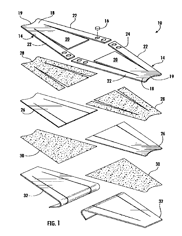
[00160] Fig. 1 is an exploded view of a flexible, multiconfigurable medical
device 10 that is multistable
(e.g., bistable), in accordance with a first embodiment of this disclosure.
The medical device 10 may
optionally be referred to as a force modulating tissue bridge, or simply
tissue bridge. Figs. 3 and 4 depict
the assembled first embodiment tissue bridge 10 in its extended and retracted
stable equilibrium
configurations, respectively. In the following, first an example of a method
of using the tissue bridge 10
is very briefly described, and thereafter the tissue bridge and other aspects
of this disclosure are described
in greater detail.
[00161] The tissue bridge 10 can be mounted to biological tissue such as, but
not limited to, a surface of
a patient's skin, for example the outer surface of the patient's epidermis.
The tissue bridge 10 is typically
mounted so that a central section of the tissue bridge extends across and at
least partially covers a wound
and/or scar. In the first embodiment, the tissue bridge 10 is in its extended
stable equilibrium
configuration (Figs. 3, 9 and 10) at the beginning of being mounted on the
patient. In an example, after
being at least partially mounted, the tissue bridge 10 can automatically,
biasedly reconfigure to, or
proximate to, its retracted stable equilibrium configuration (Figs. 4 and 13)
in response to the tissue
bridge being farther forced toward the tissue. In the first embodiment, the
reconfiguring comprises elastic
strain-induced bending. The reconfiguring between the extended and retracted
stable equilibrium
configurations seeks to, for example, reduce tension in the tissue, help close
the wound, help inhibit
wound reopening, and/or inhibit scar disfiguring (e.g., widening).
[00162] Referring to Fig. 1, the first embodiment tissue bridge 10 includes a
flexible, multistable (e.g.,
symmetrically bistable or asymmetrically bistable) body 12 (Fig. 3) formed
from two blanks 14 fastened
together by mechanical fasteners 16 (e.g., pegs, rivets, split-pin fasteners,
brad fasteners, snap fasteners)
and/or other suitable fastening mechanisms, as will be discussed in greater
detail below. Each blank 14
can include an end portion 18, at least one strut portion 20, and one or more
links or side ann portions 22
extending from the end portion. In the first embodiment, the strut portion 20
includes a proximal end
connected to the end portion 18, and a distal end opposite from the proximal
end.
[00163] Referring to Fig. 2, for each of the first embodiment blanks 14, its
arm portions 22 extend away
from the end portion 18 in a divergent manner to define an arm angle of
divergence "AA" between the
arm portions, as will be discussed in greater detail below. The body 12 can
include or be formed from
less or more than two blanks 14 of a variety of different configurations
(e.g., a variety of arm angles of
divergence or convergence AA are within the scope of this disclosure), as will
be discussed in greater
detail below.
[00164] Regarding connecting the blanks 14 to one another (e.g. connection
zones of the blanks 14), one
or more holes 24 can extend through distal end portions of the blank arm
portions 22 for respectively
receiving the peg fasteners 16 (Figs. 1, 3 and 4) and/or other suitable
fastening mechanisms can be
12
CA 03153899 2022-4-6
WO 2021/072021
PCT/US2020/054702
utilized to respectively connect the arm portions to one another. In Fig. 1, a
representative one of the four
fasteners 16 is depicted, although there may be more or less of such fasteners
and/or other fastening
mechanisms.
1001651 Referring to Fig. 2, the outer end portions 18 can each include an
inwardly recessed edge 19
configured for receiving the outer end portion of a finger or thumb of a user
during installation, as will be
discussed in greater detail below. The blanks 14 can be provided, for example,
by die cutting them from
appropriate webs or larger sheets of material, such as polymeric films or
laminates (e.g., polyethylene,
polyethylene terephthalate, or any other suitable materials), metallic sheets,
alloys, and/or other suitable
materials. The one or more strut portions 20 and one or more side arm portions
22 can be partially
defmed by cuts 25 (e.g., slits, holes, cutouts, and/or respective gaps)
between adjacent portions of the
blanks 14.
1001661 Referring to Fig. 1, the tissue bridge 10 can include one or more
layers mounted to the blanks
14 Of the body 12 (e.g., the body formed from the blanks or in any other
suitable manner). In the first
embodiment, the tissue bridge 10 includes several layers of material connected
to the underside of the
body 12. The layers can include, for example, carrier sheets 26, adhesive
layers 28, 30, and outer release
sheets or liners 32 (e.g., removable backings).
[00167] In the first embodiment, patient-contact structures comprise, consist
essentially of, or consist of
the carrier sheets 26 and the adhesive 30 being cooperatively configured so
that the patient-contact
structures can be used to attach the tissue bridge 10 to tissue (e.g., skin
tissue) after removal of the one or
more release liners 32. Accordingly and for ease of understanding, the carrier
sheets 26 may be referred
to as patient-contact carriers, and the adhesive 30 may be referred to as
patient-contact adhesive 30.
1001681 More specifically regarding layers of the tissue bridge 10, the inner
adhesive 28 can be between
and fixedly connect the patient-contact carriers 26 to the body 12, and the
patient-contact adhesive 30
can be on the outer sides of the patient-contact carriers 26 for attaching the
tissue bridge 10 to tissue
(e.g., a patient's skin), as will be discussed in greater detail below. One or
more of the adhesive layers
28, 30 or sheets 26, 32 can include cuts (e.g., slits, holes, cutouts, and/or
respective gaps) that are at least
similar to and at least partially superposed with at least respective portions
of the cuts 25 in the body 12.
1001691 One or more of the adhesive layers 28, 30 or sheets 26, 32, or
portions thereof, can be omitted.
For example, the patient-contact carriers 26 and the inner adhesive 28 can be
omitted, so that the patient-
contact adhesive 30 is mounted directly on (e.g., is in opposing face-to-face
contact with) the body 12.
As another example, at least some of, or all of, the portions of the patient-
contact adhesive 30 and release
liners 32 associated with the side arm portions 22 may be omitted. As a
further example, the connection
to tissue provided by the patient-contact adhesive 30 can be supplemented with
or replaced by one or
more suitable non-adhesive attachment mechanisms (e.g., pins, needles,
sutures, staples, and/or the like).
13
CA 03153899 2022-4-6
WO 2021/072021
PCT/US2020/054702
1001701 At least partially reiterating from above, the drawings may not be
drawn to scale. For example,
in at least some of the drawings depicting exploded views (e.g., Fig. 1), the
size of one or more of the
adhesive layers 28, 30 and sheets 26, 32, or portions thereof, may be
exaggerated as compared to the
body 12.
1001711 The inner and outer sheets 26, 32 can be provided, for example, by die
cutting them from
appropriate webs or larger sheets of material. For example, the inner or
patient-contact carriers 26 can
be made of suitable fabric materials, cast materials, cast microporous
polymeric sheet, polymeric films
(e.g., polyurethane), padding, and/or other suitable materials (e.g., of the
type from which skin-contact
layers of bandages or other wound dressings are formed). The release liner 32
can be, for example, a
paper or plastic-based film sheet coated with a release agent that is engaged
against the patient-contact
adhesive 30 so that the tissue bridge 10 is releasably mounted on the release
liner. The inner adhesive 28
can comprise adhesive materials that are compatible with the materials being
connected thereby. The
patient-contact adhesive 30 can be, for example, a pressure-sensitive adhesive
material of the type that is
typically used as an adhesive backing for wearable medical devices, bandages,
or other wound dressings.
The patient-contact adhesive 30 can have a lower adhesive strength than the
inner adhesive 28, such as
when the tissue bridge 10 is to be removably mounted to tissue (es., a
patient's skin). In other
embodiments, for example wherein at least the multistable body 12 can function
as an applicator of a
wound covering, the inner adhesive 28 can have a lower adhesive strength as
compared to one or more
other adhesives (e.g., the patient-contact adhesive 30), as will be discussed
in greater detail below.
1001721 Figs. 3 and 4 respectively depict the assembled first embodiment
tissue bridge 10 in its stable
equilibrium configurations. These stable equilibrium configurations may be
symmetrical or
asymmetrical, as further discussed below. The first embodiment tissue bridge
10 includes lateral or end
portions 40, one or more struts 42 (e.g., strut assemblies), and one or more
arms 44. In the first
embodiment, each strut 42 includes a proximal end connected to the respective
tissue bridge end portion
40, and a distal end opposite from the proximal end.
1001731 The first embodiment tissue bridge's end portions 40, struts 42, and
arms 44 respectively
include the end portions 18, strut portions 20, and arm portions 22 of the
blanks 14 and body 12, as well
as corresponding portions of any of the sheets 26, 32 and adhesive layers 28,
30 that may be included in
the tissue bridge. Whereas the tissue bridges 10 are sometimes described in
the Detailed Description
section as including two struts 42, it is within the scope of this disclosure
for each of the tissue bridges to
include any suitable number of struts, including, for example, one strut or
more than two struts.
1001741 In Figs. 3 and 4, the inner or distal end portions of the one or more
struts 42 are angled or
inclined relative to the central and outer or proximal end portions of the
struts. The bends that define the
inclination or angle of the distal end portions of the struts 42 can be
provided, for example, by bending,
14
CA 03153899 2022-4-6
WO 2021/072021
PCT/US2020/054702
thennoforniing, stamping, and/or in any other suitable manner. Alternatively,
the tissue bridges 10 can
be formed, or at least partially formed, by injection molding, 3D printing,
and/or in any other suitable
manner. The bends that define the inclination or angle of the distal end
portions of the struts 42 can be
formed in response to tissue forces associated with the tissue bridge 10 being
mounted on tissue 52 (see,
e.g., Fig. 10), for example when the bend area includes at least one feature
for helping to facilitate the
bending. For example, a feature for helping to facilitate such bending of the
distal end portion of a strut
42 can include at least one line of disruption (e.g., weakened areas, as
formed by perforations or other
suitable holes, kiss-cuts, score lines, areas of reduced thickness, and/or the
like) along which bending
may occur. Other features for helping to facilitate such bending of the distal
end portion of a strut 42
may include at least one hinge, for example a living hinge, a hinge defined by
malleable material, a hinge
including a hinge pin and associated bearing structure(s), and/or other
suitable features.
1001751 The assembling of the first embodiment body 12 and tissue bridge 10
includes causing relative
movement between the inner or distal end portions of the arm portions 22 so
that the arm inner end
portions (e.g., the holes 24) respectively become superposed with one another
in a plan view, and then
fixedly fastening the superposed arm portions with one another. The superposed
inner end portions of
the arm portions 22 can be fixedly connected to one another using the peg
fasteners 16, holes 24,
adhesive material, heat sealing, welding, and/or any other suitable fastening
mechanisms.
1001761 As an example of providing the multistability characteristics of the
multistable body 12 and the
tissue bridge 10, the blanks 14 are constructed of suitable flexible material,
and the superpositioning and
associated fastening of the inner end portions of the arm portions 22 decrease
the angle of divergence
AA (Fig. 2) between the arm portions 22 so that the tissue bridge has the
stable equilibrium
configurations depicted in Figs. 3 and 4. Additionally, the first embodiment
multistable body 12 and
tissue bridge 10 have numerous unstable configurations between the stable
equilibrium configurations
depicted in Figs. 3 and 4, as will be discussed in greater detail below.
1001771 Whereas in the first embodiment the connecting of the arm portions 22
and the providing of the
multistability characteristics includes the superpositioning and connecting of
respective portions of the
arms 22, the connecting and providing of the multistability can be achieved in
other suitable ways. For
example, the connecting and providing of the multistability characteristics
can include respectively
joining the arm portions end-to-end (e.g., end-edge to end-edge) by way of
suitable seams (e.g., welding
(e.g., laser welding)), adhering (e.g., with adhesive and/or adhesive tape),
injection molding (e.g., insert
molding), or other suitable seaming together) and/or other suitable attachment
mechanisms.
1001781 The first embodiment multistable body 12 functions as a substrate that
carries the other
components of the tissue bridge 10, and the tissue bridge is multistable
(e.g., symmetrically bistable or
asymmetrically bistable) by virtue of the multistability of the body 12.
Alternatively, it is believed that
CA 03153899 2022-4-6
WO 2021/072021
PCT/US2020/054702
any of the sheets 26, adhesive layers 28, 30, and/or other components that may
be included in the tissue
bridge 10 may be configured to contribute to or provide the multistable
characteristics of the tissue
bridge 10. The multistable body 12 and tissue bridge 10 of the first
embodiment more specifically are
bistable, so that they have the two stable equilibrium configurations depicted
in Figs. 3 and 4. As will be
discussed in greater detail below, the multiple or bistable configurates may
be altered in response to
tissue forces associated with the tissue bridge 10 being mounted on tissue 52
(see, e.g., Figs. 9-13).
[00179] Figs. 3 and 4 respectively depict the bistable tissue bridge 10 in its
extended stable equilibrium
configuration (e.g., a first stable equilibrium configuration) and its
retracted stable equilibrium
configuration (e.g., a second stable equilibrium configuration). In the
extended stable equilibrium
configuration (Fig. 3), the inner or distal end portions of the one or more
struts 42 extend (e.g., are
inclined) outwardly (e.g., downwardly) away from the arms 44. In contrast, in
the retracted
configuration (Fig. 4), the one or more struts 42 are relatively retracted
with respect to the arms 44, so
that at least a portion of (e.g., the distal end portion of) at least one
strut is closer to the arms. As another
example of the retracted configuration, when the tissue bridge 10 includes a
pair of struts that are
opposite from one another, the struts 42 of the pair can be relatively
retracted with respect to the arms 44
so that at least portions of (e.g., the distal end portions of) the struts are
closer to one another.
[00180] In the first embodiment, the strut portions 20 of the body 12 are
connected to one another by
way of at least one flexible, multistable spanning structure 46 comprising,
consisting essentially of, or
consisting of the end and arm portions 18, 22 of the body 12. Similarly for
the first embodiment tissue
bridge 10, the struts 42 are connected to one another by way of at least one
multistable spanning
structure 48 comprising, consisting essentially of, or consisting of the end
and arm portions 40,44 of the
tissue bridge 10. The spanning structures 46, 48 can each be described as
including a medial or middle
portion extending between lateral or end portions of the spanning structure.
[00181] In the first embodiment, a portion (e.g., core portion) of the tissue
bridge 10 is multistable (e.g.,
essentially bistable), and other portions of the tissue bridge are connected
to the core portion for moving
with the core portion. In the first embodiment, the tissue bridge's
multistable spanning structure 48, or
more specifically the body's multistable spanning structure 46, is the core
portion of the tissue bridge 10
that is multistable (e.g., essentially bistable). That is, the multistable
spanning structures 46,48 can be
configured to provide the multistable (e.g., symmetrically bistable or
asymmetrically bistable) behavior
of the body 12 and tissue bridge 10, respectively. For example and as depicted
in Fig. 3, the first
embodiment multistable spanning structure 48 is in its concave-up stable
equilibrium configuration
when the tissue bridge 10 is in the extended stable equilibrium
configuration). For example, at least the
arms 44 can form a central inverted arch when the multistable spanning
structure 48 is in its concave-up
stable equilibrium configuration (e.g., a first stable equilibrium
configuration). In contrast and as
16
CA 03153899 2022-4-6
WO 2021/072021
PCT/US2020/054702
depicted in Fig. 4, the first embodiment multistable spanning structure 48 is
in its concave-down stable
equilibrium configuration (e.g., a second stable equilibrium configuration)
when the tissue bridge 10 is in
the retracted stable equilibrium configuration. For example, at least the arms
44 can form a central arch
when in their concave-down stable equilibrium configuration.
1001821 Figs. 5 and 6 are isolated views that schematically depict the first
embodiment multistable
spanning structure 48 in the concave-up stable equilibrium configuration. As
depicted in Figs. 5 and 6,
the concave-up stable equilibrium configuration defined by the multistable
spanning structure 48
includes or defines both a length-wise arc and a width-wise arc (e.g.,
crosswise arcs and at least a portion
of an inverted dome). As depicted in Figs. land 8, the concave-down stable
equilibrium configuration
defined by the multistable spanning structure 48 includes or defines both a
length-wise arc and a width-
wise arc (e.g., crosswise arcs and at least a portion of a dome). One or more
of the subject curvatures
can exist throughout the entire multistable spanning structure 48.
Alternatively, it is believed that one or
more of the subject curvatures may be present only in respective portions of
the multistable spanning
structure 48. Additionally, one or more of the subject curvatures can be
modified (e.g., increased,
decreased, and/or have a changed orientation) in order to alter the three-
dimensional shape of the tissue
bridge in its multistable (e.g. bistable) configurations.
1001831 As one example, the multistable spanning structure 48 can be
symmetrically configured, for
example so that the concave-up stable equilibrium configuration and concave-
down stable equilibrium
configuration are mirror images of one another. As another example, the
multistable spanning structure
48 can be asymmetrically configured, for example so that the concave-up stable
equilibrium
configuration and concave-down stable equilibrium configuration are not mirror
images of one another
(e.g., in the concave-up and concave-down stable equilibrium configurations,
corresponding portions of
the spanning structure 48 can have different amounts of curvature from one
another (e.g., can have
radiuses of curvature that differ in magnitude from one another), or the
like). As an example of the
multistable spanning structure 48 being asymmetrically configured, one of the
stable equilibrium
configurations can be flatter than the other of the stable equilibrium
configuration.
1001841 The multistable spanning structure 48 being symmetrically bistable, or
the tissue bridge 10
being symmetrically bistable, can comprise it having a first stable
equilibrium configuration in which it
defines a first concavity having a radius of curvature (see, e.g., Fig. 5),
and a second stable equilibrium
configuration in which it defines a second concavity having a radius of
curvature (see, e.g., Fig. 7),
wherein the radius of curvature of the first concavity can be the same as, or
about the same as, the radius
of curvature of the second concavity. The multistable spanning structure 48
being asymmetrically
bistable, or the tissue bridge 10 being asymmetrically bistable, can comprise
it having a first stable
equilibrium configuration in which it defines a first concavity having a
radius of curvature (see, e.g., Fig.
17
CA 03153899 2022-4-6
WO 2021/072021
PCT/US2020/054702
5), and a second stable equilibrium configuration in which it defines a second
concavity having a radius
of curvature (see, e.g., Fig. 7), wherein the radius of curvature of the first
concavity can differ from the
radius of curvature of the second concavity.
1001851 In the first embodiment, the multistable spanning structure 48 has at
least one unstable
equilibrium configuration (e.g., a maximally unstable configuration) between
its concave-up stable
equilibrium configuration and concave-down stable equilibrium configuration.
In this regard, the tissue
bridge 10 is configured so that the multistable spanning structure's unstable
equilibrium configuration,
concave-up stable equilibrium configuration, and concave-down stable
equilibrium configuration are
respectively associated with the tissue bridge's unstable equilibrium
configuration, extended stable
equilibrium configuration, and retracted stable equilibrium configuration, as
discussed in greater detail
below.
1001861 Those of ordinary skill in the art will understand that directional
references such as "up" and
"down" are being used in the Detailed Description section of this disclosure
for ease of understanding
and may be described differently with respect to other directional frames of
reference, for example as
being directions that are opposite from one another. For example and in the
first embodiment, the
multistable spanning structure 48 can be described as having opposite first
and second sides, wherein the
first side is concave (e.g., substantially concave and/or otherwise suitably
configured (es., curved)) in a
first stable equilibrium configuration and convex (e.g., substantially convex
and/or otherwise suitably
configured (e.g., curved)) in a second stable equilibrium configuration, and
wherein the second side is
concave (e.g., substantially concave and/or otherwise suitably configured
(e.g., curved)) in the second
stable equilibrium configuration and convex (e.g., substantially convex and/or
otherwise suitably
configured (e.g., curved)) in the first stable equilibrium configuration.
1001871 In accordance with an example of a method of fabricating the first
embodiment tissue bridges
10, they can be fully assembled, sterilized, and then be enclosed in packages
that are provided to end
users that open the packages and mount the tissue bridges on tissue 52 (see,
e.g., Fig. 9). Each package
can include one or more of the tissue bridges 10. For example, in a relatively
compact package
containing a plurality of the tissue bridges 10, the tissue bridges may be
nested together in their retracted
stable equilibrium configurations, in which case a user would typically
reconfigure the tissue bridges into
their extended stable equilibrium configurations before mounting them on
tissue 52. Alternatively, the
tissue bridges 10 can be packaged in their extended stable equilibrium
configurations and/or partially
assembled configurations, as will be discussed in greater detail below. The
packaged tissue bridges 10
can be part of a kit, military pack, or the like, including one or more other
items such as, for example,
bandages, medical dressings, medical irrigation devices, wipes and/or liquids
for use in sterilizing,
gloves, and/or other suitable items.
18
CA 03153899 2022-4-6
WO 2021/072021
PCT/US2020/054702
[00188] Figs. 9-13 schematically depict an example of a sequence of steps of a
method of applying the
first embodiment tissue bridge 10 to a scar or wound 50 after the release
liners 32 have been removed.
The tissue bridge 10 is in the extended stable equilibrium configuration in
Figs. 9 and 10; in unstable
configurations in Figs. 11 and 12; in the at least one unstable equilibrium
configuration (e.g., a
maximally unstable configuration) in Fig. 12; and in, or proximate, the
retracted stable equilibrium
configuration in Fig. 13.
[00189] Referring to Fig. 9, if the tissue bridge 10 is not yet in, or
proximate, its extended stable
equilibrium configuration, the tissue bridge can be manually reconfigured, for
example, from its retracted
stable equilibrium configuration to its extended stable equilibrium
configuration. For example, the tissue
bridge 10 can be manually reconfigured from its retracted stable equilibrium
configuration to its
extended stable equilibrium configuration by manually holding the tissue
bridge 10 between a user's
finger 54 (Fig. 9) and thumb 56 (Fig. 9), so that the finger and thumb are
engaged against opposite ends
of the tissue bridge and pulling upward, while another finger pushes
downwardly on a medial portion of
the spanning structure 48. As another example, the tissue bridge 10 can be
manually reconfigured from
its retracted stable equilibrium configuration to its extended stable
equilibrium configuration by the user
holding one end of the tissue bridge with one hand and holding the other end
of the tissue bridge with
their other hand, and then causing relative rotation between their hands in a
manner that causes the tissue
bridge to be reconfigured from its retracted stable equilibrium configuration
to its extended stable
equilibrium configuration. In either case, such reconfiguring can include
manually bending the tissue
bridge 10 from its retracted stable equilibrium configuration toward and then
slightly past its maximally
unstable equilibrium configuration, and then allowing the tissue bridge to
automatically reconfigure (e.g.,
bend itself in response to its elastic potential energy) from adjacent its
maximally unstable equilibrium
configuration to its retracted stable equilibrium configuration.
[00190] As depicted in Fig. 9, the tissue bridge 10 in, or proximate, its
extended stable equilibrium
configuration can be manually held between a user's finger 54 and thumb 56, or
in any other suitable
manner, so that the length of the tissue bridge extends crosswise to, or more
specifically substantially
perpendicular to, the length of a scar, cut, or wound 50 in a patient's tissue
52. Referring to Fig. 10, the
patient-contact adhesive 30 (Fig. 1) on the lower or outer surfaces of the
angled (e.g., inclined) inner or
distal end portions of the one or more struts 42 (e.g., strut assemblies) can
be engaged against the
patient's tissue or skin 52 on either side of the scar, cut, or wound 50. The
portions of the patient-contact
adhesive 30 on the body's struts 20 can be referred to as engagement zones of
the tissue bridge's struts
42. In addition to or as an alternative to the patient-contact adhesive 30 on
the body's struts 20 being or
defining the engagement zones, the engagement zones can comprise pins,
needles, sutures, staples, barbs,
prongs, and/or other suitable fastening mechanisms or the like.
19
CA 03153899 2022-4-6
WO 2021/072021
PCT/US2020/054702
1001911 Referring to Figs. 11 and 12, the tissue bridge 10 can continue to be
manually forced or pushed
closer to the tissue 52 so that the inner portions of the one or more struts
42 become further adhered to
the patient's tissue 52 by the patient-contact adhesive 30. The action forces
applied by the user's finger
54 and thumb 56 at the recessed edges 19 (Figs. 1 and 2) or other suitable
locations on the spanning
structure 48 urge the struts 42 against the tissue 52. The tissue 52 provides
resisting or reaction forces so
that the struts 42 apply reaction forces against respective portions of the
spanning structure 48. In the
first embodiment, the locations of the action and reaction forces on the
spanning structure 48 are spaced
apart. When sufficiently large, the action and reaction forces and resulting
torque cause the tissue bridge
10w reconfigure (e.g., bend) from its extended stable equilibrium
configuration toward and past its
intermediate or maximally unstable equilibrium configuration (e.g., Fig_ 12).
In an example, after the
tissue bridge 10 is forced or pushed past its maximally unstable equilibrium
configuration, the tissue
bridge automatically transitions (e.g., bends itself due to its elastic
potential energy) at least proximate to
its retracted stable equilibrium configuration to further adhere the one or
more struts 42, and optionally
also the end portions 40 and arms 44, to the tissue 52. In the process, the
struts 42 become closer
together and push the portions of the tissue 52 to which they are adhered
toward one another (e.g., which
may evert the tissue adjacent the scar or wound 52).
1001921 With continued reference to Figs. 9-13, the first embodiment
multistable tissue bridge 10 is
configured for at least partially covering a wound 50 and/or scar tissue, and
reducing tension associated
with the wound and/or scar tissue. The multistable spanning structure 46 can
be configured to have a
plurality of configurations including the unstable configurations (e.g., Figs.
11 and 12) between the
concave-up stable equilibrium configuration (e.g., Figs. 3, 5, 9 and 10) and
the concave-down stable
equilibrium configuration (e.g., Figs. 4, 7 and 13). In the first embodiment,
the multistable spanning
structure 46 is configured to be biased toward the concave-up stable
equilibrium configuration (e.g., the
first stable equilibrium configuration) when in a configuration between the
intermediate or maximally
unstable equilibrium configuration (e.g., Fig. 12) and the concave-up stable
equilibrium configuration.
Similarly, the first embodiment multistable spanning structure 46 is biased
toward the concave-down
stable equilibrium configuration (e.g., the second stable equilibrium
configuration) when in a
configuration between the intermediate or maximally unstable equilibrium
configuration and the
concave-down stable equilibrium configuration.
1001931 The first embodiment multistable spanning structure 46 and struts 42
are cooperatively
configured so that at least the inner or distal end portions of the struts
become closer to one another at
least in response to the multistable spanning structure being transitioned
from the concave-up stable
equilibrium configuration to past the intermediate or maximally unstable
equilibrium configuration and
toward the concave-down stable equilibrium configuration. In the first
embodiment, at least lower or
CA 03153899 2022-4-6
WO 2021/072021
PCT/US2020/054702
outer surfaces of the distal end portions of the struts 42 include engagement
or connection zones (e.g.,
adhesive material 30) configured to be engaged to and move respective portions
of patient tissue 52
toward one another in response to the struts becoming closer to one another.
1001941 More specifically regarding the above-discussed method of applying the
first embodiment tissue
bridge 10 to tissue 52, there is a first series of unstable configurations
between the extended stable
equilibrium configuration and the intermediate or maximally unstable
equilibrium configuration.
Similarly, there is a second series of unstable configurations between the
retracted stable equilibrium
configuration and the intermediate or maximally unstable equilibrium
configuration. It is believed that:
(i) the degree to which the multistable spanning structure 48 defines the
concave-up shape varies serially
from a relatively maximally defined concave-up shape in the extended stable
equilibrium configuration
to a relatively minimally defined concave-up shape at the first series'
unstable configuration adjacent
intermediate or maximally unstable equilibrium configuration; 00 the
multistable spanning structure is
flat (substantially flat) in the intermediate or maximally unstable
equilibrium configuration; and (iii) the
degree to which the multistable spanning structure defines the concave-down
shape varies serially from a
relatively maximally defined concave-down shape in the retracted stable
equilibrium configuration to a
relatively minimally defined concave-down shape at the second series' unstable
configuration adjacent
the intermediate or maximally unstable equilibrium configuration.
1001951 The first embodiment tissue bridge 10 can be transitioned from either
of the stable equilibrium
configurations to the intermediate or maximally unstable equilibrium
configuration by manually, or
otherwise suitably, applying force against the tissue bridge in a manner that
seeks to flatten out the
concavity of the multistable spanning structure 48, wherein increasing
external force is required as
concavity decreases so that a maximum external force is required to achieve
the at least one unstable
equilibrium configuration (e.g., a maximally unstable configuration). In
contrast, the first embodiment
tissue bridge 10 is configured to automatically biasedly transition from any
of the first series of unstable
configurations to the extended stable equilibrium without requiring the
application of any external force
to the tissue bridge. Similarly, the first embodiment tissue bridge 10 is
configured to automatically
biasedly transition from any of the second series of unstable configurations
to the retracted stable
equilibrium without requiring the application of any external force to the
tissue bridge. As an alternative
to directly manually applying force against the tissue bridge 10 in a manner
that seeks to flatten out the
concavity of the multistable spanning structure 48, the force for flattening
out the concavity can be
provided by way of fasteners (e.g., threaded fasteners such as screws and
bolts) and tools (e.g., hand
tools), as will be discussed in greater detail below.
1001961 Fig. 12 depicts an example of an approximately maximally unstable
intermediate configuration
of at least the tissue bridge spanning structure 48. At least partially
reiterating from above and as a
21
CA 03153899 2022-4-6
WO 2021/072021
PCT/US2020/054702
further example, the first embodiment multistable spanning structure 48 is
configured so that if the
spanning structure is in an intermediate configuration between the
configurations depicted in Figs. 10
and 12, or the like, then the spanning structure would tend to return toward
the configuration depicted in
Fig. 10, or the like, if no, or minimal, external forces (e.g. digital
pressure) were applied to the tissue
bridge. Similarly, if the multistable spanning structure 48 is in an
intermediate configuration between
the configurations depicted in Figs. 12 and 13, or the like, then the spanning
structure would tend to
change toward the configuration as shown in Figure 13, or the like, if no, or
minimal, external forces
(e.g. digital pressure) were applied to the tissue bridge. The presence of
tissue forces (e.g. tension or
resistance to medial tissue advancement), adhesion forces (between the tissue
bridge and the tissue to
which it is applied), or other factors may alter the maximally unstable
intermediate configuration and
stable configurations (e.g., stable equilibrium configurations) of the tissue
bridge.
1001971 Reiterating from above, Fig. 12 depicts an unstable equilibrium
configuration of at least the
tissue bridge spanning structure 48. In the example depicted in Fig. 12, the
spanning structure is flat,
substantially flat, planar, or substantially planar. It is believed that in
other embodiments the spanning
structure 48 may be curved (e.g., may not be flat, planar, or substantially
planar) in its unstable
equilibrium configuration
1001981 At least partially reiterating from above, whereas the energy stored
by the multistable spanning
structure 48 is responsible for providing the multistability (e.g.,
bistability) of the tissue bridge 10 of the
first embodiment, the other features (e.g., layers) of the tissue bridge that
are carried by the multistable
spanning structure can affect, for example, the overall stiffness,
flexibility, and elasticity of the tissue
bridge. Characteristics (e.g., stiffness, flexibility, and/or elasticity) of
one or more of the various features
of the tissue bridge 10 can be adjusted in a predetermined manner to tune the
operability of (e.g., the
multistability of) the tissue bridge. For example, as compared to one another,
different parts of the same
tissue bridge 10 can have different characteristics (e.g., different
stiffness, flexibility, and/or elasticity
resulting from different thicknesses or volumes, different construction
materials, and/or different
manufacturing techniques) to affect the operability of (e.g., the
multistability of) the tissue bridge.
1001991 Similarly, the tissue upon which the tissue bridge 10 is mounted can
affect, for example, the
overall stiffness, flexibility, and elasticity of a system (e.g., combination)
that includes both the tissue 52
and the tissue bridge 10. As a specific example, depending upon the
configuration and properties of the
tissue 52 upon which the tissue bridge 10 is mounted, the tissue bridge per se
may not reach its retracted
stable equilibrium configuration and remain in an unstable configuration of
the tissue bridge when the
system (e.g., the combination that includes both the tissue and the tissue
bridge) is in a stable equilibrium
configuration. Similarly, the configuration and properties of the tissue 52
upon which the tissue bridge
is being mounted can affect the intermediate or maximally unstable equilibrium
configuration (e.g.,
22
CA 03153899 2022-4-6
WO 2021/072021
PCT/US2020/054702
may increase the amount of external force that is required to achieve the
intermediate, maximally
unstable equilibrium configuration of the system (e.g., the combination that
includes both the tissue and
the tissue bridge)). In accordance with one aspect of this disclosure, the
tuning of the operability of the
tissue bridge 10 can include taking into consideration the configuration and
properties of the tissue to
which the tissue bridge is to be mounted and adjusting the configuration and
properties of the tissue
bridge at least in view of that tissue.
1002001 Fig. 14 is a schematic view from above of a flat, multi-layer
precursor 58 of multiple of the
tissue bridges 10 (Figs. 3 and 4), in accordance with an example of
manufacturing for the first
embodiment. In Fig. 14, the top layer of the multi-layer precursor 58 is in
the form of several of the
transparent blanks 14 (Fig. 2) arranged side-by-side. In Fig. 14, the lowest
layer 60 of the multi-layer
precursor 58 can be a precursor sheet 60 from which the outer release liners
32 (Fig. 1) can be cut. In
Fig. 14, an intermediate layer 62 is schematically identified by cross
hatching and is positioned between
the upper blanks 14 and lowest layer 60. The intermediate layer 62 of the
multi-layer precursor 58 can
be a precursor sheet 62 from which the patient-contact carriers 26 (Fig. 1)
are cut.
1002011 Also depicted in Fig. 14 are asperities 64 or other suitable features
for facilitating or
accommodating for welding, heat sealing, adhesive, and/or other suitable
mechanisms for use in
assembling the multistable bodies 12 (Figs. 3 and 4) from pairs of the blanks
14. Also, registration
features 66 (e.g., holes, eye marks, and/or other suitable registration
features) can be included in the
lower layer 60 and/or any other suitable layer of the precursor 58 for use in
respectively aligning the
blanks 14 with one another when assembling the multistable bodies 12. The
asperities 64, registration
features 66, and/or other suitable features associated with Fig. 14 can be
utilized in other embodiments of
this disclosure, for example embodiments in which each tissue bridge 10 is at
least partially formed of
less than two blanks or more than two blanks. Tissue bridges 10 can also be
manufactured without
incorporating blanks.
1002021 Whereas the tissue bridges 10, bodies 12, strut portions 20, struts 42
(e.g., strut assemblies),
and/or multistable spanning structures 46, 48 are sometimes described in the
Detailed Description section
as being at least partially formed by erecting one or more blanks (see, e.g.,
blanks 14 of Figs. 1 and 2), it
is within the scope of this disclosure for each of the tissue bridges, bodies,
strut portions, struts, and/or
multistable spanning structures not to include one or more erected blanks. For
example, it is within the
scope of this disclosure for each of the tissue bridges 10 to be assembled in
a manner that requires
neither connecting together blanks nor connecting respective portions of a
blank together. For example,
the bodies 12 may be formed in any suitable manner, for example thermoforming,
3D printing, injection
molding, or the like. For example, each body 12 can be an injection-molded or
mechanically
thermoformed, unitary (e.g., single-piece) article such that the strut
portions 20, struts 42, and/or
23
CA 03153899 2022-4-6
WO 2021/072021
PCT/US2020/054702
multistable spanning structures 46,48 can be formed together as a single
article from an injection-
moldable or thermoformable material, or the like. As a further example, it is
believed that when tissue
bridges 10 are at least partially formed by thermoforming, temporary peelable
release liners may be used
during manufacture and/or at the time the tissue bridges are being applied to
a patient. For example, the
temporary peelable release liners may be used for indexing and/or orienting
structures, and the
thermoforming may occur after at least some of the layers have been adhered to
the main substrate (e.g.,
polymeric film) to form a laminate. It is believed that the body 12 and/or
tissue bridge 10 may be cut
from the laminate at any suitable time (e.g., before, after, andVor during the
thermoforming). Such
temporary peelable release liners may be removed from the laminate, body 12,
and/or tissue bridge 10 at
any suitable time.
1002031 For ease of understanding, the blanks 14 may be described as, or
alluded to as being, articles
that are initially separately manufactured and thereafter converted into a
multistable body 12 or tissue
bridge 10. However, it is believed that the blanks 14 or a structure
corresponding to the connected-
together blanks can also be at least schematically illustrative of a structure
that may be relatively
temporarily present during a phase of thermoforming or another suitable
manufacturing process that
provides a multistable body 12 or tissue bridge 10.
1002041 The first embodiment (e.g., its structures and associated methods) and
other embodiments (es.,
their structures and associated methods) disclosed in the following portion of
the Detailed Description
section can be alike, except for variations noted and variations that will be
apparent to those of ordinary
skill in the art. For example, Figs. 15-18 depict features of a second
embodiment of a multistable (e.g.,
symmetrically bistable or asymmetrically bistable) tissue bridge 10 that can
be like the first embodiment
multistable tissue bridge 10 (e.g., including both structures and associated
methods) except, for example,
in each blank 14 the arm portions 22 extend away from the end portion 18 in a
convergent manner, to
define an angle of convergence between the arm portions. In the second
embodiment, the assembling of
the tissue bridge 10 (e.g., superposing and connecting respective end portions
of the arm portions 22)
includes changing (e.g., decreasing) the angle of convergence between the arm
portions 22 and, thus,
causing deformation in the blanks 14 and formation of the multistable (e.g.,
symmetrically bistable or
asymmetrically bistable) body 12. Alternatively, the superposing and
connecting can be replaced by
edge-to-edge connecting by way of one or more suitable seams (e.g., welding
(e.g., laser welding)),
adhering (e.g., with adhesive and/or adhesive tape), injection molding (e.g.,
insert molding), or other
suitable seaming together) and/or other suitable attachment mechanisms.
1002051 The angle of divergence or the angle of convergence between arm
portions 22, arms 44, or other
suitable features of the tissue bridges 10 can be altered to be more acute or
more obtuse in order to alter
the three-dimensional shape of the tissue bridge in its multistable (e.g.
bistable) configurations. For
24
CA 03153899 2022-4-6
WO 2021/072021
PCT/US2020/054702
example, if the angle of divergence between the arms 22 of blanks 14 (either
single piece or multi-piece)
is increased compared to a reference embodiment, the resultant multistable
(e.g. bistable) spanning
section 46 can have, once assembled, a smaller radius in the extended (convex)
stable equilibrium
configuration and retracted (concave) stable equilibrium configuration.
Similarly, if the angle of
divergence is decreased compared to a reference embodiment, then, once
assembled, the multistable (e.g.
bistable) spanning section 46 can have a larger radius in the extended
(convex) stable equilibrium
configuration and retracted (concave) stable equilibrium configuration. If a
blank has convergent angles,
this relationship between angle changes and a reference blank is reversed. In
addition, changing the
angle of convergence or divergence can alter the force requirements to
position the tissue bridge 10 in its
maximally unstable intermediate configuration (e.g., unstable equilibrium
configuration). For any given
blank, given similar materials, thicknesses, and dimensions, with only the
angle of convergence or
divergence being altered, a greater angle of convergence or divergence will
produce greater resistance -
and therefore a greater force requirement to deform the tissue bridge 10 to
the maximally unstable
intermediate configuration - and similarly a lower angle of convergence or
divergence will produce a
lesser resistance - and therefore a lessor force requirement to deform the
tissue bridge to the maximally
unstable intermediate configuration, compared to a reference tissue bridge.
1002061 Figs, 17 and 18 depict the second embodiment bistable body 12 in its
extended stable
equilibrium configuration and its retracted stable equilibrium configuration,
respectively. In the
extended stable equilibrium configuration (Fig. 17), the inner ends of the
strut portions 20 extend
outwardly (e.g., downwardly) away from the arm portions 22. In contrast, in
the retracted stable
equilibrium configuration (Fig. 18), the strut portions 20 are relatively
retracted wit respect to the arm
portions 22, so that the inner or distal end portions of the struts are closer
to one another. In the second
embodiment, the strut portions 20 are connected to one another by way of at
least one multistable
spanning structure 46 including the end and arm portions 18, 22 of the blanks
14. In the second
embodiment, the multistable spanning structure 46 is configured to provide the
bistable behavior of the
body 12 and tissue bridge 10. At least partially reiterating from above,
rather than being formed from
one or more blanks, the body 12 can be an injection-molded or mechanically
thermoformed, unitary
(e.g., single-piece) article such that the strut portions 20 and multistable
spanning structures 46 can be
formed together as a single article from an injection-moldable or
thermoforinable material, or the like.
The bodies 12 and tissue bridges 10 can be formed from laminated structures.
1002071 Figs, 19-26 depict features of a third embodiment of a multistable
(e.g., symmetrically bistable
or asymmetrically bistable) tissue bridge 10 that can be like the first and
second embodiments (e.g.,
including both structures and associated methods) except, for example, only
two arm portions 22 are
superposed and connected to one another to form the multistable body. In the
example depicted in Figs.
CA 03153899 2022-4-6
WO 2021/072021
PCT/US2020/054702
19 and 20, one of the arms 44 is a single strip of material extending
continuously between the body end
portions 18, and the other of the arms 44 (Figs. 24 and 25) is formed from at
least two links or arm
portions 22. The superposifioning and connecting together of the inner ends of
the arm portions 22 can
be facilitated by mating together connection zones including one or more pairs
of oppositely oriented
fastening slots 70 in the arm portions (see, e.g., Figs. 24-26) and/or the arm
portions 22 can be connected
to one another using any other suitable mechanisms. Alternatively, the
superposing and connecting can
be replaced by edge-to-edge connecting by way of one or more suitable seams
(e.g., welding (e.g., laser
welding)), adhering (e.g., with adhesive and/or adhesive tape), injection
molding (e.g., insert molding),
or other suitable seaming together) and/or other suitable attachment
mechanisms.
[00208] Referring to Fig. 20, the arms 44 can have a length "AL'', width "AW",
and an angle of
divergence "AA". The one or more struts 20 can have a width "SW" and a length
"SL". A gap "SG" is
typically defined between inner ends of the struts 20. One or more of the arm
length "AL", aim width
"AW", arm divergence angle "AA", strut width "SW", strut length "SL", strut
gap "SG" and/or other
suitable features of the tissue bridge 10 can be adjusted in a predetermined
manner to tune the operability
of (e.g., the multistability of) the tissue bridge. As another example, the
configuration and manner in
which respective portions of one or more blanks 14 are connected with one
another to form the
multistable body 12 can be adjusted in a predetermined manner to tune the
operability of (e.g., the
multistability of) the associated multistable body 12 and tissue bridge 10.
The struts 20 and/or other
features of the tissue bridge 10 may be asymmetrical.
[00209] Fig. 23 depicts a version of the tissue bridge 10 that is like (e.g.,
including both structures and
associated methods) the version depicted in Figs. 21 and 22, except that one
or more portions of the
struts 42 (e.g., strut assemblies) are angled. For example, a tissue bridge 10
can be tuned by changing
any one or more angles associated with the struts 42.
[00210] In Fig. 23, the inner or distal end portions of the struts 42 are
angled or inclined relative to the
central and outer or proximal end portions of the struts. Also in Fig. 23, the
outer or proximal end
portion of each strut 42 is bent or angled relative to the tissue bridge end
portion 40 to which the strut is
connected. The bends that define the angles associated with the struts 42 can
be provided, for example,
by bending, thermoforming, stamping, and/or in any other suitable manner.
Alternatively, the tissue
bridges 10 can be formed, or at least partially formed, by injection molding,
3D printing, and/or in any
other suitable manner. At least partially reiterating from above, one or more
of the bends that define the
inclination or angle of the inner or distal end portions of the struts 42 can
be formed in response to tissue
forces associated with the tissue bridge 10 being mounted on tissue 52.
[00211] In accordance with an example of a method of fabricating the third
embodiment tissue bridges
10, they can be substantially fully assembled, sterilized, and then be
enclosed in packages that are
26
CA 03153899 2022-4-6
WO 2021/072021
PCT/US2020/054702
provided to end users. After the end users open the packages, they can connect
the inner ends of the arm
portions 22 to one another as discussed above, or in any other suitable
manner, and then mount the tissue
bridges on tissue 52 (Fig. 9). Alternatively, the tissue bridges 10 can be
fully assembled prior to
packaging, as discussed above, for example, with reference to the first
embodiment. Also and reiterating
from above, rather than being formed from blank(s), the body 12 can be an
injection-molded or
mechanically thermoformed, unitary (e.g., single-piece) article such that the
strut portions 20 and
multistable spanning structures 46 can be formed together as a single article
from an injection-moldable
or thermoformable material, or the like. The bodies 12 and tissue bridges 10
can be formed from
laminated structures.
1002121 Fig. 24 depicts the third embodiment multistable tissue bridge 10 in
its extended stable
equilibrium configuration, wherein the inner or distal end portions of the one
or more struts 42 extend
(e.g., are inclined) outwardly (e.g., downwardly) away from the arms 44, and
the multistable spanning
structure 48 is in its concave-up stable equilibrium configuration. Fig. 25
depicts the third embodiment
tissue bridge 10 in its retracted stable equilibrium configuration, wherein
the one or more struts 42 are
relatively retracted with respect to the arms 44, and the multistable spanning
structure 48 is in its
concave-down stable equilibrium configuration.
1002131 Fig. 26 is atop perspective view of the third embodiment tissue bridge
10 in, or proximate, its
retracted stable equilibrium configuration, wherein at least lower surfaces of
the one or more struts 42
(e.g., strut assemblies) are adhered to and applying force against the
patient's tissue 52 so that the wound
50 is at least partially closed and the wounded area of the patient is
everted, in accordance with an
embodiment of this disclosure. While the third embodiment tissue bridge 10 is
in its extended stable
equilibrium configuration, the tissue bridge can be forced or pushed against
the tissue 52 so that the
tissue bridge, tissue 52, and wound 50 become configured as schematically
depicted in Fig. 26, wherein
the tissue bridge can be secured to the tissue by way of the patient-contact
adhesive 30 (see, e.g., Fig. 19)
and/or any other suitable attachment mechanisms.
1002141 Fig. 27 is an isolated top view of a version of the third embodiment
multistable body 12 with its
arm portions 22 connected to one another at least partially by way of the
fastening slots 70. In the
example of Fig. 27, the cuts 25 positioned between and at least partially
defining the strut portions 20,
arms 44, and arm portions 22 are wide enough so that, depending upon the angle
of inclination of the
strut portions 20 relative to the multistable spanning structure 46, the strut
portions 20 may, in some
situations, be able to pass upwardly into the gap between the arms when the
tissue bridge transitions
from the extended stable equilibrium configuration to the retracted stable
equilibrium configuration. In
contrast, in the example of Fig. 28, the cuts 25 positioned between and at
least partially defining the strut
portions 20, arms 44, and arm portions 22 may be narrow enough (e.g., may be
in the form of slits or
27
CA 03153899 2022-4-6
WO 2021/072021
PCT/US2020/054702
relatively narrow cuts) so that inner corners 72 of the strut portions 20
restrict the strut portions 20 from
being able to pass upwardly into the gap between the arms when the tissue
bridge transitions from the
extended stable equilibrium configuration to the retracted stable equilibrium
configuration. For example,
Fig. 29 is a top view of a body 12 formed from the blank 14 of Fig. 28,
wherein corners 72 of the struts
20 are hidden from view beneath the arms 44, and the hidden strut corners 72
are schematically depicted
by dashed lines. Also and reiterating from above, rather than being formed
from blank(s), the body 12
can be an injection-molded or mechanically thermoformed, unitary (e.g., single-
piece) article such that
the strut portions 20 and multistable spanning structures 46 can be fanned
together as a single article
from an injection-moldable or thermoformable material, or the like. The bodies
12 and tissue bridges 10
can be formed from laminated structures and other suitable materials.
1002151 In addition to changing the gap between the arms 44 and struts 42 as
discussed above, the
widths of the gaps between the arms and struts and the corresponding gap
between the arms can be
altered by changing the angle of divergence or angle of convergence of the arm
or arms, or changing the
radius of the concavity of the assembled tissue bridge 10 in one or more of
its multistable (e.g. bistable)
states. A greater angle of divergence (e.g., if fabricated from a blank) or a
lessor radius of concavity
(e.g., if fabricated directly by another method such as thermoforming or
injection molding) can produce a
fully assembled/fabricated tissue bridge 10 with a relatively greater length
ratio between the at least one
strut 42 and the gap between the arms 44. Thus by altering either the gap
between the struts 20 and the
corresponding inner edge of the arm 44, by altering the relative divergence or
convergence of the arms,
or by altering the radius associated with the stable extended or retracted
configurations, the relationship
between the strut 20 and gap between the inner edge of the arms 44 can be
altered. The resultant
relationship may be that the strut width is less than the gap between the
inner edges of the arms at a
given point, that the width of the strut may be the same as the gap between
the inner edges of the arms at
a given point, and/or that the width of the strut may be greater than the gap
between the inner edges of
the arms at a given point. Depending on the configuration of the struts and
arms, there may be different
relationships between the strut and corresponding gap between the inner edges
of the arms at different
points along the length of the strut.
1002161 Referring to Figs. 30 and 31, one or more holes 76 (e.g., circular
holes, elongate holes, and/or
any other suitable configured holes) can extend through respective portions of
the blanks 14, bodies 12,
and other structures of this disclosure. The holes 76 can be configured for
providing ventilation, for
allowing for the application of medicinal substances, for facilitating
supplementary fixation (e.g., using
pins, needles, sutures, staples, and/or the like), and/or for defining a line
of disruption along which
bending may occur, for example to at least partially facilitate the bending
that defines the angle or
inclination of the inner or distal end portions of the one or more struts 20.
At least partially reiterating
28
CA 03153899 2022-4-6
WO 2021/072021
PCT/US2020/054702
from above, the bending that defines the angle or inclination of the distal
end portions of the one or more
struts 20 may not occur until the tissue bridge 10 is mounted (e.g., in
response to tissue forces associated
with the tissue bridge 10 being mounted on tissue 52). As additional examples,
one or more of the holes
76 can be used for fixation, either as part of a primary method of fixation
(e.g. the patient-contact
adhesive 30 may be omitted) or as part of a secondary, supplementary, or
reinforcing method of fixation.
1002171 As depicted in Figs. 32-34, and as will be discussed in greater detail
below, one or more flexible
webs or cover sheets 78 can be associated with or incorporated into one or
more of the multistable tissue
bridges 10 of this disclosure, for example by being mounted to the upper
surface of one or more blanks
14, the multistable body 12, and/or the like through the use of an upper
adhesive layer (see, e.g., upper
adhesive layer 120 in Fig. 61) and/or other suitable attachment mechanism(s).
In the example of Figs.
32-33, the cover sheet 78 includes an opening or hole 79 (e.g., V-shaped
hole). When the body 12 is
formed by connecting the arm blank portions 22, adjacent edges of the cover
sheet can come together to
close the opening 79 previously defined between the adjacent edges of the
cover sheet 78, for example as
shown in Fig. 34.
1002181 At least partially reiterating from above, when arm blank portions 22
are present, they can be
connected in any suitable manner. For example, inner end sections of the arm
portions 22 can be
superposed and connected (e.g., bonded) to one another by way of ultrasonic
welding, laser welding,
adhering, and/or in any other suitable manner. As another example, the cover
sheet 78 and variations
thereof can be mounted to the body 12 after the body has been fully formed
into its concave-up or
concave-down configuration. In this regard and reiterating from above, rather
than being formed from
blank(s), the body 12 can be an injection-molded or mechanically thermoformed,
unitary (e.g., single-
piece) article such that the strut portions 20 and multistable spanning
structures 46 can be formed
together as a single article from an injection-moldable or thermoformable
material, or the like. The
bodies 12 and tissue bridges 10 can be formed from laminated structures and
other suitable materials. As
will be discussed in greater detail below, the one or more struts 22 can be
formed separately from the
spanning structure 46, and thereafter the strut(s) can be mounted to the
spanning structure.
1002191 At least partially reiterating from above, erecting one or more blanks
14 to form the multistable
body 12 of a multistable (e.g., symmetrically bistable or asymmetrically
bistable) tissue bridge 10 can
include superpositioning and connecting together at least one pair of arm
portions 22. As a more general
example, the erecting or assembling can include supetpositioning and
connecting together other suitable
portions of the one or more blanks 14 or multistable spanning structures 46 at
connection zones.
Referring to Figs. 35-37, such connecting can include connecting an end
portion of an arm 44 to an end
portion 18 by way of inserting a tab 80 into a hole or slot 82, and/or the arm
44 and the end portion 18
can be connected to one another using any other suitable mechanisms.
Alternatively, the superposing
29
CA 03153899 2022-4-6
WO 2021/072021
PCT/US2020/054702
and connecting can be replaced by edge-to-edge connecting by way of one or
more suitable seams (e.g.,
welding (e.g., laser welding), adhering (e.g., with adhesive and/or adhesive
tape), injection molding (e.g.,
insert molding), or other suitable seaming together) and/or other suitable
attachment mechanisms.
[00220] As another example of such connecting, Fig. 38 depicts that an end
portion of an arm 44 can be
connected to an end portion 18 by way of at least one hole 24 and at least one
peg fastener 16. As a
further example, Fig. 39 depicts that such connecting can include connecting
end portions of anns 44 to
end portions 18 by way of holes 24 and peg fasteners 16. Reiterating from
above, the holes 24 and peg
fasteners 16 can be supplemented with and/or replaced by any suitable
connecting mechanisms. Also
reiterating from above, rather than being formed from blank(s), the body 12
can be an injection-molded
or mechanically thermoformed, unitary (e.g., single-piece) article such that
the strut portions 20 and
multistable spanning structures 46 can be formed together as a single article
from an injection-moldable
or thermoformable material, or the like. The bodies 12 and tissue bridges 10
can be formed from
laminated structures and other suitable materials.
[00221] Referring to Figs. 40-45, the erecting of at least one blank 14 (Fig.
40) to form a multistable
body 12 (Figs. 41-45) of a multistable tissue bridge 10 can include
superpositioning and connecting
together other portions of the multistable spanning structures 46. Referring
to Fig. 40, the end portions
18 and one or more strut portions 20 include cuts 88 (e.g., slits, holes,
cutouts, and/or respective gaps)
that define pairs of tabs 90 in the end portions and tapered holes 92 in the
strut portions. The multistable
body 12 of Figs. 41-45 can be formed by causing relative movement between the
adjacent tabs 90 so that
they become superposed with one another. The superposed tabs 90 can be fixedly
connected to one
another using fasteners, adhesive material, heat sealing, welding, and/or any
other suitable fastening
mechanism. The configurations of the cuts 88, tabs 90, holes 92, and/or
associated superpositioned
portions can be adjusted in a predetermined manner to tune the operability of
(e.g., the multistability of)
the associated multistable body 12 and tissue bridge 10. Alternatively, the
cuts 88 can be wider than
depicted in the drawings and the superposing and connecting can be replaced by
edge-to-edge connecting
by way of one or more suitable seams (e.g., welding (e.g., laser welding),
adhering (e.g., with adhesive
and/or adhesive tape), injection molding (e.g., insert molding), or other
suitable seaming together) and/or
other suitable attachment mechanisms.
[00222] Different embodiments of the tissue bridge 10 can have multistable
(e.g. bistable) structure /
configurations in different sections of the device. As an example, in addition
to or independent of a
spanning structure 48 having such multistable (e.g. bistable) structure /
configurations, the one or more
struts 42 can have multistable (e.g. bistable) structure / configurations. For
example, Figs. 41 and 42
depict the multistable tissue bridge or body 12 formed from the blank 14 of
Fig. 40 with the one or more
struts 20 in their concave-up stable equilibrium configurations and the
multistable spanning structure 46
CA 03153899 2022-4-6
WO 2021/072021
PCT/US2020/054702
in its concave-up stable equilibrium configuration. Figs. 43 and 44 depict the
multistable body 12
formed from the blank 14 of Fig. 40 in its extended stable equilibrium
configuration, wherein the
multistable spanning structure 46 is in its concave-up stable equilibrium
configuration, and the one or
more struts 20 are in their concave-down stable equilibrium configurations.
Fig. 45 depicts the
multistable body 12 formed from the blank of Fig. 40 in its retracted stable
equilibrium configuration,
wherein the multistable spanning structure 46 is in its concave-down stable
equilibrium configuration,
the one or more struts 20 are in their concave-up stable equilibrium
configurations, and dashed lines
schematically depict evened tissue 52 associated with a scar or wound 50.
1002231 As further examples for the above and below-discussed embodiments in
which both the
spanning structure 46 and at least one strut 20 have multistable (e.g.
bistable) configurations, the shift or
transition between the configurations of the different sections may occur
independently, essentially
contemporaneously, sequentially, and/or exclusively (es., one or more of the
multistable (e.g. bistable)
sections may remain in a single configuration). For example, in the embodiment
shown in Figs 41-45,
the multistable (e.g. bistable) spanning structure 46 may undergo
transposition from its concave-up
stable equilibrium configuration to its concave-down stable equilibrium
configuration during the course
of application of the tissue bridge 10 onto tissue, while one or both of the
multistable (e.g. bistable) strut
sections 20 may remain in either of their stable equilibrium configurations
during the course of
application, without change in configuration in relationship to itself.
1002241 Figs, 46-50 depict an embodiment like the embodiment of Figs. 40-45
(e.g., including both
structures and associated methods), except for variations noted and variations
that will be apparent to
those of ordinary skill in the art. Referring to Fig. 46, the tapered holes 92
in the one or more struts 20
can extend through the end portions 18. The inner ends of the tapered holes 92
can terminate at circular
hole portions 94 or other suitable features that seek to reduce strain in the
material that forms the strut 20
when the blank 14 is erected and/or the tissue bridge 10 is in use. As
depicted in Figs. 46-50, the arms
44 can be connected by at least one crossmember 100 (e.g., a central spanning
section). The erecting of
the blank 14 (Fig, 46) to form the multistable body 12 (Figs, 47-50) can
include superpositioning and
connecting together the margins of the blank 14 that are adjacent to, and
extend along, the holes 92.
Alternatively, the tapered holes 92 may be wider than depicted in the drawings
and the superposing and
connecting can be replaced by edge-to-edge connecting by way of one or more
suitable seams (e.g.,
welding (e.g., laser welding), adhering (e.g., with adhesive and/or adhesive
tape), injection molding (es.,
insert molding), or other suitable seaming together) and/or other suitable
attachment mechanisms. A
variety of differently configured blanks 14, struts 20, anns 44, holes 92, 94
and other features are within
the scope of this disclosure. For example and at least partially reiterating
from above, rather than being
31
CA 03153899 2022-4-6
WO 2021/072021
PCT/US2020/054702
formed from blanks 14, the bodies 12, or variations thereof, may be formed
through thermoforming, 3D
printing, injection molding, or in any other suitable manner.
1002251 Figs. 47 and 48 depict the multistable tissue bridge or body 12 formed
from the blank 14 of Fig.
46 in its extended stable equilibrium configuration, wherein the multistable
spanning structure 46 is in its
concave-up stable equilibrium configuration, and the one or more struts 20 are
in their concave-down
stable equilibrium configurations. Figs. 49 and 50 depict the multistable body
12 formed from the blank
of Fig. 46 in its retracted stable equilibrium configuration, wherein the
multistable spanning structure 46
is in its concave-down stable equilibrium configuration, and the one or more
struts 20 are in their
concave-up stable equilibrium configurations. Dashed lines in Fig. 50
schematically depict everted
tissue 52 associated with a scar or wound 50.
1002261 The spacer or crossmember 100 (Figs. 47-50) can be configured to force
apart central portions
of the arms 44 in a manner that at least partially causes the spanning
structure 46 to fimction as a
multistable spanning structure. For example, the length, stiffness, and/or
other characteristics of the
crossmember 100, or the like, can be adjusted to tune the multistability, as
will be discussed in greater
detail below. As one example that can be best understood with reference to
Fig. 46, the crossmember
100 can be enlarged to extend at least partially over, for example completely
over, one or more of the
strut portions 20, and the crossmember 100 can optionally further extend at
least partially over, for
example completely over, one or more of the end portions 18. The crossmember
100 can be configured
to extend along and cover (e.g., at least partially cover) the underlying scar
or wound 50 so that the
crossmember at least partially protects the scar or wound 50 from external
force. As another example,
the crossmember 100 can have or otherwise provide a therapeutic environment
for the scar or wound 50,
for example by supporting a pad, silicone, and/or other suitable component for
treating the wound/scar
50 (e.g., medicated, drug-eluting, wicking, and/or having other suitable
characteristics).
[00227] The blanks 14 and multistable bodies 12 can include any suitable
number of the end portions
18, strut portions 20, arm portions 22, and/or cuts 25 (e.g., slits, holes,
cutouts, and/or respective gaps) in
any suitable configurations. In this regard and as one of numerous possible
examples, Figs. 51 and 52
depict a multistable (e.g., symmetrically bistable or asymmetrically bistable)
body 12 or tissue bridge 10
in its extended and retracted stable equilibrium configurations, respectively.
In the extended stable
equilibrium configuration depicted in Fig. 51, the multistable spanning
structure 48 is in its concave-up
stable equilibrium configuration so that the multistable spanning structure 48
includes or defines
crosswise arcs and at least a portion of an inverted dome (see, e.g., Figs. 5
and 6). Also in the extended
stable equilibrium configuration depicted in Fig. 51, the struts 42 are
inclined downwardly. In the
retracted stable equilibrium configuration depicted in Fig. 52, the
multistable spanning structure 48 is in
32
CA 03153899 2022-4-6
WO 2021/072021
PCT/US2020/054702
its concave-down stable equilibrium configuration so that the multistable
spanning structure 48 includes
or defines crosswise arcs and at least a portion of a dome (see, e.g., Figs. 7
and 8).
[00228] In the example depicted in Figs. 51 and 52, the multistable body 12 or
tissue bridge is annular or
more specifically circular, and the device can be characterized as including
seven of each of the end
portions 18, strut portions 20, and arm portions 22, although different
overall shapes and numbers of
features are within the scope of this disclosure. For example, the shapes and
sizes of the multistable
bodies 12, the shapes and sizes of one or more features of the multistable
bodies (e.g., end portions 18,
strut portions 20, and portions 22), and the numbers of one or more features
can be adjusted in a
predetermined manner to tune the operability of the multistable body 12 and
associated tissue bridge to.
[00229] In at least some of the above examples, the end portions is, strut
portions 20, and arm portions
22 are integral portions of one or more devices. Alternatively, the end
portions 18, strut portions 20,
and/or arm portions 22 can be formed separately from one another and
thereafter be respectively
mounted to one another. For example, the blank 14 depicted in Fig. 53 includes
end portions 18 and arm
portions 22 without including strut portions 20.
[00230] The blank of Fig. 53 can be erected to form the multistable spanning
structure 46 of the
multistable body 12 of Figs. 54 and 55 by supeipositioning and connecting
together the margins of the
blank 14 that are adjacent to, and extend along, the cuts 88 (e.g., slits) in
the end portions 18. The
multistable body 12 is in its extended stable equilibrium configuration in
Figs. 54 and 55. Alternatively,
the cuts 88 can be wider than depicted in the drawings and the superposing and
connecting can be
replaced by edge-to-edge connecting by way of one or more suitable seams
(e.g., welding (e.g., laser
welding), adhering (e.g., with adhesive and/or adhesive tape), injection
molding (e.g., insert molding), or
other suitable seaming together) and/or other suitable attachment mechanisms.
One or more strut
assemblies 110 can be formed, for example, from blanks (not shown) including
the strut portions 20
extending from mounting portions or tabs 112. The mounting tabs 112 or the
like can be connected to
the body end portions 18 using adhesive material, heat sealing, welding,
and/or any other suitable
fastening mechanism. The bend between the strut portions 20 and mounting tabs
112 can be formed, for
example, by bending, thermoforming, stamping, or in any other suitable manner.
The connecting of the
mounting tabs 112 to the end portions 18 can occur simultaneously (e.g.,
substantially simultaneously)
with (e.g., can facilitate or at least partially facilitate) the connecting
together of the respective portions
of the blank 14 that are adjacent to, and extend along, the cuts 88 in the end
portions 18.
[00231] Reiterating from above, as compared to one another, different parts of
the same tissue bridge 10
can have different characteristics (e.g., different stiffness, flexibility,
and/or elasticity resulting from
different thicknesses or volumes, different construction materials, and/or
different manufacturing
techniques) to affect the operability of (e.g., the multistability of) the
tissue bridge. For example and
33
CA 03153899 2022-4-6
WO 2021/072021
PCT/US2020/054702
referring to Figs. 53-55, as compared to one another, the blank 14 and strut
assemblies 110 can have
different thicknesses (e.g., the blank 14 can be thinner than the blanks from
which the strut assemblies
110 are formed, and the blank 14 and strut assemblies 110 can be formed of
dissimilar materials (e.g.,
polycarbonate for the strut assemblies 110 and aciylonitrile butadiene styrene
(ABS) for the blank 14).
More generally, in a given tissue bridge 10, as compared to one another, the
spanning structure 48 and
struts 42 can have different thicknesses, can have different volumes, can be
formed of dissimilar
materials, and/or can be varied in other ways that affect the multistability
of the tissue bridge.
1002321 Fig. 56 depicts another example of a blank 14 including end portions
18 and arm portions 22
without including strut portions 20. The blank of Fig. 56 can be erected to
form the multistable spanning
structure 46 of Fig. 57 by superpositioning and connecting together the
margins of the blank 14 that are
adjacent to, and extend along, the cuts 88 or holes 92 in the end portions 18.
Alternatively, the
superposing and connecting can be replaced by edge-to-edge connecting by way
of one or more suitable
seams (e.g., welding (e.g., laser welding), adhering (e.g., with adhesive
and/or adhesive tape), injection
molding (e.g., insert molding), or other suitable seaming together) and/or
other suitable attachment
mechanisms. The multistable spanning structure 46 of Fig. 57 is in its concave-
up stable equilibrium
configuration. One or more strut assemblies 110 (see, e.g., Figs. 54 and 55)
can be mounted to the body
end portions 18 of Fig. 57, for example as discussed above.
1002331 Fig. 58 depicts a multistable spanning structure 46 similar to that of
Fig. 57, with differently
configured arms 22 and cuts or cutouts 25 configured for adjusting the
operability of the multistable
spanning structure. As other examples, Fig. 59 and 60 respectively depict
multistable tissue bridges 10
with differently configured end portions 18, arm portions 22, and cuts 25.
1002341 Fig. 61 is an exploded, top perspective view of another embodiment of
a multistable (e.g.,
symmetrically bistable or asymmetrically bistable) tissue bridge 10 that may
include the first
embodiment multistable body 12 and blanks 14, or the like, and an adhesive
bandage, or the like. The
adhesive bandage can include a flexible web or cover sheet 78 (e.g., polymer
film, laminate, or bandage
carrier material), upper adhesive layer 120, and pad 122 (e.g., gauze). The
cover material 78 and upper
adhesive 120, with or without a pad, can be generally in the form of, or can
be, an adhesive bandage
(e.g., in one example or version, the adhesive bandage can be conventional).
The cover material 78 can
be mounted by way of the upper adhesive 120 and/or other suitable fastening
mechanism(s) to the upper
surface of the multistable spanning structure 46 (e.g., end portions 18 and
arm portions 22), so that the
pad 122 is positioned in an opening or gap defined between the arms 22, 44.
There can be voids or holes
121 in the upper adhesive 120 so that the strut portions 20 are not adhered
to, and can move relative to,
the cover sheet 78. As another example, the areas 121 can be portions of the
adhesive 120 that have been
rendered ineffective such that they cannot adhere to the strut portions 20. As
a more specific example,
34
CA 03153899 2022-4-6
WO 2021/072021
PCT/US2020/054702
the areas 121 can be schematically representative of one or more sheets or
other suitable pieces of
material that are adhered to the adhesive 120 and positioned between the
adhesive 120 and the strut
portions 20 in a manner that seeks to prevent the adhesive 120 from adhering
to the strut portions 20, as
will be discussed in greater detail below. As another example, the respective
surfaces of the strut
portions 20 can be at least partially coated with a substance to which the
adhesive 120 will not adhere.
[00235] The cover material (e.g., carrier 78 and/or associated adhesive layer
120) can have various
properties to vary the function of the tissue badge 10. For example, the cover
material can be
waterproof, breathable, expandable, or have varied adhesive peel values, as
may be required or desired
for clinical use. Reiterating from above, rather than being formed from
blank(s), the body 12 can be an
injection-molded or mechanically thermoforrned, unitary (e.g., single-piece)
article such that the strut
portions 20 and multistable spanning structure 46 can be formed together as a
single article from an
injection-moldable or thermoformable material, or the like. The bodies 12 and
tissue bridges 10 can be
formed from laminated structures and other suitable materials.
[00236] Fig. 62 depicts the tissue bridge 10 of Fig. 61 in its retracted
stable equilibrium configuration.
In the example depicted in Fig. 62, and referring also to Fig, 61, the
opposite ends of the outer release
liner 32, cover sheet 78, and upper adhesive layer 120 extend outwardly beyond
the opposite ends of the
multistable body 12, patient-contact carrier 26, inner adhesive 28, and
patient-contact adhesive 30. The
pad 122 may be omitted, the central portion of the upper adhesive layer 120
may be omitted, and/or at
least central portions of the cover sheets 78 of this disclosure may be
transparent or omitted (e.g., to
define a "window" for facilitating venting and/or viewability of the scar or
wound 50 (e.g., Figs. 64 and
65)) after mounting the tissue bridge 10 to the tissue 52 adjacent the scar or
wound.
[00237] Figs. 63-68 depict a sequence of steps of a method of applying the
tissue bridge 10 of Fig. 62 to
a scar or wound with a user's fingers 54 and thumbs 56, in accordance with an
embodiment of this
disclosure. In Figs. 63 and 64, the tissue bridge 10 is in its extended stable
equilibrium configuration. In
Fig. 65, the tissue bridge 10 is in or proximate an intermediate or maximally
unstable equilibrium
configuration. In Figs. 66-68 the tissue bridge 10 is in, or proximate, its
retracted stable equilibrium
configuration.
[00238] The central portions of the release liners 32 are shown in their
partially removed configurations
in Figs. 63-66. Such partial removal of the release liners 32 seeks to prevent
adherence or contamination
of the patient-contact adhesive 30, e.g. by grasping fingers 54 and thumbs 56,
as shown in Figs. 63 and
64, or by a grasping instrument, The partial removal position of the release
liners 32 can also prevent
premature adherence of a lateral section of the patient-contact adhesive 30 to
the skin 52, as shown in
Figs. 65 and 66. The partial removal of the release liners 32 can be aided by
including in, or associating
with, the release liners any one or more of tabs, loops, folds, varying-
strength adhesive, textures, and/or
CA 03153899 2022-4-6
WO 2021/072021
PCT/US2020/054702
release coatings. See, for example, U.S. Patent Application Publication Number
2014/0227483. Figs. 67
and 68 schematically depict the release liners 32 being fully removed.
Thereafter, the outer ends of the
cover sheet 78 typically are pressed down.
1002391 At least partially reiterating from above, the tissue bridges 10, for
example the tissue bridge
depicted in Fig. 61, may not include patient-contact carriers 26 and inner
adhesive 28, and other layers or
features can be incorporated into the tissue bridges. For example, Fig. 69
depicts a multistable (e.g.,
symmetrically bistable or asymmetrically bistable) tissue bridge 10 similar to
the tissue bridge of Fig. 61,
except, for example, for omitting the patient-contact carrier 26 and inner
adhesive 28, for including a
differently configured (e.g., cross-shaped) pad 122, and further including one
or more medicinal
substances 130 (e.g., an elongate strip or bead of silicone, piece of silicone
sheet, and/or the like)
mounted to, or otherwise associated with, the pad 122.
1002401 Fig. 70 is an assembled, bottom perspective view of the tissue bridge
10 of Fig. 69, wherein the
release liners 32 have been partially pulled away. The pad 122 includes
lateral extensions 124
configured for at least partially covering the struts 42, for example when the
tissue bridge 10 is oriented
so that the cover sheet 78 extends horizontally and is the uppermost layer of
the tissue bridge. That is,
for the example depicted in Figs. 69 and 70, the pad lateral extensions 124
are positioned between the
struts 42 and the adhesive layer 120, for example so that the struts 42 are
not adhered to, and can move
relative to, the cover sheet 78. In this regard, the pad lateral extensions
124 are an example of a material
that is adhered to the adhesive 120 and positioned between the adhesive 120
and the strut portions 20 in a
manner that seeks to prevent the adhesive 120 from adhering tote struts 42.
The pad 122 can be
constructed of a wicking material so that the pad lateral extensions 124 can
carry moisture away from the
scar or wound 50, if desired. The cover material (e.g., carrier 78 and/or
associated adhesive layer 120)
can have various properties to vary the function of the tissue bridge 10, e.g.
can be waterproof,
breathable, expandable, or have varied adhesive peel values, as required for
clinical use.
1002411 Reiterating from above, rather than being formed from blank(s), the
body 12 can be an
injection-molded or mechanically thermoformed, unitary (e.g., single-piece)
article such that the strut
portions 20 and multistable spanning structures 46 can be formed together as a
single article from an
injection-moldable or thermofonnable material, or the like. The bodies 12 and
tissue bridges 10 can be
formed from laminated structures and other suitable materials.
1002421 Fig. 71 is a bottom perspective view of an embodiment of a multistable
(e.g., symmetrically
bistable or asymmetrically bistable) tissue bridge 10 like the embodiment of
Fig. 70 (e.g., including both
structures and associated methods) except, for example, the pad 122 and
silicone 130 or other suitable
medicinal substance extend over the medial portions of the arms 22. When the
tissue bridge 10 of Fig.
36
CA 03153899 2022-4-6
WO 2021/072021
PCT/US2020/054702
71 is mounted to a patient, the pad 122, silicone 130, and/or other suitable
features can be positioned
between the medial portions of the arms 22 and the patient's scar or wound 50
and associated tissue 52.
1002431 As another example like the embodiment of Fig. 70 (e.g., including
both structures and
associated methods), in the top view of Fig. 72, the arm portions 22 extend
through holes 140 in the
cover sheet 78 so that, in use, the cover sheet 78 is positioned between the
medial portions of the arms 44
and the patient's scar or wound 50 and associated tissue 52. In addition to
the holes 140 being open at
opposite major surfaces of the cover sheet 78, each of the holes 140 (e.g.,
slots) can also extend to and be
open at respective edges of the cover sheet, for example for streamlining
insertion of the ami portions 22
or arms 44 into holes 140.
1002441 Fig. 73 is an isolated top view of an example of a flexible,
multistable (e.g., symmetrically
bistable or asymmetrically bistable) tissue bridge body 12 that can be at
least partially manufactured, for
example, by thermoforming. It is believed that the cuts that at least
partially define features of the body
12 can be formed substantially during and/or after the thermoforining.
1002451 Fig. 74 is an exploded, top perspective view of a multistable (e.g.,
symmetrically bistable or
asymmetrically bistable) tissue bridge 10 including the body of Fig. 73. It is
believed that the
manufacturing process for the tissue bridge 10 may include
lamination/conversion of at least some of the
layers of the tissue bridge prior to the thermoforming. As further examples,
it is believed that the
multistable body 12, as depicted in Figs. 73 and 74, and which is part of the
tissue bridge 10 of Fig. 74,
can be fabricated by way of bending, thermoforming, stamping, and/or in any
other suitable manner,
either before the addition (e.g., by lamination or conversion) or after the
addition of at least one
additional layer of the tissue bridge (e.g. the inner adhesive 28, the patient-
contact carrier 26, the patient-
contact adhesive 30, and/or the release liner 32) and/or with the inclusion of
additional layers (e.g., one
or more temporary release liners).
1002461 As depicted in Figs. 73-76, there can be four inclined webs 145 (e.g.,
draws, or draw portions
resulting from the thermoforming) respectively connecting edges of the strut
portions 20 to inner edges
of the arms 44. The draw webs 145 can be positioned in the corners where the
edges of the strut portions
20 join with the inner edges of the arms 44. The draw webs 145 can be
configured to at least partially
stabilize the relationship (e.g., any angle or bend) between the strut
portions 20 and end portions 18 of
the body 12. Characteristics (e.g., size, thickness, stiffness, flexibility,
and/or elasticity) of the draw
webs 145 can be adjusted in a predetermined manner to tune the operability of
(e.g., the multistability of)
the tissue bridge 10. Similarly, there can be more or less than four of the
draw webs 145, and the number
and/or other characteristics of the draw webs can be selected to tune the
operability of (e.g., the
muftistability of, stiffness of, symmetry of, and/or asymmetry of) the tissue
bridge 10.
37
CA 03153899 2022-4-6
WO 2021/072021
PCT/US2020/054702
[00247] Referring to Figs. 73-76, outer edges of medial portions of the arms
44 can include inwardly
recessed edges 19 configured for receiving the outer end portion of a finger
or thumb of a user during
installation of the multistable tissue bridge 10. Figs. 75 and 76 depict the
tissue bridge of Fig. 74 in its
stable equilibrium configurations. In Fig. 75 the tissue bridge 10 is in its
extended stable equilibrium
configuration, In Fig. 76 the tissue bridge 10 is in its retracted stable
equilibrium configuration and the
release liners 32 are not included.
[00248] Referring to Figs. 77-79A, in addition to, or as an alternative to,
forming a multistable body 12
or multistable spanning structure 46 by bending, thermoforming, stamping, or
the like, central portions of
the arms 44 of a flexible spanning structure 48 can be forced apart by a
spacer assembly 150 in a manner
that causes the spanning structure to function as a multistable spanning
structure 48. For example, the
length, stiffness, and/or other characteristics of a crossbar 152 of the
spacer assembly 150, the
configuration of end-holders 154 for the crossbar, and/or the position of the
spacer assembly along the
length of the body 10 or spanning structure 48 can be adjusted to tune the
multistability.
[00249] Optionally, the crossbar 152 can be configured to be removable /
replaceable while the rest of
the tissue bridge 10 remains attached to tissue 52. Therefore, differently
configured crossbars 152 can be
substituted for one another in the spacer assembly 150 of the tissue bridge 10
while the tissue bridge
remains attached to the tissue to tune the multistability tissue bridge (e.g.,
the forced applied to the tissue
by the tissue bridge) while the tissue bridge remains attached to the tissue
(e.g., to adjust the tissue bridge
from a stronger, higher radius stable configuration to a lesser radius stable
configuration, thus allowing
the forces on the tissues to be adjusted post-application). As another
example, the crossbar 152 can have
or otherwise provide a therapeutic environment for the scar or wound 50, for
example by supporting a
pad, silicone, and/or other suitable component for treating the wound/scar 50
(e.g., medicated, drug-
eluting, wicking, and/or having other suitable characteristics).
[00250] Referring to Fig. 79B, the spacer assembly 150 can be configured, as
an example, to function as
an arrestation mechanism. For example, the spacer assembly 150 can extend
above and between the
opposite ends 18 of the spanning structure 48, or in other suitable locations,
to function as an arrestation
mechanism for restricting how far the opposite ends 18 can move toward one
another and, thus, for
restricting how far the struts 42 can move away from one another, for example
in a manner that seeks to
restrict the tissue bridge 10 from being transitioned beyond, or too far
beyond, a predetermined, desired
extended configuration of the tissue bridge. The spacer assembly 150 can
include a bar 152 having
opposite ends respectively fitted into end-holders 154 that are respectively
mounted to, or defined by,
upper surfaces of the spanning structure's ends 18, or the spacer assembly can
be in any other suitable
configuration. For example, the length, stiffiiess, and/or other
characteristics of the crossbar 152 of the
spacer assembly 150, the configuration of end-holders 154 for the crossbar,
and/or the position of the
38
CA 03153899 2022-4-6
WO 2021/072021
PCT/US2020/054702
spacer assembly along the width of the spanning structure 48 can be adjusted
to tune the multistability or
other characteristics of the tissue bridge. For example, the spacer assembly
150 can be configured to
function as an arrestation mechanism that restricts, for example, how far the
struts 20 can move away
from one another and, thus, at least partially defines the tissue bridge's
extended configuration. A
variety of differently configured arrestation mechanisms are within the scope
of this disclosure, as
further discussed below.
1002511 A variety of differently configured release liners 32 are within the
scope of this disclosure. For
example, Figs. 74 and 75 depict release liners 32 with pull tabs protruding in
a direction that differs from
the protruding direction of the pull tabs of the release liners of Figs. 77-
79A, The pull tabs of the release
liners 32 of the tissue bridges 10 can be configured in any suitable manner,
including having pull tabs
that protrude in any suitable configuration and direction for at least
partially providing a mechanism by
which a user can initiate removal of the release liners. As another example,
whereas in some of the
drawings of this disclosure the pull tab of the release liner 32 is depicted
as extending from the reminder
of the release liner at a gentle bow or curve, it may be more typical for the
pull tab to extend from the
reminder of the release liner at a sharper or more pronounced fold.
1002521 As further examples, each release liner 32 can be adhered to the
tissue bridge 10 such that
resistance to removal of the release liner varies at different zones of
attachment between the release liner
and the tissue bridge. The resistance variation may be achieved, for example,
by employing tabs, loops,
folds, varying-strength adhesive, textures, and/or release coatings. See, for
example, U.S. Patent
Application Publication Number 2014/0227483.
1002531 As other examples, one or more tissue bridges 10 can be releasably
mounted to a single release
liner 32, and some tissue bridges may not include or otherwise be associated
with a release liner. For
example, the container or packaging containing one or more tissue bridges 10
can have a surface to
which the patient-contact adhesive 30 is releasably adhered, wherein the
surface to which the patient-
contact adhesive 30 is releasably adhered can be coated with a release agent
configured so that the
patient-contact adhesive 30 is relatively weakly (i.e., readily removably)
adhered to the release agent.
1002541 Figs. 80-85 schematically depict a sequence of steps of a method of
forming a multistable tissue
bridge 10 that optionally has multiple multistable (e.g., symmetrically
bistable or asymmetrically
bistable) portions in accordance with an embodiment of this disclosure. Fig.
80 is atop view of a web or
sheet of material, such as a polymeric film or laminate (e.g., polyethylene,
polyethylene terephthalate, or
any other suitable materials), a metallic sheet, alloy sheet, and/or other
suitable materials, from which the
blank 14 of Fig. 81 can be cut, stamped, or the like. The blank 14 includes
strut precursors 160
connected to one another by multistable spanning structure 162, and holes 164
extending therethrough.
Referring to the top perspective view of Fig. 82, one or more of, for example
each of, the strut precursors
39
CA 03153899 2022-4-6
WO 2021/072021
PCT/US2020/054702
160 and multistable spanning structure 162 can be made upwardly concave by
bending, thermoforming,
stamping, and/or in any other suitable manner.
1002551 Further referring to Fig. 82, the blank 14 can be bent along lines
schematically depicted as
dashed lines. Referring to Figs. 83, 84 and 86, bends 166 can be formed in the
outer ends of the strut
precursors 160, and bends 168 can be formed between the strut precursors 160
and multistable spanning
structure 162, to further form the one or more strut portions 20 (Fig. 86) and
the multistable spanning
structure 46 (Fig. 86). Fig. 85 schematically depicts that one or more of the
layers 26, 28, 30, 32, 78, 120
(see, e.g., Figs. 1 and 61) can be incorporated into the tissue bridge 10
including the multistable body 12
formed from the blank 14 of Fig. 81.
1002561 Fig, 86 schematically depicts an example of a multistable tissue
bridge 10 formed from steps
including those described above with reference to Figs. 80-85, or the like.
Fig. 86 depicts the multistable
(e.g., symmetrically bistable or asymmetrically bistable) tissue bridge 10 in
its extended stable
equilibrium configuration, wherein the multistable spanning structure 48 is in
its concave-up stable
equilibrium configuration, and the flexible, multistable (e.g., symmetrically
bistable or asymmetrically
bistable) struts 42 are in their concave-down stable equilibrium
configurations. In the example of Fig.
86, pairs of release liners 32 are respectively attached to (i) the struts 42
(e.g., by way of patent-contact
adhesive 30) and (ii) the end portions of the cover sheet 78 (e.g., by way of
adhesive 120).
1002571 Referring back to Fig. 84, in one example the bends 168 can be
provided by living hinges that
are initially utilized to obtain the configuration depicted in Fig. 84.
Thereafter, for each of the living
hinges 168, one or more fastening mechanisms 16 (Fig, 86) can be associated
with the hinge in a manner
that disables the hinge by providing a fixed connection thereat or proximate
there. The fastening
mechanisms 16 can be snap fasteners that each include a protruding part that
fits into a receptacle part to
provide a press fit or interference fit connection, so that pivotability of
the associated hinge 168 is
disabled, and flexibility of respective portions of the tissue bridge 10
provide the biased characteristics of
the tissue bridge. As an alternative to or in addition to the snap fastener
features 16, other suitable
features (e.g., pegs, rivets, split-pin fasteners, brad fasteners) may be
included for transitioning the living
hinge into a fixed connection point, or the like. The fastening mechanisms 16
can be positioned at any
suitable positions along the lengths of the struts 42.
1002581 A method of applying the tissue bridge 10 of Fig. 86 to a scar or
wound 50 is briefly described
in the following, in accordance with an embodiment of this disclosure. The
release liners 32 attached to
struts 42 can be removed while the tissue bridge 10 is in, or proximate, its
extended stable equilibrium
configuration. The user can hold the tissue bridge 10 by way of the end
portions of the cover sheet 78
that continue to be attached to respective release liners 32 in a manner that
seeks to prevent adherence or
contamination of the patient-contact adhesive 30, e.g. by grasping fingers and
thumbs, or by a grasping
CA 03153899 2022-4-6
WO 2021/072021
PCT/US2020/054702
instrument. Then, the patient-contact adhesive 30 on the struts 42 can be
engaged against the tissue 52,
and the tissue bridge 10 can continue to be forced closer to the tissue so
that the tissue bridge 10
reconfigures to its retracted stable equilibrium configuration and is adhered
to the tissue 52 by the
patient-contact adhesive. Then, the release liners 32 attached to the end
portions of the cover sheet 78
can be removed, and the end portions of the cover sheet can be adhered to the
tissue 52 by way of the
adhesive 120.
1002591 Reiterating from above, for ease of understanding at least some of the
above-described blanks
14 may have been described as, or alluded to as being, an article that is
initially separately manufactured
and thereafter converted into a multistable body 12 or tissue bridge 10.
However, it is believed that one
or more blanks 14 may also be at least schematically illustrative of a
structure that may be relatively
temporarily present during a phase of thermoforming or another suitable
manufacturing process that
provides a multistable body 12 or tissue bridge 10.
1002601 Figs. 87 and 88 depict a multistable tissue bridge 10 in its extended
stable equilibrium
configuration, wherein the flexible multistable spanning structure 48 is in
its concave-up stable
equilibrium configuration, and the flexible, multistable struts 42 are in
their concave-down stable
equilibrium configurations. In contrast, Figs. 89 and 90 depicted the tissue
bridge 10 of Figs. 87 and 88
in its retracted stable equilibrium configuration, wherein the multistable
spanning structure 48 is in its
concave-down stable equilibrium configuration, and the struts 42 are in their
concave-up stable
equilibrium configurations.
1002611 As the tissue bridge 10 of Figs. 87-90 is being forced into closer
contact with tissue 52 during
mounting, the transition of the multistable spanning structure 48 from its
concave-up stable equilibrium
configuration to its concave-down stable equilibrium configuration can occur
simultaneously or
substantially simultaneously with the transition of the struts 42 from their
concave-down stable
equilibrium configurations to their concave-up stable equilibrium
configurations. In Fig. 90, everted
tissue 52 associated with a scar or wound 50 is schematically depicted with
dashed lines.
1002621 At least partially reiterating from above, the multistable spanning
structure 48 and one or more
multistable struts 42 of the same tissue bridge 10 can be configured to
operate independently (e.g.,
substantially or at least somewhat independently) from one another with regard
to transitioning between
their stable equilibrium configurations. For example and with regard to stable
equilibrium
configurations, a multistable tissue bridge 10 can have multistable (e.g.,
synunetrically bistable or
asymmetrically bistable) struts 42 that at least sometimes operate
independently of its multistable (e.g.,
symmetrically bistable or asymmetrically bistable) spanning structure 48, so
that the struts may not
always undergo a change between stable equilibrium configurations when the
spanning structure
reconfigures between stable equilibrium configurations.
41
CA 03153899 2022-4-6
WO 2021/072021
PCT/US2020/054702
1002631 Figs, 91 and 92 are respectively similar to Figs. 88 and 90 except,
for example, for depicting
that the multistable tissue bridge 10 can further include one or more
additional structural features 180.
The additional structural features 180 can be configured to assist in at least
the transition from the
extended stable equilibrium configuration to the retracted stable equilibrium
configuration. For example,
the transition assist structures 180 can be in the form of cylindrical or any
other suitably configured
fulcrums 180 or fulcrum-like structures mounted between the multistable
spanning structure 48 (e.g.,
arms 44) and corresponding arms of the flexible, multistable struts 42_ As a
more specific example,
during mounting of the tissue bridge 10 onto tissue, as the tissue bridge 10
is being forced into closer
contact with the tissue, the respective portions of the struts 42 can pivot
about the fulcrums 180 in a
manner that seeks to at least partially cause the struts to transition from
their concave-down stable
equilibrium configurations (Fig. 91) to their concave-up stable equilibrium
configurations (Fig. 92).
1002641 The tissue bridge embodiment of Figs. 93 and 94 can be like the tissue
bridge embodiment of
Figs. 87-90 (e.g., including both structures and associated methods) except,
for example, the spanning
structure 48 of Figs. 93 and 94 can be relatively rigid and, thus, not
multistable. Fig. 93 depicts the
multistable tissue bridge 10 in its extended stable equilibrium configuration,
wherein the spanning
structure 48 is in its concave-down stable configuration, and the flexible,
multistable struts 42 are in their
concave-down stable equilibrium configurations. In contrast, Fig. 94 depicts
the tissue bridge 10 of Fig.
93 in a retracted configuration, wherein the relatively rigid spanning
structure 48 remains in its concave-
down stable configuration, and the struts 42 are in their concave-up stable
equilibrium configurations, In
Fig. 94, everted tissue 52 associated with a scar or wound 50 is schematically
depicted with dashed lines.
1002651 More specifically referring to the embodiment of the tissue bridge 10
depicted in Figs. 93 and
94, the relatively rigid spanning structure 48 can be in the form of an arch
or any other suitable shape.
Upper ends of undertumed flanges 190 can be connected to opposite end portions
of the arch or
relatively rigid spanning structure 48, outer ends of hinges 192 can be
connected to inner ends of the
undertumed flanges 190, and outer or proximal ends of the struts 42 can be
connected to inner ends of
the hinges 192. The undertumed flanges 190 may alternatively be configured as,
or referred to as,
undertumed end portions of the relatively rigid spanning structure 48,
1002661 The hinges 192 can be living hinges formed of flexible material and/or
areas of relatively
reduced thickness or volume, or other suitable hinges. The entire tissue
bridge 10 of Figs. 93 and 94 can
be formed of the same flexible material, for example as a single piece of the
material, with the thickness
and/or volume of the flexible material being varied in a manner that causes
the hinges 192 and struts 42
to be relatively flexible and the spanning structure 48 to be relatively
rigid. For example, the living
hinges 192 may be at least partially defined by an area of relatively reduced
thickness in the tissue bridge
42
CA 03153899 2022-4-6
WO 2021/072021
PCT/US2020/054702
10. The thickness, width, and/or other features (e.g. holes) can produce
varied areas of flexibility along
the length and width of the strut(s) 42 or other portion of the tissue bridge
10.
1002671 The bodies 12 and tissue bridges 10 can be formed from laminated
structures and other suitable
materials.
1002681 The tissue bridge embodiment of Figs. 95 and 96 can be like the tissue
bridge embodiment of
Figs. 87-90 (e.g., including both structures and associated methods) except,
for example, the struts 42 of
Figs. 95 and 96 can be relatively rigid and, thus, not multistable. Fig. 95
depicts the multistable tissue
bridge 10 in its extended stable equilibrium configuration, wherein the
multistable spanning structure 48
is in its concave-up stable equilibrium configuration, and the inner or distal
end portions of the relatively
rigid struts 42 extend (e.g., are inclined) outwardly (e.g., downwardly) away
from the multistable
spanning structure 48. In contrast, Fig. 96 depicts the multistable tissue
bridge 10 in a retracted
configuration, wherein the multistable spanning structure 48 is in its concave-
down stable equilibrium
configuration, and the distal end portions of the relatively rigid struts 42
are relatively retracted with
respect to the spanning structure 48, so that at least portions of (e.g., the
distal end portions of) the struts
are closer to one another.
1002691 More specifically referring to the embodiment of the tissue bridge 10
depicted in Figs. 95 and
96, the multistable spanning structure 48 can at least sometimes be in the
form of an arch or other
suitable shape. Upper ends of undertumed flanges 190 can be connected to
opposite end portions of the
spanning structure 48, outer ends of living hinges 192 can be connected to
inner ends of the undertumed
flanges 190, and outer or proximal ends of the rigid struts 42 can be
connected to inner ends of the
hinges 192. The undertumed flanges 190 may alternatively be configured as or
referred to as
undertumed end portions of the spanning structure 48.
1002701 The hinges 192 can be living hinges formed of flexible material or
other suitable hinges may be
used. The entire tissue bridge 10 of Figs. 95 and 96 can be formed of the same
flexible material, for
example as a single piece of the material, with the thickness and/or volume of
the flexible material being
varied in a manner that causes the hinges 192 to be relatively flexible and
the struts 42 to be relatively
rigid. For example, the living hinges 192 may be at least partially defined by
an area of relatively
reduced thickness in the tissue bridge 10. The bodies 12 and tissue bridges 10
can be formed from
laminated structures and other suitable materials.
1002711 As examples, the tissue bridges 10 of Figs. 93-96 can be unitary
devices formed by
thermoforming, injection molding, 3D printing, or in any other suitable
manner. As another example,
each tissue bridge 10 can be assembled from multiple separately formed pieces,
by respectively
connecting the pieces to one another.
43
CA 03153899 2022-4-6
WO 2021/072021
PCT/US2020/054702
1002721 Fig. 97 is a view from above depicting an embodiment of a system
wherein several of the
above-discussed tissue bridges 10 are positioned above and connected in series
to the same continuous
piece of patient-contact carrier 26 and/or other suitable material, so that a
series of the tissue bridges can
be applied to a patient (e.g., to a single elongate wound of the patient)
simultaneously or substantially
simultaneously while remaining attached to the same piece of patient-contact
carrier, or the like. The
patient-contact carrier 26 can include lines of disruption 200 (e.g., tear
lines formed of perforations or in
any other suitable manner) and/or holes 202 in a manner that seeks to allow
the series of the tissue
bridges to be applied to a patient (e.g., to a single elongate, serpentine-
shaped wound of the patient)
simultaneously or substantially simultaneously while remaining attached to the
same piece of serpentine-
shaped patient-contact carrier. As another example, pieces of the patient-
contact carrier 26 can be hilly
separated from one another along the lines of disruption 200 (e.g., tear lines
formed of perforations or in
any other suitable manner) and/or holes 202 so that the tissue bridges 10 can
be individually applied to
patients.
1002731 Fig. 98 is a view from above depicting an embodiment of a system
wherein several the above-
discussed tissue bridges 10 are connected in series to the same continuous
piece or strip of padding 122
and/or other suitable material, so that a series of the tissue bridges can be
applied to a patient (e.g., to a
single elongate wound of the patient) simultaneously or substantially
simultaneously while remaining
attached to the same piece of padding, or the like. In the example of Fig. 98,
the arms 44 of the tissue
bridges 10 are positioned above and connected to the strip of padding 122
and/or other suitable material,
and the inner ends of the struts 42 are positioned beneath the padding strip
122. In Fig. 98, the inner
ends of the struts 42 are hidden from view beneath the padding strip 122 and,
thus, the inner ends of the
struts are schematically depicted by dashed lines. The lower surfaces of the
tissue bridge arms 44 can be
connected to the padding strip 122 by way of respective portions of one or
more suitable adhesive layers
(see, e.g., Fig. 1, adhesive layers 28, 30) and/or other suitable fastening
mechanisms.
1002741 As an alternative to the configuration depicted in Fig. 98, the
padding 122 and/or other suitable
material can be connected to upper surfaces of the tissue bridges 10. For
example, Fig. 99 is a
schematic, partially exploded top perspective view of an embodiment of a
system in which several of the
above-discussed tissue bridges 10 arranged in series are mounted between the
same cover strip or sheet
78 and the same release liner 32. The lower surfaces of the tissue bridges 10
can be connected to the
release liner 32 by way of one or more suitable adhesive layers (see, e.g.,
Fig. 1, adhesive layers 28, 30)
and/or other suitable fastening mechanisms. The upper surfaces of the tissue
bridges 10 can be
connected to the cover sheet 78 by an upper adhesive layer 120.
1002751 As an example of a method of mounting the tissue bridges of the system
of Fig. 99 to a patient,
the connected-together series of the tissue bridges 10 can be applied to the
patient (e.g., to a single
44
CA 03153899 2022-4-6
WO 2021/072021
PCT/US2020/054702
elongate wound of the patient) simultaneously or substantially simultaneously
while remaining attached
to the same piece of cover sheet 78, after removal of the release liner 32. In
one version, the cover sheet
78 can remain attached directly to the patient by way of the adhesive layer
120 for as long as the tissue
bridges 10 remain attached to the patient. As a contrasting version, the cover
sheet 78 and adhesive layer
120 can be removed from the tissue bridges 10 and the patient while the tissue
bridges 10 remain
attached to the patient by way of the patient-contact adhesive 30 (Fig. 1). In
such a contrasting example,
the adhesive layer 120 would typically have a lower adhesive strength than the
other adhesive layer(s)
(see, e.g., Fig. 1, adhesive layers 28, 30). In such a contrasting example,
the tissue bridges 10 mounted
on the patient can be covered with conventional wound dressings (e.g., non-
adherent dressings) that can
be periodically removed and replaced (e.g., to facilitate inspection of the
underlying scar or wound 50).
1002761 Fig. 100 is a schematic bottom perspective view of the assembled
system of Fig. 99 without the
release liner 32. Opposite marginal portions of the cover sheet 78 are
schematically identified with
dashed lines in Fig. 100, Referring to Fig. 99 and 100, the width or crosswise
dimension, or the like, of
the release and cover sheets 32,78 can be wider than the width or crosswise
dimension, or the like, of the
adhesive layer 120 so that marginal portions of the release and cover sheets
32, 78 are not adhered to one
another in a manner that facilitates initiation of manual removal of the
release liner 32 from the tissue
bridges 10 and cover sheet 78.
1002771 Other embodiments can be like the embodiment of Figs. 99 and 100
(e.g., including both
structures and associated methods), except for variations noted and variations
that will be apparent to
those of ordinary skill in the art. For example, the bottom view of Fig. 101
is similar to the bottom view
of Fig. 100 except, for example, in Fig. 101 opposite surfaces of a padding
strip 122 like that of Fig. 98,
or the like, are respectively connected (e.g., by way of suitable adhesive
material) to the tissue bridges 10
and cover sheet 78 (e.g., by way of the adhesive layer 120 (Fig. 99)).
[00278] As another example, Fig. 102 is similar to Fig. 101 except, for
example, that the padding strip
122 includes lateral extensions 124 configured for at least partially covering
the struts 42. As further
examples, Figs. 103 and 104 are similar to Fig. 101 except, for example, that
the padding strip 122 has
been replaced with pads 210 configured for being positioned between the tissue
bridges 10 and pads 212
configured for at least partially covering the struts 42. Throughout the
Detailed Description section of
this disclosure, the pads or padding 122 can be non-adherent, multilayer,
medicated, drug-eluting,
wicking, and/or have other suitable characteristics.
[00279] The tissue bridges 10 can include differently configured layers. For
example, Fig, 105 can be
described as depicting another embodiment of a multistable (e.g.,
symmetrically bistable or
asymmetrically bistable) tissue bridge 10, wherein a single patient-contact
carrier 26 has spaced apart
reinforcing sheets 220 mounted thereto by way of spaced apart intermediate
adhesive layers 222. Upper
CA 03153899 2022-4-6
WO 2021/072021
PCT/US2020/054702
surfaces of the reinforcing sheets 220 can be respectively connected to the
lower surfaces of the strut
portions 20 by the inner adhesive 28. Optionally, a pad, silicone, and/or
other suitable medicinal
substance can be mounted, for example, to the central area of the patient-
contact carrier 26.
1002801 In accordance with an embodiment of this disclosure, Figs. 105-109
depict an embodiment of a
system or multistable medical device 218 in which the multistable (e.g.,
symmetrically bistable or
asymmetrically bistable) body 12 is configured to function as an applicator
for use in carrying and
applying a reinforced elastic wound covering 228. The reinforced elastic wound
covering 228 can be
more generally referred to as a flexible web, and it can include: an elastic
patient-contact carrier 26;
patient-contact adhesive 30; any associated pad, silicone, and/or other
suitable medicinal substance
mounted, for example, to the central area of the lower surface of the patient-
contact carrier 26; the
reinforcing sheets 220 and associated intermediate adhesive layers 222; and/or
one or more release liners
32.
1002811 Two different versions of the multistable medical device 218 are
described in the following.
First, a version is discussed in the context of the multistable medical device
218 having stable equilibrium
configurations that are utilized in a method of applying the wound covering
228 to a scar or wound 50
(e.g., the elastic patient-contact carrier 26 may be relatively weak (e.g.,
have a relatively low resistance to
being stretched) as compared to forces associated with changing between the
stable equilibrium
configurations of the body 12). Thereafter, a version is discussed in which
one or more of any stable
equilibrium configurations of the multistable medical device 218 may not be
utilized in a method of
applying the wound covering 228 to a scar or wound 50 (e.g., the elastic
patient-contact carrier 26 may be
relatively strong (e.g., have a relatively high resistance to being stretched)
as compared to forces
associated with changing between any stable equilibrium configurations of the
body 12)
1002821 As best understood with reference to Fig. 105, a specific multistable
body 12 considered in
isolation can have at least one unstable equilibrium configuration between
stable equilibrium
configurations. The medical device 218 that includes the specific multistable
body 12 can have
equilibrium configurations that are different from the equilibrium
configurations of the specific
multistable body 12 in isolation. That is and for example, the equilibrium
configurations (e.g., the
asymmetrical stable equilibrium configurations) of the medical device 218 can
be at least partially defined
by characteristics (e.g., stiffness, flexibility, and/or elasticity) of one or
more of its components (e.g., the
span of the elastic patient-contact carrier 26 between the struts 20). In this
regard, the multistable medical
device 218 is in its retracted stable equilibrium configuration in Figs. 106-
108, and the multistable
medical device 218 is in its extended stable equilibrium configuration in
Figs. 109 and 110. For example,
the elastic stretching of the span of (e.g., medial portion of) the elastic
patient-contact carrier 26 between
the struts 20 can be greater in the extended stable equilibrium configuration
than any elastic stretching of
46
CA 03153899 2022-4-6
WO 2021/072021
PCT/US2020/054702
the span of the elastic patient-contact carrier between the struts in the
retracted stable equilibrium
configuration. Accordingly, the medial portion of the carrier sheet 26 can be
configured (e.g., can be
elastic) in a manner that at least partially biases the medical device 218
toward its retracted configuration
(e.g., retracted stable equilibrium configuration).
1002831 Figs. 110-112 schematically depict an example of a sequence of steps
of first and second
methods of applying the wound covering 228 to a scar or wound 50 after the
release liners 32 (Figs. 105-
108) have been removed, or the release liners 32 may be partially removed and
then farther removed
during the mounting process (see, e.g., Figs. 63-68). In accordance with the
first method, the medical
device 218 is in its extended stable equilibrium configuration in Fig.110, and
in, or proximate, its
retracted stable equilibrium configuration in Fig. 111.
1002841 Referring to Fig. 110, the medical device 218 in its extended stable
equilibrium configuration
can be manually held so that the length of the medical device 218 extends
crosswise to, or more
specifically substantially perpendicular to, the length of the scar, cut, or
wound 50. Then, the patient-
contact adhesive 30 (Fig. 105) on the lower or outer surfaces of the patient-
contact carrier 26 can be
engaged against the patient's tissue or skin 52 on either side of the scar,
cut, or wound 50. Then, the
device 218 can continue to be forced or pushed closer to the tissue 52 so that
the patient-contact carrier
26 becomes further adhered to the patient's tissue 52 by the patient-contact
adhesive 30.
1002851 Referring to Fig. 111 and in an example, after the medical device 218
is forced or pushed past
its intermediate or maximally unstable equilibrium configuration, the body
automatically transitions at
least proximate to the retracted stable equilibrium configuration to further
adhere the patient-contact
carrier 26 to the tissue 52. In the process, the struts 42 become closer
together and push the portions of
the tissue 52 on opposite sides of the scar or wound 50 toward one another to
relieve tension.
1002861 Referring to Fig. 112, after the medical device 218 reaches or is
proximate its retracted stable
equilibrium configuration, the body 12, along with the adhesive 28 directly
connected to the body, can be
removed from the wound covering 228. The adhesive 28 can have less adhesive
strength than the
adhesives 30,222 in a manner that seeks to facilitate manual removal of the
body 12 from the wound
covering 228 while the wound covering remains adhered to the tissue 52.
Alternatively, the body 12
may optionally remain mounted to the wound covering 228 while the wound
covering remains adhered
to the tissue 52, wherein the contracting elasticity of the span of (e.g.,
medial portion of) the elastic
patient-contact carrier 26 and the biasing of the body 12 toward the retracted
configuration can work
together in a manner that seeks to enhance the tension reduction and/or
eversion of the scar or wound 50.
1002871 In another version of the medical device 218, it may not be
multistable or one or more of any
multistable configurations of the multistable body 12 or medical device may
not play a significant role in
a method of applying the wound covering 228 to a scar or wound 50. For
example, the force provided by
47
CA 03153899 2022-4-6
WO 2021/072021
PCT/US2020/054702
the stretched span of the elastic patient-contact carrier 26 between the
struts 20 may function as an
arrestation mechanism that restricts the medical device 218 from reaching any
relatively widely extended
stable equilibrium configuration. In this version, one or more of the above-
discussed configurations of
the medical device 218 may be provided manually, or in another suitable
manner, without reliance upon
any tendency of the body 12 or medical device 218 to be biased toward any
stable equilibrium
configuration. For example, the configuration depicted in Fig. 110 may be
achieved by manually
grasping the opposite end portions of the laminate 228, or the like, that
includes the elastic patient-
contact carrier 26, patient-contact adhesive 30, and optionally one or more
release liners 32, and pulling
those opposite end portions away from one another in a manner that stretches
the elastic patient-contact
carrier and deforms the body 12. Reiterating from above, the release liners 32
may be partially removed
and then further removed during the mounting process (see, e.g., Figs. 63-68).
Any arrestation feature
provided by the stretched span of the elastic patient-contact carrier 26
between the struts 20 may be
adjusted or tuned by changing the length and/or elasticity of the span of the
elastic patient-contact carrier
26 between the struts 20.
1002881 Throughout the Detailed Description section of this disclosure, the
adhesive layers 28, 30, 120,
222 can be continuous, discontinuous, and/or patterned. Also, one or more of
the adhesives 28, 30, 120,
222 may be replaced with other suitable bonding or attaching features. For
example, in addition to or as
an alternative to the reinforced elastic wound covering 228 being connected to
the body 12 by the
adhesive, other features may be used to connect the reinforced elastic wound
covering to the body either
temporarily (e.g., the body is removed from the reinforced elastic wound
covering after application to
tissue) or permanently (e.g., the body remains connected to the reinforced
elastic wound covering after
application to tissue). Examples of such other features that may be used to
connect the reinforced elastic
wound covering 228 to the body 12 include hook and loop fasteners, slot and
tab fasteners, hooks, pins,
and/or other suitable features.
1002891 At least partially reiterating from above for multistable tissue
bridges 10, at least lower or outer
surfaces of the inner or distal end portions of the struts 42 typically
include engagement or connection
zones configured to move respective portions of patient tissue 52 toward one
another in response to the
struts becoming closer to one another. The engagement or connection zones of
the struts 42 can be at
least partially defined by or comprise respective portions of the adhesive
material 30 and/or other
suitable features. For example and referring to Figs. 113 and 114, the
engagement or connection zones
of the struts 42 can be at least partially defined by or comprise pins, hooks,
barbs, prongs 240, and/or
other suitable features.
1002901 Figs. 113 and 114 depict a multistable (e.g., symmetrically bistable
or asynunetrically bistable)
tissue bridge 10 that may be constructed of metallic or other suitable
material (e.g., bioabsorbable
48
CA 03153899 2022-4-6
WO 2021/072021
PCT/US2020/054702
material and/or polymeric material) by way of bending, thermoforming,
stamping, injection molding,
and/or in any other suitable manner. The cuts 25 or other suitable
discontinuities in the tissue bridge 10
can at least partially define the prongs 240 at least in the struts 42. The
tissue bridge 10 can optionally
further include one or more mounting holes 242 configured for receiving
attachment mechanisms such
as, but not limited to, liquid adhesive material, fasteners, staples, threaded
fasteners (e.g., screws, bolts,
or the like), sutures, and/or suitable mechanisms for at least partially
attaching the tissue bridge to tissue.
A variety of differently configured materials and attachment features (e.g.,
hooks, pins or prongs 240,
ingrowth holes, and mounting holes 242) are within the scope of This
disclosure, as discussed in greater
detail below.
1002911 The tissue bridge embodiment depicted in Figs. 113-1159 may be
constructed of a flexible
material configured so that the tissue bridge is multistable (e.g.,
symmetrically bistable or
asymmetrically bistable) by virtue of having an extended stable equilibrium
configuration, a retracted
stable equilibrium configuration, and at least one unstable equilibrium
configuration (e.g., a maximally
unstable equilibrium configuration) between the stable equilibrium
configurations. In this regard, Figs.
113-115A depict the tissue bridge 10 in its extended stable equilibrium
configuration, wherein the
multistable spanning structure 48 is in its concave-up stable equilibrium
configuration.
1002921 Figs. 115A-115D schematically depict an example of a sequence of steps
of a method of
applying the tissue bridge 10 of Figs. 113 and 114 to a scar or wound 50. The
tissue bridge 10 is in its
extended stable equilibrium configuration in Figs. 115A and 115B; in or
proximate its intermediate or
maximally unstable equilibrium configuration in Fig. 115C; and in, or
proximate, its retracted stable
equilibrium configuration in Fig. 115D.
1002931 Referring to Fig, 115A, the tissue bridge 10 in its extended stable
equilibrium configuration can
be manually held so that the length of the tissue bridge extends acmss the
cut, wound, or gap 50 in the
tissue 52 (e.g., fascia). In the extended stable equilibrium configuration,
the tips of the inner prongs 240
can be inserted into the tissue 52 by way of suitable relative movement
between the tissue and the inner
prongs. Referring to Figs. 115B and 115C, the tissue bridge 10 can be forced
or pushed closer to the
tissue 52 so that at least the tips of the intermediate prongs 240, and
optionally then the tips of outer
prongs 240, become inserted into the tissue 52. Referring to Fig. 115D, in an
example, after the tissue
bridge 10 is forced or pushed past (e.g., beyond) its intermediate or
maximally unstable equilibrium
configuration (e.g., Fig. 115C), the tissue bridge automatically transitions
at least proximate to the
retracted stable equilibrium configuration (Fig. 115D) to further attach the
tissue bridge to the tissue 52.
In the process, the struts 42 become closer together and push the portions of
the tissue 52 to which they
are attached farther toward one another. Referring to Fig. 115D, one or more
attachment mechanisms
49
CA 03153899 2022-4-6
WO 2021/072021
PCT/US2020/054702
244 schematically depicted as sutures 244 can extend through respective
mounting holes 242 (Figs. 113
and 114) and into the tissue 52 for at least partially further mounting the
tissue bridge 10 to the tissue.
1002941 As another example, the tissue bridge 10 of Figs_ 113 and 114 can be
mounted to an internal
defect positioned in a tissue cavity or otherwise located so that the tissue
bridge is at least partially
enveloped by the tissue. The prongs 240 and mounting holes 242 can be
configured differently than
depicted in Figs. 113 and 114. For example, the prongs 240 and mounting holes
242 can be defined in
any of the portions of the tissue bridges 10.
1002951 Fig. 116 is a side view of a version of the tissue bridge 10 of Figs.
113 and 114, wherein at least
some of the pins, hooks, barbs, or prongs 240 are curved and point medially.
Fig. 117 is atop view of a
version of the tissue bridge 10 of Figs. 113 and 114 wherein the opposite end
portions 40 of the tissue are
extended and/or tab-like as compared to Figs. 113 and 114. As also depicted in
Fig. 117, lateral edges of
the struts 42 can at least partially define (e.g., extend to the tips of)
respective prongs 240 at the inner
ends of the struts. As further examples, the edges extending between the tips
of the prongs 240 at the
inner ends of the struts 42 can be arcuate, rounded, and/or flattened in a
manner that seeks to prevent
those edges from cutting the tissue against which they are engaged.
1002961 The end portions 40 can be configured to supplement fixation of the
tissue bridge 10 to tissue,
for example by including prongs 240 and/or mounting holes 242 (e.g., for
receiving sutures). As another
example, the end portions 40, or other suitable portions of the tissue bridge
10, can be formed of, can
cover, be covered by, or otherwise be associated with surgical mesh material
or other suitable perforated
or mesh material (e.g., material configured (e.g., defining holes) for
allowing tissue ingrowth).
Alternatively, Fig_ 118 depicts that the entire tissue bridge 10 can cover, be
covered by, or otherwise be
associated with a sheet 78 of surgical mesh material, or the like.
1002971 The tissue bridge 10 and/or one or more covers 78 (e.g., surgical mesh
material 78) associated
with the tissue bridge can include holes for promoting ingrowth of tissue. The
tissue bridge 10 and/or
one or mom covers 78 (e.g., surgical mesh material 78) associated with the
tissue bridge can be
configured to be absorbed in the tissue overtime. In one example, the non-
absorbable cover 78 can span
across the midline of the multistable tissue bridge 10, and the multistable
tissue bridge can be absorbable,
so that the non-absorbable cover can provide long-term strength after
absorption of the tissue bridge.
Alternatively, the cover 78 can be absorbable in the tissue and the tissue
bridge 10 can be nonabsorbable.
Alternatively, both the cover 78 and the tissue bridge 10 can be absorbable in
the tissue. Additionally,
the materials and/or thicknesses of the cover 78 and tissue bridge 10 can
differ from one another so that
their rates of absorption (e.g., into the tissue) differ from one another.
1002981 As discussed above, the engagement zones of the struts 42 can be
defined by and/or comprise a
variety of different suitable features configured to move respective portions
of patient tissue 52 toward
CA 03153899 2022-4-6
WO 2021/072021
PCT/US2020/054702
one another in response to the struts becoming closer to one another. For
example and referring to Figs.
119-121, the engagement zones of the struts 42 can be inner or distal end
portions of the struts that are
configured to extend into receptacles 250 (e.g., bore holes) defined in the
tissue 52 (e.g., bone) on
opposite sides of the wound, gap, break 50, and/or the like. As one example of
possible contrast, tissue
bridges 10 to be mounted to bone may be constructed of a material (e.g.,
metallic material) that is
relatively rigid as compared to the flexible material (e.g., polymeric
material) from which tissue bridges
to be mounted to skin are constructed.
1002991 Referring to Fig. 120, the multistable (e.g., symmetrically bistable
or asymmetrically bistable)
tissue bridge 10 in its extended stable equilibrium configuration can be
manually held so that the length
of the tissue bridge extends across the wound, gap, or break 50 in the tissue
52 (e.g., bone). In the
extended stable equilibrium configuration, the inner ends of the struts 42 can
be inserted into the tissue
52 (e.g., into the receptacles 250 that may have been drilled, bored, and/or
otherwise formed in the
tissue) by way of suitable relative movement between the tissue and the inner
ends of the struts.
Referring to Fig. 121, in an example, the tissue bridge 10 can be forced or
pushed closer to the tissue 52
so that the tissue bridge is forced or pushed past its intermediate or
maximally unstable equilibrium
configuration, and the tissue bridge automatically transitions at least
proximate to the retracted stable
equilibrium configuration to cause the struts 42 to become closer together and
push the portions of the
tissue 52 to which they are attached toward one another.
1003001 At least partially reiterating from above, the force for flattening
out the concavity of the
multistable spanning structure 48 can be provided by way of attachment
mechanisms 244 (e.g., bone
anchors and/or threaded fasteners such as screws and bolts) and tools (e.g.,
hand tools). For example and
referring to Figs. 120 and 121 regarding the tissue bridge 10 being forced
toward and against the tissue
52, this relative movement between the tissue bridge and the tissue can be at
least partially provided by
the fasteners 244 being forced or driven a predetermined distance into the
tissue. For example, the heads
of the fasteners 244 and/or a washer or other suitable mechanism associated
with the fasteners 244 can
engage and push the tissue bridge closer to the tissue 52 in response to the
fasteners 244 being forced or
driven (e.g., screwed) a predetermined distance into the tissue. Additionally
and/or alternatively, the
heads of the fasteners 244 and/or a washer or other suitable mechanism
associated with the fastener 244
can engage the respective outer surface portion of the tissue bridge body 12
to at least further securely
mount the tissue bridge (in or proximate its retracted stable equilibrium
configuration) to the tissue 52.
[00301] In Figs. 120 and 121, guideways (e.g., guideway slots 243 (see, e.g.,
Fig. 122) that can also
function as guide slots) that are hidden from view and located in the tissue
bridge 10 end portions 40 are
schematically depicted with dashed lines. Attachment mechanisms, for example
fasteners 244 (e.g.,
bone anchors, screws, and/or bolts) can extend through the guideway slots 243
and into the tissue 52 for
51
CA 03153899 2022-4-6
WO 2021/072021
PCT/US2020/054702
at least partially guiding movement of the tissue bridge and/or at least
partially mounting the tissue
bridge 10 to the tissue.
1003021 As apparent from comparing and contrasting Figs. 120 and 121, each of
the fasteners 244 can
move in two directions relative to the guideway slots 243. Regarding the
relative movement between the
guideway slots 243 and fasteners 244 in a first direction, for forcing the
tissue bridge 10 toward the
tissue 52, the threaded fastener shafts extend through the guide slots and
travel crosswise to the lengths
of the guide slots in response to the fasteners 244 being tightened (e.g.,
rotatably driven into the tissue
52).
1003031 Regarding the relative movement between the guideway slots 243 and
fasteners 244 in a second
direction that is crosswise, or more specifically perpendicular, to the first
direction, the threaded fastener
shafts can move within and along the lengths of the guide slots, from at or
proximate the outer ends of
the guide slots to or proximate the inner ends of the guide slots. Further
regarding the fasteners 244
traveling within and along the lengths of the guide slots 243, the lengths of
one or more of the guide slots
may not be perpendicular to the axis or axes of rotation of the associated
strut(s) 42, or the like. It is
believed that this can advantageously result in the application of
predetermined torsional forces, for
example torsional forces that may adjust alignment between the pieces of bone
52 separated by the break
50.
1003041 Whereas the receptacles 250 are angled (e.g., inclined) in Figs. 119-
121, the receptacles can
extend more vertically or in any other suitable configuration. For example,
structures and methods
associated with the embodiment depicted in Figs. 119-121 can be like
structures and methods associated
with embodiments depicted in Figs. 122-133, except for variations noted and
variations that will be
apparent to those of ordinary skill in the art. For example, Figs. 122-125
depict a tissue bridge 10
wherein pivotable inner portions of the struts 42 are angled or inclined
relative to the central and outer
end portions of the struts, for example so that the strut inner end portions
can remain coaxial (e.g.,
substantially coaxial) with the vertical tissue receptacles 250 into which the
strut inner end portions
extend. The changeable inclination or angle of the inner end portions of the
struts 42 can be provided,
for example, by hinges 192 (e.g., a hinge including a hinge pin and associated
bearing structure(s), a
living hinge, and/or a hinge defined by malleable material (e.g., metallic
material)).
1003051 Figs. 123-125 schematically depict an example of a sequence of steps
of a method of applying
the tissue bridge 10 of Fig. 122 to a scar or wound 50. The tissue bridge 10
is in its extended stable
equilibrium configuration in Fig. 123; in or proximate its intermediate or
maximally unstable equilibrium
configuration in Fig. 124; and in, or proximate, its retracted stable
equilibrium configuration in Fig. 125.
1003061 Referring to Fig. 123, the tissue bridge 10 in its extended stable
equilibrium configuration can
be manually held so that the length of the tissue bridge extends across the
wound, gap, or break 50 in the
52
CA 03153899 2022-4-6
WO 2021/072021
PCT/US2020/054702
tissue 52. In the extended stable equilibrium configuration, the inner ends of
the struts 42 can be inserted
into the receptacles 250. Referring to Fig. 124, the tissue bridge 10 can be
forced or pushed closer to the
tissue 52 so that the tissue bridge is transitioned to its intermediate or
maximally unstable equilibrium
configuration. Referring to Fig. 125, in an example, the tissue bridge 10 can
be forced or pushed closer
to the tissue 52 so that the tissue bridge automatically transitions at least
proximate to its retracted stable
equilibrium configuration to cause the struts 42 to become closer together and
push the portions of the
tissue 52 to which they are attached toward one another. Fasteners 244 (see,
e.g., Figs. 120 and 121) can
extend through the guideway slots 243 (Fig. 122) in the tissue bridge end
portions 40 and into the tissue
52 for at least partially forcing the tissue bridge 10 toward the tissue 52
and mounting the tissue bridge
to the tissue. As discussed above with reference to Figs. 120 and 121, the
fasteners 244 are driven
farther into the tissue 52, the shafts of the fasteners 244 can move within
and along the lengths of the
guideway slots 243, from at or proximate the outer ends of the guide slots to
or proximate the inner ends
of the guide slots. Thereafter, one or more fasteners 244 can be applied
through the one or more
mounting holes 242 (Fig. 122) and into the tissue 52 for at least further
mounting the tissue bridge 10 to
the tissue.
1003071 Referring to Fig. 125, even after the tissue bridge 10 is fully
mounted to the tissue 52, the tissue
bridge can be maintained in or proximate retracted stable equilibrium
configuration so that the central
arch of the tissue bridge can be spaced apart from the break 50 while lateral
portions of the tissue bridge
are in closure proximity and/or intimately engaged and attached to the bone
52. It is believed that the
gap between the central portion of the tissue bridge 10 and the break 50 seeks
to avoid damaging the
bone 52 (e.g. periosteum) near the fracture 50. It is also believed that the
central arch of the tissue bridge
10 can contribute the strength of the spanning structure or plate 48 of the
tissue bridge.
1003081 Referring back to Fig. 122, the angled inner ends of the struts 42 can
be reduced in size as
compared to outer portions of the struts by virtue of the struts being tapered
or configured in any other
suitable manner_ For example, Fig. 126 depicts a multistable (e.g.,
symmetrically bistable or
asymmetrically bistable) tissue bridge 10, wherein the angled inner ends of
the struts 42 can be reduced
in size as compared to outer portions of the struts by virtue of shoulders 260
extending crosswise to, or
more specifically perpendicular to, the length of the struts. Fig. 127 depicts
the tissue bridge 10 of Fig.
126 in its extended stable equilibrium configuration.
1003091 Figs. 128 and 129 depict other examples of multistable (e.g.,
symmetrically bistable or
asymmetrically bistable) tissue bridges 10, wherein the tissue bridges include
integral lateral mounting
plate portions 290 with mounting holes 242 for receiving fasteners 244 for
further facilitating mounting
of the tissue bridges to the tissue (e.g., bone).
53
CA 03153899 2022-4-6
WO 2021/072021
PCT/US2020/054702
1003101 Figs. 130-133 depict another embodiment of a multistable (e.g.,
symmetrically bistable or
asymmetrically bistable) tissue bridge 10, wherein the tissue bridge is in its
retracted stable equilibrium
configuration in Figs. 130, 131 and 133, and in its extended stable
equilibrium configuration in Fig. 132.
In the example depicted in Figs. 130 and 131, the strut engagement or
connection zones, which are
configured to participate in moving respective portions of patient tissue 52
toward one another in
response to the tissue bridge 10 reconfiguring toward the retracted stable
equilibrium configuration,
include mounting holes 242. The mounting holes 242 that extend through inner
or distal end portions of
the struts 42 are configured to receive attachment mechanisms 244 such as, but
not limited to, fasteners
244 (see, e.g., Figs. 120, 122, 141, and 142) and/or suitable mechanisms for
at least partially attaching
the tissue bridge to tissue 52.
1003111 Figs. 132 and 133 schematically depict an example of a sequence of
steps of a method of
applying the tissue bridge 10 to a wound 50. Referring to Fig. 132, initially
the inner or distal end
portions of the struts 42 can be attached to the tissue 52 (see, e.g., the
bone 52 of Figs. 120 and 121) by
way of fasteners 244 that extend through the strut mounting holes 242 (Figs.
130 and 131) and into the
tissue while the tissue bridge is in or proximate its extended stable
equilibrium configuration. Referring
to Fig. 133, then the multistable spanning structure 48 can be driven toward
the tissue 52 (e.g., bone) by
way of fasteners 244 that extend through the guideway slots 243 (Fig. 130) and
into the tissue 52 to cause
the tissue bridge 10 to transition to proximate its retracted stable
equilibrium configuration. As the
fasteners 244 in the guideway slots 243 are driven farther into the tissue 52,
the shafts of the fasteners 244
can move within and along the lengths of the guideway slots 243, from at or
proximate the outer ends of
the guide slots to or proximate the inner ends of the guide slots. Then, the
tissue bridge 10 can be further
attached to the tissue 52 (e.g., bone) by way of one or more attachment
mechanisms 244 (e.g., bone
anchors, screws, and/or other suitable helically threaded fasteners) that
extend through the one or more
mounting holes 242 in the multistable spanning structure 48 and into the
tissue 52.
1003121 At least partially reiterating from above, the tissue bridges 10
described above with reference to
Figs. 1-133 may be configured to be multistable by virtue of including at
least one portion having, for
example, at least one unstable equilibrium configuration (e.g., a maximally
unstable configuration)
between, for example, an extended stable equilibrium configuration, and a
retracted stable equilibrium
configuration. The multistable tissue bridges 10 can be configured, for
example so that the stable
equilibrium configurations of the spanning structures 46, 48 are symmetrical
or asymmetrical. For
example, for a given tissue bridge 10, the stable equilibrium configurations
of the spanning structures 46,
48 may not be mirror images of one another.
1003131 An embodiment of an asymmetrical multistable tissue bridge 10 can be
formed by injection
themroforming, or in any other suitable manner (e.g., stamping, injection
molding, 3D printing, casting,
54
CA 03153899 2022-4-6
WO 2021/072021
PCT/US2020/054702
machining, and/or the like), so that the curvature of the spanning structure
48 is different in the concave-
up stable equilibrium configuration as compared to the concave-down stable
equilibrium configuration.
In this regard, it is believed that the multistable tissue bridges 10 can be
configured to both fit different
curvatures of the body and have different degrees of angle between the
spanning structure 48 and strut(s)
42. For increasing central movement of the tissues, a tissue bridge 10 can be
tuned, for example, by
increasing the angle between the proximal end portion of the strut 42 and the
body end portion 40 to
which the strut proximal end portion is attached. Such a tissue bridge 10 with
a relatively large angle
between the strut proximal end portion and the body end portion can be tuned
for being mounted on a
relatively flat portion of tissue, for example by adjusting the side arm
angles so that the retracted stable
equilibrium configuration is relatively flat and the extended stable
equilibrium configuration is more
curved.
1003141 The tissue bridges 10 may be configured so that they do not include an
unstable equilibrium
configuration between stable equilibrium configurations. As a more specific
example, equilibrium
configurations may not be included, so that the tissue bridges 10 are not
biased by strain or elastic
potential energy toward either of the extended or retracted configurations.
For example, in the
embodiment of the tissue bridge 10 depicted in Figs. 134-135, the spanning
structure 48 can be a
relatively thick, rigid plate 48 of material (e.g., metallic material, alloys,
stainless steel, titanium, or other
suitable material) configured so that throughout the usage of the tissue
bridge the spanning structure or
plate 48 may not deflect (e.g., may maintain its substantially planar or other
substantially non-changing
configuration), or more generally any deflection of the spanning structure or
plate 48 may be relatively
small (e.g., a relatively rigid spanning structure). In contrast, the one or
more struts 42 can be pivotable
relative to the spanning structure or plate 48, for providing the retracted
configuration depicted in Fig. 135
and the extended configuration depicted in Fig. 136A.
1003151 The one or more struts 42 can be pivotable relative to the spanning
structure or plate 48 by
virtue of one or more hinges 192 (e.g., a hinge including a hinge pin and
associated bearing structure(s),
a living hinge, and/or a hinge defined by malleable material (e.g., metallic
material)) being positioned or
defined between the proximal end or end portion of the strut and the spanning
plate 48, as will be
discussed in greater detail below. The angle defined between the strut
proximal end portions and the
spanning plate 48 at the respective hinges, or the like, can vary between an
acute angle in the tissue
bridge extended configuration (Fig. 136A) to a zero or near zero angle in the
tissue bridge retracted
configuration (Figs. 134 and 135). As a more specific example, in the
retracted configuration of the
tissue bridge 10 depicted in Figs. 134 and 135, the struts 42 can be both
coplanar with the spanning plate
48 and positioned in respective strut-receiving holes in the spanning plate.
Conversely and as depicted in
CA 03153899 2022-4-6
WO 2021/072021
PCT/US2020/054702
Fig. 136A, in the extended configuration, the struts 42 can extend obliquely
outwardly from the
respective strut-receiving holes in the spanning plate 48.
1003161 In the embodiment depicted in Figs. 134-136A, the spanning plate 48
has opposite first and
second sides 420, 422, the struts 42 are configured to pivot at least
outwardly from holes in the spanning
plate and away from the first side 420, and the first side 420 is for being in
opposing face-to-face relation
or contact with the tissue 52 (e.g., bone) to which the tissue bridge 10 is
mounted. In this instance, the
first side 420 of the tissue bridge 10 would typically be oriented toward the
bone tissue 52 at least
because the counterbored or countersunk openings of the holes 242, 243 are
located at the second side
422. Alternatively, the tissue bridge 10 can be configured for having the
second side 422 be in opposing
face-to-face relation or contact with the bone tissue 52.
1003171 An example of a sequence of steps of a method of applying the tissue
bridge 10 of Figs. 134 and
135 is described in the following with reference to Figs. 136A-136C. Initially
or after pivoting of the
struts 42, the tissue bridge 10 can be in the extended configuration depicted
in Fig. 136A, so that the
struts 42 extend outwardly from the respective holes in the spanning plate 48,
and obliquely away from
the first side 420 of the tissue bridge. Then, and as best understood with
reference to Fig. 1368, through
relative movement between the tissue bridge 10 and the tissue 52 (e.g., bone),
the distal end portions of
the struts 42 can be engaged against the tissue. The distal end portions of
the struts 42 can be attached to
the bone tissue 52 by way of attachment mechanisms 244 (e.g., bone anchors,
screws, and/or other
suitable helically threaded fasteners) that extend through the strut mounting
holes 242 and into the tissue
52.
1003181 Then, the tissue bridge spanning structure 48 can be at least
partially attached to the tissue 52
(e.g., bone) by way of attachment mechanisms 244 (e.g., bone anchors, screws,
and/or other suitable
helically threaded fasteners) that extend through the guideway holes or slots
243 in the tissue bridge
spanning structure 48 and into the tissue 52. Then, and referring to Fig.
136C, the relatively rigid
spanning structure or plate 48 can be driven toward the tissue 52 by way of
the fasteners 244 that extend
through the guideway slots 243 to both cause the tissue bridge 10 to
transition to, or at least closer to, its
retracted configuration (by way of convergent relative pivoting between the
struts 42 and spanning
structure 48), and cause The first side 420 of the tissue bridge 10 engage the
tissue. As the tissue bridge
transitions from its extended configuration toward its retracted
configuration, the fasteners 244 extending
through the strut mounting holes 242 apply force against the patient's tissue
52 so that the wound 50 is at
least partially closed.
1003191 As the shafts of the fasteners 244 extending through the guideway
slots 243 are driven farther
into the tissue 52, the shafts of the fasteners 244 can move within and along
the lengths of the guideway
slots 243, from at or proximate the outer ends of the guide slots to or
proximate the inner ends of the
56
CA 03153899 2022-4-6
WO 2021/072021
PCT/US2020/054702
guide slots. Then, the spanning plate 48 can be further attached to The tissue
52 (e.g., bone) by way of one
or more attachment mechanisms 244 (e.g., bone anchors, screws, and/or other
suitable helically threaded
fasteners) that extend through the one or more mounting holes 242 in the plate
48 and into the tissue 52.
1003201 Another example of a sequence of steps of a method of applying the
tissue bridge 10 of Figs.
134 and 135 is described in the following with reference to Figs. 136C and
136D. Initially or after
pivoting of the struts 42, the tissue bridge 10 can be in the extended
configuration depicted in Fig. 136D,
so that the struts 42 extend outwardly from the respective holes in the
spanning plate 48, and obliquely
away from the second side 422 of the tissue bridge.
1003211 Then, through relative movement between the tissue bridge 10 and the
tissue 52 (e.g., bone), the
first side 420 of the tissue bridge 10 can be engaged against the tissue.
Then, the tissue bridge spanning
structure 48 can be at least partially attached to the tissue 52 by way of
attachment mechanisms 244 (e.g.,
bone anchors, screws, and/or other suitable helically threaded fasteners) that
extend through the guideway
holes or slots 243 in the tissue bridge spanning structure 48 and into the
tissue 52. The fasteners 244
extending through the guideway slots 243 can be tightened until each of their
heads engage or become
closely proximate to the second side 422 of the tissue bridge 10, the first
side 420 of the tissue bridge
engages or becomes proximate the tissue, and any torque loading in the
attachment mechanisms in the
guideway slots 243 is relatively small so that there can be predetermined
relative movement. The
predetermined relative movement comprises relative movement between the first
side 420 of the tissue
bridge and the tissue 52, and movement between the fasteners 244 in the
guideway slots 243 along the
length of the guideway slots.
1003221 Then, the distal end portions of the struts 42 can be at least
partially attached to the bone tissue
52 by way of attachment mechanisms 244 (e.g., bone anchors, screws, and/or
other suitable helically
threaded fasteners) that extend through the strut mounting holes 242 and into
the tissue 52. Then, and
referring to Fig. 136C, the distal ends of the struts 42 can be driven toward,
and into engagement with, the
tissue 52 by way of the fasteners 244 that extend through the strut mounting
holes 242 to cause the tissue
bridge 10 to transition to, or at least closer to, its retracted configuration
(by way of convergent relative
pivoting between the struts 42 and spanning structure 48). Then, the fasteners
244 that extend through the
guideways 243 can be further tightened, and the spanning plate 48 can be
further attached to the tissue 52
by way of one or more attachment mechanisms 244 (e.g., bone anchors, screws,
and/or other suitable
helically threaded fasteners) that extend through the one or more mounting
holes 242 in the plate 48 and
into the tissue 52. As the tissue bridge transitions from its extended
configuration toward its retracted
configuration, the fasteners 244 extending through the strut mounting holes
242 apply force against the
patient's tissue 52 so that the wound 50 is at least partially closed
57
CA 03153899 2022-4-6
WO 2021/072021
PCT/US2020/054702
1003231 In the examples depicted in Figs. 1368 and 136D, for seeking to avoid
any potential
misalignment of the fasteners 244, the distal end portions of the struts 42
are angled to extend crosswise
(e.g., substantially perpendicular) to the axes of the fasteners extending
through the stmt mounting holes
242 so that the strut mounting holes are substantially coaxial with the shafts
of the fasteners extending
therethrough. The angle defined between the strut distal end portions and the
remainder of the struts 42
can vary between an acute angle in the tissue bridge extended configuration to
a zero or near zero angle
in the tissue bridge retracted configuration. The change in the angle defined
between the strut distal end
portions and the remainder of the struts 42 can be at least partially provided
by hinges 192 defined
between the strut distal end portions and the remainder of the struts 42.
1003241 In the embodiments depicted in Figs. 134-136D, the hinges 192 between
the strut distal end
portions and the remainder of the struts 42, and the hinges between the
proximal end or end portions of
the struts and the spanning plate 48 can be at least partially defined by
malleability of the material from
which the tissue bridge 10 is constructed and the struts being thinner than
the spanning plate.
Alternatively or in addition, these hinges associated with the struts 42 can
be provided in any other
suitable manner (e.g., a hinge including a hinge pin and associated bearing
structure(s), a living hinge,
and/or other suitable structures).
1003251 Alternatively or in addition to including the hinges 192 between the
strut distal end portions and
the remainder of the struts 42, the strut mounting holes 242 can be configured
for seeking to avoid
fastener misalignment and/or for other reasons (e.g., applying leverage,
facilitating relative rotation, or
for other suitable reasons). For example, the strut mounting holes 242 can be
configured as depicted in
Figs. 136E-136H. Figs. 136E and 136F depict that the axis of the strut
mounting hole 242 can extend
obliquely relative to the lengthwise axis of the strut 42. Figs. 136G and 136H
depict that the opposite
openings of the mounting hole 242 can be relatively wide or relatively small
as compared to one another,
and that the mounting hole can be at least partially frustoconical, for
example frustoconical or at least
partially frustoconical along its entire length.
1003261 The embodiment of the tissue badge 10 of Figs. 134-136C can be like
the tissue bridge
embodiment of Figs. 137 and 138 (e.g., including both structures and
associated methods), except for
variations noted and variations that will be apparent to those of ordinary
skill in the art. In the example
depicted in Figs. 137 and 138, the strut engagement or connection zones, which
are configured to move
respective portions of patient tissue 52 toward one another in response to
reconfiguring toward the
retracted configuration, are in the form of distal end portions of the struts
42. The distal end portions of
the struts 42 can be configured for being inserted into tissue holes 250 (see,
e.g., Figs. 119-121, 123-125,
and 13613-136D). In Fig. 137, the inner or distal end portions of the struts
42 are pivotable relative to the
other portions of the struts due to hinges 192, and the proximal end portions
of the struts are pivotably
58
CA 03153899 2022-4-6
WO 2021/072021
PCT/US2020/054702
connected to the tissue bridge end portions 40 by way of hinges 192. The
hinges 192 can be living
hinges and/or any other suitable hinges.
[00327] Fig. 139 schematically depicts that, for mounting the inner or distal
ends of the struts 42 of a
tissue bridge 10, a jig 246 may be used to drill, bore, and/or ream a
receptacle hole 250 adjacent the
wound or break 50 in the tissue 52 (e.g., bone). The jig 246 can be used in
conjunction with a drill bit
248 to at least partially form the receptacle or tissue hole 250. The tissue
hole 250 can be configured for
receiving the distal end portion of a strut 42, an attachment mechanism 244
(e.g., threaded fastener)
extending through a strut mounting hole 242, and/or another suitable strut
engagement or connection
zone feature.
[00328] Fig. 140 schematically depicts the inner or distal end portions of the
struts 42 being mounted in
or to the receptacle holes 250 of the type formed as discussed above with
reference to Fig. 139.
Referring to Fig. 141, after the struts 42 are mounted as indicated with
reference to Fig. 140, the tissue
bridge 10 can be sufficiently rigid so that the tissue bridge is self-
supporting in its extended
configuration, and the outer end portions of the guideway slots 243 can be
used as guides for directing
the drill bits 248 or other boring devices into the intended locations of the
receptacle holes 250
respectively coaxially aligned with the outer end portions of the guideway
slots 243.
[00329] Fig. 142 schematically depicts that the tissue bridge spanning
structure 48 has been mounted to
the tissue 52 by way of the corresponding guideway slots 243 and attachment
mechanisms 244 (e.g.,
bone anchors, screws, and/or other suitable helically threaded fasteners) to
transition the tissue bridge
from the extended configuration to the retracted configuration and, thus, move
the inner ends of the struts
42 and ends of the broken tissue 52 (e.g., bone) closer to one another, and
also move the fasteners 244
from or proximate the outer ends of the guideway slots 243 to or proximate the
inner ends of the
guideway slots 243.
[00330] The tissue bridges 10 can include any suitable number and
configurations of struts 42, spanning
structures 48 (e.g., plates), mounting holes 242, guideway slots 243, and/or
other suitable features. In
this regard, Figs. I43-147A depict additional examples of tissue bridges 10.
In the examples depicted in
Figs. 119-147A, each of the tissue bridges 10 can be a unitary structure that
is absent of separable parts.
Alternatively, the tissue bridges 10 can be formed of separate pieces that are
fixedly or removably
connected with respect to one another.
[00331] Fig. 147B is an exploded view of a multipart tissue bridge 10 in which
the strut 42 is pivotably
connected to the spanning structure or plate 48 by way of at least one
interpositioned portion of the tissue
bridge. The interpositioned portion of the tissue bridge can be a body 300 to
which the proximal end of
the strut 42 is pivotably connected by at least one living hinge 192 or other
suitable hinge. The body 300
can be configured for being releasably mounted to the spanning plate 48. The
strut 42, spanning plate
59
CA 03153899 2022-4-6
WO 2021/072021
PCT/US2020/054702
48, and body 300 can all be constructed of the same type of material (e.g.,
metallic material, alloys,
stainless steel, titanium, or other suitable material). Alternatively, the
strut 42 and body 300 and can be
constructed from a different type of material than the spanning plate 48, for
example when the strut and
body are removed from the spanning plate after the spanning plate has been
fully mounted to the
associated tissue 52 (e.g., bone), as will be discussed in greater detail
below.
1003321 In the embodiment depicted in Fig. 147B, the spanning plate 48
includes mounting holes 242
and a guideway in the form of a guideway slot 243. The body 300 can be fixedly
or releasably mounted
to the spanning plate 48 in any suitable manner, for example by way of welding
and/or one or more
suitable attachment mechanisms 244 (e.g., undercuts, pins, bone anchors,
screws, and/or other suitable
helically fasteners 244).
1003331 In the example depicted in Fig. 147B, the body 300 defines a lower
receptacle or channel 302
between downwardly extending flanges 304. The channel 302 can be configured
for receiving a
lengthwise portion of the spanning plate 48 so that inner faces of the flanges
304 are in opposing face-to-
face configuration or sliding contact with respective portions of the side
edges of the spanning plate 48.
At least one mounting hole 242 can extend through the body 300.
1003341 The at least one mounting hole 242 of the body 300 can be coaxially
aligned with a mounting
hole 242 of the spanning plate 48 by way of, for example, sliding the body
along the spanning plate
while the channel 302 is in receipt of a lengthwise portion of the spanning
plate 48 so that inner faces of
the flanges 304 are in sliding contact with respective portions of the side
edges of the spanning plate.
The externally threaded shaft of a fastener 244 can extend through the
mounting hole 242 of the body
300 and into the mounting hole 242 (e.g., internally threaded) of the spanning
plate 48 for releasably
mounting the body to the spanning plate. At least initially, the one or more
fasteners 244 connecting the
body 300 to the spanning plate 48 do not extend into the tissue 52.
1003351 As another example, the one or more mounting holes 242 of the spanning
plate 48 that are
respectively aligned (e.g., coaxially aligned) with the one or more mounting
holes 242 of the body 300
may not be internally threaded. In this case, non-threaded pins 244 can extend
through the one or more
mounting holes 242 of the body 300 and into the corresponding one or more
mounting hole 242 of the
spanning plate 48 for releasably mounting the body to the spanning plate.
[00336] The channel 302 can be referred to as a guiding structure or guideway
for guiding relative
movement between the spanning plate 48 and the body 300 for aligning the
respective mounting holes
242. Alternatively, the channel 302 can be omitted, supplemented with, and/or
replaced by any other
suitable guiding structure and/or attaching structure. The inclusion of any
such guiding structures may
be optional.
CA 03153899 2022-4-6
WO 2021/072021
PCT/US2020/054702
1003371 An example of a sequence of steps of a method of applying the tissue
bridge 10 of Fig. 14713 to
tissue 52 is described in the following with reference to Figs. 147C and 147D.
The body 300 can be
mounted to the spanning plate 48 either before or after the spanning plate is
partially mounted to tissue 52
(e.g., bone). Referring to Fig. 147C, the tissue bridge spanning structure 48
can be attached to a first
portion of the tissue 52 by way of one or more attachment mechanisms 244
(e.g., bone anchors, screws,
and/or other suitable helically threaded fasteners) that extend through
respective mounting holes 242 in
the tissue bridge spanning structure 48 and into the tissue 52, for example as
depicted in Fig. 147C.
1003381 With continued reference to Fig. 147C, the distal end portion of the
strut 42 can be at least
partially attached to a second portion of the bone tissue 52 by way of at
least one attachment mechanism
244 (e.g., a screw or other suitable helically threaded fasteners) that
extends through the strut mounting
hole 242, through the guide slot 243, and into the second portion of the
tissue 52. Then, and referring to
Fig. 147D, the distal end of the strut 42 can be driven toward the second
portion of the tissue 52 by way
of the fastener 244 that extends through the strut mounting hole 242 and guide
slot 243 to cause the tissue
bridge 10 to transition to, or at least closer to, its retracted configuration
(by way of convergent relative
pivoting between the strut 42 and spanning structure 48). Typically the strut
42 is not driven toward the
second portion of the tissue 52 by way of the fastener 244 that extends
through the strut mounting hole
242 and guide slot 243 until after the tissue bridge spanning structure 48 is
securely attached to the first
portion of the tissue 52. As a more general example, when a tissue bridge 10
with a single strut 42 is used
for mending a broken bone 52, typically at least one fixation point on the
side of the break 50 opposite the
strut 42 is fixedly secured to the spanning structure or plate 48 prior to the
distal end of the strut 42 being
driven toward the bone.
1003391 With continued reference to Fig. 147D, as the tissue bridge 10
transitions from its extended
configuration toward its retracted configuration, the at least one fastener
244 extending through the strut
mounting hole 242 and the guideway slot 243, and the one or more fasteners 244
in the first portion of the
tissue 52, apply force against the patient's tissue 52 so that the wound 50 is
at least partially closed.
Additionally, the fastener 244 extending through the strut mounting hole 242
and the guideway slot 243
moves along the length of the guideway slot while the tissue bridge 10
transitions from its extended
configuration toward its retracted configuration.
1003401 Then, the spanning plate 48 can be further attached to the tissue 52
by way of one or more
attachment mechanisms 244 (e.g., bone anchors, screws, and/or other suitable
helically threaded
fasteners) that extend through respective mounting holes 242 in the plate 48
and into the second portion
of the tissue 52, for example as depicted in Fig. 147D. Then, optionally the
strut 42 and body 300 can be
removed from the plate 48 and tissue 52, for example by removing respective
fasteners 244 respectively
extending through the body and strut into or through the plate. The spanning
plate 48 can be further
61
CA 03153899 2022-4-6
WO 2021/072021
PCT/US2020/054702
attached to the tissue 52 by way of one or more attachment mechanisms 244
(e.g., bone anchors, screws,
and/or other suitable helically threaded fasteners) that extend into the bone
52 through hole(s) 242, 243 in
the plate 48 that are at least further exposed by removal of the body 300 and
strut 42.
1003411 Figs. 147E and 147F depict an embodiment that is like the embodiment
depicted in Figs. 147B-
147D (e.g., including both structures and associated methods), except for
variations noted and variations
that will be apparent to those of ordinary skill in the art. For example, in
147E and 147F the proximal
end of the strut 42 is directly pivotably connected to the spanning structure
or plate 48. Referring to Fig.
147F, when the tissue bridge 10 reaches its retracted configuration, a strut
mounting hole 242 (Fig.
147E) can become aligned (e.g., coaxially) with a plate mounting hole 242, and
a fastener 277 can be
attached through those mounting holes into the tissue 52 (e.g., bone).
1003421 Figs. 147G and 147H depict an embodiment that is like the embodiment
depicted in Figs. 147B-
147D (e.g., including both structures and associated methods), except for
variations noted and variations
that will be apparent to those of ordinary skill in the art. For example, in
147G and 147H the struts 42
(e.g., lateral struts) pivotably connected to the body 300 can be positioned
at opposite sides of the body
and proximate (e.g., outwardly from) opposite sides or edges of the spanning
structure or plate 48.
Guide slots 234 (see, e.g., Fig. 147B) may be omitted from the spanning
structure or plate 48 since the
spanning structure or plate is not positioned beneath the strut mounting holes
242, so that any weakening
of the plate by guide slot(s) may optionally be avoided.
[00343] Referring to Fig. 147H, the distal ends of the lateral struts 42 can
be driven toward the second
portion of the tissue 52 by way of driving the fasteners 244 that extend
through the strut mounting holes
242 into the tissue 52, to cause the tissue bridge 10 to transition to, or at
least closer to, its retracted
configuration (by way of convergent relative pivoting between the lateral
struts 42 and spanning
structure 48). With continued reference to Fig. 147H, as the tissue bridge 10
transitions from its
extended configuration toward its retracted configuration, the fasteners 244
extending through the strut
mounting holes 242 and the one or more fasteners 244 in the first portion of
the tissue 52 apply force
against the patient's tissue 52 so that the wound 50 is at least partially
closed. Additionally, one of the
lateral struts 42 may be caused to pivot farther than the other of the lateral
struts in a manner that adjusts
alignment between the first and second portions of the bone tissue 52.
1003441 Figs. 1471-1470 schematically depict that tissue bridges 10 of either
the multipart or single-
piece type can include a variety of differently configured lateral struts 42.
For tuning purposes, the
lengths and angles (see, e.g., Fig, 1400) of the one or more struts 42 (e.g.,
lateral struts) can be varied.
1003451 Reiterating from above, a tissue bridge 10 can include a single strut
42. As another example,
Fig. 147P depicts an embodiment that is like the embodiment depicted in Figs.
14M and 147F (e.g.,
including both structures and associated methods), except for variations noted
and variations that will be
62
CA 03153899 2022-4-6
WO 2021/072021
PCT/US2020/054702
apparent to those of ordinary skill in the art. In the retracted configuration
of the tissue bridge 10
depicted in Fig. 147P, the entire strut 42 can be both coplanar with the
spanning plate 48 and positioned
in a strut-receiving hole in the spanning plate. In the extended configuration
of the tissue bridge 10
depicted in Fig. 147P, the entire strut 42 can extend obliquely outwardly from
the strut-receiving hole in
the spanning plate 48.
1003461 Reiterating from above, a tissue bridge 10 can include more than two
struts 42. For example,
Fig. 147Q schematically depicts an embodiment that is like the embodiment
depicted in Fig. 136D (e.g.,
including both structures and associated methods), except for variations noted
and variations that will be
apparent to those of ordinary skill in the art. In the tissue bridge 10
depicted in Fig. 147% four struts 42
are pivotably connected to a single spanning structure or plate 48. It is
believed that the tissue bridge 10
of Fig. 147Q may be used for connecting an intervening segment of bone 52
(e.g., a bone graft).
1003471 Similarly to the embodiments of Figs. 51, 52 and 1470, Figs. 147R-147T
depict examples in
which forces provided by the struts 42 of a tissue bridge 10 onto or into the
tissue 52 can extend
crosswise to one another. The directions in which the struts 42 of a tissue
bridge 10 direct forces onto or
into tissue 52 can be varied (e.g., tuned) based upon the directional forces
that may be of most benefit to
the tissue (e.g., multi-vector reduction requirements). For example, it is
believed that one or more of the
tissue bridges 10 of Figs. 147R-147T may be used for repairing fractures
involving major secondary
fracture segments of multiple bones (e.g., the radius and ulna). As a more
specific example, it is
believed that the tissue bridge 10 of Fig. 147R may be used for repairing
fractures of the proximal tibia
or distal radius, or the like.
1003481 As another example, Fig. 147T depicts that at least the spanning
structure 48 can be formed of
surgical mesh material configured for reinforcing bone, for example during
craniofacial surgery. Such
surgical mesh may be a mesh made of, for example, metallic material, alloys,
stainless steel, titanium, or
other suitable material, that includes numerous holes that may be used for
fixation of the spanning mesh
48 to the bone. As another example, it is believed that one or more struts 42
may be formed in a piece of
such surgical mesh by using a stamping mechanism, so that a user may be able
to custom fabricate such a
tissue bridge 10 and bend the struts to provide a desired amount of
compression when the tissue bridge is
mounted and configured in its retracted configuration.
1003491 The struts 42 of the embodiments of Figs. 144-147T can be configured
differently, for example
by being as depicted in Figs. 136D-136H and/or in any other suitable manner.
1003501 Referring to Fig. 147U and at least partially reiterating from above,
the one or more struts 42
can be pivotably connected to the spanning structure or plate 48 by way of at
least one inteipositionecl
portion of the tissue bridge 10. The interpositioned portion of the tissue
bridge can be a body 300 to
63
CA 03153899 2022-4-6
WO 2021/072021
PCT/US2020/054702
which the proximal end of the at least one strut 42 is pivotably connected by
at least one living hinge 192
or other suitable hinge.
1003511 In the embodiment of Figs. 147U and 147V, the body 300 comprises an
articulated arm 300.
The articulated arm 300 can include a series of links or arm portions that are
respectively movably
connected to one another. For example, the arm 300 can include a proximal
portion 600 (e.g., a
mounting bracket or base 600) fixedly connected to the spanning structure or
plate 48, or the base 600
can be part of the spanning plate. The ann 300 can further include an arm
intermediate portion 602
pivotably connected to the arm proximal portion 600 by at least one living
hinge 192 or other suitable
hinge. The arm 300 can further include an arm distal portion 604 pivotably
connected to the arm
intermediate portion 602 by at least one living hinge 192 or other suitable
hinge.
1003521 With continued reference to the embodiment depicted in Figs. 147U and
147V, the strut 42 can
include link or strut portions that are movably connected to one another. For
example, the strut 42 can
include a strut proximal portion 606 pivotably connected to the arm distal
portion 604 by at least one
living hinge 192 or other suitable hinge, and a strut distal portion 608
pivotably connected to the strut
proximal portion by at least one living hinge 192 or other suitable hinge.
1003531 An example of a sequence of steps of a method of applying the tissue
bridge 10 of Figs. 147U
and 147V is described in the following, including using the frame of reference
depicted in these figures
(e.g., "right", "left", and "upper") for convenience and not for the purpose
of limiting the scope of this
disclosure. Referring to Fig. 147U, the tissue bridge spanning structure 48
can be attached to a first
portion of the tissue 52 (e.g., bone) by way of one or more attachment
mechanisms 244 (e.g., bone
anchors, screws, and/or other suitable helically threaded fasteners) That
extend through respective
mounting holes 242 in the tissue bridge spanning structure 48 and into the
first portion of the tissue 52,
for example as depicted in Fig. 147U.
1003541 An attachment mechanism 244 (e.g., bone anchors, screws, and/or other
suitable helically
threaded fasteners) can be inserted through a mounting hole 242 in the strut
distal portion 608 and a left
portion of a guideway opening or slot 243 in the spanning plate 48, and be at
least partially attached to the
tissue 52. The fastener 244 extending through the mounting hole 242 in the
strut distal portion 608 and
the guideway slot 243 in the plate 48 can be tightened until its head engages
or becomes closely
proximate to the upper side of the strut distal portion, and any torque
loading in this fastener is relatively
small so that there can be predetermined relative movement. The predetermined
relative movement
comprises relative movement between the lower side of the plate 48 and the
tissue 52, relative movement
between the upper side of the plate 48 and the lower surface of the strut
distal portion 608, and movement
of the fastener 244 in, and along the length of, the guideway slot 243 in
plate 48.
64
CA 03153899 2022-4-6
WO 2021/072021
PCT/US2020/054702
1003551 Referring to Fig. 147U, an attachment mechanism 244 (e.g., screw or
other suitable helically
threaded fastener) can be inserted through a right portion of a guideway
opening or slot 243 in the arm
intermediate portion 602 and into an internally threaded mounting hole 242 in
the spanning plate 48.
Then, and referring to Fig. 147V, the arm intermediate portion 602 can be
driven toward the plate 48 by
way of the fastener 244 that extends through the guideway slot 243 in the arm
intermediate portion 602
and internally threaded mounting hole 242 in the spanning plate 48 to cause
the tissue bridge 10 to
transition to, or at least closer to, its retracted configuration (e.g., by
way of convergent relative pivoting
between the strut 42 and spanning structure 48).
1003561 As the tissue bridge 10 transitions from its extended configuration
(Fig. 146U) toward its
retracted configuration (Fig. 147V), the fasteners 244 extending through the
guideway slots 243 move
along the lengths of the guideway slots in opposite directions relative to one
another, and the respective
fasteners 244 apply force against the patient's tissue 52 so that the wound 50
is at least partially closed.
Then, the fastener 244 that extends through the mounting hole 242 in the strut
distal portion 608 and the
guideway slot 243 in the plate 48 can be further tightened, and the spanning
plate 48 can be thither
attached to the tissue 52 by way of one or more attachment mechanisms 244
(e.g., bone anchors, screws,
and/or other suitable helically threaded fasteners) that extend through the
one or more mounting holes 242
in the plate 48 and into a second portion of the bone tissue 52.
100351 With continued reference to the embodiment depicted in Figs. 147U and
147V, one or more of
the portions 600, 602, 604, 606, 608 can be omitted or configured differently.
For example, the strut 42
may be configured as depicted in Figs. 13E-136H and/or in any other suitable
manner. Whereas
respective portions 600, 602, 604, 606, 608 have been identified as arm
portions or strut portions for ease
of understanding, each of the portions 600, 602, 604, 606, 608 may be referred
to differently. For
example, each of the portions 600, 602, 604, 606, 608 may be referred to as a
link of an articulated
linkage, as a portion of an articulated strut, as a portion of an articulated
arm, and/or in any other suitable
manner.
1003581 The embodiment of the tissue bridge depicted in Figs. 148 and 149
includes a spanning
structure 48 and struts 42 that are relatively rigid. The spanning structure
can be in the form of an arch
or any other suitable shape. Upper ends of undeiturned flanges 190 can be
connected to opposite end
portions of the arch or spanning structure 48. The undertumed flanges 190 may
alternatively be
configured as, or referred to as, underturned end portions of the spanning
structure 48. Outer ends of
hinges 192 can be connected to inner ends of the underturned flanges 190, and
outer ends of the struts 42
can be connected to inner ends of the hinges 192. The hinges 192 can be living
hinges formed of flexible
material, or any other suitable hinges.
CA 03153899 2022-4-6
WO 2021/072021
PCT/US2020/054702
[00359] The entire body 12 of the tissue bridge 10 of Figs. 148 and 149 can be
formed of the same
flexible material, for example as a single piece of the material, with the
thickness and/or volume of the
flexible material being varied in a manner that causes the hinges 192 to be
relatively flexible and the stmt
and spanning structures 42, 48 to be relatively rigid. For example, the living
hinges 192 may be at least
partially defined by an area of relatively reduced thickness in the tissue
bridge 10. The bodies 12 and
tissue bridges 10 can be formed from laminated structures and other suitable
materials.
[00360] In Fig. 149, the tissue bridge 10 is depicted in its extended
configuration. The tissue bridge
embodiment of Figs. 148-152 can be sufficiently rigid so that the tissue
bridge is self-supporting in its
extended configuration.
[00361] Referring to Figs. 148 and 149, the tissue bridge 10 can also include
one or more engagement or
connecting mechanisms configured to at least partially stably secure the
tissue bridge 10 in its retracted
configuration. Each of the connecting mechanisms can be an interlocking
flexible fastener 400 (Fig.
152) including elongate flange fastener parts 402 respectively mounted to a
central portion of the
spanning structure 48 and an inner or distal end portion of a strut 42 so that
hooked portions of the
fastener parts mate with one another and secure the tissue bridge 10 in its
retracted configuration.
Optionally, the fasteners 400 can releasably secure the tissue bridge 10 in
its retracted configuration.
That is, the fasteners 400 can permanently or releasably secure the tissue
bridge 10 in its retracted
configuration. Alternatively, the fasteners 400 can be or comprise locking
mechanisms, hook and loop
fasteners, pin fastener, ratchets, adhesive material, and/or other suitable
connecting features.
[00362] Figs. 150-152 schematically depict an example of a sequence of steps
of a method of applying
the tissue bridge 10 of Figs. 148 and 149 to a wound 50 after the release
liners 32 have been removed.
The tissue bridge 10 is in the extended configuration in Fig. 150, in an
intermediate configuration in Fig.
151, and in its retracted configuration in Fig. 152.
1003631 As shown in Fig. 152, the fastener parts 402 become fastened together
in response to the tissue
bridge 10 becoming configured in its retracted configuration, and the
resulting fasteners 400 can secure
the tissue bridge 10 in its retracted configuration. The patient-contact
adhesive 30 secures the tissue
bridge 10 to the tissue 52 in Figs. 150-152. Referring to Fig. 153, the tissue
bridge 10 can be further
secured to the tissue 52 by way of an adhesive-backed cover sheet 78 that
extends beyond opposite ends
of the tissue bridge. Alternatively or additionally, the opposite end portions
of the cover sheet can be
attached to the tissue by way of one or more suitable non-adhesive attachment
mechanisms (e.g., pins,
needles, sutures, staples, and/or the like).
[00364] A variety of different configurations of the tissue bridges 10 are
within the scope of this
disclosure. For example, the embodiment of Figs. 148-152 can be like the
embodiment of Fig. 154 (e.g.,
including both structures and associated methods), except for variations noted
and variations that will be
66
CA 03153899 2022-4-6
WO 2021/072021
PCT/US2020/054702
apparent to those of ordinary skill in the art. In Fig. 154, the tissue bridge
is tapered so that central
portions are relatively wide and outer ends are relatively narrow, although
other configurations are
within the scope of this disclosure.
1003651 In the embodiment of Figs. 155-159, the opposite ends of the spanning
structure 48 are
connected by hinges 192 to midportions of the struts 42. The retracted
configuration can be at least
partially defined by, or secured by, fastener parts 402 in the form of
adhesive 402 (e.g., one or more
layers of pressure-sensitive adhesive) mounted on one or both of the surfaces
of the struts 42 and
spanning structure 48 that are substantially in opposing face-to-face relation
or contact with one another
when the tissue bridge 10 is in the retracted configuration (Fig. 159). For
example, the adhesive 402
may be a two-part adhesive (e.g., epoxy resin and the hardener), wherein the
respective parts are initially
separate from one another (e.g., relegated to different ones of the surfaces
of the struts 42 and spanning
structure 48) and come into contact with one another to create an adhesive
bond when the tissue bridge
transitions into the retracted configuration. As another example, there may be
an elevated or
protruding portion of the distal end portions of the struts 42 that engages
the underside of the spanning
structure 48 at a contact zone, wherein the adhesive is in the contact zone.
As a further example, the
adhesive 402 can be part of a double-sided adhesive tape (e.g., a film with
pressure-sensitive adhesive on
both sides), wherein the adhesive on one side (first adhesive) is adhered to
one of the strut 42 and the
spanning structure 48, and the adhesive on the other side (second adhesive) is
covered by a release liner
that is removed shortly before it is desired for the second adhesive to become
adhered to the other of the
strut 42 and the spanning structure 48 to secure the tissue bridge 10 in its
retracted configuration.
1003661 The hinges 192 can include hinge pins 500 that each extend into one or
more bearing
structure(s) 502. Midportions of the hinge pins 500 can respectively be
fixedly connected to opposite
ends of the spanning structure 48. Opposite end portions of each hinge pin 500
can be rotatably
journaled in bearing structure(s) 502 fixedly connected to respective struts
42. The positions,
connections, or other characteristics of the hinge pins 500 and bearings 502
can be interchanged, and the
hinges 192 can be replaced with other suitable hinges or pivotable mechanisms.
1003671 The tissue bridge 10 depicted in Figs. 155-159 includes one or more
connecting or engagement
mechanisms 520 configured in a manner that at least partially secures the
tissue bridge 10 in its extended
configuration. The engagement mechanisms 520 can be arrestation mechanisms,
for example structural
mechanical stops 520 fixedly connected to the bearings 502 for engaging
against the opposite end
portions of the spanning structure 48 when the tissue bridge 10 reaches its
extended configuration, so
that relative movement between the struts 42 and spanning structure 48 can be
ceased in at least one
direction when the tissue bridge is in its extended configuration.
67
CA 03153899 2022-4-6
WO 2021/072021
PCT/US2020/054702
1003681 Figs. 157-159 schematically depict an example of a sequence of steps
of a method of applying
the tissue bridge 10 of Figs. 155 and 156 to a wound 50 after the release
liners 32 have been removed.
The tissue bridge 10 is in the extended configuration in Fig. 157, in an
intermediate configuration in Fig.
158, and in its retracted configuration in Fig. 159. The patient-contact
adhesive 30 secures the tissue
bridge 10 to the tissue 52 in Figs. 158 and 159.
[00369] The connecting mechanisms 400, fastener parts or adhesive 402, and
arresting structures or
engagement mechanisms 520 can be replaced with other suitable features
configured to at least partially
secure the tissue bridges 10 in one or more of their configurations (e.g.,
extended and/or retracted
configurations). Examples of features 40, 402, 520 configured to at least
partially secure the tissue
bridges 10 in one or more of their configurations may include locking
mechanisms, a variety of fasteners,
snap fasteners including protruding parts for extending into respective
receptacles, hook and loop
fasteners, pins, ratchets, hinge springs, and/or other suitable features. Such
features configured to at least
partially secure the tissue bridges 10 in one or more of their configurations
or at least partially define one
or more of the tissue bridge configurations may help to accommodate different
tissue curvatures, for
example by securing the tissue bridges in their desired applied positions
(e.g. by securing the lateral
section(s) in a manner that encourages eversion).
[00370] For each of the above-described tissue bridge embodiments that are
described as being
multistable and, thus, have a plurality of stable equilibrium configurations,
it is believed that in an
alternative variation of the embodiment the number of stable equilibrium
configurations that are included
or utilized can be reduced. For example, for each of the above-described
tissue bridge embodiments that
are described as being bistable by virtue of having both a retracted stable
equilibrium configuration and
an extended stable equilibrium configuration, in an alternative variation of
the embodiment it is believed
that the extended stable equilibrium configuration may be replaced with an
extended configuration that is
not a stable equilibrium configuration (e.g., the alternative variation of the
tissue bridge may be
monostable). As a further example of variations to the above-described
embodiments having retracted
stable equilibrium configurations (e.g., symmetrical or asymmetrical), in
addition to or rather than being
biased toward their retracted configurations, they may be at least partially
secured in their retracted
configurations by connecting mechanisms 400 (Figs. 150-159), fastener parts or
adhesive 402 (Figs. 150-
159), and/or other suitable features.
[00371] Figs. 160-164 depict a flexible, multiconfigurable medical device or
tissue bridge 10 in
accordance with an embodiment that may optionally be referred to as a
preferred embodiment. For
example, this embodiment is being identified as the preferred embodiment, and
the tissue bridge 10 of
the preferred embodiment may be referred to as the preferred tissue bridge,
for ease or understandability
and readability, and not to limit the scope of this disclosure. The tissue
bridge 10 of the preferred
68
CA 03153899 2022-4-6
WO 2021/072021
PCT/US2020/054702
embodiment can be like the tissue bridges of the other embodiments of this
disclosure, and vice versa,
except for variations noted and variations that will be apparent to those of
ordinary skill in the art. For
example, the preferred tissue bridge 10 typically has at least one stable
equilibrium configuration that can
be referred to as a retracted stable equilibrium configuration. In this
regard, it is believed that the
preferred embodiment of the tissue bridge 10 may have different variations or
implementations, one of
which is multistable (e.g., symmetrically bistable or asymmetrically bistable)
by being configured to
provide both (e.g., be biased toward both) a retracted stable equilibrium
configuration and an extended
stable equilibrium configuration, and another variation that is monostable by
being configured to provide
a retracted stable equilibrium configuration and an extended configuration
that is not a stable equilibrium
configuration, as discussed further below.
1003721 As best understood with reference to Fig. 161, the tissue bridge 10
can be described as including
a multi-part body 12 having struts 20 mounted to at least one spanning
structure 48 of the body. In
different versions of the preferred embodiment, the at least one spanning
structure 48 can be multistable
(e.g., symmetrically bistable or asymmetrically bistable) and monostable,
respectively. The preferred
tissue bridge 10 further includes a flexible web or multilayer sheet 700
mounted to the struts 20 by inner
adhesive layers 28_ The multilayer sheet 700 (e.g., flexible web) can include
a flexible web or patient-
contact structure (carrier sheet 26 and patient-contact adhesive 30) and an
outer release sheet or liner 32
(e.g., removable backing) mounted to the patient-contact structure by the
patient-contact adhesive 30.
The carrier sheet 26 can be elastomeric or non-elastomeric.
1003731 In the preferred embodiment, the inner adhesive layers 28 fixedly
connect the struts 20 to the
carrier sheet 26, the patient-contact adhesive 30 is fixedly connected to the
carrier sheet 26, and the
release liner 32 is removably connected to the patient-contact adhesive 30.
The patient-contact structure
(carrier sheet 26 and patient-contact adhesive 30), optionally together with
the release liner 32, can be
recessed relative to, or extend outwardly past, respective edges of the struts
20. For example, the release
liner 32 can be larger than the carrier sheet 26 so that the release liner
includes a pull-tab portion 701 that
protrudes outwardly beyond an associated edge of the carrier sheet for
facilitating manual removal of the
release liner from the remainder of the tissue bridge 10. As best understood
with reference to Fig. 164,
the struts 20 can include lines of disruption 722 (e.g., score lines, kiss
cuts, a series of holes, and/or other
suitable disruptions) along which bending may occur, for example to at least
partially facilitate bending
that defines the angle or inclination of the inner or distal end portions of
the struts 20. The lines of
disruption 722 may be omitted and/or configured differently, as further
discussed below.
1003741 At least partially reiterating from above with reference to Fig. 161,
as one example the tissue
bridge 10 can be described as including a body 12, which includes the struts
20 and spanning structure
48, mounted to the multilayer sheet 700 (e.g., flexible web). In another
example, the tissue bridge 10 can
69
CA 03153899 2022-4-6
WO 2021/072021
PCT/US2020/054702
be described as being formed by mounting the spanning structure 48 to a
subassembly 702, wherein the
subassembly includes the struts 20 mounted to the multilayer sheet 700, as
discussed further below.
1003751 Referring to Figs. 160, 162 and 163, the multilayer sheet 700 (e.g.,
flexible web) can be
described as including a medial section or portion 704 positioned between end
or lateral sections or
portions 706. Similarly, and as best understood by also referring to Fig. 161,
each of the carrier sheet
layer 26, patient-contact adhesive layer 30, and release liner layer 32 has a
medial portion between end
or lateral portions of the layer. In the example depicted in Fig. 161, pairs
of lines of disruption in the
form of fold lines 708 identify the boundaries between the medial and lateral
portions of the layers 26,
30, 32. In Fig, 161, the medial portion of each of the carrier sheet layer 26,
patient-contact adhesive
layer 30, and release liner layer 32 is depicted in the form of an elongated
protrusion resembling an
inverted channel. In the preferred embodiment, the medial portion of the
carrier sheet 26 spans between
the inner ends of the struts 20 and may optionally function as an arrestation
mechanism that restricts, for
example, how far the struts can move away from one another, for example in a
manner that seeks to
restrict the tissue bridge 10 from being transitioned beyond, or too far
beyond, a predetermined, desired
extended configuration of the tissue bridge. When the carrier sheet 26 is non-
elastic, any arrestation
provided thereby may be instantaneous as a result of occurring at one specific
distance between the inner
ends of the struts 20 That is equal to the length of the medial portion of the
carrier sheet 26 that is
between the inner ends of the struts 20. When the carrier sheet 26 is elastic,
any arrestation provided
thereby may occur over a period of time during which the medial portion of the
carrier sheet 26 stretches
between the inner ends of the struts 20, as further discussed below.
Variations are within the scope of
this disclosure. For example, any arrestation feature provided by the
stretched span of the elastic patient-
contact carrier 26 between the struts 20 may be adjusted or tuned by changing
the length and/or elasticity
of the span of the elastic patient-contact carrier 26 between the struts 20.
As another example, any
arrestation feature provided by the carrier sheet 26 may be supplemented by,
or replaced with, other
suitable arrestation mechanism(s). As another, it is believed that the medial
portion of the patient-
contact adhesive 30 may be omitted in some implementations of the preferred
embodiment. As other
examples, for facilitating or modifying bending and/or folding, the disruption
722 in the struts 20 can be
varied or omitted, and disruptions can be included in other layers or portions
of the tissue bridge 10, at
least some examples of which are further discussed below.
1003761 In the preferred embodiment, the inner adhesive layers 28 fixedly
connect the struts 20 of the
body 12 to the lateral portions of the carrier sheet 26. The body's struts 20
respectively together with the
lateral portions of the inner adhesive layers 28, the lateral portions of the
carrier sheet 26, and the lateral
portions of the patient-contact adhesive 30 can be referred to as struts or
strut assemblies 42 (Fig. 161) of
the tissue bridge 10.
CA 03153899 2022-4-6
WO 2021/072021
PCT/US2020/054702
1003771 Figs. 160-163 depict the tissue bridge 10 in its retracted stable
equilibrium configuration (e.g., a
second configuration), and Fig. 164 depicts the tissue bridge in an extended
configuration (e.g., a first
configuration). Generally described, at least a portion of the body 12 of the
preferred tissue bridge 10 is
flexible so that it is reconfigurable between the retracted and extended
configurations, inner ends of the
struts 20 are closer to one another in the retracted stable equilibrium
configuration than in the extended
configuration, and the inner ends of the struts 20 are closer to the medial
portion of the spanning
structure 48 in the retracted stable equilibrium configuration than in the
extended configuration.
1003781 In the example of the extended depicted in Fig. 164, the length of the
portion of the multilayer
sheet 700 (e.g., flexible web) extending between the inner tips of the struts
20 (i.e., the length of the
multilayer sheet's medial section 704) is the same as the distance between the
inner tips of the struts 20.
In contrast for the examples depicted in Figs. 160-163, in the retracted
stable equilibrium configuration,
the length of the multilayer sheet's medial section 704 is greater than the
distance between the inner tips
of the struts 20, so the multilayer sheet's medial section 704 is in the form
of at least one elongate
protrusion. The arcuate protruding shape of the multilayer sheet's medial
section 704 may optionally be
at least partially defined by one or more folds 708 (Figs. 160, 162, and 163),
or the like. The protruding
medial portion 700 of the multilayer sheet 700 can be in the form of, or at
least partially in the form of, at
least one arcuate channel, U-shaped channel, rectangular channel (e.g.,
defined by four of the folds 708),
pleat, loose pleat-like structure, fold, bend, and/or doubled over portion of
the multilayer sheet 700.
Referring to Fig, 162, the protruding medial portion 700 of the multilayer
sheet 700 can extend inwardly
or outwardly, wherein the outward configuration is schematically depicted with
dashed lines. Other
characteristics (e.g., variations in length and optional elasticity) of the
multilayer sheet's medial section
704 are further discussed below.
1003791 Referring to Figs. 160-163, the spanning structure 48 of the preferred
embodiment includes a
medial portion in the form side arm portions 22 extending between end or
lateral portions 18 of the
spanning structure. In the preferred embodiment, at least the side arm
portions 22 form a central arch.
Each lateral portion 18 can include an intermediate section 714 positioned
between end sections 716 of
the lateral portion 18. In the preferred embodiment, the end sections 716 are
respectively connected to
the side arms 22. As best understood with reference to Fig. 163, the
intermediate section 714 may be
referred to as an upwardly open mounting channel or bracket 714 (e.g., weld
channel). The mounting
channel 714 can have spaced apart triangular sidewalls 718 extending
downwardly respectively from the
end sections 716, and a mounting platform or wall 720 extending crosswise
between lower edges of the
sidewalls 718. In the preferred embodiment, the mounting walls 720 are
connected to respective
portions of the struts 20, as discussed further below.
71
CA 03153899 2022-4-6
WO 2021/072021
PCT/US2020/054702
1003801 The configurations of the lateral portions 18 of the spanning
structure 48 can vary. For
example, the mounting channels 714 can be wider and, thus, extend farther into
the end sections 716. As
another example, each mounting channel 714 can extend for a different width
(e.g., the entire width) of
its end portion 18. As a further example, each end portion 18 can include
multiple side-by-side
mounting channels 714, or other suitable portions or features, connected
directly or indirectly to
respective portions of the respective strut 20. The mounting channels 714 can
be replaced with, or
supplemented with, other suitable features for mounting the struts 20 to the
spanning structure 48, the
struts 20 may be integrally formed as parts of the spanning structure 48, and
other variations are within
the scope of this disclosure.
1003811 In the preferred embodiment, both the struts 20 and the flexible
spanning structure 48 are stiffer
than the patient-contact carrier 26, for example as a result of the spanning
structure 48 and the struts 20
being thicker than the patient-contact carrier 26. More generally, the
spanning structure 48 and the struts
20 can be stiffer than the patient-contact carrier 26 because of a variety of
factors, such as being larger,
thicker, comprising material having a higher modulus of elasticity and/or
being constructed to have a
relatively high apparent modulus of elasticity.
1003821 In one example of a suitable manufacturing method for the preferred
tissue bridge 10, the
spanning structure 48 and struts 20 are formed of polycarbonate, the precursor
of the spanning structure
48 is flat, and the spanning structure's curved nature (see, e.g., Figs. 5-8)
is provided by thermoforming
the precursor of the spanning structure. In the example depicted in Fig. 163,
the thermoforming causes
the arms 22 to be twisted in opposite directions (e.g., asymmetrically) from
one another, and further
causes central portions of the arms to extend convergently toward one another.
In other examples,
variable twists and/or curves may be imparted upon one or more of the side
arms 22, end section 18, or
any other portions of the spanning structure 48 to alter the function, for
example to "tune" the structure
to be more or less asymmetrically bistable, both in structural form between
the bistable states (e.g., the
stable retracted configuration could have a smaller radius on the undersurface
than the radius of the
upper surface in the extended stable configuration, or vice-versa), in the
amount of deformation which
must be imparted upon the device to move from either bistable configuration to
the maximally unstable
configuration, or in the amount of force which must be applied to deform from
either stable
configuration to the maximally unstable configuration. In addition, other
modifications can be imparted
to the same effect, e.g. changing the dimensions or configuration of the
mounting portion 714 or the
connection between the mounting portion 714 and the strut 20, by altering the
direction or slope of the
sidewalls 718, by altering the radii of the junction 716 between the arms 22
and mounting portions, or
other modification& More generally, the spanning structure 48 and struts 20
may be constructed of
polymeric films or laminates (e.g., polyethylene, polyethylene terephthalate,
or any other suitable
72
CA 03153899 2022-4-6
WO 2021/072021
PCT/US2020/054702
materials), metallic sheets, alloys, and/or other suitable materials. In the
preferred embodiment, the
mounting walls 720 are directly connected to respective portions of the struts
20 by sonic welding. More
generally, these connections can be formed using adhesive material, heat
sealing, welding, and/or using
any other suitable fastening mechanisms. As another example, the mounting
walls 720 may be indirectly
connected to respective portions of the struts 20 due to there being one or
more intervening layers or
structures therebetween. Other examples of methods of manufacturing the
preferred tissue bridge 10
may include cold pressing, injection molding, and/or other suitable
manufacturing techniques. The struts
20 can be integrally formed with the spanning structure 48 by, for example,
injection molding the body
12 as a single component, in which case the struts may still be described as
being connected to the
spanning structure 48.
1003831 Reiterating from above, the preferred embodiment tissue bridge's
struts 42 (Fig. 161) include
the body's struts 20, the inner adhesive layers 28, the lateral portions of
the carrier sheet 26, and the
lateral portions of the patient-contact adhesive 30. The lateral portions of
the patient-contact adhesive 30
can be referred to as engagement zones of the struts 42. Reiterating from
above, in addition to or as an
alternative to the patient-contact adhesive 30 on the body's struts 20 being
or defining the engagement
zones, the engagement zones can comprise pins, needles, sutures, staples,
barbs, prongs, and/or other
suitable fasteners or the like. Other variations are within the scope of this
disclosure. For example, the
patient-contact adhesive 30 can be recessed inwardly from the opposite ends of
the spanning structure
48, as further discussed below.
1003841 Fig. 164 depicts an example of the tissue bridge 10 in its extended
configuration after the
release liner 32 has been removed. Reiterating from above, it is believed that
the preferred tissue bridge
can be configured to be multistable (e.g., symmetrically bistable or
asymmetrically bistable) or
monostable. In both bistable and monostable versions of the preferred tissue
bridge 10, the tissue bridge
can be configured to have the retracted stable equilibrium configuration
depicted in Figs. 160-163.
Bistable versions of the preferred tissue bridge 10 can be symmetrically
bistable or asymmetrically
bistable.
1003851 Figs. 165-167 depict curves (i.e., readily-available elastic potential
energy versus displacement
curves) respectively for examples of a symmetrically bistable version of the
preferred tissue bridge 10
(Fig. 165), an asymmetrically bistable version of the preferred tissue bridge
(Fig. 166), and another
asymmetrically bistable version of the preferred tissue bridge (Fig. 167). The
vertical axes represent
readily-available elastic potential energy (e.g., stored energy, bending-based
potential energy, strain
potential energy, or the like) for each tissue bridge as a whole. The
horizontal axes represent
displacement (e.g_, bending-based displacement for each tissue bridge as a
whole). For the examples
depicted in Figs. 165-167, the horizontal axes tic marks designated by numeral
724 identify the retracted
73
CA 03153899 2022-4-6
WO 2021/072021
PCT/US2020/054702
stable equilibrium configurations for which the readily-available elastic
potential energy typically is zero
or near zero for each tissue bridge as a whole (e.g., a minimum potential
energy configuration or state).
For the examples depicted in Figs. 165-167, the horizontal axes tic marks
designated by numeral 725
identify maximally unstable equilibrium configurations for which the readily-
available elastic potential
energy typically is significantly greater than zero for each tissue bridge as
a whole. For the examples
depicted in Figs. 165 and 166, the horizontal axes tic marks designated by
numeral 726 identify the
extended stable equilibrium configurations for which the readily-available
elastic potential energy
typically is zero or near zero for each tissue bridge as a whole (es., a
minimum potential energy
configuration or state). In Fig. 167, the horizontal axis tic mark designated
by numeral 727 identifies the
extended stable equilibrium configuration for which (for each tissue bridge as
a whole) the readily-
available elastic potential energy typically is greater than zero and less
than that of the maximally
unstable equilibrium configuration 725.
1003861 Referring to Fig. 165, for the symmetrically bistable preferred tissue
bridge 10, the maximally
unstable equilibrium configuration 725 is midway along the horizontal axis
between the stable
equilibrium configurations 724, 726, and the amount of applied force required
to transition from the
stable equilibrium configuration 724 to the stable equilibrium configuration
726 is believed to be the
same as the amount of applied force required to transition from the stable
equilibrium configuration 726
to the stable equilibrium configuration 724. Referring to Fig. 166, for the
asymmetrically bistable
preferred tissue bridge 10, the maximally unstable equilibrium configuration
725 is not midway along
the horizontal axis between the stable equilibrium configurations 724, 726;
along the horizontal axis, the
maximally unstable equilibrium configuration 725 can be closer to the stable
equilibrium configuration
726 than to the stable equilibrium configuration 724, or the maximally
unstable equilibrium
configuration 725 can be closer to the stable equilibrium configuration 724
than to the stable equilibrium
configuration 726; and the amount of applied force required to transition from
the stable equilibrium
configuration 724 to the stable equilibrium configuration 726 is believed to
be different from the amount
of applied force required to transition from the stable equilibrium
configuration 726 to the stable
equilibrium configuration 724.
1003871 Further referring to Figs. 165-167, in some versions of the tissue
bridges 10, they can be
deformed past the extended stable equilibrium configurations 726, 727 to, for
example, one or more
further extended, unstable configurations designated by numeral 728 in Figs.
165-167. When in a firther
extended, unstable configuration 728, a tissue bridge 10 is typically biased
toward the respective stable
equilibrium configuration 726, 727.
1003881 In the above discussions of Figs. 165-167, the depicted curves have
been described as being for
the tissues bridges 10 in their entirety (i.e., as a whole, e.g., including
their patient-contact carriers 26).
74
CA 03153899 2022-4-6
WO 2021/072021
PCT/US2020/054702
Alternatively, the curves of Figs. 165-167 may be for bistable spanning
structures 48 in isolation (e.g.,
without the other components of the tissue bridges 10). When such bistable
spanning structures 48
defining the curves of Figs. 165-167 are included in tissue bridges 10 in
which the patient-contact carrier
26 is elastic and becomes stretched as the tissue bridge is transitioned from
its stable retracted
equilibrium configuration 724 or unstable equilibrium configuration 725 toward
its extended
configuration 726, 727, the elastic contracting force provided by the
stretched medial portion of the
patient-contact carrier 26 can cause the curves for the tissues bridges 10 to
differ from those of Figs. 165-
167. For example, when a bistable spanning structure 48 which, in isolation,
has a curve as depicted in
Fig. 167 is included in a tissue bridge having a patient-contact carrier 26
with a medial portion that is
elastic and becomes stretched as the tissue bridge is transitioned from its
stable retracted equilibrium
configuration 724 toward its unstable equilibrium configuration 725 or
extended configuration 727, the
elastic contracting force of the medial portion of patient-contact carrier 26
can be large enough so that
the tissue bridge functions in a monostable manner (e.g., at least in its
reasonably finictional range of
operation). For example, the medial portion of the patient-contact carrier 26
may prevent the tissue
bridge 10 from being transitioned all the way from the stable retracted
equilibrium configuration 724 to
the unstable equilibrium configuration 725.
1003891 Reiterating from above, in the extended configuration of the preferred
tissue bridges 10, the
length of multilayer sheet's medial section 704 may be the same as, or about
the same as, the distance
between the inner tips of the struts 20, and the medial section 704 may be
elastic (e.g., the patient-contact
carrier 26 may be elastic). The non-stretched length of the elastic medial
section 704 can be varied in a
predetermined manner for tuning operability of the tissue bridges 10, for
example by establishing
whether or not the medial section 704 is stretched, or how much the medial
section 704 is stietched. For
example, in the extended configuration, the length of the imstretched medial
section 704 extending
between the inner tips of the struts 20 can be the same as or greater than the
distance between the inner
tips of the struts 20. As another example, in the extended configuration, the
length of the unstretched
medial section 704 extending between the inner tips of the struts 20 can be
the same as the distance
between the inner tips of the struts 20. As a further example, in the extended
configuration, the medial
section 704 can be stretched so that the length of the medial section 704 is
the same as the distance
between the inner tips of the struts 20.
1003901 More generally, operability of the tissue bridges 10 that are the
subject of Figs. 160-167 can be
varied (e.g., tuned) for example, by adjusting characteristics (e.g., shape,
stiffness, flexibility, and/or
elasticity) of one or more of the various components of the tissue bridges.
For example, in different
versions of the tissue bridges 10, the spanning structure 48 can be more
rectangular, more elliptical, or in
other shapes that vary from the general hour-glass shape depicted in Figs. 160
and 161. As another
CA 03153899 2022-4-6
WO 2021/072021
PCT/US2020/054702
example, in different versions of the spanning structure 48, a wide variety of
differently configured
bends can be imparted into the arms 22 along different axes of the arms in a
manner that affects the
biased nature of the arms and, thus, affects the monostable or multistable
characteristics of the tissue
bridges 10. Similarly, a wide variety of differently configured bends can be
imparted into the spanning
structure's ends or lateral portions 18 along different axes of the lateral
portions in a manner that affects
the biased nature of the lateral portions and, thus, affects the monostable or
multistable characteristics of
the tissue bridges 10. These bends and/or other features of the tissue bridges
10 may be symmetrical or
asymmetrical, and otherwise variously configured for a wide variety of tuning
purposes. These bends
may be imparted via thermoforming and/or any other suitable manufacturing or
tuning techniques. As
further examples and as at least partially alluded to above, in variations of
the preferred tissue bridges 10,
in addition to or rather than being biased toward their retracted
configurations, they may be at least
partially secured in their retracted configurations by connecting mechanisms,
fastener parts, adhesives
400, 402 (see, e.g., Figs. 150-159), and/or other suitable features.
1003911 Figs. 168 and 169 depict the tissue bridge 10 after the release liner
32 has been removed
therefrom. Referring to Figs. 168 and 169, aspects of an example of a sequence
of steps of a method of
applying the preferred embodiment tissue bridge 10 to a scar or closed wound
50 after the release liner
32 has been removed is described in the following. Referring to Fig. 168, the
tissue bridge 10 in, or
proximate, an extended configuration (see, e.g., Figs. 156-167, extended
configurations 726, 727, 228, or
the like) can be manually held between a user's finger 54 and thumb 56, or in
any other suitable manner,
so that the length of the tissue bridge extends crosswise to, or more
specifically substantially
perpendicular to, the length of a scar, cut, or wound 50 in a patient's tissue
52. Optionally, the patient-
contact adhesive 30 can be recessed inwardly from the opposite ends of the
spanning structure 48 in a
manner that inhibits the patient-contact adhesive 30 from inadvertently
becoming adhered to the finger
54 or thumb 56, any covering materials over the finger 54 or thumb 56 (e.g.
sterile or unsterile gloves, a
finger cot, etc.), or the like.
1003921 While the preferred tissue bridge 10 is in an extended configuration,
the patient-contact
adhesive 30 on the lower or outer surfaces of the inner or distal ends of the
struts 20, and medial portion
of the patient-contact adhesive 30 extending between the inner or distal end
portions of the struts 20, can
be engaged against the patient's tissue or skin 52. Alternatively, the patient-
contact adhesive 30
extending between the inner or distal ends of the struts 42 can be omitted.
1003931 For the bistable versions of the preferred tissue bridge 10 (see,
e.g., Figs. 165-167), after at least
the patient-contact adhesive 30 on the lower or outer surfaces of the inner or
distal end portions of the
struts 20 has been engaged against the patient's tissue or skin 52, the user
can continue to manually force
or push the tissue bridge 10 closer to the tissue 52. The action forces
applied by the user's finger 54 and
76
CA 03153899 2022-4-6
WO 2021/072021
PCT/US2020/054702
thumb 56 at the opposite ends or other suitable locations on the spanning
structure 48 urge the preferred
bistable tissue bridge 10 more closely against the tissue 52. The tissue 52
provides resisting or reaction
forces so that the struts 42 of the preferred bistable tissue bridge 10 apply
spaced apart reaction forces
against respective portions of the spanning structure 48. When sufficiently
large, the action and reaction
forces and resulting torque cause the preferred bistable tissue bridge 10 to
reconfigure from its extended
configuration toward and past its intermediate or maximally unstable
equilibrium configuration 725. In
an example, after the preferred bistable tissue bridge 10 is forced or pushed
past its maximally unstable
equilibrium configuration 725, the tissue bridge is operative to automatically
transition (e.g., bend itself
in response to its elastic potential energy) at least proximate to its
retracted stable equilibrium
configuration 724 to further adhere the one or more struts 42 to the tissue
52. In the process, distal end
portions of the struts 42 become closer to one another, and closer to the
medial portion of the spanning
structure 48, so that the struts push the portions of the tissue 52 to which
they are adhered toward one
another in a manner that approximates the wound and/or scar tissue 50 and
reduces tension associated
with the wound and/or scar tissue 50 (e.g., which may evert the tissue
adjacent the scar or wound).
Additionally and optionally, the patient-contact carrier 26 can be
elastomeric, and it can be stretched
during the application process, e.g. if the user overbends the tissue bridge
10 past the stable extended
configuration prior to adherence of the tissue bridge to the skin surface,
then the elastic recoil of the
patient-contact carrier 26 upon transition of the tissue bridge from the
extended configuration to the
retracted configuration further contributes to tension reduction associated
with the wound and/or scar
tissue 50.
1003941 Fora monostable version of the preferred tissue bridge 10, the
extended configuration may be
manually achieved by manually holding the tissue bridge between a user's
finger 54 and thumb 56, or in
any other suitable manner, while manually increasing force applied to the
tissue bridge, until the desired
extended configuration is achieved, and then continuing to apply manual force
to the tissue bridge to
temporarily maintain it in an extended configuration. Then, in an example,
after at least the patient-
contact adhesive 30 on the lower or outer surfaces of the inner or distal end
portions of the struts 20 has
been engaged against the patient's tissue or skin 52, the user can allow the
bias of the monostable version
of the preferred tissue bridge 10 to automatically at least partially
transition the tissue bridge at least
proximate to its retracted stable equilibrium configuration 724 to further
adhere the one or more struts 42
to the tissue 52. In the process, distal end portions of the struts 42 become
closer to one another, and
closer to the medial portion of the spanning structure 48, so that the struts
push the portions of the tissue
52 to which they are adhered toward one another in a manner that reduces
tension associated with the
wound and/or scar tissue 50 (e.g., which may evert the tissue adjacent the
scar or wound). As an
77
CA 03153899 2022-4-6
WO 2021/072021
PCT/US2020/054702
example, the user may optionally manually assist with (e.g., apply manual
force throughout) the
transitioning of the tissue bridge 10 toward its retracted stable equilibrium
configuration 724.
1003951 Further regarding the monostable version of the preferred tissue
bridge 10, the medial portion of
the carrier sheet 26, which spans between the inner ends of the struts 20, can
be configured to function as
an arrestation mechanism that restricts, for example, how far the struts can
move away from one another
and, thus, at least partially defines the tissue bridge's extended
configuration 727. Similarly regarding
the bistable versions of the preferred tissue bridge 10 (see, e.g., Figs. 165-
167), the medial portion of the
carrier sheet 26 can be configured to function as an arrestation mechanism
that restricts, for example,
how far the struts 20 can move away from one another and, thus, at least
partially defines the tissue
bridge's extended stable equilibrium configuration 726. As f-urther examples,
optionally the medial
portion of the carrier sheet 26 can be elastic (e.g., can be polyurethane or
another suitable elastic
material) so that the carrier sheet at least partially biases, temporarily
biases, or only partially biases the
preferred tissue bridges 10 toward their retracted configurations 724 (e.g.,
retracted stable equilibrium
configurations). That said, in the preferred embodiment, the majority of the
forces that bias the tissue
bridge 10 toward its retracted configuration 724 are provided by the spanning
structure 48. More
generally and for example, 0%, or at least about 2%, 5%, 10%, 20%, 30%, 40%,
50%, 60%, 70%, 80%,
90%, 95%, 98%, or 100% (or any values or subranges therebetween) of the forces
that bias the preferred
tissue bridge 10 toward its retracted configuration can be provided by the
spanning structure 48.
1003961 In the examples depicted in Figs. 168 and 169, respective portions of
the struts 20 are depicted
as being planar. In other examples, one or more of those respective portions
may not be planar,
depending upon the rigidity of the struts 20 and other factors. For example
and referring to Fig. 168, for
each strut 20, the portion of the strut between the line of disruption 722 and
the mounting channel 714
may be arcuately bent with its concavity facing upward. For facilitating
predetermined bending of the
struts, in addition to and/or alternatively to the lines of disruption 722,
the struts 20 can include a variety
of different types of features (see, e.g., disruptions 76, 722 in Figs_ 171-
176) that are configured to vary
rigidity of respective portions of the struts in a manner that promotes
predetermined bending of the
struts, as further discussed below.
1003971 An example of what is believed to be a suitable method of assembling
tissues bridges 10 is
described in the following, in accordance with the preferred embodiment.
Partially reiterating from
above, and as best understood with reference to Figs. 161 and 170, a series of
tissue bridges 10 may be
formed by respectively mounting spanning structure 48 to subassemblies 702. In
Fig. 170, the flat
subassemblies 702 are part of a precursor web 730 including a continuous, or
substantially continuous,
sheet of release liner material 32'. Reiterating from above and as best
understood with reference to Fig.
161 for the preferred embodiment, each subassembly 702 includes the struts 20
mounted to the
78
CA 03153899 2022-4-6
WO 2021/072021
PCT/US2020/054702
multilayer sheet 700 (e.g., flexible web) by the inner adhesive 28. In Fig.
170, dashed lines 732 partially
depict the boundaries between the individual release liners 32 and the
remainder of the release liner
material 32', wherein these boundaries may be defined by lines of disruption
732, for example perforated
tear lines, a series of slits or cuts, kiss cuts, and/or other suitable
features for allowing removal of the
tissue bridges 10 from scrap portions of the release liner material 32'. In a
manufacturing machine,
multiple of the precursor webs 730 may extend in the machine direction and be
positioned side-by-side.
As another example, a single precursory web may include multiple rows of the
subassemblies 702
extending in the machine direction and positioned side-by-side. As another
example, the liner material
32' with one or more (es., multiple) of the tissue bridges 10 connected
thereto can be considered to be,
or can be included as part of, a kit. The liner material 32' with one or more
(e.g., multiple) of the tissue
bridges 10 connected thereto may be formed into a roll.
1003981 While the subassemblies 702 are part of the precursor web 730 as
depicted in Fig. 170, the
elongate protrusions (see, e.g., Figs. 160 and 162) may be formed in the
medial sections 704 of the
multilayer sheets 700 (e.g., flexible web) of the precursor web 730 prior to
mounting the spanning
structures 48 to the subassemblies 702. For example, it is believed that the
elongate protrusions may be
formed in the medial sections 704 by pushing opposite edges of the multilayer
sheet 700 toward one
another. As another example, it is believed that the precursor web 730 may be
drawn into engagement
with, and relative to, a folding plow or other suitable structure in a manner
that forms the elongate
protrusion in the medial sections 704 of the multilayer sheets 700 of the
precursor web 730. While the
elongate protrusion is defined by the medial sections 704 of a multilayer
sheet 700 of the precursor web
730, the mounting walls 720 of a spanning structure 48 may be respectively
connected to struts 20, as
discussed above.
[00399] Figs. 171-176 depict isolated top plan views of flat tissue bridge
subassemblies 702 similar to
the subassemblies of Fig. 170, except for including differently configured
lines of disruption 722 in the
struts 20; and including disruptions 76 (e.g., holes, cuts, slits, scores,
and/or other suitable features) in the
patient-contact structure 26, 30, and optionally also in the struts 20, in
accordance with other versions of
the preferred embodiment. In Figs. 172, 175 and 176, portions of the
disruptions 76 that are hidden from
view are schematically depicted with dashed lines. In the examples of Figs.
174 and 176, the mounting
wall 720 (Figs. 160 and 163) may be mounted to (es., solely to) the portion of
the respective strut 20
that is positioned between the lines of disruption 722. The configurations of
the disruptions 722 in the
struts 20 can vary in a manner that helps to at least partially define
rigidity and bending characteristics of
the struts, wherein the bending can be responsive to transitioning of the
associated tissue bridge 10
between extended and retracted configurations and/or contours of the tissue 52
to which the tissue bridge
is mounted. For example, bending of a strut 20 typically occurs more easily
along a line of disruption
79
CA 03153899 2022-4-6
WO 2021/072021
PCT/US2020/054702
722. The inclusion and configurations of the disruptions 76 in the patient-
contact structure 26, 30, and
optionally also in the struts 20, can vary in a manner that helps to defme how
much fluid or gas can pass
in either direction through the structure, helps to control any expansion of
the structure, and/or helps to at
least partially define bending characteristics of the structure. More
generally, the one or more slits or
holes 76 (e.g., circular holes, elongate holes, and/or any other suitable
configured holes) can extend
through respective portions of the preferred tissue bridge 10 and be
configured for providing ventilation,
for allowing the application of medicinal substances, for facilitating
supplementary fixation (e.g., using
pins, needles, sutures, staples, and/or the like), and/or for defining a line
of disruption along which
bending may occur.
1004001 Fig. 177 is an isolated top plan view of another flat tissue bridge
subassembly 702, in
accordance with another version of the preferred embodiment. Fig. 178 is an
isolated top view of the flat
release liner 32 of the subassembly 702 of Fig. 177. In Fig. 178, holes 740
extend through the pull tab
701, and cuts in the form of slits 742 extend through both the pull tab 701
and adjacent portions of the
release liner 32. The one or more disruptions 740, 742 (e.g., holes 740, and
cuts or slits 742) are
configured in a manner that seeks to help facilitate manual removal of the
release liner 32 from the
remainder of the tissue bridge 10 (Fig. 179), for example while the tissue
bridges are in the retracted
configuration.
1004011 Any of the above embodiments of the tissue bridges 10 can be packaged
or kitted in multiple
combinations. Tissue bridges 10 may be packaged in formed and lidded trays,
wrappers, or other
acceptable packaging materials. They may be packaged individually or in
multiple units, with the
multiple unit packaging containing either a plurality of individual tissue
bridges, an array of tissue
bridges (e.g. as in Fig 170), or any combination thereof The tissue bridge(s)
10 may be packaged in an
extended stable configuration, a retracted stable configuration, or any other
configuration. Packages may
include single or multiple tissue bridges 10 as above kitted in any
combination with other medications,
solutions, tackifiers, ointments, dressings, surgical or other tools, or other
items or materials which may
be utilized in application of the tissue bridge 10, preparation of the wound
for closure, treatment of the
wound and or scar, or general treatment of the recipient to which the tissue
bridge is applied. Within
such kits the tissue bridge may be supplied in a sterile or unsterile manner
and the associated items
within the kit may be sterile or unsterile, in any combination, and the entire
kit may be sterile Of
unsterile. Sterilization of the tissue bridge(s) 10 and associated packaging
may be completed via gamma
radiation, ethylene oxide exposure, or any other accepted means of medical
device sterilization.
Different components within a kit may be sterilized by different methods
and/or at different points of
time, or the kits may be sterilized as a single unit. A kit can be in the form
of a package including a
CA 03153899 2022-4-6
WO 2021/072021
PCT/US2020/054702
conventional container containing one or more of the tissue bridges 10 of this
disclosure, and further
containing any other suitable components or materials.
1004021 Any of the embodiments of a tissue bridge 10 herein described can be
utilized in conjunction
with additional therapeutic interventions of a wound or scar as a therapeutic
system. For example, a
tissue bridge could be applied proximate (either before, during, or after)
injection or application of a
medication (e.g. steroid, 5-florouracil, etc.), biologic (e.g. growth factors,
growth factor analogs or
inhibitors), topical interventions (e.g. silicone, humectant moisturizers,
emollient moisturizers, occlusive
moisturizers, etc.) or other materials. In addition, the application of
lasers, other energy-based devices,
or temperature modulation (e.g. the application of heat of cooling) to the
wound and/or scar can be
combined with the application of a tissue bridge. The use of tissue bridges
can also be incorporated into
a treatment system or protocol utilizing general therapeutic interventions
such as oxygen therapy; oral,
intravenous, intramuscular, or other routes of pharmacologic or biologic
medication, physical therapy or
other physical activities and interventions (e.g. therapeutic massage,
lymphatic drainage, etc.). Any
combination of such interventions may be incorporated into a therapeutic
system in conjunction with
tissue bridge therapy.
1004031 Whereas the tissue bridges 10 have typically been described in the
foregoing in the context of
interaction with biological tissue, the tissue bridges 10 and/or variations
thereof can be more generally
referred to as devices 10 configured for urging workpieces toward one another
and/or devices configured
for urging portions of a workpiece toward one another. For example, each of
the devices 10 can be
configured to span/cover one or more portions of one or more workpieces,
modify the relationship
between portion(s) of workpiece(s), and/or modifying the force environment of
portion(s) of
workpiece(s). For example, it is believed that there can be non-medical uses
of the devices (e.g., to fix
together pieces of wood while adhesive material positioned at joints between
the pieces cures (e.g.,
dries)). Very generally described, the present invention relates to devices
for urging workpieces toward
one another and/or devices for urging portions of a workpiece toward one
another.
1004041 To supplement the present disclosure, this application incorporates
entirely by reference
commonly assigned U.S. Patent Application Publication Numbers 2014/0128819,
2014/0227483, and
2019/0133582.
1004051 Reiterating from above, it is within the scope of this disclosure for
one or more of the terms
"substantially," "about," "approximately," and/or the like, to qualify each of
the adjectives and adverbs
of the foregoing disclosure, for the purpose of providing abroad disclosure.
As an example, it is
believed that those of ordinary skill in the art will readily understand that,
in different implementations of
the features of this disclosure, reasonably different engineering tolerances,
precision, and/or accuracy
may be applicable and suitable for obtaining the desired result. Accordingly,
it is believed that those of
81
CA 03153899 2022-4-6
WO 2021/072021
PCT/US2020/054702
ordinary skill will readily understand usage herein of the terms such as
"substantially," "about,"
"approximately," and the like.
1004061 In the above description and drawings, examples of embodiments have
been disclosed. The
present invention is not limited to such exemplary embodiments. Unless
otherwise noted, specific terms
have been used in a generic and descriptive sense and not for purposes of
limitation. The use of the term
"and/or" includes any and all combinations of one or more of the associated
listed items.
82
CA 03153899 2022-4-6