Note: Descriptions are shown in the official language in which they were submitted.
CA 03154680 2022-03-15
WO 2021/074297 PCT/EP2020/079045
1
Method and examination apparatus for medical
examination of an animal
The present invention relates to a method for medical examination of an animal
ac-
cording to the preamble of claim 1 or 17 as well as an examination apparatus,
a
computer program and a computer-readable storage medium.
Generally, it is an aim of the present invention to enable or simplify a non-
invasive
blood pressure measurement in pets such as cats or dogs. In humans, an
inflatable
cuff, which is placed around the arm, is often used for non-invasive blood
pressure
measurement. However, measuring blood pressure with a cuff is not
unproblematic
for dogs and in particular for cats, because these animals are not used to
such ex-
aminations and in particular for cats it can thus be difficult to put on a
cuff. On the
other hand, the application of a cuff is also associated with stress for the
animal,
which should be avoided if possible, as the stress can falsify the result of
the meas-
urement.
However, the present invention is not limited to the application to pets such
as cats
or dogs, but can in principle be used for any kind of animal, in particular
humans as
well. Furthermore, the present invention is not limited to a blood pressure
measure-
ment, but is generally designed or suitable for medical examination, in
particular an
optical, non-invasive and/or percutaneous examination, particularly preferably
pho-
toplethysmography and/or pulse oximetry.
In addition to a blood pressure measurement using a cuff, other methods for
non-
invasive determination of blood pressure are already known in the prior art.
WO 85/03211 Al relates to a method for determining the arterial blood
pressure, in
which heartbeats are measured by means of an electrocardiography and an
arterial
blood flow is measured by means of a photoplethysmography. The blood pressure
is then determined from the time interval between a heartbeat and a pulse wave
in
the arteries triggered thereby and measured by the photoplethysmography. This
is
done by taking advantage of the fact that the blood pressure is correlated
with the
time span between the heartbeat and the resulting pulse wave in the arteries
trig-
gered thereby.
CA 03154680 2022-03-15
WO 2021/074297 PCT/EP2020/079045
2
The time between a heartbeat and the resulting pulse wave in the arteries is
also
called pulse transit time.
WO 89/08424 Al relates to a method for the continuous measurement of blood
pres-
sure in humans. To determine one of the three blood pressure quantity
(systolic,
diastolic or mean blood pressure), the pulse transit time is measured
continuously,
making use of a proband-specific calibration curve which indicates the pulse
transit
time as a function of the blood pressure quantity used. To measure the pulse
transit
time, an ECG is recorded by means of two electrodes placed over the patient's
heart
and a sensor is attached to the earlobe with an ear clip. A small light source
of the
sensor shines through the earlobe and the transmission of the earlobe, which
varies
proportionally with the blood pressure, is measured by a photodiode. The
temporal
transmission curve shows the arrival of the pulse wave at the earlobe relative
to the
systole registered by the ECG signal. Thus, the pulse transit time is
determined for
the distance between the heart and the earlobe.
It is an object of the present invention to provide a solution by which a
reliable, accu-
rate, fast and/or non-invasive, in particular cuff-free, medical examination,
in partic-
ular blood pressure measurement, of animals such as dogs or cats is made
possible
and the examination or measurement is made as pleasant as possible for the
animal.
The above object is solved by a method according to claim 1 or 17, an
examination
apparatus according to claim 31 or 35, a computer program according to claim
36 or
a computer-readable storage medium according to claim 37. Advantageous further
developments are the subject of the subclaims.
The present invention in particular relates to a method for the medical
examination
of an animal. In particular, a blood pressure of the animal is determined with
the
method. The determined blood pressure can in particular be a diastolic blood
pres-
sure.
Furthermore, the method is preferably configured and/or suitable for the
examination
of animals having a paw, preferably animals from the superfamily of the
Feloidea
(cat-like) or Canoidea (dog-like), in particular animals from the family of
the Felidae
(cats) or Canidae (dogs), particularly preferably animals from the subfamily
of the
Felinae (small cats) or the tribe of the Canini (true dogs), in this tribe
particularly
CA 03154680 2022-03-15
WO 2021/074297 PCT/EP2020/079045
3
animals of the genus Canis (wolf-like and jackal-like), particularly
preferably domestic
cats or domestic dogs.
In principle, however, the method is suitable for the medical examination, in
particular
blood pressure determination, of any animals, in particular humans.
In the method according to the invention, an arterial blood flow of the animal
is opti-
cally examined, preferably with a sensor device. Particularly preferably, a
photople-
thysmography is performed. Hereby, the application of a cuff can be avoided.
Fur-
thermore, the sensor device allows the animal to move freely during the
examination.
Thus, the examination can be made pleasant and, consequently, stress-free for
the
animal. This in turn is conducive to an accurate and reliable examination, in
particular
blood pressure determination.
Further, the method involves recording a curve comprising information about
the ar-
terial blood flow of the animal, in particular a photoplethysmogram, and
cutting the
curve into several curve sections in such a way that each curve section
corresponds
to a heartbeat, in particular a single and/or exactly one heartbeat. This is
conducive
to a reliable and accurate examination, in particular blood pressure
determination.
For evaluation of the curve, preferably an averaging is performed on the basis
of
several curve sections. By the averaging, the evaluation is simplified and/or
the ac-
curacy of the evaluation is increased. In particular, noise in the signals
and/or curve
sections can be suppressed and/or filtered and motion artifacts can be
compensated.
It is preferred that a subset of the curve sections is selected for
evaluation. In partic-
ular, unselected curve sections can be discarded. This increases the accuracy
and/or
reliability of the method, in particular also if the examination conditions
lead to tem-
porary disturbances, for example if the animal to be examined moves.
Preferably, a resampling method, in particular bootstrapping, is used for
evaluation,
wherein subsamples, in particular bootstrap samples, are generated from the
curve
sections. This is conducive to the reliability and accuracy of the method.
A subsample preferably has less than 200, preferably less than 100, in
particular less
than 60, and/or more than 15, preferably more than 30, particularly preferably
about
45, curve sections. It has been shown in a surprising way that in the present
case
CA 03154680 2022-03-15
WO 2021/074297 PCT/EP2020/079045
4
even such a small number of curve sections leads to a reliable and accurate
result
with relatively low computational effort.
It is further preferred that less than 1000, preferably less than 500, in
particular less
than 250, particularly preferably less than 100, very particularly preferably
less than
75, and/or more than 10, preferably more than 30, particularly preferably
about 50,
subsamples are generated. It has been shown in a surprising way that even with
such a small number of subsamples a reliable and accurate result is achieved.
From the curve sections and/or subsamples, a curve feature is preferably deter-
mined. Preferably, a curve feature is determined for each subsample and/or an
av-
erage value is determined from several curve features, which are preferably of
the
same kind. This increases the accuracy and reliability when determining the
curve
feature.
Preferably, a measure of dispersion of the curve feature, in particular an
interquartile
range and/or a standard deviation, is determined. Here it is particularly
preferred that
several curves are recorded simultaneously and/or successively and one of the
curves is selected for further evaluation on the basis of the measure of
dispersion.
This increases the reliability and accuracy in the determination of the curve
feature
and/or blood pressure.
Particularly preferred, the blood pressure is determined on the basis of the
curve
feature by means of a preferably empirically determined correlation function.
Preferably, a cardiogram, in particular an electrocardiogram, is recorded
simultane-
ously with the curve, preferably wherein the curve is cut into curve sections
using
information from the cardiogram. The cardiogram makes it easier to divide the
curve
into sections corresponding to heartbeats.
Particularly preferably, QRS complexes of the cardiogram or electrocardiogram,
in
particular R peaks of QRS complexes, are used to determine times of
heartbeats,
preferably wherein the curve is cut into curve sections at the times
determined by
means of the QRS complexes. This is conducive to a simple and accurate determi-
nation of the curve feature.
CA 03154680 2022-03-15
WO 2021/074297 PCT/EP2020/079045
The cardiogram is preferably checked automatically for usefulness. In
particular, if
the cardiogram is not useful, the cardiogram and, preferably, the curve
comprising
the information about the arterial blood flow that corresponds to the
cardiogram
and/or to the respective time segments is discarded. Preferably, a new or
different
5 cardiogram is then recorded or another time segment of the cardiogram is
then used.
Also, a new curve is preferably recorded and/or another time segment of the
curve
is used that corresponds to the other time segment of the cardiogram. As a
result,
the usefulness of the cardiogram preferably is a prerequisite for using the
curve com-
prising information about the arterial blood flow for further evaluation. This
increases
the reliability and accuracy of the method.
Preferably, the curve comprising information about the arterial blood flow is
automat-
ically checked for usefulness, wherein, if the curve is not useful, the curve
is dis-
carded and a new curve is recorded. This is conducive to a reliability and
accuracy
of the method.
Preferably, several curves are recorded ¨ simultaneously and/or consecutively
¨ and
curve sections from different or several recorded curves are used for
evaluation. This
is conducive to an increased reliability and accuracy of the method.
With the method, preferably an arterial blood flow of the animal is optically
examined
with a sensor device. In particular, a photoplethysmography is performed. This
elim-
inates the need for a cuff, making the examination pleasant and stress-free
for the
animal. This is conducive to an accurate and reliable examination, in
particular the
determination of blood pressure.
The sensor device preferably comprises one or more emitters of the same kind
for
emitting electromagnetic radiation and several detectors of the same kind for
detect-
ing the radiation emitted by the emitter(s), in particular wherein the
emitter(s) and the
detectors form several sensors of the same kind.
Preferably, a sensor or a subset of sensors is selected. This is conducive to
an ac-
curate and reliable examination, in particular blood pressure determination,
and pref-
erably reduces the effort involved in measuring and/or evaluating signals.
Preferably, the sensors each have a sensor or detection region, wherein the
sensor
region of the sensors are each located at different locations and together
form a
CA 03154680 2022-03-15
WO 2021/074297 PCT/EP2020/079045
6
recording/sensing region, so that with each sensor a different partial region
of the
sensing region is recorded/sensed or can be recorded/sensed. For medical exami-
nation, in particular blood pressure determination, a certain part of the
sensing region
is selected. In particular, this makes it possible to dispense with a very
precise posi-
tioning of the paw and/or fixing of the paw relative to the sensors and/or the
sensor
device. Thus, the examination can be made very pleasant for the animal and
thus
stress-free. This is conducive to a reliable and accurate examination, in
particular
blood pressure determination, and preferably reduces the effort involved in
measur-
ing and/or evaluating signals.
Preferably, it is checked whether a paw is located in a sensor or detection
region of
a sensor. For this check, a signal recorded with the sensor is analyzed. In
particular,
an absolute signal strength of the signal is examined for exceeding or falling
below
a threshold value. In particular, this makes it possible to dispense with a
very precise
positioning of the paw and/or fixing of the paw relative to the sensors and/or
the
sensor device. Thus, the examination can be made very pleasant for the animal
and
thus stress-free. This is conducive to an efficient, fast, accurate and/or
reliable ex-
amination, in particular blood pressure determination.
Preferably, the sensors are used to record several curves or one curve at a
time,
which contain information about an arterial blood flow, in particular
photoplethysmo-
grams. At least one of the curves or a part of this can be selected for
evaluation. In
particular, (only) a subset of all recorded curves or parts thereof is
selected for eval-
uation and/or unselected curves or parts thereof are discarded. In particular,
this al-
lows to compensate motion artifacts or errors caused by a movement of the
animal
and/or paw during measurement and/or recording. This is conducive to an
accurate
and reliable examination, in particular blood pressure determination.
Particularly preferably, a quality of the recorded curves is determined by
means of a
statistical analysis and the curve with the highest quality is selected for
evaluation.
In principle, several curves of the same or similar quality can be selected.
In particu-
lar, this allows to compensate motion artifacts or errors caused by a movement
of
the animal and/or paw during measurement and/or recording. This is conducive
to a
reliable and accurate examination, in particular blood pressure determination.
A curve selected for evaluation is preferably divided into curve sections,
particularly
preferably wherein only a subset of the curve sections of the selected curves
is used
CA 03154680 2022-03-15
WO 2021/074297 PCT/EP2020/079045
7
for evaluation. In particular, this allows to compensate motion artifacts or
errors
caused by a movement of the animal and/or paw during measurement and/or record-
ing. This is conducive to an accurate and reliable examination, in particular
blood
pressure determination.
It is preferable to record several curves ¨ in particular one after the other
¨ and to
divide the curves into curve sections, whereby curve sections of curves
recorded one
after the other with the same sensor are used for evaluation. This is
conducive to a
reliable and accurate examination, in particular the determination of blood
pressure.
In particular, this make it possible to apply the method when the animal moves
during
the examination, too, and thus individual curves or curve sections become
unusable.
Alternatively or additionally, several curves can be recorded simultaneously
and
curves can be divided into curve sections, wherein curve sections of curves
recorded
simultaneously with different sensors are used for evaluation. This is
conducive to a
reliable and accurate examination, in particular the determination of blood
pressure.
In particular, this make it possible to apply the method when the animal moves
during
the examination, too, and thus individual curves or curve sections become
unusable.
Because several curves are recorded simultaneously and/or consecutively and
curve
sections of one or more of these curves can be used for evaluation, the
proposed
method is particularly flexible. The curves simultaneously recorded with
different sen-
sors are in particular recorded at different locations, so that the curves
preferably
represent different regions of the cat paw. This allows a reliable and
accurate exam-
ination, in particular blood pressure determination, even if the paw is not
optimally
positioned for one or more of the sensors and/or the paw is moved during the
exam-
ination.
Preferably, a curve feature, in particular a pulse transit time, is determined
by means
of the curve(s). From the curve feature, in particular the pulse transit time,
the blood
pressure is preferably determined by means of a preferably empirically
determined
correlation function.
The curves are preferably each cut into curve sections that correspond to a,
in par-
ticular exactly one, heartbeat. From these several curve sections, an average
value
is preferably calculated. In particular, this allows to compensate motion
artifacts or
errors caused by a movement of the animal and/or paw during measurement and/or
CA 03154680 2022-03-15
WO 2021/074297 PCT/EP2020/079045
8
recording. This is conducive to a reliable and accurate examination, in
particular
blood pressure determination.
It is particularly preferred to record a cardiogram at the same time as the
curves and
10 cut the curves into curve sections using information from the cardiogram.
This is
conducive to a reliable and accurate examination, in particular the
determination of
blood pressure.
According to another aspect, the present invention relates to an examination
appa-
1 0 ratus for medical examination, in particular determination of a blood
pressure of ani-
mals, in particular animals having a paw, particularly preferably animals from
the
subfamily Felinae, particularly preferably domestic cats.
The examination apparatus has a sensor device for the optical examination of
an
arterial blood flow of the animal, in particular for performing a
photoplethysmography.
For this purpose, the examination apparatus preferably has at least one
emitter for
emitting electromagnetic radiation, in particular light including infrared
radiation, and
at least one detector for detecting the radiation emitted by the emitter, in
particular
light including infrared radiation.
Furthermore, the examination apparatus has means and/or a measuring and/or
eval-
uation device which are suitable for carrying out the steps of the method
according
to the invention.
According to another aspect, which can also be realized independently, the
present
invention relates to an examination apparatus for the medical examination of
ani-
mals. The examination apparatus is in particular designed for the
determination of a
blood pressure. Furthermore, the examination apparatus is preferably designed
and/or suitable for the examination of animals with one paw from the
superfamily of
the Feloidea (cat-like) or Canoidea (dog-like), in particular animals from the
family of
the Felidae (cats) or Canidae (dogs), particularly preferably animals from the
sub-
family of the Felinae (small cats) or the tribe of the Canini (true dogs), in
this tribe
particularly animals of the genus Canis (wolf-like and jackal-like),
particularly prefer-
ably domestic cats or domestic dogs.
CA 03154680 2022-03-15
WO 2021/074297 PCT/EP2020/079045
9
In principle, however, the examination apparatus according to the invention is
suita-
ble for the medical examination, in particular the determination of blood
pressure, of
any animals, in particular humans as well.
The examination apparatus has a sensor device for optical examination of an
arterial
blood flow of the animal. The examination apparatus is preferably designed for
per-
cutaneous and/or non-invasive examination of the blood flow and/or animal. The
sen-
sor device and/or examination apparatus is particularly preferably designed
for per-
forming a photoplethysmography.
The sensor device comprises one or more emitters of the same kind for emitting
electromagnetic radiation and several detectors of the same kind for detecting
the
radiation emitted by the emitter(s), the emitter(s) and the detectors forming
several
sensors of the same kind.
According to the invention, it is provided that the examination apparatus has
a control
which is designed to select a sensor or a subset of sensors. This is conducive
to a
reliable, fast and accurate examination, in particular blood pressure
determination.
The sensors preferably have several emitters each. This is conducive to a
reliable
and accurate examination, in particular the determination of blood pressure.
Alternatively or additionally, the emitters are each part of several sensors.
This way,
the number of required emitters can be reduced and/or kept low, which
simplifies the
design of the examination apparatus and makes the examination apparatus more
cost effective.
Preferably, each sensor has a sensor region, wherein the sensor regions of the
sen-
sors are each located at different locations and together form a sensing
region, so
that each sensor region forms a different partial region of the sensing region
and
different partial regions of the sensing region are selectable by means of the
control.
In particular, this makes it possible to dispense with a very precise
positioning of the
paw and/or fixing of the paw relative to the sensors and/or the sensor device.
Thus,
the examination can be made very pleasant for the animal and thus stress-free.
This
is conducive to a reliable and accurate examination, in particular blood
pressure de-
termination.
CA 03154680 2022-03-15
WO 2021/074297 PCT/EP2020/079045
The examination apparatus and/or control is preferably designed to perform the
method according to the invention. The examination apparatus preferably has
means
adapted to execute the method according to the invention.
5 According to another aspect, the present invention relates to a computer
program
comprising instructions which, when executed by the computer program, cause
the
examination apparatus to execute the steps of the method.
According to another aspect, the present invention relates to a computer-
readable
10 storage medium having stored thereon the computer program or on which
instruc-
tions are stored which, when executed, cause the examination apparatus to
execute
the steps of the method.
As a result, the present invention makes it possible to measure blood pressure
in
animals, in particular also in animals which, according to experience, have a
high
urge to move and/or a low stress tolerance with regard to manipulation of the
animal's
body, as is the case in particular with domestic dogs and domestic cats.
Here, in the past, a blood pressure measurement was always associated with con-
siderable stress for the animal. The present invention solves this problem by
a com-
plete departure from known approaches in which animals are fixed and/or sensor
technology is fixed to animals. The present invention provides a remedy in an
unpre-
dictable and surprising way by combining measures which ¨ instead of requiring
a
restriction of movement ¨ do not restrict the freedom of movement at least
essen-
tially. Instead of fixing the animal, measurement problems that may be caused
by a
possible movement of the animal during the examination are technically solved.
In
particular, so-called movement artifacts, i.e. measurement inaccuracies and
meas-
urement errors caused by movement, are eliminated and/or compensated.
In order to achieve this goal, different measures are described and/or
applied, which
can be realized individually, but interdigitate with each other and thus
enable a par-
ticularly reliable and equally low-stress blood pressure determination in a
synergistic
way.
SO on the one hand it is preferably intended that the position of the animal,
in partic-
ular thus the position of the paw, is not strictly given. Instead, several
sensors are
used and the sensor that is suitable for a measurement can be selected.
CA 03154680 2022-03-15
WO 2021/074297 PCT/EP2020/079045
11
This is preferably combined with further measures, each of which can be imple-
mented individually and combined in a particularly advantageous way, in order
to
preferably ultimately determine a curve feature from the measured curve(s),
and in
particular to determine a blood pressure on the basis of the curve feature.
Particularly advantageous and the basis of some of the further measures is the
sub-
division or cutting of signals or curves into curve sections on the basis of
the simul-
taneously determined cardiogram. Another basis of most of the proposed
measures
is the averaging between the curve sections.
In addition, there is in particular the selection of suitable curve sections
and/or the
selection from several alternative results determined for the curve feature
and/or filter
measures and/or statistical methods. In particular, these and further measures
de-
scribed in detail lead to the fact that a simple placing of a paw or paws on
or at the
sensor device and/or putting the animal on the examination apparatus is
sufficient to
achieve a meaningful determination of the curve feature and a reliable
determination
of the blood pressure therefrom. This seemed to be impossible in this form
before.
An "animal" in the sense of the present invention is preferably a vertebrate,
in partic-
ular a mammal, particularly preferably a land mammal. In particular, the term
"animal"
within the meaning of the present invention also includes humans. Preferably,
the
animal to be examined has a paw. Preferably, the animal to be examined is an
animal
from the superfamily of the Feloidea (cat-like) or Canoidea (dog-like), in
particular an
animal from the family of the Felidae (cats) or Canidae (dogs), in particular
preferred
is an animal from the subfamily of the Felinae (small cats) or the tribe of
the Canini
(true dogs), in this tribe in particular an animal of the genus Canis (wolf-
like and
jackal-like), particularly preferred a domestic cat or a domestic dog.
An "emitter" in the sense of the present invention is preferably a structure
which is
emits or is designed to emit electromagnetic radiation, in particular in the
optical
and/or infrared range. Preferably, an emitter is formed by a light-emitting
diode, a
laser diode, or generally a light-generating element. However, an emitter can
also be
formed by the end of an optical fibre at which light guided by the optical
fibre exits ¨
at least as far as a position of the emitter is concerned. Depending on the
point of
view, the combination of the light guide with its associated light source is
then the
CA 03154680 2022-03-15
WO 2021/074297 PCT/EP2020/079045
12
emitter. In principle, the term "emitter" in the sense of the present
invention is there-
fore preferably to be understood broadly.
A "detector" in the sense of the present invention is preferably a structure
which is
designed to detect electromagnetic radiation, in particular in the optical
and/or infra-
red range. Preferably, a detector is formed by a photodiode. In principle,
however, a
detector can also be formed by another structure which is designed for the
detection
of electromagnetic radiation emitted in particular by the emitter, for example
a pho-
tocathode, a photocell, a CCD sensor or the like. The detector may also have a
light
guide with one end where light guided by the light guide can enter. In this
case, the
end of the light guide is the detector, at least as far as a position of the
detector is
concerned.
An "emission region" of an emitter in the sense of the present invention is
preferably
a region into which radiation emitted by the emitter reaches or can reach.
Preferably,
an emitter emits radiation in a certain direction, for example in a certain
angular
range. The emission region is therefore preferably defined or limited by one
or more
emission angles. The emission region can be essentially conical.
A "detection region" of a detector in the sense of the present invention is
preferably
a region from which radiation reaches or can reach the detector. A detection
region
is preferably defined or limited by one or more detection angles. The
detection region
can be essentially conical.
A "sensor" in the sense of the present invention is preferably a combination
of at
least one emitter with at least one detector. In particular, a detector with
one or more
emitters forms a sensor in the sense of the present invention. A sensor
preferably
comprises exactly one detector and at least one emitter. The emitter is
designed to
emit electromagnetic radiation with a wavelength at which the detector is
sensitive
and/or can detect this electromagnetic radiation.
A "sensor region" of a sensor in the sense of the present invention is
preferably a
region which is detectable/sensable by means of the sensor or in which a
measure-
ment can be made by means of a sensor. In particular, a sensor region is a
region in
which the emission region of an emitter and the detection region of a detector
of the
sensor overlap. A sensor region can be formed by a continuous region or by
several
disjunctive or separated regions.
CA 03154680 2022-03-15
WO 2021/074297 PCT/EP2020/079045
13
A "sensor device" in the sense of the present invention is preferably a device
having
one or more sensors. In particular, a sensor device is a device for optical
examination
of a body part of an animal. The sensor device is in particular designed for
performing
a photoplethysmography.
A "sensing region" of the sensor device in the sense of the present invention
is pref-
erably a region which is detectable/sensable by means of the sensor device
and/or
the emitters and/or the detectors. The sensing region is in particular a
region in which
an emission region of an emitter and a detection region of a detector overlap.
Pref-
erably, the sensing region is formed by one or more emission regions and one
or
more detection regions that overlap. The sensing region can be connected or
can be
formed by several separate regions. In particular, the sensing region can be
formed
by one or more overlapping regions of essentially conical emission and
detection
regions.
A "periodic" arrangement of emitters and/or detectors in the sense of the
present
invention is preferably an arrangement in which the emitters and/or detectors
are
arranged in a structure which is repeated at at least substantially equal
intervals.
Such periodicity can be present in one or more directions, which are in
particular
orthogonal to each other.
An "optical examination" in the sense of the present invention is preferably
an exam-
ination in which a body part of an animal is irradiated with electromagnetic
radiation
in the optical range and/or range visible to humans and/or in the infrared
range, in
particular with a wavelength between 380 nm and 1400 nm, and in which the
radia-
tion reflected and/or scattered by the body part and/or radiation transmitted
through
the body part is measured by means of a detector. The optical examination is
pref-
erably a reflectometric examination. Conclusions can then be drawn from the re-
flected, scattered and/or transmitted radiation, for example with regard to
the arterial
blood flow. In particular, electromagnetic radiation of a defined wavelength
or a de-
fined wavelength range is used in an optical examination. Particularly
preferably, an
optical examination is a non-invasive and/or percutaneous examination of the
inside
of the body.
A "photoplethysmography" in the sense of the present invention is a method for
op-
tical examination of an arterial blood flow of an animal. In particular, a
CA 03154680 2022-03-15
WO 2021/074297 PCT/EP2020/079045
14
photoplethysmography is a method for non-invasive optical examination in which
a
body part of an animal is irradiated with electromagnetic radiation, in
particular in the
range visible to humans and/or the infrared range, and the radiation scattered
and/or
(in particular diffusely) reflected and/or transmitted by the body part is
measured by
means of a detector. The reflection and/or scattering and/or transmission, in
particu-
lar the proportion of the electromagnetic radiation reflected or transmitted
in the di-
rection of the detector, depends, among other things, on the arterial blood
flow, in
particular the volume of the arterial blood and/or the oxygen saturation of
the arterial
blood. Preferably, the variation of the arterial blood flow and/or the change
in volume
and/or the change in oxygen saturation of the arterial blood changes the
signal meas-
ured by the detector, so that variations in the measured signal and/or the
course of
the measured signal allow conclusions to be drawn about the arterial blood
flow.
Accordingly, pulse oximetry is also an (extended) photoplethysmography in the
sense of the present invention.
In the sense of the present invention, a pulse oximetry comprises at least one
pho-
toplethysmography. In a pulse oximetry, the oxygen content in the blood is
deter-
mined, wherein two photoplethysmographies are carried out, in particular
simultane-
ously, to determine the oxygen content, wherein different wavelengths are used
for
these two photoplethysmographies. From the different absorption rates at the
two
wavelengths, the oxygen saturation of the blood can then be determined.
A "photoplethysmogram" in the sense of the present invention is in particular
the
curve recorded or measured during the performance of a photoplethysmography.
However, also known from the state of the art are optical examinations, for
example
to determine the oxygen content in the blood, that do not represent or include
photo-
plethysmography. In particular, the methods of cerebral oximetry and tissue
oximetry
do not include photoplethysmography. These methods are also not suitable for
ex-
amination of the arterial blood flow, in particular due to the wavelengths of
the elec-
tromagnetic radiation used.
A "cardiogram" in the sense of the present invention is preferably a curve
represent-
ing the activity of the heart of the animal. Particularly preferably, the
cardiogram is
recorded electrically, in particular by means of electrodes which are brought
into con-
tact with the skin of the animal, and/or is an electrocardiogram. In
principle, however,
other methods for recording a cardiogram are also conceivable, for example an
CA 03154680 2022-03-15
WO 2021/074297 PCT/EP2020/079045
impedance cardiogram or an acoustic recording, so that the cardiogram is a
phono-
cardiogram.
A "detection element" in the sense of the present invention is preferably an
element
5 for detecting an activity of the heart of the animal. A detection element
is in particular
suitable or designed for recording a cardiogram. A detection element is
preferably
formed by an electrode. However, the detection element may also be formed by a
microphone or other sound sensor or the like or have this/these.
10 An "arterial blood flow" in the sense of the present invention is
preferably the flow of
blood through the arteries. Arteries are in particular blood vessels that lead
the blood
away from the heart. In particular, the arterial blood flow is a blood flow of
the animal
to be examined.
15 A "blood pressure" in the sense of the present invention is preferably a
pressure
(force per area) of the blood in a blood vessel, in particular a blood vessel
of the
animal to be examined. The blood vessel is preferably an artery. Preferably,
the
blood pressure is a blood pressure in the larger arteries. The blood pressure
can be
a systolic, diastolic and/or mean blood pressure. In particular, it has been
surprisingly
shown in the context of the present invention that the proposed method and/or
ex-
amination apparatus can also be used for the determination of a diastolic
blood pres-
sure. This is, however, not mandatory.
A "curve" in the sense of the present invention is preferably the time course
of a
signal measured by means of a detector or sensor. The term "curve" also
includes
data-technical equivalents such as individual data points, which (together)
represent
or correspond to the course. A curve is preferably a temporal course over
several
heartbeats.
A "curve section" in the sense of the present invention is preferably a
section or part
of a curve, i.e. in particular also a time course of a signal measured by a
detector or
sensor. In particular, a curve section is a section of a curve corresponding
to a heart-
beat, in particular beginning at the time of a heartbeat and preferably ending
at the
time of a subsequent heartbeat.
A "curve comprising information about an arterial blood flow" in the sense of
the pre-
sent invention is in particular a curve which allows conclusions to be drawn
about the
CA 03154680 2022-03-15
WO 2021/074297 PCT/EP2020/079045
16
arterial blood flow, in particular the arrival of a pulse wave, the change in
the blood
volume in the arteries, the change in the oxygen saturation of the blood in
the arteries
or the like. A photoplethysmogram is a particularly preferred example of a
curve com-
prising information about arterial blood flow.
A "curve feature" in the sense of the present invention is preferably a
feature of a
curve and/or a section of a curve, which in particular comprises information
about an
arterial blood flow. The curve feature is preferably a feature which is
related to a
pulse transit time and/or a blood pressure, and/or is correlated with a pulse
transit
time and/or a blood pressure. In particular, a curve feature is a feature by
means of
which the blood pressure can be determined. The curve feature is particularly
pref-
erably a feature of the curve and/or the curve section that corresponds to a
vourse
and/or a form of the curve and/or the curve section and/or contains
information about
a form of the curve and/or the curve section. For example, the curve feature
can be
a position of an (absolute) extremum, a distance between (absolute) extrema, a
po-
sition or an absolute value of a (maximum) slope, a distance between extrema
and/or
zero points of the first and/or second derivative of the curve or a feature of
a Fourier
transform of the curve.
Particularly preferably, the curve feature corresponds to a pulse transit
time.
A "pulse transit time" in the sense of the present invention is preferably the
time
required by a pulse wave to travel a distance in the vascular system. Herein,
the
pressure wave which passes through the arteries-- starting from the heart due
to a
heartbeat ¨ is denoted as pulse wave. The velocity of this pressure wave is in
partic-
ular higher than the flow velocity with which the blood flows through the
arteries. The
pulse transit time is often abbreviated as "PTT". In particular, in the
present invention,
the term pulse transit time comprises the time between a heartbeat and the
arrival of
the pulse wave caused by this heartbeat at a specific location of an artery,
i.e. the
time required for the pulse wave to travel the distance from the heart to the
location
of the artery. Preferably, however, the term pulse transit time also includes
the time
distance between the arrival of the pulse wave at a first location and a
second loca-
tion.
A "pulse wave velocity" in the sense of the present invention is preferably
the quotient
between the distance travelled by the pulse wave and the pulse transit time
required
CA 03154680 2022-03-15
WO 2021/074297 PCT/EP2020/079045
17
by the pulse wave to travel this distance. The pulse wave velocity is often
abbreviated
as "PWV".
A "subset" in the sense of the present invention is preferably a proper
subset, in
particular thus a subset which does not contain all elements of a superset
assigned
to the subset. In particular, a subset of sensors of the sensor device is a
set of sen-
sors that does not contain or have all sensors of the sensor device.
A "percutaneous" examination in the sense of the present invention is
preferably an
examination through the skin. In an optical percutaneous examination, the
interior of
the body is preferably irradiated through the skin with electromagnetic
radiation in
the (for humans) optically visible range and/or infrared range and scattered,
trans-
mitted and/or reflected portions thereof are detected.
A "non-invasive" examination within the meaning of the present invention is
prefera-
bly an examination in which the animal to be examined is not damaged or
injured.
A "resampling method" in the sense of the present invention is preferably an,
in par-
ticular mathematical and/or statistical, method, in which statistical
properties of "sam-
pie statistics", such as estimators or test statistics, are determined on the
basis of a
repeated drawing of samples, so-called subsamples, from an initial sample. A
"sam-
ple statistic" in this sense is preferably any measurable function of random
variables
of a sample, the statistic preferably being used for a statistical purpose.
Preferably,
in a resampling method, the sample statistic is calculated repeatedly on the
basis of
the drawn subsamples and, in particular, the results are used to examine their
distri-
bution properties.
The above-mentioned aspects and features as well as further aspects and
features
resulting from the claims and the following description can be realized
independently
from each other and in different combinations.
Further advantages, features, properties and aspects of the present invention
result
from the claims and the following description of preferred embodiments based
on the
drawing. It shows:
Fig. 1 a schematic top view of an examination apparatus according to
the in-
vention;
CA 03154680 2022-03-15
WO 2021/074297 PCT/EP2020/079045
18
Fig. 2 a schematic perspective view of an examination apparatus
according
to the invention with an animal placed thereon;
Fig. 3 a schematic top view of a sensor device according to a first embodi-
ment;
Fig. 4 a schematic top view of a sensor device according to a second
embod-
iment;
Fig. 5 a schematic sectional view through the sensor device;
Fig. 6 a schematic exploded view of the sensor device with an
electrode ar-
ranged thereon;
Fig. 7 a schematic sectional view of the sensor device with a paw
placed
thereon;
Fig. 8 a schematic, block diagram-like representation of the
examination ap-
paratus;
Fig. 9 a schematic representation of a cardiogram and a curve
comprising
information about arterial blood flow.
Fig. 10 a schematic representation of a sequence of a method according to
the
invention;
Fig. 11 a schematic representation of a selection of sensors and/or
curves;
Fig. 12 a schematic representation of an averaging of curve sections;
Fig. 13 a further schematic representation of a sequence of a method
accord-
ing to the invention;
Fig. 14 a schematic representation of different phases of a medical examina-
tion of an animal; and
CA 03154680 2022-03-15
WO 2021/074297 PCT/EP2020/079045
19
Fig. 15 an illustration for explaining the calculation of a curviness
of a curve.
In the partly not true to scale, only schematic figures, the same reference
signs are
used for identical or similar parts, wherein corresponding or comparable
character-
istics and advantages can be achieved, even if a repeated description is
omitted.
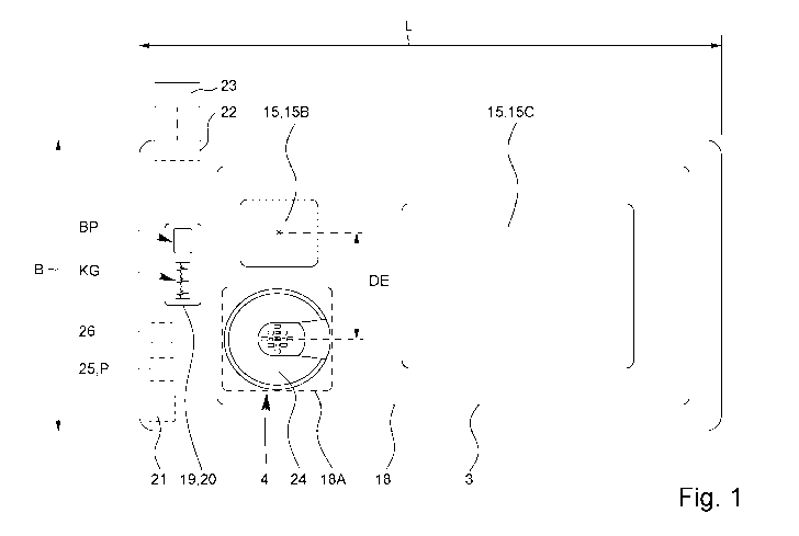
Fig. 1 shows a schematic top view of an examination apparatus 1.
The examination apparatus 1 is preferably designed for medical examination, in
par-
ticular for determining a blood pressure BP, of an animal T, in particular an
animal T
having a paw 2, preferably an animal T from the subfamily of the Felinae,
particularly
preferably a domestic cat.
In principle, however, the examination apparatus 1 is suitable for the medical
exam-
ination of any animal T, in particular humans, in particular those in which a
blood
pressure BP can be determined. For examination using the examination apparatus
1,
it is particularly advantageous if the animal T has a paw or the like.
However, the examination apparatus 1 may also be designed and/or suitable for
the
medical examination, in particular for the determination of blood pressure BP,
of
other animals T, in particular domestic animals, such as dogs, mice, rats,
rabbits,
guinea pigs or the like and/or specially adapted for the examination of these
ani-
mals T.
The blood pressure BP can be a systolic, diastolic and/or mean blood pressure
BP.
In particular, it has been surprisingly shown in the context of the present
invention
that the proposed method and/or examination apparatus can also be used for the
determination of a diastolic blood pressure BP. This is, however, not
mandatory.
In Fig. 2, an examination apparatus 1 according to the invention is shown in a
sche-
matic perspective view with an animal T arranged on it.
Preferably, the examination apparatus 1 is designed as a support for at least
one
paw 2 or any other part of the body, in particular a part similar to a paw,
for example
a hand or a finger, of the animal T.
CA 03154680 2022-03-15
WO 2021/074297 PCT/EP2020/079045
Particularly preferably, the examination apparatus 1 and/or support is
designed in
such a way that the animal T to be examined can be completely placed and/or
posi-
tioned on the examination apparatus 1 and/or support, in particular thus all
legs of
the animal T can be positioned on the examination apparatus 1. However, this
is not
5 mandatory. In principle, it is also possible that the examination
apparatus 1 is de-
signed so that only one or two paws 2 can be placed or positioned on the
examination
apparatus 1.
The examination apparatus 1 is preferably designed as mat or plate or mat-like
or
10 plate-like or in the form of a mat or plate. In particular, a plate or
mat is understood
to be a device whose width and length exceed the height by a multiple. A plate
is
preferably understood to be an at least substantially rigid apparatus. A mat
is prefer-
ably understood to be an at least partially flexible apparatus. For example,
if the ex-
amination apparatus 1 is designed as a mat, it may be at least partially
rollable and/or
15 foldable.
Preferably, the examination apparatus 1 has a rest surface 3. The animal T, in
par-
ticular a domestic dog, a domestic cat or another animal T of comparable or
smaller
size, can be, preferably completely, placed on the rest surface 3.
Preferably, the examination apparatus 1 and/or rest surface 3 is at least
essentially
flat and/or planar.
Preferably, the examination apparatus 1 has the rest surface 3 on one upper
side
and/or the rest surface 3 is formed by an upper side of the examination
apparatus 1
or a part thereof.
The rest surface 3 is or forms in its position of use, in particular during
the examina-
tion, preferably an at least substantially horizontal surface. The position of
use is a
preferred position of the examination apparatus 1, in which the animal T can
be
placed on the examination apparatus 1 for examination. The position of use is
in
particular shown in Fig. 2.
The examination apparatus 1 and/or rest surface 3 preferably has a width B of
more
than 20 cm, preferably more than 40 cm, and/or less than 80 cm, preferably
less than
60 cm.
CA 03154680 2022-03-15
WO 2021/074297 PCT/EP2020/079045
21
The examination apparatus 1 and/or rest surface 3 preferably has a length L of
more
than 40 cm, preferably more than 60 cm, and/or less than 120 cm, preferably
less
than 80 cm. In principle, a different width B and/or a different length L of
the exami-
nation apparatus 1 and/or rest surface 3 are also conceivable.
11 is preferably intended that during the examination the examination
apparatus 1
contacts the paw 2 and/or the body part only on one side, and/or rests or is
arranged
only on one side. The examination apparatus 1 is therefore preferably designed
for
one-sided contact with the animal T and/or its paw 2.
The examination apparatus 1 is preferably free of fixing means and/or
fastening
means. Preferably, the examination apparatus 1 is not designed to clasp the
paw 2.
Preferably, the examination apparatus 1 does neither have a clip for
attachment to
the paw 2 nor a cuff for application to the paw 2 or other fixing means or
fastening
means for attaching, fixing or fastening an examination means such as a sensor
or
an electrode to the animal T. In contrast, it is preferred that the
examination appa-
ratus 1 has a contact and rest surface 3, by which the examination is made
possible
when the paw 2 or body part is put on or placed on the device.
The design of the examination apparatus 1 as a support and/or with a rest
surface 3
for the animal T makes the examination particularly pleasant and thus stress-
free for
the animal T. Preferably, it is not intended that the animal T is fixed to the
examination
apparatus 1 for examination or that a part of the examination apparatus 1,
such as a
sensor or the like, is attached or fixed to the animal T. It has been shown
that such
a method causes stress in an animal T, so that the examination would be
unpleasant
for the animal T and, in addition, the blood pressure BP would be influenced
by the
stress. In contrast, by designing the examination apparatus 1 according to the
inven-
tion, the examination can be made very pleasant and stress-free for the animal
T.
Preferably, the examination apparatus 1 or rest surface 3 is designed in such
a way
that the animal T can move freely on the examination apparatus 1 and/or rest
sur-
face 3.
By the design of the examination apparatus 1 described in more detail below,
in par-
ticular the design and/or arrangement of the sensor device 4 and/or the
electrodes
15, it is accomplished that an examination of the animal T, in particular a
reliable
and/or accurate blood pressure determination, is made possible while avoiding
CA 03154680 2022-03-15
WO 2021/074297 PCT/EP2020/079045
22
fixation of the animal T or can be made without fixation of the animal T
and/or can
be made or is made possible when the animal T moves during the examination by
means of the examination apparatus 1.
The examination apparatus 1 preferably has a sensor device 4. The sensor
device 4
is designed for the optical examination of an arterial blood flow BF of the
animal T,
in particular for recording a curve K that contains information about an
arterial blood
flow BF of the animal T. In particular, the sensor device 4 is designed to
perform a
photoplethysmography and/or to record a photoplethysmogram.
A curve K comprising information about the arterial blood flow BF is shown as
an
example in Fig. 9 and will be explained in more detail later.
The sensor device 4 and/or examination apparatus 1 is preferably designed to
ena-
ble or allow movement of the animal T during the examination and/or to enable
a
reliable and accurate examination, in particular blood pressure determination,
and/or
to reduce, avoid and/or compensate for movement artifacts.
The examination apparatus 1 has the sensor device 4 preferably in the area of
the
rest surface 3. Thus, an examination with the sensor device 4 can be performed
when the paw 2 or the body part is placed on the surface.
The sensor device 4 is preferably arranged at the examination apparatus 1 or
inte-
grated into the examination apparatus 1 in such a way that a paw 2 of the
animal T
can be positioned at, above and/or in the immediate vicinity of the sensor
device 4,
in particular if the animal T is located on the examination apparatus 1 and/or
rest
surface 3. In the example shown in Fig. 1, the sensor device 4 is positioned
in such
a way that the left forepaw 2 of the animal T can be positioned above the
sensor
device 4 without any problems and in a position that is pleasant and/or
natural for
the animal T. However, the sensor device 4 can also be provided at another
position.
Figs. 2 and 7 show, by way of example, the positioning of a paw 2 during an
exami-
nation by means of the sensor device 4. For the examination by means of the
sensor
device 4, the paw 2 is preferably positioned in such a way that one or
preferably
several pads of the paw 2 contact the sensor device 4, in particular a cover
14 and/or
electrode 15.
CA 03154680 2022-03-15
WO 2021/074297 PCT/EP2020/079045
23
The examination apparatus 1 may also have several, in particular two, sensor
de-
vices 4, for example a sensor device 4 for the left forepaw 2 and a sensor
device 4
for the right forepaw 2 of an animal T to be examined. In this case, the
sensor devices
4 are preferably of a similar or identical design. This is in particular shown
in Fig. 2.
The sensor device 4 is preferably designed for a reflective measurement of an
arterial
blood flow BF.
The sensor device 4 has at least one emitter 5 for emitting electromagnetic
radia-
1 0 tion R ¨ in particular light including ultraviolet light and/or
infrared light ¨ and at least
one detector 6 for detecting electromagnetic radiation R, preferably emitted
by the
emitter 6 ¨ in particular light including ultraviolet light and/or infrared
light.
The emitter 5 is preferably designed as a light emitting diode or laser diode.
The detector 6 is preferably designed as a photodiode.
Preferably, the emitters 5 can be activated and/or deactivated and/or switched
on
and/or off separately, in particular by means of MOSFETs assigned to the
emitters 5.
Figs. 3 and 4 show an example of a schematic top view of a sensor device 4 in
different embodiments. The sensor devices 4 according to Fig. 3 and 4 are
basically
the same or similar in design and differ primarily only in the number of
emitters 5 and
detectors 6.
Preferably, the sensor device 4 has several emitters 5 and several detectors
6. In
principle, however, it is also possible that the sensor device 4 has exactly
one emit-
ter 5 and exactly one detector 6 or exactly one emitter 5 and several
detectors 6 or
several emitters 5 and exactly one detector 6.
Preferably, however, the sensor device 4 has at least nine, in the example
shown in
Fig. 1 and 3 exactly nine, emitters 5 and/or at least four, in the example
shown in Fig.
1 and 3 exactly four, detectors 6.
The emitters 5 and detectors 6 are preferably arranged in a common plane.
CA 03154680 2022-03-15
WO 2021/074297 PCT/EP2020/079045
24
The emitters 5 and detectors 6 are preferably arranged in a recurring and/or
repeat-
ing structure. Particularly preferably, the emitters 5 and detectors 6 are
arranged
periodically or in a periodic structure.
Preferably, the emitters 5 and the detectors 6 are arranged in the form of a
matrix or
in a matrix or an array with or in (virtual) columns and rows. Preferably, the
matrix or
array has more than two columns and/or more than two rows.
The emitters 5 and detectors 6 are preferably arranged alternately.
Preferably, the
emitters 5 and detectors 6 form one or more in particular rectilinear rows,
with emit-
ters 5 and detectors 6 alternating in each row. The rows can also be curved
and/or
emulate an organic shape, such as that of a paw 2.
Preferably, ¨ as the case may be with the exception of the emitters 5 and/or
detectors
6, which are the outermost and/or arranged at the edge of the sensor device 4
and/or
rows and/or matrix ¨ the detectors 6 are each (directly) surrounded by several
emit-
ters 5 and/or the emitters 5 are each (directly) surrounded by several
detectors 6.
Particularly preferably, several emitters 5 are assigned to each detector 6 or
vice
versa. This allows preferably the multiple use of emitters 5 and/or detectors
6.
An emitter 5 and detector 6 are in particular assigned to each other if the
emitter 5
and the detector 6 are arranged in such a way that the radiation R emitted by
the
emitter 5, in particular after scattering or reflection in a paw 2, reaches or
can reach
the detector 6. Particularly preferably, those emitters 5 are assigned to a
detector 6
that have the smallest distance D to this detector 6 and/or are (directly)
adjacent to
this detector 6. Analogously, in particular those detectors 6 are assigned to
an emitter
5 that have the smallest distance D to this emitter 5 and/or are (directly)
adjacent to
this emitter 5.
The distance D between an emitter 5 and a detector 6 is understood in
particular as
the distance between a center point or geometric center of the emitter 5 or
its emis-
sion surface and a center point or geometric center of the detector 6 or its
detection
surface. Preferably, the emitters 5 and detectors 6 are formed by components
of
different sizes and/or rectangular components, as also indicated by the
differently
sized rectangles in Figs. 1 to 4, wherein the emitters 5 and detectors 6 are
arranged
CA 03154680 2022-03-15
WO 2021/074297 PCT/EP2020/079045
in such a way that the center points or geometric centers of gravity of these
compo-
nents, indicated by points in Fig. 3, have the same distance D from each
other.
Preferably, the emitters 5 assigned to a detector 6 have the same distance D
to the
5 detector 6. Analogously, this also applies to the detectors 6 that are
assigned to an
emitter 5.
The distance D is preferably more than 2 mm, preferably more than 3 mm, in
partic-
ular more than 4 mm, and/or less than 10 mm, preferably less than 8 mm, in
particular
10 less than 7 mm. The distance D is particularly preferably between 4 mm
and 6 mm.
Preferably, the emitters 5 of the sensor device 4 are of the same design or
kind.
Particularly preferably, the emitters 5 of the sensor device 4 are identical
in construc-
tion and/or designed for emission at the same wavelength or in the same
wavelength
15 range.
Preferably, the detectors 6 of the sensor device 4 are of the same design or
kind.
Particularly preferably, the detectors 6 are identical in construction and/or
designed
for detection at the same radiation R or wavelength, in particular emitted by
the emit-
20 ters 5.
The sensor device 4 is preferably designed for examination with
electromagnetic ra-
diation R in the infrared range. Particularly preferably, the emitters 5 are
designed
for emission of infrared radiation and/or the detectors 6 are designed for
detection of
25 infrared radiation.
Infrared radiation is in particular electromagnetic radiation R with a
wavelength be-
tween 780 nm and 1400 nm.
Preferably, the emitters 5 are designed for the emission of electromagnetic
radia-
tion R with a wavelength of more than 900 nm and/or less than 1200 nm or 1100
nm.
Particularly preferably, the emitters 5 are designed for the emission of
electromag-
netic radiation R with a wavelength of more than 920 nm and/or less than 960
nm, in
particular (approximately) 940 nm. Alternatively or additionally, however, it
is also
possible that the emitters 5 or a subset of the emitters 5 is/are designed to
emit
electromagnetic radiation R with a wavelength of more than 1030 nm and/or less
than 1070 nm, in particular (approximately) 1050 nm.
CA 03154680 2022-03-15
WO 2021/074297 PCT/EP2020/079045
26
The detectors 6 are preferably designed to detect the radiation R emitted by
the
emitters 5.
Preferably, the sensor device 4 has at least one, preferably several, sensors
7. A
sensor 7 has at least one emitter 5 and at least one detector 6 or is formed
hereby.
Particularly preferably, a sensor 7 has exactly one detector 6 and several
emitters 5,
in the example shown in Fig. 3 and Fig. 4 exactly four emitters 5.
Preferably, the emitters 5 of a sensor 7 are arranged symmetrically around the
de-
tector 6 of the sensor 7 and/or the emitters 5 of the sensor 7 have the same
distance
D to the detector 6 of the sensor 7.
In particular, the sensor device 4 has several sensors 7 which are of the same
type
or kind, in particular identical in construction. Particularly preferably, all
sensors 7 of
the sensor device 4 are identical. Here, however, other solutions are also
possible.
In the example shown in Fig. 3, the sensor device 4 has exactly four sensors
7, one
of the four sensors 7 being indicated by the dotted line in Fig. 2. Also in
Fig. 4 some
sensors 7 are indicated by dashed lines.
Preferably, an emitter 5 is assigned to several sensors 7 and/or the emitters
5 each
form a part of several sensors 7 (apart from emitters 5, which are arranged at
the
outermost edge of the sensor device 4). In particular, each emitter 5 is
assigned to
the adjacent detectors 6 in the row or column and/or to the detectors 6 with
the small-
est distance D. In the illustration example, the emitters 5 ¨ apart from the
emitters 5
arranged at the edge ¨ are assigned to four detectors 6 each.
In the embodiment shown, several emitters 5 are assigned to each detector 6,
wherein these emitters 5 ¨ except for the outermost emitters 5 or emitters 5
arranged
at the edge ¨ are, in turn, each assigned to several detectors 6. Hereby,
several
sensors 7, in particular of the same kind or type, are formed, wherein the
emitters 5
¨ except for the outermost emitters 5 or emitters 5 arranged at the edge ¨ are
each
part of several sensors 7. In the example shown in Fig. 3, the emitter 5
arranged in
the center of the sensor device 4 is assigned to each of the four detectors 6.
The
emitters 5 located in Fig. 3 at the very top, very bottom, very left and very
right are
assigned to only one detector 6 each. The remaining four emitters 5 in Fig. 3
are
CA 03154680 2022-03-15
WO 2021/074297 PCT/EP2020/079045
27
assigned to two detectors 6 each. In this way, four sensors 7, in particular
of the
same kind or type, are formed in Fig. 3.
While Fig. 3 shows the basic design of the sensor device 4 or the basic
arrangement
of the emitters 5, detectors 6 and/or sensors 7, the sensor device 4
preferably has a
considerably larger number of emitters 5, detectors 6 and/or sensors 7, as
shown in
Fig. 4 as an example. In this way a large sensor area can be realized, so that
the
exact positioning of a paw 2 for examination and/or blood pressure
determination is
not or less decisive, but a larger area can be examined by means of the sensor
device 4. This makes it possible that the paw 2 of the animal T does not have
to be
fixed, so that the stress during the examination is reduced for the animal T
and a
faster, more accurate, more reliable and for the animal T as pleasant as
possible
examination, in particular blood pressure determination, can be realized.
The sensor device 4 preferably has more than 30, in particular more than 60,
and/or
less than 500, preferably less than 200, more preferred less than 100, in
particular
less than 100, particularly preferably about 80, emitters 5.
Preferably, the sensor device 4 has more than 20, preferably more than 40,
and/or
less than 500, preferably less than 200, in particular less than 100,
particularly pref-
erably about 60, detectors 6.
Preferably, the number of sensors 7 corresponds to the number of detectors 6,
since
preferably a detector 6 with several emitters 5 forms a sensor 7. However, if
an emit-
ter 5 with several detectors 6 forms a sensor 7, the number of sensors 7
preferably
corresponds to the number of emitters 5.
The sensor device 4 and/or matrix of emitters 5 and detectors 6 preferably has
an
area of more than 10 cm2, in particular more than 20 cm2, particularly
preferably more
than 30 cm2, very particularly preferably more than 40 cm2, and/or less than
200 cm2,
preferably less than 150 cm2, more preferably less than 100 cm2, particularly
less
than 80 cm2.
Preferably, an area density of the emitters 5, an area density of the
detectors 6, an
area density of the sensors 7 and/or a common area density of the emitters 5
and
detectors 6 is more than 0.5/cm2, preferably more than 1/cm2, in particular
more than
2/cm2, and/or less than 40/cm2, preferably less than 20/cm2, in particular
less than
CA 03154680 2022-03-15
WO 2021/074297 PCT/EP2020/079045
28
10/cm2. Herein, the number of emitters 5 and/or detectors 6 and/or sensors 7
per
area is in particular denoted as area density.
The number, arrangement, area and/or area density of the sensor device 4, emit-
ters 5, detectors 6 and/or sensors 7 preferably allow a reliable and accurate
exami-
nation, in particular photoplethysmography and/or determination of blood
pressure
BP, to be performed without fixation of the paw 2 of the animal T relative to
an ex-
amination means such as a sensor, so that the animal T can preferably move
freely
relative to the sensor device 4 during the examination. This makes the
examination
particularly pleasant and stress-free for the animal T, which improves the
measuring
accuracy.
The emitters 5 and/or detectors 6 are preferably each divided into several
groups or
preferably form several groups, which are in particular separately from each
other
and/or separately connected.
Preferably, the emitters 5 are divided into two groups and/or the emitters 5
form two
groups.
Preferably, the detectors 6 are divided into five groups and/or the detectors
6 form
five groups.
The emitters 5 within a group and/or the detectors 6 within a group are
preferably
connected or interconnected serially.
Fig. 5 shows a schematic section through the sensor device 4.
Fig. 6 shows the sensor device 4 in a schematic exploded view.
The sensor device 4 preferably has a limiting device 8.
At this point, it should be noted that the limiting device 8 as well as the
associated
features and advantages are in principle realizable independently of the above
de-
scribed design of the sensor device 4. In particular, the limiting device 8
can also be
advantageous for a sensor device 4 with exactly one emitter 5 and exactly one
de-
tector 6. Consequently, the terms "emitter" and "detector" are preferably used
in the
singular in the following. Of course, the explanations also apply to designs
of the
CA 03154680 2022-03-15
WO 2021/074297 PCT/EP2020/079045
29
sensor device 4 with several emitters 5 and/or several detectors 6, in
particular to a
sensor device 4 designed as described above.
The limiting device 8 is preferably designed to determine, define and/or limit
an emis-
sion region 9 of the emitter 5, a detection region 10 of the detector 6, a
sensor region
11 of the sensor 7 and/or a sensing region 12 of the sensor device 4. In
particular,
the limiting device 8 is designed as an aperture for the emitter 5 and/or
detector 6.
For this purpose, the limiting device 8 in the illustration example has a
barrier 13
described in more detail below or is formed hereby. Alternatively or
additionally, how-
ever, the limiting device 8 can also have one or more lenses not shown, in
particular
converging lenses, which lead to a corresponding limitation of an emission
region 9
and/or detection region 10, in particular by focusing radiation R.
The emission region 9 of an emitter 5 is generally the range into which
radiation R
can be emitted by the emitter 5. For example, the emission region 9 of an
emitter 5
can be at least essentially conical and/or defined by one or ¨ in particular
in the case
of a non-conical emission region 9 ¨ several emission angle(s) 9A.
The detection region 10 of a detector 6 is generally the range from which
radiation R
can reach the detector 6 and/or from which radiation R can be detected with
the
detector 6. For example, the detection region 10 of a detector 6 can be at
least es-
sentially conical and/or defined by one or ¨ in particular in the case of a
non-conical
detection region 10¨ several detection angle(s) 10A.
Preferably, the emitter 5 and/or the detector 6 naturally have a certain
emission re-
gion 9 or detection region 10, respectively. Preferably, this natural emission
region 9
and/or detection region 10 is limited or restricted by the limiting device 8
or the limiting
device 8 is designed for this purpose. Therefore, the terms "emission region"
and
"detection region" in the sense of the present invention preferably refer to
the emis-
sion region 9 or detection region 10 defined or limited by the limiting device
8 and
not to the natural emission region 9 or detection region 10 of the emitter 5
or detector
6 per se.
The emission region 9 is indicated in Fig. 5 by the V-shaped dotted lines
starting from
the emitter 5. The dotted lines represent the border of the emission region 9,
which
CA 03154680 2022-03-15
WO 2021/074297 PCT/EP2020/079045
is in particular defined by the limiting device 8. In particular, the emission
region 9 is
the area enclosed or limited by the lines.
The detection region 10 is indicated in Fig. 5 by the V-shaped dotted lines
starting
5 from the detector 6. The dotted lines represent the border of the
detection region 10,
which is in particular defined the limiting device 8. In particular, the
detection re-
gion 10 is the area enclosed or limited by the lines.
The emission region 9 of an emitter 5 is preferably limited by (imaginary)
lines, in
10 particular those shown in Fig. 5 as dash-dotted lines, which represent
the ray path of
the outermost rays of a beam of rays that can leave the sensor device 4
starting from
a center point or geometric center of an emission area of the emitter 5. In
particular,
the lines represent an edge or a border of the emission region 9. In
particular, the
emission region 9 is the region enclosed or limited by the lines.
In case the limiting device 8 is realized by a barrier 13, as shown in Fig. 5,
these
outermost beams are those beams that are not blocked by the limiting device 8
start-
ing from the center point or geometric center, so that the lines representing
these
beams in Fig. 5 touch an edge or corner of the limiting device 8 or barrier
13.
If the limiting device 8 has, or is formed by, a lens as an alternative or in
addition to
the barrier 13, these outermost rays are those rays that pass through the
outermost
edge of the lens from the center point or geometric center of an emission
surface of
the emitter 5.
The detection region 10 of a detector 6 is preferably limited by (imaginary)
lines, in
particular those shown in Fig. 5 as dash-dotted lines, which represent the
optical path
of the outermost rays of a beam of rays that can reach a detection surface of
the
detector 6, in particular a center point or geometric center thereof, from
outside the
sensor device 4. In particular, the lines represent an edge or a border of the
detection
region 10. In particular, the detection region 10 is the region enclosed or
limited by
the lines.
In case the limiting device 8 is realized by a barrier 13, as shown in Fig. 5,
these
outermost rays are those rays that are not blocked by the limiting device 8
and thus
can reach the center point or geometric center of the detection surface of the
detector
CA 03154680 2022-03-15
WO 2021/074297 PCT/EP2020/079045
31
6, so that the lines in Fig. 5 representing these rays touch a rim or edge or
corner of
the limiting device 8 or barrier 13.
If the limiting device 8 has or is formed by a lens as an alternative or in
addition to
the barrier 13, these outermost rays are those rays that can pass through the
outer-
most edge of the lens from outside the sensor device 4 and reach the center
point or
geometric center of the detection surface of the detector 6.
The emission angle 9A is preferably the angle between the (imaginary, in
particular
outside the sensor device 4 running) lines, which represent the borders of the
emis-
sion region 9. This is in particular shown in Fig. 5.
Preferably, the detection angle 10A is the angle between the (imaginary, in
particular
outside the sensor device 4 running) lines that represent the borders of the
detection
region 10. This is in particular shown in Fig. 5.
In the above definition of the emission region 9 and detection region 10, an
idealized
approach was chosen, with reference being made to a center point or geometric
center of an emission area or detection area, which in reality deviates from a
point
shape and forms an ¨ albeit very small ¨ extended area. This makes it possible
that
in reality radiation R from the emitter 5 can also reach an area outside the
emission
region 9 as defined above and/or radiation R from outside the detection region
10 as
defined above can reach the detector 6, in particular scattered light.
However, the
above definitions of emission region 9 and detection region 10 remain
unaffected by
this. Furthermore, the emission region 9 and detection region 10 as defined
above
also represent in reality the region into which the vast majority of the
radiation R
emitted by the emitter 5 is emitted and/or from which radiation R can reach
the de-
tector 6.
The sensor region 11 of a sensor 7 is generally the region that can be
examined or
sensed with the sensor 7. Preferably, only objects located in sensor region 11
can
be examined by means of sensor 7. In particular, the sensor region 11 of a
sensor 7
is the region in which the emission region(s) 9 of the emitter(s) 5 of the
sensor 7 and
the detection region(s) 10 of the detector(s) 6 of the sensor 7 overlap.
In Fig. 5, by way of example, arrows indicate how radiation R can pass from an
emit-
ter 5 to a detector 6. The arrows very schematically show the path of a light
beam
CA 03154680 2022-03-15
WO 2021/074297 PCT/EP2020/079045
32
which is emitted by the emitter 5, reaches a detection region 10 and thus a
region
where the emission region 9 and the detection region 10 overlap, and is
scattered or
reflected there in the direction of detector 6 by an object not shown and in
this way
reaches the detector 6.
In principle, it is possible that, deviating from the idealized view chosen
here, in reality
objects outside the sensor region 11 as defined above are at least partially
detected
or detectable by a sensor 7. On the one hand, this can take place by the fact
that, as
already described above, a small amount of radiation R in reality can also
reach a
region outside the defined emission region 9 and/or radiation R from outside
the de-
fined detection region 10 can also reach the detector 6. On the other hand,
however,
it can also happen, for example in case of multiple scattering in an object,
that an
object or a part of an object is detected with a sensor 7 which is located
outside the
defined sensor region 11.
The sensing region 12 of the sensor device 4 is the range which can be
examined
and/or detected/sensed with the sensor device 4. In particular, the sensing
region 12
comprises the emission regions 9, detection regions 10 and/or sensor regions
11 or
is formed thereby.
Preferably, the sensing region 12 is the total/entirety of the sensor regions
11 of the
sensors 7 of the sensor device 4.
The sensing region 12 can be formed by a continuous/connected region. This is
the
case if the sensor regions 11 of the sensors 7 of the sensor device 4 overlap.
However, it is also possible that the sensing region 12 is not connected or is
formed
by separate or non-connected regions or sensor regions 11. This is the case if
at
least some of the sensor regions 11 of the sensors 7 do not overlap with other
sensor
regions 11.
The sensing region 12 preferably has a border G. The border G is preferably
formed
by the edge or the entirety of the edges of the sensor regions 11. The border
G is in
particular a point or a line where an emission region 9 and a detection region
10
intersect. This is in particular shown in Fig. 5.
CA 03154680 2022-03-15
WO 2021/074297 PCT/EP2020/079045
33
The sensing region 12 and/or its border G preferably has a distance X from the
sen-
sor device 4. In particular, a (minimum) penetration depth of the radiation R
emitted
by the emitters 5 and/or detected by the detectors 6 into the paw 2 during the
exam-
ination can be achieved or ensured. In particular, this minimum penetration
depth or
distance X prevents light reflected or scattered from a surface of the paw 2
from
reaching the detector 6. This improves the accuracy and reliability of the
examina-
tion, in particular the determination of blood pressure.
The distance X is preferably a minimum distance of the sensing region 12 or
its bor-
der G from the sensor device 4. Preferably, the border G of the sensing region
12
does not run straight or parallel to the sensor device 4, as can be seen in
particular
from Fig. 5. In the sectional view as shown in Fig. 5, the border G runs
particularly
zigzag. This is particularly due to the fact that the sensor regions 11 of the
sensors
7 preferably increase (in section) in a V-shape with increasing distance from
the sen-
sor device 4. Consequently, the sensing region 12 preferably has different
distances
from the sensor device 4 at different positions of the sensor device 4,
wherein the
distance X is the smallest of these different distances.
The limiting device 8 is preferably designed such that the distance X of the
border G
of the sensing region 12 from the sensor device 4 is more than 0.5 mm,
preferably
more than 1 mm, and/or less than 10 mm, preferably less than 5 mm, in
particular
less than 3 mm.
The limiting device 8 preferably limits ¨ in particular in the sectional plane
shown in
Fig. 5 ¨ an emission angle 9A of the emitter 5 and/or a detection angle 10A of
the
detector 6 to less than 90 , preferably less than 75 , in particular about 60
. The
sectional plane shown in Fig. 5 is perpendicular to the plane defined by the
matrix of
emitters 5 and detectors 6 and intersects the emitters 5 and detectors 6 along
a row
or column of the matrix.
The limiting device 8 is preferably formed by one or more barriers 13. The
barrier 13
is arranged between an emitter 5 and a detector 6. Preferably, a barrier 13 is
ar-
ranged between each detector 6 and the respective adjacent emitters 5.
The barrier 13 is impermeable to the radiation Remitted by the emitter 5, in
particular
to infrared radiation.
CA 03154680 2022-03-15
WO 2021/074297 PCT/EP2020/079045
34
The barrier 13 is preferably arranged or designed in such a way that the above-
mentioned distance X of the border G of the detection range 8 from the sensor
device
4 is reached or realized.
The dimensions of the limiting device 8 or barrier 13, in particular its
height HB and/or
width BB, as well as the distance DB of the limiting device 8 or barrier 13
from the
emitter 5 and the detector 6 and the distance D of the emitter 5 from the
detector 6
are preferably matched to each other in such a way that the emission region 9
of the
emitter 5 and the detection region 10 of the detector 6 overlap in such a way
that the
above-mentioned distance X of the border G of the sensing region 12 from the
sensor
device 4 and/or the above-mentioned emission angle 9A and/or detection angle
10A
is/are reached or realized.
Preferably, the barrier 13 fulfills several functions and/or has several
sections 13B,
13C, which in particular realize these functions.
A function of the barrier 13 is preferably the shielding of the emitter 5 from
the detec-
tor 6, in particular in such a way that no radiation R emitted by the emitter
5 can reach
the detector 6 directly or without intermediate scattering and/or reflection.
For this
purpose, the barrier 13 preferably has a shielding section 13B. The shielding
section
13B is therefore preferably designed to shield the detector 6 from the emitter
5 or to
prevent direct crosstalk from the emitter 5 to the detector 6. The shielding
section
13B is preferably located between the emitter 5 and the detector 6. The
shielding
section 13B preferably runs at least substantially parallel to a main emission
direction
of the emitter 5 and/or transversely, in particular at least substantially
perpendicular,
to the plane formed by the emitters 5 and detectors 6.
Another function of the barrier 13 is preferably, as already mentioned above,
to limit
the emission region 9, detection region 10, sensor region 11 and/or sensing
region
12. In other words, the barrier 13 and/or a section thereof preferably
represents an
aperture for the emitter 5 and/or the detector 6. For this purpose, the
barrier 13 pref-
erably has an aperture section 13C. The aperture section 13C is preferably
designed
and/or arranged in such a way that the emission region 9 of the emitter 5
and/or the
detection region 10 of the detector 6 is limited or restricted, in particular
in the manner
described above. The aperture section 13C preferably forms an aperture. In
particu-
lar, the aperture section 13C preferably runs transversely, preferably at
least
CA 03154680 2022-03-15
WO 2021/074297 PCT/EP2020/079045
substantially perpendicularly, to the main emission direction of the emitter 5
and/or
at least substantially parallel to the plane formed by the emitters 5 and
detectors 6.
The shielding section 13B and the aperture section 13C are preferably designed
in
5 one piece and/or formed by different sections of the same component. In
particular,
the aperture section 13C can be wider than the shielding section 13B,
resulting in a
T-shaped cross-section of the barrier 13, as shown in Fig. 5. However, this is
not
mandatory.
10 The limiting device 8 and/or barrier 13, in particular the aperture
section 13C, prefer-
ably has a width BB of more than 1 mm, in particular more than 2 mm, and/or
less
than 5 mm, in particular less than 4 mm. Furthermore, the limiting device 8
and/or
barrier 13 preferably has a height HB of more than 1 mm, preferably more than
2
mm, and/or less than 5 mm, in particular less than 4 mm.
Preferably, the barriers 13 form or limit areas 13A that are transparent
and/or trans-
lucent for the radiation R emitted by the emitters 5 and/or detected by the
detectors 6.
These transparent areas 13A are each arranged corresponding to the emitters 5
and
detectors 6, so that they are located in the sensor device 4 above the
emitters 5 and
detectors 6, respectively, and the material located between the transparent
areas
13A or surrounding the transparent areas 13A forms the limiting device 8
and/or the
barriers 13. This is shown as an example in Figs. 5 and 6.
The examination apparatus 1 and/or sensor device 4 preferably has a barrier
ele-
ment 13D. Preferably, the barrier element 13D has or forms the barrier 13 or
barriers
13.
The barrier element 13D is preferably a one-piece, in particular flat and/or
plate-like,
part having the transparent areas 13A.
The transparent areas 13A are preferably formed by through holes of the
barrier
element 13D. In principle, however, it is alternatively or additionally
possible that the
transparent areas 13A are formed by or comprise a material that is transparent
for
the radiation R emitted by the emitters 5 and/or detected by the detectors 6,
for ex-
ample glass, plexiglass or the like.
CA 03154680 2022-03-15
WO 2021/074297 PCT/EP2020/079045
36
The limiting device 8 and/or barriers 13 and/or the barrier element 13D and/or
the
transparent areas 13A preferably form a grid or grating corresponding to the
emitters
and/or detectors 6, in particular a grating aperture.
5 Preferably, the sensor device 4 has a cover 14 which is transparent for
the radia-
tion R emitted by the emitter 5 and/or detected by the detector 6. The cover
14 can
be made of glass, plexiglass, a transparent plastic or the like.
Preferably, the cover 14 covers the sensor device 4 completely, continuously
and/or
gaplessly.
The cover 14 is preferably designed to protect the sensor device 4 and/or the
emit-
ters 5 and/or detectors 6 from soiling and/or damage. The cover 14 preferably
forms
or has an at least substantially flat and/or even, in particular smooth,
surface to sup-
port the paw 2.
Particularly preferably, the distance X of the border G of the sensing region
12 from
the sensor device 4 is or corresponds to the distance of the border G of the
detection
zone 12 from the cover 14, in particular the distance from the side of the
cover 14
facing away from the emitter 5 and/or detector 6.
Preferably, the examination apparatus 1 has one or more detection elements for
de-
tecting activity of the heart of the animal T, in particular for recording a
cardiogram
KG.
The cardiogram KG preferably represents an activity of the heart, in
particular of the
animal T to be examined by means of the examination apparatus 1, and/or
comprises
information about the activity of the heart.
Fig. 9 shows an example of a cardiogram KG.
In particular, the heartbeats or the times at which the heartbeats can be read
or de-
rived or determined from the cardiogram KG.
The cardiogram KG is preferably an electrocardiogram. In principle, however,
the
cardiogram KG can also be an impedance cardiogram, a phonocardiogram, a ballis-
tocardiogram or the like.
CA 03154680 2022-03-15
WO 2021/074297 PCT/EP2020/079045
37
The detection elements are preferably formed by electrodes 15. In principle,
how-
ever, the detection element(s) can also be formed by or have one or more micro-
phones or other sound sensors or the like.
Preferably, the examination apparatus 1 thus has at least one electrode 15,
prefera-
bly at least two electrodes 15. In the illustration example, the examination
appa-
ratus 1 has three electrodes 15. In principle, however, the examination
apparatus 1
can also have a significantly larger number of electrodes 15.
Preferably, a cardiogram KG can be recorded by means of the electrodes 15
and/or
the electrodes 15 are designed to record a cardiogram KG, in particular
wherein the
cardiogram KG is an electrocardiogram.
The electrodes 15 are preferably flat and/or laminar. In particular, the
electrodes 15
consist of or have an electrically conductive material.
Preferably, at least one of the electrodes 15 is designed as a tissue
electrode. This
is indicated schematically in Fig. 1 by hatching of electrodes 15. Preferably,
all elec-
trodes 15 are designed as fabric electrodes. This has proven to be
particularly ad-
vantageous for the examination of animals T such as cats or dogs, since hereby
the
examination can be made particularly pleasant for the animals T. In
particular, it has
turned out that animals T are easily irritated by metallic and/or shiny
surfaces, which
can be avoided by using tissue electrodes.
The at least two electrodes 15 are denoted below as first electrode 15A and
second
electrode 15B for better differentiation. The electrodes 15A and 15B can be
identical
or have different designs.
Explanations with reference to the first electrode 15A therefore preferably
also apply
to the second electrode 15B and vice versa.
Preferably, the electrodes 15A, 15B are each designed to contact a paw 2 of
the
animal T. Particularly preferably, the first electrode 15A is designed for
contacting
the left forepaw and the second electrode 15B is designed for contacting the
right
forepaw.
CA 03154680 2022-03-15
WO 2021/074297 PCT/EP2020/079045
38
Optionally, the examination apparatus 1 has a third electrode 15C. The third
elec-
trode 15C is preferably designed as reference electrode or collection
electrode. The
third electrode 15C is preferably designed to simultaneously contact several
parts of
the body of the animal T to be examined, in particular several paws 2, in
particular
the two hindpaws of the animal T.
The electrodes 15 are preferably arranged in such a way that when the animal T
is
placed on the examination apparatus 1, in particular in a position natural for
the ani-
mal T, such as a sitting or lying position, one paw 2 of the animal T contacts
one of
the electrodes 15. In this way, the examination can be made particularly
pleasant for
the animal T.
The arrangement, size and design of the electrodes 15 are preferably adapted
to the
anatomy of the animal T to be examined, in particular a domestic cat, so that
the
examination can take place in a natural, preferably pleasant, position for the
animal
T and/or the animal T can move freely relative to the electrodes 15 during the
exam-
ination.
The electrodes 15, in particular the first electrode 15A and the second
electrode 15B,
are preferably arranged at a distance DE of more than 2 cm, in particular more
than
5 cm, and/or less than 25 cm, in particular less than 20 cm, particularly
preferably
less than 15 cm, very particularly preferably about 10 cm.
The distance DE between two electrodes 15 is referred to in particular as the
distance
DE between the center points or geometric centers of the electrodes 15 or
their sur-
face. This is shown schematically in Fig. 1.
The distance DE of the electrodes 15, in particular of the first electrode 15A
from the
second electrode 15B, is preferably fixed and/or not variable.
The (respective) electrode 15A, 15B preferably has an area of more than 10
cm2, in
particular more than 15 cm2, and/or less than 100 cm2, in particular less than
80 cm2,
particularly preferably less than 50 cm2.
The third electrode 15C preferably has an area of more than 50 cm2, in
particular
more than 100 cm2, and/or less than 1000 cm2, preferably less than 500 cm2, in
particular less than 200 cm2.
CA 03154680 2022-03-15
WO 2021/074297 PCT/EP2020/079045
39
The third electrode 15C preferably has a larger area than the first and/or
second
electrode 15A, 15B, in particular more than double or triple, particularly
preferably
more than four times, the area of the first and/or second electrode 15A, 15B.
Preferably, the first electrode 15A is arranged in such a way that, at a paw
2, in
particular the left or right forepaw, a cardiogram KG can be recorded by means
of
the first electrode 15A and, simultaneously, the optical examination can be
per-
formed and/or the curve K, in particular a photoplethysmogram, can be recorded
by
means of the sensor device 4.
Fig. 7 shows by way of example a paw 2 that is positioned in such a way that a
cardiogram KG can be recorded by means of the first electrode 15A and,
simultane-
ously, the optical examination can be performed and/or the curve K can be
recorded
by means of the sensor device 4.
The first electrode 15A is preferably designed as tissue electrode.
A tissue electrode is preferably an electrode that has or is formed by a
tissue. In
particular, in the case of a tissue electrode, a contact surface for contact
with a body
part, in particular the paw 2, has a tissue or is formed hereby. The tissue is
preferably
a conductive tissue, for example a tissue in which conductive threads are
incorpo-
rated and/or a tissue coated with a conductive layer.
The first electrode 15A is preferably arranged on the sensor device 4 and/or
on the
cover 14, particularly preferably on the side of the cover 14 facing away from
the
emitter 5 and detector 6. This is in particular shown in Figs. 5 to 7.
The first electrode 15A is preferably arranged (only) between the emitter 5
and the
detector 6 and/or opposite the barrier 13 in a projection perpendicular to the
cover 14
and/or to the plane formed by the emitters 5 and detectors 6. Alternatively or
addi-
tionally, the electrode 15A is transparent for the radiation R emitted by the
emitter 5.
Hereby, the optical examination of the animal T and/or the paw 2 by means of
the
sensor device 4 is not affected by the first electrode 15A.
The first electrode 15A preferably has areas 16 that are transparent to the
radiation R
emitted by the emitters 5 and/or detected by the detectors 6. These
transparent areas
CA 03154680 2022-03-15
WO 2021/074297 PCT/EP2020/079045
16 are arranged corresponding to the emitters 5 and detectors 6, so that they
are
located (in a projection perpendicular to the plane of the emitters 5 and/or
detectors
6 and/or to the cover 14) above the emitters 5 and detectors 6, respectively.
5 This is in particular shown in Figs. 5 and 6.
The transparent areas 16 of the first electrode 15A are preferably formed by
through
holes of the electrode 15A. In principle, it is alternatively or additionally
possible that
the transparent areas 16 or the entire first electrode 15A are formed by or
comprise
10 a material that is transparent for the radiation R emitted by the
emitters 5 and/or
detected by the detectors 6.
The first electrode 15A and/or the transparent areas 16 preferably form a
grating or
grid corresponding to the emitters 5 and/or detectors 6.
Optionally, the examination apparatus 1 has a positioning aid 24. The
positioning aid
24 is designed to support correct positioning of the animal T or the paw 2 for
exami-
nation. In particular, the positioning aid 24 is designed to indicate or mark
an area for
positioning a paw 2 or several paws 2, in particular the left forepaw and/or
the right
forepaw. The positioning aid 24 is preferably arranged near the sensor device
4
and/or preferably surrounds the sensor device 4. Alternatively or
additionally, the
position of one or more of the electrodes 15 can be indicated by the
positioning aid
24.
The positioning aid 24 is preferably formed by an elevation or recess of the
exami-
nation apparatus 1 and/or rest surface 3. The positioning aid 24 can, for
example, be
funnel-like or have the shape of a funnel.
However, the positioning aid 24 is only optional and not mandatory.
The examination apparatus 1 preferably has a circuit board 17, in particular a
printed
circuit board (PCB).
Preferably, the circuit board 17 carries the sensor device 4 and/or the sensor
de-
vice 4 is located on the circuit board 17.
CA 03154680 2022-03-15
WO 2021/074297 PCT/EP2020/079045
41
Preferably, the circuit board 17 carries the first and/or second electrode
15A, 15B or
the first and/or second electrode 15A, 15B are arranged on the circuit board
17. Op-
tionally, the circuit board 17 carries additionally also the third electrode
15C and/or
the third electrode 15C is also arranged on the circuit board 17.
The circuit board 17 preferably has or forms peripherals and/or electrical
lines re-
quired for the operation of the sensor device 4, in particular the emitters 5
and/or
detectors 6 and/or sensors 7, and/or the electrodes 15A, 15B and/or for the
evalua-
tion of the signals measured by the detectors 6 and/or electrodes 15.
The examination apparatus 1 preferably has a scale 18. The scale 18 is
preferably
an electronic scale 18.
The scale 18 is preferably designed for weighing an animal T positioned or
placed
on the examination apparatus 1.
The examination apparatus 1 and/or scale 18 is preferably designed for a body
fat
measurement, i.e. for determining the body fat percentage of the animal T on
the
scale 18. The body fat measurement or determination of the body fat percentage
is
preferably carried out via a bioimpedance measurement. In particular, two or
more
of the electrodes 15, 15A, 15B, 15C can be used for this purpose.
The examination apparatus 1 preferably has a force sensor 18A. The force
sensor
18A is preferably designed to measure or detect a force, in particular a
weight force,
exerted by the animal T on the examination apparatus 1.
The force sensor 18A can form part of the scale 18 or be integrated into the
scale
18, but can also be provided as an alternative or in addition to the scale 18.
The force sensor 18A can, for example, be designed as a piezo element or
strain
gauge or the like.
The examination apparatus 1 can also have several force sensors 18A, in
particular
of the same kind or type. Preferably, one or more force sensors 18A are
arranged
under the sensor device 4 or the sensor devices 4, under the rest surface 3
and/or
under the electrodes 15 (each) and/or the force sensors 18A are integrated
into the
sensor device(s) 4 and/or rest surface 3 and/or electrodes IS. In particular,
the force
CA 03154680 2022-03-15
WO 2021/074297 PCT/EP2020/079045
42
sensor 18A can be designed by such an arrangement to determine a presence
and/or positioning of the animal T and/or to support such a determination.
The examination apparatus 1 preferably has a display device 19. The display
device
19 is in particular designed for optical display. The display device 19 is
preferably
formed by a display, e.g. an LCD display, an LED display, an OLED display or
the
like.
The display device 19 is preferably designed to display values measured or
deter-
mined by means of the examination apparatus 1, such as a cardiogram KG, a
heart
rate, a blood pressure BP, a weight, a body fat percentage or the like. In
particular,
the display of a blood pressure BP and a cardiogram KG by means of the display
device 19 are shown schematically in Fig. 1.
Alternatively or additionally, the display device 19 can be designed for user
guidance,
e.g. to display instructions for the operation or use of the examination
apparatus 1,
selection menus, error messages, warning messages or the like.
Furthermore, the examination apparatus 1 preferably has an input device 20.
The
input device 20 is preferably designed for making settings and/or adjustments
and/or
for controlling the examination apparatus 1. The input device 20 is preferably
ar-
ranged in the immediate vicinity of the display device 19 and/or integrated
into the
display device 19.
For example, the input device 20 can be formed by one or more keys, buttons,
switches, or the like. However, the display device 19 is particularly
preferably de-
signed as a touch display or touch-sensitive display, so that the display
device 19
has or forms the input device 20 and/or the input device 20 is integrated into
the
display device 19.
The examination apparatus 1 preferably has a power supply device 21. The power
supply device 21 is designed to supply the examination apparatus 1 with
electrical
energy.
Preferably, the power supply device 21 has an energy storage device for
storing
electrical energy, for example an accumulator, a battery or the like. In
particular, the
power supply device 21 is designed for charging the accumulator or battery,
CA 03154680 2022-03-15
WO 2021/074297 PCT/EP2020/079045
43
particularly preferably for inductive charging. For this purpose, the power
supply de-
vice 21 preferably has a corresponding charging device. Alternatively or
additionally,
the power supply device 21 can also have or form a connection for connecting
the
power supply device 21 to an external power supply, e.g. the mains. In
particular, the
connection can comprise or form the charging device or a part thereof.
The examination apparatus 1 preferably has a control device 25 for controlling
the
examination apparatus 1 and/or the examination. The control device 25 is
preferably
formed by a processor P and/or preferably has a processor P. The processor P
is
preferably a microprocessor. The control device 25 and/or the processor P
is/are
preferably designed to control the sensor device 4, in particular the emitters
5, de-
tectors 6 and/or sensors 7, to control the electrodes 15 and/or to control the
scale 18.
Accordingly, the control device 25 is preferably coupled with the sensor
device 4, the
emitters 5, the detectors 6, the sensors 7, the electrodes 15, the scale 18
and/or the
force sensor 18A.
Furthermore, the power supply device 21 is preferably designed to supply power
to
the control device 25. In particular, the control device 25 is coupled to the
power
supply device 21.
The control device 25 is preferably designed to control the display device 19
and/or
coupled to the display device 19. Preferably, the control device 25 is coupled
to the
input device 20 and/or can be operated by means of the input device 20.
The control device 25 is preferably designed for processing and/or forwarding
the
signals measured by the sensor device 4 and/or the electrodes 15.
The examination apparatus 1 preferably has a memory and/or a storage medium 26
for data storage. Preferably, the storage medium 26 is coupled with the
control de-
vice 25. In particular, the storage medium 26 is designed for at least
temporary stor-
age of signals measured by the sensor device 4 and/or the electrodes 15.
The storage medium 26 can have several separate components and/or be formed
hereby.
CA 03154680 2022-03-15
WO 2021/074297 PCT/EP2020/079045
44
Preferably, the storage medium 26 has one or more permanently installed memory
modules and/or storage elements, for example a hard disk (HDD), a solid-state
drive
(SSD), a RAM module and/or a flash memory or the like.
Alternatively or additionally, the storage medium 26 may have or be formed by
one
or more storage elements that are separate from and/or connectable to the
exami-
nation apparatus 1, such as a USB stick or the like.
In principle, the storage medium 26 may be formed by or comprise one or more
ar-
bitrary storage devices for storing electronic data, such as CD-ROMs, hard
disks,
USB sticks, flash memory, cloud memory, external databases or other computer
equipment separate from the examination apparatus 1 or external thereto and/or
mo-
bile end devices with an integrated memory, such as PCs, data centers,
supercom-
puters, cloud computers, servers, cell phones, smart phones, tablets, laptops
or the
like.
The examination apparatus 1 is preferably designed for the analysis and/or
evalua-
tion of the signals measured with the electrodes 15, the sensor device 4
and/or the
scale 18. The evaluation of the signals is preferably performed by means of
the con-
trol device 25 and/or the processor P and/or is controlled hereby, in
particular by
using the storage medium 26.
The examination apparatus 1 preferably has an interface device 22 for
connecting
the examination apparatus 1 with one or more external devices 23. The
interface
device 22 may have several, in particular different, interfaces. The
interfaces can be
wired or wireless interfaces. For example, the interface device can have one
or more
serial interfaces, one or more USB interfaces, one or more HDMI interfaces
and/or
some or more other interfaces, which are in particular designed for (in
particular
wired) data exchange between the external device 23 and the examination appa-
ratus I. Alternatively or additionally, the interface device 22 may also have
one or
more wireless interfaces, such as WiFi interfaces, Bluetooth interfaces, in
particular
Bluetooth Low Energy Interfaces (BLE interfaces), NFC interfaces or the like.
In other words, the examination apparatus 1 is preferably designed for data ex-
change with an external device 23, in particular by means of the interface
device 22.
CA 03154680 2022-03-15
WO 2021/074297 PCT/EP2020/079045
The examination apparatus 1 is preferably designed to transmit the data or
signals
measured with the sensor device 4 and/or the electrodes 15 and/or the results
or
evaluations determined on the basis of these data or signals to the external
device
23, in particular by means of the interface device 22.
5
The external device 23 is preferably a device that is separate, in particular
physically
separate, from the examination apparatus 1.
The external device 23 may be designed to control the examination apparatus 1
10 and/or to record and/or evaluate and/or analyze and/or display or
otherwise output
signals and/or data measured by the examination apparatus 1 and/or results
trans-
mitted by the examination apparatus 1. Preferably, the external device 23 is
designed
to display a cardiogram KG and/or a blood pressure BP, as shown schematically
in
Fig. 8.
The external device 23 is preferably designed as a mobile end device, for
example
a smartphone, tablet or laptop, and/or as a PC, server, computer network,
cloud,
Internet portal, app and/or other computer device.
Alternatively or additionally, the external device 23 is designed as a storage
me-
dium 26 such as a memory stick. In particular, the external device 23 can form
or
have the storage medium 26 or a part thereof.
Preferably, the examination apparatus 1 has the external device 23 or the
external
device 23 forms a part of the examination apparatus 1 or the external device
23 is
assigned to the examination apparatus 1.
Preferably, an evaluation of the signals measured by the examination apparatus
1,
in particular by the sensor device 4 and/or the electrodes 15, 15A, 15B, 15C,
is per-
formed in or by the examination apparatus 1 itself. Alternatively or
additionally, the
evaluation or parts thereof can also take place outside the examination
apparatus 1
and/or by means of the external device 23.
In Fig. 8, a wiring of the electrodes 15 as well as a processing of the
signals meas-
ured by the sensor device(s) 4 and the electrodes 15 are shown in a schematic,
block
diagram-like representation.
CA 03154680 2022-03-15
WO 2021/074297 PCT/EP2020/079045
46
The examination apparatus 1 preferably has a preprocessing device 27. The
prepro-
cessing device 27 preferably has or is formed by an amplifier, in particular a
differ-
ential amplifier. The differential amplifier is particularly preferably formed
by an oper-
ational amplifier or has such an amplifier. However, other solutions are also
possible.
The preprocessing device 27 is preferably coupled or connected to the
electrodes
and is in particular designed for preprocessing the signals measured by the
elec-
trodes 15, 15A, 15B, 15C. In particular, the preprocessing device 27 is
designed to
amplify the difference between signals measured with different electrodes 15,
in par-
10 ticular voltages such as biopotentials, particularly preferably to
amplify the difference
between the signal measured with the first electrode 15A and the signal
measured
with the second electrode 15B.
Optionally, the electrodes 15 are coupled to the preprocessing device 27 via a
ca-
15 pacitance or a capacitor. This is indicated in Fig. 8 by the capacitance
symbols in
dotted boxes.
Furthermore, the preprocessing device 27 is preferably designed for filtering
the sig-
nals measured by the electrodes IS.
Preferably, but only optionally, the preprocessing device 27 has a common mode
suppression device 28.
The common mode suppression device 28 is preferably designed to suppress or
filter
out a DC current component or DC voltage component of the signals measured by
the various electrodes IS.
The examination apparatus 1 preferably has an ND converter 29. The ND con-
verter 29 is preferably designed to convert an, in particular an analog,
signal, prepro-
cessed by the electrodes 15 and possibly by the preprocessing device 27, into
a
digital signal. The ND converter 29 is preferably downstream of the
preprocessing
device 27.
The signal measured with the electrodes IS, in particular the cardiogram KG
rec-
orded with electrodes IS, is preferably further evaluated and/or processed, in
partic-
ular after conversion into a digital signal. In particular, a usefulness check
can be
performed, e.g., by a check device 29A. During the usefulness check, it is
preferably
CA 03154680 2022-03-15
WO 2021/074297 PCT/EP2020/079045
47
determined whether the cardiogram KG is useful, i.e. whether it can be
meaningfully
evaluated and/or contains useful information. This is shown schematically in
Fig. 8
by the box in the lower right corner.
Preferably, the examination apparatus 1, as an alternative or in addition to
the pre-
processing device 27, has one or more further preprocessing devices 30. The
pre-
processing device 30 is preferably designed for the preprocessing of signals S
meas-
ured by the sensor device 4 or detectors 6 and/or sensors 7.
The preprocessing device 30 preferably has an amplifier 31. The amplifier 31
is pref-
erably designed to amplify a signal S measured by a detector 6 or sensor 7. In
par-
ticular, the amplifier 31 is a transimpedance amplifier and/or converts a
current into
a voltage.
Preferably, the preprocessing device 30 has a filter device 32 for filtering
the signal S,
which is in particular amplified by the amplifier 31.
The filter device 32 preferably has several different electrical filters. In
particular, the
filter device 32 may have or form one or more passive filters and/or one or
more
active filters. The filter device 32 may, for example, comprise or form one or
more
bandpass filters, bandstop filters, high-pass filters and/or low-pass filters.
Preferably, each detector 6 or sensor 7 is assigned a preprocessing device 30
or
each detector 6 or sensor 7 has a preprocessing device 30.
Preferably, an evaluation of the signals S measured by the sensor device 4 and
pref-
erably preprocessed by the preprocessing device 30, in particular the curves
K, is
performed together with the cardiogram KG and/or under consideration of the
cardi-
ogram KG.
The result of the evaluation can then, for example, be forwarded to an
external device
23, as already described above and schematically indicated in Fig. 8.
The examination apparatus 1 is preferably designed to perform the method de-
scribed below. Alternatively or additionally, the examination apparatus 1 can
be used
to perform the method described below. This use can also be realized
independently
of further aspects of the present invention.
CA 03154680 2022-03-15
WO 2021/074297 PCT/EP2020/079045
48
The following describes in particular a method according to the invention.
The method is preferably performed using the examination apparatus 1 described
above. The above-described examination apparatus 1 is particularly
advantageous
for carrying out the method, in particular for selecting one or more sensors 7
and/or
evaluating one or more curves K. However, the method can also be carried out
in-
dependently of the described examination apparatus 1 and can preferably also
be
carried out with an examination apparatus 1 that is designed differently from
the one
described above.
The examination apparatus 1 is preferably designed to perform the method de-
scribed below. Alternatively or additionally, the examination apparatus 1 can
be used
to perform the method described below. This use can also be realized
independently
of further aspects of the present invention.
In particular, the examination apparatus 1 has means to perform the steps of
the
method. These means preferably comprise or are formed by a computer program.
The means and/or the computer program preferably comprise instructions which,
when executed, cause the examination apparatus 1 to perform the described
method.
According to another aspect, the computer program and/or the instructions are
stored on computer-readable storage medium 26 or the computer-readable storage
medium 26 comprises the computer program and/or instructions.
For medical examination, in particular blood pressure determination, by means
of the
examination apparatus 1, it is preferably intended that the animal T, in
particular a
domestic cat or a domestic dog, is placed on the examination apparatus 1. In
partic-
ular, the animal T is placed completely on the examination apparatus 1, i.e.
prefera-
bly in such a way that all limbs, in particular paws 2, are on the examination
appa-
ratus 1 and/or the entire weight of the animal T is carried by the examination
appa-
ratus I.
Particularly preferably, the animal T is positioned on the examination
apparatus 1 in
such a way that a paw 2, in particular a forepaw, of the animal T rests on the
sensor
CA 03154680 2022-03-15
WO 2021/074297 PCT/EP2020/079045
49
device 4 and/or is positioned directly above the sensor device 4 and/or a
curve K
comprising information about the arterial blood flow BF can be recorded on the
paw 2.
Preferably, the animal T is positioned in such a way that each of the
electrodes 15,
15A, 15B, 15C contacts a body part, in particular a paw 2, of the animal T, so
that a
cardiogram KG can be recorded by means of the electrodes 15. In particular,
the
animal T is positioned so that one of the forepaws contacts the first
electrode 15A,
the other forepaw contacts the second electrode 15B and, if the examination
appa-
ratus 1 has a third electrode 15C, one or both hindpaws contact the third
electrode
15C.
After positioning the animal T, the medical examination and/or blood pressure
deter-
mination is preferably started. Optionally it can be provided that after the
positioning
of the animal T first of all it is shortly awaited, so that the animal T can
calm down
and only after a waiting period the medical examination and/or blood pressure
deter-
mination is begun. In particular, a curve K is recorded for the medical
examination or
blood pressure determination, which comprises information about an arterial
blood
flow BF of the animal T. This curve K is in particular a photoplethysmogram.
In the bottom of Fig. 9, a curve K is shown as an example.
Particularly preferably, a reflection measurement is performed for recording
the curve
K, or the examination apparatus 1 is designed for this purpose. This means in
par-
ticular that the sensor device 4 is only located on one side of the paw 2
and/or has
no components located on opposite sides of the paw 2.
Preferably, the examination or measurement is performed with radiation R in
the in-
frared range.
11 is particularly preferable that a cardiogram KG of animal T is recorded by
means
of the examination apparatus 1, in particular at the same time as the
recording of the
curve K comprising information about the arterial blood flow BF of the animal
T.
In the top of Fig. 9, a cardiogram KG is shown as an example.
CA 03154680 2022-03-15
WO 2021/074297 PCT/EP2020/079045
The examination apparatus 1 may have a processor P which receives and/or pro-
cesses information and/or signals S and/or curves K from the examination appa-
ratus 1, in particular the sensor device 4, the sensors 7, the detectors 6
and/or elec-
trodes 15. Alternatively or additionally, the processor P and/or the
examination ap-
5 paratus 1 has the storage medium 26, which has a computer program
representing
a proposed method, which can be executed with the processor P to carry out the
method. In particular, the computer program is stored on the storage medium
26.
Furthermore, results can be formed with the processor P. These results can be
out-
put, in particular via the display device 19, and/or transmitted, in
particular to the
10 external device 23.
The storage medium 26 can be integrated in the examination apparatus 1 or be
sep-
arate from it, e.g. a storage means, such as a memory stick or an external
database,
a server or the like, that is connectable to the examination apparatus 1 via
an inter-
15 face. The computer program can also be supplied to the examination
apparatus 1
from outside and stored in the examination apparatus 1. Here, however, other
solu-
tions are also possible.
The method, in particular the optical examination, is preferably performed
with at
20 least one sensor 7, preferably several sensors 7. Preferably, each
sensor 7 corre-
sponds to one measuring channel, each sensor 7 corresponds to one measuring
channel and/or one measuring channel is assigned to each sensor 7.
A "measuring channel" in the sense of the present invention is preferably a
transmis-
25 sion path for a signal S measured by a sensor 7, in particular a curve K
measured
by a sensor 7. Since in this sense the terms "measuring channel" and "sensor"
are
inseparably connected with each other, the following does not further
differentiate
between a sensor 7 and a measuring channel. Instead, the terms "measuring chan-
nel" and "sensor" will be used synonymously in the following, wherein the term
"sen-
30 sor" will be used primarily. In particular, the terms "measuring
channel" and "sensor"
are interchangeable.
Several curves K can be recorded simultaneously and/or one after the other via
sev-
eral sensors 7, preferably separately or independently of each other.
Preferably, each sensor 7 has at least one detector 6. Very particularly
preferably,
each sensor 7 has exactly one detector 6. Therefore, by selecting a sensor 7,
a
CA 03154680 2022-03-15
WO 2021/074297 PCT/EP2020/079045
51
detector 6 is also selected and vice versa. In this respect, the terms
"selection of a
sensor" and "selection of a detector" are preferably synonymous and in
particular
interchangeable.
Furthermore, as already described above, each sensor 7 preferably has a sensor
region 11. In other words, preferably each sensor 7 is assigned to a different
meas-
uring location or partial region of the sensing region 12 of the sensor device
4. In
particular, each sensor 7 thus corresponds to a certain measurement location
and/or
sensor region 11 and/or partial region of the sensing region 12. A selection
of a sen-
sor 7 can therefore be understood as a selection of a measurement location
and/or
sensor region 11 and/or partial region of the sensing region 12. The terms
"selection
of a sensor", "selection of a measurement location", "selection of a sensor
region"
and "selection of a partial region of the sensing region" are therefore
preferably syn-
onymous with each other and in particular interchangeable.
Furthermore, one or more curves K is/are preferably recorded with each sensor
7. In
other words, each curve K is assigned to a sensor 7. In particular, each curve
K thus
corresponds to a certain sensor 7. A selection of a curve K can therefore be
under-
stood as a selection of a sensor 7 and/or represents such. Indirectly, a
selection of a
curve K therefore also represents a selection of a measurement location and/or
sen-
sor region 11 and/or partial region of a sensing region 12. The terms
"selection of a
curve", "selection of a measurement location", "selection of a sensor region"
and
"selection of a partial region of the sensing region "are therefore preferably
synony-
mous with each other and in particular interchangeable.
Further, in the examination apparatus 1 described above, the sensors 7 are
prefera-
bly of the same kind or type, so that each sensor 7 performs the same
measurement
in principle and the measurements differ only in that they are measured at
different
locations, resulting in different (simultaneously) measured signals S or
curves K.
Fig. 10 shows a schematic overview of the general sequence of the method.
The method preferably has several steps S1 to S9, which are shown
schematically
in Fig. 10. In the following, at first a rough overview of the steps S1 to S9
is given.
Subsequently, the steps S1 to S9 are described in more detail.
CA 03154680 2022-03-15
WO 2021/074297 PCT/EP2020/079045
52
The method according to the invention does not necessarily include all steps
S1 to
S9. In particular, individual steps S1 to S9 or individual aspects of steps S1
to S9
can be realizable independently of each other or in different combinations.
In the method according to the invention, the animal T is medically examined.
Pref-
erably, a pulse transit time PTT and/or blood pressure BP of the animal T is
deter-
mined in the method according to the invention.
The animal T is preferably placed on the examination apparatus 1 for
examination.
Preferably, the animal T is not fixed on the examination apparatus 1, but can
move
freely, in particular relative to the sensor device 4 and/or the electrodes
15.
In step S1, it is preferably determined whether the animal T is located on the
exami-
nation apparatus 1 and/or positioned on the examination apparatus 1 in such a
way
that the medical examination can be performed by means of the examination appa-
ratus I. However, step S1 is only optional and can be omitted.
In step S2 it is preferably determined whether a paw 2 is positioned on or
above the
sensor device 4 in such a way that the optical examination, in particular
photople-
thysmography, can be performed by means of the sensor device 4. Alternatively
or
additionally, in step S2 it is determined over which of the sensors 7 the paw
2 is
located or by means of which of the sensors 7 the examination can be
performed.
Preferably, only those sensors 7 are selected and/or used over which the paw 2
is
located and/or by means of which the examination can be performed. Step S2 can
also be performed simultaneously with step S1 or replace it. Step S2 is
optional and
can also be omitted.
Preferably, a selection of a sensor 7 or a subset of sensors 7 with which the
exami-
nation is performed takes place in step S3. This is in particular advantageous
if the
sensor device 4 has a plurality or large number of sensors 7. In this way, the
effort
required for measurement and/or evaluation can be reduced considerably, in
partic-
ular by excluding and/or not selecting sensors 7 and/or detectors 6, above
which the
paw 2 is not located, from a measurement or evaluation. Step S3 can also be
per-
formed simultaneously with step S1 and/or step S2. However, step S3 is in
principle
optional and can also be omitted.
CA 03154680 2022-03-15
WO 2021/074297 PCT/EP2020/079045
53
On the other hand, the step S3 or a selection of a sensor 7 or a subset of the
sen-
sors 7 can be advantageous even without the subsequent steps and in particular
can
form an invention without the subsequent steps.
In step S4, a curve K comprising information about an arterial blood flow BF
of the
animal T, in particular a photoplethysmogram, is recorded. It is preferable to
record
a cardiogram KG, in particular at the same time as the curve K is recorded.
It is particularly preferred to record several curves K simultaneously, in
particular at
the same time as the cardiogram KG. Alternatively or additionally, several
curves K
and/or cardiograms KG can be recorded one after the other, in particular with
a tem-
poral distance. In step S4, preferably the quality of the measurements and/or
the
usefulness of the recorded curve K and/or the cardiogram KG is also checked.
In step S5, the curve(s) K comprising information about an arterial blood flow
BF
is/are preferably cut or divided into curve sections KA. This is done in
particular in
such a way that the curve sections KA correspond to heartbeats, particularly
prefer-
ably in such a way that each curve section KA corresponds to exactly one
heartbeat.
Preferably, the curve K is cut into curve sections KA using information from
the car-
diogram KG. However, other solutions are also possible here.
In step S6, preferably a selection of curve sections KA is made for further
evaluation,
in particular for the determination of a curve feature M and/or a blood
pressure BP.
For this purpose, some of the curve sections KA can be discarded in step S6.
The
selection of curve sections KA preferably constitutes a selection of one or
more sen-
sors 7, in particular if only curve sections KA are selected from a single
sensor 7 or
a subset of sensors 7. However, the step S6 is optional and can also be
omitted.
In step S7, an averaging or average determination based on the curve sections
KA
is preferably performed. Preferably, one or more curve mean values KM are
deter-
mined on the basis of the curve sections KA. A bootstrap method is preferably
used
or applied.
In step S8, a curve feature M is preferably determined. For this purpose,
preferably
several curve features M are determined first. In particular, a curve feature
M is de-
termined separately for each sensor 7, each curve section KA and/or each curve
mean value KM. Particularly preferably, in addition to each curve feature M,
an
CA 03154680 2022-03-15
WO 2021/074297 PCT/EP2020/079045
54
assigned measure of dispersion, is determined in each case. Particularly
preferably,
the curve feature M with the lowest measure of dispersion is selected as the
final
result of the curve feature M determined in step S8. This selected curve
feature M
then represents the curve feature M determined in step S8. The determined
curve
feature M can then be output and/or used as the basis for the determination of
the
blood pressure BP.
In step S9, preferably a blood pressure BP is determined, in particular from
the curve
feature M determined in step S8. This is done in particular by means of a
preferably
empirically determined correlation function F.
During one or more of the steps S5, S6, S7 and/or S8, a check can be made, in
particular for the usefulness of a cardiogram KG and/or a curve K.
A check of a cardiogram KG for usefulness is preferably performed shortly
after the
start of the examination or recording of the cardiogram KG, in particular
after a few
seconds, preferably after at most about 5 seconds, particularly preferably
after about
2 seconds.
A check of the curve K for usefulness is preferably performed after checking
the
cardiogram KG for usefulness. It is particularly preferred to check the
usefulness of
the curve K after at least about 5 s and/or at most about 45 s, particularly
preferred
after about 10 s and/or about 30 s. It is particularly preferred that a check
of the
curve K for usefulness is performed several times and/or after two different
time pe-
nods, in particular a first check is performed after about 10 s and a second
check
after about 30 s.
The (first and/or second) check of the curve K for usefulness is preferably
performed
during the recording of the curve K or parallel thereto.
If it is determined that measurements are not useful and/or further
measurements
are required, after these steps S5, S6, S7 and/or S8 a return to step S4 can
be made
and/or a new and/or additional measurement can be made, as indicated by arrows
in Fig. 10.
CA 03154680 2022-03-15
WO 2021/074297 PCT/EP2020/079045
Alternatively or additionally, it is also possible to return to step S3 after
one of the
steps S4, S5, S6, S7 and/or S8 and/or to make a new and/or different selection
of
sensors 7.
5 After a return to step S3 or to step S4, the following steps S4 to S9 or
S5 to S9,
respectively, are preferably run through again completely or partially.
Returning to a previous step and performing one or more steps more than once
and/or again in particular has the consequence that the examination, in
particular the
10 determination of the blood pressure BP, of the animal T can be performed
accurately
and reliably even if the animal T moves during the examination or the paw 2
moves
during the examination. In particular, the repetition of one or more steps
allows cu-
mulative measurements or recordings to be made until a sufficient number of
data or
curves K have been measured or are available. Hereby, measurement errors
and/or
15 movement artifacts can be compensated and a movement of the animal T or
paw 2
during the examination is made possible. Since the animal T can preferably
move
freely during the examination, the examination is very pleasant for the animal
T and
therefore stress-free. This is conducive to an accurate and reliable
examination, in
particular blood pressure determination.
The steps S1 to S9 are described in more detail below.
Step S1
Preferably, in step S1 a presence of the animal T on the examination apparatus
1 is
determined.
The examination apparatus 1 is preferably designed to identify a (potential)
presence
of an animal T, in particular a paw 2, on the examination apparatus 1 or at
the exam-
ination apparatus 1, in particular on the rest surface 3, on at least one of
the elec-
trodes 15 and/or on the sensor device 4.
In principle, different methods can be used and/or different sensors can be
provided
for this purpose. For example, the examination apparatus 1 can have a presence
sensor such as a light barrier, a motion detector or the like (not shown).
However,
the use of one or more components of the examination apparatus 1, in
particular
CA 03154680 2022-03-15
WO 2021/074297 PCT/EP2020/079045
56
sensors including electrodes, which also serve another purpose, is
particularly pre-
ferred.
Very particularly preferably, the sensor device 4 or one or more of the
sensors 7
and/or detectors 6, the force sensor 18A and/or one or more electrodes 15 are
used
to detect the presence of the animal T or paw 2 on the examination apparatus 1
and/or the sensor device 4.
Particularly preferably, the examination apparatus 1 identifies the contact of
the
paw 2 with one or more of the electrodes 15, in particular by measuring an
imped-
ance or a resistance between electrodes 15. The resistance measured with the
elec-
trodes 15 changes in particular depending on whether or not the electrodes 15
are
contacted by a paw 2 of the animal T. In this way, the presence of the animal
T and/or
a correct positioning of the paws 2 on the electrodes 15, in particular a
positioning of
the paws 2 in such a way that a cardiogram KG can be recorded by means of the
electrodes 15, can be identified.
Alternatively or additionally, the force sensor 18A and/or the scale 18 can be
used to
identify the presence of this animal T. In particular, a force or weight
threshold value
can be specified or specifiable for this purpose. In this case, the force or
weight
threshold value is preferably selected in such a way that it is exceeded when
a do-
mestic cat or a domestic dog or any other animal T to be examined is placed on
the
examination apparatus 1. Therefore, exceeding a weight threshold value is an
indi-
cation of the presence of the animal T. Falling below the weight threshold
value is an
indication that no animal T is positioned on the examination apparatus 1
and/or that
the animal T is only partially or not positioned on the examination apparatus
1 in the
intended manner.
By means of an appropriate arrangement of the force sensor(s) 18A it is
preferably
also possible to determine by means of the force sensor(s) 18A whether and/or
which
of the electrodes 15 and/or sensor device(s) 4 are contacted by the animal T.
Alternatively or additionally, the sensor device 4 or one or more of the
sensors 7
and/or detectors 6 can be used to identify or determine the presence of the
animal
T. In particular, it can be determined by means of the sensor device 4 whether
a paw
2 or any other part of the body of the animal T is located directly above the
sensor
device 4 and/or whether it is arranged in such a way that the paw 2 and/or the
body
CA 03154680 2022-03-15
WO 2021/074297 PCT/EP2020/079045
57
part can be examined optically by means of the sensor device 4, in particular
whether
a photoplethysmography can be performed. This is preferably done by comparing
the signals S measured by the sensors 7 of sensor device 4.
In this connection, on the one hand, it can be exploited that radiation R
emitted by
one or more of the emitters 5 reaches one of the detectors 6 at least
essentially only
in the presence of an object, i.e. preferably the animal T, in particular by
reflection or
scattering. On the other hand it can be exploited that by a paw 2 positioned
on the
sensor device 4 the ambient light is at least partially shielded and/or only
reaches
some of the sensors 7. Therefore, an information about the presence of the
animal T
can be gathered from the signal S measured by the respective detector 6 or
sensor 7,
in particular without the need for a detailed evaluation of signals S. For
example, it is
sufficient to identify a certain signal level, e.g. by comparing a signal
level with a
threshold value or by comparing it with signals S measured by other sensors 7
or the
like.
The presence detection or presence determination can be performed
continuously,
but for energy efficiency it is preferably performed intermittently.
The result of the presence detection or presence determination is preferably
saved.
The result is preferably a binary information, because it is either that the
animal T is
present or a presence could be determined (positive result) or that the animal
T is
not present or no presence could be determined (negative result). In
particular, the
result or the information is encoded in a signal of one or more of the sensors
7 and/or
detectors 6 and/or the electrodes 15, in particular in a bit, most preferably
the least
significant bit. Such a method is also known as "lead-off detection".
Preferably, the presence detection or presence determination is repeated
and/or
(again) performed automatically, continuously and/or at regular intervals, for
example
at intervals of less than two seconds or less than one second, during
performance of
the examination and/or recording of the curve K and/or the cardiogram KG.
If a (potential) presence of an animal T or its paw 2 is determined on the
examination
apparatus 1, the examination apparatus 1 can be switched on (automatically),
in par-
ticular it can switch from a power saving mode to an operating mode. The
examina-
tion apparatus 1 can therefore support a power saving mode and be designed to
CA 03154680 2022-03-15
WO 2021/074297 PCT/EP2020/079045
58
leave this power saving mode as soon as the presence of the animal T or its
paw 2
is detected.
The determination of the presence of the animal T or the paw 2 and in
particular the
control of the power supply of the examination apparatus 1 thereby is
advantageous,
but in principle, and in particular for further steps of the present
invention, not man-
datory, since ¨ although less convenient ¨ an activation of the examination
appa-
ratus 1 can be effected alternatively or additionally in particular by a
switch or other
operating device of the examination apparatus 1.
Step S2
Preferably, the examination apparatus 1 checks in step S2 whether and/or at
which
position a paw 2 is located on the sensor device 4 or the examination
apparatus 1 is
designed for this purpose, in particular by means of the sensor device 4.
To enable the proposed examination, the paw 2 of the animal T should rest on
or
against the sensor device 4 in such a way that the optical examination ¨ as
described
above ¨ can be performed. Particularly preferably, the paw 2 abuts directly on
the
sensor device 4 and in particular on the cover 14 for this purpose. In this
case, a
reliable optical examination can be performed. Alternatively or additionally,
the paw 2
of the animal T should have direct electrical or galvanic or, if applicable,
capacitive
contact with the electrode(s) 15, so that the recording of the cardiogram KG
can be
performed reliably.
In step S2, it is, preferably automatically, checked whether the paw 2 is
resting on
the sensor device 4 or is in contact with the sensor device 4 in an
appropriate man-
ner, so that the examination, in particular the optical examination and/or the
record-
ing of the cardiogram KG, is made possible.
On the one hand it can be provided that signals S measured by the sensor(s) 7
are
evaluated. This can consist of simply determining whether the signal S or the
signals
S correspond to a light incidence. In this way, a shadowing by the animal T or
the
paw 2 can be determined and thus a position of the paw 2 on the sensor device
4
can be detected.
CA 03154680 2022-03-15
WO 2021/074297 PCT/EP2020/079045
59
Particularly advantageous is the measurement of electromagnetic radiation R
emit-
ted by the emitter(s) 5 by the detector(s) 6. When the emitter(s) 5 is (are)
activated,
it is also possible to determine, by evaluating one or more signals S from the
detec-
tor(s) 6, whether an object and in particular the paw 2 of the animal T is
arranged in
such a way that radiation R emitted by the emitter(s) 5 reaches the
detector(s) 6. In
this case ¨ or depending on the intensity ¨ the presence of the paw 2 on the
sensor
device 4 can be deduced.
The determination of the presence and/or position of the paw 2 above the
sensor
device 4 is preferably done by comparing the signals S measured by the sensors
7
of the sensor device 4.
The comparison of signals S measured with the sensors 7 and/or detectors 6 is
pref-
erably done with activated or switched-on or emitting emitters 5, but can also
be done
with switched-off emitters 5.
By comparing the signals S from different sensors 7 and/or detectors 6 it can
prefer-
ably be determined in which position, in particular relative to the sensor
device 4
and/or the different sensors 7 and/or detectors 6, the paw 2 is located. In
particular,
it can be determined which of the sensors 7 and/or detectors 6 of the sensor
device
4 the paw 2 is located over and thus by which sensors 7 and/or detectors 6 the
ex-
amination, in particular the determination of the blood pressure BP, can be
per-
formed. In particular, the shape and/or positioning of the paw 2 can
preferably be
modelled.
If a paw 2 is located on the sensor device 4, preferably some areas of the
sensor
device 4 and/or some sensors 7 are covered by the paw 2 and other areas and/or
sensors 7 are not covered by the paw 2. In particular, this leads to
differences in the
brightness and/or radiation R measured by the individual sensors 7. For the
exami-
nation by means of the sensor device 4, it is preferably intended that a paw 2
is
positioned over the sensor device 4 in such a way that the sensor 7 or at
least one
sensor 7 is completely covered by the paw 2. In this way, no ambient light can
reach
the sensor 7 or its detector 6, but only radiation R that was emitted by the
emitter 5
or one of the emitters 5 of the sensor 7 and scattered in the paw 2 towards
the de-
tector 6.
CA 03154680 2022-03-15
WO 2021/074297 PCT/EP2020/079045
The comparison of the different sensors 7 and/or the signals S measured with
the
sensors 7 is preferably done by forming differences between the signals S of
different
sensors 7.
5 Alternatively or additionally, a position or presence determination by
means of the
sensor device 4 can be carried out by examining a signal S measured by means
of
the sensor device 4 to see whether it exceeds or falls below a threshold
value, in
particular an absolute signal strength.
10 Preferably, the threshold value represents an absolute brightness. In
this way, it can
in particular be determined whether a paw 2 and/or any other body part of the
ani-
mal T is located above a sensor 7 of the sensor device 4 and/or above which
sen-
sors 7 of the sensor device 4 a paw 2 or any other body part is located.
15 In particular, exceeding the threshold value is an indication that no
part of the body
of the animal T is above the sensor device 4 or the sensor 7 and/or falling
below the
threshold value is an indication that the paw 2 or another part of the body of
the
animal T is located above the sensor device 4 and/or the sensor 7 in such a
way that
the curve K can be recorded.
Alternatively or additionally, it can be provided that the wavelength of the
radiation R
measured by detector 6 or sensor 7 is analyzed. Preferably, the emitters 5 are
de-
signed to emit radiation R of a certain wavelength or in a narrow wavelength
range.
In other words, the emitters 5 preferably have a narrow spectrum. In contrast,
ambi-
ent light, such as sunlight and/or artificially generated light for indoor
lighting, usually
has a wide spectrum, i.e. a plurality of different wavelengths, which are
particularly
outside the wavelength range emitted by the emitter 5. Therefore, by spectral
analy-
sis of the radiation R detected by the detector 6 or sensor 7, it can
preferably be
determined whether the sensor 7 is covered by a paw 2 or ambient light is
measured.
If it is determined that the paw 2 is located only above some sensors 7 of the
sensor
device 4, in particular thus not over all sensors 7 of the sensor device 4,
these sen-
sors 7 can be selected for performing the examination and/or for recording a
curve
K comprising information about the arterial blood flow BF.
For presence and/or position determination by means of the sensor device 4, in
par-
ticular a scan or search run can be performed by means of the sensors 7, in
which
CA 03154680 2022-03-15
WO 2021/074297 PCT/EP2020/079045
61
different sensors 7 and/or emitters 5 are activated or switched on one after
the other.
In particular, the influence of ambient light can be determined hereby and/or
by com-
paring a signal S measured with the emitter 5 switched on with a signal S
measured
with the emitter 5 switched off.
For the determination of the position of the paw 2 above and/or relative to
the sensor
device 4, in particular a center of mass or gravity of the signals S measured
by the
sensors 7 and/or detectors 6 is calculated or determined. The signals S are
prefera-
bly proportional to the intensity of the radiation R measured by the
respective sensor
7 and/or detector 6.
The determination of the center of mass or gravity of the measured signals S
is done
in particular as follows:
First, preferably each emitter 5, detector 6 and/or sensor 7 is assigned a
position,
preferably wherein the position is represented by two coordinates x, y. The
position
of each emitter 5, detector 6 and/or sensor 7 can thus be specified or defined
by a
pair of coordinates (xi, yi), wherein the index i counts the emitters 5,
detectors 6
and/or sensors 7. This is in particular shown in Fig. 4 as well.
The position of the paw 2 or the center of mass or gravity of the signals S is
given by
the coordinate pair (xc,K.) with
1
xc = Sixi
Stot
and
1
Yc =
=Jtot
Here, Si is a value that corresponds to a signal strength Sorio of signal S
measured
at the respective coordinate xi or y1, respectively, or the sum of the signal
strengths
1
Sorio measured at the respective coordinate xi or yi, respectively. The factor
¨ is
stot
a normalization factor and can be omitted if necessary. Preferably, Stot =
Eisi.
The signal strength Song is preferably a value of the signal S measured by the
sensor
7 and/or detector 6, for example a voltage, current or the like, in particular
the DC
value measured by the sensor 7 and/or detector 6.
CA 03154680 2022-03-15
WO 2021/074297 PCT/EP2020/079045
62
A value Si corresponding to the signal strength Sõig,i is preferably a value
that is
directly linked to the signal strength Sõig,i, for example the value of the
signal
strength Sõig,i itself (Si = Sõig,i). Particularly preferably, the value Si is
the difference
between the signal strength Sõig,i and the mean value or median Sm of the
signal
strengths Si (Si = ¨ Sm) or the absolute value thereof (Si = 1Sorig,i
After the determination of the position of the paw 2 or the center of mass or
gravity
(x,,y,), one or preferably several sensors 7, emitters 5 and/or detectors 6
are prefer-
ably selected or used for the medical examination, in particular
photoplethysmogra-
phy, on the basis of the determined position or the determined center of mass
gravity.
Preferably, sensors 7, emitters 5 and/or detectors 6 are selected or used
which are
closest to the determined position and/or are located in a certain area around
the
determined position. For example, sensors 7, emitters 5 and/or detectors 6 are
se-
lected which lie in a square, rectangle, (regular) hexagon, (regular) octagon
or the
like around the center of mass or gravity (x,,K.).
It is preferred that during the optical examination and/or during one or more
of the
subsequent steps, in particular during one of the steps S3 and/or S4, it is
checked
whether the position of the paw 2 has changed, in particular during a
measurement
and/or after the initial position determination, and/or the determination of
the position
of paw 2 is repeated. This position check is preferably done automatically,
continu-
ously and/or at regular intervals, preferably at intervals of less than two
seconds or
less than one second.
In order to check whether the position of paw 2 has changed after the initial
position
determination, the signals S measured by the sensors 7, emitters 5 and/or
detectors
6, in particular those selected or used for the examination, or a control
value Snmõ,
determined from these signals S are compared with a reference value Sref.
The reference value Sref is preferably a value that is measured and/or
determined
and during the initial determination of the position of the paw 2 preceding
the check
and is preferably stored.
The control value Snmõ, is preferably determined in the same way as the
reference
value Sref and/or on the basis of signals S that were measured with the same
sensors
7, emitters 5 and/or detectors 6 as those signals S used to determine the
reference
CA 03154680 2022-03-15
WO 2021/074297 PCT/EP2020/079045
63
value Sõf. In other words, the only difference between the control value Snew
and the
reference value Sõf is that they are recorded or determined at different
times, namely
the reference value Sõf at the initial determination of the position of paw 2
or before
the medical examination and the control value Snew after the initial
determination of
the position of paw 2 or during the medical examination, in particular
photoplethys-
mography.
Preferably, the reference value is Sõf a value that is determined on the basis
of
signals S or signal strengths Song measured by the selected sensors 7,
emitters 5
and/or detectors 6. Particularly preferably, the reference value Sõf is the
sum of the
values Si of the (selected) sensors 7, emitters 5 and/or detectors 6 (Sõf =
r1S1) or
the average of the values Si of the (selected) sensors 7, emitters 5 and/or
detectors
6 (Sõf = 1.51), wherein Si is the value explained above, the index i
runs through
the ¨ preferably selected ¨ sensors 7, emitters 5 and/or detectors 6 and n is
the
number of (selected) sensors 7, emitters 5 and/or detectors 6.
When comparing the control value Snew with the reference value Sõf, preferably
a
deviation of the control value Snew from the reference value Sõf, a ratio
between the
control value Snew and the reference value Sõf, a difference between the
control
value Snew and the reference value Sõf or the like is determined or
calculated.
Particularly preferably, the control value Snew is compared with the reference
value
Sõf by determining the quotient Sõw/Sõf between the control value Snew and the
reference value Sõf.
Further preferably, in order to obtain the result of the comparison, it is
checked
whether a value determined in the comparison, e.g. the difference between the
con-
trol value Snew and the reference value Sõf and particularly preferably the
quotient
Snew/Sõf is greater than or equal to a specified or specifiable threshold
value.
The result of the comparison of the control value Snew with the reference
value Sõf
is preferably either that the position of the paw 2 has changed or that the
position of
the paw 2 has not changed.
If the value determined in the comparison, in particular the quotient Sõw/Sõf,
is
greater than or equal to a specified threshold value, the result of the
comparison is
CA 03154680 2022-03-15
WO 2021/074297 PCT/EP2020/079045
64
preferably that the position of the paw 2 has not changed. The threshold value
can
be 0.5, for example.
If the result of the comparison of the control value Snew with the reference
value Sõf
is that the position of the paw 2 has changed (from the position initially
determined),
the position of the paw 2 is preferably determined again, in particular by
means of
the method described above.
The comparison of the control value Snew with the reference value Sõf
preferably
takes place at regular (time) intervals, for example every second, every two
seconds,
every three seconds or the like.
If it is found that the position of paw 2 has been changed and/or the value
determined
in the comparison of reference value Sõf with the control value Snew is
greater than
or equal to the threshold value, the determination of the position of the paw
2, in
particular the search run or scan, is preferably performed again, in
particular auto-
matically.
As an alternative or in addition to the presence and/or position determination
by
means of the sensor device 4, one or more of the electrodes 15 can be used to
detect
the presence of the paw 2 on the sensor device 4. In this case, a measurement
is
preferably performed to determine whether an electrically conductive, in
particular a
direct (galvanic) or capacitive electrical connection, exists between the paw
2 and an
electrode 15 assigned to the sensor device 4 or arranged as part of or on the
sensor
device 4. If an electrical connection exists, this indicates the presence of
the paw 2.
The measures can be combined in a particularly advantageous way. In
particular, a
sufficient presence of the paw 2 is automatically detected if both a contact
with the
electrode(s) 15 and either a shading of one or more detectors 6 or the
identification
of electromagnetic radiation coming from emitter(s) 5 with the detector(s) 6
is regis-
tered.
It is preferred that the presence detection takes place in an energy-saving
manner.
For example, the presence detection of the paw 2 on the sensor device 4 can be
performed in several steps.
CA 03154680 2022-03-15
WO 2021/074297 PCT/EP2020/079045
The measures can build on one another. For example, an ¨ in particular
intermittent
and/or energy-saving ¨ measure can be used first and if a (potential) presence
of the
animal T is detected, this can be verified with one or more of the other
measures.
5 In a first step, the emitters 5 can be deactivated to save power. If then
a shadowing
is detected with a detector 6 and/or an electrical contact is detected with an
elec-
trode 15, it can be verified in a further step by another of the above-
mentioned
measures and/or by activating the emitter(s) 5 that the paw 2 also rests on or
lies
against the sensor device 4 in such a way that the examination can be
performed.
In principle, it is therefore particularly preferable to use several of the
devices pro-
vided by the examination apparatus 1 for performing the examination in
addition for
the determination of the presence of the paw 2 on the sensor device 4.
Preferably, the further steps of the method are only carried out if the
presence of the
paw 2 on or at sensor device 4 has been identified accordingly. Otherwise, it
must
be expected that energy and computing power will be expended without
meaningful
results being expected.
In principle, however, the proposed method can also do without step S2, in
particular
if in certain cases the additional effort of accepting an evaluation of
possibly not cor-
respondingly information-bearing signals is acceptable and/or if, on the basis
of an
evaluation at a later point in time of the proposed method, suitable signals S
or parts
thereof are selected and/or unsuitable ones are discarded.
In principle, step S1 can be supplemented or replaced by step S2. This is
because
the identification or detection of the presence of a paw 2 on the sensor
device 4 is
preferably accompanied by a detection of the animal T on the examination appa-
ratus I. This means that the evaluation of one or more signals S from one or
more
of the detector(s) 6 and/or the use of one or more electrodes 15 of the
examination
apparatus 1 for the determination of an electrical contact with a paw 2 can
also be
used to determine the presence of the animal T on the examination apparatus I.
Step S2, in particular the determination of the position of the paw 2 above
the sensor
device 4 and/or the checking whether the position of the paw 2 has changed,
can
also be performed several times and/or simultaneously with a measurement or re-
cording of a cardiogram KG and/or one or more curves K and/or simultaneously
with
CA 03154680 2022-03-15
WO 2021/074297 PCT/EP2020/079045
66
an evaluation of the measurements or recordings. Particularly preferably, it
is
checked automatically, continuously or regularly and/or at short intervals,
for exam-
ple at intervals of less than two seconds or one second, whether the paw 2 has
been
moved. In particular, the step S2 can thus be performed simultaneously with
one or
more of the steps S4, S5, S6, S7, S8 and/or S9.
In particular, this allows the animal T to be examined to move during the
examination
and/or the paw 2 to be moved during the examination. Measurement errors and/or
movement artifacts caused hereby can be compensated by the position determina-
tion, in particular in connection with a selection of sensors 7 and/or a
discarding of
unusable curves K or curve sections KA. In particular, it is possible that
during and/or
after a movement of the animal T or the paw 2, the examination is sustained or
con-
tinued with one or more other sensors 7 or a different subset of sensors 7
than before
the movement. The fact that the animal T can preferably move freely during the
ex-
amination makes the examination very pleasant and stress-free for the animal
T.
This is conducive to an accurate and reliable examination, in particular blood
pres-
sure determination.
Step S3
Very particularly preferably, detectors 6 and/or sensors 7 are selected. In
particular,
a selection of detectors 6 or a selection of signals S measured with sensors 7
also
represents or constitutes a selection of sensors 7 or vice versa. In
particular, a pre-
selection of sensors 7 is made, so that further steps and in particular an
evaluation
of the signals S measured by the sensors 7 is performed, if also information
and/or
an evaluability for the determination of a curve feature M and/or a blood
pressure BP
is/are to be expected.
A selection of a sensor 7 thus takes place in particular in that with this
sensor 7
measurements are made, in particular signals S and/or curves K are recorded,
and
in particular are fed to a further evaluation. Alternatively or additionally,
a selection
of a sensor 7 can also be made by operating and/or switching-on the emitter(s)
5 of
the sensor 7 and/or by recording the measured signals S.
A non-selection of a sensor 7 takes place in particular in that no signals S
are meas-
ured and/or no curves K are recorded with the sensor 7 and/or that signals S
CA 03154680 2022-03-15
WO 2021/074297 PCT/EP2020/079045
67
measured or curves K recorded with the sensor 7 are not considered in the
further
evaluation. In particular, signals S from unselected sensors 7 are thus
rejected.
The selection of detectors 6 and/or sensors 7 is shown schematically in Fig.
11,
wherein a cross symbolizes that the respective detector 6 or sensor 7 is not
selected
and the hook symbolizes that the respective detector 6 or sensor 7 is
selected. In the
example in step S3, two of the six shown detectors 6 and/or sensors 7 are
selected
and four detectors 6 and/or sensors 7 are not selected.
In this context, it should be taken into account that in order to perform the
optical
examination, in particular the photoplethysmography, a part of the paw 2 that
has at
least one artery A should be arranged in a sensor region 11 of a sensor 7, so
that
the optical examination and in particular the photoplethysmography can be per-
formed. This is shown schematically in Fig. 7.
For the optical examination, in particular for performing a
photoplethysmography, it
is therefore preferred that a body part, in particular the paw 2, is placed on
the sensor
device 4 and/or the cover 14 in such a way or vice versa that an arterial
blood flow
BF is examined by means of the sensor device 4 and/or that the signals S
comprise
information about the arterial blood flow BF. In other words, the body part,
in partic-
ular the paw 2, is in particular placed in such a way that the signals S
comprise
information about the arterial blood flow BF.
For the optical examination, in particular for performing a
photoplethysmography, it
is particularly preferred that a ball/pad of the paw 2 is put on/against the
sensor de-
vice 4 and/or the cover 14. In particular, the hairless areas on the underside
of the
paw 2 are called pads. It has been shown that in the area of the pads the
optical
examination is particularly well feasible. It has also turned out that outside
the pads
the hairs located there make it difficult to perform the optical examination
or photo-
plethysmography.
Accordingly, it is preferably detected or determined which of the sensors 7
and/or
detectors 6 are located below the paw 2, in particular below a pad, and are
therefore
particularly suitable for carrying out the optical examination. These
detectors 6 and/or
sensors 7 or signals S measured with them are preferably selected. In this
way, the
further evaluation can be limited to signals S or sensors 7, respectively,
which po-
tentially lead to curves K that show useful or evaluable information about an
arterial
CA 03154680 2022-03-15
WO 2021/074297 PCT/EP2020/079045
68
blood flow BF and/or on the basis of which a photoplethysmography can be per-
formed and in particular a determination of a pulse transit time PTT and/or a
blood
pressure BP can be performed. This allows the examination to be performed in a
particularly energy-saving manner.
By the selection of the signals S and/or sensors 7, preferably ¨ at least
indirectly ¨ a
selection of parts of the sensor device 4, of subsets of the sensors 7 and/or
detectors
6 and/or of subsets of the sensing region 12 or of sensor regions 11 and/or
detection
regions 10 is performed, so that preferably only information or signals S or
curves K
originating from the selected parts or regions are recorded and/or evaluated
and/or
processed in the further course.
The selection and/or selective evaluation of information and/or signals S is
particu-
larly advantageous because both computing power and thus energy consumption
can be saved and the computing power to be provided can be reduced, thus
saving
resources as a result.
The step S3 can be performed together or simultaneously with the step S2
and/or
the step S1. In particular, measures based on each other can enable on the one
hand the determination of the presence and/or positioning of the animal T on
the
examination apparatus 1 and the presence and/or position of the paw 2 on the
sensor
device 4 and on the other hand, preferably based on each other or
simultaneously
or based on the same signals S, the selection of detectors 6 and/or sensors 7.
In
principle, however, the further steps of the method can also be realized
without such
a selection and/or separately.
The selection of detectors 6 and/or sensors 7 in step S3 is performed in
particular
automatically and/or on the basis of the determination of the position of the
paw 2
performed in step S2, in particular by means of the search run or scan. Thus,
prefer-
ably those detectors 6 and/or sensors 7 are selected in step S3 for which it
was
determined in step S2 that the paw 2 is located above these detectors 6 and/or
sen-
sors 7 or that the paw 2 covers these detectors 6 and/or sensors 7.
The step S3 can also be performed repeatedly, in particular if in step S2 or
through
a new position determination it is found that the position of the paw 2 over
the sensor
device 4 has changed, for example through a movement of the paw 2 during the
measurement and/or examination and/or recording of a curve K. In this case,
CA 03154680 2022-03-15
WO 2021/074297 PCT/EP2020/079045
69
preferably one or more other sensors 7 or a different subset of sensors 7 is
selected
than before. In particular, this allows the animal T to be examined to move
during the
examination or the paw 2 to be moved during the examination. Measurement
errors
and/or movement artifacts caused by this can be compensated for by selecting
the
sensor 7 again and/or by a different selection of sensors 7, in particular in
connection
with determining the position (again) and/or discarding of unusable curves K
or curve
sections KA. In particular, it is possible that during or after a movement of
the ani-
mal T or the paw 2, the examination is sustained or continued with one or more
other
sensors 7 or a different subset of sensors 7 than before the movement. The
fact that
the animal T can preferably move freely during the examination makes the
examina-
tion very pleasant and stress-free for the animal T. This is conducive to an
accurate
and reliable examination, in particular blood pressure determination.
Furthermore, the step S3 or the measures carried out in step S3 can also be
realized
and advantageous independently of the further steps S4 to S9.
Step S4
In the step S4, preferably one or more measurements are performed, in
particular by
means of the sensor device 4. In particular, one or more curves K containing
infor-
mation about an arterial blood flow BF, in particular photoplethysmograms, are
rec-
orded.
This can be done with one or more detectors 6 and/or sensors 7. Accordingly,
the
curve(s) K preferably correspond(s) to the electromagnetic radiation R
detected by
the detector(s) 6, in particular an intensity of this radiation.
The electromagnetic radiation R preferably originates from the emitter(s) 5.
In this
context, the curve K shows preferably comprises the information about the
arterial
blood flow BF by the fact that the detected electromagnetic radiation R varies
with
the arterial blood flow BF, in particular in its intensity.
The radiation R emitted by the emitters 5 is scattered and/or reflected within
the paw
2 during the examination of the paw 2 and can thus reach a detector 6. This is
shown
as an example in Fig. 7. The signal S measured by the detector 6 thus
corresponds
to the scattering, reflection and/or absorption of the radiation R emitted by
the
CA 03154680 2022-03-15
WO 2021/074297 PCT/EP2020/079045
emitters 5 within the paw 2. Here, the scattering, reflection and/or
absorption de-
pends among other things on the volume of the blood in the blood vessels
running
in the paw 2 and/or on the oxygen saturation of the blood.
5 The scattering, reflection and/or absorption and thus the curve K
measured by the
detector 6 and/or sensor 7 are composed of a temporally at least approximately
con-
stant component and a temporally varying component.
The temporally constant the time course of the signal S recorded by a detector
6 or
10 sensor 7is caused in particular by the tissue surrounding the blood
vessels, such as
muscles, nerves, tendons, bones and/or skin, as the scattering and/or
absorption by
this tissue preferably does not change or only changes to a small extent. In
particular,
this temporally at least approximately constant component is not correlated
with the
heartbeat of animal T. The blood flowing through the veins can also contribute
to this
15 at least approximately constant component.
The temporally varying component is preferably caused, at least essentially,
by the
temporal change of the arterial blood flow BF, i.e. the blood flowing through
arteries
A. Arteries A are blood vessels through which the blood is carried away from
the
20 heart. The blood volume or volume flow through the arteries A and the
oxygen satu-
ration of the blood in arteries A change in a way correlated with the
heartbeats. In
particular, the absorption and/or scattering of blood in the arteries A does
not only
depend on the blood volume or blood flow in the arteries A, but also on the
oxygen
content or oxygen saturation of the blood in the arteries A.
In this context, the time course of a coherent and/or continuously recorded
signal S
is denoted as curve K. In a graphical representation of the signal S, as shown
in
Fig. 9, the curve K is the corresponding graph in the diagram.
However, the curve K can also be formed or represented by an equivalent of a
graph
or course of the signal S, in particular a data equivalent. Even if the curve
K is pref-
erably a continuous course, it can be represented or formed by single points
or data
points to be connected in thought, by vector trains or the like. The curve K
can be or
have a digitized analog signal S originating from the detector(s) 6 and/or
sensor(s) 7.
CA 03154680 2022-03-15
WO 2021/074297 PCT/EP2020/079045
71
Particularly preferably, the curve K is a digital signal S in the form of
individual data
points and/or the curve K is converted into individual data points for further
evaluation
after acquisition.
Preferably, a curve K starts with the beginning of the measurement of a signal
S or
the beginning of the recording of a signal S. Preferably, a curve K ends with
the end
or an interruption of the measurement or the recording of the signal S.
A "recording" of a signal S or a curve K is in particular a, preferably
temporary, stor-
age or intermediate storage of the signal S or the curve K. In particular, the
term
"recording" means the measurement and simultaneous storage or intermediate
stor-
age of a signal S or a curve K. The term "recording" therefore also includes a
meas-
urement, in particular a photoplethysmography.
Different curves K can be generated by carrying out different measurements,
each
of which is recorded, or by recording, storing and/or using (continuously)
measured
signals S only partially or section by section.
Preferably, several curves K are recorded simultaneously, in particular by
means of
different sensors 7 and/or detectors 6 of the sensor device 4. Alternatively
or addi-
tionally, several curves K can be recorded one after another with the same
sensor 7
and/or detector 6 and/or several curves K can be recorded one after another
with
different sensors 7 and/or detectors 6.
According to a particularly preferred aspect, several curves K are thus
recorded sim-
ultaneously, in particular with different sensors 7. Here, the different
sensors 7, as
explained above, preferably correspond to different areas of the sensor device
4 or
the paw 2, so that as a result curves K from different areas of the sensor
device 4 or
the paw 2 are recorded. Preferably, curves K are only recorded with the
detectors 6
and/or sensors 7 selected in step S3. However, this is not mandatory.
According to another aspect, several curves K are recorded one after the other
with
one detector 6 and/or sensor 7. However, simultaneously and/or with a time
delay to
the recording of the curve K with this sensor 7, further curves K can be
recorded by
other sensors 7.
CA 03154680 2022-03-15
WO 2021/074297 PCT/EP2020/079045
72
In other words, the sensors 7 are ¨ even if, as described above, some of the
emitters
preferably form a part of several sensors 7 ¨ preferably separate from each
other,
so that with each sensor 7 several curves K can be recorded or are recorded
one
after the other and independently of this, one or more curves K can be
recorded or
5 are recorded simultaneously with the other sensors 7.
However, the sensors 7 are particularly preferably synchronized, so that the
curves K
are recorded simultaneously with the sensors 7.
It is particularly preferred, although not mandatory, that a cardiogram KG, in
particu-
lar an electrocardiogram and/or impedance cardiogram, is recorded
simultaneously
with the curve K or curves K. The cardiogram KG is in particular recorded by
means
of the electrodes 15. In principle, however, the cardiogram KG can also be
recorded
with another detection element, for example a microphone or the like, or be a
phono-
cardiogram.
For recording the cardiogram KG, particularly preferably an electrode 15 is
used,
which contacts the paw 2 at which the optical examination by means of the
sensor
device 4 is performed as well. Preferably, an or the (first) electrode 15A,
which is
assigned to the sensor device 4, is used for this purpose, wherein the (first)
electrode
15A is preferably designed and arranged in such a way that when the paw 2 is
placed
on the sensor device 4, a photoplethysmography is made possible and at the
same
time an electrical coupling of the paw 2 with the electrode 15A takes place.
In the
illustration example, the first electrode 15A is arranged or formed on or in
the imme-
diate vicinity of the sensor device 4.
Preferably, the cardiogram KG is checked for usefulness, in particular
automatically
or in an automated manner. The check for usefulness can be performed during or
after the recording of the cardiogram KG.
For the recording of the cardiogram KG, preferably several electrodes 15 are
used,
of which one electrode 15A can be, but need not be, the electrode 15A assigned
to
the sensor device 4. Furthermore, the examination apparatus 1 has one or more
electrodes 15, so that the animal T or its different paws 2 or other body
parts are
preferably electrically coupled or contacted by different electrodes 15.
CA 03154680 2022-03-15
WO 2021/074297 PCT/EP2020/079045
73
Here, one of the electrodes 15, in particular the third electrode 15C, can
function as
a collecting electrode or reference electrode for one or more other electrodes
15.
Preferably, a unipolar and/or bipolar lead is used, in particular following
the lead sys-
tem according to Frank Norman Wilson of 1934, the lead system according to
Eman-
uel Goldberger of 1942 and/or the lead system according to Willem Einthoven of
1913. However, other approaches are also possible here.
The collection electrode or reference electrode 15C can be used, in order to
com-
pensate for a DC voltage or to set an electric potential, to introduce a
current into the
animal T or to set an electrical voltage. The collecting electrode or
reference elec-
trode 15C is preferably serves for the measurement of an averaged potential or
ref-
erence potential which forms a reference point for the potentials measured
with the
other electrodes 15.
In principle, a single-channel cardiogram KG and/or two electrodes 15 are
sufficient.
The use of at least a third electrode 15 is particularly preferred, allowing
several car-
diograms KG, in particular ECG channels, to be recorded. Furthermore, these
can
be used as alternatives to each other or in combination.
Preferably, the cardiogram KG is preprocessed, in particular with the
preprocessing
device 27. In particular, the cardiogram KG can be filtered, particularly
preferably
bandpass filtered. Here, a frequency range of lower frequencies and a
frequency
range of higher frequencies adjacent to a middle frequency range is
attenuated. Al-
ternatively or additionally, a notch filter and/or bandstop filter can be used
to filter the
cardiogram KG. Here, certain frequencies or frequency bands are attenuated or
sup-
pressed. Especially disturbances from the power grid can be suppressed, for
exam-
ple disturbances with a frequency of 50 Hz.
If the cardiogram KG is not useful, i.e. does not meet the check criteria for
usefulness,
the cardiogram KG is preferably discarded. In particular, if the cardiogram KG
is not
useful, any K curve(s) recorded at the same time as the cardiogram KG will
also be
discarded. Preferably, a further evaluation is performed exclusively on curves
K
and/or cardiograms KG which have not been discarded.
Particularly preferably, if the cardiogram KG is not useful, a new cardiogram
KG and,
preferably, one or more new curves K corresponding to the new cardiogram KG
are
recorded, preferably at the same time as the new cardiogram KG is recorded.
CA 03154680 2022-03-15
WO 2021/074297 PCT/EP2020/079045
74
The check for usefulness is preferably performed on a cardiogram KG or a
section
of a cardiogram KG that has or corresponds to more than 2, preferably more
than 4,
and/or less than 20, preferably less than 15, in particular less than 10, most
prefera-
bly about 6 to 8, heartbeats and/or QRS complexes.
The check for usefulness is hereby or alternatively or additionally preferably
per-
formed on a cardiogram KG or a section of a cardiogram KG whose length is or
corresponds to more than 0.5 s, preferably more than 1 s, and/or less than 10
s, in
particular less than 5 s, particularly preferably less than 3 s. Most
preferably, the
length of the cardiogram KG or the section, respectively, is about 2 s. The
length of
the cardiogram KG or section is in particular the duration of the measurement
of the
cardiogram KG or section.
Preferably, one or more criteria are checked when checking the usefulness of
the
cardiogram KG. The cardiogram KG is preferably useful if all of the criteria
explained
below are met. In principle, however, other methods are also possible in which
only
some of the criteria explained below are checked and/or a cardiogram KG is
also
considered useful if only one or a subset of the criteria are met.
Alternatively or in
addition, other criteria than those described below may also be provided for.
According to a first criterion, preferably a peak-to-peak amplitude of the
cardio-
gram KG is determined. A filtered and/or preprocessed cardiogram KG is
preferably
used for this purpose. The peak-to-peak amplitude is the difference between
the
value of an absolute maximum and the value of an absolute minimum of the
cardio-
gram KG. If the peak-to-peak amplitude is greater than or equal to a specified
or
specifiable threshold value, the criterion is considered fulfilled. Otherwise,
the crite-
rion is considered not fulfilled.
According to a second criterion, preferably a power spectral density or power
distri-
bution of the cardiogram KG is determined. In particular, it is checked
whether the
quotient of an integral over the power density spectrum in a first interval
and an inte-
gral over the power density spectrum in a second interval is greater than or
equal to
a lower threshold value and/or less than or equal to an upper threshold value.
The
criterion is considered fulfilled if the quotient is greater than or equal to
the lower
threshold value and/or less than or equal to the upper threshold value.
Otherwise,
the criterion is considered not fulfilled.
CA 03154680 2022-03-15
WO 2021/074297 PCT/EP2020/079045
According to a third criterion, preferably a skewness and/or kurtosis of an
amplitude
distribution function of the cardiogram KG is examined. The criterion is
considered
fulfilled if the kurtosis and/or skewness is greater than or equal to a
specified or spec-
5 ifiable threshold value. Otherwise, the criterion is considered not
fulfilled.
According to a fourth and fifth criterion, preferably a Pan-Tompkins plot of
the cardi-
ogram KG is examined.
10 The Pan-Tompkins algorithm is an algorithm for the detection of QRS
complexes in
a cardiogram KG, in particular an electrocardiogram. According to the Pan-
Tompkins
algorithm, the cardiogram KG is filtered, derived, squared and then convoluted
and/or integrated. The curve resulting from these steps or the application of
the Pan-
Tompkins algorithm to the cardiogram KG is called Pan-Tompkins plot. The QRS
15 complexes and/or R peaks of the cardiogram KG can be reliably determined
from
the Pan-Tompkins plot.
In the context of the present invention, it has turned out that also the
usefulness of
the cardiogram KG can be checked by means of the Pan-Tompkins plot.
According to the fourth criterion, a minimum and/or mean amplitude of the
peaks of
the Pan-Tompkins plot is examined. The criterion is considered fulfilled if
the mini-
mum and/or mean amplitude of the Pan-Tompkins plot is/are greater than or
equal
to a specified or specifiable threshold value. Otherwise, the criterion is
considered
not fulfilled. Different threshold values may be provided for the minimum
amplitude
and the mean amplitude.
According to the fifth criterion, a minimum, maximum and/or mean distance of
the
peaks of the Pan-Tompkins plot is examined. The criterion is considered
fulfilled if
the minimum, maximum, and/or mean distance of the peaks of the Pan-Tompkins
plot is greater than or equal to a lower threshold value and/or less than or
equal to
an upper threshold value. Otherwise, the criterion is considered not
fulfilled. Different
thresholds may be provided for the minimum distance, the maximum distance, and
the average distance.
According to a sixth criterion, a saturation of the cardiogram KG or the
signals meas-
ured by the electrodes 15 is examined. A saturation of the signal or
cardiogram KG
CA 03154680 2022-03-15
WO 2021/074297 PCT/EP2020/079045
76
is present when the signal takes the maximum or minimum value possible when
the
signal is measured with the electrode 15. Preferably, the sixth criterion is
used to
determine the proportion, in particular the time proportion, of the signal or
cardio-
gram KG measured with the electrode 15 that is saturated. The criterion is
consid-
ered fulfilled if the proportion is less than or equal to a specified or
specifiable thresh-
old value. Otherwise the criterion is considered not fulfilled. For example,
the thresh-
old value can be 0.15 or 15%, so that the criterion is not fulfilled if more
than 15% of
the cardiogram KG is saturated.
As an alternative or in addition to checking the cardiogram KG for usefulness,
the
curve K can be checked for usefulness. This check for the usefulness of the K
curve
is preferably performed after the measurement or recording of the curve K, in
partic-
ular on the basis of individual curve sections KA, and preferably if the
cardiogram KG
fulfills the criteria for its usefulness.
The check for the usefulness of curve K is preferably performed in step S6 and
is
therefore described in more detail below in connection with step S6. In
principle,
however, it is also possible that a (basic) usefulness check of the curve K
also or
additionally forms a part of the step S4 and/or that the usefulness check is
performed
during recording of the curve K. In order to check the usefulness of the curve
K, it is
preferable to assess it with respect to a criterion, for example an expected
basic
shape, an expected spectrum, an expected amplitude or the like.
The check for the usefulness of the curve K is preferably performed after the
meas-
urement or recording of the curve K, in particular on the basis of individual
curve
sections KA, and preferably if the cardiogram KG meets the criteria for its
usefulness.
In principle, it is not mandatory to check the usefulness of the cardiogram KG
and/or
the curve K. However, it has proven to be particularly advantageous for the
exami-
nation of animals T, in particular domestic dogs or domestic cats, because by
this in
a simple and/or fast way useless measurements, i.e. measurements which do not
contain any useful information and/or do not contribute to a reliable result
of an eval-
uation, can be sorted out and/or ignored or remain unconsidered for further
evalua-
tion. In particular, the usefulness check preferably contributes to the fact
that a med-
ical examination, in particular blood pressure determination, of the animal T
can be
performed even if the animal T is not fixed relative to the examination
apparatus 1,
in particular relative to the sensor device 4 and/or the electrodes 15, or
moves or can
CA 03154680 2022-03-15
WO 2021/074297 PCT/EP2020/079045
77
move relative to the examination apparatus 1, in particular relative to the
sensor de-
vice 4 and/or the electrodes 15, during the examination. In particular, by the
useful-
ness check, measurements during which the animal T has moved can be detect and
preferably sorted out or not be taken into account in the further evaluation.
In this
way, the examination can be made particularly pleasant and stress-free for the
ani-
mal T. This is conducive to a reliable and accurate examination, in particular
the
determination of blood pressure BP.
Preferably, the duration of a measurement or recording of a curve K is more
than 30
seconds and/or less than 60 seconds, in particular about 45 seconds. Here,
several
curves K and/or a cardiogram KG are preferably recorded simultaneously.
Particularly preferably, it is determined whether and/or when the animal T, in
partic-
ular the paw 2, is moved during the recording of a curve K and/or cardiogram
KG,
preferably this taking place by means of the checking of the position of the
paw 2
already explained in step S2. Segments of curve(s) K and/or cardiogram KG in
which
the animal T and/or paw 2 was moved are preferably removed or cut out from
curve(s) K and/or cardiogram KG. Segments in which no movement of the animal T
or paw 2 has occurred or been detected and which have a length of less than or
at
most 5 seconds are preferably also cut out of the curve K and/or cardiogram
KG.
The remaining segments of the curves K and/or the cardiogram KG, i.e. the seg-
ments in which no movement of the animal T or paw 2 was detected and/or which
were not removed, are preferably joined together, in particular to form a new
curve
K.
Preferably, the curve K or the curves K KG combined in this way and/or the
cardio-
gram KG combined in this way forms/form the basis for the further evaluation
or
medical examination, in particular determination of the blood pressure. In
other
words, the further steps S5 to S9 are preferably performed with the curve(s) K
and/or
cardiogram KG from which segments have been removed in which the animal T
and/or the paw 2 has been moved.
The removal of segments in which the animal T and/or the paw 2 has been moved
is preferably performed in addition to and/or after the (previously explained)
useful-
ness check of the cardiogram KG.
CA 03154680 2022-03-15
WO 2021/074297 PCT/EP2020/079045
78
It is preferable that the curve K and/or the cardiogram KG combined in this
way have
a length of at least 20 seconds, particularly preferably at least 30 seconds,
and/or
consist exclusively of segments which have a length of more than 3 seconds,
pref-
erably more than 5 seconds. If these requirements are not met after cutting
out the
segments in which movement has occurred, the recording of the curve(s) K
and/or
the cardiogram KG is preferably restarted or repeated.
Furthermore, it is possible that a curve K is composed of segments of several
curves K which were measured or recorded with different detectors 6 and/or
sensors
7, in particular if during a recording of several curves K the position of the
paw 2 was
changed and/or due to such a change of a position of the paw 2 curves K were
rec-
orded with different detectors 6 and/or sensors 7.
It is particularly preferred that during the step S4 or during the recording
of the
curve(s) K and/or the cardiogram KG, the presence determination performed or
de-
scribed in particular in step S1, the position determination described in
particular in
step S2 and/or the position check described in particular in step S2 are
performed.
This is performed in particular automatically, continuously and/or at regular
intervals,
preferably with intervals of less than 2 seconds or less than 1 second. In
particular,
it is possible to determine in this way if the animal T is moved or moves
and/or the
position of one or more paws 2 changes. When the animal T or its paw(s) 2 has
been
found to move, the presence and/or position determination shall preferably be
re-
peated, in particular automatically, and preferably new sensors 7 and/or
detectors 6
shall be selected and with these newly selected sensors 7 and/or detectors 6
the
measurement or recording of the curve(s) K shall be continued or further or
new
curve(s) K shall be recorded, in particular automatically. This is explained
below for
different situations or phases P1 to P7 which may occur during an examination
of the
animal T.
In Fig. 14, various possible phases P1 to P7 during a measurement or recording
of
a cardiogram KG and/or a curve K are shown schematically and by way of
example.
The sequence of the phases P1 to P7 is therefore purely exemplary for
illustration
purposes and does not represent a mandatory sequence of the phases P1 to P7.
Rather, the phases P1 to P7 can occur in any order and the phases P1 to P7 can
occur several times and/or not at all during an examination or recording of
the
curve(s) K and/or the cardiogram KG.
CA 03154680 2022-03-15
WO 2021/074297 PCT/EP2020/079045
79
In the following explanation of the phases P1 to P7 it is assumed that the
examination
apparatus1 has (at least) two electrodes 15, in particular an electrode 15A
for the left
(fore-)paw and an electrode 15B for the right (fore-)paw of the animal T. It
is further
assumed that the examination apparatus 1 has only or exactly one sensor device
4,
wherein the sensor device 4 is assigned to the left (fore-)paw or is
positioned under
the left (fore-)paw of the animal T during the examination. Preferably, the
presence
of the animal T is determined by means of the electrodes 15A, 15B and the
position
of the paw 2, in particular the left forepaw, is determined and checked with
the sensor
device 4 and the curve K is recorded. Of course, other versions of the
examination
apparatus 1 are also possible, in which case the following explanations apply
ac-
cordingly.
Figure 14 shows in four rows R1 to R4 different actions or results of actions
which
are performed during phases the P1 to P7. The abscissa or X-axis of the
diagram in
Figure 14 represents in particular a time axis.
In the row R1, in particular the result of the presence determination, which
is per-
formed in particular in step S1, is shown. During the presence determination,
it is
preferably determined, as described, whether a paw 2 of the animal T, in
particular
the right forepaw, is placed on the assigned electrode 15A, 15B in such a way
that a
cardiogram KG can be recorded. Here, the value "1" means that the presence
deter-
mination was or is successful or that the right forepaw is placed correctly
(positive
result). The value "0" means that the presence could not be determined or that
the
right forepaw is not correctly placed (negative result).
In the row R2, carrying out of a determination of the position of the paw 2,
in particular
the left forepaw, is shown. The position determination is carried out in
particular as
described above in step S2, preferably by carrying out a search run or scan
with the
sensors 7 and/or by determining the center of mass or gravity of the measured
sig-
nals S. Here, the value "1" means that the position determination and/or the
search
run or scan is carried out. The value "0" means that no position determination
or
search run or scan is carried out.
In the row R3, the result of a check of the position of the paw 2, in
particular the left
forepaw, which is preferably performed as explained above in step S2, is
shown. In
particular, as described, it is checked continuously and/or regularly whether
the po-
sition of the left forepaw has changed compared to the position initially or
previously
CA 03154680 2022-03-15
WO 2021/074297 PCT/EP2020/079045
determined. The value "1"means that the position has been successfully
detected or
has not changed from the position determined initially or previously (positive
result).
The value "0" means that no position has been detected or the position has
changed
compared to the initially previously detected position (negative result).
5
In the row R4, the performance of a measurement or examination, in particular
the
recording of a curve K and a cardiogram KG, is shown. The recording of the
curve K
is performed in particular by means of the sensor device 4 on the left forepaw
of the
animal T. The recording of the cardiogram KG is performed by means of the elec-
10 trodes 15A, 15B, wherein one electrode 15A contacts the left forepaw and
one elec-
trode 15B contacts the right forepaw of the animal T. The value "1" means that
a
curve K and a cardiogram KG are being recorded. The value "0" means that no
curve
K and/or no cardiogram KG is being recorded.
15 The phase P1 is in particular a starting phase. In the phase P1, the
animal T is placed
on the examination apparatus 1 for examination. In the phase P1, a presence
deter-
mination of the animal T or paw 2 is preferably performed first. When the
presence
of the animal T has been successfully determined or detected (value in R1
jumps
from 0 to 1), a search run and/or position determination is preferably
performed with
20 the sensors 7 to determine the position of the left forepaw (value in R2
jumps from 0
to 1). When the position of the left forepaw has been determined successfully
and
thus the search run and/or position determination is finished (value in R2
jumps from
1 to 0 and value in R3 jumps from 0 to 1), the measurement begins and/or at
least
one curve K and a cardiogram KG are recorded (value in R4 jumps from 0 to 1).
In phase P2, the position of the left forepaw is changed without removing or
lifting
the left forepaw from the sensor device 4. During this, the right forepaw
remains in
contact with the assigned electrode 15B. The result of the presence detection
during
the entire phase P2 is that the animal's paw 2 is present (value in R1 is 1).
During
the position check, it is determined that the position of the left forepaw has
changed
compared to the originally determined position (value in R3 jumps from 1 to
0). Ac-
cordingly, the recording of the curve K and cardiogram KG is interrupted or
termi-
nated (value in R4 jumps from 1 to 0) and a new position determination is
performed
(value in R2 jumps from 0 to 1). When the (new) position of the left forepaw
has been
determined successfully (value in R2 jumps from 1 to 0 and value in R3 jumps
from
0 to 1), a new recording of the curve K and the cardiogram KG starts or the
recording
of the curve K and the cardiogram KG is continued (value in R4 jumps from 0 to
1).
CA 03154680 2022-03-15
WO 2021/074297 PCT/EP2020/079045
81
In phase P3, the right forepaw is first lifted off the assigned electrode 15B
and then
placed back on the electrode 15B. During this, the position of the left
forepaw is not
changed. Accordingly, the result of the presence detection is negative when or
after
lifting the right forepaw (value in R1 jumps from 1 to 0). Since the position
of the left
forepaw does not change, the result of the position check is positive and no
new
position determination is performed (value in R3 is constant 1 and value R2 is
con-
stant 0). Since the right forepaw was lifted off the electrode 15B, no
cardiogram KG
can be recorded, so that recording of the curve K and the cardiogram KG is
inter-
rupted or terminated (value in R4 jumps from 1 to 0). After the right forepaw
has been
placed back on the electrode 15B, the result of the presence detection is
positive
again (value in R1 jumps from 0 to 1). Accordingly, the recording of the curve
K and
the cardiogram KG is continued (value in R4 jumps from 0 to 1).
In phase P4, the left forepaw is lifted off the sensor device 4 and the
assigned elec-
trode 15A and then placed in the same position on the sensor device 4 and the
as-
signed electrode 15A again. When or after lifting the left forepaw, the result
of the
presence detection is negative (value in R1 jumps from 1 to 0). In addition,
the result
of the position check is negative (value in R3 jumps from 1 to 0).
Accordingly, the
recording of the curve K and the cardiogram KG is interrupted or stopped
(value in
R4 jumps from 1 to 0). When the left forepaw is placed on the assigned
electrode15A
and the sensor device 4 again (in the same position as before), the result of
the
presence detection is positive again (value in R1 jumps from 0 to 1) and the
result of
the position check is also positive (value in R3 jumps from 0 to 3). Since
there have
been no changes compared to the previously determined or last saved position
of
the left forepaw, no new position determination is performed (value in R2 is
constant
0). After the left forepaw 2 has been put on again, a new recording of the
curve K
and the cardiogram KG begins or the recording of the curve K and the
cardiogram
KG is continued (value in R4 jumps from 0 to 1).
In phase P5, the left forepaw is lifted off the electrode 15A and/or the
sensor device
4 and then placed again in a changed position on the sensor device 4 and the
as-
signed electrode 15A. After lifting the left forepaw, the result of the
presence detec-
tion and the position check is negative (value in R1 and R3 jumps from 1 to
0), so
that the recording of the curve K and the cardiogram KG is interrupted or
terminated
(value in R4 jumps from 1 to 0). As soon as the paw is put back on, the result
of the
presence detection is positive (value in R1 jumps from 0 to 1). Since the
position has
CA 03154680 2022-03-15
WO 2021/074297 PCT/EP2020/079045
82
been changed, the result of the position check remains negative at first
(value in R3
is still 0), so the position of the left forepaw is determined again (value in
R2 jumps
from 0 to 1). When the new position of the left forepaw has been successfully
deter-
mined and the position determination has been completed (value in R2 jumps
from
1 10 0 and value in R3 jumps from 0 to 1), a new recording of curve K and
cardiogram
KG begins or recording of curve K and cardiogram KG is continued (value in R4
jumps from 0 to 1).
In phase P6, the animal T or both forepaws are removed from the assigned elec-
trodes 15A, 15B. Accordingly, the result of the presence detection and the
position
check is negative (value in R1 and R3 jumps from 1 to 0) and the recording of
the
curve K and the cardiogram KG is interrupted or terminated (value in R4 jumps
from
1 to 0). The value in R2 is constantly 0, since no renewed presence of the
animal T
is detected.
In phase P7, the forepaws are brought into contact with the assigned
electrodes 15A,
15B, but the left forepaw is placed in such a way that the position cannot be
deter-
mined and/or no meaningful measurement can be made. Accordingly, the result of
the presence detection is positive (value in R1 jumps from 0 to 1). A position
deter-
mination is repeatedly performed, but this does not lead to a successful
result (value
in R2 alternates between 0 and 1, value in R3 is 0). Accordingly, no curve K
and no
cardiogram KG is recorded (value in R4 is 0).
The step S4 or the recording of one or more cardiograms KG and/or curves K
and/or
checking for usefulness can also be carried out several times or several times
in
succession, in particular even after an evaluation or partial evaluation of
curves K
has already been carried out, in particular according to one of the steps S5,
S6, S7,
S8 and/or S9. For example, the evaluation may reveal that there are not enough
useful curve sections KA, so that further curves K must be included. This can
be
caused by a movement of the animal T or the paw 2, for example.
In particular, multiple recording of cardiograms KG and/or curves K or a
repetition of
the step S4 allows the animal T to be moved during the examination and/or the
paw 2
to be moved during the examination. Measurement errors and/or movement
artifacts
caused by this can be compensated by the multiple recording of cardiograms KG
and/or curves K, in particular in connection with the multiple presence
detection
and/or selection of sensors 7 and/or discarding of unusable curves K or curve
CA 03154680 2022-03-15
WO 2021/074297 PCT/EP2020/079045
83
sections KA. In particular, it is possible that during or after a movement of
the ani-
mal T or the paw 2, the examination is sustained or continued with one or more
other
sensors 7 or a different subset of sensors 7 than before the movement. The
fact that
the animal T can preferably move freely during the examination makes the
examina-
tion very pleasant and stress-free for the animal T. This is conducive to an
accurate
and reliable examination, in particular blood pressure determination.
Step S5
In step S5, the curve(s) K is/are preferably cut into curve sections KA, in
particular in
such a way that the curve sections KA correspond to one heartbeat each.
Particularly
preferably, each curve section KA corresponds to exactly one heartbeat.
A cutting or slicing of the curve K in the sense of the present invention is
preferably
to be understood as a division or partitioning of the curve K along the time
axis. The
curve K is thus divided into temporal sections. This can be realized by data
pro-
cessing in such a way that the beginnings and/or ends of curve sections KA are
identified and/or marked. In principle, it is possible to separate the curve
sections KA
from each other. In the further processing, the curve sections KA are also
preferably
treated separately. Against this background, however, the "slicing" or
"cutting" does
not necessarily imply a physical separation of the resulting curve sections KA
from
each other.
As can be seen from the curve K shown in Fig. 9 as an example, which
corresponds
to a photoplethysmogram actually measured on a cat, usually and in particular
in
contrast to a cardiogram KG, a regularity or periodicity, in particular
correlation with
heartbeats, of the curve K cannot be directly seen. Therefore, it is
advantageous to
cut the curve K by means of information from a cardiogram KG and then to
perform
a further evaluation based on individual curve sections KA.
In the following, the cutting or slicing of curves K is explained using the
example of a
single curve K. Preferably, all recorded curves K are cut into curve sections
KA in
the same way.
The cutting of the curve K into curve sections KA is preferably automated or
takes
place in an automated manner.
CA 03154680 2022-03-15
WO 2021/074297 PCT/EP2020/079045
84
Particularly preferably, the curve K is cut into the curve sections KA using
information
from the cardiogram KG recorded at the same time as the curve K. In principle,
how-
ever, other methods are also conceivable here.
The use of the cardiogram KG to slice/cut the curve K into curve sections KA
is par-
ticularly advantageous because the times TH of heartbeats can be determined
par-
ticularly easily and reliably in a cardiogram KG and the curve K can be cut at
or based
on these times TH.
Preferably, the times TH of heartbeats are determined on the basis of the
cardiogram
KG and the curve K at these times TH is cut into curve sections KA.
Preferably, each
curve section KA starts at the time TH of one heartbeat and ends at the time
TH of
the immediately following next heartbeat.
Generally, however, the exact determination of the end of the curve sections
KA is
not decisive, since the curve sections KA serve in particular for an exact or
reliable
determination of the curve feature M. For this purpose, it is of primary
importance to
choose the time TH of a heartbeat as exactly as possible as the beginning of
the
curve section KA and/or to choose the same point relative to the time TH of
the
heartbeat for each curve section KA as exactly as possible.
Preferably, the curve sections KA are of equal length and/or the curve K is
cut into
curve sections KA that each have the same length. Preferably, the length of
the curve
sections KA corresponds to the average heart rate or corresponds to the
duration
between the times TH of two (immediately) consecutive heartbeats at this heart
rate
or corresponds hereto. It has been shown that this simplifies the
determination of the
usefulness or quality of curves K or curve sections KA and that the
determination of
blood pressure BP can be performed with higher accuracy.
The average heart rate is preferably the, in particular arithmetic, mean
and/or median
of the heart rate, in particular wherein the heart rate is determined by means
of the
cardiogram KG. The term "heart rate" means in particular the (average) number
of
heartbeats per unit of time, in particular per minute. For example, if the
average heart
rate is 120 bpm, this corresponds to an (average) duration of a heartbeat of
0.5 s or
to an (average) interval of 0.5 s between two heartbeats.
CA 03154680 2022-03-15
WO 2021/074297 PCT/EP2020/079045
The length L of a curve section KA is preferably determined by the formula L =
dHB =
a, wherein dHB is the average duration of a heartbeat, determined in
particular on the
basis of the average heart rate, and a is a factor which preferably has a
value greater
than or equal to 1. By means of the factor a, the length L of the curve
section KA can
5 also be selected to be greater than the average duration of a heartbeat.
This has
proven to be advantageous for determining the usefulness or quality of curves
K or
curve sections KA and for determining blood pressure BP.
The length of the curve sections KA can thus be or be selected independently
of the
10 concrete! respective duration between the times TH of two consecutive
heartbeats.
Since, as described, the curve sections KA preferably each start at the time
TH of a
heartbeat and are of equal length, it is possible that the curve sections KA
overlap
and/or that segments of the curve K are contained in several curve sections
KA. This
15 is in particular the case if the length of a curve section KA is greater
than the distance
between two adjacent heartbeats.
Particularly preferably, the cardiogram KG is an electrocardiogram. In
particular on
the basis of an electrocardiogram, various characteristic structures, which
can be
20 assigned to or result from different phases of the heart's activity, can
be identified.
For the present method, the so-called QRS complex is particularly relevant.
In Fig. 9, different QRS complexes of a cardiogram KG are marked. One QRS com-
plex preferably represents one heartbeat.
Preferably, the positions of one or more of the QRS complexes of the
cardiogram KG
are used to cut the curve K into curve sections KA. In particular, the QRS
complexes
of the cardiogram KG are used to determine the time TH of heartbeats,
preferably
wherein the curve K is cut into curve sections KA at the times TH determined
by
means of the QRS complexes. In other words, the QRS complexes or parts thereof
are information by means of which cut the curve K is cut into sections KA.
A QRS complex preferably has three peaks, in particular a Q peak, an R peak
and
an S peak.
As Q peak is denoted the first, in particular negative or downward pointing,
deflection
or peak of the QRS complex.
CA 03154680 2022-03-15
WO 2021/074297 PCT/EP2020/079045
86
As R peak is denoted the, in particular negative or downward pointing,
deflection or
peak of the QRS complex which follows the Q peak.
As S peak is denoted the, in particular positive or upward pointing,
deflection or peak
of the QRS complex which follows the R peak.
In particular, the position of the R peak or of the maximum of the R peak can
be used
as time TH of the heartbeat. This is shown by way of example in Fig. 9.
As an alternative to using the R peak as the time TH of the heartbeat, it is
also con-
ceivable to use another structure or another characteristic point of the
cardiogram
KG as the time TH of the heartbeat, for example the Q peak, the S peak, a
midpoint
or inflection point between two peaks, in particular the R peak and the S
peak, or the
like.
The determination of the R peaks or their positions is preferably done by
means of a
Pan-Tompkins plot of the cardiogram KG, in particular as explained in detail
below.
For the determination of the R peaks, preferably all local peaks, in
particular all local
maxima of the Pan-Tompkins plot, are determined first.
As explained above, the cardiogram KG and thus also the Pan-Tompkins plot is
pref-
erably present as a set of discrete data points d,, where d is the value of
the Pan-
Tompkins plot at position i. The index i counts through the data points d, and
prefer-
ably corresponds to a time at which the respective data point d, was measured.
In
particular, a local maximum of the Pan-Tompkins plot is thus represented by a
data
point d, and/or some of the data points d, represent local maxima of the Pan-
Tomp-
kins plot.
A data point d, represents a local maximum in particular if di > d1_1 and di >
d1+1
applies, i.e. if the value of the data point d, is greater than the value of
the neighboring
data points.
From the data points d, that represent local maxima, in a next step preferably
only
those are selected for which no data point with a higher value exists within a
certain
interval around the data point d,. The interval preferably has a width of more
than
CA 03154680 2022-03-15
WO 2021/074297 PCT/EP2020/079045
87
200 ms, in particular more than 300 ms, and/or less than 600 ms, preferably
less
than 500 ms, in particular less than 400 ms. Particularly preferably, the
interval has
a width between 300 and 400 ms, for example about 372 ms.
For the peaks or data points d, of the Pan-Tompkins plot determined or
selected in
this way, the embrasure height or prominence is preferably determined.
Preferably, only those peaks or data points d, are selected or determined as R
peaks
of the Pan-Tompkins plot whose autonomous height or prominence is greater than
or equal to a specified or specifiable threshold value.
The threshold value is preferably an adaptive threshold value. An adaptive
threshold
value in the sense of the present invention is preferably a threshold value
which is
not the same for all data points d, or which is different for different data
points d,. For
example, an individual threshold value t, can be determined for each point of
time i
or each data point d,. Preferably, the adaptive threshold value(s) t, is/are
determined
by determining and/or calculating the convolution between the Pan-Tompkins
plot
and a window function, in particular for each point i. The threshold value t,
is then in
particular the value of the convolution of the Pan-Tompkins plot with the
window
function at position i.
In principle, any window function can be used. Particularly preferably, the
window
function is a Blackman-Nuttall window. Preferably, a window width of 0.6 s
and/or a
gain factor of 3 is used. However, other values are also possible here.
Preferably, the position of the R peaks determined in this way is still
corrected. It is,
namely, possible that the positions of the R peaks in the Pan-Tompkins plot
are
slightly shifted compared with the positions of the R peaks in the original
cardio-
gram KG and/or that shifts of the peaks result from the convolution with the
window
function. The positions of the R peaks determined by the Pan-Tompkins plot may
therefore be "incorrect" or may differ from the positions of the R peaks in
the raw
signal of the cardiogram KG and/or the filtered cardiogram KG.
By correcting the positions of the R peaks, potential shifts caused by
applying the
filters to generate the Pan-Tompkins plot and/or inadvertently using the Q
peak in-
stead of the R peak are prevented. Correcting the positions of the R peaks is
there-
CA 03154680 2022-03-15
WO 2021/074297 PCT/EP2020/079045
88
fore conducive to the accurate determination of heartbeats, thus enabling a
reliable
or accurate examination of the animal T, in particular blood pressure.
The correction of the positions of the R peaks preferably takes place on the
basis of
the filtered and/or preprocessed cardiogram KG, but can also be based on the
un-
processed cardiogram KG or, in other words, on the "raw signal" of the cardio-
gram KG.
Preferably, for the correction of the position of an R peak, starting from the
position
determined by means of the Pan-Tompkins plot, the position of this R peak is
pref-
erably searched for or determined in the cardiogram KG, in particular in the
filtered
and/or preprocessed cardiogram KG. This position in the cardiogram KG, in
particu-
lar in the filtered and/or preprocessed cardiogram KG, is then preferably used
as the
position of the R peak and in particular replaces the position of the R peak
deter-
mined by the Pan-Tompkins plot.
Particularly preferably, the gradient or the slope or the derivative of the
cardiogram
KG at the position of the R peak determined by means of the Pan-Tompkins plot
is
determined in the cardiogram KG, and on this basis the next maximum of the
cardi-
ogram KG is searched for and/or determined. Preferably, if the gradient or
slope or
derivative is positive, it is preferable to go right in the cardiogram KG
and/or to ex-
amine the next data point in the cardiogram KG. If the gradient or slope or
derivative
is negative, it is preferable to go left in the cardiogram KG and/or to
examine the
previous data point. At this point, in particular at the next or previous data
point, the
gradient or slope or derivative of the cardiogram KG is preferably determined
again
and compared in particular with the previous value of the gradient or slope or
deriv-
ative. These steps are preferably repeated until the position is found where
the gra-
dient or slope or derivative has a minimum value or amount. This position is
then the
position of the R peak.
In a more descriptive explanation, thus, based on the gradient, the cardiogram
KG is
sampled or scanned in the direction of the maximum until the gradient or its
absolute
value reaches the value zero and/or a minimum and thus the maximum of the
cardi-
ogram KG is found.
This method for determining the position of the maximum and/or R peak in the
car-
diogram KG has the advantage that the position can be calculated quickly and
the
CA 03154680 2022-03-15
WO 2021/074297 PCT/EP2020/079045
89
corresponding algorithm is easy to implement, while at the same time the
position is
reliably determined.
However, other methods or algorithms are also conceivable for determining
and/or
correcting the position of the maximum and/or R peak in the cardiogram KG.
For example, the maximum value of the cardiogram KG can be determined in an
interval around the position of the R peak determined by the Pan-Tompkins
plot.
Alternatively or additionally, the three highest peaks can be determined in an
interval
around the position of the R peak determined by the Pan-Tompkins plot and it
can
be checked whether the first and third of these peaks point in a different
direction
than the second or middle peak, i.e. the first and third peak represent a
maximum
and the second peak represents a minimum or vice versa (first and third peak
repre-
sent minimum and second peak represents maximum). In the affirmative, the
second
or middle peak represents the R peak, so that its position is determined as
the
searched or corrected position of the R peak.
Generally, different methods for determining a maximum or R peak in the cardio-
gram KG are thus conceivable in order to correct the position of the R peak
deter-
mined by the Pan-Tompkins plot.
The determination of the R peaks of the cardiogram KG is preferably performed
after
step S4. Alternatively or additionally, however, the determination of the R
peaks can
also be performed before and/or during step S4, in particular the usefulness
check
of the cardiogram KG.
Preferably, saturated sections of the cardiogram KG are removed, in particular
for
the purpose of determining the blood pressure BP and/or the pulse transit time
PTT.
A section is particularly saturated if the signal in the section assumes the
maximum
or minimum theoretically possible signal value. Saturated signals can occur,
for ex-
ample, if the paw 2 is moved and/or removed during the measurement.
Preferably, a saturated section of the cardiogram KG is removed if the section
reaches or exceeds a certain minimum length. The minimum length is preferably
more than 10 ms and/or less than 20 ms, for example 12 ms or 15 ms.
CA 03154680 2022-03-15
WO 2021/074297 PCT/EP2020/079045
In addition, preferably (already determined) R peaks that fall below a
temporal mini-
mum distance from the saturated section, for example less than 200 ms or 100
ms
before or after the saturated section, are removed from the cardiogram KG.
5 If a saturated section is removed from the cardiogram KG, a section of
the curve K
corresponding to the saturated section of the cardiogram KG is preferably also
re-
moved. A corresponding section in this sense means in particular a section of
the
curve K that was recorded or measured at the same time as the saturated
section of
the cardiogram KG.
Preferably, (already determined) R peaks are removed from the cardiogram KG,
which are below a temporal minimum distance from an adjacent R peak. Here, it
is
preferred that both of the R peaks that are have less than the minimum
distance are
removed from the cardiogram KG.
The minimum distance is preferably determined or defined on the basis of a
measure
of dispersion of the distribution of the R peaks of the cardiogram KG, for
example on
the basis of the interquartile range or the standard deviation. In particular,
the mini-
mum distance is determined in such a way that those R peaks are removed which
are too far below a mean or average distance of the R peaks.
For example, the minimum distance is defined or determined by the formula MA =
Q1 - f = IQR, wherein MA is the minimum distance, Q1 is the value of the lower
quartile (0.25 quartile), IQR is the interquartile range, i.e. the difference
between the
upper quartile (0.75 quartile) and the lower quartile, and f is a factor that
preferably
has a value greater than or equal to 1, for example 1.5.
If R peaks or a section with R peaks is/are removed from the cardiogram KG, a
sec-
tion of the curve K corresponding thereto is preferably also removed. The
corre-
sponding section in this sense is understood to be, in particular, a section
of the curve
K that was recorded or measured at the same time as the section of the
cardiogram
KG that is removed from the cardiogram KG.
Step S5 can be performed multiple times and/or repeatedly, in particular if
one or
more of the previous steps S1, S2, S3 and/or S4 are performed multiple times
and/or
repeated. This is conducive to an accurate and reliable examination, in
particular
CA 03154680 2022-03-15
WO 2021/074297 PCT/EP2020/079045
91
blood pressure determination, in particular if the animal T or the paw 2 is
moved
during the examination.
Step S6
The curve K is preferably filtered. This is preferably done at least partially
already in
the preprocessing device 30 assigned to the detector 6 and/or sensor 7.
Alternatively
or additionally, the filtering can also be done before or after the formation
of the curve
sections KA. By filtering, disturbing influences, which lie in frequency
ranges that are
not due to effects caused by the pulse wave, can be eliminated in an
advantageous
way, whereby the parts of the curve K or the curve sections KA that comprise
infor-
mation about the arterial blood flow BF are selected. The filtering can be
performed
in connection with the present step S6 or even in advance, but is not
mandatory.
Before further evaluation, in particular determination of a curve feature M by
means
of the curve sections KA, preferably some curve sections KA or a subset of the
curve
sections KA are selected and in particular the unselected curve sections KA
are dis-
carded.
Usually it is not possible to tell directly from the course of a curve section
KA whether
the curve section KA is useful or not. This is in particular apparent from the
curve
shown in Fig. 9, which at first sight might seem chaotic and might seemingly
not
contain any useful information. Here, it is to be stressed that the curve K
depicted in
Fig. 9 is not a randomly chosen curve K but corresponds to a
photoplethysmogram
actually measured on a cat.
In the context of the present invention, however, it has turned out in a
surprising way
that a reliable determination of the curve feature M can nevertheless be
achieved by
the proposed measures, preferably in combination. In particular by the
selection
and/or rejection of curve sections KA, motion artifacts can be compensated
for, so
that the examination can be carried out and, in particular, the blood pressure
BP can
be reliably determined even if the animal T or the paw 2 moves during the
examina-
tion with the sensor device 4, in particular relative to the sensor device 4.
Particularly preferably, a selection of curve sections KA on the basis of
certain criteria
explained in more detail below is performed. In particular, the usefulness of
the
CA 03154680 2022-03-15
WO 2021/074297 PCT/EP2020/079045
92
curve K or the curve sections KA can be assessed and the result of the
evaluation
can be improved by discarding unusable curve sections KA.
In particular, a more precise determination of the curve feature M can be
achieved if
unusable curve sections KA are sorted out or discarded or are no longer
considered.
The curve sections KA are preferably checked for usefulness, in particular by
means
of check criteria. Preferably, useful curve sections KA are selected and/or
unusable
curve sections KA are discarded. Discarded curve sections KA are not used for
fur-
ther evaluation.
Preferably, a subset of (usable) curve sections KA is selected for further
evaluation
and a subset of (not usable) curve sections KA is discarded.
The check of curve sections KA for usefulness in particular constitutes a
check of the
usefulness of the curve K from which the curve sections KA were generated. The
rejection of individual curve sections KA of a curve K or the rejection of a
subset of
curve sections KA of a curve K therefore in particular constitutes a partial
rejection
of the curve K. Similarly, a rejection of all curve sections KA of a curve K
constitutes
a rejection of the (complete) curve K.
Alternatively or additionally, potentially suitable curve sections KA are
selected or
chosen on the basis of the check of curve sections KA for usefulness. Selected
or
chosen curve sections KA are used for further evaluation. Not selected or
chosen
curve sections KA, however, are not used as a basis for further evaluation,
i.e. they
are discarded.
The curve sections KA that are checked for usefulness and are selected or
discarded
can originate from different curves K. In this case, it is possible that the
curve sections
KA originate from different curves K which were successively recorded with the
same
sensor 7 and/or detector 6.
Alternatively or additionally, it is possible that the curve sections KA
originate from
curves K which were ¨ simultaneously or successively ¨ recorded with different
sen-
sors 7 and/or detectors 6.
CA 03154680 2022-03-15
WO 2021/074297 PCT/EP2020/079045
93
Preferably, one or more criteria are checked during the check for the
usefulness of a
curve section KA. The curve section KA is preferably useful if one, several or
all of
the criteria explained below are fulfilled.
According to a first criterion, preferably the amplitude of the first
extremum, in partic-
ular the amplitude of the absolute maximum, of the curve section KA is
determined.
If the amplitude of the first extremum or its absolute value is greater than
or equal to
a specified or specifiable threshold value, the criterion is preferably
considered ful-
filled. Otherwise, the criterion is preferably considered not fulfilled.
As an alternative or in addition to the amplitude of the first extremum,
according to
the first criterion the peak-to-peak amplitude of the curve section KA can be
deter-
mined and preferably compared with a specified or specifiable threshold value.
The
peak-to-peak amplitude is the difference between the values of an absolute
maxi-
mum and an absolute minimum of the curve section KA. If the peak-to-peak ampli-
tude or its absolute value is greater than or equal to a specified or
specifiable thresh-
old value, the criterion is preferably considered fulfilled. Otherwise, the
criterion is
preferably considered not fulfilled.
Through the first criterion, in particular curve sections KA having a
particularly flat
course can be discarded. It has been shown that such curve sections KA do not
contain any useful information and that in particular an exact or reliable
determination
of a maximum and thus a pulse transit time PTT and/or other curve features M
is
particularly difficult. Therefore, the accuracy and/or reliability of the
evaluation is im-
proved if such curve sections KA are discarded.
According to a second criterion, it is preferably checked whether a reasonable
value
for a curve feature M, in particular the pulse transit time PTT, results or
can result
from the curve section KA. In particular, to this end, the position of the
first, preferably
absolute, maximum of the curve section KA is determined, which preferably
corre-
sponds to the pulse transit time PTT. The criterion is considered fulfilled if
this posi-
tion is greater than or equal to a lower specified or specifiable threshold
value and/or
less than or equal to an upper specified or specifiable threshold value.
Otherwise,
the criterion is considered not fulfilled.
Here, the lower threshold value preferably corresponds to a minimum pulse
transit
time PTT and/or the upper threshold value to a maximum pulse transit time PTT.
CA 03154680 2022-03-15
WO 2021/074297 PCT/EP2020/079045
94
In this way, curve sections KA can be rejected that lead to biologically,
physically or
anatomically unrealistic, in particular too low and/or too high, pulse transit
times PTT.
Because of biological, physical or anatomical principles, the pulse transit
time PTT
can only be within a certain interval. For example, there is a certain minimum
time
between the heartbeat and the arrival of the pulse wave caused by the
heartbeat at
a certain location in an artery A. Therefore, very small pulse transit times
PTT that
are below the lower threshold value are not realistic. On the other hand, an
upper
threshold value can be used, which corresponds to a pulse transit time PTT
that is
not realistic to reach or exceed.
For a domestic cat for an examination at the (fore-)paw 2, for example, the
lower
threshold value can be defined as 20 ms and/or the upper threshold value can
be
defined as 175 ms. For other animal species or body parts, however, other
threshold
values and/or minimum and/or maximum pulse transit times PTT may be reasonable
or specifiable.
According to a third criterion, the course of the curve K in the curve section
KA is
preferably examined or checked. For this purpose, the curve K is in particular
smoothed and the first derivative of the preferably smoothed curve K as well
as the
zeros of the first derivative are calculated. The criterion is considered
fulfilled if the
number of zeros of the first derivative of the, preferably smoothed, curve K
is at
least 2 and/or at most 4 and the slope of the first derivative of the curve K
or the
second derivative of the curve K at the first position of the first zero of
the first deny-
ative is negative. Otherwise, the criterion is considered not fulfilled.
By the third criterion, it is in particular checked whether the curve K
essentially has a
wave-like course with a distinct maximum and a distinct minimum, wherein first
a
maximum and then a minimum is assumed. Such an "optimal" course is shown as
an example in Fig. 12 on the right.
The first, second and third criteria are preferably absolute criteria, i.e.
criteria for
which a curve section KA is considered or analyzed or checked for usefulness
in
isolation, in particular without considering other curve sections KA in this
check.
The further criteria explained below are preferably relative criteria, i.e.
criteria for
which the usefulness of a curve section KA is checked by taking other curve
sections
CA 03154680 2022-03-15
WO 2021/074297 PCT/EP2020/079045
KA into account and/or by comparison with other curve sections KA or with
results
determined on the basis of other curve sections KA, such as mean values.
According to a fourth, fifth and/or sixth criterion it is preferably checked
whether the
5 or a certain curve section KA deviates too much from other curve sections
KA. In
particular, the fourth to sixth criterion serves to sort out or discard
extreme outliers.
The fourth, fifth and/or sixth criterion is preferably checked for each curve
K and/or
for each sensor 7 and/or detector 6 separately. In particular, only curve
sections KA
10 that are assigned to the same curve K and/or the same sensor 7 and/or
detector 6
are taken into account or used when checking a curve section KA of the fourth,
fifth
and/or sixth criterion.
In the fourth, fifth and/or sixth criterion, the curve sections KA are
preferably scaled,
15 in particular normalized. This allows the curve sections KA, in
particular those se-
lected in step S5, to have the same amplitudes, mean values, maxima and minima
and/or peak-to-valley values. This makes it easier to compare the curve
sections KA.
Subsequently, a curve mean value KM is preferably determined from the curve
sec-
20 tions KA, i.e. a mean value of the course of the curve sections KA. The
curve mean
value KM is in particular the mean or average course of a curve section KA or
the
curve K in a curve section KA. In particular, the curve mean value KM is
determined
by calculating the mean value of the curve sections KA for the respective
point in
time of the curve sections KA or at this point in time. This mean value is
preferably
25 the arithmetic mean or the median, but can also be another mean value.
As an illustration, the averaging of the curve sections KA or the
determination of the
curve mean value KM preferably corresponds to a superimposition of the curve
sec-
tions KA and a subsequent determination of the average course of the
superimposed
30 curve sections KA.
An averaging on the basis of several curve sections KA in this sense is shown
graph-
ically in Fig. 12 as an example, wherein on the left side in Fig. 12 different
curve
sections KA are shown, in the middle in Fig. 12 the curve sections KA were
super-
35 imposed and on the right side in Fig. 12 the curve mean value KM
determined from
the curve sections KA is shown.
CA 03154680 2022-03-15
WO 2021/074297 PCT/EP2020/079045
96
In another view, the curve mean value KM is the sum or superposition of the
curves
K or curve sections KA.
The curve sections KA are preferably each present in the form of individual
data
points (trJ),k. )
Preferably, the j-th curve section KA is thus represented by the
data points (ti(,KAJ),
,) J) or consists of it. Here, i is an index that counts the data
points.
Here, t. is is in particular the x-coordinate of the i-th data point
(tr k. Y,
) The
x-coordinate or the quantity tJ is referred to in the following as the
position of the
i-th data point. The position ti(KAJ) preferably corresponds to the time of
the data point
(KAJ) (KAJ) . .
(KAJ)
(ti , ki ), in particular thus to the (temporal) distance of the
data point (ti ,
(KA J)
ki ) from the beginning of the curve section KA.
Further, kJ is in particular the y-coordinate of the i-th data point, i.e., in
particular
the value or measured value of the curve section KA at the position ti(KAJ).
The y-
coordinate or the quantity kJ is referred to in the following as the value of
the i-th
data point.
For the formation of the curve mean value KM, the values ki(KAJ) present for a
certain
position or a certain point in time ti(KAJ) are added together. The result is
preferably
normalized. The curve mean value KM thus preferably consists of data points
(ti"),
ki(,Km)), in particular wherein ki(,Km) is the arithmetic mean of the values
kK) of the
curve sections KA at the position or at the time ti"). Preferably, therefore
kKm) =
m (KAJ)
¨inEki with m being the number of curve sections KA.
Analogous to the designation of the quantities trY, kJ of data points of a
curve
section KA, preferably the quantity ti") is referred to as the position of the
i-th data
point of the curve mean value KM and the quantity ki(,Km) is referred to as
the value
of the i-th data point of the curve mean value KM.
After the determination of the curve mean value KM, an, in particular
empirically de-
termined, correlation coefficient (also called product-moment correlation
coefficient),
in particular the Pearson correlation coefficient or Pearson product-moment
correla-
tion coefficient, of the curve section KA to be checked for usefulness with
the mean
CA 03154680 2022-03-15
WO 2021/074297 PCT/EP2020/079045
97
curve section is calculated for the curve section KA to be checked. The
criterion is
considered fulfilled if the correlation coefficient reaches or exceeds a
specified or
specifiable threshold value. Otherwise, the criterion is considered not
fulfilled. For
example, the threshold value can be 0.5.
The correlation coefficient is preferably calculated using the formula:
_ k(KAJ))
i(kicKm) _ oKm)) (kiwAJ)
= ____________________________________________
2
Ei=i(ki - ¨ lc(KAI
with j(KM) = km) and Tc(KAJ) 1= kry.
Here, rj is the correlation coefficient of the j-th curve section KA, PM is
the mean
value, in particular the arithmetic mean, of the values kKm) of the curve mean
value
KM, k(KAJ) is the mean value, in particular the arithmetic mean, of the values
ki(KAJ)
of the j-th curve section KA, and n is the number of data points of a curve
section
KA.
In the fourth criterion, preferably a deviation of the distance between two
extrema of
a curve section KA compared to the distance between the extrema of the
remaining
curve sections KA is examined. The distance between two extremes or peak-to-
peak
distance is understood here in particular as the temporal distance or the
distance of
the positions of the, in particular absolute, extrema, in particular thus the
distance of
the extrema on the x-axis. As can be seen, for example, from Fig. 12, the
curve
sections KA preferably each have two absolute extrema, in particular an
absolute
maximum and an absolute minimum. The distance between the extrema is in partic-
ular the difference between the position PM2 of the minimum and the position
PM1
of the maximum, or the absolute value of this difference.
In particular, in the fourth criterion, for each curve section KA the distance
of the
extrema (peak-to-peak distance) and a lower quartile (0.25-quartile), an upper
quar-
tile (0.75-quartile) and the interquartile range, i.e. the difference between
the upper
quartile and the lower quartile, the peak-to-peak distances or the
distribution of the
peak-to-peak distances. The criterion is preferably considered fulfilled if
the peak-to-
peak distance of the curve section KA to be examined reaches or exceeds an
upper
specified or specifiable threshold value and/or reaches or falls below a lower
CA 03154680 2022-03-15
WO 2021/074297 PCT/EP2020/079045
98
specified or specifiable threshold value. The upper threshold value is
preferably the
sum of the upper quartile or its position and the product of a factor f and
the inter-
quartile range, i.e. UTV = Q3 + f = IQR, wherein UTV is the upper threshold
value,
Q3 is the upper quartile or its position and IQR is the interquartile range.
The lower
threshold value is preferably the difference between the lower quartile or its
position
and the product of a factor f and the interquartile range, i.e. LTV = Q1 - f =
IQR,
wherein LTV is the lower threshold value, Q1 is the lower quartile or its
position and
IQR is the interquartile range. The factor f is preferably greater than 1 and
particularly
preferably has the value 1.5.
According to the fourth criterion, in particular those curve sections KA can
be sorted
out or discarded which (compared to other curve sections KA) have a
particularly
large and/or a particularly small peak-to-peak distance.
In the fifth criterion, preferably a deviation of the variance or sample
variance of the
values 0" of a curve section KA compared to the variance of the values of the
other curve sections KA is examined.
In particular, in the fifth criterion, the variance or sample variance of the
values ki(KAj)
and a lower quartile (0.25-quartile), an upper quartile (0.75-quartile) and
the inter-
quartile range, i.e. the difference between the upper quartile and the lower
quartile,
of the variances or the distribution of the variances are calculated for every
curve
section KA.
The variance of the values of the j-th curve section KA is preferably
calculated by the
formula
v = 1 _V (, k(KAJ))2
n
i=1
with Tc(KAj) = 2=
wherein V denotes the variance. In the formula for V, instead of the factor ;
before
the sum also the factor ¨n-1 can be used.
The fifth criterion is preferably considered fulfilled if the variance of the
values of the
curve section KA to be examined reaches or exceeds an upper specified or
specifi-
able threshold value and/or reaches or falls below a lower specified or
specifiable
CA 03154680 2022-03-15
WO 2021/074297 PCT/EP2020/079045
99
threshold value. The upper threshold value is preferably the sum of the upper
quartile
or its position and the product of a factor f and the interquartile range. The
lower
threshold value is preferably the difference between the lower quartile or its
position
and the product of a factor f and the interquartile range. The factor f is
preferably
greater than 1 and has the value 1.5, which is particularly preferred.
According to the fifth criterion, in particular curve sections KA can be
sorted out or
rejected, whose values show a particularly large and/or a particularly small
variance.
In the sixth criterion, preferably the difference between the curve section KA
and the
curve mean value KM is calculated, in particular thus for each position
ti(KAJ) the
difference ki(KAJ) ¨ Subsequently, the spectral power density for a
specified or
specifiable frequency range, for example 15 Hz to 40 Hz, is calculated for the
result-
ing curve or difference curve. The criterion is considered fulfilled if the
integral over
the spectral power density over this frequency range is smaller or equal to a
specified
or specifiable threshold value. Otherwise, the criterion is considered not
fulfilled.
By selecting curve sections KA on the basis of the described criteria, it is
preferably
possible to determine the curve feature M on the basis of a maximum of 200,
prefer-
ably a maximum of 100, in particular a maximum of 60, particularly preferably
a max-
imum of 45, particularly preferably a maximum of 30 heartbeats or curve
sections
KA. This, in turn, makes it possible to keep the time required to measure or
record
the curve(s) K and/or the cardiogram KG as short as possible.
All or a part of the criteria can be applied Alternatively or additionally,
other criteria
can be used to check the curve K or the curve sections KA for usefulness.
The curve section KA is preferably discarded and/or not used for the further
evalua-
tion if the or one of the explained criteria is not fulfilled. If the curve
section KA fulfills
all or all applied criteria, the curve section KA is preferably selected or
used for further
evaluation.
Preferably, a curve section KA is only selected if it meets all of the
criteria explained.
In principle, however, other methods are also possible in which only one or
some of
the criteria explained is/are checked and/or a curve section KA is selected
even if
only one or some of the criteria is/are fulfilled. Alternatively or
additionally, other cri-
teria than those described may be provided.
CA 03154680 2022-03-15
WO 2021/074297 PCT/EP2020/079045
100
Alternatively or additionally, the result of a measurement with the force
sensor 18A
and/or the scale 18 can be taken into account for the evaluation of the
usefulness of
a curve section KA or for the usefulness check. For example, a low measurement
value can be an indication that the animal T or a paw 2 is not correctly
positioned on
the sensor device 4 and the curve section KA is discarded accordingly.
By checking the usefulness of curve sections KA and/or by selecting useful
curve
sections KA and/or discarding unusable curve sections KA, the number of curve
sec-
tions KA required for the evaluation and thus the measuring time can be
reduced or
minimized. This is advantageous to make the examination as fast and pleasant
as
possible and thus stress-free for the animal T. This is in particular
beneficial for an
accurate and reliable examination, in particular blood pressure determination.
Also
the probability of a meaningful measurement between movements of the animal T
is
increased.
The number of required curve sections KA is reduced in particular by the fact
that
the variation or dispersion of curve sections KA is reduced by discarding
unusable
curve sections KA. In particular, this improves the statistics.
If there is a wide variation or dispersion of measurements, i.e. if the
measurement
results are very different, a particularly high number of measurements is
required to
determine a reliable mean value or the like. The better the measurements are
in
accordance, the fewer measurements are needed for good statistics. In this
way,
sorting out unusable curve sections KA leads, in a synergistic way, to fewer
curve
sections KA being required for evaluation from the outset.
The step S6 can be performed multiple times and/or repeatedly, in particular
if one
or more of the preceding steps S1, S2, S3, S4 and/or S5 are performed multiple
times and/or repeatedly. This is conducive to an accurate and reliable
examination,
in particular blood pressure determination, in particular if the animal T or
the paw 2
is moved during the examination.
In particular, it is possible to return to one of the steps S1, S2, S3 and/or
S4 if it is
found in step S6 that too many curve sections KA of one or more sensors 7
and/or
in total too many curve sections are unusable or do not meet/fulfill the
usefulness
CA 03154680 2022-03-15
WO 2021/074297
PCT/EP2020/079045
101
criteria or too few of the curve sections are useful or meet/ fulfill the
usefulness crite-
ria.
By returning to a previous step, it is in particular made possible that the
animal T
moves during the examination or that the paw 2 is moved during the
examination.
Measurement errors and/or movement artifacts generated hereby can be compen-
sated by discarding unusable curves K or curve sections KA, in particular in
connec-
tion with a multiple presence detection and/or selection of sensors 7 and/or
multiple
recording of cardiograms KG and/or curves K. In particular, it is possible
that during
or after a movement of the animal T or the paw 2, the examination is sustained
or
continued with one or more other sensors 7 or a different subset of sensors 7
than
before the movement. The fact that the animal T can preferably move freely
during
the examination makes the examination very pleasant and stress-free for the
ani-
mal T. This is conducive to an accurate and reliable examination, in
particular blood
pressure determination.
Step S7
In step S7, averaging is preferably performed on the basis of several curve
sections
KA. In particular, only the curve sections KA selected or not discarded in
step S6 are
used for this averaging.
An "averaging" in this sense is in particular the determination of an average
or mean
course of a set of several curve sections KA or an average or mean course of
the
curve K during a heartbeat.
During averaging, in particular a curve mean value KM is determined. The
averaging
or determination of the curve mean value KM from curve sections KA is
preferably
carried out as already described above in step S6. For the determination of
the curve
mean value KM, preferably the values ki(KA') present for a certain position or
for a
certain point in time t' of the curve sections KA are thus added together. The
result is preferably normalized. The curve mean value KM thus preferably
consists
of data points (ti"), ki(,Km)), in particular wherein ki(,Km) is the
arithmetic mean of the
values kK) of the curve sections KA at the position or at the time ti").
Preferably,
(KA )
therefore ki(,Km) = 1E11711 ki ,
wherein m is the number of curve sections KA.
CA 03154680 2022-03-15
WO 2021/074297 PCT/EP2020/079045
102
In particular, in step S7 only those curve sections KA selected or not
discarded in
step S6 for the determination of the curve mean value KM or the curve mean
values
KM are used. While the method for determining a curve mean value KM from curve
sections KA is thus preferably identical in step S6 and S7, the curve mean
value(s)
KM determined in step S6 and the curve mean value(s) KM determined in step S7
differ in that different quantities of curve sections KA are used or taken as
a basis for
determining the curve mean value KM.
To determine the curve mean value KM, the curve sections KA are preferably
scaled
and/or normalized, in particular so that all curve sections KA used to
determine the
curve mean value KM have the same value range, for example from -Ito 1 or
similar.
Preferably, a resampling method is used in step S7. For this purpose, so-
called sub-
samples are preferably generated from the curve sections KA.
In a resampling method, statistical properties of sample statistics, such as a
mean
value, a variance, a measure of dispersion or the like, can be determined on
the
basis of a repeated drawing of subsamples from an initial sample.
The resampling method can, for example, be a bootstrap method, a jackknife
method, a cross-validation or a permutation test or randomization test.
Particularly
preferably, however, the resampling method is a bootstrap method in the
present
invention. In the following, the bootstrap method is explained in more detail.
When using the bootstrap method, the subsample can also be called bootstrap
sam-
ple. In particular, the term "subsample" used in the following in connection
with the
bootstrap method is also interchangeable with the term "bootstrap sample".
In the present method, the initial sample is preferably formed by the entirety
of the
curve sections KA ¨ possibly selected in step S6 ¨, in particular one or more
curve(s) K and/or sensors 7. In particular, the initial sample has N curve
sections KA.
N is therefore the number of curve sections KA in the initial sample.
The initial sample preferably has only curve sections KA of the same curve K
or the
same sensor 7.
CA 03154680 2022-03-15
WO 2021/074297 PCT/EP2020/079045
103
The basic principle of the proposed bootstrap method is to create one or more
sub-
samples from the initial sample, i.e. in this case the curve sections KA, by
"drawing
with replacement".
A subsample is created by selecting M curve sections KA from the N curve
sections
KA of the initial sample.
The number M of curve sections KA of the subsample preferably corresponds to
the
number N of curve sections KA of the initial sample (M = N).
In particular, the selection of the M curve sections KA is random. The curve
sections
KA that are selected for the subsample are preferably selected independently
from
each other. This means that the first of the M curve sections KA for
generating a
subsample is randomly selected from the N curve sections KA of the initial
sample.
Then, a further curve section KA is selected from the N curve sections KA of
the
initial sample. In particular, the further curve section KA is selected from
the same
set of curve sections KA as the first curve section KA. Afterwards, further
curve sec-
tions KA are selected in the same way from the same set of curve sections KA
until
M curve sections KA have been selected.
In other words, curve sections KA that have already been selected once from
the
initial sample to generate the subsample, are taken into account again when
select-
ing further curve sections KA, so that the subsample can contain the same
curve
section KA several times. This means that curve sections KA that have already
been
selected once for the generation of the subsample can also be selected again
for
subsequent drawings of curve sections KA from the initial sample.
From a statistical or probability theory point of view, this corresponds to a
"sampling
with replacement", wherein ¨ as an illustration ¨ curve sections KA are
"drawn" from
a set of curve sections KA one after the other and are "put back" again before
draw-
ing the next curve sections KA. The drawing of a curve section KA is therefore
always
made from the same set of curve sections KA. This means that the same curve
sec-
tion KA can be drawn several times during several drawings and curve sections
KA
cannot be drawn at all.
With the present method, it is therefore in principle possible in extreme
cases that a
subsample comprises the same curve section KA M times. It is also possible, in
CA 03154680 2022-03-15
WO 2021/074297 PCT/EP2020/079045
104
principle, that the subsample does not have the same curve section KA twice,
so that
the subsample corresponds exactly to the initial sample.
Usually, however, a subsample will contain some curve sections KA of the
initial
sample several times and some curve sections KA of the initial sample will not
be
contained at all.
Preferably, less than 1000, preferably less than 500, in particular less than
250, par-
ticularly preferably less than 100, very particularly preferably less than 75,
and/or
more than 10, preferably more than 30, particularly preferably about 50,
subsamples
are generated.
Usually, when using resampling methods or bootstrap methods, a very large
number
of subsamples is generated, for example 1000 or more subsamples. Generally,
the
accuracy and/or reliability of sampling functions can be increased by
increasing the
number of subsamples generated.
With the number of subsamples, however, the computational effort required for
the
generation and evaluation of curves K or curve sections KA increases, too.
This has
a negative effect on the one hand on the energy consumption of the system used
for
the execution of the method, in particular the examination apparatus 1, and on
the
other hand on the computing capacity and/or computing time needed for the
execu-
tion of the method. It is therefore advantageous to keep the number of
generated
subsamples as low as possible.
In the context of the present invention it has been shown in a surprising way
that with
the aforementioned relatively small number of subsamples a sufficiently
reliable
and/or exact result, in particular a curve feature M, can already be achieved
or de-
termined.
However, if very little computing power is available, it may also be possible
or pref-
erable to generate an even smaller number of subsamples, for example less than
30, in particular only 15. Tests have shown that meaningful results can be
achieved
already with such a small number of subsamples.
The number of subsamples created or to be created is preferably fixed. In
particular,
the same number of subsamples is thus generated for each analysis of a curve K
or
CA 03154680 2022-03-15
WO 2021/074297 PCT/EP2020/079045
105
curve sections KA, for example for curves K successively recorded and/or
curves K
measured with different sensors 7.
In step S7 a curve mean value KM is preferably determined by means of the
curve
sections KA of the initial sample.
Preferably, as described above under step S6, the curve mean value KM of the
curve
sections KA is determined, wherein each curve section KA of the initial sample
is
included exactly once in the calculation of the curve mean value KM.
Preferably, a curve mean value KM is also determined from the subsamples
(each),
in particular in the same way as for the initial sample. For this purpose,
each of the
M curve sections KA of a subsample is considered exactly once when calculating
the
curve mean value KM of the bootstrap sample. In this sense, the M curve
sections
KA of the subsample represent different curve sections KA, even if some of the
M
curve sections KA ¨ due to a multiple selection of these curve sections KA
from the
initial sample when generating the subsample ¨ should be identical.
In other words, the averaging on the basis of several curve sections KA can in
par-
ticular be performed taking into account subsamples. In particular, the curve
mean
value KM is determined for each of the subsamples and preferably also for the
initial
sample.
As mentioned above, the initial sample preferably has only curve sections KA
that
originate from the same curve K and/or were measured with the same sensor 7
and/or detector 6. However, it is also possible that the initial sample may
have curve
sections KA that were measured with different sensors 7 and/or detectors 6, in
par-
ticular successively and/or time-shifted. This may be the case in particular
if the ani-
mal T moved during the examination or recording of one or more curves K or the
paw
2 was moved and/or thereupon one or more sensors 7 were selected again, in par-
ticular another subset of sensors 7.
Basically it is advantageous to use a resampling method or bootstrap method,
but
not mandatory.
Step S8
CA 03154680 2022-03-15
WO 2021/074297 PCT/EP2020/079045
106
In the step S8, a curve feature M is preferably determined. The determination
of the
curve feature M is based in particular on the curve mean value(s) KM
determined in
step S7.
Preferably, the curve feature M is thus determined on the basis of the curve
sections
KA and/or the subsamples.
For this purpose, several curve features M, preferably of the same kind, can
be de-
termined first, of which a curve feature M is formed or selected at the end of
step S8.
This curve feature M selected and/or formed at the end of step S8 is referred
to as
the curve feature M determined in step S8.
The determination of the curve feature M can be done separately for each curve
K
or each sensor 7 and/or detector 6. In particular, only curve sections KA that
originate
from the same curve K or different curves K of the same sensor 7 and/or
detector 6
are taken into account for the determination of curve feature M. However,
other meth-
ods are also possible here.
Particularly preferably, the curve feature M is determined for the initial
sample and
for each subsample, in particular of a curve K and/or of a sensor 7 and/or
detector 6.
It is therefore particularly preferred to determine the same curve feature M
for each
of the curve mean values KM determined previously, in particular in step S7.
From
these same curve features M, which can in principle take a different value for
each
curve mean value KM, a curve feature mean value is preferably calculated, in
partic-
ular the arithmetic mean of the curve features M.
Since the curve mean values KM are preferably determined separately for each
sen-
sor 7 and/or detector 6, i.e. when calculating a curve mean value, preferably
only
curve sections KA are used which were measured with the same sensor 7 and/or
detector 6, the calculation of the curve feature mean value is preferably
performed
separately for each sensor 7 and/or detector 6. In this way, a curve feature
mean
value is preferably available for each sensor 7 and/or detector 6. Preferably,
(at the
end of step S8) one of these curve feature mean values is selected, whereby in
par-
ticular a sensor 7 and/or detector 6 is selected. This selected curve feature
mean
value is referred to as the curve feature M determined in step S8.
CA 03154680 2022-03-15
WO 2021/074297 PCT/EP2020/079045
107
In principle, however, it is also possible that curve sections KA of curves K
that were
measured with different of the sensors 7 and/or detectors 6 are used to
calculate a
curve mean value KM. Here, the curve sections KA measured with different
sensors
7 and/or detectors 6 can be taken from curves K measured simultaneously or,
alter-
natively or additionally, from curves K measured successively and/or with a
time de-
lay.
The curve feature M is preferably a feature of the curve K or a curve section
KA. The
curve feature M is preferably a feature that is related to a pulse waveform
delay PTT
and/or a blood pressure BP and/or is correlated with a pulse waveform delay
PTT
and/or a blood pressure BP. Alternatively or additionally, the curve feature M
can
correspond to a course of curve mean value. In particular, the curve feature M
is a
feature that can be used to determine the blood pressure BP.
In the following, some characteristics of the curve K or the curve section KA
that can
represent a curve feature M are explained by way of example. However, further
char-
acteristics or characteristics other than those explained below can also
represent a
curve feature M.
Furthermore, the determination of the curve feature M is explained below with
the
aid of a curve mean value KM. In principle, however, it is also possible to do
without
the determination of a curve mean value KM or the curve mean values KM and to
determine the curve feature M directly using a curve section KA. In this case,
the
curve feature M is preferably determined separately for each curve section KA
and
preferably a curve feature mean value is determined from this subsequently.
Particularly preferably, the curve feature M is or corresponds to the pulse
transit time
PTT.
Preferably, the pulse transit time PTT corresponds to a time or a position PM1
of an
extremum, in particular a maximum, of a curve mean value KM.
Fig. 12 shows an example of a curve mean value KM or its course. The curve
mean
value KM is preferably wavelike. In particular, the curve mean value KM has
two
successive extrema, in particular an (absolute) maximum and an (absolute)
CA 03154680 2022-03-15
WO 2021/074297 PCT/EP2020/079045
108
minimum, particularly preferably wherein first the maximum and then the
minimum is
assumed.
The time or position PM1 of the maximum of the curve mean value KM preferably
corresponds to the pulse transit time PTT. The curve feature M is therefore
preferably
the position PM1 of the maximum of the curve mean value KM.
In particular, the time or position PM1 of the maximum of the curve mean value
KM
corresponds to the pulse transit time PTT, if the curve section start point
corresponds
to the time TH of the heartbeat. If, as explained at the beginning, the curve
K has
been cut on the basis of the cardiogram KG at times corresponding to the times
TH
of heartbeats, the pulse transit time PTT can thus be read off directly.
Otherwise, a
correction can be made based on the time difference between the beginning of
the
curve and the heartbeat, or the later intended blood pressure determination
can take
place on the basis of a relative pulse transit time PTT. Thus, the pulse
transit time
PTT does not necessarily have to be the absolute value of the time difference
be-
tween heartbeat and arrival of the pulse wave at the measurement location, but
can
also only (directly) correspond thereto.
Alternatively or additionally to the determination of the pulse transit time
PTT, the
pulse wave velocity can be determined. The pulse wave velocity is the quotient
of
the distance travelled by the pulse wave and the pulse transit time PTT
required to
travel this distance. In particular, the pulse wave velocity can be used
instead of the
pulse transit time PTT as a variable in a correlation function F to determine
the blood
pressure BP from the pulse transit time PTT and/or can be considered in the
corre-
lation function F in addition to the pulse transit time PTT.
Alternatively or additionally, the curve feature M is the time or position PM2
of the
first and/or absolute minimum of the curve mean value KM.
Alternatively or additionally, the curve feature M is the time or position of
a maximal
negative slope of the curve mean value KM. In this case, the curve mean value
KM
is preferably smoothed first and the curve feature M or the position of the
maximal
negative slope is determined using this smoothed curve mean value KM.
Preferably,
a smoothing filter such as a von-Hann window is used to smooth the curve mean
value KM. The position of the maximal negative slope is between the absolute
max-
imum and the absolute minimum in the illustration example according to Fig.
12.
CA 03154680 2022-03-15
WO 2021/074297 PCT/EP2020/079045
109
Alternatively or additionally (preferably instead of the position of the
maximal nega-
tive slope), the absolute value of the maximal negative slope is used. In this
case,
the curve mean value KM is preferably smoothed first and the curve feature M
or the
value or absolute value of the maximal negative slope is determined using this
smoothed curve mean value KM. Preferably, a smoothing filter such as a von-
Hann
window is used to smooth the curve mean value KM.
Alternatively or additionally, the curve feature M is the distance or time
difference
between the position of PM1 of the absolute maximum and the position PM2 of
the
absolute minimum.
Alternatively or additionally, the curve feature M is the distance between the
positions
at which the second derivative of the curve mean value KM takes a (local)
maximum
in each case. This distance corresponds to the distance between the positions
where
the curve mean value KM has the strongest curvatures.
Alternatively or additionally, the curve feature M is an instantaneous
harmonic phase
shift. This is preferably determined as follows: A, preferably discrete,
Fourier trans-
formation is applied to the curve mean value KM. Subsequently, the phase of
the first
harmonic oscillation and the phase of the second harmonic oscillation are
calculated,
in particular by calculating the real and imaginary part of the arctangent of
the Fourier
transform of the curve mean value KM. The difference between the phase of the
first
harmonic oscillation and the phase of the second harmonic oscillation is
referred to
as instantaneous harmonic phase shift.
Alternatively or additionally, the curve feature M is a section ratio. The
section ratio
is the ratio between the length of different sections of the curve mean value
KM. For
this purpose, first a first and a second section of the curve mean value KM
are de-
termined. Preferably, the first section starts at the position of the first
zero of the first
derivative of the curve mean value KM and the first section ends at the
position of
the second zero of the first derivative of the curve mean value KM.
Preferably, the
second section starts at the position of the first zero of the second
derivative of the
curve mean value KM or at the position of the first maximum of the first
derivative of
the curve mean value KM, respectively, and the second section ends at the
position
of the second zero of the first derivative of the curve mean value KM.
Preferably, the
CA 03154680 2022-03-15
WO 2021/074297 PCT/EP2020/079045
110
quotient of the length of the first segment and the length of the second
segment is
formed.
Alternatively or additionally, the curve feature M is a curviness of the curve
K or the
curve section KA. This is shown in particular in Fig. 15.
The curviness of the curve K is in particular a measure of how much the curve
K
deviates from a straight line in the curve section KA, in particular between
the position
PM1 of the first maximum and the position PM2 of the first minimum
For determining the curviness, preferably the area of the surface that the
curve K
includes between the position PM1 of the first maximum and the position PM2 of
the
first minimum with a straight line running from the first maximum to the first
minimum
is determined. The surface may consist of several unconnected sections, as
shown
in Fig. 15, where the surface consists of two sections, namely the single
hatched
section above the straight line and the double hatched section below the
straight line.
This enclosed surface is preferably normalized to or divided by the area of a
rectan-
gle which extends from the first maximum to the first minimum and/or of which
the
first maximum and the first minimum of the curve K form two opposite corner
points,
as shown in particular in Fig. 15. The line from the first maximum to the
first minimum
preferably forms a diagonal of the rectangle. The sides of the rectangle run
parallel
to the x-axis and y-axis.
The curviness is thus in particular the quotient of the surface or area which
the curve
encloses, between the first maximum and the first minimum, with a straight
line run-
ning from the first maximum to the first minimum, and the area of a rectangle
of which
the sides are parallel to the x-axis and y-axis and of which the straight line
from the
first maximum to the first minimum forms a diagonal.
To determine the curviness it is possible to count the areas or surfaces
(single
hatched in Fig. 15) that lie above the straight line with a positive sign and
to count
the areas or surfaces (double hatched in Fig. 15) that lie below the straight
line with
a negative sign. In this case, for example, the curviness would have a value
of 0 if
the enclosed surfaces above and below the straight lines each had the same
area.
Alternatively or additionally, it is however possible to determine the
absolute curvi-
ness, which is a measure for the deviation of the curve K from a straight
line. For the
CA 03154680 2022-03-15
WO 2021/074297 PCT/EP2020/079045
111
determination of the absolute curviness, preferably the absolute values of the
areas
of all surfaces that are enclosed between the curve K and the straight line
running
from the first maximum to the first minimum are added in order to determine
the
surface or area enclosed between straight line and the curve K. In other
words, these
areas preferably enter with the same sign. Thus, when calculating the absolute
curvi-
ness, it is preferably not taken into account whether the surfaces are above
or below
the straight line.
The determination of the absolute curviness is particularly preferred.
Alternatively or additionally, the curve feature M is the position or x-
coordinate of the
intersection point between a horizontal or straight line passing through the
first max-
imum with a zero slope and a straight line or tangent through the point of
greatest
gradient between the first maximum and the first minimum, the tangent having
the
slope of the curve K or the curve section KA at that point. This point of
intersection
is also called tangent intersection point for short.
In summary, the curve feature M is therefore preferably one or a combination
of sev-
eral of the following values of the curve mean value KM:
= the position PM1 of the first maximum or pulse transit time PTT,
= the position PM2 of the first minimum,
= the distance between the position PM1 of the first maximum and the
position
PM2 of the first minimum,
= the position of the maximal negative slope,
= the distance of the positions where the second derivative takes local
maxima,
= the value or absolute value of the maximal negative gradient
= the instantaneous harmonic phase shift,
= the section ratio,
= the curvature of the curve K,
= the tangent intersection point.
The curve feature M is preferably checked for plausibility. In particular, it
is checked
whether the curve feature M exceeds a specified or specifiable upper threshold
value
and/or falls below a specified or specifiable lower threshold value. This was
explained
in more detail above using the example of the pulse transit time PTT, which
can only
lie within certain intervals due to anatomical conditions. Similarly, for
curve fea-
CA 03154680 2022-03-15
WO 2021/074297 PCT/EP2020/079045
112
tures M other than the pulse transit time PTT, corresponding threshold values
can
be specified, the falling below or exceeding of which is not plausible, for
example due
to anatomical, biological and/or physical laws.
This plausibility check of the curve feature M includes in particular the
check of the
position PM1 of the first maximum and/or the determined pulse transit time PTT
for
plausibility. If a measure of dispersion, especially the interquartile range,
of the posi-
tion PM1 of the first maximum and/or the pulse transit time PTT reaches or
exceeds
a specified or specifiable upper threshold value, the curve K or the sensor 7
and/or
detector 6 with which the curve K was recorded is preferably discarded or not
se-
lected. For example, the threshold value can be 5 ms or correspond to a
measure of
dispersion, in particular interquartile range, of the pulse transit time PTT
of 5 ms. In
this way, curves K or sensors 7 and/or detectors 6 in particular can be
discarded or
not selected for which no consistent or uniform pulse transit time PTT can be
deter-
mined on the basis of the curve sections KA.
Preferably, only a single one of the various curve features M explained above
is de-
termined, in particular for each curve mean value KM, and preferably used for
the
determination of the blood pressure BP. However, it is also possible that
several of
the curve features M, in particular for each curve mean value KM, are
determined
and preferably used for the determination of the blood pressure BP.
Preferably, a curve feature mean value, in particular the arithmetic mean of
the curve
features M, is determined from the curve features M of the initial sample and
the
subsamples. The curve feature mean value is therefore in particular the mean
value,
preferably the arithmetic mean, of the curve feature M of the initial sample
and the
curve features M of the subsamples.
Particularly preferably, thus, one of the previously explained curve features
M is de-
termined for the initial sample as well as for each of the subsamples and
subse-
quently a mean value of these curve features M is determined.
Preferably, a measure of dispersion of the curve features M is also
determined, in
particular an interquartile range, a standard deviation and/or an (empirical)
variance.
The measure of dispersion and/or the interquartile range, the standard
deviation
and/or the (empirical) variance is assigned to the curve feature mean value.
CA 03154680 2022-03-15
WO 2021/074297 PCT/EP2020/079045
113
A measure of dispersion is in particular a measure that represents the
dispersion of
values, in this case in particular the dispersion of the curve features M of
the initial
sample and the subsamples.
The interquartile range of the curve features M is in particular the distance
between
the lower quartile (0.25 quartile) and the upper quartile (0.75 quartile). The
interquar-
tile range is therefore preferably the width of the interval in which the
middle 50% of
the determined curve features M lie. In principle, however, a different
quantile range
can also be used.
As an alternative to the interquartile range, the measure of dispersion can
also be a
variance, in particular empirical variance, and/or standard deviation.
However, the
use of the interquartile range has proven to be very robust and therefore
particularly
advantageous.
Preferably, for each curve K or each sensor 7 and/or detector 6, a measure of
dis-
persion assigned to this curve K or this sensor 7 and/or detector 6 is
determined
separately. In particular, thus, only those curve features M are taken into
account in
the determination of a measure of dispersion that are assigned to the initial
sample
and the subsample of the same curve K or the same sensor 7 and/or detector 6.
The curve feature M is preferably determined separately for each sensor 7
and/or
detector 6. In particular, thus, only curve mean values KM of a single sensor
7 and/or
detector 6 are used for the determination of the curve feature M.
As a result, for each sensor 7 and/or detector 6 a curve feature mean value
assigned
to this sensor 7 and/or detector 6 can thus be determined separately.
In particular, the determined values of the curve features M and/or curve
feature
mean values of the different sensors 7 and/or detectors 6 may differ.
Preferably, one of the curve feature mean values is subsequently selected. In
partic-
ular, one of the sensors 7 and/or detectors 6 and/or one of the curves K is
selected
hereby.
The selection of the curve feature mean value and/or the curve K and/or the
sensor
7 and/or detector 6 is preferably based on or under consideration of the
measure of
CA 03154680 2022-03-15
WO 2021/074297 PCT/EP2020/079045
114
dispersion determined (in step S8), in particular the interquartile range, the
(empiri-
cal) variance and/or the standard deviation.
One possibility to select a curve feature mean value and/or a curve K and/or a
sensor
7 and/or detector 6 is to select the curve feature mean value that has the
lowest
measure of dispersion, in particular the lowest interquartile range, the
lowest (empir-
ical) variance and/or the lowest standard deviation.
A further possibility, which has proved to be particularly preferred in the
context of
the present invention, is to use, in addition to the measure of dispersion or
interquar-
tile range, the amplitude of the maxima of the curve mean values KM of the
initial
sample and/or subsamples in order to select a curve feature mean value.
In this case, it is preferred to proceed as follows ¨ in particular for each
sensor 7
and/or detector 6 separately:
First, as described above, for each subsample and preferably for each initial
sample
the curve feature M, in particular the pulse transit time PTT, and possibly
one or more
further curve features M are determined. Then, the curve feature(s) M is/are
checked
for plausibility, as described above ¨ separately for each subsample and
preferably
the initial sample. If the curve feature M is plausible or if all tested curve
features M
are plausible, the respective sample is considered plausible overall. In
addition, the
amplitude of the curve mean value KM of the respective sample, in particular
the
value of the first maximum, is determined.
Furthermore, the interquartile range of the curve features M, in particular
pulse transit
times PTT, determined for the individual samples or subsamples is preferably
deter-
mined ("IQR"). In addition, the mean value or median of the amplitudes or
values of
the first maximum of the curve mean values KM of the individual samples or sub-
samples is determined ("meanA"). Moreover, the number of samples or subsamples
that were considered plausible overall is determined ("num_S_plausible").
From these values IQR, meanA and num_S_plausible, the value L =
IQR
, i.e. the quotient of the interquartile range IQR and the product of
meanA num_S_plausible
the amplitude mean value or median meanA and the number num_S_plausible of
plausible samples, is preferably determined, in particular for each detector 6
and/or
sensor 7 of which the curves were evaluated.
CA 03154680 2022-03-15
WO 2021/074297 PCT/EP2020/079045
115
In this way, a value L can be assigned separately to each sensor 7 and/or
detector 6.
Preferably, the curve feature mean value is then selected to which the
smallest value
L corresponds. Since the curve feature mean value was preferably determined on
the basis of curves K or curve sections KA of a single sensor 7, this
selection prefer-
ably corresponds to the selection of a sensor 7.
If the amplitude mean value or amplitude median meanA is less than or equal to
a
specified or specifiable threshold value, the curve K and/or the sensor 7
and/or de-
tector 6 with which the curve K was measured is preferably discarded and/or
not
selected. The threshold value can have the value 0.2, for example. As
explained
above, normalized curve sections are preferably used to determine the curve
feature
M and/or the curve mean values KM, so that the amplitude or value of the first
max-
imum in each curve section KA is 1. Accordingly, the amplitude mean or
amplitude
median meanA is a measure of how well the positions and/or courses or shapes
of
the first maximum of the curve sections KA match in a (plausible) sample,
because
if the positions and/or courses or shapes of the first maximum would match
perfectly,
the amplitude mean or median meanA would have the value 1 and the value is the
lower the more the positions and/or courses or shapes of the first maximum
vary.
If the proportion of subsamples for which the determination of the curve
feature M
leads to a realistic pulse transit time PTT is less than or equal to a
specified or spec-
ifiable threshold value, the curve K and/or the sensor 7 and/or detector 6
with which
the curve K was measured is preferably discarded and/or not selected. A
realistic
pulse transit time PTT is preferably a pulse transit time PTT that is greater
than or
equal to a lower threshold value, for example 20 ms, and/or less than or equal
to an
upper threshold value, for example 175 ms, as explained above. The threshold
value
for the proportion of subsamples where the determination of the curve feature
M
leads to a realistic pulse transit time PTT can have the value 0.8, for
example. In
particular, this means that a curve K and/or a sensor 7 and/or detector 6 is
discarded
and/or not selected if the proportion of subsamples that lead to a realistic
pulse transit
time PTT is less than or equal to 80%.
The curve feature mean value selected, preferably taking into account the
measure
of dispersion and/or the amplitude of the maximum or the maxima of the curve
mean
CA 03154680 2022-03-15
WO 2021/074297 PCT/EP2020/079045
116
values KM of the initial sample and/or subsamples, is preferably the curve
feature M
determined in step S8.
As a result, it is preferred that a curve feature M to be used for the
determination of
blood pressure BP, in particular a curve feature mean value, is selected from
several,
previously (preferably for different sensors 7) determined curve features M or
curve
feature mean values (the curve features M or curve feature mean values in
particular
being of the same kind).
The selection can be made from curve features M or curve feature mean values
that
each correspond to one detector 6 and/or sensor 7. Alternatively or
additionally, the
selection can be made from curve features M or curve feature mean values that
have
been determined in connection with the previous step S7 by using subsamples.
Alternatively or additionally, the selection can be made from curve features M
or
curve feature mean values that have been formed by combining curve sections KA
of different sensors 7 and/or detectors 6. However, it is also possible that
only one
curve feature M or curve feature mean value is determined and used in the
following.
The selection of curve features M or curve feature mean values in step S8 thus
in
particular constitutes a selection of a sensor 7 and/or detector 6. This is
shown as
an example in Fig. 11. For the sake of clarity, only steps S3 and S8 are shown
in Fig.
11, in which a selection of sensors 7 and/or detectors 6 is or can be made.
However,
this does not mean that steps S4 to S7 are necessarily omitted. As shown in
Fig. 11,
preferably both the preselection of sensors 7 and/or detectors 6 in step S3
and the
selection of curves K or curve features M or curve feature mean values in step
S8
contitute a selection of sensors 7 and/or detectors 6.
The selection of sensors 7 is therefore preferably done in several steps, in
particular
in step S3 and step S8. Preferably, (in particular in step S3) a (first)
selection of
sensors 7 is made before the optical examination, in particular
photoplethysmogra-
phy, and/or recording of the curve(s) K with the sensor device 4. Further
preferably
(alternatively or additionally, in particular in step S3), a (further)
selection of sensors
7 is made after the optical examination, in particular photoplethysmography,
and/or
recording of the curve(s) K with the sensor device 4. If in step S6 all curve
sec-
tions KA of the curve(s) K of a sensor 7 should be discarded, this is
preferably also
CA 03154680 2022-03-15
WO 2021/074297 PCT/EP2020/079045
117
a selection of sensors 7, in particular that is to say that in this case only
those sen-
sors 7 are selected whose curve sections KA are not completely discarded.
Preferably, a curve K and/or a sensor 7 and/or detector 6 with which the curve
K was
recorded is discarded and/or not selected if, after the usefulness check of
the curve
sections KA explained in step S6 , the number of remaining and/or not
discarded
curve sections KA of the curve K is less than or equal to a specified or
specifiable
threshold value. The threshold value can be 30, for example, so that curves K
with
30 or less useful curve sections KA are rejected and/or not selected. This
step of
rejecting and/or not selecting curves K with too few useful curve sections KA
does
not necessarily take place only in step S8, but can also take place after step
S6
and/or before step S7 or as a partial step of one of the steps S6 and S7.
Preferably,
the averaging in step S7 is only carried out with curves K that have
sufficient useful
curve sections KA or where the number of useful curve sections KA is greater
than
or equal to the above-mentioned threshold value.
In particular, it is possible to return to one of the steps S1, S2, S3 and/or
S4 if it is
found in the step S8 that the determined curve feature M or several or all of
the
determined curve features M is/are too inaccurate or unreliable, for example
because
the determined value L is too small or a measure of dispersion assigned to the
curve
feature(s) M is too large.
In particular, by returning to a previous step, it is made possible that the
animal T to
moves during the examination or the paw 2 is moved during the examination.
Meas-
urement errors and/or movement artifacts generated hereby can be compensated
by
discarding unusable curves K or curve sections KA, in particular in connection
with
a multiple presence detection and/or selection of sensors 7 and/or a multiple
record-
ing of cardiograms KG and/or curves K. In particular, it is possible that
during or after
a movement of the animal T or the paw 2, the examination is sustained or
continued
with one or more other sensors 7 or a different subset of sensors 7 than
before the
movement. The fact that the animal T can preferably move freely during the
exami-
nation makes the examination very pleasant and stress-free for the animal T.
This is
conducive to an accurate and reliable examination, in particular blood
pressure de-
termination.
Step S9
CA 03154680 2022-03-15
WO 2021/074297 PCT/EP2020/079045
118
In step S9, preferably a, in particular systolic, diastolic and/or mean, blood
pres-
sure BP is determined, in particular from the curve feature M determined in
step S8.
The blood pressure BP is preferably determined using a preferably empirically
deter-
mined correlation function F.
The correlation function F therefore preferably represents a link between the
curve
feature(s) M determined, in particular in step S8, and the blood pressure BP
or as-
signs a blood pressure BP to the curve feature M.
In particular, the correlation function F preferably does not explicitly take
account of
the arm length or leg length between the paw 2 and the heart of the animal T.
In other
words, it is preferably not necessary to determine the arm length or leg
length explic-
itly.
Rather, in the context of the present invention it has been shown in a
surprising way
that for different animals T of the same species or breed, in particular for
different
domestic cats, the same correlation function F can be used and leads to
meaningful
results. However, preferably different correlation functions F are used for
different
animal species or breeds.
The correlation function F is preferably determined by means of a study in
which the
blood pressure BP is determined by means of an established method for
determining
the blood pressure BP and is assigned to the curve feature M determined by
means
of the method according to the invention. The correlation function F is then
deter-
mined by adapting parameters of the correlation function F in such a way that
the
blood pressure BP determined by means of the method according to the invention
at
least substantially corresponds to the blood pressure BP determined by means
of
the established method.
The correlation function F is preferably a scalar field dependent on at least
two vari-
ables.
Preferably, the curve feature M, in particular the pulse transit time PTT,
constitutes
a variable of the correlation function F.
CA 03154680 2022-03-15
WO 2021/074297 PCT/EP2020/079045
119
It is preferred that in addition to the curve feature M, in particular the
pulse transit
time PTT, a heart rate constitutes a variable of the correlation function F.
The heart
rate describes the number of heartbeats in a certain time interval and is
preferably
determined from the cardiogram KG, in particular from the distance of QRS com-
plexes or R peaks.
The correlation function F can thus, for example, take the functional form
F(x,y)=a=x+b=y+c
wherein x represents the curve feature M, in particular the pulse transit time
PTT
and/or position PM1 of the first maximum, y represents the heart rate and a,
b, and
c are parameters to be determined.
Furthermore, the correlation function F can depend on further variables.
Particularly
preferably, the distance between the position PM1 of the first maximum and the
po-
sition PM2 of the first minimum of a curve section KA or the curve mean value
KM
constitutes a further variable of the correlation function F.
The correlation function F can therefore also take the functional form
F(x,y)=a=x+b=y+c=z+d
wherein x represents the curve feature M, in particular the pulse transit time
PTT
and/or position PM1 of the first maximum, y represents the heart rate, z
represents
the distance between the position PM1 of the first maximum and the position
PM2 of
the first minimum, and a, b, c and d are parameters to be determined.
Furthermore, the correlation function F is preferably a nonlinear function.
The corre-
lation function F can thus depend in a nonlinear way on the curve feature M
and/or
the heart rate, in particular it can thus have higher order terms in x, y
and/or z (such
as x2, x3, y2, y3, z2, z3 etc.).
Furthermore, the correlation function F can be dependent on further variables
or
more than three variables x, y, z.
CA 03154680 2022-03-15
WO 2021/074297 PCT/EP2020/079045
120
In particular, it is possible to take into account, in the determination of
the blood pres-
sure BP or as a variable in the correlation function F, the curviness of the
curve K as
an alternative or in addition to the already mentioned quantities
It has been shown that, in particular in animals T from the subfamily of
Felinae, for
example cats, it is possible that changes in blood pressure BP alternatively
or in
addition to the pulse transit time PTT cause the curviness of the curve K to
change.
In other words, in some cases the blood pressure BP may be reflected in the
curvi-
ness of the curve K, in particular without a changed blood pressure BP
resulting in a
changed pulse transit time PTT, so that it may be important to consider the
curviness
of the curve K as an alternative or in addition to the pulse transit time PTT
in the
correlation function F or in the determination of the blood pressure BP.
Various correlation functions F for the determination of a blood pressure from
a pulse
transit time and/or heart rate are described in M. Sharma et al., Cuff-Less
and Con-
tinuous Blood Pressure Monitoring: A Methodological Review, Technologies 2017,
5(2), 21. The correlation function F of the present invention may have the
functional
form of one of the mathematical models described there in chapters 3 and 4, in
par-
ticular according to one of equations (6) to (10) or according to Table 3.
In the correlation function F, in particular its parameters, various other
characteristics
of the animal T can be taken into account alternatively or additionally, for
example
the size, weight, sex, age and/or a color and/or pigmentation of the paw(s) 2
or the
pads of paw(s) 2.
In principle, the correlation function F can also depend on anatomical
peculiarities of
the respective animal T. For example, it may be provided that the size of the
animal T
and/or a measure corresponding to the size of the animal T, for example the
body
length, the shoulder height, a leg or arm length or any other parameter
corresponding
to a distance between the heart and the paw 2, is taken into account in the
correlation
function F, in particular in the form of one of the parameters a, b, c or d. A
preferred
parameter in this context may also be the weight of the animal T, since in
many cases
this allows to draw sufficiently accurate conclusions about the distance
between
heart and paw 2. In this respect, the correlation function F can thus have the
weight
of the animal T as a parameter or the weight of the animal T can be taken into
account
by one of the parameters a, b, c, d.
CA 03154680 2022-03-15
WO 2021/074297 PCT/EP2020/079045
121
Complementarily, a parameter corresponding to the body fat percentage, such as
the bioimpedance, can be taken into account. A respective measurement can be
made using the electrodes 15 for determining the cardiogram KG and/or the
scale
18. Especially the combination of the bioimpedance with the weight of the
animal T
can, taken into account in the correlation function F by implicit or actual
conclusions
about anatomical peculiarities of the animal T with regard to the distance
between
heart and paw 2, make possible a more reliable determination of the blood
pressure
BP from the curve feature M.
Taking into account properties of the animal T such as height, weight, body
fat per-
centage or the like is preferably done in the correlation function F in the
form of pa-
rameters (a, b, c, d) instead of variables (x, y, z). In other words, the
respective prop-
erty does not directly enter the correlation function F as a variable, but
preferably
only as a parameter or indirectly.
Preferably, the properties of the animal T are taken into account in the form
of dis-
crete parameters. A discrete parameter in this sense is in particular a
parameter that
can take a fixed number of different values, for example two, three or four
different
values. This makes it possible to take into account a property of the animal
to be
examined in the correlation function F without this property being explicitly
included
as a variable in the correlation function F.
In particular, a classification of the animal T into different groups
according to a prop-
erty such as height, weight, body fat percentage or the like can be made,
wherein
the property is taken into account in the correlation function F by using a
discrete
parameter, each of the different values of the parameter corresponding to one
of the
different groups.
It may be provided that the classification of the animal T and the selection
between
the possible discrete parameter values coming along therewith is done
automatically.
Alternatively or additionally, it is possible that the classification of the
animal T is
done by a manual input or the like, in particular by an input at the
examination appa-
ratus 1 and/or before the examination or measurements are performed.
Particularly preferably, in one of the parameters of the correlation function
F, in par-
ticular the parameter a linked to the curve feature M, the size and/or weight
of the
animal T, in particular the cat, is taken into account. Preferably, this is
done in the
CA 03154680 2022-03-15
WO 2021/074297 PCT/EP2020/079045
122
form of a binary parameter. A binary parameter is in particular a parameter
which
can only take two different values. In other words, preferably two different
values al
and a2 are provided for the parameter a, wherein for large and/or heavy
animals T
the value al is used as parameter a in the correlation function F and for
small and/or
light animals T the value a2 is used as parameter a in the correlation
function F. The
subdivision into large and/or heavy animals T and small and/or light animals T
is
preferably done by means of a limit value, wherein if the limit value is
exceeded, the
animal T is classified as large and/or heavy animal and if the limit value is
fallen
below, the animal T is classified as small and/or light animal. Here, the
limit value is
preferably a value corresponding to the size and/or the weight of the animal
T, such
as for example an arm and/or leg length, a shoulder height, a total length of
the
animal T, the weight of the animal T or the like.
Preferably, the systolic and/or diastolic blood pressure BP is/are determined.
Differ-
ent correlation functions F are preferably used for the systolic and diastolic
blood
pressure BP, wherein the different correlation functions F preferably have the
same
functional form or depend on the same variables and/or differ only in the
values of
the parameters (a, b, c, d).
11 is expressly pointed out that the method and/or the examination apparatus
accord-
ing to the present invention can in particular be used for the determination
of the
diastolic blood pressure as well. This has been shown in studies during the
develop-
ment of the present invention.
The blood pressure BP determined from the curve feature(s) M by means of the
correlation function F can be output or transmitted, for example output or
transmitted
to the external device 23, for example to a mobile device, a smartphone, a
server or
a database or the like. Alternatively or additionally, the determined blood
pres-
sure BP can be displayed using the examination apparatus 1, in particular the
display
device 19.
Summary
In Fig. 13, the method according to the proposal or some steps of the method
ac-
cording to the proposal are again summarized graphically.
CA 03154680 2022-03-15
WO 2021/074297 PCT/EP2020/079045
123
Preferably, a cardiogram KG is recorded. In particular, the cardiogram KG is
an elec-
trocardiogram and/or the cardiogram KG is recorded using the electrodes 15 of
the
examination apparatus 1.
Preferably, a usefulness check of the cardiogram KG is performed before any
further
measurement and/or evaluation. In particular, it is checked here whether
heartbeats
can be reliably identified in the cardiogram KG and/or whether the cardiogram
KG
contains useful information. The usefulness check of the cardiogram KG is
preferably
carried out on the basis of a cardiogram KG with a length of a few seconds
and/or
on the basis of a cardiogram KG which has or represents several heartbeats,
for
example between 5 and 10 heartbeats.
If the cardiogram KG is not useful or does not fulfill/meet the criteria of
the usefulness
check, a new cardiogram KG is preferably recorded. This is symbolized in Fig.
13 by
the arrow P1.
In addition, a curve K comprising information about an arterial blood flow BF
of ani-
mal T is preferably recorded. This is done in particular with the sensor
device 4. The
curve K is preferably recorded simultaneously with the cardiogram KG.
Preferably, the curve K is checked for usefulness. If the curve K is not
useful, a new
curve K is preferably recorded. This is symbolized in Fig. 13 by the arrow P2.
Alter-
natively or additionally, a new cardiogram KG can be recorded or the
measurement
of the cardiogram KG and/or the curve K can be started again. This is
symbolized in
particular by the arrow P3.
Subsequently, the curve K is preferably evaluated, in particular taking into
account
information from the cardiogram KG. For this purpose, the curve K is
preferably cut
into curve sections KA at times TH, wherein the times TH were determined on
the
basis of the cardiogram KG and correspond in particular to positions from its
QRS
complex, preferably the R peaks.
From the cardiogram KG and/or the curve K, a blood pressure BP is preferably
de-
termined. This is preferably done by determining at least one curve feature M
from
the curve K and determining the blood pressure BP from the curve feature M by
means of a preferably empirical correlation function F.
CA 03154680 2022-03-15
WO 2021/074297 PCT/EP2020/079045
124
The correlation function F can be preset, in particular stored in the storage
me-
dium 26 of the examination apparatus 1 or in the external device 23.
The blood pressure BP can be output, in particular with the display device 19
and/or
the external device 23.
Further aspects of the present invention which are realizable independently or
in
combination with the aspects and features described above are in particular:
1. Method for medical examination, in particular determination of a blood
pres-
sure BP, of an animal T, in particular an animal T having a paw 2,
particularly pref-
erably an animal T from the subfamily of the Felinae, wherein a curve K
comprising
information about the arterial blood flow BF of the animal T, in particular a
photople-
thysmogram, is recorded,
characterized
in that the curve K is cut into several curve sections KA in such a way that
each curve
section KA corresponds to a heartbeat.
2. Method according to aspect 1, characterized in that for the
evaluation an av-
eraging is carried out on the basis of several curve sections KA and/or a
curve mean
value KM is determined from several curve sections KA.
3. Method according to aspect 1 or 2, characterized in that a subset of the
curve
sections KA is selected for evaluation, in particular wherein one or more
curve sec-
tions KA are discarded.
4. Method according to one of the preceding aspects, characterized in that
a
resampling method, in particular bootstrap method, is used for evaluation,
wherein
subsamples, in particular bootstrap samples, are generated from the curve
sections
KA.
5. Method according to aspect 4, characterized in that a subsample has less
than 200, preferably less than 100, in particular less than 60, and/or more
than 15,
preferably more than 30, particularly preferably about 45, curve sections KA.
CA 03154680 2022-03-15
WO 2021/074297 PCT/EP2020/079045
125
6. Method according to aspect 4 or 5, characterized in that less than 1000,
pref-
erably less than 500, in particular less than 250, particularly preferably
less than 100,
very particularly preferably less than 75, and/or more than 10, preferably
more than
30, particularly preferably about 50, bootstrap samples are generated.
7. Method according to one of the preceding aspects, characterized in that
one
or more curve features M, in particular a pulse transit time PTT and/or a
value corre-
sponding thereto or correlated therewith, is or are determined from the curve
sections
KA and/or subsamples.
8. Method according to aspect 7, characterized in that for each subsample
the
curve feature M, in particular the pulse transit time PTT, is determined
and/or an
mean value and/or curve feature mean value is calculated from curve features
M, in
particular pulse transit times PTT, in particular for each subsample and
preferably an
initial sample.
9. Method according to aspect 7 or 8, characterized in that a measure of
disper-
sion of the curve feature M and/or curve feature mean value, in particular the
pulse
transit time PTT, in particular an interquartile range, is determined.
10. Method according to one of the preceding aspects, characterized in that
sev-
eral curves K are recorded simultaneously and/or successively, a measure of
disper-
sion is determined for each of the curves K and, on the basis of the measure
of
dispersion, one of the curves K is selected for further evaluation, in
particular deter-
mination of the blood pressure BP.
11. Method according to one of aspects 7 to 10, characterized in that the
blood
pressure BP is determined by means of a preferably empirically determined
correla-
tion function F from the curve feature M, in particular the pulse transit time
PTT.
12. Procedure according to one of the preceding aspects, characterized in
that a
cardiogram KG is recorded simultaneously with the curve K.
13. Method according to aspect 12, characterized in that the curve K is cut
into
curve sections KA by means of information from the cardiogram KG.
CA 03154680 2022-03-15
WO 2021/074297 PC T/EP2020/079045
126
14. Method according to aspect 12 or 13, characterized in that QRS
complexes
of the cardiogram KG, in particular the positions of R peaks of QRS complexes,
are
used to determine times TH of heartbeats, preferably wherein the curve K is
cut into
curve sections KA at the times TH determined by means of the QRS complexes.
15. Method according to one of the aspects 12 to 14, characterized in that
the
cardiogram KG is automatically checked for usefulness, wherein if the
cardiogram
KG is not usable, the cardiogram KG and curve K are discarded and a new cardio-
gram KG and a new curve K are recorded.
16. Method according to one of the preceding aspects, characterized in that
the
curve K or its curve sections KA is/are automatically checked for usefulness,
wherein, if the curve K is not usable, the curve K is discarded and a new
curve K is
recorded.
17. Method according to one of the preceding aspects, characterized in that
sev-
eral curves K are recorded and curve sections KA from different of the several
rec-
orded curves K are used for evaluation.
18. Method for medical examination, in particular determination of a blood
pres-
sure BP, of an animal T, in particular an animal T having a paw 2, from the
subfamily
of the Felinae, particularly preferably an animal T from the subfamily of the
Felinae,
preferably where the method is designed according to one of the above aspects,
wherein an arterial blood flow BF of the animal T is optically examined, in
particular
a photoplethysmography is performed, with a sensor device 4,
wherein the sensor means 4 has one or more emitters 5 of the same kind for
emitting
electromagnetic radiation R and a plurality of detectors 6 of the same kind
for detect-
ing the radiation emitted by the emitter 5, so that the emitter/s 5 and the
detectors 6
form a plurality of sensors 7 of the same kind,
characterized
in that a sensor 7 or a subset of sensors 7 is selected.
19. Method according to aspect 18, characterized in that the sensors 7
each have
a sensor region 11, the sensor regions 11 of the sensors 7 each being located
at
different locations and together forming a sensing region 12, so that with
each sensor
CA 03154680 2022-03-15
WO 2021/074297 PCT/EP2020/079045
127
7 a different partial region of the sensing region 12 is sensed, a specific
partial region
of the sensing region 12 being selected for the medical examination.
20. Method according to aspect 18 or 19, characterized in that a
presence deter-
mination is carried out, in particular wherein it is checked whether an animal
T or a
paw 2 is located on an examination apparatus 1 used for carrying out the
method
and/or above a sensor device 4 in such a way that the optical examination can
be
carried out by means of the examination apparatus 1 and/or sensor device 4.
21. Method according to one of the aspects 18 to 20, characterized in that
a po-
sition determination is carried out, in particular wherein it is checked
and/or deter-
mined above which sensors 7 of the sensor device 4 the paw 2, in particular a
pad,
is located and/or with which of the sensors 7 the optical examination can be
carried
out.
22. Method according to one of the aspects 18 to 21, characterized in that
it is
checked whether a paw 2 is located in a sensor region 11 of a sensor 7,
wherein for
this check a signal S measured with the sensor 7 is analyzed, in particular an
abso-
lute signal strength is examined for exceeding or falling below a threshold
value.
23. Method according to one of the aspects 18 to 22, characterized in that
a se-
lection of a sensor 7 or a subset of sensors 7 is made before carrying out the
optical
examination with the sensor device 4 and/or before a curve K is recorded with
the
sensor device 4.
24. Method according to one of aspects 18 to 23, characterized in that a
selection
of a sensor 7 or a subset of sensors 7 is made after carrying out the optical
exami-
nation with the sensor device 4 and/or after a curve K has been recorded with
the
sensor device 4, in particular by selection of a subset of curves K recorded
with dif-
ferent sensors 7.
25. Method according to one of the preceding aspects, characterized in that
curves K comprising information about an arterial blood flow BF, in particular
photo-
plethysmograms, are recorded with the sensors 7, wherein at least one of the
curves
K is selected for evaluation, preferably wherein a quality of the recorded
curves K is
determined by means of a statistical analysis and the curve K with the highest
quality
is selected for evaluation.
CA 03154680 2022-03-15
WO 2021/074297 PCT/EP2020/079045
128
26. Method according to one of the preceding aspects, characterized in that
a
curve K selected for evaluation is divided into curve sections KA, wherein a
subset
of the curve sections KA of the selected curve K is used for evaluation.
27. Method according to one of the preceding aspects, characterized in that
sev-
eral curves K are recorded successively and the curves K are divided into
curve
sections KA, wherein curve sections KA of curves K successively recorded with
the
same sensor 7 are used for evaluation.
28. Method according to one of the preceding aspects, characterized in that
sev-
eral curves K are recorded simultaneously and the curves K are divided into
curve
sections KA, wherein curve sections KA of curves K recorded simultaneously
with
different sensors 7 are used for evaluation.
29. Method according to one of the preceding aspects, characterized in that
a
curve feature M and/or curve feature mean value, in particular a pulse transit
time
PTT or a value corresponding thereto or correlated therewith, is determined by
means of the curve K.
30. Method according to one of the preceding aspects, characterized in that
by
means of the curve K several different curve features M and/or curve feature
mean
values are determined, preferably wherein the different curve features M
and/or
curve feature mean values are or represent different features of the same
curve K.
31. Method according to aspect 29 or 30, characterized in that the blood
pressure
BP is determined from the curve feature(s) M and/or curve feature mean
value(s), in
particular the pulse transit time PTT, by means of a preferably empirically
determined
correlation function F.
32. Method according to one of the preceding aspects, characterized in that
the
curves K are each cut into curve sections KA which correspond to a heartbeat,
wherein a mean value is calculated from several curve sections KA, preferably
wherein a cardiogram KG is recorded simultaneously with the curves K, wherein
the
curves K are cut into curve sections KA by means of information from the
cardiogram
KG.
CA 03154680 2022-03-15
WO 2021/074297 PCT/EP2020/079045
129
33. Method according to one of the preceding aspects, characterized in that
a
diastolic blood pressure BP is determined.
34. Examination apparatus 1 for medical examination, in particular
determination
of a blood pressure BP, of an animal T, in particular an animal T having a paw
2,
particularly preferably an animal T from the subfamily Felinae,
with a sensor device 4 for the optical examination of an arterial blood flow
BF of the
animal T, in particular for performing a photoplethysmography,
wherein the sensor means 4 has one or more emitters 5 of the same kind for
emitting
electromagnetic radiation R and a plurality of detectors 6 of the same kind
for detect-
ing the radiation R emitted by the emitter(s) 5 so that the emitter(s) 5 and
the detec-
tors 6 form a plurality of sensors 7 of the same kind,
characterized
in that the examination apparatus 1 has a control device 25 which is designed
to
select a sensor 7 or a subset of the sensors 7.
35. Examination apparatus according to aspect 34, characterized in that the
sen-
sors 7 each have several emitters 5.
36. Examination apparatus according to aspect 34 or 35, characterized in
that the
emitters 5 are each part of several sensors 7.
37. Examination apparatus according to one of the aspects 34 to 36,
character-
ized in that each sensor 7 has a sensor region 11, the sensor regions 11 of
the sen-
sors 7 each being located at different locations and together forming a
sensing region
12, so that each sensor region 11 forms a different partial region of the
sensing region
12 and different partial regions of the sensing region 12 can be selected by
means
of the control or control device 25.
38. Examination apparatus according to one of the aspects 34 to 37,
character-
ized in that the examination apparatus 1 and/or control device 25 is designed
to carry
out a method according to one of the aspects 1 to 33 and/or in that the
examination
apparatus 1 and/or control device 25 is designed to determine a diastolic
blood pres-
sure.
CA 03154680 2022-03-15
WO 2021/074297 PCT/EP2020/079045
130
39. Examination apparatus 1 for carrying out a medical examination, in
particular
a photoplethysmography,
preferably where the examination apparatus 1 is designed according to one of
the
aspects 35 to 38,
with at least one emitter 5 for emitting electromagnetic radiation R and at
least one
detector 6 for detecting the radiation R emitted by the emitter 5,
wherein the examination apparatus 1 has means adapted to execute the steps of
the
method according to any one of aspects 1 to 33.
40. Computer program comprising instructions which, when executed, cause
the
examination apparatus Ito execute the steps of the method according to any one
of
aspects 34 to 39.
41. Computer-readable storage medium 26, having stored thereon the computer
program according to aspect 40 is stored or having stored thereon instructions
which,
when executed, cause the examination apparatus 1 according to one of the
aspects
34 to 39 to execute the steps of the method according to one of the aspects 1
to 33.
CA 03154680 2022-03-15
WO 2021/074297 PCT/EP2020/079045
131
List of reference signs:
1 Examination apparatus 24 Positioning aid
2 Paw 25 Control device
3 Rest surface 40 26 Storage medium
4 Sensor device 27 Preprocessing device
5 Emitter 28 Common mode suppression
6 Detector device
7 Sensor 29 AID converter
8 Limiting device 45 29A Check device
9 Emission region 30 Preprocessing device
9A Emission angle 31 Amplifier
10 Detection region 32 Filter device
10A Detection angle
11 Sensor region 50 A Artery
12 Sensing region B Width (examination apparatus)
13 Barrier BB Width (barrier)
13A Transparent area BF Blood flow
(barrier) BP Blood pressure
13B Shielding section 55 D Distance (emitter ¨ detector)
13C Aperture section DB Distance (barrier ¨
emitter/de-
13D Barrier element tector)
14 Cover DE Distance (electrodes)
15 Electrode DM Distance (extrema)
15 First electrode 60 F Correlation function
15B Second electrode G Border
15C Third electrode HB Height (Barrier)
16 Transparent area K Curve
(electrode) KA Curve section
17 Circuit board 65 KG Cardiogram
18 Scale KM Curve mean value
18A Force sensor L Length
19 Display device M Curve feature
20 Input device P Processor
21 Power supply device 70 P1-P7 Phase
22 Interface device PM1 Position maximum
23 External device PM2 Position minimum
CA 03154680 2022-03-15
WO 2021/074297
PCT/EP2020/079045
132
PTT pulse transit time 5 S1 -S9 Step
R Radiation T Animal
R1-R4 Row TH Time of heartbeat
S Signal X Distance