Note: Descriptions are shown in the official language in which they were submitted.
WO 2021/077064
PCT/US2020/056245
BRAIN-CHIP MODELING NEURODEGENERATION AND
NEUROINFLAMMATION IN PARKINSON'S DISEASE
The present application claims priority to U.S. Provisional Application Seri
No.
63/045,608, filed 06-29-2020 and U.S. Provisional Application Ser. No.
62/923,256, filed 10-18-
2019.
STATEMENT OF GOVERNMENT SUPPORT
This invention was made with government support under grant numbers
UG3TR002188
awarded by the National Institute of Health, National Center for Advancing
Translational
Sciences. The government has certain rights in the invention.
FIELD OF THE INVENTION
The invention relates to modeling brain neuronal disease in a microfluidic
device,
comprising a co-culture of a variety of cells type such as iPS-derived brain
endothelial cells; iPS-
derived dopaminergic neurons; primary microglia; and primary astrocytes, a
Blood-Brain-Barrier
(BBB)-Chip and a Brain-Chip. In particular, cross-talk between glial cells
(e.g. microg,lia and
astrocytes) with neuronal cells, in further contact with endothelial cells is
contemplated for use
for identifying drug targets under conditions for inducing in vivo relevant
neuronal inflammation,
neurodegeneration and neuronal death. Thus, in one embodiment, a microfluidic
Brain-Chip
comprising a co-culture of brain cells is exposed to a-synuclein preformed
fibrils (PFF), a type
of pathogenic form of a-synuclein. Such a-synuclein PEP exposure demonstrates
an in vivo
relevant disease pathogenesis on a microfluidic device as a concentration- and
time-controlled
manner that may be used for preclinical drug evaluation for diseases related
to neuronal
inflammation, e.g. Parkinson's Disease (PD). In some embodiments, modulation
of complement
in the presence of neuronal inflammation is contemplated. In some embodiments,
drug delivery
to brain cells across the BBB is contemplated for preclinical testing of drug
efficacy for slowing
or stopping neuronal inflammation and degeneration.
1
CA 03154805 2022-4-13
WO 2021/077064
PCT/US2020/056245
BACKGROUND
Animal models are used for preclinical drug testing for treating Parkinson's
Disease
(PD). Genetic mouse models (e.g. a-synuclein, LRRK2, PINK1/Parkin, and DJ-1
engineered
mice strains) are able to recapitulate specific aspects of PD, although none
has reproduced so far
the neuronal degeneration associated with PD.
Neurotoxic models (e.g. 6-0HDA, MPTP engineered mouse strains) mimic many
aspects
of the disease, including having the ability to induce oxidative stress and to
cause cell death in
dopaminergic (DA) neuronal populations. This type of DA neuronal cell death
reflects what is
seen in PD, however this is merely one aspect related to DA death in PD.
There are many drawbacks to the use of these models, including lacking a
parallel for
mimicking the time factor of symptoms/neuronal degeneration in these models
versus the time
factor in the human condition; many clinical features in humans with PD are
not present. The
main problem with animal-identified drug targets is they do not correlate well
with human
targets. Thus, there is a lack of development of associated efficacious drugs
using mouse models.
Therefore, there is a need for a clinically relevant in vitro model for
identifying clinically
relevant drug targets and testing drugs for those targets for treating
neuronal inflammation.
SUMMARY OF THE INVENTION
The invention relates to methods, devices and systems for modeling brain
neuronal
disease in a microfluidic device, comprising a co-culture of a variety of cell
types such as iPS-
derived brain endothelial cells; iPS-derived dopaminergic neurons; primary
microglia; and
primary astrocytes, a Blood-Brain-Barrier (BBB)-Chip and a Brain-Chip. In
particular, cross-talk
between g,lial cells (e.g. microg,lia and astrocytes) with neuronal cells, in
further contact with
endothelial cells is contemplated for use for identifying drug targets under
conditions for
inducing in vivo relevant neuronal inflammation, neurodegeneration and
neuronal death. Thus, in
one embodiment, a microfluidic Brain-Chip comprising a co-culture of brain
cells is exposed to
a-synuclein preformed fibrils (PFF), a type of pathogenic form of a-synuclein.
Such a-synuclein
PFF exposure demonstrates an in vivo relevant disease pathogenesis on a
microfluidic device as a
concentration- and time-controlled manner that may be used for preclinical
drug evaluation for
diseases related to neuronal inflammation, e.g. Parkinson's Disease (PD). In
some embodiments,
modulation of complement in the presence of neuronal inflammation is
contemplated. In some
2
CA 03154805 2022-4-13
WO 2021/077064
PCT/US2020/056245
embodiments, drug delivery to brain cells across the BBB is contemplated for
preclinical testing
of drug efficacy for slowing or stopping neuronal inflammation and
degeneration.
Moreover, the invention relates to the use of a sensory neuron ECM consisting
of
collagen IV, laminin and fibronectin. In particular, the use of sensory neuron
ECM provided
longer term benefits when used with iPSC progenitor neurons for seeding
microfluidic chips.
Sensory neuron ECM is contemplated for use in microfluidic innervated chips,
such as those
described herein. Thus, use of Sensory neuron ECM encompasses other types of
innervated
microfluidic chips, including but not limited to brain chips and intestine
(gut) chips.
In one embodiment, a brain chip is provided by seeding induced pluripotent
stem cells
(iPSC)-derived cortical neurons, primary astrocytes and primary pericytes in
the neuronal
channel (top), and iPSC-derived brain microvascular endothelial cells in the
vascular channel
(bottom). It is not meant to limit a Brain chip to a particular mammal,
indeed, a Brain chip may
be seeded with cells including but not limited to humans, monkeys, rats and
mice. In one
embodiment, a human brain chip was provided by seeding human induced
pluripotent stem cells
(iPSC)-derived cortical neurons, human primary astrocytes and human primary
pericytes in the
neuronal channel (top), and iPSC-derived human brain microvascular endothelial
cells in the
vascular channel (bottom). Neuronal cells may also include iPSC-derived
neurons, glutamatergic
neurons, cortical neurons, and cortical glutamatergic neurons.
An exemplary method, comprising, a) providing, i) a plurality of altered a-
synuclein
proteins, wherein said molecules are capable of crossing an intact blood brain
barrier comprising
brain endothelial cells having a permeability level; and ii) a microfluidic
device comprising a
membrane, said membrane separating two microfluidic channels, wherein one
channel is seeded
with brain endothelial cells forming an intact cell barrier, and b) contacting
said cells with said
plurality of altered a-synuclein protein molecules, wherein said contacting
comprises flowing
said a-synuclein protein molecules into said channel for reducing said
permeability level of said
blood brain barrier. In one embodiment, said method further providing a test
compound, wherein
said compound does not cross an intact blood brain bather, and comprising a
step c) treating said
cells with said test compound for determining the amount of said compound
crossing said bather
(e.g. because the bather is not intact, i.e. has become permeable or simply
more permeable to the
compound), wherein treating comprises flowing said test compound into said
channel. In one
embodiment, said other channel is seeded with cell types selected from the
group consisting of
3
CA 03154805 2022-4-13
WO 2021/077064
PCT/US2020/056245
pericytes, astrocytes, microglial, sensory neurons, sensory neuronal
progenitors, cortex neurons
and cortex neuronal progenitors, wherein at least one cell type expresses an
inflammatory
biomarker after said contacting with said altered a-synuclein proteins for
identifying changes in
said inflammatory biomarker before and after said treatment with said test
compound for
identifying a test compound as an anti-inflammatory treatment. In one
embodiment, said other
channel is seeded with cell types selected from the group consisting of
pericytes, astrocytes,
microglial, sensory neurons, sensory neuronal progenitors, cortex neurons and
cortex neuronal
progenitors, wherein at least one cell type expresses an inflammatory
biomarker after said
contacting with said altered a-synuclein proteins for identifying changes in
said biomarkers for
identifying a drug target. In one embodiment, said method further providing a
test compound for
said identified drug target, and comprising a step c) adding said test
compound for identifying
changes in said biomarker expression for increasing said permeability level of
said barrier. In
one embodiment, said altered a-synuclein are a-synuclein preformed fibrils
(PFF). In one
embodiment, said method is for identifying cellular changes induced by altered
a-synuclein. In
one embodiment, said cellular changes are selected from the group consisting
of identifying
changes in cellular interactions, changes in biomarker expression, changes in
Ca++ signaling,
changes in cytokine expression, changes in cytokine secretion and changes in
cell viability.
In one embodiment, the invention provides a method of identifying a drug
target for a
neural disease, comprising, a) providing, i) one or more inflammatory inducing
molecules
(including but not limited to TNF); and ii) a microfluidic device comprising a
membrane, said
membrane separating two microfluidic channels, wherein one channel is seeded
with brain
endothelial cells forming an intact cell bather, and the other channel is
seeded with cell types
selected from the group consisting of pericytes, astrocytes, microglial,
sensory neurons, sensory
neuronal progenitors, cortex neurons and cortex neuronal progenitors, wherein
each cell type is
capable of expression a biomarker associated with inflammation, and iii) a
test compound, and b)
contacting said cells with said inflammatory inducing molecules, wherein said
contacting
comprises flowing said inflammatory inducing molecules into said channel for
inducing
expression of a biomarker associated with inflammation for identifying a drug
target; and c)
treating said inflamed cells with said test compound for reducing expression
of said
inflammatory biomarker. In one embodiment, said biomarker is selected from the
group
consisting of a biomarker for bather function (permeability), a biomarker for
cellular
4
CA 03154805 2022-4-13
WO 2021/077064
PCT/US2020/056245
interactions, a biomarker for changes in Ca-HE signaling, a biomarker for
changes in cytokine
expression, biomarker for changes in cytokine secretion and a biomarker for
changes in cell
viability. In one embodiment, said inflammatory inducing molecules are
selected from the group
consisting of TNF-alpha, a blocking antibody for a complement protein and
altered a-synuclein.
In one embodiment, the invention provides a method of modulating complement
proteins
in a microfluidic chip, comprising, a) providing, i) a blocking antibody for a
complement protein
(anti-complement antibody), ii) an inflammatory inducing molecule selected
from the group
consisting of TNF-alpha and altered a-synuclein (PFF), and iii) a microfluidic
device comprising
a membrane, said membrane separating two microfluidic channels, wherein one
channel is
seeded with endothelial cells forming an intact cell barrier, and the other
channel seeded with
parenchyma cells, wherein cells in at least one said channel are capable of
expressing an
inflammatory associated biomarker, and b) contacting said cells with said anti-
complement
antibody, wherein said contacting comprises flowing said antibody into said
channel for reducing
said inflammatory associated level of biomarker expression. In one embodiment,
said
complement protein is CI q (and said antibody is an Anti-Clq antibody). In one
embodiment,
said endothelial cells are primary brain endothelial cells and said other
channel is seeded with
cells selected from the group consisting of pericytes, astrocytes, microglial,
sensory neurons,
sensory neuronal progenitors, cortex neuron, for identifying changes in said
inflammatory
biomarker for identifying a drug target. In one embodiment, said cells are
from the group
consisting of primary cells, iPSC derived cells, biopsy derived cells and cell
lines. In one
embodiment, said biomarker is selected from the group consisting of a
biomarker for: bather
function (permeability), cellular interactions, Ca-H- signaling, cytokine
expression, cytokine
secretion and cell viability.
In yet another embodiment, the present invention contemplates a method,
comprising a)
providing i) a-synuclein (aSyn) fibrils or aggregates (as distinct from
monomers, which can be
used as a control); ii) a microfluidic device comprising at least one channel
and, more
preferably, a membrane, said membrane separating first and second microfluidic
channels; and
iii) a plurality of dopaminergic neurons in said first channel; b) contacting
said dopaminergic
neurons said a-synuclein fibrils or aggregates. In one embodiment, the cells
are cultured under
flow conditions (e.g. culture media is flowed through the channels at a flow
rate, e.g. 30 ul/hr)
prior to step b). It is not intended that the present invention be limited by
the method by which
5
CA 03154805 2022-4-13
WO 2021/077064
PCT/US2020/056245
the fibirls or aggregates are introduced into the device. In one embodiment,
said contacting
comprises flowing said a-synuclein fibrils into said first channel. In another
embodiment, they
are flowed into the second channel. In yet another embodiment, they are flowed
into both
channels. In one embodiment, said contacting with said fibrils causes
accumulation of
phosphorylated aSyn in said neurons. The present invention contemplates, in
one embodiment,
wherein the method further comprises detecting accumulation of phosphorylated
aSyn in said
neurons. In one embodiment, said accumulation is in a time dependent manner.
In one
embodiment, said contacting with said fibrils results in mitochondria! damage.
In one
embodiment, the present method further comprises detecting said mitochondrial
damage in said
neurons (or other cells in the microfluidic device). In one embodiment, the
contacting with fibrils
results in an increase in reactive oxygen species. In one embodiment, the
present method further
comprises detecting an increase in reactive oxygen species over time. In yet
another
embodiment, the method further comprises detecting an increase in caspase 3-
positive neurons
over time. In one embodiment, said contacting with said fibrils results in
neuroinflammation. In
one embodiment, the present method further comprises detecting
neuroinflammation. In one
embodiment, said contacting with said fibrils results in apoptosis. In one
embodiment, the
present method further comprises detecting said apoptosis. In one embodiment,
said contacting
with said fibrils results in some neuronal death. In one embodiment, the
present method further
comprises detecting neuronal death.
It is not intended that the present invention be limited to the situation
where only neurons
are in the device. In one embodiment, the present invention contemplates that
said first channel
further comprises a plurality of cells selected from the group consisting of
pericytes, astrocytes,
and microglia, and combinations thereof It is not intended to be limited to
specific
combinations. Nonetheless, in one embodiment, said first channel further
comprises astrocytes
and microglial cells. In one embodiment, the present method further comprises
detecting
microglia activation. In one embodiment, the present method further comprises
detecting
astrocyte activation. In one embodiment, further comprises detecting
astrogliosis.
It is not intended that the present invention be limited to just cells in the
first channel. In
one embodiment, said second channel comprises a population of endothelial
cells. In one
embodiment, the present invention contemplates detecting accumulation of aSyn
in said
endothelial cells. In one embodiment, the present invention contemplates
detecting an
6
CA 03154805 2022-4-13
WO 2021/077064
PCT/US2020/056245
inflammatory response of said endothelial cells. In one embodiment, said aSyn
fibrils are
introduced into said second channel.
A variety of read-outs for inflammation are contemplated, including but not
limited to
biomarker detection, whether at the protein level or RNA level. In one
embodiment, the method
further comprises detecting induced cytokine secretion. In one embodiment,
said cytokine is a
proinflammatory cytokine. In one embodiment, said cytokine is selected from
the group
consisting of IL-6, Interferon-gamma, IL-IB and TNF-alpha.
Endothelial cells allow one to measure and assess bather function_ In this
regard, it is
contemplated that said endothelial cells form tight junctions prior to step
b). In one embodiment,
said tight junctions define a bather having a level of permeability. One
embodiment, the present
method further comprises detecting a change in said level of permeability
after step b).
The present invention contemplates methods for testing compounds, including
compounds that can reduce the negative impact of contacting said cells with
said fibrils. In one
embodiment, the method further comprises c) contacting said endothelial cells
with a test
compound. In one embodiment, the method contemplates detecting the impact of
said test
compound on said permeability.
It is believed that the introduction of fibrils into the microfluidic device
can cause
inflammation in one cell type, said inflammation propagating to other cells
types in the device
over time. In this regard, the present invention contemplates that the method,
in one embodiment,
further comprising detecting the propagation of inflammation to said
microglia, astrocytes or
pericytes (e.g. from said neurons or from said endothelial cells).
It is contemplated that the present invention offers a platform for testing
cells from
healthy humans (or other animals) along with humans with disease. In this
regard, the present
invention contemplates in one embodiment that said neurons are from a human
patient. In one
embodiment, said human patient has a neurological disease (e.g. Parkinson's,
ALS, etc.).
The present invention contemplates, in one embodiment, exposing a more
complete BBB
to the fibrils. In this regard, the present invention contemplates, in one
embodiment, a method,
comprising a) providing, i) a-synuclein (aSyn) fibrils or aggregates (as
distinct from monomers);
ii) a microfluidic device comprising a membrane, said membrane separating
first and second
microfluidic channels; iii) a population of neurons in said first channel
along with a plurality of
cells selected from the group consisting of pericytes, astrocytes, microglia
and combinations
7
CA 03154805 2022-4-13
WO 2021/077064
PCT/US2020/056245
thereof; and v) a population of endothelial cells in said second channel; and
b) culturing said
plurality of cells in said first channel and culturing said population of
endothelial cells in said
second channel, under conditions such that a cell barrier forms having a level
of permeability
(e.g. under flow conditions, i.e. where culture media is introduced into the
channels at a flow
rates as discussed above); and c) contacting at least a portion of said
cultured cells (or all of the
cells) with said a-synuclein (aSyn) fibrils. For example, just the endothelial
cells can be
contacted in one embodiment, or just the neurons in another embodiment. In any
event, the
present invention contemplates, in one embodiment, a method further comprises
d) detecting a
change in said permeability (or in another feature of the BBB or the cells of
the BBB). Again, it
is not intended that the present invention be limited to how or where the
fibrils are introduced. In
one embodiment, said contacting comprises flowing said a-synuclein fibrils
into said first
channel.
Again, it is contemplated that the introduction of the fibrils will cause a
number of
phenotypes. In certain embodiments, these phenotypes can be detected. For
example, in one
embodiment, the present method further comprises detecting accumulation of
phosphorylated
aSyn in said neurons. In one embodiment, said accumulation is in a time
dependent manner. In
yet another embodiment, the present method further comprises detecting
mitochondrial damage
in said neurons. In still another embodiment, the present method further
comprises detecting an
increase in reactive oxygen species (e.g. over time). In an additional
embodiment, the present
method further comprises detecting an increase in caspase 3-positive neurons
(e.g. over time). It
yet another embodiment, the present method further comprises detecting
neuroinflammation. In
still another embodiment, the present method further comprises detecting
apoptosis. In another
embodiment, the present method contemplates, the method further comprises
detecting some
neuronal death (e.g. over time).
The presence of other cell types allows one to assess their state of
activation. Thus, in one
embodiment, the present invention contemplates that the method further
comprises detecting
microglia activation and or astrocyte activation. In one embodiment, the
method further
comprises detecting astrogliosis.
It was found, using the above described method that said contacting of the
endothelial
cells with fibrils results in accumulation of aSyn in said neurons. In one
embodiment, the method
further comprises detecting accumulation of aSyn in said endothelial cells. In
one embodiment,
8
CA 03154805 2022-4-13
WO 2021/077064
PCT/US2020/056245
the method further comprises detecting an inflammatory response of said
endothelial cells. In
one embodiment, said aSyn fibrils are introduced into said second channel.
As noted above, a variety of read outs of inflammation are contemplated
including
biomarkers, whether at the protein level or RNA level. In one embodiment, the
method further
comprises detecting induced cytokine secretion. In one embodiment, said
cytokine is a
proinflammatory cytokine. In one embodiment, said cytokine is IL-6.
It is contemplated that the introduction of fibrils can change in the level of
permeability
of the BBB. In one embodiment, the fibrils cause an increase in permeability.
In one
embodiment, the method further comprises detecting this increase.
As noted earlier, it is believed that the present invention offers a platform
for testing
drugs and test compounds that may reduce the negative impact of the fibrils.
Thus, in one
embodiment, the method further comprises d) exposing at least a portion of
said cells to a test
compound. In one embodiment, the method further comprising e) detecting an
impact of said test
compound. In one embodiment, the method further comprises detecting a
reduction in
permeability after exposure to said test compound.
Again, it is not intended that the present invention be limited to how or
where the test
compound is introduced in the device. In one embodiment, said treating
comprises flowing said
test compound into said first channel, second channel or both.
A variety of compounds can be tested. In one embodiment, said test compound is
trehalose.
In a preferred embodiment, said neurons are dopaminergic neurons. In some
embodiment, the functionality of said neurons is detected. For example, in one
embodiment, the
method further comprises detecting transient Ca++ signaling of said
dopaminergic neurons prior
to step c). In one embodiment, the method further comprises detecting the loss
of said transient
Ca-F+ signaling after step c).
As noted above, the exposure of one cell type to said fibrils can result in
inflammation
that is propagated to other cells in the microfluidic device. In one
embodiment, the method
further comprises detecting the propagation of inflammation to said microglia,
astrocytes or
pericytes (e.g. from said neurons and/or endothelial cells).
As noted above, it is believed that the present invention provides a platform
for testing
human cells (and the cells of other animals), whether healthy or diseased. In
one embodiment,
9
CA 03154805 2022-4-13
WO 2021/077064
PCT/US2020/056245
said neurons are from a human patient. In one embodiment, said human patient
has a
neurological disease (Parkinson's, ALS, etc.).
While there was extensive discussion above in terms of the use of the fibrils,
the present
invention is not limited to using only fibrils. Thus, in one embodiment, the
present invention
contemplates a method, comprising a) providing i) an inflammation inducing
compound (e.g. a
compound that can induce cells into an inflammatory response); ii) a
microfluidic device
comprising a membrane, said membrane separating first and second microfluidic
channels; and
iii) a plurality of dopaminergic neurons in said first channel; b) contacting
said dopaminergic
neurons said inflammation inducing compound. Again, it is not intended that
the present method
be limited to how or where the compound is introduced. In one embodiment, said
contacting
comprises flowing said inflammation inducing compound into said first channel,
second channel
or both.
It is contemplated that said compound will have a number of results and these
results can
be detected in a variety of ways. For example, in one embodiment, the method
further
comprising detecting mitochondrial damage in said neurons_ In one embodiment,
the method
further comprises detecting an increase in reactive oxygen species over time.
In one
embodiment, the method further comprises detecting an increase in caspase 3-
positive neurons
over time. In one embodiment, the method further comprises detecting
neuroinflammation. In
one embodiment, the method further comprising detecting apoptosis. In one
embodiment, the
method further comprises detecting neuronal death.
It is not intended that the present invention be limited to having just
neurons in the
microfluidic device. Indeed, in a preferred embodiment, other cells of the BBB
are included.
Thus, in one embodiment, said first channel further comprises a plurality of
cells selected from
the group consisting of pericytes, astrocytes, and microglia, and combinations
thereof It is not
intended that the invention be limited to particular combinations of cells.
Nonetheless, in one
embodiment, said first channel further comprises astrocytes and microglial
cells.
The presence of other cells allows one to assess their responses. Thus, in one
embodiment, the method further comprises detecting microglia activation and/or
astrocyte
activation. In one embodiment, the method further comprises detecting
astrogliosis.
CA 03154805 2022-4-13
WO 2021/077064
PCT/US2020/056245
Still additional cells are contemplated. In one embodiment, said second
channel
comprises a population of endothelial cells. In one embodiment, the method
further comprises
detecting an inflammatory response of said endothelial cells.
As noted earlier, a variety of read outs can be used. In one embodiment, the
present
method further comprises detecting induced cytokine secretion. In one
embodiment, said
cytokine is a proinflammatory cytokine. In one embodiment, said cytokine is
selected from the
group consisting of IL-6, Interferon-gamma, IL-1B and TNF-alpha.
The presence of endothelial cells allows for the formation of tight junctions,
e.g. prior to
step c). These can be detected and assessed. In one embodiment, said tight
junctions define a
bather having a level of permeability. In one embodiment, the present method
further comprises
detecting a change in said level of permeability after step c).
Again, it is believed that the present invention offers a platform for drug
testing. In one
embodiment, the method further comprises d) contacting said endothelial cells
with a test
compound. In one embodiment, the method further comprises, detecting the
impact of said test
compound on said permeability.
As noted above, the introduction of an inflammatory inducing compound may
cause
changes in one cell, with the inflammation thereafter propagated to other
cells in the device.
Thus, in one embodiment, the method further comprises detecting the
propagation of
inflammation to said microglia, astrocytes or pericytes (e.g. from the
neurons, endothelial cells,
or both).
As noted above, cells from humans and other animals can be assessed using the
present
invention. In one embodiment, said neurons are from a human patient. In one
embodiment, said
human patient has a neurological disease.
In a preferred embodiment, the microfluidic device is populated with more of
the cells
that make up the BBB in vivo. Thus, in one embodiment, the present invention
contemplates a
method, comprising a) providing i) an inflammation inducing compound; ii) a
microfluidic
device comprising a membrane, said membrane separating first and second
microfluidic
channels; iii) a population of neurons in said first channel along with a
plurality of cells selected
from the group consisting of pericytes, astrocytes, microglia and combinations
thereof; and iv) a
population of endothelial cells in said second channel; and b) culturing said
plurality of cells in
said first channel and culturing said population of endothelial cells in said
second channel, under
11
CA 03154805 2022-4-13
WO 2021/077064
PCT/US2020/056245
conditions such that a cell barrier forms having a level of permeability (e.g.
under flow
conditions, i.e. where culture media is introduced into the channels at a flow
rate); and
c)contacting at least a portion of said cultured cells with said inflammation
inducing compound.
The inflammation inducing compound may cause changes which can be detected and
assessed. Thus, in one embodiment, the method further comprises d) detecting a
change in said
permeability. However, it is not intended that the method be limited to just
assessing
permeability. In one embodiment, the method further comprises detecting
mitochondrial damage
in said neurons. In one embodiment, the method further comprises detecting an
increase in
reactive oxygen species (e.g. over time). In one embodiment, the method
further comprises
detecting an increase in caspase 3-positive neurons (e.g. over time). In one
embodiment, the
method further comprising detecting neuroinflammation. In one embodiment, the
method further
comprises detecting apoptosis. In one embodiment, the method further comprises
detecting
neuronal death.
With additional cells in the device, the state of activation of cells (other
than neurons) can
also be assessed. Thus, in one embodiment, the method further comprises
detecting microglia
activation and/or astrocyte activation. In one embodiment, the method further
comprises
detecting astrogliosis. In one embodiment, the method further comprises
detecting an
inflammatory response of said endothelial cells.
Again, it is not intended that the present invention be limited to how or
where the
compound is introduced. In one embodiment, said inflammation inducing compound
is
introduced into said second channel.
A variety of read outs are contemplated. In one embodiment, the method further
comprises detecting induced cytokine secretion. In one embodiment, said
cytokine is a
proinflammatory cytokine. In one embodiment, said cytokine is IL-6.
In one embodiment, the change in the level of permeability of said cell bather
is an
increase in permeability. In one embodiment, the present method further
comprises detecting this
increase.
Again, test compounds can be assessed for their efficacy in reducing the
negative impact
of inflammation. Thus, in one embodiment, the method further comprises d)
exposing at least a
portion of said cells to a test compound. In one embodiment, the method
further comprises e)
12
CA 03154805 2022-4-13
WO 2021/077064
PCT/US2020/056245
detecting an impact of said test compound. In one embodiment, the method
further comprises
detecting a reduction in permeability after exposure to said test compound.
Again, the test compound can be introduced in a number of ways. In one
embodiment,
said treating comprises flowing said test compound into said first channel,
second channel or
both.
A variety of test compounds can be used. In one embodiment, said test compound
is
trehalose.
In a preferred embodiment, said neurons are dopaminergic neurons. The
functionality of
such neurons can be assessed. Thus, in one embodiment, the method further
comprises detecting
transient Ca++ signaling of said dopaminergic neurons prior to step c). In one
embodiment, the
method further comprises detecting the loss of said transient Ca++ signaling
after step c).
As noted earlier, inflammation may be propagated. Thus, in one embodiment, the
method
further comprises detecting the propagation of inflammation to said microglia,
astrocytes or
pericytes.
A variety of cell types may be used. Both healthy and diseased cells can be
tested. In one
embodiment, said neurons are from a human patient. In one embodiment, said
human patient has
a neurological disease.
It was found empirically that the presence of microglia increases the
inflammatory
response on the microfluidic device. Therefore, in one embodiment, the present
invention
contemplates a method, comprising a) providing 1) an inflammation inducing
compound; ii) a
microfluidic device comprising a membrane, said membrane separating first and
second
microfluidic channels; iii) a plurality of cells comprising microglial cells
mixed with cells, said
cells selected from the group consisting of pericytes, astrocytes, and neurons
and combinations
thereof; and iv) a population of endothelial cells; b) culturing said
plurality of cells in said first
channel and culturing said population of endothelial cells in said second
channel; and
c)contacting said cultured cells with said inflammation inducing compound
under conditions
such that inflammation is induced. It is not intended that the invention be
limited to the nature of
the inflammation inducing compound. However, in eon embodiment, said
inflammation inducing
compound is TNF-alpha. In another embodiment, said inflammation inducing
compound
comprises a-synuclein (aSyn) fibrils.
13
CA 03154805 2022-4-13
WO 2021/077064
PCT/US2020/056245
A variety of read outs of inflammation are contemplated. In one embodiment,
one or
more cytokines are induced by said inflammation inducing compound. In one
embodiment, the
amount of cytokine produced is larger than the amount produced in the absence
of microglial
cells. In one embodiment, said cytokine is IL-6.
Again, it is not intended that the present invention be limited to how or
where the
compound is introduced into the device. In one embodiment, said contacting
comprises flowing
said inflammation inducing compound into said first channel, second channel or
both.
In a preferred embodiment, said neurons are dopaminergic neurons. The
functionality of
such neurons can be assessed. Thus, in one embodiment, the method further
comprises detecting
transient Ca++ signaling of said dopaminergic neurons prior to step c). In one
embodiment, the
method further comprises detecting the loss of said transient Ca++ signaling
after step c).
It is contemplated that inflammation can be propagated from one cell type to
the next.
Thus, in one embodiment, the method further comprises detecting the
propagation of
inflammation to said microglia, astrocytes or pericytes.
In one embodiment, said neurons are from a human patient. In one embodiment,
said
human patient has a neurological disease.
It was found empirically that the presence of the additional cells improves
the barrier
function of the endothelial cells. Thus, in one embodiment, the present
invention contemplates a
method comprising a) providing i) a microfluidic device comprising a membrane,
said
membrane separating first and second microfluidic channels; ii) a plurality of
cells selected from
the group consisting of pericytes, astrocytes, microglial, neurons and
combinations thereof; and
iii) a population of endothelial cells; and b) culturing said plurality of
cells in the first channel
and said population of endothelial cells in said second channel, under
conditions such that a cell
barrier forms having a permeability, wherein the permeability of said barrier
is less than the
permeability of a barrier where endothelial cells are cultured alone.
A particular combination of cells may be used. Thus, in one embodiment, said
plurality
of cells comprises a combination of pericytes, astrocytes, microglial, and
neurons. In a preferred
embodiment, said neurons are dopaminergic neurons. The functionality of these
neurons can be
assessed. Thus, in one embodiment, the method further comprises detecting
transient Ca++
signaling of said dopaminergic neurons during or after step b).
14
CA 03154805 2022-4-13
WO 2021/077064
PCT/US2020/056245
In one embodiment, said neurons are from a human patient. In one embodiment,
said
human patient has a neurological disease. In one embodiment, the present
invention
contemplates a PD model using mutant (A53T) a-synuclein for inducing
neuroinflammation in a
micofluidic brain-chip. In some embodiments, iPS cells were generated from a
PD patient known
to have a-synuclein comprising an A53T mutation to provide disease (mutant)
derived
dopaminergic neurons in an A53T Brain-chip.
Described herein is the unexpected discovery that the endothelial cells behave
differently
in response to neuroinflammation in the brain compartment (e.g. upper channel)
of the chip as
compared to when endothelial cells are inflamed by circulating inflammatory
compounds (i.e. a
systemic response) in the vascular channel. Thus, results shown herein provide
a clear
demonstration that neuroinflammation responses are different than systemic
responses, albeit
some overlapping characteristics. Moreover, microglia cells were discovered to
contribute to
neuroinflammation responses in microfluidic Brain Chips.
BRIEF DESCRIPTION OF THE DRAWINGS
Exemplary embodiments are illustrated in referenced Figures. It is intended
that the
embodiments and Figures disclosed herein are to be considered illustrative
rather than restrictive.
The file of this patent contains at least one drawing executed in color.
Copies of this patent with
color drawings will be provided by the Patent and Trademark Office upon
request and payment
of the necessary fee.
Figs. IA-F shows exemplary reconstruction of one embodiment of a neurovascular
unit in a
microfluidic device.
Fig. IA shows one embodiment of a tall two- channel microfluidic BBB chip in
vitro: 1.
Upper neuronal channel, comprising human iPS-derived neuronal cells co-
cultured with
2. Pericytes and 3. Astrocytes; 4. Optional vacuum chambers for providing
membrane
stretch. 5. Porous Membrane. 6. Endothelial cells. 7. Vascular channel. In one
embodiment, microfluidic devices (chips) are seeded with induced pluripotent
stem cells
(iPSC)-derived cortical neurons, (Glutamatergic and GABAergic neurons), human
primary astrocytes and pericytes in the neuronal channel (top), and iPSC-
derived human
brain microvascular endothelial cells in the vascular channel (bottom).
CA 03154805 2022-4-13
WO 2021/077064
PCT/US2020/056245
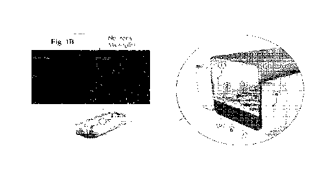
Fig. 1B upper florescent micrographs show exemplary PDGR-beta (red) stained
pericytes
and GFAP expressing astrocytes cultured on chip.
Fig. 1C florescent micrographs show exemplary immunocytochemical analysis of
hiPSC-
derived neuronal cultures in direct contact with astrocytes and pericytes
after seven days.
Specific markers were used to discriminate neurons (MAP2) and astrocytes
(GFAP) from
pericytes (NG2). Blue represents Hoechst-stained nuclei.
Fig. 1D tight, shows exemplary quantitative barrier function analysis via
permeability to
3KDa fluorescent dextran, crossing through the vascular to the neuronal
channel. Results
are mean s.e.mõ *P<0.05. n=3. Scale bar: 100 pm.
Fig. 1E shows exemplary representative merged confocal image of the vascular
channel
stained for tight junction protein marker (Z0-1, green) and Glucose
transporter (GLUT1,
red) on day 7 in culture (bar, 100 pm).
Fig. 1F shows exemplary quantitative barrier function analysis via
permeability of 3kDa
fluorescent dextran, for two independent iPSC donors crossing through the
vascular into
the neuronal channel on day 5, 6 and 7 in culture (n=6-9 independent chips,
NS, not
significant). Data are mean S.E.M. Statistical analysis was by Student's t-
test.
Figs. 2A and 2B show exemplary confocal images of brain and vascular channels.
Fig. 2A upper sets of panels show images of the entire length of an upper
channel showing the
organization of cell types and coverage across the entire channel on day 7 in
culture.
Immunofluorescence staining of the brain channel includes MAP2 (green), GFAP
(magenta),
NG2 (red) and DAPI (blue). Representative merged confocal image of the brain
channel stained
for iPS-derived cortical neurons (MAP2, green), astrocytes (GFAP, magenta) and
pericytes
(NG2, red) on day 7 in culture (bar, 50 pm).
Fig. 2B lower sets of panels show images of brain endothelial and tight
junction marker staining
for morphological characterization from the vascular channel at 7 days in
culture. Lower image
shows immunofluorescence staining of a vascular channel stained for tight
junction protein
marker ZO-1 on day 7 in culture (bar, 50 pm).
Fig. 3 shows exemplary schematics, florescent micrographs and charts
demonstrating
embodiments of a Human BBB chip as a mono-culture of HBMECs (left) and one
embodiment
16
CA 03154805 2022-4-13
WO 2021/077064
PCT/US2020/056245
of a Human Brain chip comprising neurons, astrocytes, microglia, pericytes and
BMECs (right)
for immunohistochemical (MC) analysis. A lower left chart shows permeability
assay results
demonstrating that a Brain chip maintains a tighter barrier function by day 7
of co-culture over a
monoculture of BMECs. A lower right chart shows a comparative assessment of
the permeability
of three different models (embodiments) on day 7 in culture, including a mono-
culture of iPS-
derived microvascular endothelial cells (IBMECs), a Brain-Chip cultured using
the hCMEC/D3
endothelial cell line (Brain-Chip hCIVIEC/D3), and a Brain-Chip using iPS-
derived
microvascular endothelial cell (Brain-Chip iBMECs).
Fig. 4 shows exemplary florescent micrographs demonstrating spontaneous
calcium transients
identified using fluorescence indicators (Fluo-4 AM) where neurons
consistently exhibited
spontaneous neuronal activity. Shades of green upper panels- heat mapped lower
panels, while
the charts show exemplary daily secreted neurotransmitter e.g. glutamate,
levels throughout the
experiment confirming synaptic activity of cells in a microfluidic chip.
Middle panels show representative time course images of Ca 2+ transients
(pseudocolored red
represents high levels of Ca 2+fluorescence while blue represents low levels
of Ca 2+
fluorescence). Scale bar: 50 pm.
Lower left chart: Daily secreted levels of a neurotransmitter, e.g. glutamate,
confirm the proper
synaptic activity in the neuronal channel over time from 4-7 days in culture.
Lower right chart:
ELISA for glutamate secreted levels into the medium of the brain channel on
day 5,6, and 7 in
culture (n=3 independent chips with duplicate technical replicates assayed per
condition. Data
are mean S.E.M.
Fig. 5 shows an exemplary tanscytosis receptor as a transferrin receptor, as
depicted by
schematic diagrams and by flow cytometry analysis of cells tagged with an
antibody recognizing
a C-terminal region of transferrin receptor compared to cells tagged with a
control isotype IgG.
Fig. 6 shows exemplary florescent micrographs demonstrating
Immunocytochemistry of
Phalloidin (green) staining of actin filaments (also known as F-actin),
pHrodoRed Transferrin
(red), along with Hoechst staining of nuclei (blue) in one embodiment of a
Brain chip comprising
iPS-derived HBMECs. Scale bar: 50 gm. Receptor mediated transcytosis of inAb
IgG across the
17
CA 03154805 2022-4-13
WO 2021/077064
PCT/US2020/056245
BBB is demonstrated in an exemplary chart. Data are means SEM (n= 6 chips), t-
test with
Tukey's post-hoc test, **P<0.01, ***P<0.001.
Fig. 7 shows exemplary florescent micrographs of fluorescently labeled (green)
extracellular
matrix proteins demonstrating ECM staining on an ECM coated membrane of a
microfluidic
chip under flow. It appears that SureBond-XF detaches and flows away while
sensory neuron
ECM1, shows a greater number of extracellular matrix proteins attached to the
chip membrane
than laminin alone in ECM2.
Fig. 8 shows an exemplary bright field image showing the results of direct
seeding of iPSC-
derived Sensory Neuron Progenitors on-Chip: comparing two types of ECM.
SureBond-XF;
Control upper panel). A combination of Collagen IV (400 p.g/mL); Fibronectin
(100 pg/mL);
Laminin (20 pg/mL); lower panel).
Fig. 9 shows an exemplary bright field image showing the results of direct
Seeding on-Chip of
iPSC-derived Sensory Neuron Progenitors comparing results of using sensory
neuron ECM over
time, the Day after seeding upper image and Day 7, lower image. A combination
of Collagen IV
(400 pig/mL); Fibronectin (100 pg/mL); Laminin (20 p.g/mL) was used to coat
the membrane
prior to seeding cells. iPSC-derived sensory neuron progenitors are treated
Day 5 with
Mitomycin C to eliminate proliferating cells among the progenitor pool and
maintain the
population of terminally differentiated non-proliferating neurons.
Fig. 10 shows an exemplary sensory neuron ECM coating of Collagen IV,
Fibronectin, Laminin
overnight at 4 C that supported the differentiation and maturation of sensory
neuron progenitors
(Axol Bioscience) on a tall channel (S-1) chip confirmed by mature sensory
neuron and
nociception specific markers MAP2 (green), TRPV1 (red-left panel), and Nav1.7
(red-right
panel). Merge shows superimposed images from the column of panels above the
merged image.
Co-merged staining is orange and yellow. Day 10, 5 days of flow.
Fig. 11 shows exemplary florescent micrographs of ECM proteins colored as
magenta and green
fluorescently labeled macrophages and T-cells on chips as one embodiment of a
contemplated
18
CA 03154805 2022-4-13
WO 2021/077064
PCT/US2020/056245
Live-Image Tile of Innervated Intestine-Chip. Merged image shows both ECM,
macrophages
(MO) and T-cells.
Fig. 12 shows exemplary results using one embodiment of an Intestine-Chip.
Upper left panel is
a representative image of labeled immune cells on and within the epithelial
layer of a Caco-2
Intestine-Chip. Middle panel is a representative FACs forward vs side scatter
plot of
differentiated lymphocytes. Right panel is a FACs histogram showing the
expression of
macrophage marker CD86 after differentiation of lymphocytes. These were immune
cells that
were incorporated on the chip. Lower panes show immune cell counts from chips
co-cultured for
7 and 14 days. Identical rounds of macrophage differentiation from different
PBMC donors. Co-
stimulation with anti-human CD3 & anti-human CD28.
Fig. 13 shows exemplary florescent images of one embodiment of a Brain-Chip
exposed to TNF-
alpha (100 ng/ml) (right panel where arrows point to cell membranes lacking ZO-
1 attachments)
via the neuronal channel. TNF-alpha treatment also significantly increases
GFAP expression as
well as neuronal death up to 24 hours after stimulation. Scale bar: 50 pm.
Chart demonstrates TNF-alpha induced increase in permeability by an increase
in 3kDa Dextran
diffusion from the lower to upper channel. Data are means SEM (n= 6 chips), t-
test with
Tukey's post-hoc test, **P<0.01.
Fig. 14 shows exemplary florescent images of one embodiment of a Brain-Chip
exposed to TNF-
alpha showing Neurons (MAP2-green), Astrocytes (GFAP-pink) and staining for
Nuclei:
Hoechst (blue). Scale bars: 50 micom. Chart demonstrates % of specific cell
subtypes over total
brain cells. Data are means SEM, n= 6 Chips, *P<0.05.
Fig. 15 shows exemplary florescent images of one embodiment of a healthy Brain-
Chip exposed
to TNF-alpha (100 rig/ml) (left panel Microglia (Thai)- red and Neurons (MAP-
2) ¨ green. Live
Imaging (right panel) of an Inflamed Brain-Chip (right panel Microglia (Cell
Tracker Red
CMPTX dye) and green Fluorescent latex beads. Phagocytosis of beads are an
indication of
activation of glial cells.
19
CA 03154805 2022-4-13
WO 2021/077064
PCT/US2020/056245
Fig. 16 shows an exemplary comparison of 11-6 secretion in pg/ml from Brain-
Chip (-
Microglia) and Brain-Chip (+ Microg,lia) at 24 and 48 hours. Data are means
SEM (n= 6 chips),
Anova with Tukey's post-hoc test, **P<0.01, ***P<0.001.
Fig. 17 shows an exemplary schematic diagram of examples of Clq with
neurodegenerative
diseases. Cho, "Emerging Roles of Complement Protein C1q in
Neurodegeneration." Aging and
Disease, 10(3): 652-663. June, 2019.
Fig. 18 shows an exemplary chart demonstrating that Treatment with Clq
neutralizing antibody
attenuates TNF-mediated inflammation, as indicated by IL-6 levels. Data are
means SEM (n= 6
chips), Anova with Tukey's post-hoc test, **P<0.01, ***P<0.001.
Fig. 19 shows an exemplary immunostained Brain-Chip on Day 10 demonstrating
iPS-derived
Dopaminergic Neurons double positive (yellow) for a MAP2: Neuronal Marker
(green) and TH:
Selective Marker for Dopaminergic Neurons- tyrosine hydroxylase (red). Scale
bar: 50 pm.
Shows an exemplary chart demonstrating Neurotransmitter Secretion, e.g.
Dopamine in the range
of pg/mL, at Day 7 and Day 10 (n=6 chips).
Fig. 20 shows an exemplary schematic diagram of a human brain cortex
containing GABAergic
and glutamatergic neurons representing two neuronal classes, which establish
inhibitory and
excitatory synapses, respectively. Human Dopaminergic neurons are localized in
the substantia
nigra (SN). In one embodiment, for comparing some pathological similarities
and differences
between Parkinson's disease and Alzheimer's disease. For Parkinson's disease,
red shade
indicates sites of major cell loss and a-synuclein pathology, e.g. near the
brain stem. For
Alzheimer's disease, green shade throughout the cortex indicates major regions
of cell loss and 13-
amyloid plaques and tau pathology.
Fig. 21 shows exemplary schematic diagrams depicting the progression of
Parkinson's Disease
in one embodiment of a Brain-Chip. Healthy alpha-synuclein (alpha-Syn)
(monomeric) becomes
phosphorylated at P Ser-129 (amino acid 129) forming alpha-Syn oligomers which
aggregate
into fibril aggregates with pathologic alpha-Syn (PFFs). Dopaminergic neurons
and other brain
CA 03154805 2022-4-13
WO 2021/077064
PCT/US2020/056245
cells take up extracellular PFFs inducing on a Brain-chip one or more of
neuronal dysfunction,
e.g. Impaired Calcium activity; impaired Mitochondnial Function e.g.
Expression measured by
JC-1; Neuroinflammation, e.g. Increased IL-6 secretion, Microglia activation,
Astrocyte
proliferation; and Neuronal Loss e.g. reduced number of cells after staining
with MAP2,
symptoms and pathology also observed in clinical/pathology of a PD brain.
Fig. 22 shows exemplary fluorescently stained micrographs and a chart
demonstrating a dose
response of pathogenic alpha-Syn PFFs contacting neurons in one embodiment of
a microfluidic
brain-chip over time for inducing an increasing amount of p8er129 within
neurons simulating
Syn deposition in Lewy bodies of a PD brain. Dose response is 400 ng/ml vs.
4000 ng/ml of
alpha-Syn, e.g. alpha-Syn PFFs at Day 3 and Day 6 of exposure. Panels show
results of cellular
exposure to monomers (normal alpha-Syn) in a brain chip in contrast to panels
showing exposure
of cells in a Brain-Chip to PFFs (Pathogenic alpha-Syn). pSer129-aSyn (green)
and DAPI
stained nuclei (blue). Scale bar: 50 pm. An exemplary chart shows increasing
amounts of a toxic
form of Ser129-aSyn activity (Fold change vs monomers) where at Day 3 there is
a similar
amount with 400 ng/ml vs. 4000 ng/ml (NS ¨ not significant).
Fig. 23 shows exemplary florescent micrographs of fluorescently stained
embodiments of Brain
Chips and a chart demonstrating a dose response of pathogenic alpha-Syn PFFs
contacting
neurons in one embodiment of a microfluidic brain-chip over time for inducing
an increasing
amount of JC-1 within neurons simulating JC-1 staining of a PD brain. Dose
response is 400
ng/ml vs. 4000 ng/ml of JC-1, e.g. alpha-Syn PFFs at Day 3 and Day 6 of
exposure. Red
fluorescence indicated normal mitochondrial potential, whereas green
fluorescence indicated
damage to mitochondrial potential. Panels show results of cellular exposure to
monomers
(normal alpha-Syn) in a brain chip in contrast to panels showing exposure of
cells in a Brain-
Chip to PFFs (Pathogenic alpha-Syn). JC-1 (green) and DAPI stained nuclei
(blue). Scale bar: 50
Fig. 24 shows exemplary loss of transient Ca++ signaling (no change in Ca-i-+
levels) over time
after Alpha-Syn PFFS treatment compared to Alpha-Syn monomer treatment
(signaling off and
on, see insets). FUOR-4AM fluorescent staining of Brain chips after 6 Days of
Exposure to
21
CA 03154805 2022-4-13
WO 2021/077064
PCT/US2020/056245
Monomer and PEPS, 4000 ng/ml. Column of panels, left to right, 0 sec, 10 sec,
20 sec, 30 sec.
Scale bar: 50 pun. Electrical read-outs show an almost complete loss of
transient signaling after
Alpha-Syn PFFS treatment, lower charts.
Fig. 25 shows exemplary florescent micrographs and charts comparing
fluorescently stained
Neurons (MAP2-green), Astrocytes (GFAP-red), Activated Microglia (CD11b-red),
Nuclei
(DAN-blue) 6 Days of Exposure after Alpha-Syn PFFS treatment compared to Alpha-
Syn
monomer treatment, 400 vs. 4000 ng/ml. Left chart demonstrates % of specific
cell subtypes
over total brain cells (normalized to DAPI stained nuclei). n=6 chips means
SEM.*P<0.05,
**P <0.01, ***P<0.001. Right chart demonstrates IL-6 levels pg/mL in neuronal
IL-6 channels.
** indicates a significant difference.
Fig. 26 shows exemplary results of LIVE-DEAD assay comparisons indicating
neuronal death
after 3 days of exposure to Monomer and PFFS, 4000 ng/ml along with charts
showing
LIVE/DEAD Ratios after 3 and 10 days of exposure. n=6 chips means SEM. **P <
0.01,
***P<0.001.
Fig. 27 shows exemplary results of a loss of barrier function by a Alpha-Syn
PFFS treated a
Brain chip compared to alpha-Syn monomer treatment n=8 chips. means SEM.
****P<0.0001.
Fig. 28 shows exemplary embodiments of schematics and images of Intestine-
chips. Villi-like
formations in the Intestine-Chip. Morphology was characterized with
immunofluorescence cross-
sectional view Fig. 28A of intestinal epithelial cells in the Caco-2 Intestine-
Chip and Scanning
Electron Micrograph (SEM) of Caco-2 Fig. 28B. Epithelial thickness is reduced
after an
inflammatory treatment Fig. 28D compared to control Fig. 28C. A representative
whole-chip tile
was taken showing expression of tight junction protein ZO-1 with
immunofluorescence in the
bottom image. Epithelial Layer Morphology and Barrier Function. Epithelial
cells and iPSC-
derived sensory neuron progenitors co-cultured within the chip in the presence
of continuous
flow for 14 days maintain barrier function. Maturation and differentiation of
the epithelial
morphology and villi-like structures were monitored via bright field imaging
over 14 days. The
22
CA 03154805 2022-4-13
WO 2021/077064
PCT/US2020/056245
overall viability of the epithelium was assessed by measurement of effluent
LDH and found less
than 5% leakage for each co-culture condition.
Fig. 29 shows exemplary Neuronal Immunofluorescence Staining on one embodiment
of an
Innervated Intestine-Chip by day 7-Chip, demonstrating interactions (i.e.
merged image)
between sensory neurons, e.g. nociception specific marker, TRPVI (red), MAP-2
(green),
nuclear stain (blue).
Figs. 30A-F shows exemplary schematic depiction of one embodiment of a
microfluidic human
Substantia Nigra (SN) Brain-Chip and immunohistochemistry of iPS-derived brain
endothelial
cells cultured on 4 surfaces of the lower vascular channel, and 4 additional
cell types of iPS-
derived dopaminergic neurons, primary human brain astrocytes, microglia and
pericytes on the
upper surface of the central horizontal membrane in the apical brain channel.
Fig. 30A shows a schematic depiction of one embodiment of a SN Brain-Chip of a
2-
channel microfluidic Organ-Chip having 5 cell types. In one channel (brain
channel) is a
co-culture of microglia, astrocytes, dopaminergic neurons and pericytes. In an
opposing
channel, separated by a porous membrane, are endothelial cells (vascular
channel).
Fig. 30B shows a 3D reconstruction of a confocal z-stack of fluorescent images
showing
the organization of five cell types in one embodiment of a SN Brain-Chip.
Nuclei (blue);
GFAP+ (pink); pericytes (light blue); and endothelial cells stained for a
tight junction
protein (Z0-1: red) as shown in cross section.
Fig. 30C shows a representative image of iPS-derived dopaminergic neurons that
are
stained with DAPI (colored blue), Microtubule-associated protein 2 (MAP2 -
green), TH
(red), and a merged image on day 8. Scale bars: 100 pm.
Fig. 30D shows an immunofluorescence micrographs of the human brain
endothelium
cultured on the vascular channel of Brain-Chip for 7 days post-seeding (D8)
labeled with
Claudin-1 (red), Claudin-5 (cyan), Occludin (yellow), and CD31 (white). Scale
bars: 100
PiTh
BBB integrity was observed for up to 8 days in one embodiment of a Brain-Chip.
Fig. 30E shows immunofluorescence micrographs demonstrate high levels of
expression
of ZO-1 (red) across the entire endothelial monolayer. Scale bars: 100 pm.
23
CA 03154805 2022-4-13
WO 2021/077064
PCT/US2020/056245
Fig. 30F shows a quantitative barrier function analysis via permeability to 3
kDa
fluorescent dextran and 0.5 kDa lucifer yellow (left) crossing through the
vascular to the
neuronal channel on day 5 and 8 (n=6-9 independent chips). Error bars present
mean +
SEM. Quantitative barrier function analysis via permeability of 3kDa
fluorescent dextran
(right), for two independent iPSC donors crossing through the vascular into
the neuronal
channel on day 5, 6 and 7 in culture (n=6-9 independent chips, NS, not
significant). Data
are mean S.E.M. Statistical analysis was by Student's t-test.
Figs. 31A-F shows exemplary characterization of neurons and endothelial cells
in one
embodiment of a Human Substantia Nigra Brain-Chip.
Fig. 31A Graph shows neurotransmitter release over time between 5 and 8 days
of co-
culture. Neurotransmitter release is shown as ELISA results for dopamine
secreted into
the medium of the brain channel on days 5 and 8. (n=3 independent chips with
duplicate
technical replicates assayed per condition). n= 6 chips. Error bars present
mean SEM.
Fig. 31B shows exemplary immunofluorescent microphotographs (left) validate
the
dopaminergic neurons with MAP2+ (green), astrocytes with GFAP (magenta) and
pericytes (red), and the DAPI (blue) for cell nuclei. Immunofluorescent
microphotograph
(right) validates the glia culture: astrocytes (magenta, GFAP staining), and
resting
microglia (yellow, TMEM119). Scale bars: 50 gm.
Fig. 31C shows exemplary immunofluorescent images of MAP2+ (green); TH (red);
Hoechst stained nuclei (blue) of iPS-derived dopaminergic neurons. Scale bar =
10um.
Fig. 31D shows exemplary Iimmunocytochemical analysis of iPS-derived neuronal
cultures in direct contact with astrocytes and pericytes. Specific markers
were used to
identify neurons (MAP-2), astrocytes (GFAP), and pericytes (NG2). Blue
represents
Hoechst-stained nuclei.
Fig. 31E shows exemplary immunocytochemical analysis that demonstrated
endothelial
monolayer tightness and brain specificity using ZO-1, GLUT-1, CD31, and
Occludin
markers at day 7 in culture.
Fig. 31F shows exemplary representative merged confocal image of the brain
channel co-
stained for iPS-derived cortical neurons (MAP2, green) and vesicular Glutamate
transporter 1 (VGLUT1, red) (bar, 100 gm).
24
CA 03154805 2022-4-13
WO 2021/077064
PCT/US2020/056245
Figs. 32A-E shows exemplary Differentially Expressed (DE) genes and enriched
gene ontology
(GO) categories in SN Brain-Chip and conventional cell culture (CCC) system,
as compared to
the adult in vivo substantia nigra; day 8.
Fig. 32A shows schematic drawings of devices, Transwell and microfluidic brain-
chips,
along with a volcano plot resulting from DGE analysis between SN Brain-Chip
and CCC.
For the selection of the DE genes, the following thresholds were used:
adjusted p-
value< 0.05 and ILog2(foldchange)I > 1. The identified up- (down-) regulated
genes are
highlighted in cyan (magenta) color respectively. Sample sizes were as
follows: SN
Brain-Chip, n=4, conventional cell culture system, n=4.
Fig. 32B and Fig. 32C shows exemplary list of biological processes identified
by Gene
Ontology (GO) enrichment analysis using the up- and down- regulated genes
respectively
resulted by the differentially gene expression analysis between SN Brain-Chip
and CCC.
Fig. 32B shows exemplary GO Term Enrichment Biological Processes Upregulated
Genes in Brain Chip.
Fig. 32C shows exemplary GO Term Enrichment Biological Processes Upregulated
Genes in Conventional Cell Culture.
Fig. 32D shows exemplary DGE analysis identified up- and down-regulated genes
in SN
Brain-Chip compared to CCC (cyan circle), and human adult substantia nigra
compared
to CCC (yellow circle). Sample sizes were as follows: SN Brain-Chip, n=4,
Conventional
cell culture system, n=4, and adult substantia nigra, n=8 (independent
biological
specimens). Culture in Brain-Chips and CCC were done in parallel. Samples were
collected and processed for analyses 8 days post-seeding (1)8).
Fig. 32E shows an exemplary SN Brain-Chip exhibits higher transcriptomic
similarity to
adult substantia nigra than conventional cell culture. The results of the GO
terms analysis
using the 209 DE genes showed 6 significantly enriched (FDR adjusted p-
value<0.05)
biological processes related to tissue development, response to a stimulus,
biological
adhesion, and cell surface receptor signaling pathway. The size of the bars
indicates the
fold-enrichment of the corresponding pathways.
25
CA 03154805 2022-4-13
WO 2021/077064
PCT/US2020/056245
Figs. 33A-D shows exemplary embodiments of a microfluidic Brain-Chip that
exhibits higher
transcriptomic similarity to adult cortex tissue than Transwell cultures (not
under flow); days 5
and 7 of culture.
Fig. 33A shows an exemplary Principal component analysis (PCA) generated using
RNA-seq data generated by the samples collected from the brain channel of the
Brain-
Chips and transwells on days 5 and 7 in culture (n=4 per condition), as well
as human
brain cortex. A 2D-principal component plot is shown with the first component
along the
X-axis and the second along the Y-axis. The proportion of explained variance
is shown
for each component.
Fig. 33B shows an exemplary Quantitative analysis on the distances of the
Brain-Chip or
Transwell culture from Human Brain Cortex on days 5 and 7 of culture.
Fig. 33C shows an exemplary Differential Gene Expression (DEG) analysis
identified up-
and down- regulated genes in the Brain-Chip compared to conventional cell
cultures
(blue circle), and human adult cortex brain tissue compared to conventional
cell cultures
(yellow circle). Gene lists summarized in the Venn diagram are provided in
Extended
Data. Sample sizes were as follows: Brain-Chip, n=4, transwells n=4, and adult
cortex
tissue, n=8 (independent biological specimens). Culture in Brain-Chips and
conventional
cell cultures were done in parallel.
Fig. 33D shows an exemplary Curated heatmap generated to examine particular
genes
that belong to the enriched KEGG pathways and to show the expression levels
1og2(FPKM) of these genes across different samples. Genes belonging to four
different
pathways, including: intermediate filament cytoskeleton organization, neuronal
action
potential, axon guidance, and extracellular matrix organization, are shown.
Each heatmap
has its own color scale, which corresponds to a different range of 1og2 (FPKM)
values, as
indicated on the color bars located to the left.
Figs. 34A-B shows exemplary schematic diagrams demonstrating contemplated
embodiments of
drug targets and biomarkers for normal (noninflammatory) and disease
associated inflammatory
conditions. Smyth et al., Journal of Neuroinflammation 2018.
26
CA 03154805 2022-4-13
WO 2021/077064
PCT/US2020/056245
Fig. 34A shows exemplary systemic inflammation that causes a breach in the
blood
brain-barrier (BBB) thereby allowing for the entry of immune/inflammatory
cells and
proinflammatory cytokines into the brain.
Fig. 34B shows exemplary neuroinflammation that induces and accelerates
pathogenesis
of Parkinson's disease (PD), Alzheimer's disease (AD) and Multiple sclerosis
(MS).
Fig. 35 shows an exemplary schematic experimental timeline of one embodiment
of a
neuroinflammation culture model. On days 0 and 1 cells were seeded on the top
and bottom
channel of one embodiment of a Brain-Chip, respectively. The Brain-Chip is
then allowed to
mature under flow conditions for 4 more days and on day 5, cells on the brain
side or the
vascular side are treated with TNFa, for 48 his. During exposure, effluent is
collected, and on the
last day of the experiment, cells in chips may be imaged, stained for
immunohistochemistry and
imaged, effluent may be collected from the brain channel and the vascular
channel_ In some
embodiments, cells are lysed within each channel for channel specific
transcriptomic analysis.
Fig. 36A shows one embodiment of neuroinflammation as a schematic illustration
of the Brain-
Chip showing the TNF-a perfusion within the brain channel and shows an
exemplary chart of
comparative apparent permeability showing time-depended BBB disruption
measured after 24
hrs and 48 hrs of exposure to TNFa (100 ng/mL) vs. vehicle control, introduced
on the brain
side. Transport through the BBB was significantly increased by three times
over the control.
Data are means-+SEM, *p<0.5 (n>4 chips), vehicle compared to TNF-a treated
group after 48 hrs
of treatment). Statistical analysis is two-way ANOVA with Tukey's multiple
comparisons test.
Fig. 3611 shows exemplary results of TNF-alpha induced neuroinflammation by
immunofluorescent double staining of neurons (MAP2, green), astrocytes (GFAP,
magenta),
microglia (CD68, red), and nuclei (DAPI, blue), following two days of exposure
with TNF-a
compared to the untreated group (bar, 100 nm).
Fig. 36C, Fig. 36D, and Fig. 36E shows exemplary results of TNF-alpha induced
neuroinflammation by secreted levels of proinflammatory cytokines (lFN-y, IL-
113 and IL-6) in
the healthy or 48 hr TNF-a treated Brain-Chips in the presence or absence of
microglia (n=4-7
independent chips, **p<0.01, ****p<0.0001, NS, not significant). Data are mean
&FM.
Statistical analysis was by Student's t-test.
27
CA 03154805 2022-4-13
WO 2021/077064
PCT/US2020/056245
Fig. 36F shows an exemplary quantification of the number of GFAP-positive and
MAP2 events
per field of view. Statistical analysis is Student's t-test (n=3 independent
chips with 3-4
randomly selected different areas per chip, *p<0.05, np<0.01 compared to the
untreated group).
Fig. 36G shows an exemplary quantification of the number of CD68-positive
events per field of
view. (n=3 Brain-Chips with 3-5 randomly selected different areas per chip,
*13<0.05 compared
to the untreated group). Data are mean S.E.M. Statistical analysis was by
Student's t-test.
Fig. 36H shows an exemplary ELISA for glutamate secreted levels into the
medium of the brain
channel on day 7. (n=3 independent chips with duplicate technical replicates
assayed per
condition, "p<0.01, compared to the untreated group). Data are mean S.E.M.
Statistical
analysis was by Student's t-test.
Fig. 37A shows an exemplary schematic illustration of the Brain-Chip showing
the TNF-a
perfusion within the vascular channel as a systemic inflammation culture
model. On days 0 and 1
cells were seeded on the top and bottom channel, respectively. The Brain-Chip
is then allowed to
mature under flow conditions for 4 more days and on day 5, cells on the
vascular side are treated
with TNFa, for 48 hrs. During exposure, effluent is collected, and on the last
day of the
experiment, cells in chips may be imaged, stained for immunohistochemistry and
imaged. In
some embodiments, cells are lysed for transcriptomic analysis.
Fig. 37B shows an exemplary Quantitative barrier function analysis via
permeability to 3 kDa
fluorescent dextran, over 48 hs of treatment with TNF-a perfused at the
vascular channel,
showing time-depended BBB disruption (n=3-4 independent chips, *P<0.05,
vehicle compared
to TNF-a treated group after 48 hs of treatment). Data are mean S.E.M.
Statistical analysis is
two-way ANOVA with Tukey's multiple comparisons test.
Fig. 38A Brain Channel: Neuroinflammation vs Healthy Brain-Chip on day 7,
Table I, shows an
exemplary volcano plot illustrating the number of the differentially expressed
(DE) genes (up-
magenta dots) and down-cyan dots) regulated) and how they stratify based on
their expression
changes. DE genes significantly up- or down-regulated (adj.p-value < 0.01 and
llog2FoldChangel>1); black dots: non-DE expressed genes. In total, 1174 genes
were found
significantly DE in cells of brain parenchyma, 801 up-regulated (in the
inflamed Brain-Chips)
and 373 down-regulated (in the inflamed Brain-Chips).
28
CA 03154805 2022-4-13
WO 2021/077064
PCT/US2020/056245
Fig. 38B shows an exemplary list of biological processes identified by Gene
Ontology (GO)
enrichment analysis based on the 801 up-regulated DE genes between the
TNFaBrain-Chips and
Healthy Brain Chips. Bar plot presents a subset of the significantly enriched
biological processes
identified by the enrichment analysis.
Fig. 39A Brain Channel: Systemic Inflammation vs Healthy Brain-Chip, Table 2,
shows an
exemplary volcano plot illustrating the number of the differentially expressed
(DE) genes (up-
and down-regulated) and how they stratify based on their expression changes.
Red dots: DE
genes significantly up- or down-regulated (adj.p-value < 0.01 and
llog2FoldChangel>1); black
dots: non-DE expressed genes. In total, 528 genes were found significantly DE
in cells of brain
parenchyma, 473 up-regulated (in the inflamed Brain-Chips) and 55 down-
regulated (in the
inflamed Brain-Chips).
Fig. 39B shows an exemplary list of biological processes identified within the
brain channel by
Gene Ontology (GO) enrichment analysis based on the 473 up-regulated DE genes
between the
TNFa Brain-Chips and Healthy Brain Chips. Bar plot presents a subset of the
significantly
enriched biological processes identified by the enrichment analysis.
Fig. 40 shows an exemplary chart of comparative apparent permeability measured
after 24 hrs
and 48 hrs of exposure to TNFa (100 ng/mL) vs. vehicle control, introduced on
the brain side.
Transport through the BBB was significantly increased by three times over the
control. Data are
means+SEM, *p<0.5 (n>4 chips).
Figs. 41A-B Vascular Channel: DGE Analysis (Neuroinflammation). See, Table 3.
Fig. 41A shows an exemplary volcano plot illustrating the number of the
differentially
expressed (DE) genes (up- and down-regulated) and how they stratify based on
their
expression changes. Red dots: DE genes significantly up- or down-regulated
(adip-value
<0.01 and llog2FoldChangel>1); black dots: non-DE expressed genes In total,
387 genes
were found significantly DE in endothelial cells, 371 up-regulated (in the
inflamed Brain-
Chips) and 16 down-regulated (in the inflamed Brain-Chips).
Significantly Enriched GO terms from the list of the 473 DE up-regulated genes
upon
TNF-a exposure.
29
CA 03154805 2022-4-13
WO 2021/077064
PCT/US2020/056245
Fig. 41B shows an exemplary Gene Ontology (GO) enrichment analysis based on
the 371
up-regulated DE genes between the TNFa treated Brain-Chips and Healthy Brain
Chips.
Bar plot presents a subset of the significantly enriched biological processes
identified by
the enrichment analysis. Vascular Channel: Significantly Enriched Biological
Processes.
Significantly Enriched GO terms from the list of the 371 DE up-regulated genes
upon
TNF-a exposure
Figs. 42A-B shows exemplary differential Gene Expression analysis between TNFa
exposed
Brain-Chips and Healthy Brain-Chips on day 7. See, Table 4.
Fig. 42A shows an exemplary volcano plot illustrating the number of the
differentially
expressed (DE) genes (up- and down-regulated) and how they stratify based on
their
expression changes. Red dots: DE genes significantly up- or down-regulated
(adip-value
< 0.01 and llog2FoldChangel>1); black dots: non-DE expressed genes. In total,
1174
genes were found significantly DE in endothelial cells, 801 up-regulated (in
the inflamed
Brain-Chips) and 373 down-regulated (in the inflamed Brain-Chips).
Fig. 42B shows an exemplary Gene Ontology (GO) enrichment analysis based on
the 801
up-regulated DE genes between the TNFaBrain-Chips and Healthy Brain Chips. Bar
plot
presents a subset of the significantly enriched biological processes
identified by the
enrichment analysis.
Fig. 43A Brain channel: neuroinflammation shows exemplary secretion of pro-
inflammatory
cytokines. One embodiment of a brain channel of a Brain-Chip was exposed to
continuous flow
of media containing 10Ong/mL TNFa for 48 hrs. The effluent was collected at
the 48 h
timepoint; cytokines were measured and analyzed using MSD human
proinflammatory panel.
Data are means+SEM, ***p<0.001, ****p<0.0001(n>4 chips).
Fig. 438 Brain channel: systemic inflammation
shows exemplary secretion of pro-inflammatory cytokines. One embodiment of a
brain channel
of a Brain-Chip was exposed to continuous flow of media containing 100ng/mL
TNFa for 48
his. The effluent was collected at the 48 h timepoint; cytokines were measured
and analyzed
using MSD human proinflammatory panel. Data are means:ESEM, ***p<0.001,
****p<0.0001(n>4 chips).
CA 03154805 2022-4-13
WO 2021/077064
PCT/US2020/056245
Fig. 44A vascular channel: neuroinflammation shows exemplary representative
immunofluorescent staining of endothelial and tight junction markers after 48
hrs of exposure to
TNFa on the brain-side showing a decreased expression of tight junction
protein, ZO-1, and
increased expression of intercellular adhesion molecule-1 (ICAM-1).
Fig. 448 vascular channel: Systemic inflammation shows exemplary
representative
immunofluorescent staining of endothelial and tight junction markers after 48
hrs of exposure to
TNFa on the brain-side showing a decreased expression of tight junction
protein, ZO-1, and
increased expression of intercellular adhesion molecule-1 (ICA1VI-1).
Fig. 45 brain channel: Neuroinflammation vs. Systemic inflammation shows an
exemplary
curated heatmap generated to examine particular genes resulted by the
differentially gene
expression analysis of the brain channel, between the systemic inflammation
and
neuroinflammation condition.
Fig. 46A vascular channel: Neuroinflammation vs. Systemic inflammation
Fig. 46B curated heatmaps were generated to examine particular genes resulted
by the
differentially gene expression analysis of the vascular channel, between the
systemic
inflammation and neuroinflammation condition.
Fig. 47A brain channel: Neuroinflammation vs. Systemic inflammation shows an
exemplary
Venn diagram demonstrating an overlap of 508 DE genes associated with
inflammation between
1174 DE genes in an embodiment of neuroinflammation vs. 528 healthy on Day 6
vs. Brain-
Channel.
Fig. 47B vascular channel: Neuroinflammation vs. Systemic inflammation shows
an exemplary
Venn diagram of DE genes identified in the vascular channel (endothelium)
between both types
of neuroinflammation vs healthy conditions. Inflamed Brain-Chips
(Neuroinflammation) Vs.
Healthy on Day 6 (blue). Inflamed Brain-Chips (Systemic Inflammation) Vs.
Healthy on Day 6
(blue).
31
CA 03154805 2022-4-13
WO 2021/077064
PCT/US2020/056245
Fig. 48A shows exemplary schematic diagrams demonstrating native and toxic
conformations of
a-syn. Alpha-synuclein transforms into multiple different conformations,
including monomers
(predominant in a a-helical confirmation), tetramers, higher-level oligomers
(soluble
conformations), and fibrils (highly ordered insoluble conformations
characterized by 13-sheet
conformation). Alpha-synuclein exists in a native conformation as monomers as
well in a
dynamic equilibrium with tetramers. The tetramer, less likely to form
aggregate, may form an
aggregate after disrupted into monomers in order to misfold. Many factors,
such as the
posttranscriptional modification and SNCA mutations in A53T and E46K promote
formation of
pathological oligomers, presently considered to be the most toxic structure of
a-syn, which is
further folded to form amyloid fibril (rich in (3-sheet structure), the
accumulation of which leads
to the formation of intracellular inclusions called Lewy Body.
Fig. 48B shows exemplary interactions between a-syn and cellular components
contemplated as
drug targets for use in drug screening methods as described herein. Misfolded
a-syn is degraded
through the autophagy-lysosomal pathway (ALP) and the ubiquitin-proteasome
system (UPS).
Certain oligomeric species present toxicity via interactions with cellular
components by
mechanisms that include: (1) alteration of cytoskeletal integrity; (2)
membrane disruption and
pore formation; (3) nuclear dysfunction; (4) inhibition of vesicle docking;
(5) UPS dysfunction;
(6) ALP impairment; (7) reduction of mitochondrial activity; and (8) chronic
ER stress. UPS,
ubiquitin-proteasomal system; ALP, autophagy-lysosomal pathway; ER,
endoplasmic reticulum.
Fig. 48C shows exemplary schematic summary of interactions between a-synuclein
and cellular
components, such interactions are contemplated for use as drug targets in
methods of use for
microflucidic Brain-Chips as described herein. At least six different
exemplary intracellular
pathways are affected by a-synuclein (a-syn). The protein a-syn is enriched at
the pre-synaptic
terminals of the majority of types of neurons in the brain, where it
participates in the vesicle
recycling, thereby modulating synaptic function. a-syn can be degraded by the
ubiquitin-
proteasome system (UPS) and inside the lysosomes. a-syn interacts strongly
with membranes,
such as plasma membrane and mitochondrion. When misfolded, a-syn forms
distinct structures
that are prone to aggregation, into oligomers, then into larger structures_ a-
syn oligomers in a
toxic form may impair basic neuronal processes, such as ER-Golgi trafficking,
lysosome and
UPS functions, reduced mitochondrial activity and alter the plasma membrane
through the
32
CA 03154805 2022-4-13
WO 2021/077064
PCT/US2020/056245
pore/perforations that can dysregulate calcium and cation homeostasis_ In
fact, many of these
pathways were identified as GO categories of Genes that were upregulated genes
in Brain-Chips.
Fig. 48D shows exemplary autophagy-lysosomal pathway (ALP) and ubiquitin-
proteasome
system (UPS) pathways under normal and pathological conditions. Proteins are
tagged with
ubiquitin conjugates through a sequential enzymatic mechanism involving three
classes of
enzymes, E1, E2 and E3. Under normal conditions, ubiquitylated substrates are
recognized by
ubiquitin receptors present in ALP and UPS pathways and efficiently
eliminated. In the UPS,
substrates are subsequently deubiquitylated by RPN11, a step for substrate
degradation and
amino acid recycling. Free-Ub chains formed by RPNI I activity promote ALP
function.
Ubiquitin receptors in the ALP, in contrast to the UPS, form oligomers to
facilitate substrate
recognition and autophagosomal recruitment. Under aging and Alzheimer's
disease conditions
there is a decrease in the function of the ALP and the UPS that reduces
substrate degradation and
amino acid recycling. Downregulation of RPNI1 in Alzheimer's disease (AD)
decreases free-Jib
chains disrupting substrate recognition, their recruitment into autophagosomes
and their final
degradation by the ALP. Altogether, leading to the accumulation of deleterious
protein
aggregates. Transcriptional regulation (Nrf1/2) and phosphorylation
(kinases/phosphatases) play
a crucial role in ALP and UPS function whereas their dysregulation is the
focus of intense
studies in aging and Alzheimer's disease
Fig. 49A shows exemplary schematic diagrams depicting steps towards
accumulation of Alpha-
synuclein protein (SNCA). Natural SNCA becomes misfolded under stress and
becomes
oligomers, oligomers, profibril oligomers that form fibril aggregates that
form Lewy bodies in
affected neurons of a patient's PD brain leading to dopamine (DA) neuronal
loss.
Fig. 49B shows an exemplary schematic depiction of a-synuclein fibril
contributions to
Alpha/Beta plaques, Tau tangles and a-synuclein Lewy bodies found in
degenerating neurons.
Fig. 50A shows exemplary schematics for providing embodiments of a Brain Chip:
comprising
Neurons, Astrocytes, microg,lia, pericytes, and endothelial cells_
Fig. 50B shows an exemplary Principal Components Analysis (PCA) of healthy vs.
PD disease
associated brain channel cells.
33
CA 03154805 2022-4-13
WO 2021/077064
PCT/US2020/056245
Fig. 51A Brain Channel: Volcano plot: Brain-chip A53T vs_ healthy.
Fig. 51B Brain Channel: GO-terms Enrichment Analysis Results. Go-term
enrichment analysis
results using the 320 up-regulated genes in A53T brain-Chips.
Fig. 52 Brain Channel: Principal Components Analysis (PCA) comparing the same
brain cells
cultured in either Transwell cultures or Brain Chips, as healthy cultures
without exposure to a
monomer or fibril, or exposed to monomers, fibrils or fibrils comprising an
A53T mutation.
Fig. 53 shows exemplary schematic depictions of dopaminergic neurons with
synaptic terminals.
While increasing dopamine (blue dots) in the wild-type (WT) setting is benign
(left), similar
increases in the setting of human mutant (A53T) a-synuclein (a-syn) lead to
progressive
neurodegeneration (middle and right) in mice. Synaptic loss (red X marks) in
presynaptic striatal
terminals precedes somatic degeneration, and toxicity is thought to be
mediated by a-synuclein
(red) oligomers in the presence of dopamine.
Fig. 54A-E shows images from a pathological examination of a healthy patient
(Fig. 54A)
reveals typical pigmented DA neurons in the SN (arrows); in contrast, loss of
SN neurons leads
to pigment disappearance in the PD brain (Fig. 54B, arrows). Magnification of
the SN area
reveals a dense network of melanin-pigmented SN neurons in the healthy brain
(Fig. 54C) while
most of SN neurons are lost in PD (Fig. 54D). Some of the remaining neurons in
PD contain
insoluble cytoplasmic protein aggregates (Lewy Bodies, Fig. ME) that are made
of aggregated
alpha-synuclein and other proteins. The melanin-containing granules have a red-
brown hue and
are distributed in the cytosol of all SN neurons (Fig. 54C-E). The image in
Fig_ 54E is the higher
magnification of the dark-boxed area in Fig. 54D. Adapted from Agamanolis,
2006.
Figs. 55A-B shows exemplary schematic depictions of a comparison of cellular
interactions in
the upper channel of one embodiment of a SN Brain-Chip contemplated to provide
an intact
BBB (healthy) in one embodiment of a Brain-chip Fig. 55A vs. Fig_ 55B
contemplated cellular
interactions between three types of brain cells as activated microglia and
reactive astrocytes both
cause damage inflammatory response of dopaminergic neurons, including
dystrophic neurites,
34
CA 03154805 2022-4-13
WO 2021/077064
PCT/US2020/056245
shrinkage of the soma, and neuronal loss, resulting in breakdown of BBB as a
SN Brain-Chip
model for Parkinson's Disease.
Fig. 56 shows exemplary schematic depictions of a model of aSyn actively
secreted or released
by dying neurons, e.g. neuron 1, into the extracdlular space. Extracellular
aSyn can then activate
surrounding astrocytes and microglia, eliciting glial pro-inflammatory
activity. Upon activation
microglia produce pro-inflammatory cytokines, nitric oxide, and reactive
oxygen species, which
may be toxic to neurons. aSyn can be directly transferred between neurons,
e.g. neuron 1 to
neuron 2, and so on, leading to propagation of an aberrant aSyn aggregation
process.
Fig. 57A-D shows exemplary pathological aSyn accumulation in the brain channel
was observed
following exposure to human aSyn fibrils.
Fig. 57A shows exemplary schematic depiction of one embodiment of an
Experimental
design for assessing the effects of aSyn toxic aggregates (fibrils) in the SN
Brain-Chip,
including the seeding in the Brain-Chip, the timeline for medium changes, as
well as
sampling times.
Fig. 57B Immunofluorescence micrographs show the accumulation of
phosphorylated
aSyn (green, phospho-aSyn129 staining; blue, DAPI) at day six post-exposure
(D8).
Pathology is absent in the brain channel following exposure to monomer or PBS.
Scale
bars: 100 pm.
Fig. 57C Quantitative analysis of fluorescence intensity in each group at day
three and six
post-exposure (D5 and D8, respectively).
Fig. 57D Immunofluorescence staining shows phospho-a.5yn129 (green)
accumulation
within the TH (red) positive neurons in the SN Brain-Chip, yellow indicates co-
localization of phospho-aSyn129 and TH. Statistical analysis is two-way ANOVA
with
Tukey's multiple comparisons test (n=3-4 independent chips with 3-5 randomly
selected
different areas per chip, *P<(J.05, ****P<0.0001 compared to monomeric or PBS
group).
Error bars present mean SEM.
Fig 58A-B shows exemplary accumulation of phosphorylated aSyn and
mitochondrial
impairment in the aSyn fibril model at day 5.
CA 03154805 2022-4-13
WO 2021/077064
PCT/US2020/056245
Fig 58A shows exemplary assessment of phosphorylated a.Syn resulting from a
three day
post-exposure to aSyn fibrils shown in lower row of panels. Immunofluorescence
micrographs
show the accumulation of phosphorylated aSyn (green, phospho-aSyn129 staining-
vertical
middle panels; blue, DAPI stained nuclei- vertical left panels) and merged
images - vertical right
panels. PBS treated controls upper row of panels, aSyn monomer treated middle
row of panels.
Scale bars: 100 pm.
Fig 58B shows exemplary effects of aSyn fibrils on mitochondrial membrane
potential at
three days after exposure. Mitochondrial membrane potential assessed by JC-1
staining on the
brain side. Dual emission images (527 and 590nm) represent the signals from
monomeric (green)
and J-aggregate (red) JC-1 fluorescence. Scale bars: 100 pm.
Fig 59A-D shows exemplary reduction of mitochondrial activity and increase in
ROS production
in the aSyn fibril model.
Fig 59A shows exemplary mitochondrial membrane potential assessed by JC-1
staining
in the brain side at day six post-exposure. Dual emission images (527 and 590
nm)
represent the signals from monomeric (green) and J-aggregate (red) JC-1
fluorescence.
Scale bars: 100 Rm.
Fig 59B Quantitative analysis of the ratio of Red/Green fluorescence intensity
in each
group at day three and six post-exposure (D5 and D8, respectively).
Statistical analysis is
two-way ANOVA with Tukey's multiple comparisons test (n=3 independent chips
with
3-4 randomly selected different areas per chip, *P<0.05, ****P<0.00(n compared
to
monomeric group).
Fig 59C shows exemplary representative images of ROS levels (green, CelIROX)
show
higher levels of intercellular ROS in the cells of the brain channel exposed
to aSyn fibrils
than those exposed to aSyn monomer at day six post-exposure. Scale bars: 100
Rm.
Fig 59D shows exemplary quantification of the number of CelIROX-positive
events per
field of view in each group. Statistical analysis is Student's t test (n=3
independent chips
with 3-4 randomly selected different areas per chip, ****p<0.0001 compared to
monomeric group). Error bars present mean SEM.
36
CA 03154805 2022-4-13
WO 2021/077064
PCT/US2020/056245
Fig 60A-F shows exemplary aSyn-induced apoptotic cell dopaminergic neuronal
death indicated
by caspase-3 activation (red) and neuroinflammation in the presence of
activated astrocytes and
activated microglia/macrophages. Increasing expression of glial fibrillary
acidic protein (GFAP)
represents astroglial activation and gliosis during neurodegeneration.
Increasing expression of
CD68 represents
Fig. 60A shows exemplary representative merged images showing double
immunostaining for dopaminergic neurons by MAP2 (grey) and Cleaved Caspase-3
(red,
CC3) in the brain channel at six-days post-exposure. Scale bars: 50 pm.
Fig. 60B shows exemplary quantitative data on the number of CC3 positive
neurons.
Statistical analysis is Student's t test (n=3 independent chips with 3-4
randomly selected
different areas per chip, ***p<0.001 compared to monomeric group).
Fig. 60C shows exemplary immunostaining of the astrocyte marker GFAP (magenta)
demonstrating activation of astrocytes at day 8 following exposure to aSyn
fibrils
compared to monomeric aSyn. Scale bar, 100 pm.
Fig. 60D shows exemplary immunostaining of the microglial CD68 (red)
demonstrated
activation of astrocytes and microg,lia at day 8 following exposure to aSyn
fibrils
compared to monomeric aSyn. Scale bar, 100 gm.
Fig. 60E shows exemplary secreted levels of INF-a in the aSyn fibril model.
Statistical
analysis was by Student's t-test (n=6-7 independent chips, **p<0.01).
Fig. 6OF shows exemplary secreted levels of proinflammatory cytokine IL-6 in
the aSyn
fibril model. Statistical analysis was by Student's t-test (n=4-7 independent
chips,
****p<0.0001). Error bars present mean SEM.
Fig. 61A-D shows exemplary aSyn-induced cell death and neuroinflammation.
Fig. MA shows exemplary cell viability (live/dead) assay following exposure to
human
aSyn fibrils. Live/Dead cell staining assay was designed to test the potential
cytotoxicity
of aSyn fibrils at days 5 and 8 of culture. Scale bars: 100 pm.
Fig. 61B shows exemplary data are expressed as the average live cells/total
number of
cells (sum of calcein AM positive and ethidium homodimer positive cells).
Statistical
analysis is two-way ANOVA with Tukey's multiple comparisons test (n=3
independent
37
CA 03154805 2022-4-13
WO 2021/077064
PCT/US2020/056245
chips with 3-5 randomly selected different areas per chip, ****/3<0.0001
compared to
monomeric or PBS group, NS: Not Significant).
Fig. 61C shows exemplary quantification of the number of GFAP-positive events
per
field of view. Statistical analysis is Student's t test (n=3 independent chips
with 3-4
randomly selected different areas per chip, ***p<0.001 compared to monomeric
group).
Fig. 61D shows exemplary quantification of the number of CD68-positive events
per
field of view. (n=3 Brain-Chips with 3-5 randomly selected different areas per
chip,
****P<0.0001 compared to the monomeric group). Error bars present mean SEM.
Fig. 62 shows an exemplary schematic model for measuring BBB breakdown. In one
embodiment modeling neuroinflammation when a-Syn is added to the brain
channel.
In one embodiment modeling systemic inflammation when a-Syn is added to the
vascular
channel.
Fig. 63A-D shows exemplary Blood-Brain Barrier dysfunction in the aSyn fibril
model.
Fig. 63A and 56 Fig. 638 shows exemplary quantitative bather function analysis
via
permeability to 0.5 kDa lucifer yellow and 3 kDa fluorescent dextran at day 5
and 8
following exposure to aSyn fibrils or aSyn monomers. Statistical analysis is
two-way
ANOVA with Tukey's multiple comparisons test (n=6-9 independent chips,
****P<0.0001 compared to monomeric group, NS: Not Significant).
Fig. 63C shows exemplary principal component analysis generated using the RNA-
seq
data generated by the samples collected from the vascular channel of the SN
Brain-Chip
upon exposure to aSyn monomers or aSyn fibrils (n=4 per condition). A 2D-
principal
component plot is shown with the first component along the X-axis and the
second along
the Y-axis. The proportion of explained variance is shown for each component.
Fig. 63D shows exemplary volcano plot indicating DE genes between aSyn fibrils
and
aSyn monomers, as identified by the RNA-sequencing analysis. For the selection
of the
DE genes the following thresholds were applied: adjusted p-value< 0.05 and
ILog2(foldchange)I > 0.5. The identified up- (down-) regulated genes are
highlighted in
cyan (magenta) color. Sample sizes were as follows: Brain-Chip (aSyn
monomers), n=4,
Brain-Chip (aSyn fibrils), n=4.
38
CA 03154805 2022-4-13
WO 2021/077064
PCT/US2020/056245
Fig. 64 A-B shows exemplary Blood-Brain Bather dysfunction in the aSyn fibril
model. IgG
Penetration through BBB. See, Table 6.
Fig. 64 A shows exemplary quantitative barrier function analysis via
permeability to
immunoglobulin G (IgG1) at day 5 and 8 following exposure to aSyn fibrils,
aSyn
monomers or PBS. Statistical analysis is two-way ANOVA with Tukey's multiple
comparisons test (n=5-8 independent chips, ****Pc10001 compared to monomeric
group, NS: Not Significant). Error bars present mean SEM.
Fig. 64 B shows exemplary selection of the 739 up-regulated and 541 down-
regulated
genes identified after performing DGE analysis between aSyn fibrils and aSyn
monomers. The size of the bars indicates the log2(Fold-Change) of the
corresponding
gene expressions and the colors the statistical significance (FDR adjusted p-
values) of the
corresponding changes.
Fig. 65 shows exemplary schematic depictions toxicity of a-syn as a
therapeutic target. Toxicity
of alpha-synuclein to neurodegeneration is associated tightly with the dynamic
equilibrium of the
protein synthesis, aggregation, and clearance. Levels of specific
conformations (oligomers and
protofibrils) vary in different stages of PD. Disease-modifying therapeutic
strategies are mainly
focused on these processes as well as inhibiting cell-to-cell propagation: (i)
reducing a-syn
synthesis with small interfering RNA (siRNA), microRNA (miRNA), small hairpin
RNA
(shRNA), and transcription inhibitors; (ii) increasing degradation of a-syn
via UPS and ALP; (iii)
reducing aggregation of a-syn via heat-shock proteins (hsp40/70/104),
aggregation inhibitors,
antioxidant, and posttranslational modification approaches (oxidation,
nitration, phosphorylation,
and C-terminal cleavage); (iv) blocking the propagation of a-syn with
immunotherapies by
targeting extracellular a-syn or exosome and by blocking putative receptors in
recipient cells;
and (v) seeking neuroprotective strategies including anti-inflammation and
antioxidant.
Fig. 66A shows exemplary potential mechanisms involved in propagation of a-
syn. Spreading
mechanisms of a-syn in neighboring cells are multiple and can occur via (1)
passive transmission
through membrane fusion; (2) classical exocytosis and endocytosis; (3)
packaged-exosomes; (4)
39
CA 03154805 2022-4-13
WO 2021/077064
PCT/US2020/056245
tunneling nanotubes (a direct connection between two cells); (5) axonal
transport and
transsynaptic junction; and (6) receptor-mediated internalization.
Fig. 66B shows exemplary molecules and signaling pathways involved in a-syn-
mediated
microglial activation. Excessive microglial activation can increase the
production of pro-
inflammatory cytokines (TNF-a,
IL-6, and INF-y), and induce an
oxidative stress
response, including the release of reactive oxygen species (ROS) and nitric
oxide (NO) as well as
the production of NADPH oxidase. Toll-like receptors (TLRs) play a vital role
in recognizing
pathogen-associated molecular patterns (PAMPs) and initiating innate immune
responses via
distinct signaling pathways, including NF-KB and MAPK activation. Activation
of TLR2
resulted in the accumulation of a-syn as a result of the inhibition of
autophagic activity through
regulation of the AKT/mTOR pathway. Other receptors that are involved in the a-
syn-induced
microglial response include FcyRs/CD36/P2 x 7RJEP2/Mac-1/Ion channels. Also, a-
syn induced
the expression of matrix metalloproteinases (MMPs) and stimulated the
activities of MAPK, NF-
KB, and AP-1. In addition, MMPs may activate microglial protease-activated
receptor-1 (PAR-1)
in an autocrine or paracrine manner and increase microglial inflammatory
signals (not shown in
the diagram). Furthermore, major histocompatibility complex II (MHC-H) and TM
cells were
targeted recently for the activation of microglia. Exosomes are specifically
and efficiently taken
up by microglia via a macropinocytotic mechanism and are released via
activation of 5-
hydroxytryptamine (5-HT2a, 2b, and 5-HT4) receptors. Activated exosomes
expressed a high
level of MHC¨II, which may be a potentially pathway for the activation of
microglia. In contrast,
regulator of G-protein signaling 10 (RGS10), RING finger protein 11 (RNF11),
and NF-ic.13
essential modulator (MEMO) inhibitors exert negative regulation on NF-03
signaling, producing
a dampened immune response. Finally, microglial cells are also able to
phagocytose different
forms of extracellular a-syn, via ubiquitin-proteasomal system (UPS) and
autophagy-lysosomal
pathway (ALP), presenting a mechanism of clearance that might be even
beneficial for neuronal
survival. The CD36 (a scavenger receptor), FcyRs (Fc gamma receptors), Mac-1
(macrophage
antigen-1 receptor), EP2 (prostaglandin E2 receptor subtype 2), P2 x 7R
(purinergic receptor
P2x, ligand-gated ion channel 7), and plasma membrane ion channels.
Fig. 66C shows exemplary internalization of a-synuclein fibrils and
aggregation of endogenous
a-syn protein. Recombinant a-syn fibrils are transported into the cell through
endocytosis. This
process is facilitated by the binding of a-syn PFFs to the cell membrane
through interactions
CA 03154805 2022-4-13
WO 2021/077064
PCT/US2020/056245
with cell surface molecules. In particular, the cell surface receptor LAG3
(lymphocyte activation
gene 3) can bind and mediate the endocytosis of fibrillary a-syn.
Additionally, a-syn fibrils can
bind and cluster a number of other surface receptors at the plasma membrane.
It is currently
unknown whether any of these cell surface proteins can regulate the uptake of
a-syn as well.
Heparan sulfate proteoglycans (HSPG), abundant extracellular glycoproteins
that are able to
interact with a large number of extracellular proteins and ligands, are able
to bind a-syn fibrils
and promote their uptake. Internalized PFFs travel through the early and late
endosomal
compartment to the lysosome, where they are destined for degradation. Through
some unknown
process, a-syn PFFs can escape the lumen of the endosomal compartment and
template the
misfolding of soluble endogenously expressed a-syn in the cytoplasm. (??)
indicates additional
mechanisms and molecular players.
Fig. 67 shows an exemplary schematic diagram contemplating a-Synuclein fibrils
(PFF) (red
circles) recruiting endogenous a-Synuclein (aSyn) (yellow circles) to form
aggregates and induce
neuron death. At least some aggregates are released and propagate to
neighboring cells and
further pathological damage to the brain. Enhanced lysosomal
efficiency/hydrolytic capacity
through increased Cathepsin D or enhanced autophagosome production through
trehalose
treatment may promote the sequestering and degradation of toxic aSyn species.
Redmann et al.,
Aging and Disease, 2016.
Fig. 68A shows an exemplary timeline for methods of use in testing therapeutic
test compounds.
Fig. 6813 shows exemplary morphological analysis of tight junctions in
endothelial cells in the
aSyn fibril model with or without trehalose treatment. The junction protein
expression of ZO-1
was visualized by immunofluorescence staining with a ZO-1 antibody. Scale
bars: 50 pm.
Figs. 69A-B shows an exemplary effect of a test compound as an autophagic
inducer, trehalose
on BBB integrity.
Figs. 69A shows an exemplary quantitative bather function analysis via
permeability to 3
kDa fluorescent dextran at day 8 in the aSyn fibril model with or without
trehalose
treatment. Statistical analysis is Student's t test (n=5-8 independent chips,
****P<0.0001
41
CA 03154805 2022-4-13
WO 2021/077064
PCT/US2020/056245
compared to monomeric group, ***P<0.001 compared to aSyn fibrils). Error bars
present
mean SEM.
Figs. 69B shows an exemplary morphological analysis of tight junctions in
endothelial
cells in the aSyn fibril model with or without trehalose treatment. The
junction protein
expression of Claudin-5 was visualized by immunofluorescence staining with a
Claudin-5
antibody and DAPI for cell nuclei. Scale bars: 50 gm.
DEFINITIONS
An Organ-Chip refers to a living, micro-engineered 3-D environment that
recreates the
natural physiology and mechanical forces that cells experience within the
human body. An
organ-on-a-chip is contemplated for use as described in the U.S. Patent No.
8,647,861, and the
International Patent App. No. PCT/US2014/071611, the contents of each of which
are
incorporated herein by reference in their entireties.
As used herein, the phrases "linked: "connected to," "coupled to," "in contact
with" and
"in communication with" refer to any form of interaction between two or more
entities, including
mechanical, electrical, magnetic, electromagnetic, fluidic, and thermal
interaction. For example,
in one embodiment, channels in a microfluidic device are in fluidic
communication with cells
and (optionally) a fluid reservoir. Two components may be coupled to each
other even though
they are not in direct contact with each other. For example, two components
may be coupled to
each other through an intermediate component (e.g. tubing or other conduit).
"Channels" are pathways (whether straight, curved, single, multiple, in a
network, etc.)
through a medium (e.g., silicon, plastic, etc.) that allow for movement of
liquids and gasses.
Channels thus can connect other components, i.e., keep components "in
communication" and
more particularly, "in fluidic communication" and still more particularly, "in
liquid
communication." Such components include, but are not limited to, liquid-intake
ports and gas
vents.
"Microchannels" are channels with dimensions less than 1 millimeter and
greater than 1
micron. Additionally, the term "microfluidic" as used herein relates to
components where
moving fluid is constrained in or directed through one or more channels
wherein one or more
dimensions are 1 mm or smaller (microscale). Microfluidic channels may be
larger than
microscale in one or more directions, though the channel(s) will be on the
microscale in at least
42
CA 03154805 2022-4-13
WO 2021/077064
PCT/US2020/056245
one direction. In some instances the geometry of a microfluidic channel may be
configured to
control the fluid flow rate through the channel (e.g. increase channel height
to reduce shear).
Microfluidic channels can be formed of various geometries to facilitate a wide
range of flow
rates through the channels.
The present invention contemplates a variety of "microfluidic devices,"
including but not
limited to microfluidic chips (such as that shown in Fig. 1A), perfusion
manifold assemblies
(without chips), and perfusion manifold assemblies (10) with a cover or lid
(11) engaged with
microfluidic chips (16) carrier (17) such as that shown in Fig. 28). However,
the methods
described herein for engaging microfluidic devices (e.g. by drop-to-drop
connections), and for
perfusing microfluidic devices are not limited to the particular embodiments
of microfluidic
devices described herein, and may be applied generally to microfluidic
devices, e.g. devices
having one or more microchannels and ports.
"Parenchyma" refers in general to functional cells or parts of an organ that
may also be
referred to descriptively as "parenchymal". As one example, in brain tissue,
"parenchyma" refers
to the functional tissue comprising at least two types of "parenchyma cells",
i_e_ brain cells, e.g.
neurons and glia (glial) cells. As one example, in intestinal tissue,
"parenchyma" refers to
epithelial cells, goblet cells, L-cells, etc.
Ghia cells include but are not limited to oligodendrocytes (including myelin
producing
cells), ependymal cells, astrocytes and microglia (resident specialized brain
macrophages). Such
cells comprise a ventricular system (examples include oligodendrocytes and
ependymal cells)
and a neurovascular unit comprising neurons, astrocytes, pericytes and
endothelial cells.
"Gliosis" refers to a reactivate change of glial cells, including astrocytes,
microglia, and
oligodendrocytes. Examples of change include but are not limited to
activation, proliferation
(increased number), or hypertrophy (larger cells). In some embodiments,
gliosis results from any
one or more of traumatic brain injury (TB!), cerebral infarction (stroke),
disease, etc.
"Non-parenchymal" refers to cells including but not limited to endothelial
cells,
macrophages, ependymal cells, etc.
"Brain cells" in general refer to any cell type found in vivo in any part of
the brain.
Examples of brain cells include but are not limited to parenchymal cells, non-
parenchymal cells,
neuroepithelial cells, pericytes, astrocytes, etc.
"Immune cells" refer to white blood cells.
43
CA 03154805 2022-4-13
WO 2021/077064
PCT/US2020/056245
GENERAL DESCRIPTION OF THE INVENTION
The invention relates to methods, devices and systems for modeling brain
neuronal
disease in a microfluidic device, comprising a co-culture of a variety of cell
types such as iPS-
derived brain endothelial cells; iPS-derived dopaminergic neurons; primary
neurons, primary
microglia; and primary astrocytes, a Blood-Brain-Bather (BBB)-Chip and a Brain-
Chip. In
particular, cross-talk between glial cells (e.g. microgha and astrocytes) with
neuronal cells, in
further contact with endothelial cells is contemplated for use for identifying
drug targets under
conditions for inducing in vivo relevant neuronal inflammation,
neurodegeneration and neuronal
death. Thus, in one embodiment, a microfluidic Brain-Chip comprising a co-
culture of brain cells
is exposed to a-synuclein preformed fibrils (PFF), a type of pathogenic form
of a-synuclein.
Such a-synuclein PFF exposure demonstrates an in vivo relevant disease
pathogenesis on a
microfluidic device as a concentration- and time-controlled manner that may be
used for
preclinical drug evaluation for diseases related to neuronal inflammation,
e.g. Parkinson's
Disease (PD), hi some embodiments, modulation of complement in the presence of
neuronal
inflammation is contemplated. In some embodiments, drug delivery to brain
cells across the
BBB is contemplated for preclinical testing of drug efficacy for slowing or
stopping neuronal
inflammation and degeneration.
The invention related to study of the effect of compounds altering neuronal
health on the
Blood-Brain Barrier (BBB) and the forebrain, including but not limited to
cellular cytoskeleton,
barrier function, neuronal activity, and RNA levels. In one embodiment, seeded
cells induced
pluripotent stem cells (iPSC)-derived cortical neurons, human primary
astrocytes and pericytes
in the neuronal channel (top), and iPSC-derived human brain microvascular
endothelial cells in
the vascular channel (bottom). Health of neuronal populations are shown by
readouts including
but not limited to Calcium imaging and stable release of neurotransmitter,
e.g. Glutamate. Blood-
Brain-Barrier integrity was measured using diffusion and biomarker expression.
Presynaptic
vesicles are shown to be colocalized in the neuronal axons which validate the
proper synaptic
activity of neurons. TNF-a treatment on the neuronal channel shows
neuroinflammatory
response by neuronal death and astrogliosis, while in some embodiments,
treatment of the
vascular channel with TNF-a resulted in the breakdown of the BBB,
44
CA 03154805 2022-4-13
WO 2021/077064
PCT/US2020/056245
Parkinson's disease (PD) and related synucleinopathies are characterized by
the abnormal
accumulation of alpha-synuclein (aSyn) aggregates, loss of dopaminergic
neurons, and gliosis in
the substantia nigra (SN). Although clinical evidence and in vitro studies
indicate disruption of
the Blood-Brain Barrier (BBB) in PD, the mechanisms mediating the endothelial
dysfunction
remain elusive. Lack of relevant models able to recapitulate the order of
events driving the
development of the disease in humans was a significant bottleneck in the
identification of
specific druggable factors, such as drug targets. Organ-on-Chip type
technology to design a
human SN Brain-Chip containing dopaminergic neurons, astrocytes, microglia,
pericytes, and
brain endothelial cells, cultured under fluid flow. This embodiment of a Brain-
Chip, having
dopaminergic neurons, astrocytes, microglia, pericytes in the brain channel
with brain
endothelial cells in a vascular channel, was exposed to aSyn fibrils. aSyn
fibrils surprising
disrupted the BBB in addition to showing inflammatory and neurodegerative
conditions in the
Brain channel. This aSyn fibril-induced neuroinflammation model was capable of
reproducing
several aspects of Parkinson's disease, including accumulation of
phosphorylated aSyn
(pSer129-aSyn), mitochondria' impairment, neuroinflammation, and compromised
barrier
function. These findings open areas of research that could help to elucidate
the dynamics of cell-
cell interactions in human synucleinopathies and screening of novel factors
for specific
therapeutic interventions.
I.
Examples Of Main Advantages Using Embodiments
Of Microflidic Brain-Chips.
Embodiments of a Brain-Chip described herein, recapitulates the complexity of
human
brain to study disease pathology and for use in testing therapeutic
treatments. Brain chips may
also be used for identifying side effects of therapeutics for use in
determining treatments on an
individual basis. Embodiments of brain chips include but are not limited to: a
neurovascular unit,
a cellular interface between circulation and central nervous system (Fig. 1A).
One embodiment
of a Brain-Chip has two microfluidic channels, separated by a thin porous PDMS
membrane that
combines a brain endothelial monolayer with brain parenchymal cells, enabling
highly sensitive
quantification of molecular distribution in each space independently_ The
brain channel of the
Brain-Chip accommodates excitatory neurons, inhibitory interneurons, pericytes
astrocytes, and
microglia, essential cellular elements of the NVU (McConnell et al., 2017),
interacting with
CA 03154805 2022-4-13
WO 2021/077064
PCT/US2020/056245
brain endothelial cells underneath the ECM-coated membrane creating a
capillary-like structure
at the vascular channel.
Confocal immunofluorescence microscopic analysis after 7 days of microfluidic
culture
showed specific expression of microtubule-associated protein 2 (MAP2) for
neurons, glial
fibrillary acidic protein (GFAP) for astrocytes, and neuron-glial antigen 2
(NG2) for pericytes
(Fig. 1C, 2A and 28). In addition, expression of the vesicular glutamate
transport (VGLUT1) in
neurons was verified (Fig. 31F), a component of glutamatergic neurons, and the
sustained levels
of glutamate release (Fig. 4), confirming the proper synaptic transmission
during the culture
period.
Once stable neuronal function within a Brain-Chip was achieved, the next
challenge was
to evaluate how effectively it generated tight junctions contributing to the
Blood-Brain Barrier's
selective permeability. The expression of tight junctions of the brain
endothelial monolayer was
verified with immunofluorescent staining for tight junction marker zona
occludens-1 (ZO-1)
(Fig. 31F). Notably, the human iPS-derived brain endothelial cells formed a
continuous band of
ZO-1-containing tight junctions lining the entire vascular channel (Fig. 2B).
We also confirmed
the expression of GLUT-1, the BBB glucose transporter (Fig. 31F), and the
internalization of
transferrin within the cytoplasm of iPS-derived brain endothelial cells (Fig.
5), both mechanisms
of transport across the blood¨brain barrier.
To further evaluate the BBB integrity, we measured the permeability of cascade-
blue-
labeled 3-kDa dextran. The apparent permeability (Papp) of the Brain-Chip
lined by human iPS-
derived brain endothelial cells generated from two healthy donors (Donor 1;
RUCDR, Donor 2;
iXcell) was as low as the values measured in vivo, and comparable to previous
BBB studies. Fig.
1F.
Figs. 1A-F shows exemplary reconstruction of one embodiment of a neurovascular
unit in a
microfluidic device.
Fig. 1A shows one embodiment of a tall two- channel microfluidic BBB chip in
vitro: 1. Upper
neuronal channel, comprising human iPS-derived neuronal cells co-cultured with
2. Pericytes
and 3. Astrocytes; 4. Optional vacuum chambers for providing membrane stretch.
5. Porous
Membrane. 6. Endothelial cells. 7. Vascular channel. In one embodiment,
microfluidic devices
(chips) are seeded with induced pluripotent stem cells (iPSC)-derived cortical
neurons,
46
CA 03154805 2022-4-13
WO 2021/077064
PCT/US2020/056245
(Glutamatergic and GABAergic neurons), human primary astrocytes and pericytes
in the
neuronal channel (top), and iPSC-derived human brain microvascular endothelial
cells in the
vascular channel (bottom).
Fig. 1B upper florescent micrographs show exemplary PDGR-beta (red) stained
pericytes and
GFAP expressing astrocytes cultured on chip.
Fig. 1C florescent micrographs show exemplary immunocytochemical analysis of
hiPSC-derived
neuronal cultures in direct contact with astrocytes and pericytes after seven
days. Specific
markers were used to discriminate neurons (IvIAP2) and astrocytes (GFAP) from
pericytes
(NG2). Blue represents Hoechst-stained nuclei.
Fig. 1D right, shows exemplary quantitative bather function analysis via
permeability to 31C-Da
fluorescent dextran, crossing through the vascular to the neuronal channel.
Results are
*P<0.05. n=3. Scale bar: 100 pm.
Fig. 1E shows exemplary representative merged confocal image of the vascular
channel stained
for tight junction protein marker (ZO-1, green) and Glucose transporter
(GLUT1, red) on day 7
in culture (bar, 100 pm).
Fig. IF shows exemplary quantitative barrier function analysis via
permeability of 3kDa
fluorescent dextran, for two independent iPSC donors crossing through the
vascular into the
neuronal channel on day 5, 6 and 7 in culture (n=6-9 independent chips, NS,
not significant).
Data are mean S.E.M. Statistical analysis was by Student's t-test.
In addition, applications of specific human cell sources have enhanced the
physiological
relevance of in vitro models to the unique properties of the BBB endothelium.
Figs. 2A and 213 show exemplary confocal images of the brain and vascular
channels.
Fig. 2A upper sets of panels show images of the entire length of an upper
channel showing the
organization of cell types and coverage across the entire channel on day 7 in
culture.
Immunofluorescence staining of the brain channel includes MAP2 (green), GFAP
(magenta),
NG2 (red) and DAPI (blue). Representative merged confocal image of the brain
channel stained
for iPS-derived cortical neurons MAP2, green), astrocytes (GFAP, magenta) and
pericytes
(NG2, red) on day 7 in culture (bar, 50 gin).
Fig. 2B lower sets of panels show images of Brain Endothelial and Tight
junction marker
staining for morphological characterization from the vascular channel at 7
days in culture. Lower
47
CA 03154805 2022-4-13
WO 2021/077064
PCT/US2020/056245
image shows immunofluorescence staining of a vascular channel stained for
tight junction
protein marker ZO-1 on day 7 in culture (bar, 50 rim).
Thus, brain-specific endothelial cells as described herein, when cultured with
the other
neurovascular unit cells (i.e., astrocytes, microglia, neurons, and
pericytes), exhibited bather
function with lower permeability than chips that include either monoculture of
brain endothelial
cells or co-culture with other sources of endothelial cells (hDMEC/D3, HBMEC)
(Fig 3). Taken
together, these findings demonstrate that embodiments of microfluidic Brain-
Chip recreates a
microenvironment capable of supporting multiple cell types of the
neurovascular unit,
maintaining BBB functional integrity.
Fig. 3 shows exemplary schematics, florescent micrographs and charts
demonstrating
embodiments of a Human BBB chip as a mono-culture of FIBMECs (left) and one
embodiment
of a Human Brain chip comprising neurons, astrocytes, microglia, pericytes and
BMECs (right)
for immunohistochemical (MC) analysis. A lower left chart shows permeability
assay results
demonstrating that a Brain chip maintains a tighter barrier function by day 7
of co-culture over a
monoculture of BMECs. A lower right chart shows a comparative assessment of
the permeability
of three different models (embodiments) on day 7 in culture, including a mono-
culture of iPS-
derived microvascular endothelial cells (iBMECs), a Brain-Chip cultured using
the hCMEC/D3
endothelial cell line (Brain-Chip hCMEC/D3), and a Brain-Chip using iPS-
derived
microvascular endothelial cell (Brain-Chip iBMECs).
In one embodiment, an exemplary tall channel microfluidic BBB chip further
comprises
microglia cells. In one embodiment, an exemplary tall channel microfluidic
Brain chip further
comprises brain neurons. In one embodiment, an exemplary tall channel
microfluidic Brain chip
further comprises brain cortex neurons. In one embodiment, an exemplary tall
channel
microfluidic Brain chip further comprises dopaminergic neurons. Thus in some
embodiments, an
exemplary tall channel microfluidic Brain chip comprises a co-culture of
pericytes, astrocytes,
neurons, microglia and endothelial cells. In some embodiments, membrane
stretch may be used
to mimic movement of blood vessels within a living organism. In some
embodiments, cells are
cultured under flow for particular applications.
In some embodiments, one embodiment of a brain chip may be used to study the
pathogenesis of Parkinson's Disease as it contains interacting: Human iPS-
derived dopaminergic
neurons; Human Primary Microglia; Human Primary Astrocytes and brain
endothelial cells.
48
CA 03154805 2022-4-13
WO 2021/077064
PCT/US2020/056245
Thus allowing the study of cross-talk between glial cells and neuronal cells,
and thus address
current hypotheses the field by investigating and dissecting roles for each
specific cell type in the
pathogenesis of PD. The system described herein, can provide new targets for
developing cell
and gene therapy approaches as it can provide a model for cellular
interactions in a microfluidic
device. Embodiments of Brain-Chips also includes brain endothelial cells
recapitulating the
neurovascular unit and enabling systemic delivery of potential therapeutic
factors for in vivo
relevant delivery, efficacy and safety, e.g. using Human iPS-derived Brain
Endothelial cells.
This system will allow the study of: Blood-Brain Bather pathology in PD.
Alterations in
tight junction, transport and endothelial cell surface proteins, and
permeability; Spreading of
alpha-synuclein (systemic exposure)-Blood to brain/ brain to blood (addressing
the "hot idea" in
the field of peripheral involvement in neurodegenerative diseases pathogenesis
and progress);
Mechanism of transport of alpha-synuclein from other organs (e.g. intestine);
and identify routes
of delivery of therapeutic agents in neuronal diseases, e.g. PD.
II. Brain Channel: Morphological Characterization: Cortical neurons.
Cortical neurons, astrocytes and pericytes maintain their typical
morphological
characteristics over 7 days in culture. Human iPS-derived brain endothelial
cells express tight
junction proteins and brain endothelium-specific markers in the vascular
channel of the chip.
Brain endothelial cells successfully maintained at the vascular channel in the
presence of fluid
shear exhibited hallmark features of the human BBB, such as development of
complex tight
junctions and minimal barrier permeability.
Fig. 2A shows exemplary morphological characterization as florescent
micrographs
demonstrating immunohistochemical analysis of hPSC-derived neuronal cultures
in direct
contact with astrocytes, and pericytes in the brain channel. Fig. 2B Lower
sets of panels show
images of brain endothelial and tight junction marker staining for
morphological characterization
of iPSC derived endothelial cells in the vascular channel at 7 days in
culture.
A. Impact of Supporting Cells on Barrier Formation.
iPSC-derived brain endothelium exhibits stable, long-term barrier function in
the Brain-
Chip alone or in the presence of brain cell types in the opposite channel
having 4 cell types:
neurons, astrocytes, microglia and pericytes. Surprisingly, the presence of
the brain cells
49
CA 03154805 2022-4-13
WO 2021/077064
PCT/US2020/056245
interacting with BMECs provided a less permeable, i.e., more normal BBB, than
with BMECs
alone.
Fig. 3 shows exemplary schematics, florescent micrographs and a chart
demonstrating
one embodiment of a Human BBB chip as a mono-culture of HBMECs (left) and one
embodiment of a Human Brain chip comprising neurons, astrocytes, microgila,
pericytes and
BMECs (right) for immunocytochemical analysis. A chart shows permeability
assay results
demonstrating that a Brain chip maintains a tighter barrier function by day 7
of co-culture over a
monoculture of BMECs.
B. Functionality of Cortical Neurons in the Brain-
Chip.
Calcium imaging demonstrated that neurons consistently exhibited spontaneous
neuronal
activity, while daily secreted glutamate levels throughout the experiment
confirmed proper
synaptic activity.
Fig. 4 shows exemplary florescent micrographs demonstrating spontaneous
calcium transients
identified using fluorescence indicators (Fluo-4 AM) where neurons
consistently exhibited
spontaneous neuronal activity. Shades of green upper panels- heat mapped lower
panels, while
the charts show exemplary daily secreted neurotransmitter e.g. glutamate,
levels throughout the
experiment confirming synaptic activity of cells in a microfluidic chip.
Middle panels show representative time course images of Ca 2+ transients
(pseudocolored red
represents high levels of Ca 2+fluorescence while blue represents low levels
of Ca 2+
fluorescence). Scale bar: 50 gin. Lower left chart: Daily secreted levels of a
neurotransmitter,
e.g. glutamate, confirm the proper synaptic activity in the neuronal channel
over time from 4-7
days in culture. Lower right chart: ELISA for glutamate secreted levels into
the medium of the
brain channel on day 5, 6, and 7 in culture (n=3 independent chips with
duplicate technical
replicates assayed per condition. Data are mean S.E.M.
C. Transport Across the BBB.
iPSC-derived brain endothelial cells transport molecules across the blood
brain barrier by
receptor-mediated transcytosis. Representative merged confocal image of the
vascular channel
showing the positive staining of transfenin (transferrin conjugate, pHrodo,
red) within the cell
cytoplasm of iPS-derived brain endothelial cells (F-actin, green, DAFT, blue)
(bar, 100 gm).
CA 03154805 2022-4-13
WO 2021/077064
PCT/US2020/056245
Fig. 5 shows an exemplary tanscytosis receptor as a transferrin receptor, as
depicted by
schematic diagrams and by flow cytometry analysis of cells tagged with an
antibody recognizing
a C-terminal region of transferrin receptor compared to cells tagged with a
control isotype IgG.
D. Transferrin Receptor: Major Mechanism of Drug Delivery.
Intracellular localization of Transferrin confirms the functionality of the
transferrin
system on-chip. Proof-of-concept for transferrin receptor function in the
Brain-Chip, by
demonstrating active transport of a monoclonal antibody, utilizing the
Transferrin Receptor-
mediated mechanism.
Fig. 6 shows exemplary florescent micrographs demonstrating
Immunocytochemistry of
Phalloidin, pHrodoRed Transferrin, along with Hoechst staining of one
embodiment of a Brain
chip comprising iPS-derived I-IBMECs. Scale bar: 50 pm. Transcytosis of mAb
IgG is
demonstrated in an exemplary chart. Data are means_+SEM (n= 6 chips), t-test
with Tukey's
post-hoc test, "P<0.01, ***P<0.001.
E. ECM Analysis.
A comparison of commercial ECM to a sensory neuron ECM demonstrated that an
ECM
comprising a combination of Collagen IV, Fibronectin, Laminin enhanced
development of and
differentiation of iPSC progenitor sensory neuronal cells on chip. Thus, in
one embodiment, a
sensory neuron ECM consist of Collagen IV, Fibronectin, and Laminin. In one
embodiment, a
sensory neuron ECM consist of Collagen IV (400 ptg/mL), Fibronectin (100
pg/mL), Laminin
(20 pg/mL). One example of a commercially available ECM used for comparison
(control) was
SUREBond -XF, (ax0060) Axol. In one embodiment, sensory neurons are seeded
onto ECM
coated plates. In one embodiment, sensory neurons are seeded in ECM solutions
onto plates. In
one embodiment, sensory neurons are seeded into ECM coated channels of
microfluidic devices.
In one embodiment, sensory neurons are seeded in ECM solutions into channels
of microfluidic
devices. In one embodiment, sensory neurons are seeded in ECM solutions into
ECM coated
channels of microfluidic devices. In one embodiment, ECM solution is sensory
neuron ECM. In
one embodiment, ECM solution is laminin. In one embodiment, ECM solution is
SUREBond.
After seeding iPSC progenitor sensory neuronal cells undergo maturation and
differentiation.
After seeding iPSC progenitor sensory neuronal cells, cells are induced for
undergoing
maturation and differentiation. In one embodiment, maturation and
differentiation is on plate. In
one embodiment, maturation and differentiation is on chip. In one embodiment,
microfluidic
51
CA 03154805 2022-4-13
WO 2021/077064
PCT/US2020/056245
devices comprising iPSC sensory neuronal cells further comprise parenchymal
cells for
providing an innervated microfluidic device for use as described herein.
In one embodiment, immune cells are seeded into a microfluidic device. In one
embodiment, parenchymal cells are seeded for overlaying immune cells. In one
embodiment,
immune cells are seeded into a microfluidic device for overlaying parenchymal
cells. In one
embodiment, iPSC sensory neuronal cells are seeded into a microfluidic device
for overlaying
immune cells. In one embodiment, iPSC sensory neuronal cells are overlaid with
parenchymal
cells.
Chip ECM coating of combined collagen IV, fibronectin, larninin overnight at 4
C
supported seeding of iPSC-derived sensory neuron progenitors (Axol BioScience,
ax0053) on the
S-1 Chip. compared to the commercially supplied SUREBond -XF (Axol BioScience,
ax0060)
typically used for neuronal culture on plates.
Fig. 7 shows exemplary florescent micrographs of fluorescently labeled
extracellular
matrix proteins demonstrating ECM staining on an ECM coated membrane of a
microfluidic
chip under flow. It appears that SUREBond -XF detaches and flows away while
sensory neuron,
ECM1, shows a greater number of extracellular matrix proteins attached to the
chip membrane
than laminin alone, ECM2.
Fig. 8 shows an exemplary bright field image showing the results of direct
Seeding on-
Chip iPSC-derived Sensory Neuron Progenitors comparing two types of ECM.
SUREBond -XF
Control. Collagen IV (400 itg/mL); Fibronectin (100 itg/mL); Laminin (20
pg/mL).
F. Time course of iPSC-derived Sensory Neuron
Differentiation and
Maturation.
Chip sensory neuron ECM coating of Collagen IV, Fibronectin, Laminin overnight
at
4 C supported the differentiation and maturation of sensory neuron progenitors
on the S-1 chip
(Axol) both the day after seeding (upper panel) and on Day 7 where neuronal
cells have spread
out and extended dendrites. Axol Human iPSC-Sensory Neuron Progenitors refer
to cells derived
from integration-free iPSCs and have been differentiated to neurons using
small molecules by
Axol BioScience. Headquarters: Axol Bioscience Ltd., Science Village,
Chestetford Research
Park, Little Chesterford, Cambridge, United Kingdom, CB10 1XL. US Office.
Ground Floor,
Jean Mayer Administration Building, 201 Westboro Road, North Grafton, MA
01536. United
States.
52
CA 03154805 2022-4-13
WO 2021/077064
PCT/US2020/056245
Fig. 9 shows an exemplary bright field image showing the results of direct
Seeding on-Chip of
iPSC-derived Sensory Neuron Progenitors comparing results of using sensory
neuron ECM over
time, the Day after seeding upper image and Day 7, lower image. A combination
of Collagen IV
(400 pg/mL); Fibronectin (100 p.g/mL); Laminin (20 pig/mL) was used to coat
the membrane
prior to seeding cells. iPSC-derived sensory neuron progenitors are treated
Day 5 with
Mitomycin C to eliminate proliferating cells among the progenitor pool and
maintain the
population of terminally differentiated non-proliferating neurons.
G. Immunolluorescent Analysis of Sensory Neuron
Progenitors (Axol).
Chip ECM coating of Collagen IV, Fibronectin, Laminin overnight at 4 C
supported the
differentiation and maturation of Axol (Axol Bioscience, commercial source of
integration-free
iPSCs differentiated to neurons using small molecules) sensory neuron
progenitors on the S-1
chip confirmed by mature sensory neuron and nociception specific markers MAP2,
TRPV1, and
Nav1. 7.
Fig. 10 shows an exemplary sensory neuron ECM coating of Collagen IV,
Fibronectin, Laminin
overnight at 4 C that supported the differentiation and maturation of sensory
neuron progenitors
(Axol Bioscience) on a tall channel (S-1) chip confirmed by mature sensory
neuron and
nociception specific markers MAP2 (green), TRPV1 (red-left panel), and Nav1.7
(red-right
panel). Merge shows superimposed images from the column of panels above the
merged image.
Co-merged staining is orange and yellow. Day 10, 5 days of flow.
H. ECM Characterization and Immune Cells embedded within the Chip.
Optimization of the extracellular matrix (ECM) layer established within the
chip was
assessed by immunofluorescent staining. Optimized ECM conditions were then
used for
embedding immune cell types within the chip for further experiments. Optimized
real-time,
imaging based methods was used to assess immune cell proliferation and
viability.
I. Microfluidic channels were coated with sensory neuron ECM prior to
seeding.
As one embodiment of an Innervated Intestine-Chip as described herein,
resident immune
cells and sensory neurons are incorporated in a gel in between the intestinal
epithelial cells and
the chip membrane, to recapitulate intestinal lamina propria.
Fig. 11 shows exemplary florescent micrographs of ECM proteins colored as
magenta
and green fluorescently labeled macrophages and T-cells on chips as one
embodiment of a
53
CA 03154805 2022-4-13
WO 2021/077064
PCT/US2020/056245
contemplated Live-Image Tile of Innervated Intestine-Chip. Merged image shows
both ECM,
macrophages (MO) and T-cells.
Macrophage subtypes are involved in different immune responses. Merely as
example,
MI macrophages produce characteristic cytokines when stimulated while M2
subtypes produce
other subtypes for helping coordinate the immune response to parasites and
fungal infections.
Fig. 12 shows exemplary results using one embodiment of an Intestine-Chip
Upper left
panel is a representative image of labeled immune cells on and within the
epithelial layer of a
Caco-2 Intestine-Chip. Middle panel is a representative FACs forward vs side
scatter plot of
differentiated lymphocytes. Right panel is a FACs histogram showing the
expression of
macrophage marker CD86 after differentiation of lymphocytes. These were immune
cells that
were incorporated on the chip. Lower panes show immune cell counts from chips
co-cultured for
7 and 14 days. Identical rounds of macrophage differentiation from PBMC donors
1&2
Co-stimulation with anti-human CD3 & anti-human CD28.
As demonstrated herein, embodiments of a human Brain-Chip were used to model
inflammation. In part, human brain chips were used because species differences
in brain function
and blood-brain barrier (BBB) often preclude accurate extrapolation from
animal models to
human patients. Thus, there is an unmet need for human relevant systems that
can recreate
aspects of brain physiology and pathophysiology of common diseases, in
particular for
neurodegenerative diseases and changes associated with aging. Data shown
herein, demonstrates
that embodiments of a human Brain-Chip exhibits physiologically relevant
levels of human BBB
function for at least seven days in vitro, including low barrier permeability
and expression of
tight junction proteins. Furthermore, RNA sequencing expression profiles
showed closer
relevance to the human cortex tissue than in conventional cell cultures. The
subsequent addition
of TNF-a significantly increased the number of GFAP-positive astrocytes (GFAP
positive) and
microglial activation (CD68 positive) while inducing the release of
proinflammatory cytokines
(IFNIF, IL-13, and IL-6) after two days of exposure. The paracellular
permeability of the barrier
was increased in the TNF-a induced model, and it was accompanied by decreased
expression of
tight junction protein, ZO-1, as assessed by immunofluorescence_ Experiments
with
transaiptomics were used for comparison for a more comprehensive
characterization of the
molecular mechanisms and pathways activated by this model. In summary, current
findings
demonstrate the development of a multicellular Brain-Chip support the
development of models
54
CA 03154805 2022-4-13
WO 2021/077064
PCT/US2020/056245
for the study of neuroinflammation and other altered BBB permeability
disorders. The use of
microfluidic brain chips enables studies on mechanistic aspects of neural
pathology and disease
progression which may be used for drug discovery and therapeutic tests.
Neurodegenerative diseases represent a significant proportion of diseases
burden and
affect up to one billion people globally. Inflammatory responses in the brain
have been found to
induce the pathogenesis of multiple diseases such as Alzheimer's disease (AD),
Parkinson's
disease (PD), and Multiple sclerosis (MS). Thus, pathways of inflammation have
been the aim of
novel therapeutic targets in such diseases. Although there is evidence that
inflammation is
associated with structural and functional alterations of the neurovascular
unit (NVU), often early
in the disease course, several questions remain to be addressed, and it is
unclear if the NVU
pathology is a cause or consequence of these disorders. Even after significant
advancements on
the study of such pathologies, there is still no treatment that can cure
degenerative diseases, one
reason being a lack of knowledge about the cerebral development, function and
disease states is
still very poor due to the lack of access to the human brain. Animal studies
have contributed to
current knowledge on brain function; however, they fail to recapitulate human
brain cellular
interactions due to species differences.
The introduction of more realistic multicellular in vitro models of the NVU,
such as the
organ-on-chip technology for the Blood-Brain Barrier (BBB) (Adriani et al.,
2017) and
cerebrovascular organoids derived from human induced pluripotent stem cells
(iPSCs) (Appelt-
Menzel et al., 2017), provides the opportunity to explore the molecular
interactions among NVU
cells beyond what can be achieved with conventional co-culture systems.
Recently, complex Brain/BBB-Chips have been constructed; however, they are not
brain
region-specific. In addition, most models lack a microglia component, the
resident immune cells
in the brain. To address the limitations of the classic BBB models
(transwells) and extend the
capabilities of the microfluidic ones, we have developed a human Brain-Chip
that supports
growth and development of human iPS-derived cortical neurons, human primary
astrocytes,
microglia, pericytes, and human iPS-derived microvascular endothelial cells,
all of which are
involved in the proper formation of the BBB. The Brain-Chip sustained low
barrier permeability
levels similar to those observed in the human brain for seven days in vitro.
By leveraging next-
generation sequencing data and information retrieved from well-curated
databases providing
signature gene sets characteristic for the human cortex, we were able to show
that the Brain-Chip
CA 03154805 2022-4-13
WO 2021/077064
PCT/US2020/056245
exhibits higher transcriptomic similarity to the adult cortical tissue than
the conventional culture
systems with cellular constituents of the neurovascular unit.
To aid further understanding of how the Brain-Chip responds to inflammation, a
typical
NVU pathophysiology feature, we used tumor necrosis factor-alpha (TNF-a).
Elevated levels of
TNF-a have been linked to a wide variety of diseases, including traumatic
brain injury, ischemia,
AD, PD, MS, and amyotrophic lateral sclerosis (ALS). TNF-a has also been
reported to cause
increased blood-brain barrier permeability, participating in cerebral edema.
Moreover, systemic
inflammation can result in neuroinflammation, mainly exhibited as microglial
activation,
production of inflammatory molecules, and recruitment of peripheral immune
cells in the brain,
thus shaping a cerebral inflammatory milieu that may seriously impact neuronal
function.
However, the communication between peripheral inflammatory factors, brain
inflammatory
factors, and NVU cells is not yet clearly understood. Thus, targeting systemic
and
neuroinflammatory pathways for treating brain disorders is a high priority for
neurophan-nacological drug development.
A compartmentalized culture platform using fluid flow allows spatiotemporally
controlled microenvironments for monitoring several types of inflammatory
responses of
endothelial cells and brain parenchymal cells. As shown herein, microfluidic
Brain chips
successfully reproduced several neurovascular unit responses to TNF-a, such as
astrogliosis,
microglial activation, elevated pro-inflammatory cytokine release, neuronal
death, and disruption
of the BBB. Further, experiments with transcriptomics provided a more
comprehensive
characterization of model inflammatory diseases associated molecular
mechanisms for several
types of inflammatory pathways.
Overall, the use of a microfluidic Brain-Chip as described herein, will enable
bridging the
gap between animal and human models. In part, this will be achieved by
assisting with
understanding how various assaults and manipulations can perturb the function
of the NVU, e.g.
for testing therapeutics.
'FNF-alpha Induced Inflammation For Use In Modeling Neuroinflammation And
Systemic Inflammation.
TNF-alpha is a potent pro-inflammatory cytokine implicated in
neurodegenerative
diseases that is used for modeling inflammatory diseases. However, because
conventional
56
CA 03154805 2022-4-13
WO 2021/077064
PCT/US2020/056245
neuronal cultures spontaneously degenerate over time, actual inflammatory
specific damage is
not accurately modeled. However, in one embodiment, as described herein, TNFa
treated
microfluidic Brain Chips successfully demonstrated a human relevant
inflammatory response of
co-cultures in a Brain-Chip, including astrogliosis and neuronal death.
Moreover, TNFa treated
Brain chips, in addition to inducing permeability, altered ZO-1, GFAP
expression as well as
inducing neuronal death up to 24 hours after stimulation. Such markers can be
used as a baseline
inflammatory state which will aid in the study and comparison between types of
anti-
inflammatory or other types of drug treatments.
Fig. 13 shows exemplary florescent images of one embodiment of a Brain-Chip
exposed
to TNF-alpha (100 ng/ml) (right panel where arrows point to cell membranes
lacking ZO-1
attachments) via the neuronal channel. TNF-alpha treatment also significantly
increases GFAP
expression as well as neuronal death up to 24 hours after stimulation. Scale
bar: 50 pm.
The chart demonstrates TNF-alpha induced increase in permeability by an
increase in 31(Da
Dextran diffusion from the lower to upper channel. Data are means SEM (n= 6
chips), t-test
with Tukey's post-hoc test, **P<0.01.
This study uses one embodiment of a Brain chip to identify cell secretions
(e.g.
cytokines) and expression (e.g. RNA), but it also speaks to the communication
between different
cells. For example, one of the main findings described herein involves the
unexpected discovery
that the endothelial cells behave differently in response to neuroinflammation
in the brain
compartment of the chip vs. when endothelial cells are inflamed by circulating
inflammatory
compounds. Thus, results shown herein provide a clear demonstration that
neuroinflammation
responses are different than systemic responses, albeit some overlapping
characteristics.
Moreover, microglia cells were discovered to contribute to neuroinflammation
responses in
microfluidic Brain Chips.
Figs. 34A-B shows exemplary schematic diagrams demonstrating contemplated
embodiments of
drug targets and biomarkers for normal (noninflammatory) and disease
associated inflammatory
conditions.
Fig. 34 shows exemplary systemic inflammation cause a breach in the blood
brain-barrier
(BBB) thereby allowing for the entry of immune/inflammatory cells and
proinflammatory
cytokines into the brain.
57
CA 03154805 2022-4-13
WO 2021/077064
PCT/US2020/056245
Fig. 34B shows exemplary neuroinflammation induces and accelerates
pathogenesis of
Parkinson's disease (PD), Alzheimer's disease (AD) and Multiple sclerosis
(MS).
Smyth etal., Journal of Neuroinflammation 2018.
A. Neuroinflammation.
Neuroinflammation is specifically implicated in PD, Alzheimer's, ALS,
traumatic brain
injury and a host of other diseases and conditions. In some embodiments,
cellular secretions are
contemplated for use as biomarkers (e.g. soluble markers released by the cells
that would
indicate the presence, extent or nature of the neuroinflammation). As
diagrammed in Figs. 33A-
B, contemplated inflammatory inducing cytokines; proinflammatory cytokine
responses,
responses including immune cell recruitment; etc.; are shown. Further,
diseases related to
inflammation such as infections and Epileptogenesis (referring to the gradual
process by which a
normal brain develops epilepsy) may be modeled using brain chips and methods
described
herein. Epileptogenesis is associated with subtle neuronal damage, gliosis,
and microgliosis, with
an increased, strong, and persistent inflammatory state in the
microenvironment of neural tissue.
Fig. 14 shows exemplary florescent images of one embodiment of a Brain-Chip
exposed
to TNF-alpha showing Neurons (MAP2-green), Astrocytes (GFAP-pink) and staining
for Nuclei:
Hoechst (blue). Scale bars: 50 micron. Chart demonstrates % of specific cell
subtypes over total
brain cells. Data are means SEM, n= 6 Chips, *P<0.05.
B. TNF-a-mediated neuroinflammation responses in embodiments of a
Microfluidic Brain-Chip.
Neuroinflammation incorporates a broad spectrum of complex cellular responses
that
often contribute to the pathogenesis and progression of various neurological
disorders. To study
the effects of neuroinflammatory specific signals on the Brain-Chip, TNF-a was
perfused within
the brain channel at a pathophysiological concentration of 100 ng/mL (Fig. 36A
and Fig.
35). TNF-a treatment significantly increased the proportion of the GFAP-
positive cells and
decreased the proportion of the MAP2-positive cells (Fig. 36B and Fig. 36F).
As demonstrated
by CD68 expression, activated microglia were also observed following two days
of exposure
with TNF-a (Fig. 3611 and Fig. 36G). These observations are consistent with
previous studies
that show that glial cells such as microg,lia and astrocytes in the brain
become reactive under
pathological conditions. In addition, excessive TNF-a levels have been
reported to have an
inhibitory effect on glutamate transporters, resulting in increased glutamate
concentration in the
58
CA 03154805 2022-4-13
WO 2021/077064
PCT/US2020/056245
CNS parenchyma, linking the central inflammatory response to glutamate-
mediated toxicity. To
test this hypothesis in this system, secreted glutamate levels were measured
by enzyme-linked
immunosorbent assay (ELISA). Levels of glutamate detected were significantly
higher in the
media of the brain channel following two days of exposure with TNF-a compared
to the
untreated group (Fig. 3611), supporting findings that showed a decrease in the
number of neurons
upon TNF-a treatment, likely due to glutamate-mediated toxicity.
In one embodiment, a model of neuroinflammation was provided by treating cells
as a
neurovascular unit (brain side) with TNF-alpha added to the brain channel.
TNFa, was
previously reported to mediate neuroinflammation and detected in patients with
neurological
disorders. On days 0 and 1, cells were seeded on the top and bottom channel of
one embodiment
of a Brain-Chip, respectively. The Brain-Chip is then allowed to mature under
flow conditions
for 4 more days and on day 5, cells on the brain side are treated with TNFa,
for 48 hrs. During
exposure, effluent is collected, and on the last day of the experiment, cells
in chips may be
imaged, stained for immunohistochemistry and imaged. In some embodiments,
cells are lysed for
transcriptomic analysis. Thus, modeling neuroinflammation comprises Brain
chips and methods
of use where TNF-a and/or other inflammatory compounds are added to the Brain
channel. In
some embodiments, such neuroinflamed brain chips are used in methods for
developing
treatments in addition for testing compounds for efficacy and safety.
Fig. 35 shows an exemplary schematic experimental timeline of one embodiment
of a
neuroinflammation culture model.
To determine whether the level of other inflammatory cytokines increases in
this TNF-a
model, levels of IFNy, interleukin-113 (IL-1) and IL-6, were measured
following two days of
exposure within the brain channel media. These cytokines are associated with
the increase of
BBB permeability. Levels of IFNI', IL-113, and IL-6 were significantly higher
upon exposure to
TNF-a compared to the untreated group (Fig. 36c, Fig. 36d, and Fig. 36e).
Surprisingly, the
levels of IL-6 were not stigmatically changed upon exposure to TNF-a in the
absence of
microglia in our culture system, indicating that IL-6 is a significant
contributor to the microglia-
induced exaggeration in neuroinflammation in our model (Fig 36E).
We further investigated this inflammatory response of the Brain-Chip at a
transcriptomic
level. We collected RNA samples from the brain channel of both treated and
untreated groups
(n=4 per condition). We performed Differential Gene Expression analysis, where
we identified
59
CA 03154805 2022-4-13
WO 2021/077064
PCT/US2020/056245
801 up-regulated genes (magenta dots)- and 373 down (cyan dots)- regulated
genes in the treated
Brain-Chip compared to the untreated samples after 48 hs of exposure to TNF-a
(Fig. 38A).
Next, functional enrichment analysis was performed utilizing the PANTHER
classification
system to highlight biological processes that are significantly enriched gene
ontology (GO) terms
within these gene sets . The majority of differentially expressed genes
belonged to pathways
related to inflammatory responses, such as cytokine production, astrocyte, and
microglia
activation, as well as cell death, confirming our functional findings
described above (Fig. 38B).
Neuroinflammation is a hallmark of several neurological disorders associated
with cognitive
loss. Activated microglia and secreted factors are mediators of
neuroinflammation and may
contribute to neuronal dysfunction. Notably, tumor necrosis factor-alpha (TNF-
a) was shown to
be markedly elevated in patients with Alzheimer's disease (Alsadany et al.,
2013, Heneka and
01Banion, 2007, Liu et al., 2014), suggesting a central inflammatory state
that intensifies the
secretion of associated cytoldnes and the duration of the immune response,
which together may
impact neuronal health. TNF-a can directly impair neuronal function and
suppress long-term
hippocampal potentiation (LIT), a mechanism essential for memory storage and
consolidation
(Cunningham et al., 1996; Tancredi et al., 1992). Using our Brain-Chip, we
were able to
recapitulate central inflammation pathogenesis using TNF-a. We report that
exposure of the
brain channel of the Brain-Chip to TNF-a induces time-dependent changes in BBB
function, glia
and cytokine activation, as well as a global change of the transcriptomic
profile on both the
vascular and brain channels. This data shows that TNF-a induce multiple
inflammation-related
pathways associated with biological processes in brain parenchymal cells,
including glial cell
activation, microglial cell activation, and regulation of phagocytosis,
astrocyte cell migration,
cell death, calcium-mediated signaling, and regulation of IL-6 production_
This is in line with the
well-known effect of TNF-a on glia cell activation and neuronal death;
pathological events
applied to several neurodegenerative disorders.
Further, the initial response to TNF-a stimulation on the brain side of the
Brain-Chip
resulted in BBB leakiness, increased ICAM-1 expression, and junctional
alterations. Increased
ICAM-1 expression was shown to promote the recruitment of immune cells in the
brain
parenchyma, leading to an amplification of the initial innate immune response.
A functional
interpretation of these responses was provided by surveying the effects of TNF-
a on predefined
sets of genes from the Gene Ontology (GO) enrichment analysis. Several GO
terms associated
CA 03154805 2022-4-13
WO 2021/077064
PCT/US2020/056245
with inflammation, including inflammatory response, cytoldne production, and
leukocyte
chemotaxis, were highly upregulated in endothelial cells following central
inflammation.
C. Neuroinflammatory Stimulation Effects on Blood-
Brain Barrier: Blood-
Brain Barrier disruption during neuroinflammation.
Brain bathers are uniquely poised to communicate signals between the CNS and
peripheral compartments. Cells of the BBB respond to signals that arise from
the CNS (including
brain) or blood (systemic) compartments, which may stimulate alterations in
their barrier,
transport, and secretory functions (Verma et al., 2006; Krasnow et al., 2017).
However, the
contribution of these interface functions to BBB pathology and the mechanisms
involved in these
processes are vastly unknown.
Emerging evidence suggests a complex pathological impact of TNF-a on BBB
structure
and function that involves both direct effects on the endothelial cells and
indirect paracrine
responses manifested by increased pro-inflammatory stimuli in the brain.
To demonstrate the Brain-Chip utility for probing the effect of
neuroinflammation on the
BBB, the brain channel was perfused with TNF-a at 100 ng/mL for up to two
days. To directly
measure how BBB permeability changed over time in response to TNFa, the
transport of 3-kDa
Cascade Blue dextran was evaluated across the BBB. Fluorescent dextran
molecule was
introduced in the vascular channel. After 24 hrs and 48 hrs of exposure, the
amount of
fluorescent dextran collected in the Brain-side was measured Data showed
significantly
increased permeability to 3 kDa dextran in the brain channel of the Brain-Chip
in time depended
manner (Fig. 36A).
TINF-a provoked the loss of ZO-1 and caused an excessive increase in
intercellular
adhesion molecule 1 (ICAM-1), a hallmark of inflammation, which functions in
promoting
adhesion and transmigration of circulating leukocytes across the blood-brain
bather (Fig. 44A).
BBB permeability assays were done on the Brain-Chip upon exposure to TNF-a
compared to the
untreated group.
To fithher characterize the endothelium in neuroinflammation in a Brain-Chip
model and
to determine whether the exposure to TNF-a leads to transcriptomic changes in
these cells,
RNA-Seq analysis was used. Differential Gene Expression analysis of cells
harvested from the
brain channel resulted in the identification of 1174 DE genes, either
significantly up-regulated
61
CA 03154805 2022-4-13
WO 2021/077064
PCT/US2020/056245
(801 genes) or down-regulated (373 genes) (Fig. 38A) in the TNF-ct treated
group compared to
untreated Healthy Brain chips.
However, in the vascular channel, 16 genes were upregulated while 371 genes
were
downregulated. (Fig. 41A). Notably, biological processes enriched in this gene
set were
associated with BBB functions such as leukocyte chemotaxis, leukocyte
migration involved in
inflammatory response, regulation of interferon-gamma production, cytokine
production,
regulation of transport, regulation of cell adhesion, and regulation of
angiogenesis (Fig. 41B).
These data support the concept that neuroinflammation mediated by TNF-a
increases BBB
permeability, demonstrating that this model has a use as a platform to
evaluate the destructive
effects of various neuroinflammatory mediators on the BBB integrity.
Thus, one embodiment of a Brain-Chip supports the survival, function, and
interaction of
iPS-derived cortical neurons, human primary astrocytes, microglia and
pericytes as well as 131313
integrity for 7 days in culture. Using TNFa, a human relevant inflammatory
response of one
embodiment of a Brain-Chip successfully demonstrated release of major
proinflammatory
cytokines as well as significant damage of the endothelial tight monolayer
resulting in the
breakdown of the BBB.
Fig. 40 shows an exemplary chart of comparative apparent permeability measured
after
24 hrs and 48 hrs of exposure to TNFa (100 ng/mL) vs. vehicle control,
introduced on the brain
side. Transport through the BBB was significantly increased by three times
over the control.
Data are means- SEM, *p<0.5 (n>4 chips).
Fig. 43A shows exemplary secretion of pro-inflammatory cytokines. One
embodiment of
a brain channel of a Brain-Chip was exposed to continuous flow of media
containing 100ng/mL
TNFa for 48 his. The effluent was collected at the 48 h timepoint; cytokines
were measured and
analyzed using MSD human proinflammatory panel. Data are means SEM,
***p<0.001,
****p<0.0001(n>4 chips).
Fig. 44A shows exemplary representative immunofluorescent staining of
endothelial and
tight junction markers after 48 hrs of exposure to TNFa on the brain-side
showing a decreased
expression of tight junction protein, ZO-1, and increased expression of
intercellular adhesion
molecule-1 (ICAM-1).
62
CA 03154805 2022-4-13
WO 2021/077064
PCT/US2020/056245
D. Contribution of Microglia To Neuroinflammation.
Microglia Responses to Pro-Inflammatory Stimuli. TNF-a activates microglia as
shown
by phenotypic changes and induction of phagocytosis. Activated microglia is an
emerging
feature in the pathogenesis of neurodegenerative diseases.
Presence of microglia in the Brain-Chip induces significantly the
TNFarmediated
cytokine response in a time-dependent manner.
Fig. 15 shows exemplary florescent images of one embodiment of a healthy Brain-
Chip
exposed to TNF-alpha (100 ng/ml) (left panel Microglia (Thai)- red and Neurons
(MAP-2) -
green. Live Imaging (right panel) of an Inflamed Brain-Chip (right panel
Microglia (Cell Tracker
Red ClVIPTX dye) and green Fluorescent latex beads. Phagocytosis of beads are
an indication of
activation of glial cells.
TNF-alpha was used for testing two different embodiments of Brain chips, one
embodiment having pericytes, astrocytes and neuronal cells without microglial
cells and one
embodiment with microglial cells, pericytes, astrocytes and neuronal cells.
Both embodiments
contained MBECs in the opposite channel. Although TNF-alpha induced
significant 11-6
production in both embodiments, there was an unexpected significant increase
in I1-6 secreted
after both 24 and 48 hours in embodiments of Brain chips including microglial
cells over those
without.
Fig. 16 shows an exemplary comparison of IL-6 secretion in pg/ml from Brain-
Chip (-
Microglia) and Brain-Chip (+ Microglia) at 24 and 48 hours. Data are means-
+SEM (n= 6 chips),
Anova with Tukey's post-hoc test, **P<0.01, ***P<0.001.
Additionally, TNF-alpha induced neuroinflammation in Brain chips comprising
microglia
demonstrated increased secretion of IFN-y, IL-113 along with IL-6 in effluents
sampled from the
Brain channel. See, Figs. 36C, 36D, and 36E which show exemplary results of
TNF-alpha
induced neuroinflammation by secreted levels of proinflammatory cytokines (IFN-
y, IL-10 and
IL-6) in the healthy or 48 hr TNF-a treated Brain-Chips in the presence or
absence of microglia
(n=4-7 independent chips, **p<0.01, ****p<0.0001, NS, not significant). Data
are mean
S.E.M. Statistical analysis was by Student's t-test.
63
CA 03154805 2022-4-13
WO 2021/077064
PCT/US2020/056245
E. TNF-a-mediated systemic inflammation responses
in embodiments of a
Microfluidic Brain-Chip.
In one embodiment, a model of systemic inflammation was provided by treating
cells
with TNF-alpha added to the vascular channel. Thus, modeling systemic
inflammation effects on
brain cells comprises Brain chips and methods of use where TNF-a and/or other
inflammatory
compounds are added to the vascular channel. In some embodiments, such
systemic inflamed
brain chips are used in methods for developing treatments in addition for
testing compounds for
efficacy and safety.
To evaluate whether systemic inflammation had any effect on the BBB, TNF-a was
perfused within the vascular channel at a pathophysiological concentration of
100 nWmL (Fig.
37A) Immunofluorescence analysis showed that expression of BBB tight junction
protein ZO-1
was significantly attenuated, while the expression of ICAM-1 was increased in
TNF-a treated
group compared to the control (Fig. 44B). Moreover, data showed significantly
increased
permeability to 3 kDa dextran with heightened systemic inflammation (Fig.
37B). These findings
were complemented by RNA-Seq, indicating inflammation-associated pathways such
as
leukocyte proliferation and activation, neutrophil migration, regulation of
cytokine secretion, as
well as nitric oxide biosynthetic process, and cell death to be significantly
upregulated in the
endothelium (Fig. 42B).
Fig. 37A shows an exemplary schematic illustration of the Brain-Chip showing
the TNF-
a perfusion within the vascular channel as a systemic inflammation culture
model. On days 0 and
1 cells were seeded on the top and bottom channel, respectively. The Brain-
Chip is then allowed
to mature under flow conditions for 4 more days and on day 5, cells on the
vascular side are
treated with TNFa, for 48 hrs. During exposure, effluent is collected, and on
the last day of the
experiment, cells in chips may be imaged, stained for immunohistochemistry and
imaged. In
some embodiments, cells are lysed for transcriptomic analysis.
Fig. 37B shows an exemplary Quantitative barrier function analysis via
permeability to 3
kDa fluorescent dextran, over 48 hrs of treatment with TNF-a perfused at the
vascular channel,
showing time-depended BBB disruption (n=3-4 independent chips, *P<0.05,
vehicle compared
to TNF-a treated group after 48 hrs of treatment). Data are mean S.E.M.
Statistical analysis is
two-way ANOVA with Tukey's multiple comparisons test.
64
CA 03154805 2022-4-13
WO 2021/077064
PCT/US2020/056245
F. Brain Channel vs. Vascular Channel:
Neuroinflammation vs. Systemic
Inflammation Using Healthy Brain Chips As a Baseline.
In some embodiments, neuroinflammation results were compared to untreated
Healthy
Brain Chips. In some embodiments, systemic inflammation results were compared
to untreated
Healthy Brain Chips.
Figs. 38A-B shows exemplary Differential Gene Expression analysis for
Neuroinflammation of the Brain channel: between TNFa exposed Brain-Chips and
Healthy
Brain-Chips on day 7 using a volcano plot of DGE Analysis (Neuroinflammation)
in one
embodiment of a Brain Channel. Differential Gene Expression Analysis was
applied to RNA
expression date (RNA Seq) between inflammed Brain-Chips vs Healthy Brain-Chips
on Day 6.
See Table 1.
Figs. 39A-B shows exemplary Differential Gene Expression analysis for Systemic
inflammation
of the Brain channel: between TNFa exposed Brain-Chips and Healthy Brain-Chips
on day 7
using a volcano plot of DGE Analysis (Neuroinflammation) in one embodiment of
a Brain
Channel. Differential Gene Expression Analysis was applied to RNA expression
date (RNA Seq)
between inflammed Brain-Chips vs Healthy Brain-Chips on Day 6. See Table 2.
1. Brain Channel: Neuroinflammation vs Healthy
Brain-Chip. Neuroinflammed
Brain-Chips vs Healthy on Day 6, 1174 genes were found significantly
differentially expressed
in cells within the Brain Channel, each contemplated for use as biomarkers,
see Table 1 and Fig.
38A-B including GO gene categories.
Table 1. Neuroinflammation vs Healthy Brain-Chips. Brain Channel:
Neuroinflammation_
Condition # DE Genes # up-
regulated # down-regulated
Neuroinflammation vs 1174 801
373
Healthy Brain-Chips
Figs. 38A-B shows exemplary volcano plot of DGE Analysis (Neuroinflammation)
in one
embodiment of a Brain Channel. Differential Gene Expression Analysis was
applied to RNA
expression date (RNA Seq) between Brain channels of neuroinflammed Brain-Chips
vs Healthy
Brain-Chips on Day 6. See Table 1.
CA 03154805 2022-4-13
WO 2021/077064
PCT/US2020/056245
Fig. 38A shows an exemplary volcano plot illustrating the number of the
differentially expressed
(DE) genes (up-373 and down-801 regulated) and how they stratify based on
their expression
changes. Green dots: DE genes significantly up-regulated; Red dots: DE genes
significantly
down-regulated (adip-value <0.01 and llog2FoldChangel>1); black dots: non-DE
expressed
genes. In total, 1174 genes were found significantly DE in cells of brain
parenchyma, 801 up-
regulated (in the inflamed Brain-Chips) and 373 down-regulated (in the
inflamed Brain-Chips).
Fig. 38B shows an exemplary Gene Ontology (GO) enrichment analysis based on
the 801 up-
regulated DE genes between the TNFaBrain-Chips and Healthy Brain Chips. Bar
plot presents a
subset of the significantly enriched biological processes identified by the
enrichment analysis.
2. Brain Channel: Systemic Inflammation vs Healthy Brain-Chips.
Differential Gene Expression Analysis of brain channel cells between
systemically
inflamed Brain-Chips vs Healthy on Day 6. 528 genes were found significantly
differentially
expressed in cells within the Brain Channel, each contemplated for use as
biomarkers.
DE genes selection criteria using DESeq2 R package: adj.pvalue < 0.01;
llog2FoldChangel >= 1. See Table 2.
Table 2. Brain Channel: Systemic Inflammation vs Healthy Brain-Chips. Systemic
Inflammation. 528 genes were found significantly differentially expressed in
cells within the
Brain Channel, each contemplated for use as biomarkers.
Condition # DE Genes # up-
regulated # down-regulated
Systemic 528 473
55
Inflammation vs
Healthy Brain-Chips
Fig. 39A shows an exemplary volcano plot illustrating the number of the
differentially expressed
(DE) genes (up473- and down-55 regulated) and how they stratify based on their
expression
changes. Green dots: DE genes significantly up-regulated; Red dots: DE genes
significantly up-
or down-regulated (adip-value <0.01 and llog2FoldChangel>1); black dots: non-
DE expressed
genes. In total, 528 genes were found significantly DE in cells of brain
parenchyma, 473 up-
66
CA 03154805 2022-4-13
WO 2021/077064
PCT/US2020/056245
regulated (in the inflamed Brain-Chips) and 55 down-regulated (in the inflamed
Brain-Chips).
See Table 2.
Significantly Enriched GO terms from the list of the 473 DE up-regulated genes
upon TNF-a
exposure.
Fig. 398 shows an exemplary Gene Ontology (GO) enrichment analysis based on
the 473 up-
regulated DE genes between the TNFa Brain-Chips and Healthy Brain Chips. Bar
plot presents a
subset of the significantly enriched biological processes identified by the
enrichment analysis.
3. Vascular Channel: Neuroinflammation vs Healthy
Brain-Chip.
DGE Analysis (Neuroinflammation): 387 genes were found significantly regulated
in
endothelial cells.
Differential Gene Expression Analysis between Inflamed Brain-Chips vs Healthy
on Day 6
DE genes selection criteria using DESeq2 R package: adj.pvalue < 0_01;
log2FoldChangel >= I.
Table 3. Neuroinflammation vs Healthy Brain-Chips. Vascular Channel: DGE
Analysis
(Neuroinflammation).
Condition # DE Genes # up-
regulated # down-regulated
Neuroinflammation vs 387 371
16
Healthy Brain-Chips
Figs. 41A-B Vascular Channel: DGE Analysis (Neuroinflammation). See Table 3.
Fig. 41A shows an exemplary volcano plot illustrating the number of the
differentially expressed
(DE) genes (up- and down-regulated) and how they stratify based on their
expression changes.
Red dots: DE genes significantly up- or down-regulated (adip-value < 0.01 and
llog2FoldChangel>1); back dots: non-DE expressed genes. In total, 387 genes
were found
significantly DE in endothelial cells, 371 up-regulated (in the inflamed Brain-
Chips) and 16
down-regulated (in the inflamed Brain-Chips). Significantly Enriched GO terms
are provided
from the list of the 387 DE up-regulated genes upon TNF-a exposure.
Fig. 418 shows an exemplary Gene Ontology (GO) enrichment analysis based on
the 371 up-
regulated DE genes between the TNFa. treated Brain-Chips and Healthy Brain
Chips. Bar plot
67
CA 03154805 2022-4-13
WO 2021/077064
PCT/US2020/056245
presents a subset of the significantly enriched biological processes
identified by the enrichment
analysis.
4. Vascular Channel: Systemic Inflammation vs.
Healthy Chip.
Vascular Channel: Vascular Channel: DGE Analysis (Systemic Inflammation) 371
genes
were found significantly regulated in endothelial cells.
Differential Gene Expression Analysis between Inflamed Brain-Chips vs Healthy
on Day 6. DE
genes selection criteria using DESeq2 R package: adipvalue < 0.01;
llog2FoldChangel >= 1.
Vascular Channel: Significantly Enriched Biological Processes: Significantly
Enriched GO terms
from the list of the 371 DE up-regulated genes upon TNF-a exposure
Table 4. Neuroinflammation vs Healthy Brain-Chips. Vascular Channel: DGE
Analysis
(Neuroinflammation).
Condition # DE Genes # up-
regulated # down-regulated
Systemic 422 371
51
Inflammation vs
Healthy Brain-Chips
Significantly Enriched GO terms from the list of the 371 DE up-regulated genes
upon TNF-a
exposure
Vascular Channel: Neuroinflammation vs. Systemic Inflammation.
Figs. 42A-B Vascular Channel: Systemic Inflammation vs. Healthy Chip shows
exemplary
Differential Gene Expression analysis between TNFa exposed Brain-Chips and
Healthy Brain-
Chips on day 6. See Table 4.
Fig. 42A Vascular Channel: Systemic Inflammation vs. Healthy Chip shows an
exemplary
volcano plot illustrating the number of the differentially expressed (DE)
genes (up- and down-
regulated) and how they stratify based on their expression changes. Red dots:
DE genes
significantly up- or down-regulated (adj.p-value < 0.01 and
llog2FoldChangel>1); black dots:
non-DE expressed genes. In total, 422 genes were found significantly DE in
endothelial cells,
68
CA 03154805 2022-4-13
WO 2021/077064
PCT/US2020/056245
371 up-regulated (in the inflamed Brain-Chips) and 51 down-regulated (in the
inflamed Brain-
Chips).
Fig. 42B Vascular Channel: Systemic Inflammation vs. Healthy Chip shows an
exemplary
Gene Ontology (GO) enrichment analysis based on the 371 up-regulated DE genes
between the
TNFa Brain-Chips and Healthy Brain Chips. Bar plot presents a subset of the
significantly
enriched biological processes identified by the enrichment analysis.
Fig. 42C Brain Channel: Systemic Inflammation vs. Healthy Chip shows an
exemplary Gene
Ontology (GO) enrichment analysis based on the 371 up-regulated DE genes
between the TNFa
Brain-Chips and Healthy Brain Chips. Bar plot presents a subset of the
significantly enriched
biological processes identified by the enrichment analysis.
IV. Similarities and Differences between Neuroinflammation
and Systemic
Inflammation.
In one embodiment, comparisons were made between neuroinflammation vs.
systemic
inflammation and in the Brain-Chip which identified cues on similarities and
differences. Thus,
in some embodiments, in order to model neuroinflammation, TNFa was flowed into
the brain
channel of a Brain chip for inducing neuroinflammation. In some embodiments,
in order to
model systemic inflammation, TNFa was flowed into the vascular channel of a
Brain chip for
inducing systemic inflammation. Effluent and cells were sampled or visualized
from both the
upper brain channel and lower vascular channel for each type of inflammation.
In some
embodiments, neuroinflammation results were compared to Systemically inflamed
chips. See
sections below for comparative results.
In fact, bot neuronal inflammation and systemic inflammation cause
morphological
changes in the BBB of brain chips including increasing cell adhesion molecules
for white bloods
cells, e.g. ICAM-1. Fig. 44A shows exemplary immunofluorescence of ICAM-1 and
ZO-1 on
neuroinflammed endothelial cells neuroinflammation vs. Fig. 44B systemic
inflammation.
Since systemic inflammation also compromises BBB integrity (Fig. 37A), it was
contemplated that TNF-a perfused within the vascular channel might amplify
inflammatory
responses in the brain channel. To this end, levels of1FN7, IL-113, and IL-6
following two days
of exposure within the brain channel media were measured after
neuroinflammation and
systemic inflammation. We observed significantly higher levels of brain
cytokines IFNT, IL-113,
69
CA 03154805 2022-4-13
WO 2021/077064
PCT/US2020/056245
and IL-6, as consequence of the systemic inflammation compared to the
untreated group. Fig.
43B Brain channel: systemic inflammation. These findings in microfluidic Brain-
chips support
previous studies that showing TNF-a crosses the BBB and acts on microglia to
induce brain
inflammation, including the release of pro-inflammatory cytokines, thus
augmenting
inflammation processes in the CNS (Qin et al. 2007; Joshi et al., 201;
Tangpong et al. 2006,
2007).
However, it was not known whether these observations would be mirrored on the
transcriptional level so RNA seq comparisons were done. Surprisingly, the
majority of
differentially expressed genes belonged to pathways similar to
neuroinflammatory state such as
cytokine production, astrocyte, and microglia proliferation, as well as cell
death (Fig. 418).
Despite the overall similarities in neuroinflammation and systemic
inflammation on
morphology, we found differentially expressed genes between these two
conditions on brain
cells Fig. 45. This analysis in the brain channel resulted in the
identification of 53 differentially
expressed genes, either significantly up- (26 genes) or down-regulated (27
genes) (Fig. 45).
Surprisingly, the glia-associated genes GFAP, XYLT1, H19, RGS4, TREM2, PAD12
and PADI4
were found to be increased in systemic inflammation. Given the importance of
these genes in
astrocytic scar formation (GFAP, XYLT1, H19, RGS4), microglia phagocytosis
(TREM2), and
destabilization of myelin (PADI2 and PADI4), it is plausible that their
increased expression may
underlie the lack of the ability of the CNS in self-repair and regeneration
since the glial scar is
considered the main hindrance to axonal regeneration and neuronal connectivity
recovery, due to
the production of growth-inhibitory components and the formation of physical
and chemical
barriers that hinder axon elongation.
Moreover, elevated levels of astrocyte intermediate filaments appear to be
related to
perturbances in the barrier function. On the other hand, regional applied
stimuli
(neuroinflammation) led to a strong induction of several pro-inflammatory
cytokines and MMPs
genes that can directly degrade endothelial tight junction-related proteins
and ECM molecules,
which promotes angiogenesis whereas simultaneously increases BBB permeability.
We applied a similar type of comparative analysis in the vascular channel
after induced
neurostimulation vs. systemic inflammation. This analysis resulted in the
identification of 25
differentially expressed genes, either significantly up- (8 genes) or down-
regulated (17 genes)
(Fig. 46A). In contrast, all of the 25 genes with significant differential
expression between the
CA 03154805 2022-4-13
WO 2021/077064
PCT/US2020/056245
two states (neuroinflammation vs. systemic inflammation) showed not be
involved in BBB
permeability, cellular function, and endothelial junctions (Fig. 46B). Taken
together, these
findings indicate that systemic inflammation induces distinct changes in the
brain; however, the
contribution of these changes to the development of neurodegenerative
disorders is still an open
question.
In order to discover whether there was a difference between neuroinflammation
vs.
systemic inflammation, differential Gene Expression analysis and morphological
comparisons
were done comparing neuroinflammation vs. systemic inflammation for brain
channel cells_ An
additional DE expression analysis was done comparing neuroinflammation vs.
systemic
inflammation for vascular cells. DE genes selection criteria using DESeq2 R
package: adj.pvalue
<0.01; llog2FoldChangel >= 1.
In other words, in some embodiments, at least one inflammatory inducing
compound was
added to either the brain channel or the vascular channel. Thus, these methods
may be used for
identification of potential drug targets for therapies against
neuroinflammation and/or systemic
inflammation.
Fig. 47A brain channel: Neuroinflammation vs. Systemic inflammation, each
using healthy
brain-chips as baseline, shows an exemplary Venn diagram demonstrating an
overlap of 508 DE
genes expressed in cells of the brain channel associated with inflammation.
Inflamed Brain-
Chips (Neuroinflammation) Vs. Healthy on Day 6 (blue). Inflamed Brain-Chips
(Systemic
Inflammation) Vs. Healthy on Day 6 (pink).
Fig. 4711 vascular channel: Neuroinflammation vs. Systemic inflammation, each
using healthy
brain-chips as baseline, shows an exemplary Venn diagram demonstrating an
overlap of 301 DE
genes expressed in cells of the vascular channel. Inflamed Brain-Chips
(Neuroinflammation) Vs.
Healthy on Day 6 (blue). Inflamed Brain-Chips (Systemic Inflammation) Vs.
Healthy on Day 6
(pink).
While it was shown that TNF-a influences the BBB function in several ways, few
studies
have considered its effects on the different sides of the barrier. Studies
have shown that systemic
inflammation exacerbates neuroinflammation and neurodegeneration in the brain
(Perry, 2010;
Villaran et al., 2010; Machado et al., 2011; Hernandez-Romero et al., 2012;
Trager and Tabrizi,
2013). Systemically produced TNF-a can enter the circulation and cross the BBB
through active
71
CA 03154805 2022-4-13
WO 2021/077064
PCT/US2020/056245
transport or passively after pathologic BBB disruption. TNF-a was shown to
affect synaptic
transmission and synaptic scaling, as well as regulate the production of other
cytokines and
chemokines to impact neuronal function. However, current knowledge of the
interactions
between the peripheral immune and inflammatory components with the
neuroinflammatory
mechanism is still limited (Holmes and Butchart, 2011).
To study the effect of systemic inflammation in thel Brain-Chip, TNF-a was
perfilsed
through the vascular channel. BBB was characterized by reduced tight junction
formation and
increased membrane permeability. Using RNA seq analysis, investigation of
transcriptomic
changes due to central inflammation and systemic inflammation were evaluated
in the Brain-
Chip. Despite the overall similarities in BBB permeability changes in central
inflammation and
systemic inflammation, distinct transcriptomic signatures were identified
between the two
conditions, suggesting a different pattern of responses.
Despite the complexity of these expression profiles, each cell type responds
in a different
manner. Future studies should include investigation of single-cell
transcriptomes underlying
subtype specificity associated with inflammatory responses. Collectively these
findings suggest a
potential molecular mechanism by which inflammation originating in the
periphery can induce
transcriptional modulation in the brain. Moreover, understanding the control
of microglial and
astrocytic response during systemic inflammation opens new targets for glia
modulation in
neurodegenerative diseases. Therefore, developing methods to modulate these
pathways to
control the timing, spatial distribution, and amount of BBB dysfunction are
contemplated to
contribute to developing drug treatments and evaluating therapeutics,
including for personalized
medicine.
Overall, this platform could be useful in studying the roles of BBB in various
diseases
and screening drug candidates to modulate central or systemic inflammation and
its
consequences on BBB and how the diversity in regulatory strategies employed at
inflammatory
genes provides novel opportunities for therapeutic intervention.
V. Complement Proteins In Neurological Disorders.
The complement system is a part of the immune system that enhances
(complements)
immune responses. The complement system refers to a series of >20 proteins,
circulating in the
blood and tissue fluids. Most of the proteins are normally inactive, but in
response to the
72
CA 03154805 2022-4-13
WO 2021/077064
PCT/US2020/056245
recognition of molecular components of microorganisms or other proteins, they
become
sequentially activated in an enzyme cascade, where the activation of one
protein enzymatically
cleaves and activates the next protein in the cascade. The cascade of
interacting proteins forms at
least 3 cascade pathways, where subsets of some of the complement proteins are
involved. In
other words, activation of one set of complement proteins does not always
involve all of the
complement proteins. Deficiencies or altered functioning of some complement
proteins may be
an etiologic factor in the development of disease, e.g. autoimmune disease.
Cl protein complex is an initial responder of a classical pathway of the
innate immune
system, composed of 3 subunits designated as Clq, Clr, and C 1s. Merely for
example; Clq
recognizes and binds to immunoglobulin complexed to antigen for initiating
activation of at least
some proteins in the complement cascade, e.g. Cl to C4.
In particular, Clq, which modulates the immune responses of a variety of
cells, interacts
with diverse ligands, which can perform various functions in physiological and
pathophysiologica1 conditions. Clq has a broad neuroprotective role during the
inflammatory
response to pathogens. However, Clq may also have deleterious interaction with
abnormal
protein aggregates and may be involved in the progression of neurodegenerative
diseases. Clq is
also produced by cells of the central nervous system (CNS). Cho, "Emerging
Roles of
Complement Protein C1q in Neurodegeneration." Aging and Disease, 10(3): 652-
663. June,
2019, reports that normal Clq may have deleterious interaction with abnormal
protein aggregates
and thus may be involved in the progression of neurodegenerative diseases.
Fig. 17 shows an exemplary schematic diagram of examples of Clq with
neurodegenerative diseases. Cho, "Emerging Roles of Complement Protein C1q in
Neurodegeneration." Aging and Disease, 10(3): 652-663. June, 2019.
A. Targeting of Complement to Treat
Neuroinllammation In the Brain-Chip.
In some embodiments, a BBB chip and Brain chip are contemplated for
determining
whether blockade of complement activation would reduce neuroinflammation
similar to its
known in vivo actions. In some embodiments, a BBB chip and Brain chip are
contemplated for
determining whether blockade of complement activation would reduce
neurodegeneration
triggered by neuroinflammation. In one embodiment, measuring cytokine levels,
e.g. 1L-6, is
used for determining a reduction in neuroinflammation.
73
CA 03154805 2022-4-13
WO 2021/077064
PCT/US2020/056245
Fig. 18 shows an exemplary chart demonstrating that Treatment with Clq
neutralizing
antibody attenuates TNF-mediated inflammation, as indicated by IL-6 levels.
Data are
means SEM (n= 6 chips), Anova with Tukey's post-hoc test, **P<O, 01,
***P<0.001
B. Embodiments of Inflammation Models.
As described herein, at least two types of neurotoxicity were discovered using
embodiments of Brain chips: neuroinflammation with increased permeability of
the BBB, and
systemic inflammation also associated with increased permeability of the BBB.
Thus, in some
embodiments, systemic inflammation of a brain chip is contemplated for use in
determining
whether targeting complement might be an effective treatment.
VI. Pathology of Parkinson's Disease.
Parkinson's disease (PD) is the second most common degenerative neurological
disorder
after Alzheimer's disease. Overall, as many as 1 million Americans are living
with PD, and
approximately 60,000 Americans are diagnosed with PD each year. There is no
standard
treatment for Parkinson's disease (PD). Loss of substantia nigra (SN) neurons
causes Parkinson's
disease. Some of the remaining neurons in PD contain insoluble cytoplasmic
protein aggregates
(Lewy Bodies) that are made of aggregated alpha-synuclein. Figs. 47A-E.
Figs. 54A-E shows images from a pathological examination of a healthy patient
(Fig.
54A) reveals typical pigmented DA neurons in the SN (arrows); in contrast,
loss of SN neurons
leads to pigment disappearance in the PD brain (Fig. 54B, arrows).
Magnification of the SN area
reveals a dense network of melanin-pigmented SN neurons in the healthy brain
(Fig. 54C) while
most of SN neurons are lost in PD (Fig. 54D). Some of the remaining neurons in
PD contain
insoluble cytoplasmic protein aggregates (Lewy Bodies, Fig. ME) that are made
of aggregated
alpha-synuclein and other proteins. The melanin-containing granules have a red-
brown hue and
are distributed in the cytosol of all SN neurons (Fig. 54C-E). The image in
Fig. 54E is the higher
magnification of the dark-boxed area in Fig. 54D. Adapted from Agamanolis,
2006.
In Parkinson's Disease (PD) and related synucleinopathies, the accumulation of
alpha-
synuclein (aSyn) plays a role in disease pathogenesis. Pathological assessment
of post-mortem
brains from PD patients has demonstrated abnormal inclusions, enriched in
misfolded and
aggregated forms of aSyn, including fibrilsla. These findings, combined with a
wealth of
experimental data, support the hypothesis for a role of aSyn aggregation in
the formation of the
74
CA 03154805 2022-4-13
WO 2021/077064
PCT/US2020/056245
Levvy bodies (LBs) and, therefore in the pathogenesis of synucleinopathies3-5.
Recently, aSyn
was identified in body fluids, such as blood and cerebrospinal fluidv, and was
postulated to be
also produced by peripheral tissues". However, the ability of aSyn to cross
the blood-brain
barrier (BBB) in either direction and its potential contribution to the
endothelial dysfunction
described in patients with PDH3-13, remained unclear.
Experimental models of PD, such as animal models14"5 or conventional cell
culture
systems16-18, have advanced understanding of the role of aSyn and its
aggregated forms in the
development of the disease and the neuronal toxicity. However, these models
have not been able
to uncover the dynamics of the specific interactions between the events in the
brain parenchyma
and the BBB, either in normal or pathological conditions. Animal models so
far, have shown a
minimal ability for translation of their findings to human patients, and
present with limitations to
follow the cascade of tissue responses, that have specialized imaging and very
frequent
sampling19. On the other hand, conventional cell culture systems, including co-
culture in
transwells, have limitations such as difficulty to maintain nutrient
concentrations, lack of fluid
flow (shear stress), and compromised ability, if any, to recapitulate the cell-
cell interactions at
the neurovascular unit (Nvu)2o,21,
Recently, microengineered physiological systems (MPS), often referred to as
Organ-
Chips22'23 have been successfully developed for many complex organs, including
intestine, lung,
liver, heart, and brain24-29. Organ-Chips enable co-culture of cells on tissue-
specific extracellular
matrices (ECM), combined with the application of perfitsion and other in vivo
microenvironment
relevant cues. Existing in vitro human BBB-Chip models have been designed to
reconstitute the
cerebrovascular interface. However, they have not included combinations of
essential cell types,
such as region-specific neurons, astrocytes, and microglia, to simulate the
complex physiology
of NVU
As described herein, a novel human Brain-Chip with dopaminergic neurons of the
Substantia Nigra (SN), a predominantly affected area in PD (referred to as "SN
Brain-Chip") was
provided. This embodiment of SN Brain-Chip recreated a vascular¨neuronal
interface using iPS-
derived human brain endothelial cells, pericytes, astrocytes, microglia, and
dopaminergic
neurons. To model states of exposure to abnormal aSyn aggregation and confirm
the capability
of the SN Brain-Chip to generate clinically relevant endpoints, a model of
synucleinopathy was
induced by introducing human aSyn pre-formed fibrils (PFFs), referred as "aSyn
fibrils", within
CA 03154805 2022-4-13
WO 2021/077064
PCT/US2020/056245
the brain channel. Evidence that this model can replicate pathological
hallmarks observed in
human PD brains, included pSer129-aSyn accumulation, mitochondrial
dysfunction, and
progressive neuronal death35. In parallel, activation of astrocytes and
microglia was observed in
line with the active inflammatory process operating in the SN in patients with
PD36. Further,
evidence that the worsening of the brain pathology over time affects the whole
neurovascular
unit, was unexpectedly observed by compromised BBB permeability.
When taken together, these data suggest that a human aSyn fibril-induced
disease model
on the SN Brain-Chip provides a valid model for dissecting complex
pathophysiological features
of PD, including the BBB dysfunction. It provides a platform for evaluating
the efficacy of new
therapies against PD and other synucleinopathies and can potentially be
utilized for the
evaluation of new disease biomarkers and preclinical testing of therapeutic
compounds.
Additionally, embodiments of Brain chips may be used for safety and efficacy
testing of
currently known therapeutics used for treating neurodegenerative diseases.
A. Modeling Parkinson's Disease.
Parkinson's Disease (PD): A progressive neurodegenerative disease often lethal
first targeting dopaminergic (DA) neurons. The clinical pathology in humans is
Lewy
Body formation, consisting of abnormal aggregates of a-synuclein (alpha-Syn),
a
protein expressed in healthy and diseased states. Trigger of pathology initial
event still unclear
(Phosphorylation of a-synuclein involved). Current hypotheses for
pathogenesis: intestine-
originated, neuroinfection-driven, genetic involvement, prion-like disease
etc. Currently,
research on PD-associated BBB impairment in a cell culture system is done
using conventional
cell culture systems, culturing endothelial cell lines after a short static
incubation period with
aSyn. However, such models lack many features of the human brain
microenvironment in PD
and are therefore of limited value. Despite the promise of microfluidic
technology for modeling
complex neurodegenerative diseases such as PD, no microfluidic models of PD
are reported in
the literature that includes 5 cellular components of the brain's
neurovascular unit.
1.
Successful Incorporation of
Dopaminergic Neurons in the Brain-Chip.
Demonstrated survival of iPS-derived dopaminergic neurons over 10 days of
culture on
the Brain-Chip. Confirmed functionality as indicated by the sustained dopamine
release.
Fig. 19 shows an exemplary immunostained Brain-Chip on Day 10 demonstrating
iPS-
derived Dopaminergic Neurons double positive (yellow) for a MAP2: Neuronal
Marker (green)
76
CA 03154805 2022-4-13
WO 2021/077064
PCT/US2020/056245
and a TH: Selective Marker for Dopaminergic Neurons (red). Scale bar: 50 rim.
Shows an
exemplary chart demonstrating Neurotransmitter Secretion, e.g. Dopamine in the
range of
pg/mL, at Day 7 and Day 10 (n=6 chips).
2. Comparing some pathological
similarities and differences between
Parkinson's disease and Alzheimer's disease.
For Parkinson's disease, red shade indicates sites of major cell loss and a-
synuclein
pathology, e.g. near the brain stem. For Alzheimer's disease, green shade
throughout the cortex
indicates major regions of cell loss and 13-amyloid plaques and tau pathology.
Fig. 20 shows an exemplary schematic diagram of a human brain cortex
containing
GABAergic and g,lutamatergic neurons representing two neuronal classes, which
establish
inhibitory and excitatory synapses, respectively. Human Dopaminergic neurons
are localized in
the substantia nigra (SN). In some embodiments for comparing some pathological
similarities
and differences between Parkinson's disease and Alzheimer's disease. For
Parkinson's disease,
red shade indicates sites of major cell loss and a-synuclein pathology, e.g.
near the brain stem.
For Alzheimer's disease, green shade throughout the cortex indicates major
regions of cell loss
and fl-amyloid plaques and tau pathology.
B. Microfluidic Platforms for Parkinson's Disease
The following describe exemplary publications that do not provide the benefits
of the
microfluidic devices described herein. A Novel Microfluidic Cell Co-culture
Platform for the
Study of the Molecular Mechanisms of Parkinson's Disease and Other
Synucleinopathies.
Frontiers in Neuroscience 2016, uses H4 neuroglioma cells ¨Cell line (Human),
N9 cells ¨Cell
line (mouse). A microfluidic platform for continuous monitoring of dopamine
homeostasis in
dopaminergic cells. Microsystems and Microengineering 2019, uses a SH-SY5Y-
Cell line
(Human Cell in widely used for a number of different disease states. Lacks
specificity). 3D
Cultures of Parkinson's Disease-Specific Dopaminergic Neurons for High Content
Phenotyping
and Drug Testing. Advanced Science. 2018 uses Human iPS-derived Dopaminergic
Neurons
from PD patients. Automated microfluidic cell culture of stem cell derived
dopaminergic
neurons. Scientific Reports 2019 (Mimetas) uses Automating the differentiation
of human
neuroepithelial stem cell into dopaminergic neurons (Healthy and Mutated).
They showed
Immunostaining and calcium imaging of iPS-derived dopaminergic neurons,
however no glia
cells were included in these studies. In addition, this model lacks the BBB
module. Modeling
77
CA 03154805 2022-4-13
WO 2021/077064
PCT/US2020/056245
Parkinson's disease in midbrain-like organoids. NPJ Parkinson's Disease 2019.
The main focus
of this study is to recapitulate disease-relevant phenotypes using organoids
from healthy
individuals and patients. Main finding: FOXA2, for dopaminergic neurons
generation, increases
in PD patient-derived midbrain organoids, suggesting a neurodevelopmental
defect in
dopaminergic neurons expressing LRRIC2-G20195 (mutant). No glia cells were
included in these
studies. In addition, this model lacks the BBB module. LRRK2 is not
particularly a strong model
as it shows minimal levels of neurodegeneration, and does not cover other
facets of the disease.
1. Major Sites of Brain Pathology.
In the human cortex, GABAergic and glutamatergic neurons represent 2 major
neuronal
classes, which establish inhibitory and excitatory synapses, respectively.
Human Dopaminergic
neurons are localized in the substantia nigra (SN). For Alzheimer's disease,
green shade indicates
major regions of cell loss and 13-amyloid plaques and tau pathology, while in
Parkinson's disease,
red shade indicates sites of major cell loss and a-synuclein pathology. In
some embodiments, an
innervated Brain-chip may be used for in vitro parallel of in vivo brain
pathology comprising
hiPSC-derived neuronal cultures using specific markers for discriminating
neurons from
astrocytes and pericytes, showing neurons (MPA2-F, green) in direct contact
with astrocytes
(GFAP-F, pink) and pericytes (NG2+, red), after 10 days of co-culture. Blue
represents Hoechst-
stained nuclei.
Fig. 21 shows exemplary schematic diagrams depicting the progression of
Parkinson's
Disease in one embodiment of a Brain-Chip. Healthy alpha-synuclein (alpha-Syn)
(monomeric)
becomes phosphorylated at P Ser-129 (amino acid 129) forming alpha-Syn
oligomers which
aggregate into fibril aggregates with pathologic alpha-Syn (PFFs).
Dopaminergic neurons and
other brain cells take up extracellular PFFs inducing on a Brain-chip one or
more of neuronal
dysfunction, e.g. Impaired Calcium activity; impaired Mitochondrial Function
e.g. Expression
measured by JC-1; Neuroinflammation, e.g. Increased IL-6 secretion, Microglia
activation,
Astrocyte proliferation; and Neuronal Loss e.g. reduced number of cells after
staining with
MAP2, symptoms and pathology also observed in clinical/pathology of a PD
brain.
Exposure to pathogenic alpha-Syn Drives Disease-Relevant Mechanism of Action
90% of a-Syn deposition in Lowy bodies (PD brain) is phosphorylated at Ser129,
as opposed to
no more than 4% in the healthy brain.
78
CA 03154805 2022-4-13
WO 2021/077064
PCT/US2020/056245
Increase in alpha-Syn phosphorylation upon exposure to PFFs (pathogenic form
of alpha-
Syn) in the Brain-Chip indicates induction of in vivo relevant mechanism of
disease
pathogenesis on Chip in a tightly, concentration- and time-controlled manner.
To our knowledge
this is the first report that a complex human platform model this seminal
aspect of development
of human PD.
Fig. 22 shows exemplary fluorescently stained micrographs and a chart
demonstrating a
dose response of pathogenic alpha-Syn PFFs contacting neurons in one
embodiment of a
microfluidic brain-chip over time for inducing an increasing amount of pSer129
within neurons
simulating a-Syn deposition in Lewy bodies of a PD brain. Dose response is 400
ng/ml vs. 4000
ng/ml of alpha-Syn, e.g. alpha-Syn PFFs at Day 3 and Day 6 of exposure. Panels
show results of
cellular exposure to monomers (normal alpha-Syn) in a brain chip in contrast
to panels showing
exposure of cells in a Brain-Chip to PFFs (Pathogenic alpha-Syn). pSer129-aSyn
(green) and
DAPI stained nuclei (blue). Scale bar: 50 Rm.
An exemplary chart shows increasing amounts of a toxic form of Ser129-aSyn
activity
(Fold change vs monomers) where at Day 3 there is a similar amount with 400
ng/ml vs. 4000
ng/ml (NS ¨ not significant).
2. Mitochondria Impairment Following Exposure to aSyn PFFs.
aSyn PFFs induced mitochondria damage in a concentration and time dependent
manner,
in line with the in vivo findings. JC-1 dyes can be used as an indicator of
mitochondrial
membrane potential in a variety of cell types, including myocytes and neurons.
Fig. 23 shows exemplary florescent micrographs of fluorescently stained
embodiments of
Brain Chips and a chart demonstrating a dose response of pathogenic alpha-Syn
PFFs contacting
neurons in one embodiment of a microfluidic brain-chip over time for inducing
an increasing
amount of JC-1 within neurons simulating JC-1 staining of a PD brain. Dose
response is 400
ng/ml vs. 4000 ng/ml of JC-1, e.g. alpha-Syn PFFs at Day 3 and Day 6 of
exposure. Red
fluorescence indicated normal mitochondrial potential, whereas green
fluorescence indicated
damage to mitochondrial potential. Panels show results of cellular exposure to
monomers
(normal alpha-Syn) in a brain chip in contrast to panels showing exposure of
cells in a Brain-
Chip to PFFs (Pathogenic alpha-Syn). JC-1 (green) and DAPI stained nuclei
(blue). Scale bar: 50
Rm.
3. Impairment of Ca 2+ Transients Upon Exposure to aSyn PFFs.
79
CA 03154805 2022-4-13
WO 2021/077064
PCT/US2020/056245
aSyn PFFs caused progressive impairments in neuronal network function and
excitability that
culminate in neuron death, as reported in human PD brains.
Fig. 24 shows exemplary loss of transient Ca++ signaling (no change in Ca++
levels)
over time after Alpha-Syn PFFS treatment compared to Alpha-Syn monomer
treatment
(signaling off and on, see insets). FUOR-4AM fluorescent staining of Brain
chips after 6 Days of
Exposure to Monomer and PFFS, 4000 ng/ml. Column of panels, left to right, 0
sec 10 sec 20 sec
30 sec.Scale bar: 50 Lim. Electrical read-outs show an almost complete loss of
transient signaling
after Alpha-Syn PFFS treatment, lower charts.
4. Inflammatory Responses Following Exposure to aSyn PFFs.
Alpha-Syn PFFs induced astrogliosis, microglia activation and neuronal loss,
similarly to
the findings in human PD patients.
Concentration-dependent induction of 1L-6 secretion, demonstrates support for
a role of
neuroinflammation in PD pathogenesis.
Fig. 25 shows exemplary florescent micrographs and charts comparing
fluorescently
stained Neurons (MAP2), Astrocytes (GFAP), Activated Microglia (CD11b), Nuclei
(DAP1) 6
Days of Exposure after Alpha-Syn PFFS treatment compared to Alpha-Syn monomer
treatment,
4000 ng/ml. Left chart demonstrates % of specific cell subtypes over total
brain cells
(normalized to DAPI stained nuclei). n=6 chips means th SEM.*P<0.05, **P
<0.01, ***P<0.001.
Right chart demonstrates IL-6 levels pg/mL in neuronal IL-6 channels. **
indicates a significant
difference.
5. aSyn PFFs - Mediated Cytotoxicity in the Brain-Chip.
Prolong exposure to aSyn PFFs resulted in clinically relevant progressive
cytotoxicity in
a concentration dependent manner. In some embodiments methods are provided
herein, for
testing therapeutics for their efficacy in reducing or eliminating this
toxicity.
Fig. 26 shows exemplary results of LIVE-DEAD assay comparisons indicating
neuronal
death after 3 days of exposure to Monomer and PFFS, 4000 nWm1 along with
charts showing
LIVE/DEAD Ratios after 3 and 10 days of exposure. n=6 chips means th SEM. *lip
< 0.01,
***P<0.001.
6. Blood-Brain Barrier Pathology in Parkinson's Disease.
It was initially assumed that BBB remained unaltered during the development of
the
pathology, as observed in animal models and permeability studies of PD drugs
such as levodopa
CA 03154805 2022-4-13
WO 2021/077064
PCT/US2020/056245
and benserazide (Kurkowska-Jastrzebska et at.. 1999; Haussermann et al.,
2001). More recently,
clinical studies have presented evidence of BBB disruption in PD patients
(Kortekaas et al.,
2005; Hirano et at., 2008; Ohlin et at., 2011; Lee and Pienaar, 2014). For
example, an early study
(Kortekaas et at., 2005) pointed out an increase in the brain uptake of drugs
that usually do not
cross the BBB including benzerazide and [11C] verapamil in PD patients and rat
models,
suggesting a possible BBB breakdown.
However, it was discovered as described herein, that Syn PFFs caused an
increase in
blood-brain barrier permeability after 6 days of exposure.
Fig. 27 shows exemplary results of a loss of bather function by an Alpha-Syn
PFFS
treated a Brain chip compared to alpha-Syn monomer treatment. n=8 chips. means
SEM.
****P<0.0001.
Thus, a Brain-Chip including a unique combination of brain microenvironment
and cell
features was used to recapitulate physiological relevance not previously
achieved with similar in
vitro and ex vivo models. We provided proof-of-concept that the Brain-Chip be
used as a reliable
and reproducible system for assessment of drug delivery to the brain across
the BBB, as well as
for recapitulating features of neuroinflammation by an in vivo - relevant
manner. Proof of
concept was also provided for recapitulating of aspects of a human PD model in
the Brain-Chip
by utilizing in vivo relevant neuronal inflammation, neurodegeneration and
neuronal death.
Thus, an inflammatory microfluidic cell model was developed wherein altered
neuronal
proteins associated with pathogenic neuronal aggregates were used for inducing
inflammation in
a microfluidic Brain chip. Because embodiments of microfluidic Brain chips
include multiple
cell types, such induced inflammation using types of abnormal proteins found
in vivo
neurologically associated diseases, allows for the identification of new drug
targets and also
allows for preclinical testing of drugs and other compounds using the same
type of microfluidic
systems. The use of patient derived cells for drug testing using compositions
and methods
described herein also may be applied to personalized clinical treatments.
Further, a sensory neuron ECM was developed and tested for increasing the
quality of
neuronal cells that are matured and differentiated on microfluidic chips after
seeding with iPSC
derived neuronal progenitor cells. In one embodiment, sensory neuron ECM was
used for
coating chips prior to seeding iPSC derived neuronal progenitor cells on Brain
chips. In one
81
CA 03154805 2022-4-13
WO 2021/077064
PCT/US2020/056245
embodiment, sensory neuron ECM was used in combination with immune cells and
intestinal
cells in microfluidic chips.
Moreover, methods were developed for modulating complement activity in a
microfluidic
chip. In one embodiment, an inflamed Brain chip was treated with an anti-Clq
antibody for
decreasing 11L-6 secretion.
As described herein, one contemplated part of neuronal inflammation is
proteins released
by intestinal cells. Exemplary embodiments of Intestine-chips including
inflamed Intestine-chips
are described herein. Thus, in one embodiment, effluent from in an inflamed
intestine chip is
flowed into a healthy Brain chip for identifying new drug targets and
preclinical evaluation of
drug treatments. in one embodiment, effluent from in an inflamed intestine
chip is flowed into an
inflamed Brain chip for identifying new drug targets and preclinical
evaluation of drug
treatments. In some embodiments, the present invention provides a human in
vitro cellular model
to study the role of apha-Synuclein in the pathogenesis of Parkinson's
disease.
In some embodiments, neuronal cells in the brain compartment comprise
dopaminergic
brain cells. In some embodiments, neuronal cells in the brain compartment
comprise cortical
brain cells.
VI. SN Brain-Chip: Substantia Nigra Module.
As described herein, embodiments of a novel human Brain-Chip were developed in
an
attempt to recapitulate a complex neurovascular unit by creating a
vascular¨neuronal tissue in a
microfluidic device for interface mimicking in vivo brain tissue. See Figs.
30A-F for one
example. Models of dopamine neuron vulnerability are hindered by the lack of
dopaminergic cell
death in a-synuclein models. In preferred embodiments, a SN Brain-chip
neuronal cell comprises
dopaminergic neuronal cells. In further embodiments of a SN Brain-chip
neuronal cells comprise
dopaminergic neuronal cells and cortical nerve cells.
Surprising advantages of using these embodiments of a Brain-chip include
providing a
better brain tissue model when compared to Transwells, i.e. devices lacking
flowing fluids. In
fact, data provided herein demonstrates more gene expression similarities
between one
embodiment of a Brain-Chip to in vivo brain tissue than Transwell based cell
cultures compared
to in vivo brain tissue. Further, evidence is provided herein demonstrating a
surprising discovery
of cross-talk between endothelium and brain neuronal tissue across a blood-
brain barrier in a
82
CA 03154805 2022-4-13
WO 2021/077064
PCT/US2020/056245
Brain-chip. In part, evidence includes changes in regulation of endothelial
genes in response to
changes within the adjacent neuronal (brain) compartment.
Moreover, a model of substantia nigra brain chip comprising a
vascular¨neuronal tissue
interface comprising dopaminergic neurons was developed and used as described
herein.
Dopaminergic neuron function is reduced in the substantia nigra along with the
formation of
Levvy body inclusions containing aggregated a-synuclein in Parkinson's disease
(PD). Defects of
a-synuclein are associated with both familial and idiopathic cases of
Parkinson's disease.
However, there are current challenges with therapies that target a-syn. For
some examples, there
are difficulties in identifying which variation of target a-syn conformations
are present between
different individuals; identification of additional contributing gene
products. Then after
therapeutics are developed and use clinically, there are patient safety
concerns over long-
duration large clinical trials and usage. Further, some in vitro models
showing dopamine neuron
vulnerability are hindered by the lack of dopaminergic cell death when
modeling a-synuclein
effects for testing potential use of a-synuclein targeted therapeutics.
Progressive misfolding and accumulation of a-syn are related to an imbalance
in levels
of a-syn synthesis, aggregation, and clearance. Because a-syn is the major
defected molecule
implicated in the progression of PD, more effective a-syn-directed (targeted)
therapeutics are
needed. Current treatments for PD are focused merely on symptom-controlling
and
supplementing dopamine deficiency by using dopamine replacement therapy with
levodopa with
or without related compounds. Moreover, there are no effective therapeutics
for halting the
underlying degeneration, nor to treat symptoms due to non-dopaminergic neuron
damage.
Current treatments are also limited by a lack of disease-modifying therapeutic
compounds aimed
at reducing a-syn toxicity with a lack of alleviating the neurotoxic gain of a-
syn aggregation.
Hence, successful a-syn-directed therapeutics might alleviate the neurotoxic
gain of a-syn via
drug targets associated with one or more of. a reduction of a-syn synthesis,
inhibition of a-syn
aggregation, and an increase of a-syn clearance. These types of targeted a-syn-
directed
therapeutics are contemplated to induce a healthier neuronal balance between a-
syn synthesis,
aggregation, and clearance. aSyn released into extracellular space may
interact with lipoprotein
particles released by microg,lia and astrocytes so that this type of complex
may also be a drug
target.
83
CA 03154805 2022-4-13
WO 2021/077064
PCT/US2020/056245
Thus, embodiments of methods using SN Brain-Chips for providing new drug
targets
associated with inducing a-syn damage to the cells in a SN Brain-Chip are
described herein.
Examples of additional drug targets include but are not limited to test
compounds that target such
as binding-proteins, I3-wrapins, e.g. beta-wrapin AS69, for preventing a-
synuclein from
aggregating by preventing elongation and formation of new protein fibrils in
nerve tissue and/or
cells; and test compounds such as protease stimulators and autophagy
stimulators for increasing
degradation of misfolded alpha-synuclein.
A. Alpha-synuclein and Synucleinopathies.
Alpha-synuclein (aSyn or a-syn) is found as a major component of Lewy bodies
and
Lewy neurites of neurons in patients showing symptoms of PD and patients with
a related
dementia with Lewy Bodies (DLB). Further, neocortical distribution of a-
synuclein pathology is
found in patients' with PD dementia. These protein inclusions made up
primarily of insoluble
and fibrillary aSyn protein. aSyn also accumulates in Lewy bodies of multiple
system atrophy
(MSA) patients. In MSA patients, aSyn is found predominantly within
oligodendrocytes as
cytoplasmic inclusions. These disorders share the accumulation of aSyn
aggregates as a
pathological feature so are collectively known as synucleinopathies. Figs. 45A-
B.
Human cells endogenously express a-syn. Normal human a-syn refers to an
abundant 14-
kDa protein having around 140 amino acids, comprising 3 domains: (1) an N-
terminal lipid-
binding a-helix, (2) a non-amyloid¨I3 component (NAC) domain, and (3) an
unstructured C-
terminus. The N-terminal, having seven 11 amino acid repeats, plays a role in
binding to
membranes, upon which it adopts an a-helical secondary structure. When
abnormal, it misfolds
into aggregates. When this occurs, a random coil of the NAC region, a highly
hydrophobic
sequence underlying the aggregate nature, forms 13-sheets and leads to
protofibrils and fibrils.
The unstructured C-terminus contains a large number of charged residues, that
contribute to
inhibiting this fibril formation, and may have significant abnormal post-
translational
modifications that alter aSyn.
Human SNCA protein has the following amino acid sequence:
'MDVFMKGLSXAKEGWAAAE KTKOGVAEAK GicrUGYLYV Gwravvii. GVAITVAff(Th
ttitINVGteleTAVAQK IVEGAGS1AA ATGIVKKDQL GKNEEGAPQE GarDMPVDP DNEAYEMPSE
EGYODYEPEA 1µ.
84
CA 03154805 2022-4-13
WO 2021/077064
PCT/US2020/056245
Amino acid tandem repeats of [Glu/Gly/Ser]- Lys- Thr- Lys-[Glu/Gln]-[Gly/Gln]-
Val-X-X-X-
X are highlighted. The star sign shows mutation sites in a-syn including A30P,
E46K, H50Q and
A53T, which are associated with PD, and occur in the N-terminal of a-syn in
tandem repeats.
Further, a-synuclein refers to a SNCA gene and expressed protein that is a
member of a
protein family of synucleins, together with beta (8)- synuclein and gamma (y)-
synuclein. Proteins
without coding mutations in this region share a characteristic consensus amino
acid sequence
(KTKEGV) that is repeated about six times, more or less, at the N-terminal
part of the protein. ft-
synuclein shares the closest homology (90% homology in the N-terminus and 33%
homology in
the C-terminus) with a-syn (aSyn). Point mutations in a human SNCA gene,
encoding for aSyn,
and multiplications of the SNCA locus were identified in families with
autosomal-dominant
forms of Parkinson's disease (PD). Genome-wide association studies linked
other single-
nucleotide polymorphisms in the SNCA gene with increased susceptibility to
sporadic PD.
Moreover, several SNCA gene polymorphisms were associated with increased risk
of multiple
system atrophy (MSA).
Within cells, a-syn normally adopts a-helical conformation (Fig. 44A, left)
for mediating
a range of neuronal cellular functions (Fig. 44A right and 44B), examples
described herein.
Under certain circumstances, such as a point mutation or post-translation
modifications, to
provide modified monomers that undergo a profound conformational transition
into oligomers,
and a 13-sheet-rich fibril structures that polymerizes to form toxic
oligomers, amyloid fibrils and
Lewy bodies. (Fig. 44A, middle and right). In fact, a-syn is also considered
an apolipoprotein.
Merely as one example, when the structure of the monomer is changed, such as
with at least one
point mutation in certain positions, then instead of an alpha-helical
structure the altered aSyn
monomer forms mutant oligomers and fibrils ((Fig. 44A, middle and right).
Fig. 44A shows exemplary schematic diagrams demonstrating native and toxic
conformations of a-syn. Alpha-synuclein transforms into multiple different
conformations,
including monomers (predominant in a a-helical confirmation), tetramers,
higher-level oligomers
(soluble conformations), and fibrils (highly ordered insoluble conformations
characterized by (3-
sheet conformation). Alpha-synuclein exists in a native conformation as
monomers as well in a
dynamic equilibrium with tetramers. The tetramer, less likely to form
aggregate, may form an
aggregate after disrupted into monomers in order to misfold. Many factors,
such as the
posttranscriptional modification and SNCA mutations in A53T and E46K promote
formation of
CA 03154805 2022-4-13
WO 2021/077064
PCT/US2020/056245
pathological oligomers, presently considered to be the most toxic structure of
a-syn, which is
further folded to form amyloid fibril (rich in 13-sheet structure), the
accumulation of which leads
to the formation of intracellular inclusions called Lewy Body.
Fig. 44B shows exemplary interactions between a-syn and cellular components
contemplated as drug targets for use in drug screening methods as described
herein. Misfolded a-
syn is degraded through the autophagy-lysosomal pathway (ALP) and the
ubiquitin-proteasome
system (UPS). Certain oligomeric species present toxicity via interactions
with cellular
components by mechanisms that include: (1) alteration of cytoskeletal
integrity; (2) membrane
disruption and pore formation; (3) nuclear dysfunction; (4) inhibition of
vesicle docking; (5)
UPS dysfunction; (6) ALP impairment; (7) reduction of mitochondrial activity;
and (8) chronic
ER stress. UPS, ubiquitin-proteasomal system; ALP, autophagy-lysosomal
pathway; ER,
endoplasmic reticulum.
Fig. 44C shows exemplary schematic summary of interactions between a-synuclein
and
cellular components, such interactions are contemplated for use as drug
targets in methods of use
for microflucidic Brain-Chips as described herein. At least six different
exemplary intracellular
pathways are affected by a-synuclein (a-syn). The protein a-syn is enriched at
the pre-synaptic
terminals of the majority of types of neurons in the brain, where it
participates in the vesicle
recycling, thereby modulating synaptic function. a-syn can be degraded by the
ubiquitin-
proteasome system (UPS) and inside the lysosomes. a-syn interacts strongly
with membranes,
such as plasma membrane and mitochondrion. When misfolded, a-syn forms
distinct structures
that are prone to aggregation, into oligomers, then into larger structures. a-
syn oligomers in a
toxic form may impair basic neuronal processes, such as ER-Golgi trafficking,
lysosome and
UPS functions, reduced mitochonthial activity and alter the plasma membrane
through the
pore/perforations that can dysregulate calcium and cation homeostasis. In
fact, many of these
pathways were identified as GO categories of Genes that were upregulated genes
in Brain-Chips.
Fig. 44D shows exemplary autophagy-lysosomal pathway (ALP) and ubiquitin-
proteasome system (UPS) pathways under normal and pathological conditions.
Proteins are
tagged with ubiquitin conjugates through a sequential enzymatic mechanism
involving three
classes of enzymes, El, E2 and E3. Under normal conditions, ubiquitylated
substrates are
recognized by ubiquitin receptors present in ALP and UPS pathways and
efficiently eliminated.
In the UPS, substrates are subsequently deubiquitylated by RPN11, a step for
substrate
86
CA 03154805 2022-4-13
WO 2021/077064
PCT/US2020/056245
degradation and amino acid recycling. Free-Ub chains formed by RPN11 activity
promote ALP
function. Ubiquitin receptors in the ALP, in contrast to the UPS, form
oligomers to facilitate
substrate recognition and autophagosomal recruitment. Under aging and
Alzheimer's disease
conditions there is a decrease in the function of the ALP and the UPS that
reduces substrate
degradation and amino acid recycling. Downregulation of RPN11 in Alzheimer's
disease (AD)
decreases free-Ub chains disrupting substrate recognition, their recruitment
into autophagosomes
and their final degradation by the ALP. Altogether, leading to the
accumulation of deleterious
protein aggregates. Transcriptional regulation (Nrf1/2) and phosphorylation
(kinases/phosphatases) play a crucial role in ALP and UPS function whereas
their dysregulation
is the focus of intense studies in aging and Alzheimer's disease
Fig. 45A shows exemplary schematic diagrams depicting steps towards
accumulation of
Alpha-synuclein protein (SNCA). Natural SNCA becomes misfolded under stress
and becomes
oligomers, oligomers, profibril oligomers that form fibril aggregates that
form Lewy bodies in
affected neurons of a patient's PD brain leading to dopamine (DA) neuronal
loss.
Parkinson's disease (PD) is part of a larger group of Lewy body disorders.
Fig. 45B shows an
exemplary schematic depiction of a-synuclein fibril contributions to
Alpha/Beta plaques, Tau
tangles and a-synuclein Lewy bodies found in degenerating neurons. In some
embodiments, test
compounds are added to Brain-chips comprising neurons having a-syn Lewy bodies
for
identifying compounds for use in reducing the size and or number of a-syn Lewy
bodies for use
in treating a-syn Lewy body associated diseases. In some embodiments, test
compounds are
added to Brain-chips comprising neurons having alpha-beta plaques for
identifying compounds
for use in reducing the size and or number of alpha-beta plaques for use in
treating alpha-beta
plaque associated diseases. In some embodiments, test compounds are added to
Brain-chips
comprising neurons having Tau tangle for identifying compounds for use in
reducing the size
and or number of Tau tangle for use in treating Tau tangle associated
diseases.
Synergistic effects of a-synuclein, hyperphosphorylated tau, amyloid-fl, and
other
pathologic proteins include induction and spread of protein aggregates in
neurons. In some
embodiments, a-synuclein induces hyperphosphorylation of tau protein. Under
certain conditions
mutant (changed) aSyn protein may further induce formation of or comprise an a-
syn Lewy
body, an alpha-beta plaque, or a Tau tangle. Thus in some embodiments, Brain-
Chips are
contemplated for use in testing of a-syn molecules described herein to form
any one or more of
87
CA 03154805 2022-4-13
WO 2021/077064
PCT/US2020/056245
an a-syn Lewy body, an alpha-beta plaque, or a Tau tangle associated to
Synucleinopathy
disorders. One common pathological link connecting these disorders is the
intracytoplasmic
accumulation of misfolded and aggregated forms of aSyn in neurons and in glial
cells. Fig. 45B.
Fig. 45B shows an exemplary schematic depiction of a-synuclein fibril
contributions to
Alpha/Beta plaques, Tau tangles and a-synuclein Lewy bodies found in
degenerating neurons.
As described herein, in one embodiment, methods are provided for
characterization of the
effects of aSyn fibrils on the cells in the brain channel of SN Brain-Chips.
Thus, in one
embodiment, there is a characterization of the effects of aSyn fibrils on the
permeability of the
Blood-Brain Barrier with comparisons of exposure to a monomeric form of aSyn.
However, such
methods of testing aSyn are not limited to aSyn fibrils. Indeed, nonlimiting
types of aSyn
molecules and structures are contemplated for comparative testing for neuronal
effects.
Examples of aSyn molecules and structures contemplated for testing include but
are not limited
to oligomers, profibrils, and variants including those described herein.
More examples of variants of a-syn contemplated for toxicity testing in a
Brain-Chip
include but are not limited to dinucleotide repeat REP1 located in the SNCA
promoter (SNCA-
REP!) and the 3' untranslated region (UTR) variants. Variations in these
regions may increase
susceptibility to PD by interfering with transcription factor binding sites
and creating or
destroying microRNAs target sites, which in turn modifies gene expression.
Examples of
missense mutations in SNCA locus were identified in familial forms of PD
(A53T, A30P, E46K,
and H50Q) and sporadic PD patients (A18T and A29S). Further, duplications and
triplications of
the SNCA locus cause familial parkinsonism and correlate with disease
severity.
Single nucleotide polymorphism (SNP) analyses of patients may be used for
providing a-
syn variants for toxicity testing, both for obtaining general information and
personal information
for pre-clinical use then use in patients, including the donor. At least
thirty-nine different SNPs
in the SNCA gene showed a statistically significant effect on PD
susceptibility: nine variants in
the 5' end, nine variants near the 3' end, and 25 intron variants. Moreover,
at last 28 distinct loci
that modify the individual risk to PD. AT least 2 genes whose variants
contribute to
susceptibility to sporadic PD are a-synuclein (SNCA) and micro-tubule-
associated protein tau
(MAPT) genes, which can exert independent or joint effects on the risk of PD.
Variants in other
genes previously linked with autosomal forms (LRRK2, PARK16-18, and GBA) were
shown
88
CA 03154805 2022-4-13
WO 2021/077064
PCT/US2020/056245
association with PD risk. Campelo, et al., Genetic Variants in SNCA and the
Risk of Sporadic
Parkinson's Disease and Clinical Outcomes: A Review. 2017.
Alpha-synuclein (aSyn) aggregation from monomers may also form amyloid
fibrils.
Amyloidosis refers to when abnormal amyloid protein including amyloid fibrils
builds up in
tissues and organs. Cerebral amyloid angiopathy (CAA) refers to a condition in
which amyloid
proteins build up on the walls of the arteries in the brain. CAA increases the
risk for stroke
caused by bleeding and dementia. aSyn was identified as a component of amyloid
from brain
tissues of Alzheimer's disease (AD) patients. The presence of a hydrophobic 12
amino-acid
sequence in the central part of the protein allows oligomerization and
fibrillization of aSyn.
Deletion or disruption of this domain blocks the capacity of aSyn to form
amyloid fibrils. The
process of aSyn aggregation was studied in detail in an attempt to identify
the toxic species
responsible for neuronal dysfunction and death. However, it is still unclear
what is/are the toxic
forms of the protein. There is evidence showing that inhibition of aSyn
aggregation process is
associated with a decrease of aSyn toxicity. and B-pleated sheets
Similarly to the case of amyloid-beta (Aft) plaques in AD, fibrillar forms of
aSyn might
one of many toxic aSyn species. Additionally, pre-fibrillar, soluble
oligomeric species
(comprising multiple aSyn molecules) may also be a toxic aSyn species, with
amyloid aggregates
serving as a reservoir.
Neurotoxic effects of aSyn oligomers were studied in vivo, using animal models
of
synucleinopathies. In these studies, aSyn mutant variants that promote
oligomer formation were
designed and tested for toxicity in vivo. The increasing inability of the
mutants to form fibrils
was directly correlated with toxicity and neurodegeneration in some in animal,
worm and insect
in vivo models. In another study, aSyn variants that were shown to promote
oligomer formation
caused the most prominent dopaminergic cell death upon lentiviral injection
into rat substantia
nigra (SN). Together, these studies provide evidence for the involvement of
soluble oligomers as
one of the toxic species in synucleinopathies, although the precise size and
type of additional
toxic oligomeric species remains to be determined. Further, in vitro studies
showed that the
acceleration of oligomerisation, and not fibrilization, may be a distinctive
shared property of the
A53T and A3OP aSyn mutations linked to early-onset human PD.
Thus, a-syn molecules contemplated for use in determining effects upon the BBB
include
but are not limited to forms/structures of a-syn molecules considered normal
and altered forms
89
CA 03154805 2022-4-13
WO 2021/077064
PCT/US2020/056245
structures of a-syn molecules. Normal forms of a-syn molecules themselves are
considered
nontoxic, such as monomers endogenously expressed as functional a-syn in
normally
functioning neuronal cells not associated with a disease phenotype. Some forms
of altered a-syn
molecules are considered toxic to neurons in other systems, such as altered a-
syn molecules that
form fibrils. In some embodiments, both types of a-syn molecules are
contemplated for testing
BBB responses in microfluidic Brain-Chips, including SN Brain-Chips. In some
embodiments,
altered a-syn molecules, such as those with point mutations that change
structure, binding to
other molecules, etcs., are contemplated for use in testing in Brain-Chips in
order to determine
whether a particular change will alter neuronal function, e.g. whether it will
show toxic effects.
Some changed a-syn molecules may not show effects until combined with another
alteration in
physiology, for example, when dopaminergic neurons express higher than normal
amounts of
dopamine.
In some embodiments, toxic effects of a-syn molecules may induce cell
degeneration or
death of dopaminergic neurons in microfluidic Brian-Chips over time. Thus, in
some
embodiments, a microfluidic Brain-Chip is contemplated for use in comparative
testing for
neurotoxity of a range of aSyn mutations, aSyn allele variants, aSyn
structural variants, etc., for
identifying additional forms of aSyn as drug targets for testing aSyn
therapeutics.
VIL Exemplary Methods of Modeling Neurodegenerative Diseases.
Example A- In some embodiments, a microfluidic SN Brain-Chip is exposed to a
mutant
variant of a-synuclein.
Parkinson's disease is characterized clinically by a triad of cardinal motor
symptoms
(bradykinesia, rigidity and tremor) resulting from the loss of dopaminergic
neurons in the
substantia nigra. Currently, these symptoms may be alleviated by drugs that
restore
dopaminergic neurotransmission, and/or by deep brain stimulation in some
patients. Parkinson's
disease (PD) is characterized by the accumulation of misfolded fibrillar a-
synuclein (a-syn) that
significantly features Levvy bodies and Lewy neurites (LBs/LNs).
In one embodiment, a-syn molecules, such as fibrils, as opposed to a-syn
molecules, such
as monomers, were used for inducing breakdown of a healthy BBB. In one
embodiment, a-syn
as fibrils, were used for inducing IgG penetration through a previously
healthy BBB, as opposed
CA 03154805 2022-4-13
WO 2021/077064
PCT/US2020/056245
to a-syn monomers. In other embodiments, other forms of a-syn molecules are
contemplated for
use to determine their effects on BBB permeability and/or IgG penetration from
the vascular
channel comprising endothelial cells into the brain channel. Thus in preferred
embodiments, IgG
is added to the endothelial channel then after chosen time points the amount
of the same type of
IgG is measured in effluent collected from the brain channel. See, Figs 64A-B,
for examples.
Example B - In some embodiments, a microfluidic SN Brain-Chip is exposed to a-
synuclein comprising an A53T mutation associated with PD.
In some embodiments, iPS cells were generated from a healthy individual to
provide
normal derived dopaminergic neurons in a healthy brain chip. In some
embodiments, IPS cells
were generated from a PD patient known to have a-synuclein comprising an A53T
mutation to
provide disease (mutant) derived dopaminergic neurons in an A53T Brain-chip.
After culturing on-chip, brain channel cells were harvested for RNA Sequencing
analysis
(RNA-Seq). See, Fig. 50A. Samples using the information from the first 2
Principal Components
that explain 36.36% of the total variance in the data. Brain-Chips are clearly
separated from the
Transwell cultures.
Samples of the RNA Seq data were plotted using information from the first 3
Principal
Components (PC) that explain 71.19% of the total variance in the data.
Differential Gene
Expression Analysis was applied between Brain-Chip (A53T) vs Brain-Chip
(Healthy). DE
genes selection criteria using DESeq2 R package: adj.pvalue < 0.05.
llog2FoldChangel >= 0.5.
As plotted in Fig. SOB, healthy samples are clearly separated from PD Disease
(A53T).
Brain Chips. Control samples (i.e. Healthy and Monomers) are clearly separated
from the
Disease (Fibrils and A53T Fibrils). Healthy and Monomer exposed Transwell
cultures do not
appear to have significant differences, i.e. they have overlapping clusters of
expressed genes).
Fibril and A53T Fibril exposed Transwell cultures show different responses.
Healthy and
Monomer exposed Brain Chips appear to have wider variances in responses which
are clearly
different than Transwell culture responses. However Fibril and A53T Fibril
exposed Brain Chips
do not seem have significant differences, as they show overlapping clusters of
expressed genes.
Transwells. Control samples (i.e. Monomers) overlap with the Fibrils. A53T
samples are
clearly separated from Monomers and Fibrils.
91
CA 03154805 2022-4-13
WO 2021/077064
PCT/US2020/056245
Table 5: Brain Channel: DGE Analysis (A53T vs Healthy) 482 genes were found
significantly
differentially expressed.
Condition if DE genes # up-regulated
if down-regulated
Brain-Chips 482 320
162
(A53T) Vs Brain-
Chip (Healthy)
Fig. 50A shows exemplary schematics for providing embodiments of a Brain Chip:
comprising Neurons, Astrocytes, microglia, pericytes, and endothelial cells.
Fig. 508 shows an exemplary Principal Components Analysis (PCA) of healthy vs.
PD disease
associated brain channel cells.
Fig. 51A Brain Channel: Volcano plot: Brain-chip A53T vs. healthy.
Fig, 518 Brain Channel: GO-terms Enrichment Analysis Results, Go-term
enrichment analysis
results using the 320 up-regulated genes in A53T brain-Chips
Fig. 52 Brain Channel: Principal Components Analysis (PCA) comparing the same
brain
cells cultured in either Transwell cultures or Brain Chips, as healthy
cultures without exposure to
a monomer or fibril, or exposed to monomers, fibrils or fibrils comprising an
A53T mutation.
Example C- In some embodiments, a microfluidic SN Brain-Chip demonstrating
altered
physiology, e.g., from a mutation in at least one gene that is not a-
synuclein, is exposed to a
mutant variant of a-synuclein, e.g., A53T a-synuclein. In some embodiments,
both dopamine
levels and a-synuclein expression are manipulated for observing interactions
as contemplated
drug targets for therapeutic testing.
One example is shown in Fig. 53. Nigrally targeted expression of mutant
tyrosine
hydroxylase with enhanced catalytic activity increased dopamine levels without
damaging
neurons in non-transgenic mice. Mice overexpressing dopamine in the presence
of normal
human a-syn do not show neuronal degeneration. In contrast, raising dopamine
levels in mice
also expressing human A53T mutant a-synuclein induced progressive
nigrostriatal degeneration
and reduced locomotion. Some loss of synapse function around 2.5 months after
injection of
mutant A35T human a-syn, and over another 2.5 months shows greater loss of
synapse function
with locamotor impairment and pathological signs of neuronal cell
degeneration. Thus, toxic
92
CA 03154805 2022-4-13
WO 2021/077064
PCT/US2020/056245
effects of a-syn molecules in vitro may reduce excitability of dopaminergic
neurons resulting in
loss of synapse function and over time may induce neuronal degeneration and
death.
For another example, a microfluidic SN Brain-Chip demonstrating increased
dopamine
levels from a gene mutation or gene transgenic expression of a dopamine
inducing gene, is
exposed to a-synuclein proteins or mutant a-synuclein proteins. Thus both
dopamine levels and
a-synuclein may be tested together for identifying effective aSyn therapeutics
on altered
dopamine expressing neurons. Nigrally targeted expression of mutant tyrosine
hydroxylase with
enhanced catalytic activity may increase dopamine levels without damaging
neurons in as in
non-transgenic mice. In contrast, raising dopamine levels, as in mice
expressing human A53T
mutant a-synuclein, may also induce progressive nigrostriatal degeneration and
reduced
locomotion as observed in mice. Dopamine elevation in A53T mice increased
levels of
potentially toxic a-synuclein oligomers, resulting in conformationally and
functionally modified
species. Moreover, in genetically tractable Caenorhabditis elegans models,
expression of a-
synuclein mutated at the site of interaction with dopamine prevented dopamine-
induced toxicity.
These data suggest that a unique mechanism links two features of PD found in
patients:
dopaminergic cell death and a-synuclein aggregation. Thus, SN Brain-Chips may
be used for
testing a-synuclein and a-synuclein mutated at the site of interaction with
dopamine for
identifying changes in interactions with dopamine. In particular, a-
synuclein/dompaime
interactions are contemplated for use in developing and testing aSyn
therapeutics.
Fig. 53 shows exemplary schematic depictions of dopaminergic neurons with
synaptic
terminals. While increasing dopamine (blue dots) in the wild-type (WT) setting
is benign (left),
similar increases in the setting of human mutant (A53T) a-synuclein (a-syn)
lead to progressive
neurodegeneration (middle and right) in mice. Synaptic loss (red X marks) in
presynaptic striatal
terminals precedes somatic degeneration, and toxicity is thought to be
mediated by a-synuclein
(red) oligomers in the presence of dopamine.
Contemplated Drug Targets.
The ability to model cellular characteristics of synucleinopathies in
embodiments of
Brain-Chips provides myriad opportunities for studying pathogenic mechanisms
and aid the
development and validation of future pharmacological interventions. In
particular for use in
targeting a-Syn and/or associated molecules for exposing to thereapeutics for
treating
93
CA 03154805 2022-4-13
WO 2021/077064
PCT/US2020/056245
neurodegenerative diseases. Thus, in some embodiments, a microfluidic Brain-
Chip is used for
screening drug compounds, e.g. test compounds. These disorders include
Parkinson's disease
(PD), dementia with Lewy bodies (DLB), a Lewy body variant of Alzheimer's
disease and
multiple system atrophy (MSA). Successful test compounds for halting
neurodegenerative
effects in microfluidic Brain-Chips are contemplated for further testing as
therapeutics for
patients at risk of neuronal degeneration disorders related to Lewy body
formation.
Contemplated drug targets present in cells of Brain-Chips may be used for test
compound
evaluation for preventing, slowing or halting neurological diseases, e.g.
neurodegenerative
diseases such as PD, include but are not limited to a-synuclein associated
oxidation, nitration,
proteasomal degradation, Golgi trafficking, exocytosis, endocytosis, cellular
trafficking, neuro-
inflammation, in addition to intercellular a-synuclein, intracellular a-
synuclein, etc.
Contemplated drug targets include but are not limited to one or more function
such as blocking
cellular uptake of exogenous a-syn fibrils, transport of exogenous and/or
endogenous a-syn
fibrils through a neuronal cell and release of a-syn fibrils from cells into
the intercellular areas.
Thus, contemplated drug targets include but are not limited to blocking
movement of a-syn
through axonal projections to the synapsis area.
Contemplated drug targets include but are not limited to blocking exogenous
entry of a-
syn into neuronal cells or other cells involved with an alteration in neural
function.
Because some sporadic forms of PD are associated with genes other than those
encoding
a-syn may also result in abnormal a-syn accumulation. This suggests that at
least one or more
mutations in at least one of these other genes might exacerbate the role of a-
syn function
associated upstream factors such as oxidation, nitration, and decreased
proteasomal and
lysosomal functions. a-syn oligomers may also induce microglia-mediated
inflammation. Glial
neuro-inflammation, induced by a-syn oligomers or other factors, as well as
Golgi trafficking
and calcium buffering, might lead to normal and/or abnormal a-syn accumulation
through an
independent pathway specific to at least one other gene.
In addition, a-syn may spread from neuron to neuron or neuron to glia via self-
amplification and for propagating dysregulation of neuronal function in a
stereotypical and
topographical pattern among neighboring cells. Moreover, aberrant protein
deposition within
dopaminergic neurons could be related to the dysregulation of the lysosomal
autophagy pathway.
94
CA 03154805 2022-4-13
WO 2021/077064
PCT/US2020/056245
Contemplated drug targets include but are not limited to one or more function
such as
blocking cellular uptake of exogenous a-syn fibrils, transport of exogenous
and/or endogenous
a-syn fibrils through a neuronal cell and release of a-syn fibrils from cells
into the intercellular
areas. Thus, contemplated drug targets include but are not limited to blocking
movement of a-
syn through axonal projections to the synapsis area.
Contemplated drug targets include but are not limited to blocking exogenous
entry of a-
syn into neuronal cells or other cells involved with In one embodiment, a
treatment compound is
contemplated for use in blocking cellular uptake of a-syn, e.g. a neuronal
cell such as a
dopaminergic cell, cortex cell, g,lial cell, and any other cell within a Brain-
Chip. a-syn can be
taken up by primary human cortical neurons and astrocytes in vitro. Brain
cells exposed to a-syn
may lead to impaired mitochondrial function, in turn leading to cellular
degeneration and cell
death. Thus, mitochondrial dysfunction may also be a drug target related to
toxicity of a-syn in
human cells.
a-syn fibrils may also be released via exosomes into intercellular spaces. In
some
embodiments, contemplated drug targets inhibit exosomal release of a-syn
fibrils. Aggregated a-
syn is secreted by neurons by non-canonical pathways implicating various
molecular chaperones
including USP19 and the DnaJ/Hsc70 complex. In some embodiments, contemplated
drug
targets are aggregated a-syn molecular chaperones.
Extracellular a-syn fibrils may diffuse from neuron-to-neuron or actively
transmitted
from neuron-to-neuron by tunneling nanotubes. In some embodiments,
contemplated drug targets
block extracellular movement and/or entry into other cells, e.g. using a-syn
fibril blocking
antibodies.
In some embodiments, cell surface receptors that regulate the uptake of a-syn
fibrils are
contemplated drug targets. Aggregates that enter cells are then transported
along axons, both in
the anterograde and retrograde direction. However, some aSyn variants are
associated vvtih
defects in vesicle trafficking. aSyn associated trafficking molecules may also
be drug targets. An
inhibitory effect of aSyn on ER-to-Golgi complex trafficking in mammalian
kidney and
neuroendocrine cells, with the A53T aSyn mutant causing stronger inhibition
then the wild-type
form was reported. This aSyn-elicited trafficking defect can be rescued by the
co-overexpression
of Ykt6p, a vesicle-associated SNARE that promotes vesicle fusion. In neuronal
cells, aSyn co-
localizes and interacts with prenylated Rab acceptor protein 1 (PRA1) in turn
associated with
CA 03154805 2022-4-13
WO 2021/077064
PCT/US2020/056245
multiple prenylated Rab GTPases involved with vesicle trafficking and
recycling. aSyn
overexpression resulted in accumulation of cytoplasmatic vesicles. Thus
contemplated aSyn
therapeutics may normalize trafficking when aSyn causes defects in vesicle
trafficking steps,
including impaired vesicular movement within a cell and recycling of molecules
within vesicles.
Exemplary drug targets include but are not limited to, targeting a-syn, e.g.,
targeting
oligomeric a-syn by immunotherapies. In fact, excessive microglial activation
is known to
increase the production of proinflammatory cytokines including tumor necrosis
factor alpha
(TNF-a), interleukin-1-13 (IL-113), interleukin-6 (IL-6), and interferon-y
(INF-y). Thus
contemplated drug targets include blocking a-syn induction of microglial
activation.
Contemplated readouts include but are not limited to reduction of at least one
or more
proinflammatory cytokine.
In some embodiments, methods of using Brain-Chips are contemplated for testing
compounds for neuroprotective (prophylactic) treatments for patients at risk
of developing
neurodegenerative disorders, e.g. patients having known genetic mutations
associated with onset
of disease. In some embodiments, methods of using Brain-Chips are contemplated
for testing
compounds for reducing or slowing neurodegeneration.
In some embodiments, Brain-Ships including SN Brain-Chips comprise individual
human
biopsy derived cells, as primary cells, cultured cells or iPS cell derived
cells from patient's
biopsies for seeding into microfluidic devise for use in general and
individualized personal
medicine. 1N some embodiments, therapeutics currently being used for treating
patients are used
for treating Brain-Chips for comparing clinical results to in vitro results on
chips. In particular
for individualized medicine, known or test compounds as candidate therapeutics
may be used for
treating brain cells and/or endothelial cells for determining the safety and
predicting efficacy for
the use of the known compound in the patient donating the original biopsy.
Such test compounds
for use as therapeutics for treating PD or other types of synchipathies,
include but are not limited
to known compounds such as Levodopa (L-Dopa), which crosses the blood¨brain
bather and
increases dopamine levels in the substantia nigra.
Commonly used treatments for PD, such as Levodopa (L-Dopa), are partially or
transiently effective and are available or applicable to a minority of
patients. These therapies
neither restore the lost or degenerated dopaminergic neurons, nor prevent or
delay the disease
96
CA 03154805 2022-4-13
WO 2021/077064
PCT/US2020/056245
progression, driving the need for more effective therapeutics to protect or
rescue damaged
dopaminergic neurons.
Further, a gene editing technique, clustered regularly-interspaced short
palindromic
repeats-associated protein 9 (C1tISPR-Cas9), may prove useful for treating PD
by preclinical
testing in microfluidic Brain-Chips.
Examples of test compounds for use individually or in combinations on Brain-
Chips
include but are not limited to DA targeting drugs; non-DA targeting drugs; a2-
adrenergic
antagonists, serotonergic, and adenosine A2a antagonists, novel formulae for
levodopa/carbidopa
drugs (e.g. use of 1PX066, XP21279, and Opicapone), MAO-inhibitors (e.g.
safinamide); micro-
RNA or Si-RNA approach to inhibit mRNA of misfolded protein aggregates; small
molecule-
based compounds, active compounds found in foods suggested for PD patients
including but not
limited to hydroxytyrosol, curcuminoid, isoflavone, caffeine, resveratrol
indicated for use as
antioxidants, for decreasing SNCA aggregation, anti-inflammatory, etc. Ab
against the N-
terminal or central region of SNCA, Monoclonal Ab: Syn303 (binds pathological
conformations
of human and mouse SNCA) targeting N-terminus; Single-chain fragment variables
against
oligomeric SNCA fused to the low-density lipoprotein receptor-binding domain
of APOE-B;
SNCA protofibril-selective monoclonal Ab (mAb47); C-Terminus SNCA Ab: 1H7,
9E4, 5C1,
and 5D12; etc.
Biomarkers may be used for determining effects of aSyn variants and effects of
test
compounds. Biomarkers that may find use include but are not limited to gene
expression
profiling, metabolomics, protein profiling (e.g. Al3 and tau) and inflammatory
markers (e.g. IL-
6).
Fig. 65 shows exemplary schematic depictions toxicity of a-syn as a
therapeutic target
Toxicity of alpha-synuclein to neurodegeneration is associated tightly with
the dynamic
equilibrium of the protein synthesis, aggregation, and clearance. Levels of
specific
conformations (oligomers and protofibrils) vary in different stages of PD.
Disease-modifying
therapeutic strategies are mainly focused on these processes as well as
inhibiting cell-to-cell
propagation: (i) reducing a-syn synthesis with small interfering RNA (siRNA),
microRNA
(miRNA), small hairpin RNA (shRNA), and transcription inhibitors; (ii)
increasing degradation
of a-syn via UPS and ALP; (iii) reducing aggregation of a-syn via heat-shock
proteins
(hsp40/70/104), aggregation inhibitors, antioxidant, and posttranslational
modification
97
CA 03154805 2022-4-13
WO 2021/077064
PCT/US2020/056245
approaches (oxidation, nitration, phosphorylation, and C-terminal cleavage);
(iv) blocking the
propagation of a-syn with immunotherapies by targeting extracellular a-syn or
exosome and by
blocking putative receptors in recipient cells; and (v) seeking
neuroprotective strategies
including anti-inflammation and antioxidant.
Fig. 66A shows exemplary potential mechanisms involved in propagation of a-
syn. Spreading
mechanisms of a-syn in neighboring cells are multiple and can occur via (1)
passive transmission
through membrane fusion; (2) classical exocytosis and endocytosis; (3)
packaged-exosomes; (4)
tunneling nanotubes (a direct connection between two cells); (5) axonal
transport and
transsynaptic junction; and (6) receptor-mediated internalization.
Fig. 66B shows exemplary molecules and signaling pathways involved in a-syn-
mediated
microglial activation. Excessive microglial activation can increase the
production of pro-
inflammatory cytokines (TNF-a,
IL-6, and INF-y), and
induce an oxidative stress
response, including the release of reactive oxygen species (ROS) and nitric
oxide (NO) as well as
the production of NADPH oxidase. Toll-like receptors (TLRs) play a vital role
in recognizing
pathogen-associated molecular patterns (PAMPs) and initiating innate immune
responses via
distinct signaling pathways, including NF-KB and MAPK activation. Activation
of TLR2
resulted in the accumulation of a-syn as a result of the inhibition of
autophagic activity through
regulation of the AKT/mTOR pathway. Other receptors that are involved in the a-
syn-induced
microglial response include FcyRs/CD36/P2 x 7R/EP2/Mac-1/Ion channels. Also, a-
syn induced
the expression of matrix metalloproteinases (M1VIPs) and stimulated the
activities of MAPK, NF-
KB, and AP-1. In addition, MMPs may activate microglial protease-activated
receptor-1 (PAR-1)
in an autocrine or paracrine manner and increase microglial inflammatory
signals (not shown in
the diagram). Furthermore, major histocompatibility complex II (MHC-H) and TM
cells were
targeted recently for the activation of microglia. Exosomes are specifically
and efficiently taken
up by microglia via a macropinocytotic mechanism and are released via
activation of 5-
hydroxytryptamine (5-HT2a, 2b, and 5-HT4) receptors. Activated exosomes
expressed a high
level of
which may be a potentially
pathway for the activation of microglia. In contrast,
regulator of G-protein signaling 10 (RGS10), RING finger protein 11 (RNF11),
and NF-KB
essential modulator (NEMO) inhibitors exert negative regulation on NF-KB
signaling, producing
98
CA 03154805 2022-4-13
WO 2021/077064
PCT/US2020/056245
a dampened immune response. Finally, microg,lial cells are also able to
phagocytose different
forms of extracellular a-syn, via ubiquitin-proteasomal system (UPS) and
autophagy-lysosomal
pathway (ALP), presenting a mechanism of clearance that might be even
beneficial for neuronal
survival. The CD36 (a scavenger receptor), FcyRs (Fc gamma receptors), Mac-1
(macrophage
antigen-1 receptor), EP2 (prostaglandin E2 receptor subtype 2), P2 x 7R
(purinergic receptor
P2x, ligand-gated ion channel 7), and plasma membrane ion channels.
Fig. 66C shows exemplary internalization of a-synuclein fibrils and
aggregation of endogenous
a-syn protein. Recombinant a-syn fibrils are transported into the cell through
endocytosis. This
process is facilitated by the binding of a-syn PFFs to the cell membrane
through interactions
with cell surface molecules. In particular, the cell surface receptor LAG3
(lymphocyte activation
gene 3) can bind and mediate the endocytosis of fibrillary a-syn.
Additionally, a-syn fibrils can
bind and cluster a number of other surface receptors at the plasma membrane.
It is currently
unknown whether any of these cell surface proteins can regulate the uptake of
a-syn as well.
Heparan sulfate proteoglycans (HSPG), abundant extracellular glycoproteins
that are able to
interact with a large number of extracellular proteins and ligands, are able
to bind a-syn fibrils
and promote their uptake. Internalized PFFs travel through the early and late
endosomal
compartment to the lysosome, where they are destined for degradation. Through
some unknown
process, a-syn PFFs can escape the lumen of the endosomal compartment and
template the
misfolding of soluble endogenously expressed a-syn in the cytoplasm. (??)
indicates additional
mechanisms and molecular players.
IX. Developing a Human SN Brain-Chip.
Embodiments of a Brain-Chip, such as a SN Brain-Chip, may be related to Organ-
Chip
designs.25 In some embodiments, an organ-chip has two microfluidic channels
fabricated from
polydimethylsiloxane (PDMS) elastomer separated by a thin (50 gm) PDMS
membrane
containing multiple pores (7 gm diameter, 40 gm spacing). Each channel has
dedicated inlet and
outlet ports for the inoculation of cells. In some embodiments, cells are
maintained under
precisely controlled laminar flow applied independently to each channel (Fig.
30A). Moreover,
inlet ports may be used for adding inflammatory modulators, e.g. TNF-alpha,
etc., with outlet
ports allowing the sampling of effluent fluids.
99
CA 03154805 2022-4-13
WO 2021/077064
PCT/US2020/056245
A membrane separating two channels of one embodiment of a brain chip is coated
on
both sides with a tissue-specific ECM cocktail, e.g., optimized for a Brain-
Chip to contain
collagen type IV, fibronectin, and laminin.
One brief example of methods for seeding cells follows. First (Day (D) 0), the
brain
channel was seeded with human iPS-derived dopaminergic (DA) neurons derived
from a healthy
donor, as well as human primary brain astrocytes, microglia, and pericytes, at
respective seeding
densities, as described herein. The next day (D1), human iPS-derived brain
microvascular
endothelial cells (HBMECs) were seeded on the opposite surface of the membrane
from
neuronal cells (Fig. 30B). Glia (astrocytes and microglia) and pericytes
cultured in the upper
channel support the proper development and maintenance of the BBB function, as
previously
reported33'37'38. The lower and apical channels were perfused with endothelial
cell medium
supplemented with 2% platelet-poor plasma-derived serum, and specific
Dopaminergic Neurons
Media, respectively (see Methods herein). The SN Brain-Chip was maintained for
two days (D1-
D2) in static culture to promote the formation of the endothelial lumen and
acclimate cells to the
microenvironment before switching to continuous medium flow (60 !IL 11-1).
Double-label immunofluorescence with antibodies against Tyrosine Hydroxylase
(TH)
and Microtubule-Associated Protein 2 (MAP2) after 8 days in culture, revealed
the vast majority
of neurons as TH-positive (-80%), confirming their identity as midbrain
dopaminergic neurons
(Fig. 30C).
Development of tight junctions in the endothelial monolayer in the vascular
channel of
the SN Brain-Chip was shown by the expression of Claudin-1, Claudin-5,
Occludin, ZO-1 as
well as the cell-cell adhesion protein CD3 1 (Fig. 30D, Fig. 30E), as
described for the cerebral
endothelial cells of the human blood-brain banier33=39.
The SN Brain-Chip sustained the barrier integrity for up to 8 days in culture
under
continuous perfusion, as assessed by low passive diffusion of dextran Cascade
Blue (Mw: 3
kDa), and Lucifer yellow (Mw: 0.5 kDa), Fig. 30F. Specifically, the apparent
permeability of the
BBB in the SN Brain-Chip was maintained at values within a range of 1-3x10⁻⁶
cm s't and 4-
6x10⁻⁶ cm s-1, for dextran (3 kDa) and luciferin yellow (0.5kDa) respectively,
evidence of the
size-dependent transport across the BBB on SN Brain-Chip. Notably, the low
permeability of the
Brain-Chip to dextran were comparable to the previously reported in vivo
values40'41.
100
CA 03154805 2022-4-13
WO 2021/077064
PCT/US2020/056245
Figs. 30A-F shows exemplary schematic depiction of one embodiment of a
microfluidic human
Substantia Nigra (SN) Brain-Chip having dopaminergic neurons and 4 additional
cell types, and
immunohistochemistry of iPS-derived brain endothelial cells cultured on 4
surfaces of the lower
vascular channel, and iPS-derived dopaminergic neurons, primary human brain
astrocytes,
microglia and pericytes on the upper surface of the central horizontal
membrane in the apical
brain channel.
Fig. 30A shows a schematic depiction of one embodiment of a SN Brain-Chip of a
2-
channel microfluidic Organ-Chip comprising 5 cell types. In one channel (brain
channel) is a co-
culture of microg,lia, astrocytes, dopaminergic neurons and pericytes. In an
opposing channel,
separated by a porous membrane, are endothelial cells (vascular channel).
Fig. 30B shows a 3D reconstruction of a confocal z-stack of fluorescent images
showing
the organization of five cell types in one embodiment of a SN Brain-Chip.
Nuclei (blue); GFAP+
(pink); pericytes (light blue); and endothelial cells stained for a tight
junction protein (ZO-1: red)
as shown in cross section.
Fig. 30C shows a representative image of iPS-derived dopaminergic neurons that
are
stained with DAPI (colored blue) MAP2 (green), TH (red), and a merged image on
day 8. Scale
bars: 100 pm.
Fig. 30D shows an immunofluorescence micrographs of the human brain
endothelium
cultured on the vascular channel of Brain-Chip for 7 days post-seeding (D8)
labeled with
Claudin-1 (red), Claudin-5 (cyan), Occludin (yellow), and CD31 (white). Scale
bars: 100 fun.
BBB integrity was observed for up to 8 days in one embodiment of a Brain-Chip.
Fig. 30E shows immunofluorescence micrographs demonstrate high levels of
expression
of ZO-1 (red) across the entire endothelial monolayer. Scale bars: 100 pm.
Fig. 30F shows a quantitative barrier function analysis of a five cell type
Brain-Chip via
permeability to 3 kDa fluorescent dextran, and 0.5 kDa lucifer yellow crossing
through the
vascular to the neuronal channel on day 5 and 8 (n=6-9 independent chips).
Error bars present
mean SEM.
Dopaminergic Neurons.
Functionality of a Brain-Chip was indicated by the sustained dopamine release.
Secreted
levels of dopamine were accessed via enzyme-linked immunosorbent assay (ELISA)
to confirm
101
CA 03154805 2022-4-13
WO 2021/077064
PCT/US2020/056245
the functionality of the dopaminergic neurons in the Brain-Chip (Fig. 31A).
Similarly, other cells
of the co-culture in the upper channel of the SN Brain-Chip assessed on D8 of
the culture, were
found to express the cell-specific markers glial fibrillary acidic protein
(GFAP; astrocytes),
transmembrane protein 119 (TMEM119; resting microglia), and proteoglycans
(NG2; pericytes)
(Fig. 3111). Thus, incorporation of Dopaminergic Neurons in the Brain Channel
demonstrated
survival of iPS-derived dopaminergic neurons over 8 days of culture on the
Brain-Chip (Fig. 30F
and 31A).
Figs. 31A-F shows exemplary characterization of neurons and endothelial cells
in one
embodiment of a Human Substantia Nigra Brain-Chip.
Fig. 31A Graph shows neurotransmitter release over time between 5 and 8 days
of co-
culture. Neurotransmitter release is shown as ELISA results for dopamine
secreted into
the medium of the brain channel on days 5 and 8. (n=3 independent chips with
duplicate
technical replicates assayed per condition). n= 6 chips. Error bars present
mean SEM.
Fig. 31B shows exemplary immunofluorescent microphotographs (left) validate
the
dopaminergic neurons with MAP2+ (green), astrocytes with GFAP (magenta) and
pericytes (red), and the DAPI (blue) for cell nuclei. Immunofluorescent
microphotograph
(right) validates the glia culture: astrocytes (magenta, GFAP staining), and
resting
microglia (yellow, TMEM119). Scale bars: 50 gm.
Fig. 31C shows exemplary immunofluorescent images of MAP2+ (green); TH (red);
Hoechst stained nuclei (blue) of iPS-derived dopaminergic neurons. Scale bar =
10um.
Fig. 31D shows exemplary Iimmunocytochemical analysis of iPS-derived neuronal
cultures in direct contact with astrocytes and pericytes. Specific markers
were used to
identify neurons (MAP-2), astrocytes (GFAP), and pericytes (NG2). Blue
represents
Hoechst-stained nuclei.
Fig. 31E shows exemplary immunocytochemical analysis that demonstrated
endothelial
monolayer tightness and brain specificity using ZO-1, GLUT-1, CD31, and
Occludin
markers at day 7 in culture.
Fig. 31F shows exemplary representative merged confocal image of the brain
channel co-
stained for iPS-derived cortical neurons (MAP2, green) and vesicular Glutamate
transporter 1 (VGLUT1, red) (bar, 100 gm).
102
CA 03154805 2022-4-13
WO 2021/077064
PCT/US2020/056245
X. Embodiments of Brain Chips are Superior to Conventional
Cell Culture/Transwell
Cultures.
To examine whether embodiments of a Brain-Chip, as described herein,
faithfully
recreates the human cortex tissue and better understand how much it differs
from the
conventional culture in vitro systems (transwells), RNA seq analysis was used
for comparisons.
Both the Brain-Chips and the Transwell cultures were seeded using the same
cell composition
and experimental conditions.
A. Superior gene expression of cells in the SN Brain-Chip.
Transcriptomic profiling of the SN Brain-Chip shows gene expression that is
closer to in
vivo brain tissue than conventional 2D Cell Cultures. Moreover, gene
expression in SN Brain-
Chips is significantly different than in Transwell cultures of brain cells.
Expression analysis demonstrated that brain channel co-cultures of cells a
Brain-Chip
transition toward a more matured and/ or differentiated state compared with a
more proliferating
state observed in conventional cell culture (CCC: Conventional Cell Culture)
or Transwell
cultures, neither of which have flowing fluids). Surprisingly, differential
gene expression
analysis showed that a brain channel of a SN Brain-Chip shows gene expression
closer to in vivo
brain tissue than conventional 2D Cell Cultures. More specifically, RNA seq
(sequencing) data
was obtained when embodiments of Brain-chips were compared to Transwell brain
tissue/cell
cultures.
Global RNA-sequencing (RNA-seq) profile data was obtained from embodiments of
neural vascular units (NVU) constructed using Conventional Cell Culture (CCC)
(n=4) (no flow)
and SN Brain-Chip culture under constant flow (n=4), and human adult brain-
derived SN (n=8)
retrieved from the Genotype-Tissue Expression (GTEx) Portal'. The CCC and the
SN Brain-
Chip cultures were constituted by the same cell-type composition and subjected
to the same
experimental conditions (Fig. 32A). In other words, the same experimental
protocol (all cell
types included) was used for both conventional cell culture (CCC)/Transwell
cultures and on-
chip as a microfluidic Brain-Chip.
B. Differential gene expression (DGE) analysis between the SN Brain-Chip
and
Conventional 2D Cell Culture.
To select the differentially expressed (DE) genes, the following thresholds
were applied:
adjusted p-value<0.05 and llog2FoldChangel > 1. Out of the 38,887 genes
annotated in the
103
CA 03154805 2022-4-13
WO 2021/077064
PCT/US2020/056245
genome, 1316 were significantly differentially expressed, with 646 and 670
genes respectively
up- and down-regulated in the SN Brain-Chips (Fig. 32A). Gene Ontology
analysis utilizing the
Gene Ontology knowledgebase was used to highlight the biological processes
significantly
enriched within these gene sets. Among the up-regulated genes in the SN Brain-
Chip samples,
functional gene sets were identified that significantly clustered under 669 GO
terms. These
functional gene sets were part of several relevant biological processes,
including synaptic
transmission, ion transport, metabolic and immune processes, extracellular
matrix organization,
cell adhesion, tissue development, and stimuli-evoked responses (Fig. 32B).
Figs. 32B and Fig.
32C shows lists of biological processes identified by Gene Ontology (GO)
enrichment analysis
using the up- and down- regulated genes respectively resulted by the
differentially gene
expression analysis between SN Brain-Chip and CCC, respectively.
C.
Differential Gene Expression
Analysis: Brain-Chip vs. Conventional 2D Cell
Culture.
The relative differences following comparisons between SN Brain-Chip or the
adult SN
tissue and CCC (SN Brain-Chip versus CCC and adult SN versus CCC) were
assessed. This
assessment was to determine which sets of genes underlie the closer similarity
of the SN Brain-
Chip to the adult SN tissue, as compared the CCC.
Compared to the SN Brain-Chip, the transcriptome of the CCC was enriched in
genes
involved in cell division, microtubule cytoskeleton organization implicated in
mitosis, and cell
cycle processes (Fig. 32C). These findings indicate that in the SN Brain-Chip,
the cells acquire a
more mature and/ or differentiated state compared to the cells in the CCC,
which seems to favor
the cell proliferating state. These results are in line with previous studies
showing that stem cell-
based tissue models exhibit a higher resemblance to the biological properties
of the mature tissue
when developed in Organ-Chips as compared to CCC43'44.
Additional DGE analysis revealed specific gene sets that may underlie a closer
similarity
between the SN Brain-Chip and the adult SN tissue, as compared to the CCC.
Therefore,
differences between the SN Brain-Chip or the adult SN tissue and the CCC (SN
Brain-Chip
versus CCC and adult SN versus CCC) were further evaluated. 1316 and 680 DE
genes,
respectively, were identified from each of the above comparisons, with 209
genes at the
intersection of the two (Fig. 32D).
104
CA 03154805 2022-4-13
WO 2021/077064
PCT/US2020/056245
These 209 overlapping genes differentially expressed in SN Brain-Chip and
human adult
SN tissue versus the CCC, were contemplated to identify the biological
processes that are
exclusively enriched in SN Brain-Chip and human SN tissue.
Therefore, a Gene Ontology enrichment analysis was done in order to examine
biological
processes that are enriched in this gene list. As a result, the list of 209
overlapping genes was
associated with 25 significant GO terms. The biological processes enriched in
this gene set were
associated with essential functions such as secretion, transport, as well as
tissue and system
development (Fig. 32D). This data indicates that gene expression patterns
characterizing the
primary SN brain tissue are recapitulated by the SN Brain-Chip but not by the
CCC. In other
words, at the transcriptomic level the SN Brain-Chip culture maintains better
than the CCC
patterns observed in the primary SN brain tissue.
Figs. 32A-D shows exemplary Differentially Expressed (DE) genes and enriched
gene ontology
(GO) categories in SN Brain-Chip and conventional cell culture (CCC) system,
as compared to
the adult in vivo substantia nigra.
Fig. 32A shows schematic drawings of devices, Transwell and microfluidic brain-
chips,
along with a volcano plot resulting from DGE analysis between SN Brain-Chip
and CCC.
For the selection of the DE genes, the following thresholds were used:
adjusted p-
value< 0.05 and ILog2(foldchange)I > 1. The identified up- (down-) regulated
genes are
highlighted in cyan (magenta) color respectively. Sample sizes were as
follows: SN
Brain-Chip, n=4, conventional cell culture system, n=4.
Fig. 32B and Fig. 32C shows exemplary list of biological processes identified
by Gene
Ontology (GO) enrichment analysis using the up- and down- regulated genes
respectively
resulted by the differentially gene expression analysis between SN Brain-Chip
and CCC.
Fig. 32B shows exemplary GO Term Enrichment Biological Processes Upregulated
Genes in Brain Chip.
Fig. 32C shows exemplary GO Term Enrichment Biological Processes Upregulated
Genes in Conventional Cell Culture.
Fig. 32D shows exemplary DGE analysis identified up- and down-regulated genes
in SN
Brain-Chip compared to CCC (cyan circle), and human adult substantia nigra
compared
to CCC (yellow circle). Sample sizes were as follows: SN Brain-Chip, n=4,
Conventional
105
CA 03154805 2022-4-13
WO 2021/077064
PCT/US2020/056245
cell culture system, n=4, and adult substantia nigra, n=8 (independent
biological
specimens). Culture in Brain-Chips and CCC were done in parallel. Samples were
collected and processed for analyses 8 days post-seeding (1)8).
Fig. 32E shows exemplary SN Brain-Chip exhibits higher transcriptomic
similarity to
adult substantia nigra than conventional cell culture. The results of the GO
terms analysis
using the 209 DE genes showed 6 significantly enriched (FDR adjusted p-
value<0.05)
biological processes related to tissue development, response to a stimulus,
biological
adhesion, and cell surface receptor signaling pathway. The size of the bars
indicates the
fold-enrichment of the corresponding pathways.
Figs. 33A-D shows exemplary embodiments of a microfluidic Brain-Chip that
exhibits
higher transcriptomic similarity to adult cortex tissue than Transwell
cultures (not under flow).
Fig. 33A shows an exemplary Principal component analysis (PCA) generated using
RNA-seq
data generated by the samples collected from the brain channel of the Brain-
Chips and transwells
on days 5 and 7 in culture (n=4 per condition), as well as human brain cortex.
A 2D-principal
component plot is shown with the first component along the X-axis and the
second along the Y-
axis. The proportion of explained variance is shown for each component.
Fig. 33B shows an exemplary Quantitative analysis on the distances of the
Brain-Chip or
Transwell culture from Human Brain Cortex on days 5 and 7 of culture.
Fig. 33C shows an exemplary Differential Gene Expression (DEG) analysis
identified up-
and down- regulated genes in the Brain-Chip compared to conventional cell
cultures (blue
circle), and human adult cortex brain tissue compared to conventional cell
cultures (yellow
circle). Gene lists summarized in the Venn diagram are provided in Extended
Data. Sample sizes
were as follows: Brain-Chip, n=4, transwells n=4, and adult cortex tissue, n=8
(independent
biological specimens). Culture in Brain-Chips and conventional cell cultures
were done in
parallel.
Fig. 33D shows an exemplary Curated heatmap generated to examine particular
genes
that belong to the enriched KEGG pathways and to show the expression levels
1og2(FPKM) of
these genes across different samples. Genes belonging to four different
pathways, including:
intermediate filament cytoskeleton organization, neuronal action potential,
axon guidance, and
extracellular matrix organization, are shown. Each heatmap has its own color
scale, which
106
CA 03154805 2022-4-13
WO 2021/077064
PCT/US2020/056245
corresponds to a different range of 1og2 (FPKM) values, as indicated on the
color bars located to
the left.
Further analysis of the differences were done between the Brain-Chip or the
human
cortex tissue and the conventional cell cultures (Brain-Chip versus
conventional cell cultures and
human cortex tissue versus conventional cell cultures). Samples of RNA were
taken from the
brain channel of the brain-Chip and Transwell cultures on days 5 and 7 of
culture. Principal
component analysis (PCA), identified spacial distances within the three models
(Fig 33A).
Quantified distances between the human brain cortex tissue and the transwells
and Brain-Chips
surprisingly showed that the Brain-Chip on days 5 and 7 in culture have a much
closer
transcriptomic profile to the human cortex tissue compared to that of the
conventional cell
culture system (transwell) (Fig 3313). 2100 and 128 DE genes, respectively,
from each of the
above comparisons, with 605 genes at the intersection of the two, showing the
overlapping genes
(Fig 33C). To further examine these specific genes and pathways identified in
this system which
may provide a competitive advantage over the conventional culture system,
curated heatmaps
were generated (Fig 33D).
The majority of differentially expressed genes were found to relate to
pathways specific
to essential neuronal function, structure, organization, and maturity of the
human brain. Genes
related to the intermediate filament cytoskeleton organization, whose
regulation is crucial for
neuronal cell function and associated with neuronal dysfunction were altered
in expression.
Upregulation of genes were observed involved with neuronal action potential
and axon guidance
pathways, both of which play a central role in cell-to-cell communication.
Also, upregulation of
genes involved in the extracellular matrix organization, affecting virtually
all aspects of nervous
system development and function were observed. These noteworthy findings
demonstrate that
the cortical Brain Tissue is more closely recapitulated by the Brain-Chip than
the conventional
cell cultures. Cumulatively, these data demonstrated an increased similarity
in the global gene
expression profile between Brain-Chip and human adult cortex tissue, compared
to that of
Transwell cultures.
D. Establishment of an aSyn fibril model in the SN
Brain-Chip.
Loss of substantia nigra (SN) dopaminergic neurons of the substantia nigra,
part of the
brain basal ganglion, leads to Parkinson's disease. Associated with this loss
are accumulations of
aSyn. Although therapeutics must cross the BBB to reach these affected areas,
there is little
107
CA 03154805 2022-4-13
WO 2021/077064
PCT/US2020/056245
known about the relationship between the BBB and aSyn pathology in PD or other
neurodegenerative brain disorders and diseases. Research on PD-associated BBB
impairment in
a cell culture system is done using conventional cell culture systems with a
short static
incubation time of culturing endothelial cell lines with aSyn. However, such
models lack many
features of the human brain microenvironment in PD so are of limited value.
Despite
microfluidic models of PD reported the in the literature, none includes the
cellular components
of the brain's neurovascular unit for accurate modeling complex
neurodegenerative diseases such
as PD.
Figs. 55A-B shows exemplary schematic depictions of a comparison of cellular
interactions contemplated to provide an intact BBB (healthy) in Fig. 55A vs.
contemplated
cellular interactions in the brain resulting in breakdown of BBB in
Parkinson's Disease.
aSyn can be taken up to form inclusions in microglia, pericytes and
astrocytes.
Fig. 56 shows exemplary schematic depictions of a model of aSyn actively
secreted or
released by dying neurons, e.g. neuron 1, into the extracellular space.
Extracellular aSyn can then
activate surrounding astrocytes and microglia, eliciting glial pro-
inflammatory activity. Upon
activation microglia produce pro-inflammatory cytokines, nitric oxide, and
reactive oxygen
species, which may be toxic to neurons. aSyn can be directly transferred
between neurons, e.g.
neuron 1 to neuron 2, and so on, leading to propagation of an aberrant aSyn
aggregation process.
There was a question as to whether one embodiment of a SN Brain-Chip would
respond
to abnormal, toxic protein species similar to those found in
synucleinopathies, as reflected in
disease-relevant endpoints. Altered aSyn may act as a nucleation molecule for
causing additional
aSyn aggregation. Accumulation of extracellular aberrant a-synuclein (aSyn)
fibrils within cells
induces phosphorylation of endogenous alpha synuclein at residue S129 in a
time-dependent
manner. To this end, aSyn fibrils were obtained for use in testing because
these fibrils are found
in Lewy bodies and were shown to exert toxicity in DA neurons45'46.
An exemplary experimental method and timeline is shown in Fig. 57A. Thus, the
capability of exogenously added fibrils was accessed for accumulation and
processing cells of a
SN Brain-Chip. Recombinant aSyn fibrils (4 pig/mL) was added in the culture
medium of the
brain channel under continuous flow, on Day 2 of the culture (Fig. 57A).
After three- and six-days upon-exposure to aSyn fibrils (D5 and D8 of the
experiment,
respectively), the exposed chips underwent immunostaining in order to
determine the abundance
108
CA 03154805 2022-4-13
WO 2021/077064
PCT/US2020/056245
of phosphoSer129-al ph a-synuclein (phospho-aSyn129), a post-translational
modification
characteristic of pathogenic aSyn species4748. These results show that
exposure of the SN Brain-
Chip to aSyn fibrils was sufficient to induce phosphorylation of aSyn in a
time-dependent
manner (Fig. 5713, Fig. 57C, and Fig. 58A).
Induction of phospho-aSyn129 was surprisingly evident following exposure to
aSyn
fibrils, as the same amount of aSyn monomer or PBS did not lead to the
induction of detectable
phospho-aSyn129 in the culture. Immunofluorescence staining verified phospho-
aSyn129
accumulation within the TH positive neurons in the SN Brain-Chip (Fig. 57D).
Figs. 57A-D shows exemplary pathological aSyn accumulation in the brain
channel was
observed following exposure to human aSyn fibrils.
Figs. 57A shows exemplary schematic depiction of one embodiment of an
Experimental
design for assessing the effects of aSyn toxic aggregates (fibrils) in the SN
Brain-Chip,
including the seeding in the Brain-Chip, the timeline for medium changes, as
well as
sampling times.
Figs. 57B Immunofluorescence micrographs show the accumulation of
phosphorylated
aSyn (green, phospho-aSyn129 staining; blue, DAPI) at day six post-exposure
(D8).
Pathology is absent in the brain channel following exposure to monomer or PBS.
Scale
bars: 100 pm.
Figs. 57C Quantitative analysis of fluorescence intensity in each group at day
three and
six post-exposure (D5 and D8, respectively).
Figs. 57D Immunofluorescence staining shows phospho-aSyn129 (green)
accumulation
within the TH (red) positive neurons in the SN Brain-Chip, yellow indicates co-
localization of phospho-aSyn129 and TH. Statistical analysis is two-way ANOVA
with
Tukey's multiple comparisons test (n=3-4 independent chips with 3-5 randomly
selected
different areas per chip, *P<0.05, ****P<0.0001 compared to monomeric or PBS
group).
Error bars present mean+SEM.
Figs. 58A-B shows exemplary accumulation of phosphorylated aSyn and
mitochondrial
impairment in the aSyn fibril model at day 5.
Fig. 58A shows exemplary assessment of phosphorylated aSyn resulting from a
three day
post-exposure to aSyn fibrils shown in lower row of panels. Immunofluorescence
micrographs
109
CA 03154805 2022-4-13
WO 2021/077064
PCT/US2020/056245
show the accumulation of phosphorylated aSyn (green, phospho-aSyn129 staining-
vertical
middle panels; blue, DAPI stained nuclei- vertical left panels) and merged
images - vertical right
panels. PBS treated controls upper row of panels, aSyn monomer treated middle
row of panels.
Scale bars: 100 p.m.
Fig. 58B shows exemplary effects of aSyn fibrils on mitochondrial membrane
potential at
three days after exposure. Mitochondrial membrane potential assessed by JC-1
staining on the
brain side. Dual emission images (527 and 590nm) represent the signals from
monomeric (green)
and J-aggregate (red) JC-1 fluorescence. Scale bars: 100 pm.
E. Effects of aSyn fibrils in mitochondria and ROS
production in the SN Brain-
Chip.
Some evidence indicates a role of mitochondrial dysfiinction and increased
reactive
oxygen species in the development of neurodegenerative diseases, including
sporadic PD31-49. To
assess the mitochondrial membrane potential in the cells in the SN Brain-Chip,
JC-1, a staining
probe for the detection of mitochondrial damage was used. JC-1 in the form of
a green monomer
enters the cytoplasm and accumulates in the healthy mitochondria, where it
forms numerous red
J-aggregates. The transition of fluorescence signal from red to green
indicates loss of
mitochondrial membrane potential, as in cases of significant mitochondrial
damage5 . Exposure
to aSyn fibrils led to a lower intensity of red and increased of the green
fluorescence, in a time-
dependent manner. Reduction in the red-to-green fluorescence intensity ratio
was found in the
aSyn fibrils-exposed SN Brain-Chip and not following exposure to the monomeric
aSyn species
(Fig. 59A, Fig. 59B and Fig. 58B). Red fluorescence indicated normal
mitochondrial membrane
potential, whereas green fluorescence indicated damage to mitochondrial
membrane potential.
aSyn fibrils caused mitochondrial impairment in a time-dependent manner, as
evidenced by both
reduced red fluorescence (normal mitochondria) and increased green
fluorescence (damaged
mitochondria).
Therefore, data demonstrated that the number of caspase 3-positive apoptotic
dopaminergic neurons was increased on day 8 post exposure to aSyn fibrils
compared to
monomeric aSyn.
The intracellular ROS levels in the brain channel of the SN Brain-Chip was
measured on
day 8 of the culture, using CellROX reagent. As shown (Fig. 59C, Fig. 59D),
exposure to aSyn
110
CA 03154805 2022-4-13
WO 2021/077064
PCT/US2020/056245
fibrils led to a significant increase in ROS production, as compared to the
exposure to aSyn
monomers.
Figs. 59A-D shows exemplary reduction of mitochondrial activity and increase
in ROS
production in the aSyn fibril model.
Fig. 59A shows exemplary mitochondrial membrane potential assessed by JC-1
staining
in the brain side at day six post-exposure. Dual emission images (527 and 590
nm)
represent the signals from monomeric (green) and J-aggregate (red) JC-1
fluorescence.
Scale bars: 100 Rm.
Fig. 59B Quantitative analysis of the ratio of Red/Green fluorescence
intensity in each
group at day three and six post-exposure (D5 and DS, respectively).
Statistical analysis is
two-way ANOVA with Tukey's multiple comparisons test (n=3 independent chips
with
3-4 randomly selected different areas per chip, *P<0.05, ****P<0.0001 compared
to
monomeric group).
Fig. 59C shows exemplary representative images of ROS levels (green, CellROX)
show
higher levels of intercellular ROS in the cells of the brain channel exposed
to aSyn fibrils
than those exposed to aSyn monomer at day six post-exposure. Scale bars: 100
gm.
Fig. 59D shows exemplary quantification of the number of CellROX-positive
events per
field of view in each group. Statistical analysis is Student's t test (n=3
independent chips
with 3-4 randomly selected different areas per chip, ****p<0.0001 compared to
monomeric group). Error bars present mean SEM.
F. aSyn fibrils induce cell death and
neuroinllammation in the SN Brain-Chip.
Several studies showed that aSyn fibrils initiate a series of secondary
processes leading to
neuroinflammation, neurodegeneration, and cell death45'5`. Therefore, it was
questioned whether
the cells in the SN Brain-Chip would respond to aSyn fibrils by induction of
apoptosis. Three
days (experimental D5) following exposure to aSyn, either monomeric or
fibrillar, no effect in
cell survival was detected, as reflected by the similar percentage of live
cells under these
experimental conditions. In contrast, six days upon treatment (experimental
DS), there was a
significant reduction in live cells in the SN Brain-Chip exposed to aSyn
fibrils compare to aSyn
monomers or PBS (50.63 3.9 vs 91.2 1.05 vs 87.02 0.87) (Fig. 53A, Fig. 53B).
Confocal
immunocytochemical analysis using antibodies against microtubule-associated
protein 2 (MAP2)
and cleaved caspase-3 (CC3), confirmed the increase in caspase 3-positive
neurons on 138 post-
111
CA 03154805 2022-4-13
WO 2021/077064
PCT/US2020/056245
exposure to aSyn fibrils, as compared to those exposed to monomeric aSyn (Fig_
61A and Fig.
61B).
The extent of inflammatory response mediated by aSyn fibrils in the SN Brain-
Chip was
then measured. An increase of GFAP staining observed in the aSyn fibrils-
exposed chips was
suggestive of reactive astrogliosis (Fig. 60C) and (Fig. 60D), a known
component of the brain
inflammatory response.36 A significant increase in ROS production in cells
exposed to aSyn
fibrils was observed compared to cells exposed to aSyn monomers (Fig. 59C).
aSyn fibrils caused mitochondrial impairment in a time-dependent manner, as
evidenced
by both reduced red fluorescence (normal mitochondria) and increased green
fluorescence
(damaged mitochondria)
In parallel, there was an activation of microglia, as indicated by the
increase in CD68
immunoreactivity (F Fig. 60C and Fig. 60D). The observation in a Brain-Chip
that aSyn fibrils
induced astrogliosis, microglia activation, and IL-6 secretion was similar to
findings in human
PD patients.
Secreted levels of interleukin-6 (1L-6), and tumor necrosis factor-alpha (TNF-
a) induced
in the effluent of the neuronal channel, were significantly increased
following exposure to aSyn
fibrils versus monomeric aSyn (Fig. 60E and Fig. 60F).
Figs. 60A-F shows exemplary aSyn-induced caspase-3 activation and
neuroinflammation.
Fig. 60A shows exemplary representative merged images showing double
immunostaining for MAP2 (grey) and Cleaved Caspase-3 (red, CC3) in the brain
channel
at six-days post-exposure. Scale bars: 50 pm.
Fig. 6011 shows exemplary quantitative data on the number of CC3 positive
neurons.
Statistical analysis is Student's t test (n=3 independent chips with 3-4
randomly selected
different areas per chip, ***p<0.001 compared to monomeric group).
Fig. 60C shows exemplary immunostaining of the astrocyte marker GFAP (magenta)
demonstrated activation of astrocytes at day 8 following exposure to aSyn
fibrils
compared to monomeric aSyn. Scale bar, 100 pm.
Fig. 60D shows exemplary immunostaining of the microglial CD68 (red)
demonstrated
activation of astrocytes and microglia at day 8 following exposure to aSyn
fibrils
compared to monomeric aSyn. Scale bar, 100 pm.
112
CA 03154805 2022-4-13
WO 2021/077064
PCT/US2020/056245
Fig. 60E shows exemplary secreted levels of TNF-a in the aSyn fibril model.
Statistical
analysis was by Student's t-test (n=6-7 independent chips, **p<0.01).
Fig. 60F shows exemplary secreted levels of proinflammatory cytokine IL-6 in
the aSyn
fibril model. Statistical analysis was by Student's t-test (n=4-7 independent
chips,
****p<0.0001). Error bars present mean SEM.
Figs. 61A-D shows exemplary aSyn-induced cell death and neuroinflammation.
Fig. 61A shows exemplary cell viability (live/dead) assay following exposure
to human
aSyn fibrils. Live/Dead cell staining assay was designed to test the potential
cytotoxicity
of aSyn fibrils at days 5 and 8 of culture. Scale bars: 100 pm.
Fig. 61B shows exemplary data are expressed as the average live cells/total
number of
cells (sum of calcein AM positive and ethidium homodimer positive cells).
Statistical
analysis is two-way ANOVA with Tukey's multiple comparisons test (n=3
independent
chips with 3-5 randomly selected different areas per chip, ****P<0.0001
compared to
monomeric or PBS group, NS: Not Significant).
Fig. 61C shows exemplary quantification of the number of GFAP-positive events
per
field of view. Statistical analysis is Student's t test (n=3 independent chips
with 3-4
randomly selected different areas per chip, ***p<0.001 compared to monomeric
group).
Fig. 61D shows exemplary quantification of the number of CD68-positive events
per
field of view. (n=3 Brain-Chips with 3-5 randomly selected different areas per
chip,
****)<0.0001 compared to the monomeric group). Error bars present mean+SEM.
G. Blood-Brain Barrier disruption in aSyn-
associated PD pathology.
As there is evidence of extraneuronal manifestations of PD, attention is drawn
to the
effects of the disease on the BBB. Measurable levels of aSyn were identified
in the brain and in
the systemic circulation. Therefore they are implicated in disease onset
and/or progression. The
origin of peripheral aSyn remains a subject of discussion, as well as the
possibility that aSyn
crosses the BBB in either directionu. Recent data suggests there is BBB
dysfunction in PD as in
other neurodegenerative diseases and that it may have a role in the
pathogenesis and progression
of PD52. Thus, BBB permeability assays were measured on the SN Brain-Chip
after exposure to
113
CA 03154805 2022-4-13
WO 2021/077064
PCT/US2020/056245
aSyn fibrils, as compared to exposure to aSyn monomers or PBS_ A model for
measuring BBB
breakdown is shown schematically in Fig. 62.
Fig. 62 shows an exemplary schematic model for measuring BBB breakdown. In one
embodiment modeling neuroinflammation when a-Syn is added to the brain
channel.
In one embodiment modeling systemic inflammation when a-Syn is added to the
vascular
channel.
Data indicate significantly increased permeability to 160kDa Immunoglobulins
(IgG), 3
kDa dextran, and 0.5 kDa lucifer in the brain channel of the SN Brain-Chip six
days after
exposure to aSyn fibrils (Fig. 5, 6, and Figs. 64A-B). Thus, aSyn fibrils
significantly increased
the paracellular permeability of BBB in a time-dependent manner.
Further, accumulation of aSyn in endothelial cells is accompanied by loss of
tight
junction formation compared to the aSyn monomeric group (Fig. 518). Therefore,
endothelial
cells represents a new pathogenic mechanism and contemplated as a new drug
target for
therapeutic intervention in PD.
To further characterize the endothelium in the SN Brain-Chip model and to
determine
whether the exposure to aSyn fibrils leads to transcriptomic changes in these
cells, RNA-seq
analysis was used. This analysis resulted in the identification of 1280
differentially expressed
genes, either significantly up-or down-regulated.
Principal components analysis (PCA) revealed differences in the transcriptome
profiles
between the two conditions, aSyn fibrils and aSyn monomers (Fig. 63C). This
analysis resulted
in the identification of 1280 DE genes, either significantly up-regulated (739
genes) or down-
regulated (541 genes) (Fig. 63D) in the aSyn fibril-exposed SN Brain-Chips.
Figs. 63A-D shows exemplary Blood-Brain Barrier dysfunction in the aSyn fibril
model
Fig. 63A and Fig. 63B shows exemplary quantitative bather function analysis
via
permeability to 0.5 kDa lucifer yellow and 3 kDa fluorescent dextran at day 5
and 8
following exposure to aSyn fibrils or aSyn monomers. Statistical analysis is
two-way
ANOVA with Tukey's multiple comparisons test (n=6-9 independent chips,
****P<0.0001 compared to monomeric group, NS: Not Significant).
Fig. 63C shows exemplary principal component analysis generated using the RNA-
seq
data generated by the samples collected from the vascular channel of the SN
Brain-Chip
114
CA 03154805 2022-4-13
WO 2021/077064
PCT/US2020/056245
upon exposure to aSyn monomers or aSyn fibrils (n=4 per condition). A 2D-
principal
component plot is shown with the first component along the X-axis and the
second along
the Y-axis. The proportion of explained variance is shown for each component.
Fig. 63D shows exemplary volcano plot indicating DE genes between aSyn fibrils
and
aSyn monomers, as identified by the RNA-sequencing analysis. For the selection
of the
DE genes the following thresholds were applied: adjusted p-value< 0.05 and
ILog2(foldchange)I > 0.5. The identified up- (down-) regulated genes are
highlighted in
cyan (magenta) color. Sample sizes were as follows: Brain-Chip (aSyn
monomers), n=4,
Brain-Chip (aSyn fibrils), n=4.
This set of 1280 DE genes includes several genes that were implicated in BBB
dysfunction in a number of diseases53 in addition to new genes. Multiple
members of specific
gene families were up-regulated, such as extracellular proteases of the Serpin
family
(SERPINA1), collagens (COL3A1), centromere proteins (CENPE), and kinesins
(KIF15). In
addition, multiple new genes in the aSyn fibrils-exposed chips implicated in
cellular processes
were associated with PD pathogenesis (Table 6 and Fig. 64B) such as autophagy,
oxidative
stress, mitochondria' function, inflammation, and vesicular trafficking,
highlighting the potential
for brain endothelial cells to contribute to molecular mechanisms and
functional deficits in PD.
Examples of PD associated genes include leucine-rich repeat kinase 2
(LRRK2)54, synphilin-1
(SNCAIP)55, monoamine oxidase A (MA0A)56, complement 5 (C5)57, and
apolipoprotein A-1
(AP0A1)58. BBB-related genes with altered expression are low-density
lipoprotein receptor-
related protein 113 (LRP1B)59 and ATP-binding cassette (ABC) transporters
(ABCB1)60. The
upregulation of the LRP1 gene is consistent with previous findings, where
dysfunction of LRP1B
was associated with PD61. Further, positive and negative associations between
specific ABCB1
haplotypes associated with P-glycoprotein activity and PD incidence were
reported.
Unexpectedly, endothelial genes down-regulated upon exposure to aSyn fibrils
included
the tight junctions claudin gene family (CLDN1, CLDN4, and CLDN9), and the gap
junction
protein alpha 4 (GJA4).
This analysis resulted in the identification of 1280 differentially expressed
genes, either
significantly up-or down-regulated
115
CA 03154805 2022-4-13
WO 2021/077064
PCT/US2020/056245
Additional changes in aSyn transporter molecules for transporting, e.g.
trafficking of a-
syn, are contemplated in the endothelial cells of SN Brain-Chips, e.g. exposed
to a-syn variants,
overexpression of a-syn, etc., in SN Brain-Chips and SN Brain-Chips
compromising cells
derived from PD patients. Thus, treatments that prevent vascular degeneration
are contemplated
to improve vascular remodeling in the brain and provide a novel target to
ameliorate the disease
burden in PD.
Figs. 64A-B shows exemplary Blood-Brain Bather dysfunction in the aSyn fibril
model. IgG
Penetration through BBB. See, Table 5.
Fig. 64A shows exemplary quantitative barrier function analysis via
permeability to
immunoglobulin G (IgG1) at day 5 and 8 following exposure to aSyn fibrils,
aSyn
monomers or PBS. Statistical analysis is two-way ANOVA with Tukey's multiple
comparisons test (n=5-8 independent chips, ****P.<0_0001 compared to monomeric
group, NS: Not Significant). Error bars present mean SEM.
Fig. 64B shows exemplary selection of the 739 up-regulated and 541 down-
regulated
genes identified after performing DGE analysis between aSyn fibrils and aSyn
monomers. The size of the bars indicates the 1og2(Fold-Change) of the
corresponding
gene expressions and the colors the statistical significance (FDR adjusted p-
values) of the
corresponding changes.
In some embodiments, 739 up-regulated and 541 down-regulated genes identified
after
performing DGE analysis between aSyn fibrils and aSyn monomers may be used as
biomarkers
in microfluidic Brain-Chips. In some embodiments, proteins expressed from 739
up-regulated
and 541 down-regulated genes identified after performing DGE analysis between
aSyn fibrils
and aSyn monomer may be used as biomarkers in microfluidic Brain-Chips.
As examples, APOAL apolipoprotein Al, Adenosine Triphosphate ATP-Binding
Cassette (ABC) transporter 1 (ABCB1) are potential biomarkers for PD in
microfluidic Brain-
Chips along with other biomarkers listed in Table 1. Mutations in genes
encoding leucine-rich
repeat kinase 2 (LRRK2) and a-synuclein are associated with both autosomal
dominant and
idiopathic forms of Parkinson's disease (PD) so that neurons comprising LRRK2
induced or
endogenous mutations may be tested for recovery of BBB permeability in the
presence of a-Syn
116
CA 03154805 2022-4-13
WO 2021/077064
PCT/US2020/056245
treated cells and a test compound. LRRK2 co-localizes with the a-synuclein
inclusions, and
knocking down LRRK2 increases the number of smaller inclusions in other in
vitro models_
GJA4 (Gap Junction Protein Alpha 4) expressed by brain neurons and glial cells
as Cx37
protein was downregulated in a SN Brain-Chip so when present may be also be
used as a
biomarker for healthy microfluidic Brain-Chips. Alternatively as this marker
is lost it may be
indicative of degenerating cells in the brain channel of a Brain-Chip.
H.
Exemplary evaluation of
exposing a Brain-chip to a test compound for
potential use as a treatment compound for a neurodegenerative disease.
Several models show contemplated drug targets present in a-syn treated co-
cultures
depicting neuronal dysfunction and cell death, Fig. 66A. Contemplated
microglial activation and
immunological targets are shown in Fig. 66B. Contemplated trafficking targets
are shown in Fig.
66C.
Merely for an example of a method of use of a SN Brain-Chip exposed to a-syn
preformed fibrils (PFFs) for test compound evaluation, the following is an
exemplary method of
use, e.g. trehalose as an exemplary autophagy modulator, for potential use as
a treatment for a
disease, such as PD, as described herein. See Fig. 67 for contemplated
modeling of a-syn and a-
syn preformed fibrils (PFFs). In one embodiment, an exemplary test compound
for rescuing
BBB permeability induced by a-Syn fibrils is trehalose. Trehalose refers to a
non-reducing
disaccharide (0-a,d-glucopyranosy141
1]-a-d-glucopyranoside) with two glucose
molecules
linked through an a, a-1,1-glucosidic bond. Trehalose also refers to a natural
disaccharide that is
considered a candidate for the treatment of neurodegenerative disease&
Trehalose has a
chaperone-like activity for preventing/reducing protein misfolding or
aggregation, i.e. protein
stabilization, and by promoting autophagy of misfolded or aggregates for
contributing to the
removal of accumulated proteins and anti-neuroinflammatory effects. See, Fig.
67.
Fig. 67 shows an exemplary schematic diagram contemplating a-Synuclein fibrils
(PFF)
(red circles) recruiting endogenous a-Synuclein (aSyn) (yellow circles) to
form aggregates and
induce neuron death. At least some aggregates are released and propagate to
neighboring cells
and further pathological damage to the brain. Enhanced lysosomal
efficiency/hydrolytic capacity
through increased Cathepsin D or enhanced autophagosome production through
trehalose
117
CA 03154805 2022-4-13
WO 2021/077064
PCT/US2020/056245
treatment may promote the sequestering and degradation of toxic aSyn species.
Redmann et al.,
Aging and Disease, 2016.
As described and shown herein, embodiments of a SN Brain-Chip are contemplated
for
use to assess novel treatments that protect from vascular dysfunction or
improve vascular
remodeling in the brain. To this purpose, a SN Brain-Chip was tested to see
whether the
disrupted BBB on the SN Brain-Chip in the aSyn-fibril model could be restored
by a therapeutic
agent inducing autophagy, thereby clearing accumulated aSyn protein
aggregates, i.e. targeting
the autophagy pathway. Autophagy refers to a lysosome-mediated degradation
process to remove
damaged cellular components. These include damaged organelles, such as
mitochondria,
endoplasmic reticulum (ER), and peroxisomes, as well as misfolded or
aggregated proteins and
intracellular pathogens. At least three types of autophagy are known,
macroautophagy,
chaperone-mediated autophagy (CMA), and microautophagy.
Recent reports of trehalose, a disaccharide approved by FDA, have shown
beneficial
effects against the accumulation of neurotoxic, aggregated proteins, and
neurodegeneration62. To
date, the effects of trehalose were evaluated in vitro by neuronal cell lines
and animal
models6345'66. One study of Hoffmann et al. provided evidence that trehalose
prevents or halts
the propagation of aSyn pathology by targeting 1ysosomes63 Studies in aged
mice suggest that
oral supplementation of the autophagy-stimulating disaccharide trehalose
restored vascular
autophagy and ameliorated age-related endothelial dysfunctionTM, On the basis
of these results, it
was contemplated that trehalose may disturb lysosome integrity and its
function, which might
subsequently hinder BBB disruption induced by aSyn fibrils
The results reported herein, unexpectedly demonstrate that brain pathology
induced by
aSyn fibrils may drive BBB dysfunction. Further, a treatment for reducing aSyn
fibril
accumulation will improve or rescue this aSyn fibril induced BBB dysfunction.
Fig. 68A shows an exemplary timeline for methods of use in testing therapeutic
test
compounds, such as trehalose. Trehalose was added to the SN Brain-Chip, via
the brain channel
on experimental day 5 (three days after adding aSyn fibrils). As shown herein,
differential gene
expression data shows downregulation of CLAUD1N and other tight junction
proteins induced
when brain cells are exposed to aSyn fibrils.
Fig. 68A shows an exemplary timeline for methods of use in testing therapeutic
test
compounds.
118
CA 03154805 2022-4-13
WO 2021/077064
PCT/US2020/056245
Fig. 68B shows exemplary morphological analysis of tight junctions in
endothelial cells
in the aSyn fibril model with or without trehalose treatment. The reduction of
ZO-1 by aSyn
fibrils and restoration of junction protein expression of ZO-1 was visualized
by
immunofluorescence staining with a ZO-1 antibody. Scale bars: 50 p.m.
After 72 hours (experimental D8), BBB permeability was assessed by introducing
3 kDa
dextran into the vascular channel. Trehalose-treated aSyn fibril damaged SN
Brain-Chips
showed signifi candy decreased BBB permeability (Fig. 62A) rescuing damaged
tight junctions
(Fig. 68B and Fig. 69B). Thus trehalose, at a 10 in.M dose, diminished the
accumulation of aSyn
within the cells of the brain channel when compared to cells exposed to aSyn
fibrils alone.
Figs. 69A-B shows an exemplary effect of a test compound as an autophagic
inducer, trehalose
on BBB integrity.
Fig. 69A shows an exemplary quantitative barrier function analysis via
permeability to 3
kDa fluorescent dextran at day 8 in the aSyn fibril model with or without
trehalose
treatment. Statistical analysis is Student's t test (n=5-8 independent chips,
****P<0.0001
compared to monomeric group, ***P<0.001 compared to aSyn fibrils). Error bars
present
mean SEM.
Fig. 69B shows an exemplary morphological analysis of tight junctions in
endothelial
cells in the aSyn fibril model with or without trehalose treatment. The
junction protein
expression of Claudin-5 was visualized by immunofluorescence staining with a
Claudin-5
antibody and DAPI for cell nuclei. Scale bars: 50 gm.
In summary, a novel microfluidic SN Brain-Chip comprising human brain cells
was
developed to recapitulate the complex neurovascular unit found in vivo by
recreating the
vascular¨neuronal tissue interface in vitro.
A microfluidic SN Brain-Chip described herein successfully recreates certain
aspects of
idiopathic PD after the exogenous administration of a-syn fibrils, and serves
as a well-controlled
platform to understand and test physiological and pathological mechanisms of
BBB dysfunction
in PD. In some embodiments, recreate several pathological hallmarks observed
in PD patients:
accumulation of Mph a-synuclein; Mitochondrial dysfunction; ROS production;
neuroinflammation; neuronal death; and BBB dysfunction. Surprisingly, a-syn
dysregulation
119
CA 03154805 2022-4-13
WO 2021/077064
PCT/US2020/056245
within the Brain channel, i.e. neuronal area unexpectedly dysregulated
endothelial cells as shown
herein by a loss of banier function of the BBB. Therefore results demonstrated
herein provide
unexpected evidence of cross-talk between the neuronal compartment and the
endothelial cells.
Embodiments of microfluidic Brain-Chip models are contemplated to enable the
identification of biomarkers for a-syn regulation related to neuronal
dysfunction. Embodiments
of microfluidic Brain-Chip models are contemplated to enable the discovery of
new targets of
significant value for use in testing new compounds. Additionally, embodiments
of microfluidic
Brain-Chip models are contemplated to enable translation of findings from
other systems testing
new compounds in preclinical development.
Table 6. Identification of multiple genes in the alpha-Syn fibrils condition
implicated in a
variety of cellular processes contemplated as biomarkers. See, Fig. 57B.
Biological Functions
Genes Regulation
Endothelial active efflux ABCB1
Up
Lipoprotein receptors LRP1B, LRP2,
APOA1 Up
Solute carrier-mediated transport SLC16A6, SLC2A6
Down
Tight junctions CLDN1, CLDN4,
and CLDN9 Down
Gap junctions GJA4 (commonly,
Cx37) Down
Mitochondrial oxidation MAOA
Up
Inflammation C5, 1L1R1,
SERPINA1 Up
Autophagy and proteasome system LRRK2, SNCA1P
Up
Vesicular trafficking CENPE, KI115
Up
XI. Brain-Chip in Space.
Modelling disease states, e.g. autoimmune diseases and diseases associated
with neuronal
inflammation in Space could enable us to improve human health on earth. As
such, compositions
and methods described herein, may also be done during space flight or on a
space station. In such
embodiments, additional variables such as acceleration, deceleration, micro-
gravity and
zerogravity may be evaluated, in particular for induction of inflammation and
resolution of
inflammation under nonsurface level gravity conditions. Readouts are
contemplated as described
herein. In some embodiments, methods are conducted within Space Tango's
CubeLab.
120
CA 03154805 2022-4-13
WO 2021/077064
PCT/US2020/056245
XIL Summary
PD is characterized by an array of premotor and CNS symptoms, together with
degenerative changes in the SN, which expand to more brain areas as the
disease advances.
Pathology findings reveal the existence of the characteristic Lewy bodies,
proteinaceous
aggregates containing aSyn1-3. Experimental models have significantly
contributed to
comprehension of the pathogenesis of PD and other synucleinopathies, by
demonstrating aspects
of aSyn biology, such as intracellular aggregation and neuronal death67.
Despite the strong
experimental and clinical evidence, the course of events driving the
detrimental pathology in
synucleinopathies remains unknown, as the diagnosis of the disease usually is
made in later
stages when the damage has advanced. Further, the existing animal models are
limited in their
relevance to human disease in terms of disease induction and progression in
time. Given the
complexity of the etiology and progress of synucleinopathies and the lack of
in vivo models
representative of the human disease, there is an urgent need for human cell-
based models able to
uncover the cell-cell interactions driving the tissue pathology.
To address this need, an engineered human microphysiological system was
developed as
described herein in order to capture the dynamic interactions in the human
neurovascular unit,
composed of iPSCs-derived brain endothelium and dopaminergic neurons, and
primary
astrocytes, pericytes, and microglia. A semipermeable membrane, coated with
tissue-relevant
ECM, separates the endothelial from the parenchymal cells cultured
independently in specific
media, under continuous medium flow. This setup is amenable to imaging, and it
enables
frequent sampling of the effluent from either side of the membrane for
assessment of barrier
permeability, and characterization of the secretome at different time points.
Exposure of the SN
Brain-Chip to aSyn fibrils led to progressive accumulation of phosphorylated
aSyn and the
associated induction of specific aspects of aSyn toxicity, such as
mitochondrial dysfunction and
oxidative stress. Compromised mitochondrial function, as reflected in
mitochondria! complex I
levels and development of oxidative stress, are central contributors in the
neurodegenerative
process in PD49. Additionally, exposure of the SN Brain-Chip to aSyn fibrils,
resulted in
microglia activation, astrog,hosis, and a time-dependent neuronal loss, as was
described in PD
patients36. The microfluidic-based, controlled microenvironment of the SN
Brain-Chip may
underlie the gradual development of aSyn fibrils-induced toxicity. Another
contributing factor
121
CA 03154805 2022-4-13
WO 2021/077064
PCT/US2020/056245
might be that the cells on the Brain-Chip transition towards a more mature
state, recapitulating
aspects of the brain responses that have not been captured by the conventional
cell culture
systems. This hypothesis is supported by this transcriptomic data, and is in
agreement with
reports showing that maturation of neuronslastrocytes promotes the propensity
of aSyn
aggregation46,68.
The first link between synucleinopathies and inflammation was provided by
findings on
activated microglia in the SN of PD patients69. Further, marked upregulation
of TNF-a and 11-6
mRNA levels was found in the SN of MPTP-treated animals compared to contro1s70
as well as
increased levels of inflammatory mediators in brain tissue from PD
patients71,72, Similarly
activated microglia and increased levels of secreted cytokines was detected in
the SN Brain-Chip
effluent following exposure to aSyn fibrils. Although microglia are drivers of
the
neuroinflammatory responses that propagate the neuronal cell death in PD,
additional role(s) for
microglia in the progress of synucleinopathies have been suggested73.
Therefore, embodiments
of SN Brain-chip provides unprecedented opportunities to identify the exact
interactions between
microglia and other CNS cell types and how they could be targeted to modify
the spread of aSyn
pathology. A potential caveat of the current design is the lack of recruited
peripheral immune
cells, a component of the disease. The perfusion capacity of this platform may
be leveraged in
the future to model the recruitment of disease-relevant immune cell subsets
across the BBB,
similar to previous reports74.
BBB dysfunction was recently increasingly viewed as an inherent component of
PD
progression10-13. In PD animal models, including MPTP-treated m1ce75 and 6-
hydroxydopamine (6-011DA)-treated rats14, BBB disruption has also been found,
in agreement
with the clinical data. Additional studies have suggested that aSyn deposition
increases BBB
permeability76 and PD development77. Despite the strong experimental and
clinical evidence on
the BBB disruption in PD, the underlying mechanisms remain unclear, whereas it
is suggested
that BBB involvement might even precede the dopaminergic neuronal loss in
substantial nigra78.
Finally, late studies on the peripheral origin of PD propose that BBB is
involved in the gut-
derived signaling that induces brain pathology and is considered as a likely
early mechanism in
PD pathogenesis79. Results show signs of tight junctions derangement and
progressively
compromised BBB permeability in response to aSyn fibrils. This is in line with
previous studies
showing deregulation of claudin as a determinant of the BBB integrity and
paracellular
122
CA 03154805 2022-4-13
WO 2021/077064
PCT/US2020/056245
permeability80. Transcriptomic analysis of the BBB endothelium from the SN
Brain-Chip
revealed that aSyn fibrils alter the expression of genes associated with
distinct biological
processes implicated in PD, including autophagy, oxidative stress,
mitochondrial function, and
inflammation. Excitingly, control over the amount of aSyn accumulation by
treatment with the
autophagy inducer trehalose, rescued the compromised BBB permeability and the
derangement
of the tight junctions, suggesting a prospective therapeutic approach for
treating compromised
BBB implicated in PD.
In conclusion, the development of a novel SN Brain-Chip reproduces in vivo-
relevant
aspects of synucleinopathies upon exposure to aSyn fibrils. The SN Brain-Chip
provides a
promising platform for the identification of the specific operating networks
underlying this
unmet medical need, including the dynamics of BBB dysfunction. Moreover, this
platform may
be useful to characterize the response to new PD therapies and identify
associated biomarkers
among different patients.
XIII. Blood-Brain Barrier-Chip Culture Guideline.
This exemplary guideline describes a monoculture and co-culture models,
including but
not limited to monocultures of one cell type, wherein embodiments for co-
culturing may be
isolated for seeding one cell type, two cell types, three cell types, four
cell types or more, e.g.
adding immune cells, such as isolated PBMCs, partially purified and purified
cell types, with or
without preactivation prior to seeding on chip.
Brain endothelial cells can be sourced from iPSCs, primary, or immortalized
human
primary brain microvascular endothelial cells from different vendors and
sources. Different cell
sources and vendors can greatly affect the cell quality and lead to
variability. We are currently
working to identify a cell source that provides the highest functionality and
yet is reproducible
and does not lead to large variability between lots / donors. There are still
a high level of
inconsistency in the cell quality depending on the cell source. Culturing
these cells is not
currently standardized and there are no established media compositions or
growth conditions.
Additionally, cells from a particular donor or source can be evaluated after
the cells have
completely differentiated which takes 10 days for iPSCs. These issues present
a challenge when
working with brain endothelial cells and should be recognized before
attempting this guideline.
Note that success of the chips is highly dependent on the quality of the
cells.
123
CA 03154805 2022-4-13
WO 2021/077064
PCT/US2020/056245
The microenvironment created within each Chip-S1 includes tissue-specific
cells in the
top channel and vascular cells in the bottom channel. Top and bottom channels
are separated by
a porous membrane that allows for cell-cell interaction like those that are
seen in viva These two
channels are fluidically independent. Cells in each channel are seeded with
organ- and cell-
specific extracellular matrix proteins (ECM), and can be maintained in static
culture for up to
four days, depending on cell type, and may also be under continuous flow of
cell culture media.
Proper gas equilibration of media is essential for successful Organ-Chip
culture. As the
system is sensitive to bubbles, media should be equilibrated to 37 C and
excess gas removed
prior to use in the chip.
Day -5 to -3: Prepare hBMEC, asuocyte and pericyte culture media and flasks.
Activate
the inner surface of the chip channels for proper ECM attachment. Coat inner
channels with
ECM proteins as described herein. For both the channels of the Blood-Brain
Barrier¨Chip: ECM
solution: Collagen IV: 400 pg / mL Fibronectin: 100 pg / mL Laininin: 10 pg /
mL
Day 0: hBMEC, Astrocytes and Pericytes to Chips. Seed liBMECs on the upper
surface
of the bottom channel, DMEM/F12 +10% FBS for 1113MECs. Invert the chips and
incubate at
37 C for 2-3 hours. Then seed astrocytes and pericytes in the top channel.
Days 2-5: Maintenance and Experiments. Media replenishment and sampling.
For transcytosis applications, it is not necessary to seed the lower surface
of the bottom channel.
Confirm cell attachment under a brightfield microscope. Once it was confirmed
that cells have
attached to the upper surface of the bottom channel (they are not expected to
be forming cell-cell
junctions yet), rinse the bottom channel once with hBMEC culture medium and
the top channel
with a DMEM/F12 10% FBS culture medium. Hereafter, we will refer to this media
as "top
channel medium".
IVX. Innervated Intestine (gut) ¨Chip.
Intestinal homeostasis and barrier function are maintained by a continuous
interaction
between epithelial enterocytes, enteric neurons and glia, and immune cells in
close proximity
with each other. In addition, the GI immune system is called into operation
whenever the mucosa
is affected by microbial infection, allergen exposure, inflammation and other
types of injury.
Thus, inflammation that disrupts barrier function in the gastrointestinal
tract provides a system
124
CA 03154805 2022-4-13
WO 2021/077064
PCT/US2020/056245
for evaluating microfluidic intestine ¨chips innervated with sensory neurons
as a platform for in
vivo relevant responses to neuroinflammation with and without enteric
infections. Intestine
includes small intestine and large intestine, e.g. colon.
Embodiments of microfluidic Innervated Intestine-Chip model incorporate
epithelial
cells, immune cells, iPSC-derived sensory neurons, and human large intestinal
microvascular
endothelial cells showed closer characteristics and function over other
intestinal organ models.
Barrier function evaluated by apparent permeability is maintained and specific
cellular markers
for nociception are maintained by neurons. Immune cell viability was
maintained and
incorporation of differentiated macrophages was successful within the chip. In
summary,
findings described herein, demonstrate the optimization and functionality of
an innervated
Intestine-Chip to develop gastrointestinal (GI) disease and infection for
investigating associated
responses of the human immune system.
In one embodiment, an Intestine chip is seeded with epithelial cells including
but not
limited to primary epithelial cells, iPSC derived cells, biopsy derived cells,
cell lines,
commercial sources of cells, etc.
In one embodiment, a Caco-2 Intestine-Chip model, human microvascular
endothelial
cells are seeded in the bottom compartment and Caco-2 epithelial cells in the
top compartment to
emulate intestinal function including dynamic stretch and continuously flow
producing shear
forces and nutrients/waste cycling. In another embodiment, an innervated
Intestine-Chip as
described herein, further comprises resident immune cells and sensory neurons
are incorporated
in a gel in between the intestinal epithelial cells and the chip membrane, to
recapitulate intestinal
lamina propria.
In one embodiment, as an exemplary capture of the interaction between immune
cells and
sensory neurons of the GI Tract, macrophages and iPSC-derived Sensory Neuron
Progenitors
will be embedded in a matrix microenvironment layer within the chip (plate
images shown).
Caco-2 intestinal epithelial cells establish a tight barrier by day 3 and
maintain a villi-like
morphology under the presence of continuous flow and stretch. The bottom
channel of the chip is
seeded with Human Large Intestinal Microvascular Endothelial Cells (cITIMECs)
to model the
vascular endothelium.
ECM Characterization and Immune Cells embedded within the Chip. Optimization
of the
extracellular matrix (ECM) layer established within the chip was assessed by
immunofluorescent
125
CA 03154805 2022-4-13
WO 2021/077064
PCT/US2020/056245
staining. Optimized ECM conditions were then used for embedding immune cell
types within the
chip for further experiments.
Immune Cell Co-culture Optimization and Characterization. To model the
intestinal
immune response, both CD4 T-cells and macrophages were embedded in the
Innervated
Intestine-Chip. Immune cell proliferation was evaluated by fluorescent
dilution of labelled cells
over 14 days in the presence of continuous flow and when co-cultured with
epithelial cells. FACs
analysis confirmed the differentiation of macrophages from blood-derived
monocytes.
Optimization of iPSC-derived Sensory Neuron Progenitors_ Incorporation of the
neuronal
component on the Innervated Intestine-Chip has included optimization of
different extracellular
matrix coating compositions and varied seeding density. We have established
new methods to
culture iPSC-derived sensory neurons differentiation within the chip.
A sensory neuron biomarker, e.g. nociception specific marker, TRPV1, was
expressed on
the Innervated Intestine-Chip by day 7. Positive immunofluorescent staining
for a marker of
sensory neuron differentiation indicates maturation and functionality is
present within the chip.
In one embodiment, an exemplary innervated Intestine-Chip model incorporates
epithelial cells, immune cells, iPSC-derived sensory neurons, and human large
intestinal
microvascular endothelial cells. Therefore, cross-talk between cells may be
used for providing
new drug targets using inflammatory models as described herein, including but
not limited to
responses to neuronal molecules, altered neuronal molecules, and complement
modulating
treatments.
Fig. 28 shows exemplary embodiments of schematics and images of Intestine-
chips.
Villi-like formations in the Intestine-Chip. Morphology was characterized with
immunofluorescence cross-sectional view Fig. 28A of intestinal epithelial
cells in the Caco-2
Intestine-Chip and Scanning Electron Micrograph (SEM) of Cac,o-2 Fig. 28B.
Epithelial
thickness is reduced after an inflammatory treatment Fig. 2813 compared to
control Fig. 28C. A
representative whole-chip tile was taken showing expression of tight junction
protein ZO-1 with
immunofluorescence in the bottom image. Epithelial Layer Morphology and
Barrier Function.
Epithelial cells and iPSC-derived sensory neuron progenitors co-cultured
within the chip in the
presence of continuous flow for 14 days maintain bather function. Maturation
and differentiation
of the epithelial morphology and villi-like structures were monitored via
bright field imaging
126
CA 03154805 2022-4-13
WO 2021/077064
PCT/US2020/056245
over 14 days. The overall viability of the epithelium was assessed by
measurement of effluent
LDH and found less than 5% leakage for each co-culture condition.
Fig. 29 shows exemplary Neuronal Immunofluorescence Staining on one embodiment
of
an Innervated Intestine-Chip by day 7-Chip, demonstrating interactions (i.e.
merged image)
between sensory neurons, e.g. nociception specific marker, TRPV1 (red), MAP-2
(green),
nuclear stain (blue).
Characterization of iPSC-derived Sensory Neuron Progenitors. The sensory
neuron
nociception specific marker, TRPV1, was expressed on the Innervated Intestine-
Chip by day 7.
Positive immunofluorescent staining for this marker of sensory neuron
differentiation indicates
maturation and functionality is present within the chip.
Readouts for microfluidic devices as described herein are contemplated,
including but not
limited to: cellular cytoskeleton, barrier function, neuronal activity, RNA
levels, cytolcine protein
levels, biomarkers, biochemical assays, including but not limited to those
provided by
commercially available kits, immunofluorescent images, such as digital images
of cross-sectional
and longitudinal areas of microfluidic channels, and high content imaging.
XV. Exemplary Chip Activation.
A. Chip Activation (functionalization) Compounds
In one embodiment, bifunctional crosslinkers are used to attach one or more
extracellular
matrix (ECM) proteins. A variety of such crosslinkers are available
commercially, including (but
not limited to) the following compounds:
ANB-NOS (N-5-azi do-2-nitrobenzoyloxy succini mi de)
11+
-0"N (110
0
N+
0
N
127
CA 03154805 2022-4-13
WO 2021/077064
PCT/US2020/056245
Sulfo-SAND (sulfosuccinimidyl 2[m-azido-o-nitrobenzamido]ethy1-1, 3`-
dithiopropionate):
0
1+
S
0 0
0 N+
0
N
SANPAH (N-succinimidy1-644r-azido-2r-nitrophenylamino]hexanoate)
0
0
NI
0
II
NII
N
Sulfo-SANPAH (sulfosuccinimidyl-6[4r-azido-2r-nitrophenylamino]hexanoate)
Na -
C1/4.1/4+ ,0
0
0=S
0 0
0
N ¨
By way of example, sulfosuccinimidyl 6-(4'-azido-2'-nitrophenyl-amino)
hexanoate or
"Sulfo-SANPAH" (commercially available from Pierce) is a long-arm (18.2
angstrom)
crosslinker that contains an amine-reactive N-hydroxysuccinimide (NHS) ester
and a
128
CA 03154805 2022-4-13
WO 2021/077064
PCT/US2020/056245
photoactivatable nitrophenyl azide. NHS esters react efficiently with primary
amino groups (-
NH2) in pH 7-9 buffers to form stable amide bonds. The reaction results in the
release of N-
hydroxy-succinimide. When exposed to UV light, nitrophenyl azides form a
nitrene group that
can initiate addition reactions with double bonds, insertion into C-H and N-H
sites, or subsequent
ring expansion to react with a nucleophile (e.g., primary amines). The latter
reaction path
dominates when primary amines are present.
Sulfo-SANPAH should be used with non-amine-containing buffers at pH 7-9 such
as
20mM sodium phosphate, 0.15M NaCl; 20inM HEPES; 100mIvl carbonate/bicarbonate;
or
50mM borate. Ti-is, glycine or sulfhydryl-containing buffers should not be
used. Tris and glycine
will compete with the intended reaction and thiols can reduce the azido group.
For photolysis, one should use a UV lamp that irradiates at 300-460nm. High
wattage
lamps are more effective and require shorter exposure times than low wattage
lamps. UV lamps
that emit light at 254nm should be avoided; this wavelength causes proteins to
photodestruct.
Filters that remove light at wavelengths below 300nm are ideal. Using a second
filter that
removes wavelengths above 370 nm could be beneficial but is not essential.
B. Exemplary methods of Chip Activation.
1. Prepare and sanitize hood working space
2. S-1 Chip (Tall Channel) Handling ¨ Use aseptic technique, hold Chip
using
Carrier
a. Use 70% ethanol spray and wipe the exterior of Chip package prior to
bringing
into hood
b. Open package inside hood
c. Remove Chip and place in sterile Petri dish (6 Chips/Dish)
d. Label Chips and Dish with respective condition and Lot #
3. Surface Activation with Chip Activation Compound (light and time
sensitive)
a. Turn off light in biosafety hood
b. Allow vial of Chip Activation Compound powder to fully
equilibrate to ambient
temperature (to prevent condensation inside the storage container, as reagent
is moisture
sensitive)
c. Reconstitute the Chip Activation Compound powder with ER-2
solution
i. Add 10 ml Buffer, such as HEPES, into a 15ml conical
covered with foil
129
CA 03154805 2022-4-13
WO 2021/077064
PCT/US2020/056245
ii. Take 1 ml Buffer from above conical and add to chip Activation Compound
(5mg) bottle,
pipette up and down to mix thoroughly and transfer to same conical
iii. Repeat 3-5 times until chip Activation Compound is fully mixed
iv. NOTE: Chip Activation Compound is single use only, discard immediately
after finishing
Chip activation, solution cannot be reused
d. Wash channels
i. Inject 200 ul of 70% ethanol into each channel and aspirate to remove all
fluid from both
channels
Inject 200 ul of Cell Culture Grade Water into each channel and aspirate to
remove all fluid
from both channels
iii. Inject 200 ul of Buffer into each channel and aspirate to remove fluid
from both channels
e. Inject Chip Activation Compound Solution (in buffer) in both channels
i. Use a P200 and pipette 200u1 to inject Chip
Activation Compound/Buffer into each
channel of each chip (200u1 should fill about 3 Chips (Both Channels))
ii. Inspect channels by eye to be sure no bubbles are present. If bubbles are
present,
flush channel with Chip Activation Compound/Buffer until bubbles have been
removed
UV light activation of Chip Activation Compound Place Chips into UV light box
i. UV light treat Chips for 20 min
ii. While the Chips are being treated, prepare ECM Solution.
iii. After UV treatment, gently aspirate Chip Activation Compound/Buffer from
channels via
same ports until channels are free of solution
iv. Carefully wash with 200 ul of Buffer solution through both channels and
aspirate to
remove all fluid from both channels
v. Carefully wash with 200 ul of sterile DPBS through both channels
vi, Carefully aspirate PBS from channels and move on to: ECM-
to-Chip
VDC. Exemplary ECM-to-Chip: Coat Chips with ECM
In some embodiments, chip channels are coated with ECM, e.g. a mixture of
Collagen
IV, laminin and Fibronectin; organ-specific extracellular matrix proteins;
cell-specific
130
CA 03154805 2022-4-13
WO 2021/077064
PCT/US2020/056245
extracellular matrix proteins; Matrigel (BD Corning); etc. ECM material may
be diluted in
Dulbecco's phosphate-buffered saline (DPBS) (without Ca2+, Mg2+).
A. Closed Top Microfluidic Chips Without Gels.
In one embodiment, closed top gut-on-chips, or other types of organ-chips, do
not
contain gels, either as a bulk gel or a gel layer. Thus, in one embodiment,
the device generally
comprises (i) a first structure defining a first chamber; (ii) a second
structure defining a second
chamber; and (iii) a membrane located at an interface region between the first
chamber and the
second chamber to separate the first chamber from the second chamber, the
membrane including
a first side facing toward the first chamber and a second side facing toward
the second chamber,
wherein the first and second chambers are enclosed. The first side of the
membrane may have an
extracellular matrix composition disposed thereon, wherein the extracellular
matrix (ECM)
composition comprises an ECM coating layer. In some embodiments, an ECM gel
layer e.g.
ECM overlay, is located over the ECM coating layer.
Additional embodiments are described herein that may be incorporated into
closed top
chips without gels.
B. Closed Top Microfluidic Chips With Gels.
In one embodiment, closed top gut-on-chips do contain gels, such as a gel
layer, or bulk
gel, including but not limited to a gel matrix, hydrogel, etc. Thus, in one
embodiment, the device
generally comprises (i) a first structure defining a first chamber; (ii) a
second structure defining a
second chamber; and (iii) a membrane located at an interface region between
the first chamber
and the second chamber to separate the first chamber from the second chamber,
the membrane
including a first side facing toward the first chamber and a second side
facing toward the second
chamber, wherein the first and second chambers are enclosed. In some
embodiments, the device
further comprises a gel. In some embodiments, the gel is a continuous layer.
In some
embodiments, the gel is a layer of approximately the same thickness across the
layer. In some
embodiments, the gel is a discontinuous layer. In some embodiments, the gel
has different
thicknesses across the layer. In some embodiments, the first side of the
membrane may have a
gel layer. In some embodiments, a gel is added to the first side of the
membrane without an ECM
layer. The first side of the membrane may have an extracellular matrix
composition disposed
thereon, wherein the extracellular matrix (ECM) composition comprises an ECM
coating layer.
In some embodiments, an ECM gel layer e.g. ECM overlay, is located over the
ECM coating
131
CA 03154805 2022-4-13
WO 2021/077064
PCT/US2020/056245
layer. In some embodiments, the gel layer is above the ECM coating layer. In
some
embodiments, the ECM coating layer may have a gel layer on the bottom, i.e.
the side facing the
membrane, In some embodiments, the gel overlays the ECM gel layer.
Additional embodiments are described herein that may be incorporated into
closed top
chips with gels.
C. Closed Top Microfluidic Chips With Simulated
Lumens.
A closed top gut-on-chip comprising a gel-lined simulated lumen may be used
for
generating a more physiological relevant model of gastrointestinal tissue. In
some embodiments,
closed top gut-on-chips further comprise a gel simulated three-dimensional (3-
D) lumen. In other
words, a 3-D lumen may be formed using gels by providing simulated intestinal
villi (e.g.
viscous fingers) and/or mimicking intestinal folds. In a preferred embodiment,
the gel forms a
lumen, i.e. by viscous fingering patterning.
Using viscous fingering techniques, e.g. viscous fingering patterning, a
simulated
intestinal lumen may be formed by numerous simulated intestinal villi
structures. Intestinal villi
(singular: villus) refer to small, finger-like projections that extend into
the lumen of the small
intestine. For example, healthy small intestine mucosa contains these small
finger-like
projections of tissue that are present along the lumen as folds of circular
plica finger-like
structures. A villus is lined on the luminal side by an epithelia cell layer,
where the microvillus
of the epithelial cells (enterocytes) faces the lumen (i.e. apical side).
Viscous fingers may be long
and broad, for mimicking villi in the duodenum of the small intestine, while
thinner or shorter
viscous fingers may be used for mimicking villi in other parts of the
gastrointestinal tract. As one
example, viscous fingers may be formed and used to mimic epithelial
projections in the colon.
Methods to create three-dimensional (3-D) lumen structures in permeable
matrices are
known in the art. One example of a 3-D structure forming at least one lumen is
referred to as
"viscous fingering". One example of viscous fingering methods that may be used
to for form
lumens, e.g. patterning lumens, is described by Bischel, et al. "A Practical
Method for Patterning
Lumens through ECM Hydrogels via Viscous Finger Patterning." J Lab Autom. 2012
Apr; 17(2):
96-103, Author manuscript; available in PMC 2012 Jul 16, herein incorporated
by reference in
its entirety. In one example of a viscous finger patterning method for use
with microfluidic gut-
on-chips, lumen structures are patterned with an ECM hydrogel.
132
CA 03154805 2022-4-13
WO 2021/077064
PCT/US2020/056245
"Viscous" generally refers to a substance in between a liquid and a solid,
i.e. having a
thick consistency. A "viscosity" of a fluid refers to a measure of its
resistance to gradual
deformation by shear stress or tensile stress. For liquids, it corresponds to
an informal concept of
"thickness"; for example, honey has a much higher viscosity than water.
"Viscous fingering" refers in general to the formation of patterns in "a
morphologically
unstable interface between two fluids in a porous medium.
A "viscous finger" generally refers to the extension of one fluid into another
fluid.
Merely as an example, a flowable gel or partially solidified gel may be
forced, by viscous
fingering techniques, into another fluid, into another viscous fluid in order
to form a viscous
finger, i.e. simulated intestinal villus.
In some embodiments, the lumen can be formed by a process comprising (i)
providing
the first chamber filled with a viscous solution of the first matrix
molecules; (ii) flowing at least
one or more pressure-driven fluid(s) with low viscosity through the viscous
solution to create one
or more lumens each extending through the viscous solution; and (iii) gelling,
polymerizing,
and/or cross linking the viscous solution. Thus, one or a plurality of lumens
each extending
through the first permeable matrix can be created.
In another embodiment, gel is added to a channel for making a lumen.
In some embodiments as described herein, the first and second permeable
matrices can
each independently comprise a hydrogel, an extracellular matrix gel, a polymer
matrix, a
monomer gel that can polymerize, a peptide gel, or a combination of two or
more thereof. In one
embodiment, the first permeable matrix can comprise an extracellular matrix
gel, (e.g. collagen).
In one embodiment, the second permeable matrix can comprise an extracellular
matrix gel and/or
protein mixture gel representing an extracellular microenvironment, (e.g.
MATRIGEL . In
some embodiments, the first and second permeable matrixes can each
independently comprise a
polymer matrix. Methods to create a permeable polymer matrix are known in the
art, including,
e.g. but not limited to, particle leaching from suspensions in a polymer
solution, solvent
evaporation from a polymer solution, sold-liquid phase separation, liquid -
liquid phase
separation, etching of specific "block domains" in block co-polymers, phase
separation to block-
co-polymers, chemically cross-linked polymer networks with defined
pennabilities, and a
combination of two or more thereof.
133
CA 03154805 2022-4-13
WO 2021/077064
PCT/US2020/056245
Another example for making branched structures using fluids with differing
viscosities is
described in "Method And System For Integrating Branched Structures In
Materials" to Katrycz,
Publication number US20160243738, herein incorporated by reference in its
entirety.
Regardless of the type of lumen formed by a gel and/or structure, cells can be
attached to
these structures either to lumen side of the gel and/or within the gel and/or
on the side of the gel
opposite the lumen. Thus, three-dimensional (3-D) lumen gel structures may be
used in several
types of embodiments for closed top microfluidic chips, e.g. epithelial cells
can be attached to
outside of the gel, or within the gel. In some embodiments, stoma cells are
added within the gel.
In some embodiments, stomal cells are attached to the side of the gel opposite
from the lumen. In
some embodiments, endothelial cells are located below the gel on the side
opposite the lumen. In
some embodiments, endothelial cells may be present within the gel
Additional embodiments are described herein that may be incorporated into
closed top
chips with simulated 3D lumens containing a gel.
XVI. Exemplary Material and Methods.
Cell culture. Commercial human iPSC-derived dopaminergic neurons (iCell
Neurons),
were purchased from Cellular Dynamics International (CDI, Madison, WI) and
maintained in
complete maintenance media (iCell DopaNeurons Media). iCell DopaNeurons Kit
comprising
M1010 100ml iCell Neural Base Medium 1; M1029 2m1 iCell Neural Supplement B;
and M1031
lml iCell Nervous System Supplement (Growth factor supplement for neuronal
cell types).
These neuronal cells were characterized by CDI to represent a pure neuronal
population with
>80% pure midbrain dopaminergic neurons.
Primary human astrocytes isolated from the cerebral cortex were obtained from
ScienCell
and maintained in Astrocyte Medium (ScienCell). Astrocytes refer to a subtype
of glial cells
observed as star shaped glial cells that reside in the brain and spinal cord.
the most abundant glial
cells in the brain that are closely associated with neuronal synapses. They
regulate the
transmission of electrical impulses within the brain. Astrocytes support
neuronal function by
producing antioxidants (g,lutathione), recycling neurotransmitters (glutamate
and GABA), and
maintaining the BBB (to sustain the microenvironmental equilibrium).
Primary human brain pericytes were also obtained from ScienCell and maintained
in the
Pericyte medium (ScienCell). Pericytes (PC) refer to cells found in vivo
surrounding endothelial
134
CA 03154805 2022-4-13
WO 2021/077064
PCT/US2020/056245
cells in small blood vessels. PCs show immune properties by responding to pro-
inflammatory
stimuli and engagement of functional pattern-recognition receptors. PCs
secrete a variety of
chemokines and express adhesion molecules such as ICAM-1 and VCAM-1 involved
in the
control of immune cell trafficking across vessel walls. Markers used to
identify activated PCs
include pan-macrophage marker CD68 (ED1) and CD11b (alpha chain of the
integrin Mac-
1/CR3). Additional markers include: PDGF receptor-13 (PDGFR-13), nerve-glial
antigen-
2/chondroitin sulfate proteoglycan 4 (NO2), regulator of 0-protein signaling-5
(ROSS), a-
smooth muscle actin (aSMA), desmin, aminopeptidase N (CD13), endoglin (CD105),
adhesion
molecule CD146, Fc receptors, scavenger receptors, etc.
Microglia represent a specialized population of myeloid cells residing in
vivio within the
brain parenchyma.
Resting primary human brain microglia were purchased from ATCC, e.g. HMC3
(ATCC
CRL-3304114), and cultured according to the manufacturer's instructions. The
FEVIC3 cell line was
established through SV40-dependent immortalization of a human fetal brain-
derived primary
microglia culture. The primary cells were used at passage 2-4. Resting HMC3
cells are strongly
positive for the microglia/macrophage marker IBA1, positive for the endotoxin
receptor CD14,
but negative for the astrocyte marker GFAP. Markers of activated microglia,
namely MUCH,
CD68 and CD! lb were negative in resting HMC3 cells, but upregulated after
activation by IFN-
gamma (10 ng/ml, 24 h). Base medium for this cell line is EMEM (ATCC 30-
2003Tm). As one
example for expansion and maintenance medium, for complete medium add 56 mL
FBS
(ATCC 302020TM) to a 500 InL bottle of the base medium. Primary sources of
human
microglia, include fetal tissue, biopsies from epileptic patients, normal
tissue from brain tumor
excisions, or postmortem brain tissue, e.g. for microglial-astrocyte or
microglial-pericyte
interactions
.Differentiation of iPSCs into Brain Microvascular Endothelial Cells. Brain
microvascular
endothelial cell differentiation of hiPSCs. Human-induced pluripotent stem
cells (hiPSCs)
obtained from the Rutgers University Cell and DNA Repository (ND50028; RUCDR)
were
maintained on Matrigel-coated tissue-culture treated six-well culture plates
(Corning) in
mTeSR1 (Stem Cell Technologies). The established hiPSC colonies displayed a
normal
karyotype in culture. For each independent experiment, the same cell passage
(P49) was used.
135
CA 03154805 2022-4-13
WO 2021/077064
PCT/1JS2020/056245
Prior to differentiation, hiPSCs were singularized using Accutase (Invitrogen)
and plated onto
Matrigel-coated six-well culture plates in mTeSR1 supplemented with 10 p.M Rho-
associated
protein kinase inhibitor Y27632 (ROCK inhibitor; Stem Cell Technologies) at a
density between
25,000 and 50,000 cells cm-2. Directed differentiation of hiPSCs was adapted
from a previously
reported protocol, Briefly, singularized hiPSCs were expanded for three days
in mTeSR1, then
were treated with 61.tM CHIFt99021 (Stem Cell Technologies) in DeSR1: DMEM/F-
12 (Life
Technologies), 1% non-essential amino acids (Thermo Fisher Scientific), 0.5%
GlutaMAX
(Thermo Fisher Scientific), 0.1 mM beta-mercaptoethanol (Sigma) to initiate
differentiation (day
zero). After one day, the medium was changed to DeSR2: DeSR1 plus 1 x B27
(Thermo Fisher
Scientific) and changed daily for five days. On day six, the medium was
switched to hECSR1:
hESFM (Thermo Fisher Scientific) supplemented with 20 ng/mL bFGF (R&D
Systems), 10 tilvf
all-trans retinoic acid (Sigma) and 1 x B27. The medium was not changed for 48
hrs. On Day 9,
the medium was switched to hESCR2: hECSR1 lacking RA and bFGF. On day ten,
cells were
dissociated with TrypLETm (Thermo Fisher Scientific) and replated onto a human
placenta-
derived collagen IV/human plasma-derived fibronectin/human placenta-derived
laminin-coated
flasks. After 20 mins, the flasks were rinsed using a medium composed of human
serum-free
endothelial medium supplemented with 2% platelet-poor plasma-derived serum and
10gM
Y27632, as a selection step to remove any undifferentiated cells. Human brain
microvascular
endothelial cells (HBMECs) were then left in the same medium overnight to
allow cell
attachment and growth before seeded into the Organ-Chips.
In one embodiment, Human iPSCs (Donor 1: RUCDR; ND50028, Donor 2: iXcell;
301111-002) were passaged onto Matrigel in mTeSR1 medium for 2-3 days of
expansion.
Colonies were singularized using Accutase (STEMCELL; 07920) and replated onto
Matrigel-
coated plates at a density 25-50 x 103 cells/cm2 in mTeSR1 supplemented with
10 mM Rho-
associated protein kinase (ROCK) inhibitor Y-27632 (STEMCELL; 72304),
Singularized
Human iPSCs were expanded in mTeSR1 for 3 days. Cells were then treated with 6
mM
CH11R99021 (STEMCELL; 72052) in DeSR1: DIVIEM/Ham's F12 (Thermo Fisher
Scientific;
11039021), IX MEM-NEAA (Thermo Fisher Scientific; 10370021), 0.5% GlutaMAX
(Thermo
Fisher Scientific; 35050061), and 0.1 mM b-mercaptoethanol (Sigma). On Day 1,
the medium
was changed to DeSR2: DeSRI plus IX B27 (Thermo Fisher Scientific) daily for
another 5 days.
On day 6, the medium was switched to hECSRI: hESFM (ThermoFisher Scientific)
136
CA 03154805 2022-4-13
WO 2021/077064
PCT/US2020/056245
supplemented with bFGF (20 ng/mL), 10 inM Retinoic Acid, and 1X 1127. On day
8, the medium
was changed to hECSR2 (hECSR1 without R.A. or bFGF). On day 10 cells were
dissociated
with TrypLETm and plated at 1 X 10⁶ cells/cm2 in liESFM supplemented with 5%
human serum
from platelet-poor human plasma onto a mixture of collagen IV (400pg/mL),
fibronectin
(100gg/mL), and laminin (20 gg/mL) coated flasks at a density of 1x10⁶
cells/cm2. After 20
mins the flasks were rinsed using hESFM with 5% human serum from platelet-poor
human
plasma with Y-27632 as a selection step to remove any undifferentiated cells
and allowed to
attach overnight (Qian et al., 2017).
Organ-Chip fabrication and culture module. Organ-Chips (Chip-S1, Emulate, Inc.
Boston,
MA, USA) were used to recreate the human Brain-Chip The chip, e.g. Chip-S1, is
made of
transparent, poly(dimethylsiloxane) (PDMS) flexible elastomer (elastomeric
polymer). It consists
of two channels (1 x 1 mm and 1 x 0.2 mm, "Brain" and "Vascular" channel,
respectively)
separated by a thin (50 gin), porous flexible PDMS membrane25. The membrane
has 7 pm
diameter pores with 40 pm spacing, coated with E.C.M. (400 pg/mL collagen IV,
100 pg,/mL
fibronectin, and 20 pg/mL laminin, at the brain and vascular side). Flow can
be introduced to
each channel independently to continuously provide essential nutrients to the
cells, while effluent
containing any secretion/waste components from cells is excreted/collected on
the outlet of each
channel separately. This allows for channel-specific and independent analysis
and interpretation
of results. The Zoe culture module is the instrumentation designed to automate
the maintenance
of these chips in a controlled and robust manner (Emulate Inc.).
Human Brain-Chip and cell seeding. Prior to cell seeding, chips were
functionalized using ER-
1 protocols and ER-2 reagents. After surface functionalization, both channels
of the human
Brain-Chip were coated with collagen IV (400 gg/mL), fibronectin (100 gg/mL),
and laminin (20
gWmL) overnight¨both channels of the chip and then filled with DopaNeurons
Media before
seeding cells. A mixture of dopaminergic neurons, astrocytes, microglia, and
pericytes was
seeded in the upper brain channel of the Brain-Chips at the following
concentrations: 2 million
cells/mL for dopaminergic neurons, 2 million cells/mL for astrocytes, 0.1
million cells/mL for
microglia, and 0.1 million cells/mL for pericytes.
137
CA 03154805 2022-4-13
WO 2021/077064
PCT/US2020/056245
In one embodiment, chips were seeded with human iPS-derived glutamatergic and
GABAergic neurons (NeuCyte) at a density of 2x10⁶ cells/mL and 0.75x10⁶
cells/mL
respectively, co-cultured with human primary microglia (ATCC; CRL3304) at a
density of 1x10⁵
cells/mL, human primary astrocytes (NeuCyte), and primary pericytes
(Science11;1200) at a
density of 1x10⁵ cells/mL, using "seeding medium" (NeuCyte), and incubated
overnight. The
next day, human iPS-derived Brain Microvascular Endothelial cells were seeded
in the vascular
channel at a density of 14-16x106 cells/mL using human serum-free endothelial
cell medium
supplemented with 5% human serum from platelet-poor human plasma (Sigma) and
allowed to
attach to the membrane overnight. Chips were then connected to the culture
module. At this time,
the medium supplying the brain channel was switched to maintenance medium
(Neucyte), and
the serum of the vascular medium was lowered to 2% Chips were maintained under
constant
perfusion at 60 pL/hr through all chips' brain and vascular channels until day
7. Two additional
commercial endothelial cell sources were used in experiments. Human primary
endothelial cells
(Cell Systems) and hCMEC/D3 (Millipore) were cultured in media according to
the
manufacturer's instructions.
After cell seeding, the upper channel of the Brain-Chip was maintained in
DopaNeurons
Media and incubated overnight at 37 C (Day 0). The following day (Day 1), the
lower vascular
channel was rinsed with human serum-free endothelial medium supplemented with
2% platelet-
poor plasma-derived serum, 10 pM Y27632, and then BMECs were seeded at a
concentration of
16-20 million cells/mL to ensure the very tight endothelial monolayer found in
the human blood-
brain barrier, and the chips were flipped immediately to allow BMECs to adhere
to the ECM-
coated part of the membrane. After 2 h incubation, the chips were flipped back
to let the rest of
BMECs sit on the bottom and sides of the channel to form a capillary lumen.
The vascular
channel of the Brain-Chip was maintained overnight. On Day 2, the Brain-Chips
were connected
to the culture module and perfused continuously through the brain and vascular
channel at a flow
rate of 30 pl hr-1 and 60 pi hi` respectively, using each channels respective
media.
TNF-a treatment. To mimic the inflammatory condition, cells were treated on
either brain or
vascular channel with TNF-a (Tumor Necrosis Factor-a, R&D Systems; 210-TA).
The treatment
was initiated after the formation of a confluent monolayer at ¨5 days in
culture. Cells were
further incubated in a culturing medium, including TNF-a (10Ong/m1) up to 48
hours.
138
CA 03154805 2022-4-13
WO 2021/077064
PCT/US2020/056245
Addition of exogenous alpha-synuclein to brain channel. Human recombinant aSyn
monomers and pre-formed fibrils were purchased from Abcam (Monomers; ab218819,
Fibrils;
ab218819), and were diluted in DopaNeurons Media to a final concentration of 4
ug/mL, Fibrils
are Recombinant Human Alpha-synuclein protein aggregate (Active) having
endogenous alpha-
synuclein phosphorylation capability and capable of phosphorylation at Ser-
129; 'non A-beta
component of Alzheimer disease amyloid plaque' domain (MAC domain) is involved
in fibrils
formation: WT human isoform NACP140, which may be induced to form fibrils as
purchased
from Abeam: MDVFMKGLSK AKEGVVAAAE KTKQGVAEAA GKTKEGVLYV
GSKTKEGVVH GVATVAEKTK EQV'TNVGGAV VTGVTAVAQK TVEGAGSIAA
ATGFVKKDQL GKNEEGAPQE GLLEDMPVDP DNEAYEMPSE EGYQDYEPEA.
In other embodiments, the use of other commercial types of alpha-syn fibrils
and monomers are
contemplated for use.
On the day of use, aSyn fibrils were sonicated, and their activity was
verified by
Thioflavin T assay. Endotoxin levels were evaluated by the Limulus amebocyte
lysate assay
(Endotoxin Testing Services, Lanza Europe), and the amount expressed was
negligible.
For treatment, freshly prepared monomers and fibrils were used. On Day 2, the
upper
channel of the Brain-Chip was exposed to monomeric or fibrillar aSyn. After
three days of
exposure (D5), the media was changed, and the culture was maintained using
DopaNeurons
Media (aSyn free) for three more days (1)8). Effluents, lysates, and staining
were collected/fixed
at day three- and day six post-exposure (DS and D8 respectively), and were
analyzed by a
microplate reader, ELISA kits, and immunofluorescence microscopy.
Permeability Assays, e.g., evaluate the establishment and integrity of the
barrier. Apparent
permeability (Papp) of the barrier was calculated by following a previously
described method 81.
Briefly, 100 jig mL-1 (0.1 mg/mL) of dextran (3 kDa, e.g. Cascade Blue) and/or
20 Lig m1-4- of
Lucifer (0.5 kDa) tracers were dosed (flowed) through the vascular channels
for 24 hrs. After 24
his, effluent from both channels was sampled to determine the dye's
concentration (amount) that
had diffused through the membrane. Concentration of the dextran and Lucifer
tracers in the
outlet samples from both vascular and brain channels was determined by using
BioTek (BioTek
Instruments, Inc., Winooski, VT, USA). Then, the following equation was used
to calculate
139
CA 03154805 2022-4-13
WO 2021/077064
PCT/US2020/056245
apparent paracellular permeability (Papp) based on a standard curve and using
the following
formula:
QR * QD
_______________________________________________________________________________
_________________
P = * ln if
app SA *(QR+QD)
(QR * CR,0 + QD * CD,O)
Here, SA is the surface area of sections of the channels that overlap (0.17
CM2), QR and QD are
the fluid flow rates in the dosing and receiving channels respectively, in
units of CM31s, CR,0 and
CD,0 are the recovered concentrations in the dosing and receiving channels
respectively, in any
consistent units. IgG permeability was also evaluated after dosing the
vascular channel and
measuring the IgG content on the brain channel. Detection and quantitation of
serum
immunoglobulin G (IgGl; Abeam) was performed using the ELISA kit (Abeam),
after 24 his of
perfusion.
Morphological analysis. Immunocytochemistry, including immunofluorescence
microscopy,
was conducted as previously described (Pediaditakis et al., 2020). Brief
examples of methods are
described herein. Brain-Chips were fixed with 4% parafonnaldehyde in PBS for
10 min and then
washed with PBS. Cells were blocked on the Brain-Chip in phosphate-buffered
saline (PBS)
containing 10% donkey serum (Sigma) at 4 C overnight. Immunostaining was
performed after
permeabilization in PBS with 0.1% Saponin and blocking for 30 min in 10%
donkey serum in
PBS with 0.1% Saponin. Immunostaining was performed with specific primary
antibodies (anti-
TB, anti-GFAP, anti-NG2, anti-TMEM119, anti-pSer129-aSyn, anti-Cleaved Caspase-
3, anti-
CD68; Abeam), (anti-MAP2, and anti-CD31; Thermo Fisher Scientific), (anti-
Claudin-1, anti-
Claudin-5, anti-Occludin, anti-ZO-1; Invitrogen), in a 1:100 dilution in 10%
donkey serum in
PBS with 0.1% Saponin and incubated overnight on the Brain-Chip at 4 C.
Additional primary
antibodies were MAP2 (Thermo Fisher Scientific; MA512826), VGLUT1 (Thermo
Fisher
Scientific; 48-2400), GFAP (Abeam; ab53554), NG2 (Abeam; ab83178), CD68
(Abeam;
ab213363), ICAM-1 (R&D Systems; BBA3), ZO-1 (Thermo Fisher Scientific;
402200), GLUT1
(Thermo Fisher Scientific; 5PM498).Fluorescently conjugated secondary
antibodies with Alexa
Fluor-488, Alexa Fluor- 568, or Alexa Fluor-647 (Abeam) were then used when
the primary
antibodies are not conjugated, incubated in the dark for 2 hr at room
temperature. Cells were then
counterstained with DAPI nuclear stain. Images were acquired with either an
Olympus
140
CA 03154805 2022-4-13
WO 2021/077064
PCT/US2020/056245
fluorescence microscope (IX83) or an inverted laser-scanning confocal
microscope, e.g. Zeiss
confocal microscope (AxiovertZ1 LSM880).
ELISA analysis. The levels of IFNy, IL-10, and IL-6 were measured by M.S.D. 96-
well plate
Human Pro-Inflammatory V-PLEX Human Pro-Inflammatory Assay kits. The secreted
levels of
Glutamate were measured by Glutamate Assay Kit (Fluorometric) (Abcam;
ab138883).
Visualization of transferrin receptor internalization. Human iPS-derived Brain
Microvascular
Endothelial cells were treated with 25 pig/m1 fluorescent transferrin
conjugate (Thermo Fisher
Scientific) and incubated at 37oC for 30 minutes. Cells were washed twice with
LCIS and fixed
with P.F.A. Cells labeled with Alexa F1uorTM Plus 647 Phalloidin and DAPI and
then imaged
with Zeiss LSM 880.
Mitochondrial membrane potentials assay. JC-1 probe was employed to evaluate
the
mitochondrial depolarization in cells seeded at the brain channel. Briefly,
cells were incubated
with 2 !AM of JC-1 dye at 37 C for 20 min and rinsed twice with PBS, then
replaced in fresh
medium. Finally, images were taken in the green and red fluorescence channel
by confocal laser
scanning microscopy imaging. The images were obtained at 488 nm excitation and
530 nm
emission for green (JC-1 monomers) and 543 nm excitation and 590 nm emission
for red
fluorescence (JC-1 aggregates). Four frames per chip at 10X magnification were
selected for
each treatment, and fluorescence intensity was measured using Fijiamaget
Intracellular ROS Measurement. Intracellular ROS production was measured using
CellROX
Green Reagent (Thermo Fisher Scientific) according to the manufacturer's
protocol. At day 8,
CellROX reagent was added to the brain channel at a final concentration of 5
pt.M, and cells were
incubated for 60 min at 37 "V in the dark, followed by triple washing with
prewarmed PBS.
Then, cells were examined with a confocal laser scanning microscope at an
excitation/emission
wavelength of 485/520 nm. Four frames per chip at 10X magnification were
selected for each
treatment, and particles were counted using Fiji/ImageJ.
141
CA 03154805 2022-4-13
WO 2021/077064
PCT/US2020/056245
Viability Assay. The cell viability was assessed using the LIVE/DEAD staining
kit (Thermo
Fisher Scientific). The neuronal channel of the Brain-Chips was incubated for
30 min in PBS
containing 1 p.M Calcein-AM and 21..t.M ethidium homodimer-1 (EthD1). The
channel was then
washed with PBS and imaged under a motorized fluorescent microscope (Zeiss
confocal
microscope). Four frames per chip at 10X magnification were selected for each
treatment, and
particles were counted using Fiji/Image. Data were expressed as the average
live cells/total
number of cells (sum of Calcein-AM positive and ethidium homodimer positive).
In order to
confirm the efficiency and reliability of this assay, a positive control (DMSO
treatment) and
negative control (no treatment) was used in parallel experiments with the aSyn
treatment.
Cytokine Secretion. The levels of TNF-a, and IL-6 were measured by commercial
ELISA kit
(Abeam) according to the manufacturers' instructions. The assays were
performed in duplicate in
96-well plates, and the results were presented as picograms per milliliter.
RNA isolation and sequencing. RNA was extracted using TRIzol (TR.!. reagent,
Sigma)
according to manufacturer's guidelines. The collected samples were submitted
to GENEWIZ
South Plainfield, New Jersey, USA, for next-generation sequencing. After
quality control and
RNA-seq library preparation the samples were sequenced with Illumina HiSeq
2x150 system
using sequencing depth ¨50M paired-end reads/sample.
RNA sequencing bioinformatics. Using Tiimmomatic vØ36 the sequence reads
were trimmed
to filtered-out poor quality nucleotides and possible adapter sequences. The
remaining trimmed
reads were mapped to the homo sapience reference genome GRCh38 using the STAR
aligner
v2.5.2b which generated the B.A.M. files. Using the B.A.M. files, it was
calculated that for each
sample the unique gene hit-counts were identified by applying the feature
Counts from the
subread package v.1.5.2. Note that only unique reads that fell within the exon
region were
counted. Finally, the generated hit-counts were used to perform Differentially
Gene Expression
(DGE) analysis using the "DESeq2" R package by Bioconductor.
142
CA 03154805 2022-4-13
WO 2021/077064
PCT/US2020/056245
G.O. term enrichment analysis. The gene sets resulted from the DGE analyses
were subjected
to Gene Ontology (GO) enrichment analysis. The GO terms enrichment analysis
was performed
using the Gene Ontology knowledgebase (Gene Ontology Resource
http://geneontology.org/).
GTEx human adult substantia nigra samples selection procedure. GTEx Portal
provides 114
RNA-seq samples for human adult substantia nigra. Eight representative samples
out of the 114
samples were selected and combined with 8 samples from a Brain-Chip and CCC
samples to
generate a balanced dataset. For the selection of the 8 representative samples
the following
criteria was used: (1) The samples belonged to donors who were reasonably
healthy and they had
fast and unexpected deaths from natural causes; and (2) Have the smaller
transcriptomic
distances82 from the average transcriptomic expression profile of the samples
that satisfy
criterion (1). Next, the "removeBatchEffect" function of the "limma" R package
was used in
order to remove shifts in the means between samples (Brain-Chip and CCC) and
the 8 human SN
samples retrieved from the GTEx portal. The dataset was used to perform DGE
analyses between
the different conditions. For the DGE analyses, the 'DESeq2' R package by
bioconductor83 was
used.
Statistical Analysis. Experiments were performed with controls (monomers or
PBS) side-by-
side and in random order and they were reproduced for at least two times to
confirm data
reliability. Some of the experiments were replicated at least 3 times.
GraphPad Prism was used to
perform statistical analyses (GraphPad Software). Numeric results are shown as
mean J standard
error of the mean (SEM) and represent data from a minimum of two independent
experiments of
distinct sample measurements (n>3). Analysis of significance was performed by
using two-way
ANOVA with Tukey's multiple comparisons test or unpaired t-test depending on
the data sets.
Significant differences are depicted as follows: *P<0.05, **P<0.01, ***P<
0.001, and
****P<0.0001. In some embodiments, error bars represent standard error of the
mean (s.e.m); p
values <0.05 and above were considered significant.
143
CA 03154805 2022-4-13
WO 2021/077064
PCT/US2020/056245
References, herein incorporated in their entirety:
1. Peng, C., Gathagan, R. J. & Lee, V. M. Y. Distinct a-
Synuclein strains and implications
for heterogeneity among a-Synucleinopathies. Neurobiology of Disease vol. 109
209-218
(2018).
2. Peelaerts, W., Bousset, L., Baekelandt, V. & Melki, R. a-Synuclein
strains and seeding in
Parkinson's disease, incidental Lewy body disease, dementia with Lewy bodies
and multiple
system atrophy: similarities and differences. Cell and Tissue Research vol.
373 195-212 (2018).
3. Luk, K. C. et at. Pathological a-synuclein transmission
initiates Parkinson-like
neurodegeneration in nontransgenic mice. Science 338,949-53 (2012).
4. Goedert, M., Jakes, R. & Spillantini, M. G. The Synucleinopathies:
Twenty Years on.
Journal of Parkinson's Disease vol. 7 S53¨S71 (2017).
5. Ouzounoglou, E. et al. In silico modeling of the effects of alpha-
synuclein
oligomerization on dopaminergic neuronal homeostasis. BMC Syst. Biol. 8,
(2014).
6. El-Agnaf, 0, M. A. et at. Detection of oligomeric forms of a-synuclein
protein in human
plasma as a potential biomarker for Parkinson's disease. FASEB J. 20,419-425
(2006).
7. Tokuda, T. et al. Detection of elevated levels of a-synuclein oligomers
in CSF from
patients with Parkinson disease. Neurology 75,1766-1772 (2010).
8. Beach, T. G. et at. Multi-organ distribution of phosphorylated a-
synuclein histopathology
in subjects with Lewy body disorders. Acta Neuropathol. 119,689-702 (2010).
9. Donadio, V. et al. Skin nerve a-synuclein deposits A biomarker for
idiopathic Parkinson
disease. Neurology 82,1362-1369 (2014).
10. Kortekaas, R. et al. Blood-brain bather dysfunction in Parkinsonian
midhrain in viva
Ann. Neurol. 57,176-179 (2005).
11. Rektor, I. et al. Impairment of brain vessels may contribute to
mortality in patients with
Parkinson's disease. Mov. Disord. 27,1169-1172 (2012).
12. Sui, Y.-T., Bullock, K. M., Erickson, M. A., Zhang, J. & Banks, W. A.
Alpha synuclein
is transported into and out of the brain by the blood-brain bather. Peptides
62,197-202 (2014).
13. Lee, H. & Pienaar, I. S. Disruption of the blood-brain barrier in
parkinson's disease:
Curse or route to a cure? Front. Biosci. - Landmark 19,272-280 (2014).
14. Carvey, P. M. et al. 6-Hydroxydopamine-induced alterations in blood-
brain bather
permeability. Eur. J. Neurosci. 22,1158-1168 (2005).
144
CA 03154805 2022-4-13
WO 2021/077064
PCT/US2020/056245
15. Peelaerts, W. et al. a-Synuclein strains cause distinct
synucleinopathies after local and
systemic administration. Nature 522, 340-344 (2015).
16. Stefanis, L., Larsen, K. E., Rideout, H. J., Sulzer, D. & Greene, L. A.
Expression of
A53T mutant but not wild-type a-synuclein in PC12 cells induces alterations of
the ubiquitin-
dependent degradation system, loss of dopamine release, and autophagic cell
death. J. Neurosci.
21, 9549-9560 (2001).
17. Vekrellis K., Xilouri M., Emmanouilidou E., S. L. Inducible over-
expression of wild type
alpha-synuclein in human neuronal cells leads to caspase-dependent non-
apoptotic death. J.
Neurochem. 109, 1348-1362 (2009).
18. Kuan, W.-L. et al. a-Synuclein pre-formed fibrils impair tight junction
protein expression
without affecting cerebral endothelial cell function. Exp. Neurol. 285, 72-81
(2016).
19. Lane, E. & Dunnett, S. Animal models of Parkinson's disease and L-dopa
induced
dyskinesia: How close are we to the clinic? Psychopharmacology vol. 199 303-
312 (2008).
20. Banks, W. A., Kovac, A. & Morofuji, Y. Neurovascular unit crosstalk:
Pericytes and
astrocytes modify cytokine secretion patterns of brain endothelial cells. J.
Cereb. Blood Flow
Metab. 38, 1104-1118(2018).
21. Kaisar, M. A. et at. New experimental models of the blood-brain bather
for CNS drug
discovery. Expert Opinion on Drug Discovery vol. 12 89-103 (2017).
22. Bhatia, S. N. & higher, D. E. Microfluidic organs-on-chips. Nature
Biotechnology vol. 32
760-772 (2014).
23. Hating, A. P., Sontheimer, H. & Johnson, B. N. Microphysiological Human
Brain and
Neural Systems-on-a-Chip: Potential Alternatives to Small Animal Models and
Emerging
Platforms for Drug Discovery and Personalized Medicine. Stem Cell Rev. Reports
13, 381-406
(2017).
24. Kasendra, M. et at, Duodenum Intestine-Chip for preclinical drug
assessment in a human
relevant model. Elife 9, (2020).
25. Huh, D. et al. A human disease model of drug toxicity-induced pulmonary
edema in a
lung-on-a-chip microdevice. Sci. Transl. Med. 4, (2012).
26. Jang, K. J. et al. Reproducing human and cross-species drug toxicities
using a Liver-
Chip. Sci. Transl. Med. 11, (2019).
145
CA 03154805 2022-4-13
WO 2021/077064
PCT/US2020/056245
27. Agarwal, A., Goss, I A., Cho, A., McCain, M. L. & Parker, K. K.
Microfluidic heart on a
chip for higher throughput pharmacological studies. Lab Chip 13, 3599-3608
(2013).
28. Moreno, E. L. et al. Differentiation of neuroepithelial stem cells into
functional
dopaminergic neurons in 3D microfluidic cell culture. Lab Chip 15, 2419-2428
(2015).
29. Freundt, E. C. et al. Neuron-to-neuron transmission of a-synuclein
fibrils through axonal
transport. Ann. Neurol. 72, 517-524 (2012).
30. Vatine, G. D. et al. Human iPSC-Derived Blood-Brain Bather Chips Enable
Disease
Modeling and Personalized Medicine Applications. Cell Stem Cell 24, 995-
1005.e6 (2019).
31. Park, T. E. et at. Hypoxia-enhanced Blood-Brain Barrier Chip
recapitulates human
bather function and shuttling of drugs and antibodies. Nat. Commun. 10,
(2019).
32. Shin, Y. et at. Blood-Brain Barrier Dysfunction in a 3D In Vitro Model
of Alzheimer's
Disease. Adv. Sci. (Weinheim, Baden-Wurttemberg, Ger. 6, 1900962 (2019).
33. Ahn, S. I. et al. Microengineered human blood-brain barrier platform
for understanding
nanoparticle transport mechanisms. Nat. Commun. 11, (2020).
34. Choi, J. H., Santhosh, M. & Choi, J. W. In vitro blood-brain barrier-
integrated
neurological disorder models using a microfluidic device. Micromachines vol.
11 (2020).
35. Ganjam, G. K. et al. Mitochondria' damage by a-synuclein causes cell
death in human
dopaminergic neurons. Cell Death Dis. 10, 865 (2019).
36. Hirsch, E. C., Vyas, S. & Hunot, S. Neuroinflammation in Parkinson's
disease. Park.
Relat. Di sord. 18, (2012).
37. Obermeier, B., Daneman, R. & Ransohoff, R. M. Development, maintenance
and
disruption of the blood-brain barrier. Nature Medicine vol. 19 1584-1596
(2013).
38. Qian, T. et at. Directed differentiation of human pluripotent stem
cells to blood-brain
barrier endothelial cells. Sci. Adv. 3, e1701679 (2017).
39. Kniesel, U. & Wolburg, H. Tight junctions of the blood-brain barrier.
Cell. Mol.
Neurobiol. 20, 57-76 (2000).
40. Shi, L., Zeng, M., Sun, Y. & Fu, B. M. Quantification of blood-brain
barrier solute
permeability and brain transport by multiphoton microscopy. J. Biomech. Eng.
136, (2014).
41. Yuan, W., Lv, Y., Zeng, M. & Fu, B. M. Non-invasive measurement of
solute
permeability in cerebral microvessels of the rat. Microvasc_ Res. 77, 166-173
(2009).
146
CA 03154805 2022-4-13
WO 2021/077064
PCT/US2020/056245
42. Battle A, Brown C D, Engelhardt B E, M. S. B. Genetic effects on gene
expression across
human tissues. Nature 550, 204-213 (2017).
43. Hesari, Z. et at. A hybrid microfluidic system for regulation of neural
differentiation in
induced pluripotent stem cells. J. Biomed. Mater. Res. - Part A 104, 1534-1543
(2016).
44. Samal, P., van Blitterswijk, C., Truckenmtiller, R. & Giselbrecht, S.
Grow with the Flow:
When Morphogenesis Meets Microfluidics. Adv. Mater. 31, (2019).
45. Marques, O. & Outeiro, T. F. Alpha-synuclein: From secretion to
dysfunction and death.
Cell Death and Disease vol. 3 (2012).
46. Volpicelli-Daley, L. A. et at. Exogenous a-Synuclein Fibrils Induce
Lewy Body
Pathology Leading to Synaptic Dysfunction and Neuron Death. Neuron 72, 57-71
(2011).
47. Anderson, J. P. et al. Phosphorylation of Ser-129 is the dominant
pathological
modification of a-synuclein in familial and sporadic lewy body disease. J.
Biol. Chem. 281,
29739-29752 (2006).
48. Arawaka, S., Sato, H., Sasaki, A., Koyama, S. & Kato, T. Mechanisms
underlying
extensive Ser129-phosphorylation in a-synuclein aggregates. Acta Neuropathol.
Commun. 5, 48
(2017).
49. Moon HE, P. S. Mitochondrial Dysfunction in Parkinson's Disease. Exp.
Neurobiol. 24,
103-116 (2015).
50. Sivandzade, F., Bhalerao, A. & Cucullo, L. Analysis of the
Mitochondrial Membrane
Potential Using the Cationic JC-1 Dye as a Sensitive Fluorescent Probe. BIO-
PROTOCOL 9,
(2019).
51. Gelders, G., Baekelandt, V. & Van der Perren, A. Linking
Neuroinflammation and
Neurodegeneration in Parkinson's Disease. J. Immunol. Res. 2018, 4784268
(2018).
52. Desai, B. S., Monahan, A. J., Carvey, P. M. & Hendey, B. Blood-brain
barrier pathology
in Alzheimer's and Parkinson's disease: Implications for drug therapy. in Cell
Transplantation
vol, 16285-299 (Cognizant Communication Corporation, 2007).
53. Munji, R. N. et at. Profiling the mouse brain endothelial transcriptome
in health and
disease models reveals a core blood¨brain barrier dysfunction module. Nat.
Neurosci. 22, 1892-
1902 (2019).
147
CA 03154805 2022-4-13
WO 2021/077064
PCT/US2020/056245
54. Gandhi, P. N., Chen, S. G. & Wilson-Delfosse, A. L. Leucine-rich repeat
kinase 2
(LRRK2): A key player in the pathogenesis of Parkinson's disease. Journal of
Neuroscience
Research vol. 87 1283-1295 (2009).
55. Wakabayashi, K. et al. Synphilin-1 is present in lewy bodies in
Parkinson's disease. Ann.
Neurol. 47, 521-523 (2000).
56. Youdim, M. B. H. & Bakhle, Y. S. Monoamine oxidase: Isoforms and
inhibitors in
Parkinson's disease and depressive illness. British Journal of Pharmacology
vol. 147 (2006).
57. Carpanini, S. M., Torvell, M. & Morgan, B. P. Therapeutic inhibition of
the complement
system in diseases of the central nervous system. Frontiers in Immunology vol.
10 (2019).
58. Del Giudice, R. et al. Amyloidogenic variant of apolipoprotein A-I
elicits cellular stress
by attenuating the protective activity of angiogenin. Cell Death Dis. 5,
(2014).
59. Gosselet, F. et al. Transcriptional profiles of receptors
and transporters involved in brain
cholesterol homeostasis at the blood-brain barrier: Use of an in vitro model.
Brain Res. 1249,
34-42 (2009).
60. Furuno, T. et at. Expression polymorphism of the blood-brain barrier
component P-
glycoprotein (MDR1) in relation to Parkinson's disease. Pharmacogenetics 12,
529-534 (2002).
61. Jin, U., Park, S. J. & Park, S. M. Cholesterol metabolism in the brain
and its association
with Parkinson's disease. Experimental Neurobiology vol. 28 554-567 (2019).
62. Yoon, Y.-S. et al. Is trehalose an autophagic inducer? Unraveling the
roles of non-
reducing disaccharides on autophagic flux and alpha-synuclein aggregation.
Cell Death Dis. 8,
e3091 (2017).
63. Hoffmann, A.-C. et at. Extracellular aggregated alpha synuclein
primarily triggers
lysosomal dysfunction in neural cells prevented by trehalose. Sci. Rep. 9, 544
(2019).
64. Larocca, T. J. et al. Translational evidence that impaired autophagy
contributes to arterial
ageing. J. Physiol. 590, 3305-3316 (2012).
65. Rodriguez-Navarro, J. A. et at. Trehalose ameliorates dopaminergic and
tau pathology in
parkin deletecUtau overexpressing mice through autophagy activation.
Neurobiol. Dis. 39, 423-
38 (2010).
66. Lan, D. M. et at. Effect of trehalose on PC12 cells overexpressing wild-
type or A53T
mutant a-synuclein. Neurochem. Res. 37, 2025-2032 (2012).
148
CA 03154805 2022-4-13
WO 2021/077064
PCT/US2020/056245
67. LAzaro, D. F., Pavlou, M. A. S. & Outeiro, T. F. Cellular models as
tools for the study of
the role of alpha-synuclein in Parkinson's disease. Experimental Neurology
vol. 298 162-171
(2017).
68. Wood, S. J. et at, a-Synuclein fibrillogenesis is nucleation-dependent:
Implications for
the pathogenesis of Parkinson's disease. J. Biol. Chem. 274,19509-19512
(1999).
69. McGeer, P. L., Itagaki, S., Boyes, B. E. & McGeer, E. G. Reactive
microglia are positive
for HLA-DR in the: Substantia nigra of Parkinson's and Alzheimer's disease
brains. Neurology
38,1285-1291 (1988).
70. Lofrumento, D. D. et al. MPTP-induced neuroinflammation increases the
expression of
pro-inflammatory cytokines and their receptors in mouse brain.
Neuroimmunomodulation 18,
79-88 (2010).
71. Mogi, M. et al. Tumor necrosis factor-a (TNF-a) increases both in the
brain and in the
cerebrospinal fluid from parkinsonian patients. Neurosci. Lett. 165,208-210
(1994).
72. Mogi, M., Harada, M., Kondo, T., Riederer, P., Inagaki, H., Minami, M.
and Nagatsu, T.
Interleukin-113, interleukin-6, epidermal growth factor and transforming
growth factor-a are
elevated in the brain from parkinsonian patients. Neurosci. Lett. 180,147-150
(1994).
73. Lee, H. J., Suk, J. E., Bae, E. J. & Lee, S. J. Clearance and
deposition of extracellular a-
synuclein aggregates in microglia. Biochem. Biophys. Res. Commun. 372,423-428
(2008).
74. Man, S. et at. CXCL12-induced monocyte-endothelial interactions promote
lymphocyte
transmigration across an in vitro blood-brain barrier. Sci. Transl. Med. 4,
(2012).
75. Zhao, C., Ling, Z., Newman, M. B., Bhatia, A. & Carvey, P. M. TNF-a
knockout and
minocycline treatment attenuates blood-brain barrier leakage in MPTP-treated
mice. Neurobiol.
Dis. 26,36-46 (2007).
76. Jangula, A. & Murphy, E. J. Lipopolysaccharide-induced blood brain
bather permeability
is enhanced by alpha-synuclein expression. Neurosci. Lett. 551,23-27 (2013).
77. Gray, M. T. & Woulfe, J. M. Striatal blood-brain bather permeability in
Parkinson's
disease. J. Cereb. Blood Flow Metab. 35,747-750 (2015).
78. Rite, I., Machado, A., Cano, J. & Venero, J. L. Blood-brain bather
disruption induces in
vivo degeneration of nigral dopaminergic neurons. J. Neurochem. 101,1567-1582
(2007).
149
CA 03154805 2022-4-13
WO 2021/077064
PCT/US2020/056245
79. Logsdon, A. F., Erickson, M. A., Rhea, E. M., Salameh, T. S. & Banks,
W. A. Gut
reactions: How the blood-brain bather connects the microbiome and the brain.
Exp. Biol. Med.
(Maywood). 243, 159-165 (2018).
80. Goncalves, A., Ambrosio, A. F. & Fernandes, R. Regulation of claudins
in blood-tissue
bathers under physiological and pathological states. Tissue Barriers 1, e24782
(2013)
81. Maoz, B. M. et al. A linked organ-on-chip model of the human
neurovascular unit reveals
the metabolic coupling of endothelial and neuronal cells. Nat Biotechnol. 36,
865-877 (2018).
82. Manatakis, D. V., VanDevender, A. & Manolakos, E. S. An information-
theoretic
approach for measuring the distance of organ tissue samples using their
transcriptomic
signatures. bioRxiv 2020.01.23.917245 (2020) doi:10.1101/2020.01.23.917245.
83. Love, M. I., Huber, W. & Anders, S. Moderated estimation of fold change
and dispersion
for RNA-seq data with DESeq2. Genome Biol. 15, (2014).
All patents, patent applications, and publications identified are expressly
incorporated
herein by reference for the purpose of describing and disclosing, for example,
the methodologies
described in such publications that might be used in connection with the
present invention. These
publications are provided solely for their disclosure prior to the filing date
of the present
application. Nothing in this regard should be construed as an admission that
the inventors are not
entitled to antedate such disclosure by virtue of prior invention or for any
other reason. All
statements as to the date or representation as to the contents of these
documents is based on the
information available to the applicants and does not constitute any admission
as to the
correctness of the dates or contents of these documents.
Various modifications and variations of the described methods and system of
the
invention will be apparent to those skilled in the art without departing from
the scope and spirit
of the invention. Although the invention was described in connection with
specific preferred
embodiments, it should be understood that the invention as claimed should not
be unduly limited
to such specific embodiments. Indeed, various modifications of the described
modes for carrying
out the invention that are obvious to those skilled in biochemistry,
chemistry, microbiology,
molecular biology, space biology, engineering and medicine, or related fields
are intended to be
within the scope of the following claims.
150
CA 03154805 2022-4-13