Note: Descriptions are shown in the official language in which they were submitted.
CA 03155843 2022-03-24
WO 2021/062298
PCT/US2020/052912
Biomarker Panel Targeted to Diseases Due to Multifactorial Ontology of
Glycocalyx Disruption
CROSS-REFERENCE TO RELATED APPLICATIONS
[0001] This application claims the benefit of U.S. provisional application
no.
62/907,389, filed September 27, 2019, which is hereby incorporated by
reference in its
entirety.
BACKGROUND
[0002] The epithelium is one of the four basic types of animal tissue,
along with
connective tissue, muscle tissue and nervous tissue. Epithelial tissues line
the cavities and
surfaces of structures throughout the body. Many glands are made up of
epithelial cells.
Functions of epithelial cells include secretion, selective absorption,
protection, transcellular
transport and detection of sensation. Cells of epithelial tissue are tightly
packed and form a
continuous sheet. Epithelial cells form glands and make up a major layer in
mucosal
membranes. Epithelial cells in mucosal surfaces are continuously faced with
the critical
function of forming a protective apical barrier that prevents cellular damage
and infection
while allowing the exchange of molecules with the extracellular milieu. Loss
of barrier
function is ascribed to numerous mucosal pathologies, such as dry eye, severe
asthma, and
inflammatory bowel disease. Epithelial tissue lines the mouth, lung alveoli,
and kidney
tubules. The lining of the blood and lymphatic vessels are of a specialized
form of
epithelium called endothelium.
[0003] The primary functions of epithelial tissues are: (1) to protect
the tissues that
lie beneath it from radiation, desiccation, toxins, invasion by pathogens, and
physical
trauma; (2) the regulation and exchange of chemicals between the underlying
tissues and a
body cavity; (3) the secretion of hormones into the blood vascular system,
and/or the
secretion of sweat, mucus, enzymes, and other products that are delivered by
ducts; and
(4) to provide sensation.
-1-
CA 03155843 2022-03-24
WO 2021/062298
PCT/US2020/052912
[0004] Most epithelial cells have a fuzz-like coat on the external
surface of their
plasma membranes called glycocalyx, a glycoprotein-polysaccharide covering
that
surrounds the cell membranes, including some bacteria. The existence of the
glycocalyx
was discovered about 40 years ago, when is was described as a thin layer at
the endothelial
.. surface (1966. Fed Proc 25:1773-1783). However, the significance of this
structure was not
recognized, partly because it is destroyed upon conventional tissue fixation
and not seen in
most light microscopic examinations. The glycocalyx is a protective lining at
the surface of
the endothelium found in every healthy blood vessel; it is made of
proteoglycan, a complex
network of protein (glycoprotein) and disaccharide sugar (glycosaminoglycan),
which serve
as backbone molecules for support. Generally, the carbohydrate portion of the
glycolipids
found on the surface of plasma membranes helps contributes to cell-cell
recognition,
communication, and intracellular adhesion. This complex network (originating
from
plasma and vessel wall) forms a dynamic layer between the flowing blood and
the
endothelium, continuously changing in thickness depending on shear or blood
flow
pressure. Thus, the shear generated by blood flow regulates the balance
between
biosynthesis and shedding of the various glycocalyx components. The core
protein groups
of this layer are syndecans and glypicans promiscuously bound with different
glycosaminoglycan including heparan sulfate, chondroitin sulfate, dermatan
sulfate, keratan
sulfate, and hyaluronan (or hyaluronic acid). In the vasculature, heparan
sulfate represents
roughly 50-90% of the total amount of proteoglycans followed by chondroitin
sulfate with a
typical ratio of 4:1, respectively (2007.Pflugers Arch; 454: 345-359).
[0005] The glycocalyx can also be found on the apical portion of the
microvilli
within the digestive tract, especially within the small intestine. It creates
a meshwork 0.3
micrometers thick and consists of acidic mucopolysaccharides and glycoproteins
that
project from the apical plasma membrane of epithelial absorptive cells. It
provides
additional surface for adsorption and includes enzymes secreted by the
absorptive cells that
are essential for the final steps of digestion of proteins and sugars.
[0006] Each cell is surrounded by a glycocalyx. The glycocalyx layer
of conjoined
cells of a tissue form a glycocalyx layer of a tissue's surface and form a
barrier. Once
disrupted, the underlying cell is susceptible to disruption and immune attack
by
macrophages and the like. The glycocalyx of endothelial cells, such as the
endometrium,
-2-
CA 03155843 2022-03-24
WO 2021/062298
PCT/US2020/052912
the inner surface of the lungs, the microvilli of the kidney, the pancreas,
etc., form a cellular
seal.
[0007] Further, the glycocalyx at the cellular level supports the
structural and
functional integrity of the glycoproteins and other biomolecules passing there
through.
Biomolecules that form channels, receptors, and other functional components of
the cell
membrane structurally and functionally coexist with and through the
glycocalyx.
Disruption of the glycocalyx results in disruption of the structure and
function of those
biomolecules, thereby disrupting the structure and function of the cells, as
well as the
tissues, and organs comprised of those cells.
[0008] Other generalized functions effected by status of glycocalyx include
protection (it cushions the plasma membrane and protects it from chemical
injury),
immunity to infection (it enables the immune system to recognize and
selectively attack
foreign organisms), defense against cancer (changes in the glycocalyx of
cancerous cells
enable the immune system to recognize and destroy them), transplant
compatibility (it forms
the basis for compatibility of blood transfusions, tissue grafts, and organ
transplants), cell
adhesion (it binds cells together so that tissues do not fall apart),
inflammation regulation
(glycocalyx coating on endothelial walls in blood vessels prevents leukocytes
from
rolling/binding in healthy states), fertilization (it enables sperm to
recognize and bind to
eggs), and embryonic development (it guides embryonic cells to their
destinations).
SUMMARY
[0009] Various embodiments contemplated herein may include, but need
not be
limited to, one or more of the following:
[0010] Embodiment 1: A kit including means for measuring at least two
biomarkers
selected from the group consisting of hyaluronan (HAS-1), heparin SO4 (HS),
syndecan-1
(SDC-1), plasminogen activator inhibitor (PAI-1), gamma fibrinogen (GF),
growth
differentiation factor 15 (GDF-15), and pregnancy-associated plasma protein
(PAPP-A),
wherein at least one of the at least two biomarkers is selected from the group
consisting of
gamma fibrinogen (GF), growth differentiation factor 15 (GDF-15), and
pregnancy-
associated plasma protein (PAPP-A).
-3-
CA 03155843 2022-03-24
WO 2021/062298
PCT/US2020/052912
[0011] Embodiment 2: The kit of embodiment 1, wherein the at least two
biomarkers are selected from the group consisting of gamma fibrinogen (GF),
growth
differentiation factor 15 (GDF-15), and pregnancy-associated plasma protein
(PAPP-A).
[0012] Embodiment 3: The kit of embodiment 2, wherein the kit includes
means for
measuring at least three biomarkers, the at least three biomarkers including
gamma
fibrinogen (GF), growth differentiation factor 15 (GDF-15), and pregnancy-
associated
plasma protein (PAPP-A).
[0013] Embodiment 4: The kit of any one of embodiments 1-3, wherein
the means
for measuring biomarkers comprise binding partners that specifically bind the
biomarkers.
[0014] Embodiment 5: The kit of embodiment 4, wherein each binding partner
in
the kit is labeled with a different detectable label.
[0015] Embodiment 6: The kit of embodiment 4 or embodiment 5, wherein
the
binding partners comprise detectably labeled antibodies.
[0016] Embodiment 7: A method of treating a subject having disease
characterized
by disruption of the glycocalyx, the method including administering, or
causing to be
administered, to the subject one or more compositions characterized by
Formulas I-XI:
0
NH¨N,
11
-N
(FORMULA I)
1
(FORMULA II)
-4-
CA 03155843 2022-03-24
WO 2021/062298
PCT/US2020/052912
SH
0 (Af OH
NNH
NH2 0
(FORMULA III)
= 14 =
HO
6.õ
________________________________ --- --o
(FORMULA IV)
NC
S
0
(FORMULA V)
0 H
7,1==õ, HO/ NOH
(FORMULA VI)
-5-
CA 03155843 2022-03-24
WO 2021/062298
PCT/US2020/052912
9
1
' N - (FORMULA VII)
9
1., - :",: == .. - -NH?,
h=.:::,,,,,s (FORMULA VIII)
CH3
NH".
0
HO 0 \
HOO N
H
OH
(FORMULA IX)
0 0,-" µ
11
r...Ø45 a
.L ss)
H0 - ---1 .'*ot.i
OH
(FORMULA X)
-6-
CA 03155843 2022-03-24
WO 2021/062298
PCT/US2020/052912
0
N
0
HO OH
0 H
(FORMULA XI),
[0017] the subject being one previously identified as having a
disease
characterized by disruption of the glycocalyx by measuring at least two
biomarkers selected
from the group consisting of hyaluronan (HAS-1), heparin SO4 (HS), syndecan-1
(SDC-1),
plasminogen activator inhibitor (PAI-1), gamma fibrinogen (GF), growth
differentiation
factor 15 (GDF-15), and pregnancy-associated plasma protein (PAPP-A).
[0018] Embodiment 8: The method of embodiment 7, wherein at least one
of the at
least two biomarkers measured is selected from the group consisting of gamma
fibrinogen
(GF), growth differentiation factor 15 (GDF-15), and pregnancy-associated
plasma protein
(PAPP-A).
[0019] Embodiment 9: The method of embodiment 8, wherein the at least
two
biomarkers measured are selected from the group consisting of gamma fibrinogen
(GF),
growth differentiation factor 15 (GDF-15), and pregnancy-associated plasma
protein
(PAPP-A).
[0020] Embodiment 10: The method of embodiment 9, wherein at least
three
biomarkers are measured, the at least three biomarkers including gamma
fibrinogen (GF),
growth differentiation factor 15 (GDF-15), and pregnancy-associated plasma
protein
(PAPP-A).
[0021] Embodiment 11: The method of any one of embodiments 7-10, wherein
the
method additionally includes measuring the biomarkers or causing them to be
measured.
[0022] Embodiment 12: A method for detecting biomarkers of glycocalyx
integrity
in a subject, the method including measuring, or causing to be measured, the
levels of at
least two biomarkers in a biological sample obtained from the subject, the at
least two
biomarkers being selected from the group consisting of hyaluronan (HAS-1),
heparin SO4
-7-
CA 03155843 2022-03-24
WO 2021/062298
PCT/US2020/052912
(HS), syndecan-1 (SDC-1), plasminogen activator inhibitor (PAI-1), gamma
fibrinogen
(GF), growth differentiation factor 15 (GDF-15), and pregnancy-associated
plasma protein
(PAPP-A), wherein at least one of the at least two biomarkers measured is
selected from
the group consisting of gamma fibrinogen (GF), growth differentiation factor
15 (GDF-15),
and pregnancy-associated plasma protein (PAPP-A).
[0023] Embodiment 13: The method of embodiment 12, wherein the at
least two
biomarkers measured are selected from the group consisting of gamma fibrinogen
(GF),
growth differentiation factor 15 (GDF-15), and pregnancy-associated plasma
protein
(PAPP-A).
[0024] Embodiment 14: The method of embodiment 13, wherein at least three
biomarkers are measured, the at least three biomarkers including gamma
fibrinogen (GF),
growth differentiation factor 15 (GDF-15), and pregnancy-associated plasma
protein
(PAPP-A).
[0025] Embodiment 15: The method of any one of embodiments 12-14,
wherein the
at least two biomarkers are measured.
[0026] Embodiment 16: The method of embodiment 15, wherein the
biological
sample is selected from the group consisting of blood, plasma, urine, saliva,
tears, and
cerebral spinal fluid.
[0027] Embodiment 17: The method of embodiment 15 or embodiment 16,
wherein
the biomarkers are measured using an immunoassay or mass spectrometry.
[0028] Embodiment 18: The method of any one of embodiments 12-17,
wherein an
elevated level of one of the at least two biomarkers, as compared to a
predetermined normal
level, indicates that the subject has a disease characterized by disruption of
the glycocalyx.
[0029] Embodiment 19: The method of any one of embodiments 12-18,
wherein the
subject has one or more symptoms consistent with at least two possible
diseases or two
possible stages of one disease, and the method includes: measuring, or causing
to be
measured, at least two of said biomarkers to provide a biomarker signature;
and identifying
the subject as a candidate for treatment of one of at least two possible
diseases or stages of
disease, based on the biomarker signature.
-8-
CA 03155843 2022-03-24
WO 2021/062298
PCT/US2020/052912
[0030] Embodiment 20: The method of embodiment 19, wherein the method
includes measuring, or causing to be measured, at least three of said
biomarkers to provide a
biomarker signature.
[0031] Embodiment 21: The method of embodiment 19, wherein the method
additionally includes treating the subject for said one of at least two
possible diseases or
stages of disease.
[0032] Embodiment 22: A method of treating a subject having one or
more
symptoms consistent with at least two possible diseases or two possible stages
of one
disease, the subject being one previously identified as having one of the two
possible
diseases or stages of disease by measuring at least two biomarkers selected
from the group
consisting of hyaluronan (HAS-1), heparin SO4 (HS), syndecan-1 (SDC-1),
plasminogen
activator inhibitor (PAI-1), gamma fibrinogen (GF), growth differentiation
factor 15 (GDF-
15), and pregnancy-associated plasma protein (PAPP-A) to determine a
biological signature
indicative of said one of at least two possible diseases, the method including
treating the
subject for said one of at least two possible diseases or stages of disease.
[0033] Embodiment 23: The method of embodiment 22, wherein the method
includes administering, or causing to be administered, to the subject one or
more
compositions characterized by one or more of Formulas I-XI:
0
ii
NH¨ \
HO
CH3
I
N
H FORMULA I)
0
IL
S¨S
(FORMULA II)
-9-
CA 03155843 2022-03-24
WO 2021/062298
PCT/US2020/052912
SH
0 (Af OH
NNH
NH2 0
(FORMULA III)
= 14 =
HO ________________________
õ===". \`µ,Z=Z=e
6.õ
................................ --- --o
(FORMULA IV)
NC
S
0
(FORMULA V)
0 H
7,1==õ, HO/ NOH
(FORMULA VI)
-10-
CA 03155843 2022-03-24
WO 2021/062298
PCT/US2020/052912
9
1
' N - (FORMULA VII)
9
1., - :",: == .. - -NH?,
h=.:::,,,,,s (FORMULA VIII)
CH3
NH".
0
HO 0 \
HOO N
H
OH
(FORMULA IX)
0 0,-" µ
11
r...Ø45 a
.L ss)
H0 - ---1 .'*ot.i
OH
(FORMULA X)
-11-
CA 03155843 2022-03-24
WO 2021/062298
PCT/US2020/052912
0
N
0
HO OH
0 H
(FORMULA XI).
[0034] Embodiment 24: The method of any one of embodiments 12-17,
wherein the
subject includes a non-human test subject.
[0035] Embodiment 25: The method of embodiment 24, wherein the method
includes administering a candidate drug to the test subject.
[0036] Embodiment 26: The method of embodiment 25, wherein the
candidate drug
is administered to the subject before measuring the levels of the biomarkers.
[0037] Embodiment 27: The method of any one of embodiments 24-26,
wherein the
test subject includes an animal model of a disease characterized by disruption
of the
glycocalyx, endothelial inflammation, oxidative damage to the endothelium.
[0038] Embodiment 28: The method of embodiment 27, wherein the
condition
includes arterial inflammation and/or plaque.
[0039] Embodiment 29: The method of any one of embodiments 24-28,
wherein the
test subject is produced from a mammal by one or more treatments selected from
the group
consisting of: administering a xenobiotic to the mammal; administering a
pathogen to the
mammal; and feeding the mammal an at least 21% (weight/weight) fat diet.
[0040] Embodiment 30: The method of embodiment 29, wherein the test
subject is
produced by: administering a polychlorobiphenyl (PCB) to the mammal;
administering a
bacterium to the mammal; and feeding the mammal an at least 50% fat diet.
[0041] Embodiment 31: The method of embodiment 30, wherein the test
subject is
a mouse test subject produced by: administering 3,3',4,4'-tetrachlorobiphenyl
(PCB-77) to
the mouse; administering Porphyromonas gingivalis to the mouse; and feeding
the mammal
an at least 60% fat diet.
-12-
CA 03155843 2022-03-24
WO 2021/062298
PCT/US2020/052912
[0042] Embodiment 32: The method of embodiment 31, wherein: PCB-77 is
administered at a dose of at least 150 pmol/kg; and Porphyromonas gingivalis
is
administered at a dose of at least 3 x 10¹¹ bacteria per mouse.
[0043] Embodiment 33: The method of any one of embodiments 24-32,
wherein the
candidate drug has been demonstrated to have an activity selected from the
group consisting
of anti-inflammatory activity, anti-oxidant activity, activity in reducing
disruption of the
glycocalyx, and any combination thereof.
[0044] Embodiment 34: The method of embodiment 33, wherein the
candidate drug
has been has been demonstrated to have anti-inflammatory activity, anti-
oxidant activity,
and activity in reducing disruption of the glycocalyx, and the candidate drug
includes a
combination of FTX compounds.
[0045] Embodiment 35: The kit of any one of embodiments 1-5, wherein
the kit
includes means for measuring at least four biomarkers selected from the group
consisting of
hyaluronan (HAS-1), heparin SO4 (HS), syndecan-1 (SDC-1), plasminogen
activator
inhibitor (PAI-1), gamma fibrinogen (GF), growth differentiation factor 15
(GDF-15), and
pregnancy-associated plasma protein (PAPP-A); or the method of any one of
embodiments
7-34, wherein at least four biomarkers selected from the group consisting of
hyaluronan
(HAS-1), heparin SO4 (HS), syndecan-1 (SDC-1), plasminogen activator inhibitor
(PAI-1),
gamma fibrinogen (GF), growth differentiation factor 15 (GDF-15), and
pregnancy-
associated plasma protein (PAPP-A) are measured.
[0046] Embodiment 36: The kit of any one of embodiments 1-5, wherein
the kit
includes means for measuring the biomarkers hyaluronan (HAS-1), heparin SO4
(HS),
syndecan-1 (SDC-1), plasminogen activator inhibitor (PAI-1), gamma fibrinogen
(GF),
growth differentiation factor 15 (GDF-15), and pregnancy-associated plasma
protein
(PAPP-A); or the method of any one of embodiments 7-34, wherein the biomarkers
measured comprise hyaluronan (HAS-1), heparin SO4 (HS), syndecan-1 (SDC-1),
plasminogen activator inhibitor (PAI-1), gamma fibrinogen (GF), growth
differentiation
factor 15 (GDF-15), and pregnancy-associated plasma protein (PAPP-A).
[0047] Embodiment 37: The kit of any one of embodiments 1-5, wherein
the kit
consists of means for measuring the biomarkers hyaluronan (HAS-1), heparin SO4
(HS),
syndecan-1 (SDC-1), plasminogen activator inhibitor (PAI-1), gamma fibrinogen
(GF),
-13-
CA 03155843 2022-03-24
WO 2021/062298
PCT/US2020/052912
growth differentiation factor 15 (GDF-15), and pregnancy-associated plasma
protein
(PAPP-A); or the method of any one of embodiments 7-34, wherein the biomarkers
measured consist of hyaluronan (HAS-1), heparin SO4 (HS), syndecan-1 (SDC-1),
plasminogen activator inhibitor (PAI-1), gamma fibrinogen (GF), growth
differentiation
factor 15 (GDF-15), and pregnancy-associated plasma protein (PAPP-A).
[0048] Embodiment 38: The kit of any one of embodiments 1-5, wherein
the kit
includes means for measuring the biomarker gamma fibrinogen (GF); or the
method of any
one of embodiments 7-34, wherein at least one biomarker measured includes
gamma
fibrinogen (GF).
[0049] Embodiment 39: The kit or method of embodiment 38, wherein: the kit
additionally includes means for measuring the biomarker growth differentiation
factor 15
(GDF-15); or the method wherein at least one biomarker measured includes
growth
differentiation factor 15 (GDF-15).
[0050] Embodiment 40: The kit of any one of embodiments 1-5, wherein
the kit
__ includes means for measuring the biomarker growth differentiation factor 15
(GDF-15); or
the method of any one of embodiments 7-34, wherein at least one biomarker
measured
includes growth differentiation factor 15 (GDF-15).
[0051] Embodiment 41: The kit or method of embodiment 38 or embodiment
40,
wherein: the kit additionally includes means for measuring the biomarker
pregnancy-
associated plasma protein (PAPP-A); or the method wherein at least one
biomarker
measured includes pregnancy-associated plasma protein (PAPP-A).
[0052] Embodiment 42: The kit of any one of embodiments 1-5, wherein
the kit
includes means for measuring the biomarker pregnancy-associated plasma protein
(PAPP-
A); or the method of any one of embodiments 7-34, wherein at least one
biomarker
__ measured includes pregnancy-associated plasma protein (PAPP-A).
[0053] Embodiment 43: The kit or method of any one of embodiments 38-
42,
wherein: the kit additionally includes means for measuring the biomarker
hyaluronan (HAS-
1); or the method wherein at least one biomarker measured includes hyaluronan
(HAS-1).
-14-
CA 03155843 2022-03-24
WO 2021/062298
PCT/US2020/052912
[0054] Embodiment 44: The kit or method of any one of embodiments 38-
43,
wherein: the kit additionally includes means for measuring the biomarker
heparin SO4
(HS); or the method wherein at least one biomarker measured includes heparin
SO4 (HS).
[0055] Embodiment 45: The kit or method of any one of embodiments 38-
44,
wherein: the kit additionally includes means for measuring the biomarker
syndecan-1 (S DC-
1); or the method wherein at least one biomarker measured includes syndecan-1
(SDC-1).
[0056] Embodiment 46: The kit or method of any one of embodiments 38-
45,
wherein: the kit additionally includes means for measuring the biomarker
plasminogen
activator inhibitor (PAI-1); or the method wherein at least one biomarker
measured includes
plasminogen activator inhibitor (PAI-1).
[0057] Embodiment 47: The method of any one of embodiments 7-11 and
23,
wherein the composition characterized by Formula I is administered.
[0058] Embodiment 48: The method of any one of embodiments 7-11, and
23,
wherein the composition characterized by Formula II is administered.
[0059] Embodiment 49: The method of any one of embodiments 7-11, and 23,
wherein the composition characterized by Formula III is administered.
[0060] Embodiment 50: The method of any one of embodiments 7-11, and
23,
wherein the composition characterized by Formula IV is administered.
[0061] Embodiment 51: The method of any one of embodiments 7-11, and
23,
wherein the composition characterized by Formula V is administered.
[0062] Embodiment 52: The method of any one of embodiments 7-11, and
23,
wherein the composition characterized by Formula VI is administered.
[0063] Embodiment 53: The method of any one of embodiments 7-11, and
23,
wherein the composition characterized by Formula VII is administered.
[0064] Embodiment 54: The method of any one of embodiments 7-11, and 23,
wherein the composition characterized by Formula VIII is administered.
[0065] Embodiment 55: The method of any one of embodiments 7-11, and
23,
wherein the composition characterized by Formula IX is administered.
-15-
CA 03155843 2022-03-24
WO 2021/062298
PCT/US2020/052912
[0066] Embodiment 56: The method of any one of embodiments 7-11, and
23,
wherein the composition characterized by Formula X is administered.
[0067] Embodiment 57: The method of any one of embodiments 7-11, and
23,
wherein the composition characterized by Formula XI is administered.
[0068] Embodiment 58: The method of any one of embodiments 47-57, wherein a
second composition is co-administered with the composition, the second
composition being
different from the composition, wherein the second composition is
characterized by one of
Formulas I-XI.
[0069] Embodiment 59: The method of embodiment 58, wherein a third
composition is co-administered with the composition and the second
composition, the third
composition being different from the composition and the second composition,
wherein the
third composition is characterized by one of Formulas I-XI.
[0070] Embodiment 60: The method of embodiment 59, wherein: (a) the
composition is characterized by Formula I, the second composition is
characterized by
.. Formula II, and the third composition is characterized by Formula III; (b)
the composition is
characterized by Formula I, the second composition is characterized by Formula
VI, and the
third composition is characterized by Formula VII; (c) the composition is
characterized by
Formula I, the second composition is characterized by Formula IV, and the
third
composition is characterized by Formula V; and (d) the composition is
characterized by
.. Formula II, the second composition is characterized by Formula VI, and the
third
composition is characterized by Formula VII.
[0071] Embodiment 61: The method of any one of embodiments 47-60,
wherein the
method includes administering, or causing to be administered, to the subject a
therapeutic
agent selected from the group consisting of an antihistamine, an anti-
infective agent, an
antineoplastic agent, an autonomic drug, a blood derivative, a blood formation
agent, a
coagulation agent, a thrombosis agent, a cardiovascular drug, a cellular
therapy, a central
nervous system agent, a contraceptive, a dental agent, a diagnostic agent, a
disinfectant, an
electrolytic agent, a caloric agent, a water balance agent, an enzyme, a
respiratory tract
agent, an eye preparation, an ear preparation, a nose preparation, a throat
preparation, a gold
compound, a heavy metal antagonist, a hormone or synthetic substitute
therefor, an
-16-
CA 03155843 2022-03-24
WO 2021/062298
PCT/US2020/052912
oxytocic, a radioFTX compound, a serum, a toxoid, a vaccine, a skin and/or
mucous
membrane agent, a smooth muscle relaxant, a vitamin, and combinations thereof.
[0072] Embodiment 62: The method of any one of embodiments 7-23, the
kit or
method of any one of embodiments 38-46, or the method of any one of
embodiments 47-60,
wherein the subject has a disease characterized by a disrupted glycocalyx,
endothelial
inflammation, oxidative damage to the endothelium, or any combination thereof.
[0073] Embodiment 63: The method of embodiment 62, wherein the subject
has
cardiovascular disease (CVD).
[0074] Embodiment 64: The method of embodiment 63, wherein the subject
has a
form of cardiovascular disease (CVD) selected from the group consisting of
coronary heart
disease, myocardial infarction, stroke, hypertension, atrial fibrillation,
congestive heart
failure, congenital heart condition, peripheral arterial disease, venous
thrombosis, deep
venous thrombosis, pulmonary embolism, and any combination thereof.
[0075] Embodiment 65: The method of embodiment 62, wherein the subject
has a
condition selected from the group consisting of cancer, diabetes, arthritis,
Alzheimer's
disease, or any combination thereof.
[0076] Embodiment 66: The method of any one of embodiments 62-65,
wherein the
subject is known to have, or be at risk for, said condition or disease.
[0077] Embodiment 67: The method of any one of embodiments 62-66,
wherein the
method includes improving the integrity of the glycocalyx, reducing
endothelial
inflammation or oxidative damage to the endothelium, or any combination
thereof.
[0078] Embodiment 68: The method of any one of embodiments 62-67,
wherein the
method includes measuring the disruption of the glycocalyx, endothelial
inflammation,
oxidative damage to the endothelium or any combination thereof after
administration of the
composition.
[0079] Embodiment 69: The method of any one of embodiments 62-68,
wherein the
glycocalyx or endothelium is in a region of the body selected from the group
consisting of a
gland, mouth, lung, kidney, eye, blood vessel, and endometrial or digestive
tract lining.
[0080] Embodiment 70: The method of any one of embodiments 62-69,
wherein the
composition is administered to the subject in an oral formulation.
-17-
CA 03155843 2022-03-24
WO 2021/062298
PCT/US2020/052912
[0081] Embodiment 71: The method of any one of embodiments 62-70,
wherein the
composition is administered at a dose ranging from 0.05 mg/kg to 200.0 mg/kg.
[0082] Embodiment 72: The method of embodiment 71, wherein the dose
ranges
from 0.1 mg/kg to 100 mg/kg.
BRIEF DESCRIPTION OF THE DRAWINGS
[0083] FIGURES 1A and 1B are charts of experimental protocols.
[0084] FIGURES 2A and 2B are photomicrographs of sections of group 3
and group
4 from Example 1.
[0085] FIGURE 3 is a graph of total plasminogen activation inhibitor-1
levels from
Example 1.
[0086] FIGURE 4 is a graph of heparan sulfate levels from Example 1.
[0087] FIGURE 5 is a graph of hyaluronan synthase 1 levels from
Example 1.
[0088] FIGURE 6 is a graph of syndecan-1 levels from Example 1.
[0089] FIGURE 7 is a graph of thrombin-anti-thrombin III levels from
Example 1.
[0090] FIGURE 8 is a graph of anti-thrombin levels from Example 1.
[0091] FIGURE 9 is a graph of average disease scores for treatment
groups and
sacrifice times from Example 1.
[0092] FIGURES 10: FIGURE 10 shows a photograph of plaque at 10x, a
photograph of plaque at 40X, and a photograph of a normal arterial wall.
[0093] FIGURE 11 is a depiction of three enzymes associated with the
glycocalyx
that regulate blood flow.
[0094] FIGURES 12A-12H are photographs of curative and preventive
histopathology of arterial vessels in compound combinations designated B, F,
I, and C
(compounds in each combination are identified in FIGURE 14): Preventative (A)
and
Curative (B) results for combination B ("Compound B"); Preventative (C) and
Curative (D)
results for combination F ("Compound F"); Preventative (E) and Curative (F)
results for
combination I ("Compound I"); and Preventative (G) and Curative (H) results
for
combination C ("Compound C").
-18-
CA 03155843 2022-03-24
WO 2021/062298
PCT/US2020/052912
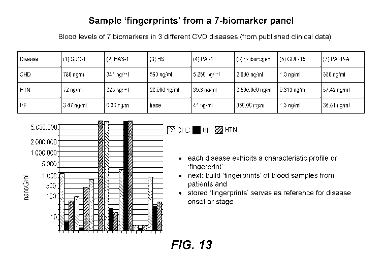
[0095] FIGURE 13 shows sample biomarker "signatures," defined by
patterns of
absolute or relative biomarker levels for three different cardiovascular
diseases when
clinical data are analyzed using a 7-biomarker combination.
[0096] FIGURE 14 is a chart showing the effects of different three-
drug
combinations, as assessed using the biomarkers hyaluronan synthase-2
("Hyaluronan"),
heparan sulfate, and plasminogen activator inhibitor ("PAI-1").
DETAILED DESCRIPTION
[0097] In some embodiments, the present disclosure is generally
directed to methods
and compositions that restore the glycocalyx. Disruption of the glycocalyx is
at the root of
many diseases, especially cardiovascular disease. The compositions of the
present
disclosure maintain the integrity of glycocalyx in many different membranes.
Definitions
[0098] Terms used in the claims and specification are defined as set
forth below
unless otherwise specified.
[0099] "Vascular disease," as used herein, refers to any disease affecting
the
circulatory system of arteries, veins, capillaries, and lymph vessels in the
body. Vascular
disease can include, but is not limited to, peripheral artery disease,
aneurysms, renal artery
disease, Raynaud's disease, Buerger's disease, peripheral venous disease,
varicose veins,
blood clots (thromboembolism), blood clotting disorders, and lymphedema.
[0100] A "thrombus" is a solid mass consisting of platelets, fibrin and
blood
components.
[0101] An "embolus" is a piece of thrombus broken free and carried
into the
bloodstream.
[0102] A "thromboembolus is a floating embolus that becomes lodged in
a blood
.. vessel and blocks blood flow
[0103] "Thromboembolism," as used herein, refers to obstruction of a
blood vessel
by a blood clot, which can occur in a family of vascular diseases including
coronary heart
disease (CHD), acute myocardial infarction (MI), stroke, hypertension, atrial
fibrillation,
-19-
CA 03155843 2022-03-24
WO 2021/062298
PCT/US2020/052912
congestive heart failure (CHF), congenital heart condition, peripheral
arterial disease
(PAD), chronic venous insufficiency (CVI), deep venous thrombosis (DVT), and
pulmonary
embolism (PE).
[0104] "Cardiovascular disease" (CVD) includes a family of diseases
affecting both
arteries and veins, as well as the heart: diseases in the arteries include
coronary heart
disease (CHD), myocardial infarction (MI), stroke, hypertension, atrial
fibrillation,
congestive heart failure (CHF), congenital heart condition, and peripheral
arterial disease
(PAD); diseases in the veins include venous thrombosis, deep venous thrombosis
(DVT),
and pulmonary embolism (PE).
[0105] "Coronary heart disease" (CHD) results from the effects of
atherosclerotic
plaque formation in coronary arteries. The reduction in blood supply to the
heart muscles
reduce the heart's efficiency and can cause heart failure. One of the first
and major
symptoms of this condition is angina (chest pain caused by reduced blood flow
to the heart
muscle).
[0106] "Myocardial infarction" (MI), commonly known as heart attack, is the
irreversible necrosis of heart muscle due to prolonged interruption of blood
supply
(ischemia). The heart requires constant supply of oxygen and nutrients; if one
of the
arteries or branches becomes blocked suddenly, the heart is starved of oxygen,
a condition
called "cardiac ischemia." If cardiac ischemia lasts too long, the starved
heart tissue dies,
which is called heart attack (myocardial infarction), literally, "death of
heart muscle."
[0107] "Stroke" occurs when brain cells die owing to a lack of blood
supply, which
may be classified as ischemic or hemorrhagic: ischemic stroke involves
decreased blood
supply to parts of the brain, leading to brain cell death and thus brain
dysfunction;
hemorrhagic stroke is due to rupture of a blood vessel or abnormal vascular
structure,
.. causing accumulation of blood in a part of the brain. The majority of
strokes (80%) are
ischemic in nature.
[0108] "Hypertension" or "high blood pressure" is defined as a
condition wherein
the pressure of the blood flowing through blood vessels remains high for a
prolonged
period, irrespective of the body's need. An increased blood pressure leads the
heart to work
harder, which makes the heart and arteries more susceptible to injury.
Hypertension further
increases the risk of incidents such as heart attack, heart failure, and
atherosclerosis.
-20-
CA 03155843 2022-03-24
WO 2021/062298
PCT/US2020/052912
[0109] Cardiac arrhythmias are heart rhythm problems, which occur when
heartbeats are not well coordinated owing to improper electric impulses. This
may cause
the heart to beat too fast (tachycardia) or too slowly (bradycardia).
Arrhythmias are
generally harmless and momentary, but frequent rhythm disturbances increase
the risk of
.. stroke and congestive heart failure. Atrial fibrillation is the most common
sustained
arrhythmia.
[0110] "Congestive heart failure" (CHF) is a condition wherein the
heart fails to
supply blood to the various parts of the body. This can be due to narrowed
arteries,
myocardial infarction, heart valve disease, high blood pressure,
cardiomyopathy, or
congenital abnormalities.
[0111] "Peripheral artery disease" (PAD) is a vascular disorder in
which the
thickening of arteries causes reduction in blood flow to limbs, leading to
intermittent leg
pain while walking. The disease is an indicator of atherosclerosis. It leads
to sores (that do
not heal) and gangrene.
[0112] "Deep Vein Thrombosis" (DVT) is a blood clot that usually forms in
the
deep veins of the lower leg or arm, which can block the venous return. A DVT
may cause
leg pain or swelling but can also present no symptoms. DVT is not usually life
threatening,
but it can become so if the blood clot breaks loose and lodges into the lungs.
This is known
as a "pulmonary embolism" (PE).
[0113] The term "healthy" as used herein refers to a state of an organ or
individual
that is free from disease (e.g., vascular disease), is in good health, and has
no particular,
known, physiologically based risk of developing the disease (e.g., vascular
disease).
[0114] The term "compound," as used herein, refers to "a substance
including atoms
or ions of two or more different elements in definite proportions joined by
chemical bonds
into a molecule.
[0115] The term "composition," as used herein, refers to a substance
that includes a
compound, often in combination with other compounds or elements.
[0116] "Disrupting" or "disruption of' the glycocalyx, as used herein
refers to any
process or disease state that affects the glycocalyx such that it is not
functioning normally.
.. Disruption can be caused by inflammation or oxidation in the body.
Disruption can cause
-21-
CA 03155843 2022-03-24
WO 2021/062298
PCT/US2020/052912
the glycocalyx to thin and lose its component proteoglycans. For example, the
dimensions
or percentage of glycocalyx relating to blood vessels are:
Vessel Diameter (nm) Glycocalyx Thickness (nm) % Glycocalyx
Venules 20,900 638 3.05
5 Arterioles 18,000 551 3.06
Capillaries 8,200 348 4.24
Thus, disruption means abnormal shedding of glycocalyx resulting in the loss
of integrity
and thickness, particularly a glycocalyx thickness less than 3.0% the diameter
of venules or
arterioles, and a glycocalyx thickness of less than 4.2% the diameter of
capillaries.
[0117] An agent is said to have "activity in reducing disruption of the
glycocalyx" if
the agent reduces disruption of the glycocalyx as determined by any means
described herein
or known in the art.
[0118] "Inflammation," as used herein, refers to a protective response
of tissue to
injury or destruction in order to eliminate or cordon off any injurious agent
and the injured
tissue and initiate tissue repair. Inflammation can cause pain, heat, redness,
swelling, and
loss of function. Inflammatory mediators (cytokines and chemoattractants) can
cause
shedding of the glycocalyx. Inflammation can also cause leukocytes to
degranulate,
releasing enzymes that can degrade the glycocalyx.
[0119] "Anti-inflammatory," as used herein refers to a molecule,
compound, or
composition that inhibits any inflammatory process or symptom thereof, such as
those
described herein or otherwise known in the art. An anti-inflammatory is said
to have "anti-
inflammatory activity."
[0120] "Oxidative damage," "oxidative stress," or "oxidation," as used
herein, refers
to an imbalance of reactive oxygen species (ROS) and the body's ability to
detoxify reactive
intermediates and repair damage caused by ROS. Inflammation can cause the
release of
ROS. The presence of ROS can cause significant damage to cell structures,
including the
glycocalyx. On a molecular level, "oxidation" refers to the loss of electrons
during a
reaction by a molecule, atom or ion.
-22-
CA 03155843 2022-03-24
WO 2021/062298
PCT/US2020/052912
[0121] As used herein, the term "symptom" refers to a mental or
physical
manifestation that is regarded as indicating a disease or condition.
[0122] The term "symptom-targeted drug," as used herein refers to a
drug that
ameliorates a symptom of a disease or condition that may or may not address
the underlying
pathology.
[0123] The term "treat" when used with reference to treating, e.g., a
disease or
condition refers to the mitigation and/or elimination of one or more symptoms
of that
disease or condition, and/or a delay in the progression and/or a reduction in
the rate of onset
or severity of one or more symptoms of that disease or condition, and/or the
prevention of
.. that disease or condition. The term treat encompasses therapeutic
treatment, as well as
prophylactic treatment which includes a delay in the onset or the prevention
of the onset of
a disease or condition.
[0124] An amount of a therapeutic compound is said to be "s" when the
amount is
effective to achieve improvement including but not limited to improved
survival rate or
more rapid recovery, or improvement or elimination of a symptom and other
indicator (such
as a biomarker) as are selected as appropriate measures by those skilled in
the art.
[0125] "Antioxidant," as used herein, refers to a molecule that
inhibits the oxidation
of other molecules and is able to neutralize or eliminate ROS. An antioxidant
is said to
have "anti-oxidant activity."
[0126] The term "assay," as used herein, refers to a procedure that
determines the
amount of a particular constituent of a mixture or sample. "Assay" is used
interchangeably
with the term "test" herein.
[0127] The term "biomarker," as used, herein refers to a substance,
such as, but not
limited to, a protein, DNA sequence, RNA sequence, or other biological
substance or
substances that, when detected, indicates a particular healthy or unhealthy
state of an
individual with respect to a disease (e.g., vascular disease).
[0128] The term "sample," as used herein, typically refers to a
biological sample
from an individual, and can be, but is not limited to, blood, plasma, urine,
saliva, tears, or
cerebral spinal fluid (CS F).
-23-
CA 03155843 2022-03-24
WO 2021/062298
PCT/US2020/052912
[0129] As used herein, the term "biomarker panel" generally refers to
combination
of reagents useful in detecting a plurality of biomarkers. A biomarker panel
is typically
provided in a kit for detecting two or more biomarkers. In the art, the term
"biomarker
panel" may be used to refer to a combination of biomarkers per se (as opposed
to reagents
for detecting the biomarkers); the sense in which this term is used herein
will be readily
apparent to those of skill in the art from the context in which this term is
used.
[0130] "Detectable labels" include any composition detectable by
spectroscopic,
photochemical, biochemical, immunochemical, electrical, optical, or chemical
means.
Useful labels include magnetic beads (e.g., DynabeadsTm), fluorescent dyes
(e.g.,
fluorescein, Texas red, rhodamine, green fluorescent protein, and the like,
see, e.g.,
Molecular Probes, Eugene, Oregon, USA), chemiluminescent compounds such as
acridinium (e.g., acridinium-9-carboxamide), phenanthridinium, dioxetanes,
luminol and the
like, radiolabels (e.g., 3H, 1251, 35s, 14,,u,
or 32P), catalysts such as enzymes (e.g., horse radish
peroxidase, alkaline phosphatase, beta-galactosidase and others commonly used
in an
ELISA), and colorimetric labels such as colloidal gold (e.g., gold particles
in the 40 -80 nm
diameter size range scatter green light with high efficiency) or colored glass
or plastic (e.g.,
polystyrene, polypropylene, latex, etc.) beads. Patents teaching the use of
such labels
include U.S. Patent Nos. 3,817,837; 3,850,752; 3,939,350; 3,996,345;
4,277,437; 4,275,149;
and 4,366,241.
[0131] As used herein, an "antibody" refers to a protein consisting of one
or more
polypeptides substantially encoded by immunoglobulin genes or fragments of
immunoglobulin genes. The recognized immunoglobulin genes include the kappa,
lambda,
alpha, gamma, delta, epsilon and mu constant region genes, as well as myriad
immunoglobulin variable region genes. Light chains are typically classified as
either kappa
.. or lambda. Heavy chains are typically classified as gamma, mu, alpha,
delta, or epsilon,
which in turn define the immunoglobulin classes, IgG, IgM, IgA, IgD and IgE,
respectively.
[0132] A typical full-length (intact) immunoglobulin (antibody)
structural unit is
known to comprise a tetramer. Each tetramer is composed of two identical pairs
of
polypeptide chains, each pair having one "light" (about 25 IcD) and one
"heavy" chain
(about 50-70 IcD). The N-terminus of each chain defines a variable region of
about 100 to
110 or more amino acids primarily responsible for antigen recognition. The
terms variable
-24-
CA 03155843 2022-03-24
WO 2021/062298
PCT/US2020/052912
light chain (VL) and variable heavy chain (VII) refer to these light and heavy
chains
respectively.
[0133] Antibodies exist as intact immunoglobulins or as a number of
well-
characterized fragments that can be produced, inter alia, by digestion with
various
peptidases. Thus, for example, pepsin digests an antibody below the disulfide
linkages in
the hinge region to produce F(ab)'2, a dimer of Fab which itself is a light
chain joined to VH-
CH1 by a disulfide bond. The F(ab)'2 may be reduced under mild conditions to
break the
disulfide linkage in the hinge region thereby converting the (Fab')2 dimer
into a Fab'
monomer. The Fab' monomer is essentially a Fab with part of the hinge region
(see,
Fundamental Immunology, W.E. Paul, ed., Raven Press, N.Y. (1993), for a more
detailed
description of other antibody fragments). While various antibody fragments are
defined in
terms of the digestion of an intact antibody, one of skill will appreciate
that such Fab'
fragments may be synthesized de novo either chemically or by utilizing
recombinant DNA
methodology. Thus, the term antibody, as used herein also includes whole
antibodies,
antibody fragments either produced by the modification of whole antibodies or
synthesized
de novo using recombinant DNA methodologies. In certain embodiments antibodies
include single chain antibodies (antibodies that exist as a single polypeptide
chain), for
example, single chain Fv antibodies (scFv) in which a variable heavy and a
variable light
chain are joined together (directly or through a peptide linker) to form a
continuous
polypeptide. In certain embodiments the single chain Fv antibody is a
covalently linked VH_
VL heterodimer that may be expressed from a nucleic acid including VH and VL
encoding
sequences either joined directly or joined by a peptide-encoding linker (see,
e.g., Huston, et
al. (1988) Proc. Nat. Acad. Sci. USA, 85: 5879-5883). While the VH and VL are
connected
to each as a single polypeptide chain, the VH and VL domains associate non-
covalently. The
first functional antibody molecules to be expressed on the surface of
filamentous phage
were single-chain Fv's (scFv), however, alternative expression strategies have
also been
successful. For example, Fab molecules can be displayed on phage if one of the
chains
(heavy or light) is fused, for example, to g3 capsid protein and the
complementary chain
exported to the periplasm as a soluble molecule. The two chains can be encoded
on the
same or on different replicons. The important point is that the two antibody
chains in each
Fab molecule assemble post-translationally and the dimer is incorporated into
the phage
particle via linkage of one of the chains to, e.g., g3p (see, e.g., U.S.
Patent No: 5,733,743).
-25-
CA 03155843 2022-03-24
WO 2021/062298
PCT/US2020/052912
The scFv antibodies and a number of other structures converting the naturally
aggregated,
but chemically separated light and heavy polypeptide chains from an antibody V
region into
a molecule that folds into a three-dimensional structure substantially similar
to the structure
of an antigen-binding site are known to those of skill in the art (see e.g.,
U.S. Patent Nos.
5,091,513, 5,132,405, and 4,956,778). Accordingly, in certain embodiments,
anti-Fc
receptor antibodies include, but are not limited to all that have been
displayed on phage or
yeast (e.g., scFv, Fv, Fab and disulfide linked Fv (see, e.g., Reiter et al.
(1995) Protein Eng.
8: 1323-1331)).
[0134] Antibodies also include "single-domain" antibodies (sdAbs),
also known as a
nanobodies. Single-domain antibodies consist of a single monomeric variable
antibody
domain. Like a common "whole antibody," it is able to bind selectively to a
specific
antigen. With a molecular weight of only 12-15 kDa, single-domain antibodies
are much
smaller than common antibodies (150-160 kDa) which are composed of two heavy
protein
chains and two light chains, and even smaller than Fab fragments (-50 kDa, one
light chain
and half a heavy chain) and single-chain variable fragments (-25 kDa, two
variable
domains, one from a light and one from a heavy chain). Some species, such as
camelids,
produce single-domain antibodies naturally.
[0135] The term "monoclonal antibody" refers to an antibody obtained
from a
population of substantially homogeneous antibodies, i.e., the individual
antibodies
comprising the population are identical except for possible naturally
occurring mutations
and/or post-translation modifications (e.g., isomerizations, amidations, etc.)
that may be
present in minor amounts. Monoclonal antibodies are typically highly specific,
being
directed against a single epitope. In contrast to polyclonal antibody
preparations which
typically include different antibodies directed against different determinants
(epitopes), each
monoclonal antibody is directed against a single determinant on the antigen.
The term
"monoclonal" indicates the character of the antibody as being obtained from,
or one of, a
substantially homogeneous population of antibodies, and is not to be construed
as requiring
production of the antibody by any particular method. For example, monoclonal
antibodies
may be made by a variety of techniques, including, but not limited to, the
hybridoma
method (see, e.g., Kohler and Milstein. (1975) Nature, 256:495-497; Hongo et
al. (1995)
Hybridotna, 14 (3): 253-260; Harlow et al. (1988) Antibodies: A Laboratory
Manual (Cold Spring
Harbor Laboratory Press, 2d ed.); Hammerling et al. (1981) In: Monoclonal
Antibodies and T-
-26-
CA 03155843 2022-03-24
WO 2021/062298
PCT/US2020/052912
Cell Hybridotnas 563-681 (Elsevier, N.Y.)), recombinant DNA methods (see,
e.g., U.S. Patent
No. 4,816,567), phage-display technologies (see, e.g., Clackson et al. (1991)
Nature, 352: 624-
628; Marks et al. ( 1992) J. MoL Biol. 222: 581-597; Sidhu et al. (2004) J.
Mol. Biol. 338(2): 299-
310; Lee et al. (2004) J. MoL Biol. 340(5): 1073-1093; and the like), and
technologies for
producing human or human-like antibodies in animals that have parts or all of
the human
immunoglobulin loci or genes encoding human immunoglobulin sequences (see,
e.g., PCT
Patent Publication Nos: WO 1998/24893; WO 1996/34096; WO 1996/33735; and WO
1991/10741; U.S. Patent Nos: 5,545,807; 5,545,806; 5,569,825; 5,625,126;
5,633,425; and
5,661,016; Jakobovits et al. (1993) Nature 362: 255-258; Bruggemann et al.
(1993) Year in
ItntnunoL 7: 33; Marks et al. ( 1992) Bio/Technology 10: 779-783; Lonberg et
al. (1994) Nature
368: 856-859; Morrison (1994) Nature 368: 812-813; Fishwild et al. (1996)
Nature BiotechnoL
14: 845-851); Neuberger (1996) Nature Biotechnol. 14: 826; Lonberg and Fluszar
(1995)
Intern. Rev. Itntnunol. 13: 65-93; and the like).
[0136] As used herein, the phrase "causing to be measured" refers to
any action
resulting in a measurement being taken. For example, a physician causes a
biomarker to be
measured when the physician orders a test for that biomarker to be performed
on a sample
from a given subject.
[0137] As used herein, a "biomarker signature" refers to a pattern of
absolute or
relative biomarker levels for two or more biomarkers that is characteristic of
a particular
disease or condition or stage of disease or condition.
[0138] As used herein with reference to a disease or condition, the
term "stage"
refers to the level of biological severity of the disease or condition. Many
diseases have
well-defined staging criteria.
[0139] As used herein, the term "xenobiotic" refers to a substance,
typically a
chemical, that is foreign to a body, wherein chronic exposure to the body
results in a
deleterious effect (on the body), manifested as one or more chronic diseases,
herein defined
as "xenodiseases."
[0140] A "pathogen" is a microorganism (e.g., a bacterium or virus)
that can cause
infectious disease.
-27-
CA 03155843 2022-03-24
WO 2021/062298
PCT/US2020/052912
[0141] As
used herein, the term "differential diagnosis" refers to the determination
of which of two or more diseases with similar symptoms is likely responsible
for a subject's
symptom(s), based on an analysis of the clinical data.
[0142] The
term "prognosis" is used herein to refer to the likely course of a disease
or condition.
The Glycocalyx and Disease
[0143] The
glycocalyx is a key structure for maintaining vascular wall integrity and
the proper function of many organs. Disruptions in the glycocalyx can be due
to: contact
with fluid flow or low shear stress particularly at arterial bends and
bifurcations; physical
damage or injury; infections or exposure to xenobiotics; oxidation and
inflammation; and
loss of protective enzymes and proteins. An unimpeded blood flow, particularly
on straight
segments of arterial vessels with high shear stress is typically characterized
by a thick
glycocalyx layer and the absence of plaque. A thin glycocalyx promotes plaque
buildup,
especially where there is whirlpool blood flow with low shear in vascular
bends. Plaques
are essentially patches that cover tiny gaps to maintain osmotic balance of
membranes. The
tiny gaps in the membrane leak electrolytes both into (Na+C1-, Ca+, HCO3) and
out of (K+,
PO4-, Mg+) cells, which can lead to a family of cardiovascular diseases.
Disruptions can
also be caused by debris trapped in the stagnant blood flow, which triggers
oxidation and
inflammation.
[0144] Any disruption or decrease in thickness of the glycocalyx can result
in many
different conditions, including chronic vascular disease (2010. Cardiovascular
Research.
Volume 87, Issue 2 pp. 300 ¨ 310). For example, chronic stagnant blood flow,
common in
bifurcated sections of the arteries, triggers glycocalyx shedding and plaque
formation. In
the heart, disrupted glycocalyx in the coronaries result in poor blood flow
(coronary
perfusion); at the arteriolar level, a damaged glycocalyx slows down blood
flow and
decreases nitric oxide (NO) production, constricting vessels; and, at the
capillary level,
disrupted glycocalyx reduces blood flow to tissues or muscles. In addition,
the glycocalyx
harbors a wide array of enzymes that regulate proper blood flow including
superoxide
dismutase (SOD), an enzyme which neutralizes reactive oxygen species;
antithrombin (AT-
III), a natural anticoagulant (blood thinner); and, lipoprotein lipase (LPL),
an enzyme that
-28-
CA 03155843 2022-03-24
WO 2021/062298
PCT/US2020/052912
releases triglycerides from chylomicrons and very low-density lipoproteins
(VLDL) for
energy. See FIGURE 11.
[0145] In case of cardiac ischemia/reperfusion injury (heart muscle
damage due to
blood flow obstruction, then re-establishment of blood supply), disrupted
glycocalyx results
in coronary constriction, poor blood flow, and edema. However, pre-treatment
of the heart
with antithrombin reduces glycocalyx shedding and restores coronary functions
(2009.
Cardiovascular Research. Volume 83, Issue 2Pp. 388 ¨ 396).
[0146] Other more general consequences of a disrupted glycocalyx
include osmotic
gradient shifts, leakage between cells (such as vascular, kidney, and lung
cells), macrophage
infiltration and inflammation, and tissue dysfunction. Eventually, glycocalyx
dysfunction
can lead to blockage of flow in vasculature, the kidneys, the pancreas, and
other organs and
tissue.
Cardiovascular disease
[0147] Cardiovascular disease (CVD) is the leading disease killer in
the world and
because of its complexity and manifested clinical sequelae, it continues to be
the main
subject in pathology research. Although members of the CVD family are totally
different in
clinical presentations, they are basically atherosclerosis-related and share a
common feature,
which is vascular damage, particularly to the endothelial glycocalyx. Once the
vasculature
is damaged, the thromboembolism cascade ensues. Thromboembolism as a process
leading
to the formation of thrombus (blood clot); once this thrombus dislodges from
its origin, it
forms an embolus, which flows downstream in the blood vessel tree as a
thromboembolus
and obstructs blood flow, which can be fatal.
[0148] The blood pressure generated by the pumping heart fluctuates
and blood flow
particularly slows down at arterial forks and bends, notably in the coronary
arteries. A
high-fat diet increases blood viscosity and further stagnates blood flow; this
stagnation
creates low shear and consequently shedding or disruption of the endothelial
glycocalyx.
Glycocalyx thickness range from 2 to 3 pm in small arteries to 4.5 pm in
carotid arteries
(2007. J Vasc Res 44:87-98), and shedding or damage to this layer decreases
its function as
a protective shield, leading to leakage of nutrients (extravasation) and
tissue edema, loss of
-29-
CA 03155843 2022-03-24
WO 2021/062298
PCT/US2020/052912
nutritional blood flow, and an increase in coagulability due to platelet and
leucocyte
clumping (adhesion).
[0149] The endothelial glycocalyx offers a "nest" for protective
enzymes including
anticoagulant anti-thrombin (AT-III), anti-oxidant (SOD), and anti-high blood
viscosity
lipoprotein lipase (LpL). Thus, the loss of endothelial glycocalyx results in
a build-up of
fibrin, a suppression of fibrinolysis, and the promotion of plaque formation.
Further
inflammation predisposes the plaque to rupture, and ruptured plaque triggers
clots
formation: this clot can be exacerbated by a seed clot formed by Roleaux cells
and thus
becoming a significant thrombus. Loose thrombi can wedge on a rigid vessel
narrowed by
plaque, particularly in individuals who already have atherosclerosis, causing
a stroke
(clogged artery to the brain), heart attack (clogged artery to the heart), or
PAD (clogged
artery to the arms or legs).
[0150] Thus, protection and/or restoration of the endothelial
glycocalyx presents a
promising therapeutic target both in an acute critical care setting and in the
treatment of
chronic vascular disease. Drugs that can specifically increase the synthesis
of glycocalyx
components, refurbish it, or selectively prevent its enzymatic degradation
have not been
widely available. (2010. Cardiovascular Research, Volume 87, Issue 2 pp. 300
¨310).
However, compounds aimed at restoring and maintaining the glycocalyx have been
described in U.S. Patent No. 9,867,842 (issued January 16, 2019 to Tunac),
which is
incorporated by reference herein for this description.
[0151] Under inflammatory conditions the integrity of the endothelial
glycocalyx
deteriorates to varying degrees particularly during generalized inflammatory
responses, but
glycocalyx can regain its original thickness after proper treatment of
inflammatory
condition (2008. Circulation Research, vol. 102, no. 7, pp. 770-776). Thus,
therapeutic
strategies can be directly aimed at preserving, supporting, or reconstituting
the glycocalyx
structure or strategies, either indirectly by down-regulating inflammatory
processes or
directly by inhibition of glycocalyx degradation with antioxidants (2006.
American Journal
of Physiology: Heart and Circulatory Physiology, vol. 290, no. 6, pp.
H2247¨H2256). An
example of an anti-inflammatory drug is etanercept (Enbrel), which inhibits
TNF-a, and
reduces the shedding of glycocalyx constituents, coagulation activation, and
functional
vessel function in humans (2009. Atherosclerosis, vol. 202, no. 1, pp. 296-
303).
-30-
CA 03155843 2022-03-24
WO 2021/062298
PCT/US2020/052912
[0152] Another approach to improving the condition of the glycocalyx
is
antithrombin therapy, since thrombin is known to cleave the syndecan component
of
glycocalyx (2009. Circulation Research, vol. 104, no. 11, pp. 1313-1317).
Indeed,
antithrombin therapy protects the glycocalyx from TNF-a and
ischemia/reperfusion-induced
shedding in hearts (2009. Basic Research in Cardiology, vol. 104, no. 1, pp.
78-89; 2010.
Shock, vol. 34, no. 2, pp. 133-139), which can result in reduced post-ischemic
leukocyte
adhesion in hearts, reduced vascular permeability, reduced coronary leak, and
reduced
interstitial edema (2009. Basic Research in Cardiology, vol. 104, no. 1, pp.
78-89).
Biomarkers of cardiovascular disease
[0153] Biomarkers have been identified that can be useful for identifying
individuals at risk of vascular diseases. For example, biomarkers of
inflammation can
indicate the presence of atherosclerosis or plaques (e.g., C-reactive protein,
IL-18, IL-6).
Biomarkers of lipid accumulation can indicate the presence of plaques (e.g.,
lipoprotein-
associated phospholipase A2). Biomarkers of thrombosis can indicate the
presence of
plaque instability or carotid disease progression (e.g., tissue plasminogen
activator (t-PA),
fibrinogen, plasminogen activator inhibitor-I (PAI-1)). However, no such
biomarkers are
currently in use by medical practitioners as a diagnostic tool.
[0154] U.S. Patent Application No. 2007/0269836 to McPherson, et al.
discloses
methods and compositions for diagnosis of venous thromboembolic disease,
pulmonary
.. embolism, and/or deep vein thrombosis, and for risk stratification in such
conditions. An
assay can be performed from test samples obtained from a subject to diagnose a
subject,
including markers used individually or in combination, such as thrombin-
antithrombin
complex (TAT), antithrombin III (ATIII), and PAI-1.
[0155] U.S. Patent No. 8,759,095 to Vink, et al. discloses diagnostic
and therapeutic
tools for diseases altering vascular function. In particular, endothelial
glycocalyx
perturbation can be diagnosed in samples from subjects by detecting heparan
sulfate (HS)
(heparan sulphate therein), hyaluronidase (HAD), and syndecan-1.
[0156] U.S. Patent Application No. 2013/0273096 to Daniels discloses
methods of
treating disorders affecting the endothelial glycocalyx. Characteristics of
the endothelial
-31-
CA 03155843 2022-03-24
WO 2021/062298
PCT/US2020/052912
glycocalyx can be determined by detecting markers in a sample from a subject,
such as
heparan sulfate (HS), hyaluronidase (HAD), and syndecan-1.
Biomarkers Haying Particular Utility in the Methods Described Herein
[0157] Biomarkers useful in the methods described herein include those
described in
PCT Pub. No. WO 2016/123163 (filed January 27, 2016 by Tunac), as well as
biomarkers
that are newly described for this purpose. The biomarkers described herein can
be used
individually or in any combination, depending on the particular type of
condition or disease
to be detected. Illustrative combinations of biomarkes have been developed, as
described
below and in the Examples.
Biomarkers described in PCT Pub. No. WO 2016/123163
[0158] In certain embodiments, the disclosure of U.S. Patent
Application No.
16/060,840 is directed to "panels" of biomarkers used to detect a disease
characterized by
disruption of the glycocalyx, endothelial inflammation, and/or oxidative
damage to the
endothelium (e.g., vascular diseases), and especially biomarkers that indicate
abnormal
biochemical elements responsible for the blood clotting cascade and biomarkers
that
indicate abnormal levels of enzymes and structural components of the blood
vessel surface
(e.g., due to vascular oxidative damage; PCT Pub. No. WO 2016/123163 is
incorporated by
reference herein for this description).
[0159] Most generally, the biomarker panels include of a set of
chemical,
immunochemical and/or enzymatic assays or tests that can be used together for
monitoring
the levels of a set of biomarkers. The biomarker panels can be used to
determine the
presence of disease, or the propensity of an individual to develop disease.
The biomarker
panels can also be used to mark the progression of disease. Evaluation of
different stages or
components of vascular disease is important for intervention or reversal of
the effects of the
disease. For each of the biomarkers discussed herein, a baseline level for the
biomarker is
known or can be established that reflects the levels in a healthy individual.
A healthy
individual should have lower levels of the biomarker than an individual
suffering from a
disease characterized by disruption of the glycocalyx endothelial
inflammation, and/or
oxidative damage to the endothelium. If a biomarker level is above the
baseline level in a
sample from a subject, it can be determined that the subject has a disease
characterized by
-32-
CA 03155843 2022-03-24
WO 2021/062298
PCT/US2020/052912
disruption of the glycocalyx, endothelial inflammation, and/or oxidative
damage to the
endothelium (e.g., vascular disease) or is at risk of developing such a
condition. Other
levels can determine the stage or progression of the disease.
4-Marker panel: soluble fibrin, thrombin-antithrombin complex,
antithrombin III, and plasminogen activator inhibitor
[0160] In some embodiments, the biomarker panel can include a four-
marker test for
endothelial glycocalyx health that detects soluble fibrin (SF), thrombin-
antithrombin
complex (TAT), antithrombin III (ATIII), and plasminogen activator inhibitor
(PAI-1).
These markers are aimed at assessing clotting or clotting risk in a subject.
[0161] Soluble fibrin (SF) is composed of fibrin monomer and fibrinogen
derivatives, existing in the circulating blood in patients with thrombosis.
Its detection and
quantification are useful for obtaining information about the condition and
degree of
intravascular coagulation in early-stage thrombosis. The level of SF increases
on
coagulation, which is related to the production of blood factor VIII. Thus,
factor VIII
circulates in the plasma bound to von Willebrand factor (vWf). Thrombin
cleaves and
activates factor VIII and releases vWf. The vWf is then free to bind to
ruptured endothelial
cell surfaces where it activates platelet aggregation. The released FVIIIa
acts as a cofactor
of factor IXa to generate factor Xa. In the presence of Ca2+ and
phospholipids, FX is
activated to FXa by FIXa. Since FVIIIa is a cofactor to FIXa, it greatly
stimulates the
reaction. A healthy individual should have lower levels of SF than a diseased
individual. If
levels are detected with the biomarker panel that are above the baseline
level, it can be
determined that the individual has vascular disease or is at risk of
developing vascular
disease and especially thrombosis. Other levels can determine the stage or
progression of
vascular disease.
[0162] Another blood component that reflects blood coagulation is the
formation of
thrombin-antithrombin complex (TAT). TAT complex is a parameter of coagulation
and
fibrinolysis. Elevated concentrations have been associated with vascular
disease.
Antithrombin deficiency promotes clot formation in the arteries and/or veins
and is
associated with a high risk of thromboembolic disorders. TAT can conveniently
be detected
.. using commercially available microtiter plates. These microtiter plates
precoated with
antibody specific to thrombin. Calibrators or samples are added to the
appropriate
-33-
CA 03155843 2022-03-24
WO 2021/062298
PCT/US2020/052912
microtiter plate wells with a biotin-conjugated polyclonal antibody
preparation specific for
ATIII. Next, avidin-conjugated to horseradish peroxidase (HRP) is added to
each
microplate well and incubated. Then, an HRP TMB substrate solution is added to
each
well. Only those wells that contain TAT, biotin-conjugated antibody and enzyme-
conjugated avidin will exhibit a change in color. The enzyme-substrate
reaction is
terminated by the addition of a sulfuric acid solution, and the color change
is measured
spectrophotometrically at a wavelength of 450 nm 10 nm. The concentration of
TAT in
the samples is then determined by comparing the O.D. of the samples to the
calibration
curve.
[0163] Antithrombin III (AT III) is a non-vitamin K-dependent protease
enzyme,
which serves as a natural blood thinner and inhibits coagulation. AT III
deficiency leads to
increased risk of developing life-threatening clots that block blood flow. For
example, deep
vein thrombosis (DVT) occurs when a clot, or thrombus, develops in one of the
deep veins,
most common in the legs. The level of AT III is reduced when blood coagulates,
which is
determined by commercially available test kits. One such example of a test kit
is the LS-
F13067, which is a 96-well enzyme-linked immunosorbent assay (ELISA) for the
quantitative detection of bovine antithrombin-III in samples of plasma and
serum. It is
based upon a sandwich assay principle and can be used to detect levels of
Antithrombin-III
as low as 78 picograms per milliliter. Another example is the AssayMax Mouse
AT III
ELISA kit (LSBio, Seattle WA 98121): this is designed for detection of mouse
AT III in
plasma, serum and cell culture supernatants. This assay employs a quantitative
sandwich
enzyme immunoassay technique, which measures AT III in 4 hours. Thus, a
microtiter
plate pre-coated with polyclonal antibody specific for mouse AT III is
commercially
available. Mouse AT III in standards and samples is sandwiched by the
immobilized
antibody and biotinylated polyclonal antibody specific for mouse AT III, which
is
recognized by a streptavidin-peroxidase conjugate. All unbound material is
then washed
away, and a peroxidase enzyme substrate is added. The color development is
stopped and
the intensity of the color is measured. A baseline level for AT III can be
established that
reflects the levels in a healthy individual. A healthy individual should have
lower levels of
AT III than a diseased individual. If levels are detected with the biomarker
panel that are
above the baseline level, it can be determined that the individual has
vascular disease or is at
-34-
CA 03155843 2022-03-24
WO 2021/062298
PCT/US2020/052912
risk of developing vascular disease. Other levels can determine the stage or
progression of
vascular disease.
[0164] Plasminogen activator inhibitor (PAI-1) is a protein, also
known as
endothelial plasminogen activator inhibitor or serpin El, and is a central
regulator of the
blood fibrinolytic system and its production precedes thrombosis. In other
words, increased
PAI-1 levels increase the risk for thrombosis, whereas decreased levels cause
recurrent
bleeding. PAI-1 is the main inhibitor of the plasminogen activators and thus
an important
component of the coagulation system that down-regulates fibrinolysis. Reduced
PAI-1
levels result in increased fibrinolysis and an associated bleeding diathesis.
The other PAI,
plasminogen activator inhibitor-2 (PAI-2), is secreted by the placenta and
only present in
significant amounts during pregnancy. Test kits for PAI-1 are available
commercially (a kit
for human PAI-1 is commercially available from Sigma-Aldrich) in which free,
latent or
complex PAI-1 present in plasma reacts with the capture antibody coated and
dried on a
microtiter plate. Any unbound PAI-1 is washed away and an anti-PAI-1 primary
antibody
is added. Excess primary antibody is washed away and bound antibody, which is
proportional to the total PAI-1 present in the samples, is then reacted with
an HRP-labeled
secondary antibody. Following an additional washing step, TMB is then used for
color
development at 450nm. The amount of color development is directly proportional
to the
concentration of total PAI-1 in the sample.
3-Marker panel: syndecan-1, heparan sulfate, and hyaluronidase
[0165] In some embodiments, the biomarker panel can include a three-
marker test
for endothelial glycocalyx health that detects syndecan-1 (SDC1), heparan
sulfate (HS), and
hyaluronidase (HAD). These markers are aimed at assessing glycocalyx
integrity.
[0166] Syndecans are transmembrane domain proteins that carry three to
five
.. heparan sulfate and chondroitin sulfate chains, which harbor a variety of
important ligands
including fibroblast growth factors, vascular endothelial growth factor,
transforming growth
factor-beta, fibronectin, and antithrombin-1. Syndecan-1 (SDC1) is a cell-
surface heparan
sulfate proteoglycan, which is an important component of the protective
endothelial
glycocalyx lining the luminal surface of blood vessels. Key roles for SDC1 is
in endothelial
mechano-sensing and regulation of endothelial integrity and function. Shedding
of
syndecan-1 and heparan sulfate into the circulation is associated with
inflammatory disease
-35-
CA 03155843 2022-03-24
WO 2021/062298
PCT/US2020/052912
and atherosclerosis. Test kits for syndecan, for example, a test kit precoated
with
monoclonal antibody specific for SDC1, are available commercially. Samples are
added to
the appropriate microtiter plate wells with a biotin-conjugated polyclonal
antibody
preparation specific for SDC1. Next, avidin conjugated to HRP is added to each
microplate
.. well and incubated. Then a TMB substrate solution is added to each well.
Only those wells
that contain SDC1, biotin-conjugated antibody and enzyme-conjugated avidin
will exhibit a
change in color. The enzyme-substrate reaction is terminated by the addition
of a sulphuric
acid solution and the color change is measured spectrophotometrically at a
wavelength of
450nm lOnm. The concentration of SDC1 in the samples is then determined by
comparing the O.D. of the samples to the standard curve.
[0167] Heparan sulfates (HSs) are highly negatively charged
polysaccharides with
1-4-linked sulfated glucosamine and uronic acid repeating disaccharide units.
HSs are
present on the cell surface as well as in the extracellular matrix and bind to
proteins
involved in anticoagulation, angiogenesis, microbial infection, and monocyte
adhesion.
HSs are glycoproteins with the common characteristic of containing one or more
covalently
attached chains, including syndecans and glycosylphosphatidylinositol-anchored
proteoglycans (glypicans), the secreted extracellular matrix HSPGs (agrin,
perlecan, type
XVIII collagen), and the secretory vesicle proteoglycan, serglycin. HSs are
implicated in
the pathogenesis of atherosclerosis by their ability to trap plasma
lipoproteins in the arterial
wall and by their influence on cellular migration, adhesion and proliferation.
Intact HS
chains are anti-atherogenic. ELISA test kits for heparan sulfate are available
commercially.
The test includes pretreatment of serum with proteinase (actinase E) to digest
serum
proteins. One volume of dissolved actinase E (20mg/mL in actinase E
dissolution buffer)
can be added against ten volumes of serum and then mixed. Proteins can be
digested at
55 C for 16-20 hours in a water bath. After digestion, the mixture can be
boiled for 5
minutes to stop digestion. After boiling, the mixture can be brought to room
temperature
(15-25 C) and then centrifuged 3,000 rpm, for 10 minutes. After
centrifugation, the
supernatant can be taken and mixed well. The supernatant can then be Heparan
Sulfate
ELISA kit. HS values can be calculated in pretreated samples according to the
Heparan
Sulfate ELISA kit procedure. The calculated HS values must be multiplied
dilution factors
as below to determine the HS concentration in serum. HS concentration =
calculated HS
value x dilution factor x 1.1.
-36-
CA 03155843 2022-03-24
WO 2021/062298
PCT/US2020/052912
[0168] Hyaluronic acid (HA, also called hyaluronan or hyaluronate) is
a negatively
charged, nonsulfated large linear glycosaminoglycan (a class of negatively
charged
polysaccharides) of repeating disaccharide structure D-Glucuronic acid (UDP-
GlcA) and N-
acetylglucosamine (UDP-G1cNac), which is a principal component of endothelial
glycocalyx. HA is distributed widely throughout connective, epithelial, and
neural tissues.
HA is the simplest glycosaminoglycan that provides compression strength,
lubrication and
hydration. A disturbed HA is atherogenic. The removal of HA-rich glycocalyx
with
hyaluronidase is associated with increased vascular permeability leading to
atherogenic
insults. Increased plasma HA and hyaluronidase (HAD) levels are found
associated with
endothelial glycocalyx damage, presence of microvascular diseases and carotid
intima-
media thickness. In an illustrative assay, coated-well immunoenzymatic assay
for the
quantitative measurement of hyaluronidase (HAD) utilizes a polyclonal anti-HAD
antibody
and an HAD-HRP conjugate. The assay sample and buffer are incubated together
with
HAD-HRP conjugate in pre-coated plate for one hour. After the incubation
period, the
wells are decanted and washed five times. The wells are then incubated with a
substrate for
HRP enzyme. The product of the enzyme-substrate reaction forms a blue colored
complex.
Finally, a stop solution is added to stop the reaction, which will then turn
the solution
yellow. The intensity of color is measured spectrophotometrically at 450nm in
a microplate
reader. The intensity of the color is inversely proportional to the HAD
concentration since
HAD from samples and HAD-HRP conjugate compete for the anti-HAD antibody
binding
site. Since the number of sites is limited, as more sites are occupied by HAD
from the
sample, fewer sites are left to bind HAD-HRP conjugate. Standards of known HAD
concentrations are run concurrently with the samples being assayed and a
standard curve is
plotted relating the intensity of the color (0.D.) to the concentration of
HAD. The HAD
.. concentration in each sample is interpolated from this standard curve.
3-4-Marker panels: hyaluronan synthase-1, heparan sulfate,
plasminogen activator inhibitor, and optionally syndecan-1
[0169] The biomarker panel can also include any combination of the
above-
described biomarkers, i.e. they are not limited to being used in combination
in just the three-
marker test and four-pan test. For example, another preferred combination can
include a
panel of hyaluronan synthase-1 (HAS-1), heparan sulfate (HS), and plasminogen
activator
-37-
CA 03155843 2022-03-24
WO 2021/062298
PCT/US2020/052912
inhibitor (PAI-1) as a blood test that defines vascular leakage and clot onset
to correlate
with plaque formation. As shown in the examples below, these three biomarkers
are highly
correlative with plaque formation. In certain embodiments, syndecan-1 (SDC-1)
is added to
these biomarkers to produce a 4-marker panel.
Additional biomarkers
[0170] In addition to the above, three other biomarkers associated
with cell
disruption are useful in the methods described herein and, in some
embodiments, can be
combined with any of the biomarkers and panels described above to complement
the
diagnosis of a wider range of chronic diseases: gamma (y') fibrinogen (GF),
growth
differentiation factor-15) (GDF-15), and pregnancy associated plasma protein-A
(PAPP-A).
Gamma fibrinogen
[0171] Plasma fibrinogen is a coagulation factor and an acute-phase
inflammatory
marker that has been implicated in the pathophysiology of cardiovascular
disease (CVD)
(2005. JAMA. 294:1799-1809). Fibrinogen is a key component of the hemostatic
system,
playing a role in both primary and secondary response. Fibrinogen is composed
of three
pairs of non-identical polypeptide chains. "Gamma (y')" fibrinogen (GF) refers
to the
gamma chain. Thrombin-catalyzed cleavage of fibrinopeptides (Fp) A and B
converts
fibrinogen into fibrin, which spontaneously polymerizes and forms double-
stranded
protofibrils that assemble into branched fibrin fibers, forming the fibrin
clot (2008.
Cardiovasc Hematol Agents Med Chem 6:181-189). GF is a biomarker for early
clotting.
GF was demonstrated to be significantly associated with coronary artery
disease and
myocardial infarction in the Stockholm Coronary Artery Risk Factor Study and
the
Framingham Heart Study. (2007.J Thromb Haemost 5:766 ¨73) and is significantly
associated with stroke, as seen in the Erasmus Stroke Study and others (2012.
Thrombosis
Research 129: 807-809). GF increases during inflammation and is differentially
regulated
from total fibrinogen under pathologic conditions, as demonstrated in the
Periodontitis and
Vascular Events Study (2011. Thromb Haemost 105:605 ¨9).
[0172] Elevated human levels of GF have been reported in a variety of
studies:
-38-
CA 03155843 2022-03-24
WO 2021/062298
PCT/US2020/052912
[0173] Study of 133 patients diagnosed with coronary artery
disease (CAD):
diseased, 0.413 g/L versus 0.299 g/L in the controls (1996. J Biol Chem
271(38):23121-
23125.
[0174] Epidemiologic study on myocardial infarction (MI) in the
Stockholm
Coronary Artery Risk Factor cohort: diseased 0.28 g/L higher than controls
(2007. J
Thromb Haemost 5:766 ¨ 73).
[0175] Study of patients with a history of CVD and periodontal
disease:
highly elevated compared to controls, 0.622 g/L (2010 Clin Chem 2010; 56:781 ¨
8).
[0176] Study validating GF as an independent predictor of CAD:
hypertensive participants 433.36 versus 405.70 mg/dL in controls (2017.Rev Esp
Cardiol.
70:34-41).
[0177] Study showing GF is positively associated with deaths due
to
peripheral artery disease (PAD), heart failure (HF), and CVD deaths: lowest
quartile 8.0 -
24.34 mg/d1; highest quartile? 35.19 mg/di (2015. Arterioscler Thromb Vasc
Biol. 35(12):
2700-2706).
[0178] Study on 3,042 participants of the Framingham Heart Study
Offspring Cohort: individuals with prevalent CVD, 0.278 mg/ml vs. 0.258 mg/ml
without
(2011. Arterioscler Thromb Vasc Biol. Oct; 31(10): 2345-2352).
[0179] Physicians Health Study of 14,916 subjects: levels of 343
mg/dL,
twofold increase in the risk of a myocardial infarction (2013. University
Heart Journal 9:40-
46).
[0180] Study reporting GF is significantly higher in patients
with ischemic
stroke, 0.37 g/L versus 0.32 g/L in controls (2011.Thromb Haemost, 105:430-4).
Growth differentiation factor-15
[0181] Growth differentiation factor-15 (GDF-15) is a protein belonging to
the
transforming growth factor beta superfamily, which functions in regulating
inflammatory
pathways, apoptosis, and cell repair and cell growth associated in
cardiovascular and
neoplastic disorders (2000. Molecular and Cellular Biology. 20 (10): 3742-51).
GDF-15
serves as a prognostic protein in patients with different diseases such as
heart diseases and
-39-
CA 03155843 2022-03-24
WO 2021/062298
PCT/US2020/052912
cancer, expressed in low concentrations in most organs and upregulated because
of injury of
organs such as such as liver, kidney, heart and lung (2005. Shock. 23 (6): 543-
8). GDF-15
is a stress-responsive cytokine, which increases during tissue injury and
inflammatory states
and is associated with cardiometabolic risk. Increased GDF-15 levels are
associated with
cardiovascular diseases such as hypertrophy, heart failure, atherosclerosis,
endothelial
dysfunction, obesity, insulin resistance, diabetes, and chronic kidney
diseases in diabetes.
GDF-15 is an inflammatory marker associated with increased cardiovascular and
noncardiovascular mortality and plays pivotal role in development and
progression of
cardiovascular diseases, such as heart failure, coronary artery diseases,
atrial fibrillation,
diabetes, cancer, and cognitive impairment (2013. Clinical Chemistry 59: 1550-
1552 2014.
Circulation 130:1847-1858). Increased GDF-15 level is linked with the
progression and
prognosis of the disease condition.
[0182]
Elevated human levels of GDF-15 have been reported in a variety of studies:
[0183] One study stratified the blood GDF-15 levels into three
categories,
that is, normal (<1200 pg/mL), moderately elevated (1200-1800 pg/mL), and
highly
elevated (>1800 pg/mL). (2010. Aging Cell, 9: 1057-1064).
[0184] Elevated levels of GDF-15 of >1800 ng/L have been
reported to be
associated with high risk for mortality within one year (2008.BMC Public
Health, 8:148).
[0185] Another studied showed that GDF-15 concentrations >1800
ng/L
correlated with increased risk for all-cause and cardiovascular death compared
to those with
<1200 ng/L. (2012. Clinical Chemistry 58:172-182).
[0186] Elevated GDF-15 levels been reported to be associated
with reduced
endothelium-dependent vasodilation in resistance vessels from <948 ng/L (1st
quartile) -
>1390 ng/L 4th quartile) (2009.Eur Heart J. 30:2346-2353).
[0187] Another study found, given a median concentration of 1253 ng/L at
baseline: hazard ratio (HR) for the highest compared to the lowest quartile
for CV death,
2.63; for sudden death, 3.06; for heart failure (HF) death, 4.3; for cancer
death, 2.5; for
hospitalization for HF, 5.8 (3.2-10) for MI 1.4; and 1.8 for stroke
(2017.Clinical Chemistry
63:1 140-151) (2017).
-40-
CA 03155843 2022-03-24
WO 2021/062298
PCT/US2020/052912
Pregnancy associated plasma protein-A
[0188] Pregnancy-associated plasma protein-A (PAPP-A) level is an
independent
predictor of acute cardiovascular event occurrence. PAPP-A is associated with
thin-cap
plaque leakage and a higher burden of coronary thin-cap fibroatheroma (TCFA).
PAPPP-A
is elevated in patients with acute coronary syndromes and patients with risk
factors, such as
obesity, hypertension, and/or diabetes, relative to healthy subjects (2015.
Biomark Med.
9:731-741). PAPP-A is highly expressed in vulnerable atheromatous plaques
(2016.
Medicine (Baltimore) 95:e2563; 2004. Circulation 109:1724-1728; 2005. Clin
Chem
52:1096-1103).
[0189] Elevated human levels of PAPP-A have been reported in a variety of
studies:
[0190] PAPP-A level is an independent predictor of acute
cardiovascular event
occurrence, associated with thin cap plaque leakage and a higher burden of
coronary thin-
cap fibroatheroma (TCFA); it is elevated in patients with acute coronary
syndromes and
patients with risk factors, such as obesity, hypertension, and/or diabetes
relative to healthy
subjects (2015. Biomark Med. 9:731-741); also, it is highly expressed in
vulnerable
atheromatous plaques (2016. Medicine (Baltimore) 95:e2563). Patients with >3
VH (virtual
histology)-TCFAs have been reported to have a higher PAPP-A level than
patients with 1 to
3 VH-TCFAs or without any VH-TCFA (13.3 11.8 versus 7.8 4.7 versus 7.4
4.7mIU/L,
P<0.001, respectively). (2016. Medicine (Baltimore).95(3): e2563).
[0191] PAPP-A levels in Acute Coronary Syndrome: PAPP-A levels have been
reported to be significantly elevated in patients with acute myocardial
infarction (AMI) and
in patients with unstable angina (UA), with mean levels of 64.26 and 36.23
ng/ml
respectively, whereas mean PAPP-A levels in controls were 10.68 1.04 ng/ml.
(2015.
Indian J Clin Biochem. 30(2): 150-154).
[0192] PAPP-A reportedly correlates with cardiovascular events in diabetic
hemodialysis patients, with a median PAPP-A concentration of 17 mIU/ patients
in the 4th
PAPP-A quartile (<20.9 mIU/L) had an adjusted 2.6-fold increased risk of
sudden death and
2.8-fold increased risk of stroke as compared to the patients in the 1st
quartile
(<13.4 mIU/L) (2014. Atherosclerosis.236:263-269).
-41-
CA 03155843 2022-03-24
WO 2021/062298
PCT/US2020/052912
3-Marker panel: gamma fibrinogen, growth differentiation factor-15,
and pregnancy-associated plasma protein-A
[0193] In some embodiments, a biomarker panel useful in any of the
methods
described herein includes gamma fibrinogen (GF), growth differentiation factor-
15 (GDF-
15), and pregnancy-associated plasma protein-A (PAPP-A).
7-Marker panel: hyaluronan synthase-1, heparan sulfate, plasminogen
activator inhibitor, syndecan-1, gamma fibrinogen, growth
differentiation factor-15, and pregnancy-associated plasma protein-A
[0194] In certain embodiments, a biomarker panel useful in any of the
methods
described herein includes hyaluronan sunthase-1 (HAS-1), heparan sulfate (HS),
plasminogen activator inhibitor (PAI-1), syndecan-1 (SDC-1), gamma fibrinogen
(GF),
growth differentiation factor-15 (GDF-15), and pregnancy-associated plasma
protein-A
(PAPP-A).
[0195] This 7-biomarker combination is useful in determining a
biomarker signature
that indicates a disease type and/or a stage of disease. Accordingly, the 7-
marker
combination can be used in making a differential diagnosis between at least
two diseases
and/or two stages of disease, allowing treatment to targeted to the particular
disease and/or
disease stage.
[0196] This 7-biomarker combination is also useful in associating a
disease
signature with a particular disease. For example, the panel can be used to
detect the levels
of the seven biomarkers in various diseases or disease stages and this raw
data analyzed to
define a disease signature for a given disease or disease stage. For a
particular disease, it
may be determined that a combination of two, three, four, five or six
biomarkers provides a
reliable biomarker signature of the disease or disease stage and/or allows
discrimination
between two or more possible diseases or disease stages, facilitating a
differential diagnosis,
and thereby facilitating treatment aimed at the particular disease or disease
stage identified
in a subject. The 7-biomarker panel can thus be used to determine that only a
subset of
these markers is needed to identify or discriminate a given disease and/or
disease stage from
another. In this way, the 7-biomarker panel can be used in research aimed at
identifying
useful biomarker panels made up of subsets of the seven biomarkers.
-42-
CA 03155843 2022-03-24
WO 2021/062298
PCT/US2020/052912
Biomarker panels and uses thereof
In general
[0197] The biomarker panels described herein can be used alone or in
combination.
For example, the biomarker panels can be used individually if there is a
strong correlation
established for any of the biomarkers in that particular panel, or the
combination of
biomarker panels can be used to ensure reliability.
[0198] The biomarker panels can be used to detect the presence of a
disease
characterized by disruption of the glycocalyx, endothelial inflammation,
and/or oxidative
damage to the endothelium (e.g., vascular disease) or propensity to develop
such a condition
in the following method. In an illustrative embodiment, a sample is taken, or
caused to be
taken (preferably by a healthcare practitioner), from a subject and tested
using the
biomarker panel (either the four-panel test for clotting, the three-panel test
for glycocalyx
integrity, both as described above, or any combination of the above-described
biomarkers).
If any or all of the biomarkers are detected, it can be determined if the
subject has, or is at
risk for, a disease characterized by disruption of the glycocalyx, endothelial
inflammation,
and/or oxidative damage to the endothelium (e.g., vascular disease) by
comparing the
biomarker levels to known baseline levels for healthy individuals. In other
words, if levels
of two biomarkers are detected that are above the baseline level, it can be
determined that
the individual has, or is at risk for, a disease characterized by disruption
of the glycocalyx,
endothelial inflammation, and/or oxidative damage to the endothelium (e.g.,
vascular
disease).
[0199] In some embodiments, the biomarker panel can be used to
determine the
stage of a disease characterized by disruption of the glycocalyx, endothelial
inflammation,
and/or oxidative damage to the endothelium (e.g., vascular disease) in an
individual (e.g., to
monitor the progression of vascular disease in the individual). The stage of a
disease
characterized by disruption of the glycocalyx, endothelial inflammation,
and/or oxidative
damage to the endothelium (e.g., vascular disease) can be determined by
comparing the
results to known stage levels. Based on the results of the stage of vascular
disease, the
individual can be proscribed medication that is appropriate for that
particular stage.
-43-
CA 03155843 2022-03-24
WO 2021/062298
PCT/US2020/052912
[0200] In some embodiments, the biomarker panel can be used to
determine a
prognosis for a disease characterized by disruption of the glycocalyx,
endothelial
inflammation, and/or oxidative damage to the endothelium (e.g., vascular
disease) in an
individual. A negative prognosis, for example, enables a physician and patient
to select a
more aggressive form of treatment than would normally be used at that point in
diagnosis
and treatment.
[0201] In certain embodiments, the biomarker panel can also be used in
a method of
monitoring the efficacy of a drug or other therapy against a disease
characterized by
disruption of the glycocalyx, endothelial inflammation, and/or oxidative
damage to the
endothelium (e.g., cardiovascular diseases or other diseases involving
inflammation,
disruption of blood vessels, plaques, or clots). Such monitoring can be
carried out during
drug development (e.g., in an animal model such as that described herein or in
human
subjects) or during the treatment of actual patients. Illustrative treatments
that can be
monitored include, but are not limited to, anti-inflammatories (such as non-
steroidal anti-
inflammatory drugs (NSAIDS), steroids, or immune selective anti-inflammatory
derivatives
(ImSAIDs)), anticoagulants (such as alteplase, ardeparin, dalteparin,
danaparoid,
enoxaparin, fondaparinux, lepirudin, urokinase, or warfarin), antioxidants
(such as
glutathione, alpha-lipoic acid, CoQ10, resveratrol, carotenoids, astaxanthin
Vitamin C, or
Vitamin E), supplements, and any other suitable therapeutics.
[0202] In some embodiments, the biomarkers included in the panels of the
present
disclosure measure factors produced early on in the clot formation process.
Therefore, each
of these biomarkers are useful alone, as well as together in the panels, in
predicting the
initiation of the biologic processes (e.g., oxidation and immunogenic and/or
inflammatory
processes) that leads to the formation of the clots. The biomarker panels
described herein
may be useful in conjunction with that "lipid panel" devised by the American
Heart
Association (measuring cholesterol and triglyceride), but since the lipid
panel is not reliable
predictive of cardiovascular disease, it is contemplated that one or more of
the biomarker
panels of the present disclosure can replace this lipid panel for routine
diagnostics.
[0203] As those of skill in the art will readily appreciate, the
absolute levels of
biomarkers can vary, depending on the biological sample tested (e.g., blood or
a blood
fraction) and on the particular assay used (since assays will vary with regard
to sensitivity
-44-
CA 03155843 2022-03-24
WO 2021/062298
PCT/US2020/052912
and dynamic range). It is within the level of skill in the art to select or
design assays
suitable for discriminating between a healthy individual and one suffering
from a disease
characterized by disruption of the glycocalyx, inflammation, and/or oxidative
damage.
Sample collection and processing
[0204] The assay methods described herein are generally carried out on
biological
samples derived from an animal, in some embodiments a mammal, and in certain
embodiments a human.
[0205] The methods described herein can be carried out using any
sample relevant
to the particular glycocalyx at issue (e.g., a blood or blood fraction, for
atherosclerosis).
Illustrative samples include, for example, blood, plasma, urine, saliva,
tears, and cerebral
spinal fluid, or a fraction of any of these (e.g., a liquid or tissue
fraction, cell, or protein).
[0206] The sample may be pretreated as necessary by dilution in an
appropriate
buffer solution or concentrated, if desired. Any of a number of standard
aqueous buffer
solutions and/or protease inhibitors, employing any of a variety of buffers,
such as
phosphate, Tris, or the like, at physiological pH, can be used.
Assaying biomarkers
[0207] The biomarkers described herein can be detected and quantified
by any of a
number of methods well known to those of skill in the art. These may include
analytic
biochemical methods such as electrophoresis, capillary electrophoresis,
electrochemiluminescence, high performance liquid chromatography (HPLC), thin
layer
chromatography (TLC), hyperdiffusion chromatography, mass spectroscopy and the
like, or
various immunological methods such as, but not limited to, Western blot,
immunoprecipitation, immunohistochemistry, enzyme-linked immunosorbent assay
(ELISA), radioimmunassay (RIA), radioreceptor assay, proteomics methods (such
as mass
spectrometry), or quantitative immunostaining methods.
[0208] The sample reacts with various reagents in the panel based on
the presence of
the biomarkers described above. In some embodiments, the sample to be applied
to the
panel is sent to a lab for analysis. Any method of detection and
quantification can be used.
In some embodiments, the panel contains one or more reagents (such as
antibodies)
-45-
CA 03155843 2022-03-24
WO 2021/062298
PCT/US2020/052912
conjugated to a detectable label. Those of skill in the art can readily
determine a suitable
detection and quantification method for a given detectable label. For example,
the assays
described herein can provide a colorimetric result and can be read in a
colorimeter.
[0209] In certain embodiments, biomarkers are detected and/or
quantified in the
biological sample using any of a number of well-known immunoassays (see, e.g.,
U.S.
Patents 4,366,241; 4,376,110; 4,517,288; and 4,837,168). For a general review
of
immunoassays, see also Methods in Cell Biology Volume 37: Antibodies in Cell
Biology,
Asai, ed. Academic Press, Inc. New York (1993); Basic and Clinical Immunology
7th
Edition, Stites & Terr, eds. (1991).
[0210] Conventional immunoassays often utilize a "capture agent" to
specifically
bind to and often immobilize the analyte on a solid phase. In preferred
embodiments, the
capture agent is an antibody.
[0211] Immunoassays also typically utilize a labeled detection agent
to specifically
bind to and label the binding complex formed by the capture agent and the
analyte. The
labeled detection agent may itself be one of the moieties making up the
antibody/analyte
complex. Alternatively, the labeled detection agent may be a third moiety,
such as another
antibody, that specifically binds to the capture agent/analyte complex. Other
polypeptides
capable of specifically binding immunoglobulin constant regions, such as
polypeptide A or
polypeptide G may also make up the labeled detection agent. These polypeptides
are
normal constituents of the cell walls of streptococcal bacteria. They exhibit
a strong non-
immunogenic reactivity with immunoglobulin constant regions from a variety of
species
(see, generally Kronval, et al. (1973) J. Immunol., 111: 1401-1406, and
Akerstrom (1985) J.
Immunol., 135: 2589-2542).
[0212] Immunoassays for detecting the target biomarker(s) can be
either competitive
or noncompetitive. Noncompetitive immunoassays are assays in which the amount
of
captured analyte is directly measured. In competitive assays, the amount of
analyte in the
sample is measured indirectly by measuring the amount of an added (exogenous)
labeled
analyte displaced (or competed away) from a capture agent by the analyte
present in the
sample. In one competitive assay, a known amount of labeled biomarker is added
to the
sample, and the sample is then contacted with a capture agent. The amount of
labeled
-46-
CA 03155843 2022-03-24
WO 2021/062298
PCT/US2020/052912
biomarker bound to the antibody is inversely proportional to the concentration
of the
biomarker present in the sample.
[0213] Biomarkers can also be measured by any available proteomics
method, such
as mass spectroscopy (MS), which measures the mass-to-charge ratio (m/z) of
gas-phase
ions. Mass spectrometers consist of an ion source that converts analyte
molecules into gas-
phase ions, a mass analyzer that separates ionized analytes based on m/z
ratio, and a
detector that records the number of ions at each m/z value. MS is particularly
suitable for
the analysis of GAGs to accurately determine oligosaccharide molecular weights
and their
distributions within a mixture. Two approaches for MS proteomics are whole-
protein
analysis ("top-down") and analysis of enzymatically or chemically produced
peptides
("bottom-up"). Either or both can be used to measure any of the biomarkers
described
herein.
Antibodies
[0214] Antibodies useful in the immunoassay methods described herein
include
polyclonal and monoclonal antibodies. Polyclonal antibodies are raised by
injecting (e.g.,
subcutaneous or intramuscular injection) an immunogen into a suitable non-
human mammal
(e.g., a mouse or a rabbit). Generally, the immunogen should induce production
of high
titers of antibody with relatively high affinity for the target antigen.
[0215] If desired, the antigen may be conjugated to a carrier protein
by conjugation
techniques that are well known in the art. Commonly used carriers include
keyhole limpet
hemocyanin (KLH), thyroglobulin, bovine serum albumin (BSA), and tetanus
toxoid. The
conjugate is then used to immunize the animal.
[0216] The antibodies are then obtained from blood samples taken from
the animal.
The techniques used to produce polyclonal antibodies are extensively described
in the
literature (see, e.g., Methods of Enzymology, "Production of Antisera With
Small Doses of
Immunogen: Multiple Intradermal Injections," Langone, et al. eds. (Acad.
Press, 1981)).
Polyclonal antibodies produced by the animals can be further purified, for
example, by
binding to and elution from a matrix to which the target antigen is bound.
Those of skill in
the art will know of various techniques common in the immunology arts for
purification
-47-
CA 03155843 2022-03-24
WO 2021/062298
PCT/US2020/052912
and/or concentration of polyclonal, as well as monoclonal, antibodies see, for
example,
Coligan, et al. (1991) Unit 9, Current Protocols in Immunology, Wiley
Interscience.
[0217] For many applications, monoclonal antibodies (mAbs) are
preferred. The
general method used for production of hybridomas secreting mAbs is well known
(Kohler
and Milstein (1975) Nature, 256:495). Briefly, as described by Kohler and
Milstein, the
technique entailed isolating lymphocytes from regional draining lymph nodes of
five
separate cancer patients with either melanoma, teratocarcinoma or cancer of
the cervix,
glioma or lung, (where samples were obtained from surgical specimens), pooling
the cells,
and fusing the cells with SHFP-1. Hybridomas were screened for production of
antibody
that bound to cancer cell lines. Confirmation of specificity among mAbs can be
accomplished using routine screening techniques (such as the enzyme-linked
immunosorbent assay, or "ELISA") to determine the elementary reaction pattern
of the
mAb of interest.
[0218] As used herein, the term "antibody" encompasses antigen-binding
antibody
fragments, e.g., single chain antibodies (scFv or others), which can be
produced/selected
using phage display technology. The ability to express antibody fragments on
the surface of
viruses that infect bacteria (bacteriophage or phage) makes it possible to
isolate a single
binding antibody fragment, e.g., from a library of greater than 1010
nonbinding clones. To
express antibody fragments on the surface of phage (phage display), an
antibody fragment
gene is inserted into the gene encoding a phage surface protein (e.g., pill)
and the antibody
fragment-pill fusion protein is displayed on the phage surface (McCafferty et
al. (1990)
Nature, 348: 552-554; Hoogenboom et al. (1991) Nucleic Acids Res. 19: 4133-
4137).
[0219] Since the antibody fragments on the surface of the phage are
functional,
phage-bearing antigen-binding antibody fragments can be separated from non-
binding
phage by antigen affinity chromatography (McCafferty et al. (1990) Nature,
348: 552-554).
Depending on the affinity of the antibody fragment, enrichment factors of 20-
fold -
1,000,000-fold are obtained for a single round of affinity selection. By
infecting bacteria
with the eluted phage, however, more phage can be grown and subjected to
another round of
selection. In this way, an enrichment of 1000-fold in one round can become
1,000,000-fold
in two rounds of selection (McCafferty et al. (1990) Nature, 348: 552-554).
Thus, even
when enrichments are low (Marks et al. (1991) J. Mol. Biol. 222: 581-597),
multiple rounds
-48-
CA 03155843 2022-03-24
WO 2021/062298
PCT/US2020/052912
of affinity selection can lead to the isolation of rare phage. Since selection
of the phage
antibody library on antigen results in enrichment, the majority of clones bind
antigen after
as few as three to four rounds of selection. Thus, only a relatively small
number of clones
(several hundred) needs to be analyzed for binding to antigen.
[0220] As those of skill in the art readily appreciate, antibodies can be
prepared by
any of a number of commercial services (e.g., Berkeley antibody laboratories,
Bethyl
Laboratories, Anawa, Eurogenetec, etc.).
Solid Phase
[0221] For embodiments of the biomarker assay that employ a solid
phase as a
support for the capture agent, the solid phase can be any suitable porous
material with
sufficient porosity to allow access by reagents and a suitable surface
affinity to bind a
capture agent. Microporous structures are generally preferred, but materials
with gel
structure in the hydrated state may be used as well. Useful solid supports
include: natural
polymeric carbohydrates and their synthetically modified, crosslinked, or
substituted
derivatives, such as agar, agarose, cross-linked alginic acid, substituted and
cross-linked
guar gums, cellulose esters, especially with nitric acid and carboxylic acids,
mixed cellulose
esters, and cellulose ethers; natural polymers containing nitrogen, such as
proteins and
derivatives, including cross-linked or modified gelatins; natural hydrocarbon
polymers, such
as latex and rubber; synthetic polymers which may be prepared with suitably
porous
structures, such as vinyl polymers, including polyethylene, polypropylene,
polystyrene,
polyvinylchloride, polyvinylacetate and its partially hydrolyzed derivatives,
polyacrylamides, polymethacrylates, copolymers and terpolymers of the above
polycondensates, such as polyesters, polyamides, and other polymers, such as
polyurethanes
or polyepoxides; porous inorganic materials such as sulfates or carbonates of
alkaline earth
.. metals and magnesium, including barium sulfate, calcium sulfate, calcium
carbonate,
silicates of alkali and alkaline earth metals, aluminum and magnesium; and
aluminum or
silicon oxides or hydrates, such as clays, alumina, talc, kaolin, zeolite,
silica gel, or glass
(these materials may be used as filters with the above polymeric materials);
and mixtures or
copolymers of the above classes, such as graft copolymers obtained by
initializing
.. polymerization of synthetic polymers on a pre-existing natural polymer. All
of these
materials may be used in suitable shapes, such as films, sheets, or plates, or
they may be
-49-
CA 03155843 2022-03-24
WO 2021/062298
PCT/US2020/052912
coated onto, bonded, or laminated to appropriate inert carriers, such as
paper, glass, plastic
films, fabrics, or the like.
[0222] The porous structure of nitrocellulose has excellent absorption
and
adsorption qualities for a wide variety of reagents including monoclonal
antibodies. Nylon
also possesses similar characteristics and also is suitable.
[0223] Porous solid phases useful in the assays described herein can
be in the form
of sheets of thickness from about 0.01 to 0.5 mm, e.g., about 0.1 mm. The pore
size may
vary within wide limits and is preferably from about 0.025 to about 15
microns, especially
from about 0.15 to about 15 microns.
[0224] Preferred solid phase materials for flow-through assay devices
include filter
paper such as a porous fiberglass material or other fiber matrix materials.
The thickness of
such material is not critical and will be a matter of choice, largely based
upon the properties
of the sample or analyte being assayed, such as the fluidity of the biological
sample.
[0225] Alternatively, the solid phase can constitute microparticles.
Microparticles
useful in the assays described herein can be selected by one skilled in the
art from any
suitable type of particulate material and include those composed of
polystyrene,
polymethylacrylate, polypropylene, latex, polytetrafluoroethylene,
polyacrylonitrile,
polycarbonate, or similar materials.
[0226] Microparticles can be suspended in the mixture of soluble
reagents and
biological sample or can be retained and immobilized by a support material. In
the latter
case, the microparticles on or in the support material are not capable of
substantial
movement to positions elsewhere within the support material.
[0227] The methods of the present disclosure can be adapted for use in
systems that
utilize microparticle technology including automated and semi-automated
systems wherein
the solid phase comprises a microparticle. Such systems include those
described in pending
U.S. App. No. 425,651 and U.S. Patent No. 5,089,424, which correspond to
published EPO
App. Nos. EP 0 425 633 and EP 0 424 634, respectively, and U.S. Patent No.
5,006,309.
[0228] In particular embodiments, the solid phase includes one or more
electrodes.
Capture agent(s) can be affixed, directly or indirectly, to the electrode(s).
In one
embodiment, for example, capture agents can be affixed to magnetic or
paramagnetic
-50-
CA 03155843 2022-03-24
WO 2021/062298
PCT/US2020/052912
microparticles, which are then positioned in the vicinity of the electrode
surface using a
magnet. Systems in which one or more electrodes serve as the solid phase are
useful where
detection is based on electrochemical interactions. Exemplary systems of this
type are
described, for example, in U.S. Patent No. 6,887,714 (issued May 3, 2005). The
basic
method is described further below with respect to electrochemical detection.
[0229] The capture agent can be attached to the solid phase by
adsorption on the
porous material, where it is retained by hydrophobic forces. Alternatively,
the surface of
the solid phase can be activated by chemical processes that cause covalent
linkage of the
capture agent to the support.
[0230] To change or enhance the intrinsic charge of the solid phase, a
charged
substance can be coated directly onto the solid phase material or onto
microparticles which
then are retained by a solid phase material. Ion capture procedures for
immobilizing an
immobilizable reaction complex with a negatively charged polymer, described in
U.S. App.
No. 150,278, corresponding to EP Publication No. 0326100, and U.S.App. No.
375,029 (EP
Publication No. 0406473), can be employed to affect a fast solution-phase
immunochemical
reaction. In these procedures, an immobilizable immune complex is separated
from the rest
of the reaction mixture by ionic interactions between the negatively charged
polyanion/immune complex and the previously treated, positively charged porous
matrix
and detected by using any of a number of signal-generating systems, including,
e.g.,
chemiluminescent systems, as described in U.S. App. No. 921,979, corresponding
to EPO
Publication No. 0273,115.
[0231] If the solid phase is silicon or glass, the surface must
generally be activated
prior to attaching the specific binding partner. Activated silane compounds
such as
triethoxy amino propyl silane (available from Sigma Chemical Co., St. Louis,
Mo.),
triethoxy vinyl silane (Aldrich Chemical Co., Milwaukee, Wis.), and (3-
mercapto-propy1)-
trimethoxy silane (Sigma Chemical Co., St. Louis, Mo.) can be used to
introduce reactive
groups such as amino-, vinyl, and thiol, respectively. Such activated surfaces
can be used to
link the capture directly (in the cases of amino or thiol), or the activated
surface can be
further reacted with linkers such as glutaraldehyde, bis (succinimidyl)
suberate, SPPD 9
succinimidyl 3-112-pyridyldithiol propionate), SMCC (succinimidyl-4-
Nmaleimidomethyll
cyclohexane-l-carboxylate), SIAB (succinimidyl Piodoacetyll aminobenzoate),
and SMPB
-51-
CA 03155843 2022-03-24
WO 2021/062298
PCT/US2020/052912
(succinimidyl 441maleimidophenyll butyrate) to separate the capture agent from
the
surface. Vinyl groups can be oxidized to provide a means for covalent
attachment. Vinyl
groups can also be used as an anchor for the polymerization of various
polymers such as
poly-acrylic acid, which can provide multiple attachment points for specific
capture agents.
.. Amino groups can be reacted with oxidized dextrans of various molecular
weights to
provide hydrophilic linkers of different size and capacity. Examples of
oxidizable dextrans
include Dextran T-40 (molecular weight 40,000 daltons), Dextran T-110
(molecular weight
110,000 daltons), Dextran T-500 (molecular weight 500,000 daltons), Dextran T-
2M
(molecular weight 2,000,000 daltons) (all of which are available from
Pharmacia,
Piscataway, N.J.), or Ficoll (molecular weight 70,000 daltons; available from
Sigma
Chemical Co., St. Louis, Mo.). Additionally, polyelectrolyte interactions can
be used to
immobilize a specific capture agent on a solid phase using techniques and
chemistries
described U.S. App. No. 150,278, filed Jan. 29, 1988, and U.S. App. No.
375,029, filed Jul.
7, 1989, each of which is incorporated herein by reference.
[0232] Other considerations affecting the choice of solid phase include the
ability to
minimize non-specific binding of labeled entities and compatibility with the
labeling system
employed. For, example, solid phases used with fluorescent labels should have
sufficiently
low background fluorescence to allow signal detection.
[0233] Following attachment of a specific capture agent, the surface
of the solid
.. support may be further treated with materials such as serum, proteins, or
other blocking
agents to minimize non-specific binding.
Labeling Systems
[0234] As discussed above, many immunoassays employ a labeled
detection agent.
[0235] The label can be attached to the detection agent prior to, or
during, or after
contact with the biological sample. So-called "direct labels" are detectable
labels that are
directly attached to or incorporated into detection agents prior to use in the
assay. Direct
labels can be attached to or incorporated into detection agents by any of a
number of means
well known to those of skill in the art.
[0236] In contrast, so-called "indirect labels" typically bind to the
detection agent at
some point during the assay. Often, the indirect label binds to a moiety that
is attached to or
-52-
CA 03155843 2022-03-24
WO 2021/062298
PCT/US2020/052912
incorporated into the detection agent prior to use. Thus, for example, an
antibody used as a
detection agent (a "detection antibody") can be biotinylated before use in an
assay. During
the assay, an avidin-conjugated fluorophore can bind the biotin-bearing
detection agent, to
provide a label that is easily detected.
[0237] In another example of indirect labeling, polypeptides capable of
specifically
binding immunoglobulin constant regions, such as polypeptide A or polypeptide
G, can also
be used as labels for detection antibodies. Such polypeptides can thus be
labeled and added
to the assay mixture, where they will bind to the detection antibody.
[0238] Some labels useful in the assays described herein may require
the use of an
indicator reagent to produce a detectable signal. In an ELISA, for example, an
enzyme
label (e.g., beta-galactosidase) will require the addition of a substrate
(e.g., X-gal) to
produce a detectable signal.
[0239] In some embodiments, the biomarker panel can use a support
structure such
as a flat microwell plate (such as an ELISA plate) that has multiple wells to
hold samples.
Various enzymes or antibodies can be applied to the wells as needed for each
test, such as
those described above. A housing can enclose the biomarker panel to prevent
contamination or unwanted spread of samples, and it can be formed from plastic
or another
suitable material.
Kits
[0240] The biomarker panels described herein can be included in a kit. Kits
include
one or more reagents useful for practicing one or more assays described
herein. The kit can
include the biomarker panels (the four-panel or three-panels or any
combination of the
above-described biomarkers), instructions for use, materials to take and apply
samples to
the panel (such as, but not limited to, swabs, syringes, or vials), and
descriptions of
biomarker levels and their meaning (such as normal values). The kit can
include various
antibodies as needed to detect the biomarkers.
[0241] In some embodiments, a kit includes a package with one or more
containers
holding the reagents, as one or more separate compositions or, optionally, as
admixture
where the compatibility of the reagents will allow. The kit can also include
other
material(s) that may be desirable from a user standpoint, such as a buffer(s),
a diluent(s), a
-53-
CA 03155843 2022-03-24
WO 2021/062298
PCT/US2020/052912
standard(s), and/or any other material useful in sample processing, washing,
or conducting
any other step of the assay.
[0242] Kits useful in the assays described herein preferably include
instructions for
carrying out one or more of the assays. Instructions included in kits of the
invention can be
affixed to packaging material or can be included as a package insert. While
the instructions
are typically written or printed materials, they are not limited to such. Any
medium capable
of storing such instructions and communicating them to an end user is
contemplated by this
invention. Such media include, but are not limited to, electronic storage
media (e.g.,
magnetic discs, tapes, cartridges, chips), optical media (e.g., CD ROM), and
the like. As
used herein, the term "instructions" can include the address of an internet
site that provides
the instructions.
Using biomarker signatures in differential diagnosis and treatment
[0243] The biomarkers described herein have been found to have
different absolute
and/or relative levels that correlate with different disease types. FIGURE 13
demonstrates
that the 7-biomarker panel described above provides different biomarker
signatures or
"fingerprints" for each of three types of cardiovascular disease, namely
coronary heart
disease (CHD), hypertension (HTN), and heart failure (HF). The levels of the
blood
markers were obtained from published clinical data; individual markers were
associated to
some degree with any of the three diseases, but as a panel, become more
specific and
precise.
[0244] Biomarker signatures can be composed of individual biomarker
levels and/or
parameters derived therefrom (e.g., the ratio of one biomarker level to that
of another).
Biomarker signatures can be derived from the use of an algorithm, pattern
recognition, dot
matrix, and the like.
Arterial Plaque Animal Model
[0245] The knockout ApoE mice (apoE*3-Leiden, apoE¨/¨) are the
standard model
for atherosclerosis but do not represent clinical settings: (1) there are no
studies yet
showing plaques in this modal that become disrupted spontaneously (2005. Circ
Res. 96:
667-674); and (2) fatal human plaques are fibrous lesions without necrotic
cores (1996.
-54-
CA 03155843 2022-03-24
WO 2021/062298
PCT/US2020/052912
Circulation. 93: 1354-1363), but no appropriate animal model of this had been
available
(2001. J Pathol. 195: 257-263; 2001.Arterioscler Thromb Vasc Bio1.21: 1470-
1476).
[0246] Incidental rat models have been used to assess glycocalyx
disruption
(shedding of syndecan-1 and heparan sulfate) in hyperglycemia (2011.Anesth
Analg.
.. 112(6): 1289-1295), haemorrhagic shock (2013. J Trauma Acute Care Surg.
75(5):759-66),
inflammation and ischaemia-reperfusion injury (2004. Am J Physiol Heart Circ
Physiol.
286(5):H1672-80), and coagulation function after hemorrhagic shock (2013.J
Trauma
Acute Care Surg.75(5):759-66). But there has been no animal model that
represents
glycocalyx disruption in relation to cardiovascular disease (CVD).
[0247] To develop the biomarkers described herein evaluate the FTX drug
leads
discussed above, we developed the first mouse model that mimics CVD and the
thromboembolic cascade, called the Tunac Arterial Plaque (TAP) mouseTM model,
(2017. J
Clin Exp Cardiolog Suppl 8:1). To generate this model, mice are fed a high-fat
diet and
administered a xenobiotic and a pathogen. This process is illustrated in
Example 1, in
which mice fed a diet that was 60% fat (by weight) and treated with the
bacterium
Porphyromonas gin givalis and a polychlorinated biphenyl resulted in a mouse
that produced
well-formed subendothelial plaques (FIGURES 10A-10C).
[0248] In various embodiments, the animal can be any of the typical
experimental
animals (e.g., mammals, such as mice, rats, dogs, cats, non-human primates).
[0249] In some embodiments, the high-fat diet is at least 21%, 30%, 35%,
40%,
45%, 50%, 55%, 60%, 65% or 70% fat by weight. In various embodiments, the
percentage
of fat in the diet falls within a range bounded by any of these values, e.g.,
30%-70%, 35%-
69%, 40%-68%, 45%-67%, 50%-65%, 55%-65% fat by weight.
[0250] The xenobiotic used is typically one that contributes to
disruption of the
.. glycocalyx, endothelial inflammation, oxidative damage to the endothelium,
or any
combination thereof. Illustrative xenobiotics include, but are not limited to,
polychlorinated
biphenyls, heavy metals (e.g., lead, arsenic, cadmium, mercury, nickel),
phytoestrogens
(e.g., resveratrol, caffeine), dioxins (e.g., chlorinated wastes), phthalates
(e.g., plasticizers),
fire retardants (e.g., halocarbons), phenols, polyaromatic hydrocarbons,
pesticides/insecticides/herbicides, microorganisms, drugs (e.g, those
containing aromatic
sulphonic acids, alkyl benzene sulphonates, polychlorination, and diazo
bonds), smoke, and
-55-
CA 03155843 2022-03-24
WO 2021/062298
PCT/US2020/052912
other particulate matter, and the like. Without being limited to any
particular mechanism of
action, it is believed that most xenobiotics act, in this context, to disrupt
electronic bonding
of molecules, e.g, stealing electrons. Thus, the molecule that loses an
electron is oxidized,
and an oxidized molecule can become antigenic and/or elicit inflammation and
inflammatory cytokines.
[0251] The pathogen can be any pathogen capable of infecting the
animal used for
the model. Examples of pathogens that can be used with typical experimental
animals (e.g.,
mammals, such as mice, rats, dogs, cats, non-human primates) include, but are
not limited
to, gut microbes (Chlamydia sp., H. pylori, Enterobacter sp.,
Cytomegalovirus); dental
.. microbes ( Porphyromonas gingivalis, Prevotella sp., Tannerella sp.,
Aggregatibacter sp.),
and the like.
[0252] The treatment regimen may vary, depending on the particular
high-fat diet,
xenobiotic, and pathogen used, and a suitable regimen for a particular case
can be
determined by those of skill in the art in light of the guidance provided
herein (see, e.g.,
Example 1).
Glycocalyx-Restoring and -Maintaining Compositions
[0253] The present disclosure provides for a composition for treating
multiple
disease characterized by disruption of the glycocalyx. This composition
includes at least
one glycocalyx-restoring and -maintaining compound. The composition preferably
treats
disruption of the glycocalyx, inflammation, and oxidative damage. The
composition can
also treat any one of these issues individually. The glycocalyx-restoring and -
maintaining
compound can be any suitable compound that is able to perform one or more of
these
functions in the body.
[0254] For example, the glycocalyx-restoring and -maintaining compound
can be a
peptide and homolog of the glycopeptides in the glycocalyx that acts to
stimulate
glycoprotein synthesis. During glycoprotein synthesis, the peptide portion of
the molecule
is synthesized first, then the sugar moieties are incorporated. Attachment of
the peptide
portion to the surface appears to be by association between a region of
repeated amino acids
and components of the glycocalyx.
-56-
CA 03155843 2022-03-24
WO 2021/062298
PCT/US2020/052912
[0255] In some embodiments, the glycocalyx-restoring and -maintaining
compound
can be any of the compounds FTX-214, FTX-218, FTX-219, FTX 216-4, FTX 224-2,
FTX 226-4, FTX 229, FTX 230, or FTX 231 (described below), alone or in
combination.
Each compound can be effective on its own for the indications described below
and for
restoring the glycocalyx (and as such they can be used individually in the
methods herein),
but in combination they can synergistically be used to restore and maintain
the glycocalyx,
and reverse inflammation, and oxidative damage that can contribute to damaging
and
disrupting the glycocalyx. In an illustrative embodiment, the glycocalyx-
restoring and -
maintaining compounds can be used in a combination of the compounds FTX-214,
FTX-
218, and FTX-219.
FTX-214
[0256] FTX-214 (indole acetamide) is an antioxidant and increases the
antioxidative
capacity to prevent build-up of reactive oxygen species that damage glycocalyx
by boosting
the antioxidant enzymes GSH, SOD, and CAT. FTX-214 is shown in FORMULA I. FTX-
214 is shown as a salt in combination with trifluoroacetic acid in FORMULA IA,
which is
the form of FTX-214 used in Example 2. Other salts of FTX-214 are contemplated
and are
described below in "Derivatives of FTX compounds." As those of skill in the
art appreciate
compounds are formed as salts to increase water solubility, which can reduce
toxicity.
0
HO,
.?,
1
CH
\)
H (FORMULA I)
-57-
CA 03155843 2022-03-24
WO 2021/062298
PCT/US2020/052912
C
NH4.,
0
HO 0 F
N H F
(FORMULA IA)
[0257] The IUPAC numbering and nomenclature for FORMULA I are shown
below.
0
HN"-K2
2
4
HO 5 3a 3
2
6
7a N
7
FTX-214
MW:334.30
N-(2-(5-hydroxyindolin-3-
yl)ethyl)acetamide
FTX-218
[0258] FTX-218 (lipoate choline) is an anti-inflammatory, neutralizes
cytokines,
and promotes glycocalyx synthesis. FTX-218 is shown in FORMULA II.
-58-
CA 03155843 2022-03-24
WO 2021/062298
PCT/US2020/052912
0
c 1
S-S
(FORMULA II)
[0259] The IUPAC numbering and nomenclature for FORMULA II are shown
below.
0
3 I
5 1 0
4 2 1
c ¨ S
1 2 FTX-218
Molecular Weight: 419.38
2-((5-(1,2-dithiolan-3-yl)pentanoyl)oxy)-N,N,N-trimethylethan-1-aminium
iodide
FTX-219
[0260] FTX-219 (lipoate-cysteine-glutamic tripeptide) repairs the
glycocalyx and
restores component building block parts and boosts synthesis of glycocalyx.
FTX-219 is
shown in FORMULA III. FTX-219 is shown as a salt in combination with
trifluoroacetic
acid in FORMULA IIIA, which is the form of FTX-219 used in Example 2. Other
salts of
FTX-219 are contemplated and are described below in "Derivatives of FTX
compounds."
SH
0
OH
1 NH
s¨S 15 NH2 0
0
(FORMULA III)
-59-
CA 03155843 2022-03-24
WO 2021/062298 PCT/US2020/052912
0
."\ A ¨84
N -NH
j 0 F
1 ............................................................... F
1$
HO/ F
(FORMULA IIIA)
[0261] The IUPAC numbering and nomenclature for FORMULA MA are shown
below.
CF3COOH 2
3 SH
0 NH2
4 5 3
3 2 I ki1011
1
5 4 2 H 5 3
S--S2 0 0
FTX-219
MW 537.63
5-(2-(5-(1,2-dithiolan-3-yl)pentanamido)-3-
mercaptopropanamido)-2-aminopentanoic acid
FTX 216-4
[0262] FTX 216-4 (6- N-oxide ribose-phenazinol or myxin-1r3 xyloside) has
the
base functionality of 6-N-oxide ribose-phenazinol, as exemplified in FORMULA
IV.
-60-
CA 03155843 2022-03-24
WO 2021/062298
PCT/US2020/052912
9H
ILL IJ
01
No
\ I
ofi
(FORMULA IV)
[0263] The IUPAC numbering and nomenclature for FORMULA IV are shown
below.
OH
4 5 F:
N+
3 7
10a ga
2 a
/-1-;
0
HO Imula IV
2
0 1
4 5
HO OH
5-hydroxy-1-43,4,5-trihydroxytetrahykirofuran-2-y1)mothoxy)phenazin-5-ium
[0264] The
composition in FORMULA IV stimulates chondroitin sulfate synthesis,
the second-most common glycosaminoglycan (GAG) in the endothelial cell
glycocalyx. B-
D-xylosides act as primers for GAG chain initiation and compete with the
xylosilated core
-61-
CA 03155843 2022-03-24
WO 2021/062298
PCT/US2020/052912
protein, which adds galactose to a xylose residue on the core protein.
Xyloside activity
varies with the aglycone (since primers compete with endogenous substrates and
inhibit
proteoglycan (PG) and glycoprotein synthesis, the type of aglycone is
important). This
composition is a broad-spectrum antimicrobial agent.
FTX-224-2
[0265] FTX 224-2 (dioxide isothiocyanate indole, also known as dioxide
isothiocyanate pyrrole and dioxide isothiocyanate choline) was designed to
inhibit blood
clotting and is shown in FORMULA V. FTX 224-2 is shown as a salt in
combination with
trifluoroacetic acid in FORMULA VA, which is the form of FTX-224-2 used in
Example 2.
Other salts of FTX-224-2 are contemplated and are described below in
"Derivatives of FTX
compounds."
NC
S N
0
(FORMULA V)
,,9
0
HCt
e=
-
........................................ /
(FORMULA VA)
[0266] The IUPAC numbering and nomenclature for FORMULA V are shown
below.
-62-
CA 03155843 2022-03-24
WO 2021/062298
PCT/US2020/052912
Formula V
0
3 3 5
1
N C
2 4 .2 4
0 2 -
3
5-43-(pyrrolidin- wyl)propyl)sulfonylVentanenitrile
[0267] Also, the compound of FORMULA V acts as an anti-inflammatory
agent, an
antiproliferation agent, and an anti-angiogenesis agent and prevents
glutathione depletion in
the liver.
FTX-226-4
[0268] FTX 226-4 is a piperidine ribose, as exemplified in FORMULA VI.
FTX
226-4 is shown as a salt in combination with HC1 in FORMULA VIA, which is the
form of
FTX 226-4 used in Example 2. Other salts of FTX 226-4 are contemplated and are
described below in "Derivatives of FTX compounds."
H
f
H
(FORMULA VI)
-63-
CA 03155843 2022-03-24
WO 2021/062298
PCT/US2020/052912
O.
t`
HO OH
(FORMULA VIA)
[0269] The IUPAC numbering and nomenclature for FORMULA VI are shown
below.
OH
0
5
0 3
4 OH
3
.Forrnula VI
2 4 HO
HN
5
6
54(piperidin-3-yloxy)methyDtetralvdrofuran-23,4-trial
[0270] The compound of FORMULA VI inhibits production of two important
proinflammatory mediators, IL6 and PGE2 (which triggers pain), and enhances
drug
bioavailability by inhibiting drug metabolism or by increasing absorption.
This compound
can be useful in combination treatments with other drugs by improving
therapeutic effect or
lowering the dose requirements of other drugs when administrated with disease-
modifying
antirheumatic drugs (DMARDs) as a therapeutic drug or dietary supplement. The
compound is an antihypertensive, as it inhibits platelet aggregation, and
stabilizes and
increases activity of eNOS, which leads to decreased blood pressure.
-64-
CA 03155843 2022-03-24
WO 2021/062298
PCT/US2020/052912
FTX-229
[0271] FTX-229 is a nicotinyl choline, as exemplified in FORMULA VII.
0
N (FORMULA VII)
[0272] The IUPAC numbering and nomenclature for FORMULA VII are shown
below.
Formula VII
0
4 e_
.5
0
6 2
NAI,N-trimethyl-2-tnirotinoyloxy)ethan-1-aminiurn
[0273] After the discovery of the nicotinic acid receptor GPR109A on
adipocytes
and immune cells, novel direct immunomodulatory properties of nicotinic acid
have been
identified including the release of inflammatory mediators from adipose tissue
and direct
anti-inflammatory activities of nicotinic acid in other cells previously
indicated as key
-65-
CA 03155843 2022-03-24
WO 2021/062298
PCT/US2020/052912
players in atherogenesis (such as hepatocytes and endothelial and vascular
cells); nicotinic
acid keeps macrophages from entering the atherosclerotic vascular wall by
activating its
receptor, thereby halting chronic inflammation. On the other hand, choline
serves various
functions in mammalian bodies: in the structure of cell membranes, in
protecting the liver
from accumulating fat, as the precursor molecule for the neurotransmitter
acetylcholine, and
more. Choline, via its metabolite, trimethylglycine (betaine), is a major
source for methyl
groups that participate in the S-adenosylmethionine synthesis pathways, used
in treating
hepatitis, glaucoma, atherosclerosis, and, possible, neurological disorders.
FTX-230
[0274] FTX-230 is an ammonium lipoate, as exemplified in FORMULA VIII.
" =
Ms'itt
(FORMULA VIII)
=
[0275] The IUPAC numbering and nomenclature for FORMULA VIII are shown
below.
0
4
3
4 3 2
1
1 ,L-di thi oh-m-3-0)pentana- nide
-66-
CA 03155843 2022-03-24
WO 2021/062298
PCT/US2020/052912
[0276] Lipoic acid is reduced to dihydrolipoic acid and serves as an
antioxidant.
Reduced glutathione (GSH) is the most abundant non-protein thiol in mammalian
cells and
the preferred substrate for several enzymes in xenobiotic metabolism and
antioxidant
defense, but direct use of GSH as a therapeutic agent is limited because of
its unfavorable
biochemical and pharmacokinetic properties.
FTX-231
[0277] FTX-231 (melatonin 643-D xyloside) is shown in FORMULA IX. This
compound can improve the condition of the glycocalyx at least by virtue of
inducing
glycosaminoglycan (GAG) chain synthesis independently of a proteoglycan core
protein
(Thorsheim K. et al. 2016. Glycoconj J.33:245-57). The biosynthesis of GAG
chains is
initiated by xylose added to primer core protein followed by galactose. The
enzyme
xylosylprotein (31,4-galactosyltransferase 7 (r34GalT7) is an essential enzyme
in the
biosynthesis of GAG chains, and xylose is the optimal acceptor substrate
(Siegbahn, A. et
al. 2914. Chem. Sci.,5: 3501-3508). Thus, the xylosilized core of GlcUA-Gal-
Gal-Xyl-
protein is galactosylized by (34GalT7 to produce chondroitin sulfate (CS) or
heparan sulfate
(HS). Alternatively, galactosyltransferase (34GalT7 transfers D-xylose from
UDP-xylose to
the core protein to produce heparan sulfate, heparin, chondroitin sulfate, and
dermatan
sulfate, depending on the tissue.
CH3
0
HO
HOO
OH
(FORMULA IX)
-67-
CA 03155843 2022-03-24
WO 2021/062298
PCT/US2020/052912
[0278] The IUPAC numbering and nomenclature for FORMULA IX are shown
below.
2
HN 1 -------
0
2 1
S 4
HO
Formula IX
5 3a
0 \ 2
4 2 6
3 78 N
HO >O H
7 i
OH
N-(2-(6-43,4,5-trihydroxytetrahydro-2 /1-pyran-2-y1)oxy)- 1 H-i ndo1-3-y I
)othyi)acetamide
5 FTX-216-1
[0279] FTX-216-1 (myxin-lbeta xyloside, also known as 6- N-oxide
ribose-
phenazinol) generates xylose for glycosaminoglycan (GAG) maintenance and
repair. GAG
is linked to protein in the proteoglycans that make up the glycocalyx. FTX-216
is shown in
FORMULA X.
0 O'''''
tki 1
r \\X X1s ' ' 5 .
: Nrõ,,,,,r,
4
f o
..**OH
Li
OH
(FORMULA X)
-68-
CA 03155843 2022-03-24
WO 2021/062298
PCT/US2020/052912
[0280] The IUPAC numbering and nomenclature for FORMULA X are shown
below.
0-
- 1 i
1\i
2
4 8
*
1 I
0 0 0-
e
2 Formula X
,,,
HI 4 'OH
OH
1-metlioxy-64((2$3R4S',SR)-3,4,5-trillydroxyleira,hydro-2H-pyran-2-
yipxy)phenazifle, 5,10-dioxide
5 FTX-226
[0281] FTX-226 (trans-trans-trans piperine ribose) targets very low
density
lipoproteins (VLDL) and is shown in FORMULA XI.
r,
.._.,
0
N ------'-----
0 --------------
HO OH
OH
10 (FORMULA XI)
[0282] The IUPAC numbering and nomenclature for FORMULA XI are shown
below.
-69-
CA 03155843 2022-03-24
WO 2021/062298
PCT/US2020/052912
0
a 4 5 a 2
O....,
4 2 N
z < e 0 2:
0 I
A
HO
OH
(2E,4E,6E)-7-(benzorgIL 1,3Idioxol-S-5, I 3- -(3-((3.45-trih)droxy-
tetrallydroti?an-2-yOmethoxy)pi peridin- 1 -
yipepta-2,4 ,6-trien- 1 -one
[0283] It should also be understood that any of the above compounds
can be used
individually and in any combination to achieve desired effects. Any of these
compounds
5 alone or together prevent damage or shedding of existing glycocalyx
layers, as well as to
provide any of the functionality described above.
Synthetic schemes for FTX compounds
FTX-214
FTX-214
r-
0 I
>
indole-netamide
-70-
CA 03155843 2022-03-24
WO 2021/062298 PCT/US2020/052912
CH3
0 0 101 0 Ni
\
C H 3 41 CN
\ \ 0
\
N ------ ' N -------a.
H H N
0
H
NH2
NH'(
I. 0 CH3
I. 0
\
\
N
-w----
N H
H
0
NH"( 0
I. 0 CH3 NA
CH3
--). HO
N
H N
H
-71-
CA 03155843 2022-03-24
WO 2021/062298 PCT/US2020/052912
CH3
PhO
PhO CH3
ON
0
Ph\/o NH2
Ph\/o
NH"(
\ _______________________________________________ Ph
CH3
0 0
HO NH"(
A
CH3 HO NH
CH3
0
HO
[0284] Synthetic steps:
1. 6-(benzyloxy)-1H-indole was treated with formaldehyde, acetic acid and
N,N-dimethylamine to get the Grammine.
2. Grammine converted to Cyano with NaCN, aq.DMF at 80 c.
3. Cyano reduction with LiA1H4 to give amine in ether at 0 C.
4. Amine converted to N- acetyl with Ac20, Et3N in Dichloromethane.
5. Debenzyltion with Pd-C/H2 in ethyl acetate.
6. Indole double bond reduction with NaCNBH3in Dichloromethane.
7. Aniline converted to TFA salt.
-72-
CA 03155843 2022-03-24
WO 2021/062298 PCT/US2020/052912
CH3
0 0 101 0 Ni
\
C H 3 41 CN
\ \ 0
\
N ------ ' N -------a.
H H N
0
H
NH2
NH'(
I. 0 CH3
I. 0
\
\
N
-w----
N H
H
0
NH"( 0
I. 0 CH3 NA
CH3
--). HO
N
H N
H
-73-
CA 03155843 2022-03-24
WO 2021/062298 PCT/US2020/052912
CH3
PhO
PhO CH3
ON
0
Ph\/o NH2
Ph\/o
NH"(
\ _______________________________________________ Ph
CH3
0 0
HO NH"(
A
CH3 HO NH
CH3
0
HO
[0285] Synthetic steps:
8. 6-(benzyloxy)-1H-indole was treated with formaldehyde, acetic acid and
N,N-dimethylamine to get the Grammine.
9. Grammine converted to Cyano with NaCN, aq.DMF at 80 c.
10. Cyano reduction with LiA1H4 to give amine in ether at 0 C.
11. Amine converted to N- acetyl with Ac20, Et3N in Dichloromethane.
12. Debenzyltion with Pd-C/H2 in ethyl acetate.
13. Indole double bond reduction with NaCNBH3 in Dichloromethane.
14. Aniline converted to TFA salt.
-74-
CA 03155843 2022-03-24
WO 2021/062298
PCT/US2020/052912
FTX-218
ok/k,
0
4{1,4.,s6.- 042
< 7
FIX-21 8
Iipotate charm
A
. 0H _____________________________________________________ M
EDCl/DIW\PIDCM
1
[0286] Synthetic steps: A solution of 1 (1.10 g, 5.34 mmol) and A
(0.475 g, 5.34
mmol) in anhydrous DCM (40 mL) was added with EDCI (1.22 g, 6.41 mmol) and
DMAP
(65.7 mg, 0.0534 mmol) at -20 C under N2. After addition, the mixture was
allowed to
warm to 20 C until no starting material was detected by TLC. Then all
volatiles were
removed under reduced pressure, which was purified by silica gel column
chromatography
to give 2 (1.20 g, yield: 81.1%) as a clear yellow oil.
11, 14i r4, t
-111.= Ey..-1Skt 0,N
s¨S
2 male
-
Name [2-({5-[(3R).-1,2-ditbio1i-m-3-34]pentanoy1),oxy)ethylitrimethylazanium
[0287] Synthetic steps: Methyl iodide (0.681 g, 4.76 mmol) was added
to a solution
of the 2 (1.2 g, 4.33 mmol) in anhydrous DCM (20 mL). The reaction mixture was
stirred
at 20 C overnight until no starting material was detected by TLC. The mixture
was slowly
poured into diethyl ether (250 mL) with vigorous stirring. The product was
isolated by
filtration as a yellow sold (1.40 g, yield: 77.3%).
-75-
CA 03155843 2022-03-24
WO 2021/062298
PCT/US2020/052912
FTX-219
tsk &k, tz.
FIX-219
Hpute-eystekievklarnate
NH2 NH2 Sir
0
zOSH a /1:: ST r b 0,
CH3
H3C H 3C
S ¨ S
0 0 3
2 NH 0
1
0 ST r
OH
S¨S 4 N
0
[0288] Synthetic steps:
1. Thiol converted to trityl protection in DMF, TrCl.
2. Lipoic acid coupled with amine via EDC, HOBt, Et3N, CH2C12.
3. Ester hydrolysis with Li0H, THF, H20, room temperature.
HNOH H3C 0-13
HNOH
cbz NH
bloc cbz HN
CH3
boc
0
H3C CH3
HN CH3
boc
[0289] Synthetic steps:
1. Amino acid converted to BOC-protection via BOC2, THF, H20.
2. Acid converted to tert. butyl ester.
3. Cbz deprotection via Pd-C/H2, ethyl acetate.
-76-
CA 03155843 2022-03-24
WO 2021/062298 PCT/US2020/052912
0
.....õ.õ.õ...../....1..., H3C CH3
H2N 0----\( (STr
H3C
HN CH3 0
i NH
boc
--)..
OtC H3
S¨S
STr 0
boc/NH 0 CH3
0
OH
NF-i--1
S¨S
0
(SH
0
O
NH H
S¨S NH2 0
0
TFA
0
/ ,,,,N.....- ---
i., r
\ N. c, j
,.,NH NH2
0 F
6 ,
¨ N. om , V _
r
d HU F
FTX-219 (lipoate-cysteine-glutamic tripeptide) (TFA salt)
[0290] Synthetic steps:
1. Amide formation with amine and acid via EDC, HOBt, Et3N, CH2C12
2. Boc and trityl deprotection with TFA in CH2C12 to get TFA salt.
-77-
CA 03155843 2022-03-24
WO 2021/062298 PCT/US2020/052912
FTX-216-4
0
OH OH
40 a 10 ______________________________ b
0> 0-----\ -31' 0 > 0.-"'N C 40
CH3 CH3
OH 0 0 OH
OH OH 0
<04
1 2 < 3
Ph Ph
0
N
N d
e
....c-
11011 .õ.....00
0
N
N
0 0
HO r 5
7 6
P
Ph h
[0291] Synthetic steps:
1. Pyrogallol converted to ketal in presence of triethoxyformate, cat p-TSA in
toluene
at 100 C.
2. Free hydroxyl group converted to benzyl protection in presence of BnBr,
K2CO3 in
DMF.
3. Ketal deprotection with methanol, cat p-TSA.
4. Diol converted to diketone.
5. Diketone coupled with 1,2-diamino benzene gives tricyclic compound.
6. Debenzylation with Pd-C/H2 in ethyl acetate gives free hydroxyl group.
HO HO
HO
\ir 0OH. \........... or 0
'
0 0\ CH3
CH3 \ ______ Z---
HO OH 0 0
HO OH
H3C CH3
0
TsZ V........0r)
CH3
00
H3C CH3
[0292] Synthetic steps:
-78-
CA 03155843 2022-03-24
WO 2021/062298 PCT/US2020/052912
1. D-ribose converted to methyl glycoside in presence of Me0H, cat. H2SO4.
2. Converted to acetal protection in presence of acetone, cat p-TSA.
3. Hydroxy converted to Tosyl in Py, TsCl.
0
z0
OH
Ts \.......,0 0
CH
r,3 N
401
101 0
0 0
- N
H3C CH3 0
N
0 0
N H35( 0
0 ---11. 0
H 3 5(0
0
C H3
HO 0 0 H3C
/ 0 0
OH H3C /
NI H30
0 0
N
----p.
0
HC-3
0
HO
OH
OH
[0293] Synthetic steps:
4. Ether coupling with tosyl give sugar attached compound.
5. N-group converted to N-Oxide.
6. Deprotection of methyl glycoside in aq. HC1.
-79-
CA 03155843 2022-03-24
WO 2021/062298 PCT/US2020/052912
QM
,s= -=Zz,'; ,*1
N.
.i40¨C.
mo' OH
6-N-oxide ribose-phenazinol
FTX-224-2
8 Lel
FTX.224
el-oxit*Igoltdogrm*pyrrok
NC
Br
NCs/\*\c)H
HS¨,OH
NCTos
NCs/\NO
0
-80-
CA 03155843 2022-03-24
WO 2021/062298
PCT/US2020/052912
0
d7
0
zi- ----1
1
HC1 .-1N
\ ..............................................
..=-= I
FTX-224 (dioxide isothiocyanate indole, also known as dioxide isothiocyanate
choline)
(HC1 salt)
[0294] Synthetic steps:
1. Thiol alkylation with TrCl.
2. Hydroxy converted to Ts group with TsCl/Py.
3. Alkylation of amine with Tosyl group.
4. Thiol group converted to S-Oxide.
FTX-226-4
*
P-,","..",=,, õ..".A0,..,,,,,,0,-',-5e ,
'- FIX .226
trans-Wm-train piperine-rittoe
H
H3C
oZcipro,CH3 0 I
HO --P.
r
CH 3 a 00 0 ____ 0
H3C
HOC
,N H3C ,CH3
H3C CH3
Ts0 BOC HN --5
0 0
X
0 0
H3C ''')< CH3 0 OH
H3C CH3
Of--
rl HO OH
HCI hIN
[0295] Synthetic steps:
-81-
CA 03155843 2022-03-24
WO 2021/062298
PCT/US2020/052912
1. Hydroxy converted to Ts group.
2. Ts group alkylation with hydroxyl group.
3. Deprotection of Boc, acetyl and methyl glycoside.
FTX-229
0
r 'r..A
-0,
FIX-229
nicothwichoilne
[0296] 1. Synthesis Route:
0 0
0 ).L a 1 i OH + g, - -ci
CI I HO
N b
2 4
1 3
0 0
/)=LoN( + i c
I
5 6 7
[0297] General Reagents and Conditions:
a. 50C12, reflux 2h;
b. Dry CH2C12 , r.t. 2h , 63.5%;
c. CH3CH2OH, r.t. 2h,61.5%
[0298] 2. Experimental Section:
-82-
CA 03155843 2022-03-24
WO 2021/062298
PCT/US2020/052912
[0299] 2-1. The synthesis of compound 3:
Sub. Quant. F.W. m.mole Equiv.
1 2.70g 123 22 1
2 10 mL 119 139 6.3
[0300] A flask with /(2.70g, 22mm01) and 2(10mL, 139mm01) was heated
to reflux
for 2h under N2. The excess of SOC12 was removed under reduced pressure and
then
dissolved in dry CH2C12(30mL).
[0301] 2-2. The synthesis of compound 5:
Sub. Quant. F.W. m.mole Equiv.
3
4 2.5mL 89 25
[0302] The mixture of the last reaction was added drop-wise to a solution
of 4 in
dry CH2C12(20mL). Then the mixture was stirred for 2h. TLC analysis showed the
starting
material disappeared, to the mixture was added ammonia solution until strongly
alkaline.
Extracted with CH2C12(100mLx3), dried over Na2SO4, and purified by flash
column
chromatography (eluent: CH2C12/CH3OH/TEA=100/6/1). This protocol gave 2.972g
of
product, yield 63.g%.
[0303] 2-3. The synthesis of compound 7:
Sub. Quant. F.W. m.mole Equiv.
5 2.97g 194 15 1
6 4.69g 142 33 2.2
[0304] 4.69g of 6 was injected slowly in the mixture of 5 in 40mL
ethanol under N2
atmosphere, and it was stirred for 2h. The mixture was filtered, and the solid
was
recrystalized from hot ethanol to give 4.537g of product, yield 61.5%.
-83-
CA 03155843 2022-03-24
WO 2021/062298
PCT/US2020/052912
FTX-230
s-s
4mtraquin iipoate
0 0
oH .............................
S-8
FTX.1 FIX.2
SOC.
Clag
1 =
ECka Hot. aq. Mj
1.0g __________________________ Nmrz,, 0.2g
EtX2 WM, NiHn:
g __________________________ 4ti S. 4g 0..2g
0.2g
FTX-231
FTX-216-1
r
FTX.21 6 =
xyloskie
-84-
CA 03155843 2022-03-24
WO 2021/062298 PCT/US2020/052912
OH 0 0 OH
40 a le ______________________
o> (DNCH-3 o>c3
OH OH
<
OH OH 0 3 </04
1 2
Ph Ph
0
(110 .4¨ 01
0
0 0
HO r 5
7 6
P
Ph h
[0305] Synthetic steps:
7. Pyrogallol converted to ketal in presence of triethoxyformate, cat p-TSA in
toluene
at 100 C.
8. Free hydroxyl group converted to Benzyl protection in presence of BnBr,
K2CO3 in
DMF.
9. Ketal deprotection with Methanol, cat p-TSA.
10. Diol converted to diketone.
11. Diketone coupled with 1,2-diamino benzene give tricyclic compound.
12. Debenzylation with Pd-C/H2 in ethyl acetate give free hydroxyl group.
HO HO
0roHO
CH3 0
0
CH3
HO OH 0 0
HO OH
H3C CH3
0
Ts/ 0
CH3
00
H3C CH3
[0306] Synthetic steps:
7. D-ribose converted to methyl glycoside in presence of Me0H, cat. H2504.
8. Converted to acetal protection in presence of acetone, cat p-TSA.
-85-
CA 03155843 2022-03-24
WO 2021/062298
PCT/US2020/052912
9. Hydroxy converted to Tosyl in Py, TsCl.
Ts- OH
NI
CH3
0 0 101 N/
H3C CH3 0
0
=H35(0
H35(0
0
0
C H3
HO 0 0 H3C
0
0
OH H3C
H3C
0
HO
0
HO OH
10. Ether coupling with tosyl give sugar attached compound.
11. N-group converted to N-Oxide.
12. Deprotection of methyl glycoside in aq. HC1.
Derivatives of FTX compounds
[0307] The FTX compound(s) can be administered in the "native" form
or, if
desired, in the form of salts, esters, amides, prodrugs, derivatives, and the
like, provided the
salt, ester, amide, prodrug, or derivative is suitable pharmacologically,
i.e., effective in at
least one of the methods described herein. Salts, esters, amides, prodrugs,
and other
derivatives of the FTX compounds can be prepared using standard procedures
known to
those skilled in the art of synthetic organic chemistry and described, for
example, by March
(1992) Advanced Organic Chemistry; Reactions, Mechanisms and Structure, 4th
Ed. N.Y.
Wiley-Interscience.
-86-
CA 03155843 2022-03-24
WO 2021/062298
PCT/US2020/052912
[0308] Pharmaceutically acceptable salts of the FTX compounds include
those
derived from pharmaceutically acceptable, inorganic and organic acids and
bases.
Examples of suitable acids include hydrochloric, hydrobromic, sulfuric,
nitric, perchloric,
fumaric, maleic, phosphoric, glycollic, lactic, salicyclic, succinic,
gluconic, isethionic,
glycinic, malic, mucoic, glutammic, sulphamic, ascorbic acid; toluene-p-
sulfonic, tartaric,
acetic, citric, methanesulfonic, formic, benzoic, malonic, naphthalene-2-
sulfonic,
trifluoroacetic and benzenesulfonic acids. Salts derived from appropriate
bases include, but
are not limited to, alkali such as sodium and ammonium. In some embodiments,
one or
more of the compounds described herein are used as salts of, e.g., NaCl, NH4F,
MgCO3, and
.. Fe2(HPO4)3.
[0309] For example, acid addition salts are prepared from the free
base using
conventional methodology that typically involves reaction with a suitable
acid. Generally,
the base form of the drug is dissolved in a polar organic solvent such as
methanol or ethanol
and the acid is added thereto. The resulting salt either precipitates or can
be brought out of
solution by addition of a less polar solvent. Suitable acids for preparing
acid addition salts
include both organic acids, e.g., acetic acid, propionic acid, glycolic acid,
pyruvic acid,
oxalic acid, malic acid, malonic acid, succinic acid, maleic acid, fumaric
acid, tartaric acid,
citric acid, benzoic acid, cinnamic acid, mandelic acid, methanesulfonic acid,
ethanesulfonic
acid, p-toluenesulfonic acid, salicylic acid, and the like, as well as
inorganic acids, e.g.,
hydrochloric acid, hydrobromic acid, sulfuric acid, nitric acid, phosphoric
acid, and the like.
An acid addition salt may be reconverted to the free base by treatment with a
suitable base.
Illustrative acid addition salts of the FTX compounds herein are halide salts,
such as may be
prepared using hydrochloric or hydrobromic acids. Conversely, basic salts of
the FTX
compounds described herein are prepared in a similar manner using a
pharmaceutically
acceptable base such as sodium hydroxide, potassium hydroxide, ammonium
hydroxide,
calcium hydroxide, trimethylamine, or the like. Illustrative basic salts
include alkali metal
salts, e.g., the sodium salt, and copper salts.
[0310] Acid addition salts useful in the methods described herein
include the
physiologically compatible acid addition salts, most preferably the
dihydrochloride. Bis-
quaternary salts useful in the methods described herein include the
physiologically
compatible bis-quaternary salts, such as the methiodide and the dimethiodide.
-87-
CA 03155843 2022-03-24
WO 2021/062298
PCT/US2020/052912
[0311] Preparation of esters typically involves functionalization of
hydroxyl and/or
carboxyl groups and/or other reactive groups that may be present within the
molecular
structure of the drug. The esters are typically acyl-substituted derivatives
of free alcohol
groups, i.e., moieties that are derived from carboxylic acids of the formula
RCOOH where
R is alky, and preferably is lower alkyl. Esters can be reconverted to the
free acids, if
desired, by using conventional hydrogenolysis or hydrolysis procedures.
[0312] Amides and prodrugs can also be prepared using techniques known
to those
skilled in the art or described in the pertinent literature. For example,
amides may be
prepared from esters, using suitable amine reactants, or they may be prepared
from an
anhydride or an acid chloride by reaction with ammonia or a lower alkyl amine.
Prodrugs
are typically prepared by covalent attachment of a moiety that results in a
compound that is
therapeutically inactive until modified by an individual's metabolic system.
[0313] When FTX compounds (or derivatives) described herein contain
chiral or
prochiral centers they can exist in different stereoisomeric forms including
enantiomers of
(+) and (-) type or mixtures of them. The present disclosure includes in its
scope both
individual isomers and mixtures thereof. It will be understood that, when
mixtures of
optical isomers are present, they may be separated according to the classic
resolution
methods based on their different physicochemical properties, e.g. by
fractional
crystallization of their acid addition salts with a suitable optically active
acid or by the
chromatographic separation with a suitable mixture of solvents.
Pharmaceutical Formulations
[0314] The FTX compounds (or derivatives) described herein are
typically
combined with a pharmaceutically acceptable carrier (excipient), such as are
described in
Remington's Pharmaceutical Sciences (1980) 16th editions, Osol, ed., 1980.
Pharmaceutically acceptable carriers can contain one or more physiologically
acceptable
compound(s) that act, for example, to stabilize the composition or to increase
or decrease
the absorption of the FTX compound(s) (or derivative(s)). A pharmaceutically
acceptable
carrier suitable for use in the methods described herein is non-toxic to
cells, tissues, or
subjects at the dosages employed, and can include a buffer (such as a
phosphate buffer,
citrate buffer, and buffers made from other organic acids), an antioxidant
(e.g., ascorbic
acid), a low-molecular weight (less than about 10 residues) peptide, a
polypeptide (such as
-88-
CA 03155843 2022-03-24
WO 2021/062298
PCT/US2020/052912
serum albumin, gelatin, and an immunoglobulin), a hydrophilic polymer (such as
polyvinylpyrrolidone), an amino acid (such as glycine, glutamine, asparagine,
arginine,
and/or lysine), a monosaccharide, a disaccharide, and/or other carbohydrates
(including
glucose, mannose, and dextrins), a chelating agent (e.g.,
ethylenediaminetetratacetic acid
.. [EDT/61), a sugar alcohol (such as mannitol and sorbitol), a salt-forming
counterion (e.g.,
sodium), and/or an anionic surfactant (such as TweenTm, PluronicsTM, and PEG).
In one
embodiment, the pharmaceutically acceptable carrier is an aqueous pH-buffered
solution.
[0315] Other pharmaceutically acceptable compounds include wetting
agents,
emulsifying agents, dispersing agents or preservatives that are particularly
useful for
preventing the growth or action of microorganisms. Various preservatives are
well known
and include, for example, phenol and ascorbic acid. One skilled in the art
would appreciate
that the choice of pharmaceutically acceptable carrier(s), including a
physiologically
acceptable compound depends, for example, on the route of administration of
the FTX
compound(s) (or derivative(s)) and on their particular physio-chemical
characteristics.
[0316] Suitable pharmaceutical formulations can be administered in a
variety of unit
dosage forms depending upon the method of administration. Suitable unit dosage
forms,
include, but are not limited to powders, tablets, pills, capsules, lozenges,
suppositories,
patches, nasal sprays, injectibles, implantable sustained-release
formulations, lipid
complexes, etc. In another embodiment, one or more components of a solution
can be
.. provided as a "concentrate," e.g., in a storage container (e.g., in a
premeasured volume)
ready for dilution or in a soluble capsule ready for addition to a volume of
water.
[0317] Pharmaceutical formulations described herein can be stored in
any standard
form, including, e.g., an aqueous solution or a lyophilized cake. Such
compositions are
typically sterile when administered to subjects. Sterilization of an aqueous
solution is
.. readily accomplished by filtration through a sterile filtration membrane.
If the composition
is stored in lyophilized form, the composition can be filtered before or after
lyophilization
and reconstitution.
[0318] In certain embodiments, the FTX compound (or derivative) may
also be
delivered through the skin using conventional transdermal drug delivery
systems, i.e.,
transdermal "patches" wherein the FTX compound(s) (or derivative(s)) are
typically
contained within a laminated structure that serves as a drug delivery device
to be affixed to
-89-
CA 03155843 2022-03-24
WO 2021/062298
PCT/US2020/052912
the skin. In such a structure, the drug composition is typically contained in
a layer, or
"reservoir," underlying an upper backing layer. It will be appreciated that
the term
"reservoir" in this context refers to a quantity of "active ingredient(s)"
that is ultimately
available for delivery to the surface of the skin. Thus, for example, the
"reservoir" may
include the active ingredient(s) in an adhesive on a backing layer of the
patch, or in any of a
variety of different matrix formulations known to those of skill in the art.
The patch may
contain a single reservoir, or it may contain multiple reservoirs.
[0319] In one embodiment, the reservoir comprises a polymeric matrix
of a
pharmaceutically acceptable contact adhesive material that serves to affix the
system to the
skin during drug delivery. Examples of suitable skin contact adhesive
materials include, but
are not limited to, polyethylenes, polysiloxanes, polyisobutylenes,
polyacrylates,
polyurethanes, and the like. Alternatively, the drug-containing reservoir and
skin contact
adhesive are present as separate and distinct layers, with the adhesive
underlying the
reservoir which, in this case, may be either a polymeric matrix as described
above, or it may
be a liquid or hydrogel reservoir or may take some other form. The backing
layer in these
laminates, which serves as the upper surface of the device, preferably
functions as a primary
structural element of the "patch" and provides the device with much of its
flexibility. The
material selected for the backing layer is preferably substantially
impermeable to the FTX
compound(s) (or derivative(s)) and any other materials that are present.
[0320] In certain embodiments, one or more active FTX compounds (or
derivatives)
described herein are administered alone or in combination with other
therapeutics in
implantable (e.g., subcutaneous) matrices, termed "depot formulations."
[0321] A major problem with standard drug dosing is that typical
delivery of drugs
results in a quick burst of medication at the time of dosing, followed by a
rapid loss of the
drug from the body. Most of the side effects of a drug occur during the burst
phase of its
release into the bloodstream. Secondly, the time the drug is in the
bloodstream at
therapeutic levels is very short; most is used and cleared during the short
burst.
[0322] Drugs (e.g., the FTX compounds or derivatives described herein)
imbedded
in various matrix materials for sustained release can mitigate these problems.
Drugs
embedded, for example, in polymer beads or in polymer wafers have several
advantages.
First, most systems allow slow release of the drug, thus creating a continuous
dosing of the
-90-
CA 03155843 2022-03-24
WO 2021/062298
PCT/US2020/052912
body with small levels of drug. This typically prevents side effects
associated with high
burst levels of normal injected or pill-based drugs. Secondly, since these
polymers can be
made to release over hours to months, the therapeutic span of the drug is
markedly
increased. Often, by mixing different ratios of the same polymer components,
polymers of
different degradation rates can be made, allowing remarkable flexibility
depending on the
agent being used. A long rate of drug release is beneficial for people who
might have
trouble staying on regular dosage, such as the elderly, but also represents an
ease of use
improvement that everyone can appreciate. Most polymers can be made to degrade
and be
cleared by the body over time, so they will not remain in the body after the
therapeutic
interval.
[0323] Another advantage of polymer-based drug delivery is that the
polymers often
can stabilize or solubilize proteins, peptides, and other large molecules that
would otherwise
be unusable as medications. Finally, many drug/polymer mixes can be placed
directly in
the disease area, allowing specific targeting of the medication where it is
needed without
losing drug to the "first pass" effect. This is certainly effective for
treating the brain, which
is often deprived of medicines that can't penetrate the blood/brain barrier.
[0324] A wide variety of approaches to designing depot formulations
that provide
sustained release of an active agent are known and are suitable for use in the
methods
described herein. Generally, the components of such formulations are
biocompatible and
may be biodegradable. Biocompatible polymeric materials have been used
extensively in
therapeutic drug delivery and medical implant applications to effect a
localized and
sustained release. See Leong et al., "Polymeric Controlled Drug Delivery,"
Advanced Drug
Delivery Rev., 1:199-233 (1987); Langer, "New Methods of Drug Delivery,"
Science,
249:1527-33 (1990); Chien et al., Novel Drug Delivery Systems (1982). Such
delivery
systems offer the potential of enhanced therapeutic efficacy and reduced
overall toxicity.
[0325] Examples of classes of synthetic polymers that have been
studied as possible
solid biodegradable materials include polyesters (Pitt et al., "Biodegradable
Drug Delivery
Systems Based on Aliphatic Polyesters: Applications to Contraceptives and
Narcotic
Antagonists," Controlled Release of Bioactive Materials, 19-44 (Richard Baker
ed., 1980);
poly(amino acids) and pseudo-poly(amino acids) (Pulapura et al. "Trends in the
Development of Bioresorbable Polymers for Medical Applications," J.
Biomaterials Appl.,
-91-
CA 03155843 2022-03-24
WO 2021/062298
PCT/US2020/052912
6:1, 216-50 (1992); polyurethanes (Bruin et al., "Biodegradable Lysine
Diisocyanate-based
Poly(Glycolide-co-.epsilon. Caprolactone)-Urethane Network in Artificial
Skin,"
Biomaterials, 11:4, 291-95 (1990); polyorthoesters (Heller et al., "Release of
Norethindrone
from Poly(Ortho Esters)," Polymer Engineering Sci., 21:11, 727-31 (1981); and
polyanhydrides (Leong et al., "Polyanhydrides for Controlled Release of
Bioactive Agents,"
Biomaterials 7:5, 364-71 (1986).
[0326] Thus, for example, the FTX compound(s) (or derivatives(s)) can
be
incorporated into a biocompatible polymeric composition and formed into the
desired shape
outside the body. This solid implant is then typically inserted into the body
of the subject
.. through an incision. Alternatively, small discrete particles composed of
these polymeric
compositions can be injected into the body, e.g., using a syringe. In an
illustrative
embodiment, the FTX compound(s) (or derivatives(s)) can be encapsulated in
microspheres
of poly (D,L-lactide) polymer suspended in a diluent of water, mannitol,
carboxymethyl-
cellulose, and polysorbate 80. The polylactide polymer is gradually
metabolized to carbon
.. dioxide and water, releasing the FTX compound(s) (or derivatives(s)) into
the system.
[0327] In yet another approach, depot formulations can be injected via
syringe as a
liquid polymeric composition. Liquid polymeric compositions useful for
biodegradable
controlled release drug delivery systems are described, e.g., in U.S. Patent
Nos. 4,938,763;
5,702,716; 5,744,153; 5,990,194; and 5,324,519. After injection in a liquid
state or,
alternatively, as a solution, the composition coagulates into a solid.
[0328] One type of polymeric composition suitable for this application
includes a
nonreactive thermoplastic polymer or copolymer dissolved in a body fluid-
dispersible
solvent. This polymeric solution is placed into the body where the polymer
congeals or
precipitates and solidifies upon the dissipation or diffusion of the solvent
into the
surrounding body tissues. See, e.g., Dunn et al., U.S. Patent Nos. 5,278,201;
5,278,202; and
5,340,849 (disclosing a thermoplastic drug delivery system in which a solid,
linear-chain,
biodegradable polymer or copolymer is dissolved in a solvent to form a liquid
solution).
[0329] The FTX compound(s) (or derivative(s)) can also be adsorbed
onto a
membrane, such as a silastic membrane, which can be implanted, as described in
.. International Publication No. WO 91/04014. Other illustrative implantable
sustained
release systems include, but are not limited to Re-Gel , SQ2Gel , and
Oligosphere by
-92-
CA 03155843 2022-03-24
WO 2021/062298
PCT/US2020/052912
MacroMed, ProLease and Medisorb by Alkermes, Paclimer and Gliadel Wafer by
Guilford pharmaceuticals, the Duros implant by Alza, acoustic biSpheres by
Point
Biomedical, the Intelsite capsule by Scintipharma, Inc., and the like.
Treatment Methods
[0330] The glycocalyx-restoring and -maintaining compounds described above
can
be used alone or in any combination to treat, restore, and maintain any
membrane that has a
glycocalyx and/or to reduce endothelial inflammation and/or oxidative damage
to the
endothelium. The membrane treated can be, but is not limited to, blood
vessels, lungs,
endometrial linings, digestive tract linings, epithelium, or any other lining
in the body.
[0331] The integrity of the endothelial glycocalyx is determined by the
balance
between shedding and synthesis, but under pathologic conditions, the balance
is disrupted,
resulting in the shedding of one or more of its components (e.g., heparan
sulfate, syndecan-
1, or hyaluronic acid) into the blood. However, the balance can be restored
and the glycalyx
rebuilt by self-assembly to its native hydrodynamical thickness within as
little as 5 to 7 days
(2009. Circul Res 2009; 104:1318-1325.) Heparan sulfate (HS) has been
demonstrated to
be restored on the surface of endothelial cells in 20 h in vitro (2013. Cell
Mol Bioeng
6:160-174). Example 2 below demonstrates the ability of combinations of
glycocalyx-
restoring and -maintaining compounds described above to prevent and/or cure
arterial
plaque in a proprietary animal model of atherosclerosis.
[0332] The present disclosure provides for a method of treating multiple
disease
causes, by administering a composition including a glycocalyx-restoring and -
maintaining
compound to an individual, restoring the glycocalyx, reversing inflammation,
and reversing
oxidative damage. The glycocalyx-restoring and -maintaining compound treats
the root
cause of a disease, restores the glycocalyx, and maintains the glycocalyx. The
glycocalyx-
restoring and -maintaining compound can be any of those described above.
Normal blood
flow shear is necessary for a balanced shedding and synthesis of the
proteoglycan
components of the glycocalyx and maintaining the residency of various enzymes
and
signaling molecules including the antioxidant superoxide dismutase (SOD), anti-
inflammatory antithrombin (AT-III), and proteases (thrombin, plasmin, protease-
3, and
elastase that are important in blood clotting, immunity, and inflammation).
Once the
balance of these resident enzymes are disrupted, glycocalyx shedding ensues
followed by a
-93-
CA 03155843 2022-03-24
WO 2021/062298
PCT/US2020/052912
cascade of pathological events. Thus, the therapeutic approach of the present
disclosure that
improves the glycocalyx structure and function also can prevent the
pathological processes
connected with vascular inflammation. The composition is able to restore the
balance of the
enzymes discussed above.
[0333] More specifically, in particular embodiments, the disease being
treated can
be any cardiovascular disease (CVD), as CVD involves disruption of the
glycocalyx,
inflammation, and oxidative damage resulting in eventual clot formation and
travel of the
clot to small vessels, resulting in flow disruption (i.e. stroke, etc.).
Therefore, the present
disclosure provides for a method of treating CVD, by administering a
composition
including glycocalyx-restoring and -maintaining compound to an individual,
restoring the
glycocalyx, reversing inflammation, or reversing oxidative damage (and
preferably
achieving all three). The CVD being treated can be, but is not limited to,
coronary heart
disease, myocardial infarction, stroke, hypertension, atrial fibrillation,
congestive heart
failure, congenital heart condition, peripheral arterial disease, venous
thrombosis, deep
venous thrombosis, and pulmonary embolism.
[0334] The disease being treated with the glycocalyx-restoring and -
maintaining
compound can also be any disease with the indications of disrupted glycocalyx,
inflammation, and/or oxidative damage. For example, a disrupted glycocalyx can
be
indicated in damage to the body (as the glycocalyx cushions the plasma
membrane and
protects it from chemical injury), impaired immunity to infection (as the
glycocalyx enables
the immune system to recognize and selectively attack foreign organisms),
cancer (as
changes in the glycocalyx of cancerous cells enable the immune system to
recognize and
destroy them), transplant rejection (as the glycocalyx forms the basis for
compatibility of
blood transfusions, tissue grafts, and organ transplants), cell adhesion
issues (as the
glycocalyx binds cells together so that tissues do not fall apart),
inflammation regulation
diseases (as the glycocalyx coating on endothelial walls in blood vessels
prevents
leukocytes from rolling/binding in healthy states), fertilization issues (as
the glycocalyx
enables sperm to recognize and bind to eggs), embryonic development issues (as
the
glycocalyx guides embryonic cells to their destinations), and diabetes.
[0335] Inflammation can be implicated in plasma cell leukemia, rheumatoid
arthritis, multiple myeloma, Lennert syndrome, Castleman's disease, cardiac
myxomas,
-94-
CA 03155843 2022-03-24
WO 2021/062298
PCT/US2020/052912
liver cirrhosis, chronic polyarthritis, bacterial and viral meningitis, graft-
versus-host
reactions, intra-amniotic infections, inflammatory intestinal disease, many
cancers and
advanced cancers (including pancreatic cancer), encephalitis, decreased gene
expression,
schizophrenia, depression, bacterial, viral, fungal, parasitic infections,
microbial toxin
exposure, tissue necrosis, foreign body presence, immune reaction, acne
vulgaris, asthma,
autoimmune diseases, celiac disease, chronic prostatitis, glomerulonephritis,
hypersensitivities, inflammatory bowel diseases, pelvic inflammatory disease,
reperfusion
injury, sarcoidosis, transplant rejection, vasculitis, interstitial cystitis,
atherosclerosis,
allergies, myopathies, leukocyte defects, endometriosis, and multiple
sclerosis.
[0336] Oxidative damage can be implicated in cancer, Parkinson's disease,
Alzheimer's disease, atherosclerosis, heart failure, myocardial infarction,
fragile X
syndrome, sickle cell disease, lichen planus, vitiligo, autism, infection, and
chronic fatigue
syndrome.
[0337] It should be understood that the glycocalyx-restoring and -
maintaining
compound can not only reverse the diseases listed above but can also prevent
their
occurrence and inhibit their progression.
[0338] In particular embodiments, the present disclosure generally
provides for a
method of treating multiple disease causes, by administering a combination
therapeutic to
an individual, and targeting multiple causes of a disease. The combination
therapeutic has
multiple components necessary to target each underlying cause of a disease.
Many diseases
(such as CVD, cancer, diabetes, or any other disease described below) have
multiple
mechanisms involved in their presentation. For example, the causes of the
disease can
include glycocalyx disruption, inflammation, and oxidative damage. In order to
treat this
disease, the combination therapeutic can include a component that can target
glycocalyx
disruption, a component that can target inflammation, and a component that can
target
oxidative damage. The multiple components can be in a single poly pill. One
example of
the combination therapeutic is the glycocalyx-restoring and -maintaining
compounds of the
combination of FTX-214, FTX-218, and FTX-219 described above. Previously,
diseases
were treated just by their symptoms and not their underlying causes, or a
single composition
was given to treat the underlying cause (as with antibiotics). The present
disclosure allows
for multiple components to be administered (preferably within a single pill)
that each target
-95-
CA 03155843 2022-03-24
WO 2021/062298
PCT/US2020/052912
a different cause of disease, making it easier for a patient to take their
medicine as well as
targeting the root causes of their disease.
[0339] Since the glycocalyx-restoring and -maintaining compound can
treat any one
of disruption of the glycocalyx, inflammation, and oxidative damage
individually or in
.. combination, the present disclosure also includes the following methods. A
method of
restoring the glycocalyx is provided by administering a composition including
the
glycocalyx-restoring and -maintaining compound to an individual and restoring
the
glycocalyx. A method of reversing inflammation is provided by administering a
composition including the glycocalyx-restoring and -maintaining compound to an
.. individual, reversing inflammation, and restoring the glycocalyx. In other
words, by
reversing inflammation which can be causing disruption and damage of the
glycocalyx, the
glycocalyx can be restored to normal function. A method of reversing oxidative
damage is
provided by administering a composition including the glycocalyx-restoring and
-
maintaining compound to an individual, reversing oxidative damage, and
restoring the
glycocalyx. In other words, by reversing oxidative damage which can be causing
disruption
and damage of the glycocalyx, the glycocalyx can be restored to normal
function.
Route of administration and dose
[0340] The composition of the present disclosure is administered and
dosed in
accordance with good medical practice, taking into account the clinical
condition of the
individual patient, the site and method of administration, scheduling of
administration,
patient age, sex, body weight, and other factors known to medical
practitioners. The
pharmaceutically "effective amount" for purposes herein is thus determined by
such
considerations as are known in the art.
[0341] Multiple FTX compounds (or derivatives), e.g., used in any of
the
combination therapies described herein can be administered by the same, or
different,
route(s) of administration. Where possible, it is generally desirable to
administer these
agents by the same route of administration, preferably in the same
formulation. However,
differences in pharmacodynamics, pharmacokinetics, or other considerations may
dictate
the co-administration of selected compound and additional agent in separate
formulations.
-96-
CA 03155843 2022-03-24
WO 2021/062298
PCT/US2020/052912
[0342] In the treatment methods of the present disclosure, the
compositions of the
present disclosure can be administered in various ways. It should be noted
that they include
one or more of the compounds and can be administered alone or as one or more
active
ingredients in combination with one or more pharmaceutically acceptable
carriers, diluents,
adjuvants and vehicles. The compounds can be administered topically, orally,
buccally,
subcutaneously, rectally, intravaginally, or parenterally, including
intravenously, intra-
arterially, intradermally, intramuscularly, intracavitarally,
intracisternally, intraperitoneally,
intratonsillarly, and intranasally, as well as intrathecally and by infusion.
Implants of the
compounds are also useful.
[0343] The subject being treated is a warm-blooded animal and, in
particular,
mammals, including humans. Examples of suitable subjects include, e.g.,
research animals
or pets, such as mice, rats, guinea pigs, rabbits, cats, dogs, as well as
monkeys and other
primates. In certain embodiments, the subject is human. The pharmaceutically
acceptable
carriers, diluents, adjuvants and vehicles as well as implant carriers
generally refer to inert,
non-toxic solid or liquid fillers, diluents or encapsulating material not
reacting with the
active ingredients of the disclosure.
[0344] In various embodiments, the FTX compounds (or derivatives)
described
herein can be administered orally, in which case delivery can be enhanced by
the use of
protective excipients. This is typically accomplished either by complexing the
FTX
compound(s) (or derivatives(s)) with a composition to render them resistant to
acidic and
enzymatic hydrolysis or by packaging the agents in an appropriately resistant
carrier, e.g. a
liposome. Means of protecting agents for oral delivery are well known in the
art (see, e.g.,
U.S. Patent No. 5,391,377).
[0345] When administering a compound of the present disclosure
parenterally, it
will generally be formulated in a unit dosage injectable form (solution,
suspension,
emulsion). Pharmaceutical compositions suitable for injection include sterile
aqueous
solutions or dispersions and sterile powders for reconstitution into sterile
injectable
solutions or dispersions. The carrier can be a solvent or dispersing medium
containing, for
example, water, ethanol, polyol (for example, glycerol, propylene glycol,
liquid
polyethylene glycol, and the like), suitable mixtures thereof, and vegetable
oils.
-97-
CA 03155843 2022-03-24
WO 2021/062298
PCT/US2020/052912
[0346] Proper fluidity can be maintained, for example, by the use of a
coating such
as lecithin, by the maintenance of the required particle size in the case of
dispersion and by
the use of surfactants. Nonaqueous vehicles, such as cottonseed oil, sesame
oil, olive oil,
soybean oil, corn oil, sunflower oil, or peanut oil, and esters, such as
isopropyl myristate,
may also be used as solvent systems for compound compositions. Additionally,
various
additives that enhance the stability, sterility, and isotonicity of the
compositions, including
antimicrobial preservatives, antioxidants, chelating agents, and buffers, can
be added.
Prevention of the action of microorganisms can be ensured by various
antibacterial and
antifungal agents, for example, parabens, chlorobutanol, phenol, sorbic acid,
and the like.
In many cases, it will be desirable to include isotonic agents, for example,
sugars, sodium
chloride, and the like. Prolonged absorption of the injectable pharmaceutical
form can be
brought about by the use of agents delaying absorption, for example, aluminum
monostearate and gelatin. According to the present disclosure, however, any
vehicle,
diluent, or additive used would have to be compatible with the compounds.
[0347] Sterile injectable solutions can be prepared by incorporating the
compounds
utilized in practicing the present disclosure in the required amount of the
appropriate solvent
with various of the other ingredients, as desired. Sterility can be ensured by
using sterile
components and a sterile process during formulation or by sterilizing post-
formulation.
[0348] Compositions including the compounds of the present disclosure
can be
administered parenterally to the subject in the form of slow-release
subcutaneous implants
or targeted delivery systems such as monoclonal antibodies, vectored delivery,
iontophoretic delivery, and/or delivery employing polymer matrices, liposomes,
and/or
microspheres. Examples of delivery systems useful in the present disclosure
include:
5,225,182; 5,169,383; 5,167,616; 4,959,217; 4,925,678; 4,487,603; 4,486,194;
4,447,233;
4,447,224; 4,439,196; and 4,475,196. Many other such implants, delivery
systems, and
modules are well known to those skilled in the art.
[0349] In illustrative embodiments, the compounds described herein can
be
administered orally and preferably in a single dosage form. Doses can be
administered as
single doses or multiple doses over a period of several days. The treatment
generally has a
length that varies with the length of the disease process and drug
effectiveness and the
species of the subject being treated.
-98-
CA 03155843 2022-03-24
WO 2021/062298
PCT/US2020/052912
[0350] In therapeutic applications, one or more of the active agents
described herein
is/are administered to a subject in an amount sufficient to treat disruption
of the glycocalyx,
inflammation, and/or oxidative damage. Amounts effective for this use may
depend upon
disease status, the degree of improvement sought, and the general state of the
subject's
health. Single or multiple administrations of the active agent(s) may be
administered
depending on the dosage and frequency as required and tolerated by the
subject.
[0351] The concentration of active agent(s) can vary widely and will
be selected
primarily based on fluid volumes, viscosities, body weight and the like in
accordance with
the particular mode of administration selected and the subject's needs. In
accordance with
standard practice, the clinician can titer the dosage and modify the route of
administration as
required to obtain the optimal therapeutic effect. Generally, the clinician
begins with a low
dose and increases the dosage until the desired therapeutic effect is
achieved. Starting doses
for a given active agent can, for example be extrapolated from in vitro and/or
animal data.
[0352] In particular embodiments, concentrations of FTX compound(s)
(or
derivative(s)) will typically be selected to provide dosages ranging from
about 10 p,g/kg/day
to about 200 mg/kg/day and sometimes higher. In various embodiments, dosages
range
from about 25 pig/kg/day to about 175 mg/kg/day, specifically from about 50
pig/kg/day to
about 150 mg/kg/day, more specifically from about 75 pig/kg/day to about 125
mg/kg/day,
even more specifically from about 90 pig/kg/day to about 100 mg/kg/day, e.g.,
about 0.1-
100 mg/kg/day. It will be appreciated that such dosages may be varied to
optimize a
therapeutic regimen in a particular subject or group of subjects, and thus any
of these values
can represent the upper or lower limit of a suitable dosage range according to
the invention
(e.g., about 10 jig/kg/day to about 100 mg/kg/day).
[0353] When used in combination, the compounds can be used as the same
or
different dosages. Thus, compounds in a two-drug combination can be used in a
1:1 ratio
(by weight) or in, e.g., a 1:2, 1:3, 1:4, 1:5, or other ratio. Compounds in a
three-drug
combination can be used in a 1:1:1 ratio (by weight) or in any of a number of
possible
ratios, including, but not limited to, e.g., 1:2:1, 1:3:1, 1:4:1, 1:5:1,
1:2:2, 1:2:3, 1:2:4, 1:2:5,
1:3:3, 1:3:4, 1:3:5, 1:4:4, 1:4:5, 1:5:5 and so forth, for combinations
containing additional
compounds. In some embodiments the effects of individual compounds are
additive. In
some embodiments, the effects of individual compounds are synergistic. I some
-99-
CA 03155843 2022-03-24
WO 2021/062298
PCT/US2020/052912
embodiments, the use of a combination of compounds allows the use of a lower
dose of one
or more of the compounds in the combination. The determination of a suitable
dose of the
compound(s) for treating a particular condition in a particular subject is
within the level of
skill in the art in light of the guidance provided herein.
[0354] In illustrative embodiments, the dose for any of the compounds can 5
mg to
750 mg (per 70 kg average human weight). In the preferred combination, the
dose can be
50 mg FTX-214, 50 mg FTX-218, and 50 mg FTX-219 (effective dose) up to 750 mg
FTX-
214, 750 mg FTX-218, and 750 mg FTX-219 (maximum tolerated dose). A 50 mg dose
proved to prevent or reverse the disruption of the glycocalyx as evidenced by
reversion of
plaque formation, as shown in FIGURES 12A-12H (B; FTX-226-4 + FTX-229 + FTX-
214;
F: FTX-224-2 + FTX-216 + FTX-214; I: FTX-216 + FTX-214 + FTX-218; and K: FTX-
214 + FTX-218 + FTX-219).
Co-administration with other FTX compounds
[0355] Compositions including the glycocalyx-restoring and -
maintaining
compounds described herein can be administered in combination with one or more
other
therapeutic agents to treat specific diseases and conditions. The therapeutic
agents can
include, but are not limited to, non-steroidal anti-inflammatory drugs
(NSAIDS) such as,
but not limited to, acetaminophen, salicylates (e.g., aspirin, diflunisal,
salsalate), acetic acid
derivatives (e.g., indomethacin, ketorolac, sulindac etodolac, diclofenac,
nabumetone),
propionic acid derivatives (e.g., ibuprofen, naproxen, flurbiprofen,
ketoprofen, oxaprozin,
fenoprofen, loxoprofen), fenamic acid derivatives (e.g., meclofenamic acid,
mefenamic
acid, flufenamic acid, tolfenamic acid), oxicam (e.g., enolic acid)
derivatives (e.g.,
piroxicam, meloxicam, tenoxicam, droxicam, lomoxicam, isoxicam), arylalkanoic
acid
derivatives (e.g., tolmetin); or selective COX-2 inhibitors (e.g., celecoxib,
rofecoxib,
valdecoxib, parecoxib, lumiracoxib, etoricoxib, firocoxib), as well as
narcotic analgesics
(e.g., morphine, codeine, oxycodone, and other opiates). The therapeutic agent
can also be
generally from the one or more of the following classes: antihistamines, anti-
infective
agents, antineoplastic agents, autonomic drugs, blood derivatives, blood
formation agents,
coagulation agents, thrombosis agents, cardiovascular drugs, cellular therapy,
central
nervous system agents, contraceptives, dental agents, diagnostic agents,
disinfectants,
electrolytic, caloric, and water balance agents, enzymes, respiratory tract
agents, eye, ear,
-100-
CA 03155843 2022-03-24
WO 2021/062298
PCT/US2020/052912
nose, and throat preparations, gold compounds, heavy metal antagonists,
hormones and
synthetic substitutes thereof, oxytocics, radioFTX compounds, serums, toxoids,
vaccines,
skin and mucous membrane agents, smooth muscle relaxants, and vitamins. These
therapeutic agents can be administered at the same time, before, or after the
glycocalyx-
restoring and -maintaining compound, they can be in separate or the same
dosage form, and
they can have different or the same release profiles.
[0356] The compositions disclosed herein offer the advantage over
conventional
therapies that they treat the underlying causes of disease and not simply the
symptoms. In
some cases, however, it may be advantageous to do both. Accordingly, the
present
disclosure provides a method that entails co-administering a composition
including a
glycocalyx-restoring and -maintaining compound and one or more symptom-
targeted drugs.
There is an array of symptom-targeted drugs currently marketed against
cardiovascular
disease including cholesterol-lowering drugs such as statins and fibrates for
CHD; diuretics,
ACE inhibitors, ARBs, calcium inhibitors, and 0-blockers for hypertension; and
anti-
clotting drugs, such as anti-coagulants (e.g. heparin, rivaroxaban, low
molecular weight
heparin, dabigatran etexilate mesylate, bivalirudin, coumadin, abciximab,
eprifibatide,
tirofiban), anti-platelet drugs (e.g. clopidogrel bisulfate, prasugrel,
ticagrelor, cilostazol,
aspirin, terutroban, dipyridamole), and fibrinolytics (e.g. tissue plasminogen
activator (tPA),
streptokinase) for stroke.
Receptor Function
[0357] Receptors are imbedded in and pass through the glycocalyx:
transmembrane
glycoprotein receptors account for a significant number of membrane-bound
receptors, and
the disruption of the glycocalyx, can render dysfunctional and structurally
disrupt these
membrane-bound receptors. The glycocalyx-restoring and -maintaining compound
can
restore the glycocalyx and restore structural integrity and function to those
receptors.
Therefore, the present disclosure provides for a method of restoring the
structural and
functional integrity of receptors in the glycocalyx by administering the
glycocalyx restoring
and maintaining compound to an individual and restoring structural integrity
and function of
receptors imbedded in and passing through the glycocalyx.
[0358] The receptors in the glycocalyx can be for various antigens and
antibodies,
both polyclonal and monoclonal. By restoring the receptor integrity, the total
systemic
-101-
CA 03155843 2022-03-24
WO 2021/062298
PCT/US2020/052912
effect of the ligand for the receptor (e.g., antigen or antibody) can be
restored to a healthy
condition and effectively increased. The activity can be metabolic,
immunologic, or any
other activity that is receptor-controlled. The response of antibodies can be
potentiated with
administration of the glycocalyx-restoring and -maintaining compound because
the
receptors they bind to are restored.
[0359] Therefore, the present disclosure provides for a method of
restoring the
glycocalyx and receptors therein and potentiating drug response, by
administering a
composition including a glycocalyx-restoring and -maintaining compound and co-
administering an antibody to an individual suffering from disease, restoring
the glycocalyx,
restoring receptors in the glycocalyx, and potentiating the response to the
antibody. The
present disclosure also provides for a composition for treating diseases
including the
glycocalyx-restoring and -maintaining compound and an antibody. The components
of this
combination can be in the same dosage form or in different dosage forms and
can be
administered with different or with the same release profiles.
[0360] The disease being treated by co-administration of a glycocalyx-
restoring and
-maintaining compound and an antibody can be any disease or condition for
which an
antibody can be used to treat, such as, but not limited to, autoimmune
diseases, cancers,
metabolic disorders, or infectious diseases. The receptor being restored can
be any receptor
that the particular antibody binds to or otherwise interacts with.
[0361] The antibody can generally be any suitable monoclonal or polyclonal
antibody, such as, but not limited to, 3F8, 8H9, abagovomab, abciximad,
abrilumab,
actoxumab, adalimumab, adecatumumab, aducanumab, afelimomab, afutuzumab,
alacizumab pegol, ALD518, alemtuzumab, alirocumab, altumomab pentetate,
amatuximab,
anatumomab mafenatox, anifrolumab, anrukinzumab, apolizumab, arcitumomab,
aselizumab, atinumab, atlizumab, atorolumumab, bapineuzumab, basiliximab,
bavituximab,
bectumomab, belimumab, benralizumab, bertilimumab, besilesomab, bevacizumab,
beziotoxumab, biciromab, bimagrumab, bivatuzumab mertansine, blinatumomab,
blosozumab, brentuximab vedotin, briakinumab, brodalumab, canakinumab,
cantuzumab
mertansine, cantuzumab ravtansine, caplacizumab, capromab pendetide, carlumab,
catumaxomab, cBR96-doxorubicin immunoconjugate, CC49, cedelizumab,
certolizumab
pegol, cetuximab, Ch.14.18, citatuzumab bogatox, cixutumumab, clazakizumab,
-102-
CA 03155843 2022-03-24
WO 2021/062298
PCT/US2020/052912
clenoliximab, clivatuzumab tetraxetan, cnatumumab, concizumab, CR6261,
crenezumab,
dacetuzumab, daclizumab, dalotuzumab, daratumumab, demcizumab, denosumab,
detumomab, dinutuximab, diridavumab, dorlimomab aritox, drozitumab,
duligotumab,
dupilumab, durvalumab, dusigitumab, ecromeximab, eculizumab, edobacomab,
.. edrecolomab, efalizumab, efungumab, eldelumab, elotuzumab, elsilimomab,
emibetuzumab,
enavatuzumab, enfortumab vedotin, enlimomab pegol, enokizumab, enoticumab,
ensituximab, epitumomab cituxetan, epratuzumab, erlizumab, ertumaxomab,
etaracizumab,
etrolizumab, evinacumab, evolocumab, exbivirumab, fanolesomab, faralimomab,
farletuzumab, fasinumab, FBTA05, felvizumab, fezakinumab, ficlatuzumab,
figitumumab,
flanvotumab, fletikumab, fontolizumab, foralumab, forvirumab, fresolimumab,
fluranumab,
futuximab, galiximab, ganitumab, gantenerumab, gavilimomab, gemtuzumab
ozogamicin,
gevokizumab, girentuximab, glembatumumab vedotin, golimumab, gomiliximab,
guselkumab, ibalizumab, ibritumomab tiuxetan, icrucumab, igovomab, IMAB362,
imciromab, imgatuzumab, inclacumab, indatuximab ravtansine, infliximab,
inolimomab,
inotuzumab ozogamicin, intetumumab, ipilimumab, iratumumab, itolizumab,
ixekizumab,
keliximab, labetuzumab, lambrolizumab, lampalizumab, lebrikizumab,
lemalesomab,
lerdelimumab, lexatumumab, libivirumab, lifastuzumab vedotin, ligelizumab,
lintuzumab,
lirilumab, lodelcizumab, lorvotuzumab, lorvotuzumab mertansine, lucatumumab,
lulizumab
pegol, lumiliximab, mapatumumab, margetuximab, maslimomab, matuzumab,
mavrilimumab, mepolizumab, metelimumab, milatuzumab, minretumomam, mitumomab,
mogamulizumab, morolimumab, motavizumab, moxetumomab pasudotox, muromonab-
CD3, nacolomab tafenatox, namilumab, naptumomab estafenatox, narnatumab,
natalizumab, nebacumab, necitumumab, nerelimomab, nesvacumab, nimotuzumab,
nivolumab, nofetumomab merpentan, obiltoxaximab, ocaratuzumab, ocrelizumab,
.. odulimomab, ofatumumab, olaratumab, olokizumab, omalizumab, onartuzumab,
ontuxizumab, oportuzumab monatox, oregovomab, orticumab, otelixizumab,
otlertuzumab,
oxelumab, ozanezumab, ozoralizumab, pagibaximab, palivizumab, panitumumab,
pankomab, panobacumab, parsatuzumab, pascolizumab, pateclizumab, patritumab,
pembrolizumab, pemtumomab, perakizumab, pertuzumab, pexelizumab, pidilizumab,
.. pinatuzumab vedotin, pintumomab, placulumab, polatuzumab vedotin,
ponezumab,
priliximab, pritoxaximab, pritumumab, PRO 140, quilizumab, racotumomab,
radretumab,
rafivirumab, ramucirumab, ranibizumab, raxibacumab, regavirumab, reslizumab,
-103-
CA 03155843 2022-03-24
WO 2021/062298
PCT/US2020/052912
rilotumumab, rituximab, robatumumab, roledumab, romosozumab, rontalizumab,
rovelizumab, ruplizumab, samilizumab, sarilumab, satumomab pendetide,
secukinumab,
seribantumab, setoxaximab, sevirumab, SGN-CD19A, SGN-CD33A, sibrotuzumab,
sifalimumab, siltuximab, simtuzumab, siplizumab, sirukumab, sofitzumab
vedotin,
solanezumab, solitomab, sonepcizumab, sontuzumab, stamulumab, sulesomab,
suvizumab,
tabalumab, tacatuzumab tetraxetan, tadocizumab, talizumab, tanezumab,
taplitumomab
paptox, tarextumab, tefibazumab, telimomab aritox, tenatumomab, teneliximab,
teplizumab,
teprotumumab, TGN1412, ticilimumab, tigatuzumab, tildrakizumab, TNX-650,
tocilizumab, toralizumab, tositumomab, tovetumab, tralokinumab, trastuzumab,
TRBS07,
tregalizumab, tremelimumab, tucotuzumab celmoleukin, tuvirumab, ublituximab,
urelumab,
urtoxazumab, ustekinumab, vantictumab, vapaliximab, varlilumab, vatelizumab,
vedolizumab, veltuzumab, vepalimomab, vesencumab, volociximab, vorsetuzumab
mafodotin, votumumab, zalutumumab, zanolimumab, zatuximab, ziralimumab, or
zolimimab aritox.
[0362] One particular receptor whose integrity can be restored by the
glycocalyx-
restoring and -maintaining compound is the LDL receptor, which mediates LDL
endocytosis in the liver, the major route of LDL clearance from circulation.
It is desired to
reduce LDL levels in individuals with high cholesterol and atherosclerosis and
other
cardiovascular diseases. Proprotein convertase subtilisin/kexin type 9 (PCSK9)
plays a
critical role in cholesterol metabolism by controlling the levels of LDL
particles that
circulate in the bloodstream. PCSK9 increases plasma LDL cholesterol by
promoting
degradation of the LDL receptor. Monoclonal antibody (MAb) anti-PCSK9 (U.S.
Patent
No. 8,062,640 to Sleeman, et al., U.S. Patent Nos. 8,030,457, 8,168,762, U.S.
Patent
Application Publication Nos. 2011/0027287, 2012/0020975, 2012/0027765,
2012/0213797,
and 2012/0251544 to Jackson, et al., W02011/027257 to Champion, et al.
describe various
MAb anti-PCSK9's) is used to bind with PCSK9, or a portion(s) thereof, in
order to block
its mechanism of action. However, the effectiveness of MAb anti-PCSK9 is
diminished
when there has been disruption of the glycocalyx and in turn disruption of the
receptors
therein, including the LDL receptor. By administering the glycocalyx-restoring
and -
maintaining compound and restoring the glycocalyx, the LDL receptor can be
restored as
well, increasing receptor binding, and thereby increasing the efficacy of MAb
anti-PCSK9
in a patient suffering from cardiovascular disease and lowering LDL levels.
-104-
CA 03155843 2022-03-24
WO 2021/062298
PCT/US2020/052912
[0363] Therefore, the present disclosure provides for a method of
restoring the
glycocalyx and receptors therein and potentiating drug response, by
administering the a
composition comprising the glycocalyx restoring and maintaining compound and
MAb anti-
PCSK9 to an individual suffering from cardiovascular disease, restoring the
glycocalyx,
restoring LDL receptors in the glycocalyx, and potentiating the response of
the MAb anti-
PCSK9. The MAb anti-PCSK9 can be any of those described above, as well as
bococizumab (Pfizer RN316, described in U.S. Patent No. 8,080,243 to Liang, et
al.). The
present disclosure also provides for a composition for treating cardiovascular
diseases
including the glycocalyx-restoring and -maintaining compound and a MAb anti-
PCSK9.
The components of this combination can be in the same dosage form or in
different dosage
forms and can be administered with different or the same release profiles.
Assessment of efficacy of treatment
[0364] Any means of assessing the efficacy of treatment with a
glycocalyx-restoring
and -maintaining composition (or other therapeutic intervention) can be used
in connection
with the methods and compounds described herein. Conventional methods of
monitoring
glycocalyx disruption include: (1) direct microscopy, which entails optical
measurement of
the distance between the endothelium and erythrocytes; (2) an indirect method
wherein two
different sizes of dextran sulfate are infused into the bloodstream (dextran-
40 and dextran-
70) and the relative distribution of these dextrans are measured (in theory,
the difference
reflects the volume of the glycocalyx); and (3) imaging via a digital camera
places under the
tongue to measure red blood cells as they travel through a perfused boundary
region (PBR;
uninterrupted blood flow indicates a healthy glycocalyx).
[0365] Example 2 illustrates one method of assessing the efficacy of
treatments to
restore the glycocalyx and/or reduce endothelial inflammation and/or oxidative
damage to
the endothelium that uses a proprietary animal model of atherosclerosis (also
described
more generally above). Such an assessment can be carried out in the context of
drug
development using non-human animals.
[0366] These methods are either cumbersome or cannot be performed on
human
patients. However, the biomarkers described herein provide a convenient means
of
assessing treatment efficacy in a subject. To this end, a sample relevant to
the particular
glycocalyx at issue (e.g., a blood or blood fraction, for atherosclerosis) is
taken and assayed
-105-
CA 03155843 2022-03-24
WO 2021/062298
PCT/US2020/052912
for one, two, three, four, five, six, or seven or more biomarkers (e.g.,
including, but not
limited to, those described above). As noted above, a reduction in a formerly
elevated
biomarker, such as those described herein, indicate a positive therapeutic
effect.
EXAMPLE 1 ¨ Arterial plaque animal model and biomarkers correlated with plaque
.. formation or vascular inflammation
[0367] A novel model of atherosclerosis in mice was developed, using a
high-fat
diet and administration of a polychlorinated biphenyl (3,3',4,4'-
Tetrachlorobiphenyl; PCB-
77) that promotes both obesity and atherosclerosis and the additional oral
administration of
bacteria responsible for tooth decay, Porphyromonas gingivalis 381 (ATCC
33277). The
objective of this study was to examine the association of biomarkers in a
murine model of
atherosclerosis.
Materials and Methods
Mice
[0368] For a pilot study, forty-eight 10-week old male C57/B16 mice
were obtained
from Jackson Laboratories. Three mice were raised from 6 weeks on a regular
diet and
served as controls, and the remaining 45 were raised on a 60% fat diet
(D12451, DIO series
diet, Opensource Diets). For a bacterial dosage test, sixteen 10-week old male
C57/B16
mice were divided into four treatment groups and maintained on a normal diet.
Treatments
[0369] 3,3',4,4'-Tetrachlorobiphenyl (PCB-77) was obtained from Neosyn
Laboratories. 100 mg of the dry chemical was suspended in 15.22 ml of corn oil
to deliver
150 pmol/kg in 0.2 ml per mouse.
[0370] Porphyromonas gingivalis 381 (ATCC 33277) was obtained from
ATCC.
The bacteria were stored frozen prior to use. The bacteria were cultured in
multiple sterile
tubes in 40 ml of supplemented tryptic soy broth at 37 C under anaerobic
conditions. The
cultures were centrifuged, the medium removed, and the samples combined. A 100-
pi
sample of the bacteria was mixed with 100 pl of medium and added to a 96 well
plate. A
microplate reader was used to measure the optical density at 600 nm to
determine the
-106-
CA 03155843 2022-03-24
WO 2021/062298
PCT/US2020/052912
concentration of bacteria. Based on the bacterial concentration, the samples
were diluted to
appropriate concentration with 2% carboxymethylcellulose in sterile phosphate
buffered
saline (PBS).
Gavage
[0371] A 20-gauge, curved feeding needle was used to administer 0.2 ml of
the
treatment into the stomach of each mouse. Light isoflurane gas anesthesia was
utilized to
facilitate introduction of the needle to the esophagus and to decrease the
risk of animal
injury due to movement during gavage.
[0372] The gavage schedule was carried out as listed in FIGURE 1A.
Sufficient
bacteria were unable to be grown to produce the suggested dosage of 3 x 10¹¹
bacteria per
mouse, which is much higher than the dosages used in the scientific
literature, so the dosage
was decreased to 5 x 10⁹. There were 3 deaths overnight following the first
bacterial
gavage, so the second bacterial gavage was delayed to day 6 and the dosage
reduced to 1.5 x
109.
[0373] For a dosing test, the mice received a gavage of bacteria on day 1
and day 6,
to simulate the gavage schedule employed in the pilot study. FIGURE 1B lists
the bacterial
dosages.
Sacrifice and harvest
[0374] The mice were sacrificed on days 10, 15, or 20 according to the
experimental
plan (three each from groups 1-5). The animals were anesthetized by
intraperitoneal
injection of 90 mg/kg ketamine and 8 mg/kg xylazine, and isoflurane gas
anesthesia. Blood
was collected by retro-orbital bleeding or from the heart and mixed with 50
mg/ml heparin
to prevent clotting. The thorax was opened to expose the heart, and saline was
injected into
the left ventricle, with the right atrium opened to allow the drainage of
blood and saline.
The heart was perfused with at least 5 ml of saline and until no blood was
observed in the
drainage from the atrium. The heart was carefully dissected and frozen for
histological
sectioning. Plasma was collected from the blood samples by centrifuging at
1000 rpm for
15 minutes and collecting the supernatant. The samples were stored at -80 C
until analysis.
-107-
CA 03155843 2022-03-24
WO 2021/062298
PCT/US2020/052912
Histology
[0375] The hearts were prepared as frozen sections: they were mounted
in blocks,
and 10 pm-thick sections were cut through the aortic valve, to yield 30
sections per mouse.
Oil Red 0 staining was used to visualize the lipid content of the plaques.
Multiple 10-pm
.. sections at the level of the aortic sinus were analyzed for the presence of
Oil Red 0 lipid
staining, plaque size, amount of fibrous tissue, and inflammation. T he
percentage of the
lumen occupied by the first three features was calculated using Image Pro
Plus. The
average percentage of each feature was used to score the three features with
the following
scale: For fibrous tissue, lipid staining, and plaque size: 0 = <2%, 1 = >2%,
2= >4%, 3 =
>6%, 4 = >10%. The level of inflammation in each section was scored on the
following
scale: 0 = no inflammatory cells observed, 1 = few macrophages with no giant
cells, 2 =
foam cells present, 3= foam cells with cholesterol, 4 = foam cells, giant
cells, and
cholesterol present. The inflammation score was averaged over all the
sections, then
converted to an overall score: For inflammation: 0 = <0.2, 1 = >0.2, 2 = >0.4,
3 = >0.6, 4 =
>1.
ELISA
[0376] Six test kits were used to analyze the collected plasma
samples: Thrombin-
Anti-Thrombin Complex ELISA (Kamiya Biomedical Company, Thousand Oaks,
California), Anti-Thrombin III ELISA (ABCam), Total Plasminogen Activation
Inhibitor-I
ELISA (Molecular-Innovive), Syndecan-1 ELISA (USCN, Houston, Texas), Heparan
Sulfate ELISA, and Hyaluronan Synthase 1 ELISA (antibodies-online). All tests
were
performed on plasma, diluted to fall within the standard curve if necessary
and carried out
according to the manufacturer's instructions.
Results
Pathology
[0377] No inflammation or plaques were found in Groups 1, 2, 5 and 6;
however,
inflammatory cells, as indicated by general lipid staining, were observed
throughout
Group 3 (FIGURE 2A), and well-defined atherosclerotic plaque was observed in
Group 4
(FIGURE 2B).
-108-
CA 03155843 2022-03-24
WO 2021/062298
PCT/US2020/052912
Correlation of biomarkers with plaque formation or vascular
inflammation:
[0378] Blood was drawn from the animals at various intervals and
analyzed for
biomarkers. Of the different markers evaluated, three showed significant
levels in groups 3
and 4 animals (particularly in day 15 and 20), indicating high correlations to
inflammation
or plaque formation (statistically analyzed by independent T-test). These
biomarkers
included plasminogen (PAI-1), heparan sulfate, and hyaluronan synthase;
syndecan-1 is a
marginally predictive biomarker.
A. Highly correlative biomarkers
1. Plasminogen Activator Inhibitor-1 (PAI-1):
[0379] FIGURE 3 shows that PAI-1 was significantly elevated in the 20-
day
sacrifice, as compared to the control (group 6: normal food).
2. Heparan Sulfate (HS)
[0380] FIGURE 4 shows that HS levels at 20-day sacrifice were
significantly higher
than in the control group.
3. Hyaluronan Synthase 1 (HAS-1)
[0381] FIGURE 5 shows that hyaluronan synthase at 20 day sacrifice was
significantly higher than the same time point in the control group.
B. Marginal biomarker: 1 Syndecan-1
[0382] FIGURE 6 shows that syndecan-1 at 10 days was significantly higher
than
the same time point in the control group.
C. Poorly prognostic of inflammation or plaque formation
1. Thrombin-anti-thrombin (TAT)
[0383] As shown in FIGURE 7, no significant correlation was observed
with TAT
complexes.
-109-
CA 03155843 2022-03-24
WO 2021/062298
PCT/US2020/052912
2. Antithrombin III
[0384] As shown in FIGURE 8, there was no significant difference in
the levels of
anti-thrombin III between the treatment groups at any time point, or between
time points
within the groups, as measured by independent T-test.
Further conclusion
[0385] FIGURE 9 shows graphically the average scores for each group
and time
point, indicating PCB77 treatment as the most significant risk factor to
producing
inflammation and plaque.
EXAMPLE 2 ¨ Efficacy of glycocalyx-restoring and -maintaining compositions
demonstrated in arterial plaque animal model
[0386] The objective of this study was to examine the association of
biomarkers
with the murine model of atherosclerosis described in Example 1 in mice
treated with a
strategy to protect and repair endothelial glycocalyx.
Materials and Methods
Mice, treatments, and gavage
[0387] Eighty-four (84) 10-week old male C57/B16 mice were obtained
from
Jackson Laboratories. Thirty-two mice were raised from 6 weeks on a regular
diet and
served as controls, and the remaining mice were raised on a 60% fat diet
(D12451, DIO
series diet, Opensource Diets). 3,3',4,4'-Tetrachlorobiphenyl (PCB-77) was
obtained from
Neosyn Laboratories. The dry chemical was suspended in 15.22 ml of corn oil to
deliver
200 umol/kg in 0.2 ml by gavage per mouse. Otherwise, treatments and gavage
were as
described in Example 1.
Sacrifice and harvest
[0388] The mice were sacrificed on days 4, 11, or 18 according to the
experimental
plan (three each from groups). The animals were anesthetized by
intraperitoneal injection
of 90 mg/kg ketamine and 8 mg/kg xylazine, and isoflurane gas anesthesia.
Blood was
collected by retro-orbital bleeding or from the heart and mixed with 50 mg/ml
heparin to
-110-
CA 03155843 2022-03-24
WO 2021/062298
PCT/US2020/052912
prevent clotting. The thorax was opened to expose the heart, and saline was
injected into
the left ventricle, with the right atrium opened to allow the drainage of
blood and saline.
The heart was perfused with at least 5 ml of saline and until no blood was
observed in the
drainage from the atrium. The heart was carefully dissected and frozen for
histological
sectioning. Plasma was collected from the blood samples by centrifuging at
1000 rpm for
minutes, and collecting the supernatant. The samples were stored at -80 C
until analysis.
ELISA
[0389] Four test kits were used to analyze the collected plasma
samples: Heparan
Sulfate ELISA and Hyaluronan Synthase 1 (HAS-1) ELISA (Antibodies-Online),
Total
10 Plasminogen Activation Inhibitor-1 (PAI-1) ELISA (Molecular-Innovive),
and Syndecan-1
(SDC1) ELISA (USCN, Houston, Texas). All tests were performed on plasma,
diluted to
the fall within the standard curve if necessary, and carried out according to
the
manufacturer's instructions.
Results
15 Hyaluronan synthase 1 (HAS-1)
[0390] FIGURE 5 shows the results for hyaluronan synthase 1 (HAS-1).
It was
observed that the highest HAS-1 levels occurred in the mice on the high-fat
diet treated with
PCB on Day 1 and 3 and sacrificed at Day 4. Reductions in HAS-1 levels were
observed in
mice treated to with compounds designed to restore and repair the endothelial
glycocalyx.
Heparan sulfate (HS)
[0391] FIGURE 4 shows the results for heparan sulfate. An elevation in
HS was
observed in mice treated PCB-77 and Porphyromonas gingivalis.
Total plasminogen activation inhibitor-1 (PAI-1)
[0392] FIGURE 3 shows the results for PAI-1. It was observed that high
PAI-1
levels occurred in all mice treated with an insult designed to provoke an
atherosclerosis
response.
-111-
CA 03155843 2022-03-24
WO 2021/062298
PCT/US2020/052912
Syndecan-1 (SDC1)
[0393] FIGURE 6 shows the results for SDC1. An elevation in SDC-1 was
observed in mice treated PCB-77 and Porphyromonas gingivalis, although a high
degree of
variability was seen in the results for this assay.
Histology
[0394] The positive control group (high-fat diet, PCB, treatment with
Porphyromonas gingivalis) revealed presence of a pathology consistent with
plaque
(FIGURE 10, 10x and 40x); fibrous material loosely attached to the surface of
the arterial
wall was observed in this sample. In contrast, the negative control group
(Normal diet, no
PCB, no treatment with Porphyromonas gingivalis) exhibited the typical
features of a
normal arterial wall (10).
Conclusion
[0395] The three biomarkers that were found highly correlative to
plaque production
are hyaluronan synthase (HAS-1), heparan sulfate (HS), and plasminogen
activation
inhibitor-1 (PAI-1). The biochemical changes that define cardiovascular
disease (CVD)
have been difficult to quantitate. In this regard, simplified and predictive
biomarkers have
now been developed enabling the use of simple blood tests to monitor the onset
of
cardiovascular disease and its progression. These biomarkers have been
developed to
provide a reliable predictor of cardiovascular events.
[0396] The results show significantly smaller endothelial glycocalyx
dimensions
and amounts for two of its major constituents, heparan sulfate and hyaluronan,
at the
atherogenic sinus region of the carotid artery bifurcation, compared with the
common
carotid region; perturbed endothelial glycocalyx content at pre-lesion areas
within the
arterial vascular tree contributes to local loss of endothelial cell (EC)
barrier properties. A
possible role of the endothelial glycocalyx in control of vascular wall
permeability emerged,
as suggested from increased local intima-to-media ratio at sites of reduced
endothelial
glycocalyx dimension at atherogenic risk areas. These early changes in local
intima-to-
media ratio were without evidence of blood cell or monocyte accumulation
within the
extended intimal layer, indicating a minimal inflammatory response at this
very early stage.
-112-
CA 03155843 2022-03-24
WO 2021/062298
PCT/US2020/052912
[0397] In conclusion, predisposed arterial vascular regions have lower
amounts of
carbohydrate structures such as heparin sulfate and hyaluronan present within
their luminal
surface endothelial glycocalyx that results in locally reduced permeability
barrier properties.
In the present study, we reveal the endothelial cell glycocalyx as a complex 3-
D matrix,
vulnerable to atherogenic risk factors, which, through preexisting differences
in local
architecture, results in locally predisposed vulnerable arterial sites.
[0398] As shown in FIGURES 12A-12H and 14, a reasonable number of
compounds tested exhibited an influence upon the biomarkers in the murine
model of
atherosclerosis, with marker changes in hyaluronan synthase 1 (HAS-1) and
total
.. plasminogen activation inhibitor-1 (PAI-1) in the curative model and
heparan sulfate (HS)
and total plasminogen activation inhibitor-1 (PAI-1) in the preventative
protocol.
-113-