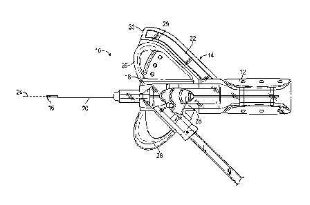
Note: Descriptions are shown in the official language in which they were submitted.
INTRAVENOUS CATHETER SYSTEM WITH ELONGATED
VISUALIZATION CHANNEL
BACKGROUND
[0001] Infusion therapy, a common healthcare procedure, may be facilitated
by a vascular access
device. Hospitalized, home care, and other patients receive fluids,
pharmaceuticals, and blood products
via a vascular access device inserted into the vascular system. Blood
withdrawal is another common
healthcare procedure that may be facilitated by a vascular access device.
[0002] A vascular access device may access a peripheral or central
vasculature of a patient. A
vascular access device may be indwelling for short term (days), moderate term
(weeks), or long term
(months to years). In some instances, the vascular access device may cause
irritation to the skin of the
patient when left in place for an extended period of time. A vascular access
device may be used for
continuous infusion therapy or for intermittent therapy.
100031 A common type vascular access device is an over-the-needle
peripheral intravenous catheter
("PIVC"). As its name implies, the "over-the-needle" PIVC may be mounted over
an introducer needle
having a sharp distal tip. The sharp distal tip may be used to pierce the skin
and the vasculature of the
patient. Insertion of the PIVC into the vasculature may follow the piercing of
the vasculature by the
needle. The needle and the PIVC are generally inserted at a shallow angle
through the skin into the
vasculature of the patient with a bevel of the needle facing away from the
skin of the patient.
[0004] In order to verify proper placement of the introducer needle and/or
the PIVC in the
vasculature, a user generally confirms that there is flashback of blood, which
may be visible to the user.
In some instances, the introducer needle may include a notch disposed towards
a distal end of the
introducer needle, and in response to the distal tip of the introducer needle
being positioned within the
vasculature, blood may flow proximally through a needle lumen, exit the
- 1 -
Date Recue/Date Received 2022-04-14
needle lumen through the notch, and then travel proximally between an outer
surface of the
introducer needle and an inner surface of the PIVC.
[0005] Accordingly, where the PIVC is at least partially transparent, the
user may visualize a
small amount of blood "flashback" and thereby confirm placement of the PIVC
within the
vasculature. Presence of a vasculature entrance indicator, such as flashback,
may facilitate
successful placement of PIVCs. Once placement of the introducer needle within
the vasculature
has been confirmed, the user may temporarily occlude flow in the vasculature
and withdraw the
introducer needle, leaving the PIVC in place for future blood withdrawal
and/or fluid infusion.
[0006] The user may also attach a device to the PIVC for fluid infusion
and/or blood
withdrawal. This process has been somewhat difficult in practice since many
catheter placement
sites simply do not allow easy occlusion of the vein. Additionally, even when
such occlusion is
achieved, it may be imperfect, resulting in blood leaking from a catheter
assembly housing the
PIVC and endangering medical personnel. Catheter assemblies have thus been
provided in the art
that provide a variety of seals or "septa" for preventing outflow of fluid
during and following
removal of the introducer needle from the blood vessel.
[0007] A septum may be secured within the catheter assembly via friction
and/or adhesive
between the septum and a wall of the catheter assembly. However, in some
instances, septum
dislodgement may occur in response to pressurization of the catheter assembly,
which may result
from venous pressure, fluid injection under high or low pressure, flush of the
catheter assembly,
blood collection, etc. Septum dislodgement presents a risk of exposure by
medical personnel to
blood or other fluids. Thus, challenges to infusion and/or blood withdrawal
using a vascular
access device still remain.
-2-
Date Recue/Date Received 2022-04-14
[0008] The
subject matter claimed herein is not limited to embodiments that solve any
disadvantages or that operate only in environments such as those described
above. Rather, this
background is only provided to illustrate one example technology area where
some
implementations described herein may be practiced.
SUMMARY
[0009] In
some embodiments, an IV catheter system may include a catheter adapter having
a
proximal end and a distal end. In some embodiments, the IV catheter system may
also include a
cannula extending through the catheter adapter. In some embodiments, a
proximal end of the
cannula may include a notch. In sonic embodiments, the IV catheter system may
also include a
needle hub, which may be coupled to the proximal end of the catheter adapter.
In some
embodiments, the needle hub may include an elongated visualization channel,
which may be in
fluid communication with the notch when the IV catheter system is in an
insertion configuration.
In some embodiments, the visualization channel and other elements described
later in further
detail may facilitate visualization of blood flashback by a user of the IV
catheter system.
[0010] In
some embodiments. a portion of the catheter adapter may be constructed of a
first material and another portion of the catheter adapter may be constructed
of a second material.
In some embodiments, the second material may have a lower durometer than the
first material
and may be more soft or flexible. In some embodiments, the second material may
improve
contact of the IV catheter system with skin of the patient and provide other
advantages, which
will be explained later in further detail.
[0011] In
some embodiments, the catheter adapter may include one or more stabilization
features, such as, for example, ribs or another type of protrusion, which may
reduce a distance
-3-
Date Recue/Date Received 2022-04-14
between a septum canister housing a septum of the IV catheter system. In some
embodiments,
the stabilization ribs may provide increased securement of the septum canister
and septum within
the catheter adapter, as will be explained later in further detail.
[0012] It
is to be understood that both the foregoing general description and the
following
detailed description are exemplary and explanatory and are not restrictive of
the invention, as
claimed. It should be understood that the various embodiments are not limited
to the
arrangements and instrumentality shown in the drawings. It should also be
understood that the
embodiments may be combined, or that other embodiments may be utilized and
that structural
changes, unless so claimed, may be made without departing from the scope of
the various
embodiments of the present invention. The following detailed description is,
therefore, not to be
taken in a limiting sense.
BRIEF DESCRIPTION OF THE SEVERAL VIEWS OF THE DRAWINGS
[0013]
Example embodiments will be described and explained with additional
specificity and
detail through the use of the accompanying drawings in which:
[0014]
Figure lA is a top view of an example catheter system, according to some
embodiments;
[0015]
Figure 1B is an upper perspective view of the catheter system of Figure IA,
according to some embodiments;
[0016]
Figure 1C is a cross-sectional view of the catheter system of Figure 1A,
according
to some embodiments;
[0017]
Figure 1D is a top view of the catheter system of Figure 1A, held in a first
grip,
according to some embodiments;
-4-
Date Recue/Date Received 2022-04-14
[0018] Figure lE is a top view of the catheter system of Figure 1A. held
in a second grip,
according to seine embodiments;
[0019] Figure 2 is a cross-sectional view of another example catheter
system, illustrating a
secondary flashback chamber, according to some embodiments;
[0020] Figure 3A is an upper perspective view of another example catheter
system,
illustrating an example sleeve, according to some embodiments;
[0021] Figure 3B is an upper perspective view of the catheter system of
Figure 3A,
illustrating the sleeve removed, according to some embodiments;
[0022] Figure 3C is a cross-sectional view of another example catheter
system, illustrating
an example secondary flashback chamber, according to some embodiments;
[0023] Figures 4A is an upper perspective view of an example retention
feature of an
example needle hub, according to some embodiments;
[0024] Figure 4B is an upper perspective view of an example corresponding
retention
feature of an example sleeve, according to some embodiments;
[0025] Figure 4C is a cross-sectional view of an example potting ring,
according to some
embodiments;
[0026] Figure 5A is a top view illustrating an example needle hub
partially withdrawn
from an example catheter adapter and an example sleeve removed from the needle
hub,
according to some embodiments;
[0027] Figure 5B is a upper perspective view illustrating the needle hub
of Figure 5A
partially withdrawn from the catheter adapter and the sleeve coupled to the
needle hub,
according to some embodiments;
-5-
Date Recue/Date Received 2022-04-14
[0028] Figure 6A is a top view illustrating an example needle hub
partially withdrawn
from an example catheter adapter and an example sleeve removed from the
catheter adapter,
according to some embodiments;
[0029] Figure 6B is a upper perspective view illustrating the needle hub
of Figure 6A
partially withdrawn from the catheter adapter and the sleeve coupled to the
needle hub,
according to some embodiments;
[0030] Figure 7A is a cross-sectional view of an example needle hub and
visualization
channel. according to some embodiments;
[0031] Figure 7B is an upper perspective view of the needle hub of Figure
7A, illustrating
an example reservoir, according to some embodiments;
[0032] Figure 7C is a cross-sectional view of an example tunnel that may
be disposed in
the needle hub of Figure 7A, according to some embodiments;
[0033] Figure 7D is a cross-sectional view of the needle hub of Figure 7B
along the line
7D-7D of Figure 7B, illustrating the needle hub disposed within an example
sleeve, according to
some embodiments;
[0034] Figure 8A is an upper perspective view of an example lens,
according to some
embodiments;
[0035] Figure 8B is a cross-sectional view of the lens of Figure 8A,
according to some
embodiments;
[0036] Figure 8C is an upper perspective view of another example lens,
according to
some embodiments;
[0037] Figure 8D is an upper perspective view of another example lens,
according to
some embodiments;
-6-
Date Recue/Date Received 2022-04-14
[0038] Figure 9A is a cross-sectional view of a portion of an example
visualization
channel. according to some embodiments;
[0039] Figure 9B is a cross-sectional view of a portion of another
example visualization
channel, according to some embodiments;
[0040] Figure 9C is a top view of another example visualization channel,
according to
some embodiments;
[0041] Figure 9D is an upper perspective view of an example needle hub
partially
removed from an example sleeve, and an example cover removed from the sleeve,
according to
some embodiments;
[0042] Figure 9E is a cross-sectional view of the needle hub and cover of
Figure 9D
secured within the sleeve, for insertion of the catheter system into the
patient, according to some
embodiments;
[0043] Figure 10A is an upper perspective view of an example bump,
according to some
embodiments;
[0044] Figure 10B is an upper perspective view of example indents,
according to some
embodiments;
[0045] Figure 10C is an upper perspective view of an example nozzle,
according to some
embodiments;
[0046] Figure 10D is an upper perspective view of an example vent,
according to some
embodiments;
[0047] Figure 10E is a cross-sectional view of an example vent plug,
according to some
embodiments;
-7-
Date Recue/Date Received 2022-04-14
[0048] Figure 1OF is an upper perspective view of an example porous
material, according
to some embodiments;
[0049] Figure 11A is a top view of an example catheter system with a
shortened sleeve,
according to some embodiments;
[0050] Figure 11B is a top view of an example catheter system in a non-
integrated
configuration without extension tubing, according to some embodiments;
[0051] Figure 11C is an upper perspective view of an example cannula
having an
external groove, according to some embodiments;
[0052] Figure 11D is an upper perspective view of another catheter system
having a non-
grip configuration, according to some embodiments;
[0053] Figures 12A is an upper perspective view of an example catheter
adapter having an
example ridge constructed of a second material, according to some embodiments;
[0054] Figure 12B is a lower perspective view of another example catheter
adapter having
an example flexible region, according to some embodiments;
[0055] Figure 12C is an upper perspective view of another example
catheter adapter
having an example strain relief feature, according to some embodiments;
[0056] Figure 12D is a front view of a portion of an example securement
platform,
according to some embodiments;
[0057] Figure 12E is a lower perspective view of another example catheter
adapter having
an another example strain relief feature, according to some embodiments;
[0058] Figure 12F is an upper perspective view of another example
catheter adapter
having another example flexible region, according to some embodiments;
-8-
Date Recue/Date Received 2022-04-14
[0059] Figure 12G is a lower perspective view of another example catheter
adapter having
an another example strain relief feature, according to some embodiments;
[0060] Figure 12H is a rear view of another catheter adapter having an
example notch
constructed of the second material. according to some embodiments;
[0061] Figure 121 is a partial cutaway view of another catheter adapter,
illustrating a first
material disposed within the securement platform 26 proximate the second
material, according to
some embodiments;
[0062] Figure 121 is a lower perspective view of an example friction
reducer, according to
some embodiments;
[0063] Figure 12K is an upper perspective view of an example withdrawal
indicator
feature, according to some embodiments;
[0064] Figure 13 is a cross-sectional view of an example strain relief
feature, according to
some embodiments;
[0065] Figure 14 is an upper perspective view of another catheter adapter
having a distal
end constructed of the second material, according to some embodiments;
[0066] Figure 15A is a front view of example interface surfaces,
according to some
embodiments;
[0067] Figure 15B is a lower perspective view of example protrusions
disposed on an
example interface surface, according to some embodiments;
[0068] Figure 15C is a lower perspective view of example geometry
features, according to
some embodiments;
[0069] Figures 15D is an upper perspective view of example wings prior to
separation,
according to some embodiments;
-9-
Date Recue/Date Received 2022-04-14
[0070] Figure 15E is an upper perspective view of the example wings of
Figure 15D after
separation, according to some embodiments;
[0071] Figure 15F is a rear view of a portion of another example catheter
adapter,
illustrating a rotating example grip;
[0072] Figure 16A is an upper perspective view of an example catheter
adapter without
stabilization ribs, according to some embodiments;
[0073] Figure 16B is a rear view of the catheter adapter of Figure 16A,
according to some
embodiments;
[0074] Figure 17A is an upper perspective view of an example catheter
adapter with
example stabilization ribs, according to some embodiments;
[0075] Figure 17B is a rear view of the catheter adapter of Figure 17A,
according to some
embodiments;
[0076] Figure 17C is an upper perspective view of the catheter adapter of
Figure 17A,
illustrating an example septum canister and example septum removed from the
catheter adapter,
according to some embodiments;
[0077] Figure 17D is an upper perspective view of the catheter adapter of
Figure 17A,
illustrating the septum canister and the septum secured within the catheter
adapter, according to
some embodiments;
[0078] Figure 18A is a cross-sectional view of the catheter adapter of
Figure 16A,
according to some embodiments;
[0079] Figure 18B is a cross-sectional view of the catheter adapter of
Figure 17A,
according to some embodiments;
-10-
Date Recue/Date Received 2022-04-14
[0080] Figure 19A is a cross-sectional view of an example septum
canister, according to
some embodiments;
[0081] Figure 19B is a cross-sectional view of another example septum
canister coupled
with an example septum, according to some embodiments; and
[0082] Figure 19C is a cross-sectional view of another example septum
canister disposed
within an example catheter adapter, according to some embodiments.
DESCRIPTION OF EMBODIMENTS
[0083] Figures 1A-19C may describe various catheter systems 10, according
to some
embodiments. In some embodiments, the catheter systems 10 may include IV
catheter systems or
PIVC systems. In some embodiments, a particular catheter system 10 may include
one or more
components or features from one or more of Figures 1A-19C.
[0084] Referring now to Figures 1A-1C, in some embodiments, a catheter
system 10 may
include a needle hub 12 and a grip 14. In some embodiments, the needle hub 12
and the grip 14
may be a single component and integrally formed. In some embodiments, the
needle hub 12 and
the grip 14 may be monolithically formed as a single unit. In some
embodiments, the grip 14
may extend outwardly from the needle hub 12. In some embodiments, the grip 14
may include a
paddle grip.
[0085] In some embodiments, a cannula 16 of the catheter system 10 may
include a notch
(not illustrated in Figures 1A-1C) towards a distal end of the cannula 16,
which may provide
primary flashback indicating that a catheter 20 of the catheter system 10 has
been properly
placed within a vein of the patient. In some embodiments, the cannula 16 may
include an
-11 -
Date Recue/Date Received 2022-04-14
introducer needle having a sharp distal tip. In some embodiments. the proximal
end of the
cannula 16 may be secured to and/or within the needle hub 12.
[0086] In some embodiments, the needle hub 12 and/or the grip 14 may be
transparent. In
some embodiments, the needle hub 12 and/or the grip 14 may be non-transparent.
In some
embodiments, a catheter adapter 18 of the catheter system 10 may be
transparent to allow the
user to observe primary flashback. In some embodiments, it may be preferred
that the needle hub
12 and/or the grip 14 arc white, which may provide a color contrast with blood
to facilitate
visualization of primary flashback by the user. In some embodiments, the grip
may include a
wing 22, which may extend outwardly from the needle hub 12.
[0087] As illustrated in Figure 1C, in some embodiments, a longitudinal
or center axis 24
of a catheter 20 extending distally from the catheter adapter 18 may be angled
with respect to a
bottom surface of the grip 14 and/or a bottom surface of a securement platform
26, which may
minimize a transition between a distal nose of the catheter adapter 18 and the
vein, once the
catheter system 10 is inserted within the vein. In some embodiments, the
center axis 24 of the
catheter 20 may be angled with respect to the bottom surface of the grip 14
and/or the bottom
surface of the securement platform 26 at an angle e between approximately 0
and 15 degrees. In
some embodiments, the angle 0 may be approximately 6 degrees.
[0088] In some embodiments, a proximal end of a cannula 16 of the
catheter system 10
may be accessible during assembly of the catheter system 10, which may allow
for lie distance
adjustment. In some embodiments, the proximal end of the cannula 10 may be
crimped and/or
glued in a well near a rear of the needle hub 12, which may provide additional
mechanical
retention of the cannula 16.
-12-
Date Recue/Date Received 2022-04-14
[0089] In some embodiments, the catheter adapter 18 may include one or
more push tab
features 28. In some embodiments, the push tab features 28 may be connected to
the securement
platform 26 on one or more sides of the catheter adapter 18, which may improve
mold filling. In
some embodiments, a distal portion of the wing 22 which may be disposed below
the securement
platform 26 of the catheter system 10, may include an edge 30 that may be
rounded and/or
tapered to facilitate taping of a dressing to the skin of the patient and/or
reduce trapped air. In
some embodiments, the edge 30 may include a transitional profile that guides a
thumb of a user
to facilitate gripping. In some embodiments, the distal portion of the wing 22
may include the
edge 30, as illustrated, for example, in Figure 1A. In some embodiments, the
wing 22 may
include a ridge 29, which may abut the securement platform 26.
[0090] Referring now to Figures 1D-1E, in some embodiments, I, T, and M
refer to the
index finger, thumb, and middle finger of the user, respectively, and indicate
approximate
positions of the I, T, and M, respectively. For example, the thumb may be
disposed proximate
the proximal end of the needle hub 12. Figures 1D-1E illustrate modified
ported grip techniques.
In some embodiments, the edge 30, which may have an angled or tapered upper
surface, may
facilitate use of these modified ported grip techniques by the user.
[0091] Referring now to Figure 2, the catheter system 10 is illustrated
according to some
embodiments. In some embodiments, the grip 14 and the needle hub 12 may be a
single
component and integrally formed. In some embodiments, the grip 14 and the
needle hub 12 may
be monolithically formed as a single unit. In some embodiments, a sleeve 36 of
the needle hub
12 may be removable from the grip 14 and/or the needle hub 12. In some
embodiments, one or
more of the sleeve 36, the needle hub 12, and the grip 14 may be a single
component and
-13-
Date Recue/Date Received 2022-04-14
integrally formed. In some embodiments, one or more of the sleeve 36, the
needle hub 12, and the grip
14 may be monolithically formed as a single unit.
[0092]
In some embodiments, a flashback chamber 32 may be provided within or
proximate the
needle hub 12 of the catheter system 10. In some embodiments, the flashback
chamber 32 may be
disposed between the sleeve 36 and the needle hub 12. In some embodiments, the
cannula 16 may include
a notch disposed towards a proximal end of the cannula 16, which may allow
blood to flow into the
flashback chamber 32. In some embodiments, the flashback chamber 32 may be a
secondary flashback
chamber in fluid communication with the notch disposed towards the proximal
end of the cannula 16
and/or an opening of the proximal end of the cannula 16.
[0093]
In some embodiments, the needle hub 12 may include or correspond to a vent
plug. In some
embodiments, the needle hub 12 and/or the sleeve 36 may include a filter or
vent permeable to air but
not blood. In some embodiments, the plug may be placed into a proximal end of
the sleeve 36 to form
an interface with the flashback chamber 32. In some embodiments, the flashback
chamber 32 may
include a visualization channel, as will be explained later in further detail.
In some embodiments, the
plug may be white or another non-transparent color, which may enhance contrast
of blood in the
visualization channel for better visibility. In some embodiments, the sleeve
36 may be transparent.
[0094]
Referring now to Figure 3A-3C, in some embodiments, the catheter system 10
may include
the sleeve 36 that may include an additional gripping surface 33. In some
embodiments, the sleeve 36
may be coupled to the needle hub 12 and provide a fluid-tight seal around the
flash chamber 32. In some
embodiments, the sleeve 36 may be transparent or clear, which may allow the
user to view blood 35
within the flashback chamber 32, which may
be
- 14 -
Date Recue/Date Received 2022-04-14
disposed between the sleeve 36 and the needle hub 12. In some embodiments, the
needle hub 12
and/or the grip 14 may be white, which may be a most familiar color for
catheter components in
the market. In some embodiments, the needle hub 12 may include the
visualization channel, and
the sleeve 36 may tightly cover the visualization channel to provide a fluid-
tight seal and prevent
blood received from the cannula 16 into the visualization channel from exiting
the visualization
channel.
[0095] In some embodiments, the sleeve 36 may be universal for all
catheter gauge sizes.
In some embodiments, all catheter gauge size dependent features may be
disposed in the needle
hub 12 and/or the grip14, while the sleeve 36 may remain standard across all
catheter gauge
sizes. Figure 3C illustrates a possible configuration of the catheter system
10 with the sleeve 36,
according to some embodiments.
[0096] As illustrated in Figure 3C, in some embodiments, a proximal end
of the cannula
16 may be secured in the needle hub 12 via an adhesive or another suitable
mechanism. In some
embodiments, the cannula 16 may include a notch 37 towards the proximal end of
the cannula
16, and the notch 37 may be in fluid communication with the flash chamber 32.
In some
embodiments, the flash chamber 32 may be disposed between the sleeve 36 and
the needle hub
12. In some embodiments, in response to insertion of the cannula 16 into the
vein of the patient,
the blood may flow into the cannula 16 and out the notch 37 into the flash
chamber 32. In some
embodiments, a venting channel 39 may be disposed proximate the flash chamber
32 and may be
permeable to air but not blood.
[0097] Referring now to Figures 4A-4C, in some embodiments, the needle
hub 12 may be
secured to the sleeve 36 to prevent separation of the needle hub 12 and the
sleeve 36 when the
sleeve 36 is gripped by the user. In some embodiments, one or more mechanisms
may be used to
-15-
Date Recue/Date Received 2022-04-14
couple the needle hub 12 and the sleeve 36 together. For example, the needle
hub 12 and the
sleeve 36 may be coupled together via an interference fit. As another example,
a mechanical lock
and/or a snap feature may be used to couple the needle hub 12 and the sleeve
36 together. As a
further example, an adhesive may be disposed in a cavity at an interface of
the needle hub 12 and
the sleeve 36 to couple the needle hub 12 and the sleeve 36 together. Figures
4A and 4B
illustrate example retention features of the needle hub 12 and the sleeve 36,
respectively. In
further detail, in some embodiments, a retention feature 34a of the needle hub
12 may be
configured to engage in a snap fit with a corresponding retention feature 34b
of the sleeve 36. In
some embodiments, the retention feature 34a may be disposed on an outer
surface of the needle
hub, and the corresponding retention feature 34b may be disposed on an inner
surface of the
sleeve 36. Figure 4C illustrates an example potting ring 38, which may include
an adhesive to
glue the needle hub 12 and the sleeve 36 together.
[0098] Referring now to Figures 5A-5B, in some embodiments, the sleeve 36
and the grip
14 may be a single component and integrally formed. In some embodiments, the
sleeve 36 may
be generally cylindrical. In some embodiments, the sleeve 36 may surround
and/or encapsulate
the needle hub 12. In some embodiments, the needle hub 12 may be coupled to
the sleeve 36 and
the grip 14 via the one or more mechanisms discussed with respect to Figure 4.
In some
embodiments, a portion of the needle hub 12 may be coupled directly to the
proximal end of the
catheter adapter 18.. In some embodiments, coupling of the sleeve 36 to the
proximal end of the
catheter adapter 18 may include an interference fit or another type of
coupling.
[0099] Referring now to Figures 6A-6B, in some embodiments, the needle
hub 12 may be
white. In some embodiments, the grip 14 and/or sleeve 36 may be transparent or
clear. In some
embodiments, the sleeve 36 may be coupled directly to the proximal end of the
catheter adapter
-16-
Date Recue/Date Received 2022-04-14
18. In some embodiments, coupling of the sleeve 36 to the proximal end of the
catheter adapter
18 may include an interference fit or another type of coupling.
[00100] In some embodiments, the catheter system 10 may enhance vein
confirmation in
vascular access systems featuring blood flashback. In some embodiments, the
catheter system 10
may provide improved visualization timing, optical amplification, continuous
motion
optimization, and fluid management. In some embodiments, the catheter system
10 may provide
a reduction in time for blood to appear in the flashback chamber 32.
accommodate pre-priming
that may otherwise flood another flashback chamber, and meter a flowrate in
the visualization
channel.
[00101] In some embodiments, the catheter system 10 may provide fluid
confinement to
guide incoming flow through the flash chamber 32, which may feature a high
surface-to-volume
ratio. In some embodiments, the catheter system 10 may provide an overflow
pattern that
compensates for various excess fluid conditions. In some embodiments, the
catheter system may
provide means of an unobstructed, sharp-contrast, real-time visualization of
blood flashback
throughout a duration of vein access. In some embodiments, the catheter system
10 may provide
immediate signaling and amplification upon low-abundance blood presence.
[00102] Referring now to Figures 7A-7B, in some embodiments, the flashback
chamber 32
may include a pocket 40, which may house the proximal end of the cannula 16.
In some
embodiments, the pocket 40 may be proximate and/or in fluid communication with
the
visualization channel 42. In some embodiments, the visualization channel 42
may be disposed at
an outer and/or upper portion of the needle hub 12 such that the user may
observe the secondary
flashback without obstruction. In some embodiments, secondary flashback may
refer to
flashback proximate the needle hub 12, while primary flashback may refer to
flashback
-17-
Date Recue/Date Received 2022-04-14
proximate the catheter 20 and/or the catheter adapter 18. In some embodiments,
the pocket 40
may include an inverse cone shape. In some embodiments, a top of the pocket 40
is widened for
better visualization of a blood droplet that comes out of the proximal end of
the cannula 16
and/or the notch disposed towards the proximal end of the cannula 16.
[00103] In some embodiments, walls of the pocket 40 may include an outward
draft that
may transduce a portion of the pocket-filling motion to an in-plane liquid
movement noticeable
by the user. In some embodiments, a volume of the pocket 40 may be reduced for
small gauges
and/or a cannula-hosting hole may be piloted into the pocket 40, as
illustrated, for example, in
Figure 7A, which may shorten a time between blood coming out of the notch
and/or proximal
end of the cannula 16 and starting to flow through the flashback chamber 32.
[00104] In some embodiments, the visualization channel 42 may include a
high surface-to-
volume aspect ratio, which may create an enhanced visualization signal with a
small volume of
blood. The aspect ratio of the visualization channel 42 may translate a
volumetric flow rate to a
steady meniscus velocity that can be easily captured by human eyes. In some
embodiments, the
longitudinal continuous motion of blood flowing through the visualization
channel 42 may
provide a clear thermometer-like signal of vein access. In some embodiments,
the visualization
channel 42 may include a length that may dictate duration of the continuous
motion in
accordance with a typical catheter insertion process. As such, in some
embodiments, extended
vein confirmation throughout insertion may be provided.
[00105] As illustrated in Figure 7B, in some embodiments. the flashback
chamber 32 may
include a cavity or reservoir 44. In some embodiments, the reservoir 44 may be
disposed
underneath the visualization channel 42 when the needle hub 12 is assembled
with the sleeve 36.
In some embodiments, the reservoir 44 may be molded in the needle hub 12
and/or connected to
-18-
Date Recue/Date Received 2022-04-14
the visualization channel 42 via a drain channel disposed at a proximal end of
the visualization channel
42. In some embodiments, a volume of the reservoir 44 may be greater than or
equal to approximately
20 microliters in order to contain excess fluid prior to blood flowing through
the visualization channel
42.
[00106] In some embodiments, the catheter system 10 may include a vent 48,
which may be disposed
on the needle hub 12. In some embodiments, the vent 48 may be formed by one or
more micro-grooves
on the needle hub 12. In some embodiments, the vent 48 may be located at an
end of an entire fluid path
through the flashback chamber 32. For example, the reservoir 44 may be
disposed proximate a front
flange of the needle hub 12 and/or above the reservoir 44. In some
embodiments, the vent 48 may throttle
movement of fluid during pre-priming (in which saline may be infused inside
the catheter system to
purge out air) and flashback (in which blood may be driven into the flashback
chamber) to reduce excess
saline volume without significant compromise on time to visualize blood in the
visualization channel 42
for large gauges. In some embodiments, the vent 48 may serve as a barrier to
prevent fluid from leaking
out of the catheter system 10.
[00107] In some embodiments, the catheter system 10 may include one or more
ribs 50, which may
be disposed on the needle hub 12. In some embodiments, the ribs 50 may be
molded with the visualization
channel 42. In some embodiments, the ribs 50 may facilitate fluid path
confinement. In some
embodiments, the ribs 50 may be disposed at both distal and proximal ends or
flanges of the needle hub
12. In some embodiments, the ribs 50 may be disposed along longitudinal edges
of the visualization
channel 42. In some embodiments, the ribs 50 may interfere with and/or contact
an interior of the sleeve
36 of the catheter system 10.
- 19 -
Date Recue/Date Received 2022-04-14
[00108] Referring now to Figure 7C, in some embodiments, the pocket 40 may
include a
tunnel portion 41, which may include and/or protect a proximal end of the
cannula 16. Referring
now to Figure 7D, in some embodiments, the reservoir 44 may be disposed on one
or both sides
of the needle hub 12. Figure 7C illustrates the reservoir 44 disposed on both
sides of the needle
hub 12.
[00109] In some embodiments, a depth of the visualization channel 42 may
be dependent
on a gauge of the cannula 16. For example, the depth of the visualization
channel 42 may be less
when the gauge size is smaller and greater when the gauge size is bigger.
Thus, in some
embodiments, the visualization channel 42 may be formed in a gauge-specific
manner in order to
have a consistent duration of the continuous motion and such that the meniscus
velocity is well
within human recognition domain.
[00110] Referring now to Figures 8A-8B, in some embodiments, a lens 52 may
be
disposed in the sleeve 36 or another component of the catheter system 10. In
some embodiments,
the lens 52 may include a single-sided convex lens. In some embodiments, the
lens 52 may be
built into the sleeve 36 to minimize impact on manufacturing. In some
embodiments, the lens 52
may be disposed above the pocket 40 and/or above the proximal end of the
cannula 16 to capture
presence of blood in the flashback chamber 32 immediately. In some
embodiments, the lens 52
may be disposed above the pocket 40 and/or the visualization channel 42. In
some embodiments,
the lens 52 may span across a top of the sleeve 36, which may provide an
adequate viewing
angle. In some embodiments, a shape of the lens 52 with respect to an exterior
surface of the
sleeve 36 may reduce impact of the lens 52 on use techniques.
[00111] In some embodiments, the lens 52 may include various shapes,
sizes, and
curvatures dependent on the particular catheter system 10. For example, the
lens 52 may include
-20-
Date Recue/Date Received 2022-04-14
a double-sided convex lens or an asymmetrical shape with directional
distortion. In some
embodiments, the lens 52 can be any size that may be integrated on an exterior
profile of a
component in the catheter system 10. In some embodiments, the lens 52 may be
conformal or
non-conformal to the exterior profile. In some embodiments, a particular non-
conformal lens 52
may be disposed on a stand-out platform on the exterior profile. In some
embodiments, the lens
52 may be integrated into an interior of the component. In some embodiments,
the lens 52 may
be translucent or partially clear. In a preferred embodiment, the lens 52 may
be clear. In some
embodiments, the lens 52 may be integrated or molded into the sleeve 36.
[00112] In some embodiments, the lens 52 may be integrated at various
locations in the
fluid path for optical amplification. For example, the lens 52 may be on top
of the catheter
adapter 18 to better visualize blood entering the catheter adapter 18 after
primary flashback.
[00113] Referring now to Figure 8C, in some embodiments, the lens 52 may
be
longitudinally extended along all or a portion of the visualization channel
42, which may provide
better view of blood flowing through the visualization channel 42. In some
embodiments,
multiple lenses 52 may be disposed along the visualization channel 42.
[00114] Referring now to Figure 8D, in some embodiments, the sleeve 36 may
be molded
to include a cavity or hole 54 in an inner wall of the sleeve 36. In some
embodiments, the lens 52
may be inserted into the cavity during assembly, as illustrated in Figure 8D.
As such, in some
embodiments, inconsistent wall thickness at a location of the lens 52 may be
addressed, and the
lens 52 can be independent from restrictions of molding material and geometry.
[00115] The catheter system 10 may be compatible with a wide variety of
fluid conditions,
including overflow conditions. In some embodiments, fluid comes in from the
cannula 16 and
-21-
Date Recue/Date Received 2022-04-14
enters the visualization channel 42. The fluid may then be drained into the
reservoir 44 before
the fluid is finally held off by the vent 48.
[00116] The catheter system 10 may provide various advantages. In some
embodiments,
the catheter system 10 may provide immediate visualization of blood once it
comes out of the
proximal end of the cannula 16 or the notch disposed towards the proximal end
of the cannula
16. In some embodiments, the catheter system 10 may provide continuous motion
of blood
flowing through the visualization channel 42 at a steady meniscus velocity of
greater than or
equal to approximately 0.25 mm/s axially. In some embodiments, the meniscus
may flow
through an entire length of the flashback chamber 32 in between approximately
5 and 20
seconds. The continuous motion provides a real-time vein confirmation, as
opposed to a more
static signal, within a duration that covers the catheter insertion process.
[00117] In some embodiments, the catheter system 10 may provide enhanced
visualization
of blood flashback. In some embodiments, blood entering the flash chamber 32
may be forced to
a ceiling of the flash chamber 32, e.g., the visualization channel 42 on top
of the needle hub 12,
which may provide an unobstructed view for the user. In some embodiments, the
visualization
channel 42 provides a large, substantially flat visualization area, which is a
stronger signal than
prior art devices in which blood falls to a bottom of a chamber and
accumulates in the chamber
before a noticeable signal can be generated. In some embodiments, the needle
hub 12 is white,
which may provide a sharp background contrast upon blood presence of blood in
the flashback
chamber 32, which may be formed by the needle hub 12 and/or the sleeve 36. In
some
embodiments, the notch disposed towards the proximal end of the cannula 16
and/or the
proximal end of the cannula 16 may be visible within the pocket 40.
-22-
Date Recue/Date Received 2022-04-14
[00118] In some embodiments, a top portion of the pocket 40 may have a
larger diameter
than a bottom portion of the pocket 40 and/or the pocket 40 may include
drafted walls,
facilitating a fast signal when blood presents. In some embodiments, the lens
52 may provide
optical amplification of blood disposed within the flashback chamber 32,
including the pocket 40
and/or the visualization channel 42. This may be particularly useful for small
gauge cannulas
and/or catheters. In some embodiments, the catheter system 10 may allow an
effective signal for
vein confirmation with less than 10 microliters of blood. Prior art devices
may require 50-500
microliters to generate an effective signal.
[00119] In some embodiments, the catheter system 10 may provide a means of
extended
vein confirmation that can cover a lengthy period corresponding to the
insertion of the catheter
20 within the vein. In some embodiments, the flashback chamber 32 may be used
in combination
with a primary flashback feature to facilitate vein confirmation throughout
various phases of the
catheter insertion process. In some embodiments, the various phases may
include cannula
penetration. "hooding" in which the cannula may be retracted by approximately
2 mm to reduce
a risk of transfixing the vein, catheter advancement, and cannula retraction.
[00120] In some embodiments, the enhanced flashback visualization features
outlined in
the present disclosure may be used with any vascular access device that
includes a flash
chamber. For example, the enhanced flashback visualization features outlined
in the present
disclosure may be used with a standard or modified plug without an additional
component or
manufacturing step.
[00121] In some embodiments, the visualization channel 42 may be disposed
in various
locations within a particular catheter system 10. For example, the
visualization channel 42 may
be disposed on an interior of the sleeve 36 and/or the needle hub 12 may serve
to seal the
-23-
Date Recue/Date Received 2022-04-14
visualization channel 42. As another example and referring now to Figures 9A,
the visualization
channel 42 may be co-axial with the cannula 16, which may reduce the volume of
the pocket 40.
[00122] Referring now to Figure 9B, in some embodiments, a center axis of
the
visualization channel 42 may be transitional between the cannula 16 and a top
of the needle hub
12. In these and other embodiments, the visualization channel 42 may be
slanted towards the top
of the needle hub 12. Referring now to Figure 9C, in some embodiments. the
visualization
channel 42 may be tapered. The visualization channel 42 of Figure 9C may
correspond to any of
the visualization channels 42 discussed with respect to the present
disclosure. As further
illustrated in Figure 9D, in some embodiments, the flashback chamber 32 may be
lower and
closer to the center axis of the cannula 16, which may generate a steady
meniscus velocity. In
some embodiments, at least a portion the sleeve 36 disposed above the
visualization channel 42
may be transparent.
[00123] Referring to Figures 9D-9E, in some embodiments, a top of the
sleeve 36 may
include a separate component, such as, for example, a cover 56. In some
embodiments, the cover
56 may be transparent or clear. In these and other embodiments, a portion of
the sleeve 36 other
than the cover 56 may not be limited to any particular material. In some
embodiments, an outer
diameter of the needle hub 12 and an inner diameter of the sleeve 36 are
disposed in a tight
geometric tolerance fit. In some embodiments, the cover 56 may be coupled to
the sleeve 36 in
any number of ways, including, for example, gluing, a mechanical snap fit,
etc. Hence, in some
embodiments, the coupling of the cover to the sleeve 36 may not depend on a
tight geometric
tolerance fit between the needle hub 12 and the sleeve 36.
[00124] In some embodiments, the visualization channel 42 can include
various geometries
and locations. In some embodiments, the visualization channel 42 may be
straight. In some
-24-
Date Recue/Date Received 2022-04-14
embodiments, the visualization channel 42 may include a serpentine, curved, or
jagged portion to
increase a length of the visualization channel 42. In some embodiments,
multiple visualization
channels 42 may be used in conjunction. In some embodiments, the multiple
visualization
channels 42 may be parallel. In some embodiments, the visualization channel 42
may be
integrated into sides of the needle hub 12 to accommodate grip techniques
(e.g., central grip or
conventional ported grip) that may partially obstruct a top view of the
catheter system 10. In
some embodiments, instead of a visualization channel 42, the catheter system
10 may include a
visualization area that is an open space for a larger volume of blood to flow
through larger
cannula gauges. In some embodiments, the open space may include an annular
space between the
needle hub 12 and the sleeve 36 or an empty chamber separate from the needle
hub 12.
[00125] Referring now to Figure 10A, in some embodiments, a height of the
visualization
channel 42, or a distance between a bottom and ceiling of the visualization
channel 42, may be
within sub-millimeter range to trigger capillary effect and/or accelerate
blood flow through the
visualization when the visualization channel 42 is pre-wetted. In some
embodiments, the needle
hub 12 may include a bump 57, which may form a microscale gap corresponding to
the
visualization channel 42 when the needle hub 12 is assembled with the sleeve
36.
[00126] Referring now to Figure 10B, in some embodiments, one or more
marks or indents
58 may be integrated along the longitudinal edges of the visualization channel
42 to provide a
better indication of travel distance of blood flow within the visualization
channel 42. In some
embodiments, the marks 58 may be spaced apart. In some embodiments, when blood
flows
through the visualization channel 42, the blood may color the marks 58
sequentially. In some
embodiments, the marks 58 may be molded into the needle hub 12.
-25-
Date Recue/Date Received 2022-04-14
[00127] Referring now to Figure 10C, in some embodiments, one or more
functional
structures may be integrated into the needle hub 12 to provide localized fluid
manipulation. For
example, a diverging diffuser or nozzle 60 may be molded into the needle hub
12 a distal end of
the visualization channel 42 to buffer transient pressure spikes. In some
embodiments, a shape of
the nozzle 60 may be increase a diameter of the fluid pathway as the blood
flows proximally.
[00128] Retelling now to Figure 10D-10F. in some embodiments, one or more
vents may
be integrated in the catheter system 10 at multiple locations. In some
embodiments, the vents
may include the vent 48 discussed with respect to Figure 7B. In some
embodiments. a particular
vent 51 may be integrated proximate an exit of the visualization channel 42
for additional fluid
restriction and/or throttling for large gauges.
[00129] In some embodiments, the vents may be configured to allow air but
not fluid to
pass. The vents may be created in various ways including, for example, one or
more of the
following: grooves molded in the needle hub 12, a small cut at a rib 50 (see,
e.g., Figure 10D), a
separate porous vent plug 62 proximate a proximal end of the visualization
channel 42 (see, e.g.,
Figure 10E), and a porous material 64 deposited in at least a portion of the
visualization channel
42 (see, e.g., Figure 10F). In some embodiments, the small cut at the sealing
rib 50 may be
disposed on a parting line to intentionally compensate molding mismatch. In
some embodiments,
the porous material 64 may be deposited via over-molding, spun-coating,
stamping, etc. In some
embodiments, the vents 48 may have mechanical geometries that allow air to
pass but prevent
fluid from passing. In some embodiments, the vents 48 may be created by paper,
fiber,
membrane, or other materials with a porosity that allow air to escape while
limiting fluid from
escaping.
-26-
Date Recue/Date Received 2022-04-14
[00130] In some embodiments, the catheter system 10 may include various
types of
catheter adapters 18, regardless of grip technique or type of securement
platfotm 26 (if any). In
some embodiments, the catheter system 10 may include straight, integrated, or
ported catheter
adapters. In some embodiments, the catheter system 10 may include a luer
accessible vascular
access device with a flow control plug.
[00131] In some embodiments, the catheter system 10 may not include
primary flashback.
However, in some embodiments, the catheter system 10 is compatible with
various primary
flashback features, such as, for example BD INSTAFLASHTm technology featuring
a notch on
the cannula 16 towards a bevel of the cannula 16, a separate fluid path
featuring a groove on an
exterior of the cannula 16, a double-notch of the cannula 16, a vented
extension tube, or another
primary flashback feature.
[00132] Referring now to Figure 11A, in some embodiments, the catheter
adapter 18 may
not include a central grip area such as the push tab feature 28. In these and
other embodiments,
the needle hub 12 and/or the sleeve 36 may be shortened significantly, as
illustrated, for
example, in Figure 11A. In some embodiments, the catheter adapter 18 may be a
two shot or
single shot catheter adapter 18. In some embodiments, the catheter adapter 18
may be
constructed of a flexible material, such as, for example, one or more of the
following:
polypropylene, high-density polyethylene, low-density polyethylene,
copolyester, polycarbonate,
and another polymer material. In some embodiments, the flexible material may
allow a
securement platform 26 with a foldable hinge.
[00133] It is understood that the catheter 20 of the catheter system 10
may include one or
more diffuser holes near the distal tip of the catheter 20 for improved flow
rates. It is further
understood that the one or more components of the catheter system 10 may
include an
-27-
Date Recue/Date Received 2022-04-14
antimicrobial or anti-pathogenic agent. In some embodiments, the antimicrobial
or anti-
pathogenic agent may include a coating or a component in a fluid pathway of
the catheter system
10. In some embodiments, the antimicrobial or anti-pathogenic agent may
include an eluting
coating or an additive.
[00134] It is understood that the catheter system 10 may include a single
or multi-use blood
control valve system. In some embodiments, the blood control valve system may
be disposed in
one or more luer ports on an end of an extension tube. Referring now to Figure
11B, in some
embodiments, the blood control valve system may be disposed in a non-
integrated configuration
without extension tubing. Referring now to Figure 11C, in some embodiments,
the catheter
system 10 may include an external groove in the cannula 16 for blood
visualization. In further
detail, in some embodiments, the external groove may allow primary flashback
between the
cannula 16 and the catheter 20, which may be transparent.
[00135] Referring now to Figure 11D, in some embodiments, the catheter
system 10 may
have a non-grip or non-paddle grip configuration such that the user does not
hold the catheter
system 10 using the grip 14. In these and other embodiments, the sleeve 36 may
accommodate a
number of grip styles, including, for example, a "straight grip" style or a
"ported grip" style. The
"straight grip" style, in which the thumb and middle finger are on either side
of the device and
the index finger is used to advance the catheter adapter, is illustrated in
Figure 11D.
[00136] Referring now to Figure 12A-12K, catheter adapters 18 are most
often
manufactured using a single material for reasons of simplicity and cost. Due
to functional
constraints placed on the chosen material, catheter adapters 18 comprised of a
single material
often exhibit tradeoffs in one or more areas. Consider high pressure
capability as an example. A
catheter adapter 18 designed to support high pressure capability will often
reflect a high level of
-28-
Date Recue/Date Received 2022-04-14
structural rigidity. This rigidity characteristic is in direct contrast with
product attributes
including patient comfort. user ease-of-use, efficient assembly processes, and
part tolerance
accommodation.
[00137] In some embodiments described in the present disclosure, a second
material is
introduced into the catheter adapter 18 via an integrated manufacturing
process such as two-shot
injection molding that allows improvement of a wider range of product
attributes. In some
embodiments, key catheter-specific fluid path geometry may be produced with an
appropriate
first material while attributes such as product stabilization, patient
comfort, user grip style and
flexible product integrity may be improved using the second material. In some
embodiments, due
to the integrated nature of the assembly process, the addition of the second
material may not
induce a notable increase in cost or assembly complexity.
[00138] Portions of the catheter system illustrated in Figures 12A-12K
that may optionally
include the second material are illustrated with a stippled shading. In some
embodiments, the
second material may be disposed at locations of the catheter system 10 other
than the areas with
the stippled shading. Portions of the catheter system 10 that may include the
first material are
illustrated with a cross-hatched shading. In some embodiments, the first
material may be
disposed at locations of the catheter system 10 other than the areas with the
cross-hatched
shading. In some embodiments, areas that include the second material may only
partially be
constructed of the second material. Figures 12A-12F illustrate various
catheter adapter 18,
according to some embodiments. It should be understood that the embodiments
illustrated in
Figures 12A-12F may be combined, and a particular catheter adapter 18 may
include features
from one or more of Figures 12A-12F.
-29-
Date Recue/Date Received 2022-04-14
[00139] In some embodiments, the second material is a softer material
designed to
optimize patient comfort, product stabilization, user grip compatibility,
catheter kink resistance,
securement compatibility, and component assembly flexibility. In some
embodiments, the
second material may be flexible or semi-flexible. These improvements are
enabled by a multi-
material catheter adapter 18 produced in using a highly-integrated
manufacturing approach. In
some embodiments, the second material may be useful with respect to skin
sensitivity and
biocompatibility.
[00140] In some embodiments, the second material is softer in nature with
a lower
durometer than the first material. In some embodiments, the introduction of
the second material
enables the patient comfort improvements via a softer and larger contact area
72 with the skin of
the patient. An example contact area 72 constructed of the second material are
illustrated in
Figure 12B, according to some embodiments. In some embodiments, a particular
contact area 72
may extend to the proximal end of the catheter adapter 18 or beyond the
proximal end of the
catheter adapter 18, which may further enhance patient comfort and safety as
well as play a
supporting role in the stability of the adapter 18.
[00141] In some embodiments, the securement platform 26 may include the
first material
and/or the second material. In some embodiments, a profile of a perimeter of
the second material
may be independent of a perimeter of the first material. For example, the
second material
perimeter may extend beyond the surface area of the first material or the
first material perimeter
70 may extend beyond the surface area of the second material.
[00142] As illustrated in Figure 12A, in some embodiments, a proximal end
of the catheter
adapter 18 may include a ridge 71, which may be configured to contact the skin
of the patient
when the catheter system 10 is inserted into the vasculature of the patient.
-30-
Date Recue/Date Received 2022-04-14
[00143] As illustrated in Figure 12B, in some embodiments, the
introduction of the second
material may also facilitate improved post-dressing product stabilization via
a softer and larger
contact area 72 with the skin and, in some embodiments, a flexible region 74
(see, e.g., Figure
12B-12C) proximal to the septum 66 (illustrated, for example, in Figure 1C)
designed to deflect
under the pressure applied from the dressing. An example of a flexible region
74 and contact
area 72 constructed of the second material are illustrated in Figure 12B. As
illustrated in Figure
12C, in some embodiments, the proximal end of the catheter adapter 18 may
include another
flexible region 75 disposed at least partially on an upper portion of the
catheter adapter 18.In
some embodiments, part tolerances may be loosened slightly due to an
accommodating nature of
the second material.
[00144] In some embodiments, the introduction of the second material also
provides
improved dressing stabilization via specific features located on the perimeter
of the product. For
example, the edge 30 of the securement platform 26 may include a tapered upper
surface, as
illustrated in Figure 12D. In some embodiments, the tapered round profile may
reduce the air gap
between securement tape, which may be used to secure the catheter system 10 to
the patient, and
the catheter adapter 18.
[00145] In some embodiments, the benefits of the specific features may not
be tied to
material durometer. In some embodiments, the introduction of the second
material may also
provide user grip compatibility via the introduction of a flexible central
push tab feature 28
and/or a flexible securement platform 26 to accommodate a traditional winged
insertion style.
Examples of the flexible central grip area or push tab feature 28 and the
flexible securement
platform 26 constructed of the second material are illustrated in Figure 12A.
-31-
Date Recue/Date Received 2022-04-14
[001461 As
illustrated in Figure 12E, in some embodiments, the introduction of the second
material provides improved catheter 20 kink resistance via the introduction of
a strain relief
feature 78 at the distal end of the catheter adapter 18. An example strain
relief feature 78
constructed of the second material is illustrated in Figure 12E. In some
embodiments, the strain
relief '78 may be designed to be as short as possible to avoid negatively
influencing system
stiffness. In some embodiments, the effectiveness of this shortened design is
a byproduct of the
second material flow path via a wide channel that also deflects under catheter
loading.
[001471 In
some embodiments, the introduction of the second material may provide
improved extension tube kink resistance via the introduction of a strain
relief feature 78 at a
junction coupling the extension tube to the catheter adapter. An example of
the strain relief
feature 68 at the junction and constructed of the second material is
illustrated in Figure 12F. In
some embodiments, the strain relief feature 78 may be further described in
United States Patent
Application Serial No. 15/286,212, filed October 5, 2016, entitled "Extension
Tubing Strain
Relief ." In
some embodiments, securement features added to
an extension tube port, for example, may improve a lay of the dressing
visually and/or by
providing strain relief. As example, Figure 12F illustrates a side port that
includes a strain relief
feature 79.
[001481 As
illustrated in Figure 12G, in some embodiments, the second material, which
may be lower-durometer, may be formed to mimic a semi-circular shape 81 to
target stress
reduction in off-axis areas, potentially accommodating a wider array of
loading scenarios. In
some embodiments, the second material may include a surface layer of the
catheter adapter 18.
In some embodiments, the second material may extend all the way through a wall
of the catheter
adapter 18.
-32-
Date Recue/Date Received 2022-04-14
[00149] In some embodiments, the second material may allow reduced
assembly
complexity of, for example, safety mechanism components. In some embodiments,
under the
force of assembly, the deflection of the second material at a location
proximate a safety
mechanism component, such as, for example, a notch 80 that may be configured
to contact a
needle safety clip. An example notch is illustrated in Figure 12H. second
material
[00150] In some embodiments, the second material may be coupled to the
first material in a
highly integrated manufacturing sequence such as two-shot injection molding.
In some
embodiments, the first material and second material may be formulated to
enable chemical-level
bonding as opposed to purely mechanical bonding via geometric features. In
some embodiments,
the second material is a lower-durometer material in the approximately 50A to
95A range,
depending on product and application. In some embodiments, the second material
may include a
lower-durometer material in the approximately 10A to 95A range. In some
embodiments, certain
additives may be compounded to the base material of the second material to
improve
characteristics such as comfort against skin and improved lubricity. In some
embodiments, the
catheter adapter 18 utilizes the second material to improve many aspects of
the product
including, not limited to, patient comfort, user grip comfort and
compatibility, catheter kink
resistance, improved securement and stabilization, and reduced cost burden due
to ease of
assembly and looser part tolerances.
[00151] In some embodiments, the first material may be rigid or semi-
rigid. In some
embodiments, the first material may be flexible. In some embodiments, the
first material may be
more stiff or hard than the second material. In some embodiments, the first
material and/or the
second material may include plastic, an elastomer such as silicone rubber, or
another suitable
material. In some embodiments, the first material may be disposed proximal to
the septum 66. In
-33-
Date Recue/Date Received 2022-04-14
some embodiments, the catheter system 10 may include the second material, but
there may be
no current integration of ease of assembly and looser part tolerance-specific
features.
[00152] As illustrated in Figure 121, in some embodiments, the first
material may extend
into the second material to provide shape and/or support. In these and other
embodiments, the
first material may not be outwardly visible. In some embodiments, the first
material may extend
beyond surfaces of the second material in order to improve product
functionality. In these and
other embodiments, the first material may act as one or more interface
friction reducers 82, as
illustrated, for example, in Figure 12J. In some embodiments, the friction
reducers 82 may
decrease friction between the catheter system 10 and the skin of the patient
and/or the wing 22.
In some embodiments, one or more of the interface friction reducers 82 may
include a
protrusion.
[00153] Referring now to Figure 12K, in some embodiments, the second
material may be
used for additional purposes such as adding a withdrawal indicator feature 84.
A length 85 of the
withdrawal indicator feature 84 may vary. In some embodiments, the withdrawal
indicator
feature 84 may be about 2 mm.
[00154] Figure 13 illustrates an example strain relief feature 86, which
may include or
correspond to any of the strain relief features 78 and/or 79 described with
respect to Figure 12. In
some embodiments, the strain relief feature 86 may be integrated into the
catheter system 10 via
the second material and/or may be as short as possible for various reasons. In
some
embodiments, the strain relief feature 86 may form a channel 88. In some
embodiments, an
underside of a distal end of the channel 88 closest to the skin of the patient
may include the
second material, which may enable deflection based on loads placed on the
catheter 20. In some
embodiments, placement of the channel 88 may reduce potential stress
concentrations between
-34-
Date Recue/Date Received 2022-04-14
the first material and the second material in an area of significant and
direct loading. In some
embodiments, in response to a 0.5 inch deflection applied at a single node at
the distal end of the
catheter, a single material catheter adapter 18 may experience peak stresses
at the distal end of
the single material catheter adapter 18 in a range 10-50% higher than peak
stresses at an
equivalent location on another multi-material catheter adapter 18 that
includes the strain relief
feature 86 with the second material.
[00155] In some embodiments, the strain relief feature 86 at least
partially constructed of
the second material may impact a bend profile of the catheter 20 as the
catheter 20 rests against
the skin of the patient. In some embodiments, the strain relief feature 86
having the second
material leads to a larger bend radius in the catheter 20 and a reduced
insertion angle into the
vein of the patient.
[00156] In some embodiments, the strain relief feature 86 may be designed
to work within
a specific angular range depending on product requirements, for example. As
illustrated in
Figure 13, in some embodiments, the strain relief feature 86 includes both the
first material and
the second that extend to the distal end of the catheter adapter 18 to limit
an impact of the strain
relief function. As illustrated in Figure 13. in some embodiments, the strain
relief feature 86 may
include a mix of the primary and second materials depending on, for example,
desired
performance characteristics. In some embodiments, the strain relief feature 86
may include an
antimicrobial agent to improve the indwell of the catheter 20. In some
embodiments, the
antimicrobial agent may be an additive in the first material and/or the second
material. In some
embodiments, the antimicrobial agent may include a coating applied to the
strain relief feature
86.
-35-
Date Recue/Date Received 2022-04-14
[00157] As illustrated in Figure 14, in some embodiments, the second material
may encompass a full
diameter at the distal end of the catheter adapter 18, providing strain
relief. In some embodiments, the
entire distal end of the catheter adapter 18 may be constructed of the second
material.
[00158] The catheter insertion process may require a high level of skill.
Persons challenged with
building and supporting an insertion skill set often develop preferences and
techniques based on the
equipment they work with. In the case of peripheral IV catheters, a number of
insertion grip styles have
evolved. Through manipulation of geometry and materials, catheter system 10
designs have changed to
better support the refinement and mastering of these grip styles.
[00159] One of the more widely used grips involves a pinching action between
the thumb and
forefinger. In some embodiments, a hub component of the catheter system 10 may
include the needle
hub 12 and/or the sleeve 36, which may separate from the catheter adapter 18.
In some embodiments,
the hub component may include a wing-like feature (which may be referred to in
the present disclosure
as "wing 22") to act as a point-of-contact for catheter insertion as well as
cannula withdrawal and
removal. In some embodiments, the wing 22 may be captured between the thumb
and forefinger to further
describe the pinching action. During the insertion process, the wing 22 may
act as a grip stabilizer and
control feature.
[00160] In some embodiments, the catheter adapter 18 may include one or more
stabilization features
to benefit patients and users. In some embodiments, the stabilization features
may extend medially and/or
laterally from the catheter adapter 18. In some embodiments, the stabilization
features may include one
or more wings 33, which may be part of the securement platform 26 and/or
extend outwardly from the
catheter adapter 18. In some embodiments, combining a catheter adapter 18 with
a stabilization feature
and the hub component with the wing 22 yields a more complex interface between
the catheter adapter
18 and the hub component. In some embodiments, as opposed to pinching solely
the wing 22 on the
- 36 -
Date Recue/Date Received 2022-04-14
needle hub 12 during insertion, the interface may allow the user to pinch the
wing 22 in combination
with the stabilization feature on the catheter adapter 18.
[00161] Upon completion of a successful initial insertion of the catheter 20,
the user may then separate
the hub component from the catheter adapter 18 prior to securing the catheter
adapter 18 to the patient.
In some embodiments, the catheter system 10 described in the present
disclosure may facilitate separating
of the respective geometric features of the hub component and the catheter
adapter 18.
[00162] Referring now to Figures 15A-15B, in some embodiments, the interface
between the wing 22
and the wing 33 may allow easy and efficient part separation under a range of
potential pinch forces.
This separation ease may be accomplished through various technological means.
For example, materials
used to create interface surfaces 90 on the wing 22 and/or the wing 33 may be
modified on the bulk level
(e.g., pre-pellet) to include additives or chemical compound modifiers
designed to enhance a specific
effect. In some embodiments, the interface surfaces 90 may include surfaces of
the wing 22 and/or the
wing 33. In some embodiments, interface surfaces 90a of the wing 33 may
contact or interface with
interface surfaces 90b of the wing 22 (the interface surfaces 90a and the
interface surfaces 90b may be
referred to collectively herein as "interface surfaces 90"). In some
embodiments, the interface surface
90a may interface with or contact the interface surface 90b when the catheter
system 10 is in an insertion
configuration for insertion into the patient.
- 37 -
Date Recue/Date Received 2022-04-14
[00163] In some embodiments, the interface surfaces 90 may include a lower
surface of the
wing 33 and an upper surface of the wing 22. In some embodiments, the second
material on the
catheter adapter 18 may be modified to increase lubricity against a co-
polyester mating
component. This may be a preferred approach due to the elimination of at least
one additional
manufacturing operation. Additionally, product-to-product functional variation
may be reduced.
[00164] As another example, contact surfaces on the respective interface
surfaces 90 may
be geometrically modified to improve certain characteristics such as effective
coefficients of
friction. In some embodiments, contact surfaces of the interface surfaces 90
may be textured on
the micro or nano scale; the interface surfaces 90 may contain geometric
patterns in forms and
depths that provide ideal interface characteristics. In these and other
embodiments, a micro-scale
texture may be applied to one or more of the interface surfaces 90. This
modification represents
only one of many possible modifications.
[00165] As a further example, a third material may be added to one or more
of the interface
surfaces 90 to serve as an agent in reducing variables such as coefficient-of-
friction. The third
material may take many forms. In some embodiments, the third material may
include one or
more powders, such as, for example, talc or starch. In some embodiments, the
third material may
include one or more lubes that may be used in different phase states. In some
embodiments, the
third material may include one or more additive mechanical components such as,
for example, a
tape or similar adhesive component. In some embodiments, the third material
may include one or
more insert-molded components.
[00166] As yet another example, a geometry on one or both of the interface
surfaces 90
may contain features to accept and promote force vectors in a direction of
separation. The
-38-
Date Recue/Date Received 2022-04-14
interface surfaces 90 may take many forms but they may be generally opposed in
the assembly
layout to ease separation difficulty.
[00167] With regard to the specifics of the interface between the wing 22
and the wing 33,
any reasonable material combination may be used to improve or highlight
certain characteristics.
In some embodiments, the interface surfaces 90 may be constructed using
polymers. In some
embodiments, materials utilized to construct the interface surfaces 90 may
include one or more
of the following: polycarbonate, co-polyester, polyester, polypropylene,
acrylonitrile butadiene
styrene (ABS), acetal, polyethylene, nylon, any of the various sub-categories
of thermoplastic
elastometers (TPE), silicones, and other suitable materials. In some
embodiments, the third
material may be applied on top of the material utilized to construct the
interface surfaces 90.
[00168] In some embodiments, a sub-component may be insert-molded into one
or both of
the interface surfaces 90. In some embodiments, the sub-component may be
metallic, polymeric
or ceramic or a combination of these classifications. In some embodiments, the
sub-component
may include intentional surface modifications such as texturing on a certain
size scale. Referring
now to Figure 15B, in some embodiments, the sub-component may include
protrusions 91 and/or
grooves. In some embodiments, the protrusions 91 may be disposed on the
interface surface 90a
and/or additional portions of the bottom of the securement platform 26, which
may contact skin
of the patient or the hand of the user.
[00169] In some embodiments, the material used to create the securement
platform 26,
which may include the wing 33, may be modified at the bulk level to improve
characteristics
related to the interface, which may include a wing-to-wing interface. As
illustrated in Figure
15B, in some embodiments, one or more of the interface surfaces 90 may include
surface texture
variation to reduce a coefficient of friction between the interface surfaces
90 during separation.
-39-
Date Recue/Date Received 2022-04-14
In some embodiments, the third material may be applied on one or more of the
interface surfaces
90.
[00170] Referring now to Figure 15C, in some embodiments, one or more
geometry
features 92 may be disposed on a surface configured to contact a hand of the
user and/or the skin
of the patient, such as a bottom surface of the wing 22, for example. In some
embodiments, the
geometry features 92 may include protrusions or indents. In some embodiments,
the geometry
features 92 may promote force vectors in the direction of separation. The
geometry features 92
may include various shapes and sizes. In some embodiments, the geometry
features 92 may be
placed in opposing locations to ease separation difficulty. Figures 15D-15E
illustrate the wing 22
and the wing 33 prior to and after separation, respectively, according to some
embodiments.
[00171] In some embodiments, the catheter system 10 may combine a robustly-
stabilized
catheter adapter 18 with benefits of a winged grip insertion technique. In
some embodiments, the
interface surfaces 90 between the wing 33 and the wing 22 may enable a more
efficient, single-
handed separation and withdrawal opportunity. In some embodiments, while
pinching the
overlapping wings 33, 22, moving the fingers involved in the pinch in opposite
directions
parallel or near-parallel with a central axis of the catheter 20 may quickly
and efficiently initiate
cannula withdrawal.
[00172] In some embodiments, the securement platform 26 may be added to an
integrated
catheter (e.g, BD NEXIVATM Closed IV Catheter System) as a step toward an
improved product
experience for patients. In some embodiments, the securement platform 26 is
compatible with a
"central grip" insertion style and/or to some degree a "ported grip" insertion
style. The "winged
grip" insertion style is not accounted for in the current state-of-the-art. In
some embodiments,
addition of the wing 22 below the wing 33 results in an improved integrated
catheter for users
-40-
Date Recue/Date Received 2022-04-14
who prefer the winged grip insertion technique. In some embodiments, the wing
22 disposed below the
wing 33, and having the wing 22 and the wing 33 slidable with respect to each
other, enables a product
with a similar insertion experience to a traditional winged product such as,
e.g., BD INTIMA IITM IV
Catheter, along with the patient benefits of a securement platform 26.
[00173] In some embodiments, the interface surfaces 90 may be de-coupled
and/or slid past each other
in a quick and controlled manner as soon as the initial insertion concludes.
The ease of de-coupling is
critical and one or more of the following techniques may be employed to
optimize this ease of de-
coupling: modification of a bulk material(s) used for construction of the
interface surfaces 90 via
introduction of a compounded additive; surface texture variation on one or
more areas of one or more of
the interface components; addition of a component or medium such as a lube or
tape applied to one or
more areas of one or more of the interface surfaces 90; and creation of
geometry to minimize finger-
based pinch force vector application perpendicular or near-perpendicular to
the primary direction of part
separation.
[00174] In some embodiments, the catheter adapter 18 may include one or more
particular wings 33
that may be somewhat symmetrically-placed relative to a longitudinal axis of
the catheter 20. In some
embodiments, the wings 33 may create a seamless securement platform 26 to rest
against the patient's
skin. In some embodiments, the securement platform 26 may include a lower-
durometer material relative
to a material of the hub component and a material of a body of catheter
adapter 18 through which a lumen
may extend. In some embodiments, the lower-durometer material may be in the
range of 50A to 95A
durometer.
[00175] In some embodiments, the hub component may include the wing 22
asymmetrically opposed
to the extension tube port on the catheter adapter 18. In some embodiments,
the wing 22 may be
-41 -
Date Recue/Date Received 2022-04-14
constructed of a harder, stiffer material relative to the securement platform
26 of the catheter adapter 18.
In some embodiments, the wing 22 includes geometry for gripping with the
fingers of the right hand.
[00176] In some embodiments, in a fully-assembled configuration, the wing 33
nests directly against
the profile of the wing 22. In some embodiments, in the context of typical
gripping orientations and
forces, very little deformation may occur in the wing 33 due to the load
transfer through the stiffer wing
22. Referring now to Figure 15F, in some embodiments, the hub component may
rotate around the
catheter axis relative to the catheter adapter 18 to accommodate certain
insertion grip styles. In some
embodiments, under pinch-based force application, the overlapping wings 22, 33
captured in the pinch
enable a stiffer system for an improved insertion. In these and other
embodiments, the wing 22 may rest
on top of the wing 33 as opposed to the wing 33 resting on top of the wing 22.
In these embodiments,
the thumb of the user may contact or rest on top of the wing 22 and may move
proximally in order to
hood the cannula 16 and/or withdraw the needle hub 12 from the catheter
adapter 18.
[00177] In some embodiments, when the user is ready to withdraw the cannula
16, the fingers of the
right hand used to pinch the wing 22 and the wing 33 are moved in an opposing
direction near-parallel
to the axis of the catheter 20. Typically the thumb will move distally along
with the wing 33 while the
opposing finger (the forefinger in some cases) moves proximally along with the
wing 22. In some
embodiments, the wing 22 and the wing 33 may move relative to each other with
a coefficient of friction
of approximately 0.2 in a preferred range of 0.1 to 0.5. In some embodiments,
the coefficient of friction
may range from approximately 0.1 to 1.0 depending on, for example, markets and
target performance
characteristics. In some embodiments, the coefficient of friction may range
from 0.1 to 1.5.
-42 -
Date Recue/Date Received 2022-04-14
[00178] In some embodiments, the wing 33 may rest on a top surface of the wing
22. In some
embodiments, the thumb may contact the wing 33. In some embodiments, at a time
of withdrawal, the
catheter adapter 18 moves distally with respect to the hub component. In some
embodiments, the hub
component rotates in a range of 0 to 45 degrees around the catheter axis
relative to the catheter adapter
18. In some embodiments, the durometer of the wing 33 is approximately 70A.
[00179] In some embodiments, the interface surfaces 90 between the wing 33 and
the wing 22 may
include medial and/or lateral locations of the catheter system 10.
Additionally or alternatively, in some
embodiments, the interface surfaces 90 may include central locations. In some
embodiments, the
interface surfaces 90 may include elements of the catheter system 10 other
than the wing 33 and the wing
22. In some embodiments, a particular interface surface 90, such as, for
example, the wing 33, may reside
against a bottom surface of the wing 22.
[00180] In some embodiments, mechanics of the wing 33 and the wing 22 geometry
and the unique
catheter system 10 layout may be rooted in usability studies. Separating the
catheter adapter 18 from the
hub component at the conclusion of the initial insertion into the vasculature
of the patient behaves largely
according to the F=IN equation where F represents the force required to
separate the components, II
represents the effective coefficient of friction between the wing 33 and the
wing 22, and N represents the
surface normal force generated by pinching.
[00181] In some embodiments, [I may be reduced while N may be controlled and
directed without
introducing excessive cost into the manufacturing and assembly of the catheter
system 10. In some
embodiments, [I may be reduced via the material used to create the interface
surfaces 90, geometric
modifications of the interface surfaces 90 to improve coefficients of
friction, and
-43 -
Date Recue/Date Received 2022-04-14
addition of a third material, as previously described, for example. In some
embodiments,
controlling N may be accomplished by geometries on the interface surfaces 90
to promote force
vectors in the direction of separation, as previously described, for example.
[00182] In the pursuit of a reduced coefficient of friction, early
prototypes utilized scotch
tape as a surface modifier. The tape was placed on the underside of the wing
33 so that the
downward facing, adhesive-free side of the tape interfaced with the wing 22.
In this
configuration, the coefficient of friction was effectively minimized; the tape
worked very well.
[00183] Attempts were made to add the tape to the wing 22 instead of the
wing 33. In this
arrangement the upward-facing, adhesive-free side of the tape interfaced
directly with the
underside of the wing 33. This configuration did not yield any notable
reduction in the
coefficient of friction. This was a surprising result.
[00184] The same experiments were performed substituting various lubes in
place of the
tape. Similar results occurred with lubes. Applying the lube directly to the
underside of the wing
33 may yields a better reduction in coefficient of friction relative to a lube
application on the
wing 22. The wing 33 was notably lower in durometer than the wing 22 for these
evaluations.
[00185] Methods for coupling catheter adapters 18 to septum sub-
assemblies, which may
include the septum 66 and/or a septum canister, may include press fits,
snapping, adhesive
bonding, welding, etc. Secure attachment between the catheter adapter 18 and
the septum sub-
assembly may prevent failure of the catheter system 10 under high injection
pressures, such as,
for example, 300 psi. However, processes such as bonding and welding may add
cost and
complexity to the manufacturing process, and snapping may typically be used as
an assembly
method.
-44-
Date Recue/Date Received 2022-04-14
[00186] Referring now to Figures 16A-16B, Figure 16A illustrates the
catheter adapter 18
without any stabilization ribs, according to some embodiments, and Figure 16B
is a rear or
proximal view of the catheter adapter 18 of Figure 16A. In some embodiments,
the catheter
adapter 18 includes the septum 66, which may be disposed within a septum
canister 98.
[00187] Referring now to Figures 17A-17D, Figure 17A illustrates the
catheter adapter 18
with one or more stabilization features, such as, for example, stabilization
ribs 94, according to
some embodiments. Figure 17B is a rear view of the catheter adapter 18 of
Figure 17A. In some
embodiments, the stabilization ribs 94 may stabilize and prevent rocking of
the septum canister
98 containing a septum 66. In some embodiments, the stabilization ribs 94 may
be aligned with
the central axis of the catheter adapter 18. In some embodiments, the
stabilization ribs 94 may be
disposed at the proximal and/or distal ends of the catheter adapter 18. In
some embodiments, the
stabilization ribs 94 may extend along a portion of a length of the inner wall
of the catheter
adapter 18. In some embodiments, the stabilization ribs 94 may extend from a
proximal end to a
distal end of the catheter adapter 18. In some embodiments, the stabilization
ribs 94 may be
generally linear. In some embodiments, the stabilization ribs 94 may be
disposed within a lumen
96 of the catheter adapter 18. In some embodiments, the stabilization ribs 94
may be replaced by
other suitable protrusions.
[00188] Figure 17C illustrates an exploded view of the catheter adapter 18
and a septum
sub-assembly that includes the septum canister 98 and the septum 66. Figure
17D illustrates an
upper perspective view of the septum sub-assembly inserted in the proximal end
of the catheter
adapter 18. In some embodiments, the septum 66 may include any number of
pieces. In some
embodiments, the septum 66 may be a one-piece septum or, as illustrated, for
example, in Figure
I 8A, a two-piece septum 66.
-45-
Date Recue/Date Received 2022-04-14
[00189] In some embodiments. the catheter adapter 18 that includes the
stabilization ribs
94 may enable a robust snap-fit between the catheter adapter 18 and the septum
canister 98. In
some embodiments, welding, bonding, or other securement methods may be used in
addition to
the stabilization ribs 98, which may increase a stability of the septum
canister 98 within the
catheter adapter 18.
[00190] Refening now to Figures 18A-18B, a comparison between the catheter
adapter 18
with stabilization ribs 98 (e.g., Figure 18A) and the catheter adapter 18
without ribs 98 (e.g.,
Figure 18B) is illustrated, according to some embodiments. In some
embodiments, the
stabilization ribs 98 may facilitate a reduced gap between an inner wall of
the catheter adapter 18
and the septum canister 98. In some instances, a larger gap between the inner
wall and the
septum canister 98 may result in increased radial displacement and increased
potential for the
septum canister 98 to become unsnapped when forced off axis due to eccentric
loading, for
example.
[00191] In some embodiments, the stabilization ribs 94 provide securement
of the septum
canister 98 while providing easy extraction from a core pin during
manufacturing. In some
embodiments, stresses may be radial only for a short undercut, then
deformation due to the core
pin may take a shape.
[00192] In some embodiments, the stabilization ribs 94 may be molded in a
soft second
short, such as, for example, from a thermoplastic elastomer ("TPE") resin.
This may allow a
relatively low-stress press fit between the catheter adapter 18 and the septum
canister 98. with
little or no clearance between the inner wall of the catheter adapter 18 and
the septum canister
98.
-46-
Date Recue/Date Received 2022-04-14
[00193] Various types of septum canisters 98 may be used. In some
embodiments, the
septum canister 98 may correspond to the septum canister 98 currently used in
the BD
NEXIVA1" and BD PEGASUS 'TM products. Figures 19A and 19B illustrate example
septum
canisters 98, according to some embodiments. Referring to Figure 19A, in some
embodiments, a
proximal end of the canister 98 may include a snap flange 99 or other snap
feature that may snap
into the catheter adapter 18. In some embodiments, the snap flange 99 may be
annular. In some
embodiments, a draft on the inner wall of the catheter adapter 18 may be
compensated for by a
taper in the canister 98 along a length of the canister 98. In some
embodiments, because the snap
flange 99 may be close to the proximal end of the catheter adapter 18, an
undercut in molding
may be very short, making the adapter core pin easier to extract. In some
embodiments, the
septum canister 98 illustrated in Figure 19A may be molded in a simple "open
and shut" mold
configuration.
[00194] Referring now to Figure 19B, in some embodiments, the snap flange
99 may be
disposed at a proximal end of the canister 98. In some embodiments, the snap
flange 99 may be
disposed at any position along a length of the canister 98 and/or the catheter
adapter 18. In some
embodiments, one or more other stabilization ribs 94 may be added to take up
the clearance
between the inner wall of the catheter adapter 18 and the septum canister 98.
[00195] As illustrated in Figure 19C, in some embodiments, ribs 97 on the
septum canister
98 may be disposed on either or both sides of an adhesive port 95 to contain
the adhesive and
allow annular distribution of the adhesive. In some embodiments, the ribs 97
may direct flow of
an adhesive for a full annular bond proximate and/or between the ribs 97.
[00196] Various embodiments of the present invention may further comprise
a cannula
safety mechanism. In some embodiments, the catheter system 10 may include
various types of
-47-
Date Recue/Date Received 2022-04-14
safety mechanisms to provide cannula tip coverage. In some embodiments, a
particular safety
mechanism may be releasably-joined to the catheter adapter 18 via an external
or internal
interlock or an interference fit. In some embodiments, the particular safety
mechanism may be
releas ably-joined to the catheter adapter 18 with an external or internal
stability interface.
[001971 In some embodiments, the catheter system 10 may include a cannula
safety
mechanism. In some embodiments, the safety mechanism may include any safety
mechanism
configured to secure a sharpened, distal tip of the cannula 16, which may
include an introducer
needle, when the cannula 16 is withdrawn from a catheter 20 of the particular
catheter device,
preventing accidental needle sticks.
[001981 The safety mechanism may be coupled with the catheter system 10 in
any number
of ways. In some embodiments, the safety mechanism may include an internal
interlock in which
the safety mechanism is coupled with an internal surface of a catheter adapter
18. Coupling may
include threading, fitting, snapping, connecting, attaching, fastening,
clipping, hooking, or any
other suitable means of coupling. Non-limiting examples of safety mechanisms
that include an
internal interlock are provided in: U.S. Patent No. 8,496,623, titled BI-
DIRECTIONAL
CANNULA FEATURE CAPTURE MECHANISM, filed March 2, 2009; U.S. Patent No.
9,399,120, titled BI-DIRECTIONAL CANNULA FEATURE CAPTURE MECHANISM, filed
July 11, 2013; U.S. Patent Application No. 62/314,262, titled CANNULA CAPTURE
MECHANISM, filed March 28, 2016.
In some embodiments, the safety mechanism may include a clip disposed within
the
catheter adapter, a non-limiting example of which is provided in U.S. Patent
No. 6,117,108,
titled SPRING CLIP SAFETY IV CATHETER, filed June 12, 1998.
-48-
Date Recue/Date Received 2022-04-14
[001991 In some embodiments, the safety mechanism may include an external
interlock in
which the safety mechanism is coupled with an external surface of the catheter
adapter 18. In
some embodiments, the safety mechanism may be coupled with an external surface
of the
catheter adapter 18 and an internal and/or external surface of a needle hub
12. Coupling may
include threading, fitting, snapping, connecting, attaching, fastening,
clipping, hooking, or any
other suitable means of coupling. Non-limiting examples of safety mechanisms
that include an
external interlock are provided in U.S. Patent Application No. 14/295,953,
titled PORTED IV
CATHETER HAVING EXTERNAL NEEDLE SHIELD AND INTERNAL BLOOD
CONTROL SEPTUM, filed June 4, 2014.
In some embodiments, the safety mechanism may include a V-clip or a similar
clip. A
non-limiting example of a V-clip is provided in U.S. Patent Application No.
14/295,953, titled
PORTED IV CATHETER HAVING EXTERNAL NEEDLE SHIELD AND INTERNAL
BLOOD CONTROL SEPTUM, filed June 4, 2014.
The V-clip may selectively retain a portion of the catheter adapter.
[002001 In some embodiments, a defeatable mechanical connection is provided
between
the safety mechanism and at least one other component of the catheter system
10. In some
instances, the mechanical connection is defeated upon securement of the distal
tip of the cannula
16 within the safety mechanism. In some embodiments, a surface of the safety
mechanism is
selectively coupled to one or more of the following: the catheter adapter 18,
a blood control
valve, an extension tube, and the grip 14.
[002011 In some embodiments, the safety mechanism may include a safety
barrel, which
may be spring-loaded. For example, the safety barrel may be spring loaded as
in the BDTM
Insyte AUTOGUARDTm BC shielded protective IV catheter. In some embodiments,
the safety
-49-
Date Recue/Date Received 2022-04-14
mechanism may be passively and/or actively activated. In some embodiments, the
safety mechanism
may be configured to interact with a needle feature, such as a ferrule, notch,
crimp or bump on the needle.
In some embodiments, the safety mechanism may include an arm or lever that may
be actuated to capture
the distal tip within the safety mechanism and prevent the tip from emerging
prior to safe disposal. In
some embodiments, the safety mechanism may be attached to a body of the needle
and may be capable
of sliding along the length thereof.
[00202]
In some embodiments, in an assembled position prior to catheterization, the
safety
mechanism may be disposed between the catheter adapter 18 and the needle hub
12. In some
embodiments, the catheter adapter 18 and the needle hub 12 may be spaced apart
by at least a portion of
the safety mechanism in the assembled position prior to catheterization. In
some embodiments, in the
assembled position prior to catheterization, a proximal end of the catheter
adapter 18 may be disposed
between a distal end of the safety mechanism and a distal end of a grip 14 of
the hub component, such
as, for example, a paddle grip. In some embodiments, in the assembled position
prior to catheterization,
the proximal end of the catheter adapter 18 body may be disposed between the
distal end of the safety
mechanism and a proximal end of the grip 14 of the needle hub 12. In some
embodiments, a portion of
the safety mechanism may overlap with a portion of the grip 14 of the needle
hub 12. In some
embodiments, at least a portion of at least one of the catheter adapter 18 and
the grip 14 overlaps at least
some portion of the safety mechanism. In some embodiments, no portion of the
catheter adapter 18 or
the grip 14 overlaps any portion of the safety mechanism.
[00203] In any of the above described embodiments, the components of the
securement platform 26
may be formed of the same material by injection molding or other processes.
This material may be an
el astomeri c or other low-durometer material
that is relatively gentle against
- 50 -
Date Recue/Date Received 2022-04-14
the patient's skin and/or dressings used to keep the catheter component in
place during fluid
delivery. For example, some embodiments of the present invention comprise a
low-durometer
material having a durometer hardness of from approximately 30 Shore A to
approximately 90
Shore D. In some embodiments, a low-durometer material may include a durometer
hardness of
from approximately 50 Shore A to approximately 90 Shore D. In some
embodiments, the
components of the securement platform 26 may be formed of a thermoplastic
elastomer (TPE) or
the like.
[002041 All
examples and conditional language recited herein are intended for pedagogical
objects to aid the reader in understanding the invention and the concepts
contributed by the
inventor to furthering the art, and are to be construed as being without
limitation to such
specifically recited examples and conditions. Although embodiments of the
present inventions
have been described in detail, it should be understood that the various
changes, substitutions, and
alterations could be made hereto without departing from the spirit and scope
of the invention.
-5 1 -
Date Recue/Date Received 2022-04-14