Note: Descriptions are shown in the official language in which they were submitted.
CA 03156350 2022-03-30
WO 2021/002776 PCT/RU2020/000344
IMMUNOBIOLOGICAL AGENT FOR INDUCING SPECIFIC IMMUNITY AGAINST
SEVERE ACUTE RESPIRATORY SYNDROME VIRUS SARS-COV-2
Field of the Invention
The invention relates to biotechnology, immunology and virology. The claimed
agent
can be used for the prevention of diseases caused by severe acute respiratory
syndrome virus
SARS-CoV-2.
Background of the Invention
SARS-CoV-2 is a new strain of the coronavirus isolated at the end of 2019 in
Wuhan
(China) which spread around the world within several months. In January 2020,
the World
Health Organization declared the SARS-CoV-2-related outbreak to be a public
health
emergency of international concern and in March described the spread of the
disease as a
pandemic. At the beginning of April 2020, over 1 million cases of illness were
confirmed and
60 thousand people died.
The disease caused by SARS-CoV-2 has been given a specific name: COVID-19. It
is
a potentially severe acute respiratory infection with varying clinical course
from mild to
severe cases that can cause such complications as pneumonia, acute respiratory
distress
syndrome, acute respiratory failure, acute heart failure, acute kidney injury,
septic shock,
cardiomyopathy, etc.
SARS-CoV-2 is spread by human-to-human transmission through an airborne route
or
direct contact. The basic reproduction number (RO) of SARS-CoV-2, i.e. the
number of
people who will catch the disease from a single person, according to different
publications
ranges from 2.68 (Wu JT, Leung K, Leung GM. Nowcasting and forecasting the
potential
domestic and international spread of the 2019-nCoV outbreak originating in
Wuhan, China: a
modelling study. Lancet. 2020) to 6.6 (Sanche S, Lin YT, Xu C, Romero-Severson
E,
Hengartner N, Ke R. The Novel Coronavirus, 2019-nCoV, is Highly Contagious and
More
Infectious Than Initially Estimated. medRxiv. 2020) and the median incubation
period is 5.2
days (Li Q, Guan X, Wu P, Wang X, Zhou L, Tong Y. et al. Early Transmission
Dynamics in
Wuhan, China, of Novel Coronavirus-Infected Pneumonia. N Engl J Med. 2020).
Phylogenetic analysis of strains isolated from patients with COVID-19
demonstrated
that the most closely related to SARS-CoV-2 viruses were found in bats (Zhou
P.et al. A
1
CA 03156350 2022-03-30
WO 2021/002776 PCT/RU2020/000344
pneumonia outbreak associated with a new coronavirus of probable bat origin.
Nature. 2020;
579: 270-273). Also, there is an assumption that other mammal species might
serve as
"intermediate hosts" in which SARS-CoV-2 could acquire some or all mutations
needed for
its effective transmission to human (Zhang YZ, Holmes EC. A Genomic
Perspective on the
Origin and Emergence of SARS-CoV-2. Cell. 2020 Mar 26.).
High mortality rates, rapid geographic spread of SARS-CoV-2, and not clearly
defined
etiology of the disease have caused an urgent need to develop effective
products for the
prevention and treatment of diseases caused by this virus.
Over the last years, multiple efforts have been made for creating various
vaccines for
coronavirus infections. The developed vaccine candidates can be divided into
six classes: 1)
viral-vector vaccines; 2) DNA vaccines; 3) subunit vaccines; 4) nano-particles-
based
vaccines; 5) inactivated whole-virus vaccines; and 6) live attenuated
vaccines.
These vaccines were based on selected viral proteins, such as the nucleocapsid
(N)
protein, envelope (E) protein, NSP16 protein, and coronavirus S protein (Ch.
Yong et al.
Recent Advances in the Vaccine Development Against Middle East Respiratory
Syndrome-
Coronavirus. Front Microbiol. 2019 Aug 2; 1 0: 1 78 1 .).
Some of these products have reached the stage of clinical trials
(https://www.clinicaltrials.gov/). However, these products are not effective
against the novel
SARS-CoV-2 virus mainly due to a low homology between this coronavirus and
SARS-CoV
or MERS-CoV. For example, S protein of SARS-CoV-2 and SARS-CoV shows only 76%
of
homology (Xu X, Chen P, Wang J, Feng J, Zhou H, Li X, et al. Evolution of the
novel
coronavirus from the ongoing Wuhan outbreak and modeling of its spike protein
for risk of
human transmission. Sci China Life Sci. 2020;63(3):457-60). Thus, at the
present time not a
single registered vaccine is available against the diseases caused by SARS-COV-
2.
There is a solution according to patent US7452542B2 which suggests using a
live,
attenuated Coronaviridae vaccine, wherein the virus is characterized as
comprising a genome
encoding an ExoN polypeptide comprising a substitution at tyr0sine6398 of MHV-
A59, or an
analogous position thereof, and 0rf2a polypeptide comprising a substitution at
leul 6 of
MHV-A59, or an analogous position thereof, and a pharmaceutically acceptable
solvent.
There is a solution according to patent W02016116398A1 which relates to the
Middle
East Respiratory Syndrome Coronavirus (MERS-CoV) N nucleocapsid protein and/or
an
2
CA 03156350 2022-03-30
WO 2021/002776 PCT/RU2020/000344
immunogenic fragment thereof, or a nucleic acid molecule encoding the MERS-CoV
N
nucleocapsid protein and/or the immunogenic fragment thereof, for use as a
vaccine.
There is a solution according to patent CN100360557C which describes the use
of a S
protein of SARS virus, which has mutation in one of the positions: 778D ¨> Y;
77D ¨> G;
244T ¨> I; 1182K ¨> Q; 360F S; 479N R Hall K; 480D G; 609A L, to produce
vaccine against the severe acute respiratory syndrome. The priority date of
filing of patent
application: 10.07.2003.
There is a solution according to claim for invention US20080267992A1 which
describes the vaccine against severe acute respiratory syndrome based on
recombinant human
adenovirus serotype 5, containing a sequence of the full-length S protective
antigen of the
SARS-CoV virus, or a sequence which includes S1 domain of S antigen of the
SARS-CoV
virus or S2 domain of S antigen of the SARS-CoV virus, or the both domains. In
addition, this
recombinant virus within the expression cassette contains the human
cytomegalovirus
promoter (CMV-promoter) and bovine growth hormone polyadenylation (bgh-PolyA)
signal.
The authors of the claimed invention chose as a prototype the technical
solution
according to this patent as the most similar. A significant drawback of this
solution is the use
of antigens of another species of the family Coronaviridae.
Thus, background of the invention elicits an urgent need for developing a
novel
immunobiological agent that ensures the induction of effective immune response
to the
SARS-CoV-2 coronavirus.
Disclosure of the Invention
The aim of the claimed group of inventions is to create an immunobiological
agent for
the effective induction of immune response to the SARS-CoV-2 virus.
The technical result of the invention is the creation of an effective agent
for inducing
specific immunity to the SARS-Cov-2.
This technical result is achieved by the creation of an immunobiological agent
for the
prevention of diseases caused by the severe acute respiratory syndrome virus
(SARS-CoV-2)
based on recombinant human adenovirus serotype 5, or recombinant human
adenovirus
serotype 26, containing optimized for the expression in mammalian cells the
sequence of S
protective antigen of the SARS-CoV-2 virus with gene C'-terminal deletion of
18 amino acids
(SEQ ID NO:2).
3
CA 03156350 2022-03-30
WO 2021/002776 PCT/RU2020/000344
This technical result is also achieved by the creation of an immunobiological
agent for
the prevention of diseases caused by the severe acute respiratory syndrome
virus (SARS-
CoV-2) based on recombinant human adenovirus serotype 5, or recombinant human
adenovirus serotype 26, containing optimized for the expression in mammalian
cells the
SARS-CoV-2 virus full-length S protective antigen sequence and the human IgG1
Fc-
fragment sequence (SEQ ID NO:3).
This technical result is also achieved by the creation of an immunobiological
agent for
the prevention of diseases caused by the severe acute respiratory syndrome
(SARS-CoV-2)
virus based on recombinant human adenovirus serotype 5, or recombinant human
adenovirus
serotype 26, containing optimized for the expression in mammalian cells the
SARS-CoV-2
virus S protein receptor-binding domain sequence with the viral leader peptide
sequence
(SEQ ID NO:4).
This technical result is also achieved by the creation of an immunobiological
agent for
the prevention of diseases caused by the severe acute respiratory syndrome
(SARS-CoV-2)
virus based on recombinant human adenovirus serotype 5, or recombinant human
adenovirus
serotype 26, containing optimized for the expression in mammalian cells the
SARS-CoV-2
virus protein S receptor-binding domain sequence with the transmembrane domain
of
vesicular stomatitis virus glycoprotein (SEQ ID NO:5).
This technical result is also achieved by the creation of an immunobiological
agent for
the prevention of diseases caused by the severe acute respiratory syndrome
(SARS-CoV-2)
virus based on recombinant human adenovirus serotype 5, or recombinant human
adenovirus
serotype 26, containing optimized for the expression in mammalian cells the
SARS-CoV-2
virus S protein receptor-binding domain sequence with the leader peptide
sequence and the
human IgG1 Fc-fragment sequence (SEQ ID NO:6).
This technical result is also achieved by the creation of an immunobiological
agent for
the prevention of diseases caused by the severe acute respiratory syndrome
(SARS-CoV-2)
virus based on recombinant human adenovirus serotype 5, or recombinant human
adenovirus
serotype 26, containing optimized for the expression in mammalian cells the
SARS-CoV-2
virus full-length S protective antigen sequence on the basis of sequences of S
protein genes of
the SARS-CoV-2 virus (SEQ ID NO:1) in combination with immunobiological agents
(SEQ
ID NO:2), and/or (SEQ ID NO:3), and/or (SEQ ID NO:4), and/or (SEQ ID NO:5),
and/or
(SEQ ID NO:6),.
4
CA 03156350 2022-03-30
WO 2021/002776 PCT/RU2020/000344
This technical result is also achieved through the method of induction of
specific
immunity against the SARS-CoV-2 virus, comprising the administration to
mammals of one
or more agents (SEQ ID NO:1), and/or (SEQ ID NO:2), and/or (SEQ ID NO:3),
and/or (SEQ
ID NO:4), and/or (SEQ ID NO:5), and/or (SEQ ID NO:6) in an effective amount.
This technical result is also achieved through the method of induction of
specific
immunity against the SARS-CoV-2 virus, wherein two different immunobiological
agents
based on recombinant human adenovirus serotype 5, or two different
immunobiological
agents based on recombinant human adenovirus serotype 26 are sequentially
administered to
mammals with a time interval of more than one week.
This technical result is also achieved through the method of induction of
specific
immunity against the SARS-CoV-2 virus, wherein any of the immunobiological
agents based
on recombinant human adenovirus serotype 5 and any of the immunobiological
agents based
on recombinant human adenovirus serotype 26 are sequentially administered to
mammals
with a time interval of more than one week; or any of the immunobiological
agents based on
recombinant human adenovirus serotype 26 and any of the immunobiological
agents based on
recombinant human adenovirus serotype 5 are sequentially administered to
mammals with a
time interval of more than one week.
This technical result is also achieved through the method of induction of
specific
immunity against the SARS-CoV-2 virus, wherein any two of the immunobiological
agents
based on recombinant human adenovirus serotype 5 or serotype 26 are
simultaneously
administered to mammals.
Essence of the claimed group of inventions may be better understood by
reference to
drawings, wherein Figures 1 ¨ 5 illustrate the results of assessment of the
immunization
effectiveness.
The implementation of the invention
Short description of the figures
Fig. 1
illustrates the results of effectiveness assessment of the immunization with
the
developed immunological agent based on recombinant adenovirus containing
optimized for
the expression in mammalian cells the protective antigen sequence (of proteins
S, RBD, S-del,
CA 03156350 2022-03-30
WO 2021/002776 PCT/RU2020/000344
S-Fc, RBD-G, RBD-Fc) of SARS-CoV-2 with a sequence selected from SEQ ID NO:1,
SEQ
ID NO:2, SEQ ID NO:3, SEQ ID NO:4, SEQ ID NO:5, SEQ ID NO:6, as estimated by
the
percentage of proliferating CD4+ lymphocytes re-stimulated by S glycoprotein
of the SARS-
CoV-2 virus at Day 8 after the immunization of experimental animals.
Y-axis ¨ the number of proliferating cells, %
X-axis ¨ different groups of animals:
1) phosphate buffer (100 pi)
2) Ad5-S-CoV-2 108 PFU/mouse
3) Ad5-S-del-CoV-2 108 PFU/mouse
4) Ad5-S-Fc-CoV-2 108 PFU/mouse
5) Ad5-RBD-CoV-2 108 PFU/mouse
6) Ad5-RBD-G-CoV-2 108 PFU/mouse
7) Ad5-RBD-Fc-CoV-2 108PFU/mouse
Fig. 2
illustrates the results of effectiveness assessment of the immunization with
the
developed immunological agent based on recombinant adenovirus containing
optimized for
the expression in mammalian cells the protective antigen sequence (of proteins
S, RBD, S-del,
S-Fc, RBD-G, RBD-Fc) of SARS-CoV-2 with a sequence selected from SEQ ID NO:1,
SEQ
ID NO:2, SEQ ID NO:3, SEQ ID NO:4, SEQ ID NO:5, SEQ ID NO:6, as estimated by
the
percentage of proliferating CD4+ lymphocytes re-stimulated by S glycoprotein
of the SARS-
CoV-2 virus at Day 15 after the immunization of experimental animals.
Y-axis ¨ the number of proliferating cells, %
X-axis ¨ different groups of animals:
1) phosphate buffer (100 0)
2) Ad5-S-CoV-2 108 PFU/mouse
3) Ad5-S-del-CoV-2 108 PFU/mouse
4) Ad5-S-Fc-CoV-2 108 PFU/mouse
5) Ad5-RBD-CoV-2 108 PFU/mouse
6) Ad5-RBD-G-CoV-2 108 PFU/mouse
7) Ad5-RBD-Fc-CoV-2 108 PFU/mouse
6
CA 03156350 2022-03-30
WO 2021/002776 PCT/RU2020/000344
Fig. 3
illustrates the results of effectiveness assessment of the immunization with
the
developed immunological agent based on recombinant adenovirus containing
optimized for
the expression in mammalian cells the protective antigen sequence (of proteins
S, RBD, S-del,
S-Fc, RBD-G, RBD-Fc) of SARS-CoV-2 with a sequence selected from SEQ ID NO:1,
SEQ
ID NO:2, SEQ ID NO:3, SEQ ID NO:4, SEQ ID NO:5, SEQ ID NO:6, as estimated by
the
percentage of proliferating CD8+ lymphocytes re-stimulated by S glycoprotein
of the SARS-
CoV-2 virus at Day 8 after the immunization of experimental animals.
Y-axis ¨ the number of proliferating cells, %
X-axis ¨ different groups of animals:
1) phosphate buffer (100 IA)
2) Ad5-S-CoV-2 108 PFU/mouse
3) Ad5-S-del-CoV-2 108 PFU/mouse
4) Ad5-S-Fc-CoV-2 108 PFU/mouse
5) Ad5 -RBD-CoV-2 108 PFU/mouse
6) Ad5-RBD-G-CoV-2 108 PFU/mouse
7) Ad5-RBD-Fc-CoV-2 108 PFU/mouse
Fig. 4
illustrates the results of effectiveness assessment of the immunization with
the
developed immunological agent based on recombinant adenovirus containing
optimized for
the expression in mammalian cells the protective antigen sequence (of proteins
S, RBD, S-del,
S-Fe, RBD-G, RBD-Fc) SARS-CoV-2 with a sequence selected from SEQ ID NO:1, SEQ
ID
NO:2, SEQ ID NO:3, SEQ ID NO:4, SEQ ID NO:5, SEQ ID NO:6, as estimated by the
percentage of proliferating CD8+ lymphocytes re-stimulated by S glycoprotein
of the SARS-
CoV-2 virus at Day15 after the immunization of experimental animals.
Y-axis ¨ the number of proliferating cells, %
X-axis ¨ different groups of animals:
1) phosphate buffer (100 1.11)
2) Ad5-S-CoV-2 108 PFU/mouse
3) Ad5-S-del-CoV-2 108 PFU/mouse
4) Ad5-S-Fc-CoV-2 108 PFU/mouse
7
CA 03156350 2022-03-30
WO 2021/002776 PCT/RU2020/000344
5) Ad5-RBD-CoV-2 108 PFU/mouse
6) Ad5-RBD-G-CoV-2 108PFU/mouse
7) Ad5-RBD-Fc-CoV-2 108 PFU/mouse
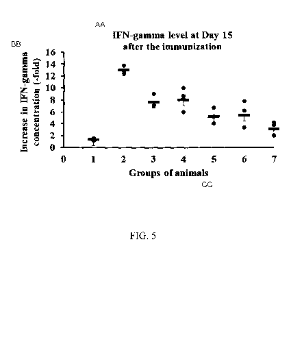
Fig. 5
illustrates the results of effectiveness assessment of the developed
immunobiological
agent based on recombinant adenovirus containing optimized for the expression
in
mammalian cells the protective antigen sequence (of proteins S, RBD, S-del, S-
Fe, RBD-G,
RBD-Fc) of SARS-CoV-2 with a sequence selected from SEQ ID NO:1, SEQ ID NO:2,
SEQ
ID NO:3, SEQ ID NO:4, SEQ ID NO:5, SEQ ID NO:6, as estimated by increase in
IFN-
gamma concentration in the medium after the splenocytes of C57/BL6 mice,
immunized with
the adenoviral constructs, were stimulated with the SARS-CoV-2 virus full-
length S protein,
at Day15 after the immunization of experimental animals.
Y-axis ¨ the values of increase in IFN-gamma concentration in the medium with
stimulated cells compared with intact cells (-fold).
X-axis ¨ studied groups of animals: intact animals and animals with
administered
108PFU/mouse
1) phosphate buffer (100 1)
2) Ad5-S-CoV-2 108 PFU/mouse
3) Ad5-S-del-CoV-2 108PFU/mouse
4) Ad5-S-Fc-CoV-2 108 PFU/mouse
5) Ad5-RBD-CoV-2 108 PFU/mouse
6) Ad5-RBD-G-CoV-2 108 PFU/mouse
7) Ad5-RBD-Fc-CoV-2 108 PFU/mouse
The first stage in the development of immunobiological agent against the SARS-
CoV-
2 coronavirus was the selection of a vaccine antigen. As a part of this
process, the literature
search was performed which demonstrated that the coronavirus S protein was the
most
promising antigen for creating a candidate vaccine. Type 1 transmembrane
glycoprotein is
responsible for virus particles binding, fusion and entry into the cells. As
demonstrated, it was
an inducer of neutralizing antibodies (Liang M et al, SARS patients-derived
human
recombinant antibodies to S and M proteins efficiently neutralize SARS-
coronavirus
infectivity. Biomed Environ Sci. 2005 Dec;18(6):363-74).
8
CA 03156350 2022-03-30
WO 2021/002776 PCT/RU2020/000344
The S protein consists of a signal peptide (amino acids 1-12) and 3 domains:
an
extracellular domain (amino acids 13-1193), transmembrane domain (amino acids
1194-
1215), and an intracellular domain (amino acids 1216-1255). The extracellular
domain
consists of two subunits Si and S2, and a small region between them, whose
functions are not
fully understood. The Si subunit is responsible for binding the virus to ACE2
(angiotensin-
converting enzyme 2) receptor. A fragment located in the middle region of the
Si subunit
(amino acids 318-510) has been named the receptor-binding domain (RBD). The S2
subunit
which contains a putative fusion peptide and two heptad repeats (HR1 and HR2)
promotes the
fusion of the virus and the target cell membrane. The infection is initiated
by the viral Si
subunit binding through its RBD to the ACE2 cell receptor.
Next, a fusion core between HR1 and HR2 regions of the S2 subunit is formed.
As a
result, the viral and cellular membranes get into close proximity followed by
their fusion and
the virus enters the cell. Therefore, the use of S protein or its fragment in
a vaccine formula
may induce antibodies that inhibit the virus entry into the cell.
To achieve the most effective induction of immune response, the authors
claimed
multiple variants of modification of this antigen, as well as its potential
combination with the
transmembrane domain of vesicular stomatitis virus glycoprotein for increasing
the level of
expression of the target protein.
Six different variants of nucleotide sequences were obtained (of modified S
gene of
the SARS-CoV-2 virus, or the receptor-binding domain of S protein) by
optimizing these
sequences for expression in mammalian cells.
Then, multiple constructs based on recombinant human adenovirus serotype 5 or
serotype 26 were developed to ensure that the modified genes are delivered
effectively to
mammalian cells. The adenoviral vectors were selected, since they have such
advantages as
the safety, broad tissue tropism, well-characterized genome, simplicity of
genetic
manipulations, capability to integrate large transgenic DNA inserts, intrinsic
adjuvant
properties, and the ability to induce stable T-cell-mediated and humoral
immune response.
Human adenoviruses of serotype 5 are the best studied ones among the known
adenoviruses, and therefore they are most commonly used in gene therapy for
deriving
vectors. Technologies were developed to produce first- and second-generation
vectors,
chimeric viral vectors (containing proteins of other viral serotypes) (J.N.
Glasgow et. al., An
adenovirus vector with a chimeric fiber derived from canine adenovirus type 2
displays novel
tropism, Virology, 2004, N2 324, 103-116), and multiple other vectors. Also,
vectors derived
from other serotypes were produced (H. Chen et. al., Adenovirus-Based
Vaccines:
9
CA 03156350 2022-03-30
WO 2021/002776 PCT/RU2020/000344
Comparison of Vectors from Three Species of Adenoviridae, Virology, 2010, N2
84(20),
10522-10532).
Vectors based on human adenovirus serotype 26 demonstrate a high level of
immunogenicity in primates, where they are able to induce a strong CD8+ T-cell
response
which, in terms of quality, is superior to T-cell-mediated response elicited
in the host body by
vectors based on human adenovirus serotype 5 (J. Liu et. al., Magnitude and
phenotype of
cellular immune responses elicited by recombinant adenovirus vectors and
heterologous
prime-boost regimens in rhesus monkeys, Virology, 2008, N2 82, 4844-4852).
With that,
more epitopes are recognized and the production of a broader spectrum of
factors (rather than
a predominant production of gamma-interferon) is induced (J. Liu et. al.,
Magnitude and
phenotype of cellular immune responses elicited by recombinant adenovirus
vectors and
heterologous prime-boost regimens in rhesus monkeys, Virology, 2008, N2 82,
4844-4852).
These data suggest that human adenovirus seroptype-26-based vectors have
fundamental
differences as regards their ability to induce immune response to the target
antigen as
compared with other adenoviral vectors.
Variant 1 invention is the recombinant human adenovirus serotype 5, or the
recombinant human adenovirus serotype 26, containing optimized for the
expression in
mammalian cells the sequence of full-length protective S antigen of the SARS-
CoV-2 virus
based on S protein gene sequences of the SARS-CoV-2 virus (SEQ ID NO:1).
Variant 2 invention is the recombinant human adenovirus serotype 5, or the
recombinant human adenovirus serotype 26 containing optimized for the
expression in
mammalian cells the sequence of full-length S protective antigen of the SARS-
CoV-2 virus
with gene C'-terminal deletion of 18 amino acids (SEQ ID NO:2).
Variant 3 invention is the recombinant human adenovirus serotype 5, or the
recombinant human adenovirus serotype 26 containing optimized for the
expression in
mammalian cells the sequence of full-length S protective antigen of the SARS-
CoV-2 virus
and the human IgG1 Fc-fragment sequence (SEQ ID NO:3).
Variant 4 invention is the recombinant human adenovirus serotype 5, or the
recombinant human adenovirus serotype 26 containing optimized for the
expression in
mammalian cells the SARS-CoV-2 virus S protein receptor-binding domain
sequence with
the viral leader peptide sequence (SEQ ID NO:4).
Variant 5 invention is the recombinant human adenovirus serotype 5, or the
recombinant human adenovirus serotype 26, containing optimized for the
expression in
CA 03156350 2022-03-30
WO 2021/002776 PCT/RU2020/000344
mammalian cells the SARS-CoV-2 virus S protein receptor-binding domain
sequence with
the transmembrane domain of vesicular stomatitis virus glycoprotein (SEQ ID
NO:5).
Variant 6 invention is the recombinant human adenovirus serotype 5, or the
recombinant human adenovirus serotype 26 containing optimized for the
expression in
mammalian cells the SARS-CoV-2 virus S protein receptor-binding domain
sequence with
the leader peptide sequence and the human IgG1 Fc-fragment sequence (SEQ ID
NO:6).
The authors have developed a method for inducing specific immunity to the SARS-
CoV-2 virus, which involves the administration to mammals of one or more
agents from
variants 1-6 in an effective amount. This method envisages:
1) sequential administration to mammals of two different immunobiological
agents
based on recombinant human adenovirus serotype 5 or two different
immunobiological agents based on recombinant human adenovirus serotype 26
presented herein in variants 1-6 with a time interval of more than one week.
2) sequential administration to mammals of any of the immunobiological agents
based on recombinant human adenovirus serotype 5 and any of the
immunobiological agents based on recombinant human adenovirus serotype 26
presented herein in variants 1-6 with a time interval of more than one week,
or
sequential administration to mammals of any of the immunobiological agents
based on recombinant human adenovirus serotype 26 and any of the
immunobiological agents based on recombinant human adenovirus serotype 5
presented herein in variants 1-6 with a time interval of more than one week.
3) simultaneous administration to mammals of any two immunobiological agents
based on recombinant human adenovirus serotype 5 or serotype 26 presented
herein in claim 1 and/or claim 2, and/or claim 3, and/or claim 4, and/or claim
5,
and/or claim 6.
The implementation of the invention is proven by the following examples:
Example 1. Obtaining of different variants of the SARS-CoV-2 virus S
glycoprotein
At the first stage, the authors developed several modifications of the vaccine
antigen to
achieve the most effective immune response.
11
CA 03156350 2022-03-30
WO 2021/002776 PCT/RU2020/000344
As a basis the SARS-CoV-2 virus S protein with a sequence SEQ ID NO:1 was
taken
which was then modified by several methods:
1) In order to present S protein on the plasma membrane, the deletion of 18
amino
acids on gene C' -terminal (S-del) SEQ ID NO:2 was performed (used for variant
2).
2) Also, the SARS-CoV-2 virus full-length S protective antigen sequence,
optimized for the expression in mammalian cells, with the human IgG1 Fc-
fragment sequence
was obtained (used for variant 3).
This modification enhances immunogenicity through a potential binding of
protein Fe
fragment to Fe receptor in antigen presentation cells (Li Z., Palaniyandi S.,
Zeng R., Tuo W.,
Roopenian D.C., Zhu X., Transfer of IgG in the female genital tract by MHC
class I-related
neonatal Fe receptor (FcRn) confers protective immunity to vaginal infection.
Proc. Natl.
Acad. Sci. U.S.A., 2011, N2108, 4388-93), and also increases the protein
stability and
prolongs its half-life in vivo (Zhang M.Y., Wang Y., Mankowski M.K., Ptak
R.G., Dimitrov
D.S., Cross-reactive HIV-1-neutralizing activity of serum IgG from a rabbit
immunized with
gp41 fused to IgG1 Fe: Possible role of the prolonged half-life of the
inununogen, Vaccine,
2009, N227, 857-863).
3) To assess the immunogenicity solely of the receptor-binding domain (RBD) of
the
SARS-CoV-2 virus S protein in a secreted form, a sequence SEQ ID NO:4 (used
for variant
4) was created which contains the S protein receptor-binding domain sequence
with the leader
peptide sequence (added for protein secretion).
4) To investigate the SARS-CoV-2 virus S protein RBD in a non-secreted form, a
sequence SEQ ID NO:5 was selected (used for variant 5) consisting of the SARS-
CoV-2 virus
S protein RBD to which the sequence of transmembrane domain of vesicular
stomatitis virus
glycoprotein (RBD-G) was added.
5) To investigate a secreted form of the S protein RBD with the leader peptide
sequence and the human IgG1 Fe-fragment sequence, a sequence SEQ ID NO:6 was
selected
(used for variant 6). The addition of the human IgG1 Fe-fragment sequence
enhances
immunogenicity through a potential binding of protein Fe fragment to Fe
receptor in antigen
presentation cells (Z. Li et. al., Transfer of IgG in the female genital tract
by MHC class I-
related neonatal Fe receptor (FcRn) confers protective immunity to vaginal
infection,
Proceedings of the National Academy of Sciences USA, 2011, N2 108, 4388-4393),
and also
may increase the protein stability and prolong its half-life in vivo (M.Y.
Zhang et. al.,
Crossreactive HIV-1-neutralizing activity of serum IgG from a rabbit immunized
with gp41
12
CA 03156350 2022-03-30
WO 2021/002776 PCT/RU2020/000344
fused to IgG1 Fc: Possible role of the prolonged half-life of the immunogen,
Vaccine, 2008,
N2 27, 857-63).
Example 2. Obtaining of genetic constructs encoding the S protein gene in
different
variants.
At the next stage, amino acid sequences presented herein in example 1 (SEQ ID
NO:1,
SEQ ID NO:2, SEQ ID NO:3, SEQ ID NO:4, SEQ ID NO:5, SEQ ID NO:6) were
translated
to nucleotide sequences. Next step comprised the optimization of obtained
sequences for the
expression in mammalian cells. All nucleotide sequences were obtained using
the method of
synthesis of the ZAO "Evrogen" Company (Moscow). As a result, the following
genetic
constructs were available:
1) pVax-S-CoV-2, containing nucleotide sequence of the SARS-CoV-2 virus full-
length S gene;
2) pVax-S-del-CoV-2, containing nucleotide sequence of the SARS-CoV-2 virus S
gene with deletion of 18 amino acids at the gene C'-terminal;
3) pVax-S-Fc-CoV-2, containing nucleotide sequence of the SARS-CoV-2 virus
full-
length S gene and sequence of the human IgG1 Fe-fragment;
4) pAL2-T-RBD-CoV-2, containing nucleotide sequence of the S protein receptor-
binding domain with the leader peptide gene sequence;
5) pAL2-T-RBD-G-CoV-2, containing nucleotide sequence of the S protein
receptor-
binding domain with G gene of the vesicular stomatitis virus;
6) pAL2-T-RBD-Fc-CoV-2, containing nucleotide sequence of the S protein
receptor-binding domain with the leader peptide gene sequence, and nucleotide
sequence of the human IgG1 Fe-fragment.
Then, by genetic engineering methods, the S protein gene sequence from
construct
pVax-S-CoV-2 was cloned, using XbaI restriction endonuclease, into a shuttle
plasmid
pShuttle-CMV (Stratagene, US); and, the obtained plasmid was named pShuttle-S-
CoV-2.
Thus, the shuttle plasmid pShuttle-S-CoV-2 was created which carries the
nucleotide
sequence of S amino acid sequence (SEQ ID NO:1), optimized for the expression
in
mammalian cells (as obtained in example 1).
Similarly, the nucleotide sequences of modified variants of the SARS-CoV-2
virus S
protein were cloned into a shuttle plasmid pShuttle-CMV (Stratagene, US) and
the following
shuttle plasmids were obtained:
13
CA 03156350 2022-03-30
WO 2021/002776 PCT/RU2020/000344
- pShuttle-S-del-CoV-2 (contains the optimized nucleotide sequence of the SARS-
CoV-2 virus S gene with deletion of 18 amino acids at the gene C'-terminal);
- pShuttle-S-Fc-CoV-2, containing the optimized nucleotide sequence of the
SARS-
CoV-2 virus full-length S gene and the sequence of Fe-fragment from human
IgGl;
- pShuttle-RBD-CoV-2 (contains the optimized nucleotide sequence of the SARS-
CoV-2 virus S receptor-binding domain);
- pShuttle-RBD-G-CoV-2 (contains the optimized nucleotide sequence of the SARS-
CoV-2 virus S receptor-binding domain with the transmembrane domain of the
vesicular
stomatitis virus glycoprotein);
- pShuttle-RBD-Fc-CoV-2 (contains the optimized nucleotide sequence of the
SARS-
CoV-2 virus S receptor-binding domain with the optimized sequence of Fe-
fragment from
human IgG1).
Example 3. Obtaining of an immunobiological agent based on recombinant human
adenovirus serotype 5.
At the next stage, a recombinant adenoviral plasmid pAd5-S-CoV-2 was obtained
which contains the sequence of SARS-CoV-2 full-length S protective antigen
(SEQ ID NO:1)
(variant 1) optimized for the expression in mammalian cells. This plasmid was
obtained by
the process of homologous recombination between the plasmid pAd containing the
genomic
region of human adenovirus serotype 5 with deleted El and E3 sites, and the
shuttle plasmid
pShuttle-S (obtained in example 3) which carries homologous sites of the
adenovirus genome
and an expression cassette with the target gene (of S protein). For this
purpose, the shuttle
plasmid pShuttle-S obtained in example 3 was linearized by restriction
endonuclease PmeI.
Homologous recombination was performed in the cells of E. coli strain BJ5183.
Plasmid pAd was mixed with plasmid pShuttle-S, and then the received mixture
was used to
transform the E.coli cells by electroporation method according to the Guide
"MicroPulserTm
Electroporation Apparatus Operating Instructions and Applications Guide" (Bio-
Rad, US). As
the transformation was completed, the cells of E. coli strain BJ5183 were
inoculated in LB-
agar dishes, containing a selective antibiotic, and grown for 18 hours at +37
C. A
transformation effectiveness was 1010 ¨1011 transformed clones per tig of
plasmid pBluescript
II SK(-).
As a result of homologous recombination, a cassette with the target transgene
(of S
protein) appeared in the plasmid pAd, and the antibiotic-resistance gene
changed.
14
CA 03156350 2022-03-30
WO 2021/002776 PCT/RU2020/000344
Thus, the recombinant adenoviral plasmid pAd5-S-CoV-2 was constructed which
contains a full-length genome of recombinant human adenovirus serotype 5 (with
El and E3
sites deleted from the genome) with the integrated genetic construct obtained
in example 3.
Then, the plasmid pAd5-S-CoV-2 was hydrolyzed with the restriction
endonuclease Pac I and
used for the transfection of permissive cell culture of human embryonic kidney
cell line HEK
293. The genome of HEK 293 cells contains the integrated El site of human
adenovirus
serotype 5 genome, so that the replication of recombinant replication-
defective human
adenoviruses serotype 5 may occur. At day 6 after the transfection, the first
blind passages
were performed to ensure a more effective production of the recombinant
adenovirus. Upon
occurrence of the cytopathic virus effect (microscopy data), the cells with
culture medium
were frozen for three times to facilitate the disruption of cells and the
virus release. The
obtained material was then used for the accumulation of preparative amounts of
the
recombinant adenoviruses.
Activity of the preparation pAd5-S-CoV-2 hereinafter was assessed by the
standard
titration technique in the culture of susceptible 293 HEK cells using a plague
forming cell
assay.
In order to verify the creation of a construct of potential recombinant pseudo-
adenoviral particle derived from human adenovirus serotype 5, expressing the
SARS-CoV-2
virus S gene, the polymerase chain reaction (PCR) was performed according to
the
established standard technique.
In a similar way, additional five recombinant viruses were obtained: Ad5-S-del-
CoV-
2, Ad5-S-Fc-CoV-2, Ad5-RBD-CoV-2, Ad5-RBD-G-CoV-2, Ad5-RBD-Fc-CoV-2.
Thus, as a result, variants of the immunobiological agent based on recombinant
human
adenovirus serotype 5 were obtained, containing:
1) optimized nucleotide sequence of the SARS-CoV-2 virus S receptor-binding
domain (variant 1);
2) optimized nucleotide sequence of the SARS-CoV-2 virus S protective
antigen
with deletion of 18 amino acids at the gene C'-terminal (variant 2);
3) sequence of the SARS-CoV-2 virus full-length S protective antigen and
sequence of the human IgG1 Fc-fragment optimized for the expression in
mammalian cells
(variant 3);
CA 03156350 2022-03-30
WO 2021/002776 PCT/RU2020/000344
4) optimized nucleotide sequence of the S protein receptor-binding domain
with
the leader peptide sequence (variant 4);
5) optimized nucleotide sequence of the S protein receptor-binding domain
with
the transmembrane domain of vesicular stomatitis virus glycoprotein (variant
5),
6) optimized sequence of the S protein receptor-binding domain with the
leader
peptide sequence and the human IgG1 Fc-fragment sequence (variant 6).
Example 4. Obtaining of an immunobiological agent based on recombinant human
adenovirus serotype 26.
At the first stage, an expression cassette with the SARS-CoV-2 virus S gene
was
placed in the recombinant vector pAd26-ORF6-Ad5. For this purpose, the vector
pAd26-
ORF6-Ad5 was linearized with the restriction endonuclease PmeI, while the
plasmid
construct pShuttle-S, obtained in example 3, was processed with the
restriction endonucleases
PmeI. Hydrolysis products were ligated, and then the plasmid pAd26-S-CoV-2 was
produced
using standard techniques.
At the next stage, the plasmid pAd26-S-CoV-2 was hydrolyzed with the
restriction
endonucleases Pad I and SwaI and used for the transfection of permissive cell
line HEK 293
culture. On the third day after the transfection, the first blind passages
were performed to
ensure a more effective generation of recombinant virus. Upon occurrence of
the cytopathic
virus effect (microscopy data), the cells with culture medium were frozen for
three times to
facilitate the disruption of cells and the virus release. The obtained
material was then used for
the accumulating preparative amounts of recombinant adenoviruses. Activity of
the
preparation pAd26-S-CoV-2 hereinafter was assessed by the standard titration
technique in
the culture of 293 HEK cells using a plague forming cell assay.
In order to verify that construct of the proposed recombinant pseudo-
adenoviral
particle based on human adenovirus serotype 26, expressing the SARS-CoV-2
virus S gene,
has been generated, polymerase chain reaction (PCR) was performed according to
the
established standard technique.
In a similar way, additional five recombinant viruses were obtained: pAd26-S-
dek-
CoV-2, pAd26-S-Fc-CoV-2, pAd26-RBD-CoV-2, pAd26-RBD-G-CoV-2, pAd26-RBD-Fc-
CoV-2.
16
CA 03156350 2022-03-30
WO 2021/002776 PCT/RU2020/000344
Thus, as a result, variants of the immunobiological agent based on recombinant
human
adenovirus serotype 26 were obtained, containing:
1) optimized nucleotide sequence of the SARS-CoV-2 virus S receptor-binding
domain
(variant 1);
2) optimized nucleotide sequence of the SARS-CoV-2 virus S protective antigen
with
deletion of 18 amino acids at the gene C'-terminal (variant 2);
3) sequence of the SARS-CoV-2 virus full-length S protective antigen and
sequence of
the human IgG1 Fc-fragment optimized for the expression in mammalian cells
(variant 3);
4) optimized nucleotide sequence of the S protein receptor-binding domain with
the
leader peptide sequence (variant 4);
5) optimized nucleotide sequence of the S protein receptor-binding domain with
the
transmembrane domain of vesicular stomatitis virus glycoprotein (variant 5);
6) optimized sequence of the S protein receptor-binding domain with the leader
peptide
sequence and the human IgG1 Fc-fragment sequence (variant 6).
Example 5. Verification of the expression of different variants of S
glycoprotein gene
of the SARS-CoV-2 virus in HEK293 cells after the addition of immunobiological
agent
based on recombinant human adenovirus serotype 5.
The aim of this experiment was to verify the ability of constructed
recombinant
adenoviruses Ad5-S-CoV-2, Ad5-S-del-CoV-2, Ad5-S-Fc-CoV-2, Ad5-RBD-CoV-2, Ad5-
RBD-G-CoV-2, Ad5-RBD-Fe-CoV-2 to express different variants of S protein gene
in
mammalian cells.
EK293 cells were cultured in DMEM medium containing 10% fetal calf serum in
incubator at 37 C and 5% CO2. The cells were placed in 35mm2 culture Petri
dishes and
incubated for 24 hours until reaching 70% confluence. Then, the studied
preparations of
recombinant adenoviruses (Ad5-S-CoV-2, Ad5-S-del-CoV-2, Ad5-S-Fc-CoV-2, Ad5-
RBD-
CoV-2, Ad5-RBD-G-CoV-2, Ad5-RBD-Fc-CoV-2), and control preparation (Ad5-null ¨
recombinant adenovirus containing no inserts) in an amount of 100 PFU/cell and
phosphate
buffer saline (PBS), as a negative control, were added to the cells. Two days
after the
transduction, the cells were collected and lysed in 0.5 ml of normal strength
buffer CCLR
(Promega). The lysate was diluted with carbonate-bicarbonate buffer and placed
in ELISA
plate wells. The plate was incubated over the night at +4 C.
17
CA 03156350 2022-03-30
WO 2021/002776 PCT/RU2020/000344
The plate wells were washed for three times with normal strength washing
buffer at an
amount of 200 1 per well, and then 100 1.11 of blocking buffer were added to
every well; the
plate was covered with a lid and incubated for 1 hour at 37 C in shaker at 400
rpm. Then, the
plate wells were washed for three times with normal strength buffer at an
amount of 200 I
per well and 100 jl of convalescent blood serum were added to every well. The
plate was
covered with a lid and incubated at room temperature in shaker at 400 rpm for
2 hours. Then,
the plate wells were washed for three times with normal strength washing
buffer at an amount
of 200 1 per well, and then 100 1 of secondary antibodies conjugated with
biotin were
added. The plate was covered with a lid and incubated at room temperature in
shaker at 400
rpm for 2 hours. Next, a solution of streptavidin conjugated with horseradish
peroxidase was
prepared. For this purpose the conjugate in the amount of 60 IA was diluted in
5.94 ml of an
assay buffer. The plate wells were washed twice with normal strength washing
buffer at an
amount of 200 1 per well and 100 I of streptavidin solution conjugated with
horseradish
peroxidase were added to each of the plate wells. The plate was incubated at
room
temperature in shaker at 400 rpm for 1 hour. Then, the plate wells were washed
twice with
normal strength washing buffer at an amount of 200 I per well and 100 1 of
TMB substrate
were added to each of the plate wells. The plate was incubated under darkness
at room
temperature for 10 minutes, then 100 I of stop solution were added to each of
the plate wells.
The optical density was measured using plate spectrophotometer (Multiskan FC,
Thermo) at a
wavelength of 450 nm. The experiment results are presented in Table 1.
Table 1 ¨ Results of the experiment for verifying the expression of different
variants
of S glycoprotein gene of the SARS-CoV-2 virus in HEK293 cells after the
addition of
immunobiological agent based on recombinant human adenovirus serotype 5.
Mean optical density at a wavelength of 450 nm
PBS 0.19 ( 0.05)
Ad5-null 0.23 ( 0.08)
Ad5-S-CoV-2 1.85 ( 0.15)
Ad5-S-del-CoV-2 1.63 ( 0.19)
Ad5-S-Fc-CoV-2 1.57 ( 0.30)
Ad5-RBD-CoV-2 1.47 ( 0.21)
Ad5-RBD-G-CoV-2 1.52 ( 0.19)
Ad5-RBD-Fc-CoV-2 1.58 (0.11)
18
CA 03156350 2022-03-30
WO 2021/002776 PCT/RU2020/000344
As shown by the received data, the expression of different variants of the
target
protein was observed in all cells transduced with recombinant adenoviruses Ad5-
S-CoV-2,
Ad5-S-del-CoV-2, Ad5-S-Fc-CoV-2, Ad5-RBD-CoV-2, Ad5-RBD-G-CoV-2, Ad5-RBD-Fc-
CoV-2.
Example 6. Verification of the expression of different variants of S
glycoprotein gene
of the SARS-CoV-2 virus in HEK293 cells after the addition of immunobiological
agent
based on recombinant human adenovirus serotype 26.
The aim of this experiment was to verify the ability of constructed
recombinant
adenoviruses pAd26-S-CoV-2, Ad26-S-del-CoV-2, Ad26-S-Fc-CoV-2, pAd26-RBD-CoV-
2,
pAd26-RBD-G-CoV-2, pAd26-RBD-Fc-CoV-2 to express different variants of S
protein gene
in mammalian cells.
HEK293 cells were cultured in DMEM medium containing 10% fetal calf serum in
incubator at 37 C and 5% CO2. The cells were placed in 35mm2 culture Petri
dishes and
incubated for 24 hours until reaching 70% confluence. Then, the studied
preparations of
recombinant adenoviruses (pAd26-S-CoV-2, Ad26-S-del-CoV-2, Ad26-S-Fc-CoV-2,
pAd26-
RBD-CoV-2, pAd26-RBD-G-CoV-2, pAd26-RBD-Fc-CoV-2), and control preparation
(Ad26-null ¨ recombinant adenovirus containing no inserts) in an amount of 100
PFU/cell
and phosphate buffer saline (PBS), as a negative control, were added to the
cells. Two days
after the transduction, the cells were collected and lysed in 0.5 ml of normal
strength buffer
CCLR (Promega). The lysate was diluted with carbonate-bicarbonate buffer and
placed in
ELISA plate wells. The plate was incubated over the night at +4 C.
The plate wells were washed for three times with normal strength washing
buffer at an
amount of 200 1 per well, and then 100 1 of blocking buffer were added to
each well; the
plate was covered with a lid and incubated for 1 hour at 37 C in shaker at 400
rpm. Then, the
plate wells were washed for three times with normal strength buffer at an
amount of 200 1
per well and 100 1 of convalescent blood serum was added to every well. The
plate was
covered with a lid and incubated at room temperature in shaker at 400 rpm for
2 hours. Then,
the plate wells were washed for three times with normal strength washing
buffer at an amount
of 200 I per well, and 100 ul of secondary antibodies conjugated with biotin
were added.
The plate was covered with a lid and incubated at room temperature in shaker
at 400 rpm for
2 hours. Next, solution of streptavidin conjugated with horseradish peroxidase
was prepared.
19
CA 03156350 2022-03-30
WO 2021/002776 PCT/RU2020/000344
For this purpose, the conjugate in the amount of 60 p,1 was diluted in 5.94 ml
of assay buffer.
The plate wells were washed twice with normal strength washing buffer at an
amount of 200
pl per well and 100 pl of streptavidin solution conjugated with horseradish
peroxidase were
added to each of the plate wells. The plate was incubated at room temperature
in shaker at 400
rpm for 1 hour. Then, the plate wells were washed twice with normal strength
washing buffer
at an amount of 200 pl per well and 100 pl of TMB substrate were added to each
of the plate
wells and incubated under darkness at room temperature for 10 minutes. Then
100 pl of stop
solution was added to each of the plate wells. The value of optical density
was measured
using plate spectrophotometer (Multiskan FC, Thermo) at a wavelength of 450
nm. The
experiment results are presented in Table 2.
Table 2 ¨ Results of the experiment for verifying the expression of different
variants
of S glycoprotein gene of the SARS-CoV-2 virus in 11EK293 cells after the
addition of
immunobiological agent based on recombinant human adenovirus serotype 26.
Mean optical density at a wavelength of 450 nm
PBS 0.17 ( 0.08)
Ad26-null 0.22 ( 0.09)
Ad26-S-CoV-2 1.68 ( 0.21)
Ad26-S-del-CoV-2 1.65 (0.14)
Ad26-S-Fc-CoV-2 1.71 ( 0.13)
Ad26-RBD-CoV-2 1.61 ( 0.18)
Ad26-RBD-G-CoV-2 1.45 ( 0.22)
Ad26-RBD-Fc-CoV-2 1.51 (0.14)
As shown by the received data, the expression of different variants of the
target
protein was observed in all cells transduced with recombinant adenoviruses
pAd26-S-CoV-2,
Ad26-S-del-CoV-2, Ad26-S-Fc-CoV-2, pAd26-RBD-CoV-2, pAd26-RBD-G-CoV-2, pAd26-
RBD-Fc-CoV-2.
Example 7. A method of utilization of the developed immunobiological agent by
a
single administration to mammals in an effective amount for the induction of
specific
immunity to SARS-CoV-2.
CA 03156350 2022-03-30
WO 2021/002776 PCT/RU2020/000344
The developed immunobiological agent based on recombinant human adenoviruses
serotypes 5 and 26, containing optimized for the expression in mammalian cells
the protective
antigen sequence (of proteins S, S-del, S-Fc, RBD, RBD-G, RBD-Fc) of SARS-CoV-
2 with a
sequence selected from SEQ ID NO:1, SEQ ID NO:2, SEQ ID NO:3, SEQ ID NO:4, SEQ
ID
NO:5, SEQ ID NO:6 is utilized by administering to mammals through any of the
administration routes known for this viral vector (subcutaneously,
intramuscularly,
intravenously, intranasally). This way, immune response to the target protein
of SARS-CoV-2
glycoprotein develops in mammals.
One of the main characteristics of immunization effectiveness is an antibody
titer.
Example presents data relating to changes in the titer of antibodies against
SARS-CoV-2
glycoprotein at day 21 after a single intramuscular immunization of animals
with the
immunobiological agent, containing the recombinant human adenovirus of
serotype 5 or 26,
comprising optimized for the expression in mammalian cells the sequence of
protective
antigen (of proteins S, S-del, S-Fe, RBD, RBD-G, RBD-Fc) of SARS-CoV-2 with a
sequence
selected from SEQ ID NO:1, SEQ ID NO:2, SEQ ID NO:3, SEQ ID NO:4, SEQ ID NO:5,
SEQ ID NO:6.
Mammals used in the experiment ¨ mice C57BL/6, females, 18 g. All the animals
were divided into 43 groups, 5 animals each, injected intramuscularly with:
1) Ad5-S-CoV-2 107 PFU/mouse
2) Ad5-S-del-CoV-2 107 PFU/mouse
3) Ad5-S-Fc-CoV-2 107 PFU/mouse
4) Ad5-RBD-CoV-2 107 PFU/mouse
5) Ad5-RBD-G-CoV-2 107 PFU/mouse
6) Ad5-RBD-Fc-CoV-2 107PFU/mouse
7) Ad5-null 107 PFU/mouse
8) Ad5-S-CoV-2 108 PFU/mouse
9) Ad5-S-del-CoV-2 108 PFU/mouse
10) Ad5-S-Fc-CoV-2 108 PFU/mouse
11) Ad5-RBD-CoV-2 108 PFU/mouse
12) Ad5-RBD-G-CoV-2 108 PFU/mouse
13) Ad5-RBD-Fc-CoV-2 108 PFU/mouse
14) Ad5-null 108 PFU/mouse
15) Ad5-S-CoV-2 109 PFU/mouse
16) Ad5-S-del-CoV-2 109 PFU/mouse
21
CA 03156350 2022-03-30
WO 2021/002776 PCT/RU2020/000344
17) Ad5-S-Fc-CoV-2 109 PFU/mouse
18) Ad5-RBD-CoV-2 109 PFU/mouse
19) Ad5-RBD-G-CoV-2 109 PFU/mouse
20) Ad5-RBD-Fc-CoV-2 109 PFU/mouse
21) Ad5-null 109 PFU/mouse
22) Ad26-S-CoV-2 107 PFU/mouse
23) Ad26-S-del-CoV-2 107 PFU/mouse
24) Ad26-S-Fc-CoV-2 107 PFU/mouse
25) Ad26-RBD-CoV-2 107 PFU/mouse
26) Ad26-RBD-G-CoV-2 107 PFU/mouse
27) Ad26-RBD-Fc-CoV-2 107 PFU/mouse
28) Ad26-null 107 PFU/mouse
29) Ad26-S-CoV-2 108 PFU/mouse
30) Ad26-S-del-CoV-2 108 PFU/mouse
31) Ad26-S-Fc-CoV-2 108 PFU/mouse
32) Ad26-RBD-CoV-2 108 PFU/mouse
33) Ad26-RBD-G-CoV-2 108 PFU/mouse
34) Ad26-RBD-Fc-CoV-2 108 PFU/mouse
35) Ad26-null 108 PFU/mouse
36) Ad26-S-CoV-2 109 PFU/mouse
37) Ad26-S-del-CoV-2 109 PFU/mouse
38) Ad26-S-Fc-CoV-2 109 PFU/mouse
39) Ad26-RBD-CoV-2 109 PFU/mouse
40) Ad26-RBD-G-CoV-2 109 PFU/mouse
41) Ad26-RBD-Fc-CoV-2 109 PFU/mouse
42) Ad26-null 109 PFU/mouse
43) phosphate buffer saline
Three weeks later, blood samples were taken from the tail vein of animals, and
blood
serum was isolated. Antibody titers were determined by ELISA using the
following protocol:
1) Protein (S) was adsorbed onto wells of 96-well ELISA plate for 16 hours
at
+4 C.
2) Then, for preventing a non-specific binding, the plate was "blocked"
with 5%
milk dissolved in TPBS in an amount of 100 IA per well. It was incubated in
shaker at 37 C
for one hour.
22
CA 03156350 2022-03-30
WO 2021/002776 PCT/RU2020/000344
3) Serum samples from the immunized mice were diluted using a 2-fold
dilution
method. Totally, 12 dilutions of each sample were prepared.
4) 50 IA of each of the diluted serum samples were added to the plate
wells.
5) Then, incubation at 37 C for 1 hour was performed.
6) After incubation the wells were washed three times with phosphate
buffer.
7) Then, secondary antibodies against mouse immunoglobulins conjugated with
horseradish peroxidase were added.
8) Then, incubation at 37 C for 1 hour was performed.
9) After incubation the wells were washed three times with phosphate
buffer.
10) Then, tetramethylbenzidine (TMB) solution was added which serves as a
substrate for horseradish peroxidase and is converted into a colored compound
by the
reaction. The reaction was stopped after 15 minutes by the adding sulfuric
acid. Next, using a
spectrophotometer the optical density (OD) of the solution was measured in
each well at a
wavelength of 450 nm.
Antibody titer was determined as the last dilution at which the optical
density of the
solution was significantly higher than in the negative control group. The
obtained results
(geometric mean) are presented in Table 3.
Table 3 ¨ Antibody titers against S protein in the blood serum of mice
(geometric
mean of antibody titers)
Table 3.
Recombinant PFU/mouse
adenovirus 10/ 108 109
Ad5 -null 0 0 0
Ad5-S-CoV-2 1:10809 1:18820 1:57052
Ad5-S-del-CoV-2 1:21619 1:28526 1:114105
Ad5-S-Fc-CoV-2 1:14263 1:18820 1:57052
Ad5 -RBD-CoV -2 1:12417 1:14263 1:99334
Ad5-RBD-G-CoV-2 1:32768 1:49667 1:172951
Ad5-RBD-Fc-CoV-2 1:10809 1:12417 1:28526
Ad26-null 0 0 0
Ad26-S-CoV-2 1:18820 1:24834 1:43238
Ad26-S-del-CoV-2 1:24834 1:43238 1:57052
23
CA 03156350 2022-03-30
WO 2021/002776 PCT/RU2020/000344
Ad26-S-Fc-CoV-2 1:28526 1:32768 1:86475
Ad26-RBD-CoV-2 1:12417 1:18820 1:86475
Ad26-RBD-G-CoV-2 1:24834 1:32768 1:150562
Ad26-RBD-Fc-CoV-2 1:9410 1:12417 1:24834
The results of experiment have shown that the developed immunobiological agent
administered to mammals induces humoral immune response to SARS-CoV-2
glycoprotein
over the entire selected dose range. It is obvious that dose escalation will
result in antibody
titer increase in the mammalian blood till the toxic effect occurs.
Example 8. A method of utilization of the developed immunobiological agent by
sequential administration to mammals in an effective amount for the induction
of specific
immunity to SARS-CoV-2.
This example describes a method of utilization of the developed
immunobiological
agent based on recombinant human adenoviruses serotype 5, containing optimized
for the
expression in mammalian cells the protective antigen sequence (of proteins S,
S-del, S-Fe,
RBD, RBD-G, RBD-Fc) of SARS-CoV-2 with a sequence selected from SEQ ID NO:1,
SEQ
ID NO:2, SEQ ID NO:3, SEQ ID NO:4, SEQ ID NO:5, SEQ ID NO:6 by their
sequential
administration to mammals with a time interval of 1 week for the inducing
specific immunity
to SARS-CoV-2.
The experiment was performed according to the protocol described in example 7.
All the animals were divided into 29 groups (3 animals each,) injected
intramuscularly
with:
1. phosphate buffer (1001A), and a week later phosphate buffer (100 1)
2. phosphate buffer (100 1), and a week later Ad5-null 108 PFU/mouse
3. Ad5-null 108 PFU/mouse, and a week later phosphate buffer (100 Ill)
4. Ad5-null 108 PFU/mouse, and a week later Ad5-null 108 PFU/mouse
5. Ad5-S-CoV-2 108 PFU/mouse, and a week later Ad5-S-CoV-2 108 PFU/mouse
6. Ad5-S-CoV-2 108 PFU/mouse, and a week later Ad5-RBD-CoV-2 108 PFU/mouse
7. Ad5-S-CoV-2 108 PFU/mouse, and a week later Ad5-S-del-CoV-2 108 PFU/mouse
8. Ad5-S-CoV-2 108 PFU/mouse, and a week later Ad5-S-Fc-CoV-2 108 PFU/mouse
9. Ad5-S-CoV-2 108 PFU/mouse, and a week later Ad5-RBD-CoV-2 108 PFU/mouse
10. Ad5-S-CoV-2 108 PFU/mouse, and a week later Ad5-RBD-G-CoV-2 108 PFU/mouse
24
CA 03156350 2022-03-30
WO 2021/002776 PCT/RU2020/000344
11. Ad5-S-CoV-2 108 PFU/mouse, and a week later Ad5-RBD-Fc-CoV-2 108 PFU/mouse
12. Ad5- S-del -CoV-2 108 PFU/mouse, and a week later Ad5-S-CoV-2 108
PFU/mouse
13. Ad5-S-del-CoV-2 108 PFU/mouse, and a week later Ad5-S-del-CoV-2 108
PFU/mouse
14. Ad5- S-del -CoV-2 108 PFU/mouse, and a week later Ad5-S-Fe-CoV-2 108
PFU/mouse
15. Ad5- S-del -CoV-2 108 PFU/mouse, and a week later Ad5-RBD-CoV-2 108
PFU/mouse
16. Ad5- S-del -CoV-2 108 PFU/mouse, and a week later Ad5-RBD-G-CoV-2 108
PFU/mouse
17. Ad5- S-del -CoV-2 108 PFU/mouse, and a week later Ad5-RBD-Fc-CoV-2
108PFU/mouse
18. Ad5- S-Fe -CoV-2 108 PFU/mouse, and a week later Ad5-S-CoV-2 108 PFU/mouse
19. Ad5- S-Fe -CoV-2 108 PFU/mouse, and a week later Ad5-S-del-CoV-2 108
PFU/mouse
20. Ad5- S-Fe -CoV-2 108 PFU/mouse, and a week later Ad5-S-Fc-CoV-2 108
PFU/mouse
21. Ad5- S-Fc -CoV-2 108 PFU/mouse, and a week later Ad5-RBD-CoV-2 108
PFU/mouse
22. Ad5- S-Fe -CoV-2 108 PFU/mouse, and a week later Ad5-RBD-G-CoV-2 108
PFU/mouse
23. Ad5- S-Fe -CoV-2 108 PFU/mouse, and a week later Ad5-RBD-Fc-CoV-2 108
PFU/mouse
24. Ad5-RBD-CoV-2 108 PFU/mouse, and a week later Ad5-S-CoV-2 108 PFU/mouse
25. Ad5-RBD-CoV-2 108 PFU/mouse, and a week later Ad5-S-del-CoV-2 108
PFU/mouse
26. Ad5-RBD-CoV-2 108 PFU/mouse, and a week later Ad5-S-Fc-CoV-2 108 PFU/mouse
27. Ad5-RBD-CoV-2 108 PFU/mouse, and a week later Ad5-RBD-CoV-2 108 PFU/mouse
28. Ad5-RBD-CoV-2 108 PFU/mouse, and a week later Ad5-RBD-G-CoV-2 108
PFU/mouse
29. Ad5-RBD-CoV-2 108 PFU/mouse, and a week later Ad5-RBD-Fc-CoV-2 108
PFU/mouse
30. Ad5-RBD-G-CoV-2 108 PFU/mouse, and a week later Ad5-S-CoV-2 108 PFU/mouse
31. Ad5-RBD-G-CoV-2 108 PFU/mouse, and a week later Ad5-S-del-CoV-2 108
PFU/mouse
32. Ad5-RBD-G-CoV-2 108 PFU/mouse, and a week later Ad5-S-Fe-CoV-2 108
PFU/mouse
33. Ad5-RBD-G-CoV-2 108 PFU/mouse, and a week later Ad5-RBD-CoV-2 108
PFU/mouse
34. Ad5-RBD-G-CoV-2 108 PFU/mouse, and a week later Ad5-RBD-G-CoV-2
108PFU/mouse
35. Ad5-RBD-G-CoV-2 108 PFU/mouse, and a week later Ad5- RBD-Fe-CoV-2 108
PFU/mouse
36. Ad5-RBD-Fe-CoV-2 108 PFU/mouse, and a week later Ad5-S-CoV-2 108 PFU/mouse
37. Ad5-RBD-Fc-CoV-2 108 PFU/mouse, and a week later Ad5-S-del-CoV-2 108
PFU/mouse
38. Ad5-RBD-Fe-CoV-2 108 PFU/mouse and a week later Ad5-S-Fc-CoV-2 108
PFU/mouse
CA 03156350 2022-03-30
WO 2021/002776 PCT/RU2020/000344
39. Ad5-RBD-Fc-CoV-2108 PFU/mouse, and a week later Ad5-RBD-CoV-2 108
PFU/mouse
40. Ad5-RBD-Fc-CoV-2 108 PFU/mouse, and a week later Ad5-RBD-G-CoV-2 108
PFU/mouse
41. Ad5-RBD-Fc-CoV-2 108 PFU/mouse, and a week later Ad5-RBD-Fc-CoV-2 108
PFU/mouse
The results are presented in Tables 4 and 5.
Table 4. ¨ Antibody titers against the SARS-CoV-2 virus S protein in the blood
serum of
mice from control groups
Table 4.
Second immunization (a week
later)
PBS Ad5-null
First PBS 0 0
immunization Ad5-null 0 0
Table 5. ¨ Antibody titers against the SARS-CoV-2 virus S protein in the blood
serum of
mice from experimental groups
Table 5.
Second immunization (a week later)
Ad5-S- Ad5-S- Ad5- Ad5-
Ad5-S- Ad5- RBD-
del-CoV- Fc-CoV- RBD- RBD-Fc-
CoV-2 G-CoV-2
2 2 CoV-2 CoV-2
Ad5-S-
1:32768 1:131072 1:104032 1:131072 1:65536 1:104032
CoV-2
Ad5-S-del- 1:65536 1:131072 1:131072 1:131072 1:65536 1:104032
CoV-2
Ad5-S-Fc- 1:65536 1:104032 1:65536 1:104032 1:52016 1:131072
CoV-2
26
CA 03156350 2022-03-30
WO 2021/002776 PCT/RU2020/000344
Ad5-RBD- 1:52016 1:65536 1:131072 1:65536 1:32768 1:52016
CoV-2
Ad5-RBD- 1:13107 1:131072 1:104032 1:131072 1:65536 1:104032
G-CoV-2 2
Ad5-RBD- 1:82570 1:131072 1:65536 - 1:32768 1:65536
1:65536
Fc-CoV-2
Thus, the experimental results have fully confirmed that the sequential
immunization
with the developed immunobiological agents in different combinations that
include different
forms of S protein of SARS-CoV-2 will cause a more powerful induction of
immune response
than the immunization with the one antigen performed according to a similar
regimen.
Example 9. Effectiveness assessment of the immunization with the developed
immunobiological agent by the percentage of proliferating lymphocytes.
Lymphocyte proliferation assay enables to assess the ability of lymphocytes to
divide
more actively after encountering an antigen. In order to assess proliferation,
the authors used
fluorescent dye CFSE for staining lymphocytes. This dye binds to cellular
proteins and stays
there for a long time, but it never spreads to the neighboring cells in the
population. However,
the fluorescent label is passed onto the daughter cells. The label
concentration and,
consequently, the fluorescence intensity in the daughter cells is decreased
precisely twice.
Thus, dividing cells can be easily traced by a decrease in their fluorescence.
Therefore
dividing cells can be easily traced by the reducing fluorescence intensity.
C57BL/6 mice were used in the experiment. All the animals were divided into 8
groups (3 animals each,) and injected intramuscularly with:
1) Phosphate buffer (100W)
2) Ad5-null 108 PFU/mouse
3) Ad5-S-CoV-2 108 PFU/mouse
4) Ad5-S-del-CoV-2 108 PFU/mouse
5) Ad5-S-Fc-CoV-2 108 PFU/mouse
6) Ad5-RBD-CoV-2 108 PFU/mouse
7) Ad5-RBD-G-CoV-2 108 PFU/mouse
8) Ad5-RBD-Fc-CoV-2 108 PFU/mouse
27
CA 03156350 2022-03-30
WO 2021/002776 PCT/RU2020/000344
Two weeks later the animals were euthanized. Lymphocytes were isolated from
the
spleen by Ficoll-Urografin density gradient centrifugation. Then, the isolated
cells were
stained with CFSE according to the CFSE technique (Monitoring lymphocyte
proliferation in
vitro and in vivo with the intracellular fluorescent dye carboxy fluorescent
diacetate
succinimidy 'ester/ Quah BJ, Warren HS, Parish CR Nat Protoc. 2007; 2(9),
p:2049-2056) and
were cultured in the presence of antigen. Then, the cells were analyzed using
flow cytometry.
The obtained results are shown in Fig. 1, 2, 3, 4. Thus, it could be concluded
that the obtained
adenoviral constructs induce antigen-specific immune response (both CD4+ and
CD8+).
As shown by the experiment results (Fig. 1, 2, 3, 4), the immunobiological
agents
developed according to claim 1, claim 2, claim 3, claim 4, claim 5 effectively
stimulate
proliferation of lymphocytes in the dose used.
Example 10. A method of utilization of the developed immunobiological agents
based
on recombinant human adenoviruses serotypes 5 and 26 by their sequential
administration to
mammals for the induction of specific immunity to SARS-CoV-2.
The experiment was performed according to the protocol described in example 7.
Combinations of immunobiological agents were selected on the basis of examples
7 and 8.
All the animals were divided into 31 group (3 animals each,) injected
intramuscularly
with:
1. phosphate buffer (100 mlui), and a week later phosphate buffer (100
1.11)
2. Ad26-null 108 PFU/mouse, and a week later phosphate buffer (100 p.1)
3. phosphate buffer (100 1), and a week later Ad26-null 108 PFU/mouse
4. Ad26-null 108 PFU/mouse, and a week later Ad26-null 108 PFU/mouse
5. Ad5-null 108 PFU/mouse, and a week later phosphate buffer (100 1)
6. phosphate buffer (100 1), and a week later Ad5-null 108 PFU/mouse
7. Ad5-null 108 PFU/mouse, and a week later Ad5-null 108 PFU/mouse
8. Ad5-null 108 PFU/mouse, and a week later Ad26-null 108 PFU/mouse
9. Ad26-null 108 PFU/mouse, and a week later Ad5-null 108 PFU/mouse
10. Ad5-S-CoV-2 108 PFU/mouse, and a week later Ad26-RBD-G-CoV-2 108
PFU/mouse
11. Ad5-RBD-G-CoV-2 108 PFU/mouse, and a week later Ad26-S-CoV-2 108
PFU/mouse
12. Ad26-S-CoV-2 108 PFU/mouse, and a week later Ad5-RBD-G-CoV-2 108
PFU/mouse
13. Ad26-RBD-G-CoV-2 108 PFU/mouse, and a week later Ad5-S-CoV-2 108
PFU/mouse
14. Ad5-S-del-CoV-2 108 PFU/mouse, and a week later Ad26-RBD-CoV-2 108
PFU/mouse
28
CA 03156350 2022-03-30
WO 2021/002776 PCT/RU2020/000344
15. Ad26-S-G-CoV-2 108 PFU/mouse, and a week later Ad5-RBD-CoV-2 108
PFU/mouse
16. Ad5-RBD-CoV-2 108 PFU/mouse, and a week later Ad26-S-del-CoV-2 108
PFU/mouse
17. Ad26-S-G-CoV-2 108 PFU/mouse, and a week later Ad5-RBD-CoV-2 108
PFU/mouse
18. Ad26-RBD-CoV-2 108 PFU/mouse, and a week later Ad5- S-del -CoV-2 108
PFU/mouse
19. Ad5-RBD-CoV-2 108 PFU/mouse, and a week later Ad26- S-del -CoV-2 108
PFU/mouse
20. Ad5-S-del-CoV-2 108 PFU/mouse, and a week later Ad26-RBD-G-CoV-2 108
PFU/mouse
21. Ad5-RBD-G-CoV-2 108 PFU/mouse, and a week later Ad26- S-del -CoV-2 108
PFU/mouse
22. Ad26-S-del-CoV-2 108 PFU/mouse, and a week later Ad5-RBD-G-CoV-2 108
PFU/mouse
23. Ad26-RBD-G-CoV-2 108 PFU/mouse, and a week later Ad5-S-G-CoV-2 108
PFU/mouse
24. Ad5-S-CoV-2 108 PFU/mouse, and a week later Ad26-RBD-CoV-2 108
PFU/mouse
25. Ad26-S-CoV-2 108 PFU/mouse, and a week later Ad5-RBD-CoV-2 108
PFU/mouse
26. Ad5-RBD-CoV-2 108 PFU/mouse, and a week later Ad26-S-CoV-2 108
PFU/mouse
27. Ad26-RBD-CoV-2 108 PFU/mouse, and a week later Ad5-S-CoV-2 108
PFU/mouse
28. Ad5- S-del -CoV-2 108 PFU/mouse, and a week later Ad26-S-CoV-2 108
PFU/mouse
29. Ad26- S-del -CoV-2 108 PFU/mouse, and a week later Ad5-S-CoV-2 108
PFU/mouse
30. Ad5-S-CoV-2 108 PFU/mouse, and a week later Ad26- S-del -CoV-2 108
PFU/mouse
31. Ad26-S-CoV-2 108 PFU/mouse, and a week later Ad5- S-del -CoV-2 108
PFU/mouse
The results are presented in Tables 6 and 7.
Table 6. ¨ Antibody titers against the SARS-CoV-2 virus S protein in the blood
serum of
mice from experimental groups
Table 6.
Second immunization (a week later)
PBS Ad5-null Ad26-null
First PBS 0 0 0
immunization Ad5-null 0 0 0
29
CA 03156350 2022-03-30
WO 2021/002776 PCT/RU2020/000344
Ad26-null 0 0 0
Table 7. ¨ Antibody titers against the SARS-CoV-2 virus S protein in the blood
serum of
mice from experimental groups
Table 7.
Group of animals Geometric mean of antibody titers
Ad5-S-CoV-2 108 PFU/mouse, and a week 1:208064
later Ad26-RBD-G 108 PFU/mouse
Ad5-RBD-G-CoV-2 108 PFU/mouse, and a 1:1321123
week later Ad26-S-CoV-2 108 PFU/mouse
Ad26-S-CoV-2 108 PFU/mouse, and a week 1:832255
later Ad5-RBD-G-CoV-2 108 PFU/mouse
Ad26-RBD-G-CoV-2 108 PFU/mouse, and a 1:1321123
week later Ad5-S-CoV-2 108 PFU/mouse
Ad5- S-del -CoV-2 108 PFU/mouse, and a 1:165140
week later Ad26-RBD-CoV-2 108 PFU/mouse
Ad26- S-del -CoV-2 108 PFU/mouse, and a 1:104032
week later Ad5-RBD -CoV-2 108 PFU/mouse
Ad5-RBD-CoV-2 108 PFU/mouse, and a week 1:104032
later Ad26- S-del -CoV-2 108 PFU/mouse
Ad26- S-del -CoV-2 108 PFU/mouse, and a 1:52016
week later Ad5-RBD-CoV-2 108 PFU/mouse
Ad26-RBD-CoV-2 108 PFU/mouse, and a 1:131072
week later Ad5- S-del -CoV-2 108 PFU/mouse
Ad5-RBD-CoV-2 108 PFU/mouse, and a week 1:104032
later Ad26- S-del -CoV-2 108 PFU/mouse
Ad5- S-del -CoV-2 108 PFU/mouse, and a 1:165140
CA 03156350 2022-03-30
WO 2021/002776 PCT/RU2020/000344
week later Ad26-RBD-G-CoV-2
108PFU/mouse
Ad5-RBD-G-CoV-2 108 PFU/mouse, and a 1:208064
week later Ad26- S-del -CoV-2 108PFU/mouse
Ad26- S-del -CoV-2 108 PFU/mouse, and a 1:660561
week later Ad5-RBD-G-CoV-2 108PFU/mouse
Ad26-RBD-G-CoV-2 108 PFU/mouse, and a 1:416128
week later Ad5- S-del -CoV-2 108 PFU/mouse
Ad5-S-CoV-2 108 PFU/mouse, and a week 1:208064
later Ad26-RBD-CoV-2 108 PFU/mouse
Ad26-S-CoV-2 108 PFU/mouse, and a week 1:65536
later Ad5-RBD-CoV-2 108 PFU/mouse
Ad5-RBD-CoV-2 108 PFU/mouse, and a week 1:131072
later Ad26-S-CoV-2 108 PFU/mouse
Ad26-RBD-CoV-2 108 PFU/mouse, and a 1:165140
week later Ad5-S-CoV-2 108 PFU/mouse
Ad5- S-del -CoV-2 108 PFU/mouse, and a 1:208064
week later Ad26-S-CoV-2 108 PFU/mouse
Ad26-S-G-CoV-2 108 PFU/mouse, and a week 1:208064
later Ad5-S-CoV-2 108 PFU/mouse
Ad5-S-CoV-2 108 PFU/mouse, and a week 1:165140
later Ad26- S-del -CoV-2 108 PFU/mouse
Ad26-S-CoV-2 108 PFU/mouse, and a week 1:165140
later Ad5- S-del -CoV-2 108 PFU/mouse
Thus, the experimental results have fully confirmed that the sequential
immunization
with the developed immunobiological agents which include different adenoviral
vectors
(based on human adenovirus serotypes 5 and 26) will cause a more powerful
induction of
31
CA 03156350 2022-03-30
WO 2021/002776 PCT/RU2020/000344
immune response than the immunization with one vector performed according to a
similar
immunization regimen.
Example 11. Effectiveness assessment of the immunization with the developed
immunobiological agent by IFN-gamma induction
This experiment was conducted to assess the effectiveness of immunization with
the
developed immunobiological agent based on recombinant adenovirus containing
optimized
for the expression in mammalian cells the protective antigen sequence (of
proteins S, S-del, S-
Fc, RBD, RBD-G, RBD-Fc) of the SARS-CoV-2 virus with a sequence selected from
SEQ ID
NO:1, SEQ ID NO:2, SEQ ID NO:3, SEQ ID NO:4, SEQ ID NO:5, SEQ ID NO:6, as
estimated by increase in IFN-gamma concentration in the medium after the
splenocytes of
C57/BL6 mice, immunized with the adenoviral constructs, were stimulated with
the SARS-
CoV-2 virus recombinant full-length S protein.
Mouse IFN gamma Platinum ELISA Kit (Affymetrix eBioscience, USA) was used to
determine IFN-gamma level.
ELISA protocol. Plate wells were washed twice with normal strength washing
buffer
at an amount of 200 1 per well, and then 100 p1 of reference solutions and
100 1 of sample
diluent, as a negative control, were added. 50 I of sample diluent were
placed in each of the
wells, and then 50 pl of samples (medium from the stimulated splenocytes) were
added to
every well. Solution of biotin-conjugated antibodies was prepared. For this
purpose, the
conjugate in an amount of 60 IA was diluted in 5.94 ml of an assay buffer.
Then, 50 1 of
biotin-conjugated antibodies solution were placed in each of the wells. The
plate was covered
with a lid and incubated at room temperature in shaker at 400 rpm for 2 hours.
Next, solution
of streptavidin conjugated with horseradish peroxidase was prepared. For this
purpose, the
conjugate in an amount of 60 pl was diluted in 5.94 ml of assay buffer. The
plate wells were
washed twice with normal strength washing buffer at an amount of 200 I per
well and 100 pl
of streptavidin solution conjugated with horseradish peroxidase were added to
each of the
plate wells. The plate was incubated at room temperature in shaker at 400 rpm
for 1 hour.
Then, the plate wells were washed twice with normal strength washing buffer at
an amount of
200 pl per well and 100 pl of TMB substrate were added to each of the plate
wells, and the
plate was incubated under darkness at room temperature for 10 minutes. Then
100 1 of stop
solution was added to each of the plate wells. The optical density was
measured using plate
spectrophotometer (Multiskan FC, Thermo) at a wavelength of 450 nm.
32
CA 03156350 2022-03-30
WO 2021/002776 PCT/RU2020/000344
The results of measurement of IFN-gamma production at Day 15 after the
immunization of experimental animals with adenoviral constructs are presented
graphically in
Fug. 5 as an increase in IFN-gamma concentration (-fold), wherein the cells
stimulated with
the SARS-CoV-2 virus recombinant full-length S protein are compared with
intact cells.
The study results have demonstrated that the administration of the obtained
constructs
to animals was followed by a high level of induction of IFN-gamma expression
in the
splenocytes stimulated with the SARS-CoV-2 virus recombinant S protein,
suggesting that
specific T-cell-mediated immune response was formed.
Example 12. A method of utilization of the developed immunobiologieal agents
based
on recombinant human adenoviruses serotype 5, containing optimized for the
expression in
mammalian cells the protective antigen sequence (of S proteins and RBD-G) of
SARS-CoV-2
with a sequence selected from SEQ ID NO:1 and SEQ ID NO:5 by their
simultaneous
administration to mammals for the induction of specific immunity to SARS-CoV-
2.
The experiment was performed according to the protocol described in example 7.
Combination of immunobiological agents was selected on the basis of examples 8
and 11.
All the animals were divided into 17 groups (5 animals each,), injected
intramuscularly with:
1. phosphate buffer (100 1.t1c)
2. Ad5-null 105 viral particles/mouse
3. Ad5-null 106 viral particles/mouse
4. Ad5-null 107 viral particles/mouse
5. Ad5-null 108 viral particles/mouse
6. Ad5-null 109 viral particles/mouse
7. Ad5-null 1010 viral particles/mouse
8. Ad5-null 5*1010 viral particles/mouse
9. Ad5-null 1011 viral particles/mouse
10. Ad5-S-CoV-2 + Ad5-RBD-G-CoV-2 105 viral particles/mouse
11. Ad5-S-CoV-2 + Ad5-RBD-G-CoV-2 106 viral particles/mouse
12. Ad5-S-CoV-2 + Ad5-RBD-G-CoV-2 107 viral particles/mouse
13. Ad5-S-CoV-2 Ad5-RBD-G-CoV-2 108 viral particles/mouse
14. Ad5-S-CoV-2 + Ad5-RBD-G-CoV-2 109 viral particles/mouse
33
CA 03156350 2022-03-30
WO 2021/002776 PCT/RU2020/000344
15. Ad5-S-CoV-2 + Ad5-RBD-G-CoV-2 1010 viral particles/mouse
16. Ad5-S-CoV-2 + Ad5-RBD-G-CoV-2 5*1010 viral particles/mouse
17. Ad5-S-CoV-2 + Ad5-RBD-G-CoV-2 1011 viral particles/mouse
The results are presented in Tables 8 and 9.
Table 8. ¨ Antibody titers against the SARS-CoV-2 virus S protein in the blood
serum of
mice from experimental groups
Table 8.
Geometric mean of antibody titers to
Group of animals
S protein of SARS-CoV-2
phosphate buffer 0
Ad5-null 105 viral particles/mouse 0
Ad5-null 106 viral particles/mouse 0
Ad5-null 107 viral particles/mouse 0
Ad5-null 108 viral particles/mouse 0
Ad5-null 109 viral particles/mouse 0
Ad5-null 1010 viral particles/mouse 0
Ad5-null 5*1010 viral particles/mouse 0
Ad5-null 1011 viral particles/mouse 0
Table 9. ¨ Antibody titers against the SARS-CoV-2 virus S protein in the blood
serum of
mice from experimental groups
Table 9.
Geometric mean of antibody titers to S protein
Group of animals, viral particles/mouse
of SARS-CoV-2
Ad5-S-CoV-2+ Ad5-RBD-G-CoV-2 105 0
Ad5-S-CoV-2+ Ad5-RBD-G-CoV-2 106 1:14263
Ad5-S-CoV-2+ Ad5-RBD-G-CoV-2 107 1:99334
34
CA 03156350 2022-03-30
WO 2021/002776 PCT/RU2020/000344
Ad5-S-CoV-2+ Ad5-RBD-G-CoV-2 108 1:131072
Ad5-S-CoV-2+ Ad5-RBD-G-CoV-2 109 1:172951
Ad5-S-CoV-2+ Ad5-RBD-G-CoV-2 1010 1:301124
Ad5-S-CoV-2+Ad5-RBD-G-CoV-2 5*1010 1:345901
Ad5-S-CoV-2+ Ad5-RBD-G-CoV-2 1011 1:524288
Thus, the results of experiment have fully confirmed that the simultaneous
immunization with the developed immunobiological agents induces humoral immune
response to SARS-CoV-2 glycoprotein over the dose range from 106 viral
particles/mouse to
1011 viral particles/mouse. Thus, it is obvious that dose escalation will
result in antibody titer
increase in the blood of mammals till the toxic effect occurs.
Example 13. A method of utilization of the developed immunobiological agent by
sequential administration to mammals at different time intervals in an
effective amount for the
induction of specific immunity to SARS-CoV-2.
This example describes a method of utilization of the developed
immunobiological
agent based on recombinant human adenoviruses serotype 5, containing optimized
for the
expression in mammalian cells the protective antigen sequence (of proteins S,
S-del, RBD,
RBD-G, RBD-Fc) of SARS-CoV-2 with a sequence selected from SEQ ID NO:1, SEQ ID
NO:2, SEQ ID NO:3, SEQ ID NO:4, SEQ ID NO:5, SEQ ID NO:6, by their sequential
administration to mammals with a time interval of 1 week or with a time
interval of 3 weeks
for the induction of specific immunity to SARS-CoV-2.
The experiment was performed according to the protocol described in example 7.
All the animals were divided into 28 groups (3 animals each,), injected
intramuscularly with:
1. phosphate buffer (100 il), and a week later phosphate buffer (100 til)
2. Ad5-null 108 PFU/mouse, and a week later Ad5-null 108 PFU/mouse
3. Ad5-S-CoV-2 108 PFU/mouse, and a week later Ad5-S-CoV-2 108 PFU/mouse
CA 03156350 2022-03-30
WO 2021/002776 PCT/RU2020/000344
4. Ad5-S-del-CoV-2 108 PFU/mouse, and a week later Ad5-S-del-CoV-2 108
PFU/mouse
5. Ad5-S-Fc-CoV-2 108 PFU/mouse, and a week later Ad5-S-Fc-CoV-2 108 PFU/mouse
6. Ad5-RBD-CoV-2 108 PFU/mouse, and a week later Ad5-RBD-CoV-2 108 PFU/mouse
7. Ad5-RBD-G-CoV-2 108 PFU/mouse, and a week later Ad5-RBD-G-CoV-2
108PFU/mouse
8. Ad5-RBD-Fc-CoV-2 108 PFU/mouse, and a week later Ad5-RBD-Fc-CoV-2
108PFU/mouse
9. Ad26-S-CoV-2 108 PFU/mouse, and a week later Ad26-S-CoV-2 108 PFU/mouse
10. Ad26-S-del-CoV-2 108 PFU/mouse, and a week later Ad26-S-del-CoV-2 108
PFU/mouse
11. Ad26-S-Fc-CoV-2 108 PFU/mouse, and a week later Ad26-S-Fc-CoV-2 108
PFU/mouse
12. Ad26-RBD-CoV-2 108 PFU/mouse, and a week later Ad26-RBD-CoV-2 108
PFU/mouse
13. Ad26-RBD-G-CoV-2 108 PFU/mouse, and a week later Ad26-RBD-G-CoV-2 108
PFU/mouse
14. Ad26-RBD-Fc-CoV-2 108 PFU/mouse, and a week later Ad26-RBD-Fc-CoV-2 108
PFU/mouse
15. phosphate buffer (100 ill), and 3 weeks later phosphate buffer (100 I)
16. Ad5-null 108PFU/mouse, and 3 weeks later Ad5-null 108 PFU/mouse
17. Ad5-S-CoV-2 108PFU/mouse, and 3 weeks later Ad5-S-CoV-2 108PFU/mouse
18. Ad5-S-del-CoV-2 108PFU/mouse and 3 weeks later Ad5-S-del-CoV-2
108PFU/mouse
19. Ad5-S-Fc-CoV-2 108PFU/mouse, and 3 weeks later Ad5-S-Fc-CoV-2 108
PFU/mouse
20. Ad5-RBD-CoV-2 108PFU/mouse, and 3 weeks later Ad5-RBD-CoV-2 108 PFU/mouse
21. Ad5-RBD-G-CoV-2 108PFU/mouse, and 3 weeks later Ad5-RBD-G-CoV-2
108PFU/mouse
22. Ad5-RBD-Fc-CoV-2 108PFU/mouse, and 3 weeks later Ad5-RBD-Fc-CoV-2
108PFU/mouse
23. Ad26-S-CoV-2 108PFU/mouse, and 3 weeks later Ad26-S-CoV-2 108PFU/mouse
24. Ad26-S-del-CoV-2 108PFU/mouse, and 3 weeks later Ad26-S-del-CoV-2
108PFU/mouse
25. Ad26-S-Fc-CoV-2 108PFU/mouse, and 3 weeks later Ad26-S-Fc-CoV-2
108PFU/mouse
26. Ad26-RBD-CoV-2 108PFU/mouse, and 3 weeks later Ad26-RBD-CoV-2 108PFU/mouse
27. Ad26-RBD-G-CoV-2 108PFU/mouse, and 3 weeks later Ad26-RBD-G-CoV-2
108PFU/mouse
28. Ad26-RBD-Fc-CoV-2 108PFU/mouse, and 3 weeks later Ad26-RBD-Fc-CoV-2
108PFU/mouse
36
CA 03156350 2022-03-30
WO 2021/002776 PCT/RU2020/000344
The results are presented in Table 10.
Table 10. ¨ Antibody titers against the SARS-CoV-2 virus S protein in the
blood serum of
mice
Table 10.
Second immunization
with a time interval of 1 with a time interval of 2
week weeks
PBS/PBS 0 0
Ad5-null/ Ad5-null 0 0
Ad5-S-CoV-2/ Ad5-S-CoV- 1:32768 1:41285
2
Ad5-S-del-CoV-2/ Ad5-S- 1:41285 1:52016
del-CoV-2
Ad5-S-Fc-CoV-2/ Ad5-S- 1:82570 1:104032
Fc-CoV-2
Ad5-RBD-CoV-2/ Ad5- 1:65536 1:82570
RBD-CoV-2
Ad5-RBD-G-CoV-2/ Ad5- 1:65536 1:82570
RBD-G-CoV-2
Ad5-RBD-Fc-CoV-2/ Ad5- 1:65536 1:104032
RBD-Fc-CoV-2
Ad26-S-CoV-2/ Ad26-S- 1:26008 1:32768
CoV-2
Ad26-S-del-CoV-2/ Ad26- 1:52016 1:65536
S-del-CoV-2
Ad26-S-Fc-CoV-2/ Ad26-S- 1:26008 1:52016
Fc-CoV-2
Ad26-RBD-CoV-2/ Ad26- 1:20643 1:26008
RBD-CoV-2
Ad26-RBD-G-CoV-2/ 1:41285 1:52016
37
CA 03156350 2022-03-30
WO 2021/002776 PCT/RU2020/000344
Ad26-RBD-G-CoV-2
Ad26-RBD-Fc-CoV-2/ 1:13004 1:16384
Ad26-RBD-Fc-CoV-2
Thus, the results of the experiment prove that the sequential immunization
with the developed
immunobiological agent generates higher immune response levels than a single
immunization. For an average-level specialist it appears obvious that the
final regimen of
immunization with finished product is based on multi-year studies and
frequently adjusted by
physician, depending various factors, such as the target group of patients,
their age,
epidemiological situation, etc.
Example 14.
A method of utilization of the developed immunobiological agent by the
sequential
administration to mammals with a time interval of one week in an effective
amount for the
induction of specific immunity to SARS-CoV-2.
This example describes a method of utilization of the developed
immunobiological
agent based on recombinant human adenoviruses serotype 5 and recombinant human
adenoviruses serotype 26, by their sequential administration to mammals with a
time interval
of 1 week for the induction of specific immunity to SARS-CoV-2.
The experiment was performed according to the protocol described in example 7.
All the animals were divided into 9 groups (5 animals each,), injected
intramuscularly
with:
1. phosphate buffer (100 41), and then a week later phosphate buffer (100 1),
and then a
week later phosphate buffer (100 ill)
2. Ad5-null 108 PFU/mouse, and then a week later Ad5-null 108 PFU/mouse, and
then a
week later Ad5-null 108 PFU/mouse
3. Ad26-null 108 PFU/mouse, and then a week later Ad26-null 108 PFU/mouse, and
then
a week later Ad26-null 108 PFU/mouse
38
CA 03156350 2022-03-30
WO 2021/002776 PCT/RU2020/000344
4. Ad5-S-CoV-2 108 PFU/mouse, and then a week later Ad5-S-CoV-2 108 PFU/mouse,
and then a week later Ad5-S-CoV-2 108 PFU/mouse
5. Ad5-S-CoV-2 108 PFU/mouse, and then a week later Ad26-S-CoV-2 108
PFU/mouse,
and then a week later Ad5-S-CoV-2 108 PFU/mouse
6. Ad5-S-CoV-2 108 PFU/mouse, and then a week later Ad26-S-CoV-2 108
PFU/mouse,
and then a week later Ad26-S-CoV-2 108 PFU/mouse
7. Ad26-S-CoV-2 108 PFU/mouse, and then a week later Ad26-S-CoV-2
108PFU/mouse,
and then a week later Ad26-S-CoV-2 108 PFU/mouse
8. Ad26-S-CoV-2 108 PFU/mouse, and then a week later Ad5-S-CoV-2 108
PFU/mouse,
and then a week later Ad26-S-CoV-2 108 PFU/mouse
9. Ad26-S-CoV-2 108 PFU/mouse, and then a week later Ad5-S-CoV-2 108
PFU/mouse,
and then a week later Ad5-S-CoV-2 108 PFU/mouse
The results are presented in Table 11.
Table 11. ¨ Antibody titers against the SARS-CoV-2 virus S protein in the
blood serum of
mice
Table 11.
Group of animals Antibody titer
PBS/ PBS / PBS 0
Ad5-null/ Ad5-null/ Ad5-null 0
Ad26-null/ Ad26-null/ Ad26-null 0
Ad5-S-CoV-2/ Ad5-S-CoV-2/ Ad5-S-CoV-2 1:150562
Ad5-S-CoV-2/ Ad26-S-CoV-2/ Ad5-S-CoV-2 1:301124
Ad5-S-CoV-2/ Ad26-S-CoV-2/ Ad26-S-CoV-2 1:228209
Ad26-S-CoV-2/ Ad26-S-CoV-2/ Ad26-S-CoV-2 1:172950
Ad26-S-CoV-2/ Ad5-S-CoV-2/ Ad26-S-CoV-2 1:262144
Ad26-S-CoV-2/ Ad5-S-CoV-2/ Ad5-S-CoV-2 1:301124
Thus, the results of this experiment with the developed immunobiological agent
based
on recombinant human adenoviruses serotype 5 or serotype 26, containing the
SARS-CoV-2
virus S protein sequence, optimized for the expression in mammalian cells,
have
demonstrated that the sequential three-times administration of any variant of
this agent will
39
CA 03156350 2022-03-30
WO 2021/002776 PCT/RU2020/000344
cause a more powerful induction of immune response to the antigen than when
its
administered once or twice. For an average-level specialist it appears obvious
that the
developed immunobiological agent can be administered according to a multiple-
dose schedule
that will cause antibody titer increase in the blood of mammals up to the
level when the toxic
effect occurs. The required number of immunizations may vary, depending on the
target
population category (nationality, age, occupation, etc.). The frequency of
immunization is
also dependent on a cost-benefit assessment.
Example 15.
A method of utilization of the developed immunobiological agent administered
once
to mammals by different routes in an effective amount for the induction of
specific immunity
to SARS-CoV-2.
This example describes a method of utilization of the developed
immunobiological
agent based on recombinant human adenoviruses serotype 5 and recombinant human
adenoviruses serotype 26, administered once to mammals by 3 routes
(intranasal,
subcutaneous, intramuscular) for the induction of specific immunity to SARS-
CoV-2.
The experiment was performed according to the protocol described in example 7.
All the animals were divided into 15 groups (3 animals each,), injected with:
1. PBS intranasally
2. PBS subcutaneously
3. PBS intramuscularly
4. Ad5-null 109 PFU/mouse intranasally
5. Ad5-null 109 PFU/mouse subcutaneously
6. Ad5-nu11 1 09 PFU/mouse intramuscularly
7. Ad26-null 109 PFU/mouse intranasally
8. Ad26-null 109 PFU/mouse subcutaneously
9. Ad26-null 109 PFU/mouse intramuscularly
10. Ad5-S-CoV-2 109 PFU/mouse intranasally
11. Ad5-S-CoV-2 109 PFU/mouse subcutaneously
CA 03156350 2022-03-30
WO 2021/002776 PCT/RU2020/000344
12. Ad5-S-CoV-2 109 PFU/mouse intramuscularly
13. Ad26-S-CoV-2 109 PFU/mouse intranasally
14. Ad26-S-CoV-2 109 PFU/mouse subcutaneously
15. Ad26-S-CoV-2 109 PFU/mouse intramuscularly
The results are presented in Table 12.
Table 12. ¨ Antibody titers against the SARS-CoV-2 virus S protein in the
blood serum of
mice
Table 12.
Group of animals Antibody titer
PBS intranasally 0
PBS subcutaneously 0
PBS intramuscularly 0
Ad5-null intranasally 0
Ad5-null subcutaneously 0
Ad5-null intramuscularly 0
Ad26-null intranasally 0
Ad26-null subcutaneously 0
Ad26-null intramuscularly 0
Ad5-S-CoV-2 intranasally 1:16384
Ad5-S-CoV-2 subcutaneously 1:26008
Ad5-S-CoV-2 intramuscularly 1:57052
Ad26-S-CoV-2 intranasally 1:13004
Ad26-S-CoV-2 subcutaneously 1:24300
Ad26-S-CoV-2 intramuscularly 1:43238
Thus, the results of this experiment confirm the possibility of utilization of
the
developed immunobiological agent for inducing specific immunity to the SARS-
CoV-2 virus
by its intranasal, intramuscular or subcutaneous route of administration.
41
CA 03156350 2022-03-30
WO 2021/002776 PCT/RU2020/000344
Industrial Applicability
The advantage of claimed technical solution is a utilization of such doses of
recombinant adenoviruses, expressing the full-length protein gene, that allow
enhancing
immunogenecity, but not yet causing toxic effects in animals. An additional
increase in
immunogenicity of the receptor-binding domain of the SARS-CoV-2 virus S gene,
as a result
of linking the leader sequence for facilitating protein secretion from the
cell to the
environment, could be also considered as an advantage. The presence of
adequate 1-cell-
mediated response (both CD4+, and CD8+) to the administered antigen is a
further advantage
of claimed technical solution.
Thus, an immunobiological agent has been created which is based on recombinant
human adenoviruses serotype 5, containing human adenoviruses serotype 5 with
deleted
E 1/E3 sites, and an integrated genetic construct, encoding the developed
optimal amino acid
sequences of the SARS-CoV-2 virus S protective antigen.
Also, an immunobiological agent has been created which is based on recombinant
human adenoviruses serotype 26, containing human adenoviruses serotype 26 with
deleted
E 1 /E3 sites and an integrated genetic construct, encoding the developed
optimal amino acid
sequences of the SARS-CoV-2 virus S protective antigen. The expression of
encoding
sequences of different types of the SARS-CoV-2 virus S protein is ensured by
recombinant
pseudo-adenoviral particles in the subject's body.
The developed immunobiological agent could be considered for use in pre-
clinical
trials as an antiviral vaccine, capable to provide effective human protection
against infection
caused by the SARS-CoV-2 coronavirus. Technology of production of such vaccine
is
claimed.
42