Note: Descriptions are shown in the official language in which they were submitted.
CA 03157192 2022-04-06
WO 2021/071823
PCT/US2020/054377
COMPOSITIONS AND METHODS FOR PULMONARY
SURFACTANT-BIOMIMETIC NANOPARTICLES
FEDERALLY SPONSORED RESEARCH OR DEVELOPMENT
This invention was made with Government support under Grant Nos. AI089779,
AI070785, and AI097696 awarded by the National Institutes of Health. The
Government
has certain rights in the invention.
TECHNICAL FIELD
Described herein are compositions comprising, and methods of preparing and
using, pulmonary surfactant-biomimetic nanoparticles, e.g., PS-GAMP.
BACKGROUND
Current influenza vaccines protect against viral infections primarily by
inducing
neutralizing antibodies specific for viral surface hemagglutinin (HA) and
neuraminidase
(NA). However, these surface proteins undergo constant antigenic drift/shift,
greatly
limiting the efficacy of these vaccines (/). Studies demonstrating the
essential role of
lung CD8+ resident memory T (Ti) cells in heterosubtypic immunity may provide
an
explanation to this limitation (2, 3). Induced sufficiently by natural viral
infections, these
cells not only recognize highly conserved internal proteins that are shared
amongst
heterosubtypic influenza viruses, but are also capable of clearing viruses at
the site of
viral entrance when their numbers are low (4-6). Similarly, live vector-
engineered and
attenuated influenza vaccines can induce lung CD8+ TRM cells (7, 8), but a
delicate
balance must be struck between their safety and immunogenicity. Moreover,
these
replicating vaccines are often compromised by pre-existing immunity and are
consequently suitable in only some populations (9). On the contrary, non-
replicating
influenza vaccines induce poor T cell immunity in the respiratory tract and
require potent
mucosal adjuvants to overcome the immunoregulatory mechanisms of the
respiratory
mucosa.
CA 03157192 2022-04-06
WO 2021/071823
PCT/US2020/054377
SUMMARY
Described herein is a safe and potent mucosal adjuvant that can be used, e.g.,
to
augment influenza vaccines.
Other features and advantages of the invention will be apparent from the
following detailed description and figures, and from the claims.
In one aspect, the disclosure is related to a composition comprising a
nanoparticle
with an average size of 200-400 nm, including a plurality of pulmonary
surfactant-
biomimetic molecules, wherein the nanoparticle is negatively charged; and one
or more
cargo molecules that are enveloped by the nanoparticle, wherein the cargo
molecule has a
molecular weight up to 1200 Da.
In some embodiments, the pulmonary surfactant-biomimetic molecules comprise
50%-90% of 1,2-dipalmitoyl-sn-glycero-3-phosphocholine (DPPC) by weight, 5%-
15%
of a negatively charged lipid by weight, and/or 5%-15% of a neutral lipid by
weight.
In some embodiments, the negatively charged lipid is 1,2-dipalmitoyl-sn-
glycero-
3-phospho-(1'-rac-glycerol) (DPPG) and the neutral lipid is cholesterol.
In some embodiments, the nanoparticle further comprises a plurality of
polyethylene glycol (PEG) with an average molecular weight of 500-5000 Da. In
some
embodiments, the polyethylene glycol is linked to an external surface of the
nanoparticle.
In some embodiments, the nanoparticle further comprises 5-15% of 1,2-
dipalmitoyl-sn-glycero-3-phosphoethanolamine-N-[methoxy(polyethylene glycol)-
2000]
(DPPE-PEG2000) by weight.
In some embodiments, the cargo molecule is a stimulator of interferon genes
(STING) agonist.
In some embodiments, the STING agonist is or comprises cyclic Guanosine
monophosphate [GMP]-Adenosine monophosphate [AMP] (cGAMP).
In some embodiments, the cGAMP is present in a concentration of 10-100 g/ml.
In some embodiments, the cargo molecule is long acting-I32-agonists (LABAs)
(e.g., formoterol, salmeterol, or vilanterol); cortisosteroids (ICS) (e.g.,
budesonide,
fluticasone propionate, or fluticasone furoate); leukotriene-pathway
modulators (e.g.,
montelukast, or zileuton); inhibitors targeting kinases (e.g., spleen tyrosine
kinase, p38
mitogen-activated protein kinase (MAPK), phosphatidylinosito1-4,5-bisphosphate
3-
2
CA 03157192 2022-04-06
WO 2021/071823
PCT/US2020/054377
kinase (PI3K), Janus kinase (Jak), or phosphodiesterase-4 (PDE4)); agonists or
antagonists of receptors (e.g., chemoattractant receptor-homologous molecule
expressed
on Th2 cells (CR1'1-I2), chemokine receptor 2 (CCR2)); agonists or antagonists
of ion
channels (e.g., GABA receptor, transient receptor potential cation channel,
subfamily A,
member 1 (TRPA1), or voltage-gated sodium channel); inducers of IFN-a; long-
acting
muscarinic antagonists/anticholinergics (LAMAs); inhibitors against IL-5, IL-
13, IL-33,
or thymic stromal lymphopoietin; CXCR2 antagonists; molecules blocking
proinflammatory cytokines (e.g., TNF-a, 'INF-(3, or IL-6); molecules blocking
IL-
17/TH17; macrolides; molecules activating HDAC2; STAT6 inhibitors (e.g.,
AS1517499); anti-virus small molecule drug (e.g., Oseltamivir (Tamiflu),
Relenza, or
Zanamivir); Favipiravir (T705); agonists for intracellular Toll-like receptor
(TLR) TLR3
(e.g. imiquimod, resiquimod (R848), imidazoquinolines (IMQs), motolimod, CU-
CPT4a,
IPH-3102, or Rintatolimod); agonists for Nodinitib (NOD1), NOD2, NLPR3 or
NPLRC3
(e.g., muramyldipeptide (MDP), FK565, or FK156; TLR7 or TLR8 agonists (e.g.,
Isatoribine, Loxoribine, gardiquimod, AZD8848, IMO-8400, ANA773, IMO-3100,
5M360320, or 852A); TLR8 agonists (e.g., VTX-1463, VTX-2337, IMO-8400, or 2,3-
Diamino-furo[2,3-c] pyridine); and/or TLR9 agonists (IM0-8400, IMO-3100, SAR-
21609, AZD1419, SD-101, IMO-2055, IMO-2125, QAX-935, AVE0675, DIMS0150,
MGN-1703, MGN-1706, ISS1018, or Agatolimod).
In one aspect, the disclosure is related to a method of promoting an immune
response to an antigen, the method comprising administering to a subject an
effective
amount of the composition as described herein; and administering to the
subject the
antigen.
In some embodiments, the subject is a mammal.
In some embodiments, the antigen is enveloped within the nanoparticle; the
nanoparticle and antigen are administered in a single composition; or the
nanoparticle and
antigen are administered in separate compositions.
In one aspect, the disclosure is related to a method of treating a subject who
has
influenza, the method comprising administering to the subject a
therapeutically effective
amount of the composition as described herein; and administering to the
subject an
antigen,
3
CA 03157192 2022-04-06
WO 2021/071823
PCT/US2020/054377
In some embodiments, the cargo molecule is cGAMP and the antigen is an
influenza vaccine.
In some embodiments, the subject is a human and the antigen is a human
influenza vaccine.
In one aspect, the disclosure is related to a method of treating a subject who
has
airway disease, the method comprising administering to the subject a
therapeutically
effective amount of the composition as described herein.
In some embodiments, the cargo molecule is long acting-I32-agonists (LABAs)
(e.g., formoterol, salmeterol, or vilanterol); cortisosteroids (ICS) (e.g.,
budesonide,
fluticasone propionate, or fluticasone furoate); leukotriene-pathway
modulators (e.g.,
montelukast, or zileuton); inhibitors targeting kinases (e.g., spleen tyrosine
kinase, p38
mitogen-activated protein kinase (MAPK), phosphatidylinosito1-4,5-bisphosphate
3-
kinase (PI3K), Janus kinase (Jak), or phosphodiesterase-4 (PDE4)); agonists or
antagonists of receptors (e.g., chemoattractant receptor-homologous molecule
expressed
on Th2 cells (CRTH2), chemokine receptor 2 (CCR2)); agonists or antagonists of
ion
channels (e.g., GABA receptor, transient receptor potential cation channel,
subfamily A,
member 1 (TRPA1), or voltage-gated sodium channel); inducers of IFN-a; long-
acting
muscarinic antagonists/anticholinergics (LAMAs); inhibitors against IL-5, IL-
13, IL-33,
or thymic stromal lymphopoietin; CXCR2 antagonists; molecules blocking
proinflammatory cytokines (e.g., TNF-a, 'INF-13, or IL-6); molecules blocking
IL-
17/TH17; macrolides; molecules activating HDAC2; STAT6 inhibitors (e.g.,
AS1517499); anti-virus small molecule drug (e.g., Oseltamivir (Tamiflu),
Relenza, or
Zanamivir); and/or Favipiravir (T705).
In some embodiments, the subject is a human and the airway disease is one or a
combination of asthma, chronic obstructive pulmonary disease (COPD), allergy,
or lung
viral infection.
In one aspect, method of treating a subject who has cancer, the method
comprising administering to a subject a therapeutically effective amount of a
composition
as described herein. In some embodiments, the cargo molecule is a chemotherapy
agent
In some embodiments, the subject is a mammal.
4
CA 03157192 2022-04-06
WO 2021/071823
PCT/US2020/054377
In some embodiments, the cancer is a lung cancer and the chemotherapy agent is
Gefitinib, Erlotinib, Crizotinib, Everolimus, Afatinib, Crizotinib
Doxorubicin, etoposide,
Opdivo, and/or Trexall.
In some embodiments, the cancer is nasopharyngeal cancer and the chemotherapy
agent is Cisplatin, Carboplatin, Gemcitabine, Doxorubicin, and/or D5-
fluorouracil (5-
FU).
In some embodiments, the cancer is trachea cancer and the chemotherapy agent
is
etoposide, cisplatin, and/or carboplatin.
In some embodiments, the cancer is bronchial cancer and the chemotherapy agent
is etoposide, cisplatin, carboplatin, 5-FU, docetaxel, paclitaxel, and/or
epirubicin.
Unless otherwise defined, all technical and scientific terms used herein have
the
same meaning as commonly understood by one of ordinary skill in the art to
which this
invention belongs. Methods and materials are described herein for use in the
present
invention; other, suitable methods and materials known in the art can also be
used. The
materials, methods, and examples are illustrative only and not intended to be
limiting. All
publications, patent applications, patents, sequences, database entries, and
other
references mentioned herein are incorporated by reference in their entirety.
In case of
conflict, the present specification, including definitions, will control.
Other features and
advantages of the invention will be apparent from the following detailed
description and
figures, and from the claims.
DESCRIPTION OF DRAWINGS
The patent or application file contains at least one drawing executed in
color.
Copies of this patent or patent application publication with color drawing(s)
will be
provided by the Office upon request and payment of the necessary fee.
FIGS. 1A-1J. PS-GAMP uptake by AMs requires SP-A and SP-D. (A) A
schematic diagram of PS-liposomes labeled with SRB and DiD. (B-E) Free SRB (20
gg)
or SRB-DiD-nano4 or SRB-DiD-nano5 (20 gg SRB) was i.n. administered to mice,
followed 12 h later by flow cytometric analysis of SRB + and/or DiD+ pulmonary
cells.
The percentages of SRB + cells that were also CD1le AMs (red) or CD1 1 c AECs
(blue)
were analyzed (B) and quantitated (C and D) (n=4). (E) A representative
overlay flow
5
CA 03157192 2022-04-06
WO 2021/071823
PCT/US2020/054377
cytometry plot of AM and AEC staining for DiD and SRB. (F) AMs were isolated
from
MHC II-GFP mice and incubated with DiD-nano4 or DiD-nano5 for 4 h after pre-
incubation with (low panel) or without (upper panel) PS for 30 min. Scale bar:
10 pm.
Alternatively, AMs were isolated from wildtype (WT) mice and incubated for 4 h
with
DiD-nano4 that was pretreated with WT or Sfipari-Sfipd-1-PS for 30 min (I).
The cells
were imaged by fluorescent microscopy and quantified for DiD fluorescence
intensity in
individual cells with Image J (G and I). n=18-36. (H) Lungs were visualized by
fluorescent microscopy 12 h after receiving DiD-nano4 or DiD-nano5. Scale bar:
50 pm.
(J) DiD-nano4 was i.n. administered to WT or Sfipal-IWtpari- mice.
CD11c+CD11b-CD24- AMs were analyzed 12 h later for DiD. n=6. Each symbol
represents individual mice in (C, D, and J) or cells in (G and I). The results
were
presented as means SEM. Statistical analysis, one-way ANOVA for (C, D, G,
and I),
and Student's t-test for (J). **p<0.0I and ***p<0.001 between indicated
groups. All
experiments were repeated three times with similar results.
FIGS. 2A-2L. Adjuvanticity of PS-GAME (A and B) Swiss Webster mice were
i.n. immunized with VN04 H5N1 vaccine plus 20 pg of free cGAMP or PS-GAMP
containing an indicated amount of cGAMP. Ag-specific serum HA! (A) and BALF
IgA
(B) titers were measured 2 weeks later. n=8. (C to E) C57BL/6 mice were i.n.
immunized
with VN04 H5N1 vaccine in presence or absence of PS-GAMP (20 pg cGAMP) on d 0
and boosted on d 14. Sera were collected on d 14 (prime) or 21 (Boost) and
measured for
Ag-specific IgG (C), TgG2c (D), and IgG1 (E) titers. n=4. (F to L) C57BL/6
mice were
i.n. immunized with CA09 H1N1 vaccine with or without 20 pg of PS-GAMP or poly
IC.
Serum TgG (F), BALF IgA (G), and serum HAI (H) titers were measured 2 weeks
later.
(I-J) Splenocytes were isolated 7 d post-immunization and stimulated with the
CA09
.. H1N1 vaccine. CD8+ (1) and CD4+ (J) T cells producing IFN-y after viral Ag
stimulation
were determined by flow cytometry. (K and L) Survival curves (K) and body
weight
changes (L) of un-immunized mice (black) or mice that received a single
immunization
of vaccine alone (green), the vaccine combined with polyIC (blue) or PS-GAMP
(red)
were challenged 28 d later with 10xLD5o CA09 HI NI virus. n=6. The results are
presented as means SEM. Each symbol represents individual mice in (A to J).
Statistical analysis, one-way ANOVA for (A to .1), two-way ANOVA for (L), and
Log-
6
CA 03157192 2022-04-06
WO 2021/071823
PCT/US2020/054377
rank test for (K). *p<0.05, **p<0.01, and ***p<0.001 in the presence or
absence of PS-
GAMP. ns, no significance. All experiments were repeated twice with similar
results.
FIGS. 3A-3I. CDS+ T cell responses induced by PS-GA1VIP. (A) Numbers of
CD4+ and CD8 T cells, NK cells, and CD11 b' and CD1 1b DCs in the lung (upper)
and
MLN (lower) were analyzed by flow cytometry at an indicated d after mice were
i.n.
administered with PS-GAMP. n=4. (B) CD11b mono-DCs and CD11b+ tDC were
quantified by flow cytometry in the lung and MLN at an indicated d after mice
were i.n.
immunized with PS-GAMP or infected with 1 xLD5o CA09 H1N1 virus. n=4. (C to E)
Mice were i.n. vaccinated with OVA-AF647 with or without PS-GAMP. DCs
capturing
OVA were enumerated in the MLN 36 h post-immunization (C). n=6. The mean
fluorescence intensity (MFI) of CD40 (E) or CD86 (F) on these DCs was
quantified by
flow cytometry. n=4. (F and G) Mice were i.n. immunized with CA09 H1N1 vaccine
with or without PS-GAMP or PBS alone as unirnmunized controls. CD8' T cells in
the
lung and MLN were analyzed at indicated d post-immunization for their Ag-
specificity
.. by staining with NP366-374 tetramer. n=4-8. (H) Mice were immunized as
described in (F
and G) and challenged 2 d later with 10xLD5o CA09 H1N1 virus. BALF and lung
cells
were enumerated for GB CD8+ T cells at indicated d post-immunization. n=4. (I)
Mice
were similarly immunized and challenged as (H), except that 20 tig of poly IC
or
Pam2CSK4 was used in place of PS-GAMP for immunization. GIrCD8+ T cells were
counted 4 d post-challenge as (H). n=4. Each symbol represents individual mice
in (A, C-
E, and I). The results were presented as means SEM. Statistical analysis,
one-way
ANOVA for (A, B, and I), two-way ANOVA for (F to H), and Student's t-test for
(C to
E). *p<0.05; **p<0.01, and ***p<0.001 compared to d 0 (A and B), influenza
vaccine
alone (F to H), or between indicated groups. All experiments were repeated
twice with
similar results.
FIGS. 4A-4J. PS-GAMP-mediated early protection. (A-B) Survival rates of
immunized C57BL/6 mice after 10x LD5o CA09 H1N1 viral challenge. (A) The mice
were i.n. immunized with CA09 H1N1 vaccine (0.5ps HA) and PS-GAMP (20 lig
cGAMP) on d 2, 4, 6, 8, or 14 before viral challenging as depicted in FIG.
28A. n=6-11.
(B) Mice were immunized and challenged either on the same day (0) or 2 d (-2)
post-
immunization. n=6. (C) Mice were immunized and challenged 2 d later as (A).
CD8' T
7
CA 03157192 2022-04-06
WO 2021/071823
PCT/US2020/054377
cells were depleted in some mice by injections of anti-CD8 antibody 2 d before
and 0, 2,
and 4 d after vaccination. n=4. (D) Survival rates of mice immunized with VN04
H5N1
vaccine plus PS-GAMP at indicated d prior challenge on d 0 with 10xLD50
rg'VN04
H5N1 virus as depicted in FIG. 28A. n=4-8. (E) Survival rates of mice
immunized with
VN04 H5N1 vaccine, PS-GAMP, or the vaccine plus free cGAMP, CT, or PS-GAMP,
followed with rgVN04 H5N1 viral challenge 2 d later. n=4-8. (F) Mice were i.n.
immunized with H7-Re1 H7N9 vaccine and 20 p.g of PS-GAMP or poly IC and
challenged 2 d later by a clinically isolated SH13 H7N9 virus at 40xLD5o. n=8-
12. (G to
J) Ferrets were i.n. immunized with CA09 H1N1 vaccine (9 gg) with or without
200 [tg
of PS-GAMP and challenged with 106 TCID50 CA09 H1N1 virus 2 d later. Body
weight
(G), disease score (H), temperature (I), and viral titers in nasal wash (J)
were monitored
for 12 d. n=4. The results were presented as means SEM. *p<0.05, **p<0.01,
and
***p<0.001 compared to d 0 (A, D), vaccine alone (B, E, and F), or in the
presence or
absence of anti-CD8 antibody (C). Mouse experiments were repeated twice with
similar
results. As for ferrets, * indicates significance between PBS and vaccine+PS-
GAMP and
# indicates significance between vaccine and vaccine+PS-GAMP. *, #p<0.05; **,
1#1#
p<0.01; and ***, #1#p<0.001. Statistical analysis, two-way ANOVA for (C, G, I,
and J),
Kruskal Wallis test for (II), and the Log-rank test for (A, B, and D-F).
FIGS. 5A-5N. AECs make an indispensable contribution to PS-GAMP
adjuvanticity. (A) Mice were i.p. administered CBX once a day for 3
consecutive days,
after which SRB-nano4 was i.n. given to the mice. SRB+ AMs (red) and AECs
(blue)
were analyzed 12 h later and percentages of these cells were shown in (B and
C). n=6.
(D) Mice were i.p. administered with CBX, tonabersat, or meclofenamate and
i.n.
immunized with CA09 H1N1 vaccine with or without 20 mg of poly IC or PS-GAMP.
Sera were collected 14 d later and analyzed for IgG2c. n=6. (E) Mice were
immunized
with CA09 H1N1 vaccine and PS-GAMP in the presence or absence of CBX as (D).
Lung CD11 b DCs were counted 24 h later. n=4. (F and G) Mice receiving an
indicated
gap junction inhibitor were immunized as (D) and challenged with 10xLD5o CA09
H1N1
virus 2 d later. GB+CD8+ T cells in BALF (F) and the lung (G) were analyzed by
flow
cytometry. (H) A schematic diagram of generating chimeric mice. Mice were
administered lethal irradiation prior to bone marrow (BM) cell transfer.
Chimeras were
8
CA 03157192 2022-04-06
WO 2021/071823
PCT/US2020/054377
confirmed after 3 months (FIG. 32), immunized, and challenged as (F). Four d
after
challenge, GIrCD8 T cells were enumerated by flow cytometry in BALF (I) and
lung
(J) and body weight change relative to d 0 (K) and lung viral titers (L) were
measured in
these mice. n=4-7. (M and N) A correlation between the number of GBTD8+ T
cells and
viral titers was determined by regression analysis. The results were presented
as means
SEM. Each symbol represents individual mice. Statistical analysis, one-way
ANOVA for
(D, F, G, and I-L), Student's t-test for (B, C, and E). *p<0.05, **p < 0.01,
and
***p<0.001. All experiments were repeated twice with similar results.
FIGS. 6A-60. PS-GAMP broadens cross-protection against heterosubtypic
influenza A viruses. (A to H) Mice were i.n. immunized with CA09 H1N1 vaccine
except for SHO9 H1N1 vaccine in (G and H) or the vaccine plus PS-GAMP and
challenged 2 d (first panel) or 2 weeks (second panel) later with 5xLD5o
distant PR8
HINI virus (A and B) and heterosubtypicAichi H3N2 (C and D), rgVN04 H5N1 (E
and
F), or SH13 H7N9 virus (G and H). n=6-7 for (A to F) and n=8-13 for (G and H).
(I)
Mice were immunized as (A) and challenged 2 d later by 10xLD5o oseltamivir-
resistant
NC09 H1N1 virus. Unimmunized mice were treated with oseltamivir (20 mg/kg/day)
6 h
before the challenge and then daily after viral challenge until the end of the
study. The
treated mice were challenged by either 1 OxLD5o CA09 111N1 or NC09 H1N1 virus.
n=6.
(J) Mice were immunized with 2018-19 trivalent seasonal influenza vaccine
(51V18-19)
alone or alongside PS-GAMP and challenged I month later with 5xLD5o mismatched
GZ89 H3N2 virus. n=6-12. (K) Mice were immunized with CA09 H1N1 vaccine alone
or together with PS-GAMP and challenged 6 months later with 5xLD5o
heterosubtypic
rgVN04 H5N1 virus. Alternatively, mice were infected with 1xLD5o PR8 Hi NI
virus
and the mice that survived the infection were challenged again 6 months later
with
5 xLD5o rgVN04 H5N1 virus for comparison (pre-infection). n=6-7. (L to 0)
Ferrets were
i.n. immunized with inactivated Perth H3N2 vaccine (15 [tg) with or without PS-
GAMP
(200 ig). Thirty days after immunization, ferrets were challenged with 106
TCID5o
heterosubtypic Michigan15 H1N1 virus. Body weight (L), disease score (M),
temperature (N), and viral titers in the nasal wash (0) were monitored for 12
d. The
results were presented as means SEM. Mice: *p<0.05, **p<0.01, and ***p<0.001
compared to unimmunized mice. Experiments with mice were repeated twice with
similar
9
CA 03157192 2022-04-06
WO 2021/071823
PCT/US2020/054377
results. As for ferrets, * indicates significance between PBS and Vaccine+PS-
GAMP and
# indicates significance between Vaccine and Vaccine+PS-GAMP. # p<0.05; **, ##
p<0.01; and ***, iifitt p<0.001. Statistical analysis, two-way ANOVA for (L,
N, and 0),
Kruskal-Wallis test for (M), and the Log-rank test for (A to K).
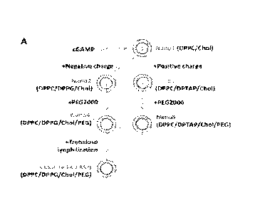
FIGS. 7A-7K. PS-GAMP fabrication and characterization. (A) A schematic
diagram of PS-GAMP fabrication. The liposomes were synthesized in the basis of
PS
ingredients of mammals, which typically consists of 90% lipids and 10%
proteins and is
evolutionally conserved. The lipids contain 8-10% of cholesterol, 60-70% of
zwitterionic
phosphatidylcholines (PC), mainly dipalmitoylated phosphatidylcholine (DPPC),
up to 8-
15% of anionic phosphatidylglycerol (DPPG), and a relatively small portion of
other
lipids (17). PEG2000 was utilized in place of hydrophilic proteins and DPPG
was
replaced with cationic DPTAP in nano3 and nano5 to determine the importance of
charges. These PS lipids and PEG2000 form liposomes with a single lipid
bilayer
encapsulating cGAMP by reverse-phase evaporation as detailed in Materials and
Methods. (B to E) Swiss Webster mice were i.n. immunized with VN04 H5N1
vaccine
(1 pg HA content) plus 10 ttg free cGAMP or an equal amount of cGAMP packaged
in
the indicated liposomes. Serum IgG (B) and bronchoalveolar lavage fluid (BALF)
IgA
(C) were measured two weeks later, body weight was monitored for 7 d after
immunization (D) and the area under the curve (AUC) was calculated from (D) by
PRISM software (E). n=5. Sizes (F), encapsulation rate (G), and zeta
potentials (J) of
indicated liposomes were measured. (H and I) Free cGAMP or cGAMP-encapsulated
liposomes were cultured with BMDCs (H) and BMMs (I) at a final cGAMP
concentration of 10 pg/m1 for 8 h, after which IFN-0 (Ifnb 1) was measured by
real-time
RT-PCR. n=4. (K) STING-deficient mice (Ming') (Red) or wild-type (WT) (Blue)
control mice were i.n. immunized with 'VN04 H5N1 vaccine alone or together
with PS-
GAMP and serum IgG titers were measured 2 weeks later as above. n=4. The
results
were presented as means SEM. Each symbol represents individual mice in (B,
C, E,
and K) or independent duplicates in (G, H, and I). Statistical analysis, one-
way ANOVA
for (B, C, E, and H-K), two-way ANOVA for (D). *p<0.05, **p<0.01, and
***p<0.001
compared to vaccine alone group or between indicated groups. All experiments
were
repeated twice with similar results.
CA 03157192 2022-04-06
WO 2021/071823
PCT/US2020/054377
FIGS. 8A-8F. Kinetics of nanoparticle uptake in different tissues. Mice were
i.n. administered PBS, free SRB, SRB-encapsulated and DiD-labeled nano4 (Nano4-
SRB) or SRB-encapsulated and DiD-labeled nano5 (Nan05-SRB) and analyzed by
flow
cytometry at varying times. (A) Representative flow plots for SRB' cells in
the brain
(upper), nasal tissue (middle), and MLN (lower). Data are representative of
two separate
experiments each assayed in triplicate. (B and C) SRB+ cells in the lungs of
mice
receiving nano4-SRB (B) or nano5-SRB (C). Alveolar macrophages (AM),
interstitial
macrophages (IM), CD1 1 VDCs, and CD1 1 bl3Cs were gated as FIGS. 9A-9D. (D-F)
SRI3+ cells in MLN (D), brain (E), or nasal tissue (F) of mice receiving nano4-
SRB or
nano5-SRB. n=4. The results were presented as means SEM. Statistical
analysis, two-
way ANOVA for (B-F). *p<0.05, **p<0.01, and ***p<0.001. The experiment was
repeated twice with similar results.
FIGS. 9A-9D. Gating strategy for flow cytometric analysis of cells isolated
from indicated tissues. (A) NK cells were identified by NK1. P and CD3- in
pulmonary
cells and CD3+ cells were separated into CD4+ and CDS+ T cells. Pulmonary CD1
1c
cells were divided into neutrophils as CD11b+146C+Ly6G+, whereas inflammatory
monocytes were recognized as CDIlbay6ChiLy6G-. On the gate of CD1 1 c+ cells,
four
populations were discriminated with CD24 and CD1 lb markers, among which AMs
were
CD24-CD11b-Sig1ec F, IMs were CD24-CD1 1 b+, CD24+CD11b-DCs were CD1 03+
MI-IC II, and tissue-resident CD241-CD1 lb DCs also expressed MHC II but not
CD1 03.
(B) During influenza virus infection or after PS-GAMP administration, CD1 1 b
DCs
could be separated into monocyte-derived DCs (Mono-DCs) or tissue resident-
like DCs
(tDCs). Mono-DCs were Ly6Chi and MI-IC II expression varied with their
activation
status. On the other hand, tDCs were Ly6C1914HC 11111. (C and D) Gating
strategy for
CD11VDCs and CD11b-DCs in MLN (C) or DCs in nasal tissue (D). Gating
strategies
for T cells, NK cells, neutrophils, and monocytes in MLN, nasal tissue, and
brain were
similar to those in the lung (A).
FIGS. 10A-10B. Analysis of cells capturing PS-liposomes in the lung. (A)
Mice were i.n. administered nano4-SRB prepared as FIG. IA. Twelve h later,
CD11c+SRB' cells were characterized mostly as CD24-CD1 1 b- AMs, and CD11c-
SRB+
cells were mostly EpCAM+CD1 1 If AECs, which were also positive for MHC II.
(B)
1!
CA 03157192 2022-04-06
WO 2021/071823
PCT/US2020/054377
AMs, CD103+ DCs, and CD11b DCs gated as FIG. 9A were analyzed for direct
nanoparticle uptake (SR1r)iD+) in mice receiving nano4-SRB. About half of AMs
ingested nano4-SRB shown as SRB+DiD+, whereas DCs rarely captured the
nanoparticles. The results were presented as means SEM. n=4 mice. The
experiment
was repeated twice with similar results.
FIGS. 11A-11C. Nano4 delivered cGAMP into AMs. (A) Schematic diagrams
of DiD-labeled empty PS-mimetic nanoparticles (DiD-PS) and cGAMP-encapsulated
PS-
mimetic nanoparticles (DiD-PS-GAMP). (B) DiD-PS or DiD-PS-GAMP (20 pg
cGAMP) were i.n. inoculated. Pulmonary cells were analyzed for DiD+ CD11 c+
cells by
.. flow cytometry 12 or 36 h later in mice receiving DiD-PS (Red) or DiD-PS-
GAMP
(Blue). These cells were also assessed for CD40 expression to verify STING
activation in
the cells. Representative histogram of CD40 expression is given in the middle
and Mean
Fluorescence Intensity (MFI) is summarized in the right panel. n=4. (C) Di.13+
(Blue) or
DiD AMs (Red) expressing CD40 were analyzed similarly as (B). n=4. The results
were
presented as means SEM. Each symbol represents individual mice in the right
panels of
B and C. Statistical analysis, t-test for (B and C). "p<0.01 and ***p<0.001 in
the
presence or absence of cGAMP. The experiment was repeated twice with similar
results.
FIG. 12. Positively charged nano5 was entrapped by PS in ex vivo culture.
DiD-labeled Nano4 or nano5 was incubated with PS for 30 min. Nanoparticle
aggregates
on PS were visualized by confocal microscopy. The areas outlined in the 2nd
panel were
enlarged on the right. BF, Bright Field. Scale bar, 100 pm in panel 1 and 2
and 10 pm in
panel 3 and 4. Data are representative of ten similar results in two separate
experiments.
FIG. 13A-13C. Nano4 uptake by AMs isolated from non-human primates
(NHP). AMs and PS were isolated from rhesus macaques. (A) DiD-nano4 or DiD-
nano5
.. was incubated for 30 min with rhesus macaque PS. Nano5, but not nano4,
aggregated on
PS and visualized by confocal microscopy. The areas outlined were enlarged in
the
corresponding panels on the right. Scale bar, 10 pm. Data are representative
of five
similar results. (B) Monkey AMs were isolated and cultured with DiD-nano4
(upper) or
DiD-nano5 (low) for 3 h with or without PS pre-treatment of the nanoparticles
and
imaged by fluorescent microscopy. Live cells were stained by Calcein-AM and
nuclei
were stained by Hoechst Scale bar, 50 gm. DiD fluorescence intensity in cells
was
12
CA 03157192 2022-04-06
WO 2021/071823
PCT/US2020/054377
quantified by Image J (C). n=221-275. Each symbol represents individual cells.
The
results were presented as means SEM. Statistical analysis, one-way ANOVA for
(C).
***p<0.001 between indicated groups. We thank Prof. Wanli Liu, Dr. Junyi Wang,
and
Ms. Shaoling Qi for their help in the NHP study.
FIG. 14. AM capture nano4 in the lung. Lungs were collected 12 h after mice
received DiD-nano4 intranasally and frozen thin sections were stained for an
AM-
specific marker Siglec F and visualized by fluorescent microscopy. Scale bar,
30 pm.
The square in the 2nd panel is enlarged on the right panels. Data are
representative of six
similar results in two separate experiments.
FIG. 15. TEM of nanoparticle distribution in the lung. Nanogold was
encapsulated within nano4 (nano4-gold) and nano5 (nano5-gold) as FIG. 7A. Mice
were
i.n. administered with the nanoparticles. Lungs were collected 6 h later and
prepared for
TEM. Note: nano4-gold was entrapped within cellular vesicles inside AM (red
arrows)
and some nanogolds were observed within a cellular vesicle (open red arrows,
lower
panel). In contrast, nano5-gold was mostly presented on the surface of alveoli
(blue
arrows). The areas outlined in upper panels are enlarged in the corresponding
lower
panels. Scale bar, 2 pm for the upper panel and 500 nm for the lower panel.
FIG. 16. AMs from SlipallSflpd-1- mice had a similar capability as WT AMs
in nano4 uptake. SP-A/D are hydrophilic large proteins and well established as
a first
.. line of the innate defense. These two collectins are capable of integrating
into PS-
wrapped bacteria, viruses, cellular debris, apoptotic cells, and various
nanoparticles, to
facilitate their endocytosis or phagocytosis by AMs (20). To test whether this
might be
the mechanism for nano4 uptake by AMs, AMs were isolated from WT or
,SfipallSftpctl-
mice and incubated with DiD-nano4 that was pre-treated with WT PS for 30 min.
DiD
fluorescence in cells was captured by confocal microscopy and quantified by
Image J
software. AMs from Sftpa 1 ISfipd-i- mice were found to ingest nano4 as
efficiently as WT
AMs in the presence of WT PS (FIG. 16), but not in the presence of SP-AID-
deficient PS
(FIG. 11). n=25-32. Each symbol represents individual cells. Statistical
analysis, one-way
ANOVA. *p<0.05 and **p<0.01 between indicated groups and ns, not significant.
The
experiment was repeated twice with similar results.
13
CA 03157192 2022-04-06
WO 2021/071823
PCT/US2020/054377
FIGS. 17A-17C. PS-GAMP did not induce overt inflammation in the lung,
nose, and central nervous system (CNS). Swiss Webster mice were i.n.
administered
with PBS, PS-GAMP, VN04 H5N1 vaccine, or the vaccine plus PS-GAMP or CT. (A)
Histological examination of the lung (1' and 2nd panel), nose (3rd and 4th
panel), and CNS
(5th and 6th panel) in 2 d post-immunization. Alveolar and bronchus of lungs,
the nasal
associated lymphoid tissue of noses, and the olfactory bulb region of the
brain tissue are
outlined by a dashed rectangle and enlarged in the corresponding bottom
panels. The
olfactory bulb region of the brain tissue connects directly with olfactory
nerves in the
nasal cavity. Data are representative of two separate experiments each assayed
in
triplicate. Scale bars for the lung and nose, the upper panel 400 pm and the
lower panel
100 pm. Scale bars for CNS, the upper panel 800 pm and the lower panel 200 pm.
(B)
Eosinophil infiltrates, epithelium damage, and necrosis of each mouse were
analyzed as
previously reported (48). The number indicates the number of mice with (+) or
without
(¨) eosinophil infiltrates, epithelium damage or necrosis. n=6 mice. (C)
Expression of
indicated cytokines and chemokine in the CNS of mice receiving VN04 H5N1
vaccine in
the presence or absence of PS-GAMP or CT was determined by real-time RT-PCR 2
d
post-immunization. n=2-8. Each symbol represents individual mice. The results
were
presented as means SEM.
FIGS. 18A-18V. Alterations of inflammatory and immune cells after PS-
GAMP administration or viral infection. C57BL6 mice were i.n. administered
with
CA09 H1N1 vaccine plus 20 tig of PS-GAMP (Blue) or infected with 1xLD5o CA09
H1N1 influenza virus (Red). Neutrophils, NK, CD4+, and CD8+ T cells,
monocytes, and
CD11b and CD1 1b- DCs in the lung (A to G) or MLN (H to N) were analyzed by
flow
cytometry on indicated d post-infection or post-immunization (d.p.i).
Neutrophils, NK,
CD4+, and CD8 T cells, monocytes and DCs in nasal tissue (0 to T) or
neutrophils and
monocytes in brain (U and V) were similarly analyzed. n=4. The results were
presented
as means SEM. Statistical analysis, one-way ANOVA. *p<0.05, **p<0.01, and
***p<0.001 compared to d 0. The experiment was repeated twice with similar
results.
FIGS. 19A-19C. PS-GAMP did not induce overt inflammation in the lung in
contrast to viral infection. Mice were i.n. immunized with CA09 H1N1 vaccine
plus 20
pg of PS-GAMP (A) or infected with 1 xLD5o CA09 H1N1 influenza virus (B).
Lungs
14
CA 03157192 2022-04-06
WO 2021/071823
PCT/US2020/054377
were analyzed by H&E staining on indicated d after immunization or infection.
Data are
representative of two separate experiments each assayed in triplicate. Scale
bar, 400 pm
for upper panel and 100 gm for lower panel. (C) The lung inflammation was
quantified
according to a standard scoring system shown on the right (4 9) . n=6. Data
were
presented as means SEM. *p<0.05 and ***p<0.001 compared to d 0 by
nonparametric
test.
FIG. 20. PS-GAIVIP induces transient production of immune mediators in the
lung. Mice were i.n. given 20 pg of PS-GAMP (Blue) or infected with 1xLD5o
CA09
H1N1 influenza virus (Red). mRNA levels of indicated mediators were measured
by
real-time RT-PCR at various time points and normalized against untreated mice.
n=4.
The results were presented as means SEM. Statistical analysis, one-way
ANOVA.
*p<0.05, ** p<0.01, and ***p<0.001 compared to d 0. The experiment was
repeated
twice with similar results.
FIGS. 21A-21C. PS-GAMP briefly elevates IFNI) protein in BALF. Mice
were i. n. administered 20 pg of PS-GAMP (Blue) or infected with 1xLD5o CA09
HI NI
influenza virus (Red). Protein levels of IFN-(3 (A), 'INF-a (B), and IL-l0 (C)
in BALF
were measured by ELISA at various time points. n=4. The results were presented
as
means SEM. Statistical analysis, one-way ANOVA. *p<0.05 and ***p<0.001
compared to d 0 (before treatments). The experiment was repeated twice with
similar
results.
FIGS. 22A-22G. PS-GAMP did not induce any inflammation systemically.
Mice were i.n. immunized with CA09 H1N1 vaccine plus 20 pg of PS-GAMP. Body
weight (A) and temperature (B) were monitored for 6 d. Mice receiving PBS
served as
control. n=5. Serum IFN-fl (C), 'INF-a (D), IFN-y (E), IL-6 (F), and IL-10 (G)
were also
monitored for 6 d by ELISA. n=4. The results were presented as means SEM.
The
experiment was repeated twice with similar results.
FIGS. 23A-23D. PS-GAMP increases the number of CD11VDCs ingesting
extracellular Ag in the lung and MLN. (A) Mice were i.n. vaccinated with OVA-
AF647 with or without 20 pg of PS-GAMP. Pulmonary CD11 e cells capturing OVA
were analyzed for CD1 lb and CD24 expression. The numbers in the plots are
mean
percentages SEM of individual cell subsets. (B) Mice receiving OVA (non-
CA 03157192 2022-04-06
WO 2021/071823
PCT/US2020/054377
fluorescence) + PS-GAMP served as controls to gate out cell activation-related
autofluorescence. OVA uptake was analyzed 36 h later on the gate of DCs
prepared from
MLNs revealing OVA+DCs to be mostly CD11b DCs. (C) DCs did not directly ingest
PS-GAMP in the MLN as shown by few CD11c+DiD+ cells when mice were i.n.
administered with 20 gg of DiD-PS-GAMP and analyzed similarly. (D) DiD+ cells
were
also tracked in MLNs from 0 to 60 h after PS-GAMP administration. n=4. The
results
were presented as means SEM. The experiment was repeated twice with similar
results.
FIGS. 24A-24E. PS-GAMP did not augment Ag-uptake or processing in vivo.
Whether PS-GAMP influenced Ag-uptake or Ag-processing was evaluated using
AF647-
labeled OVA and DQ-OVA. DQ-OVA is OVA conjugation with a BODIPY fluorescent
dye (DQ) and remains self-quenched until OVA is proteolytically processed to
generate
DQ-green fluorescence, which is commonly used to assess Ag-processing. To this
end,
mice were i.n. administered with PBS (Gray) or AF647-OVA together with DQ-OVA
in
the presence (Red) or absence (Blue) of PS-GAMP and euthanized 24 h later for
flow
.. cytometric analysis (A). AF647-OVA was analyzed on the gate of DC11 b DCs,
which
were further quantified for OVA cleavage based on DQ-green fluorescence.
Percentages
and cell numbers of AF647' CD1 1b DCs were summarized in (B) and (C). AF647
and
DQ-Green MFIs in these cells were given in (D) or (E), respectively. Each
symbol
represents individual mice in B to E. The results were presented as means
SEM.
Statistical analysis, one-way ANOVA for (B-E). *p<0.05, **p<0.01, and
***p<0.001 in
the presence or absence of PS-GAMP. ns, no significance. The experiment was
repeated
twice with similar results. Note: there was no difference in MFI of DQ-green
fluorescence or OVA in AF64TCD11b DCs irrespective of whether or not PS-GAMP
was presented (D and E). However, percentages and numbers of CD1lb' DCs
positive to
OVA were robustly increased in the presence of PS-GAMP (B and C), which was
attributed primarily from an increased number of CD11b+ DCs secondarily to
immune
mediators induced by PS-GAMP.
FIGS. 25A-25D. PS-GA1VIP enhances Ag cross-presentation. (A) Mice were
i.n. vaccinated with 60 lig of OVA with or without 20 lig of PS-GAMP.
Carboxyfluorescein succinimidyl ester (CFSE)-labeled OT-I cells were
transferred into
vaccinated mice 1 d later. Lungs and MLN were collected 3 d post-cell
transfer. (B) OT-I
16
CA 03157192 2022-04-06
WO 2021/071823
PCT/US2020/054377
cells were analyzed for Ag-specific proliferation by step-wise decreases of
CFSE
fluorescence. Inset in the first two panels (PBS and OVA): a reduced scale of
the y-axis
to show CFSE decreases. Cells of high divisions (>6, hi) were gated. Numbers
of highly
divided cells in lungs (C) and MLNs (D) were summarized. n-4-6. Each symbol
represents individual mice in C and D. Statistical analysis, one-way ANOVA for
(C and
D). The results were presented as means SEM. "p<0.01 and ***p<0.001 in the
presence or absence of PS-GAMP. The experiment was repeated twice with similar
results.
FIGS. 26A-26C. CD81- T cell responses in the spleen, lung and MLN.
C57BL/6 mice were i.n. immunized with CA09 H1N1 vaccine plus 20 Lig of PS-
GAMP.
Mice received PBS as a control. (A) Splenocytes were isolated 7 d post-
immunization
and stimulated with the CA09 H1N1 vaccine. Representative cytometric profiles
of CD4+
and CD8+ T cells producing IFNI are shown. (B) Representative cytometric
profiles of
NP366-374+ CD8 T cells in the lung 4 d after immunization. (C) Percentages of
PA224-233
(Blue) or PB1703-7it (Red) positive cells were determined on gate of CD3+CD8+T
cells.
Each plot is representative of four similar results in the same group. n=4.
Data are
presented as means SEM. The experiment was repeated twice with similar
results.
FIGS. 27A-27C. Early viral specific GIIICD8+ T cells and BALF antibodies.
(A) C57BL/6 mice were either left unimmunized or immunized with CA09 H1N1
vaccine alone or the vaccine plus 20 j.tg of PS-GAMP, followed 2 d later by
challenging
with 10xLD5o CA09 H1N1 virus. Lungs were collected 4 d post-infection. (B) The
percentages of NP366-374 tetramer (Blue), PB 1703-711 (Red), or PA224-233
(Green) positive
cells were obtained on the gate of GB+CD8+ T cells. CD8+ T cells from un-
immunized/un-challenged mice were analyzed in parallel as negative controls
(Gray).
n=3. (C) BALF were analyzed for Ag-specific IgA and IgM titers 6 d post-
immunization.
n=4. Data are presented as means SEM. The experiment was repeated twice with
similar results.
FIGS. 28A-28I. Supplementary data for FIGS. 4A-4J. (A) A schematic
diagram of vaccination and viral challenge schedule. (B to F) The body weight
changes
(B, C, E, and F) or survival (D) of mice corresponding to those described in
FIGS. 4A-
4E, respectively. (G and H) Mice were i.n. immunized with H7-Rel H7N9 vaccine
alone
17
CA 03157192 2022-04-06
WO 2021/071823
PCT/US2020/054377
or alongside 20 gg of PS-GAMP or poly IC and challenged 14 d later by a
clinically
isolated SH13 H7N9 virus. n=10-13. (I) Body weight change of mice described in
FIG.
4F. Statistical analysis, two-way ANOVA for (B, C, E, F, H, I), Log-rank test
for (D and
G). *p< 0.05, **p<0.01, *p<0.001, and #p<0.05. All experiments were repeated
at
least twice with similar results.
FIGS. 29A-29B. An inverse correlation of SRB+AMs vs. SRWAECs over
time in vivo while DiD+AMs remained unaltered in percentages. SRB-DiD-nano4
was i.n. inoculated into mice. (A) Percentage changes of SRB+AMs and SRB+AECs
relative to a total number of lung cells were tracked over time after the
inoculation. n=4.
(B) DiD AMs were analyzed by flow cytometry 12 and 18 h later following
nanoparticle
administration. n=4. The results were presented as means SEM. Statistical
analysis, t-
test. *p<0.05 and **p<0.01 compared between 18 and 12 h. All experiments were
repeated twice with similar results.
FIGS. 30A-30D. Entry of cGAMP from AMs into AECs. (A) Mice were i.p.
injected with a gap junction inhibitor CBX or PBS for 3 consecutive d, after
which 20 lig
of PS-GAMP was i.n. administrated. AMs and AECs were sorted 12 h later and
analyzed
for /fill)/ (B) and Gmesi (C) expression by real-time RT-PCR. mRNA levels were
first
normalized to Gapdh and then to corresponding cells isolated from naive mice.
n=4. (D)
Unsorted lung cells were also analyzed similarly for comparisons. n=8. The
results were
presented as means SEM. Each symbol represents individual mice in B to D.
Statistical
analysis, 1-test. *p<0.05 and ***p<0.001 in the presence or absence of CBX.
ns, no
significance. All experiments were repeated twice with similar results.
FIGS. 31A-31E. Tissue and cell distribution of poly IC. Mice received 20 jig
of rhodamine-labeled poly IC intranasally. (A) Lungs were dissected and
digested 12 h
later for flow cytometric analysis of poly IC uptake by CD11 c+ and CD11 c-
subsets.
CD1lepoly IC + cells were further confirmed to be CD24- CD1 1 b- AMs. Poly IC
uptake
was next analyzed on the gate of EpCAM+ AECs (B) or CD11c+CD24+ DCs (C). MLNs
(D) and nasal epithelium and lymphatic tissue (E) were also prepared for
single-cell
suspensions to determine poly IC uptake. Data are representative of two
separate
experiments each assayed in triplicate.
18
CA 03157192 2022-04-06
WO 2021/071823
PCT/US2020/054377
FIGS. 32A-32B. Cell reconstitution efficacy after bone marrow (BM) cell
transfer. Mice were pre-conditioned with lethal irradiation prior to infusion
with BM
cells isolated from mice carrying reciprocal CD45 alleles, CD45.1 and CD45.2,
a surface
biomarker for all leukocytes. Donor cells were distinguished from recipients
by a
specific antibody for CD45.1 or CD45.2. The transfer efficacy was analyzed by
quantifying CD45.1 or CD45.2 expression on leukocytes in various tissues in
the
recipients after three months of infusion (A) and summarized in (B). Each
symbol
represents individual mice in (B). n=5-7. The experiment was repeated twice
with similar
results.
FIGS. 33A-33K. Supplementary data for cross-protection studies. (A to K)
Body weight changes corresponding to mice described in FIGS. 6A-6K,
respectively.
The results were presented as means SEM. Statistical analysis, two-way
ANOVA.
*p<0.05, **p< 0.01, ***p<0.001, and # p<0.05. All experiments were repeated
twice
with similar results.
FIGS. 34A-34B. Vaccination with trivalent seasonal influenza vaccine and
PS-GAP induces cross-protective immunity against mismatched influenza B virus.
(A) A schematic of the vaccination/sampling schedule. BALB/c mice were
immunized
with trivalent seasonal influenza vaccine (2018-19) (SW) alone or together
with 20 pg of
PS-GAMP and challenged 1 month later with 4x10⁵ TCID5o mismatched Florida06 B
virus. (B) Lungs were isolated 4 d after the immunization and analyzed for
viral titers.
Each symbol represents individual mice in (B). n=8-9. The results were
presented as
means SEM. Statistical analysis, one-way ANOVA. **p<0.01 in the presence or
absence of PS-GAMP. ns, no significance. The experiment was repeated twice
with
similar results.
FIGS. 35A-35E. PS-GAMP/inactivated influenza vaccine induces viral-
specific lung CDS+ TRm cells. (A) A schematic of the vaccination/sampling
timeline of
OT-1 mouse model. Mice were transferred with OT-I cells and i.n. immunized 1 h
later
with OVA in presence or absence of PS-GAMP. Thirty-five d later, mice were
i.v.
injected with anti-CD8I3 antibody to exclude circulating CD8+ T cells before
sacrificed
for flow cytometric analysis. (B) Total CD8+ T cells in the lung were gated by
CD3 and
CD8a+ (profile not shown) and lung OT-I cells were recognized as CD45.2' and
CD813-
19
CA 03157192 2022-04-06
WO 2021/071823
PCT/US2020/054377
(antibody i.v. injected) (1' two panels). 01-1 cells with TRM phenotype were
identified
as CD103+CD69+CD49e. OT-I cells in the spleen served as the control (Gray).
The
number of lung OT-I TRm cells were summarized in (C). n=6. (D and E) Mice were
i.n.
immunized with CA09 HINT vaccine in the presence or absence of PS-GAMP. Lungs
were isolated 6 months later for flow cytometry. NP366-374+ CD8 T cells were
gated and
validated for CD103 and CD69 expression (D) and the number of NP366-374+ CD8"
TRM
cells were summarized in (E). n=4. NP366-r4" CD8" T cells in the spleen served
as the
control. The results were presented as means SEM. Statistical analysis, t-
test for (C),
one-way ANOVA for (E). ***p<0.001 in the presence or absence of PS-GAMP. All
experiments were repeated twice with similar results.
FIGS. 36A-36D. FTY720 did not affect the cross-protection elicited by
influenza vaccine/PS-GAMP. (A and B) Mice were i.n. immunized with CA09 HINT
vaccine alone or together with 20 lig of PS-GAMP and challenged 1 month later
with
5xLD50 GZ89 H3N2 virus. (C and D) Mice were immunized and challenged as A and
B
except that the mice additionally received daily injections of FTY720 (1
mg/kg/day) from
¨2 to 14 days after the challenge. n=6-8. Statistical analysis, two-way ANOVA
for (B
and D) and Log-rank test for (A and C). *p<0.05, **p<0.01, and ***p<0.001
compared
to the vaccine alone. All experiments were repeated twice with similar
results.
FIGS. 37A-37E. Safety and efficacy of PS-GAMP in ferrets. Ferrets were i.n.
immunized with an inactivated viral vaccine (Perth H3N2 15 Lig) with or
without 20014
of PS-GAMP. Body weight (A) and temperature (B) of the animals were monitored
for 6
d. (C) Sera were collected 4 weeks after the immunization and tested for
PerthH3N2-
specific IgG titers (C). HAT titers were also measured against PerthH3N2 (D)
or
MichiganH1N1 (E) viral strains. n=4. Each symbol represents individual animals
in C to
E. The results were presented as means SEM. Statistical analysis, one-way
ANOVA for
(C and D). **p< 0.01 compared in the presence vs. absence of PS-GAMP.
DETAILED DESCRIPTION
The cGAS-cGAMP-STING pathway is an important immune surveillance
pathway that is activated in the presence of cytoplasmic DNA, e.g., due to
microbial
infection or patho-physiological conditions including cancer and autoimmune
disorders.
CA 03157192 2022-04-06
WO 2021/071823
PCT/US2020/054377
Cyclic GMP-AMP synthase (cGAS) belongs to the nucleotidyltransferase family
and is a
universal DNA sensor that is activated upon binding to cytosolic dsDNA to
produce the
signaling molecule cyclic GMP-AMP (or 2'-3'-cGAMP or cyclic guanosine
monophosphate-adenosine monophosphate). Acting as a second messenger during
microbial infection, 2'-3'-cGAMP binds and activates STING, leading to
production of
type I interferon (IFN) and other co-stimulatory molecules that trigger the
immune
response. Besides its role in infectious disease, the cGAS/STING pathway has
emerged
as a promising new target for autoimmune diseases and cancer immunotherapy.
DNA
fragments present in the tumor microenvironment are proposed to activate cGAS
in
dendritic cells (DC), followed by IFN-induced DC maturation and activation of
a potent
and beneficial immune response against cancer cells. In a separate context,
dysregulation
of the cGAS/STING pathway has been implicated in self DNA triggered
inflammatory
and autoimmune disorders, such as systemic lupus erythematosus (SLE) and
Aicardi-
Goutieres syndrome.
There continues to be a dearth of effective mucosal adjuvants despite decades
of
investigation. 2'-31-cGMP-AMP (cGAMP), a natural agonist of the stimulator of
interferon genes (STING), is a secondary messenger generated in response to
DNA viral
infections or tissue damage (10, 11). It stimulates the production of type I
interferons
(IFN-Is), which help determine the magnitude of T-helper 1 (Thl) immune
responses,
particularly those of CD8+ T cells (12, 13). STING agonists are potent
adjuvants capable
of eliciting robust anti-tumor immunity following intratumoral administration
and
augmenting intradermal influenza vaccines (13, 14). Using these small, water-
soluble
agonists as mucosal adjuvants, however, is a challenge. They must be delivered
into the
cytosol of antigen (Ag)-presenting cells (APCs) and/or alveolar epithelial
cells (AECs)
without breaching the integrity of the pulmonary surfactant (PS) layer, a
mixture of lipids
and proteins secreted by type II AECs. This PS layer forms a strong barrier,
which
separates exterior air from internal alveolar epithelium in alveoli, and
prevents
nanoparticles and hydrophilic molecules from accessing AECs (15, 16).
Development of a "universal" influenza vaccine that confers protection against
not only intrasubtypic variants, but also other subtypes of influenza viruses
is highly
desirable. However, whether such universal influenza vaccines are achievable
remains
21
CA 03157192 2022-04-06
WO 2021/071823
PCT/US2020/054377
unclear. It has been long recognized in both humans and animal models that
viral
infection can stimulate heterosubtypic immunity primarily mediated by CD8+I
cells (2,
3, 6). Here, a single immunization with inactivated H1N1 vaccine adjuvanted
with PS-
GAMP conferred protection against lethal challenges with H1N1, H3N2, H5N1 or
H7N9
viruses as early as 2 days (d) post-immunization. This cross-protection was
sustained for
at least 6 months, concurrent with durable virus-specific CD8+ TRM cells in
the lung.
This was largely due to the fact that PS-GAMP-adjuvant influenza vaccine
simulated
viral infection-induced immunity, characterized by AEC activation, rapid CDI I
b DC
recruitment and differentiation, and robust CDS'I cell responses in the
respiratory
.. system. PS-GAMP is a standalone adjuvant, compatible with not only
inactivated
influenza viral vaccines, but also other vaccines, e.g., vaccines comprising
cocktails of
multiple B and T cell epitopes or influenza vaccine subunits. The ability of
PS-GAN4P to
potentiate non-replicating influenza vaccines for strong beterosubtypic
immunity makes
it a promising adjuvant for "universal" influenza vaccines if its efficacy is
shown in
humans. As such, it would offer a significant advantage over "replicating"
vaccines.
Distinct from conventional vaccine adjuvants targeting primarily APCs, PS-
GAMP activated both AMs and AECs; without wishing to be bound by theory, AEC
activation appeared to be crucial for adjuvanticity, as blockades in gap
junctions as well
as STING deficiency in AECs diminished the adjuvanticity considerably whereas
STING
deficiency in myeloid cells did not. The pivotal role played by AECs over AMs
in
orchestrating innate and adaptive immune responses is in agreement with what
has been
described during the early phase of influenza viral infection (24). The
ability of cGAMP
to enter AECs without breaching the PS layer was ascribed to SP-A/D-receptor-
mediated
endocytosis after incorporation of SP-A and SP-D into PS-biomimetic liposomes,
which
is not feasible in any non-PS-biomimetic liposomes (39-41). In addition, this
adjuvant
was able to induce robust protection within just 2 d post-immunization, in
sharp contrast
to current influenza vaccines, which require at least 10-14 d to be effective.
Early cross-
protection is extremely important to protect first responders and high-risk
individuals,
especially when antiviral drug-resistant viruses or highly pathogenic viruses
such as
H5N1 and H7N9 viruses emerge to become pandemics. Because viral spreading can
accelerate exponentially after expanding from an epidemic to pandemic early
protection
22
CA 03157192 2022-04-06
WO 2021/071823
PCT/US2020/054377
during an epidemic would be the most effective means to confine viral
spreading and
minimize or prevent epidemics becoming pandemics, saving millions of lives
(42).
Pulmonary Surfactant (PS)-Biomimetic Nanoparticle
Provided herein are compositions comprising PS-biomimetic nanoparticles with
an average size of 200-400 nm. The nanoparticle includes a plurality of
pulmonary
surfactant-biomimetic molecules, wherein the nanoparticle is negatively
charged; and one
or more cargo molecules that are enveloped by the nanoparticle, wherein the
cargo
molecule has a molecular weight up to 1200 Da.
Provided herein are methods of promoting an immune response to an antigen. The
methods include administering to a subject an effective amount of the
composition as
described herein; and administering to the subject the antigen.
Provided herein are methods of treating a subject who has influenza. The
methods
include administering to the subject a therapeutically effective amount of the
composition
as described herein; and administering to the subject an antigen. In some
embodiments,
the cargo molecule is cGAMP and the antigen is an influenza vaccine.
1. Provided herein are methods of treating a subject who has an airway
disease. The
methods include administering to the subject a therapeutically effective
amount of the
composition as described herein, wherein the cargo molecule is long acting-02-
agonists
(LABAs) (e.g., formoterol, salmeterol, or vilanterol); cortisosteroids (ICS)
(e.g.,
budesonide, fluticasone propionate, or fluticasone furoate); leukotriene-
pathway
modulators (e.g., montelukast, or zileuton); inhibitors targeting kinases
(e.g., spleen
tyrosine kinase, p38 mitogen-activated protein kinase (MAPK),
phosphatidylinosito1-4,5-
bisphosphate 3-kinase (P13K), Janus kinase (Jak), or phosphodiesterase-4
(PDE4));
agonists or antagonists of receptors (e.g., chemoattractant receptor-
homologous molecule
expressed on Th2 cells (CRTH2), chemokine receptor 2 (CCR2)); agonists or
antagonists
of ion channels (e.g., GABA receptor, transient receptor potential cation
channel,
subfamily A, member 1 (TRPA1), or voltage-gated sodium channel); inducers of
IFN-a;
long-acting muscarinic antagonists/anticholinergics (LAMAs); inhibitors
against IL-5,
IL-13, IL-33, or thymic stromal lymphopoietin; CXCR2 antagonists; molecules
blocking
proinflammatory cytokines (e.g., 'TNF-a, TNF-I3, or IL-6); molecules blocking
IL-
23
CA 03157192 2022-04-06
WO 2021/071823
PCT/US2020/054377
17/TH17; macrolides; molecules activating HDAC2; STAT6 inhibitors (e.g.,
AS1517499); anti-virus small molecule drug (e.g., Oseltamivir (Tamiflu),
Relenza, or
Zanamivir); and/or Favipiravir (T705).
Provided herein are methods of treating a subject who has cancer. The methods
include administering to a subject a therapeutically effective amount of a
composition as
described herein. In some embodiments, the cargo molecule is a chemotherapy
agent.
The methods described herein can provide improvement of the delivery efficacy
of the cargo molecules as described herein by at least 1-fold, at least 2-
fold, at least 3-
fold, at least 4-fold, at least 5-fold, at least 6-fold, at least 7-fold, at
least 8-fold, at least 9-
fold, at least 10-fold, at least 15-fold, at least 20-fold, at least 50-fold,
at least 100-fold, at
least 200-fold, at least 500-fold, at least 1000-fold compared to a similar
method
performed without the use of PS-biomimetic nanoparticles.
Nanoparticles
In some embodiments, the nanoparticle is a liposome, a vesicle, an emulsion,
or a
micelle.
In some embodiments, the nanoparticle may contain one or more types of
surfactants including detergent, wetting agents, emulsifiers, foaming agents,
or
dispersants. In some embodiments, the surfactant comprises at least one
hydrophobic end
and/or at least one hydrophilic end. In some embodiments, the surfactant is
positively
charged, neutral, or negatively charged.
In some embodiments, the surfactant is a lipid. In some embodiments, the
surfactant is a phospholipid. In some embodiments, the nanoparticle may
comprise 1, 2,
3, 4, 5, 6, 7, 8, 9, 10, or more layers of surfactant. In some embodiments,
the nanoparticle
is a water-in-oil-in-water emulsion.
In some embodiments, the percent of surfactant in a nanoparticle can range
from
0% to 100% by weight, from 5% to 100% by weight, from 10% to 100% by weight,
from
15% to 100% by weight, from 20% to 100% by weight, from 25% to 100% by weight,
from 30% to 100% by weight, from 35% to 100% by weight, from 40% to 100% by
weight, from 45% to 100% by weight, from 50% to 100% by weight, from 55% to
100%
by weight, from 60% to 100% by weight, from 65% to 100% by weight, from 70% to
24
CA 03157192 2022-04-06
WO 2021/071823
PCT/US2020/054377
100% by weight, from 75% to 100% by weight, from 80% to 100% by weight, from
85%
to 100% by weight, from 90% to 100% by weight, or from or from 95% to 100% by
weight. In some embodiments, the percent of surfactant in a nanoparticle can
range from
0% to 95% by weight, from 0% to 90% by weight, from 0% to 85% by weight, from
0%
to 80% by weight, from 0% to 75% by weight, from 0% to 70% by weight, from 0%
to
65% by weight, from 0% to 60% by weight, from 0% to 55% by weight, from 0% to
50%
by weight, from 0% to 45% by weight, from 0% to 40% by weight, from 0% to 35%
by
weight, from 0% to 30% by weight, from 0% to 25% by weight, from 0% to 20% by
weight, from 0% to 15% by weight, from 0% to 10% by weight, or from 0% to 5%
by
weight. In some embodiments, the percent of surfactant in a nanoparticle can
be 0% by
weight, approximately 1% by weight, approximately 2% by weight, approximately
3% by
weight, approximately 4% by weight, approximately 5% by weight, approximately
10%
by weight, approximately 15% by weight, approximately 20% by weight,
approximately
25% by weight, approximately 30% by weight, approximately 35% by weight,
approximately 40% by weight, approximately 45% by weight, approximately 50% by
weight, approximately 55% by weight, approximately 60% by weight,
approximately
65% by weight, approximately 70% by weight, approximately 75% by weight,
approximately 80% by weight, approximately 85% by weight, approximately 90% by
weight, approximately 95% by weight, or approximately 100% by weight
In some embodiments, the nanoparticle as described herein can have an average
size from 200 nm to 210 nm, from 210 nm to 220 nm, from 220 nm to 230 nm, from
230
nm to 240 nm, from 240 nm to 250 nm, from 250 nm to 260 nm, from 260 nm to 270
nm,
from 270 to 280 nm, from 280 nm to 290 nm, from 290 nm to 300 nm, from 300 nm
to
310 nm, from 310 nm to 320 nm, from 320 nm to 330 nm, from 330 nm to 340 nm,
from
340 nm to 350 nm, from 350 nm to 360 nm, from 360 nm to 370 nm, from 370 nm to
380
nm, from 380 nm to 390 nm, or from 390 nm to 400 nm.
Pulmonary Sutfaciant (PS)
Pulmonary surfactant is a surface-active lipoprotein complex
(phospholipoprotein) formed by type II alveolar cells. The proteins and lipids
that make
up the surfactant have both hydrophilic and hydrophobic regions. By adsorbing
to the air-
CA 03157192 2022-04-06
WO 2021/071823
PCT/US2020/054377
water interface of alveoli, with hydrophilic head groups in the water and the
hydrophobic
tails facing towards the air, the main lipid component of surfactant, 1,2-
dipalmitoyl-sn-
glycero-3-phosphocholine (DPPC), reduces surface tension.
Pulmonary surfactant typically consists of 90% lipids and 10% proteins and is
evolutionally conserved. The lipids contain 8-10% of cholesterol, 60-70% of
zwitterionic
phosphatidylcholines (PC), mainly dipalmitoylated phosphatidylcholine (DPPC),
up to 8-
15% of anionic phosphatidylglycerol (DPPG), and a relatively small portion of
other
lipids (17).
Pulmonary Surfactant (PS)-Biomimetic Nanoparticle
In some embodiments, a PS-biomimetic nanoparticle can be a nanoparticle that
comprises a plurality of PS-biomimetic molecules, including but not limited
to, 1,2-
dipalmitoyl-sn-glycero-3-phosphocholine (DPPC), 1,2-dipalmitoyl-sn-glycero-3-
phospho-(1'-rac-glycerol) (DPPG), cholesterol, polyethylene glycol (e.g.,
PEG2000), 1,2-
dipalmitoyl-sn-glycero-3-phosphoethanolamine-Ntmethoxy(polyethylene glycol)-
2000]
(DPPE-PEG2000), phosphatidylethanolamine, phosphatidylinositol,
phosphatidylserine,
sphingomyelin, and/or lysophospholipid.
In some embodiments, the PS-biomimetic molecule is a lipid, a protein, a
lipoprotein, a phospholipid, or a phospholipoprotein.
In some embodiments, the PS-biomimetic molecule is a domain, a moiety, a
portion or a whole molecule of a pulmonary surfactant In some embodiments, the
PS-
biomimetic molecule is a natural product. In some embodiments, the PS-
biomimetic
molecule is artificially synthesized.
In some embodiments, the PS-biomimetic molecule is positively, neutral, or
negatively charged. In some embodiments, the PS-biomimetic molecule has at
least one
hydrophobic end and/or at least one hydrophilic end.
In some embodiments, the PS-biomimetic molecule comprises one or more fatty
acid groups or salts thereof, and/or one or more head group. In some
embodiments, a
fatty acid group may comprise digestible, long chain (e.g., C8-050),
substituted or
unsubstituted hydrocarbons. In some embodiments, a fatty acid group may be a
C10-C20
fatty acid or salt thereof. In some embodiments, a fatty acid group may be a
C15-C20
26
CA 03157192 2022-04-06
WO 2021/071823
PCT/US2020/054377
fatty acid or salt thereof. In some embodiments, a fatty acid group may be a
C15-C25
fatty acid or salt thereof. In some embodiments, a fatty acid group may be
unsaturated.
In some embodiments, a fatty acid group may be monounsaturated. In some
embodiments, a fatty acid group may be polyunsaturated. In some embodiments, a
double
bond of an unsaturated fatty acid group may be in the cis conformation. In
some
embodiments, a double bond of an unsaturated fatty acid may be in the trans
conformation. In some embodiments, the fatty acid group is a palmitic acid. In
some
embodiments, the head group is a phosphatidylcholine.
Cargo Molecules of PS-Biomimetic Nanoparticles
Cargo molecules that can be carried in the nanoparticles described herein can
include those that have a therapeutic or prophylactic effect on the cells of
the lung, e.g.,
on alveolar epithelial cells (AECs) and/or alveolar macrophages (AMs).
Examples
include agents (immunostimulants) that enhance an immune response to a co-
administered antigen, e.g., to act as an adjuvant to stimulate an immune
response; agents
(anti-inflammatories or immunosuppressants) that block signaling pathways
associated
with inflammation, e.g., to suppress inflammation-associated lung diseases
including
allergy, asthma, and chronic obstructive pulmonary diseases (COPD), inter
alia; and anti-
cancer agents such as chemotherapeutics. The cargo molecules can be wholly
enveloped
by the PS (e.g., contained inside a PS membrane forming the outer surface of
the
nanoparticle), can be mixed into the PS (e.g., in a solid nanoparticle), or
can be on the
outside/in the membrane/attached to the membrane.
In some embodiments, the cargo molecule can be transferred via gap junctions
present between AMs and AECs, and is limited to those small molecules that are
small
enough to transit the gap junctions. A detailed description can be found in
references 29
and 30. Thus, in some embodiments, the cargo molecule can have a molecular
weight
ranging from 10 Da to 1200 Da, from 50 Da to 1200 Da, from 100 Da to 1200 Da,
from
200 Da to 1200 Da, from 300 Da to 1200 Da, from 400 Da to 1200 Da, from 500 Da
to
1200 Da, from 600 Da to 1200 Da, from 700 Da to 1200 Da, from 800 Da to 1200
Da,
from 900 Da to 1200 Da, from 1000 Da to 1200 Da, or from 1100 Da to 1200 Da.
In
some embodiments, the cargo molecule can have a molecular weight ranging from
10 Da
27
CA 03157192 2022-04-06
WO 2021/071823
PCT/US2020/054377
to 50 Da, from 10 Da to 100 Da, from 10 Da to 200 Da, from 10 Da to 300 Da,
from 10
Da to 400 Da, from 10 Da to 500 Da, from 10 Da to 600 Da, from 10 Da to 700
Da, from
Da to 800 Da, from 10 Da to 900 Da, from 10 Da to 1000 Da, from 10 Da to 1100
Da,
or from 10 Da to 1200 Da. In some embodiments, the cargo molecule can have a
5 molecular weight of approximately 10 Da, 20 Da, 50 Da, 100 Da, 200 Da,
300 Da, 400
Da, 500 Da, 600 Da, 700 Da, 800 Da, 900 Da, 1000 Da, 1100 Da, or 1200 Da.
In some embodiments, the cargo molecule can be an immunostimulant (for use as
adjuvants), e.g., stimulator of interferon genes (STING) agonists (e.g.,
cGAMP, CDN,
MK-1454, ADU-S100, E7766); agonists for intracellular Toll-like receptors
including
10 'TLR3, TLR7, 'TLR8, or TLR9 (e.g. imiquimod, resiquimod (R848),
imidazoquinolines
(IMQs), motolimod, CU-CPT4a, IPH-3102, or Rintatolimod); and/or agonists for
Nodinitib (NOD!), NOD2, NLPR3 or NPLRC3 (e.g., muramyldipeptide (MDP), FK565,
or FK156).
In some embodiments, the cargo molecule can be an anti-inflammatories for
airway diseases (e.g., asthma, chronic obstructive pulmonary disease (COPD),
or
allergy), e.g., long acting-f32-agonists (LABAs) (e.g., formoterol,
salmeterol, or
vilanterol); cortisosteroids (ICS) (e.g., budesonide, fluticasone propionate,
or fluticasone
furoate); leukotriene-pathway modulators (e.g., montelukast, or zileuton);
inhibitors
targeting kinases (e.g., spleen tyrosine kinase, p38 mitogen-activated protein
kinase
(MAPK), phosphatidylinosito1-4,5-bisphosphate 3-kinase (PI3K), Janus kinase
(Jak), or
phosphodiesterase-4 (PDE4)); agonists or antagonists of receptors (e.g.,
chemoattractant
receptor-homologous molecule expressed on Th2 cells (CRTH2), chemokine
receptor 2
(CCR2)); agonists or antagonists of ion channels (e.g., GABA receptor,
transient receptor
potential cation channel, subfamily A, member 1 (TRPA1), or voltage-gated
sodium
channel); inducers of IFN-a; long-acting muscarinic
antagonists/anticholinergics
(LAMAs); inhibitors against IL-5, IL-13, IL-33, or thymic stromal
lymphopoietin;
CXCR2 antagonists; molecules blocking proinflammatory cytokines (e.g., TNF-a,
TNF-
13, or IL-6); molecules blocking 1L-17/TH17; macrolides; molecules activating
HDAC2;
and/or STAT6 inhibitors (e.g., AS1517499). A detailed description can be found
in
Barnes, "Therapeutic approaches to asthma¨chronic obstructive pulmonary
disease
overlap syndromes." Journal of Allergy and Clinical Immunology 136.3 (2015):
531-545;
28
CA 03157192 2022-04-06
WO 2021/071823
PCT/US2020/054377
Glossop etal. "Small-molecule anti-inflammatory drug compositions for the
treatment of
asthma: a patent review (2013-2014)." Expert opinion on therapeutic patents
25.7 (2015):
743-754, each of which is incorporated by reference in the entirety.
In some embodiments, the cargo molecule can be an anti-virus small molecule
drug for treatment of lung viral infection, e.g., flu A and B viruses,
respiratory syncytial
virus (RSV), rhinoviruses, parainfluenza virus, or Severe Acute Respiratory
Syndrome
(SARS) coronavirus. The anti-virus small molecule drugs include Oseltamivir
(Tamiflu),
Relenza, and Zanamivir for inhibiting neuraminidase of flu virus; and
Favipiravir (1705)
for treatment of various lung viral infections.
In some embodiments, the cargo molecule is a chemotherapy agent against a
cancer, e.g., Gefitinib, Erlotinib, Everolimus, Afatinib, and/or Crizotinib
for non-small
cell lung cancer; Doxorubicin, etoposide, Opdivo, and/or Trexall for small
cell lung
cancer; Cisplatin, Carboplatin, Gemcitabine, Doxorubicin , and/or D5-
fluorouracil (5-FU)
for nasopharyngeal cancer; etoposide, cisplatin, and/or carboplatin for
trachea cancer;
etoposide, cisplatin, carboplatin, 5-FU, docetaxel, paclitaxel and/or
epirubicin for
bronchial cancer.
In some embodiments, the cargo molecule is a labeling agent, e.g., the
nanoparticles can include one or more detectable moieties, e.g., in addition
to a cargo
molecule, e.g., a fluorescent dye, e.g., a carbocyanine, indocarbocyanine,
oxacarbocyanine, thuicarbocyanine, merocyanine, polymethine, coumarine,
rhodamine,
Sulforhodamine B (SRB), xanthene, fluorescein, a boron-dipyrromethane (BODIPY)
dye,
or derivatives thereof, including, but not limited to, BODIPY FL, BODIPY R6G,
BODIPY TR, BODIPY TMR, BODIPY 581/591, BODIPY 630/650, and BODIPY
650/665, Cy5, Cy5.5, Cy7, VivoTag-680, VivoTag-5680, VivoTag-5750,
AlexaFluor660, AlexaFluor680, AlexaFluor700, AlexaFluor750, AlexaFluor790,
Dy677,
Dy676, Dy682, Dy752, Dy780, DyLight547, Dylight647, HiLyte Fluor 647, HiLyte
Fluor 680, HiLyte Fluor 750, IR800 (Dimethyl (441,5,5-tris(4-
dimethylaminopheny1)-
2,4-pentadienylidene]-2,5-- cyclohexadien-l-ylidene}ammonium perchlorate),
1RDye
800CW, IRDye 800RS, 1RDye 700DX, ADS780WS, ADS830WS, ADS832WS, 1,1-
dioctadecy1-3,3,3',3'-tetramethylindodicarbocyanine, 4-chlorobenzenesulfonate
salt
(DiD), 1,1'-dioctadecy1-3,3,3'3'-tetramethylindocarbocyanine (DiI, also known
as
29
CA 03157192 2022-04-06
WO 2021/071823
PCT/US2020/054377
DiIC18(3)), or any other detectable moieties known in the art. The detectable
moiety can
be, e.g., inside the nanoparticle or outside (e.g., in or linked to the outer
surface
membrane).
In some embodiments, the cargo molecule is a small molecule or antibody
fragment, e.g., an antigen-binding fragments of antibodies.
STING agonist
The stimulator of interferon genes (STING) agonist may be any appropriate
agonist. In some embodiments, the STING agonist is a nucleic acid, a protein,
a peptide,
or a small molecule.
In some embodiments, the STING agonist can be a nucleotidic STING agonist or
a non-nucleotidic STING agonist.
The nucleotidic STING agonist includes natural cyclic dinucleotides (CDNs),
e.g., cGAMP; or synthetic CDNs, e.g., the `dithio' analog ADU-S100 (sulfur-
modified
phosphodiester linkages on a c-di[AMP] scaffold), or MK-1454. The non-
nucleotidic
STING agonist includes vascular disrupting agents, e.g., 5,6-Dimethy1-9-oxo-9H-
xanthene-4-acetic acid (DMXAA, also known as vadimezan or A5A404); or
amidobenzimidazole STING agonists (see W02019069270A). Other STING agonists
are
described in W02015185565A1 (including fluorinated derivatives) and
W02019079261A1, which are incorporated herein by reference. A detailed
description
can be found in Marloye et al. "Current patent and clinical status of
stimulator of
interferon genes (STING) agonists for cancer immunotherapy." (2019): 87-90,
which is
incorporated herein by reference.
cGAMP
As used herein, "cGAMP", or cyclic GMP-AMP, or 2'-3'-cGMP-AMP, refers to
cyclic guanosine monophosphate¨adenosine monophosphate.
Antigens
In some embodiments, the nanoparticles include, or are co-administered with,
an
antigen. In some embodiments, the antigen is a viral antigen.
CA 03157192 2022-04-06
WO 2021/071823
PCT/US2020/054377
In some embodiments, the antigen is a respiratory syncytial virus (RSV)
antigen.
In some embodiments, the antigen is a RSV F protein antigen. In some
embodiments, the
antigen is a SARS coronaviral (Co'V) antigen. In some embodiments, the antigen
is the
spike (S) protein of SARS-CoV. In some embodiments, the antigen is rhinoviral
antigens. In some embodiments, the antigen is parainfluenza antigen
In some embodiments, the antigen is an Influenza virus antigen. In some
embodiments, the antigen is an influenza B virus antigen. In some embodiments,
the
antigen is influenza viral nucleocapsid protein (NP), RNA polymerases PB1,
PB2, PA,
Hemagglutinin (HA), or neuraminidase (NA) either individually or in various
combinations of the proteins.
Chemotherapy Agent
As used herein, a "chemotherapy agent" is a cytotoxic drug or cytotoxic
mixture
of drugs that that are intended to destroy malignant cells and tissues. Non-
limiting
examples of chemotherapeutic agents include one or more alkylating agents;
anthracyclines; cytoskeletal disruptors (taxanes); epothilones; histone
deacetylase
inhibitors; inhibitors of topoisomerase I; inhibitors of topoisomerase II;
kinase inhibitors;
nucleotide analogs and precursor analogs; peptide antibiotics; platinum-based
agents;
retinoids; and/or vinca alkaloids and derivatives; or any combination thereof.
In some
embodiments, the chemotherapeutic agent is a nucleotide analog or precursor
analog,
e.g., azacitidine; azathioprine; capecitabine; cytarabine; doxifluridine;
fluorouracil;
gemcitabine; hydroxyurea; mercaptopurine; methotrexate; or tioguanine. Other
examples
include cyclophosphamide, rnechlorethamine, chlorabucil, melphalan,
daunorubicin,
doxorubicin, epirubicin, idarubicin, mitoxantrone, valrubicin, paclitaxel,
docetaxel,
etoposide, teniposide, tafluposide, bleomycin, carboplatin, cisplatin,
oxaliplatin, all-trans
retinoic acid, vinblastine, vincristine, vindesine, vinorelbine, and
bevacizumab (or an
antigen-binding fragment thereof). Additional examples of chemotherapeutic
agents are
known in the art.
In some embodiments, a chemotherapy agent can be used for cancer treatment,
e.g., Gefitinib, Erlotinib, Everolimus, Afatinib, and/or Crizotinib for non-
small cell lung
cancer; Doxorubicin, etoposide, Opdivo, and/or Trexall for small cell lung
cancer;
31
CA 03157192 2022-04-06
WO 2021/071823
PCT/US2020/054377
Cisplatin, Carboplatin, Gemcitabine, Doxorubicin , and/or D5-fluorouracil (5-
FU) for
nasopharyngeal cancer; etoposide, cisplatin, carboplatin for trachea cancer;
etoposide,
cisplatin, carboplatin, 5-FU, docetaxel, paclitaxel and/or epirubicin for
bronchial cancer.
Methods of making PS-Biominietic Nanoparticles
The nanoparticles described herein can be made using methods known in the art.
For example, in some embodiments, 1,2-dipalmitoyl-sn-glycero-3-phosphocholine
(DPPC), 1,2-dipalmitoyl-sn-glycero-3-phospho-(1'-rac-glycerol) (DPPG), and 1,2-
dipalmitoyl-sn-glycero-3-phosphoethanolamine-N4methoxy(polyethylene glycol)-
2000]
(DPPE-PEG2000) and cholesterol can be mixed, e.g., with the mass ratio at
about
10:1:1:1, or 5-12: 0.5-1.5: 0.5-1.5: 0.5-1.5 dependent on the cargo molecule.
In some embodiments, one or more surfactants can be mixed at any mass ratio
known in the art.
The mixture can be dissolved in chloroform, dichloromethane,
trichloroethylene,
methylchloroform, or other organic solvent known in the art.
In some embodiments, a mixture of lipids was dissolved in a solvent and mixed
with a cGAMP solution. The volume ratio between the solvent and the cGAMP
solution
can be about 1:5, about 1:4, about 1:3, about 1:2, about 1:1, about 2:1, about
3:1, about
4:1, about 5:1, about 6:1, about 7:1, about 8:1, about 9:1, about 10:1 or
greater.
In some embodiments, the concentration of cGAMP in the nanoparticle solution
can be about 0.1 Lig
about 0.5 gy,/ml, about 1 ttglinl, about 5 ttg/ml, about 10 OA,
about 20 gy,/ml, about 30 ttglinl, about 40 tig/ml, about 50 ig/ml, about 60
Ligiml, about
70 tig/ml, about 80 tig/ml, about 90 ig/ml, about 100 tig/ml, about 200
jig/ml, about 300
jig/ml, about 500 tiglml, about 1 mg/ml, about 5 mg/ml, about 10 mg/ml, about
50
mg/ml, about 100 mg/nil, or great
In some embodiments, trehalose can be added to the nanoparticle suspension at
a
final concentration of about 1%, about 2%, about 2.5%, about 3%, about 5%, or
about
10%.
32
CA 03157192 2022-04-06
WO 2021/071823
PCT/US2020/054377
Pharmaceutical Compositions and Methods of Administration
The methods described herein include the use of pharmaceutical compositions
comprising the nanoparticles described herein as an active ingredient
Pharmaceutical compositions typically include a pharmaceutically acceptable
carrier. As used herein the language "pharmaceutically acceptable carrier"
includes
saline, solvents, dispersion media, coatings, antibacterial, antiviral, and
antifungal agents,
isotonic and absorption delaying agents, and the like, compatible with
pharmaceutical
administration. Supplementary active compounds can also be incorporated into
the
compositions, e.g., additional adjuvants.
Pharmaceutical compositions are typically formulated to be compatible with its
intended route of administration. Examples of routes of administration include
nasal
(e.g., inhalation).
Methods of formulating suitable pharmaceutical compositions are known in the
art, see, e.g., Remington: The Science and Practice of Pharmacy, 21st ed.,
2005; and the
books in the series Drugs and the Pharmaceutical Sciences: a Series of
Textbooks and
Monographs (Dekker, NY). For example, solutions, powders, or suspensions used
for
intranasal inhalation or sprays can include the following components: a
sterile diluent
such as water for injection, saline solution, fixed oils, polyethylene
glycols, glycerine,
propylene glycol or other synthetic solvents; antibacterial agents such as
benzyl alcohol
or methyl parabens; antioxidants such as ascorbic acid or sodium bisulfite;
chelating
agents such as ethylenediaminetetraacetic acid; buffers such as acetates,
citrates or
phosphates and agents for the adjustment of tonicity such as sodium chloride
or dextrose.
pH can be adjusted with acids or bases, such as hydrochloric acid or sodium
hydroxide.
The parenteral preparation can be enclosed in ampoules, disposable syringes or
multiple
dose vials made of glass or plastic.
For intranasal administration or administration by inhalation, the
nanoparticles
can be delivered, e.g., in the form of a solution, powder, aerosol, or
suspension from a
pump spray container that is squeezed or pumped by the subject, or as an
aerosol spray
presentation from a pressurized container or a nebulizer, optionally with a
suitable
propellant. Formulations suitable for intranasal administration can be in the
form of a dry
powder (either alone, as a mixture, for example, in a dry blend with a carrier
such as
33
CA 03157192 2022-04-06
WO 2021/071823
PCT/US2020/054377
lactose, or as a mixed component particle, for example, mixed with
phospholipids, such
as phosphatidylcholine) from a dry powder inhaler or as an aerosol spray from
a
pressurized container, pump, spray, atomizer (e.g., an atomizer using
electrohydrodynamics to produce a fine mist), or nebulizer, with or without
the use of a
suitable propellant, such as 1,1,1,2-tetrafluoroethane or 1,1,1,2,3,3,3-
heptafluoropropane.
The pharmaceutical compositions can also be prepared in the form of
suppositories (e.g., with conventional suppository bases such as cocoa butter
and other
glycerides) or retention enemas for nasal delivery.
The pharmaceutical compositions can be included in a container, pack, or
dispenser, e.g., in an inhaler, nebulizer, dropper, optionally with
instructions for
administration for use in a method described herein.
Methods of using PS-Biomimetic Nanoparticles
In some embodiments, the PS-biomimetic nanoparticles can be used to promote a
protective immune response to an antigen, e.g., as part of a vaccine, e.g., to
treat or
reduce the risk of developing a viral or bacterial infection, e.g., influenza
(or flu), e.g., in
the lungs. In some embodiments, the PS-biomimetic nanoparticles can be used to
treat,
asthma, respiratory allergies, or chronic obstructive pulmonary disease
(COPD), or
reduce one or more symptoms of.
In some embodiments, the PS-biomimetic nanoparticles are administered to
mucosal (e.g., nasal or lung tissue). In some embodiments, the PS-biomimetic
nanoparticles can be administered intranasally (e.g., by an inhaler,
nebulizer).
In some embodiments, the PS-biomimetic nanoparticles can be used to increase
immune response (e.g., activating innate immunity in the lung; eliciting CD8+
T cell
responses; protection against viruses, e.g., intrasubtypic protection against
influenza
viruses; or heterosubtypic protection against influenza viruses).
In some embodiments, the PS-biomimetic nanoparticles can be used as a
chemotherapy adjuvant to treat cancer, e.g., lung cancer. In these methods,
the cargo is a
chemotherapeutic agent, and the methods include administering a
therapeutically
effective amount of the nanoparticles, e.g., an amount sufficient to result in
a reduction in
tumor size, tumor number, tumor growth rate, or metastasis.
34
CA 03157192 2022-04-06
WO 2021/071823
PCT/US2020/054377
EXAMPLES
The invention is further described in the following examples, which do not
limit
the scope of the invention described in the claims.
Materials and Methods
PS-GAMP synthesis
All lipids were purchased from Avanti Polar Lipids, including 1,2-dipalmitoyl-
sn-
glycero-3-phosphocholine (DPPC), 1,2-dipalmitoyl-sn-glycero-3-phospho-(1'-rac-
glycerol) (DPPG), 1,2-dipalmitoy1-3-trimethylammonium-propane (DPTAP), and 1,2-
glycol)-2000]
(DPPE-PEG2000). Cholesterol was obtained from Sigma Aldrich. The mass ratio of
nano4 and nano6 was DPPC/DPPG/DPPE-PEG/Chol at 10:1:1:1. The lipids were
dissolved in 3 ml of chloroform and mixed with 1 ml cGAMP solution (200 jig
cGAMP,
13.7 mM NaCI, 0.27 mM KCI, 0.43 mM Na2HPO4, and 0.147 mM KH2PO4).
Alternatively, cGAMP was replaced with SRB (Sigma Aldrich) and/or 0.5 ilmol
DiD dye
(Life Technologies) was added to the lipid mixture to label cargo or liposome
membrane,
respectively. The liposomes were synthesized by reverse-phase evaporation (-
13). In brief,
the mixture of lipids and cGAMP was sonicated to achieve a water-in-oil
emulsion under
N2 for 30 min at 50 C, followed by gentle removal of the solvent via rotary
evaporation
at a speed of 220 rpm. An excess amount of buffer was added to the mixture and
continuously rotated for another 5 min at 50 C. Resultant liposomes were
extruded
through 400- and 200-nm membranes (Avanti Polar Lipids) at 50 C. The size and
zeta
potential of liposomes were measured by Zetasizer (Malvern). Encapsulation
efficiency
was determined by UV absorption of cGAMP at 260 nm in Nanodrop (Life
Technologies) and confirmed by liquid chromatography-mass spectrometry (LC-MS)
(Agilent). Free cGAMP was removed by a size-exclusion column G-50 (GE
Healthcare).
To stabilize the liposomes, trehalose was added to the liposome suspension at
a final
concentration of 2.5%. The resultant suspension was frozen in dry ice/ethanol
bath and
then lyophilized at ¨45 C under vacuum by Freezone 4.5 (Labconco). The
lyophilized
liposome (PS-GAMP) was stored at ¨20 C until use and used in all in vivo
studies
unless otherwise specified.
CA 03157192 2022-04-06
WO 2021/071823
PCT/US2020/054377
Animals
C57BL/6J and BALB/c mice were purchased from Jackson Laboratories or
Shanghai SLAC Laboratory Animal Co., Ltd. Sting-deficient mice (C57BL/6J-
Tmem173gta), Sfipal-IWipd-1- mice (B6.Cg-Sftpaltm2Haw Sftpdtm2Haw/f), C57BL/6
CD45.1 mice (B6.SJL-Ptprca Pepcb/BoyJ), and Swiss Webster mice were attained
from
Jackson Laboratories or Charles River Laboratories. MHC II-EGFP mice
expressing
MHC class 11 molecule infused into enhanced green fluorescent protein (EGFP)
was a
kind gift of Dr. H. Ploegh, Massachusetts Institute of Technology. Influenza-
free 4-
month-old female ferrets were purchased from Marshall BioResources. Healthy
naive 6-
year-old male rhesus macaques were obtained from Beijing Institute of Xieerxin
Biology
Resource, China. The animals were housed in the pathogen-free animal
facilities of
Massachusetts General Hospital (MGH) or Fudan University in compliance with
institutional, hospital, and NIH guidelines. The studies were reviewed and
approved by
the MGH or Fudan University Institutional Animal Care and Use Committee.
Influenza viruses and vaccines
SH13 H7N9 virus (A/Shanghai/4664T/2013), SHO9 111N1 virus
(A/Shanghai/37T/2009), and rgGZ89 H3N2 virus consisting of H3 and N2 of
A/Guizhou/54/1989 H3N2 virus and A/Puerto Rico/8/1934 (PR8) viral backbone
were
obtained from Fudan University. Pandemic CA09 H1N1 virus was requested from
the
American Type Culture Collection (ATCC, #FR-201). PR8 (NR-348), A/AichiJ2/68
H3N2 (Aichi, NR-3177), rgPerth H3N2 [A/Perth/16/2009 H3N2xPR8 (NR-3499)], and
B/Florida/4/2006 (Florida06, NR-9696) viral strains were obtained from BE!
Resources,
MAID. Reverse-genetically (rg) modified VN04 (rgVN04) H5N1 virus was a kind
gift
of Dr. R. Webby, St. Jude Children's Research Hospital, which comprised H5 and
Ni
genes from A/Vietnam/1203/2004 H5N1 virus and a PR8 viral backbone.
A/Michigan/45/2015 Hi Ni (Michigan15, FR-1483) and antiviral drug-resistant
A/North
Carolina/39/2009 H1N1 viruses (NC09, FR-488) were acquired from International
Reagent Resources, CDC. The viruses were expanded in 10-day-old embryonated
chicken eggs (Charles River Laboratories) at 35 C for 3 d, harvested, purified
by sucrose
gradient ultracentrifugation, and frozen at ¨80 C. To challenge mice, the
virus was
adapted in mice for three cycles of i.n. instillation¨lung homogenate
preparation and their
36
CA 03157192 2022-04-06
WO 2021/071823
PCT/US2020/054377
infectivity in mice was assayed by a 50% lethal dose (LD5o) following a
standard
protocol.
Monovalent CA09 H1N1 vaccine (NR-20347, Sanofi Pasteur, Inc.) and whole
inactivated H5N1 vaccine (NR-12148, Baxter AG) were obtained from BEI
Resources,
MAID. H7-Re1 H7N9 whole inactivated vaccine was a kind gift from Harbin
Veterinary
Research Institute, Chinese Academy of Agricultural Sciences. Trivalent
seasonal
influenza vaccine 2018-2019 (SIV 18-19) was attained from Hualan Biological
Bacterin
Co., Ltd., China. SHO9 H1N1 and Perth H3N2 inactivated vaccines were made by
inactivation of the viruses with 0.02% formalin for 24 h at 37 C and purified
as above.
Ag concentration was quantified by the BCA protein assay and SDS-PAGE based on
HA
content.
Mouse immunizations and challenges
Mice were sedated with ketamine/xylazine and i.n. inoculated with 30 pl (15 pi
per nostril) of an indicated influenza vaccine or a mixture of the vaccine and
an adjuvant.
VN04 H5N1, SW 18-19, and CA09 H1N1 SV vaccines were employed at a
corresponding dose of 1 mg (HA content), 1 pg. or 0.5 pg per mouse,
respectively,
whereas H7-Re1 and SHO9 H1N1 vaccines each were administered at 0.25 mg or 3
mg per
dose, respectively. Poly IC (Invivogen), Pam2CSK4 (Invivogen), and cholera
toxin
(Sigma) each were administered at 20, 20, or 10 lig per mouse, respectively.
To block gap
junctions, CBX, tonabersat, and meclofenamate were obtained from Sigma Aldrich
and
i.p. injected into individual mice for 4 consecutive days (from 2 d prior to 1
d post-
immunization) at corresponding dosages of 25, 10, or 20 mg/kg/day,
respectively (31,
32). To deplete CD8+ T cells during vaccination and challenge, mice were
administered
anti-CD8a (53-6.7, BioLegend) antibody 2 d prior and in 0, 2, and 4 d post-
immunization
at a dose of 200 pg/day. C57B116 mice were used for the challenge studies,
except for
Aichi H3N2, Florida06 influenza B, and GZ89 viruses which challenged Swiss
Webster
mice or BALB/c mice instead unless otherwise indicated, because C57BL/6 mice
were
relatively less susceptible to these viruses. To verify antiviral drug
resistance of the NC09
virus, unimmunized mice were treated with oseltamivir (20 mg/kg/day) at 6 h
before the
challenge and then daily until the end of the study. Immunized and control
mice were
challenged by i.n. instillation of 10 xLD50 mouse-adapted homologous virus at
an
37
CA 03157192 2022-04-06
WO 2021/071823
PCT/US2020/054377
indicated d after immunization, except for H7N9 virus at 40xLD5o. However,
heterologous viruses each at 5xLD5o were utilized for challenges except for
Florida06
influenza B virus at a dose of 4x10⁵ TCID5o as this virus is not lethal to
mice. Body
weight and survival were monitored daily for 12 d after the challenge.
Ferret immunizations and challenges
Four-month-old female ferrets negative to anti-influenza virus antibody were
anesthetized by ketamine/xylazine/atropine and i.n. immunized with a vehicle,
an
influenza vaccine, or a mixture of the vaccine and PS-GAMP. To assay early
protection,
each ferret receiving 9 gg of CA09 H1N1 vaccine alone or alongside 200 gg of
PS-
.. GAMP was challenged with 106 TCID5o CA09 H1N1 viruses 2 d post-
immunization. To
evaluate cross-protection, each ferret was i.n. immunized with 15 gg of
PerthH3N2
vaccine in the presence or absence of 200 gg of PS-GAMP and challenged with
106
TCID5o heterosubtypic Michigan15 RIM viruses 30 d post-immunization. Body
temperature was monitored by two microchips implanted in each animal (BioMedic
Data
Systems) and clinical symptoms were scored according to a published protocol
(Table 1)
(-t-l). Animals were euthanized humanely 2 weeks after viral challenge by
sedation and
injection of 0.5 ml of Euthanasia-III into the heart.
Table 1. Ferret Clinical Symptom Scores (44)
Score Nasal symptoms Activity level (playfulnsF.)
0 No symptoms Fully playful
1 Nasal rattling or sneezing .. Responds to play overtures
but does not
initiate play activity
Nasal discharge on external nares Alert but not playful
3 Mouth breathing Not playful. not alcrl
Tissue processing and flow cytomeny
Lungs, nasal tissues, MLNs, and spleens were dissected from indicated mice and
processed into single-cell suspensions for flow cytometric analyses.
Specifically, the lung
and nasal tissues were minced into 1-mm2 pieces, digested with 1 ml of
collagenase D (2
mg/m1)/DNase 1(5 mg/ml), both from Roche, at 37 C for 60 min, and then passed
through 40-gm cell strainers (18). To collect BALF, mice were first perfused
thoroughly
38
CA 03157192 2022-04-06
WO 2021/071823
PCT/US2020/054377
with ice-cold PBS followed by intratracheal lavage with 0.5% BSA in PBS.
Single-cell
suspensions of the spleen and MLN were prepared by passing the tissues through
40- m
cell strainers directly. After removal of red blood cells in ACK buffer, the
remaining cells
were washed, blocked by anti-CD16/CD32 antibody (clone 93,10 pg/ml, BioLegend)
for
20 minutes, and stained with fluorescently conjugated antibodies for 30
minutes on ice or
NP366-374, PA224-233, PB1 703-711MHC I tetramers for 1 h on ice. Activated T
cells were
fixed and permeabilized after surface staining, followed by intracellular
staining with
anti-granzyme B (GB) antibody at 4 C overnight. Stained cells were acquired on
a
FACSAria II (BD) and analyzed using FlowJo software (Tree Star). Cell
populations and
subsets in the mouse respiratory system were gated and analyzed as described
(1 8) . The
information of various antibodies was given in Table 2.
Table 2. Antibodies and Tetramers for flow cytometry
Antibody (1010.- i ;thci Vendw- on centration
N P366-374 MHC I N/A AF647 N1H Tetramer Core 7.5 ug/m1
tetramers
PA224-233 MHC I N/A APC NIH Tetramer Core 7.5 ug/m1
tetramers
PB1703-711MHC I N/A APC N1H Tetramer Core 7.5 ag/m1
tetramers
an ti-Ly6G 1A8 APC Biolcgend 2.0 p./ml
anti-CD11c N418 AF488 Biolegend 2.5 gig/m1
anti-CD1lb M1/70 BV421 Bioleend 2.0 ug/m1
M1/70 PerCP/Cy5.5 Biolegend 2.0 Ag/m1
M1/70 PE Biolegend 2.0 ug/m1
anti-Ly6C HKI.4 Pacific Blue Biolegend 5.0 ug/m1
HK1.4 APC Biolegend 2.0 Ag/ml
anti-CD24 M1/69 Pacific Blue Biolegend 5.0 ug/m1
anti-EpCAM G8.8 PerCP/Cy5.5 Biolegend 2.0 uS/1111
G8.8 PE Biolegend 1.0 kg/m1
G8.8 APC Biolegend 2.0 us/m1
anti-CD40 3/23 PE Biolegend 2.0 us/m1
anti-CD86 GL-1 PerCP/Cy5.5 Biolegend 1.0 kg/m1
anti-CD8u 53-6.7 PerCP/Cy5.5 Biolegend 1.0 uglml
53-6.7 APC Biolegend 1.0 Rem I
53-6.7 PE Biolegend 2.5 .pg/m1
53-6.7 AF488 Biolegend 5.0 tig/m1
................... 53-6.7 APC/y7 Biolegend 4.0 ug/m1
anti-CD 8p 53-5.8 PEICy7 Biolegend 2.0 fl/m1
H35-7.2 PE/Cy7 eBioscience 4.0 gig/m1
anti-CD3 17A2 PE Biolegend 2.0 kg/m1
39
CA 03157192 2022-04-06
WO 2021/071823
PCT/US2020/054377
Antibod:s 4_ lone Label Vendor Concentration
500A2 AF647 eStoscieoec 1.0 Ag/m1
145-2C11 APC/C 7 Biolee,end 2.0 pg/ml
17A2 APC/C 7 ___ Biolegend ______ 4.0 tig/m1
500A2 FITC Biolegend 5.0 pglm I
an ti-Granzyme B GB 11 AF647 Biolegend I test
GB11 Pacific Blue Biolegend 1
test
anti-CD4 GK1.5 APC/Cy7 Biolegend 5.0 itg/m1
GK1.5 FITC Biolegend 5.0 Its/m1
GK1.5 Pacific Blue Biolegend 5.0 ttg/m1
anti-IFNy XMG1.2 BV421 Biolegend 1.0 ttg/m1
an ti-MHC II M5/114.15.2 APC/Fire750 Biolegend 4.0 itg/m1
anti-Siglec F S17007L APC Biolegend 2.0 ttg/m1
anti-CD103 2E7 PE/Cy7 Biolegend 5.0 tg/m1
2E7 BV421 Biolegend 4.0 ag/m1
anti-CD69 Hi .2F3 BV510 Biolegend 2.0 itg/m1
anti-CD49a HMal PE Biolegend 4.0 . g/m1
anti-NK1.1 PK136 PE Biolegend 2.0 g/m1
anti-CD45.1 A20 PE Biolegend 2.0 g/m1
anti-CD45.2 104 AF647 Biolegend 2.5 g/m1
Cytokine and chemokine measurements
C5713116 mice were i.n. administered 20 jig of PS-GAMP or infected with
1xLD5o CA09 Ill N1 virus. Lungs were harvested at indicated times and prepared
for
total RNA extraction with an RNA purification kit (Roche). To measure
cytokines in
brains, mice were i.n. administered VN04 H5N1 vaccine (1 mg HA) alone or
together
with PS-GAMP (20 jig) or CT (10 jig) and sacrificed 48 h later to collect the
brain tissue
for RNA extraction as above. The RNA was reverse-transcribed (Life
technologies) and
amplified by real-time PCR using an SYBR Green PCR kit (Roche). Glyceraldehyde
3-
phosphate dehydrogenase (GAPDH) served as an internal control. All primers
used are
listed in Table 3. Murine GM-CSF (eBioscience), IFN-13 (Invivogen), TNF-a
(BioLegend), IFN-y (eBioscience), IL-6 (eBioscience), and IL-10 (BioLegend)
levels in
BALF and serum were measured by specific ELBA kits.
Table 3. Primers for Real-time PCR
40
CA 03157192 2022-04-06
WO 2021/071823
PCT/US2020/054377
Gene Forward 5'-3' Reverse 5'-3'
Gapdh ATCAAGAAGGTGGTGAAGCA AGACAACCTGGTCCTCAGTGT
Ifith 1 AGCTCCAAGAAAGGACGAACA GCCCTGTAGGTGAGGTTGAT
Tnf CCTGTAGCCCACGTCGTAG GGGAGTAGACAAGGTACAACCC
/110 GCTGGACAACATACTGCTAACC ATTTCCGATAAGGCTTGGCAA
Cxc//0 CCAAGTGCTGCCGTCATTTTC TCCCTATGGCCCTCATTCTCA
Cc12 TCTGGGCCTGCTGTTCACA CCTACTCATTGGGATCATCTTGCT
Cc13 TGTACCATGACACTCTGCAAC CAACGATGAATTGGCGTGGAA
C'cl5 GCCCACGTCAAGGAGTATTTCTA ACACACTTGGCGGTTCCTTC
Gmcsf GAAGCATGTA GA GGCCATCA GAATATCTTCAGGCGGGTCT
Histology
Swiss Webster mice were i.n. administered PBS, PS-GAMP (20 lig), H5N1
vaccine (1 ug HA), or the vaccine plus PS-GAMP or CT (5 ttg). Some mice were
infected by CA09 H1N1 virus (250 PFU) as positive controls. Lungs, nasal
tissue, and
brains were dissected at indicated days after immunization or infection,
fixed, and stained
using a standard H&E procedure. The slides were scanned and analyzed using a
NanoZoomer (Hamamatsu).
Confocal microscopy
To track DiD-labeled liposomes in the lung, C57BL/6 mice were i.n.
administered
an equal amount of DiD-nano4 or DiD-nano5. Lungs were excised after 12 h,
embedded
in an optimal cutting temperature (OCT) compound (Sakura Finetek), and cut
into 5-gm
frozen sections. The slides were mounted with a ProLong Antifade Mountant
containing
DAPI (Life Technologies) and imaged by confocal microscopy (Olympus FV1000,
UPLSAPO 60XW). To visualize AM uptake of nanoparticles ex vivo, mouse lungs
were
lavaged six times with 1 ml of PBS containing 0.5% BSA and 5 mM EDTA. The lung
lavage was pooled and centrifuged at 220xg. The cells were collected, washed
thoroughly by PBS, and cultured in RPM 1640 medium for 45 min, followed by
removal
of nonadherent cells. The adherent cells were collected as AMs, suspended at
2x10⁵
cells/ml in medium, and added to 96-well-plates at 200 gl/well. To purify PS,
lung
lavage was prepared by washing the lung for six times with 1 ml of PBS and
centrifuged
at 220xg for 10 min to remove cell debris and then at 100,000xg for 1 h to
pellet PS. The
supernatant (6 ml) was concentrated to 200 gl by 3-kDa Amicon Ultra
Centrifugal Filter
Units (Merk Millipore) and mixed with PS pellet prepared above. The resultant
PS (100
41
CA 03157192 2022-04-06
WO 2021/071823
PCT/US2020/054377
lig total protein) was then mixed with DiD-nano4 or DiD-nano5 (12 lig lipid
content in
nanoparticles) for 30 min before added to AM cell culture with 4x10 cells in
200 j.tl of
medium. After 4-h incubation under 5% CO2 at 37 C, cells were stained with a
vital dye
Calcein-AM (Life Technologies). Uptake of liposomes was quantified by confocal
microscopy (Olympus FV1000, UPLSAPO 60XW) followed by ImageJ software
analysis.
Statistical analysis
A two-tailed Student's t-test was used to analyze differences between two
groups.
ANOVA or Kniskal-Wallis test was used to analyze differences among multiple
groups
by PRISM software (GraphPad). A p of <0.05 was considered statistically
significant.
Sample sizes were determined on the basis of preliminary experiments to give a
statistical
power of 0.8. Most of the experiments were repeated at least twice with
similar results.
The investigators were not blinded to the experiments which were carried out
under
highly standardized and predefined conditions, except for microscopy images
and H&E
slide examinations, which were evaluated in an investigator-blind manner.
Hemagglutination inhibition (HAD assays
Serum samples were collected at indicated times from immunized and control
animals and treated with receptor-destroying enzyme (RDE) (Denka Seiken,
Tokyo,
Japan) at 37 C for 20 hrs followed by heat inactivation at 56 C for 30 min.
The resultant
serum samples were serially diluted and incubated with 4 hemagglutination
units (HAU)
of an indicated influenza virus at 37 C for 1 h. The serum-treated virus was
incubated
with 0.5% chicken red blood cells (for H1NI and H7N9) or horse red blood cells
(for
H5N1) at room temperature for 30 minutes. The HAI titer was defined as the
reciprocal
of the highest serum dilution that inhibited 4 HAU of a given virus.
Enzyme-linked immunosorbent assay (ELISA)
Influenza-specific IgG, IgGI, IgG2a, IgA, and IgG2c antibody titers were
measured by ELISA. In brief, 1 i.tgiml of recombinant HA was coated onto ELISA
plates
in NaHCO3 buffer, pH 9.6 overnight, to which serially diluted serum samples
were
added. Antibody subtypes were quantified by HRP-conjugated goat anti-mouse IgG
(NA931V, GE healthcare, dilution 1:6000), IgG1 (1073-05, Southern Biotech,
1:4000),
IgG2c (1079-05, Southern Biotech, 1:4000), IgA (A90-103P, Bethyl, 1:10000),
IgM
42
CA 03157192 2022-04-06
WO 2021/071823
PCT/US2020/054377
(ab97230, 1:20000) or IgG2a (1083-05, Southern Biotech, 1:4000) antibody.
Titers of
specific antibody subtypes were quantified by using SIGMAFASTM OPD as the
substrate and reading the reaction at A490 on a plate reader (Molecular
Devices).
Cellular immune responses
Splenocytes were isolated from mice 7 d post-immunization by passing the
spleens through 40-pm strainers, followed by lysis of red blood cells with ACK
(Ammonium-Chloride-Potassium) buffer for 4 min on ice. Cells at lx 106/m1 were
incubated with influenza vaccine (1 jig/m1) and 4 jig/m1 of anti-CD28 (clone
37.51, BD
Pharmingen) antibody overnight. Golgi-Plug (BD Pharmingen) was added to the
culture
and incubated for another 5 h. The stimulated cells were first stained with
fluorescence-
conjugated antibodies against CD3, CD4, and CD8, followed by intracellular
staining
with anti-IFN-y antibody. All antibodies were listed in Table S2. The stained
cells were
acquired on a FACSAria II (BD) and analyzed using FlowJo software (Tree Star).
Chimeric mice generated by bone marrow transplantation
Chimeric mice were generated by bone marrow (BM) transplantation as described
(33). Briefly, BM cells were harvested from femur and tibia of gender- and age-
matched
donor mice different in CD45 alleles. Recipient mice received lethal
irradiation from
137Cs gamma irradiator (Mark I, 30 J.L. Shepherd) at a dose of 1100 rad
administered in
two fractions at 3 h apart. Right after the second irradiation, 5 x10⁶ donor
BM cells were
intravenously injected into recipient mice. BM cells of STING-deficient mice
(Sting¨/¨
or ST) were transferred to age and gender-matched WT mice or vice versa. WT
mice
receiving WT BM cells or ST mice receiving ST BM cells were also prepared in
parallel.
Mice were supplied with antibiotics-containing water from 5 d before
irradiation to 14 d
after irradiation and housed for 3 months to establish complete reconstitution
of donor
populations, which was corroborated by flow cytometric analysis of lungs,
MLNs,
spleens, and peripheral blood mononuclear cells (PBMCs) after staining with
anti-
CD45.1 (clone A20, BioLegend, 2 1.1g/m1) or anti-CD45.2 (clone 104, BioLegend,
2.5
1.1g/m1) antibody.
BM-derived dendritic cells (BMDCs) and BM-derived macrophages (BMMs)
BMDCs and BMMs were prepared as previously described (45). Briefly, BM
cells were harvested from tibiae and femurs of 4-6-week-old C57BL/6 mice.
Cells at a
43
CA 03157192 2022-04-06
WO 2021/071823
PCT/US2020/054377
concentration of lx106 / ml were cultured with 10 ng/ml granulocyte macrophage
colony
stimulating factor (GM-CSF) or macrophage colony stimulating factor (M-CSF)
for 7
days to generate BMDCs or BMMs, respectively. CD1 1 c+ BMDCs were further
purified
by high-speed cell sorting in FACSAria 11 (BD).
Requirement of PS for AM uptake of nano4 in non-human primates (NHP)
Lungs were surgically removed after rhesus macaques were euthanized, filled
with 150 ml of cold RPM" 1640 medium supplemented with antibiotics, immersed
in the
cold medium, and transported to the laboratory on ice. The AMs and PS were
isolated as
described (46). Briefly, the filled RPM" 1640 medium was collected from the
lung and
centrifuged at 200xg to remove cell debris and then at 8000xg for 20 min to
pellet PS.
The supernatant (30 ml) was concentrated to 1 ml by 10 kDa Amicon Ultra
Centrifugal
Filter Units (Merk Millipore) and mixed with PS pellet prepared above to
obtain
concentrated PS with both lipids and surfactant proteins. AMs were isolated by
washing
the lung six times with 100 ml of PBS containing 0.5 mlvi EDTA. The lung
lavages were
pooled and centrifuged at 200xg to collect the cells. The cells were washed
thoroughly
with PBS and cultured in RPMI 1640 for 20 min, followed by removal of
nonadherent
cells. The concentrated PS at 2 mg of total proteins was mixed with DiD-nano4
or DiD-
nano5 (48 pg lipid content) for 30 min and then incubated with 1.6 X 10⁵ AMs
in 1 ml of
medium for 3 h at 37 C with 5% CO2. AMs were stained with a vital dye Calcein-
AM
(Life Technologies) and Hoechst (Sigma). AM uptake of the nanoparticles was
evaluated
by confocal microscopy (Olympus FV3000, UPLSAPO 40x) and analyzed by ImageJ
software.
Transmission electron microscopy (TEA)
To determine ultrastructural localization of nano4 and nano5 in alveoli,
nanogold
(5 nm, Alfa Aesar) was encapsulated into nano4 or nano5 by reverse-phase
evaporation
as described (47). Mice were i.n. administered with nanogold-nano4 or nano5 at
an equal
amount, 12 h after which lungs were isolated, fixed in Karnovsky fixative at 4
C
overnight, post-fixed in 1% 0s04 in 0.1 M sodium cacodylate buffer for 1.5 h,
dehydrated in gradient alcohol series, infiltrated with s-propylene oxide/Epon
t812
gradient mixture, and embedded in Epon t812 (Tousimis). Ultrathin sections
were cut at
80 nm on a microtome (Reichert-Jung Ultracut E), collected on 100-mesh copper
grids,
44
CA 03157192 2022-04-06
WO 2021/071823
PCT/US2020/054377
stained with 2% Uranyl Acetate and Lead Citrate (2.66% lead nitrate, 3.52%
sodium
citrate), and examined on a CM-10 transmission electron microscope (Philips).
Digital
TEM images were taken by AMT-XR41M 4.0 Megapixel Cooled sCMOS camera
(Advanced Microscopy Techniques).
EXAMPLE 1: PS-GAMP is fabricated with PS constituents
We synthesized a series of liposomes, based on PS constituents (17), to
encapsulate cGAMP (FIG. 7A). The negatively charged nano4 was closest to PS in
terms of lipid composition and charge. It was the only liposome that, when
intranasally
(i.n.) introduced alongside whole inactivated ANietnam/1203/2004(VN04) H5N1
vaccine, vigorously stimulated the production of serum IgG and bronchoalveolar
lavage
fluid (BALF) IgA, concomitant with no body weight loss over vaccine alone
controls
(FIGS. 7B-7E). In contrast, liposomes that were neutral (e.g. nanol), replaced
anionic
phosphatidylglycerol (DPPG) with cationic 1,2-dipalmitoy1-3-trimethylammonium-
propane (DPTAP) (e.g. nano3 or naon5), or lacked PEG2000 (e.g. nano2 and
nano3)
showed substantially less adjuvanticity while causing significant body weight
loss
(FIGS. 7A-7E). This was despite their similar size and encapsulation rate to
nano4
(FIGS. 7F-7G). Thus, negative charge and PEG2000 appear to play an important
role in
the function and safety of the liposomes. Unexpectedly, bone marrow-derived
dendritic
cells (BMDCs) stimulated in vitro with cGAMP encapsulated in positively
charged
liposomes (nano3 or nano5) expressed higher levels of Ifnbl than when
stimulated with
negatively charged liposomes (nano2 and nano4) (FIG. 71I). A similar pattern
emerged
when bone marrow-derived macrophages (BMMs) were stimulated with positively or
negatively charged liposomes encapsulated with cGAMP (FIG. 71). This
highlights the
need for in vivo assessments of nanoparticles for their safety and efficacy.
Trehalose was
then added to the liposome suspension before lyophilization to increase nano4
stability
(FIG. 7A). The resultant nano6 liposome, which we termed PS-GAMP, was stable
at
¨20 C for at least 6 months and exhibited similar zeta potential, size,
function, and safety
as freshly prepared nano4 (FIGS. 7A, 7F, 7J, and 7B-7E). Moreover, high Ag-
specific
IgG titers induced by PS-GAMP-adjuvantecl influenza vaccine in wild type (WT)
but not
CA 03157192 2022-04-06
WO 2021/071823
PCT/US2020/054377
in STING-deficient mice corroborated that cGAMP, rather than any other
constituents,
was responsible for PS-GAMP's adjuvanticity (FIG. 7K).
EXAMPLE 2: PS-GAMP uptake by alveolar macrophages requires surfactant
proteins A and D
Cellular targets of nano4 and its cargo were next studied by labeling nano4
and
nano5 membranes with DiD, a fluorescent lipophilic carbocyanine, and packaging
another fluorescent dye with a molecular mass and net negative charge
comparable to
cGAMP (sulforhodamine B, SRB) within the liposomes (FIG. 1A). The liposomes
were
i.n. administered to mice and their nasal tissue, brains, mediastinal lymph
nodes (MLNs),
and lungs were analyzed by flow cytometry at various time points. The lung was
the only
tissue in which we found SR.13+ signals over controls (FIG. 1B and FIG. 8A).
There,
nano4 was taken up directly by CD11b- CD11O+CD24- alveolar macrophages (AMs)
and
indirectly by CD! I b-CD11c- EpCAMIVIHC II+ AECs (FIGS. 1B-1D and 10A) (18).
Thus, AECs were SRB + but Dig-, whereas most of SR13-+AMs were also Dia+,
suggesting that the cells engaged in nano4 directly (FIG. 1E). More than 95%
of
CD I I c+SRI3+ cells were identified as AMs (FIG. 10A, second panel) or 44% of
total
AMs in the lung took up the liposomes shown as SRB+DiD+ (FIG. 10B, first
panel). The
proportions of SRB-AMs and SR13+AECs peaked at 12 h and 18 h, respectively,
returning to basal levels within 36 h (FIG. 8B). In marked contrast, very few
pulmonary
CD103- dendritic cells (DCs) (<2%) and CD11b+ DCs (<2%) were DiD- and SRB,
which ruled out the direct uptake of the liposomes by these cells (FIG. 10B).
The ability
of PS-GAMP to deliver cGAMP into AMs was functionally verified by CD40
upregulation in DiD+ AMs after i.n. inoculation with DiD-labeled and cGAMP-
encapsulated nano4 (DiD-PS-GAMP). The same nanoparticle lacking cGAMP (DiD-PS)
had no effect on CD40 expression (FIGS. 11A-11B) (19). Thus, AM activation
appears
to result directly from PS-GAMP uptake rather than through a bystander effect
(FIG.
11C). In contrast with nano4, nano5 did not significantly associate with
either AMs or
AECs when compared with free SRB (FIGS. 1B-1D and FIG. 8C).
Surprisingly, AMs isolated from lung lavage did not efficiently ingest nano4
ex
vivo. AMs in fact took up more nano5 than nano4 as evidenced by higher DID
46
CA 03157192 2022-04-06
WO 2021/071823
PCT/US2020/054377
fluorescence (FIGS. 1F-1G), which complemented our earlier observation that
nano5
induced higher tinb 1 expression in BMDCs and BMMs (FIGS. 7H-71). Differences
between in vivo and ex vivo uptake of these liposomes may have been due to the
lack of
PS in ex vivo cultures. We therefore purified PS from BALF and incubated the
PS with
nanoparticles for 30 min before adding them to AMs. Nano4 uptake increased
substantially, whereas nano5 uptake was diminished (FIGS. 1F-1G). Notably,
positively
charged nano5 aggregated on the negatively charged PS, explaining its poor
entry of
AMs (FIG. 12). No such aggregates were formed when PS was incubated with nano4
under similar conditions (FIG. 12). Similar results were obtained when AMs and
PS
were isolated from non-human primates (N1113) (FIGS. 13A-13C). Thus, PS may
play an
evolutionarily conserved role in PS-GAMP endocytosis. Consistent with these ex
vivo
observations, DiD-nano4 localized within individual cells positive for Siglec
F, a
biomarker for AMs following i.n. administration (FIG. 1H and FIG. 14). In
contrast,
positively charged nano5 electrostatically interacted and fused with
negatively charged
PS, exhibiting diffuse staining along the alveolar surface (FIG. 1111).
Distinct
localizations of nano4 and nano5 were corroborated by transmission electron
microscopy
(TEM) using nanogold-labeled nano5 and nano4 (FIG. 15). In vitro validation of
effective uptake of nano4 only in the presence of PS hinted that surfactant
proteins (SP)-
A and -D (termed "collectins") played a role in this uptake. Indeed, PS
isolated from
Sjipa SAW' mice failed to enhance nano4 uptake by WT AMs in vitro over
controls,
in marked contrast to PS isolated from WT mice (FIG. 1I). Moreover, nano4
uptake was
severely impeded in SfipartSftperi- mice (FIG. IJ), which was not due to any
defect of
Sjipa .5ftpd AMs, since Sftpa
AMs took up comparable amounts of nano4
as WT AMs did after pre-incubation in vitro with WT PS (FIG. 16).
EXAMPLE 3: PS-GAMP transiently activates innate immunity in the lung
Reliance on SP-A and SP-D in nano4 uptake suggested that a natural and
molecule-specific mechanism of particle clearance in the lung was involved,
which
would be the best approach to sustain the integrity of PS and alveolar
epithelial barriers
.. (20). Indeed, 2 d after PS-GAMP, whole inactivated VNO4H5N1 vaccine, or a
combination of both was i.n. administered, mouse lungs, nasal tissue, and
brains were
47
CA 03157192 2022-04-06
WO 2021/071823
PCT/US2020/054377
histologically indistinguishable from PBS controls (FIGS. 17A-17B). There was
no cell
death, damage to the epithelial barrier, or overt infiltration of inflammatory
cells in these
tissues (FIGS. 17A-17C). Only modest and transient infiltration of monocytes
was found
in the lung on day 3, which was substantially less severe than the monocyte
response to
viral infection (FIG. 18E). We also did not observe any significant cytokine
production
in the brain over controls (FIG. 17C). In sharp contrast, VN04 H5N1 vaccine
formulated
with cholera toxin (CT) provoked both massive inflammatory cell infiltrates in
the lung
and measurable cytokine mRNA expression in the brains of some mice (FIGS. 17A-
17C).
Despite the lack of overt lung inflammation over time histologically (FIG.
19A),
PS-GAMP was found to rapidly and robustly, but only transiently, activate
innate
immunity. 111th 1, GmaY,' and Tnfas well as Cc12, Cc13, Cc15, and Cxe//0 mRNA
expressions peaked 12 h post-stimulation and resolved within 48 h (HG. 20). By
contrast, a low-dose infection with CA09 H1N1 influenza virus induced
substantially
higher levels of these mediators (HG. 20), giving rise to overt lung
inflammation that
was worsening over the course of viral infection, despite robust 1110
expression (FIGS.
19B-19C and 20). The transient IFNI3 production was also corroborated at
protein levels
in BALF, but TNF-a and IL-10 were beyond the detection limit (FIGS. 21A-21C).
In
marked contrast, these cytokine were produced substantially higher from d 2 to
6 as
infection proceeded (FIGS. 21A-21C). The transient activation of innate
immunity was
confined to the lung as serum IFN-I3, IFN-y, IL-6, IL-10, and TNF-a levels
were
unaltered comparied to controls (FIGS. 22C-22G). This concured with the lack
of
adjuvant side effects in terms of mouse body weight and temperature (FIGS. 22A-
22B).
EXAMPLE 4: PS-GAMP is a powerful adjuvant for both humoral and cellular
immune responses
Although PS-GAMP only transiently activated innate immunity, this effect
appeared to be sufficient to augment both humoral and cellular immune
responses,
consistent with our previous findings that prolonged activation of innate
immunity was
not necessary for strong adaptive immunity (13, 21, 22). PS-GAMP elevated
serum
hemagglutination inhibitory (HAI) antibody and BALF IgA titers in a dose-
dependent
48
CA 03157192 2022-04-06
WO 2021/071823
PCT/US2020/054377
manner (FIGS. 2A-2B). The adjuvant was potent in both primary and booster
immune
responses, raising Ag-specific IgG1 tenfold, IgG more than 100-fold, and IgG2c
¨1,000-
fold over VN04 H5N1 vaccine alone in the serum (FIGS. 2C-2E). In addition to
the
whole inactivated VN04 H5N1 vaccine, PS-GAMP also exhibited strong
adjuvanticity
when combined with split virion (SV) vaccines like the AICalifornia/7/2009
(CA09)
H1N1 vaccine. The adjuvant augmented HAI titers tenfold, BALF IgA 60-fold, and
IgG
10,000-fold over the SV vaccine alone (FIGS. 2F-2H). Under similar conditions,
poly IC
showed fivefold, 30-fold, and 100-fold lower efficacy in augmenting HA!
titers, BALF
IgA, and serum IgG, respectively (FIGS. 2F-2H). PS-GAMP not only augmented
humoral immune responses, but also profoundly enhanced cellular immune
responses.
PS-GAMP-adjuvanted CA09 H1N1 vaccine increased IFN-7+CD8+ T cells 24-fold
compared to vaccine alone or eightfold over the vaccine formulated with poly
IC (FIG.
21 and FIG. 26A). The duo also induced the highest amount of IFN-7+CD4+ T
cells
among all vaccination groups (FIG. 2J and FIG. 26A). The robust immune
responses
translated into full protection against 10xLD5o CA09 H1N1 viral challenge,
concurrent
with only mild to no body weight loss (FIGS. 2K-2L). In contrast, poly IC-
adjuvanted
CA09H1N1 vaccine conferred only partial (33%) protection against the viral
challenge
with severe body weight loss.
EXAMPLE 5: PS-GAMP elicits robust CDS+ T cell responses
We interrogated which DC subsets were involved in PS-GAMP-mediated
adjuvanticity and found that after i.n. administration of PS-GAMP, CD11b DCs,
but not
CD1 1 b- DCs, were elevated 14-fold and 36-fold on d 3 relative to d 0 in the
lung (upper)
and MLN (low), respectively (FIG. 3A). Among CD11b- DCs, monocyte-derived
CD11b DCs (Mono-DCs) and tissue-resident CD11 b DCs (tDCs) were distinguished
by
MHC II and Ly6C expression (FIG. 3B) (23, 24). MHC 1PCD11b+ tDCs have been
shown to be the most competent lung DCs for cross-presentation during
influenza viral
infection (23). Remarkably, following PS-GAMP administration, these cells were
vigorously accumulated resembling those in the early phase (the first 3 d) of
viral
infection, whereas pro-inflammatory mono-DCs were increased only slightly,
albeit
significantly relative to d 0, during the same experimental period (FIG. 3B).
CD11b+
49
CA 03157192 2022-04-06
WO 2021/071823
PCT/US2020/054377
tDCs declined thereafter in the lung and MLN receiving PS-GAMP, in marked
contrast to
continuous accumulation of these CD11b+ DCs in both the lung and MLN beyond 3
d of
infection (FIG. 3B). Changes of other immune cell types were detailed in the
lungs,
MLNs, nasal tissue, and brains after immunization or infection in FIGS. I8A-
18V. In
addition to DCs, natural killer (NK) cells and CD4+ T cells were briefly
elevated for 1 or
2 d in the lung, while other immune cells were unaltered during the
experimental period
(FIG. 3A).
These CD11b+DCs appeared to be efficient at Ag cross-presentation and could
induce robust CD8+ T cell proliferation. When fluorescently labeled ovalbumin
(OVA)
was i.n. administered, very few lung CD11b DCs (0.3%) showed OVA uptake. The
proportion of these DCs ingesting OVA, however, rose substantially from 3% at
12 h to
26% at 36 h post-immunization in the presence of PS-GAMP (FIG. 23A). This
translated to a tenfold increase in OVA + DCs in the MLN with predominant CD1
lb+
DCs, compared with mice receiving OVA alone (FIG. 3C and FIG. 23B). These DCs
had matured and were activated, as suggested by upregulation of CD40 and CD86
(FIGS. 3D-3E). This effect was presumably secondary to AEC and AM activation,
considering that most MLN DCs were negative for PS-GAMP (FIGS. 23C-23D). The
increase in Ag-specific CD! !b DCs did not result from altered Ag-processing
or Ag-
uptake, because OVA uptake or its proteolytic cleavage was unaffected by PS-
GAMP
(FIGS. 24D-24E). Thus, the vigorous proliferation of OT-I cells in the
presence of PS-
GAMP was likely due to the augmented differentiation and maturation of CD1 lb+
DCs.
These cells, in turn, gave rise to an over sixfold increase in highly
proliferating OT-I cells
in both the lungs and MLNs when OVA was introduced with PS-GAMP compared to
OVA alone (FIGS. 25A-25D).
A large number of nucleoprotein (NP)366-374-specific CD8+ T cells were
observed
in the lung and to a lesser extent, in the MLN, as early as 4 d after
immunization with PS-
GAMP-adjuvanted influenza vaccine (FIGS. 3F-3G and FIG. 26B). NP366-374 was
the
dominant CD8+ T cell epitope and CD8+ T cells specific for other epitopes,
such as
PA224-233 or PB17o3-711, were undetectable in these animals, probably due to a
low copy
number of these proteins in inactivated influenza vaccine (FIG. 26C) (25).
These virus-
specific CD8+ T cells expressed the early activation biomarker, granzyme B
(GB), upon
CA 03157192 2022-04-06
WO 2021/071823
PCT/US2020/054377
viral challenge (FIG. 27A) (26). GB+CD8+ T cells rose significantly 4 d in
BALF and 6 d
in the lung after receiving PS-GAMP-adjuvanted CA09 H1N1 vaccine (FIG. 3H).
More
than 65% of these GB+CD8+ T cells were positive for NP366-374, whereas only a
few cells
were positive for PA224-233 or PB 1703-711 (FIG. 27B). Under similar
conditions, the
vaccine alone failed to expand GB+CD8 T cells significantly (FIG. 311). The
CD8+ T
cell response evoked by PS-GAMP was found to be superior to poly IC or TLR2
agonist
Pam2CSK4 (27, 28) (FIG. 31). Although T cell immune responses were induced
soon
after immunization, Ag-specific BALF IgA and IgM were undetectable at these
early
time points (FIG. 27C). Thus, PS-GAMP mimics the crucial events of viral
infection in
terms of CD8' T cell induction without provoking excessive lung inflammation
or
immunopathology (FIGS. 17A-17C, 18A-18V, 19A-19C, 20, 21A-21C, and 22A-22G).
EXAMPLE 6: PS-GAMP offers robust protection as early as 2 d post-immunization
The rapid induction of CD8+ T cells prompted us to determine how quickly
protection could be achieved by PS-GAMP. To this end, mice were challenged on
day 0,
2, 4, 6, 8, or 14 after immunization as depicted in FIG. 28A. Inclusion of PS-
GAMP in
the vaccination fully protected mice from homologous viral challenges as early
as 2 d
post-immunization (FIG. 4A). At all early challenge timepoints (d ¨2, ¨4, and
¨6), mice
experienced only slight body weight loss (<10%) and all mice survived (FIG. 4A
and
FIG. 28B). When challenged 8 d post-immunization, the mice did not suffer from
any
body weight loss with 100% survival (FIG. 4A and FIG. 28B). This early
protection did
not result directly from innate immunity, as PS-GAMP alone given on day 0 or 2
prior
did not confer any protection (FIG. 4B and FIG. 28C). To determine whether
CD8+ T
cells were responsible for the early protection, CD8+ T cells were depleted by
intraperitoneal (i.p.) injections of anti-CD8 antibody every other d starting
2 d prior and
ending 4 d post-immunization. Depletion of CD8+ T cells abolished the early
protection,
as evidenced by a precipitous body weight loss and 100% mortality similar to
those of
mice receiving vaccine alone (FIG. 4C and FIG. 28D). To preclude that this
early
protection was unique for CA09 H1N1 vaccine, we extended the investigation to
H5N1
vaccine, which is an immunogenically weak vaccine compared with CA09 H1N1
vaccines. Once again, the presence of PS-GAMP conferred 75%400% protection
against
51
CA 03157192 2022-04-06
WO 2021/071823
PCT/US2020/054377
rgVN04 H5N1 viral challenge for mice that had been immunized 2-8 d prior in a
manner
dependent on adaptive immune responses (FIG. 4D and FIG. 28E), because no
protection was attained with PS-GAMP alone (FIG. 4E and FIG. 28F). Under
similar
conditions, the vaccine combined with CT did not provide any early protection
(FIG. 4E
and FIG. 28F), arguing persuasively that the heightened inflammation is not
necessarily
associated with strong adaptive immune responses (FIGS. 17A-17C). Besides
rgH5N1
virus, mice were significantly or fully protected from a lethal challenge of a
clinical
isolate of pre-pandemic AIShanghai/4664T/2013 (SH13) H7N9 virus 2 or 14 d
after
immunization with PS-GAMP-adjuvanted inactivated H7N9 vaccine (H7-Re1) (FIG.
4F
and FIGS. 28G-28I). The vaccine adjuvanted by poly IC did not provide any
benefit
over the vaccine alone under similar conditions (FIG. 4F and FIG. 281).
The ability of PS-GAMP to quickly establish protection was also validated in
an
FDA-approved ferret model. Ferrets receiving PS-GAMP-adjuvanted CA09 H1N1
vaccine 2 d prior experienced <5% body weight loss when infected with
homologous
CAO9H1N1 virus, concomitant with mild to no clinical symptoms, and only a
brief fever
on d 2 following viral challenges (FIGS. 4G-4I). The virus shedding was
significantly
blunted from d 4 and beyond (FIG. 4J). However, CA09H1N1 vaccine alone failed
to
prevent the animals from body weight loss following similar viral challenge
and did not
improve clinical symptoms or reduce viral shedding over controls, despite
moderately
lowering body temperature (FIGS. 4G-4J).
EXAMPLE 7: AEC are indispensable for PS-GAMP-mediated adjuvanticity
cGAMP is well documented as readily transferred via gap junctions presented
between AMs and AECs (29, 30). A dynamic flux from AMs to AECs was
demonstrated
by the gradual loss of SRB in AMs, concurrent with the continuous gain of SRB
in AECs
from 12 h to 18 h after i.n. administration of SRB-nano4 (FIG. 29A). The loss
of SRB in
AMs could not be ascribed to a loss of the liposomes, since the number of DiD+
cells was
unaltered up to 18 h later (FIG. 29B). The entry of SRB into AECs was blocked
by
carbenoxolone (CBX) (FIGS. 5A and 5C), a gap-junction blocker (29), which did
not
affect SRB uptake by AMs (FIGS. 5A-5B). In AMs and AECs sorted from lungs
receiving PS-GAMP, CBX greatly diminished the transcription of /fhb/ and Gmcsf
in
52
CA 03157192 2022-04-06
WO 2021/071823
PCT/US2020/054377
AECs, while increasing Ifnbl transcription in AMs (FIGS. 30B-30C). This was
most
likely a consequence of elevated cGAMP levels in the cells. Thus, there is a
gap-
junction-mediated flux of cGAMP to AECs from AMs. Poly IC, on the other hand,
remained primarily within AMs (>97%) after i.n. immunization (FIG. 31A). Only
0.4%
of total AECs and 4% of total DCs took up poly IC in the lung (FIGS. 31B-31C).
MLN
and nasal tissue DCs as well as nasal epithelial cells rarely internalized
poly IC (FIGS.
31D-31E).
PS-GAMP induced 100-fold higher IgG2c titers than poly IC (FIG. 5D). The
adjuvanticity was however blunted substantially when mice were treated with
CBX or
two other gap junction inhibitors tonabersat and meclofenamate prior to and
during
immunization (FIG. 5D) (31, 32). In contrast, these inhibitors had few effects
on poly
IC-mediated adjuvanticity (FIG. 5D), consistent with the inability of poly IC,
a large
molecule, to enter the neighbor cells via gap junctions (FIG. 31A). The
blockade on the
entry of cGAMP into AECs reduced recruitment of CD11b DCs by 50% (FIG. SE)
and
exhibited more profound effects on the early CD8+ T cell responses in both the
BALF
and lung (FIGS. 5F-5G). Moreover, chimeric mice (ST¨*WT) comprising Sting-
deficient (Stine or ST) bone marrow (BM) cells and WT AECs had similar levels
of
CD8+ T cells as WT¨>WT mice in both BALF and lung (FIGS. 5H-5J and FIGS. 32A-
32B) (33). In contrast, mice with STING-deficiency in AECs prepared by
transferring
WT BM cells into Sting-deficient mice (WT¨*ST mice), generated significantly
lower
levels of Ag-specific CD8+ T cells (FIGS. 51-54 These WT¨>ST mice showed poor
protection by PS-GAMP-adjuvanted CA09 H1N1 vaccine, as suggested by body
weight
loss and high lung viral titers, in contrast to the similarly observed
protection between
ST¨>WT and WT¨*WT mice (FIGS. 5K-5L). We also found an inverse correlation
between the number of GIrCD8 T cells in BALF and lung with viral titers,
further
supporting the pivotal role of GB+CD8+ T cells in control of the infections
(FIGS. 5M-
5N). Thus, AECs rather than AMs appear to be essential for determining the
potency of
PS-GAMP, consistent with their pivotal role in orchestrating innate and
adaptive immune
responses in the respiratory system during viral infection (24, 34-36).
53
CA 03157192 2022-04-06
WO 2021/071823
PCT/US2020/054377
EXAMPLE 8: PS-GA1VIP broadens protection against heterosubtypic influenza
viruses
The robust CD8+ T cell immunity provoked by PS-GAMP permitted us to study
its role in heterosubtypic protection, an issue of intense debate in the
universal influenza
vaccine field. Mice receiving CAO9H1N1 vaccine (FIGS. 6A-6F) or
A/Shanghai/37T/2009 (SH09) H1N1 vaccine (FIGS. 6G-6H), together with PS-GAMP,
were highly protected from lethal challenges with distinct PR8 H1N1 virus and
heterosubtypic AlAichi/2/1968 (Aichi) H3N2, rgVN04 H5N1 or the highly
pathogenic
SH13 H7N9 virus, irrespective of whether the animals were infected at either 2
d (FIGS.
6A, 6C, 6E, and 6G) or 14 d (FIGS. 6B, 6D, 6F, and 611) post-immunization
(FIGS.
33A-331I). The vaccination also protected against an oseltamivir-resistant
Ail=Torth
Carolina/39/2009 H1N1 virus with an H275Y mutation (NC09) (FIG. 61 and FIG.
331),
which emerged during 2009 H1N1 pandemic and H7N9 epidemic (37, 38). The
resistance of this virus to oseltamivir was verified by the ability of
oseltamivir to
effectively control CAO9H1N1, but not NC09, viral infection (FIG. 61). Under
similar
conditions, the H1N1 vaccines alone provided no or low protection against the
challenges
of these heterosubtypic variants (FIGS. 6A-6I and FIGS. 33A-33I). In contrast
to PS-
GAMP, poly IC-adjuvanted SHO9 HI N1 vaccine failed to provoke significant
heterosubtypic protection against H7N9 virus (FIGS. 6G-61I and FIGS. 33G-
3311). In
addition to monovalent vaccines, PS-GAMP enhanced the breadth of immune
responses
induced by trivalent 2018-2019 seasonal influenza vaccines (SIV18-19) against
mismatched reassortant A/Guizhou/54/1989 H3N2 (rgGZ89) virus (FIG. 6J and FIG.
33J) or Florida/4/2006 influenza B virus from Yamagata-lineage (FIGS. 34A-
34B).
These findings suggest that PS-GAMP can simultaneously augment multiple
influenza
vaccines and is similarly effective for both influenza A and B viral vaccines.
Long-lived, Ag-specific memory CD8+ T cells capable of rapid recall upon viral
infection are pivotal for sufficient control of viral replication in the lung
(2, 3). In mice
receiving OT-I cells, the number of lung CD8+ TRM cells, as marked by
CD103TD49a+CD69+, rose 20-fold after immunization with OVA combined with PS-
GAMP relative to OVA alone (FIGS. 35A-35C). Moreover, PS-GAMP-adjuvanted
CA09 H1N1 vaccine fully protected mice from heterosubtypic rgVN04 H5N1 viral
54
CA 03157192 2022-04-06
WO 2021/071823
PCT/US2020/054377
challenge 6 months after a single immunization (FIG. 6K and FIG. 33K). This
long-
term cross-protection concurred with durable influenza-specific CD8+ TRM cells
in the
lung, which could be readily detected in 6 months post-immunization (FIGS. 35D-
35E).
These CD8+ TRm cells, rather than circulating memory CD8+ T cells, contributed
to the
long-term protection observed because their function was not compromised by T
cell
egress inhibitor FTY720 (FIGS. 36A-36D) (3).
The heterosubtypic immunity was further corroborated in ferrets by
immunization
with PS-GAMP alongside inactivated rgPerthH3N2 vaccine. Body weight and
temperature of the animals were not affected by the immunization when compared
with
those receiving PBS or the vaccine alone, demonstrating a good safety profile
for PS-
GAMP in ferrets (FIGS. 37A-37B). The immunization induced 40-fold higher serum
IgG
titers and fivefold higher HAT titers against homologous Perth H3N2 virus than
vaccine
alone did 28 d post-immunization (FIGS. 37C-37D), but no HAI antibody was
detected
against heterosubtypic A./Michigan/45/2015 H1N1 (Michigan H1N1) virus, as
anticipated
(FIG. 37E). Upon challenging with Michigan HI N1 virus, ferrets receiving the
vaccine
and PS-GAMP showed significantly less body weight loss and milder clinical
symptoms,
especially in the late phase (>7 d) of the infection, and normalized their
temperature
much faster than animals receiving PBS or vaccine alone (FIGS. 6L-6N). The
animals
also shed significantly a lower amount of virus after 2 d of infection (FIG.
60). The
.. ability of the vaccination to suppress viral replication resulting in
significant
improvement of clinical outcomes and acceleration of body weight recovery was
likely a
result of predominant T cell immunity in the animals.
REFERENCES
1. C. I. Paules, S. G. Sullivan, K. Subbarao, A. S. Fauci, Chasing Seasonal
Influenza - The Need for a Universal Influenza Vaccine. N. Engl. J. Med. 378,
7-9
(2018).
2. A. Pizzolla et al., Influenza-specific lung-resident memory T cells are
proliferative and polyfunctional and maintain diverse TCR profiles. J. Clin.
Invest 128,
721-733 (2018).
CA 03157192 2022-04-06
WO 2021/071823
PCT/US2020/054377
3. K. D. Zens, J. K. Chen, D. L. Farber, Vaccine-generated lung tissue-
resident memory T cells provide heterosubtypic protection to influenza
infection. JCI
Insight 1, (2016).
4. S. Sridhar et al., Cellular immune correlates of protection against
symptomatic pandemic influenza. Nat Med. 19, 1305-1312 (2013).
5. J. T. Weinfurter et al., Cross-reactive T cells are involved in rapid
clearance of 2009 pandemic H1N1 influenza virus in nonhuman primates. PLoS
Pathog.
7, e1002381 (2011).
6. M. Koutsakos et al., Human CD8(+) T cell cross-reactivity across
influenza A, B and C viruses. Nat Immunol. 20, 613-625 (2019).
7. L. Si et al., Generation of influenza A viruses as live but replication-
incompetent virus vaccines. Science 354, 1170-1173 (2016).
8. L. Wang et al., Generation of a Live Attenuated Influenza Vaccine that
Elicits Broad Protection in Mice and Ferrets. Cell Host Microbe 21, 334-343
(2017).
9. D. F. Hoft et al., Live and inactivated influenza vaccines induce
similar
humoral responses, but only live vaccines induce diverse 1-cell responses in
young
children. J. Infect. Dis. 204, 845-853 (2011).
10. A. Ablasser et al., cGAS produces a 2'-5'-linked cyclic
dinucleotide
second messenger that activates STING. Nature 498, 380-384 (2013).
11. J. Wu et al., Cyclic GMP-AMP is an endogenous second messenger in
innate immune signaling by cytosolic DNA. Science 339, 826-830 (2013).
12. X. D. Li et al., Pivotal roles of cGAS-cGAMP signaling in antiviral
defense and immune adjuvant effects. Science 341, 1390-1394 (2013).
13. J. Wang, P. Li, M. X. Wu, Natural STING Agonist as an "Ideal" Adjuvant
for Cutaneous Vaccination. J. Invest. Dermatol. 136, 2183-2191 (2016).
14. L. Corrales et al., Direct Activation of STING in the Tumor
Microenvironment Leads to Potent and Systemic Tumor Regression and Immunity.
Cell
Rep. 11, 1018-1030 (2015).
15. J. A. Whitsett, S. E. Wert, T. E. Weaver, Alveolar surfactant
homeostasis
and the pathogenesis of pulmonary disease. Annu. Rev. Med. 61, 105-119(2010).
56
CA 03157192 2022-04-06
WO 2021/071823
PCT/US2020/054377
16. K. A. Woodrow, K. M. Bennett, D. D. Lo, Mucosal vaccine design and
delivery. Annu. Rev. Biomed. Eng. 14, 17-46(2012).
17. E. Parra, J. Perez-Gil, Composition, structure and mechanical
properties
define performance of pulmonary surfactant membranes and films. Chem. Phys.
Lipids
185, 153-175 (2015).
18. A. V. Misharin, L. Morales-Nebreda, G. M. Mutlu, G. R Budinger, H.
Perlman, Flow cytometric analysis of macrophages and dendritic cell subsets in
the
mouse lung. Am. J. Respir. Cell Mol. Biol. 49, 503-510 (2013).
19. H. Qin, C. A. Wilson, S. J. Lee, X. Zhao, E. N. Benveniste, LPS induces
CD40 gene expression through the activation of NF-kappaB and STAT-lalpha in
macrophages and microglia. Blood 106, 3114-3122(2005).
20. A. Haczku, Protective role of the lung collectins surfactant protein A
and
surfactant protein D in airway inflammation. J. Allergy Clin. Immunol. 122,
861-879;
quiz 880-861 (2008).
21. J. Wang, B. Li, M. X. Wu, Effective and lesion-free cutaneous influenza
vaccination. Proc. Natl. Acad. Sci. U. S. A. 112, 5005-5010 (2015).
22. J. Wang, D. Shah, X. Chen, R. R. Anderson, M. X. Wu, A micro-sterile
inflammation array as an adjuvant for influenza vaccines. Nat. commun. 5, 4447
(2014).
23. A. Ballesteros-Tato, B. Leon, F. E. Lund, T. D. Randall, Temporal
changes in dendritic cell subsets, cross-priming and costimulation via CD70
control
CD8(+) T cell responses to influenza. Nat. Immunol. 11, 216-224 (2010).
24. B. Unkel et al., Alveolar epithelial cells orchestrate DC function in
murine
viral pneumonia. J. Clin. Invest. 122, 3652-3664 (2012).
25. J. S. Sullivan et al., Heterosubtypic anti-avian H5N1 influenza
antibodies
in intravenous immunoglobulins from globally separate populations protect
against H5N1
infection in cell culture. J. Mol. Genet Med. 3, 217-224(2009).
26. C. W. Lawrence, R. M. Ream, T. J. Braciale, Frequency, specificity, and
sites of expansion of CD8+ T cells during primary pulmonary influenza virus
infection. J.
Immunol. 174, 5332-5340(2005).
57
CA 03157192 2022-04-06
WO 2021/071823
PCT/US2020/054377
27. T. Ichinohe et al., Synthetic double-stranded RNA poly(I:C) combined
with mucosal vaccine protects against influenza virus infection. J. Virol. 79,
2910-2919
(2005).
28. J. Y. Kang et al., Recognition of lipopeptide patterns by Toll-like
receptor
2-Toll-like receptor 6 heterodimer. Immunity 31, 873-884(2009).
29. A. Ablasser et al., Cell intrinsic immunity spreads to bystander cells
via
the intercellular transfer of cGAMP. Nature 503, 530-534 (2013).
30. K. Westphalen et al., Sessile alveolar macrophages communicate with
alveolar epithelium to modulate immunity. Nature 506, 503-506 (2014).
31. P. W. Furlow et al., Mechanosensitive pannexin-1 channels mediate
microvascular metastatic cell survival. Nat. Cell Biol. 17, 943-952 (2015).
32. Q. Chen et al., Carcinoma-astrocyte gap junctions promote brain
metastasis by cGAMP transfer. Nature 533, 493-498 (2016).
33. H. Ramsey et al., Stress-induced hematopoietic failure in the absence
of
.. immediate early response gene X-1 (IEX-1, IER3). Haematologica 99, 282-291
(2014).
34. J. H. Fritz, L. Le Bourhis, J. G. Magalhaes, D. J. Philpott, Innate
immune
recognition at the epithelial barrier drives adaptive immunity: APCs take the
back seat.
Trends Immunol. 29, 41-49 (2008).
35. S. A. Saenz, B. C. Taylor, D. Artis, Welcome to the neighborhood:
.. epithelial cell-derived cytokines license innate and adaptive immune
responses at
mucosal sites. Immunol. Rev. 226, 172-190(2008).
36. J. A. Whitsett, T. Alenghat, Respiratory epithelial cells orchestrate
pulmonary innate immunity. Nat Immunol. 16, 27-35 (2015).
37. R. Hai et al., Influenza A(H7N9) virus gains neuraminidase inhibitor
resistance without loss of in vivo virulence or transmissibility. Nat Commun
4, 2854
(2013).
38. C. Moore et al., Evidence of person-to-person transmission of
oseltamivir-
resistant pandemic influenza A(H1N1) 2009 virus in a hematology unit. J.
Infect. Dis.
203, 18-24 (2011).
39. A. Hidalgo, A. Cruz, J. Perez-Gil, Barrier or carrier? Pulmonary
surfactant
and drug delivery. Eur. J. Pharm. Biopharm. 95, 117-127(2015).
58
CA 03157192 2022-04-06
WO 2021/071823
PCT/US2020/054377
40. B. Olsson et al., in Controlled Pulmonary Drug Delivery, A. J. H. Hugh
D.C. Smyth, Ed. (Springer-Verlag New York, New York, 2011), pp. 21-50.
41. J. C. Sung, B. L. Pulliam, D. A. Edwards, Nanoparticles for drug
delivery
to the lungs. Trends Biotechnol. 25, 563-570 (2007).
42. I. J. Bakken et al., Febrile seizures after 2009 influenza A (H1N1)
vaccination and infection: a nationwide registry-based study. BMC Infect. Dis.
15, 506-
015-1263-1267 (2015).
43. F. Szoka, Jr., D. Papahadjopoulos, Procedure for preparation of
liposomes
with large internal aqueous space and high capture by reverse-phase
evaporation. Proc.
Natl. Acad. Sci. U. S. A. 75, 4194-4198 (1978).
44. Y. Matsuoka, E. W. Lamirande, K. Subbarao, The ferret model for
influenza. Curr. Protoc. Microbiol. Chapter 15, Unit 15G 12 (2009).
45. J. Wang et al., Physical activation of innate immunity by spiky
particles.
Nat. Nanotechnol. 13, 1078-1086 (2018).
46. C. L. Schengrund, X. Chi, J. Sabol, J. W. Griffith, Long-term effects
of
instilled mineral dusts on pulmonary surfactant isolated from monkeys. Lung
173, 197-
208 (1995).
47. L. Wang, M. P. Pileni, Encapsulation of Zwitterionic Au Nanocrystals
into
Liposomes by Reverse Phase Evaporation Method: Influence of the Surface
Charge.
Langmuir 32, 12370-12377 (2016).
48. A. V. Li et al., Generation of effector memory T cell-based mucosal and
systemic immunity with pulmonary nanoparticle vaccination. Sci. Transl. Med.
5,
204ra1 30 (2013).
49. G. Matute-Bello et al., An official American Thoracic Society workshop
report: features and measurements of experimental acute lung injury in
animals. Am. J.
Respir. Cell Mol. Biol. 44, 725-738 (2011).
OTHER EMBODIMENTS
It is to be understood that while the invention has been described in
conjunction
with the detailed description thereof, the foregoing description is intended
to illustrate
59
CA 03157192 2022-04-06
WO 2021/071823
PCT/US2020/054377
and not limit the scope of the invention, which is defined by the scope of the
appended
claims. Other aspects, advantages, and modifications are within the scope of
the
following claims.