Note: Descriptions are shown in the official language in which they were submitted.
WO 2021/087610
PCT/CA2020/051501
STENT AND CATHETER SYSTEMS FOR TREATMENT OF
UNSTABLE PLAQUE AND CEREBRAL ANEURYSM
RELATED APPLICATIONS
[0001] This application is related to US provisional application 62/846,467
filed May 10,
2019 and US Patent Application 16/239,296 filed January 3, 2019, both
incorporated herein
by reference.
FIELD OF THE INVENTION
[0002] The invention generally relates to co-axial stern and catheter systems
and medical
procedures utilizing these systems. The co-axial stent system is characterized
by two-
coaxial stems, including an outer resorbable stent and an inner metal stent
used to effect
deployment of the resorbable stent. The stems may be used for treatment of
unstable
plaque and/or thrombus at the carotid bifurcation and particularly those that
are not causing
any significant stenosis. The stents may also be used for treatment of
cerebral aneurysms.
The invention further describes related, equipment, uses and kits for the
treatment of
unstable plaque and/or thrombus and/or aneurysms.
BACKGROUND OF THE INVENTION
1. Introduction
[0003] Acute ischemic stroke (AS) or Transient Ischemic Attack (TIA) are acute
diseases
where tissue death (infarction) may occur in the brain if timely treatment is
not applied.
[0004] A common cause of AS/TIA is when an emboli breaks free from a
development site
(typically within the arterial system), which then travels into brain blood
vessels. Emboli may
have a variety of morphological and/or compositional characteristics, such as
being
predominantly fatty tissues (atherosclerosis plaque) and/or a blood clot
(thrombus).
[0005] Atherosclerosis plaques and/or thrombi may form in a number of
locations in the
body from a variety of triggering factors. One common sources of emboli
causing ASITIA is
plaque and/or thrombus that forms at the common carotid artery (C(1A)
bifurcation where
the CCA branches into the internal carotid artery (ICA) and the external
carotid artery (ECA).
[0006] As atherosclerotic plaque grows within an artery, it will increasingly
cause a
narrowing or stenosis of the artery and hence a restriction to blood flow. As
stenosis
increases, a patient may become symptomatic as the decreased blood supply
affects
- 1 -
CA 03157491 2022-5-5
WO 2021/087610
PCT/CA2020/051501
tissues distal to the obstruction_ In addition, emboli may break off the
plaque. Generally,
symptoms caused by a narrowing of a vessel will not present until a vessel is
more than
50% obstructed. In this case, if a patient becomes symptomatic due to stenosis
(for example
the patient experiencing sudden weakness) and is not showing symptoms of acute
stroke
(for example, loss of neurological functions), a number of treatment options
are available
as will be described below.
[0007] In situations where an emboli has broken free and the patient is
showing signs of
AS/TIA the severity of symptoms, diagnosis of the location of the resting
place of the emboli
and/or the origin of the emboli may all contribute to a treatment option
decision. For
example, one common signal of a significant ASMA is amaurosis fugax which
presents as
a transient loss of vision in the ipsilateral eye. In this case, an emboli may
have had origins
within the common carotid artery (CCA) and specifically at the CCA
bifurcation.
2. Unstable Plaque
[0008] Importantly, there are also situations where stenosis of an artery such
as the CCA
and/or the origin of the ICA, is less than 50% and the patient has had or is
exhibiting stroke
symptoms. Generally, in these cases, symptoms have presented not necessarily
because
of the blood flow restriction but due to emboli breaking free from the
atherosclerotic
plaquelthrombus which may then present various neurological symptoms.
[0009] These types of plaque/thrombus are referred to as unstable
plaque/thrombus
insomuch as they are characterized as plaque/thrombus where stenosis is less
than 50%
and where the patient is exhibiting symptoms.
[0010] For reference, FIG 1 is a schematic diagram of a CCA bifurcation 100.
The CCA
bifurcation 100 includes a CCA 102, an ICA 104 and an ECA 106. A direction of
blood flow
101 shows the normal direction of flow from the CCA 102 to both the ICA 104
and the ECA
106. Exemplary plaque deposits 108a, 108b and 108c are shown at locations
where plaque
could be deposited proximal to the CCA bifurcation 100. Plaque deposit 108a is
located in
the ICA 104 and extends annularly around the ICA. Plaque deposit 108b is
located on a
portion of the ECA 106. Plaque deposit 108c is located on a portion of the CCA
102. For
the purposes of description, as an unstable plaque can be varying degrees of
atherosclerotic tissue or thrombus and the proportions cannot be readily
diagnosed or
quantified, this description will refer to unstable plaque with the
understanding that an
unstable plaque may be comprised of varying proportions of atherosclerotic and
thrombus
material.
- 2 -
CA 03157491 2022-5-5
WO 2021/087610
PCT/CA2020/051501
[0011] Furthermore, for the purposes of background description, it is
important to note that
blood supply to the brain is somewhat unique due in part due to the connection
between
ICAs on both sides of the body through the Circle of Willis. FIG 2 is a
schematic diagram of
a Circle of Willis showing a left ICA and a right ICA, which are connected
through two
pathways: one comprising left and right anterior cerebral arteries and the
anterior
communicating artery, and the other comprising left and right posterior
communicating
arteries and left and right posterior cerebral arteries. As such, if blood
flow is cut off to one
CCA (the ipsilateral side), blood flow may still be maintained to the
ipsilateral ICA through
the Circle of Willis. The ECA also includes various cross connections where,
in the event of
occlusion of one ECA (e.g. at the CCA bifurcation; ipsilateral side), the
cross connections
can provide blood flow to the distal ipsilateral vessels. As is known, there
are a number of
anatomical variations between individuals that can provide a variety of cross
connection
patterns.
[0012] A variety of treatments are known for treating patients having various
types and
sizes of plaque at the CCA bifurcation and particularly those causing severe
stenosis. For
example, in the case of severe stenosis, one common procedure is carotid
endarterectomy
in which the plaque is removed surgically after opening the vessel. Another
procedure is
carotid stenting (also referred to as scaffolding) that involves placement of
a metal stent (or
scaffold) within the stenosed artery to open the vessel and provide a means of
holding the
plaque against the arterial wall. One particular advantage of using metal
stents is that metal
stents are radio-opaque which facilitates deployment procedures as they are
visible with
imaging equipment.
[0013] Importantly, in cases where carotid stenting is performed using a metal
stent, the
physician must consider the short-term and long-term risks and benefits of
deploying a
metal sten( to treat the particular plaque/thrombus characteristics. One
important
consideration is that once a metal stent has been deployed, it cannot be
removed; hence
future treatment options are thereafter reduced when a metal stent has been
used.
Permanent placement of a metal carotid stent can provide positive benefits of
opening a
vessel and thus improving blood flow whilst reducing the risk of the plaque
breaking free
but it can also result in long-term complications such as in-stent stenosis.
If a longer term
complication does arise, there are then fewer options available.
[0014] In general, when a patient has exhibited symptoms, the degree of
stenosis of the
vessel due to plaque plays a major role in decision making regarding
intervention (surgery
or stenting). In addition, presence of symptoms related to the plaque/thrombus
are
important as well. This approach is backed by several randomized controlled
trials. For
- 3 -
CA 03157491 2022-5-5
WO 2021/087610
PCT/CA2020/051501
example, for symptomatic patients with > 70% stenosis, carotid endarterectomy
has shown
clear benefit.
[0015] There is also increasing data for intervention in symptomatic patients
with 50-69%
stenosis as well
[0016] For asymptomatic patients with severe stenosis, there is quite a bit of
variation of
practice around the world as the data is equivocal. In such situations, other
factors may
come into play such as patency of the circle of Willis, patients' wishes,
surgeon/interventionist perceived procedural risk amongst other factors.
[0017] As noted above, when a patient has exhibited symptoms, and upon
diagnosis, the
plaque/thrombus shows relatively low stenosis (<50%), the plaque may also have
an
unstable appearance where a physician may consider that the risk of the
plaquelthrombus
breaking free within a relatively shod time frame is reasonably high.
[0018] There are a number of techniques that help diagnose unstable plaque. It
has been
shown that plaques may get inflamed and become unstable (such plaques may show
enhancement of high-resolution contrast enhanced MR imaging). Hemorrhage into
the
plaque may also lead to unstable plaque. However, in a significant number of
cases, an
unstable plaque may "settle down" wherein, over a period of time, the risk of
it breaking free
becomes lower. It is in certain presentations of those unstable plaques that
the present
invention is directed.
[0019] US provisional application 62/846,467 describes the use of resorbable
stents to treat
unstable plaque. Generally, while resorbable stents can be engineered to have
an outward
spring pressure sufficient to properly deploy the stent, in some cases it may
be desired or
necessary to be able to apply additional outward force to deploy and position
the stent.
[0020] Accordingly, there has been a need for improved treatment options for
unstable
plaques that in particular may provide a temporary solution to stabilize the
plaque whilst
maintaining the potential for a surgeon to conduct future treatments.
3. Aneurysms
[0021] As described in US patent application 16/239,296, an aneurysm is a
blood-filled
balloon-like bulge in the wall of a blood vessel, typically caused by flowing
blood forcing a
weakened section of the blood vessel wall outwards. Aneurysms can occur in any
blood
vessel but can be particularly problematic when they occur in a cerebral
artery. Known as
an intracranial or cerebral or brain aneurysm, if a brain aneurysm ruptures,
it can lead to a
hemorrhagic stroke and potentially cause death or severe disability. The risk
of rupture
increases with the size of the aneurysm. Most people with un-ruptured brain
aneurysms do
- 4 -
CA 03157491 2022-5-5
WO 2021/087610
PCT/CA2020/051501
not have any symptoms and the aneurysm goes undetected. If the aneurysm is by
chance
detected, which often occurs incidentally, it may be desirable to treat the
aneurysm to
prevent it from growing, thereby reducing the risk of rupture.
[0022] When a patient presents to the hospital with a ruptured brain aneurysm:
known as
sub-arachnoid hemorrhage (SAH), it is a serious medical emergency. Ruptured
aneurysms
have a high likelihood of re-rupture which can have devastating consequences.
As such,
ruptured aneurysms need to be treated as a surgical emergency.
[0023] Brain aneurysms 10 develop in various shapes and sizes as shown in
Figures 3A,
3B, 3C and MA with each aneurysm generally characterized by a neck 12 that
opens from
an artery 14 into an enlarged capsular structure or body. An aneurysm
generally has a neck
diameter ND, internal radius R and neck angle NA_ Figures 3A (side view) and
3AA (end
view) show the most common type namely a saccular aneurysm that is a "berry-
like" bulge
or sac that occurs in an artery. In this example, the neck diameter is
relatively small
compared to the internal radius and the neck angle is less than 90 degrees.
Figure 3B
shows a different aneurysm structure having a less spherical shape and that is
characterized by a wider neck and a neck angle around 90 degrees. Figure 3C
shows an
aneurysm structure where the neck diameter is also greater relative to the
internal radius
and the neck angle is greater than 90 degrees on at least one side of the
aneurysm.
Variations in these general types include eccentrically inclined aneurysms
(not shown). As
will be discussed in greater detail below, the treatment of each of these
aneurysms is
different.
[0024] Generally, the size of the neck typically varies from 2-7 mm and the
internal diameter
(2 times internal radius) may vary from 3-8 rum_ Some aneurysms may also have
an
irregular protrusion of the wall of the aneurysm, i.e.. a "daughter sac".
[0025] The size, shape and location of a brain aneurysm influence the
availability and type
of treatment. Historically, some brain aneurysms were treated surgically by
clipping or
closing the base or neck of the aneurysm. Due to the risks and invasiveness of
open brain
surgery, treatment has moved towards less invasive intravascular techniques.
With
intravascular techniques, a microcatheter is inserted into the arterial system
of a patient,
usually through the groin, and threaded through the arterial system to the
site of the
aneurysm. With one technique, as shown in Figure 4A, a wire 15 is pushed from
a
microcatheter 16 and coiled into the body of the aneurysm, so as to pack the
aneurysm
body with a coil of wire. This wire coil 15 is subsequently detached from the
microcatheter
by known techniques to enable the microcatheter and remaining wire within the
- 5 -
CA 03157491 2022-5-5
WO 2021/087610
PCT/CA2020/051501
microcatheter to be withdrawn. The wire coil prevents or slows the flow of
blood into the
aneurysm, causing a thrombus to form in the aneurysm and which then ideally
prevents the
aneurysm from growing and/or rupturing. During placement and subsequently, it
is
important that the coil stays within the aneurysm body and does not protrude
into the artery.
Therefore, this endovascular coiling technique, works best in aneurysms that
have narrow
necks as shown in Figure 3A and more specifically with neck diameters less
than
approximately < 4mm, so as to keep the coiled wire within the aneurysm body
[0026] In aneurysms with slightly wider necks, that is similar to an aneurysm
as shown in
Figure 3B, balloon-assisted coiling may be used to prevent the coil from
protruding into the
artery. As shown in Figures 4B-4E, a first catheter 16 containing a wire 15 is
inserted into
the aneurysm body 10. A second catheter 18 having a balloon 20 is placed in
the artery
adjacent the neck 12 of the aneurysm. As the wire 15 is coiled into the
aneurysm, the
balloon 20 is temporarily inflated to keep the coiled wire 15 within the
aneurysm body. After
coiling is complete, or after enough wire has been coiled to keep the wire in
place, the
balloon is deflated and removed from the artery. One of the risks associated
with this type
of procedure is that the microcatheter may be too rigid because of the
pressure from the
balloon and hence may cause the aneurysm to rupture. Other risks are the
presence of an
inflated balloon in the parent vessel that can lead to thrombus formation.
Rarely the vessel
may rupture because of over-inflation of the balloon. Most importantly, there
is a chance
that the coils may prolapse out of the aneurysm once the balloon has been
deflated.
[0027] In another approach called stent assisted coiling, a stent is placed
into the parent
vessel preventing the prolapse of the coils. It has some of the disadvantages
of balloon
assisted coiling but in addition, the other problem is that stents are quite
thrombogenic and
hence, patients need to be placed on blood-thinners in preparation for stent
placement. Of
note, some patients have resistance to different blood thinners further adding
to the
complexity. In addition, generally speaking it is difficult to use stent
assisted coiling in
acutely ruptured aneurysms as there isn't sufficient time for the blood
thinners to act and in
addition blood thinners may not be safe in the presence of SAH.
[0028] In another endovascular treatment option, instead of a coiled wire, a
pre-formed and
compressed/collapsed wire mesh ball 22 is pushed out of the catheter and
deployed into
the body of the aneurysm 10 as shown in Figure 5A. In this case, the physician
chooses a
mesh ball size that will best fit within the aneurysm when expanded.
Generally, preformed
and compressed wire mesh balls are spherical and have specific diameters that
can fit
within an aneurysm. When deployed and detached, like the coiled wire, the mesh
ball seals
- 6 -
CA 03157491 2022-5-5
WO 2021/087610
PCT/CA2020/051501
and/or prevents or slows the flow of blood into the aneurysm, causing a
thrombus to form
in the aneurysm. This approach typically works best in aneurysms that are more
spherical
in shape and have a narrow neck to keep the mesh ball within the aneurysm
body. However,
as shown in Figure 5B, if the neck is wide and the mesh ball is substantially
spherical,
regions of the aneurysm may not be completely filled which can result in
unfilled pockets
10a, 10b such that if turbulent blood flow is created in those regions, it can
result in growth
of the aneurysm. In addition, there is also a possibility of aneurysm rupture
and thrombus
formation that can subsequently break away and cause stroke_
[0029] In another intravascular treatment approach for aneurysms as shown in
Figure 6A,
a tubular stent 24, i.e. a metal mesh device in the shape of a tube, is placed
inside the artery
at the site of the aneurysm to cover the neck of the aneurysm. The stent
diverts the flow of
blood away from the aneurysm, allowing a thrombus to form in the aneurysm.
Hence, these
devices are often referred to as "flow diverters". Often the aneurysm will
shrink over time
after the stent is in place. A stent 24 is particularly useful for large
aneurysms and/or
aneurysms with wide necks and/or irregular shaped bodies. A stent may be used
on its own
or in conjunction with another device like a coiled wire or mesh ball. The
stent can help keep
the coiled wire or mesh ball within the aneurysm body if the aneurysm has a
wide neck. The
disadvantages of a stent are that it creates a large area of metal within the
artery which
increases the chance of thrombi forming on the stent. Patients with stents
typically need to
take antiplatelet medication indefinitely to prevent blood clots from forming
and growing.
While stents can work well for certain types of aneurysms, particularly ones
that are located
in straight arterial passageways, they are not ideal for all aneurysms. That
is, if there are
one or more bifurcations 14a in the arterial vessel near the aneurysm, the
stent would block
off flow to the other vessel and would therefore not be suitable for use if
the aneurysm is
located near a bifurcation 14a as shown in Figure 6B.
[0030] In addition, once one of these flow diverters are placed across the
neck of the
aneurysm, it practically obviates any future option for an alternative
treatment into the sac
of the aneurysm as the pores of the flow diverter are so small that no device
can be
introduced through it.
[0031] Accordingly, there continues to be a need for improved systems and
methods for
treating brain aneurysms, particularly ones that are irregular shaped and/or
have wide
necks. There is also a need for treating brain aneurysms that are at arterial
sites with
bifurcations nearby.
- 7 -
CA 03157491 2022-5-5
WO 2021/087610
PCT/CA2020/051501
[0032] Furthermore, there continues to be a need for systems and methods for
the
treatment of aneurysms where resorbable stents are utilized.
SUMMARY OF THE INVENTION
[0033] According to a first aspect of the invention, a method of deploying a
resorbable stent
(RS) in an arterial vessel of a patient is provided, comprising the steps of:
advancing a
catheter system operatively retaining a collapsed RS to a desired location
within the patient;
and deploying and releasing the RS within the vessel; where the RS has: a
collapsible
cylindrical body for compressed containment within the catheter system;
sufficient self-
expansion properties enabling the RS to engage with the arterial vessel upon
deployment;
and resorption properties where the RS is resorbed over a resorb time.
[0034] In one embodiment, the method is for treatment of an unstable
plaque/web/thrombus in a patient with or without significant stenosis, the
method to stabilize
the unstable plaque/web/thrombus for a therapeutically effective time period
and the desired
location is at or adjacent to a bifurcation of a Common Carotid Artery (CCA)
into an Internal
Carotid Artery (ICA) and the step of deploying further includes: deploying the
RS over the
unstable plaque/web/thrombus; and where the RS has: a pore size sufficiently
small to
prevent embolization of plaque/thrombus fragments after deployment.
[0035] In another embodiment, the method is for treatment of an arterial
aneurysm and the
step of deployment includes deploying the RS over an aneurysm neck and where
the RS
has a pore size sufficiently small to prevent blood flow into the aneurysm
after deployment.
[0036] Various embodiments of the methods further may comprise various steps
including:
= substantially arresting blood flow adjacent to the desired location prior
to
deploying the RS;
= advancing a balloon guide catheter (BGC) proximal to the desired location
and
inflating a first balloon to occlude blood flow through the desired location;
= advancing a micro-balloon (MB) through the BGC and inflating the MB in an
external carotid artery (ECA) adjacent a CCA bifurcation; and/or,
= establishing retrograde flow through the BGC to remove debris adjacent
the
CCA bifurcation.
[0037] The RS may have a pore size enabling the RS to act as a distal
protection device
(DPD) during RS deployment.
- 8 -
CA 03157491 2022-5-5
WO 2021/087610
PCT/CA2020/051501
[0038] The resorb time may be variable and designed to be one week or less;
one month
or less; two months or less or longer.
[0039] The RS may be a drug-eluting RS that may be adapted to release one or
more anti-
mitotic drugs and/or one or more anti-thrombogenic drugs and/or one or more
anti-
inflammatory drugs such as heparin.
[0040] The RS may be adapted for reduced thrombogenicity.
[0041] The RS may have a taper to accommodate for the reduction of diameter
between
the CCA and ICA.
[0042] In another aspect, the invention provides a method of deploying a
resorbable stent
(RS) in an arterial vessel of a patient, comprising the steps of: advancing a
catheter system
operatively retaining a collapsed co-axial stent system (COSS) having an outer
resorbable
stent (RS) and a metal stent (MS); deploying the co-axial stent system (COSS)
from the
catheter at a desired location within the patient and releasing the RS;
allowing sufficient
time for the MS to assist in seating the RS in the vessel; and, re-sheathing
the MS into the
catheterwhere the RS has: a collapsible cylindrical body for compressed
containment within
the catheter system; and, resorption properties where the RS is resorbed over
a resorb time
and the MS has: a collapsible cylindrical body for compressed containment
within the
catheter system and inside the RS; and sufficient self-expansion properties
enabling the
MS to bias the RS against the arterial vessel upon deployment.
[0043] The method may be used for treatment of an unstable plaque/web/thrombus
in a
patient with or without significant stenosis, the method to stabilize the
unstable
plaque/web/thrombus for a therapeutically effective time period and the
desired location is
at or adjacent to a bifurcation of a Common Carotid Artery (CCA) into an
Internal Carotid
Artery (ICA) and where the step of deploying further includes: deploying the
RS over the
unstable plaquelweb/thrombus; and where the RS has: a pore size sufficiently
small to
prevent embolization of plaque/thrombus fragments after deployment.
[0044] The method may be used for treatment of an arterial aneurysm and the
step of
deployment may include deploying the RS over an aneurysm neck and where the RS
has
a pore size sufficiently small to prevent blood flow into the aneurysm after
deployment.
[0045] The method may include various steps including:
= substantially arresting blood flow adjacent to the desired location prior
to deploying
the COSS;
- 9 -
CA 03157491 2022-5-5
WO 2021/087610
PCT/CA2020/051501
= advancing a balloon guide catheter (BGC) proximal to the desired and
inflating a
first balloon to occlude blood flow;
= advancing a micro-balloon (MB) through the BGC and inflating the MB in an
ECA
adjacent a CCA bifurcation and/or,
= establishing retrograde flow through the BGC to remove debris adjacent
the CCA
bifurcation_
[0046] In another aspect the invention describes the use of a resorbable stent
to stabilize
an unstable plaque for a therapeutically effective time period in a patient at
or adjacent to a
bifurcation of a Common Carotid Artery (CCA) into an Internal Carotid Artery
(ICA) and an
External Carotid Artery (ECA) (the CCA bifurcation) in a patient.
[0047] In another aspect the invention describes the use of a resorbable stent
to stabilize
an aneurysm for a therapeutically effective time period in a patient.
[0048] In another aspect, the invention describes the use of a co-axial stent
system (COSS)
at a desired location in an arterial vessel, the COSS having a combined inner
metal stent
(MS) and outer resorbable stent (RS) to a) stabilize an unstable plaque for a
therapeutically
effective time period in a patient at or adjacent to a bifurcation of a Common
Carotid Artery
(CCA) into an Internal Carotid Artery (ICA) and an External Carotid Artery
(ECA) (the CCA
bifurcation) or b) to occlude an aneurysm neck in a patient.
[0049] In one embodiment, the MS is re-sheathed and removed after deployment
of the
RS_
[0050] In one embodiment, the MS is detached after deployment of the RS and
remains at
the desired location.
[0051] In another aspect the invention provides a kit for the treatment of an
unstable plaque
or an aneurysm at a desired location in a patient, the kit comprising: at
least one guide
catheter (GC) for placement proximal to the desired location; at least one
guide wire for
placement distal to the desired location; at least one microcatheter for
placement distal to
the desired location over the guide wire; at least one resorbable stent (RS)
assembly for
placement adjacent to the desired location and deployable through the at least
one
microcatheter each RS assembly having a RS to stabilize the unstable plaque or
aneurysm
for a therapeutically effective time period and resorbable into the patient
over a resorb time.
The GC may be at least one balloon guide catheter (BGC) for occluding blood
flow and may
include at least one micro-balloon (MB) for occluding blood flow through the
EGA.
- 10 -
CA 03157491 2022-5-5
WO 2021/087610
PCT/CA2020/051501
[0052] A kit may include at least two resorbable stent assemblies each having
a resorbable
stent, and where the resorbable stents have at least one structural and/or
functional
property different from each other, selected from any one of or a combination
of stent
diameter, stent length, stent taper, stent compressive stiffness, stent pore
size; stent drug
coating and stent resorb time.
[0053] In another aspect, the invention provides a resorbable stent (RS) for
deployment
within an arterial vessel of a patient at a desired location, the RS
comprising: a cylindrical
body having a plurality of pore openings in the range of 110-250 microns
diameter and a
void space of greater than 50% of the cylindrical body, the cylindrical body
collapsible within
a microcatheter and deployable from the microcatheter for placement with the
arterial vessel
at the desired location and wherein the cylindrical body is self-expanding
upon deployment
within an artery and resorbable into the patient after deployment.
[0054] The resorbable stent may include a cylindrical body comprising a weave
of poly
lactic-co-glycolic acid fibers, the fibers having a diameter in the range of
30-50 microns.
[0055] The resorbable stent may have a rate of resorption proportional to
blood flow
through stent tines wherein regions of the stent subjected to higher blood
flow will resorb
faster than regions of the stent having lower blood flow.
[0056] The resorbable stent may have resorb properties where the cylindrical
body resorbs
progressively along exposed edges of the cylindrical body not in contact with
a vessel wall
towards a vessel wall so as to maintain a structural integrity of the
cylindrical body during
resorption.
[0057] The resorbable stent may have resorb properties such that during
resorption of
exposed edges of the cylindrical body not in contact with a vessel wall,
surfaces of the
cylindrical body in contact with a vessel wall endothelialize and do not
resorb.
[0058] In another aspect, the invention provides a co-axial stent system
(COSS)
comprising: a catheter system for retaining: a collapsible resorbable stent
(RS) having: a
collapsible cylindrical body for compressed containment within the catheter
system; and,
resorption properties where the RS is resorbable within a patient over a
resorb time; a
collapsible metal stent (MS) affixed to a stent wire (SW) passing through
catheter system,
the MS having: a collapsible cylindrical body for compressed containment
within the
catheter system and the RS; sufficient self-expansion properties enabling the
MS to bias
the RS against the arterial vessel upon deployment; wherein the MS may be
unsheathed
and re-sheathed from the catheter system and wherein upon deployment of the RS
and re-
sheathing of the MS, the RS remains deployed within an arterial vessel.
-11 -
CA 03157491 2022-5-5
WO 2021/087610
PCT/CA2020/051501
[0059] In another aspect, the invention provides a co-axial stent system
(COSS)
comprising: a catheter system for retaining: a collapsible resorbable stent
(RS) having: a
collapsible cylindrical body for compressed containment within the catheter
system; and,
resorption properties where the RS is resorbable within a patient over a
resorb time; a
collapsible metal stent (MS) affixed to a stent wire (SW) passing through
catheter system,
the MS having: a collapsible cylindrical body for compressed containment
within the
catheter system and the RS; sufficient self-expansion properties enabling the
MS to bias
the RS against the arterial vessel upon deployment; wherein the MS may be
unsheathed
and re-sheathed from the catheter system and wherein upon deployment of the RS
and re-
sheathing of the MS, the RS remains deployed within an arterial vessel.
BRIEF DESCRIPTION OF THE DRAWINGS
[0060] Various objects, features and advantages of the invention will be
apparent from the
following description of particular embodiments of the invention, as
illustrated in the
accompanying drawings. Similar reference numerals indicate similar components:
Figure 1 is a schematic diagram of a CCA bifurcation.
Figure 2 is a schematic diagram of the anatomy of a typical Circle of Willis.
Figures 3A, 3B, 3C and 3AA are schematic diagrams of different aneurysm
structures showing typical variations in neck diameter and neck angle.
Figures 4A-4E are schematic diagrams of wire coiling methodologies for
treating
aneurysms including narrow neck and wider neck aneurysms with a balloon
catheter
(Figures 4B-4D) and without a balloon catheter (Figure 4A) in accordance with
the
prior art.
Figures 5A and 5B are schematic diagrams showing the methodology of placing
and deploying a wire mesh ball for the treatment of an aneurysm in accordance
with
the prior art.
Figures 6A and 613 are schematic diagrams showing a methodology of placing a
wire mesh stent for the treatment of an aneurysm away from a bifurcation
(Figure
6A) and near a bifurcation (Figure 6B) in accordance with the prior art.
Figure 7 is a flow chart of a method for treatment of an unstable plaque,
according
to one embodiment of the invention.
- 12 -
CA 03157491 2022-5-5
WO 2021/087610
PCT/CA2020/051501
Figure 8 is a schematic diagram of a CCA bifurcation showing an unstable
plaque
and a balloon guided catheter (BGC) inserted in a CCA with a first balloon
being
inflated, according to one embodiment.
Figure 9 is a schematic diagram of the CCA bifurcation of FIG 8, with the BGC
extending into the ECA, the first balloon being fully inflated, and a second
balloon
being inflated.
Figure 10 is a schematic diagram of the CCA bifurcation of FIG 9, with the
second
balloon fully inflated and a guide wire inserted through an aperture of the
BGC and
into the ICA, past the unstable plaque.
Figure 10A is a schematic diagram showing a combined balloon guide catheter
(BGC) and micro-balloon (MB).
Figure 11 is a schematic diagram of the CCA bifurcation of FIG 10, showing a
microcatheter extending along the guide wire.
Figure 12 is a schematic diagram of the CCA bifurcation of FIG 11, with the
guide
wire removed.
Figure 13 is a schematic diagram of the CCA bifurcation of FIG 12, showing a
stent
assembly that has been advanced inside the microcatheter.
Figure 14 is a detailed view of a portion of a proximal end of a stent
assembly as
shown in FIG 13.
Figure 15 is a schematic diagram of the CCA bifurcation of FIG 13, showing a
resorbable stent of the stent assembly being deployed and acting as a distal
protection device.
Figure 15A is a schematic diagram showing a resorbable stent being deployed
over
an unstable plaque.
Figure 16 is a schematic diagram of the CCA bifurcation of FIG 15, showing the
resorbable stent being further deployed.
Figure 17 is a schematic diagram of the CCA bifurcation of FIG 16, showing the
resorbable stent in the deployed position with the BGC and the microcatheter
having
been removed.
Figure 18 is a schematic diagram of a resorbable stent being deployed without
flow
cessation.
- 13 -
CA 03157491 2022-5-5
WO 2021/087610
PCT/CA2020/051501
Figures 19A-19H are schematic diagrams of a co-axial stent system (COSS) and a
method of deployment in accordance with one embodiment of the invention.
Figures 20A and 20B are schematic diagrams of a co-axial stent system (COSS)
and a method of deployment in accordance with one embodiment of the invention.
Figures 21A, 21A1, 21B, 21C and 21C1 are schematic cross-sectional diagrams
of a resorbable stent showing placement and the progression of resorption in
accordance with one embodiment of the invention.
DETAILED DESCRIPTION OF THE INVENTION
4. Introduction and Rationale
[0061] The inventor understood that patients may be at high risk for AS/TIAs
if they have
aggressive-looking or unstable plaque at the CCA bifurcation even if they
don't have
significant carotid stenosis.
[0062] An unstable plaque will typically have produced symptoms in the
ipsilateral
circulation (e.g. amaurosis fugax, TIA) and have an irregular shape and
generally be
adhered to a smaller proportion of the arterial vessel as compared to an
atherosclerotic
plaque where the degree of stenosis is greater than 50%. Due to its irregular
shape, bbocl
flow around the unstable plaque may be turbulent which may lead to the plaque,
or portions
of the plaque, breaking free.
[0063] The diagnosis of unstable plaque may be made using a combination of
factors after
a patient has exhibited various symptoms. These factors include: presence of
irregular
plaque at the ipsilateral carotid origin determined by imaging; absence of any
other risk
factors (e.g. cardiac issues such as atrial fibrillation); strokes limited to
that circulation on
diffusion MRI; presence of blood products within the plaque or enhancement of
the plaque
on high resolution MRI; and presence of 'donut sign' on CT angiography.
[0064] Modification in the shape or morphology of the plaque over short term
repeat
imaging is another pointer.
[0065] Current literature does not advocate procedures to acutely manage these
plaques
to immediately reduce the risk of sudden embolic stroke without potentially
introducing long
term risks. Further, it is not uncommon for an unstable plaque to stabilize or
settle down by
itself over the next several weeks. Therefore, patients with unstable plaques
may be
managed with heparin and other anti-coagulation drugs in hopes that the
unstable plaque
with stabilize before it embolizes into the distal circulation.
- 14 -
RECTIFIED SHEET (RULE 91.1)
CA 03157491 2022-5-5
WO 2021/087610
PCT/CA2020/051501
[0066] The present inventor, having a background in the medical treatment of
strokes and
TIAs, is familiar with technological developments occurring in this field in
recent years. The
inventor recognized that further options must be developed for the acute
treatment of
ASTTIAs that do not introduce long term health risks. The inventor realized
that it is desirable
to stabilize an unstable plaque in the short term to minimize the risk of it
suddenly breaking
free without introducing further or long-term risks.
[0067] The present inventor has also recognized that the placement of
resorbable stents
may require additional outward force/pressure to ensure proper deployment.
[0068] Further still, the present inventor has recognized that improved
placement of
resorbable stents is also applicable to the placement of flow diverters in the
treatment of
aneurysms including wide-neck aneurysms.
5. Terminology
[0069] The terminology used herein is for the purpose of describing particular
embodiments
only and is not intended to be limiting of the invention. As used herein, the
singular forms
"a", "an" and "the" are intended to include the plural forms as well, unless
the context clearly
indicates otherwise. It will be further understood that the terms "comprises"
and/or
"comprising," when used in this specification, specify the presence of stated
features, steps,
operations, elements, and/or components, but do not preclude the presence or
addition of
one or more other features, steps, operations, elements, components, and/or
groups
thereof. As used herein, the term "and/or includes any and all combinations of
one or more
of the associated listed items.
[0070] Spatially relative terms, such as "distal", "proximal", "forward",
"rearward", "under",
"below", "lower", "over", "upper and the like, may be used herein for ease of
description to
describe one element or feature's relationship to another element(s) or
feature(s) as
illustrated in the figures. It will be understood that the spatially relative
terms are intended
to encompass different orientations of the device in use or operation in
addition to the
orientation depicted in the figures. For example, if a feature in the figures
is inverted,
elements described as "under" or "beneath" other elements or features would
then be
oriented "over the other elements or features. Thus, the exemplary term "under
can
encompass both an orientation of over and under. A feature may be otherwise
oriented
(rotated 90 degrees or at other orientations) and the spatially relative
descriptors used
herein interpreted accordingly. Similarly, the terms "upwardly, "downwardly",
"vertical",
"horizontal" and the like are used herein for the purpose of explanation only
unless
specifically indicated otherwise.
- 15 -
CA 03157491 2022-5-5
WO 2021/087610
PCT/CA2020/051501
[0071] It will be understood that when an element is referred to as being
"on", "attached"
to, "connected" to, "coupled" with, "contacting", etc., another element, it
can be directly on,
attached to, connected to, coupled with or contacting the other element or
intervening
elements may also be present. In contrast, when an element is referred to as
being, for
example, "directly on", "directly attached" to, "directly connected" to,
"directly coupled" with
or "directly contacting" another element, there are no intervening elements
present.
[0072] It will be understood that, although the terms "first", "second", etc
may be used herein
to describe various elements, components, etc., these elements, components,
etc. should
not be limited by these terms. These terms are only used to distinguish one
element,
component, etc. from another element, component. Thus, a "first" element, or
component
discussed herein could also be termed a "second" element or component without
departing
from the teachings of the present invention. In addition, the sequence of
operations (or
steps) is not limited to the order presented in the claims or figures unless
specifically
indicated otherwise.
[0073] Other than described herein, or unless otherwise expressly specified,
all of the
numerical ranges, amounts, values and percentages, such as those for amounts
of
materials, elemental contents, times and temperatures, ratios of amounts, and
others, in
the following portion of the specification and attached claims may be read as
if prefaced by
the word "about" even though the term "about" may not expressly appear with
the value,
amount, or range. Accordingly, unless indicated to the contrary, the numerical
parameters
set forth in the following specification and attached claims are
approximations that may vary
depending upon the desired properties sought to be obtained by the present
invention. At
the very least, and not as an attempt to limit the application of the doctrine
of equivalents to
the scope of the claims, each numerical parameter should at least be construed
in light of
the number of reported significant digits and by applying ordinary rounding
techniques.
[0074] Unless otherwise defined, all technical and scientific terms used
herein have the
same meaning as commonly understood by one of ordinary skill in the art to
which this
invention belongs.
[0075] Various aspects of the invention will now be described with reference
to the figures.
The invention may, however, be embodied in many different forms and should not
be
construed as limited to the embodiments set forth herein; rather, these
embodiments are
provided so that this disclosure will be thorough and complete, and will fully
convey the
scope of the invention to those skilled in the art.
- 16 -
CA 03157491 2022-5-5
WO 2021/087610
PCT/CA2020/051501
6. Systems and Methods for Treatment of Unstable Plaque
[0076] An example method for the treatment of an unstable plaque at the CCA
bifurcation
will now be described with reference to FIGS. 7 to 18. In this description, a
resorbable stent
is resorbable and has the following properties:
= resorbable over a period of time (for example 1 week to a few months);
= self-expanding upon deployment from a catheter
= outward spring strength to push against an arterial wall independently
and/or in conjunction with a co-axial stent system as described herein,
= low porosity relative to the size of potential emboli breaking off the
surface
of the unstable plaque whilst enabling blood cells to pass through the stent;
a typical pore size may be 110-250 microns);
and optionally may be:
= tapering to enable effective placement in tapered arterial vessels;
= having resorption characteristics that are related to the flow rate of
blood
over or through the resorbable stent; and/or
= a substrate for local drug delivery or to reduce thrombogenicity.
[0077] FIG 7 shows a flow chart of a method 300 for treatment of an unstable
plaque,
according to one embodiment. The method includes, at step 302, substantially
arresting
blood flow adjacent to the CCA bifurcation and the unstable plaque and at step
304,
deploying a resorbable stent over the unstable plaque to stabilize the
unstable plaque for a
therapeutically effective time period and wherein the stent is resorbed over a
resorb time.
[0078] The method 300 will be further illustrated with regard to the example
steps shown in
FIGS 7 to 18. FIGS 8 to 18 show similar features. Features that are common
between FIGS
8 to 18 have not necessarily been relabeled for clarity of the drawings.
[0079] FIG 8 is a schematic of a CCA bifurcation 400 having a CCA 400a, an ICA
400b
and an ECA 400c. FIG 8 also shows an unstable plaque 404 located in the ICA
400b, and
a balloon guide catheter (BGC) 402 inserted into the CCA 400a and proximal to
the CCA
bifurcation.
[0080] Flow lines 406a, 406b show the direction of blood flow from the CCA
400a to both
the ICA 400b and the ECA 400c.
- 17 -
CA 03157491 2022-5-5
WO 2021/087610
PCT/CA2020/051501
[0081] The balloon guide catheter (BGC) 402 includes a first catheter 402a
having a balloon
402b. In FIG 4, the balloon 402b is in the process of being inflated and FIG 9
shows the
balloon fully inflated.
[0082] Within the BGC is a micro-balloon (MB) 402d (forming part of a
microcatheter 402c
such that it can be inserted through the BGC and still leave suitable space
for a resorbable
stent to be deployed through the BGC). As shown in FIG 8, the MB is advanced
through
the first BGC in an uninflated configuration. The MB is expandable to a
caliber to completely
fill the lumen of the ECA and be occlusive as shown in FIG 10.
[0083] In an alternative design as shown in FIG 10A, the BOG and MB are
constructed as
one piece where the MB is attached to the tip of the BGC and both the balloons
share a
common connection for inflation from the outside. The distance between the tip
of the BGC
and the distal micro-balloon would typically be 5-10 cm. The purpose of this
alternate design
is to have greater space within the lumen of the BGC 402b to accommodate the
resorbable
stent.
[0084] The BGC 402 (and MB if a unitary design) may be inserted into the CCA
400a by
known techniques. For example, the BGC 402 may be inserted through the aortic
arch
according to standard procedures. The BGC 402 is then manipulated to be in the
CCA 400a
proximal to the unstable plaque 404, and the balloon on the BGC 402b is
inflated as
described above.
[0085] Once inflated, the first balloon 402b arrests antegrade flow through
the CCA, ICA
and ECA.
[0086] Turning now to FIG 9 and FIG 10, as shown the MB 402d is in a position
to be fully
inflated and the second catheter 402c of the MB 402d has been advanced through
an
aperture 502 of the BGC 402a and into the ECA 400c. The MB 402d is in the
process of
being inflated in FIG 9. When inflated, the two balloons (BGC and MB) prevent
essentially
all antegrade flow from the CCA and retrograde flow down the ECA thus
providing a
substantially zero flow area at the level of the unstable plaque to conduct a
stenting
procedure.
[0087] While flow in the CCA, ICA and ECA on the ipsilateral side has been
stopped, flow
through the Circle of Willis (COIN) and other vessels will usually provide
enough circulation
to keep the brain alive for a period of time. Moreover, as is understood,
there are variations
in patients' anatomies that may affect how a surgeon chooses to conduct a
procedure
having consideration to the specifics of a case. However, generally it is
desirable that all
- 18 -
CA 03157491 2022-5-5
WO 2021/087610
PCT/CA2020/051501
procedures be conducted as quickly as possible to minimize the time where
blood flow
through the ipsilateral CCA is being occluded.
Importantly, the aperture 502 of the BGC allows selective communication
between the BGC
and the treatment area.
7. Stenting Procedures
[0088] The stenting procedure is conducted with reference to FIGs 10-18. FIG
10 is a
schematic diagram of the CCA bifurcation 400, with the MB 402d fully inflated.
As noted
above, with both the balloons 402b, 402d fully inflated, blood flow adjacent
the unstable
plaque has been substantially arrested.
[0089] With blood flow arrested, a guide wire or microwire 602 (hereinafter
referred to as a
"guide wire", for simplicity) is extended though the BGC 402a, through the
aperture 502 and
into the ICA 400b, past the unstable plaque 404. The guide wire 602 is placed
to enable
the deployment of a resorbable stent over the plaque as described below.
[0090] In various embodiments, the guidewire may have a distal protection
device (DPD),
such as a basket with small pores that allow blood to go through but would
capture any
emboli dislodged during the procedure (not shown) to provide an additional
level of
protection against procedural strokes. However, as explained below the need
for DPD is
reduced by the stents described herein.
[0091] With the guide wire in place, FIG 11 shows a microcatheter 702
extending over the
guide wire 602 to a position distal to the unstable plaque 404. The
microcatheter 702 may
be advanced over the guide wire 602 by known techniques.
[0092] With the microcatheter 702 in place, the guide wire 602 is removed as
shown in FIG
8.
[0093] After the guide wire 602 has been removed a resorbable stent 902a,
which is part
of a stent assembly 902, may be advanced within the microcatheter 702 to a
location where
the resorbable stent will be deployed, namely at the site of the unstable
plaque. FIGS 9
and 10 show the resorbable stent 902a as part of a stent assembly 902 and will
therefore
be discussed together.
[0094] Turning first to FIG 14, the stent assembly 902 is shown to include a
resorbable
stent 902a, an engagement or push wire 902b connected to or engageable with
the
resorbable stent, and a sheath 902c enveloping the resorbable stent. The
resorbable stent
902a is in the undeployed position, with the sheath 902c surrounding the
resorbable stent.
The engagement or push wire 902b is used to hold the resorbable stent 902a in
position
while the sheath 902c and the microcatheter 702 are removed in the proximal
direction.
- 19 -
CA 03157491 2022-5-5
WO 2021/087610
PCT/CA2020/051501
[0095] FIG 13 also shows the stent assembly 902 in a position where the
resorbable stent
902a extends slightly beyond the unstable plaque 404.
[0096] In the embodiment shown in FIGS 9 and 10, the resorbable stent 902a is
a self-
expanding resorbable stent, whereby withdrawing the sheath 902c will deploy
the stent by
spring energy stored in the compressed stent. Generally, the resorbable stent
902a will be
sufficiently flexible to resist substantial deformation when the patent moves
their neck.
[0097] The resorbable stent 902a may include certain features complementary
with its
deployment at the unstable plaque 404. For example, the resorbable stent 902a
may be
made of poly (lactic-co-glycolic) acid (PLGA) or any other material that is
sufficiently rigid
but may dissolve in the blood stream without deleterious effects. In an
embodiment, the
resorbable stent 902a may be adapted for reduced thrombogenicity. Certain
features of
such stents can include stents with specific coatings or geometries. In one
embodiment, the
resorbable stent 902a has a pore size sufficiently small to prevent small
pieces of the plaque
emerging through the pores and breaking free whilst providing sufficient
outward force to
maintain and outward pressure against the plaque and the adjacent arterial
walls.
[0098] In an embodiment, although not required, the resorbable stent 902a may
be a drug-
eluting resorbable stent. For example, the drug-eluting resorbable stent may
be adapted to
release one or more anti-mitotic drugs and/or one or more anti-thrombogenic
drugs and/or
one or more anti-inflammatory drugs. The anti-inflammatory drugs may include
heparin or
warfarin, or a combination thereof, which may help stabilize the plaque.
[0099] FIGS 15, 15A and 16 shows the resorbable stent 902a being deployed.
Specifically,
the sheath 902c and the microcatheter 702 are withdrawn while the resorbable
stent 902a
is held in position by the engagement or push wire 902b. As the resorbable
stent 902a
expands it pushes against and/or compresses the unstable plaque 108a, thereby
stabilizing
the unstable plaque. Once the resorbable stent 902a is fully unsheathed, the
engagement/push wire is withdrawn together with the microcatheter.
[0100] The resorbable stent 902a may then remain at the site for a
therapeutically effective
time period and/or until it is resorbed. During the therapeutically effective
time period the
unstable plaque 404 may convert to atherosclerotic plaque, may dissolve in the
blood
stream and/or may be absorbed by the blood vessel of the ICA 400b, or a
combination
thereof. In an embodiment, the therapeutically effective time period and/or
resorb time
period may be less than one week. In another embodiment, the therapeutically
effective
time period and/or resorb time period may be less than one month, less than
two months
or less than three months. The length of the therapeutically effective time
period and/or
resorb time period may be determined by a number of factors including: how
unstable the
- 20 -
CA 03157491 2022-5-5
WO 2021/087610
PCT/CA2020/051501
plaque is; the desired treatment outcome; the type of stent that is deployed;
and the
postoperative treatment protocol. After the therapeutically effective time
period, the
resorbable stent 902a may have substantially resorbed into the blood stream.
[0101] FIGS 16 and 17 show the resorbable stent 902a deployed or bearing
against the
unstable plaque. The diameter, circumference and length of the resorbable
stent 902a is
merely exemplary.
[0102] Generally, during and/or after the resorbable stent 902a deployment,
debris is
removed from the area via suction through the BGC 402. In another embodiment,
a filter
may be used to remove any accumulated debris.
[0103] Once the resorbable stent 902a is deployed, the microcatheter 702 is
removed, the
first balloon 402b and the MB 402d are deflated and removed, thus re-
establishing flow.
Blood flow lines 1302a,1302b,1302c show that normal blood flow from the CCA
400a to
both the ICA 400b and the ECA 400c has been restored. As shown by the flow
lines
1302a,1302c, blood may pass within the deployed resorbable stent 902a.
[0104] In the embodiment shown in FIG 17, the resorbable stent 902a partially
occludes
the ECA 400c. Specifically, while the resorbable stent extends into the CCA
400a, at least
some blood may be able to flow around or over the edges of the resorbable
stent 902a and
arterial walls and/or through pores in the resorbable stent. In another
embodiment, the
resorbable stent 902a may completely cover the origin of the ECA 400c,
however, blood
flow to the ECA is still maintained by virtue of the Circle of Willis and
other cross-
connections, described above.
[0105] Before and during the procedure, an anti-platelet and anti-coagulation
drug regime
may help reduce the risk that any debris released during the procedure will
form a clot.
[0106] The procedure (from insertion of the BGC/MB and stent placement to
removal), may
be accomplished within about 3-5 minutes.
[0107] Importantly, the procedure does not affect the ability to do other
procedures in the
future in the event of stenosis, growth or changes to the plaque at the site
and/or a continued
unstable appearance of the plaque. That is, to the extent that the stent has
dissolved and
the plaque has characteristics that may warrant the same or different
treatment, these future
procedures may be conducted.
8. Co-Axial Stent System (COSS)
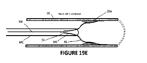
[0108] In another embodiment as shown in Figures 19A-19H, a co-axial stent is
described.
In this embodiment, a combined inner metal (MS) and outer resorbable (RS)
stent are
- 21 -
CA 03157491 2022-5-5
WO 2021/087610
PCT/CA2020/051501
deployed within a vessel 19 showing a representative lesion 19a. The primary
objectives of
the co-axial system are:
= Improve the deployment of the resorbable stent by using the metal
memory/outward spring pressure of the inner metal stent to aid placement
of the resorbable stent against the plaque and arterial wall;
= Remove the inner metal stent after the outer resorbable stent has been
deployed and thus not-limit future treatment options; and,
= Enable improved positioning of a non-radio opaque stent (or moderately
opaque stent).
[0109] In a first embodiment for the placement of a resorbable stent utilizing
a co-axial
stent, the following steps are undertaken (Figures 19A-19H).
a) A nnicrowire (MW) and nnicrocatheter (MC) are advanced to a position past
the zone
of interest (eg. a plaque) utilizing known procedures and the MW is then
removed
(Steps 1-3).
b) A co-axial stent system (COSS) is introduced into the proximal end of the
MC
outside the body and advanced to the distal end of the MC in a compressed
state
(Step 4). As shown, the COSS includes both an inner metal stent (MS) having a
proximal end 50 fixed to a stent wire (SW) at a connection point and an outer
resorbable stent (RS) that is frictionally engaged over the MS but is not
affixed to
either the stent wire or the MS. The RS stent is positioned over the MS such
that the
distal end of the RS extends a few mm X beyond the distal end of the MS. The
proximal end of the RS does not extend proximally beyond the connection point.
In
other words, the proximal end of the RS is a few mm distal to the connection
point
50 as shown by y.
0) When the distal end RSe of the RS is in position, the stent wire is held
and the MC
is withdrawn such that both the RS and MS are deployed from the distal tip MCe
of
the MC. As the MC is progressively withdrawn, the RS and MS will expand and
engage with the vessel wall 19. Generally, as the MS may have greater spring
pressure than the RS, the MS will push against the RS ensuring expansion and
engagement of the RS with the vessel wall (Steps 5 and 6). The MS may also be
designed to be slightly oversized for the vessel wherein its relaxed state has
a
diameter greater than the vessel 19.
- 22 -
CA 03157491 2022-5-5
WO 2021/087610
PCT/CA2020/051501
d) As the MC is continued to be pulled proximally, the proximal end 54 of the
RS will
exit the MC (Step 6). Continued withdrawal of the MC will deploy more of the
MS
which will ensure the proximal end of the RS is engaged with the vessel wall.
e) Once the MC has been withdrawn, the COSS will be left in position for a few
minutes
to allow time for the full expansion of the MS to occur and/or to enable the
RS to
settle into position.
f) After this time, the MC is advanced distally with the SW being held so that
the
proximal end of the MC re-sheaths the MS (Step 7). That is, as the MC is
pushed
distally, the MS will disengage with RS leaving the RS in place while the MS
is
withdrawn back into the MC (Step 8).
g) When the MS is fully within the MC, the system can be fully withdrawn.
[0110] In a separate embodiment, a MC system incorporating a MS is described.
As shown
in Figures 20A and 20B, catheter systems 200 can include catheters where the
MW is
conveyed to the distal tip of the MC outside of the MC, wherein it passes
through the outer
wall of the MC into a MC lumen a short distance from the distal tip of the MC.
[0111] In accordance with this embodiment, the system 200 includes outer wall
catheter 60
and an inner wall catheter 62. The inner wall catheter 62 includes a distal
tip inner lumen
64 that defines an inner lumen 64a passageway allowing a MW to passage from
outside
the system and through the inner lumen to the distal tip 66 of the system.
Preferably, an
atraumatic tip 80 is attached to the distal tip of the inner wall catheter.
[0112] As the inner wall catheter 62 and outer wall catheter 60 are co-axially
engaged, the
two can move with respect to one another. In order to enable this movement to
occur, due
to the passage of a MW through the outer wall catheter, the outer wall
catheter 60 includes
a slot 60a that prevents interference of the MW with the outer wall catheter
during co-axial
movement.
[0113] The inner wall catheter 64 further includes a MS 68 having a proximal
end 68a
affixed to the outer surface of the distal tip inner lumen 64. The MS is
positioned such it is
substantially adjacent the distal tip of the system with its distal tip a few
mm inside the distal
tip as explained in greater detail below. As such, the MS is compressed within
the outer wall
catheter within the outer wall lumen 60b (not shown to scale). In addition, a
RS 70 is
compressed within the outer wall lumen 60b outside the MS.
[0114] Accordingly, by holding inner wall catheter 62 and pulling the outer
wall catheter 60
proximally, the distal end 60c of the outer wall catheter 60 will move
proximally relative to
- 23 -
CA 03157491 2022-5-5
WO 2021/087610
PCT/CA2820/051501
the distal tip 64b of the distal tip inner lumen 64. Thus, during this
movement, the Rs and
MS will project beyond distal end 60c and be able to expand into the vessel
(Figure 20B).
[0115] Similarly, by reversing the process, that is holding the inner wall
catheter 62 and
pushing the outer wall catheter 60 distally, the MS can be made to collapse
back into the
outer lumen 60b.
[0116] As noted, a RS 70 is configured to the outer surface of the MS during
manufacturing
such that both the MS and RS are collapsed with the outer lumen 60b.
Preferably, as noted
above, the distal tip of the RS projects slightly distally beyond the MS and
the MS projects
slightly proximally with respect to the RS.
[0117] The RS is thus deployed in a manner described above, with the main
difference
being that the process of deployment of the RS and MS and re-sheathing of the
MS involves
manipulation of the inner and outer wall catheters 60 and 62.
[0118] The procedures can be applied to both the treatment of unstable plaque
and
aneurysm.
9. Alternate Techniques ¨ Unstable Plaque
9.1. Alternate
[0119] In another embodiment, the resorbable sten' is deployed without
complete flow
cessation by the BGC and/or MB. In a first alternate technique, the BGC is
positioned as
described above and a guidewire, microcatheter and stent assembly are advanced
past the
unstable plaque utilizing the techniques described above.
[0120] Preferably, during the advancement of the microwire and microcatheter
to beyond
the clot, the balloon on the BGC is inflated and active aspiration is
conducted during this
step to produce transient retrograde flow thus reducing the chance of distal
emboli.
[0121] The stent assembly is advanced over the guide wire and deployed.
[0122] The guide wire is withdrawn through the stent, the BGC is deflated and
all equipment
is withdrawn.
9.2. Alternate 2
[0123] In a second alternate technique, the procedure is conducted without any
balloons
and hence without flow cessation as shown in FIG 18. This technique provides
an
advantage over single or double balloon techniques by reducing the potential
for blood
pressure fluctuations during the procedure. That is, during a balloon
technique, the
cessation of blood flow can stimulate the carotid body (carotid glomus) at or
adjacent to the
CCA bifurcation which can cause significant blood pressure fluctuations during
the
- 24 -
CA 03157491 2022-5-5
WO 2021/087610
PCT/CA2020/051501
procedure. As a result of this effect, single or double balloon procedures are
generally
conducted with an anesthetist to control patient blood pressure as necessary.
[0124] Accordingly, procedures conducted without the need of an anesthetist
are generally
advantaged by speed and cost.
[0125] Importantly, if the resorbable stenting is conducted without flow
cessation, the
resorbable stent can act as distal protection device (DPD) as explained below.
9.3. Distal Protection Devices
[0126] As introduced above, current metal stenting procedures of stenosed
vessels will
usually deploy a distal protection device (DPD) mounted on the guide wire
prior to stent
deployment. A DPD is typically an inverted basket that can be advanced in a
collapsed state
past the plaque and deployed by withdrawing a protective sheath. After the DPD
is
deployed, the metal stent is brought up along the same guide wire and
deployed. During
this step, the DPD serves to trap any emboli that may be dislodged during
stent deployment.
After stent deployment, the DPD is collapsed and withdrawn into the its
protective sheath.
[0127] In the present method and as shown in FIGs 15 and 18, the use of a DPD
would
generally not be necessary and thus can save the time used to deploy the DPD
as well as
the expense of this equipment.
[0128] That is, as the resorbable stent of the subject system has a pore size
similar to the
pore size of a DPD, that is in the range of about 110-250 microns, the act of
deploying the
resorbable stent will provide the same emboli capturing capabilities of a DPD
insomuch as
the resorbable stent is self-expanding. In other words, as the resorbable
stent deploys
distally to the plaque, the distal end will expand against the intima and
progressively be
deployed in the proximal direction. Thus, any emboli 902b breaking free from
the plaque
during deployment will be caught between the stent and the intima as shown in
FIGs 15
and 18. Importantly, during this step, the surgeon should ensure that the
stent is deployed
sufficiently distal to the plaque that the distal tip of the stent is fully
contacts the vessel
before the stent is deployed across the plaque. This will generally require
that the stent is
long enough to be deployed in a straight distal section of the ICA.
[0129] This technique by virtue of the stent pore size, which is significantly
smaller than a
typical metal stent will thus retain any emboli between the stent and the
intima. Importantly,
while the stent is resorbing overtime, the emboli will also be resorbed into
the intima and/or
dissolved as a result of normal blood thinning regimes.
- 25 -
CA 03157491 2022-5-5
WO 2021/087610
PCT/CA2020/051501
9.4. Treatment of Aneurysm
[0130] The treatment of aneurysms using the COSS described above in relation
to unstable
plaque are similar. Like the placement of a COSS in the CCA/ICA, the COSS can
be used
as a flow diverter in the treatment of aneurysm utilizing a similar series of
steps to deploy
the RS and MS and to re-sheath the MS.
[0131] Importantly, the COSS can improve the positioning of a RS in that the
MS being
radio-opaque can provide for accurate positioning of the MS and thus the RS.
Depending
on the structure of the RS which will generally be constructed of non-radio-
opaque
materials, the RS can be fabricated with a small amount of metal (eg.
tantalum) that could
provide some desirable properties to the COSS.
[0132] In addition, in some treatment scenarios, it may be desirable to deploy
both the RS
and MS and leave the MS in place. In this scenario, the RS could be fabricated
with a
smaller porosity and the MS fabricated with a larger porosity. If both are
left in place after
deployment, the RS will ensure that the aneurysm stabilizes over a period of
time by fully
occluding blood flow into and around the edges of the aneurysm thus providing
the
appropriate period of time for the aneurysm to heal. However, as the RS will
resorb over a
period of time, the tight porosity of the RS will disappear, and the larger
porosity of the MS
will remain. As a result, while metal may remain, the porosity of the MS may
still permit
access to the aneurysm at a time in the future through the pores of the MS
thus making
available some additional treatment options available should access to the
aneurysm be
required. This is different than treatment with a tight MS as deployment of a
single MS will
generally utilize a MS having small pores that prevent blood flow through
them.
[0133] If a COSS is designed where both the RS and MS are deployed, the RS/MS
may be
conveyed and deployed through a MC as described above and the MS detached from
the
stent wire utilizing known detachment techniques.
9.5. Equivalents
[0134] At least the following equivalents and scope are contemplated.
[0135] An example location for the unstable plaque 404 is described with
respect to FIGS
8 to 18. However, this location is merely exemplary. An unstable plaque may be
located in
the CCA 400a or the ICA 400b or a combination thereof. The geometry of the
resorbable
stent would be readily apparent to the skilled person in view of the
discussion provided
herein.
[0136] FIGS 8 to 17 contemplate balloon deployment in each of the CCA and the
ECA to
substantially arrest blood flow at an unstable plaque. It will be appreciated
that occlusion of
- 26 -
CA 03157491 2022-5-5
WO 2021/087610
PCT/CA2020/051501
at least any two of the three arteries proximal to the CCA bifurcation could
substantially
arrest blood flow at the unstable plaque.
[0137] If one or more balloons are used to substantially arrest blood flow at
an unstable
plaque, it will be appreciated that the balloons may be deflated either by
manual input by
someone operating the BGC or may automatically deflate after a predetermined
period of
time. In a further embodiment, the distal balloons may be a self deflating
detachable balloon
that may be detached into the ECA.
[0138] In another embodiment, although not required, a microcatheter does not
need to be
advanced along a guidewire, and instead a resorbable stent may be advanced
directly along
the guide wire. In a further embodiment the guide wire may not be necessary if
adequate
control of the resorbable stent can be effected without the guidewire or the
microcatheter.
9.6. Uses and Kits
[0139] In addition to the methods described above, uses of a resorbable stent
and kits are
also contemplated. The uses and kits described below encompass at least
features
described in the methods disclosed above and its equivalents.
[0140] A use of a resorbable stent is contemplated to stabilize an unstable
plaque in a
patient for a therapeutically effective time period at a bifurcation of a CCA
into an ICA and
an ECA, where the resorbable stent is deployed under substantial arrest of
blood flow at
the unstable plaque. A use of a RS as a flow diverter for the treatment of
aneurysm is also
contemplated.
[0141] A use of a co-axial resorbable and metal stent is contemplated.
Specifically, the use
may be of a COSS to stabilize an unstable plaque in a patient for a
therapeutically effective
time period at a bifurcation of a CCA into an ICA and an ECA, where the
resorbable stent
is deployed under substantial arrest of blood flow at the unstable plaque. A
use of a COSS
as a flow diverter for the treatment of aneurysm is also contemplated.
[0142] A kit for the treatment of an unstable plaque and as flow diverter for
the treatment
of aneurysm in a patient is also contemplated. Kits may include one or more
devices, the
one or more devices adapted to substantially arrest blood flow at the unstable
plaque
adjacent to a bifurcation of a CCA into an ICA and an ECA or as a flow
diverter for the
treatment of aneurysm. The kit may further include or merely comprise at least
one COSS
having a resorbable stent adapted to stabilize the unstable plaque/aneurysm
for a
therapeutically effective time period.
[0143] Kits may comprise within individual or separate packing a combination
one or more
of a first BGC, a second BGC that is deployable through the first GBC, one or
more guide
- 27 -
CA 03157491 2022-5-5
WO 2021/087610
PCT/CA2020/051501
wires, one or more microcatheters and one or more stent assemblies having one
or more
resorbable stents as well as re-sheathable metal stents. The resorbable stents
may be
provided with a variety of features that allow a surgeon to select desired
functional and
structural characteristics for a specific case.
[0144] For example, metal and resorbable stents may combinations of the
following
functional/structural characteristics including a range of:
= diameters;
= lengths;
= tapers;
= compressive stiffnesses;
= pore sizes;
= drug coatings; and,
= resorb times.
[0145] Generally, a RS stent will be designed to resorb at a rate proportional
to blood flow.
Hence, to the extent that a RS protrudes into a blood vessel or covers a blood
vessel, the
RS will begin to resorb/erode at positions having the highest blood flow rates
and progress
to areas having lower blood flow rates.
[0146] With reference to Figures 21A, 21A1, 21B, 21C and 21C1 which are
various
schematic cross-sections of a vessel with a deployed RS (eq. a CCNICA/ECA
bifurcation),
the process by which a RS is resorbed is shown. That is, as shown in Figures
21A and
21A1, a RS 70 may be deployed such that it partially extends into/over another
vessel
wherein edges/surfaces 70j of the RS will not be engaged with the vessel wall.
In this
example, at deployment, the RS occludes one vessel such that blood flow into
the occluded
vessel is low and only occurs through pores of the RS as shown schematically
by the flow
arrows in each vessel segment.
[0147] Over time, the edges of the RS that are not engaged with the vessel
wall 70j and
exposed to the greatest flow, will begin to erode/resorb as shown by dotted
lines 70i in
Figure 21B thus forming a hole through the RS and allowing increased blood
flow into the
occluded vessel as shown in Figure 21C and 21C1. Typically, the pattern of
erosion will be
a progressively larger ellipse as shown by ellipses t1 and t2 which represent
the size of the
elliptical hole at two times.
- 28 -
CA 03157491 2022-5-5
WO 2021/087610
PCT/CA2020/051501
[0148] Those surfaces 70k that are in contact with the vessel wall may
depending on a
number of factors (including the design of the rate of resorption) either
become
endotheliazied and thus not resorb or may partially or completely resorb.
[0149] The size of the RS pores may also affect the rate of resorption as the
rate of flow
through the pores may be variable.
[0150] Importantly, the integrity of the stent contacting the vessel wall will
be maintained
during the endotheliazation process and/or during resorption of RS in that
resorption will
generally progress from an exposed edge of the RS away from a vessel wall
towards the
vessel wall. Thus, when a resorbed edge reaches the vessel wall, resorption
may cease in
the case where the RS has become endothelialized or may continue at a lower
rate as blood
flow rate at the wall may be slower. Also, in the case where a vessel is
partially obstructed,
blood flow rate reduction would preferably only occur for a shod period of
time and full flow
may be re-established within a few days/weeks. This may provide a further
advantage of
reducing the need for anti-platelet medication in the patient.
[0151] Various stents may have different combinations of each of the above
structures and
functionalities.
10. Stent Design
[0152] As noted, RS and MS may have a plurality of features that make it
suitable for use
in treating unstable plaque or aneurysm. Given the variability in the size and
location of
plaque being treated adjacent the CCA bifurcation, stents having different
lengths and
features may be utilized. Similarly, given the variability of the size and
structure of
aneurysms, MS and RS having different lengths and features may be utilized.
[0153] For example, a plaque in the ICA may be 7-9 mm in length and extend
into the ICA
0.5-1mm. The center of the plaque may be 4-6 mm from the bifurcation.
Generally, in order
to enable the stent to be useful as a DPD, the stent would typically be longer
than a stent
that is used with a separate DPD.
[0154] That is, as shown in Figure 18, as the stent must contact the intima
before it is fully
effective as a DPD, and there is a distance between the distal tip of the
stent and the distal
tip of the deployment catheter before the distal tip of the stent is fully
engaged with the
intima, the surgeon will typically need to deploy the stent a few mm further
in the distal
direction to enable this. Hence, in comparison to current stents used at this
location, the
stent in accordance with the invention will typically be a few mm longer.
Moreover,
particularly when the procedure is conducted without flow cessation, the
initial step of stent
deployment should be conducted further in the distal direction to minimize
contact with the
- 29 -
CA 03157491 2022-5-5
WO 2021/087610
PCT/CA2020/051501
plaque and the risk of disturbing it. As such, a stent will typically be 30-50
mm long and
more specifically 40-42 mm long.
[0155] The ultimate selection of the length and other features of the stent
will be determined
by the surgeon having regard to the particular characteristics of the
plaque/aneurysm.
[0156] It should also be noted that braided metal stents having the above
structural features
could be developed and utilized. In particular, these stents could also be
effective as DPDs
as described above. In a COSS system, the RS may also function as a DPD.
[0157] Further still, in the design of a COSS system for aneurysms, as noted
above, one
design contemplates a RS having smaller pore sizes and a MS having larger pore
sizes
such that when the RS has resorbed the larger pore sizes of the MS may enable
access
into the aneurysm at a later time. In another embodiment, the RS is a mesh of
very fine
wires with a defined pore size that are solution cast with a RS material so as
to partially fill
in the pores of the MS. Thus, in this embodiment, the RS component and MS
component
are overlaid with respect to one another such that the effective pore size of
the MS increases
over time as resorbable material is eroded away from the MS material, thus
enabling future
access through the MS to gain access to an aneurysm if and when necessary.
[0158] In another embodiment, the RS is seated by positioning and inflation of
a balloon
after the RS has been deployed. In this case, the radio-opaque markers on the
balloon can
provide positioning information to the physician when deploying the RS.
Conclusion
[0159] While this invention has been particularly shown and described with
references to
embodiments thereof, it will be understood by those skilled in the art that
various changes
in form and details may be made therein without departing from the scope of
the invention
encompassed by the appended claims.
- 30 -
CA 03157491 2022-5-5