Note: Descriptions are shown in the official language in which they were submitted.
CA 03158504 2022-04-21
WO 2020/086865 PCT/US2019/057891
NEURAL NETWORKS FOR ATRIAL FIBRILLATION SCREENING
CROSS-REFERENCE TO RELATED APPLICATIONS
[0001] This application claims priority to U.S. Application Serial No.
62/751,395, filed
on October 26, 2018. The disclosure of the prior application is considered
part of the disclosure
of this application, and is incorporated by reference in its entirety into
this application.
BACKGROUND
[0002] Cryptogenic stroke (CS) is a form of stroke or cerebral ischemia
of obscure or
unknown origin. The precise causes of CS remain undetermined because the CS is
typically a
transitory or reversible condition. It is estimated that 30-percent of strokes
in the United States
(approximately 200,000 cases a year) are cryptogenic.
[0003] One of the major causes of stroke is atrial fibrillation (AF)
which is associated
with a 5-fold risk of stroke. In patients with CS, the current AHA guidelines
state, "Long term
monitoring for AF may be beneficial in patients with CS and has the potential
to shift the
management paradigm ... Cardiac embolism secondary to paroxysmal AF may be a
common
cause of assumed CS." The same guidelines argue that the detection of
paroxysmal AF in post-
CS patients is a priority in order to reduce the risk for recurrent events.
[0004] In recognition of this priority, post-CS patients are often
provided an implantable
loop recorder (ILR), which records an electrocardiogram (ECG) of the patent
and alerts a
physician or other healthcare provider for the presence of AF when AF is
detected to have
occurred in the ECG data. While the ILRs have high detection rates, the
process is invasive,
expensive, and generally involves a continuous monitoring infrastructure to
communicate alerts
to the clinicians.
SUMMARY
[0005] This specification generally describes systems, methods, devices,
and other
techniques for atrial fibrillation screening. A machine-learning model such as
a deep neural
network can be trained to process a short-recording of ECG data from a patient
to generate a
prediction indicating a likelihood that the patient has or will experience
atrial fibrillation (e.g.,
paroxysmal atrial fibrillation) and/or other supraventricular tachycardia
(e.g., atrial flutter, atrial
1
CA 03158504 2022-04-21
WO 2020/086865 PCT/US2019/057891
tachycardia). Unlike other techniques that involve long-term or continuous
monitoring (e.g.,
implantable loop recorders) to detect actual occurrences of atrial
fibrillation, the neural networks
described herein can detect a likelihood of atrial fibrillation and/or other
supraventricular
tachycardia (SVT) in a patient from an ECG recording that nominally represents
a normal sinus
rhythm. Due to structural irregularities of the heart, or the presence of
other factors that can lead
to atrial fibrillation, the patient's ECG in normal sinus rhythm can include
features, not
detectable by the human eye, but which are nonetheless highly predictive of a
patient that has
experienced atrial fibrillation or is susceptible to atrial fibrillation. A
neural network can be
trained to learn these features and predict patients that have or will
experience atrial fibrillation
based on ECG recordings. Moreover, because the neural network can generate
atrial fibrillation
predictions and/or other SVT predictions based on ECG recordings reflecting
normal sinus
rhythm, it is not necessary to monitor the patient for long periods of time to
detect actual
occurrences of atrial fibrillation and/or other SVT. Instead, a patient that
has experienced
cryptogenic stroke, for example, may take a brief ECG, e.g., with a 12-lead
system at a
healthcare provider's location or at home with a single-lead smartphone or
patch system, and the
neural network can process the ECG recording to determine a likelihood of
atrial fibrillation
and/or other SVT for the patient much more quickly. If the prediction
indicates a sufficiently
high likelihood of past or expected atrial fibrillation and/or other SVTs,
appropriate action may
be taken such as administration of medication (e.g., anticoagulants), longer
term monitoring
(e.g., with an implantable loop recorder) to validate the prediction, or both.
[0006] Some aspects of the subject matter disclosed herein include a
method for
screening for atrial fibrillation and/or other SVT (e.g., atrial flutter,
atrial tachycardia). The
method can include obtaining a first neural network input, where the first
neural network input
represents an electrocardiogram (ECG) recording of a mammal, and processing
the first neural
network input with a neural network to generate an atrial fibrillation
prediction and/or other SVT
prediction for the mammal.
[0007] These aspects and others can be implemented on a system having a
data
processing apparatus, e.g., which can include one or more computers in one or
more locations.
In some implementations, one or more computer-readable media have instructions
stored thereon
that, when executed by data processing apparatus, cause the data processing
apparatus to perform
2
CA 03158504 2022-04-21
WO 2020/086865 PCT/US2019/057891
this and other computer-based methods or processes described herein.
Optionally, these aspects
and others can include one or more of the following features.
[0008] The ECG recording represented by the first neural network input
can describe a
normal sinus rhythm for the mammal, such that the neural network generates the
atrial
fibrillation prediction and/or other SVT prediction for the mammal based on
features of the
mammal's normal sinus rhythm as indicated by the ECG recording.
[0009] The ECG recording represented by the first neural network input
can span a time
interval that is less than or equal to thirty seconds, fifteen seconds, ten
seconds, or five seconds.
[0010] The ECG recording represented by the first neural network input
can span a time
interval that is less than or equal to ten minutes, five minutes, one minute,
or forty-five seconds.
[0011] The mammal can be a human.
[0012] The neural network can include at least one of a feedforward
portion, a
convolutional portion, a recurrent portion, or a capsule portion.
[0013] The ECG recording of the mammal can include a 12-lead ECG
recording.
[0014] The ECG recording of the mammal can include a single-lead ECG
recording.
[0015] The ECG recording of the mammal can be based on fewer than twelve
leads.
[0016] The atrial fibrillation and/or other SVT predictions can indicate
a likelihood of the
mammal experiencing atrial fibrillation and/or other SVT (e.g., atrial
flutter, atrial tachycardia).
[0017] The atrial fibrillation prediction and/or other SVT prediction can
indicate a
selection of one of a plurality of possible monitoring or treatment plans.
[0018] The plurality of possible monitoring or treatment plans include a
first plan to
administer anticoagulants to the mammal with high likelihood of atrial
fibrillation, a second plan
to not administer anticoagulants, and a third plan to administer a continuous
ECG for further
monitoring.
[0019] The atrial fibrillation predication can indicate at least a
threshold likelihood of the
mammal experiencing atrial fibrillation, and the method can further include
administering a
treatment to lower a risk of stroke in the mammal in response to identifying
that the atrial
fibrillation prediction indicates at least the threshold likelihood of the
mammal experiencing
atrial fibrillation.
[0020] Administering the treatment can include administering an
anticoagulant to the
mammal.
3
CA 03158504 2022-04-21
WO 2020/086865 PCT/US2019/057891
[0021] The method can further include operations for obtaining data
describing a non-
ECG profile for the mammal; generating one or more second neural network
inputs representing
the non-ECG profile for the mammal; and processing the first neural network
input along with
the one or more second neural network inputs with the neural network to
generate the atrial
fibrillation prediction and/or other SVT prediction for the mammal.
[0022] The method can further include determining one or more
morphological features
of the ECG recording of the mammal; generating one or more second neural
network inputs
representing the one or more morphological features of the ECG recording; and
processing the
first neural network input along with the one or more second neural network
inputs with the
neural network to generate the atrial fibrillation prediction and/or other SVT
prediction for the
mammal.
[0023] The ECG recording of the mammal can be recorded over a first time
interval, and
the method can further include: obtaining a second neural network input, the
second neural
network input representing a second ECG recording of the mammal that was
recorded over a
second time interval, the first time interval and the second time interval
separated by a third time
interval; and processing the first neural network input along with the second
neural network
input with the neural network to generate the atrial fibrillation prediction
and/or other SVT
prediction for the mammal.
[0024] The third time interval can be at least a minute, an hour, a day,
a week, or a
month.
[0025] The neural network can further process, along with the first
neural network input
and the second neural network input, a third neural network input that
indicates a length of the
third time interval between the first and second time intervals when the ECG
recording and the
second ECG recordings were recorded, respectively.
[0026] Some aspects of the subject matter disclosed herein include a
computing system
having an interface and a neural network. The interface can be configured to
obtain an
electrocardiogram (ECG) recording of a mammal, and to generate a first neural
network input
representing the ECG recording. The neural network can be implemented on data
processing
apparatus and configured to process the first neural network input to generate
an atrial fibrillation
prediction and/or other SVT prediction. The system can further include at
least one of a storage
device for storing data representing the atrial fibrillation prediction and/or
other SVT prediction,
4
CA 03158504 2022-04-21
WO 2020/086865 PCT/US2019/057891
a presentation device for presenting the atrial fibrillation prediction and/or
other SVT prediction,
or a network interface device configured to transmit the atrial fibrillation
prediction and/or other
SVT prediction over a network to a provider or other interested party.
[0027] Unless otherwise defined, all technical and scientific terms used
herein have the
same meaning as commonly understood by one of ordinary skill in the art to
which this invention
pertains. Although methods and materials similar or equivalent to those
described herein can be
used to practice the invention, suitable methods and materials are described
below. All
publications, patent applications, patents, and other reference mentioned
herein are incorporated
by reference in their entirety. In case of conflict, the present
specification, including definitions,
will control. In addition, the materials, methods, and examples are
illustrative only and not
intended to be limiting.
[0028] The details of one or more embodiments of the invention are set
forth in the
accompanying drawings and the description below. Other features, objects, and
advantages of
the invention will be apparent from the description and drawings, and from the
claims.
BRIEF DESCRIPTION OF DRAWINGS
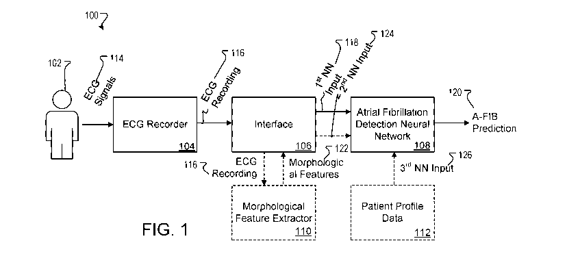
[0029] Figure 1 is a diagram of an example system for ECG recording and
atrial
fibrillation screening.
[0030] Figure 2 depicts an example system for training an atrial
fibrillation detection
neural network.
[0031] Figure 3 is a flowchart of an example process for recording an ECG
of a patient
and processing data representative of the recording to generate an atrial
fibrillation prediction.
The atrial fibrillation prediction can be used by a physician or other human
or automated
decision-maker to determine or recommend a monitoring and/or treatment regime
for the patient.
[0032] Figure 4 is a diagram of an example neural network that processes
temporally
spaced ECG recordings of a patient to generate an atrial fibrillation
prediction for the patient
based in part on features representing differences between the ECG recordings
over time.
[0033] Figure 5 is an example diagram of a segment of an ECG recording
for a patient.
[0034] Figure 6 is an illustration showing an example ECG selection for
two patients
with multiple ECGs over the same year. The example study implementation used
all normal
sinus rhythm ECGs for patients with no ECGs with atrial fibrillation recorded
and the window of
CA 03158504 2022-04-21
WO 2020/086865 PCT/US2019/057891
interest began on the date of their first ECG. For patients with at least one
atrial fibrillation
rhythm recorded, the first ECG recording atrial fibrillation or atrial flutter
was the index ECG
and the window of interest began 31 days before the index ECG. For all
patients, the window of
interest extended until the study ended.
[0035] Figure 7 is a patient flow diagram.
[0036] Figure 8 is a plot of ROC curves for the convolutional neural
networks on the
testing dataset. In the main analysis, only the score of the first normal
sinus rhythm ECG in the
window of interest was used. In the secondary analysis, the highest score for
all ECGs done in
the first month of the window of interest was used.
[0037] Figure 9 depicts a table representing model performance study from
the example
study implementation.
[0038] Figure 10 depicts the architecture of an example neural network
configured to
process inputs representing a recording of a patient's ECG during normal sinus
rhythm to
generate an output representing a likelihood of the patient having or
developing atrial fibrillation.
DETAILED DESCRIPTION
[0039] This specification generally describes systems, methods, devices,
and other
techniques for atrial fibrillation screening, e.g., using neural networks or
other machine-learning
models. Neural networks are machine-learning models that employ multiple
layers of operations
to predict one or more outputs from one or more inputs. Neural networks
typically include one
or more hidden layers situated between an input layer and an output layer. The
output of each
layer is used as input to another layer in the network, e.g., the next hidden
layer or the output
layer. Each layer of a neural network specifies one or more transformation
operations to be
performed on input to the layer. Some neural network layers have operations
that are referred to
as neurons. Often, each neuron can receive one or more inputs and generates an
output that is
received by another neural network layer. The transformation operations of
each layer can be
carried out by one or more computers at one or more locations having installed
software modules
that implement the transformation operations.
[0040] Referring to Figure 1, a diagram is shown of an example system 100
for ECG
recording and atrial fibrillation screening. The system 100 is configured to
record an ECG of a
patient 102 and to process the recording and, optionally, additional
(auxiliary) data to generate an
6
CA 03158504 2022-04-21
WO 2020/086865 PCT/US2019/057891
atrial fibrillation prediction 120. The atrial fibrillation prediction 120 can
indicate a likelihood
that the patient 102 has experienced or is susceptible to developing atrial
fibrillation. The
prediction 120 can be expressed as a probability or confidence score
representing a probability or
confidence that the patient 102 has experienced or is susceptible to
developing atrial fibrillation.
In some implementations, the prediction 120 is expressed as a selection of a
particular
classification from multiple possible classifications that represents a most
likely condition of the
patient 102. For example, a binary classification can be made indicating
whether there is at least
a threshold probability or confidence level that the patient 102 has
experienced or is susceptible
to developing atrial fibrillation. The atrial fibrillation prediction 120 can
identify this binary
classification. As another example, the atrial fibrillation prediction 120 can
indicate a
recommendation or selection of a monitoring or treatment option for the
patient 102 based on a
likelihood of the patient 102 having experienced or being susceptible to
development of atrial
fibrillation. For instance, the prediction 120 can indicate a selection from
the trinary of options
to administer an anticoagulation medication to the patient, to not administer
an anticoagulant but
continue with periodic screenings, or to initiate continuous monitoring, e.g.,
using an implantable
loop recorder ECG. Thresholds used for any decision boundaries can be
extracted from
retrospective analysis and can be presented with positive and negative
predictive value (PPV and
NPV).
[0041] The patient 102 can be a human or any other mammal for which an
atrial
fibrillation screening is desired. To obtain an ECG recording of the patient
102, one or more
electrodes are brought into contact with a surface of the patient's body. The
electrodes can be
arranged according to a standard 12-lead ECG configuration, or in other known
configurations.
The electrodes may or may not be affixed to the patient 102. In some
implementations, fewer
than 12-leads are provided to for obtaining the ECG. For example, a single-
lead smartphone-
based ECG sensor may be employed to sense the patient's ECG based on finger
contact, or a
patch with an electrode array may be affixed to the patient's chest. The ECG
recorder 104
includes hardware and/or software for sensing and capturing ECG signals 114
from the
electrodes in contact with the patient 102. For example, the signals 114 may
be filtered,
amplified, and digitally sampled by recorder 104, and a recording 116 can be
generated that
represents the patient's ECG for each available lead over a period of time.
Typically, the
recording 116 may be made based on a relatively short period of measurement.
For example,
7
CA 03158504 2022-04-21
WO 2020/086865 PCT/US2019/057891
since a sample ECG representing the patient's normal sinus rhythm may suffice
to predict the
atrial fibrillation condition of the patient 102, a relatively short sample
corresponding to just a
few beats may be all that is required to be captured for purposes of making a
prediction. In some
implementations, a minimum recording time may be specified that is less than
or equal to ten
minutes, five minutes, one minute, forty-five seconds, thirty seconds, fifteen
seconds, ten
seconds, or five seconds. The ECG can be recorded while the patient 102 is in
the supine
position or other positions that correspond to positions of the patients whose
ECGs were used as
training examples for the system.
[0042] An interface 106 can be implemented on a computer or other data
processing
apparatus. The interface 106 receives a digitized ECG recording 116 from the
ECG recorder 104
and processes the recording 116 to generate a first neural network input 118.
The first neural
network input 118 is a representation of the ECG recording that is suitable
for processing by the
atrial fibrillation detection neural network 108. The first neural network
input 118, for example,
can identify values of the ECG signal level for each lead over the full
recording time or over a
subset of the recording time (e.g., a time interval that corresponds to a
single heartbeat). The
first neural network input 118 can represent the ECG recording for one or more
individual beats
or can represent an averaged beat based on ECG recordings from several
measured beats.
[0043] The atrial fibrillation detection neural network 108 is configured
to process the
first neural network input 118 and to generate atrial fibrillation prediction
120 based on the first
neural network input 118. The neural network 108 can include multiple layers
of operations that
have been trained to discern an atrial fibrillation condition of a patient
based on ECG recordings
of a patient's normal sinus rhythm. The neural network 108 can be a
feedforward neural
network, a recurrent neural network, a convolutional neural network, a capsule
network, or may
include various portions having different characteristics, such as feedforward
layers, recurrent
layers, and/or convolutional layers. The atrial fibrillation detection neural
network 108 can be
implemented on one or more computers or other data processing apparatus in one
or more
locations. The network 108 may be implemented on a smartphone or other
personal device (e.g.,
tablet, desktop or notebook computer) in the same location as the patient 102,
or may be
implemented on one or more remote servers in communication with the interface
106.
[0044] In some implementations, the atrial fibrillation detection neural
network 108 is
configured to process additional (auxiliary) information in generating atrial
fibrillation prediction
8
CA 03158504 2022-04-21
WO 2020/086865 PCT/US2019/057891
120. For example, the network 108 may process a second neural network input
124 in addition
to the first neural network input 118 to generate the atrial fibrillation
prediction 120. The second
neural network input 124 represents morphological features of the patient's
ECG. Figure 5, for
instance, depicts a waveform or tracing 500 for a single beat from a patient's
ECG. The
waveform includes several segments including a P-wave, a QRS-complex, and a T-
wave. The
interface 106 can provide the ECG recording 116 to a morphological feature
extractor 110 for
analysis, and the extractor 110 can measure various morphological features of
one or more beats
(or a composite or averaged beat) from the ECG recording 116. The
morphological features are
parameters that describe attributes of the shape of the beat, including
attributes of individual
segments of the beat and attributes between segments. A number of
morphological features that
may be employed for atrial fibrillation prediction are labeled in Figure 5,
such as a duration of
the QRS-complex, am amplitude of the P-wave, R-wave, or T-wave, an area of the
P-wave,
QRS-complex, or T-wave, slopes of any of the waves, distances between the
waves, and centers-
of-gravity of the waves. The morphological feature extractor 110 provides
values for the
morphological features 122 to the interface 106, and the interface 106 formats
them into an
acceptable form for processing by atrial fibrillation detection neural network
108 as second
neural network input 124. The atrial fibrillation detection neural network 108
processes the first
and second inputs 118, 124 to generate the atrial fibrillation prediction 120.
[0045] In some implementations, the neural network 108 processes one or
more third
neural network inputs 126 representing patient profile data from a database
112. The patient
profile data is another form of auxiliary information, and in particular it
indicates non-ECG
descriptions of the patient 102. For example, the third neural network inputs
126 representing
patient profile data can include indications of one or more of age, weight, or
sex of the patient
102, and/or other attributes of the patient 102. The atrial fibrillation
detection neural network
108 can process the first neural network input 118 and none, one, or both of
second neural
network input 124 and third neural network input 126 to generate atrial
fibrillation prediction
120.
[0046] Figure 2 depicts an example system 200 for training an atrial
fibrillation detection
neural network. The training system 200 can be hosted within a data center
112, which can be a
distributed computing system having hundreds or thousands of computers in one
or more
locations.
9
CA 03158504 2022-04-21
WO 2020/086865 PCT/US2019/057891
[0047] The training system 200 includes a training neural network
subsystem 206 that
can implement the operations of each layer of a neural network that is
designed to make atrial
fibrillation predictions from ECG recordings and, optionally, auxiliary
information such as
morphological features and patient profile data. The training neural network
subsystem 206
includes a plurality of computing devices having software or hardware modules
that implement
the respective operations of each layer of the neural network according to an
architecture of the
neural network. Generally, the training neural network subsystem 206 has the
same architecture
as the atrial fibrillation detection neural network 108. However, the training
system 200 need
not use the same hardware to compute the operations of each layer. In other
words, the training
system 200 can use CPUs only, highly parallelized hardware, or some
combination of these.
[0048] The training neural network subsystem 206 can compute the
operations of each
layer of the training neural network subsystem 206 (or atrial fibrillation
detection neural network
108) using current parameter values 216 stored in a collection of model
parameter values 214.
Although illustrated as being logically separated, the model parameter values
214 and the
software or hardware modules performing the operations may actually be located
on the same
computing device or on the same memory device.
[0049] The training neural network subsystem 206 can generate, for each
training
example 204, an atrial fibrillation prediction 208. A training engine 210
analyzes the predictions
208 and compares the predictions 208 to labels in the training examples 204
that indicate target
predictions for each training example 204. The training engine 210 then
generates updated
model parameter values 214 by using an appropriate updating technique, e.g.,
stochastic gradient
descent with backpropagation. The training engine 210 can then update the
collection of model
parameter values 214 using the updated model parameter values 212. For
example, each training
example 204 can include a first component representing a single- or multi-lead
ECG recording of
a patient and a label indicating a target atrial fibrillation prediction. The
first component can
represent an ECG of a patient under normal sinus rhythm, and the label can
indicate whether that
particular patient is known to have actually experienced atrial fibrillation
at another time. In this
way, the neural network 108 can be trained using sinus rhythm ECGs obtained in
patients known
and validated atrial fibrillation versus patients with no known atrial
fibrillation. The training
examples can also include additional components representing morphological
features or patient
profile data, for example.
CA 03158504 2022-04-21
WO 2020/086865 PCT/US2019/057891
[0050] After training is complete, the training system 200 can provide a
final set of
parameter values 218 to the system 100 for use in making atrial fibrillation
predictions 120. The
training system 200 can provide the final set of model parameter values 218 by
a wired or
wireless connection to the system 100 and neural network 108, for example.
[0051] Figure 3 is a flowchart of an example process 300 for recording an
ECG of a
patient and processing data representative of the recording to generate an
atrial fibrillation
prediction. The atrial fibrillation prediction can be used by a physician or
other human or
automated decision-maker to determine or recommend a monitoring and/or
treatment regime for
the patient. A patient is identified for atrial fibrillation screening, e.g.,
due to the patient having
suffered a cryptogenic stroke (302). An ECG recording is obtained from the
patient (304). The
ECG recording may be obtained using a single-lead or multi-lead (e.g.,
standard 12-lead) ECG.
Optionally, auxiliary data such as morphological features for the ECG and/or
patient profile data
can be obtained (306). The system generates neural network inputs based on the
ECG recording
and the auxiliary data, if available (308). The atrial fibrillation detection
neural network
processes the neural network inputs to generate the atrial fibrillation
prediction (310). A
physician or other healthcare provider can assess the need for further
monitoring or treatment of
the patient's condition based on the atrial fibrillation prediction (312). For
example, to lower the
risk of stroke once the patient has been identified as likely having atrial
fibrillation, medication
such as anticoagulants may be prescribed to the patient. Additionally, the
patient may undergo
longer-term continuous monitoring to identify actual episodes of atrial
fibrillation, e.g., using an
implantable loop recorder (314).
[0052] Figure 4 is a diagram of an example neural network system 400 that
processes
temporally spaced ECG recordings of a patient to generate an atrial
fibrillation prediction for the
patient based in part on features representing differences between the ECG
recordings over time.
Here, the atrial fibrillation detection neural network 108 is configured to
process a first neural
network input 402 representing an ECG recording (e.g., under normal sinus
rhythm) of the
patient at a first time (e.g., over a short time interval such as less than or
equal to 30, 20, 15, 10,
or 5 seconds) and a second neural network input 404 representing a second ECG
recording (e.g.,
under normal sinus rhythm) of the patient at a second time (e.g., over a short
time interval such
as less than or equal to 30, 20, 15, 10, or 5 seconds). In some cases, the
neural network 108
further processes a neural network input 406 that indicates an amount of
elapsed time between
11
CA 03158504 2022-04-21
WO 2020/086865 PCT/US2019/057891
the times the first and second ECG recordings representing in inputs 402 and
404 were recorded.
For example, the input 406 can indicate that the ECG recordings were taken a
number of hours,
days, weeks, months, or years apart from each other. The neural network 108
can then process
each of the inputs 402, 404, and 406 to generate an atrial fibrillation
prediction 120.
Example Implementation Study
[0053] This example describes the results of a study in which an
artificial intelligence
(AI)-model including a convolutional neural network was developed and tested
to detect the
electrocardiographic signature of atrial fibrillation present during normal
sinus rhythm. The
model was developed to process an ECG signature for a patient using a standard
10-second, 12-
lead ECG recording. The example implementation was trained based on ECGs
acquired from a
set of patients aged 18 years or older having at least one digital, normal
sinus rhythm, standard
10-second, 12-lead ECG acquired in the supine position at the MAYO CLINIC ECG
laboratory
between December 31, 1993, and July 21, 2017, with rhythm labels validated by
trained
personnel under cardiologist supervision. ECG samples were assigned binary
classification
labels indicating either (1) positive for atrial fibrillation or (2) negative
for no atrial fibrillation.
ECG samples that demonstrated atrial fibrillation were classified as positive
for atrial fibrillation.
Further, various ECG samples were allocated to either the training, internal
validation, or testing
datasets in a 7:1:2 ratio. The area under the curve (AUC) of the receiver
operating characteristic
curve was calculated for the internal validation dataset to select a
probability threshold, which
was applied to the testing dataset. Model performance was evaluated on the
testing dataset by
calculating the AUC and the accuracy, sensitivity, specificity, and Fl score
with two-sided 95%
confidence intervals (CIs).
[0054] The study included ECGs from 180,922 patients, which provided
649,931 normal
sinus rhythm ECG samples for analysis: 454,789 ECGs recorded from 126,526
patients in the
training dataset, 64,340 ECGs from 18,116 patients in the internal validation
dataset, and
130,802 ECGs from 36,280 patients in the testing dataset. 3,051 (8.4%)
patients in the testing
dataset had verified atrial fibrillation before the normal sinus rhythm ECG
tested by the model.
The example implementation of the neural network system identified atrial
fibrillation with an
AUC of 0.87 (95% CI 0.86-0.88), sensitivity of 79.0% (77.5-80.4), specificity
of 79.5% (79.0-
79.9), Fl score of 39.2% (38.1-40.3), and overall accuracy of 79.4% (79.0-
79.9). Including all
12
CA 03158504 2022-04-21
WO 2020/086865 PCT/US2019/057891
ECGs acquired during the first month of each patient's window of interest
(i.e., the study start
date or 31 days before the first recorded atrial fibrillation ECG) increased
the AUC to 0.90
(0.90-0.91), sensitivity to 82.3% (80.9-83.6), specificity to 83.4% (83.0-
83.8), Fl score to
45.4% (44.2-46.5), and overall accuracy to 83.3% (83.0-83.7).
[0055] Data Sources and Study Population. The study included all patients
aged 18 years
or older with at least one digital, normal sinus rhythm, standard 10-second,
12-lead ECG
acquired in the supine position at the MAYO CLINIC ECG laboratory between
December 31,
1993, and July 21, 2017. All ECGs were acquired at a sampling rate of 500 Hz
using a GE-
MARQUETTE ECG machine (Marquette, WI, USA) and the raw data were stored using
the
MUSE data management system. ECGs are initially read by the GE-MARQUETTE ECG
system
and then over-read by a physician-supervised, trained technician, with
corrections made to the
diagnostic labels as needed. For the purposes of the present study, any ECG
with a rhythm of
atrial fibrillation or atrial flutter was classified as having atrial
fibrillation. This classification was
chosen because guidelines recommend anticoagulation in the presence of either
atrial fibrillation
or atrial flutter and both rhythms often coexist.
[0056] Identifying Study Groups. Patients were classified into two
groups: patients
positive for atrial fibrillation, who had at least one atrial fibrillation
rhythm recorded on a
MAYO CLINIC ECG, and patients negative for atrial fibrillation, who had no
ECGs with atrial
fibrillation recorded and additionally had no reference to atrial fibrillation
in the diagnostic codes
in their electronic medical record. Patients with a diagnosis code for atrial
fibrillation but no
ECG documentation of atrial fibrillation were considered to have unverified
atrial fibrillation and
were excluded from the analysis to avoid ambiguity. ECGs with paced rhythms
were also
excluded.
[0057] ECG Selection For Patients With Multiple ECGs. Many study patients
had
multiple ECGs recorded over the inclusion period. The study defined a window
of interest for
each patient for the purpose of analysis (Figure 6). For patients who had had
at least one atrial
fibrillation rhythm recorded, the first recorded atrial fibrillation ECG was
defined as the index
ECG and the first day of the window of interest was defined as 31 days before
the date of the
index ECG. This window of interest was chosen with the assumption that the
structural changes
associated with atrial fibrillation would be present before the first recorded
atrial fibrillation
episode; a relatively short time interval was chosen as a conservative measure
to avoid using
13
CA 03158504 2022-04-21
WO 2020/086865 PCT/US2019/057891
ECGs before any structural changes developed. For patients with no ECGs with
atrial fibrillation
recorded, the index ECG was defined as the date of the first ECG available for
that patient in the
MAYO CLINIC Digital Data Vault. During training, all the ECGs in the window of
interest
were used to allow the network to have more samples; for the testing and
validation sets, only the
first normal sinus rhythm ECG within the window of interest was used to avoid
repeated
measurements and to mimic a real screening scenario.
[0058] Outcomes. The primary outcome of the study was the development of
an AT
model (e.g., a system implementing a trained convolutional neural network)
capable of
identifying patients with atrial fibrillation based on an input representing a
standard 10-second,
12-lead ECG recorded during sinus rhythm. This performance was mathematically
assessed by
the area under the curve (AUC) of the receiver operating characteristic (ROC)
curve, as well as
the sensitivity, specificity, accuracy, and Fl score of the model. A secondary
analysis was
performed to determine whether use of more than one sinus rhythm ECG per
patient improved
the AUC of the AI-enabled ECG for the detection of a history of atrial
fibrillation. A secondary
analysis included only the first normal sinus rhythm after the index atrial
fibrillation ECG.
[0059] Overview Of The Al Model. The Al model that is the subject of the
present study
implemented a convolutional neural network (CNN) using the KERAS FRAMEWORK
with a
TENSORFLOW (GOOGLE; Mountain View, CA, USA) backend and PYTHON. The 12-lead
ECG was recorded using eight physical leads and four augmented leads created
as a linear
function of leads I and II, which do not contain incremental information. To
optimize
performance, only the eight independent leads (leads I, II, and V1-6) were
selected because any
linear function of the leads could be learned by the models. This reduced the
original 12x5000
matrix (i.e., 12 leads by 10-second duration sampled at 500 Hz) to an 8x5000
matrix. The long
axis (5000) represents the temporal axis and most of the convolutions were
used on it to allow
the model to extract morphological and temporal features, while the short axis
(8) represents the
lead or spatial axis and was only used on layer to fuse the data from all the
leads. The network
was composed of ten residual blocks, which allowed the signals to feed
directly to the next layer
in addition to the processing performed in the current layer; this allowed the
network to learn
even when using a very large number of layers. Each residual block was
implemented using two
blocks, each composed of a batch-normalization layer that accounts for
normalization of the data
distribution; a non-linear ReLU activation function with output zero for
negative inputs and
14
CA 03158504 2022-04-21
WO 2020/086865 PCT/US2019/057891
identity output for positive inputs, the non-linearity of which allows the
network to create a
complex non-linear representation of the ECGs for automatic feature
extraction; and a
convolution layer. The residual blocks were completed with a shortcut link to
allow gradient
propagation implemented using a 1 xl convolution layer between the input of
the residual block
to its output and finally a max pooling layer. The nine different residual
blocks had access to a
single lead and the last convolution layer fused all eight independent leads
using a 1x8
convolutional layer. Following the last convolutional layer, the data were fed
to a dropout layer
and to the final output layer that was activated using the softmax function,
which generated a
probability of atrial fibrillation. The model was trained on a computer with
224 GB ram and four
K-80 (NVIDIA) graphics processing units (GPUs) that were used to train the
model in parallel
using the KERAS single machine-multi GPU parallelism.
[0060] All patients and their digitally available MAYO CLINIC ECGs
included in the
cohort were randomly assigned in a 7:1:2 ratio to one of three groups:
training, internal
validation, and testing datasets. The training dataset contained ECGs from 70%
of the patient
cohort and was used to train the network; the internal validation dataset with
ECGs from 10% of
the cohort was used to optimize the network and select the network hyper-
parameters; and the
testing dataset, including ECGs from the remaining 20% of patients who were
not in the training
or validation datasets, was used to assess the AI-enabled ECGs' ability to
detect a history of
atrial fibrillation. A ROC curve was created for the testing and validation
datasets to assess the
AUC of the AI-enabled ECG acquired during normal sinus rhythm to determine
whether atrial
fibrillation was present. Using the ROC curve for the small internal
validation set, a probability
threshold was selected and applied the same threshold to the testing dataset
for derivation of the
testing dataset accuracy, sensitivity, specificity, and Fl score.
[0061] Statistical Analysis. Statistical optimization of the CNN was done
through
iterative training using the KERAS package. Once a final fitted model was
obtained, the
diagnostic performance was more formally analyzed. Measures of diagnostic
performance
included the ROC AUC, accuracy (ie, a weighted average of sensitivity and
specifcity indicating
the percentage of patients whose labels were predicted correctly),
sensitivity, specificity, and the
Fl score (i.e., the harmonic mean of the sensitivity and positive predictive
value). Two-sided
95% confidence intervals (CIs) were used to summarize the sample variability
in the estimates.
Exact (Clopper-Pearson) CIs were employed to be conservative for accuracy,
sensitivity, and
CA 03158504 2022-04-21
WO 2020/086865 PCT/US2019/057891
specificity. The CI for the AUC was estimated using the Sun and Su
optimization of the Delong
method using the pROC package whereas the CI for Fl was obtained using the
bootstrap method
with 2,000 replications. All analyses were performed using R, version 3.4.2.
[0062] Results. The study identified 210,414 patients with 1,000,000 ECGs
and, after
applying exclusion criteria, included 180,922 patients with 649,931 normal
sinus rhythm ECGs
for analysis (Figure 7). The model was trained model using 454,789 ECGs
recorded from
126,526 patients, with a mean of 3-6 ECGs (standard deviation 4.8) per
patient. In patients with
at least one atrial fibrillation recorded in the testing dataset, 1,698
(55.7%) of the 3,051 first
normal sinus rhythm ECGs in the window of interest were within 1 week of the
index atrial
fibrillation ECG (median number of days between ECGs 0, IQR ¨4 to 24). Among
all included
patients, the mean age was 60.3 years (standard deviation 16.5) on the date of
the index ECG,
89,791 (49.6%) patients were men, and 15,419 (8.5%) had at least one recorded
atrial fibrillation.
In the internal validation set, there were 64,340 ECGs from 18,116 patients
with a mean of 3.6
ECGs (standard deviation 4.8) per patient. Patients had a mean age of 60.3
years (standard
deviation 16.7) at their first visit, 8,983 (49.6%) were men, and 1,573 (8.7%)
had at least one
recorded atrial fibrillation. In the testing dataset, there were 130 802 ECGs
from 36 280 patients
with a mean of 3.6 ECGs (4.9) per patient. Patients had a mean age of 60.1
years (16.8) at their
first visit, 18,068(498%) were men, and 3,051 (8.4%) had at least one recorded
atrial
fibrillation.
[0063] When testing the model on the first sinus rhythm ECG for each
patient, the ROC
AUC for the detection of atrial fibrillation was 0.87 (0.86-0.88) using the
internal validation set
and 0.87 (0.86-0.88) using the testing dataset (FIG. 9). The probability value
that yielded similar
sensitivity, specificity, and accuracy of 79.2% on the internal validation set
was applied to the
testing set and yielded an Fl score of 39.2% (95% CI 38.1-40.3), sensitivity
of 79.0% (77.5-
80.4), specificity of 79.5% (79.0-79.9), and an overall accuracy of 79.4%
(79.0-79.9; table).
The effect of using multiple sinus rhythm ECGs from the same patient was also
tested, as the
additional data seemed likely to improve the network performance of AI-enabled
ECG. Multiple
ECGs provide the model with more information about each patient and can mask
outliers. When
testing the model on all of the sinus rhythm ECGs in the first 31 days from
the study start date
and selecting the average and maximum probability of atrial fibrillation
scores, the AUC
improved to 0.89 (0.89-0.90) using the average score on the test dataset and
to 0.90 (0.90-0.91)
16
CA 03158504 2022-04-21
WO 2020/086865
PCT/US2019/057891
when applying a more sensitive approach of using the score of the ECG with the
highest risk
(Figures 8-9). Similar improvements were found when doing the same analysis on
the internal
validation set: the AUC improved to 0.89 (0.89-0.90) using the average score
and to 0.90 (0.89-
0.91) when applying a more sensitive approach of using the score of the ECG
with the highest
risk. In another secondary analysis on the testing dataset, only the first
normal sinus rhythm after
the onset of atrial fibrillation was included, and the AUC of the network
improved to 0.90 (0.89-
0.91). As in the primary analysis, we found the probability threshold that
yielded a similar
sensitivity and specificity on the internal validation set and used that to
classify the patients in
the testing dataset. When using the maximum score with the calculated
threshold, the Fl score
improved to 45.4% (95% CI 44.2-46.5), sensitivity improved to 82.3% (80.9-
83.6), and
specificity improved to 83.4% (83.0-83.8) with an overall accuracy of 83.3%
(83.0-83.7) on the
testing dataset.
[0064]
Architecture. Figure 10 depicts the architecture of an example neural network
consistent with the model employed in this study configured to process inputs
representing a
recording of a patient's ECG during normal sinus rhythm to generate an output
representing a
likelihood of the patient having or developing atrial fibrillation. The
network employs a
collection of layers structured in a repetitive way. Residual blocks are made
of group of layers in
a particular order that allows the information to flow in parallel, in one
arm, six layers of batch
normalization, non-linear activation and convolutional layers are used for
feature extraction, and
on the other arm, the data flows direction (downsampling by a factor of two to
match the output
size of the first arm). Residual blocks allow the network to be deep as the
gradients can flow
through the skip link, after each tree Resblock, a dropout layer is used to
randomly mask 20% of
the data for each training step. This practice is used to reduce overfitting
and acts as a regulizer,
preventing the network from using only small groups of features (as sometime
the important
features are masked, the network is forced to learn other features). After the
9 residual blocks, a
convolutional layer is used to combine the features from the various leads.
Batch normalization
blocks are used across the model to reduce covariates shift and normalizes the
data. The
"ReLU" activation function and the "max pooling" functions are used in this
example to allow
the model to represent non-linear functions learn more complex features. They
can also create a
certain buffer between the different layers to prevent the model from
collapsing into a shallow
17
CA 03158504 2022-04-21
WO 2020/086865 PCT/US2019/057891
linear model. The use of max pooling can also help to reduce temporal
resolution as more
features are learned.
[0065] Discussion. Implementations of the AT model (i.e., the model
including the
aforementioned convolutional neural network) developed and tested through this
study can, in
certain cases, provide various advantages. In some examples, the model can be
used to
identify/screen for undetected atrial fibrillation with an inexpensive, widely
available, point-of-
care test. For instance, once the model is trained, it can be implemented on a
typical consumer
device (e.g., a smartphone, smartwatch or other wearable device, tablet,
laptop, or personal
desktop computer) and configured to process standard digital 12-lead ECG
recordings. The
model can thus facilitate point-of-care diagnosis by allowing application of
the algorithm on
low-cost, widely available technologies. For example, other implementations of
the model may
process inputs representing ECGs having signals from just a single lead or
another number of
leads fewer than the 12-lead standard. Additionally, the recording period for
the input may be
shorter or longer than the 10-seconds used in this study.
[0066] It is noted that the threshold for a positive result (i.e., a
positive classification of
atrial fibrillation) could be altered to suit the purposes of different
clinical applications. The
current binary cutoff was chosen to balance sensitivity and specificity, but a
more sensitive
cutoff point might be useful in excluding patients who do not need monitoring
of atrial
fibrillation after stroke or a more specific cutoff point could be used for
screening of otherwise
healthy people with a low pretest probability of atrial fibrillation, for
instance.
[0067] Embodiments of the subject matter described in this specification
can be
implemented as one or more computer programs, i.e., one or more modules of
computer program
instructions encoded on a tangible non-transitory storage medium for execution
by, or to control
the operation of, data processing apparatus. The computer storage medium can
be a machine-
readable storage device, a machine-readable storage substrate, a random or
serial access memory
device, or a combination of one or more of them. Alternatively or in addition,
the program
instructions can be encoded on an artificially-generated propagated signal,
e.g., a machine-
generated electrical, optical, or electromagnetic signal, which is generated
to encode information
for transmission to suitable receiver apparatus for execution by a data
processing apparatus.
18
CA 03158504 2022-04-21
WO 2020/086865 PCT/US2019/057891
[0068] The term "data processing apparatus" refers to data processing
hardware and
encompasses all kinds of apparatus, devices, and machines for processing data,
including by way
of example a programmable processor, a computer, or multiple processors or
computers. The
apparatus can also be, or further include, off-the-shelf or custom-made
parallel processing
subsystems, e.g., a GPU or another kind of special-purpose processing
subsystem. The apparatus
can also be, or further include, special purpose logic circuitry, e.g., an
FPGA (field
programmable gate array) or an ASIC (application-specific integrated circuit).
The apparatus
can optionally include, in addition to hardware, code that creates an
execution environment for
computer programs, e.g., code that constitutes processor firmware, a protocol
stack, a database
management system, an operating system, or a combination of one or more of
them.
[0069] A computer program which may also be referred to or described as a
program,
software, a software application, an app, a module, a software module, a
script, or code) can be
written in any form of programming language, including compiled or interpreted
languages, or
declarative or procedural languages, and it can be deployed in any form,
including as a stand-
alone program or as a module, component, subroutine, or other unit suitable
for use in a
computing environment. A program may, but need not, correspond to a file in a
file system. A
program can be stored in a portion of a file that holds other programs or
data, e.g., one or more
scripts stored in a markup language document, in a single file dedicated to
the program in
question, or in multiple coordinated files, e.g., files that store one or more
modules, sub-
programs, or portions of code. A computer program can be deployed to be
executed on one
computer or on multiple computers that are located at one site or distributed
across multiple sites
and interconnected by a data communication network.
[0070] As used in this specification, an "engine," or "software engine,"
refers to a
software implemented input/output system that provides an output that is
different from the
input. An engine can be an encoded block of functionality, such as a library,
a platform, a
software development kit ("SDK"), or an object. Each engine can be implemented
on any
appropriate type of computing device, e.g., servers, mobile phones, tablet
computers, notebook
computers, music players, e-book readers, laptop or desktop computers, PDAs,
smart phones, or
other stationary or portable devices, that includes one or more processors and
computer readable
media. Additionally, two or more of the engines may be implemented on the same
computing
device, or on different computing devices.
19
CA 03158504 2022-04-21
WO 2020/086865 PCT/US2019/057891
[0071] The processes and logic flows described in this specification can
be performed by
one or more programmable computers executing one or more computer programs to
perform
functions by operating on input data and generating output. The processes and
logic flows can
also be performed by special purpose logic circuitry, e.g., an FPGA or an
ASIC, or by a
combination of special purpose logic circuitry and one or more programmed
computers.
[0072] Computers suitable for the execution of a computer program can be
based on
general or special purpose microprocessors or both, or any other kind of
central processing unit.
Generally, a central processing unit will receive instructions and data from a
read-only memory
or a random access memory or both. The essential elements of a computer are a
central
processing unit for performing or executing instructions and one or more
memory devices for
storing instructions and data. The central processing unit and the memory can
be supplemented
by, or incorporated in, special purpose logic circuitry. Generally, a computer
will also include,
or be operatively coupled to receive data from or transfer data to, or both,
one or more mass
storage devices for storing data, e.g., magnetic, magneto-optical disks, or
optical disks.
However, a computer need not have such devices. Moreover, a computer can be
embedded in
another device, e.g., a mobile telephone, a personal digital assistant (PDA),
a mobile audio or
video player, a game console, a Global Positioning System (GPS) receiver, or a
portable storage
device, e.g., a universal serial bus (USB) flash drive, to name just a few.
[0073] Computer-readable media suitable for storing computer program
instructions and
data include all forms of non-volatile memory, media and memory devices,
including by way of
example semiconductor memory devices, e.g., EPROM, EEPROM, and flash memory
devices;
magnetic disks, e.g., internal hard disks or removable disks; magneto-optical
disks; and CD-
ROM and DVD-ROM disks.
[0074] To provide for interaction with a user, embodiments of the subject
matter
described in this specification can be implemented on a computer having a
display device, e.g., a
CRT (cathode ray tube) or LCD (liquid crystal display) monitor, for displaying
information to
the user and a keyboard and pointing device, e.g, a mouse, trackball, or a
presence sensitive
display or other surface by which the user can provide input to the computer.
Other kinds of
devices can be used to provide for interaction with a user as well; for
example, feedback
provided to the user can be any form of sensory feedback, e.g., visual
feedback, auditory
feedback, or tactile feedback; and input from the user can be received in any
form, including
CA 03158504 2022-04-21
WO 2020/086865 PCT/US2019/057891
acoustic, speech, or tactile input. In addition, a computer can interact with
a user by sending
documents to and receiving documents from a device that is used by the user;
for example, by
sending web pages to a web browser on a user's device in response to requests
received from the
web browser. Also, a computer can interact with a user by sending text
messages or other forms
of message to a personal device, e.g., a smartphone, running a messaging
application, and
receiving responsive messages from the user in return.
[0075] While this specification contains many specific implementation
details, these
should not be construed as limitations on the scope of any invention or on the
scope of what may
be claimed, but rather as descriptions of features that may be specific to
particular embodiments
of particular inventions. Certain features that are described in this
specification in the context of
separate embodiments can also be implemented in combination in a single
embodiment.
Conversely, various features that are described in the context of a single
embodiment can also be
implemented in multiple embodiments separately or in any suitable
subcombination. Moreover,
although features may be described above as acting in certain combinations and
even initially be
claimed as such, one or more features from a claimed combination can in some
cases be excised
from the combination, and the claimed combination may be directed to a
subcombination or
variation of a subcombination.
21