Note: Descriptions are shown in the official language in which they were submitted.
CA 03159363 2022-04-27
WO 2021/086945
PCT/US2020/057708
SYSTEMS AND METHODS FOR SAMPLE PREPARATION
RELATED APPLICATIONS
This application claims the benefit under 35 U.S.C. 119(e) of the filing
date of U.S.
Provisional Application Serial No. 63/101,213, filed October 29, 2019, the
entire contents of
which is incorporated herein by reference.
BACKGROUND OF INVENTION
One mechanism for purifying, separating, or concentrating molecules of
interest is called
Synchronous Coefficient Of Drag Alteration (or "SCODA") based purification.
SCODA, known
in some embodiments as scodaphoresis, is an approach that may be applied for
purifying,
separating, or concentrating particles.
SCODA based transport is used to produce net motion of a molecule of interest
by
synchronizing a time-varying driving force, which would otherwise impart zero
net motion, with
a time-varying drag (or mobility) alteration. If application of the driving
force and periodic
mobility alteration are appropriately coordinated, the result is net motion
despite zero time-
averaged forcing. With careful choice of both the temporal and spatial
configuration of the
driving and mobility altering fields, unique velocity fields can be generated,
in particular a
velocity field that has a non-zero divergence, such that this method of
transport can be used for
separation, purification and/or concentration of particles.
SUMMARY OF INVENTION
Aspects of the instant disclosure provide methods, compositions, systems,
and/or devices
for use in a process to prepare a sample for analysis and/or analyze (e.g.,
analyze by sequencing)
one or more target molecules in a sample. In some embodiments, a target
molecule is a nucleic
acid (e.g., DNA or RNA, including without limitation, cDNA, genomic DNA, mRNA,
and
derivatives and fragments thereof). In some embodiments, a target molecule is
a protein or a
polypeptide.
In some aspects, the disclosure provides a device for enriching a target
molecule from a
biological sample, the device comprising an automated sample preparation
module comprising a
cartridge housing that is configured to receive a removable cartridge.
In some embodiments, the removable cartridge is a single-use cartridge or a
multi-use
cartridge. In some embodiments, the removable cartridge is configured to
receive the biological
sample. In some embodiments, the removable cartridge further comprises the
biological sample.
In some embodiments, the cartridge comprises one or more microfluidic channels
configured to
-1-
CA 03159363 2022-04-27
WO 2021/086945
PCT/US2020/057708
contain and/or transport a fluid used in a sample preparation process. In some
embodiments, the
cartridge comprises one or more affinity matrices, wherein each affinity
matrix comprises an
immobilized capture probe that has a binding affinity for the target molecule.
In some embodiments, the biological sample is a blood, saliva, sputum, feces,
urine or
buccal swab sample. In some embodiments, the target molecule is a target
nucleic acid. In some
embodiments, the target nucleic acid is a RNA or DNA molecule. In some
embodiments, the
target molecule is a target protein.
In some embodiments, the immobilized capture probe is an oligonucleotide
capture
probe, and wherein the oligonucleotide capture probe comprises a sequence that
is at least
partially complementary to the target nucleic acid. In some embodiments, the
oligonucleotide
capture probe comprises a sequence that is at least 80%, 90% 95%, or 100%
complementary to
the target nucleic acid. In some embodiments, the device or cartridge produces
target nucleic
acids with an average read-length for downstream sequencing applications that
is longer than an
average read-length produced using control methods.
In some embodiments, the immobilized capture probe is a protein capture probe
that
binds to the target protein. In some embodiments, the protein capture probe is
an aptamer or an
antibody. In some embodiments, the protein capture probe binds to the target
protein with a
binding affinity of 10-9 to 10-8 M, 10-8 to 10-7 M, 10-7 to 10-6 M, 10-6 to 10-
5 M, 10-5 to 104 M, 10-
4 to le M, or 10-3 to 10-2M.
In some embodiments, the device further comprises a sequencing module. In some
embodiments, the automated sample preparation module is directly or indirectly
connected to the
sequencing module. In some embodiments, the device is configured to deliver
the target
molecule from the automated sample preparation module to the sequencing
module.
In some embodiments, the sequencing module performs nucleic acid sequencing.
In
some embodiments, the nucleic acid sequencing comprises single-molecule real-
time
sequencing, sequencing by synthesis, sequencing by ligation, nanopore
sequencing, and/or
Sanger sequencing.
In some embodiments, the sequencing module performs polypeptide sequencing. In
some embodiments, the polypeptide sequencing comprises Edman degradation or
mass
spectroscopy. In some embodiments, the sequencing module performs single-
molecule
polypeptide sequencing.
In some aspects, the disclosure provides a method for purifying a target
molecule from a
biological sample, the method comprising: (i) lysing the biological sample;
(ii) fragmenting the
lysed sample of (i); and (iii) enriching the sample using an affinity matrix
comprising an
- 2 -
CA 03159363 2022-04-27
WO 2021/086945
PCT/US2020/057708
immobilized capture probe that has a binding affinity for the target molecule
(e.g., a target
nucleic acid or target protein), thereby purifying the target molecule.
In some embodiments, the immobilized capture probe is an oligonucleotide
capture
probe, and wherein the oligonucleotide capture probe comprises a sequence that
is at least
partially complementary to the target nucleic acid. In some embodiments, the
oligonucleotide
capture probe comprises a sequence that is at least 80%, 90% 95%, or 100%
complementary to
the target nucleic acid. In other embodiments, the immobilized capture probe
is a protein capture
probe that binds to the target protein. The protein capture probe may be an
aptamer or an
antibody. In some embodiments, the protein capture probe binds to the target
protein with a
binding affinity of 10-9 to 10-8 M, 10-8 to 10-7 M, 10-7 to 10-6 M, 10-6 to 10-
5 M, 10-5 to 104 M, 10-
4 to 10 M, or 10-3 to 10-2M.
In some embodiments, step (i) of a method for purifying a target molecule
comprises an
electrolytic method, an enzymatic method, a detergent-based method, and/or
mechanical
homogenization. In some embodiments, step (i) comprises multiple lysis methods
performed in
series. The sample may be purified following lysis and prior to step (ii) or
(iii) of a method for
purifying a target molecule. In some embodiments, step (ii) comprises
mechanical, chemical
and/or enzymatic fragmentation methods. The sample may be purified following
fragmentation
and prior to step (iii). In some embodiments, step (iii) comprises enrichment
using an
electrophoretic method (e.g., affinity SCODA, FIGE, or PFGE).
In some embodiments, a method for purifying a target molecule from a
biological sample
further comprises (iv) detecting the target molecule. In some embodiments,
step (iv) comprises
detection using absorbance, fluorescence, mass spectroscopy, and/or sequencing
methods.
In some embodiments, the biological sample is a blood, saliva, sputum, feces,
urine or
buccal sample. A biological sample may be from a human, a non-human primate, a
rodent, a
dog, a cat, or a horse. In some embodiments, the biological sample comprises a
bacterial cell or
a population of bacterial cells.
In further aspects, the disclosure provides a device for enriching a target
molecule from a
biological sample, the device comprising an automated sample preparation
module, wherein the
automated sample preparation module performs the following steps: (i) receives
a biological
.. sample; (ii) lyses the biological sample; (iii) fragments the sample of
(ii); and (iv) enriches the
sample using an affinity matrix comprising an immobilized capture probe that
has a binding
affinity for the target molecule (e.g., a target nucleic acid or protein). In
some embodiments, the
device further comprises a sequencing module (e.g., directly connected or
indirectly connected to
the sample preparation module).
- 3 -
CA 03159363 2022-04-27
WO 2021/086945
PCT/US2020/057708
In some embodiments, the device produces target nucleic acids with an average
sequencing read-length that is longer than an average sequencing read-length
produced using
control methods.
In addition to the exemplary aspects and embodiments described above, further
aspects
and embodiments will become apparent by reference to the drawings and by study
of the
following detailed descriptions.
BRIEF DESCRIPTION OF DRAWINGS
Exemplary embodiments are illustrated in referenced figures of the drawings.
The
embodiments and figures disclosed herein are to be considered illustrative
rather than restrictive.
Figure 1 shows a plot of equation [10] showing the SCODA drift velocity in one
dimension over the domain extending from -L to +L.
Figure 2 shows a plot of equation [23] near the duplex melting temperature Tm
illustrating the relative change in mobility as a function of temperature.
Figure 3 shows a plot of mobility versus temperature for two different
molecules with
different binding energies to immobilized probe molecules. The mobility of the
high binding
energy target is shown by the curve on the right, while the mobility of the
low binding energy
target is shown by the curve on the left.
Figure 4 shows the effect of an applied DC washing bias on molecules with two
different
binding energies. The solid curve represents the drift velocity of a target
molecule with a lower
binding energy to the bound probes than the molecules represented by the
dashed curve.
Figure 5 shows an example of an electric field pattern suitable for two
dimensional
SCODA based concentration in some embodiments. Voltages applied at electrodes
A, B, C and
D, are -V, 0, 0, and 0 respectively. Arrows represent the velocity of a
negatively charged analyte
molecule such as DNA. Color intensity represents electric field strength.
Figure 6 shows stepwise rotation of the electric field leading to focusing of
molecules
whose mobility increases with temperature in one embodiment of affinity SCODA.
A particle
path is shown by the arrows.
Figure 7 shows the gel geometry including boundary conditions and bulk gel
properties
used for electrothermal modeling.
Figure 8 shows the results of an electrothermal model for a single step of the
SCODA
cycle in one embodiment. Voltage applied to the four electrodes was -120 V, 0
V, 0 V, 0 V.
Spreader plate temperature was set to 55 C (328 K).
Figure 9 shows SCODA velocity vector plots in one exemplary embodiment of the
invention.
- 4 -
CA 03159363 2022-04-27
WO 2021/086945
PCT/US2020/057708
Figures 10A and 10B show predictions of SCODA focusing under the application
of a
DC washing bias in one embodiment. Figure 10A shows the SCODA velocity field
for perfect
match target. A circular spot indicates final focus location. Figure 10B shows
the SCODA
velocity field for the single base mismatch target.
Figure 11 shows the results of the measurement of temperature dependence of
DNA
target mobility through a gel containing immobilized complementary
oligonucleotide probes for
one exemplary separation.
Figure 12 shows a time series of affinity SCODA focusing under the application
of DC
bias according to one embodiment. Perfect match DNA is tagged with 6-FAM
(green) (leading
bright line that focuses to a tight spot) and single base mismatch DNA is
tagged with Cy5 (red)
(trailing bright line that is washed from the gel). Images taken at 3 minute
intervals. The first
image was taken immediately following injection.
Figures 13A, 13B, 13C and 13D show the results of performing SCODA focusing
with
different concentrations of probes and in the presence or absence of 200 mM
NaCl. Probe
concentrations are 100 t.M, 10 t.M, 1 t.M, and 100 t.M, respectively. The
buffer used in Figures
13A, 13B and 13C was 1X TB with 0.2 M NaCl. The buffer used in Figure 13D was
1X TBE.
Different amounts of target were injected in each of these experiments, and
the camera gain was
adjusted prevent saturation.
Figure 14 shows an experiment providing an example of phase lag induced
rotations. The
field rotation is counterclockwise, that induces a clockwise rotation of the
targets in the gel.
Images were taken at 5 minute intervals.
Figure 15A shows the focus location under bias for 250 bp and 1000 bp
fragments
labeled with different fluorescent markers, with squares indicating data for
the application of a
10 V DC bias and circles indicating data for the application of a 20 V DC
bias. Figure 15B
shows an image of the affinity gel at the end of the run, wherein images
showing the location of
each fluorescent marker have been superimposed.
Figures 16A and 16B show respectively the normalized fluorescence signal and
the
calculated rejection ratio of a 100 nucleotide sequence having a single base
mismatch as
compared with a target DNA molecule according to one example.
Figures 17A, 17B and 17C show enrichment of cDNA obtained from an EZH2 Y641N
mutation from a mixture of wild type and mutant amplicons using affinity SCODA
with the
application of a DC bias. Images were taken at 0 minutes (Figure 17A), 10
minutes (Figure 17B),
and 20 minutes (Figure 17C).
Figure 18 shows experimental results for the measurement of mobility versus
temperature
for methylated and unmethylated targets. Data points were fit to equation
[23]. Data for the
- 5 -
CA 03159363 2022-04-27
WO 2021/086945
PCT/US2020/057708
unmethylated target is fit to the curve on the left; data for the methylated
target is fit to the curve
on the right.
Figure 19 shows the difference between the two mobility versus temperature
curves
which were fit to the data from Figure 18. The maximum value of this
difference is at 69.5 C,
which is the temperature for maximum separation while performing affinity
SCODA focusing
with the application of a DC bias.
Figure 20 shows experimental results for the separation of methylated (6-FAM,
green)
and unmethylated (Cy5, red) targets by using SCODA focusing with an applied DC
bias.
Figures 21A-21D show the separation of differentially methylated
oligonucleotides using
affinity SCODA. Figures 21A and 21B show the results of an initial focus
before washing
unmethylated target from the gel for 10 pmol unmethylated DNA (Figure 21A) and
0.1 pmol
methylated DNA (Figure 21B). Figures 21C and 21D show the results of a second
focusing
conducted after the unmethylated sequence had been washed from the gel for
unmethylated and
methylated target, respectively.
Figures 22A-22K show the results of the differential separation of two
different
sequences in the same affinity matrix using different oligonucleotide probes.
Figure 22A shows
the gel after loading. Figures 22B and 22C show focusing at 55 C after 2
minutes and 4
minutes, respectively. Figures 22D and 22E show focusing at 62 C after 2
minutes and 4
minutes, respectively. Figures 22 F, 22G and 22H show focusing of the target
molecules to an
extraction well at the center of the gel after 0.5 minutes and 1 minute at 55
C and at 3 minutes
after raising the temperature to 62 C, respectively. Figures 221, 22J and 22K
show the
application of a washing bias to the right at 55 C after 6 minutes, 12
minutes and 18 minutes,
respectively.
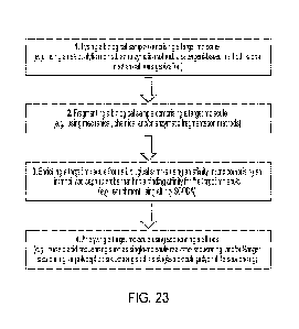
Figure 23 shows an example method for preparing a target molecule from a
biological
sample (e.g., using an automated sample preparation module of the disclosure).
Figure 24 shows a schematic diagram of a cross-section view of a cartridge 100
along the
width of channels 102, in accordance with some embodiments.
Figure 25 shows sequencing data output from DNA libraries generated with
automated
end-to-end (DNA extraction-to-finished library) sample preparation using a
sample preparation
device of the disclosure compared to libraries generated from manually
extracted and purified
DNA.
Figures 26A-26B show sequencing data output from a DNA library generated with
automated end-to-end (DNA extraction-to-finished library) sample preparation
using a sample
preparation device of the disclosure compared to DNA libraries derived from
samples that were
size selected using commercial and manual methods.
- 6 -
CA 03159363 2022-04-27
WO 2021/086945
PCT/US2020/057708
DETAILED DESCRIPTION OF INVENTION
Throughout the following description specific details are set forth in order
to provide a
more thorough understanding to persons skilled in the art. However, well known
elements may
not have been shown or described in detail to avoid unnecessarily obscuring
the disclosure.
Accordingly, the description and drawings are to be regarded in an
illustrative, rather than a
restrictive, sense.
As used herein, the term "differentially modified" means two molecules of the
same kind
that have been chemically modified in different ways. Non-limiting examples of
differentially
modified molecules include: a protein or a nucleic acid that has been
methylated is differentially
modified as compared with the unmethylated molecule; a nucleic acid that is
hypermethylated or
hypomethylated (e.g. as may occur in cancerous or precancerous cells) is
differentially modified
as compared with the nucleic acid in a healthy cell; a histone that is
acetylated is differentially
modified as compared with the non-acetylated histone; and the like.
In some embodiments, molecules that are differentially modified are identical
to one
another except for the presence of a chemical modification on one of the
molecules. In some
embodiments, molecules that are differentially modified are very similar to
one another, but not
identical. For example, where the molecules are nucleic acids or proteins, one
of the
biomolecules may share at least 95%, at least 96%, at least 97%, at least 98%,
or at least 99%
sequence identity with the differentially modified molecule.
SCODA
SCODA can involve providing a time-varying driving field component that
applies forces
to particles in some medium in combination with a time-varying mobility-
altering field
component that affects the mobility of the particles in the medium. The
mobility-altering field
component is correlated with the driving field component so as to provide a
time-averaged net
motion of the particles. SCODA may be applied to cause selected particles to
move toward a
focus area.
In one embodiment of SCODA based purification, described herein as
electrophoretic
SCODA, time varying electric fields both provide a periodic driving force and
alter the drag (or
equivalently the mobility) of molecules that have a mobility in the medium
that depends on
electric field strength, e.g. nucleic acid molecules. For example, DNA
molecules have a mobility
that depends on the magnitude of an applied electric field while migrating
through a sieving
matrix such as agarose or polyacrylamide. By applying an appropriate periodic
electric field
pattern to a separation matrix (e.g. an agarose or polyacrylamide gel) a
convergent velocity field
can be generated for all molecules in the gel whose mobility depends on
electric field. The field
dependent mobility is a result of the interaction between a repeating DNA
molecule and the
- 7 -
CA 03159363 2022-04-27
WO 2021/086945
PCT/US2020/057708
sieving matrix, and is a general feature of charged molecules with high
conformational entropy
and high charge to mass ratios moving through sieving matrices. Since nucleic
acids tend to be
the only molecules present in most biological samples that have both a high
conformational
entropy and a high charge to mass ratio, electrophoretic SCODA based
purification has been
shown to be highly selective for nucleic acids.
The ability to detect specific biomolecules in a sample has wide application
in the field of
diagnosing and treating disease. Research continues to reveal a number of
biomarkers that are
associated with various disorders. Exemplary biomarkers include genetic
mutations, the presence
or absence of a specific protein, the elevated or reduced expression of a
specific protein, elevated
or reduced levels of a specific RNA, the presence of modified biomolecules,
and the like.
Biomarkers and methods for detecting biomarkers are potentially useful in the
diagnosis,
prognosis, and monitoring the treatment of various disorders, including
cancer, disease,
infection, organ failure and the like.
The differential modification of biomolecules in vivo is an important feature
of many
biological processes, including development and disease progression. One
example of
differential modification is DNA methylation. DNA methylation involves the
addition of a
methyl group to a nucleic acid. For example a methyl group may be added at the
5' position on
the pyrimidine ring in cytosine. Methylation of cytosine in CpG islands is
commonly used in
eukaryotes for long term regulation of gene expression. Aberrant methylation
patterns have been
implicated in many human diseases including cancer. DNA can also be methylated
at the 6
nitrogen of the adenine purine ring.
Chemical modification of molecules, for example by methylation, acetylation or
other
chemical alteration, may alter the binding affinity of a target molecule and
an agent that binds
the target molecule. For example, methylation of cytosine residues increases
the binding energy
of hybridization relative to unmethylated duplexes. The effect is small.
Previous studies report an
increase in duplex melting temperature of around 0.7 C per methylation site
in a 16 nucleotide
sequence when comparing duplexes with both strands unmethylated to duplexes
with both
strands methylated.
Affinity SCODA
SCODAphoresis is a method for injecting biomolecules into a gel, and
preferentially
concentrating nucleic acids or other biomolecules of interest in the center of
the gel. SCODA
may be applied, for example, to DNA, RNA and other molecules. Following
concentration, the
purified molecules may be removed for further analysis. In one specific
embodiment of
SCODAphoresis¨affinity SCODA¨binding sites which are specific to the
biomolecules of
interest may be immobilized in the gel. In doing so one may be able generate a
non-linear motive
- 8 -
CA 03159363 2022-04-27
WO 2021/086945
PCT/US2020/057708
response to an electric field for biomolecules that bind to the specific
binding sites. One specific
application of affinity SCODA is sequence-specific SCODA. Here
oligonucleotides may be
immobilized in the gel allowing for the concentration of only DNA molecules
which are
complementary to the bound oligonucleotides. All other DNA molecules which are
not
complementary may focus weakly or not at all and can therefore be washed off
the gel by the
application of a small DC bias.
SCODA based transport is a general technique for moving particles through a
medium by
first applying a time-varying forcing (i.e. driving) field to induce periodic
motion of the particles
and superimposing on this forcing field a time-varying perturbing field that
periodically alters
the drag (or equivalently the mobility) of the particles (i.e. a mobility-
altering field). Application
of the mobility-altering field is coordinated with application of the forcing
field such that the
particles will move further during one part of the forcing cycle than in other
parts of the forcing
cycle. Specifically, the drift velocity v(t) of a particle driven by an
external force F(t) with a time
varying drag coefficient (t) (i.e. a varying mobility) is given by:
F
v(t) [11
(4)
If the external force and drag coefficient vary periodically such that
F(t) = f1 sin(w) [2]
and,
I (wi +
[3=
1
C.'3 ci
then the drift velocity averaged over one complete cycle is given by:
E)
.00 c(ç) [4]
By varying the drag (i.e. mobility) of the particle at the same frequency as
the external
applied force, a net drift can be induced with zero time-averaged forcing. The
result of equation
[4] can be used with an appropriate choice of driving force and drag
coefficients that vary in time
and space to generate a convergent velocity field in one or two dimensions. A
time varying drag
coefficient and driving force can be utilized in a real system to specifically
concentrate (i.e.
preferentially focus) only certain molecules, even where the differences
between the target
molecule and one or more non-target molecules are very small, e.g. molecules
that are
differentially modified at one or more locations, or nucleic acids differing
in sequence at one or
more bases.
- 9 -
CA 03159363 2022-04-27
WO 2021/086945
PCT/US2020/057708
One Dimensional SCODA Concentration
By combining a spatially uniform driving force that varies periodically in
time, with a
drag coefficient that varies in time as well as in space it is possible to
generate a convergent
velocity field in one dimension. Consider the case of a charged particle with
mobility t moving
under the influence of an applied electric field E; its velocity will be given
by:
t) = zGr, t) [51
If electric field is varied periodically in time such that:
E(x, t) = Et) (c.ot ) [61
and a linear mobility gradient is provided within the domain -
L.ltoreq.x.ltoreq.L that varies at the
same period:
t) (kx) (b) E7i
where k can be thought of as the amplitude of the mobility variation, SCODA-
based separation
of particles can be achieved.
There are a number of ways to establish a mobility gradient for charged
molecules
moving in solution under the influence of an applied external electric field.
For example, a time-
varying electric field may be provided as described above, a temperature
gradient may be
established, a pH gradient may be established, a light gradient may be
established for molecules
which undergo a conformational change in the presence or absence of light, or
the like.
With the mobility gradient of equation [7] provided, the velocity becomes:
v(x,t) [po (kx) iin(wt 0)1[ Ebsin(wol [81
Taking the time average of this velocity over one complete cycle yields the
following
drift velocity:
t) = 1)(.17 fyit [9]
CO8(01:) [101
2
This velocity field has an equilibrium point at x=0 and can be made convergent
or
.. divergent depending on the sign of kE0 cos*. For positive values the
velocity field is divergent
and for negative values it is convergent. Figure 1 shows the velocity plotted
as a function of x for
the case where kE0 cos()<O. The arrows in Figure 1 indicate the direction of
drift. All particles
between -L and +L will drift towards the zero velocity point at x=0. Outside
of the domain the
time averaged velocity is zero as the mobility is only altered between -L and
+L.
- 10 -
CA 03159363 2022-04-27
WO 2021/086945
PCT/US2020/057708
In the embodiment illustrated in Figure 1, the velocity takes on a positive
value for
negative values of x and vice versa for positive values of x resulting in all
particles within the
domain drifting towards x=0 where the velocity is zero.
Two Dimensional SCODA
To extend the result of equation [10] to two dimensions, in some embodiments a
rotating
electric field is used as the driving field and a rotating mobility gradient
is established:
I ,,- Ea cos(wt) ---= .E,-, sin(u..4)3 [11]
0 0c, + k Ex cos(t #-4) ¨ y siTi(wt + 0) [121
As in the one dimensional case {right arrow over (v)}= {right arrow over
(E)}, and the
same integration as in equation [9] can be performed to yield the time
averaged drift velocity in
two dimensions:
f ?:Er.
G ---- ' .............. ,. E(I (.4.)h(tAjt) (i.to l' k(X COS(Wt + (5) '"'
V Sin (Wt ''F' 0)))/it [131
' '1 ' ' '. = ' .... . . '
A( /: 4)
7 N
I N - -Ett tri.D.V4)t)(1.40 -7 A.(z c..(4..a -r 0) ¨ y i-
iii(t. + 0)))6f.t [141
7 0
This results in the following expression for the drift velocity:
E01,7 ' .
( (z cos (0) ¨ y ..4iiri (0)) '',.- + (.1; sin 1:49 + y co) 40 : )
s si. [15]
2 , =
=
Rewriting in polar coordinates and simplifying yields:
==A Zo:Yr i
_____________________________________________________________ cios(0)i. +
siii(r,)6) [16]
9 , = . ¨ ,
...
.
This result highlights a number of aspects of SCODA in two dimensions. It
shows that
despite the zero time averaged forcing there will be non-zero drift everywhere
except at the point
in the medium where r=0. It shows that the nature of the drift depends on the
relative phase, (I), of
the two signals, with the strength of focusing (the radial, {circumflex over
(r)}, term) being
proportional to the cosine of the phase lag between the electric driving field
oscillations and the
mobility oscillations. For a 0 phase angle there is a purely focusing
velocity field with net drift
directed towards the center of the domain. For a 180 phase angle the velocity
field is pure de-
focusing with net drift away from the center of the gel. And for phase angles
of 90 and 270 the
velocity field is purely rotational. At intermediate angles the resultant
velocity field will be a
combination of both rotational and focusing components. To achieve efficient
focusing, in some
embodiments the phase difference between the driving force and the mobility
variation is as
small as possible.
- 11 -
CA 03159363 2022-04-27
WO 2021/086945
PCT/US2020/057708
Generation of a Time Varyink Mobility Field
Previous applications of SCODA based concentration used the fact that the
mobility of
DNA in a sieving matrix such as agarose or polyacrylamide depends on the
magnitude of the
applied electric field. In some applications, the molecules of interest may
have a mobility that
does not normally depend strongly on electric field, such as short nucleic
acids less than 200
bases, biomolecules other than nucleic acids (e.g. proteins or polypeptides),
or the like. In some
applications, it may be desired to purify only a subset of the nucleic acids
in a sample, for
example purifying or detecting a single gene from a sample of genomic DNA or
purifying or
detecting a chemically modified molecule (e.g. methylated DNA) from a
differentially modified
molecule having the same basic structure (e.g. unmethylated DNA having the
same sequence), or
the like.
SCODA-based purification of molecules that do not have a mobility that is
strongly
dependent on electrical field strength (i.e. which have a low value of k based
on variations in
electric field strength) can be achieved by using a SCODA matrix that has an
affinity to the
molecule to be concentrated. An affinity matrix can be generated by
immobilizing an agent with
a binding affinity to the target molecule (i.e. a probe) in a medium. Using
such a matrix,
operating conditions can be selected where the target molecules transiently
bind to the affinity
matrix with the effect of reducing the overall mobility of the target molecule
as it migrates
through the affinity matrix. The strength of these transient interactions is
varied over time, which
has the effect of altering the mobility of the target molecule of interest.
SCODA drift can
therefore be generated. This technique is called affinity SCODA, and is
generally applicable to
any target molecule that has an affinity to a matrix.
Affinity SCODA can selectively enrich for nucleic acids based on sequence
content, with
single nucleotide resolution. In addition, affinity S CODA can lead to
different values of k for
molecules with identical DNA sequences but subtly different chemical
modifications such as
methylation. Affinity SCODA can therefore be used to enrich for (i.e.
preferentially focus)
molecules that differ subtly in binding energy to a given probe, and
specifically can be used to
enrich for methylated, unmethylated, hypermethylated, or hypomethylated
sequences.
Exemplary media that can be used to carry out affinity SCODA include any
medium
through which the molecules of interest can move, and in which an affinity
agent can be
immobilized to provide an affinity matrix. In some embodiments, polymeric gels
including
polyacrylamide gels, agarose gels, and the like are used. In some embodiments,
microfabricated/microfluidic matrices are used.
- 12 -
CA 03159363 2022-04-27
WO 2021/086945
PCT/US2020/057708
Exemplary operating conditions that can be varied to provide a mobility
altering field
include temperature, pH, salinity, concentration of denaturants, concentration
of catalysts,
application of an electric field to physically pull duplexes apart, or the
like.
Exemplary affinity agents that can be immobilized on the matrix to provide an
affinity
matrix include nucleic acids having a sequence complementary to a nucleic acid
sequence of
interest, proteins having different binding affinities for differentially
modified molecules,
antibodies specific for modified or unmodified molecules, nucleic acid
aptamers specific for
modified or unmodified molecules, other molecules or chemical agents that
preferentially bind to
modified or unmodified molecules, or the like.
The affinity agent may be immobilized within the medium in any suitable
manner. For
example where the affinity agent is an oligonucleotide, the oligonucleotide
may be covalently
bound to the medium, acrydite modified oligonucleotides may be incorporated
directly into a
polyacrylamide gel, the oligonucleotide may be covalently bound to a bead or
other construct
that is physically entrained within the medium, or the like.
Where the affinity agent is a protein or antibody, in some embodiments the
protein may
be physically entrained within the medium (e.g. the protein may be cast
directly into an agarose
or polyacrylamide gel), covalently coupled to the medium (e.g. through use of
cyanogen bromide
to couple the protein to an agarose gel), covalently coupled to a bead that is
entrained within the
medium, bound to a second affinity agent that is directly coupled to the
medium or to beads
entrained within the medium (e.g. a hexahistidine tag bound to NTA-agarose),
or the like.
Where the affinity agent is a protein, the conditions under which the affinity
matrix is
prepared and the conditions under which the sample is loaded should be
controlled so as not to
denature the protein (e.g. the temperature should be maintained below a level
that would be
likely to denature the protein, and the concentration of any denaturing agents
in the sample or in
the buffer used to prepare the medium or conduct SCODA focusing should be
maintained below
a level that would be likely to denature the protein).
Where the affinity agent is a small molecule that interacts with the molecule
of interest,
the affinity agent may be covalently coupled to the medium in any suitable
manner.
One exemplary embodiment of affinity SCODA is sequence-specific SCODA. In
sequence specific SCODA, the target molecule is or comprises a nucleic acid
molecule having a
specific sequence, and the affinity matrix contains immobilized
oligonucleotide probes that are
complementary to the target nucleic acid molecule. In some embodiments,
sequence specific
SCODA is used both to separate a specific nucleic acid sequence from a sample,
and to separate
and/or detect whether that specific nucleic acid sequence is differentially
modified within the
sample. In some such embodiments, affinity SCODA is conducted under conditions
such that
- 13 -
CA 03159363 2022-04-27
WO 2021/086945
PCT/US2020/057708
both the nucleic acid sequence and the differentially modified nucleic acid
sequence are
concentrated by the application of SCODA fields. Contaminating molecules,
including nucleic
acids having undesired sequences, can be washed out of the affinity matrix
during SCODA
focusing. A washing bias can then be applied in conjunction with SCODA
focusing fields to
separate the differentially modified nucleic acid molecules as described below
by preferentially
focusing the molecule with a higher binding energy to the immobilized
oligonucleotide probe.
Mobility of a Target in an Affinity Matrix
The interactions between a target and immobilized probes in an affinity matrix
can be
described by first order reaction kinetics:
+ P ______________________________ 11%, = P 1171
Here [T] is the target, [P] the immobilized probe, [T. P] the probe-target
duplex, kf is the
forward (hybridization) reaction rate, and kr the reverse (dissociation)
reaction rate. Since the
mobility of the target is zero while it is bound to the matrix, the effective
mobility of the target
will be reduced by the relative amount of target that is immobilized on the
matrix:
LI
kcea-,-, taw
FU [18]
T-..P
where to is the mobility of the unbound target. Using reasonable estimates for
the forward
reaction rate6 and an immobilized probe concentration that is significantly
higher than the
concentration of the unbound target, it can be assumed that the time constant
for hybridization
should be significantly less than one second. If the period of the mobility-
altering field is
maintained at longer than one second, it can be assumed for the purposes of
analysis that the
binding kinetics are fast and equation [17] can be rewritten in terms of
reaction rates:
k47 [P] ......................... ¨ kr [T, - Pj [19]
- ________________________ [201
k1 [P]
Inserting [20] into equation [18] and simplifying yields:
= ito ------------------- 1211
1 + [Pj
From this result it can be seen that the mobility can be altered by modifying
either the
forward or reverse reaction rates. Modification of the forward or reverse
reaction rates can be
achieved in a number of different ways, for example by adjusting the
temperature, salinity, pH,
concentration of denaturants, concentration of catalysts, by physically
pulling duplexes apart
with an external electric field, or the like. In one exemplary embodiment
described in greater
- 14 -
CA 03159363 2022-04-27
WO 2021/086945
PCT/US2020/057708
detail below, the mechanism for modifying the mobility of target molecules
moving through an
affinity matrix is control of the matrix temperature.
To facilitate analysis, it is helpful to make some simplifying assumptions.
First it is
assumed that there are a large number of immobilized probes relative to target
molecules. So
long as this is true, then even if a large fraction of the target molecules
become bound to the
probes the concentration of free probes, [P], will not change much and it can
be assumed that [P]
is constant. Also, it is assumed that the forward reaction rate kf does not
depend on temperature.
This not strictly true, as the forward reaction rate does depend on
temperature. Secondary
structure in the immobilized probe or in the target molecule can result in a
temperature
dependent forward reaction rate. However, in embodiments operating at a
temperature range near
the duplex melting temperature the reverse reaction rate has an exponential
dependence on
temperature and the forward reaction rate has a much weaker temperature
dependence, varying
by about 30% over a range of 30 C around the melting temperature. It is
additionally assumed
that the target sequence is free of any significant secondary structure.
Although this final
assumption would not always be correct, it simplifies this initial analysis.
To determine the temperature dependence of the reverse reaction rate, an
Arrhenius
model for unbinding kinetics is assumed. This assumption is justified by
recent work in nanopore
force spectroscopy.
k, ¨ 4c4-a7 1221
Here A is an empirically derived constant, AG is the probe-target binding
energy, kb is
the Boltzmann constant, and T the temperature. Inserting this into [21],
rewriting the free energy
AG as AH-TAS, and collecting constant terms allows the mobility to be
rewritten as:
Ikctiv==== ........................... 1stizrTa,S 1231
I 4- tieTT
Equation [23] describes a sigmoidal mobility temperature dependence. The shape
of this
curve is shown in Figure 2. At low temperature the mobility is nearly zero.
This is the regime
where thermal excitations are insufficient to drive target molecules off of
the affinity matrix. At
high temperature target molecules move at the unbound mobility, where the
thermal energy is
greater than the binding energy. Between these two extremes there exists a
temperature range
within which a small change in temperature results in a large change in
mobility. This is the
operating regime for embodiments of affinity SCODA that utilize temperature as
the mobility
altering parameter.
- 15 -
CA 03159363 2022-04-27
WO 2021/086945
PCT/US2020/057708
In embodiments of affinity SCODA used to separate nucleic acids based on
sequence, i.e.
sequence-specific SCODA, this temperature range tends to lie near the melting
temperature of
the probe-target duplex. Equations [10] and [16] state that the speed of
concentration is
proportional to k, which is a measure of how much the mobility changes during
one SCODA
cycle. Operating near the probe-target duplex melting temperature, where the
slope of the
mobility versus temperature curve is steepest, maximizes k for a given
temperature swing during
a SCODA cycle in embodiments where temperature is used as the mobility
altering parameter.
In some embodiments, affinity SCODA may be conducted within a temperature
gradient
that has a maximum amplitude during application of SCODA focusing fields that
varies within
about 20 C, within about 10 C, within about 5 C, or within about 2 C
of the melting
temperature of the target molecule and the affinity agent.
It is possible to describe affinity SCODA in one dimension by replacing the
time
dependent mobility of equation [7] with the temperature dependent mobility of
equation [23] and
a time dependent temperature:
T(a7 t) = T ¨) Sin Pt -1- (15) [241
L.
Here, the temperature oscillates around Tni, the probe target melting
temperature, and Ta
is the maximum amplitude of the temperature oscillations at x= L. To get an
analytical
expression for the drift velocity, vd=1..tE, as a function of temperature, a
Taylor expansion of
equation [23] is performed around Tm:
-617+1'as
.11 :
NiTectivf: ¨ It (2-yi ______________________________________________________
(T ¨ T.,õ) + 0¶:1` ¨ Zõ.)2.) [251
(1_ +
which can be rewritten as:
ileffectiw =4.117m.) aCr 0((T ¨1;02) [26]
Here the first term in the Taylor expansion has been collected into the
constant .alpha..
Combining [24] and [26] into an expression for the mobility yields an
expression similar to [7]:
F __ Sin(a)t [27]
Equation [27] can be used to determine the time averaged drift velocity for
both the one
dimensional and two dimensional cases by simply replacing k with:
H4.T.IR
77:\
[28]
L / ______________ L )
'44,1121= tl- "iXs
=
- 16 -
CA 03159363 2022-04-27
WO 2021/086945
PCT/US2020/057708
The drift velocity is then given by:
t) = E70 cos(0) [29]
2L
in one dimension, and:
Eoo. r
v . _______________________________ m8(0' sinkb)6)
[30]
2 t:
in two dimensions. This result shows that if a two dimensional gel
functionalized with
immobilized probes (i.e. an affinity matrix), then by combining a rotating
temperature gradient
with a rotating dipole electric field, all target molecules should be forced
towards a central region
in the gel, thus concentrating a target molecule that binds to the immobilized
probes.
Molecular Separation with Affinity SCODA
In some embodiments, affinity SCODA is used to separate two similar molecules
(e.g.
the same molecule that has been differentially modified, or which differs in
sequence at only one
or a few locations) with differing binding affinities for the immobilized
probe. Beginning with
two molecular species, each with a different binding energy to the immobilized
probes, these two
molecular species can be separated by superimposing a washing motive force
over the driving
and mobility altering fields used to produce SCODA focusing, to provide net
motion of
molecules that have a lesser binding affinity for the immobilized probe (i.e.
the molecules that
have a higher binding affinity for the immobilized probe are preferentially
focused during the
application of the SCODA focusing fields). In some embodiments, the washing
force is a small
applied DC force, referred to herein as a DC bias.
In the one dimensional case when a small DC force is applied as a washing or
bias force,
the electric field becomes:
Pi Gr. t ¨ Er) sin (an' ) [31]
where Eb is the applied DC bias. The final drift velocity has superimposed on
the SCODA
focusing velocity a constant velocity proportional to the strength of the bias
field:
a71..r
E00t;(0) p(T)E [321
2 L
This drift velocity will tend to move the final focus location either to the
left or right
depending on the direction of bias. The amount by which this bias moves a
focus off center
depends on the strength of the interaction between the target and probe
molecules. The
differential strength of the target-probe interaction can therefore serve as a
mechanism to enable
molecular separation of two highly similar species.
- 17 -
CA 03159363 2022-04-27
WO 2021/086945
PCT/US2020/057708
Consider two molecules that have different binding affinities for an
immobilized probe.
Reducing the probe-target binding energy, AG in equation [23], will serve to
shift the mobility
versus temperature curve to the left on the temperature scale as shown in
Figure 3. The mobility
of the high binding energy target is shown by the curve on the right, while
the mobility of the
low binding energy target is shown by the curve on the left.
If the SCODA system in this exemplary embodiment is operated at the optimal
focusing
temperature for the higher binding energy molecule, Tn, in Figure 3, then the
mobility of the
lower binding energy molecule will be higher and will have weaker temperature
dependence. In
terms of equation [32] the molecule with lower binding energy will have a
larger value of i.t(Tn,)
and a smaller value of a. This means that a lower binding energy molecule will
have a lower
SCODA drift velocity and a higher velocity under DC bias, resulting in a
different final focus
location than the high binding energy molecule as illustrated in Figure 4.
Figure 4 shows the effect of an applied DC bias on molecules with two
different binding
energies for the immobilized probe according to one embodiment. The solid
curve represents the
drift velocity of a target molecule with a lower binding energy to the bound
probes than the
molecules represented by the dashed curve. The final focus location is the
point where the drift
velocity is equal to zero. The molecules represented by the solid curve have
both a lower
SCODA drift velocity and a higher DC velocity compared to the molecules
represented by the
dashed curve. When SCODA focusing is combined with a DC bias the lower binding
energy
molecules will focus further away from the unbiased focus at x=0, resulting in
two separate foci,
one for each molecular species. The final focus position for the high binding
energy molecule is
indicated by reference numeral 30. The final focus position for the low
binding energy molecule
is indicated by reference numeral 32.
The two dimensional case is the same as the one dimensional case, the
superimposed
velocity from the applied washing bias moves the final focus spot off center
in the direction of
the washing bias.
In some embodiments, if the difference in binding energies between the
molecules to be
separated is large enough and a sufficiently high washing bias is applied, the
low binding energy
molecules can be washed off of the affinity matrix while molecules with higher
binding energy
are retained in the affinity matrix, and may be captured at a focus location
within the affinity
matrix (i.e. preferentially focused) through the application of SCODA focusing
fields.
Generation of a Time Varvink Temperature Gradient
Embodiments of affinity SCODA that use variations in temperature as the
mobility
altering field may use a periodically varying temperature gradient to produce
a convergent
velocity field. A periodically varying temperature gradient may be provided in
any suitable
- 18 -
CA 03159363 2022-04-27
WO 2021/086945
PCT/US2020/057708
manner, for example by the use of heaters or thermoelectric chillers to
periodically heat and cool
regions of the medium, the use of radiative heating to periodically heat
regions of the medium,
the application of light or radiation to periodically heat regions of the
medium, Joule heating
using the application of an electric field to the medium, or the like.
A periodically varying temperature gradient can be established in any suitable
manner so
that particles that are spaced a farther distance from a desired focus spot
experience greater
mobility (i.e. are at a higher temperature and hence travel farther) during
times of application of
the driving field towards the desired focus spot than during times of
application of the driving
field away from the desired focus spot. In some embodiments, the temperature
gradient is rotated
to produce a convergent velocity field in conjunction with the application of
a time-varying
driving force.
In some embodiments, Joule heating using an electric field is used to provide
a
temperature gradient. In some embodiments, the electric field used to provide
Joule heating to
provide a temperature gradient is the same as the electric field that provides
the driving field. In
some embodiments, the magnitude of the electric field applied is selected to
produce a desired
temperature gradient within an affinity matrix.
In some embodiments, a spatial temperature gradient is generated using a
quadrupole
electric field to provide the Joule heating. In some such embodiments, a two
dimensional gel
with four electrodes is provided. Voltages are applied to the four electrodes
such that the electric
field in the gel is non-uniform, containing regions of high electric field
(and consequently high
temperature) and low electric field. The electric field is oriented such that
the regions of high
electric field tend to push negatively charged molecules towards the center of
the gel, while
regions of low electric field tend to push such molecules away from the center
of the gel. In
some such embodiments, the electric field that provides the temperature
gradient through Joule
heating is also the electric field that applies a driving force to molecules
in the gel.
An example of such a field pattern is illustrated in Figure 5. Voltages
applied at
electrodes A, B, C and D in Figure 5 are -V, 0, 0, and 0 respectively. Arrows
represent the
velocity of a negatively charged analyte molecule. Color intensity represents
electric field
strength. The regions near electrode A have a high electric field strength,
which decreases
towards electrode C. The high field regions near electrode A tend to push
negatively charged
molecules towards the center of the gel, while the lower field regions near
electrodes B, C, and D
tend to push negatively charged molecules away from the center of the gel. In
embodiments in
which the electric field also provides the temperature gradient, the affinity
matrix will become
hotter in regions of higher field strength due to Joule heating. Hence,
regions of high electric
field strength will coincide with regions of higher temperature and thus
higher mobility.
- 19 -
CA 03159363 2022-04-27
WO 2021/086945
PCT/US2020/057708
Accordingly, molecules in the high electric field regions near electrode A
will tend to move a
greater distance toward the center of the gel, while molecules in the lower
electric field regions
near electrodes B, C, and D have a lower mobility (are at a cooler
temperature) and will move
only a short distance away from the center of the gel.
In some embodiments, the electric field pattern of Figure 5 is rotated in a
stepwise
manner by rotating the voltage pattern around the four electrodes such that
the time averaged
electric field is zero as shown in Figure 6. This rotating field will result
in net migration towards
the center of the gel for any molecule that is negatively charged and has a
mobility that varies
with temperature. In some embodiments, the electric field pattern is varied in
a manner other
than rotation, e.g. by sequentially shifting the voltage pattern by 180 , 90 ,
180 , and 90 , or by
randomly switching the direction of the electric field. As shown above, the
mobility of a
molecule moving through an affinity matrix depends on temperature, not
electric field strength.
The applied electric field will tend to increase the temperature of the matrix
through Joule
heating; the magnitude of the temperature rise at any given point in the
matrix will be
proportional to the square of the magnitude of the electric field.
In embodiments in which the thermal gradient is provided by Joule heating
produced by
the electric field that also provides the driving field, the oscillations in
the thermal gradient will
have the same period as the electric field oscillations. These oscillations
can drive affinity
SCODA based concentration in a two dimensional gel.
Figure 6 illustrates the stepwise rotation of the electric field leading to
focusing of
molecules whose mobility increases with temperature or electric field
according to such an
embodiment. A particle path for a negatively charged molecule is shown. After
four steps the
particle has a net displacement toward the center of the gel. Molecules that
do not experience a
change in mobility with changing temperature or electric field will experience
zero net motion in
a zero time averaged electric field.
Theoretical Predictions of Focusing- and Separation
In some embodiments, the electric field and subsequently the Joule heating
within an
affinity SCODA gel are controlled by both the voltage applied to the source
electrodes, and the
shape of the gel. Marziali et al. used superimposed rotating dipole and
quadrupole fields to drive
electrophoretic SCODA concentration. The ratio of the strength of these two
fields, the dipole to
quadrupole ratio (D/Q), has an impact on the efficiency of SCODA focusing with
a maximum at
around D/Q=4.5, however the optimum is relatively flat with the SCODA force
staying relatively
constant for values between 1.75 and 1013. One convenient choice of D/Q is 2.
With this
particular choice, only two distinct potentials need to be applied to the
source electrodes, which
can be achieved by connecting one electrode to a common voltage rail,
grounding the other
- 20 -
CA 03159363 2022-04-27
WO 2021/086945
PCT/US2020/057708
three, and rotating this pattern in a stepwise manner through the four
possible configurations as
shown in Table 1. Although analog amplifiers can be used and were used in the
examples
described herein, using a D/Q ratio of 2 allows one to use discrete MOSFET
switches, which
simplifies and reduces the required size and complexity of the power supplies.
Table 1. Voltage pattern for SCODA focusing with D/Q=2
Electrode A Electrode B Electrode C Electrode D
Step 1 -V 0 0 0
Step 2 0 -V 0 0
Step 3 0 0 -V 0
Step 4 0 0 0 -V
A starting point for a sequence specific gel geometry was the four-sided gel
geometry
used for the initial demonstration of electrophoretic SCODA. This geometry can
be defined by
two numbers, the gel width and the corner radius. The inventors started by
using a geometry that
had a width of 10 mm and a corner radius of 3 mm. An electro-thermal model of
this geometry
was implemented in COMSOL Multiphysics modeling software (COMSOL, Inc,
Burlington
Mass., USA) to estimate the electric field and temperature profiles within the
gel and establish
whether or not those field and temperature profiles could drive concentration
of a target with a
temperature dependent mobility. The model used simultaneously solves Ohm's Law
and the heat
equation within the domain, using the power density calculated from the
solution of Ohm's Law
as the source term for the heat equation and using the temperature solution
from the heat
equation to determine the temperature dependent electrical conductivity of the
electrolyte in the
gel.
To obtain an accurate estimate of the temperature profile within the gel, the
heat
conducted out of the top and bottom of the gel are modeled. Boundary
conditions and other
model parameters are illustrated in Figure 7. The thermal properties of water
and electrical
properties of 0.2 M NaCl were used. The gel cassettes are placed on an
aluminum spreader plate
that acts as a constant temperature reservoir. To model heat flow into the
spreader plate the heat
transfer coefficient of the glass bottom, given by lilt, was used. The
temperature and electric field
profiles solved by this model for a single step of the SCODA cycle are shown
in Figure 8. The
voltage applied to the four electrodes was -120 V, 0 V, 0 V, 0 V, and the
spreader plate
temperature was set to 55 C (328 K). The colour map indicates gel temperature
and the vector
field shows the relative magnitude and direction of the electric field within
the gel. Note that as
DNA is negatively charged its migration direction will be opposite to the
direction of the electric
field.
- 21 -
CA 03159363 2022-04-27
WO 2021/086945
PCT/US2020/057708
Using experimentally determined values of mobility versus temperature for a
given
molecule and the thermal model described above, it is possible to determine
the SCODA velocity
everywhere in the gel for that particular molecule by taking the time average
of the instantaneous
drift velocity integrated over one complete cycle:
i
[33]
r , 0
where i.t. is the temperature dependent mobility, E the electric field and T
the period of the
SCODA cycle. The temperature and electric field were solved for four steps in
the SCODA cycle
and coupled with the mobility function in equation [23]. In this manner, the
SCODA velocity
everywhere in the gel can be calculated. Since discrete steps are being used,
if it is assumed that
the period is long enough that the phase lag between the electric field and
temperature can be
neglected, then the integral in equation [33] becomes a sum:
iro --- 1341
F k
where the velocity is summed over all four steps in the cycle.
As an example, Figure 9 shows a vector plot of the SCODA velocity using the
experimentally determined mobility versus temperature curve for the perfect
match target shown
in Figure 11 (example described below) and the temperature and electric field
values calculated
above.
The velocity field plotted in Figure 9 shows a zero velocity point at the
geometric center
of the gel, with the velocity at all other points in the gel pointing towards
the center. Thus, target
molecules can be collected within the gel at the center of the electric field
pattern.
In embodiments that are used to separate two similar molecules based on
differences in
binding affinity for the immobilized probe, a washing force is superimposed
over the SCODA
focusing fields described above. In some embodiments, the washing force is a
DC electric field,
described herein as a DC bias. For molecules having affinity to the
immobilized probe, the
SCODA focusing force applied by the SCODA focusing fields described above will
tend to
counteract movement of a molecule caused by the washing field, i.e. the SCODA
focusing fields
will tend to exert a restoring force on the molecules and the molecules will
be preferentially
focused as compared with molecules having a smaller binding affinity.
Molecules that have a
smaller binding affinity to the immobilized probe will have a greater mobility
through the
affinity matrix, and the restoring SCODA force will be weaker. As a result,
the focus spot of
molecules with a smaller binding affinity will be shifted. In some cases, the
restoring SCODA
- 22 -
CA 03159363 2022-04-27
WO 2021/086945
PCT/US2020/057708
force will be so weak that such molecules with a smaller binding affinity will
be washed out of
the affinity matrix altogether.
In order to enrich for a specific biomolecule from a population of other
similar
biomolecules using affinity SCODA, one may operate SCODA focusing electric
fields with a
superimposed DC bias. The DC bias may move the focused molecules off center,
in such a way
that the molecules with a lower binding energy to the immobilized binding
sites move further off
center than the molecules with higher binding energies, thus causing the focus
to split into
multiple foci. For molecules with similar binding energies, this split may be
small while washing
under bias. The DC bias may be superimposed directly over the focusing fields,
or a DC field
may be time multiplexed with the focusing fields.
In one exemplary embodiment used to separate nucleic acids having similar
sequences, a
DC bias is superimposed over the voltage pattern shown in Table 1, resulting
in the voltage
pattern shown below in Table 2. In some embodiments, the DC bias is applied
alternately with
the SCODA focusing fields, i.e. the SCODA focusing fields are applied for a
period of time then
stopped, and the DC bias is applied for a period of time then stopped.
Table 2. Applied voltages for focusing under a DC bias. Shown are values for a
120 V SCODA
focusing potential superimposed over a 10 V DC bias
Electrode A Electrode B Electrode C Electrode D
Step 1 -120 5 10 5
Step 2 0 -115 10 5
Step 3 0 5 -110 5
Step 4 0 5 10 -115
The resulting velocity plots of both the perfect match and single base
mismatch targets in
the presence of the applied DC bias are shown in Figures 10A and 10B,
respectively. Electric
field and temperature were calculated using COMSOL using a spreader plate
temperature of 61
C. Velocity was calculated using equation [34] and the experimentally obtained
data fits shown
in Figure 11 (example described below). The zero velocity location of the
perfect match target
has been moved slightly off center in the direction of the bias (indicated
with a circular spot),
however the mismatch target has no zero velocity point within the gel. These
calculations show
that it is possible to completely wash a target with a smaller binding
affinity from the
immobilized probe from the gel area while capturing the target with a higher
binding affinity,
enabling selective purification, concentration and/or detection of a specific
sequence, even where
the nucleotide targets differ in sequence at only one position.
In some embodiments, the optimal combination of the driving field and the
mobility
altering field used to perform SCODA focusing where there is a maximum
difference in focusing
force between similar molecules is empirically determined by measuring the
velocity of sample
- 23 -
CA 03159363 2022-04-27
WO 2021/086945
PCT/US2020/057708
molecules through a medium as a function of the mobility varying field. For
example, in some
embodiments the mobility of a desired target molecule and a non-desired target
molecule at
various temperatures is measured in an affinity matrix as described above, and
the temperature
range at which the difference in relative mobility is greatest is selected as
the temperature range
for conducting affinity SCODA. In some embodiments, the focusing force is
proportional to the
rate at which the velocity changes with respect to the perturbing field dv/df,
where v is the
molecule velocity and f the field strength. One skilled in the art may
maximize dv/df so as to
maximize SCODA focusing and to enable fast washing of contaminants that do not
focus. To
maximally separate two similar molecules, affinity SCODA may be carried out
under conditions
such that dva/df-dvb/df (where va is the velocity of molecule a, and vb is the
velocity of molecule
b) is maximized.
In some embodiments, the strength of the electric field applied to an affinity
matrix is
calculated so that the highest temperature within the gel corresponds
approximately to the
temperature at which the difference in binding affinity between two molecules
to be separated is
highest.
In some embodiments, the temperature at which the difference in binding
affinity
between the two molecules to be separated is highest corresponds to the
temperature at which the
difference between the melting temperature of a target molecule and the
affinity agent and the
melting temperature of a non-target molecule and the affinity agent is
highest. In some
embodiments, the maximum difference between the melting temperature of a
target molecule
and the affinity agent and the melting temperature of a non-target molecule
and the affinity agent
is less than about 9.3 C, in some embodiments less than about 7.8 C, in some
embodiments less
than about 5.2 C, and in some embodiments less than about 0.7 C.
In some embodiments, the ratio of target molecules to non-target molecules
that can be
separated by affinity SCODA is any ratio from 1:1 to 1:10,000 and any value
therebetween, e.g.
1:100 or 1:1,000. In some embodiments, after conducting affinity SCODA, the
ratio of non-
target molecules relative to target molecules that is located in a focus spot
of the target molecules
has been reduced by a factor of up to 10,000 fold.
Phase Lak Induced Rotation
In some embodiments, to separate molecules with different affinities for the
immobilized
affinity agent, a DC bias is superimposed over the SCODA focusing fields as
described above. If
the separation in binding energy is great enough then the mismatched target
can be washed
entirely off of the gel. The ability to wash weakly focusing contaminating
fragments from the gel
can be affected by the phase lag induced rotation discussed above, where the
SCODA velocity of
a two dimensional system was given by:
- 24 -
CA 03159363 2022-04-27
WO 2021/086945
PCT/US2020/057708
'ilscoDA = ("SCODA (COO, sink I. = [351
where (I) is the phase lag between the electric field oscillations and the
mobility varying
oscillations. Aside from reducing the proportion of the SCODA velocity that
contributes to
concentration this result has additional implications when washing weakly
focusing
contaminants out of an affinity matrix. The rotational component will add to
the DC bias and can
result in zero or low velocity points in the gel that can significantly
increase the time required to
wash mismatched targets from the gel.
To counteract the effects of a rotational component of motion that may arise
in
embodiments in which there is a phase lag between the electric field
oscillations and the mobility
varying oscillations, the direction in which the SCODA focusing fields are
applied may be
rotated periodically. In some embodiments, the direction in which the SCODA
focusing fields
are rotated is altered once every period.
Optical Feedback
In some embodiments where one molecule of interest (the target molecule) is
concentrated in an affinity matrix while a second, similar, molecule (the non-
target molecule) is
washed off of the affinity matrix, optical feedback may be used to determine
when washing is
complete and/or to avoid running the target molecule out of the affinity
matrix.
The two foci of similar molecules may be close together geographically, and
optical
feedback may be used to ensure the molecule of interest is not washed off the
gel. For example,
using a fluorescent surrogate for the molecule of interest or the
contaminating molecules (or
both) one can monitor their respective positions while focusing under bias,
and use that
geographical information to adjust the bias ensuring that the molecule of
interest is pushed as
close to the edge of the gel as possible but not off, while the contaminating
molecule may be
removed from the gel.
In some embodiments, the molecules to be separated are differentially labeled,
e.g. with
fluorescent tags of a different color. Real-time monitoring using fluorescence
detection can be
used to determine when the non-target molecule has been washed off of the
affinity matrix, or to
determine when the foci of the target molecule and the non-target molecule are
sufficiently far
apart within the affinity matrix to allow both foci to be separately extracted
from the affinity
matrix.
In some embodiments, fluorescent surrogate molecules that focus similarly to
the target
and/or non-target molecules may be used to perform optical feedback. By using
a fluorescent
surrogate for a target molecule, a non-target molecule, or both a target
molecule and a non-target
molecule, the respective positions of the target molecule and/or the non-
target molecule can be
monitored while performing affinity focusing under a washing bias. The
location of the surrogate
- 25 -
CA 03159363 2022-04-27
WO 2021/086945
PCT/US2020/057708
molecules within the affinity matrix can be used to adjust the washing bias to
ensure that the
molecule of interest is pushed as close to the edge of the gel as possible but
not off, while the
contaminating molecule may be washed off the gel.
In some embodiments, fluorescent surrogate molecules that focus similarly to
the target
and/or non-target molecules but will not amplify in any subsequent PCR
reactions that may be
conducted can be added to a sample to be purified. The presence of the
fluorescent surrogate
molecules within the affinity matrix enables the use of optical feedback to
control SCODA
focusing conditions in real time. Fluorescence detection can be used to
visualize the position of
the fluorescent surrogate molecules in the affinity matrix. In embodiments
where the fluorescent
surrogate mimics the focusing behavior of the target molecule, the applied
washing force can be
decreased when the fluorescent surrogate approaches the edge of the affinity
matrix, to avoid
washing the target molecule out of the affinity matrix. In embodiments where
the fluorescent
surrogate mimics the focusing behavior of the non-target molecule that is to
be separated from
the target molecule, the applied washing force can be decreased or stopped
after the fluorescent
surrogate has been washed out of the affinity matrix, or alternatively when
the location of the
fluorescent surrogate approaches the edge of the affinity matrix.
Separation of Differentially Modified Molecules
In some embodiments, molecules that are identical except for the presence or
absence of
a chemical modification that alters the binding affinity of the molecule for a
probe are separated
using affinity SCODA. Some embodiments of affinity SCODA are sufficiently
sensitive to
separate two molecules that have only a small difference in binding affinity
for the immobilized
affinity agent. Examples of such molecules include differentially modified
molecules, such as
methylated and unmethylated nucleic acids, methylated or acetylated proteins,
or the like.
For example, it has been previously shown that methylation of cytosine
residues
increases the binding energy of hybridization relative to unmethylated DNA
sequences. RNA
sequences would be expected to display a similar increase in the binding
energy of hybridization
when methylated as compared with unmethylated sequences. The inventors have
shown that one
embodiment of affinity SCODA can be used to separate nucleic acid sequences
differing only by
the presence of a single methylated cytosine residue. Other chemical
modifications would be
expected to alter the binding energy of a nucleic acid and its complimentary
sequence in a
similar manner. Modification of proteins, such as through methylation, can
also alter the binding
affinity of a protein of interest with a protein, RNA or DNA aptamer,
antibody, or other
molecule that binds to the protein at or near the methylation site.
Accordingly, embodiments of
affinity SCODA can be used to separate differentially modified molecules of
interest. While the
examples herein are directed to methylation enrichment, affinity SCODA can
also be applied to
- 26 -
CA 03159363 2022-04-27
WO 2021/086945
PCT/US2020/057708
enrichment and selection of molecules with other chemical differences,
including e.g.
acetylation.
Affinity SCODA, and sequence-specific SCODA, may be used to enrich a specific
sequence of methylated DNA out of a background of methylated and unmethylated
DNA. In this
application of affinity SCODA, the strength of the SCODA focusing force may be
related to the
binding energy of the target DNA to the bound oligonucleotides. Target
molecules with a higher
binding energy may be made to focus more strongly than targets with lower
binding energy.
Methylation of DNA has previously been documented to slightly increase the
binding energy of
target DNA to its complementary sequence. Small changes in binding energy of a
complementary oligonucleotide may be exploited through affinity SCODA to
preferentially
enrich for methylated DNA. SCODA operating conditions may be chosen, for
example as
described above, such that the methylated DNA is concentrated while
unmethylated DNA of the
same sequence is washed off the gel.
Some embodiments can separate molecules with a difference in binding energy to
an
immobilized affinity agent of less than kT, the thermal excitation energy of
the target molecules.
Some embodiments can separate molecules with a difference in binding energy to
an
immobilized affinity agent of less than 0.19 kcal/mol. Some embodiments can
separate
molecules with a difference in binding energy to an immobilized affinity agent
of less than 2.6
kcal/mol. Some embodiments can separate molecules with a difference in binding
energy to an
immobilized affinity agent of less than 3.8 kcal/mol. Some embodiments can
separate molecules
that differ only by the presence of a methyl group. Some embodiments can
separate nucleic acid
sequences that differ in sequence at only one base.
Applications of Affinity SCODA
Systems and methods for separating, purifying, concentrating and/or detecting
differentially modified molecules as described above can be applied in fields
where detection of
biomarkers, specific nucleotide sequences or differentially modified molecules
is important, e.g.
epigenetics, fetal DNA detection, pathogen detection, cancer screening and
monitoring, detection
of organ failure, detection of various disease states, and the like. For
example, in some
embodiments affinity SCODA is used to separate, purify, concentrate and/or
detect differentially
methylated DNA in such fields as fetal diagnostic tests utilizing maternal
body fluids, pathogen
detection in body fluids, and biomarker detection in body fluids for detecting
cancer, organ
failure, or other disease states and for monitoring the progression or
treatment of such conditions.
In some embodiments, a sample of bodily fluid or a tissue sample is obtained
from a
subject. Cells may be lysed, genomic DNA is sheared, and the sample is
subjected to affinity
SCODA. In some embodiments, molecules concentrated using affinity SCODA are
subjected to
- 27 -
CA 03159363 2022-04-27
WO 2021/086945
PCT/US2020/057708
further analysis, e.g. DNA sequencing, digital PCR, fluorescence detection, or
the like, to assay
for the presence of a particular biomarker or nucleotide sequence. In some
embodiments, the
subject is a human.
It is known that fetal DNA is present in maternal plasma, and that
differential
methylation of maternal versus fetal DNA obtained from the maternal plasma can
be used to
screen for genetic disorders (see e.g. Poon et al., 2002, Clinical Chemistry
48:1, 35-41).
However, one problem that is difficult to overcome is discrimination between
fetal and maternal
DNA. Affinity SCODA as described above may be used to preferentially separate,
purify,
concentrate and/or detect DNA which is differentially methylated in fetal DNA
versus maternal
DNA. For example, affinity SCODA may be used to concentrate or detect DNA
which is
methylated in the fetal DNA, but not in maternal DNA, or which is methylated
in maternal DNA
but not fetal DNA. In some embodiments, a sample of maternal plasma is
obtained from a
subject and subjected to affinity SCODA using an oligonucleotide probe
directed to a sequence
of interest. The detection of two foci after the application of SCODA focusing
fields may
indicate the presence of DNA which is differentially methylated as between the
subject and the
fetus. Comparison to a reference sample from a subject that exhibits a
particular genetic disorder
may be used to determine if the fetus may be at risk of having the genetic
disorder. Further
analysis of the sample of DNA obtained through differential modification SCODA
through
conventional methods such as PCR, DNA sequencing, digital PCR, fluorescence
detection, or the
like, may be used to assess the risk that the fetus may have a genetic
disorder.
One embodiment of the present systems and methods is used to detect
abnormalities in
fetal DNA, including chromosome copy number abnormalities. Regions of
different
chromosomes that are known to be differentially methylated in fetal DNA as
opposed to
maternal DNA are concentrated using affinity SCODA to separate fetal DNA from
maternal
DNA based on the differential methylation of the fetal DNA in a maternal
plasma sample.
Further analysis of the separated fetal DNA is conducted (for example using
qPCR, DNA
sequencing, fluorescent detection, or other suitable method) to count the
number of copies from
each chromosome and determine copy number abnormalities.
Most cancers are a result of a combination of genetic changes and epigenetic
changes,
such as changes in DNA methylation (e.g. hypomethylation and/or
hypermethylation of certain
regions, see e.g. Ehrich, 2002, Oncogene 21:35, 5400-5413). Affinity SCODA can
be used to
separate, purify, concentrate and/or detect DNA sequences of interest to
screen for oncogenes
which are abnormally methylated. Embodiments of affinity SCODA are used in the
detection of
biomarkers involving DNA having a different methylation pattern in cancerous
or pre-cancerous
cells than in healthy cells. Detection of such biomarkers may be useful in
both early cancer
- 28 -
CA 03159363 2022-04-27
WO 2021/086945
PCT/US2020/057708
screening, and in the monitoring of cancer development or treatment progress.
In some
embodiments, a sample obtained from a subject, e.g. a sample of a bodily fluid
such as plasma or
a biopsy, may be processed and analyzed by differential modification SCODA
using
oligonucleotide probes directed to a sequence of interest. The presence of two
foci during the
application of SCODA fields may indicate the presence of differential
methylation at the DNA
sequence of interest. Comparison of the sample obtained from the subject with
a reference
sample (e.g. a sample from a healthy patient and/or a sample known to
originate from cancerous
or pre-cancerous tissue) can indicate whether the cells of the subject are at
risk of being
cancerous or pre-cancerous. Further analysis of the sample of DNA obtained
through differential
modification SCODA through conventional methods such as PCR, DNA sequencing,
digital
PCR, fluorescence detection, or the like, may be used to assess the risk that
the sample includes
cells that may be cancerous or pre-cancerous, to assess the progression of a
cancer, or to assess
the effectiveness of treatment.
In some embodiments, a specific nucleotide sequence is captured in the gel
regardless of
methylation (i.e. without selecting for a particular methylation status of the
nucleic acid).
Undesired nucleotide sequences and/or other contaminants may be washed off the
gel while the
specific nucleotide sequence remains bound by oligonucleotide probes
immobilized within the
separation medium. Then, differential methylation SCODA is used to focus the
methylated
version of the sequence while electrically washing the unmethylated sequence
toward a buffer
chamber or another gel where it can then be recovered. In some embodiments,
the unmethylated
sequence could be preferentially extracted.
In some embodiments, biomolecules in blood related to disease states or
infection are
selectively concentrated using affinity SCODA. In some embodiments, the
biomolecules are
unique nucleic acids with sequence or chemical differences that render them
useful biomarkers
of disease states or infection. Following such concentration, the biomarkers
can be detected
using PCR, sequencing, or similar means. In some embodiments, a sample of
bodily fluid or
tissue is obtained from a subject, cells are lysed, genomic DNA is sheared,
and affinity SCODA
is performed using oligonucleotide probes that are complimentary to a sequence
of interest.
Affinity SCODA is used to detect the presence of differentially methylated
populations of the
nucleic acid sequence of interest. The presence of differentially methylated
populations of the
target sequence of interest may indicate a likelihood that the subject suffers
from a particular
disease state or an infection.
In some embodiments, the focusing pattern of the target nucleic acid produced
by affinity
SCODA from a subject is compared with the focusing pattern of the target
nucleic acid produced
by affinity SCODA from one or more reference samples (e.g. an equivalent
sample obtained
- 29 -
CA 03159363 2022-04-27
WO 2021/086945
PCT/US2020/057708
from a healthy subject, and/or an equivalent sample obtained from a subject
known to be
suffering from a particular disease). Similarities between the focusing
pattern produced by the
sample obtained from the subject and a reference sample obtained from a
subject known to be
suffering from a particular disease indicate a likelihood that the subject is
suffering from the
same disease. Differences between the focusing pattern produced from the
sample obtained from
the subject and a reference sample obtained from a healthy subject indicate a
likelihood that the
subject may be suffering from a disease. Differences in the focusing pattern
produced from the
sample obtained from the subject and a reference sample obtained from a
healthy subject may
indicate the presence of a differential modification or a mutation in the
subject as compared with
the healthy subject.
Use of Multiple Affinity Agents to Capture Multiple Target Molecules
In some embodiments, affinity SCODA is used to separate, purify, concentrate
and/or
detect more than one sequence per sample. The examples described herein
demonstrate that it is
possible to concentrate target DNA at probe concentrations as low as 1 tM, as
well as with
probe concentrations as high as 100 tM. In some embodiments, multiplexed
concentration is be
performed by immobilizing a plurality of different affinity agents in the
medium to provide an
affinity matrix. In some embodiments, at least two different affinity agents
are immobilized
within a medium to separate, purify, concentrate and/or detect at least two
different target
molecules. In some embodiments, each one of the affinity agents is an
oligonucleotide probe
with a different sequence. In some embodiments, anywhere between 2 and 100
different
oligonucleotide probes are immobilized within a medium to provide an affinity
matrix, and
anywhere between 2 and 100 different target molecules are separated, purified,
concentrated
and/or detect simultaneously in a single affinity gel. Each one of the target
molecules may be
labeled with a different tag to facilitate detection, for example each one of
the target molecules
could be labeled with a different color of fluorescent tag.
In some embodiments where the binding energy between each of the two or more
affinity
agents and the two or more target molecules differs, the two or more target
molecules may be
differentially separated within the affinity matrix by the application of
SCODA focusing fields at
an appropriate temperature. In some embodiments, a first target molecule with
a lower melting
temperature for its corresponding affinity agent may be preferentially
separated from a second
target molecule with a relatively higher melting temperature for its
corresponding affinity agent.
In some such embodiments, the first molecule is preferentially concentrated by
conducting
SCODA focusing at a temperature that is sufficiently low that a second target
molecule with a
relatively higher melting temperature for its corresponding affinity agent
does not focus
efficiently (i.e. a temperature at which the mobility of the second target
molecule within the
- 30 -
CA 03159363 2022-04-27
WO 2021/086945
PCT/US2020/057708
affinity matrix is relatively low), but sufficiently high to enable efficient
focusing of the first
molecule. In some such embodiments, the first and second molecules are
differentially separated
through the application of a washing bias, e.g. a DC bias, at a temperature
that is sufficiently low
that the second target molecule is not displaced or is displaced only slowly
by the washing bias,
but sufficiently high that the first target molecule is displaced or is
displaced at a higher velocity
by the washing bias.
Apparatus for Performink Affinity SCODA
In some embodiments, affinity SCODA is performed on an electrophoresis
apparatus
comprising a region for containing the affinity matrix, buffer reservoirs,
power supplies capable
of delivering large enough voltages and currents to cause the desired effect,
precise temperature
control of the SCODA medium (which is a gel in some embodiments), and a two
color
fluorescence imaging system for the monitoring of two different molecules in
the SCODA
medium.
Sample Preparation Process
In some aspects, the disclosure provides processes for preparing a sample,
e.g., for
detection and/or analysis. In some embodiments, a process described herein may
be used to
identify properties or characteristics of a sample, including the identity or
sequence (e.g.,
nucleotide sequence or amino acid sequence) of one or more target molecules in
the sample. In
some embodiments, a process may include one or more sample transformation
steps, such as
sample lysis, sample purification, sample fragmentation, purification of a
fragmented sample,
library preparation (e.g., nucleic acid library preparation), purification of
a library preparation,
sample enrichment (e.g., using affinity SCODA), and/or detection/analysis of a
target molecule.
In some embodiments, a sample may be a purified sample, a cell lysate, a
single-cell, a
population of cells, or a tissue. In some embodiments, a sample is any
biological sample. In
some embodiments, a sample (e.g., a biological sample) is a blood, saliva,
sputum, feces, urine
or buccal swab sample. In some embodiments, a biological sample is from a
human, a non-
human primate, a rodent, a dog, a cat, a horse, or any other mammal. In some
embodiments, a
biological sample is from a bacterial cell culture (e.g., an E. coli bacterial
cell culture). A
bacterial cell culture may comprise gram positive bacterial cells and/or gram
negative bacterial
cells. In some embodiments, a sample is a purified sample of nucleic acids or
proteins that have
been previously extracted via user-developed methods from metagenomic samples
or
environmental samples. A blood sample may be a freshly drawn blood sample from
a subject
(e.g., a human subject) or a dried blood sample (e.g., preserved on solid
media (e.g., Guthrie
cards)). A blood sample may comprise whole blood, serum, plasma, red blood
cells, and/or white
blood cells.
-31 -
CA 03159363 2022-04-27
WO 2021/086945
PCT/US2020/057708
In some embodiments, a sample (e.g., a sample comprising cells or tissue), may
be lysed
(e.g., disrupted, degraded and/or otherwise digested) in a process in
accordance with the instant
disclosure. In some embodiments, a sample comprising cells or tissue is lysed
using any one of
known physical or chemical methodologies to release a target molecule (e.g., a
target nucleic
acid or a target protein) from said cells or tissues. In some embodiments, a
sample may be lysed
using an electrolytic method, an enzymatic method, a detergent-based method,
and/or
mechanical homogenization. In some embodiments, a sample (e.g., complex
tissues, gram
positive or gram negative bacteria) may require multiple lysis methods
performed in series. In
some embodiments, if a sample does not comprise cells or tissue (e.g., a
sample comprising
purified nucleic acids), a lysis step may be omitted. In some embodiments,
lysis of a sample is
performed to isolate target nucleic acid(s). In some embodiments, lysis of a
sample is performed
to isolate target protein(s). In some embodiments, a lysis method further
includes use of a mill to
grind a sample, sonication, surface acoustic waves (SAW), freeze-thaw cycles,
heating, addition
of detergents, addition of protein degradants (e.g., enzymes such as
hydrolases or proteases),
and/or addition of cell wall digesting enzymes (e.g., lysozyme or zymolase).
Exemplary
detergents (e.g., non-ionic detergents) for lysis include polyoxyethylene
fatty alcohol ethers,
polyoxyethylene alkylphenyl ethers, polyoxyethylene-polyoxypropylene block
copolymers,
polysorbates and alkylphenol ethoxylates, preferably nonylphenol ethoxylates,
alkylglucosides
and/or polyoxyethylene alkyl phenyl ethers. In some embodiments, lysis methods
involve
heating a sample for at least 1-30 min, 1-25 min, 5-25 min, 5-20 min, 10-30
min, 5-10 min, 10-
20 min, or at least 5 min at a desired temperature (e.g., at least 60 C, at
least 70 C, at least 80
C, at least 90 C, or at least 95 C).
In some embodiments, a sample (e.g., a sample comprising a target nucleic acid
or a
target protein) may be purified, e.g., following lysis, in a process in
accordance with the instant
disclosure. In some embodiments, a sample may be purified using chromatography
(e.g., affinity
chromatography that selectively binds the sample) or electrophoresis. In some
embodiments, a
sample may be purified in the presence of precipitating agents. In some
embodiments, after a
purification step or method, a sample may be washed and/or released from a
purification matrix
(e.g., affinity chromatography matrix) using an elution buffer. In some
embodiments, a
purification step or method may comprise the use of a reversibly switchable
polymer, such as an
electroactive polymer. In some embodiments, a sample may be purified by
electrophoretic
passage of a sample through a porous matrix (e.g., cellulose acetate, agarose,
acrylamide).
In some embodiments, a sample (e.g., a sample comprising a target nucleic acid
or a
target protein) may be fragmented in a process in accordance with the instant
disclosure. In
some embodiments, a nucleic acid sample may be fragmented to produce small (<1
kilobase)
- 32 -
CA 03159363 2022-04-27
WO 2021/086945
PCT/US2020/057708
fragments for sequence specific identification to large (up to 10+ kilobases)
fragments for long
read sequencing applications. Fragmentation of nucleic acids or proteins may,
in some
embodiments, be accomplished using mechanical (e.g., fluidic shearing),
chemical (e.g., iron
(Fe+) cleavage) and/or enzymatic (e.g., restriction enzymes, tagmentation
using transposases)
methods. In some embodiments, a protein sample may be fragmented to produce
peptide
fragments of any length. Fragmentation of proteins may, in some embodiments,
be
accomplished using chemical and/or enzymatic (e.g., proteolytic enzymes such
as trypsin)
methods. In some embodiments, mean fragment length may be controlled by
reaction time,
temperature, and concentration of sample and/or enzymes (e.g., restriction
enzymes,
transposases). In some embodiments, a nucleic acid may be fragmented by
tagmentation such
that the nucleic acid is simultaneously fragmented and labeled with a
fluorescent molecule (e.g.,
a fluorophore). In some embodiments, a fragmented sample may be subjected to a
round of
purification (e.g., chromatography or electrophoresis) to remove small and/or
undesired
fragments as well as residual payload, chemicals and/or enzymes (e.g.,
transposases) used during
the fragmentation step. For example, a fragmented sample (e.g., sample
comprising nucleic
acids) may be purified from an enzyme (e.g., a transposase), wherein the
purification comprises
denaturing the enzyme (e.g., by a combination of heat, chemical (e.g. SDS),
and enzymatic (e.g.
proteinase K) processes).
In some embodiments, a sample comprising a target nucleic acid may be used to
generate
a nucleic acid library for subsequent analysis (e.g., genomic sequencing) in a
process in
accordance with the instant disclosure. A nucleic acid library may be a linear
library or a
circular library. In some embodiments, nucleic acids of a circular library may
comprise elements
that allow for downstream linearization (e.g., endonuclease restriction sites,
incorporation of
uracil). In some embodiments, a nucleic acid library may be purified (e.g.,
using
chromatography, e.g., affinity chromatography), or electrophoresis.
In some embodiments, a library of nucleic acids (e.g., linear nucleic acids)
is prepared
using end-repair, a process wherein a combination of enzymes (e.g., Taq DNA
Ligase,
Endonuclease IV, Bst DNA Polymerase, Fpg, Uracil-DNA Glycosylase, T4
Endonuclease V
and/or Endonuclease VIII) extend the 3' end of the nucleic acids, generating a
complement to the
5' payload, and repairing any abasic sites or nicks in the nucleic acids. In
some embodiments, a
library of linear nucleic acids is prepared using a self-priming hairpin
adaptor, a process which
may obviate the need to anneal a unique sequencing primer to an individual
nucleic acid
fragment primer prior to formation of a polymerase complex. Following end-
repair, a library of
nucleic acids (e.g., linear nucleic acids) may be purified using solid-phase
adsorption with
subsequent elution into a fresh buffer, using passage of the nucleic acids
through a size-selective
- 33 -
CA 03159363 2022-04-27
WO 2021/086945
PCT/US2020/057708
matrix (e.g., agarose gel). The size-selective matrix may be used to remove
nucleic acid
fragments that are smaller than the size of the target nucleic acids.
In some embodiments, a sample (e.g., a sample comprising a target nucleic acid
or a
target protein) may be enriched for a target molecule in a process in
accordance with the instant
disclosure. In some embodiments, a sample is enriched for a target molecule
using an
electropheretic method. In some embodiments, a sample is enriched for a target
molecule using
affinity SCODA. In some embodiments, a sample is enriched for a target
molecule using field
inversion gel electrophoresis (FIGE). In some embodiments, a sample is
enriched for a target
molecule using pulsed field gel electrophoresis (PFGE). In some embodiments,
the matrix used
during enrichment (e.g., a porous media, electrophoretic polymer gel)
comprises immobilized
affinity agents (also known as 'immobilized capture probes') that bind to
target molecule present
in the sample. In some embodiments, a matrix used during enrichment comprises
1, 2, 3, 4, 5, or
more unique immobilized capture probes, each of which binds to a unique target
molecule and/or
bind to the same target molecule with different binding affinities.
In some embodiments, an immobilized capture probe is an oligonucleotide
capture probe
that hybridizes to a target nucleic acid. In some embodiments, an
oligonucleotide capture probe
is at least 50%, 60%, 70%, 80%, 90% 95%, or 100% complementary to a target
nucleic acid. In
some embodiments, a single oligonucleotide capture probe may be used to enrich
a plurality of
related target nucleic acids (e.g., 2, 3, 4, 5, 6, 7, 8, 9, 10, 20, 30, 40,
50, or more related target
nucleic acids) that share at least 50%, 60%, 70%, 80%, 90% 95%, or 99%
sequence identity.
Enrichment of a plurality of related target nucleic acids may allow for the
generation of a
metagenomic library. In some embodiments, an oligonucleotide capture probe may
enable
differential enrichment of related target nucleic acids. In some embodiments,
an oligonucleotide
capture probe may enable enrichment of a target nucleic acid relative to a
nucleic acid of
identical sequence that differs in its modification state (e.g., single
nucleotide polymorphism,
methylation state, acetylation state). In some embodiments, an oligonucleotide
capture probe is
used to enrich human genomic DNA for a specific gene of interest (e.g., HLA).
A specific gene
of interest may be a gene that is relevant to a specific disease state or
disorder. In some
embodiments, an oligonucleotide capture probe is used to enrich nucleic
acid(s) of a
metagenomic sample.
In some embodiments, for the purposes of enriching nucleic acid target
molecules with a
length of 0.5-2 kilobases, oligonucleotide capture probes may be covalently
immobilized in an
acrylamide matrix using a 5' Acrydite moiety. In some embodiments, for the
purposes of
enriching larger nucleic acid target molecules (e.g., with a length of >2
kilobases),
oligonucleotide capture probes may be immobilized in an agarose matrix. In
some
- 34 -
CA 03159363 2022-04-27
WO 2021/086945
PCT/US2020/057708
embodiments, oligonucleotide capture probes may be immobilized in an agarose
matrix using
thiol-epoxide chemistries (e.g., by covalently attached thiol-modified
oligonucleotides to
crosslinked agarose beads). Oligonucleotide capture probes linked to agarose
beads can be
combined and solidified within standard agarose matrices (e.g., at the same
agarose percentage).
In some embodiments, enrichment of nucleic acids using methods described
herein (e.g.,
enrichment using SCODA) produces nucleic acid target molecules that comprise a
length of
about 0.5 kilobases (kb), about 1 kb, about 1.5 kb, about 2 kb, about 3 kb,
about 4 kb, about 5 kb,
about 6 kb, about 7 kb, about 8 kb, about 9 kb, about 10 kb, about 12 kb,
about 15 kb, about 20
kb, or more. In some embodiments, enrichment of nucleic acids using methods
described herein
.. (e.g., enrichment using SCODA) produces nucleic acid target molecules that
comprise a length
of about 0.5-2 kb, 0.5-5 kb, 1-2 kb, 1-3 kb, 1-4 kb, 1-5 kb, 1-10 kb, 2-10 kb,
2-5 kb, 5-10 kb, 5-
kb, 5-20 kb, 5-25 kb, 10-15 kb, 10-20 kb, or 10-25 kb.
In some embodiments, an immobilized capture probe is a protein capture probe
(e.g., an
aptamer or an antibody) that binds to a target protein or peptide fragment. In
some
15 .. embodiments, a protein capture probe binds to a target protein or
peptide fragment with a
binding affinity of 10-9 to 10-8 M, 10-8 to 10-7 M, 10-7 to 10-6 M, 10-6 to 10-
5 M, 10-5 to 104 M, 10-
4 to 10-3 M, or 10-3 to 10-2 M. In some embodiments, the binding affinity is
in the picomolar to
nanomolar range (e.g., between about 10-12 and about 10-9 M). In some
embodiments, the
binding affinity is in the nanomolar to micromolar range (e.g., between about
10-9 and about 10-6
M). In some embodiments, the binding affinity is in the micromolar to
millimolar range (e.g.,
between about 10-6 and about 10-3 M). In some embodiments, the binding
affinity is in the
picomolar to micromolar range (e.g., between about 10-12 and about 10-6 M). In
some
embodiments, the binding affinity is in the nanomolar to millimolar range
(e.g., between about
10-9 and about 10-3 M). In some embodiments, a single protein capture probe
may be used to
enrich a plurality of related target proteins that share at least 50%, 60%,
70%, 80%, 90% 95%, or
99% sequence identity. In some embodiments, a single protein capture probe may
be used to
enrich a plurality of related target proteins (e.g., 2, 3, 4, 5, 6, 7, 8, 9,
10, 20, 30, 40, 50, or more
related target proteins) that share at least 50%, 60%, 70%, 80%, 90% 95%, or
99% sequence
homology. Enrichment of a plurality of related target proteins may allow for
the generation of a
metaproteomics library. In some embodiments, a protein capture probe may
enable differential
enrichment of related target proteins.
In some embodiments, multiple capture probes (e.g., populations of multiple
capture
probe types, e.g., that bind to deterministic target molecules of infectious
agents such as
adenovirus, staphylococcus, pneumonia, or tuberculosis) may be immobilized in
an enrichment
- 35 -
CA 03159363 2022-04-27
WO 2021/086945
PCT/US2020/057708
matrix. Application of a sample to an enrichment matrix with multiple
deterministic capture
probes may result in diagnosis of a disease or condition (e.g., presence of an
infectious agent).
In some embodiments, a target molecule or related target molecules may be
released from
the enrichment matrix after removal of non-target molecules, in a process in
accordance with the
instant disclosure. In some embodiments, a target molecule may be released
from the
enrichment matrix by increasing the temperature of the enrichment matrix.
Adjusting the
temperature of the matrix further influences migration rate as increased
temperatures provide a
higher capture probe stringency, requiring greater binding affinities between
the target molecule
and the capture probe. In some embodiments, when enriching related target
molecules, the
matrix temperature may be gradually increased in a step-wise manner in order
to release and
isolate target molecules in steps of ever-increasing homology. In some
embodiments,
temperature is increased by about 5%, 10%, 15%, 20%, 25%, 30%, 40%, 50%, or
more in each
step or over a period of time (e.g., 1-10 min, 1-5 min, or 4-8 min). In some
embodiments,
temperature is increased by 5%-10%, 5-15%, 5%-20%, 5%-25%, 5%-30%, 5%-40%, 5%-
50%,
10%-25%, 20%-30%, 30%-40%, 35%-50%, or 40%-70% in each step or over a period
of time
(e.g., 1-10 min, 1-5 min, or 4-8 min). In some embodiments, temperature is
increased by about 1
C, 2 C, 3 C, 4 C, 5 C, 6 C, 7 C, 8 C, 9 C, or 10 C in each step or
over a period of time
(e.g., 1-10 min, 1-5 min, or 4-8 min). In some embodiments, temperature is
increased by 1-10
C, 1-5 C, 2-5 C, 2-10 C, 3-8 C, 4-9 C, or 5-10 C in each step or over a
period of time
(e.g., 1-10 min, 1-5 min, or 4-8 min). This may allow for the sequencing of
target proteins or
target nucleic acids that are increasingly distant in their relation to an
initial reference target
molecule, enabling discovery of novel proteins (e.g., enzymes) or functions
(e.g., enzymatic
function or gene function). In some embodiments, when using multiple capture
probes (e.g.,
multiple deterministic capture probes), the matrix temperature may be
increased in a step-wise or
gradient fashion, permitting temperature-dependent release of different target
molecules and
resulting in generation of a series of barcoded release bands that represent
the presence or
absence of control and target molecules.
Enrichment of a sample (e.g., a sample comprising a target nucleic acid or a
target
protein) allows for a reduction in the total volume of the sample. For
example, in some
embodiments, the total volume of a sample is reduced after enrichment by at
least 10%, at least
20%, at least 30%, at least 40%, at least 50%, at least 60%, at least 70%, at
least 80%, at least
90%, at least 100%, or at least 120%. In some embodiments, the total volume of
a sample is
reduced after enrichment from 1-20 mL initial volume to 100-1000 i.tt final
volume, from 1-5
mL initial volume to 100-1000 i.tt final volume, from 100-1000 i.tt initial
volume to 25-100 i.tt
final volume, from 100-500 i.tt initial volume to 10-100 i.tt final volume, or
from 50-200 i.tt
- 36 -
CA 03159363 2022-04-27
WO 2021/086945
PCT/US2020/057708
initial volume to 1-25 i.it final volume. For example, in some embodiments,
the final volume of
a sample after enrichment is 10-100 t.L, 10-50 t.L, 10-25 t.L, 20-100 t.L, 20-
50 t.L, 25-100 t.L,
25-250 i.tt, 25-1000 i.tt, 100-1000 i.tt, 100-500 i.tt, 100-250 i.tt, 200-1000
i.tt, 200-500 i.tt,
200-750 i.tt, 500-1000 i.tt, 500-1500 i.tt, 500-750 i.tt, 1-5 mL, 1-10 mL, 1-2
mL, 1-3 mL, or 1-4
mL.
In some embodiments, a target molecule or target molecules may be detected
after
enrichment and subsequent release to enable analysis of said target
molecule(s) and its upstream
sample, in a process in accordance with the instant disclosure. In some
embodiments, a target
nucleic acid may be detected using gene sequencing, absorbance, fluorescence,
electrical
conductivity, capacitance, surface plasmon resonance, hybrid capture,
antibodies, direct labeling
of the nucleic acid (e.g., end-labeling, labeled tagmentation payloads), non-
specific labeling with
intercalating dyes (e.g., ethidium bromide, SYBR dyes), or any other known
methodology for
nucleic acid detection. In some embodiments, a target protein or peptide
fragment may be
detected using absorbance, fluorescence, mass spectroscopy, amino acid
sequencing, or any
other known methodology for protein or peptide detection.
Sample Preparation Devices and Modules
Devices or modules including apparatuses, cartridges (e.g., comprising
channels (e.g.,
microfluidic channels)), and/or pumps (e.g., peristaltic pumps) for use in a
process of preparing a
sample for analysis are generally provided. Devices can be used in accordance
with the instant
disclosure to enable capture, concentration, manipulation, and/or detection of
a target molecule
from a biological sample. In some embodiments, devices and related methods are
provided for
automated processing of a sample to produce material for next generation
sequencing and/or
other downstream analytical techniques. Devices and related methods may be
used for
performing chemical and/or biological reactions, including reactions for
nucleic acid and/or
protein processing in accordance with sample preparation or sample analysis
processes described
elsewhere herein.
In some embodiments, a sample preparation device or module is positioned to
deliver or
transfer to a sequencing module or device a target molecule or a plurality of
target molecules
(e.g., target nucleic acids or target proteins). In some embodiments, a sample
preparation device
or module is connected directly to (e.g., physically attached to) or
indirectly to a sequencing
device or module.
In some embodiments, a sample preparation device or module is used to prepare
a sample
for diagnostic purposes. In some embodiments, a sample preparation device that
is used to
prepare a sample for diagnostic purposes is positioned to deliver or transfer
to a diagnostic
module or diagnostic device a target molecule or a plurality of molecules
(e.g., target nucleic
- 37 -
CA 03159363 2022-04-27
WO 2021/086945
PCT/US2020/057708
acids or target proteins). In some embodiments, a sample preparation device or
module is
connected directly to (e.g., physically attached to) or indirectly to a
diagnostic device.
In some embodiments, a device comprises a cartridge housing that is configured
to
receive one or more cartridges (e.g., configured to receive one cartridge at a
time). In some
embodiments, a cartridge comprises one or more reservoirs or reaction vessels
configured to
receive a fluid and/or contain one or more reagents used in a sample
preparation process. In
some embodiments, a cartridge comprises one or more channels (e.g.,
microfluidic channels)
configured to contain and/or transport a fluid (e.g., a fluid comprising one
or more reagents) used
in a sample preparation process. Reagents include buffers, enzymatic reagents,
polymer
matrices, capture reagents, size-specific selection reagents, sequence-
specific selection reagents,
and/or purification reagents. Additional reagents for use in a sample
preparation process are
described elsewhere herein.
In some embodiments, a cartridge includes one or more stored reagents (e.g.,
of a liquid
or lyophilized form suitable for reconstitution to a liquid form). The stored
reagents of a
cartridge include reagents suitable for carrying out a desired process and/or
reagents suitable for
processing a desired sample type. In some embodiments, a cartridge is a single-
use cartridge
(e.g., a disposable cartridge) or a multiple-use cartridge (e.g., a reusable
cartridge). In some
embodiments, a cartridge is configured to receive a user-supplied sample. The
user-supplied
sample may be added to the cartridge before or after the cartridge is received
by the device, e.g.,
manually by the user or in an automated process. In some embodiments, a
cartridge is a sample
preparation cartridge. In some embodiments, a sample preparation cartridge is
capable of
isolating or purifying a target molecule (e.g., a target nucleic acid or
target protein) from a
sample (e.g., a biological sample).
In some embodiments, a cartridge comprises an affinity matrix for enrichment
as
described herein. In some embodiments, a cartridge comprises an affinity
matrix for enrichment
using affinity SCODA, FIGE, or PFGE. In some embodiments, a cartridge
comprises an affinity
matrix comprising an immobilized affinity agent that has a binding affinity
for a target nucleic
acid or target protein.
In some embodiments, a sample preparation device of the disclosure produces
(e.g.,
enriches or purifies) target nucleic acids with an average read-length for
downstream sequencing
applications that is longer than an average read-length produced using control
methods (e.g.,
Sage BluePippin methods, manual methods (e.g., manual bead-based size
selection methods)).
In some embodiments, a sample preparation device produces target nucleic acids
with an average
read-length for sequencing that comprises at least 700, 800, 900, 1000, 1100,
1200, 1300, 1400,
1500, 1600, 1700, 1800, 1900, 2000, 2100, 2200, 2300, 2400, 2500, 2600, 2700,
2800, 2900, or
- 38 -
CA 03159363 2022-04-27
WO 2021/086945
PCT/US2020/057708
3000 nucleotides in length. In some embodiments, a sample preparation device
produces target
nucleic acids with an average read-length for sequencing that comprises 700-
3000, 1000-3000,
1000-2500, 1000-2400, 1000-2300, 1000-2200, 1000-2100, 1000-2000, 1000-1900,
1000-1800,
1000-1700, 1000-1600, 1000-1500, 1000-1400, 1000-1300, 1000-1200, 1500-3000,
1500-2500,
1500-2000, or 2000-3000 nucleotides in length.
Devices in accordance with the instant disclosure generally contain mechanical
and
electronic and/or optical components which can be used to operate a cartridge
as described
herein. In some embodiments, the device components operate to achieve and
maintain specific
temperatures on a cartridge or on specific regions of the cartridge. In some
embodiments, the
.. device components operate to apply specific voltages for specific time
durations to electrodes of
a cartridge. In some embodiments, the device components operate to move
liquids to, from, or
between reservoirs and/or reaction vessels of a cartridge. In some
embodiments, the device
components operate to move liquids through channel(s) of a cartridge, e.g.,
to, from, or between
reservoirs and/or reaction vessels of a cartridge. In some embodiments, the
device components
.. move liquids via a peristaltic pumping mechanism (e.g., apparatus) that
interacts with an
elastomeric, reagent-specific reservoir or reaction vessel of a cartridge. In
some embodiments,
the device components move liquids via a peristaltic pumping mechanism (e.g.,
apparatus) that is
configured to interact with an elastomeric component (e.g., surface layer
comprising an
elastomer) associated with a channel of a cartridge to pump fluid through the
channel. Device
components can include computer resources, for example, to drive a user
interface where sample
information can be entered, specific processes can be selected, and run
results can be reported.
In some embodiments, a cartridge is capable of handling small-volume fluids
(e.g., 1-10
i.tt, 2-10 i.tt, 4-10 i.tt, 5-10 i.tt, 1-8 i.tt, or 1-6 i.tt fluid). In some
embodiments, the sequencing
cartridge is physically embedded or associated with a sample preparation
device or module (e.g.,
.. to allow for a prepared sample to be delivered to a reaction mixture for
sequencing. In some
embodiments, a sequencing cartridge that is physically embedded or associated
with a sample
preparation device or module comprises microfluidic channels that have fluid
interfaces in the
form of face sealing gaskets or conical press fits (e.g., Luer fittings). In
some embodiments,
fluid interfaces can then be broken after delivery of the prepared sample in
order to physically
separate the sequencing cartridge from the sample preparation device or
module.
The following non-limiting example is meant to illustrate aspects of the
devices,
methods, and compositions described herein. The use of a sample preparation
device or module
in accordance with the instant disclosure may proceed with one or more of the
following
described steps. A user may open the lid of the device and insert a cartridge
that supports the
.. desired process. The user may then add a sample, which may be combined with
a specific lysis
- 39 -
CA 03159363 2022-04-27
WO 2021/086945
PCT/US2020/057708
solution, to a sample port on the cartridge. The user may then close the
device lid, enter any
sample specific information via a touch screen interface on the device, select
any process
specific parameters (e.g., range of desired size selection, desired degree of
homology for target
molecule capture, etc.), and initiate the sample preparation process run.
Following the run, the
user may receive relevant run data (e.g., confirmation of successful
completion of the run, run
specific metrics, etc.), as well as process specific information (e.g., amount
of sample generated,
presence or absence of specific target sequence, etc.). Data generated by the
run may be
subjected to subsequent bioinformatics analysis, which can be either local or
cloud based.
Depending on the process, a finished sample may be extracted from the
cartridge for subsequent
use (e.g., genomic sequencing, qPCR quantification, cloning, etc.). The device
may then be
opened, and the cartridge may then be removed.
In some embodiments, the sample preparation module comprises a pump. In some
embodiments, the pump is peristaltic pump. Some such pumps comprise one or
more of the
inventive components for fluid handling described herein. For example, the
pump may comprise
an apparatus and/or a cartridge. In some embodiments, the apparatus of the
pump comprises a
roller, a crank, and a rocker. In some such embodiments, the crank and the
rocker are configured
as a crank-and-rocker mechanism that is connected to the roller. The coupling
of a crank-and-
rocker mechanism with the roller of an apparatus can, in some cases, allow for
certain of the
advantages describe herein to be achieved (e.g., facile disengagement of the
apparatus from the
cartridge, well-metered stroke volumes). In certain embodiments, the cartridge
of the pump
comprises channels (e.g., microfluidic channels). In some embodiments, at
least a portion of the
channels of the cartridge have certain cross-sectional shapes and/or surface
layers that may
contribute to any of a number of advantages described herein.
One non-limiting aspect of some cartridges that may, in some cases, provide
certain
benefits is the inclusion of channels having certain cross-sectional shapes in
the cartridges. For
example, in some embodiments, the cartridge comprises v-shaped channels. One
potentially
convenient but non-limiting way to form such v-shaped channels is by molding
or machining v-
shaped grooves into the cartridge. The recognized advantages of including a v-
shaped channel
(also referred to herein as a v-groove or a channel having a substantially
triangularly-shaped
cross-section) in certain embodiments in which a roller of the apparatus
engages with the
cartridge to cause fluid flow through the channels. For example, in some
instances, a v-shaped
channel is dimensionally insensitive to the roller. In other words, in some
instances, there is no
single dimension to which the roller (e.g., a wedge shaped roller) of the
apparatus must adhere in
order to suitably engage with the v-shaped channel. In contrast, certain
conventional cross
sectional shapes of the channels, such as semi-circular, may require that the
roller have a certain
- 40 -
CA 03159363 2022-04-27
WO 2021/086945
PCT/US2020/057708
dimension (e.g., radius) in order to suitably engage with the channel (e.g.,
to create a fluidic seal
to cause a pressure differential in a peristaltic pumping process). In some
embodiments, the
inclusion of channels that are dimensionally insensitive to rollers can result
in simpler and less
expensive fabrication of hardware components and increased
configurability/flexibility.
In certain aspects, the cartridges comprise a surface layer (e.g., a flat
surface layer). One
exemplary aspect relates to potentially advantageous embodiments involving
layering a
membrane (also referred to herein as a surface layer) comprising (e.g.,
consisting essentially of)
an elastomer (e.g., silicone) above the v-groove, to produce, in effect, half
of a flexible tube.
Figure 24 depicts an exemplary cartridge 100 according to certain such
embodiments, and is
described in more detail below. Then, in some embodiments, by deforming the
surface layer
comprising an elastomer into the channel to form a pinch and by then
translating the pinch,
negative pressure can be generated on the trailing edge of the pinch which
creates suction and
positive pressure can be generated on the leading edge of the pinch, pumping
fluid in the
direction of the leading edge of the pinch. In certain embodiments, this
pumping by interfacing a
cartridge (comprising channels having a surface layer) with an apparatus
comprising a roller,
which apparatus is configured to carry out a motion of the roller that
includes engaging the roller
with a portion of the surface layer to pinch the portion of the surface layer
with the walls and/or
base of the associated channel, translating the roller along the walls and/or
base of the associated
channel in a rolling motion to translate the pinch of the surface layer
against the walls and/or
base, and/or disengaging the roller with a second portion of the surface
layer. In certain
embodiments, a crank-and-rocker mechanism is incorporated into the apparatus
to carry out this
motion of the roller.
A conventional peristaltic pump generally involves tubing having been inserted
into an
apparatus comprising rollers on a rotating carriage, such that the tubing is
always engaged with
the remainder of the apparatus as the pump functions. By contrast, in certain
embodiments,
channels in cartridges herein are linear or comprise at least one linear
portion, such that the roller
engages with a horizontal surface. In certain embodiments, the roller is
connected to a small
roller arm that is spring-loaded so that the roller can track the horizontal
surface while
continuously pinching a portion of the surface layer. Spring loading the
apparatus (e.g., a roller
arm of the apparatus) can in some cases help regulate the force applied by the
apparatus (e.g.,
roller) to the surface layer and a channel of a cartridge.
In certain embodiments, each rotation of the crank in a crank-and-rocker
mechanism
connected to the roller provides a discrete pumping volume. In certain
embodiments, it is
straightforward to park the apparatus in a disengaged position, where the
roller is disengaged
from any cartridge. In certain embodiments, forward and backward pumping
motions are fairly
- 41 -
CA 03159363 2022-04-27
WO 2021/086945
PCT/US2020/057708
symmetrical as provided by apparatuses described herein, such that a similar
amount of force
(torque) (e.g., within 10%) is required for forward and backward pumping
motions.
In certain embodiments, it may be advantageous to, for a particular size of
apparatus,
have a relatively high crank radius (e.g., greater than or equal to 2 mm,
optionally including
associated linkages). Consequently, it may, in certain embodiments, also be
advantageous to
have a relatively high stroke length (e.g., greater than or equal to 10 mm) to
engage with an
associated cartridge. Having relatively high crank radius and stroke length,
in certain
embodiments, ensures no mechanical interference between the apparatus and the
cartridge when
moving components of the apparatus relative to the cartridge.
In certain embodiments, having v-shaped grooves advantageously allows for
utilization
with rollers of a variety of sizes having a wedge-shaped edge. By contrast,
for example, having a
rectangular channel rather than a v-groove results in the width of the roller
associated with the
rectangular channel needing to be more controlled and precise in relation to
the width of the
rectangular channel, and results in the forces being applied to the
rectangular channel needing to
be more precise. Similarly, the channel(s) having a semicircular cross-section
may also require
more controlled and precise dimension for the width of the associated roller.
In certain embodiments, an apparatus described herein may comprise a multi-
axis system
(e.g., robot) configured so as to move at least a portion of the apparatus in
a plurality of
dimensions (e.g., two dimensions, three dimensions). For example, the multi-
axis system may
be configured so as to move at least a portion of the apparatus to any pumping
lane location
among associated cartridge(s). For example, in certain embodiments, a carriage
herein may be
functionally connected to a multi-axis system. In certain embodiments, a
roller may be indirectly
functionally connected to a multi-axis system. In certain embodiments, an
apparatus portion,
comprising a crank-and-rocker mechanism connected to a roller, may be
functionally connected
to a multi-axis system. In certain embodiments, each pumping lane may be
addressed by
location and accessed by an apparatus described herein using a multi-axis
system.
Nucleic Acid Sequencink Process
Some aspects of the instant disclosure further involve sequencing nucleic
acids (e.g.,
deoxyribonucleic acids or ribonucleic acid). In some aspects, compositions,
devices, systems,
and techniques described herein can be used to identify a series of
nucleotides incorporated into
a nucleic acid (e.g., by detecting a time-course of incorporation of a series
of labeled
nucleotides). In some embodiments, compositions, devices, systems, and
techniques described
herein can be used to identify a series of nucleotides that are incorporated
into a template-
dependent nucleic acid sequencing reaction product synthesized by a
polymerizing enzyme (e.g.,
RNA polymerase).
- 42 -
CA 03159363 2022-04-27
WO 2021/086945
PCT/US2020/057708
Accordingly, also provided herein are methods of determining the sequence of a
target
nucleic acid. In some embodiments, the target nucleic acid is enriched (e.g.,
enriched using
electrophoretic methods, e.g., affinity SCODA) prior to determining the
sequence of the target
nucleic acid. In some embodiments, provided herein are methods of determining
the sequences
of a plurality of target nucleic acids (e.g., at least 2, 3, 4, 5, 10, 15, 20,
30, 50, or more) present in
a sample (e.g., a purified sample, a cell lysate, a single-cell, a population
of cells, or a tissue). In
some embodiments, a sample is prepared as described herein (e.g., lysed,
purified, fragmented,
and/or enriched for a target nucleic acid) prior to determining the sequence
of a target nucleic
acid or a plurality of target nucleic acids present in a sample. In some
embodiments, a target
nucleic acid is an enriched target nucleic acid (e.g., enriched using
electrophoretic methods, e.g.,
affinity SCODA).
In some embodiments, methods of sequencing comprise steps of: (i) exposing a
complex
in a target volume to one or more labeled nucleotides, the complex comprising
a target nucleic
acid or a plurality of nucleic acids present in a sample, at least one primer,
and a polymerizing
enzyme; (ii) directing one or more excitation energies, or a series of pulses
of one or more
excitation energies, towards a vicinity of the target volume; (iii) detecting
a plurality of emitted
photons from the one or more labeled nucleotides during sequential
incorporation into a nucleic
acid comprising one of the at least one primers; and (iv) identifying the
sequence of incorporated
nucleotides by determining one or more characteristics of the emitted photons.
In another aspect, the instant disclosure provides methods of sequencing
target nucleic
acids or a plurality of target nucleic acids present in a sample by sequencing
a plurality of
nucleic acid fragments, wherein the target nucleic acid(s) comprises the
fragments. In certain
embodiments, the method comprises combining a plurality of fragment sequences
to provide a
sequence or partial sequence for the parent nucleic acid (e.g., parent target
nucleic acid). In
some embodiments, the step of combining is performed by computer hardware and
software. The
methods described herein may allow for a set of related nucleic acids (e.g.,
two or more nucleic
acids present in a sample), such as an entire chromosome or genome to be
sequenced.
In some embodiments, a primer is a sequencing primer. In some embodiments, a
sequencing primer can be annealed to a nucleic acid (e.g., a target nucleic
acid) that may or may
not be immobilized to a solid support. A solid support can comprise, for
example, a sample well
(e.g., a nanoaperture, a reaction chamber) on a chip or cartridge used for
nucleic acid sequencing.
In some embodiments, a sequencing primer may be immobilized to a solid support
and
hybridization of the nucleic acid (e.g., the target nucleic acid) further
immobilizes the nucleic
acid molecule to the solid support. In some embodiments, a polymerase (e.g.,
RNA Polymerase)
is immobilized to a solid support and soluble sequencing primer and nucleic
acid are contacted to
- 43 -
CA 03159363 2022-04-27
WO 2021/086945
PCT/US2020/057708
the polymerase. In some embodiments a complex comprising a polymerase, a
nucleic acid (e.g.,
a target nucleic acid) and a primer is formed in solution and the complex is
immobilized to a
solid support (e.g., via immobilization of the polymerase, primer, and/or
target nucleic acid). In
some embodiments, none of the components are immobilized to a solid support.
For example, in
some embodiments, a complex comprising a polymerase, a target nucleic acid,
and a sequencing
primer is formed in situ and the complex is not immobilized to a solid
support.
In some embodiments, sequencing by synthesis methods can include the presence
of a
population of target nucleic acid molecules (e.g., copies of a target nucleic
acid) and/or a step of
amplification (e.g., polymerase chain reaction (PCR)) of a target nucleic acid
to achieve a
population of target nucleic acids. However, in some embodiments, sequencing
by synthesis is
used to determine the sequence of a single nucleic acid molecule in any one
reaction that is being
evaluated and nucleic acid amplification may not be required to prepare the
target nucleic acid.
In some embodiments, a plurality of single molecule sequencing reactions are
performed in
parallel (e.g., on a single chip or cartridge) according to aspects of the
instant disclosure. For
example, in some embodiments, a plurality of single molecule sequencing
reactions are each
performed in separate sample wells (e.g., nanoapertures, reaction chambers) on
a single chip or
cartridge.
In some embodiments, sequencing of a target nucleic acid molecule comprises
identifying at least two (e.g., at least 3, at least 4, at least 5, at least
6, at least 7, at least 8, at least
9, at least 10, at least 11, at least 12, at least 13, at least 14, at least
15, at least 16, at least 17, at
least 18, at least 19, at least 20, at least 25, at least 30, at least 35, at
least 40, at least 45, at least
50, at least 60, at least 70, at least 80, at least 90, at least 100, or more)
nucleotides of the target
nucleic acid. In some embodiments, the at least two nucleotides are contiguous
nucleotides. In
some embodiments, the at least two amino acids are non-contiguous nucleotides.
In some embodiments, sequencing of a target nucleic acid comprises
identification of less
than 100% (e.g., less than 99%, less than 95%, less than 90%, less than 85%,
less than 80%, less
than 75%, less than 70%, less than 65%, less than 60%, less than 55%, less
than 50%, less than
45%, less than 40%, less than 35%, less than 30%, less than 25%, less than
20%, less than 15%,
less than 10%, less than 5%, less than 1% or less) of all nucleotides in the
target nucleic acid.
For example, in some embodiments, sequencing of a target nucleic acid
comprises identification
of less than 100% of one type of nucleotide in the target nucleic acid. In
some embodiments,
sequencing of a target nucleic acid comprises identification of less than 100%
of each type of
nucleotide in the target nucleic acid.
- 44 -
CA 03159363 2022-04-27
WO 2021/086945
PCT/US2020/057708
Protein Seouencink Process
Aspects of the instant disclosure also involve methods of protein sequencing
and
identification, methods of polypeptide sequencing and identification, methods
of amino acid
identification, and compositions, systems, and devices for performing such
methods. Such
protein sequencing and identification is performed, in some embodiments, with
the same
instrument that performs sample preparation and/or genome sequencing,
described in more detail
herein. In some aspects, methods of determining the sequence of a target
protein are described.
In some embodiments, the target protein is enriched (e.g., enriched using
electrophoretic
methods, e.g., affinity SCODA) prior to determining the sequence of the target
protein. In some
aspects, methods of determining the sequences of a plurality of proteins
(e.g., at least 2, 3, 4, 5,
10, 15, 20, 30, 50, or more) present in a sample (e.g., a purified sample, a
cell lysate, a single-
cell, a population of cells, or a tissue) are described. In some embodiments,
a sample is prepared
as described herein (e.g., lysed, purified, fragmented, and/or enriched for a
target protein) prior
to determining the sequence of a target protein or a plurality of proteins
present in a sample. In
some embodiments, a target protein is an enriched target protein (e.g.,
enriched using
electrophoretic methods, e.g., affinity SCODA).
In some embodiments, the instant disclosure provides methods of sequencing
and/or
identifying an individual protein in a sample comprising a plurality of
proteins by identifying one
or more types of amino acids of a protein from the mixture. In some
embodiments, one or more
amino acids (e.g., terminal amino acids or internal amino acids) of the
protein are labeled (e.g.,
directly or indirectly, for example using a binding agent) and the relative
positions of the labeled
amino acids in the protein are determined. In some embodiments, the relative
positions of amino
acids in a protein are determined using a series of amino acid labeling and
cleavage steps. In
some embodiments, the relative position of labeled amino acids in a protein
can be determined
without removing amino acids from the protein but by translocating a labeled
protein through a
pore (e.g., a protein channel) and detecting a signal (e.g., a Forster
resonance energy transfer
(FRET) signal) from the labeled amino acid(s) during translocation through the
pore in order to
determine the relative position of the labeled amino acids in the protein
molecule.
In some embodiments, the identity of a terminal amino acid (e.g., an N-
terminal or a C-
terminal amino acid) is determined prior to the terminal amino acid being
removed and the
identity of the next amino acid at the terminal end being assessed; this
process may be repeated
until a plurality of successive amino acids in the protein are assessed. In
some embodiments,
assessing the identity of an amino acid comprises determining the type of
amino acid that is
present. In some embodiments, determining the type of amino acid comprises
determining the
actual amino acid identity (e.g., determining which of the naturally-occurring
20 amino acids an
- 45 -
CA 03159363 2022-04-27
WO 2021/086945
PCT/US2020/057708
amino acid is, e.g., using a binding agent that is specific for an individual
terminal amino acid).
However, in some embodiments, assessing the identity of a terminal amino acid
type can
comprise determining a subset of potential amino acids that can be present at
the terminus of the
protein. In some embodiments, this can be accomplished by determining that an
amino acid is
not one or more specific amino acids (i.e., and therefore could be any of the
other amino acids).
In some embodiments, this can be accomplished by determining which of a
specified subset of
amino acids (e.g., based on size, charge, hydrophobicity, binding properties)
could be at the
terminus of the protein (e.g., using a binding agent that binds to a specified
subset of two or more
terminal amino acids).
In some embodiments, a protein or polypeptide can be digested into a plurality
of smaller
proteins or polypeptides and sequence information can be obtained from one or
more of these
smaller proteins or polypeptides (e.g., using a method that involves
sequentially assessing a
terminal amino acid of a protein and removing that amino acid to expose the
next amino acid at
the terminus).
In some embodiments, a protein is sequenced from its amino (N) terminus. In
some
embodiments, a protein is sequenced from its carboxy (C) terminus. In some
embodiments, a
first terminus (e.g., N or C terminus) of a protein is immobilized and the
other terminus (e.g., the
C or N terminus) is sequenced as described herein.
As used herein, sequencing a protein refers to determining sequence
information for a
.. protein. In some embodiments, this can involve determining the identity of
each sequential
amino acid for a portion (or all) of the protein. In some embodiments, this
can involve
determining the identity of a fragment (e.g., a fragment of a target protein
or a fragment of a
sample comprising a plurality of proteins). In some embodiments, this can
involve assessing the
identity of a subset of amino acids within the protein (e.g., and determining
the relative position
of one or more amino acid types without determining the identity of each amino
acid in the
protein). In some embodiments amino acid content information can be obtained
from a protein
without directly determining the relative position of different types of amino
acids in the protein.
The amino acid content alone may be used to infer the identity of the protein
that is present (e.g.,
by comparing the amino acid content to a database of protein information and
determining which
protein(s) have the same amino acid content).
In some embodiments, sequence information for a plurality of protein fragments
obtained
from a target protein or sample comprising a plurality of proteins (e.g., via
enzymatic and/or
chemical cleavage) can be analyzed to reconstruct or infer the sequence of the
target protein or
plurality of proteins present in the sample. Accordingly, in some embodiments,
the one or more
types of amino acids are identified by detecting luminescence of one or more
labeled affinity
- 46 -
CA 03159363 2022-04-27
WO 2021/086945
PCT/US2020/057708
reagents that selectively bind the one or more types of amino acids. In some
embodiments, the
one or more types of amino acids are identified by detecting luminescence of a
labeled protein.
In some embodiments, the instant disclosure provides compositions, devices,
and
methods for sequencing a protein by identifying a series of amino acids that
are present at a
terminus of a protein over time (e.g., by iterative detection and cleavage of
amino acids at the
terminus). In yet other embodiments, the instant disclosure provides
compositions, devices, and
methods for sequencing a protein by identifying labeled amino content of the
protein and
comparing to a reference sequence database.
In some embodiments, the instant disclosure provides compositions, devices,
and
methods for sequencing a protein by sequencing a plurality of fragments of the
protein. In some
embodiments, sequencing a protein comprises combining sequence information for
a plurality of
protein fragments to identify and/or determine a sequence for the protein. In
some embodiments,
combining sequence information may be performed by computer hardware and
software. The
methods described herein may allow for a set of related proteins, such as an
entire proteome of
an organism, to be sequenced. In some embodiments, a plurality of single
molecule sequencing
reactions are performed in parallel (e.g., on a single chip or cartridge)
according to aspects of the
instant disclosure. For example, in some embodiments, a plurality of single
molecule sequencing
reactions are each performed in separate sample wells on a single chip or
cartridge.
In some embodiments, methods provided herein may be used for the sequencing
and
identification of an individual protein in a sample comprising a plurality of
proteins. In some
embodiments, the instant disclosure provides methods of uniquely identifying
an individual
protein in a sample comprising a plurality of proteins. In some embodiments,
an individual
protein is detected in a mixed sample by determining a partial amino acid
sequence of the
protein. In some embodiments, the partial amino acid sequence of the protein
is within a
contiguous stretch of approximately 5-50, 10-50, 25-50, 25-100, or 50-100
amino acids.
Without wishing to be bound by any particular theory, it is expected that most
human
proteins can be identified using incomplete sequence information with
reference to proteomic
databases. For example, simple modeling of the human proteome has shown that
approximately
98% of proteins can be uniquely identified by detecting just four types of
amino acids within a
stretch of 6 to 40 amino acids (see, e.g., Swaminathan, et al. PLoS Comput
Biol. 2015,
11(2):e1004080; and Yao, et al. Phys. Biol. 2015, 12(5):055003). Therefore, a
sample
comprising a plurality of proteins can be fragmented (e.g., chemically
degraded, enzymatically
degraded) into short protein fragments of approximately 6 to 40 amino acids,
and sequencing of
this protein-based library would reveal the identity and abundance of each of
the proteins present
in the original sample. Compositions and methods for selective amino acid
labeling and
- 47 -
CA 03159363 2022-04-27
WO 2021/086945
PCT/US2020/057708
identifying polypeptides by determining partial sequence information are
described in in detail in
U.S. Pat. Application No. 15/510,962, filed September 15, 2015, entitled
"SINGLE
MOLECULE PEPTIDE SEQUENCING," which is incorporated herein by reference in its
entirety.
Sequencing in accordance with the instant disclosure, in some aspects, may
involve
immobilizing a protein (e.g., a target protein) on a surface of a substrate
(e.g., of a solid support,
for example a chip or cartridge, for example in a sequencing device or module
as described
herein). In some embodiments, a protein may be immobilized on a surface of a
sample well
(e.g., on a bottom surface of a sample well) on a substrate. In some
embodiments, the N-
terminal amino acid of the protein is immobilized (e.g., attached to the
surface). In some
embodiments, the C-terminal amino acid of the protein is immobilized (e.g.,
attached to the
surface). In some embodiments, one or more non-terminal amino acids are
immobilized (e.g.,
attached to the surface). The immobilized amino acid(s) can be attached using
any suitable
covalent or non-covalent linkage, for example as described in this disclosure.
In some
embodiments, a plurality of proteins are attached to a plurality of sample
wells (e.g., with one
protein attached to a surface, for example a bottom surface, of each sample
well), for example in
an array of sample wells on a substrate.
In some embodiments, the identity of a terminal amino acid (e.g., an N-
terminal or a C-
terminal amino acid) is determined, then the terminal amino acid is removed,
and the identity of
the next amino acid at the terminal end is determined. This process may be
repeated until a
plurality of successive amino acids in the protein are determined. In some
embodiments,
determining the identity of an amino acid comprises determining the type of
amino acid that is
present. In some embodiments, determining the type of amino acid comprises
determining the
actual amino acid identity, for example by determining which of the naturally-
occurring 20
amino acids is the terminal amino acid is (e.g., using a binding agent that is
specific for an
individual terminal amino acid). In some embodiments, the type of amino acid
is selected from
alanine, arginine, asparagine, aspartic acid, cysteine, glutamine, glutamic
acid, glycine, histidine,
isoleucine, leucine, lysine, methionine, phenylalanine, proline,
selenocysteine, serine, threonine,
tryptophan, tyrosine, and valine. In some embodiments, determining the
identity of a terminal
amino acid type can comprise determining a subset of potential amino acids
that can be present
at the terminus of the protein. In some embodiments, this can be accomplished
by determining
that an amino acid is not one or more specific amino acids (and therefore
could be any of the
other amino acids). In some embodiments, this can be accomplished by
determining which of a
specified subset of amino acids (e.g., based on size, charge, hydrophobicity,
post-translational
- 48 -
CA 03159363 2022-04-27
WO 2021/086945
PCT/US2020/057708
modification, binding properties) could be at the terminus of the protein
(e.g., using a binding
agent that binds to a specified subset of two or more terminal amino acids).
In some embodiments, assessing the identity of a terminal amino acid type
comprises
determining that an amino acid comprises a post-translational modification.
Non-limiting
.. examples of post-translational modifications include acetylation, ADP-
ribosylation, caspase
cleavage, citrullination, formylation, N-linked glycosylation, 0-linked
glycosylation,
hydroxylation, methylation, myristoylation, neddylation, nitration, oxidation,
palmitoylation,
phosphorylation, prenylation, S-nitrosylation, sulfation, sumoylation, and
ubiquitination.
In some embodiments, a protein or protein can be digested into a plurality of
smaller
.. proteins and sequence information can be obtained from one or more of these
smaller proteins
(e.g., using a method that involves sequentially assessing a terminal amino
acid of a protein and
removing that amino acid to expose the next amino acid at the terminus).
In some embodiments, sequencing of a protein molecule comprises identifying at
least
two (e.g., at least 3, at least 4, at least 5, at least 6, at least 7, at
least 8, at least 9, at least 10, at
least 11, at least 12, at least 13, at least 14, at least 15, at least 16, at
least 17, at least 18, at least
19, at least 20, at least 25, at least 30, at least 35, at least 40, at least
45, at least 50, at least 60, at
least 70, at least 80, at least 90, at least 100, or more) amino acids in the
protein molecule. In
some embodiments, the at least two amino acids are contiguous amino acids. In
some
embodiments, the at least two amino acids are non-contiguous amino acids.
In some embodiments, sequencing of a protein molecule comprises identification
of less
than 100% (e.g., less than 99%, less than 95%, less than 90%, less than 85%,
less than 80%, less
than 75%, less than 70%, less than 65%, less than 60%, less than 55%, less
than 50%, less than
45%, less than 40%, less than 35%, less than 30%, less than 25%, less than
20%, less than 15%,
less than 10%, less than 5%, less than 1% or less) of all amino acids in the
protein molecule. For
example, in some embodiments, sequencing of a protein molecule comprises
identification of
less than 100% of one type of amino acid in the protein molecule (e.g.,
identification of a portion
of all amino acids of one type in the protein molecule). In some embodiments,
sequencing of a
protein molecule comprises identification of less than 100% of each type of
amino acid in the
protein molecule.
In some embodiments, sequencing of a protein molecule comprises identification
of at
least 1, at least 5, at least 10, at least 15, at least 20, at least 25, at
least 30, at least 35, at least 40,
at least 45, at least 50, at least 55, at least 60, at least 65, at least 70,
at least 75, at least 80, at
least 85, at least 90, at least 95, at least 100 or more types of amino acids
in the protein.
- 49 -
CA 03159363 2022-04-27
WO 2021/086945
PCT/US2020/057708
Sequencink Device or Module
Sequencing of nucleic acids or proteins in accordance with the instant
disclosure, in some
aspects, may be performed using a system that permits single molecule
analysis. The system
may include a sequencing device or module and an instrument configured to
interface with the
sequencing device or module. The sequencing device or module may include an
array of pixels,
where individual pixels include a sample well and at least one photodetector.
The sample wells
of the sequencing device or module may be formed on or through a surface of
the sequencing
device or module and be configured to receive a sample placed on the surface
of the sequencing
device or module. In some embodiments, the sample wells are a component of a
cartridge (e.g.,
a disposable or single-use cartridge) that can be inserted into the device.
Collectively, the
sample wells may be considered as an array of sample wells. The plurality of
sample wells may
have a suitable size and shape such that at least a portion of the sample
wells receive a single
target molecule or sample comprising a plurality of molecules (e.g., a target
nucleic acid or a
target protein). In some embodiments, the number of molecules within a sample
well may be
distributed among the sample wells of the sequencing device or module such
that some sample
wells contain one molecule (e.g., a target nucleic acid or a target protein)
while others contain
zero, two, or a plurality of molecules.
In some embodiments, a sequencing device or module is positioned to receive a
target
molecule or sample comprising a plurality of molecules (e.g., a target nucleic
acid or a target
protein) from a sample preparation device or module. In some embodiments, a
sequencing
device or module is connected directly (e.g., physically attached to) or
indirectly to a sample
preparation device or module.
Excitation light is provided to the sequencing device or module from one or
more light
sources external to the sequencing device or module. Optical components of the
sequencing
device or module may receive the excitation light from the light source and
direct the light
towards the array of sample wells of the sequencing device or module and
illuminate an
illumination region within the sample well. In some embodiments, a sample well
may have a
configuration that allows for the target molecule or sample comprising a
plurality of molecules to
be retained in proximity to a surface of the sample well, which may ease
delivery of excitation
light to the sample well and detection of emission light from the target
molecule or sample
comprising a plurality of molecules. A target molecule or sample comprising a
plurality of
molecules positioned within the illumination region may emit emission light in
response to being
illuminated by the excitation light. For example, a nucleic acid or protein
(or pluralities thereof)
may be labeled with a fluorescent marker, which emits light in response to
achieving an excited
state through the illumination of excitation light. Emission light emitted by
a target molecule or
- 50 -
CA 03159363 2022-04-27
WO 2021/086945
PCT/US2020/057708
sample comprising a plurality of molecules may then be detected by one or more
photodetectors
within a pixel corresponding to the sample well with the target molecule or
sample comprising a
plurality of molecules being analyzed. When performed across the array of
sample wells, which
may range in number between approximately 10,000 pixels to 1,000,000 pixels
according to
some embodiments, multiple sample wells can be analyzed in parallel.
The sequencing device or module may include an optical system for receiving
excitation
light and directing the excitation light among the sample well array. The
optical system may
include one or more grating couplers configured to couple excitation light to
the sequencing
device or module and direct the excitation light to other optical components.
The optical system
may include optical components that direct the excitation light from a grating
coupler towards
the sample well array. Such optical components may include optical splitters,
optical combiners,
and waveguides. In some embodiments, one or more optical splitters may couple
excitation light
from a grating coupler and deliver excitation light to at least one of the
waveguides. According
to some embodiments, the optical splitter may have a configuration that allows
for delivery of
excitation light to be substantially uniform across all the waveguides such
that each of the
waveguides receives a substantially similar amount of excitation light. Such
embodiments may
improve performance of the sequencing device or module by improving the
uniformity of
excitation light received by sample wells of the sequencing device or module.
Examples of
suitable components, e.g., for coupling excitation light to a sample well
and/or directing
emission light to a photodetector, to include in a sequencing device or module
are described in
U.S. Pat. Application No. 14/821,688, filed August 7, 2015, titled "INTEGRATED
DEVICE
FOR PROBING, DETECTING AND ANALYZING MOLECULES," and U.S. Pat. Application
No. 14/543,865, filed November 17, 2014, titled "INTEGRATED DEVICE WITH
EXTERNAL
LIGHT SOURCE FOR PROBING, DETECTING, AND ANALYZING MOLECULES," both of
which are incorporated herein by reference in their entirety. Examples of
suitable grating
couplers and waveguides that may be implemented in the sequencing device or
module are
described in U.S. Pat. Application No. 15/844,403, filed December 15, 2017,
titled "OPTICAL
COUPLER AND WAVEGUIDE SYSTEM," which is incorporated herein by reference in
its
entirety.
Additional photonic structures may be positioned between the sample wells and
the
photodetectors and configured to reduce or prevent excitation light from
reaching the
photodetectors, which may otherwise contribute to signal noise in detecting
emission light. In
some embodiments, metal layers which may act as a circuitry for the sequencing
device or
module, may also act as a spatial filter. Examples of suitable photonic
structures may include
spectral filters, a polarization filters, and spatial filters and are
described in U.S. Pat. Application
-51 -
CA 03159363 2022-04-27
WO 2021/086945
PCT/US2020/057708
No. 16/042,968, filed July 23, 2018, titled "OPTICAL REJECTION PHOTONIC
STRUCTURES," which is incorporated herein by reference in its entirety.
Components located off of the sequencing device or module may be used to
position and
align an excitation source to the sequencing device or module. Such components
may include
optical components including lenses, mirrors, prisms, windows, apertures,
attenuators, and/or
optical fibers. Additional mechanical components may be included in the
instrument to allow for
control of one or more alignment components. Such mechanical components may
include
actuators, stepper motors, and/or knobs. Examples of suitable excitation
sources and alignment
mechanisms are described in U.S. Pat. Application No. 15/161,088, filed May
20, 2016, titled
"PULSED LASER AND SYSTEM," which is incorporated herein by reference in its
entirety.
Another example of a beam-steering module is described in U.S. Pat.
Application No.
15/842,720, filed December, 14, 2017, titled "COMPACT BEAM SHAPING AND
STEERING
ASSEMBLY," which is incorporated herein by reference in its entirety.
Additional examples of
suitable excitation sources are described in U.S. Pat. Application No.
14/821,688, filed August 7,
2015, titled "INTEGRATED DEVICE FOR PROBING, DETECTING AND ANALYZING
MOLECULES," which is incorporated herein by reference in its entirety.
The photodetector(s) positioned with individual pixels of the sequencing
device or
module may be configured and positioned to detect emission light from the
pixel's
corresponding sample well. Examples of suitable photodetectors are described
in U.S. Pat.
Application No. 14/821,656, filed August 7, 2015, titled "INTEGRATED DEVICE
FOR
TEMPORAL BINNING OF RECEIVED PHOTONS," which is incorporated herein by
reference in its entirety. In some embodiments, a sample well and its
respective photodetector(s)
may be aligned along a common axis. In this manner, the photodetector(s) may
overlap with the
sample well within the pixel.
Characteristics of the detected emission light may provide an indication for
identifying
the marker associated with the emission light. Such characteristics may
include any suitable type
of characteristic, including an arrival time of photons detected by a
photodetector, an amount of
photons accumulated over time by a photodetector, and/or a distribution of
photons across two or
more photodetectors. In some embodiments, a photodetector may have a
configuration that
allows for the detection of one or more timing characteristics associated with
a sample's
emission light (e.g., luminescence lifetime). The photodetector may detect a
distribution of
photon arrival times after a pulse of excitation light propagates through the
sequencing device or
module, and the distribution of arrival times may provide an indication of a
timing characteristic
of the sample's emission light (e.g., a proxy for luminescence lifetime). In
some embodiments,
the one or more photodetectors provide an indication of the probability of
emission light emitted
- 52 -
CA 03159363 2022-04-27
WO 2021/086945
PCT/US2020/057708
by the marker (e.g., luminescence intensity). In some embodiments, a plurality
of photodetectors
may be sized and arranged to capture a spatial distribution of the emission
light. Output signals
from the one or more photodetectors may then be used to distinguish a marker
from among a
plurality of markers, where the plurality of markers may be used to identify a
sample within the
sample. In some embodiments, a sample may be excited by multiple excitation
energies, and
emission light and/or timing characteristics of the emission light emitted by
the sample in
response to the multiple excitation energies may distinguish a marker from a
plurality of
markers.
In operation, parallel analyses of samples within the sample wells are carried
out by
exciting some or all of the samples within the wells using excitation light
and detecting signals
from sample emission with the photodetectors. Emission light from a sample may
be detected by
a corresponding photodetector and converted to at least one electrical signal.
The electrical
signals may be transmitted along conducting lines in the circuitry of the
sequencing device or
module, which may be connected to an instrument interfaced with the sequencing
device or
module. The electrical signals may be subsequently processed and/or analyzed.
Processing
and/or analyzing of electrical signals may occur on a suitable computing
device either located on
or off the instrument.
The instrument may include a user interface for controlling operation of the
instrument
and/or the sequencing device or module. The user interface may be configured
to allow a user to
input information into the instrument, such as commands and/or settings used
to control the
functioning of the instrument. In some embodiments, the user interface may
include buttons,
switches, dials, and/or a microphone for voice commands. The user interface
may allow a user
to receive feedback on the performance of the instrument and/or sequencing
device or module,
such as proper alignment and/or information obtained by readout signals from
the photodetectors
on the sequencing device or module. In some embodiments, the user interface
may provide
feedback using a speaker to provide audible feedback. In some embodiments, the
user interface
may include indicator lights and/or a display screen for providing visual
feedback to a user.
In some embodiments, the instrument or device described herein may include a
computer
interface configured to connect with a computing device. The computer
interface may be a USB
interface, a FireWire interface, or any other suitable computer interface. A
computing device
may be any general purpose computer, such as a laptop or desktop computer. In
some
embodiments, a computing device may be a server (e.g., cloud-based server)
accessible over a
wireless network via a suitable computer interface. The computer interface may
facilitate
communication of information between the instrument and the computing device.
Input
information for controlling and/or configuring the instrument may be provided
to the computing
- 53 -
CA 03159363 2022-04-27
WO 2021/086945
PCT/US2020/057708
device and transmitted to the instrument via the computer interface. Output
information
generated by the instrument may be received by the computing device via the
computer
interface. Output information may include feedback about performance of the
instrument,
performance of the sequencing device or module, and/or data generated from the
readout signals
of the photodetector.
In some embodiments, the instrument may include a processing device configured
to
analyze data received from one or more photodetectors of the sequencing device
or module
and/or transmit control signals to the excitation source(s). In some
embodiments, the processing
device may comprise a general purpose processor, and/or a specially-adapted
processor (e.g., a
.. central processing unit (CPU) such as one or more microprocessor or
microcontroller cores, a
field-programmable gate array (FPGA), an application-specific integrated
circuit (ASIC), a
custom integrated circuit, a digital signal processor (DSP), or a combination
thereof). In some
embodiments, the processing of data from one or more photodetectors may be
performed by both
a processing device of the instrument and an external computing device. In
other embodiments,
.. an external computing device may be omitted and processing of data from one
or more
photodetectors may be performed solely by a processing device of the
sequencing device or
module.
According to some embodiments, the instrument that is configured to analyze
target
molecules or samples comprising a plurality of molecules based on luminescence
emission
characteristics may detect differences in luminescence lifetimes and/or
intensities between
different luminescent molecules, and/or differences between lifetimes and/or
intensities of the
same luminescent molecules in different environments. The inventors have
recognized and
appreciated that differences in luminescence emission lifetimes can be used to
discern between
the presence or absence of different luminescent molecules and/or to discern
between different
.. environments or conditions to which a luminescent molecule is subjected. In
some cases,
discerning luminescent molecules based on lifetime (rather than emission
wavelength, for
example) can simplify aspects of the system. As an example, wavelength-
discriminating optics
(such as wavelength filters, dedicated detectors for each wavelength,
dedicated pulsed optical
sources at different wavelengths, and/or diffractive optics) may be reduced in
number or
.. eliminated when discerning luminescent molecules based on lifetime. In some
cases, a single
pulsed optical source operating at a single characteristic wavelength may be
used to excite
different luminescent molecules that emit within a same wavelength region of
the optical
spectrum but have measurably different lifetimes. An analytic system that uses
a single pulsed
optical source, rather than multiple sources operating at different
wavelengths, to excite and
- 54 -
CA 03159363 2022-04-27
WO 2021/086945
PCT/US2020/057708
discern different luminescent molecules emitting in a same wavelength region
may be less
complex to operate and maintain, may be more compact, and may be manufactured
at lower cost.
Although analytic systems based on luminescence lifetime analysis may have
certain
benefits, the amount of information obtained by an analytic system and/or
detection accuracy
may be increased by allowing for additional detection techniques. For example,
some
embodiments of the systems may additionally be configured to discern one or
more properties of
a sample based on luminescence wavelength and/or luminescence intensity. In
some
implementations, luminescence intensity may be used additionally or
alternatively to distinguish
between different luminescent labels. For example, some luminescent labels may
emit at
.. significantly different intensities or have a significant difference in
their probabilities of
excitation (e.g., at least a difference of about 35%) even though their decay
rates may be similar.
By referencing binned signals to measured excitation light, it may be possible
to distinguish
different luminescent labels based on intensity levels.
According to some embodiments, different luminescence lifetimes may be
distinguished
.. with a photodetector that is configured to time-bin luminescence emission
events following
excitation of a luminescent label. The time binning may occur during a single
charge-
accumulation cycle for the photodetector. A charge-accumulation cycle is an
interval between
read-out events during which photo-generated carriers are accumulated in bins
of the time-
binning photodetector. Examples of a time-binning photodetector are described
in U.S. Pat.
.. Application No. 14/821,656, filed August 7, 2015, titled "INTEGRATED DEVICE
FOR
TEMPORAL BINNING OF RECEIVED PHOTONS," which is incorporated herein by
reference in its entirety. In some embodiments, a time-binning photodetector
may generate
charge carriers in a photon absorption/carrier generation region and directly
transfer charge
carriers to a charge carrier storage bin in a charge carrier storage region.
In such embodiments,
the time-binning photodetector may not include a carrier travel/capture
region. Such a time-
binning photodetector may be referred to as a "direct binning pixel." Examples
of time-binning
photodetectors, including direct binning pixels, are described in U.S. Pat.
Application No.
15/852,571, filed December, 22, 2017, titled "INTEGRATED PHOTODETECTOR WITH
DIRECT BINNING PIXEL," which is incorporated herein by reference in its
entirety.
In some embodiments, different numbers of fluorophores of the same type may be
linked
to different components of a target molecule (e.g., a target nucleic acid or a
target protein) or a
plurality of molecules present in a sample (e.g., a plurality of nucleic acids
or a plurality of
proteins), so that each individual molecule may be identified based on
luminescence intensity.
For example, two fluorophores may be linked to a first labeled molecule and
four or more
fluorophores may be linked to a second labeled molecule. Because of the
different numbers of
- 55 -
CA 03159363 2022-04-27
WO 2021/086945
PCT/US2020/057708
fluorophores, there may be different excitation and fluorophore emission
probabilities associated
with the different molecule. For example, there may be more emission events
for the second
labeled molecule during a signal accumulation interval, so that the apparent
intensity of the bins
is significantly higher than for the first labeled molecule.
The inventors have recognized and appreciated that distinguishing nucleic
acids or
proteins based on fluorophore decay rates and/or fluorophore intensities may
enable a
simplification of the optical excitation and detection systems. For example,
optical excitation
may be performed with a single-wavelength source (e.g., a source producing one
characteristic
wavelength rather than multiple sources or a source operating at multiple
different characteristic
wavelengths). Additionally, wavelength discriminating optics and filters may
not be needed in
the detection system. Also, a single photodetector may be used for each sample
well to detect
emission from different fluorophores. The phrase "characteristic wavelength"
or "wavelength"
is used to refer to a central or predominant wavelength within a limited
bandwidth of radiation.
For example, a limited bandwidth of radiation may include a central or peak
wavelength within a
20 nm bandwidth output by a pulsed optical source. In some cases,
"characteristic wavelength"
or "wavelength" may be used to refer to a peak wavelength within a total
bandwidth of radiation
output by a source.
Combined Sample Preparation and Seouencink Device
In some embodiments, a device herein comprises a sample preparation module and
a
sequencing module. In some embodiments, a device that comprises a sample
preparation
module and a sequencing module involves a sequencing chip or cartridge that is
embedded into a
sample preparation cartridge, such that the two cartridges comprise a single,
inseparable
consumable. In some embodiments, the sequencing chip or cartridge requires
consumable
support electronics (e.g., a PCB substrate with wirebonds, electrical
contacts). The consumable
support electronics may be in direct physical contact with the sequencing chip
or cartridge. In
some embodiments, the sequencing chip or cartridge requires an interface for a
peristaltic pump,
temperature control and/or electropheresis contacts. These interfaces may
allow for precise
geometric registration for the many electrical contacts and laser alignment.
In some
embodiments, different sections of a chip or cartridge may comprise different
temperatures,
physical forces, electrical interfaces of varying voltage and current,
vibration, and/or competing
alignment requirements. In some embodiments, disparate instrument sub-systems
associated
with either the sample preparation or sequencing module must be in close
proximity in order to
share resources. In some embodiments, a device that comprises a sample
preparation module
and a sequencing module is hands-free (i.e., can be used without the use of
hands).
- 56 -
CA 03159363 2022-04-27
WO 2021/086945
PCT/US2020/057708
In some embodiments, a device that comprises a sample preparation module and a
sequencing module produces (e.g., enriches or purifies) target nucleic acids
with an average
read-length for downstream sequencing applications that is longer than an
average read-length
produced using control methods (e.g., Sage BluePippin methods, manual methods
(e.g., manual
bead-based size selection methods)). In some embodiments, a sample preparation
device
produces target nucleic acids with an average read-length for sequencing that
comprises at least
700, 800, 900, 1000, 1100, 1200, 1300, 1400, 1500, 1600, 1700, 1800, 1900,
2000, 2100, 2200,
2300, 2400, 2500, 2600, 2700, 2800, 2900, or 3000 nucleotides in length. In
some embodiments,
a sample preparation device produces target nucleic acids with an average read-
length for
sequencing that comprises 700-3000, 1000-3000, 1000-2500, 1000-2400, 1000-
2300, 1000-
2200, 1000-2100, 1000-2000, 1000-1900, 1000-1800, 1000-1700, 1000-1600, 1000-
1500, 1000-
1400, 1000-1300, 1000-1200, 1500-3000, 1500-2500, 1500-2000, or 2000-3000
nucleotides in
length.
In some embodiments, a device that comprises a sample preparation module and a
sequencing module allows for shortened times between initiation of sample
preparation and
detection of a target molecule contained within the sample than control or
traditional methods
(e.g., Sage BluePippin methods followed by sequencing). In some embodiments, a
device that
comprises a sample preparation module and a sequencing module is capable of
detecting a target
molecule using sequencing in less time (e.g., 2-fold, 3-fold, 4-fold, 5-fold,
or 10-fold less time)
than control or traditional methods (e.g., Sage BluePippin methods followed by
sequencing).
In some embodiments, a device that comprises a sample preparation module and a
sequencing module is capable of detecting a target molecule with lower inputs
of sample than
control or traditional methods (e.g., Sage BluePippin methods followed by
sequencing). In some
embodiments, a device of the disclosure requires as little as 0.1 i.tg, 0.2
i.tg, 0.3 i.tg, 0.4 i.tg, 0.5
j..tg, 0.6 j..tg, 0.7 j..tg, 0.8 j..tg, 0.9 j..tg, or li.tg of sample (e.g.,
biological sample). In some
embodiments, a device of the disclosure requires as little as 10 i.t.L, 20
i.t.L, 30 i.t.L, 40 i.t.L, 50 i.t.L,
60 i.t.L, 70 i.t.L, 80 i.t.L, 90 i.t.L, 100 i.t.L, 110 i.t.L, 130 i.t.L, 150
i.t.L, 175 i.t.L, 200 i.t.L, 225 i.t.L, or 250
0_, of sample (e.g., biological sample such as blood).
Devices or Modules
In some embodiments, devices or modules (e.g., sample preparation devices;
sequencing
devices; combined sample preparation and sequencing devices) are configured to
transport small
volume(s) of fluid precisely with a well-defined fluid flow resolution, and
with a well-defined
flow rate in some cases. In some embodiments, devices or modules are
configured to transport
fluid at a flow rate of greater than or equal to 0.1 L/s, greater than or
equal to 0.5 L/s, greater
than or equal to 1 i.tIls, greater than or equal to 2 L/s, greater than or
equal to 5 i.tIls, or higher.
- 57 -
CA 03159363 2022-04-27
WO 2021/086945
PCT/US2020/057708
In some embodiments, devices or modules herein are configured to transport
fluid at a flow rate
of less than or equal to 100 L/s, less than or equal to 75 tt/s, less than or
equal to 50 L/s, less
than or equal to 30 L/s, less than or equal to 20 L/s, less than or equal to
15 L/s, or less.
Combinations of these ranges are possible. For example, in some embodiments,
devices or
modules herein are configured to transport fluid at a flow rate of greater
than or equal to 0.1 i.tt/s
and less than or equal to 100 tt/s, or greater than or equal to 5 i.tt/s and
less than or equal to 15
i.1.11s. For example, in certain embodiments, systems, devices, and modules
herein have a fluid
flow resolution on the order of tens of microliters or hundreds of
microliters. Further description
of fluid flow resolution is described elsewhere herein. In certain
embodiments, systems, devices,
and modules are configured to transport small volumes of fluid through at
least a portion of a
cartridge.
Some aspects relate to configurations of pumps and apparatuses that include a
roller (e.g.,
in combination with a crank-and-rocker mechanism). Other aspects relate to
cartridges
comprising channels (e.g., microchannels) having cross-sectional shapes (e.g.,
substantially
triangular shapes), valving, deep sections, and/or surface layers (e.g., flat
elastomer membranes).
Certain aspects relate to a decoupling of certain components of the
peristaltic pump (e.g., the
roller) from other components of the pump (e.g., pumping lanes). In some
cases, certain
elements of apparatuses (e.g., edges of the roller) are configured to interact
with elements of the
cartridge (e.g., surface layers and certain shapes of the channels) in such a
way (e.g., via
engagement and disengagement) that any of a variety of advantages are
achieved. In some non-
limiting embodiments, certain inventive features and configurations of the
apparatuses,
cartridges, and pumps described herein contribute to improved automation of
the fluid pumping
process (e.g., due to the use of a translatable roller and a separate
cartridge containing multiple
different fluidic channels that can be indexed by the roller). In some cases,
features described
herein contribute to an ability to handle a relatively high number of
different fluids (e.g., for
multiplexing with multiple samples) with a relatively high number of
configurations using a
relatively small number of hardware components (e.g., due to the use of
separate cartridges with
multiple different channels, each of which may be accessible to the roller).
As one example, in
some cases, the features described herein allow for more than one apparatus to
be paired with a
cartridge to pump more than one lane simultaneously or use two pumps in one
lane for other
functionality. In some cases, the features contribute to a reduction in
required fluid volume
and/or less stringent tolerances in roller/channel interactions (e.g., due to
inventive cross-
sectional shapes of the channels and/or the edge of the roller, and/or due to
the use of inventive
valving and/or deep sections of channels). In some cases, features described
herein result in a
reduction in required washing of hardware components (e.g., due to a
decoupling of an apparatus
- 58 -
CA 03159363 2022-04-27
WO 2021/086945
PCT/US2020/057708
and a cartridge of the peristaltic pump). In some embodiments, aspects of the
apparatuses,
cartridges, and pumps described herein are useful for preparing samples. For
example, some
such aspects may be incorporated into a sample preparation module upstream of
a detection
module (e.g., for analysis/sequencing/identification of biologically-derived
samples).
In another aspect, peristaltic pumps are provided. In some embodiments, a
peristaltic
pump comprises a roller and a cartridge, wherein the cartridge comprises a
base layer having a
surface comprising channels, wherein at least a portion of at least some of
the channels (1) have
a substantially triangularly-shaped cross-section having a single vertex at a
base of the channel
and having two other vertices at the surface of the base layer, and (2) have a
surface layer,
comprising an elastomer, configured to substantially seal off a surface
opening of the channel.
Embodiments of peristaltic pumps are further described elsewhere herein.
In some embodiments, a system (e.g., pump, device) described herein undergoes
a pump
cycle. In some embodiments, a pump cycle corresponds to one rotation of a
crank of the system.
In some embodiments, each pump cycle may transport greater than or equal to 1
i.tt, greater than
or equal to 2 i.tt, greater than or equal to 4 i.tt, less than or equal to 10
i.tt, less than or equal to 8
i.tt, and/or less than or equal to 6 i.it of fluid. Combinations of the above-
referenced ranges are
also possible (e.g., between or equal to 1 i.it and 10 ilt). Other ranges of
volumes of fluid are
also possible.
In some embodiments, a system described herein has a particular stroke length.
In certain
embodiments, given that each pump cycle may transport on the order of between
or equal to 1
i.it and 10 i.it of fluid, and/or given that channel dimensions may preferably
be on the order of 1
mm wide and on the order of 1 mm deep (e.g., depending on what can be machined
or molded to
decrease channel volume and maintain reasonable tolerances), a stroke length
may be greater
than or equal to 10 mm, greater than or equal to 12 mm, greater than or equal
to 14 mm, less than
or equal to 20 mm, less than or equal to 18 mm, and/or less than or equal to
16 mm.
Combinations of the above-referenced ranges are also possible (e.g., between
or equal to 10 mm
and 20 mm). Other ranges are also possible. As used herein, "stroke length"
refers to a distance
a roller travels while engaged with a substrate. In certain embodiments, the
substrate comprises
a cartridge.
In another aspect, cartridges are provided. In some embodiments, a cartridge
comprises a
base layer having a surface comprising channels, and at least a portion of at
least some of the
channels (1) have a substantially triangularly-shaped cross-section having a
single vertex at a
base of the channel and having two other vertices at the surface of the base
layer, and (2) have a
surface layer, comprising an elastomer, configured to substantially seal off a
surface opening of
the channel. Embodiments of cartridges are further described elsewhere herein.
- 59 -
CA 03159363 2022-04-27
WO 2021/086945
PCT/US2020/057708
In some embodiments, a cartridge comprises a base layer. In some embodiments,
a base
layer has a surface comprising one or more channels. For example, Figure 24 is
a schematic
diagram of a cross-section view of a cartridge 100 along the width of channels
102, in
accordance with some embodiments. The depicted cartridge 100 includes a base
layer 104
having a surface 111 comprising channels 102. In certain embodiments, at least
some of the
channels are microchannels. For example, in some embodiments, at least some of
channels 102
are microchannels. In certain embodiments, all of the channels microchannels.
For example,
referring again to Figure 24, in certain embodiments, all of channels 102 are
microchannels.
As used herein, the term "channel" will be known to those of ordinary skill in
the art and
may refer to a structure configured to contain and/or transport a fluid. A
channel generally
comprises: walls; a base (e.g., a base connected to the walls and/or formed
from the walls); and a
surface opening that may be open, covered, and/or sealed off at one or more
portions of the
channel.
As used herein, the term "microchannel" refers to a channel that comprises at
least one
dimension less than or equal to 1000 microns in size. For example, a
microchannel may
comprise at least one dimension (e.g., a width, a height) less than or equal
to 1000 microns (e.g.,
less than or equal to 100 microns, less than or equal to 10 microns, less than
or equal to 5
microns) in size. In some embodiments, a microchannel comprises at least one
dimension
greater than or equal to 1 micron (e.g., greater than or equal to 2 microns,
greater than or equal to
10 microns). Combinations of the above-referenced ranges are also possible
(e.g., greater than or
equal to 1 micron and less than or equal to 1000 microns, greater than or
equal to 10 micron and
less than or equal to 100 microns). Other ranges are also possible. In some
embodiments, a
microchannel has a hydraulic diameter of less than or equal to 1000 microns.
As used herein, the
term "hydraulic diameter" (DH) will be known to those of ordinary skill in the
art and may be
determined as: DH = 4A/P, wherein A is a cross-sectional area of the flow of
fluid through the
channel and P is a wetted perimeter of the cross-section (a perimeter of the
cross-section of the
channel contacted by the fluid).
In some embodiments, at least a portion of at least some channel(s) have a
substantially
triangularly-shaped cross-section. In some embodiments, at least a portion of
at least some
channel(s) have a substantially triangularly-shaped cross-section having a
single vertex at a base
of the channel and having two other vertices at the surface of the base layer.
Referring again to
Figure 24, in some embodiments, at least a portion of at least some of
channels 102 have a
substantially triangularly-shaped cross-section having a single vertex at a
base of the channel and
having two other vertices at the surface of the base layer.
- 60 -
CA 03159363 2022-04-27
WO 2021/086945
PCT/US2020/057708
As used herein, the term "triangular" is used to refer to a shape in which a
triangle can be
inscribed or circumscribed to approximate or equal the actual shape, and is
not constrained
purely to a triangle. For example, a triangular cross-section may comprise a
non-zero curvature
at one or more portions.
A triangular cross-section may comprise a wedge shape. As used herein, the
term
"wedge shape" will be known by those of ordinary skill in the art and refers
to a shape having a
thick end and tapering to a thin end. In some embodiments, a wedge shape has
an axis of
symmetry from the thick end to the thin end. For example, a wedge shape may
have a thick end
(e.g., surface opening of a channel) and taper to a thin end (e.g., base of a
channel), and may
have an axis of symmetry from the thick end to the thin end.
Additionally, in certain embodiments, substantially triangular cross-sections
(i.e., "v-
groove(s)") may have a variety of aspect ratios. As used herein, the term
"aspect ratio" for a v-
groove refers to a height-to-width ratio. For example, in some embodiments, v-
groove(s) may
have an aspect ratio of less than or equal to 2, less than or equal to 1, or
less than or equal to 0.5,
and/or greater than or equal to 0.1, greater than or equal to 0.2, or greater
than or equal to 0.3.
Combinations of the above-referenced ranges are also possible (e.g., between
or equal to 0.1 and
2, between or equal to 0.2 and 1). Other ranges are also possible.
In some embodiments, at least a portion of at least some channel(s) have a
cross-section
comprising a substantially triangular portion and a second portion opening
into the substantially
triangular portion and extending below the substantially triangular portion
relative to the surface
of the channel. In some embodiments, the second portion has a diameter (e.g.,
an average
diameter) significantly smaller than an average diameter of the substantially
triangular portion.
Referring again to Figure 24, in some embodiments, at least a portion of at
least some of
channels 102 have a cross-section comprising a substantially triangular
portion 101 and a second
portion 103 opening into substantially triangular portion 101 and extending
below substantially
triangular portion 101 relative to surface 105 of the channel, wherein second
portion 103 has a
diameter 107 significantly smaller than an average diameter 109 of
substantially triangular
portion 101. In some such cases, the second portion of a channel having a
significantly smaller
diameter than that of the average diameter of the substantially triangular
portion of the channel
can result in the substantially triangular portion being accessible to the
roller of the apparatus and
deformed portions of the surface layer, but the second portion being
inaccessible to the roller and
deformed portions of the surface layer. For example, referring again to Figure
24, substantially
triangular portion 101 of channel 102 is accessible to a roller (not pictured)
and deformed
portions of surface layer 106, while second portion 103 is inaccessible to the
roller and deformed
portions of surface layer 106, in accordance with certain embodiments. In some
such cases, a
-61 -
CA 03159363 2022-04-27
WO 2021/086945
PCT/US2020/057708
seal with the surface layer 106 cannot be achieved in portions of the channel
102 having a
second portion 103, because fluid can still move freely in second portion 103,
even when surface
layer 106 is deformed by a roller such that it fills substantially triangular
portion 101 but not
second portion 103. In some embodiments, a portion along a length of a channel
may have both
a substantially triangular portion and a second portion ("deep section"),
while a different portion
along the length of the channel has only the substantially triangular portion.
In some such
embodiments, when the apparatus (e.g., roller) engages with the portion having
both a
substantially triangular portion and a second portion (deep section), pump
action is not started,
because a seal with the surface layer is not achieved. However, as the
apparatus engages along
the length direction of the channel, when the apparatus deforms the surface
layer at the portion of
the channel having only a substantially triangular section, pump action begins
because the lack
of second portion (deep section) at that portion allows for a seal (and
consequently a pressure
differential) to be created. Therefore, in some cases, the presence and
absence of deep sections
along the length of the channels of the cartridge can allow for control of
which portions of the
channel are capable of undergoing pump action upon engagement with the
apparatus.
The inclusion of such "deep sections" as second portions of at least some of
the channels
of the cartridge may contribute to any of a variety of potential benefits. For
example, such deep
sections (e.g., second portion 103) may, in some cases, contribute to a
reduction in pump volume
in peristaltic pumping processes. In some such cases, pump volume can be
reduced by a factor
of two or more for higher volume resolution. In some cases, such deep sections
may also
provide for a well-defined starting point for the pump volume that is not
determined by where
the roller lands on the channel. For example, the interface between a portion
of a channel having
both a substantially triangular portion and a second portion (deep section)
and a portion of a
channel having only a substantially triangular portion can, in some cases, be
used as a well-
defined starting point for the pump volume, because only fluid occupying the
volume of the
latter channel portion can be pumped. In some cases, where the rollers lands
on the channel may
have some error associated depending on any of a variety of factors, such as
cartridge
registration. The inclusion of deep sections may, in some cases, reduce or
eliminate variations in
pump volume associated with such error.
As used herein, an average diameter of a substantially triangular portion of a
channel may
be measured as an average over the z-axis from the vertex of the substantially
triangular portion
to the surface of the channel.
EXAMPLES
Embodiments of the invention are further described with reference to the
following
examples, which are intended to be illustrative and not restrictive in nature.
Although the
- 62 -
CA 03159363 2022-04-27
WO 2021/086945
PCT/US2020/057708
examples below are described with reference to the separation of DNA
oligonucleotides and
methylated DNA oligonucleotides, embodiments of the present invention also
have application
in the purification and separation of other molecules having an affinity for
agents immobilized
within a medium, including other differentially modified molecules. Examples
of such molecules
include polypeptides or proteins, differentially modified polypeptides or
proteins, differentially
modified nucleic acids including differentially methylated DNA or RNA, or the
like. Examples
of agents that can be immobilized as probes in embodiments of the invention
include DNA,
RNA, antibodies, polypeptides, proteins, nucleic acid aptamers, and other
agents with affinity for
a molecule of interest.
Example 1.0 - Affinity SCODA with Sinkle Base Mismatch
To verify the predicted temperature dependent mobility expressed in equation
[23],
experiments were performed to measure the response of target DNA velocity to
changes in
temperature. Initial experiments were done with 100 nucleotide
oligonucleotides as target DNA.
Oligonucleotides are single stranded so do not need to be denatured to
interact with the affinity
gel. The oligonucleotides are also sufficiently short that they have a
negligible field dependent
mobility. Longer nucleic acid molecules, e.g. greater than about 1000
nucleotides in length, may
be difficult to separate based on sequence, as longer molecules have a
tendency to focus in a
non-sequence specific manner from the electrophoretic SCODA effect in
embodiments using
Joule heating provided by an electric field to provide the temperature
gradient.
To perform these measurements a polyacrylamide gel (4% T, 2% C) in 1X TB (89
mM
tris, 89 mM boric acid) with 0.2 M NaCl and 10 i.t.M acrydite probe (SEQ ID
NO. 1) oligo was
cast in a one dimensional gel cassette containing only two access ports.
Polymerization was
initiated through the addition of 2 ill of 10% w/v APS and 0.2 ill TEMED per
ml of gel.
Mobility measurements were performed on two different 100 nucleotide
oligonucleotides
differing by a single base containing sequences with a perfect match (PM) (SEQ
ID NO. 2) to the
probe and a single base mismatch (sbMM) (SEQ ID NO. 3). These target
oligonucleotides were
end labeled with either 6-FAM or Cy5 (IDT DNA). Probe and target sequences
used for these
experiments are shown in Table 3. The regions of the PM and sbMM target
oligonucleotides that
are complementary to the immobilized probe are shown in darker typeface than
the other
portions of these oligonucleotides. The position of the single base mismatch
is underlined in the
sbMM target sequence.
TABLE 3. Probe and target oligonucleotide sequences used for sequence specific
SCODA.
Sequence
Probe 5' ACT GGC CGT CGT TTT ACT 3'
(SEQ ID NO.: 1)
- 63 -
CA 03159363 2022-04-27
WO 2021/086945
PCT/US2020/057708
PM Target 5' CGA TTA AGT TGA GTA ACG CCA CTA TTT TCA CAG TCA
(SEQ ID NO.: 2) TAA CCA TGT AAA ACG ACG GCC AGT GAA TTA GCG ATG
CAT ACC TTG GGA TCC TCT AGA ATG TAC C 3'
sbMM Target 5' CGA TTA AGT TGA GTA ACG CCA CTA TTT TCA CAG TCA
(SEQ ID NO.: 3) TAA CCA TGT AAA ACT ACG GCC AGT GAA TTA GCG ATG
CAT ACC TTG GGA TCC TCT AGA ATG TAC C 3'
The probe sequence was chosen to be complementary to pUC19 for subsequent
experiments with longer targets, discussed below. The 100 nucleotide targets
contain a sequence
complementary to the probe (perfect match: PM) or with a single base mismatch
(sbMM) to the
probe with flanking sequences to make up the 100 nucleotide length. The
flanking sequences
were designed to minimize the effects of secondary structure and self-
hybridization. Initial
sequences for the regions flanking the probe binding site were chosen at
random. Folding and
self-hybridization energies were then calculated using Mfold and the sequences
were altered one
base at a time to minimize these effects ensuring that the dominant
interactions would be
between target strands and the probe.
Table 4 shows the binding energies and melting temperatures for the sequences
shown in
Table 3 calculated using Mfold. The binding energy, AG, is given as AH-TAS,
where AH is the
enthalpy and AS the entropy in units of kcal/mol and kcal/mol K respectively.
The following
parameter values were used for calculation of the values in Table 2:
temperature=50 C, [Na-F] =
0.2 M, [Mg++] = 0 M, strand concentration = 10 ii.M. The largest T. for non
probe-target
hybridization is 23.9 C and the greatest secondary structure T. is 38.1 C.
Both of these values
are far enough below the sbMM target-probe Tn, that they are not expected to
interfere target-
probe interactions.
TABLE 4. Binding energies and melting temperatures for Table 3 sequences.
Probe PM Target sbMM Target Secondary
(SEQ ID NO.: 1) (SEQ ID NO.: 2) (SEQ ID NO.: 3)
Structure
Probe -35.4 + 0.1012 *T -145.3 + 0.4039 * -126.8 + 0.3598 * -
20.3 + 0.07049 *
(SEQ ID NO.: 1) Tm = 12.2 C T T T
Tm= 65.1 C Tm = 55.8 C Tm = 14.8
C
PM Target -145.3 + 0.4039 * -40.2 + 0.1124 * T -40.2 + 0.1111 * T -
24.3 + 0.07808 *
(SEQ ID No.: 2) T Tm = 23.9 C Tm = 20.9 C T
Tm= 65.1 C Tm= 38.1 C
sbMM Target -126.8 + 0.3598 * -40.2 + 0.1111 * T -40.2 + 0.1124 * T -
24.3 + 0.07808 *
(SEQ ID NO.: 3) T Tm = 20.9 C Tm = 23.9 C T
Tm = 55.8 C Tm= 38.1 C
To measure the velocity response as a function of temperature the
fluorescently labeled
target was first injected into the gel at high temperature (70 C), and driven
under a constant
electric field into the imaging area of the gel. Once the injected band was
visible the temperature
of the spreader plate was dropped to 55 C. An electric field of 25 V/cm was
applied to the gel
- 64 -
CA 03159363 2022-04-27
WO 2021/086945
PCT/US2020/057708
cassette while the temperature was ramped from 40 C to 70 C at a rate of 0.5
C/min. Images
of the gel were taken every 20 seconds. Image processing software written in
Lab View
(National Instruments, Austin Tex.) was used to determine the location of the
center of the band
in each image and this position data was then used to calculate velocity.
Figure 11 shows a plot of target DNA mobility as a function of temperature.
Using the
values of AG for the probe and target sequences shown in Table 3, the velocity
versus
temperature curves were fit to equation [23] to determine the two free
parameters: the mobility
0, and 0 a constant that depends on the kinetics of the hybridization
reaction.
A fit of the data shown in Figure 11 shows good agreement with the theory of
migration
presented above. Data for the mismatch mobility are shown as the curve on the
left, and data for
the perfect match mobility are shown as the curve on the right. The R2 value
for the PM fit and
MM fits were 0.99551 and 0.99539 respectively. The separation between the
perfect match and
single base mismatch targets supports that there is an operating temperature
where the focusing
speed of the perfect match target is significantly greater than that of the
mismatched target
enabling separation of the two species through application of a DC bias field
as illustrated in
Figure 4.
Example 2.0 - Selective Separation of Molecules Usink Affinity SCODA
A 4% polyacrylamide gel containing 10 i.t.M acrydite modified probe oligos
(Integrated
DNA Technologies, www.idtdna.com) was cast in a gel cassette to provide an
affinity matrix.
Equimolar amounts of the perfect match and single base mismatch targets were
injected
into the affinity gel at 30 C with an electric field of 100 V/cm applied
across the gel such that
both target molecules would be initially captured and immobilized at the gel
buffer interface.
The temperature was then increased to 70 C and a constant electric field of
20 V/cm applied to
the gel to move the target into the imaging area of the gel. The temperature
was then dropped to
62 C and a 108 V/cm SCODA focusing field superimposed over an 8 V/cm DC bias
as shown
in Table 2 was applied to the four source electrodes with a period of 5
seconds. The rotation
direction of the SCODA focusing field was altered every period.
TABLE 5. Focusing plus bias potentials applied
Electrode A Electrode B Electrode C Electrode D
Step 1 -108 4 8 4
Step 2 0 -104 8 4
Step 3 0 4 -100 4
Step 4 0 04 8 -104
Figure 12 shows images of concentration taken every 2 minutes. The perfect
match target
was tagged with 6-FAM and shown in green (leading bright spot which focuses to
a tight spot),
- 65 -
CA 03159363 2022-04-27
WO 2021/086945
PCT/US2020/057708
the mismatch target was tagged with Cy5 and is shown in red (trailing bright
line that is washed
from the gel). The camera gain was reduced on the green channel after the
first image was taken.
DNA was injected into the right side of the gel and focusing plus bias fields
were applied. The
perfect match target (green) experiences a drift velocity similar to that
shown in Figure 10A and
moves towards a central focus location. The more weakly focusing mismatch
target (red)
experiences a velocity field similar to that shown in Figure 10B and is pushed
off the edge of the
gel by the bias field. The direction of application of the applied washing
field is indicated by the
white arrow.
This experiment verifies the predictions of Figures 10A and 10B demonstrating
that it is
possible to generate two different velocity profiles for two DNA targets
differing by only a
single base enabling preferential focusing of the target with the higher
binding energy to the gel.
The images in Figure 12 confirm that there are two distinct velocity profiles
generated for the
two different sequences of target DNA moving through an affinity matrix under
the application
of both a SCODA focusing field and a DC bias. A dispersive velocity field is
generated for the
single base mismatch target and a non dispersive velocity field is generated
for the perfect match
target. This example demonstrates that it is possible to efficiently enrich
for targets with single
base specificity, and optionally wash a non-desired target off of an affinity
matrix, even if there
is a large excess of mismatch target in the sample.
Example 3.0 - Optimization of Operatink Conditions
Different parameters of the SCODA process may be optimized to achieve good
sample
enrichment at reasonable yields. In embodiments having immobilized (and
negatively charged)
DNA in the gel, a relatively high salinity running buffer was found to provide
both efficient and
stable focusing, as well as minimizing the time required to electrokinetically
inject target DNA
from an adjacent sample chamber into the SCODA gel.
Example 3.1 - Optimization of Buffer Salinity
Early attempts of measuring the temperature dependent mobility of molecules in
an
affinity gel as well as the first demonstrations of sequence specific SCODA
were performed in
buffers used for electrophoretic SCODA. These are typically standard
electrophoresis buffers
such as tris-borate EDTA (TBE), often diluted 4 to 6 fold to reduce the gel
conductivity,
enabling the application of high electric fields within thermal limitations
imposed by Joule
heating, resulting in shorter concentration times. Although it is possible to
achieve sequence
specific SCODA based concentration in a 1X TBE buffer (89 mM tris, 89 mM boric
acid, 2 mM
disodium EDTA), conditions can be further optimized for performance of
sequence specific
SCODA due to the relatively low concentration of dissociated ions at
equilibrium in lx TBE
buffer. A low concentration of dissociated ions results in slow hybridization
kinetics, exacerbates
- 66 -
CA 03159363 2022-04-27
WO 2021/086945
PCT/US2020/057708
ionic depletion associated with immobilizing charges (oligonucleotide probes)
in the gel, and
increases the time required to electrokinetically inject target DNA into the
gel. Calculations
using 89 mM tris base and 89 mM boric acid, with a pKa of 9.24 for boric acid
and a pKa of 8.3
for tris shows a concentration of 1.49 mM each of dissociated tris and
dissociated boric acid in
1X TBE buffer.
Example 3.2 - Effect of Salt Concentration on DNA Hybridization
In embodiments used to separate nucleic acids, the presence of positive
counter ions
shields the electrostatic repulsion of negatively charged complementary
strands of nucleic acid,
resulting in increased rates of hybridization. For example, it is known that
increasing the
concentration of Na+ ions affects the rate of DNA hybridization in a non-
linear manner (see
Tsuruoka et al. Optimization of the rate of DNA hybridization and rapid
detection of methicillin
resistant Staphylococcus aureus DNA using fluorescence polarization. Journal
of Biotechnology
1996; 48(3):201-208., which is incorporated by reference herein). The
hybridization rate
increases by about 10 fold when [NaCl] is increased from 10 mM to 1 M of
[NaCl], with most of
the gain achieved by the time one reaches about 200 mM. At low concentrations
of positive
counter ions, below about 10 mM, the rate of hybridization is more strongly
dependent on salt
concentration, roughly proportional to the cube of the salt concentration6.
Theoretical
calculations suggest that the total positive counter ion concentration of 1X
TBE is around 5.5
mM (1.5 mM of dissociated tris, and 4 mM of Na+ from the disodium EDTA). At
this ion
concentration it was possible to achieve focusing however the slow
hybridization rates resulted
in weak focusing and large final focus spot sizes.
A slow rate of hybridization can lead to weak focusing through an increase in
the phase
lag between the changes in electric field and changes in mobility. Equation
[16] describes the
SCODA velocity as being proportional to cos*, where (I) represents the phase
lag between the
mobility oscillations and the electric field oscillations. In the case of
ssSCODA a phase lag can
result from both a slow thermal response as well as from slow hybridization
kinetics.
This phase lag results in slower focusing times and larger spot sizes since
the final spot
size is a balance between the inward SCODA-driven drift, and outward diffusion-
driven drift.
Faster focusing times are always desirable as this tends to reduce the overall
time to enrich a
target from a complex mixture. A smaller spot size is also desirable as it
improves the ability to
discriminate between different molecular species. As discussed above, when
performing SCODA
focusing under application of a DC bias, the final focus spot will be shifted
off center by an
amount that depends on both the mobility of the target and the speed of
focusing, both of which
depend on the strength of the interaction between the target and the gel bound
probes. The
amount of separation required to discriminate between two similar molecules
when focusing
- 67 -
CA 03159363 2022-04-27
WO 2021/086945
PCT/US2020/057708
under bias therefore depends on the final focus spot diameter. Smaller spot
diameters should
improve the ability to discriminate between two targets with similar affinity
to the gel bound
probes.
At the low rates of hybridization achieved with 1X TBE buffer, reliable
focusing was
only achievable with probe concentrations near 100 t.M. Increasing the salt
concentration from
around 5 mM to 200 mM through the addition of NaCl, while keeping the probe
concentration at
100 i.t.M had the effect of reducing the final focus spot size as shown in
Figures 13A-D. All
images in Figures 13A-D were taken after a similar amount of focusing time
(approximately 5
min), however the increased salinity resulted in increased Joule heating,
which required a four
fold reduction of field strength to prevent boiling when focusing with 200 mM
NaCl. Probe
concentrations are 100 t.M, 10 t.M, 1 t.M, and 100 t.M, respectively in
Figures 13A, 13B, 13C
and 13D. The buffer used in Figures 13A, 13B and 13C was 1X TB with 0.2 M
NaCl. The buffer
used in Figure 13D was 1X TBE. Focusing was not reliable at 10 i.t.M and 1
i.t.M probe in 1X
TBE and these results are not shown. Under equivalent conditions in this
example, addition of
200 mM NaCl to the gel also allowed for focusing of complementary targets at
100 fold lower
probe concentrations.
Equation [30] states that the focusing speed is proportional to the electric
field strength,
so that fact that comparable focusing times are achieved with a four fold
reduction in electric
field strength suggests that the field normalized focusing speed is
considerably faster under high
salinity conditions.
Although the total time for focusing was not reduced by the addition of 200 mM
NaCl,
focusing at lower electric field strength may be desirable in some embodiments
because lower
field strength can limit the degree of non-specific electrophoretic SCODA that
may occur in an
affinity matrix in some embodiments. For example, all target nucleic acid
molecules will focus
irrespective of their sequence in the affinity gels used for sequence specific
SCODA in
embodiments where the thermal gradient is established by an electric field due
to electrophoretic
SCODA. The speed of electrophoretic SCODA focusing increases with electric
field, so
decreasing the field strength will have the effect of reducing the non-
specific SCODA focusing
speed, allowing one to wash non-target DNA molecules from the gel more easily
by applying a
DC bias.
Example 3.3 - Ion Depletion and Bound Charkes
The rate at which ions are depleted (or accumulated) at a boundary increases
as the
fraction of charges that are immobile increases. The 100 i.t.M probe
concentration required to
achieve efficient concentration in 1X TBE results in 2 mM of bound negative
charges within the
gel when a 20 nucleotide probe is used, which is comparable to the total
amount of dissolved
- 68 -
CA 03159363 2022-04-27
WO 2021/086945
PCT/US2020/057708
negative ions within the gel (around 5.5 mM). This high proportion of bound
charge can result in
the formation of regions within the gel that become depleted of ions when a
constant electric
field is placed across the gel as it is during injection and during SCODA
focusing under DC bias.
A high salinity running buffer can therefore help to minimize many of the ion
depletion
problems associated with immobilizing charges in an ssSCODA gel by enabling
focusing at
lower probe concentrations, as well as reducing the fraction of bound charges
by adding
additional free charges.
Example 3.4 - Denaturation of Double Stranded DNA
Target DNA will not interact with the gel immobilized probes unless it is
single stranded.
The simplest method for generating single stranded DNA from double stranded
DNA is to boil
samples prior to injection. One potential problem with this method is that
samples can re-anneal
prior to injection reducing the yield of the process, as the re-annealed
double stranded targets
will not interact with the probes and can be washed off of the gel by the bias
field. Theoretical
calculations show that the rate of renaturation of a sample will be
proportional to the
concentration of denatured single stranded DNA. Provided target concentration
and sample
salinity are both kept low, renaturation of the sample can be minimized.
To measure the effect of target concentration on renaturation and overall
efficiency,
fluorescently labeled double stranded PCR amplicons complementary to gel bound
probes were
diluted into a 250 ill volume containing about 2 mM NaCl and denatured by
boiling for 5 min
followed by cooling in an ice bath for 5 min. The sample was then placed in
the sample chamber
of a gel cassette, injected into a focusing gel and concentrated to the center
of the gel. After
concentration was complete the fluorescence of the final focus spot was
measured, and compared
to the fluorescence of the same quantity of target that was manually pipetted
into the center of an
empty gel cassette. This experiment was performed with 100 ng (2X 10¹¹ copies)
and 10 ng (2X
1010 copies) of double stranded PCR amplicons. The 100 ng sample resulted in a
yield of 40%
and the 10 ng sample resulted in a yield of 80%. This example confirms that
lower sample DNA
concentration will result in higher yields.
Example 3.5 - Phase Lak Induced Rotation
As discussed above, in embodiments in which there is a phase lag between the
electric
field oscillations and the mobility varying oscillations, a rotational
component will be added to
the velocity of molecules moving through the affinity matrix. An example of
this problem is
shown in Figure 14. The targets shown in Figure 14 focus weakly under SCODA
fields and
when a small bias is applied to wash them from the gel, the wash field and the
rotational velocity
induced by the SCODA fields sum to zero near the bottom left corner of the
gel. This results in
long wash times, and in extreme cases weak trapping of the contaminant
fragments. The
- 69 -
CA 03159363 2022-04-27
WO 2021/086945
PCT/US2020/057708
direction of rotation of the electric field used to produce SCODA focusing is
indicated by arrow
34. The direction of the applied washing force is indicated by arrow 36.
To overcome this problem the direction of the field rotation can be altered
periodically.
In other examples described herein, the direction of the field rotation was
altered every period.
This results in much cleaner washing and focusing with minimal dead zones.
This scheme was
applied during focus and wash demonstrations described above and shown in
Figure 12, an
example in which the mismatched target was cleanly washed from the gel without
rotation.
Under these conditions there is a reduced SCODA focusing velocity due to the
phase lag, but
there is not an additional rotational component of the SCODA velocity.
Example 3.6 - Effect of Secondary Structure
Secondary structure in the target DNA will decrease the rate of hybridization
of the target
to the immobilized probes. This will have the effect of reducing the focusing
speed by increasing
the phase lag described in equation [16]. The amount by which secondary
structure decreases the
hybridization rate depends on the details of the secondary structure. With a
simple hairpin for
example, both the length of the stem and the loop affect the hybridization
rate9. For most
practical applications of sequence specific SCODA, where one desires to enrich
for a target
molecule differing by a single base from contaminating background DNA, both
target and
background will have similar secondary structure. In this case the ability to
discriminate between
target and background will not be affected, only the overall process time. By
increasing the
.. immobilized probe concentration and the electric field rotation period one
can compensate for
the reduced hybridization rate.
There are potentially cases where secondary structure can have an impact on
the ability to
discriminate a target molecule from background molecules. It is possible for a
single base
difference between target and background to affect the secondary structure in
such a way that
background DNA has reduced secondary structure and increased hybridization
rates compared to
the target, and is the basis for single stranded conformation polymorphism
(SSCP) mutation
analysis. This effect has the potential to both reduce or enhance the ability
to successfully enrich
for target DNA, and care must be taken when designing target and probe
sequences to minimize
the effects of secondary structure. Once a target molecule has been chosen,
the probe position
can be moved around the mutation site. The length of the probe molecule can be
adjusted. In
some cases, oligonucleotides can be hybridized to sequences flanking the
region where the probe
anneals to further suppress secondary structure.
Example 4.0 - Ouantitation of Sequence Specific SCODA Performance
The length dependence of the final focus location while focusing under DC bias
was
measured and shown to be independent of length for fragments ranging from 200
nt to 1000 nt in
- 70 -
CA 03159363 2022-04-27
WO 2021/086945
PCT/US2020/057708
length; an important result, which implies that ssSCODA is capable of
distinguishing nucleic
acid targets by sequence alone without the need for ensuring that all targets
are of a similar
length. Measurements confirmed the ability to enrich for target sequences
while rejecting
contaminating sequences differing from the target by only a single base, and
the ability to enrich
for target DNA that differs only by a single methylated cytosine residue with
respect to
contaminating background DNA molecules.
Example 4.1 - Lenkth Independence of Focusink
The ability to purify nucleic acids based on sequence alone, irrespective of
fragment
length, eliminates the need to ensure that all target fragments are of similar
length prior to
enrichment. The theory of sequence specific SCODA presented above predicts
that sequence
specific SCODA enrichment should be independent of target length. However,
effects not
modeled above may lead to length dependence, and experiments were therefore
performed to
confirm the length independence of sequence specific SCODA.
According to the theory of thermally driven sequence specific SCODA developed
above,
the final focus location under bias should not depend on the length of the
target strands. Length
dependence of the final focus location enters into this expression through the
length dependence
of the unimpeded mobility of the target 1..1Ø However, since both i.t(Tn,)
and a are proportional to
1..1.0, the length dependence will cancel from this expression. The final
focus location of a target
concentrated with thermally driven ssSCODA should therefore not depend on the
length of the
target, even if a bias is present.
There are two potential sources of length dependence in the final focus
location, not
modeled above, which must also be considered: electrophoretic SCODA in
embodiments where
the temperature gradient is established by an electric field, and force based
dissociation of probe
target duplexes. DNA targets of sufficient length (>200 nucleotides) have a
field dependent
.. mobility in the polyacrylamide gels used for sequence specific SCODA, and
will therefore
experience a sequence independent focusing force when focusing fields are
applied to the gel.
The total focusing force experienced by a target molecule will therefore be
the sum of the
contributions from electrophoretic SCODA and sequence specific SCODA. Under
electrophoretic SCODA, the focusing velocity tends to increase for longer
molecules, while the
DC velocity tends to decrease so that under bias the final focus location
depends on length. The
second potential source of length dependence in the final focus location is
force based
dissociation. The theory of affinity SCODA presented above assumed that probe-
target
dissociation was driven exclusively by thermal excitations. However it is
possible to dissociate
double stranded DNA with an applied force. Specifically, an external electric
field pulling on the
charged backbone of the target strand can be used to dissociate the probe-
target duplex. The
-71 -
CA 03159363 2022-04-27
WO 2021/086945
PCT/US2020/057708
applied electric field will tend to reduce the free energy term AG in equation
[22] by an amount
equal to the energy gained by the charged molecule moving through the electric
field. This force
will be proportional to the length of the target DNA as there will be more
charges present for the
electric field to pull on for longer target molecules, so for a given electric
field strength the rate
of dissociation should increase with the length of the target.
To measure whether or not these two effects contribute significantly to the
length
dependence of the final focus location, two different lengths of target DNA,
each containing a
sequence complementary to gel immobilized probes, were focused under bias and
the final focus
location measured and compared. The target DNA was created by PCR
amplification of a region
of pUC19 that contains a sequence complementary to the probe sequence in Table
3. Two
reactions were performed with a common forward primer, and reverse primers
were chosen to
generate a 250 bp amplicon and a 1000 bp amplicon. The forward primers were
fluorescently
labeled with 6-FAM and Cy5 for the 250 bp and 1000 bp fragments respectively.
The targets
were injected into an affinity gel and focused to the center before applying a
bias field. A bias
field of 10 V/cm was superimposed over 120 V/cm focusing fields for 10 min at
which point the
bias was increased to 20 V/cm for an additional 7 min. Images of the gel were
taken every 20
sec, with a 1 second delay between the 6-FAM channel and the Cy5 channel. The
field rotation
period was 5 seconds. Images were post processed to determine the focus
location of each
fragment. Figures 15A and 15B show the focus location versus time for the 250
bp (green) and
1000 bp (red) fragments. Figure 15B is an image of final focus of the two
fragments at the end of
the experiment.
There is a small difference in final location that can be attributed to the
fact that the two
images were not taken at the same phase in the SCODA cycle. This example shows
that the final
focus position does not depend on length. Thus, under these operating
conditions electrophoretic
SCODA focusing is much weaker than affinity SCODA focusing, and that affinity
SCODA is
driven largely by thermal dissociation rather than force-based dissociation.
This result confirms
that affinity SCODA is capable of distinguishing nucleic acid targets by
sequence alone without
the need for ensuring that all targets are of a similar length.
Example 4.2 - Sink,le Base Mismatch Rejection Ratio
To demonstrate the specificity of ssSCODA with respect to rejection of
sequences
differing by a single base, different ratios of synthetic 100 nt target DNA
containing either a
perfect match (PM) or single base mismatch (sbMM) to a gel bound probe, were
injected into an
affinity gel. SCODA focusing in the presence of DC wash fields was performed
to remove the
excess sbMM DNA. The PM target sequence was labeled with 6-FAM and the sbMM
with Cy5;
.. after washing the sbMM target from the gel the amount of fluorescence at
the focus location was
- 72 -
CA 03159363 2022-04-27
WO 2021/086945
PCT/US2020/057708
quantified for each dye and compared to a calibration run. For the calibration
run, equimolar
amounts of 6-FAM labeled PM and Cy5 labeled PM target DNA were focused to the
center of
the gel and the fluorescence signal at the focus location was quantified on
each channel. The
ratio of the signal Cy5 channel to the signal on the 6-FAM channel measured
during this
calibration is therefore the signal ratio when the two dye molecules are
present in equimolar
concentrations. By comparing the fluorescence ratios after washing excess sbMM
from the gel to
the calibration run it was possible to determine the amount of sbMM DNA
rejected from the gel
by washing.
Samples containing target sequences shown in Table 3 were added to the sample
chamber
and an electric field of 50 V/cm was applied across the sample chamber at 45
C to inject the
sample into a gel containing 10 i.t.M of immobilized probe. Once the sample
was injected into the
gel, the liquid in the sample chamber was replaced with clean buffer and SCODA
focusing was
performed with a superimposed DC wash field. A focusing field of 60 V/cm was
combined with
a DC wash field of 7 V/cm, the latter applied in the direction opposite to the
injection field. It
was found that this direction for the wash field led to complete rejection of
the mismatched target
DNA in the shortest amount of time. Table 6 below shows the amount of DNA
injected into the
gel for each experiment.
TABLE 6 List of targets run for measuring the rejection ratio of affinity
SCODA with respect to
single base differences.
Run Description: Cy5 Labeled Target 6-FAM Labeled Target
1:1 Calibration 10 fmol PM 10 fmol PM
100:1 1 pmol sbMM 10 fmol PM
1,000:1 10 pmol sbMM 10 fmol PM
10,000:1 100 pmol sbMM 10 fmol PM
100,000:1 1 nmol sbMM 10 fmol PM
After the mismatched target had been washed from the gel, the focusing fields
were
turned off and the temperature of the gel was reduced to 25 C prior to taking
an image of the gel
for quantification. It was important to ensure that all images used for
quantification were taken at
the same temperature, since Cy5 fluorescence is highly temperature dependent,
with the
fluorescence decreasing at higher temperatures. The ratio of fluorescence on
the Cy5 and 6-FAM
channels were compared to the 1:1 calibration run to determine the rejection
ratio for each run.
Figures 16A and 16B show the results of these experiments. Four different
ratios of sbMM:PM
were injected into a gel and focused under bias to remove excess sbMM. The PM
DNA was
tagged with 6-FAM and the sbMM DNA was tagged with Cy5. Figure 16A shows the
fluorescence signal from the final focus spot after excess sbMM DNA had been
washed from the
- 73 -
CA 03159363 2022-04-27
WO 2021/086945
PCT/US2020/057708
gel. The fluorescence signals are normalized to the fluorescence measured on
an initial
calibration run where a 1:1 ratio of PM-FAM:PMCy5 DNA was injected and focused
to the
center of the gel. Figure 16B shows the rejection ratios calculated by
dividing the initial ratio of
sbMM:PM by the final ratio after washing.
It was found that rejection ratios of about 10,000 fold are achievable.
However it should
be noted that images taken during focusing and wash at high sbMM:PM ratios
suggest that there
were sbMM molecules with two distinct velocity profiles. Most of the mismatch
target washed
cleanly off of the gel while a small amount was captured at the focus. These
final focus spots
visible on the Cy5 channel likely consisted of Cy5 labeled targets that were
incorrectly
synthesized with the single base substitution error that gave them the PM
sequence. The 10,000:1
rejection ratio measured here corresponds to estimates of oligonucleotide
synthesis error rates
with respect to single base substitutions, meaning that the mismatch molecule
synthesized by
IDT likely contains approximately 1 part in 10,000 perfect match molecules.
This implies that
the residual fluorescence detected on the Cy5 channel, rather than being
unresolved mismatch
may in fact be Cy5 labeled perfect match that has been enriched from the
mismatch sample.
Consequently the rejection ratio of ssSCODA may actually be higher than
10,000:1.
Example 4.3 - Mutation Enrichment for Clinically Relevant Mutation
The synthetic oligonucleotides used in the example above were purposely
designed to
maximize the difference in binding energy between the perfect match-probe
duplex and the
mismatch-probe duplex. The ability of affinity SCODA to enrich for
biologically relevant
sequences has also been demonstrated. In this example, cDNA was isolated from
cell lines that
contained either a wild type version of the EZH2 gene or a Y641N mutant, which
has previously
been shown to be implicated in B-cell non-Hodgkin Lymphoma. 460 bp regions of
the EZH2
cDNA that contained the mutation site were PCR amplified using fluorescent
primers in order to
generate fluorescently tagged target molecules that could be visualized during
concentration and
washing. The difference in binding energy between the mutant-probe duplex and
the wild type-
probe duplex at 60 C was 2.6 kcal/mol compared to 3.8 kcal/mol for the
synthetic
oligonucleotides used in the previous examples. This corresponds to a melting
temperature
difference of 5.2 C for the mutant compared to the wild type. Table 7 shows
the free energy of
hybridization and melting temperature for the wild type and mutants to the
probe sequence.
TABLE 7 Binding energy and melting temperatures of EZH2 targets to the gel
bound probe
Target Binding Energy
Wild Type -161.9 + 0.4646T
Tm = 57.1 C
Y641N Mutant -175.2 + 0.4966T
Tm = 62.3 C
- 74 -
CA 03159363 2022-04-27
WO 2021/086945
PCT/US2020/057708
A 1:1 mixture of the two alleles were mixed together and separated with
affinity
SCODA. 30 ng of each target amplicon was added to 300 ill of 0.01X sequence
specific SCODA
running buffer. The target solution was immersed in a boiling water bath for 5
min then placed in
an ice bath for 5 min prior to loading onto the gel cassette in order to
denature the double
stranded targets. The sample was injected with an injection current of 4 mA
for 7 min at 55 C.
Once injected, a focusing field of 150 V/cm with a 10 V/cm DC bias was applied
at 55 C for 20
minutes.
The result of this experiment is shown in Figures 17A, 17B and 17C. The
behavior of
these sequences is qualitatively similar to the higher Tn, difference
sequences shown in the above
examples. The wild type (mismatch) target is completely washed from the gel
(images on the
right hand side of the figure) while the mutant (perfect match) is driven
towards the center of the
gel (images on the left hand side of the figure). In this case the efficiency
of focusing was
reduced as some of the target re-annealed forming double stranded DNA that did
not interact
with the gel bound probes.
The lower limit of detection with the optical system used was around 10 ng of
singly
labeled 460 bp DNA.
Example 5.0 - Met/illation Enrichment
The ability of affinity SCODA based purification to selectively enrich for
molecules with
similar binding energies was demonstrated by enriching for methylated DNA in a
mixed
population of methylated and unmethylated targets with identical sequence.
Fluorescently tagged PM oligonucleotides having the sequence set out in Table
3 (SEQ
ID NO. 2) were synthesized by IDT with a single methylated cytosine residue
within the capture
probe region (residue 50 in the PM sequence of Table 3). DC mobility
measurements of both the
methylated and unmethylated PM strands were performed to generate velocity
versus
temperature curves as described above; this curve is shown in Figure 18.
Fitting of these curves to equation [23] suggests that the difference in
binding energy is
around 0.19 kcal/mol at 69 C, which is about a third of the thermal energy
FN1. The curve
further suggests that separation of the two targets will be most effective at
an operating
.. temperature of around 69 C, where the two fragments have the greatest
difference in mobility as
shown in Figure 19. In this example, the maximum value of this difference is
at 69.5 C, which
is the temperature for maximum separation while performing SCODA focusing
under the
application of a DC bias at 69 C kbT=0.65 kcal/mol.
This temperature is slightly higher than that used in the above examples, and
although it
should result in better discrimination, focus times are longer as the higher
temperature limits the
maximum electric field strength one can operate at without boiling the gel.
- 75 -
CA 03159363 2022-04-27
WO 2021/086945
PCT/US2020/057708
Initial focusing tests showed that it is possible to separate the two targets
by performing
affinity SCODA focusing with a superimposed DC bias. Figure 20 shows the
result of an
experiment where equimolar ratios of methylated and unmethylated targets were
injected into a
gel, focused with a period of 5 sec at a focusing field strength of 75 V/cm
and a bias of 14 V/cm
at 69 C. Methylated targets were labeled with 6-FAM (green, spot on right)
and unmethylated
targets were labeled with Cy5 (red, spot on left). The experiment was repeated
with the dyes
switched, with identical results.
Achieving enrichment by completely washing the unmethylated target from the
gel
proved to be difficult using the same gel geometry for the above examples, as
the gel buffer
interface was obscured by the buffer wells preventing the use of visual
feedback to control DC
bias fields while attempting to wash the unmethylated target from the gel. To
overcome this
problem gels were cast in two steps: first a gel without probe
oligonucleotides was cast in one of
the arms of the gel and once the first gel had polymerized the remainder of
the gel area was filled
with gel containing probe oligonucleotides. The gels were cast such that the
interface between
the two was visible with the fluorescence imaging system. This system allowed
for real time
adjustments in the bias voltage so that the unmethylated target would enter
the gel without
immobilized probes and be quickly washed from the gel, while the methylated
target could be
retained in the focusing gel. Figures 21A-21D show the result of this
experiment. Figures 21A
and 21B show the results of an initial focus before washing unmethylated
target from the gel for
10 pmol unmethylated DNA (Figure 21A) and 0.1 pmol methylated DNA (Figure
21B). Figures
21C and 21D show the results of a second focusing conducted after the
unmethylated sequence
had been washed from the gel for unmethylated and methylated target,
respectively. All images
were taken with the same gain and shutter settings.
In this experiment a 100 fold excess of unmethylated target was injected into
the gel,
focused to the center without any wash fields applied. The targets were then
focused with a bias
field to remove the unmethylated target, and finally focused to the center of
the gel again for
fluorescence quantification. Fluorescence quantification of these images
indicates that the
enrichment factor was 102 fold with losses of the methylated target during
washing of 20%. This
experiment was repeated with the dye molecules swapped (methylated Cy5 and
unmethylated 6-
FAM) with similar results.
Example 6.0 - Multiplexed Affinity SCODA
Two different oligonucleotide probes described above, one having affinity for
EZH2 and
one having affinity for pUC, were cast in a gel at a concentration of 10 i.t.M
each to provide an
affinity matrix containing two different immobilized probes. A 100 nucleotide
target sequence
with affinity for the EZH2 probe and a theoretical melting temperature of 62.3
C was labeled
- 76 -
CA 03159363 2022-04-27
WO 2021/086945
PCT/US2020/057708
with Cy5. A 100 nucleotide target sequence with affinity for the pUC probe and
a theoretical
melting temperature of 70.1 C was labeled with FAM. The theoretical
difference in melting
temperature between the two target molecules is 7.8 C.
The target molecules were loaded on the affinity gel (Figure 22A), and
focusing was
conducted with the temperature beneath the gel boat maintained at 55 C
(Figures 22B, focusing
after two minutes, and 22C, after four minutes). The EZH2 target focused under
these conditions
(four red spots), while the pUC target focused only weakly under these
conditions (three diffuse
green spots visible on the gel). The central extraction well did not contain
buffer during the
initial portions of this experiment, resulting in the production of four focus
spots, rather than a
single central focus spot. The temperature beneath the gel was then increased
to 62 C, a
temperature increase of 7 C (Figures 22D, focusing two minutes after
temperature increase, and
22E, after four minutes), resulting in the formation of four clear focus spots
for the pUC target.
The EZH2 target remained focused in four tight spots at this higher
temperature.
The temperature beneath the gel was reduced to 55 C and buffer was added to
the central
extraction well. Application of SCODA focusing fields at this temperature
resulted in the EZH2
target being selectively concentrated into the central extraction well
(diffuse red spot visible at
the center of Figures 22F, 0.5 minutes, and 22G, 1 minute) while the pUC
target remained
largely focused in four spots outside the central extraction well. The
temperature beneath the gel
was increased to 62 C, a temperature increase of 7 C. Within two minutes,
the pUC target had
been focused into the central extraction well (Figure 22H, diffuse red and
green fluorescence
visible at the center of the gel).
A second experiment was conducted under similar conditions as the first. After
focusing
the EZH2 target at 55 C and the pUC target at 62 C as described above, a DC
washing bias was
applied to the gel with the temperature beneath the gel maintained at 55 C.
Under these
conditions, the EZH2 target experienced a greater bias velocity than the pUC
target. The focus
spot for the EZH2 target shifted more quickly after the application of the
bias field (red spot
moving to the right of the gel in Figures 221, 6 minutes after application of
bias field, 22J, after
12 minutes, and 22K, after 18 minutes). The focus spot for the EZH2 target was
also shifted a
farther distance to the right within the gel. In contrast, the focus spot for
the pUC target shifted
more slowly (initial green focus spots still largely visible in Figure 221
after 6 minutes, shifting
to the right through Figure 22J, 12 minutes, and 22K, 18 minutes), and was not
shifted as far to
the right as the focus spot for the EZH2 target by the washing bias.
Affinity SCODA Yield vs Purity
Because affinity SCODA relies on repeated interactions between target and
probe to
generate a non-dispersive velocity field for target molecules, while
generating a dispersive field
- 77 -
CA 03159363 2022-04-27
WO 2021/086945
PCT/US2020/057708
for contaminants (so long as a washing bias is applied), high specificity can
be achieved without
sacrificing yield. If one assumes that the final focus spot is Gaussian, which
is justified by
calculating the spot size for a radial velocity field balanced against
diffusion, then the spot will
extend all the way out to the edge of the gel. Here diffusion can drive
targets off the gel where
there is no restoring focusing force and an applied DC bias will sweep targets
away from the gel
where they will be lost. In this manner the losses for ssSCODA can scale with
the amount of
time one applies a wash field; however the images used to generate Figures 13A-
13D indicate
that in that example the focus spot has a full width half maximum (FWHM) of
300 p.m and under
bias it sits at approximately 1.0 mm from the gel center. If it is assumed
that there is 10 fmol of
target in the focus spot, then the concentration at the edge of the gel where
a bias is applied is le-
352 M; there are essentially zero target molecules present at the edges of the
gel where they can
be lost under DC bias. This implies that the rate at which losses accumulate
due to an applied
bias (i.e. washing step) is essentially zero. Although the desired target may
be lost from the
system in other ways, for example by adsorbing to the sample well prior to
injection, running off
the edge of the gel during injection, re-annealing before or during focusing
(in the case of double
stranded target molecules), or during extraction, all of these losses are
decoupled from the purity
of the purified target.
Example 7.0 ¨ Use of a Sample Preparation Device
An automated sample preparation device of the disclosure was used to prepare a
sample
of DNA extracted from human blood.
The sample preparation device comprised a fluidics module (comprising a
peristaltic
pumping system), a temperature control module (to provide temperature and
mechanical
precision), a touch screen interface on the device that allowed the user to
select any process-
specific parameters (e.g., range of desired size of the nucleic acids, desired
degree of homology
for target molecule capture, etc.), and a lid that the user was able open in
order to insert a sample
preparation cartridge of the disclosure. The device was powered with a 1000-
volt electrode
supply. The sample preparation cartridge comprised thirteen discrete
microfluidics channels (or
pumping lanes) and was fabricated such that it could perform end-to-end sample
preparation.
The microfluidic channels were designed to manipulate reagents and the
cartridge enabled, in
automated succession: (1) Pipet introduction of combined sample lysis using
lysis+lysis buffer
and subsequent extraction of target DNA; (2) DNA purification; (3) DNA
tagmentation using
transposase Tn5 succeeded by DNA repair; (4) selection of DNA fragments of
particular size
range using nucleic acid capture probes and SCODA; and (5) DNA clean-up.
100 i.t.L of whole human blood was mixed with lysis buffer and Proteinase K
was
incubated at 55 C for 10 minutes then mixed with isopropanol; lysate mixture
was subsequently
- 78 -
CA 03159363 2022-04-27
WO 2021/086945
PCT/US2020/057708
added to a sample port in the sample preparation cartridge, the loaded
cartridge was inserted into
the sample preparation device, and DNA was extracted. The automated device, as
described
above, yielded 1.2 i.t.g extracted DNA; 1 i.t.g of that extracted DNA was
further processed using
the successive steps described above to generate 530 ng of a DNA library at a
concentration of
6.5 nM. This purified DNA library produced by the sample preparation device
was then
subjected to sequencing using a glass sequencing chip.
As a control experiment, 100 i.t.L of whole human blood (from the same sample
as above)
was manually processed to generate DNA library for sequencing using
traditional DNA
extraction and purification techniques.
The inventors found that sequencing data acquired using DNA library prepared
using the
automated sample preparation device was similar in quality (e.g., as assessed
by average read
length) relative to the sequencing data acquired using DNA manually prepared
using traditional
DNA extraction and purification techniques. As shown in Table 8, the automated
device
generated more total reads (72 total reads using automated process compared to
27 total reads
using manual process) and greater read lengths (1989.0 760.1 base pair read
lengths using
automated process compared to 1132.1 324.5 base pair read lengths using
manual process)
than the manual process, with no significant difference observed between the
processes in terms
of accuracy and GC content of the resulting reads.
Table 8. Sequencing results from DNA libraries generated from whole human
blood
Standard Standard
Standard
Average Average Average
Deviation Deviation
Deviation
Total Read Read GC
Read Read
GC
Reads Length Accuracy content
Length Accuracy
content
(bp) (%) (%)
(bp) (%)
(%)
Manual
27 1132.1 324.5 60.7% 4.1% 35.2%
4.5%
process
Automated
process
using
Sample 72 1989.0 760.1 59.9% 4.3% 37.0%
4.7%
Preparation
device of this
disclosure
Example 8.0 ¨ Use of a Sample Preparation Device to enrich DNA for sequencink
An automated sample preparation device of the disclosure was used to prepare a
sample
of DNA extracted from cultured E. coli cells.
The sample preparation device comprised a fluidics module (comprising a
peristaltic
pumping system), a temperature control module (to provide temperature and
mechanical
- 79 -
CA 03159363 2022-04-27
WO 2021/086945
PCT/US2020/057708
precision), a touch screen interface on the device that allowed the user to
select any process-
specific parameters (e.g., range of desired size of the nucleic acids, desired
degree of homology
for target molecule capture, etc.), and a lid that the user was able open in
order to insert a sample
preparation cartridge of the disclosure. The device was powered with a 1000-
volt electrode
supply. The sample preparation cartridge comprised thirteen discrete
microfluidics channels (or
pumping lanes) and was fabricated such that it could perform end-to-end sample
preparation.
The microfluidic channels were designed to manipulate reagents and the
cartridge enabled, in
automated succession: (1) Pipet introduction of combined sample + Lysis buffer
and subsequent
extraction of target DNA; (2) DNA purification; (3) DNA tagmentation using
transposase Tn5
succeeded by DNA repair; (4) selection of DNA fragments of particular size
range using
SCODA; and (5) DNA clean-up.
A sample of seven-hundred million E.coli cells from an overnight culture mixed
with
lysis buffer and Proteinase K was incubated at 55 C for 10 minutes then mixed
with isopropanol;
lysate mixture was added to a sample port in the sample preparation cartridge,
the loaded
cartridge was inserted into the sample preparation device, and DNA was
extracted. Automated
processing continued to render the DNA into DNA library ready for sequencing
with a brief
pause for the user to add DNA Repair Enzyme and DNA Repair Buffer Mix to the
cartridge just
prior to the DNA Repair step. The automated device transported the DNA Repair
Enzyme and
DNA Repair Buffer Mix to the reaction location in the cartridge. The automated
device, as
described above, yielded 0.96 i.t.g extracted DNA; subsequent automated steps
generated 279 ng
of a DNA library at a concentration of 2.89 nM.
As a control experiment, a sample of seven-hundred million E.coli cells (from
the same
sample as above) was manually processed to generate DNA using traditional DNA
extraction
and purification techniques. This manually prepared DNA was subjected to the
same automated
library preparation process on the automated device generating 199 ng of a DNA
library at a
concentration of 2.65 nM.
The purified DNA libraries produced by the sample preparation device were
concentrated
using Aline beads and then subjected to sequencing on a Pacific Biosciences
RSII DNA
Sequencer.
The inventors found that sequencing data acquired using DNA purified and
prepared into
library format using the automated sample preparation device generated
sequencing reads that
were slightly shorter in length, but similar in quality (as assessed by Rsq
score) relative to the
sequencing data acquired using DNA manually prepared with traditional DNA
extraction and
purification techniques followed by automated DNA library preparation (Figure
25).
- 80 -
CA 03159363 2022-04-27
WO 2021/086945
PCT/US2020/057708
As shown in Table 9, the fully automated library generated reads with
identical read quality (Rsq
0.82) to those generated with manual DNA extraction, with roughly equivalent
read lengths (851
base average reads lengths versus 922 for manual).
Table 9. Sequencing results from DNA libraries generated from E. coli cells
extracted and
purified via an Automated Sample Preparation Device versus manually extracted
and purified
DNA run on the same automated device.
Median
Seq Library Treatment
Reads read RSq
name length
From lysate, E.coli library (Sample Prep
C1856 E2E 5756 851
0.82
device of this disclosure)
From purified DNA, E.coli library
C890 MEAL 7674 922 0.82
(Sample Prep device of this disclosure)
Example 9.0 ¨ Use of a Sample Preparation Device to enrich DNA for sequencink
An automated sample preparation device of the disclosure was used to select
DNA
fragments of a particular size range using SCODA for a DNA library manually
prepared from E.
coli cultured cells.
Four micrograms of manually purified E.coli DNA was subjected to Tn5a
tagmentation
and then split into four separate samples consisting of 1 tg each. Selection
of DNA fragments of
a particular size was conducted separately by four different methods (1) Sage
BluePippin with
program to collect fragments from 3 kb to 10kb in size, (2) Sage BluePippin
with program to
collect fragments greater in size than 4 kb to 10 kb, (3) manual Aline bead
size selection with
0.45x bead addition, or (4) SCODA technology as in the automated sample
preparation device
(described in Example 8.0).
After size selection, each sample was separately prepared into DNA library and
sequenced on a Pacific Biosciences RSII DNA Sequencer.
The inventors found that sequencing data acquired using DNA library size
selection using
the automated sample preparation device was superior to or equivalent to
replicate DNA libraries
selected for size by the standard manual bead-based process or the automated
Sage BluePippin size
selection method (Figure 26).
As shown in Table 10 (below), the automated device generated read lengths
longer than
the manual size selection process and equivalent to the BluePippin methods
with no significant
difference observed among the processes in terms of accuracy and GC content of
the resulting
reads.
-81 -
CA 03159363 2022-04-27
WO 2021/086945
PCT/US2020/057708
Table 10. Sequencing metrics from DNA libraries generated automated size
selection compared
to those derived from samples size selected by commercial and manual methods
Size selection Reads Median read
length
Sage BluePippin, selecting for 3-10kb range 675 2389
Sage BluePippin, selecting >4-10kb high pass 2253 2409
Manual bead-based size selection (Aline) 2296 1478
Automated size selection (Sample Prep device of
18707 2358
this disclosure)
Additional Embodiments
Embodiments of the present invention relate to the induced movement of
particles such
as nucleic acids, proteins and other molecules through media such as gels and
other matrices.
Some embodiments provide methods and apparatus for selectively purifying,
separating,
concentrating and/or detecting particles of interest. Some embodiments provide
methods and
apparatus for selectively purifying, separating, concentrating and/or
detecting differentially
modified particles of interest. Some embodiments provide methods and apparatus
for selectively
purifying, separating, concentrating and/or detecting differentially
methylated DNA. Some
embodiments are used in fields such as epigenetics, oncology, or various
fields of medicine.
Some embodiments are used to detect fetal genetic disorders, biomarkers
indicative of cancer or
a risk of cancer, organ failure, disease states, infections, or the like.
The following embodiments and aspects thereof are described and illustrated in
conjunction with systems, tools and methods which are meant to be exemplary
and illustrative,
not limiting in scope. In various embodiments, one or more of the above-
described problems
have been reduced or eliminated, while other embodiments are directed to other
improvements.
One embodiment provides a method for concentrating a molecule of interest from
a
biological sample. A biological sample is obtained from the subject and loaded
on an affinity
matrix. The affinity matrix has an immobilized affinity agent that has a first
binding affinity for
the molecule of interest and a second binding affinity for at least some of
the other molecules in
the biological sample. The first binding affinity is higher than the second
binding affinity.
Affinity SCODA is conducted to selectively concentrate the molecule of
interest into a focus
spot, wherein the concentration of the molecule of interest in the focus spot
is increased relative
to the concentration of the other molecules in the biological sample. The
molecules may be
nucleic acids. The molecule of interest may have the same sequence as at least
some of the other
molecules in the biological sample. The molecule of interest may be
differentially modified as
compared to at least some of the other molecules in the biological sample. The
molecule of
interest may be differentially methylated as compared to at least some of the
other molecules in
the biological sample. The biological sample may be maternal plasma and the
molecule of
- 82 -
CA 03159363 2022-04-27
WO 2021/086945
PCT/US2020/057708
interest may be fetal DNA that is differentially methylated as compared to
maternal DNA. The
biological sample may be a tissue sample and the molecule of interest may be a
gene that is
implicated in cancer that is differentially methylated as compared to the gene
in a healthy
subject.
One embodiment provides a method for separating a first molecule from a second
molecule in a sample. An affinity matrix is provided with immobilized probes
that bind to the
first and second molecules. A binding energy between the first molecule and
the probe is greater
than a binding energy between the second molecule and the probe. A spatial
gradient that is a
mobility altering field that alters the affinity of the first and second
molecules for the probe is
provided within the affinity matrix. A driving field that effects motion of
the molecules within
the affinity matrix is applied. The orientation of both the spatial gradient
and the driving field is
varied over time to effect net motion of the first molecule towards a focus
spot. A washing field
is applied and is positioned to effect net motion of both the first and second
molecules through
the affinity matrix. The first and second molecules may be nucleic acids. The
first and second
molecules may be differentially modified. The first and second molecules may
be differentially
methylated. The first molecule may be fetal DNA and the second molecule may be
maternal
DNA that has the same sequence as the fetal DNA but is differentially
methylated as compared
to the fetal DNA. The first molecule and the second molecule may be a gene
that is implicated in
cancer, and the first molecule may be differentially methylated as compared to
the second
molecule.
One embodiment provides the use of a time-varying driving field in combination
with a
time-varying mobility altering field to separate first and second
differentially methylated nucleic
acid molecules, wherein the first and second nucleic acid molecules have the
same DNA
sequence. A time-varying driving field and a time-varying mobility altering
field are applied to a
matrix including an oligonucleotide probe that is at least partially
complementary to said DNA
sequence. The first nucleic acid molecule has a first binding energy to the
oligonucleotide probe
and the second nucleic acid molecule has a second binding energy to the
oligonucleotide probe,
and the first binding energy is higher than the second binding energy. The
first nucleic acid
molecules may be fetal DNA, the second nucleic acid molecules may be maternal
DNA, and the
first and second nucleic acid molecules may be obtained from a sample of
maternal blood. The
first and second nucleic acid molecules may be a gene that is implicated in a
fetal disorder. The
first and second molecules may be differentially methylated forms of a gene
that is implicated in
cancer. The first and second molecules may be obtained from a tissue sample of
a subject.
One embodiment provides the use of synchronous coefficient of drag alteration
(SCODA) to
detect the presence of a biomarker in a subject.
- 83 -
CA 03159363 2022-04-27
WO 2021/086945
PCT/US2020/057708
Further Aspects of the Invention
Aspects of the exemplary embodiments and examples described above may be
combined
in various combinations and subcombinations to yield further embodiments of
the invention. To
the extent that aspects of the exemplary embodiments and examples described
above are not
mutually exclusive, it is intended that all such combinations and
subcombinations are within the
scope of the present invention. It will be apparent to those of skill in the
art that embodiments of
the present invention include a number of aspects. Accordingly, the scope of
the claims should
not be limited by the preferred embodiments set forth in the description and
examples, but should
be given the broadest interpretation consistent with the description as a
whole.
- 84 -