Note: Descriptions are shown in the official language in which they were submitted.
WO 2021/110735
PCT/EP2020/084247
LUMINESCENT ZWITTERIONIC POLYMERIC NANOPARTICLES
The present invention concerns luminescent zwitterionic polymeric
nanoparticles, their method of preparation and the use of these nanoparticles
in
S the medical field and in the biological research field.
The emergence of single-molecule and super-resolution fluorescence
microscopy have been opening a new area of imaging with the promise to
visualize
biological processes at a molecular level in living cells. For tracking or
visualizing
single biomolecules in the cytoplasm, the fluorescent emitters need to be very
bright. Indeed, the spatio-temporal resolution is directly linked to the
number of
collected photons. Recently, dye-loaded polymer nanoparticles have achieved
very
high brightness through the encapsulation of a large amount of fluorescent dye
in
nanoparticles (Reisch, A.; Klymchenko, A. S. Fluorescent Polymer Nanoparticles
1.5 Based on Dyes: Seeking Brighter Tools for Bioimaging. Small 2016, 12 (15)/
1968-1992.). Several new strategies, such as fluorescent organic dots with
aggregation-induced emission' or use of salts of fluorophores with hydrophobic
bulky counterions2, had been used to overcome aggregation-caused quenching of
the fluorophores encapsulated at very high concentrations.
However, in order to be used in biological environments, single-molecule
imaging requires not only high brightness of nanoparticles, but also small
probe
sizes and an absence of non-specific interactions with biological
environments.3
Firstly, in order to increase localization precision, the particle diameter
needs to
be of the order of the size of the biomolecule (e.g. protein) it is intended
to label.
Secondly, the size and the surface coating of nanoparticles play a crucial
role in
defining their intracellular behavior and their influence on the behavior of
the
labeled biomolecules should be limited.45 Diffusion of nanoparticles in
biological
environments, such as in cells, can be limited due to structural restrictions
and
macromolecular crowding effects inside cells.6Etoc. et al. determined that
particles
of s50 nm can have Brownian diffusion in the cytoso1.4 The inventors also
showed
in the past that the limit core size, for which the NPs can reach almost all
parts of
the cytosol, is around 23 nm.6 At the same time, the surface chemistry also
has a
major effect on the behavior of nanoparticles in biological systems7 and can
affect
the diffusion and the motion of NPs in the cytoso1.4 One reason for this is
that the
surface chemistry will determine interactions with proteins. In particular,
the
CA 03159400 2022-5-25
WO 2021/110735
PCT/EP2020/084247
2
adsorption of proteins leading to the formation of a protein corona affects
all
aspects of NP behavior from the cellular uptake to their degradation,8 and as
long
as there are strong interactions between proteins and the NP surface,
nanoparticles are not able to freely move in the cytoso1.4 Therefore, the
control of
the surface properties to reduce (suppress) protein adsorption is essential.
Till now, introducing polyethylene glycol (PEG) is the best known strategy
for suppressing protein adsorption on nanoparticles.9 However, PEGylation is
not
fully adapted to produce nanoparticles having a size similar to that of
biomolecules
(e.g. protein), since the PEG chains needed for efficiently suppressing
interactions
with proteins also significantly increase the particle diameter by several
nanometers and often by around 10 nm.1 ,11
For example, WO 2018/044688 described a water-dispersible fluorescent
particle comprising a mixture of one or more hydrophobic polymers, which are
encapsulated in one or more amphiphilic polymers. Said amphiphilic polymers
are
block copolymers comprising at least one hydrophobic segment and at least one
hydrophilic segment, such as PS-b-PEG.
An alternative is the use of zwitterionic (ZI) groups, mimicking the outer
surface of the cell membrane.12 The mechanism of suppression of protein
adsorption in the case of zwitterions is linked to their very hydrophilic
nature and
the fact that they contain their own counterion,11,12 rather than on steric
repulsion
as in the case of PEG. Therefore, unlike PEG coatings, zwitterionic coatings
can be
very thin, limiting the increase of particle diameter. Several approaches have
been
developed to implement zwitterionic, especially carboxybetaine, sulfobetaine
and
phosphorylcholine, groups on NP surfaces. However, most of zwitterionic
nanoparticles developed in the past are inorganic nanoparticles as quantum
dots
(QDs),13 silica NPs111 or Au NPs.14
Till now, in the case of polymeric nanoparticles, zwitterionic polymeric
particles are mainly obtained from block copolymers bearing a zwitterionic
block
and a hydrophobic block.
The shortcoming of this approach is that the diameters of these NPs are not
satisfactory, which limits their use in in vivo environments. By the way,
their
synthesis process is often very complex.
Thus, in order to improve in vitro or in vivo detection or tracking of a
target
biological molecule, it is necessary to provide new families of zwitterionic
polymeric
CA 03159400 2022-5-25
WO 2021/110735
PCT/EP2020/084247
3
nanoparticles having high brightness and controllable smaller size and limited
protein surface interactions.
In order to satisfy these requirements, the Inventors of the present
invention developed novel luminescent zwitterionic nanoparticles, based on
specially designed random hydrophobic polymers, bearing apolar, charged,
zwitterionic and reactive groups for bioconjugation.
Contrary to block zwitterionic copolymers known in prior art wherein the
zwitterionic block and the hydrophobic block are separated and localized on
two
ends of the polymer chain, the zwitterionic copolymers forming the
nanoparticles
in of the present invention have both negatively charged groups and
zwitterionic
groups which are randomly arranged on the polymeric backbone chain among the
hydrophobic pendant groups of the copolymer.
Against all odds, it is surprising to observe that this random arrangement of
a small amount of zwitterionic groups and negatively charged groups can on the
same time further reduce nanoparticle's size and efficiently prevent protein
adsorption on nanoparticle's surface while maintaining a high luminescence
brightness.
The nanoparticles of the present invention can have a diameter as small as
7-20 nm, that is particularly suitable for being monitored directly at the
single
particle level in the cytoplasm of living and fixed cells and tissues.
The first aspect of the present invention is to provide a zwitterionic
luminescent polymeric nanoparticle, said nanoparticle comprising at least one
luminescent dye and at least one random copolymer, said random copolymer
comprising:
- from 0.5 to 20 mol% of repeating units having a pendant group which is
a negatively charged group chosen from phosphonate, sulfonate and
carboxylate,
- from 3 to 30 mol%, in particular from 5 to 20 mol%, more particularly
from 7 to 17 mol%, of repeating units having a pendant group which is
a zwitterionic group,
- from 0 to 5 mol% of repeating units having a pendant group which is a
reactive group which is suitable to be functionalized by a natural or
synthetic biologically interesting molecule,
CA 03159400 2022-5-25
WO 2021/110735
PCT/EP2020/084247
4
- from 60 to 95 mol% of repeating units having a pendant group, which is
a hydrophobic group.
Without being bound by any theory, the nanoparticles of the invention
probably have a hydrophobic core made up predominantly of the hydrophobic part
of the copolymers and of the dye salt. The excellent resistance to protein
adsorption and the high mobility of the particles in the cytosol, on the other
hand,
suggest a very hydrophilic surface with a high density of zwitterionic groups.
Thus,
the nanoparticles of the invention are supposed to have a core-shell structure
with
the hydrophobic parts of the amphiphilic polymers predominantly concentrated
in
in the core, responsible for encapsulation of luminescent dyes, and
the charged and
zwitterionic parts abundant in the shell, responsible for controlling the
interactions.
For a given particle-core size, the nanoparticles of the invention make it
possible to reduce by a factor of 2 the total size of nanoparticles with
respect to
that of PEGylated nanoparticles based on the same major monomer. This is due
to
the smaller size of the particle shell in the case of zwitterionic particles
compared
to their PEGylated counterparts. The brightness of the particles depends on
the
number of dyes encapsulated in their core and hence, for a given total size,
on the
ratio of core to the total particle diameter (core and shell). In consequence,
for the
same total particle diameter, the brightness of the particles of invention,
which
have nearly 2-fold larger core diameter because of very thin shell, can be
increased
by nearly 8-fold. High loading (>20 wt%) and quantum yields of the present
particles (>30%) have been confirmed. That means the nanoparticles of the
invention are ultra-bright compared to non-zwitterionic nanoparticles.
Thus, the nanoparticles of the present invention combined, on the one hand,
efficient dye encapsulation and high quantum yields, resulting in high
particle
brightness, and on the other hand, excellent stability and resistance to
protein
adsorption.
As used herein, the term "random copolymer" is meant to a copolymer
having two or more kinds of monomers which are polymerized into no particular
sequence. In a random copolymer, the individual repeating units are randomly
distributed along the backbone chain of the copolymer.
The term "repeating unit" is meant to a unit whose repetition would
produce the complete polymer chain by linking the repeating units together
successively along the chain.
CA 03159400 2022-5-25
WO 2021/110735
PCT/EP2020/084247
As used herein, the term "pendant group" with respect to a polymer, is
meant to a group of molecules attached to the backbone chain of a polymer.
As a consequence, the pendant group of a major monomer is also the
major pendant group of the random copolymer polymerized with that major
5 monomer.
The term "backbone" of polymer is referred to a linear chain of a polymer
on which all other long chains or short or both, can be considered as pending.
As used herein, the expression "a reactive group which is suitable to be
functionalized by a natural or synthetic biologically interesting molecule" is
meant
in to any reactive group for bioconjugation, i.e. to any reactive
group which is able
to chemically react, for example by mean of a covalent bond, with a
corresponding
reactive function of a molecule that recognizes a target biomolecule, in
particular
in cells, tissues or biological fluids, such as for example an antibody, a
fragment
of antibody, a ligand, an agonist or antagonist of a natural biological
molecule, a
peptide, an aptamer, an oligonucleotide, a toxin or a chemical drug.
According to the invention, said random copolymer can be a (C1-C6)alkyl
methacrylate based polymer, an (C1-C6)alkyl acrylate based polymer, an
acrylamide based polymer, a polyester based polymer, a polyamide (polypetide)
based polymer, a styrene based polymer and copolymers thereof.
The term "(Ci-C6) alkyl methacrylate based polymer" in the context of the
present invention is to be understood as a copolymer which is formed by (C1-
C6)
alkyl methacrylate as major monomer and other minor monomers.
In the context of the present invention, the terms "(C1-C6)alkyl acrylate
based polymer", "acrylamide based polymer", "polyester based polymer',
"polyamide based polymer", "styrene based polymer", should be interpreted in
the
similar way.
Examples of (Ci-C6) alkyl methacrylate based polymer can be methyl
methacrylate based copolymer, ethyl methacrylate based copolymer, propyl
methacrylate based copolymer, isopropyl methacrylate based copolymer, or butyl
methacrylate based copolymer.
Examples of polyester based polymer can be cited as, but not limited to,
polyglycolic acid (PGA) based copolymer, poly(lactide co-glycolide) (PLGA)
based
copolymer, Polylactic acid (PLA) based copolymer, or polycaprolactone (PCL)
based
copolymer.
CA 03159400 2022-5-25
WO 2021/110735
PCT/EP2020/084247
6
An embodiment of random copolymer comprised in the nanoparticles of
the present invention is represented by the formula I below:
CmH2m+1
CmH2m+1 CmH2m+1
CmH2m+1
1-x-y-2 - Y -
x
C
R1
R2=="10 R7c:::Lttt
nH2
Formula I
Wherein:
- m is 0 or 1,
- n is an integer chosen from 1 to 6
- Ri is a zwitterionic group,
- R2 is a negatively charged group,
- R3 is a reactive group which is suitable to be functionalized by a natural
or synthetic biologically interesting molecule,
- x is in the range from 0.5 to 20 mol%,
- y is in the range from 3 to 30 mol%, in particular from 5 to 20 mol%,
more particularly from 7 to 17 mol%,
- z is in the range from 0 to 5 mol%.
A more particular embodiment of a random copolymer comprised in the
nanoparticles of the present invention can be represented by the formula Ia
below:
Crnilzri CmH2TrI*1 CmH2rn*i CmH2m+1
0 0
4,
fir.k0-z
CnH28.1 1-x-y-z X
-
014')
-0 0 \`µ
Formula la
CA 03159400 2022-5-25
WO 2021/110735
PCT/EP2020/084247
7
wherein:
- m is 0 or 1,
- n is an integer chosen from 1 to 6
- x is in the range from 0.5 to 20 mol%,
- y is in the range from 3 to 30 mol%, in particular from 5 to 20 mol%,
more particularly from 7 to 17 mol%,
- z is in the range from 0 to 5 mol%,
- Ft2 is a negatively charged group, e.g 0- or Ao-(cit)3-sch-,
in - R.3 is a reactive group which is suitable to be functionalized
by a natural
or synthetic biologically interesting molecule.
Another embodiment of a random copolymer comprised in the
nanoparticles of the present invention is represented by the formula II below:
- - -
- - -
-
- x z
Y
0 1101 C
Ri R2
R3
Formula II
Wherein:
- Ri is a zwitterionic group,
- R2 is a negative charged group,
- R3 is a reactive group which is suitable to be functionalized by a
natural
or synthetic biologically interesting molecule,
- x is in the range from 0.5 to 20 mol%,
- y is in the range from 3 to 30 mol%, in particular from 5 to 20 mol%,
more particularly from 7 to 17 mol%,
- z is in the range from 0 to 5 mol%.
CA 03159400 2022-5-25
WO 2021/110735
PCT/EP2020/084247
8
According to a particular embodiment, the zwitterionic luminescent
polymeric nanoparticle of the invention comprises a random copolymer that is a
(0.-C6)alkyl methacrylate based polymer, said random copolymer comprising :
- from 0.5 to 20 mol% of repeating units having a pendant group which
is a negatively charged group chosen from phosphonate, sulfonate and
carboxylate,
- from 3 to 30 mol%, in particular from 5 to 20 mol%, more in particular
from 7 to 17mol%, of repeating units having a pendant group which is
a zwitterionic group,
in - from 0 to 5 mol% of repeating units having a pendant group which
is a
reactive group which is suitable to be functionalized by a natural or
synthetic biologically interesting molecule,
- from 70 to 95 mol% of repeating units having a pendant group which is
a (Ci-C6)alkyl.
One kind of minor pendant groups comprised in the random polymer of the
present invention is negative charged groups chosen from phosphonate,
sulfonate
and carboxylate.
A random copolymer comprised in the nanoparticles of the present
invention has from 0.5 to 2004 of repeating units having a negatively charged
group as pendant group.
Another kind of minor pendant groups comprised in the random polymer of
the present invention is zwitterionic group.
The term of "zwitterionic group" is referred to a chemical group which
contains at the same time a part having a positive charge and another
nonadjacent
part having a negative charge. The examples of zwitterionic groups which can
be
comprised in above described random copolymer can be chosen from
ammoniophosphates,
am mon iophosphonates, ammoniophosphinates,
ammoniosulfonates, ammoniosulfates,
ammoniocarboxylates,
ammoniosulfonam ides,
ammoni-sulfon-imides, guanidiniocarboxylates,
pyridiniocarboxylates, pyridiniosulfonates, ammonio(alkoxy)dicyanoethenolates,
ammonioboronates, sulfoniocarboxylates,
phophoniosulfonates,
phosphoniocarboxylates.
CA 03159400 2022-5-25
WO 2021/110735
PCT/EP2020/084247
9
A random copolymer comprised in the nanoparticles of the present
invention has from 3 to 30%, in particular from 5 to 20%, more particularly
from
7 to 17%, of repeating units having a zwitterionic group as pendant group.
The presence of very hydrophilic zwitterionic groups in said random
copolymer increases hydrophilicity of copolymer. However, the random copolymer
comprised in the nanoparticle of the present invention remains basically
hydrophobic due to the major part of hydrophobic pendant groups.
Thanks to the presence of zwitterionic groups and negatively charged
groups, the nanoparticle of the invention has a particular reduced size.
The diameter of the nanoparticle of the present invention is varied from 5
nm to 200 nm, particularly from 5 nm to 150 nm, more particularly from 5 nm to
100 nm, still more particularly from 5 nm to 50 nm, still more particularly
from 7
to 30 nm, still more particularly from 7 to 20 nm.
Nanoparticlers diameter and size distribution can be measured according
to a conventional method by electron microscopy or dynamic light scattering.
The nanoparticles of the invention comprise also luminescent dyes.
Within the scope of the present invention, said luminescent dyes can be
fluorescent dyes or phosphorescent dyes.
Said luminescent dyes can also be luminescent metal complexes.
In a preferred embodiment, said luminescent dye is a fluorescent dye
optionally with its counter-ion, said dye and said counter-ion being
encapsulated
inside the nanoparticle.
Examples of fluorescent dyes can be cited as, but not limited to, rhodamine
derivatives, cyanine derivative, fluorescein derivatives, BODIPY derivatives,
aza-
BODIPY derivatives, coumarines, squaraines, porphyrins or phthalocyanines.
Their suitable counter-ions for ionic dyes can be an inorganic counter-ion or
a bulky organic counter-ion.
Examples of inorganic counterion may include, without limitation, chloride,
perchlorate, sulfonate, nitrate, tetrafluoroborate, hexafluorophosphate.
The term "bulky organic counterion" as used herein means a large organic
anion bearing aromatic and/or aliphatic residues. Examples of bulky organic
counterion can be cited, but not limited to tetrakis(pentafluorophenyl)borate,
CA 03159400 2022-5-25
WO 2021/110735
PCT/EP2020/084247
tetrakis(4-fluorophenyOborate, tetrakis[3,5 - bisarifluoromethyDphenyl]borate,
tetrakis[3,5 - bis(1,1,1,3,3,3 - hexafluoro - 2 - methoxy - 2 -
propyl)phenyl]borate,
tetrakis[perfluorotert-butoxy]aluminate, or tetraphenylborate.
5
According to some embodiments, the
random copolymer of the present
invention can also comprise a third kind of minor pendant group that is a
reactive
group which is suitable to be functionalized by a natural or synthetic
biologically
interesting molecule.
For example, said reactive group can be a reactive group suitable for "click
in chemistry", such as azider tetrazine, alkenyl groups, or other reactive
groups
which can carry out a cycloaddition, or reactive groups, such as maleimide
groups,
or active esters, such as pentafluoro phenyl esters or NHS-esters.
This third kind of minor pendant groups makes it possible to combine a
natural or synthetic biologically interesting molecule with the luminescent
zwitterionic nanoparticles of the invention.
Examples of said natural or synthetic biologically interesting molecule can
be, but not limited to, a molecule that recognizes a target biomolecule in
cells,
tissues or biological fluids, such as an antibody, a fragment of antibody, a
ligand,
an agonist or antagonist of a natural biological molecule, a peptide, an
aptamer,
an oligonucleotide, a toxin or a chemical drug.
This kind of luminescent zwitterionic nanoparticles bearing reactive groups
is particularly interesting, since they can be easily further functionalized
according
to destined applications of the nanoparticles.
According to some embodiments, the luminescent zwitterionic polymeric
nanoparticle of the present invention can comprise a random copolymer as
described before as the only polymeric material.
According to some other embodiments, the luminescent zwitterionic
polymeric nanoparticles of the present invention can comprise a random
copolymer
as described above with another polymer, or comprise at least two kinds of
random
copolymers as described above.
In a preferred embodiment, the luminescent zwitterionic polymeric
nanoparticles of the present invention comprise:
CA 03159400 2022-5-25
WO 2021/110735
PCT/EP2020/084247
11
- a first random copolymer as described above with
reactive group suitable
for nanoparticles functionalization with a biologically interesting molecule,
and
- a second random copolymer as described above but free of reactive
groups for nanoparticle functionalization.
The mixture of a reactive group-bearing random copolymer with another
random copolymer free of reactive group makes it possible to control with more
accuracy the quantity of biologically interesting molecules that will be bound
to a
nanoparticle via these reactive groups, and that will further impact for
example
the detection sensibility of the nanoparticles, target molecule
quantification, or
single-molecule tracking.
In a particular embodiment, the zwitterionic polymeric nanoparticle of the
invention comprises a first random copolymer and a second random copolymer,
the first random copolymer comprising:
- from 0.5 to 20 mol% of repeating units having a pendant group which is
negatively charged group chosen from phosphonate, sulfonate and
carboxylate,
- from 3 to 30 mol%, in particular from 5 to 20 mol%, more particularly
from 7 to 17 mol% of repeating units having a pendant group which is
zwitterionic group,
- higher than 0 but lower or equal to 5 mol%, of
repeating units having a
pendant group which are reactive groups which are suitable to be
functionalized by a natural or synthetic biologically interesting molecule,
the second random copolymer being free of reactive groups which are suitable
to
be functionalized by a natural or synthetic biologically interesting molecule
and
comprising:
- from 0.5 to 20 mol% of repeating units having a pendant group which is
negatively charged group chosen from phosphonate, sulfonate and
carboxylate,
- from 3 to 30 mol%, in particular from 5 to 20 mol%, more particularly
from 7 to 17 mol%, of repeating units having a pendant group which is
zwitterionic group.
CA 03159400 2022-5-25
WO 2021/110735
PCT/EP2020/084247
12
In another particular embodiment, the luminescent zwitterionic polymeric
nanoparticles of the present invention comprise:
- a first random copolymer as described above with a reactive group
suitable for nanoparticles functionalization with a first type of biologically
interesting molecule, and
- a second random copolymer as described above with a reactive group
suitable for nanoparticle functionalization with a second type of
biologically interesting molecule.
in
This kind of nanoparticles can
therefore be functionalized with two kinds of
biologically interesting molecules.
The presence of reactive groups on the luminescent zwitterionic
nanoparticles allows said nanoparticles to be easily functionalized by a
natural or
synthetic biologically interesting molecules through a covalent bond to said
polymeric chain.
Thus, another aspect of the present invention is to provide a novel
functionalized zwitterionic luminescent polymeric nanoparticle.
Said functionalized zwitterionic luminescent polymeric nanoparticle
comprises at least one random copolymer comprising:
- from 0.5 to 20 mol% of repeating units having a pendant group which is
negatively charged group chosen from phosphonate, sulfonate and
carboxylate,
- from 3 to 30 mol%, in particular from 5 to 20 mol%, more in particular
from 7 to 17 mol%, of repeating units having a pendant group which is
zwitterionic group,
- higher than 0 but lower or equal to 5 mol%, of repeating units having a
pendant group which is a natural or synthetic biologically interesting
molecule which is covalently bound to said polymeric chain
- from 60 to 95 mol% of repeating units having a pendant group, which is
a hydrophobic group.
In a particular embodiment, said functionalized zwitterionic luminescent
polymeric nanoparticles of the present invention can be represented by the
formula
HI below:
CA 03159400 2022-5-25
WO 20211110735
PCT/EP2020/084247
13
CmH2m+1
CmH2m+1 CmH2m+1
CmH2m+1
_
1 -x-y-2 - Y - - x - z
e''''.-----z------ 0 R1 R2/o Rzi0
Cn H2r0-1
Formula III
- Wherein m is 0 or 1,
- n is an integer chosen from 1 to 6
- Ri is a zwitterionic group,
- R2 is a negatively charged group,
- R4 is a reactive group conjugated with a natural or synthetic
biologically
interesting molecule,
- x is in the range from 0.5 to 20 mol%,
- y is in the range from 3 to 30 mol%, in particular from 5 to 20 mol%,
more particularly from 7 to 17 mol%,
- z is in the range higher than 0 but lower or equal to 5 mol%.
In a particular embodiment, said functionalized zwitterionic luminescent
is polymeric nanoparticles of the present invention can be represented by
the formula
IV below:
_
- - -
- -
_
_
_
1-x-y-z
Y C _ x z
SO
I ---.-
/
Ri
R2 R4
Formula IV
- Ri is a zwitterionic group,
- R2 is a negatively charged group,
- R4 is a reactive group conjugated with a natural or synthetic
biologically
interesting molecule,
CA 03159400 2022-5-25
WO 2021/110735
PCT/EP2020/084247
14
- X is in the range from 0.5 to 20 mol%,
- y is in the range from 3 to 30 mol%, in particular
from 5 to 20 mol%,
more particularly from 7 to 17 mol%,
- z is in the range higher than 0 but lower or equal
to 5 mol%.
Said natural or synthetic biologically interesting molecule is chosen from
an antibody, a fragment of antibody, a peptide, an aptamer, an
oligonucleotide, a
toxin or a chemical drug.
The presence of such natural or synthetic biologically interesting molecule
in on a nanoparticle confers to said nanoparticle the ability to
detect a corresponding
biomolecule.
According to the target biomolecule to be detected, a luminescent
zwitterionic nanoparticle bearing reactive groups can be functionalized by a
corresponding natural or synthetic biologically interesting molecule. For
example,
in order to detect the expression of a particular protein, the luminescent
zwitterionic nanoparticles can be functionalized by an antibody directed to
said
protein.
These functionalized luminescent zwitterionic nanoparticles of the invention
combining small size, ultrahigh brightness, low protein adsorption with target
specificity are particularly useful for the application in in vitro or in vivo
disease
diagnosis, therapeutic treatment or in biological research.
The functionalized nanoparticles of the invention can be used for example
as a biosensor for detecting target biomolecules, such as for detecting a
particular
protein, antibody or peptide.
Thanks to its high brightness, the functionalized luminescent zwitterionic
nanoparticles of the invention are more sensitive, easier to use than ELISA
test.
Particularly, these functionalized nanoparticles can be used as a contrast
agent or medical imaging agent that can be used in in vivo detection, tracking
of
target molecules or cells for in vivo medical diagnosis, or as a diagnostic
agent
that can be used in in vitro detection or identification of a biomolecule or a
cell
expressing said biomolecule.
CA 03159400 2022-5-25
WO 2021/110735
PCT/EP2020/084247
The present invention concerns also a method for in vitro or in vivo detection
or tracking of a target biomolecule by means of a functionalized luminescent
zwitterionic polymeric nanoparticle as described above.
Example of said target biological molecule can be, but not limited to, a
5 protein, an antibody, a DNA, a RNA, a siRNA, a microRNA, a toxin.
Thanks to its ultrahigh brightness, the nanoparticle of the present invention
can be used in single-molecule tracking.
According to a particular embodiment, the invention concerns a method for
in in vitro detection or tracking of a target biological molecule, in
particular an antigen,
in a sample.
Said sample can be a biological sample obtained from biological fluid, from
an in vitro cell culture or from a tissue, from plants, from microorganisms, a
solution containing biological molecules, an environmental sample, a food
sample,
15 a pharmaceutical sample.
By "biological fluid" is meant a liquid contained, excreted or secreted from
a living animal or plant, for example: blood, different fraction of blood,
lymph, bile,
saliva, exudates. In a preferred embodiment of the present invention, the
biological fluid is a human or animal origin fluid chosen from serum,
inactivated
serum, plasma, or blood.
By "tissue" is meant to a human, animal or vegetal tissue. In a particular
embodiment of the invention, the sample of a tissue is a sample obtained by
biopsy
or during surgical operation. In a more particular embodiment, the tissue is a
tumoral tissue obtained by biopsy or during surgical operation from a patient
suffering from a cancer, or suspected to develop a cancer.
By "environmental sample" is meant to a sample collected from an
environment, such as soil, sludge, or waste water.
For the detection, the functionalized luminescent zwitterionic nanoparticles
of the invention can be suspended in solution or immobilized on surfaces of
microplates.
The present invention concerns also a pharmaceutical composition
comprising the functionalized luminescent zwitterionic nanoparticles of the
CA 03159400 2022-5-25
WO 2021/110735
PCT/EP2020/084247
16
invention for use as a contrast agent, a diagnostic agent or as medical
imaging
agent.
Said composition can be used as an in vivo diagnostic agent.
The random copolymer contained in luminescent zwitterionic hydrophobic
polymeric nanoparticles as described before can be synthesized through free
radical polymerization, controlled radical polymerization, condensation,
addition
ring opening polymerization.
The luminescent zwitterionic hydrophobic polymeric nanoparticles can be
obtained by the method of nanoprecipitation.
The present invention provides also a method for preparing a functionalized
fluorescent zwitterionic polymeric nanoparticle as described before, said
method
comprising:
- nanoprecipitation of at least one random copolymer comprising:
- from 0.5 to 20 mol% of repeating units having a pendant group which is
negatively charged group chosen from phosphonate, sulfonate and
carboxylate,
- from 3 to 30 mol%, in particular from 5 to 20 mol%, more in particular
from 7 to 17 mol%, of repeating units having a pendant group which is
zwitterionic group,
- higher than 0 but lower or equal to 5 mol% of repeating units having a
pendant group which is a natural or synthetic biologically interesting
molecule which is covalently bound to said polymeric chain.
Another method for preparing a functionalized luminescent zwitterionic
polymeric nanoparticle as described before comprises:
- the nanoprecipitation at least one random copolymer comprising:
- from 0.5 to 20 mol% of repeating units having a pendant group which
is negatively charged group chosen from phosphonate, sulfonate and
carboxylate,
- from 3 to 30 mol%, in particular from 5 to 20 mol%, more in particular
from 7 to 17 M01%, of repeating units having a pendant group which
is zwitterionic group,
CA 03159400 2022-5-25
WO 2021/110735
PCT/EP2020/084247
17
-
higher than 0 but lower or
equal to 5 mol% of repeating units having
a pendant group which is a reactive group suitable to be functionalized
by a natural or synthetic biologically interesting molecule,
- reaction of a luminescent zwitterionic polymeric nanoparticle obtained after
nanoprecipitation with a natural or synthetic biologically interesting
molecule.
The present invention is exposed more in detail in the following figures and
examples.
in
Fiaures:
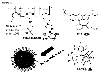
Figure 1 illustrates a simplified chemical structure of methacrylate-based
copolymers bearing sulfonate and zwitterionic (ZI) group, the dye (R18) and
its
counterion (F5-TPB) used for preparation of zwitterionic luminescent dye-
loaded
NPs through nanoprecipitation. It is to be noted that the hydrophobic groups,
the
zwitterionic groups and sulfonate groups are randomly arranged on the backbone
polymeric chain.
Figure 2 (A) shows TEM analysis of the sizes of zwitterionic sulfonate
nanoparticles (PMMA-ZI 10%-S03H 1% and 2%), and non zwitterionic sulfonate
nanoparticles (PMMA-S03H 1%, PMMA-S03H 2%). Nanoparticles loaded with 10
wt% R18/F5-TPB were prepared by nanoprecipitation; and Figure 2(B) shows TEM
analysis of the sizes of different methacrylate polymers with 10% zwitterionic
and
1% SO3H groups. Below each image size distributions of corresponding
nanoparticles are given. Scale bars correspond to 50 nm. At least 100
nanoparticles were analyzed per condition.
Figure 3 shows stability of NPs and their interactions with proteins as
determined using FCS: (A) Influence of the ZI density on the maximum salt
concentration in which the particles remained stable. (B) Size of PMMA-S03H 2%
NPs with different ZI percentage and (C) 1% sulfonate NPs made from different
methacrylate monomers with or without ZI groups in the presence of 10% fetal
bovine serum (FBS). All NPs were loaded with 1% R18/F5-TPB. Given are mean
values from three independent measurements. The error bars correspond to
s.e.m.
CA 03159400 2022-5-25
WO 2021/110735
PCT/EP2020/084247
18
Figure 4 shows representative epi-fluorescence micrographs of HeLa cells
microinjected with different types of NPs loaded with 10 wt% R18/F5-TPB.
Maximum projections over 60 s are shown. Scale bars correspond to 10 pm.
Figure 5 shows single particle tracking: (A) Trajectory of a PEMA-ZI 10%-
S03H 1% NP loaded with 20 wt% R18/F5-TPB in the cytoplasm over 30 s. (B) Mean
square displacement (MSD) of PEMA-ZI 10%-503H 1% NPs. The black curve
corresponds to the mean MSD curves and the straight gray line is the fitted
plot.
1.0
The error bars correspond to
s.e.m. (C) Diffusion coefficient distribution of PEMA-
ZI 10%-S03H 1% NPs. (D) Mean diffusion coefficients of different polymer NPs.
The error bars give FWHM. At least 50 trajectories were analyzed per sample.
Figure 6: (A) illustrates a simplified chemical scheme of azide bearing
fluorescent NPs assembled through nanoprecipitation of an ethyl methacrylate
based polymer bearing carboxylate (charged), sulfobetaine (zwitterionic), and
azide groups with the dye salt R18/F5-TPB. (B) DBCO bearing antibodies are
obtained by reaction of DBCO-EG4-maleimide with antibodies treated with TCEP.
Figure 7 shows micrographs of SKBR-3 cells, expressing HER2 receptor,
incubated with azide bearing zwitterionic NPs only (top), or with DBCO
modified
antibodies against HER2 receptors and then with azide bearing zwitterionic
nanoparticles (bottom). Given are, from left to right, the overlay of
fluorescence
and DIC images, the fluorescence images, and the DIC images.
EXAMPLES
EXAMPLE 1
1. Materials and Methods
1.1 Materials
Methyl methacrylate (99%, M55909), methacrylic acid (99%, 155721), 3-
sulthpropyl methacrylate potassium salt (99%, 251658) and 2-(N-3-Sulfopropyl-
N,N-dimethyl ammonium)ethyl methacrylate (99%, 537284) were purchased from
Sigma-Aldrich. Dimethylsulfoxide (DMSO, analytical grade) was obtained from
Fisher-Scientific. Milli-Q water (Millipore), acetonitrile (a- 99.9%, Sigma-
Aldrich)
CA 03159400 2022-5-25
WO 2021/110735
PCT/EP2020/084247
19
and methanol (a. 99.9%, Carlo Erba reagents) were used for preparation of
nanoparticles. dichloromethane (a-99.80/0) from CarloErba and methanol (HPLC
grade) from VWR. 6-carboxy- tetramethylrhodamine (TMR from Sigma-Aldrich),
phosphate buffered saline (PBS, Fisher Scientific), fetal bovine serum (FBS,
Lonza)
were used for stability study. Monomers were purified using column
chromatography or re-crystallization. Azobis isobutyronitrile (Aldrich, 98%)
was
recrystallized twice from ethanol. The other compounds were used as received.
R18/F5-TPB was synthesized from rhodamine B octadecyl ester perchlorate
(Aldrich, >98.0%) and lithium tetrakis(pentafluorophenyl)borate ethyl etherate
in (AlfaAesar, 97%) through ion exchange followed by purification through
column
chromatography as described previously.2
1.2 Polymer synthesis
Synthesis of polymers of invention: The different polymers were synthesized
through free radical polymerization. The different monomers were all dissolved
in
degased DMSO and mixed at the desired ratio. 0.01 eq. of AIBN were added and
the round bottom flask was placed in an oil bath preheated to 70 C. Once the
conversion reached 25 0/0, the reaction was stopped and the polymers
reprecipitated twice in methanol and/or water. After drying, the polymers were
characterized through NMR and, where possible, size exclusion chromatography.
As an example the synthesis of PMMA-S03H-ZI is given:
Poly(methyl methacrylate-co-3-sulfopropyl methacrylate-co- 2-(N-3-
Sulfopropyl-N,N-cfimethyl ammonium)ethyl methacrylate) (PMMA-ZI-S031-1):
Methyl methacrylate, 2-(N-3-Sulfopropyl-N, N-
di methyl am mon ium )ethyl
methacrylate, and 3-sulfopropyl methacrylate potassium salt were dissolved in
degassed DMSO at a concentration of 2 M (1M for 2-(N-3-Sulfopropyl-N,N-
dinnethyl annnnoniunn)ethyl methacrylate with addition of a small amount of
methanol). The three solutions were then mixed in a 50 mL two-neck round
bottom
flask equipped with a stirring bar at the desired ratio to give a total volume
of 20
mL. The mixture was degassed by bubbling argon for 5 min and placed under
argon atmosphere. 0.01 eq. of AIBN in DMSO (40 mg/mL) were added and the
round bottom flask was placed in an oil bath preheated to 70 C. At regular
intervals samples were drawn, dissolved in DMSO-d6 and analyzed by NMR. Once
the conversion reached 25 %, the reaction was stopped by quickly cooling it to
RT.
The reaction mixture was then added dropwise to methanol, or, for higher
CA 03159400 2022-5-25
WO 2021/110735
PCT/EP2020/084247
percentages of the charged and zwitterionic monomers, to water. After
filtration,
the precipitate was redissolved in a small amount of acetonitrile (when needed
with a small amount of methanol) and reprecipitated twice in methanol (water
for
highest percentages of ZI). The obtained polymer was dried under vacuum. 1H
5 NMR (400 MHz, DMSO-d6, 5): 4.36 (br, 0.04-0.2 H), 3.96 (br. s, 0.02-0.04 H),
3.55 (m, 3.4 H), 3.13 (br, 0.12-0.6 H), 2.45 (br, covered by the solvent
peak),
2.04 (br, 0.04-0.2 H), 2.00-0.30 (m, 6 H). Fraction obtained of ZI and SO3H
groups
based on the peak at 4.36 and 3.97 ppm, respectively (feed: obtained): ZI 10
0/0:
10.2 % - SO3H 1 A: 1.4%; ZI 10 0/0: 11 0/0- SO3H 2 0/0: 2.8%. Molecular
weight
in by GPC: 1% SO3H: Mw = 51 200, Mw /Mn = 1.58; 1% ZI: Mw = 43
100, Mw /Mn =
1.14. For polymers with higher amounts of zwitterionic monomers no suitable
solvent systems for GPC were available.
Poly(ethyl methacrylate-co-3-sulfopropyl methacrylate-co- 2-(V-3-
15 Sulfopropyl-N,N-dimethyl ammon(um)ethyl methacrylate) (PEMA-ZI-SO3H): 1FI
NMR (400 MHz, Me0D-, 6): 4.50 (br, 0.2 H), 4.34-3.92 (m, 2.02 H), 3.81 (br,
0.2
H), 3.70 (br, 0.2 H), 3.29 (br, partial covered by the solvent peak), 2.92
(br, 0.2
H), 2.31 (br, 0.2 H), 2.23-0.5 (m, 9 H). Fraction obtained of ZI groups based
on
the peak at 4.50 ppm: 10.3 %.
Poly(propyl methacrylate-co-3-sulfopropyl methacrylate-co- 2-(N-3-
Sulfopropyl-NIN-dimethyl ammonium)ethyl methacrylate) (PPMA-ZI-5031-1): 1H
NMR (400 MHz, Me0D, 5): 4.50 (br, 0.2 H), 4.29-3.54 (m, 2.42 H), 3.29 (br,
partial covered by the solvent peak), 2.92 (br, 0.2 H), 2.31 (br, 0.2 H), 2.23-
0.5
(rrl, 11 H).
Fraction obtained of ZI groups based on the peak at 4.50 ppm: 9.4%.
Poly(butyl methacrylate-co-3-sulfopropyl methacrylate-co- 2-(N-3-Sulfopropyl-
N,N-dimethyl ammonium)ethyl methacrylate) (PBMA-ZI-50311): 1H NMR (400
MHz, CDCI3, 5): 4.40 (br, 0.2H), 4.30-3.55 (m, 2.42 H), 3.33 (br, 0.6 H), 2.99
(br,
0.2 H), 2.36 (br, 0.2 H), 2.28-0.4 (m, 13 H). Fraction obtained of ZI groups
based
on the peak at 4.40 ppm: 8.3%.
Poly(methyl methacrylate-co-3-sulfopropyl methacrylate) (PMMA-SO3H):
1H NMR (400 MHz, DMSO-de, 6): 3.97 (br. s, 0.02-0.04 H), 3.57 (s, 3 H), 2.45
(m,
partial covered by the solvent peak), 2.1 - 0.5 (m, 6 H). Fraction of SO3H
groups
CA 03159400 2022-5-25
WO 2021/110735
PCT/EP2020/084247
21
based on the peak at 3.97 ppm (feed: obtained): 503H 1%: 1.1%; 503H 2%:
2.3%.
1.3 Preparation of nanoparticies
Stock solutions of copolymers were prepared at a concentration of 10g.I-1
in acetonitrile (20 vol.% methanol for ZI polymers). These solutions were
diluted
at 2g.I-1 in the corresponding solvent containing 1, 10 or 20 wt% of R18/F5-
TPB
(relative to the polymer). This solution was quickly added to a 10-fold volume
excess of water, under shaking (Thermomixer comfort, Eppendorf, 1000 rpm, at
in 21 C) followed by a second dilution in water.
1.4 Characterization of nanoparticies
Absorption and emission spectra were recorded on a Cary 4000 Scan
ultraviolet-visible spectrophotometer (Varian) and on a FS5 Spectrofluorometer
(Edinburgh Instruments) equipped with a thermostated cell compartment,
respectively. The excitation wavelength was set to 530 nm and emission was
recorded from 540 to 750 nm. QYs were calculated using rhodamine 101 in
ethanol
as reference (QY = 0.9).
Transmission electron microscopy: 5p1 of nanoparticle solution were
deposited onto carbon-coated copper-rhodium electron microscopy grids
following
amylamine glow-discharge. They were then treated for 20 s with a 2% uranyl
acetate solution for staining. The obtained grids were observed using a
Philips
CM120 transmission electron microscope equipped with a LaB6 filament and
operating at 100kV.The acquisition of areas of interest was recorded with a
Peltier
cooled CCD camera (Model 794, Gatan, Pleasanton, CA). Images were analyzed
using Fiji software.
Fluorescence correlation spectroscopy: Measurements were performed on
a home-built confocal set-up using excitation at 532 nm using TMR in water as
reference. The solution of NPs containing 1 wt% dyes were diluted 2 times
before
depositing 200 pl on 96-well optical-bottom plates for measurements. NPs
stability
in presence of salts and proteins were investigated by adding drop-by-drop 50
vol.
% of 10-fold PBS (10x), FBS or water to the solutions of NPs in low-binding
1.5m1
Eppendorf tubes. The data were recorded 5 min after addition and then analyzed
using the PyCorrFit software.
CA 03159400 2022-5-25
WO 2021/110735
PCT/EP2020/084247
22
1.5 Cellular experiments:
HeLa cells were grown in Dulbecco's modified Eagle medium (DMEM,
without phenolred, Gibco-Invitrogen), supplemented with 10% fetal bovine serum
(FBS, Lanza), L-glutamine, and 1% antibiotic solution (penicillin-
streptomycin,
Gibco-Invitrogen) at 37 C in humidified atmosphere containing 5% CO2. Cells
were seeded onto a round microscope cover glasses (diameter 18 mm) deposited
in 6 well plates at a density of 125x103 cells/well 24h before the
microinjection.
Microinjection of NPs and cellular imaging: For microinjection experiments,
subconfluent HeLa cells plated on glass coverslips were mounted in a Ludin
Chamber (Life Imaging Services, Basel, Switzerland). The cells were then
placed
on a Leica DMIRE 2 microscope (37 C, 5% CO2, 100x objective, sCMOS camera,
Xenon lamp) and solutions of the different nanoparticles at particle
concentrations
of 0.5 to 2 nM were microinjected into the perinuclear region of the cells,
using a
Femtojet/InjectMan NI2 microinjector (Eppendorf). Images sequences were then
is acquired either on the same setup or on an iMIC microscope (Till Photonics)
equipped with a Mutli-LED Spectra X (Lurnencor), an Olympus 60x TIRFM (1.45
NA) objective, and a Flash 4 V2-1- camera (Hamamatsu) after transfer of the
samples.
Live cells were maintained at 37 C in a 5% CO2 humidified atmosphere
using an environmental control system (Ufe Imaging Services). Time-lapse
movies
were recorded over 60 s with a frame rate of 50 ms and binning 2. They were
then
analyzed using the Image3 (National Institutes of Health, USA). For single
particle
tracking, time-lapse movies were recorded over 30 s with a frame rate of 43 ms
for the Imic microscope or 50 ms for the Leica DMIRE 2 microscope and a
binning
1 in order to increase the resolution. With Fiji software, the trajectories
were
recovered from TrackMate plugin. Then MSD curves were plotted and slopes
extracted from the MSD curves with a MATLAB script for particle tracking
analysis.
2. Experimental Results
2.1 Influence of negative charged groups and zwitterionic groups on the
particle size
Methacrylate based copolymers bearing sulfonate and zwitterionic (ZI)
groups were synthetized according to the method described above in "Materials
and Methods". In the following text the polymers are noted PXMA-ZI-x%-503H-
CA 03159400 2022-5-25
WO 2021/110735
PCT/EP2020/084247
23
y%, where XMA stands for the corresponding major monomer (MMA for methyl
methacrylate, EMA for ethyl methacrylate, PMA for propyl methacrylate, BMA for
butyl methacrylate), x and y correspond to the molar percentage of the
zwitterionic
and sulfonate groups. By the way, polymers bearing only sulfonate but no
zwitterionic groups were also prepared as control.
These polymers were then used to assemble dye-loaded polymer NPs
through nanoprecipitation. For this, solutions of the polymers in acetonitrile
(with
a small amount of methanol) containing different amounts of the salt of a
rhodannine B derivative (R18) with a perfluorinated borate (F5-TPB) were added
quickly to a large excess of water. The size of the formed NPs was analyzed
through
transmission electron microscopy (TEM, Figure 2, Table 1).
In the case of polymers bearing only sulfonate but no zwitterionic groups,
very small particles were observed: PMMA-SO3H 1% yielded NPs of about 13 nm
and the particle size decreased to about 9 nm for 2% sulfonate. The
introduction
of 10% ZI on the polymer chains had only a minor influence on the particle
size of
PMMA-ZI-503H based NPs. They reached 11 and 9 nm of diameter, respectively
for 10/0 and 2% sulfonate NPs. The presence of the ZI groups hence did not
affect
the process of particle formation and the obtained "ZI shell" is thin enough
for not
influencing the size of NPs. Increasing the hydrophobicity of the alkyl
methacrylate
monomers on the other hand led to increasing particle size (Figure 2, Table 1)
in
the order PMMA-ZI 10%-503H 1% < PEMA-ZI 10%-S03H 1% < PPMA-ZI 10 /0-
503H 1% < PBMA-ZI 10%-503H 1%, from 11 to 35 nm.
Table 1. Sizes of NPs made from different polymers as obtained from
transmission
electron microscopy and fluorescence correlation spectroscopy. Errors
correspond
to width of the distribution at half maximum for TEM, and variation over 3
measurements for FCS.
CA 03159400 2022-5-25
WO 2021/110735
PCT/EP2020/084247
24
TABLE 1
Polymer
Size (nm)
Main monomer SO3H ZI
TEM" FCS2)
MMA 1 mol% -
13 3 14 1
MMA 1 mo I% 10 mol%
11 3 15 1
MMA 2 mol% -
9 th 3 14 1
MMA 2m01% 10 mol%
9 th 2 13 1
EMA 1 mol% 10 mol%
14 3 11 1
PMA 1 mol% 10 nnol%
22 4 13 1
BMA 1 mol% 10 mol%
35+7 32+4
1) NPs prepared with 10 wt% R18/F5-TPB. 2) NPs prepared with 1 wt% R18/F5-TPB.
The nanoparticles were made fluorescent through the encapsulation of high
.5 amounts of dyes (between 1 and 20 wt% relative to the polymer). Here, the
cationic rhodamine R18 was associated to the large and very hydrophobic
counterion F5-TPB as this association avoids aggregation-caused quenching and
leads to a very hydrophobic dye counterion pair. Dialysis of the dye-loaded
NPs
over 48 h showed a release of less than 5% of the used dye, indicating that
this
approach yielded very efficient encapsulation of the dye salt in the
particles, in
agreement with previous results showing very efficient encapsulation of dyes
with
F5-TPB counterion.15 Furthermore/ the quantum yield of the particles remained
>
30% for a 10 wt% loading for all zwitterion bearing polymers, thus ensuring
excellent particle brightness.
2.2 Stability of zwitterionic polymeric nanoparticles
The stability of the resulting particles in biological media, and in
particular
the potential of the zwitterionic groups to improve this, was investigated by
fluorescence correlation spectroscopy (FCS). The percentage of ZI groups in
the
polymers was varied from 0 to 10% in order to modify the density of ZI on the
particle. The particle sizes of NPs obtained from FCS data were in very good
agreement with those obtained from TEM (Figure 3, Table 1. The smaller size is
associated with the lower amount of dye salt used in FCS experiments.). In a
first
CA 03159400 2022-5-25
WO 2021/110735
PCT/EP2020/084247
step, the stability of NPs was evaluated through addition of NaCI solutions
with
increasing concentrations (Figure 3A). The stability limit was defined as the
last
NaCI concentration for which no aggregation of NPs was observed. Particles
made
from polymers bearing no ZI groups already started to aggregate (and
precipitate)
5 as soon as a small amount of salt was added (Figure 3A). Addition of ZI
groups
led to an increase in the particle stability with increasing amount of ZI
groups. At
10% of zwitterionic groups the NPs did not show any change in size and no
signs
of precipitation up to 1 M NaCI.
The influence of the zwitterionic groups on interactions of the NPs with
in proteins and other biomolecules was then tested by adding fetal bovine
serum
(FBS), a complex mixture of salts and biomolecules containing notably numerous
proteins (Figure 3B). In the case of particles without ZI groups a size
increase of
about 10 nm was observed, which corresponds to the adsorption of at least a
monolayer of proteins and thus the formation of a "hard" protein corona
(Figure
15 3B). Starting from 5% of ZI groups the size increase of the NPs upon
interaction
with proteins was significantly reduced. At 10% of ZI groups no significant
increase
in particle size was observed, indicating that the surfaces of these particles
resisted
protein adsorption (Figure 3C).
20 2.3 In vitro comportment of zwitterionic polymeric nanoparticles
These NPs were then directly rnicroinjected in the perinuclear region of the
cytosol of living HeLa cells and their behavior was monitored using
epifluorescence
microscopy (Figure 4). In all cases, diffuse staining of cytosolic structures
was
virtually absent. This confirms the efficient and stable encapsulation of the
dyes
25 within the NPs and the absence of dye leaching. Maximum projections of the
fluorescence intensities over 60s gave a general idea of the overall
distribution of
the particles throughout the cytosol: In the absence of ZI groups, most of the
particles remained stuck at the injection point and the few particles in the
cytosol
had a low mobility, indicating strong interactions of the particles with the
cellular
constituents. On the other hand, particles bearing 10% ZI groups distributed,
in
general, well throughout the cytosol. PMMA-based ZI-bearing NPs showed a
distribution all over the cytosol and a high mobility, even though there is
sometime
a "projection" on the nucleus close to the injection point. PEMA-ZI particles
showed
a homogeneous distribution and high mobility for practically all NPs. This was
also
observed for PPMA-ZI particles, though here a certain part of the particles
CA 03159400 2022-5-25
WO 2021/110735
PCT/EP2020/084247
26
remained close to the injection point. In the case of PBMA-based particles,
they
were more localized at the injection point than particles based on other
tested
polymers. These results confirmed that 10% ZI groups are sufficient to
strongly
reduce interactions of the particles with cellular constituents and enable
spreading
throughout the cytosol. However, these results also confirm that the particle
size
also has a major influence on intracellular particle diffusion, and only
particles
below a critical core size of around 23 nm can spread throughout the cytosol
due
to steric hindrance by cellular structures.6 Here, the PBMA particles had
sizes
clearly above this threshold, leading to their immobilization. Part of the
PPMA
in particles were also above this threshold, resulting in immobilization of
part of the
particles.
2.4 Single particle tracking
The high brightness of the particles of the invention enabled monitoring the
mobility and diffusion behavior of the NPs at the single particle level and so
to
better understand the influence of the ZI groups and the type of polymer used
on
the intracellular behavior of our NPs. An example of a cytoplasmic trajectory
of a
PEMA-based ZI-bearing NP is represented in Figure 5A. Plotting the mean square
displacement (MSD) vs lag time for these NPs showed a linear increase up to
about
10 pm2 with an exponent a of 1 (Figure 5B). This indicates normal or Brownian
diffusion of these particles in the cytosol, which is described, in two
dimensions,
as MSD = 4DAt with DI the diffusion coefficient and a = 1.16 The diffusion
coefficients of individual NPs were then extracted from the corresponding MSD
curves. Figure 5C shows that for PEMA-ZI NPs the distribution of the diffusion
coefficients is centered around 1 pm2.s-1, with a mean D of 0.80 pm2.s-1, in
good
agreement with values obtained for QDs of similar size.4 The distribution
shows
only a small fraction of NPs with diffusion coefficients below 0.2 prn2.5-1.
PMMA-ZI
NPs had a similar distribution of diffusion coefficients with a slightly lower
mean of
0.65 pm2.s-1 (Figure 5D), in agreement with the slightly larger hydrodynamic
size.
PPMA-ZI NPs, on the other hand, showed a clearly lower mean diffusion
coefficient
of 0.25 pm2.s-1. This is probably due to their larger size leading to a
restricted
cytoplasmic diffusion, as already indicated by their stronger clustering
around the
injection point. As PMMA-S03H 1% NPs without ZI groups remained clustered
around the injection point, and hence it was not possible to characterize
their
diffusion behavior. However, we could show earlier that simple adsorption of
CA 03159400 2022-5-25
WO 2021/110735
PCT/EP2020/084247
27
Tween 80 (T80), a PEGylated surfactant, on such nanoparticles strongly reduces
their interaction with proteins and permits their diffusion in the
cytoso1.5,15
Interestingly, the mean diffusion coefficient of these NPs was with 0.2 pm2.s-
1
three to four fold lower than those of the PMMA and PEMA ZI NPs, even though
their core sizes were very close (Figure 2). One reason for this is certainly
the fact
that the addition of the PEGylated surfactant increases the NP size by > 5 nm.
The
large difference in diffusion coefficients further indicates that the ZI
groups are
more effective in reducing non-specific interactions with intracellular
biomolecules
and structures than the adsorbed PEG shell.
in
EXAMPLE 2: Preparation of zwitterionic fluorescent nanooarticles
for soecific interactions with biological molecules
In the present example fluorescent nanoparticles (NPs) bearing zwitterionic
groups have been designed to prevent non-specific interactions and bearing
targeting groups to introduce specific interactions with biomolecules. In
order to
introduce specific interactions, antibodies were used, more specifically
cetuximab,
which is an antibody against the HER2 receptor. To achieve conjugation of the
antibodies to the nanoparticles, it has been relied on copper-free
cycloaddition
click chemistry between an azide group and a strained alkyne,
dibenzylcyclooctyne
(DBCO). On the one side, zwitterionic brightly fluorescent nanoparticles with
azide
groups on their surface have been assembled, through nanoprecipitation of a
hydrophobic co-polymer bearing zwitterionic, charged, and azide groups,
together
with a hydrophobic dye-salt. On the other side, the antibody has been modified
in
order to introduce reactive DBCO groups. Cellular assays have shown that with
the
antibody the fluorescent nanoparticles bind specifically to HER2 expressing
cells.
1. Materials and methods
1.1 Synthesis of poly(ethyl methacrylate-co-methacrylic acid-co- 2-
(N-3-sulfopropyl-N,N-dimethyl ammonium)ethyl methacrylate-co-
Asp(OtBu)-N3) (PEMA-ZI-MAA-Asp(OtBu)-N3)
This polymer can be represented by the following formula (Ia-1)
CA 03159400 2022-5-25
WO 2021/110735
PCT/EP2020/084247
28
*
_
.õ)H z
HO 00
0,1)
µb
(Ia-1)
PEMA-ZI-MAA-Asp(OtBu)-N3 was obtained in 3 steps from poly(ethyl
methacrylate-co-methacrylic acid-co-
2-(N -3-sulfopropyl- N, N -d
'methyl
ammonium)ethyl methacrylate) (PEMA-ZI-MAA). PEMA-ZI-MAA was obtained
through free radical polymerization as described above in example 1.
1H NMR (400 MHz, DMS0-06) 6, ppm: 4.35 (br, 0.33 H), 3.98 (m, 2.00 H),
3.66 (br, partial covered with solvent peak), 3.10 (m, 0.87 H), 1.82 (br, 1.78
H),
1.19 (m, 3.06 H), 0.94 (m, 1.12 H), 0.78 (m, 1.53).
In a first step PEMA-ZI-MAA was reacted with Asp(OtBu)-N3 (Tert-butyl 3-
amino-4-((3-azidopropyflamino)-4-oxobutanoate, synthesized according to
procedures described by Melnychuk, N. et al. (A.S. DNA-Functionalized Dye-
Loaded Polymeric Nanoparticles: Ultrabright FRET Platform for Amplified
Detection
of Nucleic Acids, J. Am. Chem. Soc. 2018, 140, 10856.) in dimethylformamide
(DM F, Sigma Aldrich) using benzotriazol-1-yl-oxytripyrrolidinophosphonium
hexafluorophosphate (PyBOP, TCI) as coupling agent in the presence of IVA-
Diisopropylethylamine (DI PEA, Sigma Aldrich) as base. The obtained polymer
was
purified through precipitation in methanol water mixtures.
In a second step the tert-butyl group was removed through treatment with
a 1-to-1 mixture of trifluoroacetic acid (Sigma Aldrich) and dichloromethane
(Sigma Aldrich).
After evaporation, and in a third step, the polymer was purified through
precipitation and column chromatography.
1H NMR (400 MHz, DMS0-06) 6, ppm: 4.34 (br, 0.23 H), 3.96 (m, 2.00 H),
3.63 (m, 0.22 H), 3.51 (m, 0.23 H), 3.09 (m, 0.54 H), 1.78 (br, 1.97 H), 1.37
(s,
0.41 H), 1.17 (m, 3.55 H), 0.92 (m, 1.06 H), 0.76 (m, 1.65 H).
CA 03159400 2022-5-25
WO 2021/110735
PCT/EP2020/084247
29
In this polymer, the main monomer is thus ethylmethacrylate, and the molar
amounts of COOH groups and zwitterionic groups are respectively 5 mo10/0 and
10
mol%.
1.2 Preparation of nanoparticles
Stock solutions of PEMA-ZI-MAA-Asp(OtBu)-N3 as obtained above in 1.1
were prepared at a concentration of 10 g/L in acetonitrile with 20 vol.%
methanol.
These solutions were diluted at 2 g/L in the corresponding solvent containing
10,
20, or 30 wt% of R18/F5-TPB which is a fluorescent hydrophobic dye-salt
(relative
to the polymer). This solution was quickly added to a 9-fold volume excess of
phosphate buffer (20 mM, pH 7.4), under shaking (Thermomixer comfort,
Eppendorf, 1000 rpm, at 21 C) followed by a second dilution with the aqueous
phase.
1.3 Modification of antibodies
Cetuximab (Merck) is a chimeric IgG1 full length antibody directed against
the HER2 receptor. The antibody was obtained in its clinical formulation. The
buffer
exchange of antibody was carried out for borate buffer pH 8.14 via
ultrafiltration
(MWCO 50 kDa, Vivaspin). Concentration of antibody was determined by UV-vis
absorbance (e280= 210.000 11-1 cm-1 for cetuximab mAb), adjusted to 48 pM
(10.0
mg/mL) and was stored as aliquot at -20 C. For experiments, aliquots were
thawed
and used immediately.
1.4 Conjugation of cetuximab with maleimide-PEas-DBCO
The conjugates were prepared using the modification of a reported
protoco1.1 Cetuximab (23 pM, 300 pL, 0.0069 pmol) was prepared in a borate
buffer pH 8.4. Next, tris(2-carboxyethyl)phosphine hydrochloride (TCEP, Sigma
Aldrich) was added (45.8 nnIA, 1.2 pL, 4 eq) and the reaction was incubated at
37 C for 2 h under mild agitation (450 RPM). Then, a solution of
dibenzocyclooctyne-PEG4-maleimide (DBCO-PEGemaleimide, Sigma Aldrich) in
dry DMF (10 mM) was prepared and added to cetuximab (8 pL, 8 eq).
Subsequently, the temperature was reduced to 4 C and the incubation was
continued for 18 h. Afterwards, excess reagents were removed by
ultrafiltration
(50 kDa MWCO) with PBS buffer (pH 7.4) to afford the modified antibody-
maleimide-PEG4-DBCO in PBS buffer with yield 60-70%, as determined by UV-vis.
CA 03159400 2022-5-25
WO 2021/110735
PCT/EP2020/084247
1.5 Cell maintenance and fluorescence imaging
SKBR-3 cells, a human breast cancer cell line that overexpresses the Her2
(Neu/ErbB-2) were cultured in Dulbecco's modified Eagle medium (Gibco, DMEM)
supplemented with 10 /o fetal bovine serum (Gibco) and 1%
5 penicillin/streptomycin (100 U/mL, Gibco). The SKBR-3 cell were seeded at
a cell
density of 2.0 x 10⁴ on 8 wells Lab-Tek Chambered Coverglasses (Thermo
Scientific) followed by incubation for 48 h at 37 C and 5% CO2. For
fluorescence
imaging, the medium was discarded and the cells washed with PBS buffer. Then,
200 pL of Ab-maleimide-PEG4-DBCO (10 pg/mL in Optimem, Gibco ) and 200 pL
in of Optimem medium only (control) were added to the cells and incubated
for 20
min at 37 C and 5% CO2. Then the cells were washed repeatedly with PBS, and
fixed with 4 % paraformaldehyde in PBS for 12 min at 37 C and 5% CO2,
followed
by additions of 3% BSA in PBS was and incubation for another 15 minutes at 37
C
and 5% CO2. Then, fluorescent NPs bearing zwitterionic, charged and azide
groups
15 (see 1.21 100 pM in 0.1% BSA in PBS solution) were added and the cells were
incubated for 3 h at 37 C and 5% CO2. Finally, the cells were washed with 0.1
%
BSA in PBS solution and examined through epi-fluorescence microscopy using
Nikon Ti-E inverted microscope with a 60x objective (Apo TIRF, oil, NA 1.49,
Nikon). The excitation was provided by light emitting diodes (SpectraX,
Lumencor)
20 at 550 nm.
2. Results and Discussion
In the present example, fluorescent zwitterionic nanoparticles with reactive
25 groups, allowing for introduction of biologically interesting molecules,
were
assembled. For this an ethyl methacrylate based polymer, bearing carboxylate
and
sulfobetaine groups, was synthesized through radical polymerization (Figure
6A).
In this polymer we then introduced azide groups through reaction with a
trifunctional molecule bearing an azide group, an amino group for coupling
with
30 COOH groups on the polymer, and a protected carboxylic acid group
(Asp(OtBu)-
N3, derived from aspartic acid). After deprotection of the carboxylate, a
hydrophobic polymer combining ZI groups, COOH groups for the nanoparticle size
control during nanoprecipitation, and azide reactive groups (e.g. 10 mol%
zwitterionic groups, 5 mol% COOH, 3-5 mol% N3) was obtained (Formula Ia-1
above).
CA 03159400 2022-5-25
WO 2021/110735
PCT/EP2020/084247
31
This polymer was then used to assemble dye-loaded nanoparticles (NPs)
through nanoprecipitation: Acetonitrile solutions (containing 10% methanol) of
the
polymer and 10-30 wt% (relative to the polymer) of the dye salt R18/F5-TPB
were
quickly added to a 9-fold excess of phosphate buffer at pH 7.4, followed by
further
dilution. This resulted in the formation of NPs with sizes between 15 and 18
nm,
which increased slightly with dye loading as detailed in the following Table
2. These
NPs showed a bright fluorescence with fluorescence quantum yields around 32%.
Based on the size and the loading of the particles, the number of fluorophores
can
be estimated to be 100, 250 and 500 fluorophores per nanoparticles for 10, 20,
and 30wt% loading, respectively. The per particle brightness can then be
calculated using the formula sxNxQY, where & is the extinction coefficient of
rhodamine (125 000 M-1.cm-1), N the number of dyes per particle, and QY the
quantum yield and x the multiplication operator. This results in the case of
30 wt%
loading in a per particle brightness of 2.1 x 10⁷ M-1.cm-1.
TABLE 2
Loading R18/F5-TPB ( Size of the NPs
(inwt%) Quantum yield (%)
nm)
10 15 + 2 31 + 1
20 16 + 2 30 + 4
30 18 + 2 34 + 2
DBCO bearing antibodies, on the other hand, were obtained in a two-step
one-pot process (Figure 6B). In the first step (tris(2-carboxyethyl)phosphine)
(TCEP) was used to open disulfide links of cetuximab, the antibody. In a
second
step the antibody was then reacted with a bifunctional reagent bearing
maleimide
for conjugation to thiol groups on the one side and a DBCO group on the other
side, connected by a short oligo(ethylene glycol) linker. Following
purification, the
presence of reactive DBCO groups through conjugation with fluorophores bearing
azide groups has been confirmed by UV-visible absorption measurements.
The antibody/NP system has then been applied to the specific binding of NPs
to cells expressing the HER2 receptor. The antibody-NP conjugation was
achieved
in situ. SKBR-3 cells were cultured in wells on glass coverslips (Labtek) and
after
48h of culture treated with our DBCO modified antibodies. After fixation of
the
CA 03159400 2022-5-25
WO 2021/110735
PCT/EP2020/084247
32
cells, we then added the azide bearing NPs. As control, the same experiment
was
performed without addition of the antibodies.
The results of fluorescence microscopy that are reported in figure 7 showed
that in the absence of the antibody practically no NPs were detected at the
SKBR-
3 cells. However, after treatment with the DBCO modified antibodies against
the
HER2 receptor, addition of the NPs led to a strong fluorescence, especially at
the
periphery of the cells, probably at the plasma membrane containing the
receptor.
This showed that our system of conjugation of DBCO bearing antibodies to
azide bearing fluorescent zwitterionic NPs enables specific binding of the NPs
to
biological motives (receptors) on cells.
CA 03159400 2022-5-25
WO 2021/110735
PCT/EP2020/084247
33
(1) Li, K.; Qin, W.; Ding, D.; Tomczak, N.; Geng, J.; Liu, R.; Liu, J.; Zhang,
X.;
Liu, H.; Liu, B.; et al. Photostable Fluorescent Organic Dots with
Aggregation-Induced Emission (AIE Dots) for Noninvasive Long-Term Cell
Tracing. Sci. Rep. 2013, 3. https://doi.org/10.1038/srep01150.
(2) Reisch, A.; Didier, P.; Richert, L.; Oncul, S.; Arntz, Y.; Moly, Y.;
Klymchenko, A. S. Collective Fluorescence Switching of Counterion-
Assembled Dyes in Polymer Nanoparticles. Nat. Commun. 2014, 5.
https://doi.org/10.1038/ncomms5089.
(3) Sahl, S. J.; Hell, S. W.; Jakobs, S. Fluorescence Nanoscopy in Cell
Biology.
Nat. Rev. Mot Cell Biol. 2017, 18 (11), 685-701.
https://doi.org/10.1038/nrnn.2017.71.
(4) Etoc, F.; Balloulr E.; Vicario, C.; Normanno, D.; LiBe, D.; Sittner, A.;
Piehler, J.; Dahan, M.; Coppey, M. Non-Specific Interactions Govern
Cytosolic Diffusion of Nanosized Objects in Mammalian Cells. Nat. Mater.
2018, 17(8), 740-746. https://doi.org/10.1038/541563-018-0120-7.
(5) Reisch, A.; Heimburger, D.; Ernst, P.; Runser, A.; Didier, P.; Dujardin,
D.;
Klymchenko, A. S. Protein-Sized Dye-Loaded Polymer Nanoparticles for
Free Particle Diffusion in Cytosol. Adv. Fund. Mater. 2018, 28 (48)1
1805157. https://doi.org/10.1002/adfm.201805157.
(6) Luby-Phelps, K. Cytoarchitecture and Physical Properties of Cytoplasm:
Volume, Viscosity/ Diffusion, Intracellular Surface Area. Int. Rev. Cytol.
2000, 192, 189-221.
(7) Walkey, C. D.; Chan, W. C. W. Understanding and Controlling the
Interaction of Nanomaterials with Proteins in a Physiological Environment.
Chem. Soc. Rev. 2012, 41 (7), 2780-2799.
https://doi.org/10.1039/C1CS15233E.
(8) Monopoli, M. P.; Aberg, C.; SaIvati, A.; Dawson, K. A. Biomolecular
Coronas Provide the Biological Identity of Nanosized Materials. Nat.
Nanotechnot 2012, 7 (12), 779-786.
https://doi.org/10.1038/nnano.2012.207.
(9) Jokerst, J. V.; Lobovkina, T.; Zare, R. N.; Gambhir, S. S. Nanoparticle
PEGylation for Imaging and Therapy. Nanomed. 2011, 6 (4), 715-728.
https://doi.org/10.2217/nnm.11.19.
(10) Rabanel, J.-M.; Hildgen, P.; Banquy, X. Assessment of PEG on Polymeric
Particles Surface, a Key Step in Drug Carrier Translation. J. Controlled
Release 2014, 185, 71-87. https://doi.org/10.1016/j.jconre1.2014.04.017.
(11) Estephan, Z. G.; Schlenoff, P. S.; Schlenoff, J. B. Zwitteration As an
Alternative to PEGylation. Langmuir 2011, 27 (11)1 6794-6800.
https://doi.org/10.1021/1a200227b.
(12) Garcia, K. P.; Zarschlerf K.; Barbaro, L.; Barreto, J. A.; O'Malley, W.;
Spiccia, L.; Stephan, H.; Graham, B. Zwitterionic-Coated "Stealth"
Nanoparticles for Biomedical Applications: Recent Advances in Countering
Biomolecular Corona Formation and Uptake by the Mononuclear Phagocyte
System. Small 2014, 10 (13), 2516-2529.
https://doi.org/10.1002/sm11.201303540.
(13) Muro, E.; Pons, T.; Lequeux, N.; Fragola, A.; Sanson, N.; Lenkei, Z.;
Dubertret, B. Small and Stable Sulfobetaine Zwitterionic Quantum Dots for
Functional Live-Cell Imaging. J. Am. Chem. Soc. 2010, 132 (13), 4556-
4557. https://doi.org/10.1021/ja1005493.
(14) Rouhana, L. L.; Jaber, J. A.; Schlenoff, J. B. Aggregation-Resistant
Water-
Soluble Gold Nanoparticles. Langmuir 2007, 23 (26)1 12799-12801.
https://doi.org/10.1021/1a702151q.
CA 03159400 2022-5-25
WO 2021/110735
PCT/EP2020/084247
34
(15) Reisch, A.; Runser, A.; Arntz, Y.; Mely, Y.; Klymchenko, A. S. Charge-
Controlled Nanoprecipitation as a Modular Approach to Ultrasmall Polymer
Nanocarriers: Making Bright and Stable Nanoparticles. ACS Nano 2015, 9
(5), 5104-5116. https://doi.org/10.1021/acsnano.5b00214.
(16) Ruthardt, N.; Lamb, D. C.; Brauchle, C. Single-Particle Tracking as a
Quantitative Microscopy-Based Approach to Unravel Cell Entry Mechanisms
of Viruses and Pharmaceutical Nanoparticles. Mol. Then 2011, 19 (7)1
1199-1211. https://doi.org/10.1038/mt.2011.102.
CA 03159400 2022-5-25