Note: Descriptions are shown in the official language in which they were submitted.
WO 2021/113596
PCT/US2020/063243
1
COMPOSITIONS AND METHODS COMPRISING AN ANTI-CD47 ANTIBODY
IN COMBINATION WITH A TUMOR TARGETING ANTIBODY
INTRODUCTION
CD47 is a cell surface antigen overexpressed on many tumor cells. CD47 can
inhibit
phagocytosis by innate immune cells such as macrophages by engaging its
receptor, signal
regulatory protein alpha (SIRPa), on the surface of the immune cells. (Because
it inhibits
phagocytosis, CD47 is sometimes referred to as the "don't eat me" molecule.)
Administration
of anti-CD47 antibodies can relieve the inhibition of the native immune system
by blocking
the CD47- SIRPa interaction and thus provides an anticancer strategy.
In addition to being overexpressed on many tumor cells, CD47 is also expressed
on
some normal cells, including platelets and erythrocytes. Treatment of patients
with anti-CD47
antibodies therefore can result in toxic effects to the patient resulting from
normal blood cell
binding. For example, the Phase I trial of anti-CD47 monoclonal antibody Hu5F9
(magrolimab) resulted in 57% of the treated patients experiencing transient
anemia and 36%
exhibiting hemagglutination of peripheral blood cells (Sikic et al. (2019)J.
Clinical Oncol.
37:946-953).
SUMMARY
The present disclosure provides compositions and methods comprising a first
antibody comprising a fully human anti-CD47 antibody and a second antibody
that
specifically binds a cell surface antigen and comprises an Fc portion that can
bind an Fey
receptor on an effector cell In various embodiments, the second antibody
comprises a tumor-
targeting antibody, such as an antibody that binds CD20, PD-L1, CD38 or SLAMF7
antigens.
The fully human anti-CD47 antibody in various embodiments has a heavy chain
variable region having at least 95%, at least 96%, at least 97%, at least 98%,
or at least 99%
identity to SEQ ID NO:1 and a light chain variable region having at least 95%,
at least 96%,
at least 97%, at least 98%, or at least 99% identity to SEQ ID NO:2. In some
embodiments
the fully human antibody is an IgG2 antibody or an IgG4 antibody. In some
embodiments the
fully human antibody is an IgG1 antibody having one or more mutations in the
Fc region,
where the one or more mutations result in reduced interaction of the Fc region
with an Fc
receptor.
CA 03160173 2022- 5- 31
WO 2021/113596
PCT/US2020/063243
2
Also provided herein are methods of treating a subject having cancer,
comprising
administering a therapeutically effective amount of 1) a first antibody of an
antigen binding
fragment thereof that binds CD47 and 2) a second antibody that binds an
antigen present on a
cancer cell, where the first antibody binds to CD47 and blocks binding between
CD47
antigen and SIRPa antigen, and the second antibody comprises an Fc region that
binds an Fey
receptor on an effector cell. In various embodiments the first antibody is an
anti-CD47
antibody as disclosed herein that comprises a heavy chain variable region
having at least 95%
identity to SEQ ID NO:1 and a light chain variable region having at least 95%
identity to
SEQ ID NO:2. In various embodiments the second antibody of the antibody that
includes an
Fc region binds a tumor antigen, such as CD20, CD38, PD-L1, or SLA1VIF7. For
example, the
second antibody can be an anti-CD20 antibody such as rituximab or an anti-CD38
antibody
such as Daratumumab.
Also included are methods for killing at least one cancer cell in a population
of cancer
cells, wherein the at least one cancer cell overexpresses CD47 antigen, the
method
comprising: contacting the at least one cancer cell with a therapeutically
effective amount of
a first antibody or an antigen binding fragment thereof that binds CD47
antigen and a second
antibody that binds a tumor antigen, where the first antibody binds to CD47
antigen and
blocks binding between CD47 antigen and SIRPa antigen, and wherein the second
antibody
binds a tumor cell and comprises Fc portion that binds an Fcy receptor on an
effector cell.
Also included are methods for treating a subject having a cancer that
overexpresses
CD47 antigen, the method comprising: administering to the subject a
therapeutically
effective amount of a first antibody or an antigen binding fragment thereof
that binds CD47
antigen and a second antibody that binds a tumor antigen, where the first
antibody binds to
CD47 antigen and blocks binding between CD47 antigen and SIRPa antigen, and
wherein the
second antibody binds a tumor cell and comprises Fc portion that binds an Fcy
receptor on an
effector cell.
The methods can use any of the CD47 antibodies disclosed herein, such as the
STI-
6643 antibody and variants thereof, and can use any tumor targeting
antibodies, including but
not limited to antibodies that specifically bind CD20, CD38, or PD-L1
CA 03160173 2022- 5- 31
WO 2021/113596
PCT/US2020/063243
3
DESCRIPTION OF THE FIGURES
Figure 1 shows a schematic of a hemagglutination reaction (upper) and a
photograph
of a hemagglutination assay comparing activity of anti-CD47 antibodies STI-
6643 and
Hu5F9.
Figure 2A is a schematic of a competition assay with anti-CD47/SIRP-alpha-Fc
for
CD47 binding.
Figure 2B is a graph of the competition assay comparing the activity of anti-
CD47
antibodies STI-6643 and Hu5F9.
Figure 3 is a graph of an antibody dependent cellular phagocytosis (ADCP)
assay
comparing the activity of anti-CD47 antibodies STI-6643 and Hu5F9.
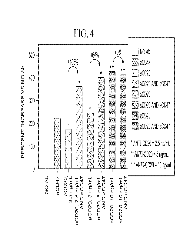
Figure 4 is a bar graph comparing increases in phagocytosis killing in assays
testing
the combination of anti-CD47 antibody clone STI-6643 and suboptimal amounts of
anti-
CD20 antibody Rituximab.
Figure SA shows anti-tumor activity in a disseminated human Raji-Fluc
xenograft
mouse model comparing the activity of a control isotype IgG4, anti-CD47
antibodies STI-
6643 and Hu5F9.
Figure 5B are graphs showing the anti-tumor activity of the mouse model
described
in Figure 5A. The upper graph shows total flux detected in mice treated with a
control
isotype IgG4 or anti-CD47 antibody clone STI-6643. The lower graph shows the
total flux
detected in mice treated with a control isotype IgG4 or anti-CD47 antibody
Hu5F9. See
Example 5.
Figure SC is a graph showing a statistical significance analysis of the data
shown in
Figures 5B and 5C.
Figure SD is a graph showing animal survival analysis based on the data shown
in
Figures 5A-C.
Figure SE is a graph of circulating antibody detection in the animals
described in
Figures 5A-C.
Figure 6A shows anti-tumor activity in a disseminated human Raji-Fluc
xenograft
mouse model comparing the activity of a control isotype TgG4, anti-CD47
antibodies STT-
6643 or Hu5F9 as mono-therapy, or the combination of anti-CD47 antibodies STI-
6643 and
Hu5F9. See Example 6.
CA 03160173 2022- 5- 31
WO 2021/113596
PCT/US2020/063243
4
Figure 6B are graphs showing the anti-tumor activity of the mouse model
described
in Figure 6A. The left graph shows total flux detected in mice treated with a
control isotype
IgG4 or anti-CD47 antibody clone STI-6643 as a mono-therapy. The middle graph
shows
total flux detected in mice treated with a control isotype IgG1 or anti-CD20
antibody
Rituximab as a mono-therapy. The right graph shows the total flux detected in
mice treated
with a combination control isotype IgG1 and IgG4 isotype, or the combination
of anti-CD47
antibody clone STI-6643 and anti-CD20 antibody Rituximab.
Figure 6C is a graph showing a statistical significance analysis of the data
shown in
Figures 6B and C.
Figure 6D is a graph showing animal survival analysis based on the data shown
in
Figures 6A-C.
Figure 7A is a graph reproduced from Liu, et al., 2015 PLoS ONE (10)9:
e0137345
(doi:10.1371/journal.pone.0137345 (see Figure 4A in Liu which shows
pharmacokinetic
analysis (hemoglobin) of cynomolgus monkeys administered single intravenous
infusions of
anti-CD47 antibody Hu5F9 at doses indicated in the figure. The shaded bar
indicates the
range of hemoglobin that might trigger transfusion in humans).
Figure 7B is a graph showing our pharmacokinetic analysis of cynomolgus
monkeys
administered anti-CD47 antibody STI-6643 (each dose at 150 mg/kg) once weekly
via IV
bolus for four weeks. The shaded bar indicates the range of hemoglobin that
might trigger
transfusion in humans).
Figure 8A contains four graphs showing preferential binding of anti-CD47
antibody
clone STI-6643 to tumor cells with respect to red blood cells (RBCs) as
compared to anti-
CD47 antibody Hu5F9 binding to tumor cells and RBCs. The graphs display the
results of
flow cytometry data from a binding assay on mixed-cell samples.
Figure 8B is a bar graph showing binding of anti-CD47 antibody clone STI-6643
to
RBCs and tumor cells (Raji, CD19-expressing tumor cells, and CD3-expressing
tumor cells)
by anti-CD47 antibody clone ST1-6643. The binding of antibody Hu5F9 to Raji
cells is set at
100% on the y- axis for comparison.
Figure 9 shows a schematic of a hemagglutinati on reaction (upper) and a
photograph
of another hemagglutination assay comparing activity of anti-CD47 antibodies
STI-6643 and
Hu5F9.
CA 03160173 2022- 5- 31
WO 2021/113596
PCT/US2020/063243
Figure 10 shows four graphs from a three-way mixed lymphocyte reaction (MLR)
assay.
Figure 11A is a bar graph showing the results of a Staphylococcal Enterotoxin
B
(SEB) assay. Each concentration along the x-axis includes from left to right:
no antibody
5 control; isotype IgG4 control; Hu5F9; and STI-6643.
Figure 11B shows the results of the SEB assay described in Figure 11A above,
with
the number of CD4+ and CD8+ T cells shown in two separate graphs.
Figure 11C shows the results of the SEB assay described in Figure 11A above,
with
the number of CD25+ CD4+ and CD25+ CD8+ activated T cells shown in two
separate
graphs.
Figure 12A is a graph showing the percent survival from a dose study in a Raji
mouse
tumor model.
Figure 12B is a Table listing the p values of each treatment group in the
mouse Raji
tumor model described in Figure 12A above.
Figure 12C is a graph showing the cumulative circulating concentration of
antibody
from the Raji mouse tumor model described in Figure 12A above.
Figure 13A is a graph showing the average tumor volume from an efficacy study
in
mouse NCI-H82 lung solid tumor model.
Figure 13B shows tumor volumes from individual animals treated with either
isotype
IgG4 antibody or STI-6643 antibody, in the mouse NCI-H82 lung solid tumor
model
described in Figure 13A above.
Figure 13C is a bar graph showing the relative tumor weight from the mouse NCI-
H82 lung solid tumor model described in Figure 13A above.
Figure 13D is a bar graph showing the circulating antibody concentrations from
the
mouse NCI-H82 lung solid tumor model described in Figure 13A above. Each time
post
along the x-axis includes from left to right: isotype IgG4 control; and STI-
6643.
Figure 14 shows several graphs of tumor volume and percent survival from a
dose
efficacy study in a mouse NCI-H82 lung solid tumor model.
Figure 15A is a graph showing tumor volume from an efficacy study in a mouse
A375 melanoma solid tumor study.
CA 03160173 2022- 5- 31
WO 2021/113596
PCT/US2020/063243
6
Figure 15B shows tumor volumes from individual animals treated with either
isotype
IgG4 antibody or STI-6643 antibody, in the mouse A375 melanoma solid tumor
study
described in Figure 15A above.
Figure 15C shows a percent survival graph from the mouse A375 melanoma solid
tumor study described in Figure 15A above.
Figure 16A shows a percent survival graphs from an efficacy study in a mouse
Raji
tumor model in which the mice were treated with a combination of STI-6643 and
an anti-
CD38 antibody (Daratumumab).
Figure 16B is a Table listing the p values of each treatment group in the
mouse
combination therapy study described in Figure 16A above.
Figure 17A provides examples of positive and negative hemagglutination assays.
Figure 17B provides a picture of the results of hemagglutination assays using
anti-CD47
antibodies STI-6643, Hu5F9, A0-176, and 13H3. Figure 17C provides pictures of
the results
of hemagglutination assays using anti-CD47 antibodies STI-6643 and Hu5F9 with
human,
cynomolgus, and canine RBCs.
Figure 18 provides graphs of binding of anti-CD47 antibodies STI-6643 and
Hu5F9
to human, cynomolgus, and canine RBCs as a function of antibody concentration.
Figure 19 provides graphs of binding of anti-CD47 antibodies STI-6643, Hu5F9,
A0-176, and 13H3 to Raji tumor cells and RBCs as a function of antibody
concentration.
Figure 20A provides graphs of numbers of CD4+, CD8+, CD19+, and CD56+ cells
recovered from PBMCs after incubation with anti-CD47 antibodies STI-6643,
Hu5F9, AO-
176, and 13H3.
Figure 20B provides graphs of CD4+, CD8+, CD19+, and CD56+ cells recovered
from PBMCs after incubation with anti-CD47 antibodies STI-6643, Hu5F9, A0-176,
and
13H3 as a percentage of the same cell types recovered after incubation with
the isotype
control.
Figure 21 provides graphs of tumor volume over time in tumor-bearing mice
treated
with different dosages of anti-CD47 antibodies.
Figures 22A-C show the amino acid sequences of various anti-CD47 antibodies, a
CD47 antigen, anti-CD20 antibodies and a CD20 antigen.
Figures 23A-E show the amino acid sequences of various anti-CD38 antibodies
and
CD38 target antigens.
CA 03160173 2022- 5- 31
WO 2021/113596
PCT/US2020/063243
7
DESCRIPTION
Headings provided herein are solely for the convenience of the reader and do
not limit
the various aspects of the disclosure, which aspects can be understood by
reference to the
specification as a whole.
The disclosures of all publications, patents, and patent applications cited
herein are
hereby incorporated by reference in their entireties into this application.
Definitions
Unless defined otherwise, technical and scientific terms used herein have
meanings
that are commonly understood by those of ordinary skill in the art unless
defined otherwise.
Generally, terminologies pertaining to techniques of cell and tissue culture,
molecular
biology, immunology, microbiology, genetics, transgenic cell production,
protein chemistry
and nucleic acid chemistry and hybridization described herein are well known
and commonly
used in the art. The methods and techniques provided herein are generally
performed
according to conventional procedures well known in the art and as described in
various
general and more specific references that are cited and discussed herein
unless otherwise
indicated. See, e.g., Sambrook et al. Molecular Cloning: A Laboratory Manual,
2d ed., Cold
Spring Harbor Laboratory Press, Cold Spring Harbor, N.Y. (1989) and Ausubel et
al.,
Current Protocols in Molecular Biology, Greene Publishing Associates (1992). A
number of
basic texts describe standard antibody production processes, including,
Borrebaeck
(ed) Antibody Engineering, 2nd Edition Freeman and Company, NY, 1995;
McCafferty et
al. Antibody Engineering, A Practical Approach IRL at Oxford Press, Oxford,
England,
1996; and Paul (1995) Antibody Engineering Protocols Humana Press, Towata,
N.J., 1995;
Paul (ed.), Fundamental Immunology, Raven Press, N.Y, 1993; Coligan (1991)
Current
Protocols in Immunology Wiley/Greene, NY; Harlow and Lane (1989) Antibodies: A
Laboratory Manual Cold Spring Harbor Press, NY; Stites et al. (eds.) Basic and
Clinical
Immunology (4th ed.) Lange Medical Publications, Los Altos, Calif., and
references cited
therein; Coding Monoclonal Antibodies: Principles and Practice (2nd ed.)
Academic Press,
New York, N.Y., 1986, and Kohler and Milstein Nature 256: 495-497, 1975. All
of the
references cited herein are incorporated herein by reference in their
entireties. Enzymatic
reactions and enrichment/purification techniques are also well known and are
performed
according to manufacturer's specifications, as commonly accomplished in the
art or as
CA 03160173 2022- 5- 31
WO 2021/113596
PCT/US2020/063243
8
described herein. The terminology used in connection with, and the laboratory
procedures
and techniques of, analytical chemistry, synthetic organic chemistry, and
medicinal and
pharmaceutical chemistry described herein are well known and commonly used in
the art.
Standard techniques can be used for chemical syntheses, chemical analyses,
pharmaceutical
preparation, formulation and delivery, and treatment of patients.
Unless otherwise required by context herein, singular terms shall include
pluralities
and plural terms shall include the singular. Singular forms "a", "an" and
"the", and singular
use of any word, include plural referents unless expressly and unequivocally
limited on one
referent.
It is understood the use of the alternative (e.g., "or") herein is taken to
mean either
one or both or any combination thereof of the alternatives.
The term -and/or" used herein is to be taken mean specific disclosure of each
of the
specified features or components with or without the other. For example, the
term "and/or" as
used in a phrase such as "A and/or B" herein is intended to include "A and B,"
"A or B," "A"
(alone), and "B" (alone). Likewise, the term "and/or" as used in a phrase such
as "A, B,
and/or C" is intended to encompass each of: A, B, and C; A, B, or C; A or C; A
or B; B or C;
A and C; A and B; B and C; A (alone); B (alone); and C (alone).
As used herein, terms "comprising", "including", "having" and "containing",
and
their grammatical variants, as used herein are intended to be non-limiting so
that one item or
multiple items in a list do not exclude other items that can be added to the
listed items. It is
understood that wherever aspects are described herein with the language
"comprising,"
otherwise analogous aspects described in terms of "consisting of' and/or
"consisting
essentially of' are also provided.
As used herein, the term "about" refers to a value or composition that is
within an
acceptable error range for the particular value or composition as determined
by one of
ordinary skill in the art, which will depend in part on how the value or
composition is
measured or determined, i.e., the limitations of the measurement system. For
example,
"about" or "approximately" can mean within one or more than one standard
deviation per the
practice in the art Alternatively, "about" or "approximately" can mean a range
of up to 10%
(i.e., 10%) or more depending on the limitations of the measurement system.
For example,
about 5 mg can include any number between 4.5 mg and 5.5 mg. Furthermore,
particularly
with respect to biological systems or processes, the terms can mean up to an
order of
CA 03160173 2022- 5- 31
WO 2021/113596
PCT/US2020/063243
9
magnitude or up to 5-fold of a value. When particular values or compositions
are provided in
the instant disclosure, unless otherwise stated, the meaning of "about" or
"approximately"
should be assumed to be within an acceptable error range for that particular
value or
composition.
The terms "peptide", "polypeptide" and "protein" and other related terms used
herein
are used interchangeably and refer to a polymer of amino acids that is not
limited to any
particular length. Polypeptides may comprise natural and non-natural amino
acids.
Polypeptides include recombinant and chemically-synthesized polypeptides.
Polypeptides
include precursor molecules and mature (e.g., processed) molecules. Precursor
molecules
include those that have not yet been subjected to cleavage, for example
cleavage of a
secretory signal peptide or by enzymatic or non-enzymatic cleavage at certain
amino acid
residue(s). Polypeptides include mature molecules that have undergone
cleavage. These terms
encompass native proteins, recombinant proteins, and artificial proteins,
protein fragments
and polypeptide analogs (such as muteins, variants, chimeric proteins and
fusion proteins) of
a protein sequence as well as post-translationally, or otherwise covalently or
non-covalently,
modified proteins.
The terms "nucleic acid", "nucleic acid molecule", "polynucleotide" and
"oligonucleotide" and other related terms used herein are used interchangeably
and refer to
polymers of nucleotides that are not limited to any particular length. Nucleic
acids include
recombinant and chemically-synthesized forms. Nucleic acids include DNA
molecules (e.g.,
cDNA or genomic DNA, expression constructs, DNA fragments, etc.), RNA
molecules (e.g.,
mRNA), analogs of the DNA or RNA generated using nucleotide analogs (e.g.,
peptide
nucleic acids and non-naturally occurring nucleotide analogs), and hybrids
thereof, as well as
peptide nucleic acids, locked nucleic acids, and other synthetic nucleic acid
analogs and
hybrids thereof A nucleic acid molecule can be single-stranded or double-
stranded. In one
embodiment, the nucleic acid molecules of the disclosure comprise a contiguous
open
reading frame encoding an antibody, or a fragment or scFv, derivative, mutein,
or variant
thereof. In some embodiments, nucleic acids comprise one type of
polynucleotides or a
mixture of two or more different types of polynucl eoti des
The term "recover" or "recovery" or "recovering", and other related terms,
refer to
obtaining a protein (e.g., an antibody or an antigen binding portion thereof),
from host cell
culture medium or from host cell lysate or from the host cell membrane. In one
embodiment,
CA 03160173 2022- 5- 31
WO 2021/113596
PCT/US2020/063243
the protein is expressed by the host cell as a recombinant protein fused to a
secretion signal
peptide sequence (e.g., leader peptide sequence) which mediates secretion of
the expressed
protein. The secreted protein can be recovered from the host cell medium. In
one
embodiment, the protein is expressed by the host cell as a recombinant protein
that lacks a
5 secretion signal peptide sequence which can be recovered from the host
cell lysate. In one
embodiment, the protein is expressed by the host cell as a membrane-bound
protein which
can be recovered using a detergent to release the expressed protein from the
host cell
membrane. In one embodiment, irrespective of the method used to recover the
protein, the
protein can be subjected to procedures that remove cellular debris from the
recovered protein.
10 For example, the recovered protein can be subjected to chromatography,
gel electrophoresis
and/or dialysis. In one embodiment, the chromatography comprises any one or
any
combination or two or more procedures including affinity chromatography,
hydroxyapatite
chromatography, ion-exchange chromatography, reverse phase chromatography
and/or
chromatography on silica. In one embodiment, affinity chromatography comprises
protein A
or protein G (cell wall components from Staphylococcus aureus).
The term "isolated" refers to a protein (e.g., an antibody or an antigen
binding portion
thereof) or polynucleotide that is substantially free of other cellular
material. The term
isolated also refers in some embodiments to protein or polynucleotides that
are substantially
free of other molecules of the same species, for example other proteins or
polynucleotides
having different amino acid or nucleotide sequences, respectively. The purity
or homogeneity
of the desired molecule can be assayed using techniques well known in the art,
including low
resolution methods such as gel electrophoresis and high resolution methods
such as HPLC or
mass spectrometry. In various embodiments any of the anti-CD47 antibodies or
tumor
targeting antibodies disclosed herein are isolated.
Antibodies can be obtained from sources such as serum or plasma that contain
immunoglobulins having varied antigenic specificity. If such antibodies are
subjected to
affinity purification, they can be enriched for a particular antigenic
specificity. Such enriched
preparations of antibodies usually are made of less than about 10% antibody
having specific
binding activity for the particular antigen Subjecting these preparations to
several rounds of
affinity purification can increase the proportion of antibody having specific
binding activity
for the antigen. Antibodies prepared in this manner are often referred to as
"monospecific."
Monospecific antibody preparations can be made up of about 10%, 20%, 30%, 40%,
50%,
CA 03160173 2022- 5- 31
WO 2021/113596 PC
T/US2020/063243
11
60%, 70%, 75%, 80%, 85%, 90%, 95%, 97%, 99%, or 99.9% antibody having specific
binding activity for the particular antigen. Antibodies can be produced using
recombinant
nucleic acid technology as described below.
The term "leader sequence" or "leader peptide" or "[peptide] signal sequence"
or
"signal peptide" or "secretion signal peptide" refers to a peptide sequence
that is located at
the N-terminus of a polypeptide. A leader sequence directs a polypeptide chain
to a cellular
secretory pathway and can direct integration and anchoring of the polypeptide
into the lipid
bilayer of the cellular membrane. Typically, a leader sequence is about 10-50
amino acids in
length and is cleaved from the polypeptide upon secretion of the mature
polypeptide or
insertion of the mature polypeptide into the membrane. Thus, proteins provided
herein such
as membrane proteins and antibodies having signal peptides that are identified
by their
precursor sequences that include a signal peptide sequence are also intended
to encompass
the mature forms of the polypeptides lacking the signal peptide, and proteins
provided herein
such as membrane proteins and antibodies having signal peptides that are
identified by their
mature polypeptide sequences that lack a signal peptide sequence are also
intended to
encompass forms of the polypeptides that include a signal peptide, whether
native to the
protein or derived from another secreted or membrane-inserted protein.. In one
embodiment,
a leader sequence includes signal sequences comprising CD8a, CD28 or CD16
leader
sequences. In one embodiment, the signal sequence comprises a mammalian
sequence,
including for example mouse or human Ig gamma secretion signal peptide. In one
embodiment, a leader sequence comprises a mouse Ig gamma leader peptide
sequence
1VIEWSWVFLFFLSVTTGVHS (SEQ ID NO:40).
An "antigen-binding protein" and related terms used herein refer to a protein
comprising a portion that binds to an antigen and, optionally, a scaffold or
framework portion
that allows the antigen binding portion to adopt a conformation that promotes
binding of the
antigen-binding protein to the antigen. Examples of antigen-binding proteins
include
antibodies, antibody fragments (e.g., an antigen binding portion of an
antibody), antibody
derivatives, and antibody analogs. As used herein an "antigen-binding protein
derived from [a
referenced] antibody" is an antigen-binding protein that includes the variable
light chain
sequence and variable heavy chain sequence of the referenced antibody. The
antigen binding
protein can comprise, for example, an alternative protein scaffold or
artificial scaffold with
grafted CDRs or CDR derivatives. Such scaffolds include, but are not limited
to, antibody-
CA 03160173 2022- 5- 31
WO 2021/113596
PCT/US2020/063243
12
derived scaffolds comprising mutations introduced to, for example, stabilize
the three-
dimensional structure of the antigen binding protein as well as wholly
synthetic scaffolds
comprising, for example, a biocompatible polymer. See, for example, Korndorfer
et al., 2003,
Proteins: Structure, Function, and Bioinformatics, Volume 53, Issue 1:121-129;
Roque et al.,
2004, Biotechnol. Frog. 20:639-654. In addition, peptide antibody mimetics
("PAMs") can be
used, as well as scaffolds based on antibody mimetics utilizing fibronection
components as a
scaffold.
An antigen binding protein can have, in some examples, the structure of an
immunoglobulin. In one embodiment, an "immunoglobulin" refers to a tetrameric
molecule
composed of two identical pairs of polypeptide chains, each pair having one
"light" (about 25
kDa) and one "heavy" chain (about 50-70 kDa). The amino-terminal portion of
each chain
includes a variable region of about 100 to 110 or more amino acids primarily
responsible for
antigen recognition. The carboxy-terminal portion of each chain defines a
constant region
primarily responsible for effector function. Human light chains are classified
as kappa or
lambda light chains. Heavy chains are classified as mu, delta, gamma, alpha,
or epsilon, and
define the antibody's isotype as IgM, IgD, IgG, IgA, and IgE, respectively.
Within light and
heavy chains, the variable and constant regions are joined by a "J" region of
about 12 or more
amino acids, with the heavy chain also including a "D" region of about 10 more
amino acids.
See generally, Fundamental Immunology Ch. 7 (Paul, W., ed., 2nd ed. Raven
Press, N.Y.
(1989)) (incorporated by reference in its entirety for all purposes). The
heavy and/or light
chains may or may not include a leader sequence for secretion. The variable
regions of each
light/heavy chain pair form the antibody binding site such that an intact
immunoglobulin has
two antigen binding sites. In one embodiment, an antigen binding protein can
be a synthetic
molecule having a structure that differs from a tetrameric immunoglobulin
molecule but still
binds a target antigen or binds two or more target antigens. For example, a
synthetic antigen
binding protein can comprise antibody fragments, 1-6 or more polypeptide
chains,
asymmetrical assemblies of polypeptides, or other synthetic molecules.
The variable regions of immunoglobulin chains exhibit the same general
structure of
three hypervariable regions, also called complementarity determining regions
or CDRs,
joined by relatively conserved framework regions (FR). From N-terminus to C-
terminus, both
light and heavy chains comprise the segments FR1, CDR1, FR2, CDR2, FR3, CDR3
and
FR4.
CA 03160173 2022- 5- 31
WO 2021/113596
PCT/US2020/063243
13
One or more CDRs may be incorporated into a molecule either covalently or
noncovalently to make it an antigen binding protein. An antigen binding
protein may
incorporate the CDR(s) as part of a larger polypeptide chain, may covalently
link the CDR(s)
to another polypeptide chain, or may incorporate the CDR(s) noncovalently. The
CDRs
permit the antigen binding protein to specifically bind to a particular
antigen of interest.
The assignment of amino acids to each domain is in accordance with the
definitions of
Kabat et al. in Sequences of Proteins of Immunological Interest, 5th Ed., US
Dept. of Health
and Human Services, PHS, NIH, NIH Publication no. 91-3242, 1991 (e.g., "Kabat
numbering"). Other numbering systems for the amino acids in immunoglobulin
chains
include EVIGT® (international ImMunoGeneTics information system; Lefranc
et al, Dev.
Comp. Irnmunol. 29:185-203; 2005) and AHo (Honegger and Pluckthun, J. Mol.
Biol.
309(3):657-670; 2001); Chothia (Al-Lazikani et al., 1997 J. Mol. Biol. 273:927-
948; Contact
(Maccallum et al., 1996 Mol. Biol. 262:732-745, and Aho (Honegger and
Pluckthun 2001
I Mol. Biol. 309:657-670.
An "antibody" and "antibodies" and related terms used herein refers to an
intact
immunoglobulin or to an antigen binding portion thereof (or an antigen binding
fragment
thereof) that binds specifically to an antigen. Antigen binding portions (or
the antigen binding
fragment) may be produced by recombinant DNA techniques or by enzymatic or
chemical
cleavage of intact antibodies. Antigen binding portions (or antigen binding
fragments)
include, inter alia, Fab, Fab', F(abl)2, Fv, single domain antibodies (dAbs),
and
complementarity determining region (CDR) fragments, single-chain antibodies
(scFv),
chimeric antibodies, diabodies, triabodies, tetrabodies, nanobodies, and
polypeptides that
contain at least a portion of an immunoglobulin that is sufficient to confer
specific antigen
binding to the polypeptide.
Antibodies include recombinantly produced antibodies and antigen binding
portions.
Antibodies include non-human, chimeric, humanized and fully human antibodies.
Antibodies
include monospecific, multispecific (e.g., bispecific, trispecific and higher
order
specificities). Antibodies include tetrameric antibodies, light chain
monomers, heavy chain
monomers, light chain dimers, heavy chain dimers Antibodies include F(ab')2
fragments,
Fab' fragments and Fab fragments. Antibodies include single domain antibodies,
monovalent
antibodies, single chain antibodies, single chain variable fragment (scFv),
camelized
CA 03160173 2022- 5- 31
WO 2021/113596
PCT/US2020/063243
14
antibodies, affibodies, disulfide-linked Fvs (sdFv), anti-idiotypic antibodies
(anti-Id),
minibodies. Antibodies include monoclonal and polyclonal antibody populations.
The term "monoclonal antibody" as used herein refers to an antibody obtained
from a
population of substantially homogeneous antibodies, i.e., the individual
antibodies
comprising the population are identical except for possible naturally-
occurring mutations that
may be present in minor amounts. Monoclonal antibodies are highly specific,
being directed
against a single antigenic site. Furthermore, in contrast to polyclonal
antibody preparations,
which typically include different antibodies directed against different
determinants (epitopes),
each monoclonal antibody is directed against a single determinant on the
antigen.
Monoclonal antibodies include monoclonal antibodies produced using hybridoma
methods
that provide a cell line producing a population of identical antibody
molecules, and also
include chimeric, hybrid, and recombinant antibodies produced by cloning
methods such that
a cell transfected with the construct or constructs that include the antibody-
encoding
sequences and the progeny of the transfected cell produce a population of
antibody molecules
directed against a single antigenic site. For example, variable regions of an
antibody (variable
heavy chain and light chain regions or variable heavy and light chain CDRs)
may be cloned
into an antibody framework that includes constant regions of any species,
including human
constant regions, where expression of the construct in a cell can produce a
single antibody
molecule or antigen-binding protein that is referred to herein as monoclonal.
The modifier "monoclonal" thus indicates the character of the antibody as
being
obtained from a substantially homogeneous population of antibodies and is not
to be
construed as requiring production of the antibody by any particular method.
For example, the
monoclonal antibodies to be used in accordance with the present invention may
be made by
the hybridoma method first described by Kohler and Milstein, Nature, 256:495
(1975), or
may be made by recombinant DNA methods such as described in U.S. Pat. No.
4,816,567.
The "monoclonal antibodies" may also be isolated from phage libraries
generated using the
techniques described in McCafferty et al., Nature, 348:552-554 (1990), for
example.
An "antigen binding domain," "antigen binding region," or "antigen binding
site" and
other related terms used herein refer to a portion of an antigen binding
protein that contains
amino acid residues (or other moieties) that interact with an antigen and
contribute to the
antigen binding protein's specificity and affinity for the antigen. For an
antibody that
CA 03160173 2022- 5- 31
WO 2021/113596
PCT/US2020/063243
specifically binds to its antigen, this will include at least part of at least
one of its CDR
domains.
The terms "specific binding", "specifically binds" or "specifically binding"
and other
related terms, as used herein in the context of an antibody or antigen binding
protein or
5 antibody fragment, refer to non-covalent or covalent preferential binding
to an antigen
relative to other molecules or moieties (e.g., an antibody specifically binds
to a particular
antigen relative to other available antigens). In various embodiments, an
antibody specifically
binds to a target antigen if it binds to the antigen with a dissociation
constant (Ka) of 10-5M
or less, or 10' M or less, or 10' M or less, or 10-8 M or less, or 10-9M or
less, or 10-10 M or
10 less, or 10-11 or less, or 10-12 or less.
Binding affinity of an antigen-binding protein for a target antigen can be
reported as a
dissociation constant (Ka) which can be measured using a surface plasmon
resonance (SPR)
assay. Surface plasmon resonance refers to an optical phenomenon that allows
for the
analysis of real-time interactions by detection of alterations in protein
concentrations within a
15 biosensor matrix, for example using a BIACORE system (Biacore Life
Sciences division of
GE Healthcare, Piscataway, NJ).
An "epitope" and related terms as used herein refers to a portion of an
antigen that is
bound by an antigen binding protein (e.g., by an antibody or an antigen
binding portion
thereof). An epitope can comprise portions of two or more antigens that are
bound by an
antigen binding protein. An epitope can comprise non-contiguous portions of an
antigen or of
two or more antigens (e.g., amino acid residues that are not contiguous in an
antigen's
primary sequence but that, in the context of the antigen's tertiary and
quaternary structure, are
near enough to each other to be bound by an antigen binding protein).
Generally, the variable
regions, particularly the CDRs, of an antibody interact with the epitope.
With respect to antibodies, the term "antagonist" and "antagonistic" refers to
a
blocking antibody that binds its cognate target antigen and inhibits or
reduces the biological
activity of the bound antigen. The term -agonist" or -agonistic" refers to an
antibody that
binds its cognate target antigen in a manner that mimics the binding of the
physiological
ligand which causes antibody-mediated downstream signaling
An "antibody fragment", "antibody portion", "antigen-binding fragment of an
antibody", or "antigen-binding portion of an antibody" and other related terms
used herein
refer to a molecule other than an intact antibody that comprises a portion of
an intact antibody
CA 03160173 2022- 5- 31
WO 2021/113596
PCT/US2020/063243
16
that binds the antigen to which the intact antibody binds. Examples of
antibody fragments
include, but are not limited to, Fv, Fab, Fab', Fab'-SH, F(ab')2; Fd; and Fv
fragments, as well
as dAb; diabodies; linear antibodies; single-chain antibody molecules (e.g.
scFv);
polypeptides that contain at least a portion of an antibody that is sufficient
to confer specific
antigen binding to the polypeptide. Antigen binding portions of an antibody
may be produced
by recombinant DNA techniques or by enzymatic or chemical cleavage of intact
antibodies.
Antigen binding portions include, inter alia, Fab, Fab', F(ab')2, Fv, domain
antibodies (dAbs),
and complementarity determining region (CDR) fragments, chimeric antibodies,
diabodies,
triabodies, tetrabodies, and polypeptides that contain at least a portion of
an immunoglobulin
that is sufficient to confer antigen binding properties to the antibody
fragment.
The terms "Fab", "Fab fragment" and other related terms refers to a monovalent
fragment comprising a variable light chain region (VL), constant light chain
region (CL),
variable heavy chain region (NTH), and first constant region (Cm). A Fab is
capable of binding
an antigen. An F(ab')2 fragment is a bivalent fragment comprising two Fab
fragments linked
by a disulfide bridge at the hinge region. A F(Ab')2 has antigen binding
capability. An Fd
fragment comprises VI-1 and CI-11 regions. An Fv fragment comprises VL and VI-
1 regions. An
Fv can bind an antigen. A dAb fragment has a NTH domain, a VL domain, or an
antigen-
binding fragment of a NTH or VL domain (U.S. Patents 6,846,634 and 6,696,245;
U.S.
published Application Nos. 2002/02512, 2004/0202995, 2004/0038291,
2004/0009507,
2003/0039958; and Ward et al., Nature 341:544-546, 1989).
A single-chain antibody (scFv) is an antibody in which a VL and a Vx region
are
joined via a linker (e.g., a synthetic sequence of amino acid residues) to
form a continuous
protein chain. In one embodiment, the linker is long enough to allow the
protein chain to fold
back on itself and form a monovalent antigen binding site (see, e.g., Bird et
al., 1988, Science
242:423-26 and Huston et al., 1988, Proc. Natl. Acad. Sci. USA 85:5879-83).
Diabodies are bivalent antibodies comprising two polypeptide chains, wherein
each
polypeptide chain comprises VFT and VL domains joined by a linker that is too
short to allow
for pairing between two domains on the same chain, thus allowing each domain
to pair with a
complementary domain on another polypeptide chain (see, e.g, T-Tolliger et al
, 1993, Proc.
Natl. Acad. Sci. USA 90:6444-48, and Poljak et al., 1994, Structure 2:1121-
23). If the two
polypeptide chains of a diabody are identical, then a diabody resulting from
their pairing will
have two identical antigen binding sites. Polypeptide chains having different
sequences can
CA 03160173 2022- 5- 31
WO 2021/113596
PCT/US2020/063243
17
be used to make a diabody with two different antigen binding sites. Similarly,
tribodies and
tetrabodies are antibodies comprising three and four polypeptide chains,
respectively, and
forming three and four antigen binding sites, respectively, which can be the
same or different.
Diabody, tribody and tetrabody constructs can be prepared using antigen
binding portions
from any of the anti-CD47 antibodies described herein.
A "humanized antibody" refers to an antibody originating from a non-human
species
that has one or more variable and constant regions that has been sequence
modified to
conform to corresponding human immunoglobulin amino acid sequences. For
example, the
constant regions of a humanized antibody may be human constant region
sequences, where
the amino acid sequence of a variable domains may be from an antibody sequence
of another
species, such as a mouse (in which the antibody may have been generated). A
humanized
antibody is less likely to induce an immune response, and/or induces a less
severe immune
response, as compared to the non-human species antibody, when it is
administered to a
human subject. In one embodiment, certain amino acids in the framework and
constant
domains of the heavy and/or light chains of the non-human species antibody are
mutated to
produce the humanized antibody. In some embodiments, the constant domain(s)
from a
human antibody are fused to the variable domain(s) of a non-human species. In
some
embodiments, one or more amino acid residues in one or more CDR sequences of a
non-
human antibody is changed to reduce the likely immunogenicity of the non-human
antibody
when it is administered to a human subject, wherein the changed amino acid
residues either
are not critical for immunospecific binding of the antibody to its antigen, or
the changes to
the amino acid sequence that are made are conservative changes, such that the
binding of the
humanized antibody to the antigen is not significantly worse than the binding
of the non-
human antibody to the antigen. Examples of how to make humanized antibodies
may be
found in U.S. Pat. Nos. 6,054,297, 5,886,152 and 5,877,293.
In some embodiments, an antibody can be a "fully human" antibody in which all
of
the constant and variable domains (optionally excepting from the CDRs) are
derived from
human immunoglobulin sequences. A fully human antibody as disclosed herein may
have
one or more mutations (which may be, for example amino acid substitutions,
deletions, or
insertions) in the constant regions, such as for example the Fe constant
regions of the heavy
chain, with respect to a wild type human antibody sequence. For example, a
fully human
antibody can have one or more mutation in the constant regions of either the
light or heavy
CA 03160173 2022- 5- 31
WO 2021/113596
PCT/US2020/063243
18
chain of the antibody, where the sequence of either or both of the light chain
constant region
or heavy chain constant regions (CH1, CH2, and CH3) of the fully human
antibody are
greater than 95%, greater than 96%, greater than 97%, and preferably greater
than 98% or at
least 99% identical to the sequence of the non-mutant human constant regions.
Humanized
and fully human antibodies may be prepared in a variety of ways, examples of
which are
described below, including through recombinant methodologies or through
immunization
with an antigen of interest of a mouse that is genetically modified to express
antibodies
derived from human heavy and/or light chain-encoding genes, e.g., the
"Xenomouse II" that,
when challenged with an antigen, generates high affinity fully human
antibodies Mendez et
al. ((1997) Nature Genetics 15: 146-156). This was achieved by germ-line
integration of
megabase human heavy chain and light chain loci into mice with deletion of the
endogenous
IFT region. The antibodies produced in these mice closely resemble that seen
in humans in all
respects, including gene rearrangement, assembly, and repertoire.
Alternatively, phage display technology (McCafferty et al., Nature 348, 552-
553
[1990]) can be used to produce human antibodies and antibody fragments in
vitro, from
immunoglobulin variable (V) domain gene repertoires from immunized or
nonimmunized
donors. According to this technique, antibody V domain genes are cloned in-
frame into either
a major or minor coat protein gene of a filamentous bacteriophage, such as M13
or fd, and
displayed as functional antibody fragments on the surface of the phage
particle. Because the
filamentous particle contains a single-stranded DNA copy of the phage genome,
selections
based on the functional properties of the antibody also result in selection of
the gene
encoding the antibody exhibiting those properties. Thus, the phage mimics some
of the
properties of the B-cell. Phage display can be performed in a variety of
formats; see, e.g.,
Johnson, Kevin S. and Chiswell, David J., Current Opinion in Structural
Biology 3, 564-571
(1993). Any of a number of sources of V-gene segments can be used for phage
display, e.g.,
the spleens of immunized mice (Clackson et al., Nature 352, 624-628 (1991)) or
blood cells
of nonimmunized human donors can be used to generate antibodies to a diverse
array of
antigens (including self-antigens) can be isolated essentially following the
techniques
described by Marks et al , !Viol Rini 222, 581-597 (1991) or Griffith et al .,
1IV/R0 ,/ 12,
725-734 (1993).
The term "chimeric antibody" and related terms used herein refers to an
antibody that
contains one or more regions from a first antibody and one or more regions
from one or more
CA 03160173 2022- 5- 31
WO 2021/113596
PCT/US2020/063243
19
other antibodies. In one embodiment, one or more of the CDRs are derived from
a human
antibody. In another embodiment, all of the CDRs are derived from a human
antibody. In
another embodiment, the CDRs from more than one human antibody are mixed and
matched
in a chimeric antibody. For instance, a chimeric antibody may comprise a CDR1
from the
light chain of a first human antibody, a CDR2 and a CDR3 from the light chain
of a second
human antibody, and the CDRs from the heavy chain from a third antibody. In
another
example, the CDRs originate from different species such as human and mouse, or
human and
rabbit, or human and goat. One skilled in the art will appreciate that other
combinations are
possible.
Further, the framework regions of a chimeric antibody may be derived from one
of
the same antibodies, from one or more different antibodies, such as a human
antibody, or
from a humanized antibody. In one example of a chimeric antibody, a portion of
the heavy
and/or light chain is identical with, homologous to, or derived from an
antibody from a
particular species or belonging to a particular antibody class or subclass,
while the remainder
of the chain(s) is/are identical with, homologous to, or derived from an
antibody (-ies) from
another species or belonging to another antibody class or subclass. Also
included are
fragments of such antibodies that exhibit the desired biological activity
(i.e., the ability to
specifically bind a target antigen).
As used herein, the term "variant- polypeptides and "variants- of polypeptides
refers
to a polypeptide comprising an amino acid sequence with one or more amino acid
residues
inserted into, deleted from and/or substituted into the amino acid sequence
relative to a
reference polypeptide sequence. Polypeptide variants include fusion proteins.
In the same
manner, a variant polynucleotide comprises a nucleotide sequence with one or
more
nucleotides inserted into, deleted from and/or substituted into the nucleotide
sequence relative
to another polynucleotide sequence. Polynucleotide variants include fusion
polynucleotides.
As used herein, the term "derivative" of a polypeptide is a polypeptide (e.g.,
an antibody) that has been chemically modified, e.g., via conjugation to
another chemical
moiety such as, for example, polyethylene glycol, albumin (e.g., human serum
albumin),
ph osph oryl ati on, and glycosyl ati on
Unless otherwise indicated, the term "antibody" includes, in addition to
antibodies
comprising full-length heavy chains and full-length light chains, derivatives,
variants,
fragments, and muteins thereof, examples of which are described below.
CA 03160173 2022- 5- 31
WO 2021/113596
PCT/US2020/063243
The term "hinge" refers to an amino acid segment that is generally found
between two
domains of a protein and may allow for flexibility of the overall construct
and movement of
one or both of the domains relative to one another. Structurally, a hinge
region comprises
from about 10 to about 100 amino acids, e.g., from about 15 to about 75 amino
acids, from
5 about 20 to about 50 amino acids, or from about 30 to about 60 amino
acids. In one
embodiment, the hinge region is 10, 11, 12, 13, 14, 15, 16, 17, 18, 19, 20,
21, 22, 23, 24, 25,
26, 27, 28, 29, 30, 35, 40, 45, 50, 55, 60, 65, 70, 75, 80, 85, 90, 95, or 100
amino acids in
length. The hinge region can be derived from is a hinge region of a naturally-
occurring
protein, such as a CD8 hinge region or a fragment thereof, a CD8a hinge
region, or a
10 fragment thereof, a hinge region of an antibody (e.g., IgG, IgA, IgIVI,
IgE, or IgD antibodies),
or a hinge region that joins the constant domains CH1 and CH2 of an antibody.
The hinge
region can be derived from an antibody and may or may not comprise one or more
constant
regions of the antibody, or the hinge region comprises the hinge region of an
antibody and the
CH3 constant region of the antibody, or the hinge region comprises the hinge
region of an
15 antibody and the CH2 and CH3 constant regions of the antibody, or the
hinge region is a non-
naturally occurring peptide, or the hinge region is disposed between the C-
terminus of the
scFv and the N-terminus of the transmembrane domain. In one embodiment, the
hinge region
comprises any one or any combination of two or more regions comprising an
upper, core or
lower hinge sequences from an IgGl, IgG2, IgG3 or IgG4 immunoglobulin
molecule. In one
20 embodiment, the hinge region comprises an IgG1 upper hinge sequence
EPKSCDKTHT
(SEQ ID NO:41). In one embodiment, the hinge region comprises an IgG1 core
hinge
sequence CPXC, wherein X is P. R or S. In one embodiment, the hinge region
comprises a
lower hinge/CH2 sequence PAPELLGGP ((SEQ ID NO:42)). In one embodiment, the
hinge
is joined to an Fc region (CH2) having the amino acid sequence SVFLFPPKPKDT
(SEQ ID
NO:43). In one embodiment, the hinge region includes the amino acid sequence
of an upper,
core and lower hinge and comprises EPKSCDKTHTCPPCPAP ELLGGP (SEQ ID NO:44).
In one embodiment, the hinge region comprises one, two, three or more
cysteines that can
form at least one, two, three or more interchain disulfide bonds.
The term "Fc" or "Fc region" as used herein refers to the portion of an
antibody heavy
chain constant region beginning in or after the hinge region and ending at the
C-terminus of
the heavy chain. The Fc region comprises at least a portion of the CH2 and CH3
regions and
may, or may not, include a portion of the hinge region. An Fc domain can bind
Fc cell
CA 03160173 2022- 5- 31
WO 2021/113596
PCT/US2020/063243
21
surface receptors and some proteins of the immune complement system. An Fe
region can
bind a complement component Clq. An Fc domain exhibits effector function,
including any
one or any combination of two or more activities including complement-
dependent
cytotoxicity (CDC), antibody-dependent cell-mediated cytotoxicity (ADCC),
antibody-
dependent phagocytosis (ADP), opsonization and/or cell binding. An Fc domain
can bind an
Fc receptor, including FcyRI (e.g., CD64), FcyRII (e.g, CD32) and/or FcyRIII
(e.g., CD16a).
An Fc region can include a mutation that increases or decreases any one or any
combination
of these functions. For example, the Fc region can comprise a LALA mutation
(e.g.,
equivalent to L234A, L235A according to Kabat numbering) which reduces
effector function.
In one example, the Fc domain comprises a LALA-PG mutation (e.g., equivalent
to L234A,
L235A, P329G according to Kabat numbering) which reduces effector function. An
Fc
domain can also include one or more mutations that can increase or decrease
the serum half-
life of the antibody.
The term "labeled" or related terms as used herein with respect to a
polypeptide refers
to joinder antibodies and their antigen binding portions thereof that are
unlabeled or joined to
a detectable label or moiety for detection, wherein the detectable label or
moiety is
radioactive, colorimetric, antigenic, enzymatic, a detectable bead (such as a
magnetic or
electrodense (e.g., gold) bead), biotin, streptavidin or protein A. A variety
of labels can be
employed, including, but not limited to, radionuclides, fluorescers, enzymes,
enzyme
substrates, enzyme cofactors, enzyme inhibitors and ligands (e.g., biotin,
haptens). Any of the
anti-PD-1 antibodies described herein can be unlabeled or can be joined to a
detectable label
or moiety.
The term "labeled" or related terms as used herein with respect to a
polypeptide refers
to joinder thereof to a detectable label or moiety for detection. Exemplary
detectable labels or
moieties include radioactive, colorimetric, antigenic, enzymatic
labels/moieties, a detectable
bead (such as a magnetic or electrodense (e.g., gold) bead), biotin,
streptavidin or protein A.
A variety of labels can be employed, including, but not limited to,
radionuclides, fluorescers,
enzymes, enzyme substrates, enzyme cofactors, enzyme inhibitors and ligands
(e.g., biotin,
haptens) Any of the anti-CD47 antibodies described herein or tumor antigen-
binding
antibodies that are described herein can be unlabeled or can be joined to a
detectable label or
detectable moiety.
CA 03160173 2022- 5- 31
WO 2021/113596
PCT/US2020/063243
22
The "percent identity" or "percent homology" and related terms used herein
refers to
a quantitative measurement of the similarity between two polypeptide or
between two
polynucleotide sequences. The percent identity between two polypeptide
sequences is a
function of the number of identical amino acids at aligned positions that are
shared between
the two polypeptide sequences, taking into account the number of gaps, and the
length of
each gap, which may need to be introduced to optimize alignment of the two
polypeptide
sequences. In a similar manner, the percent identity between two
polynucleotide sequences is
a function of the number of identical nucleotides at aligned positions that
are shared between
the two polynucleotide sequences, taking into account the number of gaps, and
the length of
each gap, which may need to be introduced to optimize alignment of the two
polynucleotide
sequences. A comparison of the sequences and determination of the percent
identity between
two polypeptide sequences, or between two polynucleotide sequences, may be
accomplished
using a mathematical algorithm. For example, the "percent identity" or
"percent homology"
of two polypeptide or two polynucleotide sequences may be determined by
comparing the
sequences using the GAP computer program (a part of the GCG Wisconsin Package,
version
10.3 (Accelrys, San Diego, Calif.)) using its default parameters. Expressions
such as
"comprises a sequence with at least X% identity to Y" with respect to a test
sequence mean
that, when aligned to sequence Y as described above, the test sequence
comprises residues
identical to at least X% of the residues of Y.
In one embodiment, the amino acid sequence of a test antibody may be similar
but not
necessarily identical to any of the amino acid sequences of the polypeptides
that make up any
of the anti-CD47 antibodies described herein. The similarities between the
test antibody and
the polypeptides can be at least 95%, or at or at least 96% identical, or at
least 97% identical,
or at least 98% identical, or at least 99% identical, to any of the
polypeptides that make up
any of the anti-CD47 antibodies, or antigen binding protein thereof, described
herein. In one
embodiment, similar polypeptides can contain amino acid substitutions within a
heavy and/or
light chain. In one embodiment, the amino acid substitutions comprise one or
more
conservative amino acid substitutions. A "conservative amino acid
substitution" is one in
which an amino acid residue is substituted by another amino acid residue
having a side chain
(R group) with similar chemical properties (e.g., charge or hydrophobicity).
In general, a
conservative amino acid substitution will not substantially change the
functional properties of
a protein. In cases where two or more amino acid sequences differ from each
other by
CA 03160173 2022- 5- 31
WO 2021/113596
PCT/US2020/063243
23
conservative substitutions, the percent sequence identity or degree of
similarity may be
adjusted upwards to correct for the conservative nature of the substitution.
Means for making
this adjustment are well-known to those of skill in the art. See, e.g.,
Pearson (1994)Methods
Mol. Biol. 24: 307-331, herein incorporated by reference in its entirety.
Examples of groups
of amino acids that have side chains with similar chemical properties include
(1) aliphatic
side chains: glycine, alanine, valine, leucine and isoleucine; (2) aliphatic-
hydroxyl side
chains: serine and threonine; (3) amide-containing side chains: asparagine and
glutamine; (4)
aromatic side chains: phenylalanine, tyrosine, and tryptophan; (5) basic side
chains: lysine,
arginine, and histidine; (6) acidic side chains: aspartate and glutamate, and
(7) sulfur-
containing side chains are cysteine and methionine.
A "vector" and related terms used herein refers to a nucleic acid molecule
(e.g., DNA
or RNA) which can be operably linked to foreign genetic material (e.g.,
nucleic acid
transgene). Vectors can be used as a vehicle to introduce foreign genetic
material into a cell
(e.g., host cell). Vectors can include at least one restriction endonuclease
recognition
sequence for insertion of the transgene into the vector. Vectors can include
at least one gene
sequence that confers antibiotic resistance or a selectable characteristic to
aid in selection of
host cells that harbor a vector-transgene construct. Expression vectors can
include one or
more origin of replication sequences. Vectors can be single-stranded or double-
stranded
nucleic acid molecules. Vectors can be linear or circular nucleic acid
molecules. One type of
vector is a "plasmid," which refers to a linear or circular double stranded
extrachromosomal
DNA molecule which can be linked to a transgene, and is capable of replicating
in a host cell,
and transcribing and/or translating the transgene. A viral vector typically
contains viral RNA
or DNA backbone sequences which can be linked to the transgene. The viral
backbone
sequences can be modified to disable infection but retain insertion of the
viral backbone and
the co-linked transgene into a host cell genome. Examples of viral vectors
include retroviral,
lentiviral, adenoviral, adeno-associated viral, baculoviral, papovaviral,
vaccinia viral, herpes
simplex viral and Epstein Barr viral vectors. Certain vectors are capable of
autonomous
replication in a host cell into which they are introduced (e.g., bacterial
vectors comprising a
bacterial origin of replication and episomal mammalian vectors) Other vectors
(e g , non-
episomal mammalian vectors) are integrated into the genome of a host cell upon
introduction
into the host cell, and thereby are replicated along with the host genome.
CA 03160173 2022- 5- 31
WO 2021/113596
PCT/US2020/063243
24
An "expression vector" is a type of vector that can contain one or more
regulatory
sequences, such as inducible and/or constitutive promoters and enhancers.
Expression vectors
can include ribosomal binding sites and/or polyadenylation sites. Expression
vectors can
include one or more origin of replication sequences. Regulatory sequences
direct
transcription, or transcription and translation, of a transgene linked to or
inserted into the
expression vector which is transduced into a host cell. The regulatory
sequence(s) can control
the level, timing and/or location of expression of the transgene. The
regulatory sequence can,
for example, exert its effects directly on the transgene, or through the
action of one or more
other molecules (e.g., polypeptides that bind to the regulatory sequence
and/or the nucleic
acid). Regulatory sequences can be part of a vector. Further examples of
regulatory
sequences are described in, for example, Goeddel, 1990, Gene Expression
Technology:
Methods in Enzymology 185, Academic Press, San Diego, Calif and Baron et al.,
1995,
Nucleic Acids Res. 23:3605-3606.
A transgene is "operably linked" to a regulatory sequence (e.g., a promoter)
when the
regulatory sequence affects the expression (e.g., the level, timing, or
location of expression)
of the transgene.
The terms "transfected" or "transformed" or "transduced" or other related
terms used
herein refer to a process by which exogenous nucleic acid (e.g., transgene) is
transferred or
introduced into a host cell, such as an antibody production host cell. A
"transfected" or
"transformed" or "transduced" host cell is one which has been introduced with
exogenous
nucleic acid (transgene). The host cell includes the primary subject cell and
its progeny.
Exogenous nucleic acids encoding at least a portion of any of the anti-CD47
antibodies
described herein can be introduced into a host cell. Expression vectors
comprising at least a
portion of any of the anti-CD47 antibodies described herein can be introduced
into a host cell,
and the host cell can express polypeptides comprising at least a portion of
the anti-CD47
antibody.
In this context, a host cell can be a cultured cell that can be transformed or
transfected
with a polypeptide-encoding nucleic acid, which can then be expressed in the
host cell. The
phrase "transgenic host cell" or "recombinant host cell" can be used to denote
a host cell that
has been introduced (e.g., transduced, transformed or transfected) with a
nucleic acid either to
be expressed or not to be expressed. A host cell also can be a cell that
comprises the nucleic
acid but does not express it at a desired level unless a regulatory sequence
is introduced into
CA 03160173 2022- 5- 31
WO 2021/113596
PCT/US2020/063243
the host cell such that it becomes operably linked with the nucleic acid. It
is understood that
the term host cell refers not only to the particular subject cell but also to
the progeny or
potential progeny of such a cell. Because certain modifications may occur in
succeeding
generations due to, e.g., mutation or environmental influence, such progeny
may not, in fact,
5 be identical to the parent cell, but are still included within the scope
of the term as used
herein.
Thus the terms "host cell" or "or a population of host cells" or related terms
as used
herein may refer to a cell (or a population thereof or a plurality of host
cells) to be used for
production of the antibody or fragment thereof, is a cell or cells into which
foreign
10 (exogenous or transgene) nucleic acids have been introduced, for
example, to direct
production of the anti-CD47 antibody by the production host cell. The foreign
nucleic acids
can include an expression vector operably linked to a transgene, and the host
cell can be used
to express the nucleic acid and/or polypeptide encoded by the foreign nucleic
acid
(transgene). A host cell (or a population thereof) can be a cultured cell, can
be extracted from
15 a subject, or can be the cell of an organism, including a human subject.
The host cell (or a
population of host cells) includes the primary subject cell and its progeny
without any regard
for the number of generations or passages. The host cell (or a population
thereof) includes
immortalized cell lines. Progeny cells may or may not harbor identical genetic
material
compared to the parent cell. In one embodiment, a production host cell
describes any cell
20 (including its progeny) that has been modified, transfected, transduced,
transformed, and/or
manipulated in any way to express an antibody, as disclosed herein. In one
example, the host
cell (or population thereof) can be transfected or transduced with an
expression vector
operably linked to a nucleic acid encoding the desired antibody, or an antigen
binding portion
thereof, as described herein. Production host cells and populations thereof
can harbor an
25 expression vector that is stably integrated into the host's genome or
can harbor an
extrachromosomal expression vector. In one embodiment, host cells and
populations thereof
can harbor an extrachromosomal vector that is present after several cell
divisions or is present
transiently and is lost after several cell divisions.
The term "subject" as used herein refers to human and non-human animals,
including
vertebrates, mammals, and non-mammals. In one embodiment, the subject can be
human,
non-human primates, simian, ape, murine (e.g., mice), bovine, porcine, equine,
canine, feline,
caprine, lupine, ranine, or piscine.
CA 03160173 2022- 5- 31
WO 2021/113596
PCT/US2020/063243
26
The term "administering", "administered" and grammatical variants refers to
the
physical introduction of an agent to a subject, using any of the various
methods and delivery
systems known to those skilled in the art. Exemplary routes of administration
for the
formulations disclosed herein include intravenous, intramuscular,
subcutaneous,
intraperitoneal, spinal or other parenteral routes of administration, for
example by injection or
infusion. The phrase "parenteral administration" as used herein means modes of
administration other than enteral and topical administration, usually by
injection, and
includes, without limitation, intravenous, intramuscular, intraarterial,
intrathecal,
intralymphatic, intralesional, intracapsular, intraorbital, intracardiac,
intradermal,
intraperitoneal, transtracheal, subcutaneous, subcuticular, intraarticular,
subcapsular,
subarachnoid, intraspinal, epidural and intrasternal injection and infusion,
as well as in vivo
electroporation. In one embodiment, the formulation is administered via a non-
parenteral
route, e.g., orally. Other non-parenteral routes include a topical, epidermal
or mucosal route
of administration, for example, intranasally, vaginally, rectally,
sublingually or topically.
Administering can also be performed, for example, once, a plurality of times,
and/or over one
or more extended periods. Any of the anti-CD47 antibodies described herein (or
tumor
antigen binding antibodies disclosed herein) can be administered to a subject
using art-known
methods and delivery routes.
The terms "effective amount", "therapeutically effective amount- or "effective
dose"
or related terms may be used interchangeably and refer to an amount of
antibody or an
antigen binding protein (e.g., any of the anti-CD47 antibodies described
herein or tumor
antigen-binding antibodies disclosed herein) that when administered to a
subject, is sufficient
to effect a measurable improvement or prevention of a disease or disorder
associated with
tumor or cancer antigen expression. Therapeutically effective amounts of
antibodies provided
herein, when used alone or in combination, will vary depending upon the
relative activity of
the antibodies and combinations (e.g. , in inhibiting cell growth) and
depending upon the
subject and disease condition being treated, the weight and age and sex of the
subject, the
severity of the disease condition in the subject, the manner of administration
and the like,
which can readily be determined by one of ordinary skill in the art
In one embodiment, a therapeutically effective amount will depend on certain
aspects
of the subject to be treated and the disorder to be treated and may be
ascertained by one
skilled in the art using known techniques. In general, the polypeptide is
administered to a
CA 03160173 2022- 5- 31
WO 2021/113596
PCT/US2020/063243
27
subject at about 0.01 g/kg - 50 mg/kg per day, about 0.01 mg/kg - 30 mg/kg per
day, or about
0.1 mg/kg - 20 mg/kg per day. The polypeptide may be administered daily (e.g.,
once, twice,
three times, or four times daily) or less frequently (e.g., weekly, every two
weeks, every three
weeks, monthly, or quarterly). In addition, as is known in the art,
adjustments for age as well
as the body weight, general health, sex, diet, time of administration, drug
interaction, and the
severity of the disease may be necessary.
Antibody Combination
Provided herein is a composition comprising at least two antibodies, where one
antibody is an anti-CD47 antibody that blocks binding of CD47 to the Fcy
receptor (e.g., an
FcyRI, FcyRII, or FcyRIII) and a second antibody of the composition
specifically binds a
tumor antigen and includes an Fc region.
The anti-CD47 antibody can be any described herein, such as an antibody having
a
heavy chain variable region having at least 95%, at least 96%, at least 97%,
at least 98%, or
at least 99% identity to SEQ ID NO:1 and a light chain variable region having
at least 95%,
at least 96%, at least 97%, at least 98%, or at least 99% identity to SEQ ID
NO:2. In some
embodiments the anti-CD47 antibody is the C47A8-CL antibody (US 10,035,855,
incorporated herein by reference) having a heavy chain variable region
sequence of SEQ ID
NO:1 and a light chain variable region sequence of SEQ ID NO:2 or a variant
thereof having
a heavy chain variable region having at least 98% or at least 99% identity to
SEQ ID NO:1
and a light chain variable region having at least 98% or at least 99% identity
to SEQ ID
NO:2. The anti-CD47 antibody can be an IgG2 or IgG4 antibody, for example may
be an
IgG4 antibody.
In some embodiments the antigen-binding protein is an IgG1 antibody having one
or
more mutations in the Fc region, for example one or more mutations that
decreases
interaction with an Fey receptor and/or one or more mutations that increases
antibody half-
life. Mutations that reduce or eliminate interaction of the Fc region of an
antibody with its
receptor (e.g., FcyRs) include, without limitation L234A; L235A or L235E;
N297A, N297Q,
or N297D; and P329A or P329G. For example, the anti-CD47 antibody can include
the
mutations L234A and L235A (LALA).
In some alternative embodiments the anti-CD47 antibody can be a single chain
antibody, e.g., an ScFv having a heavy chain variable region sequence having
at least 95%, at
least 96%, at least 97%, at least 98%, or at least 99% identity to SEQ ID NO:1
and a light
CA 03160173 2022- 5- 31
WO 2021/113596
PCT/US2020/063243
28
chain variable region sequence having at least 95%, at least 96%, at least
97%, at least 98%,
or at least 99% identity to SEQ ID NO:2. In further embodiments the anti-CD47
antibody can
be a Fab fragment of an antibody, e.g., of an IgG antibody having a heavy
chain variable
region having at least 95%, at least 96%, at least 97%, at least 98%, or at
least 99% identity
to SEQ ID NO:1 and a light chain variable region having at least 95%, at least
96%, at least
97%, at least 98%, or at least 99% identity to SEQ ID NO:2.
The antibody that binds a tumor antigen and includes an Fc region that binds
an Fcy
receptor can be, for example, and IgG1 antibody, and can optionally be an
opsonizing
antibody that marks the cell to which it binds for destruction by the immune
system by means
of antibody-dependent cellular cytotoxicity (ADCC) or other mechanisms. The
tumor
antigen-binding antibody can specifically bind a cell surface antigen
expressed on a solid or
liquid tumor. For example, the antibody can be an antibody that specifically
binds CD19,
CD20, CD33, CD38, PD-L1, or SLAMF7. An anti-CD20 antibody can be, as
nonlimiting
examples, rituximab, ocrelizumab, obinutuzumab, ofatumumab, ibritumomab
tiuxetan,
tositumomab, or ublituximab, or any of the anti-CD20 antibodies having heavy
and light
chain variable sequences disclosed in Figure 22. An anti-CD38 antibody can be,
as
nonlimiting examples, daratumumab (Darzalex) or any of the anti-CD38
antibodies having
heavy and light chain variable sequences disclosed in Figure 23, or any
disclosed in US
10,059,774, US 9,951,144, or WO 2019/245616, all of which are incorporated by
reference
herein in their entireties. A PD-Li antibody can be, as nonlimiting examples,
durvalumab,
pembrolizumab, atezolizumab, avelumab, or any of the anti-PD-Li antibodies
disclosed in
US 9,175,082, incorporated herein by reference.
Polypeptides of the present disclosure (e.g., antibodies and antibody
fragments) can
be produced using any methods known in the art. In one example, the
polypeptides are
produced by recombinant nucleic acid methods by inserting a nucleic acid
sequence (e.g.,
DNA) encoding the polypeptide into a recombinant expression vector which is
introduced
into a host cell and expressed by the host cell under conditions promoting
expression.
The recombinant DNA can also optionally encode any type of protein tag
sequence
that may be useful for purifying the protein Examples of protein tags include
but are not
limited to a histidine (his) tag, a FLAG tag, a myc tag, an HA tag, or a GST
tag. Appropriate
cloning and expression vectors for use with bacterial, fungal, yeast, and
mammalian cellular
hosts can be found in Cloning Vectors: A Laboratory Manual, (Elsevier, N.Y.,
1985).
CA 03160173 2022- 5- 31
WO 2021/113596
PCT/US2020/063243
29
The expression vector construct can be introduced into a host cell, e.g., a
production
host cell, using a method appropriate for the host cell. A variety of methods
for introducing
nucleic acids into host cells are known in the art, including, but not limited
to,
electroporation; transfection employing calcium chloride, rubidium chloride,
calcium
phosphate, DEAE-dextran, or other substances; viral transfection; non-viral
transfection;
microprojectile bombardment; lipofection; and infection (e.g., where the
vector is a viral
vector).
Suitable bacteria include gram negative or gram positive organisms, for
example, E.
coil or Bacillus spp. Yeast, for example from the Saccharomyces species, such
as S.
cerevisiae, may also be used for production of polypeptides. Various mammalian
or insect
cell culture systems can also be employed to express recombinant proteins.
Baculovirus
systems for production of heterologous proteins in insect cells are reviewed
by Luckow and
Summers, (Bio/Technology, 6:47, 1988). Examples of suitable mammalian host
cell lines
include endothelial cells, COS-7 monkey kidney cells, CV-1, L cells, C127,
3T3, Chinese
hamster ovary (CHO), human embryonic kidney cells, HeLa, 293, 293T, and BHK
cell lines.
Purified polypeptides are prepared by culturing suitable host/vector systems
to express the
recombinant proteins. For many applications, E. coil host cells are suitable
for expressing
small polypeptides. The protein can then be purified from culture media or
cell extracts.
Antibodies and antigen binding proteins disclosed herein can also be produced
using
cell-translation systems. For such purposes the nucleic acids encoding the
polypeptide must
be modified to allow in vitro transcription to produce mRNA and to allow cell-
free
translation of the mRNA in the particular cell-free system being utilized
(eukaryotic such as a
mammalian or yeast cell-free translation system or prokaryotic such as a
bacterial cell-free
translation system).
Nucleic acids encoding any of the various polypeptides disclosed herein may be
synthesized chemically or using gene synthesis methods (available for example
through
commercial entities such as Blue Heron, DNA 2.0, GeneWiz, etc.). Codon usage
may be
selected so as to improve expression in a cell. Such codon usage will depend
on the
production host cell type Specialized codon usage patterns have been developed
for E.
coli and other bacteria, as well as mammalian cells, plant cells, yeast cells
and insect cells.
See for example: Mayfield et al., Proc. Natl. Acad. Sci. USA. 2003 100(2):438-
42; Sinclair et
al. Protein Expr. Purif. 2002 (1):96-105; Connell ND. Curr. Opin. Biotechnol.
2001
CA 03160173 2022- 5- 31
WO 2021/113596
PCT/US2020/063243
12(5):446-9; Makrides etal. Microbiol. Rev. 1996 60(3):512-38; and Sharp et
al. Yeast. 1991
7(7):657-78.
Antibodies and antigen binding proteins described herein can also be produced
by
chemical synthesis (e.g., by the methods described in Solid Phase Peptide
Synthesis, 2nd ed.,
5 1984, The Pierce Chemical Co., Rockford, Ill.). Modifications to the
protein can also be
produced by chemical synthesis.
Antibodies and antigen binding proteins described herein can be purified by
isolation/purification methods for proteins generally known in the field of
protein chemistry.
Non-limiting examples include extraction, recrystallization, salting out
(e.g., with ammonium
10 sulfate or sodium sulfate), centrifugation, dialysis, ultrafiltration,
adsorption chromatography,
ion exchange chromatography, hydrophobic chromatography, normal phase
chromatography,
reversed-phase chromatography, gel filtration, gel permeation chromatography,
affinity
chromatography, electrophoresis, countercurrent distribution or any
combinations of these.
After purification, polypeptides may be exchanged into different buffers
and/or concentrated
15 by any of a variety of methods known to the art, including, but not
limited to, filtration and
dialysis.
The purified antibodies and antigen binding proteins described herein can be
at least
65% pure, at least 75% pure, at least 85% pure, at least 95% pure, or at least
98% pure.
Regardless of the exact numerical value of the purity, the polypeptide is
sufficiently pure for
20 use as a pharmaceutical product. Any of the anti-CD47 antibodies or
tumor antigen-binding
antibodies described herein can be expressed by transgenic host cells and then
purified to
about 65-98% purity or high level of purity using any art-known method.
In certain embodiments, the antibodies and antigen binding proteins herein can
further
comprise post-translational modifications. Exemplary post-translational
protein modifications
25 include phosphorylation, acetylation, methylation, ADP-ribosylation,
ubiquitination,
glycosylation, carbonylation, sumoylation, biotinylation or addition of a
polypeptide side
chain or of a hydrophobic group. As a result, the modified polypeptides may
contain non-
amino acid elements, such as lipids, poly- or mono-saccharide, and phosphates.
In one
embodiment, a form of glycosyl ati on can be si alyl ati on, which conjugates
one or more sialic
30 acid moieties to the polypeptide. Sialic acid moieties improve
solubility and serum half-life
while also reducing the possible immunogenicity of the protein. See
Rajuetal. Biochemistry 2001 31; 40:8868-76.
CA 03160173 2022- 5- 31
WO 2021/113596
PCT/US2020/063243
31
In some embodiments, the antibodies and antigen binding proteins described
herein
can be modified to increase their solubility and/or serum half-life which
comprises linking
the antibodies and antigen binding proteins to non-proteinaceous polymers. For
example,
polyethylene glycol ("PEG-), polypropylene glycol, or polyoxyalkylenes can be
conjugated
to antigen-binding proteins, for example in the manner as set forth in U.S.
Pat. Nos.
4,640,835; 4,496,689; 4,301,144; 4,670,417; 4,791,192; or 4,179,337.
The term "polyethylene glycol" or "PEG" is used broadly to encompass any
polyethylene glycol molecule, without regard to size or to modification at an
end of the PEG,
and can be represented by the formula: X ___ 0(CH2CH20)n
___________________________ CH2CH2OH (1), where n is 20 to
2300 and X is H or a terminal modification, e.g., a C1-4 alkyl. In one
embodiment, the PEG
terminates on one end with hydroxy or methoxy, i.e., X is H or CH3("methoxy
PEG"). A
PEG can contain further chemical groups which are necessary for binding
reactions; which
results from the chemical synthesis of the molecule; or which is a spacer for
optimal distance
of parts of the molecule. In addition, such a PEG can consist of one or more
PEG side-chains
which are linked together. PEGs with more than one PEG chain are called
multiarmed or
branched PEGs. Branched PEGs can be prepared, for example, by the addition of
polyethylene oxide to various polyols, including glycerol, pentaerythriol, and
sorbitol.
Branched PEG molecules are described in, for example, EP-A 0 473 084 and U.S.
Pat. No.
5,932,462. One form of PEGs includes two PEG side-chains (PEG2) linked via the
primary
amino groups of a lysine (Monfardini et al., Bioconjugate Chem. 6 (1995) 62-
69).
Pharmaceutical Compositions
The present disclosure provides pharmaceutical compositions comprising 1) any
of
the anti-CD47 antibodies described herein and 2) an antibody that specifically
binds a tumor
antigen, in a pharmaceutically acceptable excipient. The pharmaceutical
compositions
comprise an anti-CD47 antibody as disclosed herein, comprising a heavy chain
variable
region with an amino acid sequence having at least 95%, at least 96%, at least
97%, at least
98%, or at least 99% sequence identity to the amino acid sequence of SEQ ID
NO:1 (the
heavy chain variable region of antibody STI-6643) and an amino acid sequence
having at
least 95%, at least 96%, at least 97%, at least 98%, or at least 99% sequence
identity to the
amino acid sequence of SEQ ID NO:2 (the light chain variable region of
antibody STI-6643).
The antibody that specifically binds a tumor antigen can be any described
herein, where the
antibody includes an Fc region that can engage an Fcy receptor.
CA 03160173 2022- 5- 31
WO 2021/113596
PCT/US2020/063243
32
The pharmaceutical compositions can be produced to be sterile and stable under
the
conditions of manufacture and storage. The antigen-binding proteins provided
herein can be
in powder form, for example for reconstitution in the appropriate
pharmaceutically acceptable
excipient before or at the time of delivery. Alternatively, the antigen-
binding proteins can be
in solution with an appropriate pharmaceutically acceptable excipient or a
pharmaceutically
acceptable excipient can be added and/or mixed before or at the time of
delivery, for example
to provide a unit dosage in injectable form. Preferably, the pharmaceutically
acceptable
excipient used in the present invention is suitable to high drug
concentration, can maintain
proper fluidity and, in some embodiments, can delay absorption.
Excipients encompass carriers and stabilizers. Examples of pharmaceutically
acceptable excipients include for example inert diluents or fillers (e.g.,
sucrose and sorbitol),
buffering agents, stabilizing agents, preservatives, non-ionic detergents,
antioxidants, and
isotonifiers. Depending on the type of formulation and the method of delivery,
excipients can
include lubricating agents, glidants, and anti-adhesives (e.g., magnesium
stearate, zinc
stearate, stearic acid, silicas, hydrogenated vegetable oils, or talc).
Therapeutic compositions and methods for preparing them are well known in the
art
and are found, for example, in "Remington: The Science and Practice of
Pharmacy" (20th
ed., ed. A. R. Gennaro A R., 2000, Lippincott Williams & Wilkins,
Philadelphia, Pa.).
Therapeutic compositions can be formulated for parenteral administration may,
and can for
example, contain excipients, sterile water, saline, polyalkylene glycols such
as polyethylene
glycol, oils of vegetable origin, or hydrogenated napthalenes. Biocompatible,
biodegradable
lactide polymer, lactide/glycolide copolymer, or polyoxyethylene-
polyoxypropylene
copolymers may be used to control the release of the antibodies (or an antigen
binding
protein thereof) described herein. Nanoparticulate formulations (e.g.,
biodegradable
nanoparticles, solid lipid nanoparticles, liposomes) may be used to control
the biodistribution
of an antibody (or antigen binding protein thereof). Other potentially useful
parenteral
delivery systems include ethylene-vinyl acetate copolymer particles, osmotic
pumps,
implantable infusion systems, and liposomes. The concentration of an antibody
(or antigen
binding protein thereof) in the formulation varies depending upon a number of
factors,
including the dosage of the drug to be administered, and the route of
administration.
Any of the anti-CD47 antibodies and anti-tumor antibodies as disclosed herein
may be
optionally administered as a pharmaceutically acceptable salt, such as non-
toxic acid addition
CA 03160173 2022- 5- 31
WO 2021/113596
PCT/US2020/063243
33
salts or metal complexes that are commonly used in the pharmaceutical
industry. Examples of
acid addition salts include organic acids such as acetic, lactic, pamoic,
maleic, citric, malic,
ascorbic, succinic, benzoic, palmitic, suberic, salicylic, tartaric,
methanesulfonic,
toluenesulfonic, or trifluoroacetic acids or the like; polymeric acids such as
tannic acid,
carboxymethyl cellulose, or the like; and inorganic acid such as hydrochloric
acid,
hydrobromic acid, sulfuric acid phosphoric acid, or the like. Metal complexes
include zinc,
iron, and the like. In one example, the antibody (or antigen binding portions
thereof) is
formulated in the presence of sodium acetate to increase thermal stability.
Any of the anti-CD47 antibodies and anti-tumor antibodies as disclosed herein
may be
formulated for oral use include tablets containing the active ingredient(s) in
a mixture with
non-toxic pharmaceutically acceptable excipients. Formulations for oral use
may also be
provided as chewable tablets, or as hard gelatin capsules wherein the active
ingredient is
mixed with an inert solid diluent, or as soft gelatin capsules wherein the
active ingredient is
mixed with water or an oil medium.
Also provided is a kit comprising an anti-CD47 antibody as disclosed herein
and an
antibody that specifically binds a tumor antigen and includes an Fc region.
The antibodies
can be provided together, for example in a mixture, or may be provided in
separate vials,
ampules, packets, or other containers. The kit can further include one or more
sterile
pharmaceutically acceptable solutions for resuspension or dilution of one or
both of the
antibodies, and can include one or more additional pharmaceutical
formulations, which may
be, as nonlimiting examples, any of an additional antibody, an analgesic, or
an antibiotic. The
kit can be used for treating a subject having cancer. The components of the
kit of can be
provided in suitable containers and labeled for treatment of cancer. The above-
mentioned
components may be stored in unit or multi-dose containers, for example, sealed
ampules,
vials, bottles, syringes, and test tubes, as an aqueous, preferably sterile,
solution or as a
lyophilized, preferably sterile, formulation for reconstitution. The
containers may be formed
from a variety of materials such as glass or plastic and may have a sterile
access port (for
example, the container may be an intravenous solution bag or a vial having a
stopper
pi erceabl e by a hypodermic injection needle) The kit may further comprise
more containers
comprising a pharmaceutically acceptable buffer, such as phosphate-buffered
saline, Ringer's
solution and dextrose solution. It may further include other materials
desirable from a
commercial and user standpoint, including other buffers, diluents, filters,
needles, syringes,
CA 03160173 2022- 5- 31
WO 2021/113596
PCT/US2020/063243
34
culture medium for one or more of the suitable hosts. Associated with the kits
can be
instructions customarily included in commercial packages of therapeutic,
prophylactic or
diagnostic products, that contain information about, for example, the
indications, usage,
dosage, manufacture, administration, contraindications and/or warnings
concerning the use of
such therapeutic, prophylactic or diagnostic products.
Methods of lreatment
The present disclosure provides methods for treating a subject having a
disease/disorder associated with expression or over-expression of one or more
tumor-
associated antigens. The disease comprises cancer or tumor cells expressing
the tumor-
associated antigens, such as for example CD38 or CD20 antigen. In one
embodiment, the
cancer or tumor includes cancer of the prostate, breast, ovary, head and neck,
bladder, skin,
colorectal, anus, rectum, pancreas, lung (including non-small cell lung and
small cell lung
cancers), leiomyoma, brain, glioma, glioblastoma, esophagus, liver, kidney,
stomach, colon,
cervix, uterus, endometrium, vulva, larynx, vagina, bone, nasal cavity,
paranasal sinus,
nasopharynx, oral cavity, oropharynx, larynx, hypolarynx, salivary glands,
ureter, urethra,
penis and testis.
In various embodiments, the cancer comprises hematological cancers, including
leukemias, lymphomas, myelomas, and B cell lymphomas. Hematologic cancers
include
multiple myeloma (MM), non-Hodgkin's lymphoma (NHL) including Burkitt's
lymphoma
(BL), B chronic lymphocytic leukemia (B-CLL), systemic lupus erythematosus
(SLE), B and
T acute lymphocytic leukemia (ALL), acute myeloid leukemia (AML), chronic
lymphocytic
leukemia (CLL), diffuse large B cell lymphoma, chronic myelogenous leukemia
(CML),
hairy cell leukemia (HCL), follicular lymphoma, Waldenstrom's
Macroglobulinemia, mantle
cell lymphoma, Hodgkin's Lymphoma (HL), plasma cell myeloma, precursor B cell
lymphoblastic leukemia/lymphoma, plasmacytoma, giant cell myeloma, plasma cell
myeloma, heavy-chain myeloma, light chain or Bence-Jones myeloma, lymphomatoid
g.,Tanulornatosis, posL-transplant lymphoproliferative disorder, an
inniunoregui atory disorder,
rheumatoid arthritis, myasthenia gravis, idiopathic thrombocytopenia purpura,
anti-
pliospholipid syndrome, Chagas' disease, Grave's disease, Wegel/er's
granulornatosis, poly
-
arteritis nodosa, Sjogren's syndrome, pemphigus vulgaris, sclerodenna, -
multiple sclerosis,
anti-phospholipid syndrome, ,A.INCA associated vaseulitis, Goodpastures
disease, Kawasaki
disease, autoimmune hemolytic anemia, and rapidly progressive
elomenilonephritis, heavy-
CA 03160173 2022- 5- 31
WO 2021/113596
PCT/US2020/063243
chaii/ disease, primary or ilrimunocyte-associated an/yloidosis, and
mo.noclonal garnri pa tl /y
of undetermined significance.
The methods include administering to the subject a therapeutically effective
amount
of a first antibody or an antigen binding fragment thereof that binds CD47
antigen and a
5 second antibody that binds a tumor antigen, where the first antibody
binds to CD47 antigen
and blocks binding between CD47 antigen and SIRPa antigen, and wherein the
second
antibody binds a tumor cell and comprises Fc portion that binds an Fcy
receptor on an
effector cell. The cancer can be a cancer that overexpresses CD47.
Also included are methods for killing at least one cancer cell in a population
of cancer
10 cells, wherein the at least one cancer cell overexpresses CD47 antigen,
the method
comprising: contacting the at least one cancer cell with a therapeutically
effective amount of
a first antibody or an antigen binding fragment thereof that binds CD47
antigen and a second
antibody that binds a tumor antigen, where the first antibody binds to CD47
antigen and
blocks binding between CD47 antigen and SIRPa antigen, and wherein the second
antibody
15 binds a tumor cell and comprises Fc portion that binds an Fcy receptor
on an effector cell.
The methods can use any of the CD47 antibodies disclosed herein, such as the
STI-
6643 antibody and variants thereof, and can use any tumor targeting
antibodies, including but
not limited to antibodies that specifically bind CD19, CD20, CD38, SLANIF7, or
PD-L1,
such as but not limited to those disclosed herein.
20
In some embodiments, treatment of a subject with cancer with a combination of
the
CD47 antibody provided herein in addition to a tumor targeting antibody, such
as an anti-
CD38 or anti-CD20 antibody can have a synergistic effect with respect to
treatment of a
subject with cancer with only the tumor targeting antibody or only the CD47
antibody. The
synergistic effects can be reduction in tumor volume or increased
survivorship, as
25 nonlimiting examples.
In some embodiments, treatment of a subject with cancer with a combination of
a
tumor targeting antibody and a CD47 antibody as provided herein, i.e., ST1-
6643, or an
antibody having a heavy chain variable region having at least 95%, at least
96%, at least
97%, at least 98%, or at least 99% identity to SFQ TT) NO:1 and a light chain
variable region
30 having at least 95%, at least 96%, at least 97%, at least 98%, or at
least 99% identity to SEQ
ID NO:2 can result in less toxicity to the subject, including, without
limitation, less anemia,
sustained hemoglobin concentration, reduced hemagglutination of red blood
cells and/or
CA 03160173 2022- 5- 31
WO 2021/113596
PCT/US2020/063243
36
reduction of healthy immune cells, than treatment of a patient with the same
tumor targeting
antibody and a different anti-CD47 antibody.
In some embodiments, administration of the antibody that specifically binds
CD47
can be by oral delivery. Oral dosage forms can be formulated for example as
tablets, troches,
lozenges, aqueous or oily suspensions, dispersible powders or granules,
emulsions, hard
capsules, soft gelatin capsules, syrups or elixirs, pills, dragees, liquids,
gels, or slurries. These
formulations can include pharmaceutically excipients including, but not
limited to, inert
diluents such as calcium carbonate, sodium carbonate, lactose, calcium
phosphate or sodium
phosphate; granulating and disintegrating agents such as corn starch or
alginic acid; binding
agents such as starch, gelatin or acacia; lubricating agents such as calcium
stearate, glyceryl
behenate, hydrogenated vegetable oils, magnesium stearate, mineral oil,
polyethylene glycol,
sodium stearyl, fumarate, stearic acid, talc, zinc stearate; preservatives
such as n-propyl-p-
hydroxybenzoate; coloring, flavoring or sweetening agents such as sucrose,
saccharine,
glycerol, propylene glycol or sorbitol; vegetable oils such as arachis oil,
olive oil, sesame oil
or coconut oil; mineral oils such as liquid paraffin; wetting agents such as
benzalkonium
chloride, docusate sodium, lecithin, poloxamer, sodium lauryl sulfate,
sorbitan esters; and
thickening agents such as agar, alginic acid, beeswax, carboxymethyl cellulose
calcium,
carageenan, dextrin or gelatin.
In various embodiments, administration can be by injection or intravenous or
intra-
arterial delivery, and may be, for example, by epidermal, intradermal,
subcutaneous,
intramuscular, intraperitoneal, intrapleural, intra-abdominal, or
intracavitary delivery.
Formulations for parenteral administration can be inter alia in the form of
aqueous or non-
aqueous isotonic sterile non-toxic injection or infusion solutions or
suspensions. Preferred
parenteral administration routes include intravenous, intra-arterial,
intraperitoneal, epidural,
and intramuscular injection or infusion. The solutions or suspensions may
comprise agents
that are non-toxic to recipients at the dosages and concentrations employed
such as 1,3-
butanediol, Ringer's solution, Hank's solution, isotonic sodium chloride
solution, oils such as
synthetic mono- or diglycerides or fatty acids such as oleic acid, local
anesthetic agents,
preservatives, buffers, viscosity or solubility increasing agents, water-
soluble antioxidants
such as ascorbic acid, cysteine hydrochloride, sodium bisulfate, sodium
metabisulfite, sodium
sulfite and the like, oil-soluble antioxidants such as ascorbyl palmitate,
butylated
hydroxyanisole (BHA), butylated hydroxytoluene (BHT), lecithin, propyl
gallate, alpha-
CA 03160173 2022- 5- 31
WO 2021/113596
PCT/US2020/063243
37
tocopherol, and the like, and metal chelating agents, such as citric acid,
ethylenediamine
tetraacetic acid (EDTA), sorbitol, tartaric acid, phosphoric acid, etc.
In some embodiments, the methods result in lower toxicities to the patient
than
treatment with other antibodies that exhibit a higher degree of binding to red
blood cells.
EXAMPLES
The following examples are meant to be illustrative and can be used to further
understand embodiments of the present disclosure and should not be construed
as limiting the
scope of the present teachings in any way.
Example 1. STI-6643 Demonstrates Drastically Reduced Hemagglutination as
Compared to Competitor Antibody Hu5F9.
Preparation of Red Blood Cells (RBCs): Peripheral blood was obtained from
healthy
human donors. 4 mL of blood was pipetted into a 15 mL conical tube and topped
off with 1X
PBS at room temperature (RT). Cells were centrifuged at 800 rpm for 10
minutes. The
supernatant was aspirated without disturbing the RBCs at the bottom of the
tubes and 12m1 of
1X PBS were added. The cells were mixed by inverting the tube. The cells were
centrifuged
at 800 rpm for 5 minutes and the wash was repeated twice. The supernatant was
aspirated
after the final wash without disturbing the blood cells and enough 1X PBS was
added to
make a 10% solution of RBCs (this solution was useable for 1 week). To make a
final
working solution the 10% solution pf RBCs in 1X PBS was diluted to obtain a
0.5% solution.
For the hemagglutination assay, 0.5% RBCs working solution was mixed by
inverting
the tube. 0.5% RBCs working solution was added to each well of a U-bottom 96-
well plate
RT (50 pL). The following antibodies were used in this assay: STI-6643, anti-
CD47 IgG4
Hu5F9, and isotype IgG4 control. Serial dilutions of antibodies were prepared
in an ultra-low
attachment 96-well plate in 1X PBS (2-fold dilutions starting at 75 tg/mL).
The antibody
dilutions (100 pL/well) were transferred into the plate containing RBCs (final
starting
antibody concentration: 50 pg/mL) and mixed slowly a few times with a
multichannel pipet.
The plate was placed into a tissue culture incubator (5% CO2, 37 C) for a ¨20h
incubation.
Negative results (no hemagglutination) appear as red dots in the centre of
round-bottomed
plates. Positive results (hemagglutination) will form a uniform reddish color
across the well.
Figure 1 provides a diagram of agglutination and lack of agglutination over
the
visible appearance of corresponding wells. Antibody Hu5F9 shows agglutination
in the wells
CA 03160173 2022- 5- 31
WO 2021/113596
PCT/US2020/063243
38
corresponding to antibody concentrations of from 50 Ftg/mL to 0.05 ug/mL, with
agglutination disappearing at antibody concentration of 0.012 !.i.g/mL and
below. In contrast,
even at the highest antibody concentration of STI-6643 (50 ug/mL),
agglutination is not
apparent.
Example 2. STI-6643 Blocks Human CD47 / SIRl'a Interaction in a Dose-Dependent
Manner.
To determine whether binding of STI-6643 to cells of T lymphoblastic leukemia
cell
line CCRF-CEM could block binding of cells to SIRPa (receptor for CD47), CCRF-
CEM
cells (20,000 cells in 50 uL /well) were incubated with either anti-CD47
(clones STI-6643 or
Hu5F9) or isotype IgG4 control antibodies at concentrations ranging from 400
to 0.18 ug/mL
in FACS buffer (1X PBS+2% FCS) and incubated for 15 minutes at 37 C. Without
washing,
purified SIRPa-Fc fusion protein (R&D system; Cat#4546-SA-050) was added to
each well
at a concentration of 0.4 ug/mL (50 pt/well) in FACS buffer (maintained at 37
C) and the
incubation was continued for another 20 minutes at 37 C Then, CCRF-CEM cells
were
washed thrice by centrifugation at 524 g for 2 minutes at room temperature
(RT) and
resuspended each time in 170 pt/well of RT FACS buffer. To reveal the binding
of SIRPa-
Fc fusion protein to CCRF-CEM cells, a PE-labelled anti-SIRPa antibody (R&D
system;
Cat#FAB4546P) was used at 5 pt/well in 70 u.L of RT FACS buffer. The cells
were
incubated for 20 minutes at RT in the dark. Then, CCRF-CEM cells were washed
twice by
centrifugation at 524 g for 2 minutes at room temperature (RT) and resuspended
each time in
170 uL/well of RT FACS buffer. Samples were immediately acquired by flow
cytometry and
analyzed by using FlowJo.
Figure 2B shows that addition of the IgG4 isotype control antibody has no
effect on
binding of SIRPa to the CCRF-CEM cells (upper curve demonstrating no dose
dependence).
On the other hand, there is a strong dose dependence of STI-6643 when it is
added to the
CCRF-CEM cells, with a strong decrease in binding of SIRPa to the CCRF-CEM
cells with
increasing concentrations of STI-6643 antibody (curve descending diagonally
across the
graph from MFI > 4,000 to less than 1,000. The Hu5F9 antibody strongly
inhibits binding of
SIRPa to CCRF-CEM cells at antibody concentrations of 1 ug/mL and above.
CA 03160173 2022- 5- 31
WO 2021/113596
PCT/US2020/063243
39
Example 3. STI-6643 Promotes Phagocytosis of Tumor Cells in a Dose-Dependent
Manner.
10,000 RAJI-GFP cells in 50 p.L of RPMI 1640 supplemented with 10% FBS and
antibiotics at room temperature (RT) were transferred into a flat bottom 96
well plate.
Antibodies (STI-6643, Hu5F9, and isotype IgG4) were serially diluted starting
from
concentration of 400 g/mL. Antibody dilutions (50 pL) were added to the wells
containing
the tumor cells.
For the ADCP assay, peripheral blood obtained from two healthy human donors
was
used as the source for PBMCs (containing CD14+ phagocytic cells). 30,000 PBMCs
in 100
p.L were added in each well for a 3:1 ratio of PBMCs to Raji cells in each
well. The wells
were mixed and spun for 1 minute at 1,500 rpm and the wells were incubated for
90 minutes
at 37 C before harvesting for analysis by flow cytometry.
Nonadherent cells were transferred to a 96-well V bottom plate, the plate was
spun 3
minutes at 1,500 rpm and the cells were resuspended in 100 pi FACS Buffer at 4
C (PBS 2%
FBS, 2 mM EDTA). 100 p.L Tryple (Thermo Fisher, cat. 12604013) was added to
the first
plate to detach the remaining nonadherent cells, and those cells were
transferred to the V
bottom plate containing nonadherent cells. After centrifuging 3 minutes at
1,500 rpm and
resuspending in 100 p.L of staining mix containing anti-CD14-PE (1.5 p.L/well
in 90 p,L)
diluted in FACS Buffer, the cells were incubated for 15 minutes at 4 C and
then washed once
with 100 pL FACS Buffer. The cells were then resuspended in 100 pL of Fixation
Buffer
(Biolegend, cat. 420801) for 20 minutes at 4 C. 100 p.L FACS Buffer was
directly added
(final volume 200 pL) and run on a flow cytometer. Data was analyzed by using
the FlowJo
Software. Figure 3 shows that phagocytosis in the presence of the Isotype IgG4
did not
increase in a dose dependent manner, remaining at between about 35% and 40%.
When
antibody STI-6643 was used in the assay, a large increase in % phagocytosis
was observed as
the concentration of antibody increased, beginning at an antibody
concentration of
approximately 10-9. The Hu5F9 antibody also showed dose-dependence, with a
rapid rise in
phagocytosis observed beginning at an antibody concentration of approximately
10-10 and
leveling off at a concentration of about 10-8
CA 03160173 2022- 5- 31
WO 2021/113596
PCT/US2020/063243
Example 4. STI-6643 Improves Rituximab-induced Phagocytosis When Combined at
Suboptimal Doses.
CD14+ cells were isolated from human PBMCs and differentiated into macrophages
by culturing the cells in RPMI-1640 supplemented with 20% FBS, antibiotics and
20 ng/mL
5 M-CSF. Cells were plated in a flat bottom 96-well plate (30,000 cells in
100 tiL per well) and
incubated for 7 days or until differentiation is observed. Medium was
refreshed every 2-3
days with complete medium.
60,000 RAJI-GFP cells in 50 u.L of RPMI 1640 supplemented with 10% FBS and
antibiotics at room temperature (RT) were transferred into a U-bottom 96 well
plate.
10 Antibodies were serially diluted with a final dilution of 10 ug/mL for
STI-6643 and 2.5, 5, or
10 ng/mL for Rituximab. 50 uL of antibody was added to the wells containing
tumor cells.
The ADCP assay was initiated by transferring 100 tiL of the RAJI-antibody
mixture
on top of the human macrophages in the flat bottom 96-well plate. The cells
and antibodies
were mixed and centrifuged for 1 minute at 1,500 rpm, after which the plate
was incubated
15 for 30 minutes at 37 C before harvesting for analysis by flow cytometry.
After 20 minutes 1
uL of anti-CD11b-PE was added to each well.
For flow cytometry, nonadherent cells were transferred into a 96-well V-bottom
plate,
centrifuged for 3 minutes at 1,500 rpm and resuspended in 100 ul FACS Buffer
at 4 C (PBS
2% FBS 2 mM EDTA). Accutase (150 !IL) (Fisher Scientific, cat. NC9839010) was
added to
20 the flat bottom plate to detach the remaining nonadherent cells, the
cells were pipeted and
transferred to the V bottom plate containing nonadherent cells. The cells were
centrifuged 5
minutes at 1,500 rpm and resuspended in 150 ttL/well of stabilizing fixative
(Fisher
Scientific, cat. 338036). Data were acquired on a flow cytometer and analyzed
by using the
FlowJo Software.
25 Figure 4 shows that the anti-CD20 antibody rituximab (solid colored
bars) and the
anti-CD47 antibody (solid black bar) when administered separately each induce
phagocytosis. When the anti-CD20 antibody and the anti-CD47 antibody are
administered
together (bars with black diagonals), there is an enhancement of phagocytosis
with respect to
the use of anti-CD20 antibody alone. At the lowest concentration of anti-CD20
antibody, the
30 enhancement of phagocytosis when STI-6643 is included is greatest,
resulting in
approximately double the phagocytosis observed when anti-CD20 is used on its
own.
CA 03160173 2022- 5- 31
WO 2021/113596
PCT/US2020/063243
41
Example 5. STI-6643 Shows Comparable Anti-Tumor Activity Than Hu5F9 in a
Preclinical Mouse Model.
Fox Chase SCID mice (n=8 for Isotype Igat; n=5 for Hu5F9 and n=15 for STI-6643
group) transplanted intravenously with luciferase expressing Raji cells (RAJI-
Fluc) were
treated with 30 mg/kg of the indicated antibodies three times a week for 2
consecutive weeks
starting on day 7 post RAJI-Fluc tumor inoculation. Luciferase imaging of
representative
mice from day 7 to 48 post tumor cells inoculation (Figure 5A) and
luminescence data for
individual mice (Figure 5B) or mean +/- SD of total flux values plotted only
until d22, before
survival declines (Figure 5C) are shown. Statistical significance was assessed
by 2-way
ANOVA where each row represents a different time point (so match values are
spread across
a row), comparing column means (main column effect) and corrected for multiple
comparisons using the Tukey's comparisons test, with individual variances
computed for
each comparison. Kaplan-Meier survival analysis was performed (Figure 5D). The
p values
comparing each group with the other two groups are shown (p<0.05 is considered
statistically
significant). The interval of time during which mice received treatment is
represented by a
grey area. Circulating antibody concentration was evaluated in each animal
treated with anti-
CD47 (STI-6643 or Hu5F9) from day 0 to 13 post treatment initiation (Figure
5E). Data are
given as a mean +/- S.D. Arrows represent the time of antibody treatments for
each group.
All statistical tests were 2-sided, and results were considered statistically
significant at P <
0.05.
Example 6. Combination of STI-6643 and Rituximab improves anti-tumor activity
and
prolonged survival.
Combination Therapy with Anti-CD47 Antibody and Rituximab Eliminates
Lymphoma in Disseminated Human RAJI xenograft Mouse Models. Fox Chase SCID
mice
(n=8 per group) transplanted intravenously with luciferase expressing Raji
cells (RAJI-Fluc)
were treated with 3 mg/kg of Rituximab (anti-CD20 antibody) or its IgGi
isotype control or
20 mg/kg of STI-6643 or its IgG4 isotype control antibodies alone or in
combination as
indicated. Treatment was given three times a week for 2 consecutive weeks
starting on day 7
post RAJI-Fluc inoculation (day 7, 9, 11, 14, 16 and 18). Luciferase imaging
of
representative mice from day 14 to 37 post tumor cells inoculation (Figure
6A).
Luminescence data for individual mice (Figure 6B) or mean +/- SEM of total
flux values
plotted only until d23, before survival declines (Figure 6C) are shown.
Statistical
CA 03160173 2022- 5- 31
WO 2021/113596
PCT/US2020/063243
42
significance was assessed by 2-way ANOVA followed by Tukey's multiple
comparison test.
Kaplan-Meier survival analysis was performed (Figure 6D). p values comparing
each group
with the others are shown. The interval of time during which mice received
treatment is
represented by a grey area. All statistical tests were 2-sided, and results
were considered
statistically significant at P < 0.05. Treatment of mice with STI-6643 in
combination with
anti-CD20 had the greatest degree of survival (top line), followed by
treatment with STI-6643
alone, and then by anti-CD20 alone. The increase in survival between mice
treated with
Rituximab (anti-CD20 antibody) plus STI-6643 (anti-CD47 antibody) and
Rituximab below
was statistically significant.
Example 7. STI-6643 is Well Tolerated and Safe in Non-Human Primates at 150
mg/kg.
The objective of this Non-GLP dose-finding Toxicity study was to determine the
potential toxicity of STI-6643 when given by intravenous (IV) bolus injection
once weekly
(on Days 1, 8, 15, and 22) for a total of 4 doses to cynomolgus monkeys
without any priming
dose (as shown in Table 1). Animals underwent a 28-day recovery period before
being
necropsied on Day 57. The study design was as follows:
Table 1:
Group Test Dose Dose Dose
No. of Animals'
No. material (mg/kg) volume Conc'n. No.
No.
(mL/kg) (mg/mL) males females
1 Control 0 5 0 (vehicle) 2
2
(vehicle)
2 STI-6643 30 5 30 2
2
3 STI-6643 90 5 90 2
2
4 STI-6643 150 5 150 2
2
Groups 1 and 2 were dosed first, Group 3 commenced dosing approximately 2
weeks
after Groups 1 and 2; Group 4 commenced dosing approximately 5 weeks after
Group 3
All animals' underwent necropsy on day 57. A graph showing the circulating
hemoglobin concentration over time is shown in Figure 7B.
The following parameters and end points were evaluated in this study: clinical
observations (cage side, post-dose, and detailed), body weights, qualitative
food
consumption, clinical pathology parameters (hematology, coagulation, and
clinical
CA 03160173 2022- 5- 31
WO 2021/113596
PCT/US2020/063243
43
chemistry), bone marrow evaluation, bioanalysis, anti-drug antibodies,
toxicokinetic
parameters, gross necropsy observations, organ weights, and histopathologic
examinations.
All animals survived to scheduled necropsy.
Since anemia is known to be one of the major adverse events upon anti-CD47
treatment, we decided to compare the hemoglobin level over time when
cynomolgus
monkeys where treated with a single dose of Hu5F9 as published by Liu J et at.
(Plos One,
2015) (Figure 7A) or four consecutive doses of STI-6643 (Figure 7B).
Conclusion: STI-6643-related changes in clinical chemistry parameters were
limited to non-
adverse, slightly decreased urea nitrogen on Day 29 at 150 mg/kg/dose. The Day
29 urea
nitrogen at 150 mg/kg/dose was also statistically significantly decreased
compared to control
values.
There were no STI-6643-related changes in clinical observations, body weights,
qualitative food consumption, hematology, coagulation parameters, bone marrow
evaluation,
gross necropsy observations, organ weights, or histopathology. In conclusion,
administration
of STI-6643 by intravenous bolus injection once weekly (on Days 1, 8, 15, and
22) for a total
of 4 doses was well-tolerated in cynomolgus monkeys at levels up to 150
mg/kg/dose. Based
on these results, the no-observed-adverse-effect level (NOAEL) was considered
to be 150
mg/kg/dose for up to four doses.
Example 8. Anti-CD47 clone STI-6643 Shows Preferential Binding to Tumor cells.
Binding Assay on Mixed-Cell Samples.
Human whole blood (25 pL/well) and RAJI-GFP (5,000 cells per well) were mixed
and stained with various concentrations (from 300 p.g/mL to 1 ng/mL) of anti-
CD47 (STI-
6643 or competitor Hu5F9 expressed in-house) or Isotype IgG4 control
antibodies for 45
minutes at 37 C. Cells were washed twice then incubated with the following
antibody
mixture: Secondary antibody (APC-labeled anti-human-Fc), anti-CD45-BV711, anti-
CD3-
BV510, anti-CD19-APC-Cy7 and anti-CD235a-PB for 20 minutes at 37 C. After two
washes, cells were fixed and analyzed by flow cytometry. Figure 8A shows that
the Hu5F9
antibody (upper curve in each graph) binds both Raji tumor cells and RBCs with
ECsos of 5.1
x 10-6 and 3.3. x 10-6, respectively, while the binding of Raji tumor cells
and RBCs by anti-
CD47 antibody STI-6643 was found to have an EC of approximately 6.4 x 10⁻⁵ and
5.6 x 10-
5, respectively. For both Raji tumor cells and red blood cells, specific
binding begins to occur
at a concentration of about 10-5 g/m1; however binding of Raji tumor cells by
the STI-6643
CA 03160173 2022- 5- 31
WO 2021/113596
PCT/US2020/063243
44
antibody rises to the level of binding demonstrated by antibody Hu5F9 at a
concentration of
approximately 5 x 10⁻³ g/ml, whereas binding of RBCs by the STI-6643 antibody
remains
very low with respect to the binding of RBCs by the Hu5F9 antibody.
The percentage of binding of STI-6643 to RAH tumor cell, red blood cells
(RBC), B
(CD19+) or T (CD3+) cells was evaluated at the highest dose (300 [tg/mL) as
compared to a
relative 100% binding given by the Hu5F9 clone at the same antibody
concentration (as
calculated by the geometric mean fluorescence intensity) is shown in the bar
graph in Figure
8B.
Conclusion: STI-6643 anti-CD47 antibody bound Raji tumor cells at least as
well as
the Hu5F9 antibody bound Raji cells, whereas the binding of STI-6643 to RBCs
was only
approximately 10% of the binding to RBCs exhibited by the Hu5F9 antibody.
Example 9. STI-6643 Drastically Reduces Hemagglutination as Compared to
Competitor Antibody Hu5F9 (additional hemagglutination experiment).
To prepare red blood cells (RBCs), peripheral blood was obtained from a
healthy
human donor. 4 mL of blood was pipetted into a 15 mL conical tube and topped
off with 1X
PBS at room temperature (RT). The tube was spun at 140 g for 10 minutes. The
supernatant
was aspirated without disturbing the RBCs at the bottom of the tubes. 12 mL of
1X PBS was
added and mixed by inverting the tube. The cells were centrifuged at 140 g for
5 minutes and
the wash was repeated twice. The supernatant was aspirated after the final
wash without
disturbing the RBCs and enough 1X PBS was added to make a 10% solution of RBCs
To
make a final working solution the 10% solution RBCs was diluted in 1X PBS to
obtain a
0.5% solution.
For the hemagglutination assay, 50 [it of 0.5% RBCs working solution were
added to
each well of a U-bottom 96-well plate and the plate was maintained at RT. The
following
antibodies were used in this assay: anti-CD47 Igth (clones STI-6643 and Hu5F9)
and isotype
Igth control. Serial dilutions of antibodies were made in an ultra-low
attachment 96-well
plate in 1X PBS (2-fold dilutions starting at 450 ps/mL). The antibody
dilutions were
transferred into the plate containing RBCs (100 pL/well, starting antibody
concentration: 300
fig/mL) and slowly mixed a few times with a multichannel pipet. The plate was
placed in a
tissue culture incubator (5% CO2, 37 C) for a --20 h incubation. Negative
results (no
hemagglutination) appeared as dots in the centre of round-bottomed plates.
Positive results
(hemagglutination) formed a uniform reddish color across the well. Figure 9
(top) provides
CA 03160173 2022- 5- 31
WO 2021/113596
PCT/US2020/063243
a diagram and example of positive and negative results. Figure 9 (bottom)
shows the results
of the assay.
Conclusion: STI-6643 does not induce hemagglutination even at high
concentration.
Example 10: STI-6643 Preserves the Adaptive and Innate Immune System: 3-way
1VILR
5 Assay
On day 0, peripheral blood mononuclear cells (PBMCs) from three different
human
healthy donors were prepared and resuspended into complete RPMI-10AB medium
(RPMI1640 supplemented with 10% human AB serum from Valley Biomedical, ref
HP1022,
lot 6F1131). An equal number of PBMCs from each donor was plated on a flat-
bottom 96-
10 well plate to obtain a 1.65E+05 cells/donor/well (-5.0E+05 cells/well
final) seeding density
in 100 [IL of RPMI-10AB. Isotype control or anti-CD47 (STI-6643 or Hu5F9)
human IgG4
antibodies were diluted in complete RPMI-10AB medium at a 2X concentration (10-
fold
serial dilutions starting at 200 [tg/mL), then subsequently 100 hL/well was
added to the
appropriate wells for a final concentration ranging from 100 [tg/mL to 1
ng/mL. The plate
15 was incubated for 6 days in a humidified tissue culture incubator (37
C, 5% CO2).
On day 6 post-co-culture, the cells were spun at 300 g for 5 minutes and
washed using
cold FACS buffer (Dulbecco's Phosphate Buffered Saline supplemented with 2 mM
EDTA
and 2% Fetal Bovine Serum). Then, cells were incubated for 30 minutes at 4 CC
with an
antibody mixture composed of PE-conjugated CD4 (clone OKT4; Biolegend, Cat.
no.
20 317410, lot# B264363), FITC-conjugated CD8 (clone HIT8a; Biolegend,
Cat. no. 300906,
lot# B275277), APC-Cy7-conjugated CD19 (clone SJ25C1; Biolegend, Cat. no.
363010, lot#
B276795), Pacific Blue-conjugated CD56 (clone HCD56; Biolegend, Cat. no.
318326, lot#
B280451). After washing cells twice with 150 uL/well of FACS buffer, they were
fixed using
100 [IL/well of fixation buffer (Biolegend; Cat. 420801) for 20 minutes at
room temperature.
25 Subsequently, cells were washed once with 150 [tL/well of FACS buffer
and the number of
CD4+, CD8+, CD19+ and CD56+ cells was measured by flow cytometry and analyzed
using
the FlowJo software. Data represent the mean +/- S.E.M of triplicate values
for each point.
The results of CD4+, CD8+, CD19+ and CD56+ are shown in Figure 10.
Conclusion: STI-6643 sustains T, B and NK cells viability in an MLR-induced
30 proinflammatory milieu.
CA 03160173 2022- 5- 31
WO 2021/113596
PCT/US2020/063243
46
Example 11: STI-6643 Better Preserves T Cells Number and Activation in a SEB
Assay
Fresh PBMCs were isolated and diluted at 2.0E+06 cells/mL in complete RPMI
(RPMI 1640+10% FCS + Pen/Strep). Cells were plated out at 2.0E+05 cells/well
in a in U-
bottom plate (100 L/well). Next, anti-CD47 (STI-6643 or Hu5F9) or isotype
control
antibody clones were serially diluted (from 100 mg/mL to 1 ng/mL) in complete
RPMI
containing SEB (Staphylococcal Enterotoxin B from List Biological
laboratories; Cat. no.
122, lot# 1224171) at 100 ng/mL final concentration (50 L of antibody
preparation mixed
with 50 laL of SEB both at 4X concentration). The plates were placed in a 37
C incubator for
3 days.
On day 3, the cells were spun down for 420 g for 5 minutes. Supernatants were
transferred to a new 96-well plate and the IFNy cytokine content was measured
using the
proinflammatory panel 1 (human) kit from Meso Scale Discovery (MSD; Cat. No.
K15049D)
by following the manufacturer recommendations (Figure 11A). In Figure 11A,
each
concentration along the x-axis includes from left to right: no antibody
control; isotype IgG4
control; Hu5F9; and STI-6643. Total number of CD4+, CD8+, CD19+ and CD56+
cells
(Figure 11B) as well as percentage of activated CD4 CD25+ and CD8 CD25+ T
cells
(Figure 11C) were evaluated as follows: Cells were washed using cold FACS
buffer
(Dulbecco's Phosphate Buffered Saline supplemented with 2 mM EDTA and 2% Fetal
Bovine Serum). Then, cells were incubated for 20 minutes at 4 C with an
antibody mixture
composed of PE-conjugated CD8 (clone SKI; Biolegend, Cat. no. 344706, lot#
B267519),
BV421-conjugated CD4 (clone OKT4; Biolegend, Cat. no. 317434, lot# B280597),
AF647-
conjugated CD25 (clone M-A251; Biolegend, Cat. no. 356128, lot# B269090).
After washing
cells twice with 150 L/well of FACS buffer, the cells were resuspended in 200
pt/well of
FACS buffer and acquired immediately by flow cytometry and analyzed using the
FlowJo
software. Data represent the mean +/- S.E.M of triplicate values for each
point.
Conclusion: STI-6643 sustains activated T cell viability and IFNy response in
a pro-
inflammatory environment.
Example 12: Dose-Efficacy Study of STI-6643 in the RAJI Tumor Model
Fox Chase SCID mice were inoculated intravenously with luciferase expressing
Raji
cells (RAJI-Fluc) and randomized into different treatment groups (30 mpk STI-
6643, n=16
CA 03160173 2022- 5- 31
WO 2021/113596
PCT/US2020/063243
47
mice; 10 mpk STI-6643, n= 24 mice; 1 mpk STI-6643, n=24 mice; 0.1 mpk STI-
6643, n=16
mice; 10 mpk isotype control, n=24 mice and 30 mpk isotype control , n=8
mice).
Treatments (0.1 ¨ 1 ¨ 10 or 30 mpk of STI-6643, and 10 or 30 mpk for human
'gat of
isotype control) were given thrice a week for 2 to 3 consecutive weeks (total
6 to 8 doses)
starting on day 7 post RAJI-Fluc inoculation. Kaplan-Meier survival analysis
was performed
using the GraphPad Prism software by combining the data from three independent
experiments, each containing both STI-6643 and isotype treated groups. Time of
survival was
determined for each animal as the first day where signs of hindlimb paralysis
were observed.
The percent survival results are shown in Figure 12A. p values comparing each
treatment
group with the others are shown in the Table (p<0.05) is considered
statistically significant)
(see the Table in Figure 12B).
Circulating antibody concentrations were evaluated in treated animals. Blood
samples
were collected as follows: For each sample 10 p.L of whole blood was mixed
with 90 p.L of
BlockerTm Casein in PBS (Thermo Fisher; Cat. 37528) and quickly stored at -80
C until the
ELISA was run. A multi-array 96-well plate (Meso Scale Discovery, Cat. L15XA-
3) was
coated with unlabeled mouse anti-human IgG (CH2 domain) antibody (Thermo
Fisher; Cat.
MA5-16929, lot. UE2781631A) at 2 p.g/mL in 1X PBS (50 pL/well). After washing
with 1X
KPL washing solution (Sera care; Cat. 5150-0008, lot. 10214473), the plate was
blocked with
BlockerTm Casein in PBS for 1 hour at 37 C. A standard curve was prepared
using STI-6643
mAb in BlockerTM Casein in PBS by performing serial dilutions covering
concentrations
ranging from 50 to 0 ng/mL. Subsequently, the 96-well plate was washed and
both blood
samples (diluted 1:10,000) and standard curve samples were transferred into
the wells (50
pL/well) and incubated for 2 hours at room temperature (RT) under slow
shaking. Plate was
washed thrice with 1X KPL washing solution and incubated for 1 hour at RT with
a goat anti-
human/NHP SULFO-TAG antibody from Meso Scale Discovery (Cat. D2OJL-6, lot.
W00180455) at a 1:500 dilution in BlockerTm Casein in PBS (25 pt/well). After
washing
thrice with 1X KPL washing solution, the presence of antibodies was revealed
by adding 150
pt/well of 2X Read buffer (Meso Scale Discovery; Cat. R92TC-3, lot. Y0140368).
The plate
was immediately read on an MSD instrument (Meso Sector S600 Model 1201; Serial
number
1201160919484). Data are given as a mean +/- S.D. The results are shown in
Figure 12C.
Conclusion: STI-6643 showed dose-dependent anti-tumor activity in the RAJI
tumor model.
CA 03160173 2022- 5- 31
WO 2021/113596
PCT/US2020/063243
48
Example 13: Efficacy Study in the NCI-H82 Lung Solid Tumor Model
Balb/SCID mice were inoculated subcutaneously into the right flank with
5.0E+06
NCI-H82 lung tumor cells prepared in 1X HESS (150 [EL/mouse and randomized
into
different treatment groups on day 12 (when a tumor bump was present in more
than 80% of
the animals). If a mouse did not present a tumor bump at treatment start, it
was removed from
the study. STI-6643 (n=9 mice) or isotype control (n=8 mice) human IgG4
antibodies were
administered systemically at 90 mg/kg by subcutaneous injections (150
[EL/mouse) every
other day for a total of 6 doses.
Average (Figure 13A) and individual (Figure 13B) tumor volumes were measured
over time and tumor growth inhibition (TGI) calculated at the end of the study
day 31 post
tumor cell implantation). Tumors were resected and weighted at the time of
take down
(Figure 13C). Data are displayed as an average of relative tumors weights
(tumor weight/day
of tumor resection). Average data represents the mean +/- S.E.M (p<0.05 is
considered
statistically significant). Circulating antibody concentrations were evaluated
in treated
animals (Figure 13D). in Figure 13D, each time post along the x-axis includes
from left to
right: isotype IgG4 control; and STI-6643. Blood samples were collected as
follows: For
each sample 10 [EL of whole blood was mixed with 90 [EL of BlockerTm Casein in
PBS
(Thermo Fisher; Cat. 37528) and quickly stored at -80 C until the ELISA was
run. A multi-
array 96-well plate (Meso Scale Discovery, Cat. L15XA-3) was coated with
unlabeled mouse
anti-human IgG (CH2 domain) antibody (Thermo Fisher; Cat. MA5-16929, lot.
UE2781631A) at 2 [tg/mL in 1X PBS (50 [EL/well). After washing with 1X KPL
washing
solution (Sera care; Cat. 5150-0008, lot. 10214473), the plate was blocked
with Blocker"
Casein in PBS for 1 hour at 37 C. A standard curve was prepared using STI-
6643 mAb in
BlockerTm Casein in PBS by performing serial dilutions covering concentrations
ranging
from 50 to 0 ng/mL. Subsequently, the 96-well plate was washed and both blood
samples
(diluted 1:10,000) and standard curve samples were transferred into the wells
(50 pI/well)
and incubated for 2 hours at room temperature (RT) under slow shaking. Plate
was washed
thrice with 1X KPL washing solution and incubated for 1 hour at RT with a goat
anti-
human/NT-FP SULFO-TAG antibody from Meso Scale Discovery (Cat D2OJT-6, lot
W0018045S) at a 1:500 dilution in Blocker Casein in PBS (25 [EL/well). After
washing
three times with lx KPL washing solution, the presence of antibodies was
revealed by
adding 150 [EL/well of 2X Read buffer (Meso Scale Discovery; Cat. R92TC-3,
lot.
CA 03160173 2022- 5- 31
WO 2021/113596
PCT/US2020/063243
49
Y0140368). The plate was immediately read on an MSD instrument (Meso Sector
S600
Model 1201; Serial number 1201160919484). Data are given as a mean +/- S.D.
Conclusion: STI-6643 showed anti-tumor activity in the NCI-H82 tumor model.
Example 14: Dose Efficacy Study in the NCI-H82 Lung Solid Tumor Model
SCID mice were inoculated subcutaneously into the right flank with 5.0E+06 NCI-
H82 lung tumor cells prepared in HBSS 1X (150 L/mouse and randomized into
different
treatment groups when a tumor bump was present in more than 80% of the animals
(on day
11 or 12). If a mouse did not present a tumor bump at treatment start, it was
removed from
the study.
STI-6643 or isotype control human IgG4 antibodies were administered
systemically at
20, 60 or 90 mpk (mg/kg) by subcutaneous injections (150 [tL/mouse) using a
different
treatment schedule. Treatments for the 20 and 60 mpk groups were 5 consecutive
injections
on week 1 then 3 times per week for weeks 2 and 3. For the 90 mpk group, the
treatment
schedule was every other day (Q2D) for a total of 6 doses. Individual tumor
volumes and
Kaplan-Meier survival curves were obtained for each concentration tested
(Figure 14). The
upper curve in each Kaplan-Meier plot is based on STI-6643 treated mice, and
the lower
curve is based on Isotype treated mice. Survival was calculated based on a
tumor volume of
1,500 mm3 for 20 and 60 mpk groups and 1,000 mm3 for the 90 mpk group (as this
study was
terminated earlier on day 31). p<0.05 is considered statistically significant.
The tumor
volume and percent survival results are shown in Figure 14.
Conclusion: STI-6643 showed dose-dependent anti-tumor activity in the NCI-H82
tumor model, when comparing the total amount of antibody injected in mg/mouse.
Example 15: Efficacy Study in the A375 Melanoma Solid Tumor Model
NBSGW mice were inoculated subcutaneously into the right flank with 5.0E+06
A375 lung tumor cells prepared in HESS IX (150 pL/mouse and randomized into
different
treatment groups on day 7 (when a tumor bump was present in more than 80% of
the
animals). If a mouse did not present a tumor bump at treatment start, it was
removed from the
study.
STT-6643 (n=10 mice) or isotype control (n=10 mice) human TgG4 antibodies were
administered systemically at 20 mg/kg by subcutaneous injections (150
[tL/mouse) every
other day for a total of 6 doses.
CA 03160173 2022- 5- 31
WO 2021/113596
PCT/US2020/063243
Average (Figure 15A) and individual (Figure 15B) tumor volumes were measured
over time and tumor growth inhibition (TGI) calculated at the end of the study
day 31 post
tumor cell implantation). Kaplan-Meier survival curves were plotted using the
GraphPad
Prism software (Figure 15C). Survival was calculated based on a tumor volume
of 800 mm3.
5 p<0.05 is considered statistically significant.
Conclusion: STI-6643 showed anti-tumor activity at 20 mpk in the A375 tumor
model.
Example 16: Efficacy Study of Anti-CD47 (STI-6643) in Combination with
Daratumumab (Anti-CD38)
10 Fox Chase SCID mice (n=8 per group) transplanted intravenously with
luciferase
expressing Raji cells (RAJI-Fluc) were treated with either 5 mpk (mg/kg) of
anti-CD38
Daratumumab alone, 10 mpk of STI-6643 alone, a combination of (5 mpk
Daratumumab +
10 mpk of STI-6643) or a combination of (5 mpk human IgGi + 10 mpk of human
IgG4
isotype controls). Treatments were given on day 7, 9, 11, 14, 16 and 18 post
RAJI-Fluc
15 inoculation.
Kaplan-Meier survival analysis was performed using the GraphPad Prism
software.
Time of survival was determined for each animal as the first day where signs
of hindlimb
paralysis were observed. The percent survival graph is shown in Figure 16A. p
values
comparing each treatment group with the others are shown in the Table (p<0.05
is considered
20 statistically significant) (see the Table in Figure 16B).
Conclusion: Combination Therapy with Anti-CD47 Antibody and Daratumumab
Prolonged Survival in Disseminated Human RAJI xenograft Mouse Models.
Example 17. Anti-CD47 antibody STI-6643 Drastically Reduces Hemagglutination
as
Compared to Anti-CD47 Antibodies 11u5F9, A0176, and 13113.
25 Red Blood Cells (RBCs) preparation:RBCs were prepared from 4 mL of
peripheral
blood from a healthy human donor, cynomolgus monkey, and beagle dog using the
methods
of Example 9. For the hemagglutination assay, 50 pi, of 0.5% RBCs of human,
monkey, and
dog origin were added to each well of a U-bottom 96-well plate and the plate
was maintained
at RT. Antibodies used in this assay were: anti-CD47 IgG4 STI-6643; anti-CD47
IgG4Hu5F9
30 (Liu et al. (2015) PLoSONE, incorporated herein by reference); anti-CD47
IgG4 A0-176
(Puro et al. (2020)Mol. Cancer Ther. 19:835-846, incorporated herein by
reference), and
anti-CD47 IgG4 13H3 (see US2020/0140565A1, incorporated herein by reference),
and
CA 03160173 2022- 5- 31
WO 2021/113596
PCT/US2020/063243
51
isotype human IgG4 control. Serial dilutions of antibodies were made in an
ultra-low
attachment 96-well plate in 1X PBS (2-fold dilutions). The antibody dilutions
were
transferred into the plate containing RBCs (100 [tL/well, starting antibody
concentration: 300
[tg/mL) and slowly mixed a few times with a multichannel pipet. The plate was
placed in a
tissue culture incubator (5% CO2, 37 C) for a ¨20 h incubation. Negative
results (no
hemagglutination) appeared as dots in the centre of round-bottomed plates.
Positive results
(hemagglutination) formed a uniform reddish color across the well. Figure 17A
(top)
provides an example of positive and negative results. Figure 17B (bottom)
shows the results
of hemagglutination assays with anti-CD47 antibodies STI-6643, Hu5F9, A0-176,
and 13H3,
demonstrating that unlike the other anti-CD47 antibodies tested, STI-6643 does
not induce
hemagglutination even at high concentration.
Figure 17C demonstrates that the STI-6643 antibody does not induce
hemagglutination of human and cynomolgus (monkey) RBCs, although some
hemagglutination is seen to occur with dog RBCs at concentrations of antibody
greater than 3
11 g/mL.
Example 18. Binding of anti-CD47 Antibodies STI-6643and Hu5F9 to Human,
cynomologus, and Canine RBCs.
RBCs of were prepared as described in Example 17, above. Binding to RBCs was
tested in a multiwell format. FACS Buffer (1X PBS + 2% FCS + 2 mM EDTA) was
used
throughout the assay. 1.25E+06 RBCs per well were plated in a V-bottom 96-well
plate in 50
1.1L 1X PBS. 50 1AL of FACS buffer at RT containing various concentrations of
anti-CD47
IgG4 antibodies (STI-6643 or Hu5F9) or isotype IgG4 control were added. Cells
were
incubated in the presence of antibodies for 45 min at 37 C and gently mixed
with a
multichannel pipet every 15 min. Then, cells were washed twice with 100
pt/well of FACS
buffer at RT, spun down by centrifugation (524 x g for 3 min) and supernatants
were
aspirated. The RBC pellets were resuspended in 50 FiL/well of FACS buffer at
RT containing
APC-labelled anti-human IgG Fc antibody (BioLegend, clone HP6017, Cat. No.
409306, Lot.
B86581) diluted at 1:200 and incubated for 30 min at 37 C. Cells were washed
twice with
150 [tL/well of FACS buffer at RT, spun down by centrifugation (524g; 3 min)
and
supernatants were aspirated and discarded. Cells were then fixed by
resuspending the pellets
in 100 IAL of fixation buffer (BioLegend; Cat. No. 420801) for a 20 min
incubation at 4 C.
After addition of 100 L/well of 4 C-cold FACS buffer, cells were spun down by
CA 03160173 2022- 5- 31
WO 2021/113596
PCT/US2020/063243
52
centrifugation (524g; 3 min), the supernatants were removed by slow aspiration
and the
pellets resuspended in 200 1.1.L of 4 C-cold FACS Buffer. Samples were
analyzed by flow
cytometry within 24 hours.
Figure 18 provides the results of the anti-CD47 binding assays using RBCs of
human,
monkey, and dog. In the graphs showing binding to human and cynomologus RBCs,
the
upper curve in the graph shows binding by anti-CD47 antibody Hu5F9, which
shows binding
increasing as the antibody concentration rises above 10-7 g/ml. STI-6643 shows
no specific
binding of human RBCs in this assay (the curve coincides with the isotype
control) and
binding of cynomologus RBCs occurs to a much lesser degree at concentrations
above about
10-5 g/ml. The upper curve of the rightmost graph, showing binding to dog
RBCs, is binding
by the STI-6643 antibody and below it is the curve of Hu5F9 binding to dog
RBCs. Both of
these antibodies bind dog RBCs at concentrations above about 10-7 g/ml.
Example 19. Binding of anti-CD47 Antibodies STI-6643, Hu5F9, 13113, and A0-176
to
Raji tumor cells and Human RBCs.
RBCs were prepared according to Example 17, above, and FACS Buffer (1X PBS +
2% FCS +2 mM EDTA) was used throughout the assays.
30,000 RAJI cells per well were plated in a V-bottom 96-well plate. Plates
were spun
down by centrifugation at 524g for 3 min and the supernatant was removed by
quickly
flicking the plate. Cells were resuspended in 50 tL/well of FACS buffer at RT
containing
various concentrations of anti-CD47 IgG4 antibodies (STI-6643, Hu5F9, A0-176
and 13H3)
or isotype IgG4 control (1:10 serial dilutions starting at 100 mg,/mL) and
incubated for 25 min
at 37 C. Cells were then washed with 150 pt/well of FACS buffer at RT, spun
down by
centrifugation (524g; 3 min) and supernatants were removed by quickly flicking
the plate.
Cells were resuspended in 50 pt/well of FACS buffer at RT containing APC-
labelled anti-
human IgG Fc antibody (BioLegend; clone HI'6017, Cat. No. 409306; Lot. B86581)
diluted
at 1:200 and incubated for 20 min at 37 C. Cells were washed with 150
[tL/well of FACS
buffer at RT, spun down by centrifugation (524g; 3 min) and supernatants were
removed by
quickly flicking the plate. The cells were fixed by resuspending the pellets
in 100 tit of
fixation buffer (BioLegend; Cat No 420801) for a 20-min incubation at 4 C.
After addition
of 4 C-cold FACS buffer (100 ittL/well) the samples were analysed by flow
cytometry within
24 hours.
CA 03160173 2022- 5- 31
WO 2021/113596
PCT/US2020/063243
53
RBCs (1.25E+06 per well) were plated in a V-bottom 96-well plate in 50
lx PBS.
50 p.1_, of FACS buffer at RT containing various concentrations of anti-CD47
Igat antibodies
(STI-6643, Hu5F9, 13H3 and A0-176) or isotype Igat control were added. Cells
were
incubated in the presence of antibodies for 45 min at 37 C and gently mixed
with a
multichannel pipet every 15 min. Then, cells were washed twice with 100
L/well of FACS
buffer at RT, spun down by centrifugation (524g; 3 min) and supernatants were
aspirated.
The RBC pellets were resuspended in 50 pL/well of FACS buffer at RT containing
APC-
labelled anti-human IgG Fc antibody (BioLegend; clone HP6017, Cat. No. 409306;
Lot.
B86581) diluted at 1:200 and incubated for 30 min at 37 C. Cells were washed
twice with
150 L/well of FACS buffer at RT, spun down by centrifugation (524g; 3 min)
and
supernatants were aspirated and discarded. Then, cells were fixed by
resuspending the pellets
in 100 tL of fixation buffer (BioLegend; Cat. No. 420801) for a 20 min
incubation at 4 C.
After addition of 100 [IL/well of 4 C-cold FACS buffer, cells were spun down
by
centrifugation (524g; 3 min), the supernatants were removed by slow aspiration
and the
pellets resuspended in 200 [IL of 4 C-cold FACS Buffer. Samples were analysed
by flow
cytometry within 24 hours.
Figure 19 shows binding to Raji cells by the anti-CD47 antibodies in the graph
at the
left of the figure. The upper curve is antibody Hu5F9, followed by antibodies
13H3 and AO-
176 showing very similar binding curves, and then antibody ST-6643. The
isotype control is
a flat line at low MFI (no concentration dependence). The graph on the right
of the figure
shows that antibody Hu5F9 has the highest level of RBC binding at all
concentrations,
followed by the 13H3 antibody (reduced by approximately 68% at the maximal
binding
concentration) with respect to Hu5F9 binding) and then the A0-176 binding
curve (reduced
by approximately 80% with respect to Hu5F9 binding at the maximal binding
concentration).
STI-6643 is the lowest curve, showing the lowest amount of binding to RBCs at
concentrations less than about 10-7 g/ml, reduced by approximately 93% with
respect to the
binding of RBCs by anti-CD47 antibody Hu5F9 at the maximal binding
concentration.
Example 20. Assessment of Immune Cells after PBMC incubation with anti-CD47
Antibodies STI-6643, Hu5F9, 13H3, and A0-176.
To test the effects of anti-CD47 antibody STI-6643 on normal immune cells,
assays
were performed where PBMCs were incubated with anti-CD47 antibodies and then
stained
for markers of immune cell types.
CA 03160173 2022- 5- 31
WO 2021/113596
PCT/US2020/063243
54
Freshly purified PBMCs from three different donors were mixed in equivalent
proportions and plated at 0.5E+06 cells/well in 200 p..L of RPMI1640 10% human
AB serum
(Valley Biomedical, Cat. No. HP1022, Lot. 6F1131) + P/S at 37 C in a flat-
bottom 96-well
plate in the presence of isotype IgG4 Ab, anti-CD47 mAbs STI-6643, Hu5F9, A0-
176 or
13H3 at final concentrations ranging from 1 ng/mL to 100 mg/mL. After a 6-day
incubation at
37 'V, cells were spun down by centrifugation for 3 min at 524g, supernatant
was discarded
by flicking the plate, cells were washed with 200 L FACS buffer at 4 C and
stained for 30
minutes at 4 C with the following fluorochrome-conjugated mAbs diluted in 100
pL of
FACS buffer at 4 C: anti-human CD4-PE (clone OKT4, BioLegend, Cat. No. 317410,
Lot.
B264363, 2 gL/well), anti-human CD8-FITC (clone HIT8a, BioLegend, Cat. No.
300906,
Lot. B275277, 2 pL/well), anti-human CD19-APC-Cy7 (clone SJ25C1, BioLegend,
Cat. No.
363010, Lot. B276795, 2 p.L/well), and CD56-PB (clone HCD56, BioLegend, Cat.
No.
318326, Lot. B280451, 2 pL/well). Cells were washed by adding 100 [IL of FACS
buffer at 4
C, centrifugation for 3 min at 524g and removing supernatant by flicking the
plate, then
resuspended in 100 p.L of fixation buffer (BioLegend; Cat. No. 420801) for 20
minutes
minimum at 4 C. 100 [IL of FACS buffer at 4 C was added without washing and
the
numbers of CD4+, CD8+, CD19+ and CD56+ cells recovered at the end of the 6-day
incubation period are analysed by flow cytometry, data are presented as a mean
+/- S.E.M.
One representative experiment is shown in Figure 20A, where data are presented
as a
mean +/- S.E.M. of the numbers of cells recovered after incubation with the
anti-CD47
antibodies. In Figure 20B, data are presented as the percentage of the number
of cells
recovered in presence of Isotype IgG4 at the same concentration. Data from 2
experiments
(13H3 and A0-176) or 4 experiments (Isotype IgG4, STI-6643 and Hu5F9) were
used to
generate the normalized graphs. Data are presented as a mean +/- S.E.M. of 2-4
independent
experiments.
In the graphs of A), the greatest reductions in cell numbers for CD4+, CD8+,
CD19+,
and CD56+ cells are seen for antibody Hu5F9 and 13H3, with the A0-176 antibody
also
resulting in a concentration-dependent reduction in CD19+ cells. In the graphs
of B), based
on percentages of cells recovered after isotype antibody incubation, the
percentage of
recovered cells after incubation with STI-6643 tracks the isotype control at
the top of the
graph, showing concentration-dependent loss of cells only to a slight degree
at the highest
concentration of antibody in the case of CD19+ cells.
CA 03160173 2022- 5- 31
WO 2021/113596
PCT/US2020/063243
Example 21: Efficacy Study of Anti-CD47 (STI-6643) in a MDA-MB-231 Pseudo-
Humanized Mouse Model
On day 0, NSG-Tg(Hu-IL15) mice (stock number 30890; The Jackson Laboratory)
were humanized using an intraperitoneal injection of 1.0E+07 human peripheral
blood
5 mononuclear cell (PBMCs). On day 8, mice were inoculated subcutaneously
into the right
flank with 5.0E+06 MDA-MB-231 breast tumor cells prepared in HBSS 1X (100
pi/mouse)
and randomized into different treatment groups on day 15 (when tumor size
reached 50-100
mm3 in more than 90% of the animals). If a mouse did not present a tumor bump
at treatment
start, it was removed from the study. STI-6643 antibody was administered
systemically at
10 0.1, 1 and 10 mg/kg by subcutaneous injections (100 L/mouse; n=5
mice/group) every other
day for a total of 3 doses. Individual and average tumor volumes were measured
over time.
Data were plotted and statistic obtained using the GraphPad Prism software.
Statistical
analyses were performed using the 2-way ANOVA, Sidak multiple comparison test.
p<0.05
is considered statistically significant.
15 Figure 21 provides the results depicting that treatment with STI-6643
resulted in
reduced tumor growth in a humanized MDA-MB-231 tumor model.
CA 03160173 2022- 5- 31