Note: Descriptions are shown in the official language in which they were submitted.
CA 03160617 2022-04-26
- 1 -
Marker element for marking tissue
The invention relates to a marking body provided for implantation into soft
tissue (e.g., fatty
tissue, muscle tissue, tumor tissue, breast tissue, liver tissue, lymph nodes,
in particular
axillary lymph nodes, or the like), having an elastic, compressible and self-
expanding
support structure. The support structure is formed by interconnected elastic
and preformed
webs. The marking body has a shape that is at least approximately rotationally
symmetrical
about a longitudinal axis. The marking body is visually detectable or
physically detectable
in other ways, or machine-detectable, even in automated or semiautomated
fashion. The
invention furthermore relates to an implantation system and a method for
implantation.
Implantable marking bodies for labeling tissue sites are well known. As a
rule, such marking
bodies are designed so that they can be implanted in the tissue regions to be
labeled by
way of a suitable apparatus, in order to remain there permanently or over a
certain period
of time, for example between two interventions. In this way, tissue relevant
to the treatment,
for example tissue containing tumors or other tissue abnormalities or else
potentially
healthy tissue intended to be observed, can be labeled for a relatively long
period of time.
The labeling effect of these marking bodies is attained as a result of the
visibility thereof
during examinations using methods of imaging diagnostics, in particular in the
case of
methods based on x-ray radiation, nuclear magnetic resonance or ultrasonic
waves.
CA 03160617 2022-04-26
A
- 2 -
WO 2006/000568 A2 discloses a marker for marking a tissue site following the
insertion of
said marker using an applicator or cannula with a known structure. What is
attained here
is that the marker remains at the tissue site to be marked for a relatively
long time and
consequently clearly marks a tissue site for a subsequent diagnostic and
therapeutic
activity. The market consists of one or more wires which are twisted in the
central marker
section and which may have different shapes in the two end sections of the
marker.
A surgical instrument, more particularly a marker instrument for marking body
tissue
sections, is furthermore described in EP 1 782 745 B1. In particular, the
instrument should
be suitable for marking tumor tissue prior to the surgical removal of said
tissue.
From the field of surgical orthopedics for treating bone necrosis, US
8,112,869 B2 has
disclosed a manufacturing method for producing spherical cage structures
consisting of
nitinol. The cage structures produced in accordance with the method described
therein are
provided for stabilizing the femoral head by virtue of being introduced in
compressed form
via a channel drilled into the femur, expanding in the femoral head and
cavities
subsequently being filled with solidifying bone graft. In this field of
application, the diameters
of the cage structures range between 20 and 30 mm.
US 9,216,069 B2 describes a marker system for breast biopsy, in which a
multiplicity of
marker elements are preloaded in compressed fashion in an administering tube,
said
marker elements containing at least one radiopaque wire segment.
For breast biopsies, US 8,060,183 B2 discloses, in general, markers that
enclose a cavity
for labeling in imaging methods. In one variant, the marker consists of an
outer hollow body
closed at both elongate ends and a smaller permanent marker situated within
the outer
body. The description goes on to explain that the outer hollow body consists
of a
bioresorbable material and decomposes over certain period of time while the
inner
permanent marker continues to remain in the tissue.
It is an object of the invention to specify an improved marking body for
implantation in a
tissue.
A marking body as claimed in claim 1 is proposed for achieving this object.
Accordingly,
the marking body has an at least approximately rotationally symmetrical shape
about a
CA 03160617 2022-04-26
1
- 3 -
longitudinal axis, and is able to adopt a radially compressed and a radially
expanded state.
The marking body is formed by interconnected elastic and preformed webs which
yield an
elastic, compressible and self-expandable support structure. In its expanded
state, the
marking body has the greatest diameter in a central longitudinal section and
tapers off in
the longitudinal direction toward both longitudinal ends starting from the
central longitudinal
section. At least in the central longitudinal section, the marking body is
formed by 5 to 100
webs in the circumferential direction, said webs extending substantially in
the longitudinal
direction of the marking body in the compressed state of the latter and
crossing pairwise at
their longitudinal ends and being interconnected in cohesive and/or
interlocking fashion.
Extending substantially in the longitudinal direction of the marking body
means that, in the
compressed state of the marking body, the webs extend at an angle of less than
100 with
respect to the longitudinal axis of the marking body.
Such a marking body can advantageously fulfill two requirements: firstly, it
offers good
detectability on the basis of its physical parameters, for example visually in
medical
imaging, e.g., x-rays, or automatically by way of data analysis of ultrasound
data or MRI
data, for example. In this case, the data analysis can be implemented
manually, visually,
semiautomatically or automatically. Moreover, the marking body fulfills the
requirement of
staying true to its location and, as a result of its design, acts against
migration, that is to
say a movement of the marker in the tissue shortly after the implantation, or
during the time
in which the marker is implanted.
Should a biopsy, for example a vacuum biopsy, have been carried out before
marking, the
tissue pressure acting against the propagation direction of the marking body
may be
accordingly lower or nonexistent on account of an already present cavity. In
such a case,
the expansion of the marking body after placement prevents the marking body
falling back
into the biopsy cannula or being rinsed away through the puncture channel of
the vacuum
biopsy unit.
An implantation system having a marking body and an implantation apparatus is
proposed
as a further aspect of the invention.
The invention is based on the idea that the visibility of the marking bodies
should be
ensured even in the case of imaging methods that are based on different
operational
principles. Furthermore, the unique and clear visibility of marking bodies
should be ensured
CA 03160617 2022-04-26
- 4 -
under the largest possible range of examination conditions and application
cases. In the
case of ultrasound-based imaging methods, a good recognizability of the marker
arises by
way of the highest possible sound reflection of the support structure formed
by metal or
hard plastic.
In the case of medical ultrasound (e.g., B mode, 1 MHz ¨ 40 MHz), the support
structure
of the marking body causes incident ultrasound waves to strike a circular
structure in cross
section in the central longitudinal section of the marking body. What is
obtained by
matching the parameters of web diameter (or width and thickness), web number,
web
intensity and web material is that only some of the acoustic energy is
reflected by the
structure and the remaining part of the energy is transmitted. As a result, a
full circle or a
circular arrangement of individual points, depending on resolution and
parameter settings
of the ultrasound, arises as a representation in the ultrasound image. In the
case of other
structures of this form, the ultrasound energy is largely reflected at the
first surface of the
marker and a shadow arises in the image.
A further feature of this structure comes to bear within the scope of imaging
at different
angles of the ultrasound transmission waves (e.g., compound imaging). Within
the scope
of compound imaging, the sound beam is transmitted from the static ultrasonic
transducer
at different angles. Subsequently, the echoes of transmission waves with
different angles
are summated and processed. In this case, the ultrasonic waves strike the
structure of the
marking body from different angles but as a result of the circular cross-
section the echoes
will in end effect arrive at the same receivers and the representation of the
marking body
is amplified in comparison with nonuniform anatomical structures.
For ultrasound imaging, the marking body is preferably formed of hollow tubes,
or at least
some of the webs are hollow, such that a large difference in the acoustic
impedance arises
between the material of the webs and the hollow interior, so that there is a
significant
ultrasonic reflection at the site.
In the case of x-ray-based imaging methods, too, for example in mammography,
an
absorption of the x-ray radiation by the support structure leads to good
recognizability in
the x-ray image. The absorption of the x-ray radiation by the support
structure can, e.g., be
traced back to the metal in the support structure or is caused by additives,
for example the
metal wires or the metal particles embedded in plastic.
CA 03160617 2022-04-26
a
- 5 -
In the case of magnetic resonance imaging (MRI), the magnetic properties of
the material
of the marking body lead to susceptibility artifacts in the MR imaging and
hence to the good
recognizability thereof in MRI data and images.
Advantageous developments of the invention can be gathered from the dependent
claims
and, in detail, specify advantageous options of realizing the above-described
concept within
the scope of the problem and in respect of further advantages.
In particular, provision is made for the support structure to be woven,
braided, wound or
knitted. The advantage here consists in the economical producibility of a
structure that is
spread out over an area, which, within the scope of a subsequent production
step, is
brought into a hollow, approximately spherical form.
Alternatively, the support structure can be formed by a wire or tube that is
slotted in the
longitudinal direction and compressed such that the sections separated from
one another
by the slits bulge toward the outside. If the compressed state of such a
support structure is
its relaxed state, the support structure is self-expanding.
A further alternative for the support structure is a support structure made of
a plastic, for
example a basket manufactured within the scope of an injection molding method,
for
example made of PEEK or PLA.
The support structure of the marking body is preferably designed in such a way
that it is
self-expanding and can be elastically compressed under a radial force. The
radial force
depends on the expanded and the compressed diameter, and ranges between 1
newton
and 50 newtons. If the marking body is implanted in the tissue in the
elastically compressed
state, the marking body independently transitions into its expanded state and
keeps the
latter if the tissue exerts a radial force that is less than the radial
expansion force of the
marker on the marking body.
For implantation purposes, the marking body is initially brought to the
desired location by
means of a cannula and is then pushed out of the lumen of the cannula such
that it can
subsequently flare in the tissue. The expansion force with which the marking
body kept in
a compressed state in the cannula flares immediately following the ejection
from the
cannula is preferably at least 1 newton.
CA 03160617 2022-04-26
=
- 6 -
By way of example, the support structure of the marking body can be designed
in such a
way that the latter has an expansion force which is more than 40 newtons in a
state of the
marking body where it has been compressed to less than 1 mm maximum diameter
and
still is more than three newtons, for example six newtons, in the case of a
maximum
diameter of 1.5 mm. The support structure of the marking body can be designed
in such a
way that its expansion force substantially corresponds to the minimum radial
force that
needs to be applied to elastically compress the marking body.
The energy stored in the support structure of the marking body can be set by a
suitable
choice of the cross-sectional dimensions (e.g., diameter) of the webs of the
support
structure or of the number of webs of the support structure or of the diameter
of the support
structure or of the treatment of the support structure (e.g., heat treatment
versus
electropolishing). The energy stored in the support structure of the
elastically compressed
marking body furthermore depends on the material that forms the webs of the
support
structure of the marking body (e.g., nitinol or PEEK). Accordingly, it is
possible to also
produce the marking body according to the invention in such a way that a
radial force of
more than 1.5 newtons, two newtons or even more than three newtons must be
applied to
compress the marking body to a maximum diameter of less than 1.5 mm. It is
likewise
possible to produce the marking body according to the invention in such a way
that a radial
force of 0.5 newtons is already sufficient to compress the marking body to a
maximum
zo diameter of less than 1.5 mm.
Since the support structure of the marking body is designed to be self-
expanding, the
marking body independently transitions into its expanded state as soon as the
radial force
drops below levels required to elastically compress the marking body. The
support structure
of the marking body is preferably formed by braided individual wires.
Accordingly, the webs
of the marking body are preferably formed by 5 to 100 wires, for example 18 to
48 wires
and in particular 24 or 36 wires, which each extend from one to the other
longitudinal end
of the marking body and which cross over multiple times and thus form a
lattice-like support
structure made of a braided wire mesh with a multiplicity of crossing points.
A marking body
formed by 12 to 48, in particular 24 braided wires preferably consisting of a
titanium alloy,
in particular nitinol, is particularly preferred.
The webs of the marking body, that is to say for example the wires, are in
this case
interconnected, preferably pairwise interconnected, at their free longitudinal
ends and are
CA 03160617 2022-04-26
- 7 -
particularly preferably welded, in particular twisted and welded. To this end,
the free
longitudinal ends are preferably each located on a crossing point of the
support structure,
that is to say for example where the wires in the braided wire mesh cross, or
in the direct
vicinity of a crossing point.
The webs of the marking bodies may also be cohesively interconnected, in
particular
welded, at the crossing points as well. However, this is preferably not
envisaged.
Alternatively or in addition, the webs of the marking bodies can be twisted
with one another
at the crossing points or longitudinal ends.
In the expanded state of the marking body, the external diameter of the latter
preferably
decreases continuously in the longitudinal direction to both the longitudinal
ends starting
from the central longitudinal section, and so the marking body has its minimum
diameter at
both longitudinal ends.
The marking body is preferably side symmetrical in relation to a plane
transverse to the
longitudinal axis of the marking body.
Preferably, the webs of the marking body are formed from hollow tubes or
wires. In the
case of a marking body formed from braided webs, the external web diameter is
preferably
less than 0.5 mm, preferably less than or equal to 0.1 mm, for example between
0.08 mm
and 0.1 mm. A small external web diameter in this case has a positive effect
on the
compressibility of the marking body, which is required in the case of
implantation by way of
a cannula with the smallest possible diameter. By contrast, a greater external
web diameter
has a positive influence on the set-up force of the support structure of the
marking body.
This leads to the marking body also being able to expand against tissue
pressure prevalent
in a hard tissue, for example tumor tissue.
Furthermore, it is advantageous if the diameter of the marking body in the
expanded state
is less than 20 mm or less than 10 mm, preferably between 2.0 mm and 6.0 mm. A
marking
body in this diameter range represents a compromise between visibility in the
imaging
methods on the one hand and the spatial requirement of a foreign body in the
tissue on the
other hand.
CA 03160617 2022-04-26
- 8 -
An expanded marking body with a certain minimum size offers the advantage that
it can be
sensed by a surgeon during the treatment.
Furthermore, it is preferable for the diameter of the marking body in the
compressed state
to be less than 3 mm, preferably less than 1.0 mm. A small diameter in the
elastically
compressed state or a significant compressibility of the marking body
facilitates an
implantation of the marking body using a relatively thin cannula, that is to
say a cannula
with a small diameter. A smaller diameter reduces the risk to the patient in
relation to injury
and pain, and a stab incision and/or anesthetics can be dispensed with within
the scope of
simplified handling. This furthermore yields advantages in respect of
application duration
.. and costs.
Preferably, the support structure, for example its webs and/or sleeves formed
by wires or
tubes, has been roughened, for example by sandblasting, in order to thus
increase
ultrasound visibility.
Preferably, the surface of the support structure has an additional coating.
Pharmaceutical
agents which detach from the marker during implantation (e.g., anticoagulants)
can be used
as a coating. Materials that improve the biocompatibility of the support
structure (e.g.,
parylene) can also be used as a coating. Fluorescent or phosphorescent
substances, which
facilitate the activation by different light spectra, can also be used as a
coating.
The marking body can preferably be coated with a membrane from the outside or
from the
inside. The membrane can be produced from silicone or polyurethane or
parylene, for
example. The membrane serves to fully or partly separate the interior of the
marker from
the surroundings. This can improve the recognizability and detectability, or
this can create
a space which can be filled with other substances or gases (e.g., cytostatic
agents, ICG,
etc.).
The webs of the marking body preferably consist of a titanium alloy, in
particular nitinol. On
account of the material properties of nitinol as a superelastic material, this
leads to the
advantage that the marking body independently transitions from an elastically
compressed
state to an expanded state after being driven out of the implantation
apparatus, in particular
transitions against the pressure which acts against the expansion direction
and is
developed by the tissue adjoining the marking body. The use of a further
superelastic
CA 03160617 2022-04-26
- 9 -
materials and/or shape-memory alloys is also possible. Similar properties can
also be
achieved by some polymers.
By way of example, a fast self-expansion of the marking body post-
implantation, as
facilitated by the use of nitinol, is decisive for preventing a migration of
the marking body,
especially just after the implantation.
The material of the support structure can be resorbable or non-resorbable.
The webs of the support structure need not all consist of the same material
and may differ
in terms of their cross-sectional shape. Rather, individual webs made of
different materials
may also be included in the braid in order to optimize the visibility or
detectability in
magnetic resonance imaging or in an ultrasound image, or else increase the x-
ray visibility
in computed tomography or under C-arms. By way of example, suitable materials
include
titanium, gold, iron-containing alloys and/or nitinol, PLA, PEEK, other
polymers and
composite materials.
Preferably, the marking body contains labeling features, e.g., sleeves of
different shape
.. and/or length, for example metallic or other radiopaque molded parts within
the support
structure, complementing or in addition to the support structure. Amongst
other things, the
advantage obtained thereby is that a plurality of different marking bodies
implanted
simultaneously in a patient can be clearly distinguished, or at least be
distinguished more
easily, in imaging methods. By way of example, these molded parts can be webs
or spheres
located within the support structure or fastened to the support structure, and
can
furthermore have different dimensions for improved distinguishability. By way
of example,
these molded parts can be formed from metal.
A further aspect relates to the detectability of the marking body following
implantation. It
may be possible to sense the marking body and the latter can be found as a
result of
sensing during a percutaneous intervention. The marking body may also be
distinguished
visually from the surrounding tissue in images (e.g., x-rays) or in the
visualization of data
(e.g., MRI, ultrasound). As a result of its properties, the marking body may
also be detected
automatically or semiautomatically by algorithms (e.g., by machine learning or
deep
learning algorithms).
CA 03160617 2022-04-26
-
A further aspect relates to an implantation system having a marking body of
the type
claimed here, and to an implantation apparatus.
The implantation apparatus is designed for implantation of the marking body
according to
the invention and comprises a cannula to this end. Consequently, by way of the
5 implantation apparatus, the marking body can advantageously be placed at
the tissue site
to be labeled by puncturing the skin layers and the tissue located therebelow,
with the
imaging method being used in particular. Advantageously, provision is made for
the
external diameter of the cannula of the implantation apparatus to be less than
3 mm,
preferably between 1.6 mm and 1.2 mm. This leads to the advantage that the
marking body
io can be implanted percutaneously, in particular on account of the small
cannula diameter.
In particular, a small external cannula diameter facilitates the implantation
of the marking
body without having to resort to a stab incision of the skin at the entry site
of the cannula
or anesthetization of the relevant tissue.
As a result of the overall system, the marking body can be applied together
with a suitable
implantation apparatus that fits in terms of dimensions. In particular, the
implantation
system as overall system comprising both marking body and implantation
apparatus may
in the delivered state contain the marking body already in the compressed
state within the
cannula, and so the method step of compressing the marking body and pre-
loading the
implantation apparatus is dispensed with for the user and the application is
further
simplified in this way.
A method for producing a marking body is also proposed according to the
invention. The
latter comprises the following steps:
providing a tubular braided mesh which is formed by 5 to 200 braided
individual
wires, and
- compressing the braided mesh in the longitudinal direction and thereby
causing the
braided mesh to flare radially in a central longitudinal section, and/or
constricting the
braided mesh at its longitudinal ends or compressing the braided mesh in the
radial
direction at the longitudinal ends.
Preferably, the method includes the following further method steps:
CA 03160617 2022-04-26
-11-
- braiding individual wires to form a tube such that the individual
wires alternately cross
over and under one another at crossing points, the crossing points being
approximately arranged on crossing point planes which extend transversely to a
longitudinal axis of the tube, and
- separating a tube section by laser cutting the wires at all crossing
points in a
separation plane, which is a crossing point plane, for providing the tubular
braided
wire mesh. The tubular braided wire mesh separated from the tube can
subsequently
be shaped into the marking body.
Preferably, the individual wires are welded pairwise to one another upon
separation.
Preferably, the individual wires are twisted around one another at crossing
point planes
provided as separation planes by virtue of the respective two individual wires
being wound
around one another through at least 1800, preferably 360 , 5400 or 720 .
Preferably, the individual wires cross over or under one another 8 to 12
times, preferably 9
to 11 times or 10 times between the longitudinal ends of the tubular braided
wire mesh.
Accordingly, every ninth to thirteenth, preferably each tenth, eleventh or
twelfth crossing
point plane of the tube braided from the individual wires represents a
separation plane
where the individual wires are twisted around one another, preferably
pairwise.
A marking body of the type presented here serves for percutaneous marking in
the soft
tissue, for example breast tissue, and for marking axillary lymph nodes
following a lymph
zo node biopsy or before a subsequent lymph node resection.
The fields of application include the marking of suspicious tissue, the
marking of lesions
before or during chemotherapy, and the marking of a biopsy removal site. The
location of
a removed tumor may likewise be marked for improved orientation within the
scope of
radiation treatment planning.
The marking body can be used as set forth below within the scope of an
intervention:
CA 03160617 2022-04-26
- 12 -
Initially, the marking body is implanted at a desired site by virtue of the
distal end of a
cannula of an implantation apparatus being pierced up to the desired
implantation location
in body tissue and a marking body being ejected from the distal end of the
cannula.
Subsequently, the body tissue can be examined using an imaging ultrasound
method, an
ultrasound recording of the marked tissue being made. The marking body can be
recognized in the ultrasound recording on account of a circular artifact.
According to a preferred application, the marking body is placed in a soft
tissue without air
inclusions (e.g., fatty tissue) and is insonated with ultrasonic waves ranging
between 1 MHz
and 40 MHz. In this case, the marking body is designed in such a way that it
reflects only
part of the ultrasonic power at the first side, which faces the ultrasound,
and reflects a
second part of the ultrasonic power at the second side, which is distant from
the ultrasound,
as a result of which the marking body has a circular representation in an
ultrasound image
within the scope of medical ultrasound imaging.
Preferably, the marking body is detected by means of an automatic or
semiautomatic
method, preferably by analyzing ultrasound data or by analyzing x-ray
recordings or by
analyzing MRI data.
Further advantages, features and details of the invention arise from the
following
description of the preferred embodiments and the illustrating figures, in
which:
Fig. 1: shows a schematically represented marking body in a perspective
view;
Fig. 2a and 2b: show the marking body shown in figure 1 in an end view
(figure 2a) and
in a side view (figure 2b);
Fig. 3a to 3h: show different cross-sectional shapes for webs of a marking
body
according to figures 1 and 2;
Fig. 4a to 4f: show different variants of how individual webs of the
marking body from
figures 1 and 2 can be interconnected at crossing points;
CA 03160617 2022-04-26
- 13 -
Fig. 5a to 5f: show different variants of how free ends of two webs of a
marking body
according to figures 1 and 2 can be connected;
Fig. 6: shows a braided wire mesh as a section of the braided tube which
can be
used as an initial product for shaping a marking body as depicted in figures 1
and 2;
Fig. 7: shows a section of a tube braided from wires, from which three
braided wire
meshes according to figure 3 can be produced by separation;
Fig. 8: shows the braided wire tube from figure 4, in the case of which
the wires are
separated at two sites by means of a laser;
Fig. 9a to 9c: show a longitudinal section of various forms which a marking
body
according to figures 1 and 2 can adopt;
Fig. 10: shows a perspective view of an implantation apparatus for a
marking body
according to figures 1 and 2;
Fig. 11: shows a side view of the implantation apparatus depicted in figure
10;
Fig. 12A to 12C: show schematic representations of the details of the
implantation
apparatus depicted in figures 10 and 11;
Fig. 13a and 13b: show plan views of the implantation apparatus depicted in
figures 10
and 11;
Fig. 14: shows an illustration of an ultrasound recording of a marking
body according
to figures 1 and 2, and ultrasound images resulting therefrom;
Fig. 15: illustrates how ultrasound is reflected from the webs of the
marking body
according to figures 1 and 2;
Fig. 16: shows an ultrasound image having an artifact of the marking body,
as viewed
from the side;
CA 03160617 2022-04-26
- 14 -
Fig. 17: shows
an ultrasound image having an artifact of the marking body in the
longitudinal direction;
Fig. 18a and 18b: show a twist of the longitudinal ends of two wires (figure
18a) and a twist
of the free longitudinal ends of three wires (figures 18b);
Fig. 19: shows a braided wire mesh similar to that illustrated in figure 6,
with additional
membranes spanned between individual wires; and
Fig. 20a to 20d:
illustrate how a marking body can look like in cross section, without a
membrane (figure 20b), with an interior membrane (figure 20b) and with
an exterior membrane (figure 20d).
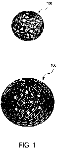
Figure 1 shows a perspective view of a schematically illustrated marking body
100 in the
expanded state.
Figure 2a shows an end view of the marking body 100 in the expanded state and
figure
2b shows a side view of the marking body 100 in the expanded state.
The marking body 100 comprises a support structure formed by a braided wire
mesh 101.
The wires 108 extend from one longitudinal end of the marking body 100 to its
other
longitudinal end. On the path from one longitudinal end to the other
longitudinal end, the
wires 108 cross other wires 108 and are braided in particular, that is to say
each wire 108
is alternately guided first below and then above another wire 108 of the
braided wire mesh
101. As a result, a lattice-like support structure with a multiplicity of
crossing points 110
arises. In relation to the depicted representation in figures 1 and 2, it
should be observed
that these crossing points 110, at which two wires 108 in each case cross over
one another
and are in lateral contact, are not reproduced with accurate detail. The
braided structure of
the marking body 100 is better depicted in figures 6 to 8, which show the
initial product.
The crossing points 110 at which two wires 108 are in contact in each case may
for example
be designed like in the braided wire mesh 101, formed by crossing wires, in
figure 6.
The free ends 112 of the wires 108 located at the respective longitudinal ends
114, 116 of
the marking body 100 are each twisted around and welded to one or more free
ends of the
further wires 108. Preferably, two wires 108 are always interconnected with
the respective
CA 03160617 2022-04-26
1
- 15 -
longitudinal ends 114, 116 by twisting and welding at a crossing point at the
respective
longitudinal end of the marking body 100.
At its longitudinal ends, the marking body 100 has two longitudinal sections
102, 104, from
where the marking body 100 flares to a central longitudinal section 106.
Consequently, the
external diameter of the marking body 100 has a maximum in the central
longitudinal
section 106.
In the illustrated example, the braided wire mesh 101 comprises 24 wires which
consist of
nitinol and have a diameter of approximately 0.1 mm. In alternative
embodiments of the
marking body not shown here, the braided wire mesh comprises between 8 and 200
wires,
for example 48 or 96 wires. In the embodiments not shown here, the marking
bodies
comprise braided wire meshes which are formed by wires with diameters ranging
between
0.05 mm and 0.15 mm. Wires that consist of other metals, for example other
titanium alloys
other than nitinol can also be used. "Wires" made of plastic, for example PEEK
or PLA,
may also be provided in alternative embodiments of the marking body.
The individual webs may have different diameters and also different cross-
sectional
shapes. Figures 3a to 3h show different cross-sectional shapes. By way of
example, the
webs can be formed as a round solid wire and have a cross section as depicted
in figure
3a. Preferably, the webs consist of a hollow wire ¨ that is to say a type of
tube ¨ which may
have a cross section as depicted in figure 3b. Such a hollow wire is
advantageous in that it
reflects sound particularly well on account of the acoustic impedance
differences between
the material of the wire wall and the hollow interior. Figures 3c and 3d
illustrate that the
cross-sectional form can also be a square, in particular quadrilateral.
Figures 3e and 3f
show a triangular cross-sectional form for webs, in the form of solid material
(figure 3i) or
as hollow webs (figure 3f). Figures 3g and 3h illustrate that the webs in
principle can each
have an arbitrary, prismatic cross-sectional shape, and hence also a hexagonal
shape as
shown in figures 3g and 3h.
Since the marking body 100 is preferably formed from a braided wire mesh, the
wires
typically contact each other once the crossing points. Then, a crossing point
can have an
appearance as depicted in exemplary fashion in figure 4a. A secure connection
between
the two crossing wires can be produced by welding at such a crossing point.
Figure 4b
illustrates this on the basis of a weld spot 118 on the crossing point. Should
the webs not
CA 03160617 2022-04-26
- 16 -
be braided but simply contact one another laterally in an arc, as depicted in
figure 4c, a
stable marking body can also be produced by virtue of the fact that the
contacting webs are
connected by welding, as depicted in figure 4d. A weld spot 118 is also shown
here. Finally,
= the webs can also be twisted at the crossing points. Figure 4e shows a
twist, within the
scope of which the webs are wrapped around one another by 360 and are
subsequently
interconnected by means of a weld spot 118; see also figure 4f. Instead of a
360 twist, a
1800 twist is also sufficient. The arising image then is similar to figure 4c,
with the exception
that the webs are then hooked in one another.
Figures 5a to 5f illustrate that the webs can be connected by welding (figure
5b), by
o twisting (figures 5c and 5e), or by twisting and welding (figures 5d and
5f) not only at the
crossing points but also at the free longitudinal ends 112. Weld beads 120
that typically
have a larger diameter than an individual web 103 or a wire that forms a web
103 then arise
as a result of welding the webs 103 at their free longitudinal ends 112.
The marking body 100 has a length LM of 6 mm; in alternative embodiments not
shown
here, this length may also range between 4 mm and 8 mm, however.
The maximum external diameter DMA of the marking body in the central
longitudinal
section 106 is 4 mm and can be between 3.5 mm and 10 mm in alternative
embodiments
not shown here.
To bring the marking body 100 into an elastically compressed state from the
expanded
zo state, a radial force of at least one newton must be exerted on the
marking body 100.
In alternative embodiments not shown here, the self-expanding marking body 100
may
have more wires and accordingly more crossing points, and so said marking body
is
comparatively stiffer. Accordingly, a comparatively greater radial force then
is required to
bring the marking body into an elastically compressed state. Likewise, the
number of wires
can be lower in alternative embodiments not shown here, in order to realize a
marking body
which already transitions into its elastically compressed state when a radial
force of less
than one newton is exerted.
Figures 3, 4 and 5 illustrate various phases of a production method for
producing a marking
body which has a support structure formed by a braided wire mesh. By way of
example, a
CA 03160617 2022-04-26
- 17 -
marking body as described in relation to figures 1 and 2 can be produced in
accordance
with the method described below.
Initially, a tubular braided wire mesh is provided, the latter for example
being able to
comprise between 8 and 200 individual wires which are braided with one another
and, as
a consequence, cross at crossing points. These are 24 individual wires in the
depicted
example.
As may be gathered from figure 2b, the marking body 100 preferably has a
length LM
ranging between 5 mm and 8 mm. The external diameter DMA in the fully expanded
state
is between 4 mm and 6 mm. The diameter of the individual wires 108 is
preferably slightly
less than 0.1 mm. The weld beads 120 at the free ends 112 of the wires have a
diameter
of greater than 0.1 mm, the latter preferably being at least 0.12 mm Hence,
the marking
body 100 is suitable for use with an implantation apparatus 1004 in which the
difference
between an internal cannula diameter DKI and a driving-out element external
diameter DA
is no more than 0.1 mm ¨ even when the manufacturing tolerances are taken into
account.
Incidentally, the internal cannula diameter DKI is not necessarily greater
than the external
diameter d of the compressed marking body 100.
As can already be gathered from figure 3, the webs of the support structure
can either be
solid (wires) or hollow (tubes). The profiles may have a circular or
ellipsoidal cross section.
They may also be triangular, quadrilateral or in the form of an n-gon. The
profiles can also
zo change along a web. By way of example, a web could have a rectangular
profile in the
center and a circular profile at the longitudinal ends.
Figure 4, which is explained in more detail above, shows examples of possible
interlocking
connections as a result of crossing, contacting or twisting at the crossing
points. Figure 4
likewise illustrates that the connection of the crossing points can
additionally be cohesive,
for example as a result of adhesive bonding, welding or soldering.
Figure 5, likewise explained in more detail above, shows examples of possible
forms of
interlocking and cohesive connections at the free longitudinal ends 112 of the
wires 108.
The ends can be interconnected in pairs or in groups; see also figure 18.
CA 03160617 2022-04-26
- 18 -
What can likewise be gathered from figure 6 is that the free longitudinal ends
218 of the
wires 202 are not only welded to one another but also twisted around one
another. In
combination, this ensures that the interconnected longitudinal ends of the
wires do not
separate from one another.
Unlike what is depicted in idealized fashion in the figures 3 to 5, the
longitudinal ends of
the individual wires 202 are not all exactly in one (separation) plane 212
(see figures 4 and
5), but are alternately slightly offset in relation to such an idealized
plane, preferably in the
longitudinal direction. This has the advantageous effect that the marking body
200 can be
better compressed at its longitudinal ends 218 because the weld beads 220 are
not all
located next to one another but are at least slightly offset from one another
in the
longitudinal direction of the marking body 200.
The marking body 100 is preferably formed from a braided wire mesh 200, as
depicted in
figure 6 in exemplary fashion. Figure 6 shows a braided wire mesh 200 as a
section of a
braided wire tube 202 (see figure 7), which is braided from 24 individual
wires in the
depicted example. The braided wire mesh 200 that will form the marking body
100 is formed
from 24 individual wires 108 which cross under or over one another nine times
between
their longitudinal ends 112 and which are twisted around one another and
welded to one
another at their longitudinal ends 112 in pairs such that the braided wire
mesh 200 has
respective weld beads 120 at the longitudinal ends 112 of the wires 108. As
can be
gathered from figure 6, the longitudinal ends 112 of the interconnected wires
108 are not
only welded to one another but also twisted around one another.
To produce a braided wire mesh 200 as depicted in figure 3, a wire tube 202 as
depicted
in figure 4 is produced first. To produce the tube 202, 24 individual wires
108, for example,
are braided with one another such that they alternately cross over and under
one another
at the crossing points 210. Crossing point planes 214 that extend transversely
to a
longitudinal direction of the tube 202 arise in this way. Once the individual
wires 108 have
each crossed one another in pairs nine times, two individual wires are twisted
around one
another in each case such that twists 216 arise. The wire tube 202 thus forms
crossing
point planes 214 that alternate with separating planes 212 at which a
respective braided
wire mesh 200 should be separated from the wire tube 202. In the example
illustrated, nine
crossing point planes 214 are followed in each case by a respective separation
plane 212.
In the separation planes, the wires 108 are in each case fully wrapped about
one another
CA 03160617 2022-04-26
- 19 -
twice in pairs such that a wrap-around angle of 7200 arises. In other
exemplary
embodiments not shown, the wrap-around angle can also be only 360 or else 540
.
Figure 7 shows the tube 202 formed by the wires 108, the tube 206 having been
separated
at two separation sites 208 by means of a laser beam. The separation sites 208
are situated
in precisely one separation plane 212, that is to say where the twists 216 are
situated. The
weld beads 120 arise from the laser cutting such that the then free, pairwise
interconnected
longitudinal ends 112 of the wires 108 are interconnected both by twisting and
by a laser
welding. As a result of the twisting 216, the connected longitudinal ends 112
of in each
case two wires 108 arise as depicted in figures 5d and 5f following the laser
cutting.
Following the separation of the braided wire mesh 200 from the wire tube 202,
the former
can be shaped into the marking body 100 by virtue of being compressed in the
longitudinal
direction. As a result, the braided wire mesh 200 bulges outward in a central
longitudinal
section while the longitudinal ends are constricted if the compression is for
example
implemented by means of two tools, each formed by a hollow hemisphere, moving
toward
.. one another. Depending on the tool shape, the marking body 100 can adopt
shapes as
depicted in longitudinal sections in figures 9a to 9c.
Figures 10, 11, 12 and 13 show an implantation apparatus 1004 for implanting a
marking
body 100. The implantation apparatus 1004 comprises a handle 1010 and an
implantation
part 1008. The cannula 1006, in which the marking body 100 is initially
situated, is part of
the implantation part 1008.
A cannula tip 1012 at the distal end of the cannula 1006 has been whetted in
such a way
that it facilitates a percutaneous implantation of the marking body 100 by
piercing the
cannula 1006 into body tissue. The cannula 1006 preferably consists of
stainless steel.
To eject the marking body 100 from the cannula 1006, provision is made of a
displaceable
driving-out element 1018, which can be actuated from the handle 1010 by means
of the
sliding element 1016.
Figure 12A shows an implantation system 1000 having a marking body 100 of an
implantation apparatus 1004. In this case, the marking body 100 in the pre-
loaded state,
that is to say with a compressed support structure, is situated within the
cannula 1006 of
CA 03160617 2022-04-26
- 20 -
the implantation apparatus 1004. This state of the implantation system 1000
represents a
typical delivery state, in which the implantation system 1000 is made
available in a ready-
to-use state for the user, for example a surgeon.
The implantation part 1008 of the implantation apparatus 1004 substantially
consists of a
cannula 1006 which has a cannula tip 1012 at its distal end, that is to say
the end distant
from the handle 1010. As a rule, the marking body 100 in the preloaded state
is situated in
this region within the cannula 1006, just inside the outlet at the cannula tip
1012. In
particular, the cannula 1006 can be formed from a suitable metal.
The cannula 1006 has a length LKA which for example can adopt a value ranging
between
25 mm and 200 mm, preferably between 50 mm and 150 mm. The length LKA of the
cannula 1006 has an influence on the range of the implantation apparatus 1004
in respect
of the reachability of tissue sites in the body of a patient to be labeled.
The longer cannulas
are used when adjustment aids are used, for example stereotaxis.
The implantation apparatus 1004 comprises a handle 1010 and an implantation
part 1008.
The handle 1010 comprises a handle housing 1014 and a sliding element 1016,
which for
example could be produced from a suitable plastic.
The sliding element 1016 is connected to the handle housing 1014 but is
movable relative
to the handle housing 1014 in the axial direction of the cannula 1006.
Consequently, the
sliding element 1016 can be moved along a straight, guided sliding path
between a pre-
loaded position 1020 and a driving-out position 1022.
This movement is transferred from the sliding element 1016 via a driving-out
element 1018,
which is connected to the sliding element 1016 and which can be formed for
example by
way of a wire or a sufficiently stable plastics fiber, to the distal region at
a distance from the
handle 1010. Consequently, when the sliding element 1016 is moved to the
driving-out
position 1022, the pre-loaded marking body 100 can be driven out of the
cannula 1006 to
the tissue site to be labeled at the distal end of the cannula 1006 by way of
a sliding
movement of the driving-out element 1018.
This is achieved by virtue of the driving-out element 1018 that is aligned
coaxially with
respect to the cannula 1006 being moved in the direction of the cannula tip
1012 and hence
CA 03160617 2022-04-26
- 21 -
pushing the pre-loaded marking body 100 out of the cannula 1006 past the
cannula tip
1012.
Figure 12B depicts detail B of figure 12A, specifically a detailed view in the
region of the
cannula tip 1012 of the implantation system 1000 in the pre-loaded state. In
this view, the
marking body 100, in particular, can be seen in the compressed state, said
marking body
being situated within the cannula 1006 behind the driving-out element 1018 and
in front of
the cannula tip 1012 from the view of the handle 1010. On account of its
prestress, the
marking body maintains the position in the cannula 1006 and cannot fall out on
its own. On
account of this property, additional features or apparatuses for fixing the
marking body 100
within the cannula 1006 can be dispensed with.
Figure 12C shows a further detailed, schematic view of the cannula 1006, this
time as detail
C from figure 12B. In this view, the distal end of the driving-out element
1018 is visible
within the cannula 1006. Furthermore, the external diameter DKA and the
internal diameter
DKI of the cannula 1006 are labeled.
Together with the cannula length LKA, the internal diameter DKI of the cannula
1006
describes the size of the internal cavity formed by the cannula 1006 and at
the same time
restricts the maximum possible diameter DM of the marking body 100 in the
compressed
state or, optionally, the maximum possible diameter DK of a clamp (should the
marking
body comprise the latter), in order to ensure an ability of the marking body
100 to pass
through or move in the cannula 1006 during pre-loading and driving out. An
internal
diameter DKI of less than 1.1 mm, particularly preferably of 1.0 mm was found
to be
preferable.
The external diameter DKA of the cannula 1006 describes the diameter of the
external
cannula wall. Under the assumption of a constant cannula wall thickness that
is as small
as possible, the internal diameter DKI of the cannula 1006 simultaneously
increases with
increasing external diameter DKA, and hence there also is an increase in the
maximum
possible external diameter of a marking body 100 to be implanted. However, at
the same
time, an increasing external diameter DKA leads to a greater degree of
invasiveness or
injury to skin and tissue when carrying out the implantation.
CA 03160617 2022-04-26
- 22 -
A sufficiently small external diameter DI<A ensures the option of a
percutaneous
implantation of the marking body 100 without having to resort to a stab
incision of the skin
at the entry site of the cannula 1006 or anesthetization of the relevant
tissue. An external
diameter DKA of between 1 mm and 1.5 mm, particularly preferably of 1.2 mm was
found
to be preferable.
By means the implantation apparatus, a marking body of the type presented here
for
percutaneous marking can be implanted into soft tissue, such as breast tissue
or axillary
lymph nodes following a lymph node biopsy.
The fields of application include the marking of suspicious tissue, the
marking of lesions
before or during chemotherapy, and the marking of a biopsy removal site. The
location of
a removed tumor may likewise be marked for improved orientation within the
scope of
radiation treatment planning.
By way of example, within the scope of an intervention, the marking body 100
is used as
follows:
Initially, the marking body is implanted at a desired site by virtue of the
distal end 1012 of
the cannula 1006 of the implantation apparatus 1004 being pierced up to the
desired
implantation location in body tissue and a marking body 100 being ejected from
the distal
end 1012 of the cannula 1006.
Subsequently, the body tissue can be examined using an imaging ultrasound
method for
example, an ultrasound recording of the marked tissue being made. This is
depicted in
figures 14 and 15. The marking body can be recognized in the ultrasound
recording on
account of a circular artifact 310 or X-shaped artifact 312; see figures 16
and 17.
In the case of sonography using medical ultrasound (1 MHz to 40 MHz, for
example in the
B mode [brightness modulation]; the mode in which two-dimensional brightness
images are
generated), the support structure of the marking body 100 causes incident
ultrasound
waves in the central longitudinal section of the marking body to strike a
structure that is
circular in cross section. What is obtained by matching the parameters of web
diameter (or
width and thickness), web number, web intensity and web material is that only
some of the
acoustic energy is reflected by the structure and the remaining part of the
energy is
CA 03160617 2022-04-26
- 23 -
transmitted, as depicted in exemplary fashion in figure 15. As a result, a
full circle or a
circular arrangement of individual points, depending on resolution and
parameter settings
of the ultrasound, arises as a representation in the ultrasound image; see
figure 16. In the
case of other structures of this form, the ultrasound energy would be largely
reflected at
the first surface of the marker and a shadow would arise in the image. A
marking body 100
of the type described here can consequently be distinguished from other
markers with a
similar shape.
To make ultrasound recordings by means of sonography, use is made of a probe
300 which
can transmit ultrasound 302 and can receive reflected ultrasound. By way of
example, if
io the transmitted ultrasound strikes an object with a different acoustic
impedance to the
surrounding body tissue, the ultrasound is scattered and partly 304 reflected
back to the
probe 300; see figure 15. The wires of a marking body can be ultrasound-
reflecting objects.
A marking body of the type described here is then represented in ultrasound
recordings
(sonography recordings) as is indicated in figure 14 and as is visible in
figures 16 and 17.
The characteristic circular shape of the image representation of the marking
body in the
ultrasound image allows the marking body to be automatically recognized and
hence
detected in an ultrasound recording, for example by means of an appropriately
trained
convolutional neural network (CNN).
The marking body 100 can be placed in a soft tissue without air inclusions
(e.g., fatty tissue)
and can be insonated with ultrasonic waves ranging between 1 MHz and 40 MHz.
It then
reflects only part of the ultrasonic power at the first side, which faces the
ultrasound, and
reflects a second part of the ultrasonic power at the second side, which is
distant from the
ultrasound, as a result of which the marking body has a circular
representation in an
ultrasound image within the scope of medical ultrasound imaging; see figures
14 and 15.
Then, the marking body is detectable by means of an automatic or semiautomatic
method,
preferably by analyzing ultrasound data or by analyzing x-ray recordings or by
analyzing
MRI data.
To improve the visibility in the ultrasound image, or for other purposes, the
marking body
can be provided with a membrane 400. This can be realized in various ways; see
figures
19 and 20. By way of example, the marking body 100 can be coated with a
membrane 400
CA 03160617 2022-04-26
- 24 -
from the outside or from the inside; see figures 20b and 20d. The membrane 400
can be
produced from silicone or polyurethane or parylene, for example. The membrane
serves to
fully or partly separate the interior of the marking body from the
surroundings. This can
improve the recognizability and detectability, or this can create a space
which can be filled
with other substances or gases (e.g., cytostatic agents, ICG, etc.). As shown
in figure 19,
the membrane may also only be spanned between the relevant wires in individual
fields of .
the lattice-like support structure formed by the wires 108.
CA 03160617 2022-04-26
- 25 -
List of reference signs
100 Marking body
101 Braided wire mesh
102, 104 Constricted longitudinal sections
103 Webs
105 Crossing points
106 Central longitudinal section
108 Wires
110 Crossing points
112 Free ends of the wires
114, 116 Longitudinal ends of the marking body
118 Weld spot
120 Weld beads
200 Braided wire mesh
202 Wire tube
208 Separation point
212 (Separation) plane
214 Crossing point plane
216 Twist
300 Ultrasound probe
302 Transmitted ultrasound
304 Reflected ultrasound
310, 312 Image representation of a marking body in the ultrasound image
400 Membrane
LM Length of the marking body
DMA Maximum external diameter of the marking body in the flared state
DKI Internal cannula diameter
DKA External cannula diameter
LKA Cannula length
1000 Implantation system
1004 Implantation apparatus
1005 Clamp
1006 Cannula
1008 Implantation part
1010 Handle
CA 03160617,2022-04-26
- 26 -
1012 Cannula tip
1014 Handle housing
1016 Sliding element
1018 Driving-out element
1020 Pre-loaded position
1022 Driving-out position
1102 Wires
1104 Braided wire mesh
1106, 1108 Longitudinal ends of the marking body
1110 Crossing points
1112 Central longitudinal section
1118 Free ends
1120 Weld beads
1122 Sleeve
1124 Weld spot
1200 Braided wire mesh
1202 Wire tube
1206 Twist
1210 Crossing point planes
1212 Separation plane
1214 Separation points
1300 Circular artifact
1302 X-shaped artifact