Note: Descriptions are shown in the official language in which they were submitted.
CA 03160977 2022-05-10
WO 2021/096766
PCT/US2020/059304
TRANSCATHETER MEDICAL IMPLANT DELIVERY
CROSS REFERENCE TO RELATED APPLICATIONS
[0001] This application claims the benefit of U.S. Provisional Patent
Application
No. 62/935,214, filed November 14, 2019, entitled "TRANSCATHETER MEDICAL
IMPLANT DELIVERY", the disclosure of which is hereby expressly incorporated by
reference herein in its entirety for all purposes.
BACKGROUND
Field
[0002] The present disclosure generally relates to the field of
transcatheter
delivery of medical implant devices and/or therapies.
Description of Related Art
[0003] Transcatheter delivery of medical implant devices and/or
therapies to a
target vessel, channel, chamber and/or organ can be performed to treat various
ailments.
Delivery of medical implant devices and/or therapies to the heart can be
performed to address
various heart conditions, including elevated pressure in the left atrium.
SUMMARY
[0004] Described herein are systems and methods for providing minimally
invasive transcatheter delivery of medical implant devices and/or therapies,
including
delivery of a medical implant device to the left atrial wall for alleviating
elevated left atrial
pressure.
[0005] In some implementations, a medical implant delivery system can
comprise
a puncture needle, a medical implant delivery catheter, a medical implant
device, and an
outer delivery catheter. The puncture needle can comprise an elongate portion
and a puncture
component associated with a distal end of the elongate portion, the puncture
component being
configured to pierce and form an opening in a target tissue wall. The medical
implant
delivery catheter can comprise a puncture needle lumen, the puncture needle
being
configured to be advanced through the puncture needle lumen, the puncture
component being
configured to be extended through a puncture needle outlet opening associated
with a distal
portion of the medical implant delivery catheter. The medical implant device
can be
positioned on the medical implant delivery catheter. The outer delivery
catheter can comprise
1
CA 03160977 2022-05-10
WO 2021/096766
PCT/US2020/059304
a medical implant delivery lumen, the medical implant delivery catheter being
configured to
be advanced through the medical implant delivery lumen, the distal portion of
the medical
implant delivery catheter being configured to be received within the medical
implant delivery
lumen and extended through a side outlet opening associated with a distal
portion of the outer
delivery catheter.
[0006] In some embodiments, the medical implant delivery system can
comprise a
puncture needle sheath configured to be passed through the puncture needle
lumen, wherein
the puncture needle is configured to be passed through a lumen of the puncture
needle sheath.
A radially expandable member can be associated with a distal portion of the
puncture needle
sheath, wherein the radially expandable member is configured to be positioned
within the
opening formed in the target tissue wall and to enlarge the opening when the
radially
expandable member is in an expanded configuration. In some embodiments, the
medical
implant delivery system can comprise a medical implant delivery guide wire,
wherein the
medical implant delivery guide wire is configured to be slidably advanced
through the lumen
of the puncture needle sheath and to be exchangeable with the puncture needle.
[0007] In some embodiments, a distal portion of the medical implant
delivery
catheter is configured to be inserted into the opening formed in the target
tissue wall, the
distal portion comprising a predetermined diameter configured to enlarge the
opening.
[0008] In some embodiments, the medical implant delivery system can
comprise
an outer delivery catheter guide wire, and wherein the outer delivery catheter
comprises a
guide wire lumen configured to receive the outer delivery catheter guide wire.
In some
embodiments, the outer delivery catheter guide wire lumen extends along an off-
center
longitudinal axis of the outer delivery catheter.
[0009] In some embodiments, a distal portion of the medical implant
delivery
catheter comprises a lateral cross section having a shape configured to
interlock with a shape
of the side outlet opening on the distal portion of the outer delivery
catheter to orient the
medical implant delivery catheter as the medical implant delivery catheter
extends through
the side outlet opening, wherein the lateral cross section extends along a
lateral dimension of
the distal portion of the outer delivery catheter.
[0010] In some embodiments, the medical implant delivery catheter
comprises a
first tapered distal portion comprising a first pre-formed curvature and the
outer delivery
catheter comprises a second tapered distal portion comprising a second pre-
formed curvature,
wherein the first curvature and the second curvature comprise the same
orientation. In some
embodiments, the first pre-formed curvature comprises a radius of curvature
smaller than that
2
CA 03160977 2022-05-10
WO 2021/096766
PCT/US2020/059304
of the second pre-formed curvature. In some embodiments, the second pre-formed
curvature
comprises a shape configured to conform to at least a portion of a curvature
along a length of
a coronary sinus. In some embodiments, the side outlet opening is on an inner
edge of the
second pre-formed curvature of the outer delivery catheter.
[0011] In some embodiments, the puncture needle comprises a third pre-
formed
curvature on a distal portion of the puncture needle. In some embodiments, the
third pre-
formed curvature comprises a radius of curvature smaller than that of the
first pre-formed
curvature of the medical implant delivery catheter.
[0012] In some embodiments, the medical implant delivery system can
comprise
an expandable anchor, wherein the expandable anchor and the side outlet
opening are on
opposing portions of the outer delivery catheter, and wherein the expandable
anchor is
configured to assume an expanded configuration to position the outer delivery
catheter
against the target tissue wall. In some embodiments, the outer delivery
catheter comprises a
second tapered distal portion comprising a second pre-formed curvature and
wherein the
anchor is on an outer edge of the second pre-formed curvature.
[0013] In some implementations, a method for delivering a medical
implant
device can comprise advancing a guide wire into a vessel, and advancing a
medical implant
delivery system along the guide wire into the vessel. The medical implant
delivery system
can comprise an outer delivery catheter comprising a medical implant delivery
lumen, a
medical implant delivery catheter comprising a medical implant device
positioned thereon
and a puncture needle lumen extending therethrough, the medical implant
delivery catheter
slidably extending through the medical implant delivery lumen, and a puncture
needle
slidably extending through the puncture needle lumen, and a puncture component
of the
puncture needle extending through a distal outlet opening on a distal end of
the medical
implant delivery catheter. The method can comprise advancing the medical
implant delivery
catheter through a side outlet opening on a distal portion of the outer
delivery catheter and
piercing tissue at a target tissue site using the puncturing component
extending through the
distal outlet opening to form an opening in the tissue. A distal portion of
the medical implant
delivery catheter can be extended through the opening formed in the tissue;
and the medical
implant device positioned on the distal portion of the medical implant
delivery catheter can
be deployed into the opening formed in the tissue.
[0014] In some embodiments, the method can comprise inserting a
radially
expandable member positioned on a distal portion of a puncture needle sheath
in the opening
formed in the tissue, and expanding the radially expandable member to enlarge
the opening,
3
CA 03160977 2022-05-10
WO 2021/096766
PCT/US2020/059304
wherein the puncture needle sheath slidably extends through the puncture
needle lumen, and
the puncture needle slidably extends through a lumen of the puncture needle
sheath.
[0015] In some embodiments, the method can comprise exchanging the
puncture
needle for a medical implant delivery guide wire and inserting the medical
implant delivery
guide wire through the lumen of the puncture needle sheath.
[0016] In some embodiments, the method can comprise inserting a distal
portion
of the medical implant delivery catheter into the opening formed at the target
tissue site to
enlarge the opening.
[0017] In some embodiments, the method can comprise orienting a distal
portion
of the medical implant delivery catheter within the medical implant delivery
lumen and
extending the distal portion through the side outlet opening, wherein
orienting comprises
rotating the distal portion of the medical implant delivery catheter to match
a shape of a
lateral cross section of the distal portion with a shape of the side outlet
opening, and wherein
the lateral cross section extends along a lateral dimension of the distal
portion of the outer
delivery catheter.
[0018] In some embodiments, the method can comprise expanding an
expandable
anchor on the distal portion of the outer delivery catheter to contact the
expandable anchor
with a wall of the vessel.
[0019] In some embodiments, positioning the guide wire into a vessel
comprises
positioning the guide wire transfemorally into a coronary sinus through a
right atrium via an
ostium of the coronary sinus. In some embodiments, piercing tissue at the
target tissue site
comprises piercing tissue of a left atrial wall to form the opening in the
left atrial wall, and
wherein deploying the medical implant device comprises deploying the medical
implant
device into the opening formed in the left atrial wall.
[0020] In some embodiments, a medical implant delivery system can
comprise a
puncture needle, a medical implant delivery catheter, a medical implant
device, an elongate
housing, and an outer sheath. The puncture needle can comprise an elongate
portion and a
puncture component at a distal end of the elongate portion, the puncture
component being
configured to pierce tissue to form an opening in the tissue. The medical
implant delivery
catheter can comprise a puncture needle lumen and the puncture needle being
configured to
slidably extend through the puncture needle lumen, and the puncture component
being
configured to extend through a puncture needle outlet opening at a distal end
of the medical
implant delivery catheter. The medical implant device can be carried on the
medical implant
delivery catheter. The elongate housing can be adjacent to and be slidable
relative to the
4
CA 03160977 2022-05-10
WO 2021/096766
PCT/US2020/059304
medical implant delivery catheter, wherein a distal end of the elongate
housing is distal of the
distal end of the medical implant delivery catheter when the medical implant
delivery
catheter is in a retracted configuration. The outer sheath can be around a
portion of the
medical implant delivery catheter and the elongate housing to maintain the
medical implant
delivery catheter adjacent to the elongate housing, wherein the outer sheath
is slidable
relative to the medical implant delivery catheter and extends over the distal
end of the
medical implant delivery catheter when the medical implant delivery catheter
is in the
retracted configuration.
[0021] In some embodiments, the elongate housing comprises a recess
along at
least a portion of a length of the elongate housing, a portion of the medical
implant delivery
catheter being received in the recess. In some embodiments, the elongate
housing comprises a
guide wire lumen configured to receive an elongate housing guide wire, and the
distal end of
the elongate housing comprises a guide wire outlet opening configured to allow
extension
therethrough of the elongate housing guide wire.
[0022] In some embodiments, the medical implant delivery catheter
comprises a
first tapered distal portion comprising a first pre-formed curvature and the
elongate housing
comprises a second tapered distal portion comprising a second pre-formed
curvature, wherein
the first pre-formed curvature and the second pre-formed curvature comprise
the same
orientation. In some embodiments, the first pre-formed curvature comprises a
radius of
curvature smaller than that of the second pre-formed curvature. In some
embodiments, the
second pre-formed curvature comprises a shape configured to conform to a
curvature along a
length of a coronary sinus.
[0023] In some embodiments, the medical implant delivery system can
comprise
an expandable anchor on the distal portion of the elongate housing opposite a
portion of the
elongate housing adjacent to the medical implant delivery catheter, the
expandable anchor
being configured to assume an expanded configuration to position the medical
implant
delivery catheter against a target tissue site. k some embodiments, the
elongate housing
comprises a second tapered distal portion comprising a second pre-formed
curvature and the
expandable anchor is on an outer edge of the second pre-formed curvature.
[0024] In some embodiments, the medical implant delivery system can
comprise a
puncture needle sheath slidably extending through the puncture needle lumen,
and the
puncture needle sheath slidably extending through the puncture needle. A
radially expandable
member can be on a distal portion of the puncture needle sheath, the radially
expandable
member being configured to enlarge the opening formed in the tissue.
CA 03160977 2022-05-10
WO 2021/096766
PCT/US2020/059304
[0025] In some embodiments, the puncture needle is configured to be
exchangeable for a medical implant delivery guide wire.
[0026] In some implementations, a method for delivering a medical
implant
device can comprise positioning a guide wire into a vessel, and advancing a
medical implant
delivery system along the guide wire into the vessel. The medical implant
delivery system
can comprise an elongate housing, a medical implant delivery catheter adjacent
to the
elongate housing, the medical implant delivery catheter carrying a medical
implant device
and comprising a puncture needle lumen, a puncture needle slidably extending
through the
puncture needle lumen, and a puncture component of the puncture needle
extending through a
distal outlet opening on a distal end of the medical implant delivery
catheter, and an outer
sheath around the elongate housing and the medical implant delivery catheter.
The method
can comprise proximally sliding the outer sheath to release a distal portion
of the medical
implant delivery catheter, distally sliding the medical implant delivery
catheter relative to the
elongate housing and piercing tissue at a target tissue site to form an
opening in the tissue
using the puncture component extending through the distal outlet opening of
the medical
implant delivery catheter, extending the distal portion of the medical implant
delivery
catheter through the opening in the tissue, and deploying the medical implant
device carried
on the medical implant delivery catheter into the opening.
[0027] In some embodiments, the method can comprise positioning a
radially
expandable member on a distal portion of a puncture needle sheath in the
opening formed in
the tissue, and expanding the radially expandable member to enlarge the
opening, wherein the
puncture needle sheath slidably extends through the puncture needle lumen, and
the puncture
needle slidably extends through a lumen of the puncture needle sheath. In some
embodiments, the method can comprise exchanging the puncture needle for a
medical
implant delivery guide wire and inserting the medical implant delivery guide
wire through the
lumen of the puncture needle sheath.
[0028] In some embodiments, the method can comprise inserting the
distal
portion of the medical implant delivery catheter into the opening formed at
the target tissue
site to enlarge the opening.
[0029] In some embodiments, the method can comprise expanding an
expandable
anchor on a distal portion of the elongate housing to contact the expandable
anchor with a
wall of the vessel.
[0030] In some embodiments, positioning the guide wire into the vessel
comprises
positioning the guide wire transfemorally into a coronary sinus through a
right atrium via an
6
CA 03160977 2022-05-10
WO 2021/096766
PCT/US2020/059304
ostium of the coronary sinus. In some embodiments, piercing tissue at the
target tissue site
comprises piercing tissue of a left atrial wall, and wherein deploying the
medical implant
device comprises deploying the medical implant device into the opening formed
in the left
atrial wall.
[0031] For purposes of summarizing the disclosure, certain aspects,
advantages
and novel features have been described herein. It is to be understood that not
necessarily all
such advantages may be achieved in accordance with any particular embodiment.
Thus, the
disclosed embodiments may be carried out in a manner that achieves or
optimizes one
advantage or group of advantages as taught herein without necessarily
achieving other
advantages as may be taught or suggested herein.
BRIEF DESCRIPTION OF THE DRAWINGS
[0032] Various embodiments are depicted in the accompanying drawings
for
illustrative purposes and should in no way be interpreted as limiting the
scope of the
inventions. In addition, various features of different disclosed embodiments
can be combined
to form additional embodiments, which are part of this disclosure. Throughout
the drawings,
reference numbers may be reused to indicate correspondence between reference
elements.
However, it should be understood that the use of similar reference numbers in
connection
with multiple drawings does not necessarily imply similarity between
respective
embodiments associated therewith. Furthermore, it should be understood that
the features of
the respective drawings are not necessarily drawn to scale, and the
illustrated sizes thereof are
presented for the purpose of illustration of inventive aspects thereof.
Generally, certain of the
illustrated features may be relatively smaller than as illustrated in some
embodiments or
configurations.
[0033] Figure 1 is a cross-sectional view of a human heart.
[0034] Figure 2 is another cross-sectional view of the human heart.
[0035] Figure 3 is a side view of an example of a medical implant
delivery system
in accordance with one or more embodiments.
[0036] Figure 4 is another side view of the medical implant delivery
system of
Figure 3, in accordance with one or more embodiments.
[0037] Figure 5 is a side view of an outer delivery catheter of the
medical implant
delivery system of Figure 3, in accordance with one or more embodiments.
[0038] Figure 6 is a side view of a medical implant delivery catheter
of the
medical implant delivery system of Figure 3, in accordance with one or more
embodiments.
7
CA 03160977 2022-05-10
WO 2021/096766
PCT/US2020/059304
[0039] Figure 7 shows a proximal handle of the medical implant delivery
system
of Figure 3, in accordance with one or more embodiments.
[0040] Figures 8A and 8B are longitudinal cross-sectional views of the
medical
implant delivery system of Figure 3 in a first configuration and a second
configuration,
respectively, in accordance with one or more embodiments.
[0041] Figure 9 shows a lateral cross-sectional view of the outer
delivery catheter
of Figure 5, in accordance with one or more embodiments.
[0042] Figure 10A is a top-down view, and Figure 10B is a longitudinal
cross-
sectional view, of an outer delivery catheter of an example of a medical
implant delivery
system in accordance with one or more embodiments.
[0043] Figures 10C and 10D provide top-down and side views,
respectively, of a
medical implant delivery catheter of the medical implant delivery system of
Figures 10A and
10B, in accordance with one or more embodiments.
[0044] Figure 11 shows insertion of a guide wire into the coronary
sinus, in
accordance with one or more embodiments.
[0045] Figure 12 shows advancement of the medical implant delivery
system of
Figure 3 into the coronary sinus, in accordance with one or more embodiments.
[0046] Figure 13 shows use of the puncture needle of the medical
implant
delivery system of Figure 3 to puncture tissue at the target tissue site on
the left atrial wall, in
accordance with one or more embodiments.
[0047] Figure 14 shows the puncture needle of the medical implant
delivery
system of Figure 3 inserted into the left atrium, in accordance with one or
more
embodiments.
[0048] Figure 15 shows deployment of a shunt device onto the left
atrial wall
using the medical implant delivery system of Figure 3, in accordance with one
or more
embodiments.
[0049] Figure 16 shows withdrawal of the medical implant delivery
system of
Figure 3 from the coronary sinus after deployment of the medical implant
device onto the left
atrial wall, in accordance with one or more embodiments.
[0050] Figure 17 is a process flow diagram of an example of a process
to deploy
the medical implant delivery system of Figure 3 to deliver a medical implant
device, in
accordance with one or more embodiments.
[0051] Figure 18 is a side view of another example of a medical implant
delivery
system, in accordance with one or more embodiments.
8
CA 03160977 2022-05-10
WO 2021/096766
PCT/US2020/059304
[0052] Figure 19 is a side view of the medical implant delivery system
of Figure
18 comprising the puncture needle in an extended configuration, in accordance
with one or
more embodiments.
[0053] Figure 20 is a side view of the medical implant delivery system
of Figure
18 with the outer sheath translated proximally, in accordance with one or more
embodiments.
[0054] Figure 21 shows advancement of the medical implant delivery
system of
Figure 18 into the coronary sinus, in accordance with one or more embodiments.
[0055] Figure 22 shows proximal translation of the outer sheath and
distal
translation of the medical implant delivery catheter of the medical implant
delivery system of
Figure 18, while the medical implant delivery system is positioned in the
coronary sinus, in
accordance with one or more embodiments.
[0056] Figure 23 shows the puncture needle of the medical implant
delivery
system of Figure 18 inserted into the left atrium, in accordance with one or
more
embodiments.
[0057] Figure 24 shows deployment of a shunt device onto the left
atrial wall
using the medical implant delivery system of Figure 18, in accordance with one
or more
embodiments.
[0058] Figure 25 is a process flow diagram of an example of a process
to deploy
the medical implant delivery system of Figure 18 to deliver a medical implant
device, in
accordance with one or more embodiments.
DETAILED DESCRIPTION
[0059] The headings provided herein are for convenience only and do not
necessarily affect the scope or meaning of the claimed invention.
[0060] The present disclosure relates to systems and methods for
providing
minimally invasive transcatheter delivery of medical implant devices and/or
therapies to a
target tissue site on a vessel, channel, chamber and/or organ, including
delivery of a shunt
device to a target tissue wall, such as a left atrial wall.
[0061] Although certain preferred embodiments and examples are
disclosed
below, inventive subject matter extends beyond the specifically disclosed
embodiments to
other alternative embodiments and/or uses and to modifications and equivalents
thereof.
Thus, the scope of the claims that may arise herefrom is not limited by any of
the particular
embodiments described below. For example, in any method or process disclosed
herein, the
acts or operations of the method or process may be performed in any suitable
sequence and
9
CA 03160977 2022-05-10
WO 2021/096766
PCT/US2020/059304
are not necessarily limited to any particular disclosed sequence. Various
operations may be
described as multiple discrete operations in turn, in a manner that may be
helpful in
understanding certain embodiments; however, the order of description should
not be
construed to imply that these operations are order dependent. Additionally,
the structures,
systems, and/or devices described herein may be embodied as integrated
components or as
separate components. For purposes of comparing various embodiments, certain
aspects and
advantages of these embodiments are described. Not necessarily all such
aspects or
advantages are achieved by any particular embodiment. Thus, for example,
various
embodiments may be carried out in a manner that achieves or optimizes one
advantage or
group of advantages as taught herein without necessarily achieving other
aspects or
advantages as may also be taught or suggested herein.
[0062] Certain standard anatomical terms of location are used herein to
refer to
the anatomy of animals, and namely humans, with respect to the preferred
embodiments.
Although certain spatially relative terms, such as "outer," "inner," "upper,"
"lower," "below,"
"above," "vertical," "horizontal," "top," "bottom," and similar terms, are
used herein to
describe a spatial relationship of one device/element or anatomical structure
to another
device/element or anatomical structure, it is understood that these terms are
used herein for
ease of description to describe the positional relationship between
element(s)/structures(s), as
illustrated in the drawings. It should be understood that spatially relative
terms are intended
to encompass different orientations of the element(s)/structures(s), in use or
operation, in
addition to the orientations depicted in the drawings. For example, an
element/structure
described as "above" another element/structure may represent a position that
is below or
beside such other element/structure with respect to alternate orientations of
the subject patient
or element/structure, and vice-versa.
[0063] Various features of a heart 1 are described with reference to
Figure 1 to
assist in understanding the present disclosure. The heart 1 includes four
chambers, namely the
left atrium 2, the left ventricle 3, the right ventricle 4, and the right
atrium 5. A wall of
muscle, referred to as the septum 10, separates the left atrium 2 and right
atrium 5, and the
left ventricle 3 and right ventricle 4. Blood flow through the heart 1 is at
least partially
controlled by four valves, the mitral valve 6, aortic valve 7, tricuspid valve
8, and pulmonary
valve 9. The mitral valve 6 separates the left atrium 2 and the left ventricle
3 and controls
blood flow therebetween. The aortic valve 7 separates and controls blood flow
between the
left ventricle 3 and the aorta 12. The tricuspid valve 8 separates the right
atrium 5 and the
CA 03160977 2022-05-10
WO 2021/096766
PCT/US2020/059304
right ventricle 4 and controls blood flow therebetween. The pulmonary valve 9
separates the
right ventricle 4 and the pulmonary artery 11, controlling blood flow
therebetween.
[0064] In a healthy heart, the heart valves can properly open and close
in response
to a pressure gradient present during various stages of the cardiac cycle
(e.g., relaxation and
contraction) to at least partially control the flow of blood to a respective
region of the heart
and/or to blood vessels. Deoxygenated blood arriving from the rest of the body
generally
flows into the right side of the heart for transport to the lungs, and
oxygenated blood from the
lungs generally flows into the left side of the heart for transport to the
rest of the body.
During ventricular diastole, deoxygenated blood arrive in the right atrium 5
from the inferior
vena cava 15 and superior vena cava 16 to flow into the right ventricle 4, and
oxygenated
blood arrive in the left atrium 2 from the pulmonary veins to flow into the
left ventricle 3.
During ventricular systole, deoxygenated blood from the right ventricle 4 can
flow into the
pulmonary artery 11 for transport to the lungs (e.g., via the left 14 and
right 13 pulmonary
arteries), and oxygenated blood can flow from the left ventricle 3 to the
aorta 12 for transport
to the rest of the body.
[0065] A number of conditions can contribute to a higher than normal
pressure in
the left atrium. Dysfunction of the mitral valve can contribute to elevated
left atrial pressure.
Conditions such as mitral valve regurgitation and/or stenosis may result in
difficulty in
pumping blood from the left atrium to the left ventricle, contributing to
elevated pressure in
the left atrium. Valve stenosis can cause a valve to become narrowed or
obstructed. Mitral
valve stenosis can restrict blood flow from the left atrium to the left
ventricle. Valve
regurgitation occurs when a valve does not close properly. For example,
regurgitation can
occur due to improper coaptation of the valve leaflets. Mitral valve
regurgitation can result in
blood flow leakage back into the left atrium 2 from the left ventricle 3 when
the left ventricle
3 contracts. Restricted flow of blood from the left atrium 2 into the left
ventricle 3, and blood
flow leakage from the left ventricle 3 back into the left atrium 2 can both
contribute to
elevated atrial pressure. Dysfunction in the left ventricle 3 can also
contribute to elevated left
atrial pressure. Elevated left atrial pressure may lead to left atrial
enlargement, producing
symptoms such as shortness of breath during exertion, fatigue, chest pain,
fainting, abnormal
heartbeat, and swelling of the legs and feet.
[0066] Figure 2 is another view of the heart 1 and shows the coronary
sinus 18
around the left atrium 2. To alleviate elevated left atrial pressure, a
conduit can be provided to
allow blood flow from the left atrium 2 into a portion of the heart with lower
pressure, such
as the coronary sinus 18. A conduit can be formed on the wall of the left
atrium 2 adjacent to
11
CA 03160977 2022-05-10
WO 2021/096766
PCT/US2020/059304
the coronary sinus 18 to allow blood flow from the left atrium 2 into the
coronary sinus 18.
The coronary sinus 18 receives blood from coronary veins and empties into the
right atrium
5. Blood diverted into the coronary sinus 18 from the left atrium 2 can then
be delivered into
the right atrium 5. A shunt device, including an expandable shunt device, can
be positioned at
a location on the left atrial wall, such as a location which is accessible
from the coronary
sinus 18, to form a blood flow pathway from the left atrium 2 into the
coronary sinus 18.
Access into the coronary sinus 18 can comprise navigating into the right
atrium 5 and
entering through the coronary sinus ostium 17.
[0067] Traditional minimally invasive transcatheter deployment of a
medical
implant device and/or therapy to a target tissue site, including transcatheter
delivery of a
shunt device to the left atrial wall, can include separately advancing a
puncture needle
delivery catheter and a medical implant delivery catheter. After an opening is
formed at the
target tissue site using the puncture needle, the puncture needle delivery
catheter can be
exchanged for the medical implant delivery catheter to deliver the medical
implant device
and/or therapy. The medical implant device and/or therapy can be delivered to
a target tissue
site in the heart for altering blood flow in the heart to treat various
abnormal heart conditions.
For example, a shunt device can be delivered to the left atrial wall to
provide a blood flow
pathway between the left atrium and the coronary sinus for relieving elevated
left atrial
pressure. Delivery of the shunt device to the left atrial wall can require
formation of an
opening in the left atrial wall. Typically, a puncture needle delivery
catheter and a shunt
device delivery catheter are separately introduced for forming the opening at
the target tissue
site and for positioning the shunt device into the opening, respectively. The
puncture needle
delivery catheter can be retracted and exchanged for the shunt device delivery
catheter.
Requiring exchange of the shunt device delivery catheter with the puncture
needle delivery
catheter can result in undesired trauma to patients, increased procedural
complexity and/or
increased procedural time.
[0068] The present disclosure provides systems and methods relating to
minimally invasive transcatheter delivery of medical implant devices and/or
therapies to a
target tissue site. In some embodiments, described herein are medical implant
delivery
systems configured to enable simultaneously positioning to or proximate to a
target tissue site
both a puncture needle for piercing the tissue at the target tissue site and a
medical implant
device and/or therapy for deployment to the target tissue site. The puncture
needle and the
medical implant device and/or therapy can be delivered together to or
proximate to the target
tissue site without having to exchange a puncture needle delivery catheter for
a delivery
12
CA 03160977 2022-05-10
WO 2021/096766
PCT/US2020/059304
catheter to deliver the medical implant device and/or therapy. Delivering the
puncture needle
and medical implant device and/or therapy together to the target tissue site,
without having to
separately advance a puncture needle delivery catheter and a delivery catheter
for the medical
implant device and/or therapy, can facilitate reduced trauma to the patient,
provide a
simplified procedure, and/or improved procedure time. In some embodiments, use
of one or
more systems described herein can facilitate up to 50% reduction in procedure
time.
[0069] In some embodiments, provided herein are medical implant
delivery
systems for delivering a puncture needle and a shunt device, such as an
expandable shunt
device, to a target tissue wall, including the left atrial wall. In some
embodiments, the
medical implant delivery systems can be configured to be positioned into the
coronary sinus
to access a portion of the left atrial wall from within the coronary sinus.
For example, a
medical implant delivery system can be advanced into the coronary sinus from
the right
atrium via the coronary sinus ostium. The right atrium can be accessed via the
superior vena
cava (SVC) or via the inferior vena cava (IVC). A transjugular or trans-
subclavian approach
can be used to access the right atrium via the superior vena cava.
Alternatively, a
transfemoral approach can be used to position the medical implant delivery
system into the
inferior vena cava, and from the inferior vena cava into the right atrium.
[0070] In some embodiments, a medical implant delivery system as
described
herein can comprise an outer delivery catheter and a medical implant delivery
catheter
slidably extending through a medical implant delivery lumen of the outer
delivery catheter.
The medical implant delivery catheter can comprise a puncture needle lumen
configured to
slidably receive a puncture needle. The puncture needle can extend through an
opening on a
distal end of the medical implant delivery catheter. A medical implant device
can be
positioned on the medical implant delivery catheter, such as on a distal
portion of the medical
implant delivery catheter. For example, a shunt device, such as an expandable
shunt device,
can be circumferentially positioned around the distal portion of the medical
implant delivery
catheter.
[0071] The outer delivery catheter can be advanced along a guide wire
into the
coronary sinus. The distal end of the medical implant delivery catheter can be
positioned at or
proximate to an outlet opening of the outer delivery catheter. In some
embodiments, the
medical implant delivery catheter can be pre-loaded into the outer delivery
catheter. In some
embodiments, the medical implant delivery catheter may not be pre-loaded and
instead can be
advanced from a proximal portion of the outer delivery catheter to the outlet
opening after the
outer delivery catheter is positioned at a desired location within the
coronary sinus. A distal
13
CA 03160977 2022-05-10
WO 2021/096766
PCT/US2020/059304
portion of the medical implant delivery catheter can be extended through the
outlet opening
to enable contact between the puncture needle and the target tissue site so as
to form an
opening at the target tissue site. After the puncture needle pierces the
tissue, the medical
implant delivery catheter can be further extended such that the distal portion
of the medical
implant delivery catheter extends through the opening formed at the target
tissue site so as to
deploy the shunt device into the opening, thereby enabling deployment of the
shunt device
without exchanging a delivery catheter for deploying the shunt device with a
delivery
catheter for delivering the puncture needle.
[0072] In some embodiments, a medical implant delivery system described
herein
can comprise an elongate housing, a medical implant delivery catheter
extending alongside
the elongate housing, and an outer sheath around the elongate housing and the
medical
implant delivery catheter. A medical implant device, including a shunt device,
such as an
expandable shunt device, can be positioned on the medical implant delivery
catheter. For
example, the shunt device can be circumferentially around a distal portion of
the medical
implant delivery catheter. The medical implant delivery catheter can comprise
a puncture
needle lumen and a puncture needle can be slidably extended through the
puncture needle
lumen. The puncture needle can be configured to extend through an opening on a
distal end
of the medical implant delivery catheter.
[0073] The elongate housing and the medical implant delivery catheter
can be
advanced along a guide wire into the coronary sinus. While being advanced to a
desired
location within the coronary sinus, the outer sheath can be positioned over
the distal end of
the medical implant delivery catheter to maintain the medical implant delivery
catheter
alongside the elongate housing. The outer sheath can be subsequently
translated proximally
relative to the medical implant delivery catheter to release the medical
implant delivery
catheter. The medical implant delivery catheter can be extended distally to
enable contact
between the puncture needle and the target tissue site so as to form an
opening at the target
tissue site. After the puncture needle pierces the tissue, the medical implant
delivery catheter
can be further extended such that the distal portion of the medical implant
delivery catheter
extends through the opening formed at the target tissue site so as to deploy
the shunt device
into the opening, thereby enabling deployment of the shunt device without
having to
exchange a delivery catheter for deploying the shunt device with a delivery
catheter for
delivering the puncture needle.
[0074] The term "associated with" is used herein according to its broad
and
ordinary meaning. For example, where a first feature, element, component,
device, or
14
CA 03160977 2022-05-10
WO 2021/096766
PCT/US2020/059304
member is described as being "associated with" a second feature, element,
component,
device, or member, such description should be understood as indicating that
the first feature,
element, component, device, or member is physically coupled, attached, or
connected to,
integrated with, embedded at least partially within, or otherwise physically
related to the
second feature, element, component, device, or member, whether directly or
indirectly.
[0075] Reference herein to "catheters" and/or "delivery catheters" can
refer or
apply generally to any type of elongate tubular delivery device comprising an
inner lumen
configured to slidably receive instrumentation, such as for positioning within
an atrium or
coronary sinus, including for example delivery sheaths and/or cannulas.
[0076] Figure 3 is a side view of an example of a medical implant
delivery system
100 comprising an outer delivery catheter 200 and a medical implant delivery
catheter 300
slidably extending through a medical implant delivery lumen of the outer
delivery catheter
200. The medical implant delivery catheter 300 can comprise a puncture needle
lumen
configured to slidably receive a puncture needle 400. In some embodiments, the
medical
implant delivery system 100 can comprise a puncture needle sheath (not shown)
extending
through the puncture needle lumen and the puncture needle 400 can extend
through the
puncture needle sheath. The puncture needle 400 can be delivered to a target
tissue site so as
to form an opening on the tissue. A medical implant device 350 (not shown) can
be
positioned on the medical implant delivery catheter 300 for deployment to the
target tissue
site. The medical implant device 350 can be positioned on the medical implant
delivery
catheter 300 such that a delivery catheter for delivering the puncture needle
400 does not
need to be exchanged for a delivery catheter for delivering the medical
implant device 350.
[0077] In Figure 3, a distal portion 302 of the medical implant
delivery catheter
300 is shown as extending through a side outlet opening 214 on a distal
portion 202 of the
outer delivery catheter 200, and the puncture needle 400 is shown as having a
portion
extending beyond a distal end 310 of the medical implant delivery catheter
300. The distal
portion 202 of the outer delivery catheter 200 is shown in Figure 3. As shown
in the figure,
the distal portion 202 can comprise a pre-formed curvature 204. The pre-formed
curvature
204 can be configured to facilitate positioning of the outer delivery catheter
200 into a vessel,
channel, chamber and/or organ so as to access the target tissue site. The
target tissue site may
be a part of the same vessel, channel, chamber and/or organ, or a different
vessel, channel,
chamber and/or organ. The target tissue site may be on a wall of a vessel,
channel, chamber
and/or organ. For example, the outer delivery catheter 200 can be positioned
into a coronary
sinus to access a portion of a left atrial wall accessible from within the
coronary sinus. The
CA 03160977 2022-05-10
WO 2021/096766
PCT/US2020/059304
side outlet opening 214 can be on an inner edge 206 of the pre-foimed
curvature 204. In some
embodiments, the distal portion 202 can comprise one or more radiopaque
markers thereon to
facilitate visualization of the outer delivery catheter 200 for positioning
the outer delivery
catheter 200 at the desired location within the vessel, channel, chamber
and/or organ.
[0078] A guide wire 50 can be slidably extended through a guide wire
lumen of
the outer delivery catheter 200. The guide wire 50 is shown in Figure 3 as
extending beyond a
distal end 210 of the outer delivery catheter 200. The guide wire 50 can be
positioned into the
vessel, channel, chamber and/or organ such that the outer delivery catheter
200 can be
subsequently advanced along the guide wire 50 into the vessel, channel,
chamber and/or
organ.
[0079] In some embodiments, the outer delivery catheter 200 can
comprise an
expandable anchor 250. In some embodiments, the expandable anchor 250 can
comprise an
expandable balloon anchor. The expandable anchor 250 can comprise any number
of
configurations which enables controlled triggering and/or activation such that
the expandable
anchor 250 can assume an expanded state from a collapsed state, and vice
versa. The
expandable anchor 250 is shown in Figure 3 in a collapsed state. The
expandable anchor 250
can be on an outer edge 208 of the pre-formed curvature 204. For example, the
expandable
anchor 250 can be on a portion of the outer delivery catheter 200 opposite
that of the side
outlet opening 214. In some embodiments, the expandable anchor 250 can be
inserted to or
proximate to the target tissue site in a collapsed configuration. The
expandable anchor 250
can be subsequently triggered and/or actuated to assume an expanded
configuration so as to
contact tissue near the target tissue site to facilitate stable positioning of
the outer delivery
catheter 200. Reliable positioning of the outer delivery catheter 200 relative
to the target
tissue site can facilitate puncture of the tissue at the target tissue site.
In some embodiments,
the expandable anchor 250 can press against tissue near the target tissue
site, including tissue
at an opposing location relative to the target tissue site. Expansion of the
expandable anchor
250 can push the delivery catheter 200 toward tissue adjacent to the target
tissue, for example
pressing the delivery catheter 200 against tissue adjacent to the target
tissue. The inner edge
206 portion comprising the side outlet opening 214 of the outer delivery
catheter 200 can be
positioned against tissue adjacent to the target tissue site such that the
puncture needle 400
can be reliably positioned at the target tissue site as the puncture needle
400 is advanced
through the side outlet opening 214. In some embodiments, the expandable
anchor 250 can be
expanded such that the inner edge 206 of the delivery catheter 200 can be
pressed against a
portion of the left atrial wall adjacent to the target tissue site. The side
outlet opening 214 can
16
CA 03160977 2022-05-10
WO 2021/096766
PCT/US2020/059304
thereby be reliably positioned at and/or around the target tissue site on the
left atrial wall. The
expandable anchor 250 can resume a collapsed state for retraction from the
target tissue site.
[0080] In some embodiments, an outer delivery catheter may not have an
expandable anchor. In some embodiments, an outer delivery catheter comprising
an
expandable anchor can be selected for a patient with a vessel, channel,
chamber and/or organ
having a wider diameter at or proximate to the target tissue site.
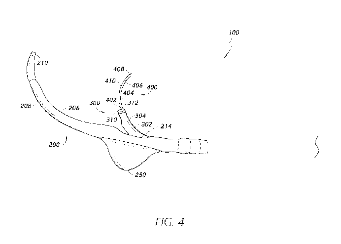
[0081] Figure 4 is another side view of the medical implant delivery
system 100.
The distal portion 302 of the medical implant delivery catheter 300 is shown
extending
beyond the side outlet opening 214. The expandable anchor 250 is shown in
Figure 4 in an
expanded state. As described in further detail herein, the medical implant
delivery catheter
300 can comprise a pre-formed curvature 304 configured to facilitate delivery
of the puncture
needle 400 to the target tissue site and/or deployment of the medical implant
device 350
positioned on the medical implant delivery catheter 300.
[0082] A puncture needle outlet opening 312 can be associated with the
distal
portion 302, including the distal end 310, of the medical implant delivery
catheter 300. The
puncture needle 400 can comprise an elongate portion 402. A distal portion 404
of the
elongate portion 402 is shown extending through the puncture needle outlet
opening 312. A
puncture component 408 associated with a distal end 406 of the elongate
portion 402 can be
used to pierce the tissue at the target tissue site to form the opening in the
tissue. In some
embodiments, the distal portion 404 of the puncture needle 400 can comprise a
pre-formed
curvature 410. In some embodiments, the pre-formed curvature 410 of the
puncture needle
400 can facilitate piercing of tissue at the target tissue site. A target
tissue site can be to a side
of the outer delivery catheter 200 (e.g., at or proximate to the side outlet
opening 214). As
described herein, the medical implant delivery catheter 300 can be advanced
through the side
outlet opening 214 to access the target tissue site. The puncture needle 400
can be advanced
through the puncture needle outlet opening 312 of the medical implant delivery
catheter 300
to puncture the tissue. The curvature in the pre-formed curvature 410 can
facilitate access of
the target tissue site to the side of the outer delivery catheter 200 as the
puncture needle 400
is advanced through the puncture needle outlet opening 312. Improved access of
the puncture
needle 400 to the target tissue site to a side of the outer delivery catheter
200 can provide
effective puncturing of the tissue.
[0083] In some embodiments, the pre-formed curvature 410 can have the
same or
similar orientation as the pre-formed curvature 204 of the outer delivery
catheter 200. In
some embodiments, the pre-formed curvature 410 can have the same or similar
orientation as
17
CA 03160977 2022-05-10
WO 2021/096766
PCT/US2020/059304
the pre-formed curvature 304 of the medical implant delivery catheter 300. In
some
embodiments, the pre-formed curvature 410 can comprise a radius of curvature
smaller than
that of the pre-formed curvature 304.
[0084] In some embodiments, the medical implant delivery catheter 300
can be
pre-loaded into the medical implant delivery lumen of the outer delivery
catheter 200. The
pre-loaded medical implant delivery catheter 300 can remain within the outer
delivery
catheter 200 until the outer delivery catheter 200 is positioned at or
proximate to the target
tissue site. For example, the distal end 310 of the medical implant delivery
catheter 300 can
remain within the medical implant delivery lumen and may not extend beyond the
side outlet
opening 214 until the outer delivery catheter 200 is positioned within the
desired vessel,
channel, chamber and/or organ. The distal end 310 of the medical implant
delivery catheter
300 may not extend beyond the side outlet opening 214 while the medical
implant delivery
catheter 300 is in a retracted configuration. In the retracted state, the
distal end 310 may be
positioned proximal of, at, or distal of the side outlet opening 214. As
described herein, the
distal end 310 of the medical implant delivery catheter 300 can be pre-loaded
and positioned
adjacent to and proximal of the side outlet opening 214. In some embodiments,
the distal end
310 of the medical implant delivery catheter 300 can be pre-loaded and
positioned adjacent to
and distal of the side outlet opening 214. In some embodiments, the medical
implant delivery
catheter 300 may not be pre-loaded and can be advanced from a proximal portion
of the outer
delivery catheter 200 to the side outlet opening 214 after the outer delivery
catheter 200 has
been positioned in the desired vessel, channel, chamber and/or organ.
[0085] In some embodiments, the puncture needle 400 can be pre-loaded
into the
puncture needle lumen of the medical implant delivery catheter 300. In some
embodiments,
the puncture needle 400 can remain within the medical implant delivery
catheter 300 until the
outer delivery catheter 200 is positioned at or proximate to the target tissue
site. For example,
the puncture component 408 may not extend beyond the puncture needle outlet
opening 312
until the outer delivery catheter 200 is positioned within the vessel,
channel, chamber and/or
organ. The puncture component 408 can remain within the medical implant
delivery catheter
300 while the puncture needle is in a retracted configuration. In some
embodiments, a portion
of the puncture needle 400 can be extended through the puncture needle outlet
opening 312
while the puncture needle 400 is in the retracted configuration, but that
portion of the
puncture needle 400 can remain within the outer delivery catheter 200. For
example, the
puncture component 408 can extend through the puncture needle outlet opening
312 while
the puncture needle 400 is in the retracted configuration. The puncture
component 408
18
CA 03160977 2022-05-10
WO 2021/096766
PCT/US2020/059304
extending beyond the puncture needle outlet opening 312 can reside within the
medical
implant delivery lumen of the outer delivery catheter 200 until the outer
delivery catheter 200
is positioned into the desired vessel, channel, chamber and/or organ,
including after the side
outlet opening 214 is positioned at a desired position within the vessel,
channel, chamber
and/or organ. In some embodiments, the puncture needle 400 may not be pre-
loaded and can
be advanced from a proximal portion of the medical implant delivery catheter
300 to the
puncture needle outlet opening 312 after the outer delivery catheter 200 has
been positioned
in the desired vessel, channel, chamber and/or organ.
[0086] In some embodiments, the distal portion 404 of the puncture
needle 400
can comprise a shape memory material. As described herein, the distal portion
404 can
remain within the medical implant delivery catheter 300 until the outer
delivery catheter 200
is positioned at or proximate to the target tissue site, such as the side
outlet opening 214 of
the outer delivery catheter 200. The distal portion 404 can assume a
configuration having a
reduced curvature while received within the medical implant delivery catheter.
In some
embodiments, the distal portion 404 can assume a linear or substantially
linear configuration
while received within the medical implant delivery catheter. After extension
of the distal
portion 404 through the puncture needle outlet opening 312, the pre-formed
curvature 410 on
the distal portion 404 can assume the curved configuration. The distal portion
404 can again
assume the reduced curvature configuration, including the linear or
substantially linear
configuration, after retraction back into the medical implant delivery
catheter 300.
[0087] In some embodiments, the medical implant delivery system 100 can
comprise a radially expandable member (not shown) configured to be inserted
into and dilate
the opening formed in the tissue by the puncture needle 400. Enlargement of
the opening can
facilitate positioning of the medical implant device 350 (not shown) into the
opening. The
radially expandable member can be associated with a distal portion of a
puncture needle
sheath (not shown). For example, the puncture needle sheath can extend through
the puncture
needle lumen of the medical implant delivery catheter 300 and the puncture
needle 400 can
extend through the puncture needle sheath. For example, at least a portion of
the puncture
needle 400 can extend within the puncture needle sheath and at least a portion
of the puncture
needle sheath can extend within the medical implant delivery catheter 300. In
some
embodiments, the distal portion 302 of the medical implant delivery catheter
300 can be
configured to provide desired dilation of the opening. For example, the distal
portion 302 can
be inserted into the opening to enlarge the opening. The distal portion 302
can comprise a
size and/or shape to facilitate dilation of the opening, for example, having a
predetermined
19
CA 03160977 2022-05-10
WO 2021/096766
PCT/US2020/059304
diameter configured to provide the desired enlargement. In some embodiments,
both the
radially expandable member and the distal portion 302 can be used to dilate
the opening. For
example, the radially expandable member can be used to provide an initial
dilation of the
opening to facilitate insertion of the distal portion 302 into the opening to
provide any desired
remaining enlargement of the opening.
[0088] In some embodiments, the puncture needle 400 can be exchanged
for a
medical implant guide wire configured to be inserted through the opening
formed at the target
tissue site to guide deployment of the medical implant device 350. For
example, the puncture
needle 400 can be retracted, and the medical implant guide wire can be
advanced through the
puncture needle lumen or a puncture needle sheath extending through the
puncture needle
lumen. The medical implant delivery catheter 300 can be advanced along the
medical implant
guide wire such that the distal portion 302 of the medical implant delivery
catheter 300 can
be inserted through the opening and the medical implant device 350 can be
positioned into
the opening. In some embodiments, the puncture needle 400 can be further
advanced into the
opening after formation of the opening to guide subsequent advancement of the
medical
implant delivery catheter 300, rather than guide wire. The medical implant
delivery catheter
300 can be advanced along the puncture needle 400 such that the medical
implant device 350
can be positioned into the opening.
[0089] Figure 5 is a side view of the distal portion 202 of the outer
delivery
catheter 200. A guide wire outlet opening 212 can be associated with the
distal end 210 of the
outer delivery catheter 200. For example, the guide wire outlet opening 212
can be at the
distal end 210. As described herein, the guide wire 50 can extend through the
guide wire
lumen of the outer delivery catheter 200. The guide wire lumen can extend
along a length of
the outer delivery catheter 200 such that the outer delivery catheter 200 can
be passed along
the guide wire 50 to advance the outer delivery catheter 200 to a desired
location within the
vessel, channel, chamber and/or organ. In some embodiments, the guide wire
lumen can
extend along an entire or substantially an entire length of the outer delivery
catheter 200. For
example, the guide wire 50 can extend through an entire or substantially an
entire length of
the outer delivery catheter 200 and exit through the guide wire outlet opening
212 at the distal
end 210.
[0090] The side outlet opening 214 can be on the distal portion 202 of
the outer
delivery catheter 200. As described herein, the distal portion 202 of the
outer delivery
catheter 200 can comprise a pre-formed curvature 204. The side outlet opening
214 can be on
the pre-formed curvature 204, for example on an inner edge 206 of the pre-
formed curvature
CA 03160977 2022-05-10
WO 2021/096766
PCT/US2020/059304
204. For example, after the outer delivery catheter 200 is advanced to a
desired location
within the desired vessel, channel, chamber and/or organ, the inner edge 206
can be oriented
towards the target tissue site, orienting the side outlet opening 214 towards
the target tissue,
such that the medical implant delivery catheter 300 can be extended through
the side outlet
opening 214 towards the target tissue site. The puncture needle 400 can then
be deployed to
contact tissue at the target tissue site. The outer edge 208 of the pre-formed
curvature 204 can
be oriented away from the target tissue site, such as oriented toward an
opposing location
relative to the target tissue site.
[0091] In some embodiments, the distal portion 202 can comprise a
taper. A size
of the distal portion 202 can taper toward the distal end 210. For example, a
diameter of the
outer delivery catheter 200 can narrow toward the distal end 210. The taper in
the distal
portion 202 and/or the pre-formed curvature 204 can be configured to
facilitate positioning of
the outer delivery catheter 200 into a desired vessel, channel, chamber and/or
organ. In some
embodiments, a radius of curvature and/or a length of the pre-formed curvature
204 can be
selected based at least in part on a shape and/or dimension of the desired
vessel, channel,
chamber and/or organ. In some embodiments, the medical implant delivery system
100 can
be configured to be positioned within the coronary sinus. As described in
further detail
herein, the medical implant delivery system 100 can be positioned into the
coronary sinus via
the coronary sinus ostium. The outer delivery catheter 200 can be positioned
within the
coronary sinus to access a site on the left atrial wall accessible from within
the coronary
sinus. The tapering in the distal portion 202 can facilitate insertion into
narrower portions of
the coronary sinus. The length of the pre-formed curvature 204 can be selected
based at least
in part on a distance of the target tissue site from the coronary sinus
ostium. The radius of
curvature of the pre-formed curvature 204 can be selected based at least in
part on a shape of
the coronary sinus, including a degree of curvature of the coronary sinus, in
which the outer
delivery catheter 200 is positioned. The radius of curvature of the pre-formed
curvature 204
can be selected such that the pre-formed curvature 204 can conform or
substantially conform
to the curvature of a curve of the coronary sinus adjacent into which the
outer delivery
catheter 200 is positioned. The pre-formed curvature 204 can be positioned
such that the
inner edge 206 of the pre-formed curvature 204 is oriented towards a portion
of the left atrial
wall and the outer edge 208 of the curvature is oriented towards a portion of
the wall of the
coronary sinus, such as an opposing portion of the wall of the coronary sinus.
For example,
the pre-formed curvature 204 can follow or substantially follow the curve. In
some
embodiments, the distal portion 202 of the outer delivery catheter 200 can
comprise a shape
21
CA 03160977 2022-05-10
WO 2021/096766
PCT/US2020/059304
memory material. For example, the pre-formed curvature 204 can assume the
curved
configuration after the outer delivery catheter 200 is advanced into the
coronary sinus.
[0092] Figure 6 is a side view of the distal portion 302 of the medical
implant
delivery catheter 300 and the puncture needle 400 extending through the
puncture needle
outlet opening 312 associated with the distal end 310 of the medical implant
delivery catheter
300. The medical implant delivery catheter 300 can comprise the pre-formed
curvature 304
on the distal portion 302. The pre-formed curvature 304 can have a same or
similar
orientation as the pre-formed curvature 204 of the outer delivery catheter
200. For example,
when the medical implant delivery catheter 300 is positioned in the coronary
sinus, an inner
edge 306 of the pre-formed curvature 304 can be oriented towards the left
atrial wall and an
outer edge 308 of the pre-formed curvature 304 can be oriented towards an
opposing portion
of a wall of the coronary sinus. In some embodiments, the pre-formed curvature
304 can have
a smaller radius of curvature than the pre-formed curvature 204.
[0093] The medical implant device 350 can be carried by the medical
implant
delivery catheter 300. Referring to Figure 6, in some embodiments, the medical
implant
device 350 can be positioned on the pre-formed curvature 304. The medical
implant device
350 can be circumferentially positioned around the distal portion 302,
including on the pre-
formed curvature 304 of the pre-formed curvature. In some embodiments, the
medical
implant device 350 can comprise a shunt device, including an expandable shunt
device. For
example, the expandable shunt device can be configured for delivery to the
left atrial wall for
addressing elevated left atrial pressure. The expandable shunt device can be
positioned
around a portion of the distal portion 302, such as around a portion of the
pre-formed
curvature 304 of the distal portion 302.
[0094] The expandable shunt device can have a number of configurations.
In
some embodiments, the expandable shunt device can comprise an expandable
tubular shunt
device. Examples of suitable expandable shunt devices for the medical implant
delivery
system 100 are provided in U.S. Patent Application No. 15/335,891, entitled
"Systems for
Deploying an Expandable Cardiac Shunt," which is incorporated herein in its
entirety.
[0095] The pre-formed curvature 304 can facilitate accessing the target
tissue site
while the medical implant delivery system 100 is positioned in the vessel,
channel, chamber
and/or organ. For example, the pre-formed curvature 304 can facilitate proper
positioning of
the puncture needle 400 at the target tissue site as the medical implant
delivery catheter 300 is
extended through the side outlet opening 214 of the outer delivery catheter
200. As described
herein, a target tissue site can be to a side of the outer delivery catheter
200 (e.g., at or
22
CA 03160977 2022-05-10
WO 2021/096766
PCT/US2020/059304
proximate to the side outlet opening 214). The curvature in the distal portion
302 of the
implant delivery catheter 300 can facilitate access of the target tissue site
to the side of the
delivery catheter 200 as the distal portion 302 is advanced through the side
outlet opening
214, thereby facilitating proper positioning of the puncture needle 400.
Proper positioning of
the puncture needle 400 can enable effective puncturing of the target tissue
site.
[0096] In some embodiments, the distal portion 302 can comprise a
taper. A size
of the distal portion 302 can taper toward the distal end 310. For example, a
diameter of the
medical implant delivery catheter 300 can narrow toward the distal end 310. In
some
embodiments, the taper of the distal portion 302 and/or the pre-formed
curvature 304 can
facilitate desired deployment of the medical implant device 350 to the target
tissue site. As
described in further detail herein, the distal portion 302 can be further
extended through the
opening formed at the target tissue site, such that the medical implant device
350 can be
positioned within the opening and deployed. The taper in the distal portion
302 can ease
insertion of the distal portion 302 through the opening formed in the tissue.
The pre-formed
curvature 304 can enable the distal portion 302 to follow a pre-determined
trajectory as the
distal portion 302 is extended through the side outlet opening 214 of the
outer delivery
catheter 200 to access the target site to a side of the outer delivery
catheter 200 and such that
the distal portion 302 can be inserted into the opening.
[0097] In some embodiments, the distal portion 302 of the medical
implant
delivery catheter 300 can comprise a shape memory material. As described
herein, the distal
portion 302 can remain within the outer delivery catheter 200 until the outer
delivery catheter
200 is positioned at or proximate to the target tissue site. While in a
retracted state within the
outer delivery catheter, the pre-formed curvature 304 of the distal portion
302 can comprise a
configuration having a reduced curvature relative to its relaxed state, such
as when the pre-
formed curvature 304 is deployed through the side outlet opening 214. In some
embodiments,
the distal portion 302 can assume a linear or substantially linear
configuration, while received
within the outer delivery catheter 200. After extension of the distal portion
302 through the
side outlet opening 214, the pre-formed curvature 304 on the distal portion
302 can assume
the curved configuration. The pre-formed curvature 304 can again assume the
reduced
curvature configuration, including the linear or substantially linear
configuration, after
retraction back into the outer delivery catheter 200. In some embodiments, the
outer delivery
catheter 200, medical implant delivery catheter 300, and/or puncture needle
400 can be
flexible such that the outer delivery catheter 200, medical implant delivery
catheter 300,
23
CA 03160977 2022-05-10
WO 2021/096766
PCT/US2020/059304
and/or puncture needle 400 can conform to the shape of anatomical pathways as
the medical
implant delivery system 100 is advanced through tortuous pathways.
[0098] Figure 7 is a perspective view of an example of a proximal
handle 500 of
the medical implant delivery system 100. The proximal handle 500 can be
configured to
provide control for the deployment of the medical implant delivery system 100.
The proximal
handle 500 can comprise control mechanisms for both puncturing tissue at the
target tissue
site using the puncture needle 400 and deploying the medical implant device
350 positioned
on the medical implant delivery catheter 300. Use of one proximal handle for
both tissue
puncture and medical implant device deployment can simplify the process for
the operator,
thereby facilitating a shortened procedure.
[0099] An advancer 502 on the proximal handle 500 can be configured to
translate proximally and/or distally through a rear bracket 504. Advancement
and/or
retraction of the medical implant delivery catheter 300 and outer delivery
catheter 200 can be
controlled by the advancer 502. For example, the advancer 502 can be
translated distally to
advance the medical implant delivery catheter 300 and the outer delivery
catheter 200. The
medical implant delivery catheter 300 and outer delivery catheter 200 can
extend through the
locking nut 506. The locking nut 506 can be fixed relative to a forward
bracket 508 such that
the advancer 502, the medical implant delivery catheter 300 and outer delivery
catheter 200
can be fixed relative to the proximal handle 500. The proximal handle 500 can
comprise a
pair of flexible arms 510 configured to allow controlled deployment of the
medical implant
device 350. For example, the pair of flexible arms 510 can be used to trigger
and/or actuate
controlled release of the medical implant device 350 from the medical implant
delivery
catheter 300 into the opening formed at the target tissue site.
[0100] Figures 8A and 8B show longitudinal cross-sectional views of the
medical
implant delivery system 100 in a first configuration and a second
configuration, respectively.
The longitudinal cross-sectional view can comprise a cross-sectional view of
the outer
delivery catheter 200 along a longitudinal axis of the outer delivery catheter
200. The distal
portion 302 of the medical implant delivery catheter 300 is shown as being
received within
the medical implant delivery lumen of the outer delivery catheter 200. The
guide wire 50 can
extend through the guide wire lumen 220 of the outer delivery catheter 200.
The medical
implant device 350 can be positioned on the distal portion 302 of the medical
implant
delivery catheter 300. The expandable anchor 250 is shown in Figures 8A and 8B
in an
expanded state. As described herein, the medical implant delivery catheter 300
can be pre-
loaded into the outer delivery catheter 200 such that medical implant delivery
catheter 300
24
CA 03160977 2022-05-10
WO 2021/096766
PCT/US2020/059304
can be advanced together with the outer delivery catheter 200 into the desired
vessel, channel,
chamber and/or organ. Figure 8A shows the medical implant delivery catheter
300 pre-loaded
in a first retracted configuration, and Figure 8B shows the medical implant
delivery catheter
300 pre-loaded in a second retracted configuration. As shown in Figure 8A, in
the first
retracted configuration, the distal end 310 of the medical implant delivery
catheter 300 is
distal of the side outlet opening 214. For example, prior to deploying the
medical implant
delivery catheter 300, such as while advancing the outer delivery catheter 200
along the guide
wire 50 to the desired location, the distal end 310 can be distal of the side
outlet opening 214.
In some embodiments, the distal end 310 can be distal of and adjacent to
(e.g., adjacent to
and in contact with) the side outlet opening 214. The medical implant delivery
catheter 300
can be translated proximally relative to the outer delivery catheter 200 to
extend the distal
end 310 through the side outlet opening 214 for deployment of the medical
implant delivery
catheter 300. Referring to Figure 8B, the distal end 310 of the medical
implant delivery
catheter 300 can be positioned proximal of the side outlet opening 214. In
some
embodiments, the distal end 310 can be proximal of and adjacent to (e.g.,
adjacent to and in
contact with) the side outlet opening 214. To deploy the medical implant
delivery catheter
300, the medical implant delivery catheter 300 can be translated distally
relative to the outer
delivery catheter 200. The distal end 310 can be adjacent to the side outlet
opening 214 to
reduce or minimize the distance needed to advance or retract the medical
implant delivery
catheter 300 for deployment.
[0101] In some embodiments, the guide wire lumen of the outer delivery
catheter
200 can extend along an off-center longitudinal axis. Referring to Figure 9, a
lateral cross-
sectional view is shown of the outer delivery catheter 200. The lateral cross
section can be
taken along a plane which is perpendicular or substantially perpendicular to a
longitudinal
axis of the outer delivery catheter 200. The lateral cross section can include
a width of the
outer delivery catheter 200. As shown, the guide wire lumen 220 can extend off-
set from the
central longitudinal axis (C) of the outer delivery catheter 200. The medical
implant delivery
catheter 300 (not shown) can thereby be advanced through the outer delivery
catheter 200
off-set from the central longitudinal axis (C). For example, the medical
implant delivery
lumen can be offset from the central longitudinal axis (C). The off-center
guide wire lumen
220 can reduce or prevent axial rotation of the medical implant delivery
catheter 300 while
the medical implant delivery catheter 300 is positioned within the lumen 220.
For example,
advancing the medical implant delivery catheter 300 along a path off-set from
the central
longitudinal axis (C) can facilitate maintaining axial rotation orientation of
the medical
CA 03160977 2022-05-10
WO 2021/096766
PCT/US2020/059304
implant delivery catheter 300 while the medical implant delivery catheter 300
is extended
through tortuous anatomical pathways. Maintaining a desired rotational
orientation of the
medical implant delivery catheter 300 can facilitate a desired exit trajectory
of the medical
implant delivery catheter 300 as the medical implant delivery catheter 300 is
extended
through the side outlet opening 214 (not shown). Maintaining a desired exit
trajectory of the
medical implant delivery catheter 300 can facilitate effective puncturing of
the tissue at the
target tissue site using the puncture needle 400 (not shown).
[0102] Figures 10A through 10D show another example of a medical
implant
delivery system 1000. The medical implant delivery system 1000 can comprise an
outer
delivery catheter 1100 and a medical implant delivery catheter 1200. At least
a portion of the
medical implant delivery catheter 1200 can be slidably received within the
outer delivery
catheter 1100. In some embodiments, at least a portion of the medical implant
delivery
catheter 1200 can be passed through at least a portion of the outer delivery
catheter 1100.
Figure 10A is a top-down view of the outer delivery catheter 1100, and Figure
10B is a side
cross-sectional view of the outer delivery catheter 1100. Figure 10C is a top-
down view of
the medical implant delivery catheter 1200 and Figure 10D is a side view of
the medical
implant delivery catheter 1200. A puncture needle (not shown) can be slidably
received
within the medical implant delivery catheter 1200. The puncture needle can be
configured to
be extended through a puncture needle outlet opening 1212 associated with a
distal portion
1202, including a distal end 1210, of the medical implant delivery catheter
1200. In some
embodiments, at least a portion of the puncture needle can be passed through
at least a
portion of the medical implant delivery catheter 1200 and extended through the
puncture
needle outlet opening 1212.
[0103] In some embodiments, the medical implant delivery catheter 1200
can be
pre-loaded within the outer delivery catheter 1100. In some embodiments, the
puncture
needle can be pre-loaded within the medical implant delivery catheter 1200.
The medical
implant delivery system 1000 may or may not be used in combination with a
puncture needle
sheath. For example, the puncture needle sheath may extend slidably within the
puncture
needle lumen of the medical implant delivery catheter 1200 and the puncture
needle may
extend slidably within a lumen of the puncture needle sheath. The puncture
needle may be
pre-loaded within the puncture needle sheath and the puncture needle sheath
may be pre-
loaded within the medical implant delivery catheter 1200. Alternatively, the
medical implant
delivery catheter 1200, puncture needle and/or puncture needle sheath may not
be pre-loaded.
26
CA 03160977 2022-05-10
WO 2021/096766
PCT/US2020/059304
[0104] A distal portion 1102 of the outer delivery catheter 1100 can
comprise a
side outlet opening 1114. Deployment of the medical implant delivery catheter
1200 can
include extending the medical implant delivery catheter 1200 through the side
outlet opening
1114. The distal portion 1102 can comprise a pre-formed curvature 1104. The
pre-formed
curvature 1104 can be configured to facilitate positioning of the outer
delivery catheter 1100
into a vessel, channel, chamber and/or organ so as to access the target tissue
site. The side
outlet opening 1114 can be on an inner edge 1106 of the pre-formed curvature
1104. While
the outer delivery catheter 1100 is positioned in a desired orientation at the
target position,
the inner edge 1106 can be oriented toward the target tissue site. An outer
edge 1108 of the
pre-formed curvature 1104 can be oriented away from the target tissue site,
such as toward an
opposing position relative to the target tissue site. The distal portion 1202
of the medical
implant delivery catheter 1200 can comprise a pre-formed curvature 1204. In
some
embodiments, the pre-formed curvature 1204 can have the same orientation as
the pre-formed
curvature 1104 when the medical implant delivery catheter 1200 is in the
deployed
configuration, such as when the distal portion 1202 is extended beyond the
side outlet
opening 1114. The pre-formed curvature 1204 can be configured to facilitate
access to the
target tissue site while the outer delivery catheter 1100 is positioned at the
target position. In
some embodiments, the outer delivery catheter 1100 and/or the medical implant
delivery
catheter 1200 can be flexible such that the outer delivery catheter 1100
and/or medical
implant delivery catheter 1200 can conform to the shape of anatomical pathways
as the
medical implant delivery system 1000 is advanced through tortuous pathways. In
some
embodiments, the medical implant delivery catheter 1200 can comprise a reduced
curvature
configuration while retracted within the outer delivery catheter 1100. In some
embodiments,
the medical implant delivery catheter 1200 can comprise a linear or
substantially linear
configuration while retracted within the outer delivery catheter 1100 and
assume the curved
configuration after release from the outer delivery catheter 1100. In some
embodiments, the
medical implant delivery catheter 1200 and/or the outer delivery catheter 1100
can comprise
shape memory material, such as the distal portions 1202, 1102.
[0105] The side outlet opening 1114 can have a shape to facilitate
extension of the
medical implant delivery catheter 1200 through the side outlet opening 1114 in
a desired
orientation. For example, a distal portion 1202 of the medical implant
delivery catheter 1200
can be configured to interlock with the side outlet opening 1114 as the distal
portion 1202 is
extended through the side outlet opening 1114. In some embodiments, the side
outlet opening
1114 can comprise an asymmetry around a central axis. The asymmetry can reduce
or
27
CA 03160977 2022-05-10
WO 2021/096766
PCT/US2020/059304
prevent rotation of the distal portion 1202 as the distal portion 1202 is
extended through the
side outlet opening 1114. The distal portion 1202 can comprise a corresponding
shape to
interlock with the shape of the side outlet opening 1114. For example, the
distal portion 1202
can comprise a configuration configured to interlock with the asymmetry of the
side outlet
opening 1114 as at least a portion of the distal portion 1202 is advanced
through the side
outlet opening 1114. In some embodiments, a shape of the distal portion 1202
can match, for
example being the same or similar as, a shape of the side outlet opening 1114.
Matching
between the shapes of the distal portion 1202 and the side outlet opening 1114
can facilitate
interlocking between the two.
[0106] The side outlet opening 1114 can comprise a proximal portion
1114a and a
distal portion 1114b. The proximal portion 1114a and/or the distal portion
1114b can
comprise a shape to interlock with the distal portion 1202. In some
embodiments, the distal
portion 1114b can have a width narrower than that of the proximal portion
1114a. In some
embodiments, the distal portion 1114b can comprise an elongated extension. In
some
embodiments, the distal portion 1114b can have a "v" shape. In some
embodiments, the
proximal portion 1114a can comprise a segment of a circle. In some
embodiments, the
proximal portion 1114a can comprise a segment of an oval.
[0107] In some embodiments, the distal portion 1202 of the medical
implant
delivery catheter 1200 can comprise a shape which interlocks with the shape of
the side outlet
opening 1114. The distal portion 1202 can comprise a lateral cross section
which has a shape
configured to interlock with a shape of the side outlet opening 1114, where
the lateral cross
section can extend along a lateral dimension of the distal portion of the
outer delivery
catheter 1100. In some embodiments, a cross-sectional shape, such as a shape
of a lateral
cross section, of the distal portion 1202 can have a shape configured to
interlock with the
shape of the side outlet opening 1114 to orient the medical implant delivery
catheter 1200 as
the delivery catheter 1200 is extended through the side outlet opening 1114.
The distal
portion 1202 can be rotated prior to extending the distal portion 1202 through
the side outlet
opening 1114 to properly orient the distal portion 1202 relative to the side
outlet opening
1114, for example matching a shape of the lateral cross section of the distal
portion 1202 with
a shape of the side outlet opening 1114. In some embodiments, the lateral
cross section can
be taken along a plane which divides the medical implant delivery catheter
1200 into an
upper portion and a lower portion, the upper portion comprising the inner edge
1206 of the
pre-formed curvature 1204 and the lower portion comprising the outer edge 1208
of the pre-
formed curvature 1204. The lateral cross section can be taken along a lateral
cross-sectional
28
CA 03160977 2022-05-10
WO 2021/096766
PCT/US2020/059304
plane which extends along a longitudinal axis of the medical implant delivery
catheter 1200
and which is parallel or substantially parallel to a plane containing at least
a portion of the
side outlet opening 1114. For example, the lateral cross section can bisect
the medical
implant delivery catheter 1200 into the upper portion and the lower portion.
For example, the
distal portion 1202 can comprise a narrowed distal end portion 1214. The
narrowed distal end
portion 1214 can be oriented towards and/or align with the distal portion
1114b of the side
outlet opening 1114 so as to guide the orientation of the medical implant
delivery catheter
1200 as the medical implant delivery catheter 1200 is extended through the
side outlet
opening 1114. For example, interlocking the distal portion 1202 and the side
outlet opening
1114 can enable orienting the inner edge 1206 of the pre-formed curvature 1204
proximally
and the outer edge 1208 of the pre-formed curvature 1204 distally as the
distal portion 1202
is deployed.
[0108] The medical implant delivery system 1000 can comprise other
features the
same as or similar to those of the medical implant delivery system 100, such
as described
with reference to Figures 3 through 9. For example, a puncture needle (not
shown) can be
received in a puncture needle lumen of the medical implant delivery catheter
1200. The
puncture needle can be extended through the puncture needle outlet opening
1212 associated
with the distal end 1210 of the medical implant delivery catheter 1200 for
puncturing tissue at
the target tissue site. A medical implant device 1250 can be positioned on the
distal portion
1202 such that a delivery catheter for delivering the puncture needle does not
need to be
exchanged for a delivery catheter for delivering the medical implant device
1250. The
medical implant device 1250 can be deployed into the tissue opening formed by
the puncture
needle. The outer delivery catheter 1100 can comprise a guide wire lumen 1120
configured to
receive a guide wire. The guide wire can extend through a guide wire outlet
opening 1112
associated with a distal end 1110 of the outer delivery catheter such that the
outer delivery
catheter 1100 can be advanced along the guide wire for positioning into the
desired vessel,
channel, chamber and/or organ. For example, the medical implant delivery
system 1000 can
be configured for positioning into the coronary sinus from the right atrium
via the coronary
sinus ostium such that the puncture needle extending through the medical
implant delivery
catheter 1200 can be used to form an opening on a portion of the left atrial
wall accessible
from within the coronary sinus. The medical implant device 1250 can be a shunt
device. The
shunt device can be deployed into the opening for treating elevated atrial
pressure. The
medical implant delivery catheter 1200 can be advanced further into the
opening formed in
29
CA 03160977 2022-05-10
WO 2021/096766
PCT/US2020/059304
the left atrial wall such that the medical implant device 1250 can be
positioned into the
opening.
[0109] Figures 11 through 16 show various steps of an example of a
process for
deploying the medical implant device 350 using the medical implant delivery
system 100 as
described herein. Referring to Figure 11, the guide wire 50 can be inserted
from the right
atrium 5, through the coronary sinus ostium 17 and into the coronary sinus 18.
The right
atrium 5 can be accessed via the superior vena cava or via the inferior vena
cava. For
example, a transjugular or trans-subclavian approach can be used to access the
right atrium 5
via the superior vena cava. The medical implant delivery system 100 can be
inserted into the
subclavian veins or jugular veins, and advanced into the superior vena cava.
Alternatively, a
transfemoral approach can be used to position the medical implant delivery
system 100 into
the inferior vena cava.
[0110] As shown in Figure 12, the medical implant delivery system 100
can be
advanced along the guide wire 50 into the coronary sinus 18. The medical
implant delivery
system 100 can be advanced along the guide wire 50 until the medical implant
delivery
system 100 is at a desired location relative to the left atrial wall. For
example, the outer
delivery catheter 200 can be advanced along the guide wire 50 to position the
side outlet
opening 214 at or proximate to the target tissue site on the left atrial wall.
The pre-formed
curvature 204 of the outer delivery catheter 200 can be positioned to follow
the curvature of
the coronary sinus 18. For example, the pre-formed curvature 204 can be
positioned such that
the inner edge 206 of the pre-formed curvature 204 is oriented towards a
portion of the left
atrial wall and the outer edge 208 of the curvature is oriented towards an
opposing portion of
a wall of the coronary sinus 18.
[0111] As described herein, in some embodiments, the medical implant
delivery
system 100 can have the expandable anchor 250 on the outer edge 208 of the
curvature 204.
After the outer delivery catheter 200 is positioned at the desired location,
the expandable
anchor 250 can be triggered and/or actuated to assume an expanded
configuration. In the
expanded configuration, the expandable anchor 250 can contact a wall of the
coronary sinus
18 to push the outer delivery catheter 200 toward the left atrial wall. For
example, the
expandable anchor 250 can contact the wall of the coronary sinus 18 such that
the outer
delivery catheter 200 can be positioned against the wall of the left atrium 2,
thereby
facilitating stably positioning the medical implant delivery system 100 at the
desired location.
In some embodiments, the inner edge 206 of the pre-formed curvature 204 can
contact the
left atrial wall such that the side outlet opening 214 is positioned at and/or
surrounding the
CA 03160977 2022-05-10
WO 2021/096766
PCT/US2020/059304
target tissue site. In some embodiments, the inner edge 206 may not contact
the left atrial
wall while the expandable anchor 250 is in the expanded state.
[0112] As shown in Figure 12, the medical implant delivery catheter 300
can be
pre-loaded within the medical implant delivery catheter lumen of the outer
delivery catheter
200, and the puncture needle 400 can be pre-loaded within the puncture needle
lumen of the
medical implant delivery catheter 300. While advancing the outer delivery
catheter 200 to the
desired location, the medical implant delivery catheter 300 can be received
within the
medical implant delivery catheter lumen of the outer delivery catheter 200.
For example, the
distal end 310 of the medical implant delivery catheter 300 can be received
within the
medical implant delivery catheter lumen while the outer delivery catheter 200
is being
advanced to the desired location. The puncture needle 400 can be pre-loaded
within the
puncture needle lumen of the medical implant delivery catheter 300 such that
the puncture
component 408 is positioned within the puncture needle lumen, or
alternatively, such that the
puncture component 408 extends through the puncture needle outlet opening 312
associated
with the distal end 310 of the medical implant delivery catheter 300. While
being advanced to
the target tissue site, the puncture component 408 can extend through the
puncture needle
outlet opening 312 and remain within the medical implant delivery catheter
lumen of the
outer delivery catheter 200.
[0113] Referring to Figure 13, after the outer delivery catheter 200 is
positioned at
the desired location, the medical implant delivery catheter 300 can be
extended through the
side outlet opening 214. After the outer delivery catheter 200 is positioned
at the desired
location, the distal portion 302 of the medical implant delivery catheter 300,
including the
distal end 310, can be extended through the side outlet opening 214. In some
embodiments,
the distal portion 302 can be rotated to properly orient the distal portion
302 relative to the
side outlet opening 214 prior to extending the distal portion 302 through the
side outlet
opening.
[0114] As described herein, the puncture needle 400 can be pre-loaded
within the
puncture needle lumen of the medical implant delivery catheter 300. The
puncture needle can
be pre-loaded such that the puncture component 408 extends through the
puncture needle
outlet opening 312 associated with the distal end 310. The implant delivery
catheter 300 can
be extended through the side outlet opening 214 such that the puncture
component 408 can be
used to contact the wall of the left atrium 2 to puncture the tissue. In some
embodiments, the
puncture needle 400 can be extended further to complete formation of the
opening. In some
embodiments, the puncture component 408 can be received within the puncture
needle lumen
31
CA 03160977 2022-05-10
WO 2021/096766
PCT/US2020/059304
of the implant delivery catheter 300 while the outer delivery catheter 200 is
inserted to the
desired location. The puncture component 408 can be subsequently extended
through the
puncture needle outlet opening 312 such that the puncture component 408 can be
used to
contact the target tissue site.
[0115] The pre-formed curvature 304 (not shown) of the medical implant
delivery
catheter 300 can provide a desired trajectory for the distal portion 302 (not
shown) of the
medical implant delivery catheter 300 as it is advanced through the side
outlet opening 214.
For example, the pre-formed curvature 304 can facilitate contact between the
puncture needle
400 and the target tissue site on the left atrial wall at a lateral position
relative to the medical
implant delivery system 100. The pre-formed curvature 304 can facilitate
subsequent
insertion of the distal portion 302 into the opening formed at the target
tissue site. The pre-
formed curvature 410 (not shown) of the puncture needle 400 can facilitate
insertion of the
distal portion 404 (not shown) of the puncture needle 400 into the tissue at
the target tissue
site, thereby providing effective puncture of tissue at the target site at a
lateral position
relative to the medical implant delivery system 100.
[0116] As described herein, the expandable anchor 250 can assume the
expanded
configuration after the outer delivery catheter 200 is positioned at a desired
location within
the coronary sinus 18. In some embodiments, expansion of the expandable anchor
250 can
facilitate reliable positioning of the outer delivery catheter 200 against a
wall of the coronary
sinus. In some embodiments, the outer delivery catheter 200 can be pushed
toward the left
atrial wall, for example such that the inner edge 206 of the pre-formed
curvature 204 can
contact the left atrial wall. In some embodiments, positioning of the outer
delivery catheter
200 against the left atrial wall can facilitate puncturing of the tissue using
the puncture
component 408 when the puncture needle 400 is extended through the side outlet
opening
214. In some embodiments, the medical implant delivery system 100 may not
include the
expandable anchor 250 and/or the inner edge 206 is not positioned in contact
with the left
atrial wall. For example, the puncture needle 400 can be advanced further
through the side
outlet opening 214, such as compared to the extent to which the puncture
needle 400 is
advanced if the expandable anchor 250 is used, such that the puncture
component 408 can
contact the left atrial wall and push against the left atrial wall so to
pierce the tissue at the
target tissue site.
[0117] In some embodiments, the medical implant delivery system 100 can
comprise a puncture needle sheath. The puncture needle 400 can extend along a
puncture
needle lumen of the puncture needle sheath. The puncture needle sheath can be
received
32
CA 03160977 2022-05-10
WO 2021/096766
PCT/US2020/059304
within the medical implant delivery catheter 300. For example, at least a
portion of the
puncture needle sheath can be configured to be passed through at least a
portion of the
puncture needle lumen of the medical implant delivery catheter 300 and at
least a portion of
the puncture needle 400 can be passed through at least a portion of a lumen of
the puncture
needle sheath. In some embodiments, the medical implant delivery system 100
can comprise
a radially expandable member associated with a distal portion of the puncture
needle sheath,
the radially expandable member being configured to enlarge the opening formed
in the tissue.
The distal portion of the puncture needle sheath can be advanced through the
opening such
that at least a portion of the radially expandable member is positioned within
the opening.
The radially expandable member can subsequently be triggered and/or actuated
to assume an
expanded configuration so as to provide desired enlargement of the opening.
The radially
expandable member can be deflated and retracted from the opening after desired
enlargement
is accomplished. In some embodiments, the distal portion 302 of the medical
implant delivery
catheter 300 can be inserted into the opening to enlarge the opening, for
example without
using a radially expandable member associated with a puncture needle sheath.
In some
embodiments, both the radially expandable member and the distal portion 302 of
the medical
implant delivery catheter 300 can be used to enlarge the opening.
[0118] Referring to Figure 14, as described herein, in some
embodiments, the
puncture needle 400 can be extended into the left atrium 2 to serve as a guide
wire along
which the medical implant delivery catheter 300 can be advanced so as to
deploy the medical
implant device 350. Alternatively, the puncture needle 400 can be retracted
after the opening
in the left atrial wall is formed. The puncture needle 400 can be exchanged
for a medical
implant guide wire. The medical implant guide wire can be advanced through the
puncture
needle lumen of the medical implant delivery catheter 300 into the left atrium
2. In some
embodiments, the medical implant guide wire can be advanced through a puncture
needle
sheath extending through the puncture needle lumen. In some embodiments, the
puncture
needle 400 can be exchanged for the medical implant guide wire before or after
desired
enlargement of the opening on the left atrial wall is achieved. For example,
the puncture
needle 400 can be retracted and the medical implant guide wire can be inserted
in its place
prior to activating and/or triggering the radially expandable anchor.
[0119] Figure 15 shows deployment of the medical implant device 350.
The
medical implant delivery catheter 300, such as the distal portion 302 of the
medical implant
delivery catheter 300, can be advanced through the opening in the left atrial
wall such that the
medical implant device 350 can be positioned into the opening. The medical
implant delivery
33
CA 03160977 2022-05-10
WO 2021/096766
PCT/US2020/059304
catheter 300 is shown as being advanced along the puncture needle 400 extended
into the left
atrium 2. As described herein, the medical implant device 350 can comprise an
expandable
shunt device. The expandable shunt device can be positioned into the opening
for shunting
blood flow from the left atrium 2 into the coronary sinus 18 so as to
alleviate elevated left
atrial pressure. Examples of processes for deploying an expandable shunt
devices into the left
atrial wall are provided in U.S. Patent Application No. 15/335,891, entitled
"Systems for
Deploying an Expandable Cardiac Shunt," which is incorporated herein in its
entirety.
[0120] Figure 16 shows withdrawal of the medical implant delivery
system 100
from the coronary sinus 18 along the guide wire 50, after the medical implant
device 350 is
positioned into the opening in the left atrial wall. As described herein, the
medical implant
device 350 can comprise an expandable shunt device. Referring to Figure 16,
the expandable
shunt device deployed into the opening formed in the left atrial wall can
allow flow of blood
from the left atrium 2 into the coronary sinus 18. The blood flow can then
drain into the right
atrium 5. Creating a blood flow pathway from the left atrium 2 into the
coronary sinus 18 can
thereby alleviate elevated left atrial pressure. After the expandable shunt
device is deployed
into the left atrial wall, the puncture needle 400 (not shown) and the medical
implant delivery
catheter 300 (not shown) can be retracted back into the outer delivery
catheter 200. For
example, the puncture needle 400 can be retracted back into the puncture
needle lumen of the
medical implant delivery catheter 300 and the medical implant delivery
catheter 300 can be
retracted back into the outer delivery catheter 200. The expandable anchor 250
can assume a
collapsed and/or deflated state prior to being withdrawn from the coronary
sinus 8.
Alternatively, in the case that a medical implant guide wire is used, the
medical implant guide
wire can be retracted. Subsequently, the outer delivery catheter 200 can be
retracted along the
guide wire 50 out of the coronary sinus.
[0121] Figure 17 is a flow diagram of an example of a process 1600 to
deploy a
medical implant delivery system for delivering a medical implant device. The
medical
implant delivery system can comprise one or more features as described herein.
The medical
implant delivery system can comprise an outer delivery catheter, and a medical
implant
delivery catheter configured to extend through a lumen of the outer delivery
catheter. The
medical implant delivery system can comprise a puncture needle extending
through a lumen
of the medical implant delivery catheter. hi block 1602, the process 1600 can
involve
advancing the medical implant delivery system along a guide wire into a
vessel, channel,
chamber and/or organ. For example, the medical implant delivery system can be
positioned
into the coronary sinus. The guide wire can be advanced into the coronary
sinus and the
34
CA 03160977 2022-05-10
WO 2021/096766
PCT/US2020/059304
medical implant delivery system can be advanced into the coronary sinus along
the already
positioned guide wire. The outer delivery catheter can comprise a guide wire
lumen and the
guide wire can slidably extend through the guide wire lumen.
[0122] In block 1604, the process 1600 can involve advancing the
medical
implant delivery catheter through a side outlet opening of outer delivery
catheter. After the
medical implant delivery system is positioned at a desired location within the
coronary sinus,
the medical implant delivery catheter can be deployed. For example, the
medical implant
delivery catheter can be extended through a side outlet opening of the outer
delivery catheter
such that the puncture needle can be used to pierce the tissue at the target
tissue site. In block
1606, the process 1600 can involve extending the puncture component through a
distal outlet
opening of the medical implant delivery catheter to pierce tissue at the
target tissue site to
form an opening in the tissue. The target tissue site can be on a portion of
the left atrial wall
accessible from within the coronary sinus.
[0123] In block 1608, the process 1600 can involve extending a distal
portion of
medical implant delivery catheter through the opening formed in the tissue.
The distal portion
can be advanced through the opening to place the medical implant device into
the opening. In
some embodiments, the distal portion can be configured to enlarge the opening
in the tissue.
The distal portion can have a predetermined diameter to provide desired
enlargement of the
opening. In some embodiments, the medical implant delivery system can comprise
a puncture
needle sheath configured to be slidably passed through the puncture needle
lumen of the
medical implant delivery catheter, and the puncture needle is configured to be
slidably passed
through a lumen of the puncture needle sheath. A radially expandable member
can be
associated with a distal portion of the puncture needle sheath. The radially
expandable
member can be positioned within the opening formed at in the target tissue
site wall to
enlarge the opening when the radially expandable member is in an expanded
configuration.
[0124] In block 1610, the process 1600 can involve deploying the
medical implant
device positioned on the medical implant delivery catheter into opening formed
in tissue. For
example, an expandable shunt device can be deployed into the opening formed on
the left
atrial wall.
[0125] Figures 18 through 20 show side views of another example of a
medical
implant delivery system 1700. Referring to Figure 18, the medical implant
delivery system
1700 can comprise an elongate housing 1800, a medical implant delivery
catheter 1900
extending alongside the elongate housing 1800, and an outer sheath 2200 around
the elongate
housing 1800 and the medical implant delivery catheter 1900. A puncture needle
sheath 2100
CA 03160977 2022-05-10
WO 2021/096766
PCT/US2020/059304
can slidably extend through a puncture needle lumen of the medical implant
delivery catheter
1900. A puncture needle 2000 can slidably extend through the puncture needle
sheath 2100.
In some embodiments, the puncture needle 2000 can extend through the puncture
needle
lumen of the medical implant delivery catheter 1900 without the puncture
needle sheath
2100.
[0126] A medical implant device 1950 can be positioned on the medical
implant
delivery catheter 1900. The medical implant device 1950 can be positioned on
the medical
implant delivery catheter 1900 such that a delivery catheter for delivering
the puncture needle
does not need to be exchanged for a delivery catheter for delivering the
medical implant
device 1950. In some embodiments, positioning the medical implant delivery
catheter 1900
alongside the elongate housing 1800, for example rather than extending the
medical implant
delivery catheter 1900 through an outer delivery catheter, can provide
improved
maneuverability in deploying the medical implant delivery catheter 1900. The
medical
implant delivery catheter 1900 can be navigated along a tighter curve when
deployed, such as
to facilitate improved access to a target tissue site. The ability to advance
the medical implant
delivery catheter 1900 toward and/or into an opening formed in the target
tissue without
having to extend the medical implant delivery catheter 1900 through a side
outlet opening of
an outer delivery catheter can facilitate navigation through a tighter curve.
Navigation
through a tighter curve can thereby improve access to target tissue sites. For
example, the
ability to navigate through a narrower curvature can enable controlled
puncture of target
tissue and/or delivery of the medical implant device 1950 for patients with
smaller and/or
narrower anatomical features at and/or proximate to the target tissue site.
[0127] The elongate housing 1800 and the medical implant delivery
catheter 1900
can be slidably positioned alongside one another. For example, the medical
implant delivery
catheter 1900 can be translated proximally and/or distally relative to the
elongate housing
1800. In some embodiments, the elongate housing 1800 and the medical implant
delivery
catheter 1900 can be adjacent to and in contact with one another. The outer
sheath 2200 can
be around the elongate housing 1800 and the medical implant delivery catheter
1900 to
maintain the position of the elongate housing 1800 and the medical implant
delivery catheter
1900 alongside one another.
[0128] The medical implant delivery system 1700 can be advanced and/or
retracted along the guide wire 50 to and/or from the target location. For
example, the elongate
housing 1800 can comprise a guide wire lumen extending therethrough configured
to receive
the guide wire 50. In some embodiments, the guide wire lumen can extend along
an entire
36
CA 03160977 2022-05-10
WO 2021/096766
PCT/US2020/059304
length of the elongate housing 1800 and extend through a guide wire outlet
opening 1812
associated with a distal end 1810 of the elongate housing 1800.
[0129] The outer sheath 2200 is shown in Figure 18 as being positioned
proximal
of the medical implant device 1950. For example, a distal end 2202 of the
outer sheath 2200
is shown as being proximal of the medical implant device 1950. As described
herein, the
distal end 2202 can be positioned distal of the medical implant device 1950,
such as while the
medical implant delivery system 1700 is advanced to the target location. In
some
embodiments, the outer sheath 2200 can be maintained over the medical implant
device 1950
prior to deployment of the medical implant delivery catheter 1900 and/or
medical implant
device 1950. The outer sheath 2200 can be slidable relative to the elongate
housing 1800 and
the medical implant delivery catheter 1900. The outer sheath 2200 can be
translated
proximally and/or distally relative to the medical implant delivery catheter
1900 and the
elongate housing 1800. For example, the outer sheath 2200 can be translated
proximally
relative to the medical implant delivery catheter 1900 and the elongate
housing 1800 to
facilitate release a corresponding portion of the medical implant delivery
catheter 1900 from
being positioned adjacent to the elongate housing 1800. In some embodiments,
after delivery
of the medical implant device 1950, the outer sheath 2200 can be translated
distally relative
to the medical implant delivery catheter 1900 and the elongate housing 1800 to
position the
medical implant delivery catheter 1900 adjacent to and alongside the elongate
housing 1800
prior to withdrawal of the medical implant delivery system 1700.
[0130] In some embodiments, a distal portion 1802 of the elongate
housing 1800
can comprise a taper. A size of the distal portion 1802 can taper toward the
distal end 1810.
For example, a diameter of the elongate housing 1800 can narrow toward the
distal end 1810.
In some embodiments, the distal portion 1802 of the elongate housing 1800 can
comprise a
pre-formed curvature 1804. The pre-formed curvature 1804 and/or the taper in
the distal
portion 1802 can be configured to facilitate positioning of the elongate
housing 1800 into a
vessel, channel, chamber and/or organ so as to deliver a medical implant
device and/or
therapy to a target site accessible from within the vessel, channel, chamber
and/or organ. A
radius of curvature and/or a length of the pre-formed curvature 1804 can be
selected based at
least in part on a shape and/or dimension of the vessel, channel, chamber
and/or organ, such
as the coronary sinus. In some embodiments, the length of the pre-formed
curvature 1804 can
be predetermined based at least in part on a distance of the target tissue
site from the coronary
sinus ostium. In some embodiments, the radius of curvature of the pre-formed
curvature 1804
can be predetermined based at least in part on a shape of the coronary sinus,
including a
37
CA 03160977 2022-05-10
WO 2021/096766
PCT/US2020/059304
degree of curvature of a portion of the coronary sinus in which the elongate
housing 1800 is
positioned. The radius of curvature of the pre-formed curvature 1804 can be
selected such
that the pre-formed curvature 1804 can conform or substantially conform to a
curvature of the
coronary sinus. For example, when positioned in the coronary sinus, the pre-
formed curvature
1804 can be positioned such that the inner edge 1806 of the pre-formed
curvature 1804 is
oriented towards a portion of the left atrial wall and the outer edge 1808 of
the pre-formed
curvature 1804 is oriented towards an opposing portion of a wall of the
coronary sinus. In
some embodiments, the elongate housing 1800, including the distal portion 1802
can
comprise a shape memory material. The distal portion 1802 can assume a reduced
curvature
configuration while being advanced to the target tissue site. The pre-formed
curvature 1804
can assume the curved configuration after positioning in the target position.
The tapering in
the distal portion 1802 can facilitate insertion into narrower portions of the
coronary sinus. In
some embodiments, the distal portion 1802 can comprise one or more radiopaque
markers
thereon to facilitate visualization of positioning the elongate housing 1800
at the desired
location within the vessel, channel, chamber and/or organ, such as the
coronary sinus.
[0131] The elongate housing 1800 can comprise a recess 1814 configured
to
receive at least a portion of the medical implant delivery catheter 1900. The
recess 1814 can
extend along at least a portion of a length of the elongate housing 1800. For
example, the
recess 1814 can extend from a proximal portion, including a proximal end, to
the distal
portion 1802 of the elongate housing 1800. In some embodiments, a distal end
1816 of the
recess 1814 can be proximal of the pre-formed curvature 1804. In some
embodiments, the
distal end 1816 of the recess 1814 can be on the pre-formed curvature 1804.
The recess 1814
can open along the same side as the inner edge 1806 of the pre-formed
curvature 1804.
[0132] In some embodiments, the elongate housing 1800 can comprise an
expandable anchor 1850 on the distal portion 1802. The expandable anchor 1850
is shown in
a deflated state in Figure 18. In some embodiments, the expandable anchor 1850
can be on
the same side as an outer edge 1808 of the pre-formed curvature 1804. In some
embodiments,
the expandable anchor 1850 can be on the pre-formed curvature 1804. For
example, the
expandable anchor 1850 can be on the outer edge 1808 of the pre-formed
curvature 1804. In
some embodiments, the expandable anchor 1850 can be on a portion of the
elongate housing
1800 opposite the distal end 1816 of the recess 1814. In some embodiments, the
expandable
anchor 1850 can be on a portion of the elongate housing 1800 opposite and
proximal of the
pre-formed curvature 1804.
38
CA 03160977 2022-05-10
WO 2021/096766
PCT/US2020/059304
[0133] The expandable anchor 1850 can be inserted to or proximate to
the target
tissue site in a collapsed configuration. The expandable anchor 1850 can be
subsequently
triggered and/or actuated to assume an expanded configuration so as to contact
tissue near the
target tissue site to facilitate stable positioning of the elongate housing
1800 relative to the
target tissue site. In some embodiments, expansion of the expandable anchor
1850 can
facilitate reliable positioning of the elongate housing 1800 against tissue
portion at a location
opposite that of the target tissue site. In some embodiments, expansion of the
expandable
anchor 1850 can push the elongate housing 1800 and/or the medical implant
delivery catheter
1900 against tissue adjacent to the target tissue site. Reliable positioning
of the elongate
housing 1800 at the target tissue site can facilitate desired deployment of
the puncture needle
2000 to form the opening. In some embodiments, the elongate housing 1800 may
not have an
expandable anchor 1850. The puncture needle 2000 can be extended further, such
as
compared to the extent to which the puncture needle 2000 would be advanced if
the
expandable anchor 1850 is used, such that the puncture component 2008 can
contact and
push against the target tissue site to facilitate effective puncture of the
tissue. For example,
the puncture needle 2000 can be further extended to contact and push against
the left atrial
wall so as to pierce the tissue on the left atrial wall.
[0134] Referring again to Figure 18, the distal portion 1902 of the
medical
implant delivery catheter 1900 can comprise a taper. A size of the distal
portion 1902 can
taper toward the distal end 1910, a diameter of the medical implant delivery
catheter 1900
narrowing toward the distal end 1910. In some embodiments, the medical implant
delivery
catheter 1900 can have a pre-formed curvature 1904 on a distal portion 1902.
The pre-formed
curvature 1904 can have a same or similar orientation as the pre-formed
curvature 1804 of
the elongate housing 1800. For example, when the medical implant delivery
catheter 1900 is
positioned in the coronary sinus, an inner edge 1906 of the pre-formed
curvature 1904 can be
oriented towards the left atrial wall and an outer edge 1908 of the pre-formed
curvature 1904
can be oriented towards a wall of the coronary sinus. In some embodiments, the
pre-formed
curvature 1904 can have a smaller radius of curvature than the pre-formed
curvature 1804.
[0135] In some embodiments, the taper of the distal portion 1902 and/or
the pre-
formed curvature 1904 can facilitate accessing the target tissue site while
the medical implant
delivery system 1700 is positioned in the vessel, channel, chamber and/or
organ. The taper
can facilitate effective puncture of the target tissue site and/or deployment
of the medical
implant device 1950 positioned on the medical implant delivery catheter 1900.
The distal
portion 1902 can be further extended through the opening formed at the target
tissue site,
39
CA 03160977 2022-05-10
WO 2021/096766
PCT/US2020/059304
such that the medical implant device 1950 can be positioned within the opening
and
deployed. The taper in the distal portion 1902 can ease insertion of the
distal portion 1902
through the opening formed in the tissue. The pre-formed curvature 1904 can
enable the
distal portion 1902 to follow a pre-determined trajectory to facilitate
extending the distal
portion 1902 through the opening.
[0136] The medical implant delivery catheter 1900 can comprise a
puncture
needle lumen configured to slidably receive the puncture needle 2000. The
puncture needle
2000 can be extended through a puncture needle outlet opening 1912 associated
with, the
distal portion 1902, including a distal end 1910, of the medical implant
delivery catheter
1900. In some embodiments, the pre-formed curvature 1904 can facilitate proper
positioning
of the puncture needle 2000 at the target tissue site as the medical implant
delivery catheter
1900 is advanced relative to the elongate housing 1800. Proper positioning of
the puncture
needle 2000 can enable effective puncturing of the target tissue site.
[0137] In some embodiments, the medical implant delivery system 1700
can
include a puncture needle sheath 2100 configured to allow the puncture needle
2000 to
extend therethrough. The puncture needle 2000 can extend through a puncture
needle outlet
opening 2104 associated with a distal end 2106 of the puncture needle sheath
2100. The
puncture needle sheath 2100 can slidably extend through the puncture needle
lumen of the
medical implant delivery catheter 1900. In some embodiments, at least a
portion of the
puncture needle sheath 2100 can be passed through at least a portion of the
puncture needle
lumen of the medical implant delivery catheter 1900. In some embodiments, at
least a portion
of the puncture needle 2000 can be passed through at least a portion of the
lumen of the
puncture needle sheath 2100. In some embodiments, a distal portion 2102 of the
puncture
needle sheath 2100 can have associated therewith a radially expandable member
2150
configured to enlarge the opening formed at the target tissue site. The
radially expandable
member 2150 can be triggered and/or actuated after it has been positioned into
the opening to
assume an expanded configuration so as to enlarge the opening. In some
embodiments, the
radially expandable member 2150 can comprise an inflatable balloon
circumferentially
positioned around the distal portion 2102 of the puncture needle sheath 2100.
[0138] In some embodiments, the distal portion 1902 of the medical
implant
delivery catheter 1900 can be configured to provide desired dilation of the
opening. For
example, the distal portion 1902 can be inserted into the opening to enlarge
the opening. The
distal portion 1902 can comprise a size and/or shape to facilitate dilation of
the opening, for
example, having a predetermined diameter configured to provide the desired
enlargement. In
CA 03160977 2022-05-10
WO 2021/096766
PCT/US2020/059304
some embodiments, the distal portion 1902 can be used to dilate the opening
instead of the
radially expandable member 2150. For example, the puncture needle sheath 2100
and/or the
radially expandable member 2150 may not be included. In some embodiments, the
puncture
needle 2000 can be positioned within the puncture needle lumen of the medical
implant
delivery catheter 1900 without the puncture needle sheath 2100. In some
embodiments, the
distal portion of the puncture needle 2000 can be configured to enlarge the
opening. The
distal portion can be inserted into the opening to enlarge the opening. For
example, the distal
portion can have a predetermined diameter to provide desired enlargement of
the opening. In
some embodiments, one or more of the puncture needle 2000, the distal portion
1902 and the
radially expandable member 2150 can be used to dilate the opening formed in
the target
tissue.
[0139] In some embodiments, the puncture needle 2000 can be exchanged
for a
medical implant guide wire. For example, the puncture needle 2000 can be
retracted after the
puncture needle 2000 has been used to pierce the tissue and the opening is
formed. The
medical implant guide wire can be advanced through the puncture needle lumen
in place of
the puncture needle 2000. The medical implant device 1950 can be advanced
along the
medical implant guide wire to the target tissue site. For example, an
expandable shunt device
can be advanced along the medical implant guide wire to an opening formed on
the left atrial
wall. In some embodiments, the medical implant guide wire can be advanced
through the
puncture needle sheath 2100. In some embodiments, the medical implant delivery
system
1700 may not include the puncture needle sheath 2100. For example, the
puncture needle
2000 and/or the medical implant guide wire can extend through the puncture
needle lumen of
the medical implant delivery catheter 1900 without the puncture needle sheath
2100.
[0140] The medical implant device 1950 can be positioned on the distal
portion
1902 of the medical implant delivery catheter 1900. In some embodiments, the
medical
implant device 1950 can comprise a shunt device, including an expandable shunt
device. As
described in further detail herein, the distal portion 1902 can be further
extended through the
opening formed at the target tissue site, such that the medical implant device
1950 can be
positioned within the opening and deployed. In some embodiments, the medical
implant
device 1950 can be positioned on the pre-formed curvature 1904. The medical
implant device
1950 can be circumferentially positioned around the distal portion 1902,
including on the pre-
formed curvature 1904.
[0141] The expandable shunt device can have a number of configurations.
In
some embodiments, the expandable shunt device can comprise an expandable
tubular shunt
41
CA 03160977 2022-05-10
WO 2021/096766
PCT/US2020/059304
device. Examples of suitable expandable shunt devices for the medical implant
delivery
system 1700 are provided in U.S. Patent Application No. 15/335,891, entitled
"Systems for
Deploying an Expandable Cardiac Shunt," which is incorporated herein in its
entirety.
[0142] Figure 19 is a side view of the medical implant delivery system
1700
where the puncture needle 2000 is further extended through the puncture needle
outlet
opening 1912. A portion of the puncture needle 2000 is shown as being extended
through
puncture needle outlet opening 2104 associated with the distal end 2106 of the
puncture
needle sheath 2100. The puncture needle sheath 2100 is shown as being extended
through the
puncture needle outlet opening 1912 associated with the distal end 1910 of the
medical
implant delivery catheter 1900. The expandable anchor 1850 of the elongate
housing 1800 is
shown in an expanded state.
[0143] The puncture needle 2000 can comprise an elongate portion 2002.
A distal
portion 2004 of the elongate portion 2002 is shown extending through the
puncture needle
outlet opening 1912 of the medical implant delivery catheter 1900. The
puncture needle 2000
can have a puncture component 2008 associated with a distal end 2006 of the
elongate
portion 2002, the puncture component 2008 being configured for tissue
piercing. The
puncture needle 2000 can comprise a pre-formed curvature 2010 on the distal
portion 2004.
The pre-formed curvature 2010 can have a same or similar orientation as the
pre-formed
curvature 1904 of the medical implant delivery catheter 1900. In some
embodiments, the pre-
formed curvature 2010 can have a smaller radius of curvature than the pre-
formed curvature
1904. In some embodiments, the pre-formed curvature 2010 can have a same or
similar
orientation as the pre-formed curvature 1804 of elongate housing 1800. In some
embodiments, the pre-formed curvature 2010 can facilitate effective tissue
puncture at the
target tissue site while the medical implant delivery system 1700 is
positioned in the vessel,
channel, chamber and/or organ.
[0144] In some embodiments, the distal portion 2004 of the puncture
needle 2000
can comprise a shape memory material. The distal portion 2004 can remain
within the
medical implant delivery catheter 1900 prior to the medical implant delivery
system 1700
being positioned at or proximate to the target tissue site and/or after the
distal portion 2004 is
retracted back into the medical implant delivery catheter 1900. In some
embodiments, the
pre-formed curvature 2010 can assume a less curved configuration while
retracted within the
medical implant delivery catheter 1900 and/or the puncture needle sheath 2100.
In some
embodiments, the distal portion 2004 can assume a linear or substantially
linear configuration
while received within the medical implant delivery catheter 1900 and/or the
puncture needle
42
CA 03160977 2022-05-10
WO 2021/096766
PCT/US2020/059304
sheath 2100. The pre-formed curvature 2010 on the distal portion 2004 can
assume the
curved configuration after extension of the distal portion 2004 through the
puncture needle
outlet opening 1912 and/or the puncture needle outlet opening 2104 associated
with the distal
end 2106 of the puncture needle sheath 2100.
[0145] Figure 20 is another side view of the medical implant delivery
system
1700. The outer sheath 2200 is positioned over the medical implant device
1950. For
example, the medical implant device 1950 is not shown in Figure 20 as the
distal end 2202 of
the outer sheath 2200 is distal of the medical implant device 1950, thereby
extending over the
medical implant device 1950. The distal end 1910 of the medical implant
delivery catheter
1900 is shown as extending distally of the outer sheath 2200. The puncture
needle 2000 and
the puncture needle sheath 2100, including the radially expandable member
2150, are shown
as extending from puncture needle outlet 1912 associated with the distal end
1910 of the
medical implant delivery catheter 1900. The puncture needle 2000 is shown as
extending
from the puncture needle outlet opening 2104 associated with the distal end
2106 of the
puncture needle sheath 2100.
[0146] In some embodiments, while the medical implant delivery catheter
1900 is
in a retracted configuration, the outer sheath 2200 can be positioned over the
distal end 1910
of the medical implant delivery catheter 1900. The distal end 2202 of the
outer sheath 2200
can be at or distal of the distal end 1910 of the medical implant delivery
catheter 1900. In
some embodiments, the distal portion 1902 can comprise a shape memory
material. For
example, the pre-formed curvature 1904 can assume a less curved configuration,
including a
linear or substantially linear configuration, while the outer sheath 2200 is
positioned around
the pre-formed curvature 1904. The outer sheath 2200 positioned around the pre-
formed
curvature 1904 can maintain the pre-formed curvature 1904 in the linear or
substantially
linear configuration. In some embodiments, the outer sheath 2200, elongate
housing 1800,
and/or the medical implant delivery catheter 1900 can be flexible such that
the outer sheath
2200, elongate housing 1800 and/or medical implant delivery catheter 1900 can
conform to
the shape of anatomical pathways as the medical implant delivery system 1700
is advanced
through tortuous pathways. The outer sheath 2200 can maintain the medical
implant delivery
catheter 1900 in the recess 1814 of the elongate housing 1800. The outer
sheath 2200 can be
configured to be translated proximally relative to the implant delivery
catheter 1900 to
release the medical implant delivery catheter 1900. For example, the distal
portion 1902 can
be displaced from the recess 1814. The pre-formed curvature 1904 can be
allowed to assume
the curved configuration, such as after being released from the recess 1814.
The medical
43
CA 03160977 2022-05-10
WO 2021/096766
PCT/US2020/059304
implant delivery catheter 1900 can be distally translatable relative to the
outer sheath 2200
and/or elongate housing 1800 to position the medical implant device into the
opening formed
in the tissue.
[0147] The expandable anchor 1850 is shown in Figure 20 an expanded
state. The
expandable anchor 1850 can remain in a collapsed state until the medical
implant delivery
system 1700 is positioned at or proximate to the target tissue site. The
expandable anchor
1850 can be in the collapsed state while the medical implant delivery catheter
1900 is in a
retracted configuration. In some embodiments, while the medical implant
delivery catheter
1900 is in the retracted configuration, the outer sheath 2200 can be
positioned over the
expandable anchor 1850. The distal end 2202 of the outer sheath 2200 can be
distal of the
expandable anchor 1850. In some embodiments, the outer sheath 2200 can remain
proximal
of the expandable anchor 1850 while the medical implant delivery catheter 1900
is in the
retracted configuration.
[0148] In some embodiments, the puncture needle 2000 and/or the
puncture
needle sheath 2100 can be pre-loaded into the puncture needle lumen of the
medical implant
delivery catheter 1900. In some embodiments, a pre-loaded puncture needle 2000
and pre-
loaded puncture needle sheath 2100 can remain within the medical implant
delivery catheter
1900 until the medical implant delivery system 1700 is positioned at or
proximate to the
target tissue site. For example, the puncture component 2008 may not extend
beyond the
puncture needle outlet opening 1912 until the medical implant delivery system
1700 is
positioned within the vessel, channel, chamber and/or organ. The puncture
component 2008
can remain within the medical implant delivery catheter 1900 while the
puncture needle 2000
is in a retracted configuration. In some embodiments, a portion of the
puncture needle 2000
can extend through the puncture needle outlet opening 1912 while the puncture
needle 2000
is in the retracted configuration. In some embodiments, the puncture component
2008
extending beyond the puncture needle outlet opening 1912 can be proximal of
the distal end
2202 of the outer sheath 2200 when in the puncture needle 2000 is in the
retracted
configuration. For example, the puncture component 2008 can be received within
the recess
1814 of the elongate housing 1800 until the medical implant delivery system
1700 is
positioned into the desired vessel, channel, chamber and/or organ. In some
embodiments, the
puncture needle 2000 and/or the puncture needle sheath 2100 can be advanced
from a
proximal portion of the medical implant delivery catheter 1900 to the puncture
needle outlet
opening 1912 after the medical implant delivery system 1700 has been
positioned in the
desired vessel, channel, chamber and/or organ.
44
CA 03160977 2022-05-10
WO 2021/096766
PCT/US2020/059304
[0149] Figures 21 through 24 show various steps of an example of a
process for
delivering a medical implant device, such as an expandable shunt device, to a
left atrial wall
from within the coronary sinus using the medical implant delivery system 1700
as described
herein. Referring to Figure 21, the medical implant delivery system 1700 can
be advanced
along the guide wire 50 into the coronary sinus 18. As described herein, the
guide wire 50
can be inserted from the right atrium 5, through the coronary sinus ostium 17
and into the
coronary sinus 18. In some embodiments, a transjugular or trans-subclavian
approach can be
used to access the fight atrium 5 via the superior vena cava. Alternatively, a
transfemoral
approach can be used to position the medical implant delivery system 1700
through the
inferior vena cava and into the right atrium 5. The medical implant delivery
system 1700 can
be positioned at a desired location within the coronary sinus 18. While the
medical implant
delivery system 1700 is advanced along the guide wire 50 to the desired
location within the
coronary sinus 18, the outer sheath 2200 can be positioned over the distal end
1910 (not
shown) of the medical implant delivery catheter 1900 (not shown). The medical
implant
delivery catheter 1900 can be maintained within the recess 1814 of the
elongate housing
1800. The outer sheath 2200 can be proximal of the expandable anchor 1850. In
some
embodiments, the outer sheath 2200 can be positioned over the expandable
anchor 1850
while the medical implant delivery system 1700 is positioned to the desired
location. In some
embodiments, the distal end 2202 of the outer sheath 2200 can be at or distal
of the distal end
1816 of the recess 1814 while the medical implant delivery system 1700 is
advanced along
the guide wire 50.
[0150] In Figure 22, after the medical implant delivery system 1700 has
been
advanced to the desired location, the outer sheath 2200 can be translated
proximally relative
to the medical implant delivery catheter 1900 and the elongate housing 1800.
The outer
sheath 2200 can be slid proximally to release the distal portion 1902 of the
medical implant
delivery catheter 1900. The distal end 2202 of the outer sheath 2200 can be
translated
proximally such that at least a portion of the distal portion 1902 can be
released from the
recess 1814 of the elongate housing 1800. For example, the pre-formed
curvature 1904 can
assume its curved configuration after the distal portion 1902 is released. The
inner edge 1906
of the pre-formed curvature 1904 can be oriented towards the wall of the left
atrium 2 and the
outer edge 1908 of the pre-formed curvature 1904 can be oriented towards a
wall of the
coronary sinus 18. As described herein, in some embodiments, the puncture
needle 2000 can
be pre-loaded such that the puncture component 2008 extends through the
puncture needle
outlet opening 1912 of the medical implant delivery catheter 1900. In some
embodiments, the
CA 03160977 2022-05-10
WO 2021/096766
PCT/US2020/059304
puncture component 2008 can extend beyond the puncture needle outlet opening
2104 of the
puncture needle sheath 2100. For example, the puncture component 2008 can be
extended
beyond the puncture needle outlet opening 2104 and the puncture needle outlet
opening 1912
while remaining proximal of the distal end 2202 of the outer sheath 2200 while
the within the
medical implant delivery system 1700 is advanced to its target location.
Alternatively, the
puncture component 2008 can be extended through the puncture needle outlet
opening 1912
and/or the puncture needle outlet opening 2104 after the medical implant
delivery system
1700 has been positioned at or proximate to the desired location. After the
distal portion 1902
is released, the medical implant delivery catheter 1900 can be advanced
distally such that the
puncture component 2008 can contact the left atrial wall and can be used to
pierce the tissue
at the target tissue site on the left atrial wall. In some embodiments, the
puncture needle
sheath 2100 can be advanced, such as relative to the medical implant delivery
catheter 1900,
to facilitate contact between the puncture component 2008 and the left atrial
wall. The
puncture needle 2000 can be further advanced, such as relative to the medical
implant
delivery catheter 1900 and/or the puncture needle sheath 2100, to form the
opening on the left
atrial wall.
[0151] In some embodiments, the pre-formed curvature 1904 of the
medical
implant delivery catheter 1900 can facilitate positioning the puncture needle
2000 at a target
tissue site on the left atrial wall and/or subsequent insertion of the distal
portion 1902 of the
medical implant delivery catheter 1900 into the opening formed at the target
tissue site. The
pre-formed curvature 1904 can provide a desired trajectory for the medical
implant delivery
catheter 1900 as it is advanced distally such that the puncture needle 2000
can contact a target
tissue site on the left atrial wall at a lateral position relative to the
medical implant delivery
system 1700 and the distal portion 1902 of the implant delivery catheter 1900
can be inserted
into the opening formed at the target tissue site.
[0152] In some embodiments, the expandable anchor 1850 can be triggered
and/or
actuated to assume an expanded state after the medical implant delivery system
1700 is
positioned at or proximate to the desired location within the coronary sinus.
In the expanded
configuration, the expandable anchor 1850 can contact a wall of the coronary
sinus 18 to
facilitate stably positioning the medical implant delivery system 1700 at the
desired location.
Stable positioning of the medical implant delivery system 1700 can facilitate
tissue puncture
at the target tissue site and/or reliable deployment of the medical implant
device 1950 into the
opening. In some embodiments, expansion of the expandable anchor 1850 can
provide stable
46
CA 03160977 2022-05-10
WO 2021/096766
PCT/US2020/059304
positioning of the elongate housing 1800 against the coronary sinus 18 so as
to facilitate
reliable puncture of the target tissue site on the left atrial wall.
[0153] Referring to Figure 23, the puncture needle sheath 2100 can be
extended
into the opening formed in the left atrial wall such that the radially
expandable member 2150
associated with the distal portion 2102 of the puncture needle sheath 2100 can
be positioned
into the opening to enlarge the opening. Enlargement of the opening can
facilitate subsequent
positioning of the medical implant device 1950 into the opening. The radially
expandable
member 2150 can be triggered and/or actuated after it has been positioned into
the opening to
assume an expanded configuration so as to enlarge the opening. As described
herein, in some
embodiments, the distal portion 1902 of the medical implant delivery catheter
1900 can be
inserted into the opening to provide desired dilation of the opening.
[0154] The puncture needle 2000 can be further advanced through the
puncture
needle outlet opening 2104 associated with the distal end 2106 of the puncture
needle sheath
2100. The puncture needle 2000 can serve as a guide wire along which the
medical implant
delivery catheter 1900 can be advanced such that the medical implant device
1950 (not
shown) can be positioned into the opening in the tissue. Alternatively, the
puncture needle
2000 can be exchanged for a medical implant guide wire. For example, the
puncture needle
2000 can be retracted after the opening in the left atrial wall is formed. The
medical implant
guide wire can be advanced through the puncture needle lumen of the medical
implant
delivery catheter 1900 into the left atrium 2 and the implant delivery
catheter 1900 can be
advanced along the medical implant guide wire into the left atrium 2 to deploy
the medical
implant device 1950. Figure 23 shows that the outer sheath 2200 remains over
the medical
implant device 1950 while the opening is formed in the left atrial wall. In
some embodiments,
the medical implant device 1950 can be distal of the outer sheath 2200 while
the opening is
formed in the left atrial wall.
[0155] Referring to Figure 24, the distal portion 1902 of the medical
implant
delivery catheter 1900 can be advanced through the opening formed in the left
atrial wall
such that the medical implant device 1950 can be positioned into the opening.
The medical
implant delivery catheter 1900 can be advanced along the puncture needle 2000
positioned
into the left atrium 2 to position the medical implant device 1950 into the
opening. Examples
of processes for deploying an expandable shunt devices into the left atrial
wall are provided
in U.S. Patent Application No. 15/335,891, entitled "Systems for Deploying an
Expandable
Cardiac Shunt," which is incorporated herein in its entirety.
47
CA 03160977 2022-05-10
WO 2021/096766
PCT/US2020/059304
[0156] The expandable shunt device deployed into the opening formed in
the left
atrial wall can allow flow of blood from the left atrium 2 into the coronary
sinus 18, and then
into the right atrium 5, thereby alleviating elevated left atrial pressure.
After the expandable
shunt device is deployed into the left atrial wall, the puncture needle 2000
and the medical
implant delivery catheter 1900 can be retracted. Subsequently, the medical
implant delivery
system 1700 can be retracted along the guide wire 50 out of the coronary
sinus.
[0157] Figure 25 is a flow diagram of an example of a process 2400 to
deploy a
medical implant delivery system for delivering a medical implant device. The
medical
implant delivery system can comprise one or more features as described herein,
such as that
of the medical implant delivery system 1700 described with reference to
Figures 18 through
24.
[0158] In block 2402, the process 2400 can involve advancing the
medical
implant delivery system along a guide wire into a vessel, channel, chamber
and/or organ. For
example, the medical implant delivery system can be advanced into the coronary
sinus along
the guide wire. The medical implant delivery system can be used to access the
target tissue
site on a target vessel, channel, chamber and/or organ. For example, the
medical implant
delivery system can be used to access a target tissue site on a portion of the
left atrial wall
from within the coronary sinus. The medical implant delivery system can
comprise an
elongate housing, a medical implant delivery catheter extending alongside the
elongate
housing, and an outer sheath around the elongate housing and the medical
implant delivery
catheter. A medical implant device can be positioned on the medical implant
delivery catheter
for deployment onto the target vessel, channel, chamber and/or organ. For
example, a shunt
device, including an expandable shunt device, can be carried on the medical
implant delivery
catheter for deployment onto the left atrial wall.
[0159] After the medical implant delivery system is positioned at a
desired
location within the coronary sinus, the puncture needle can be deployed to
pierce the tissue at
the target tissue site. A puncture needle can slidably extend through a
puncture needle lumen
of the medical implant delivery catheter. In block 2404, the process 2400 can
involve
translating the outer sheath proximally to release a distal portion of medical
implant delivery
catheter. As described herein, the distal portion of the medical implant
delivery catheter can
comprise a pre-formed curvature. The distal portion can comprise a shape
memory material
such that the distal portion can assume the curved configuration after the
outer sheath is slid
proximally such that the outer sheath is no longer around the distal portion
to position the
distal portion alongside the elongate housing.
48
CA 03160977 2022-05-10
WO 2021/096766
PCT/US2020/059304
[0160] In block 2406, the process 2400 can involve translating the
medical
implant delivery catheter distally relative to the elongate housing. The
medical implant
delivery catheter can be advanced distally such that the puncture needle can
contact the target
tissue. In some embodiments, the puncture component of the puncture needle can
be
extended through a puncture needle outlet opening associated with a distal end
of the medical
implant delivery catheter to facilitate contact between the puncture component
and the target
tissue. In block 2408, the process 2400 can involve piercing tissue at the
target tissue site to
form an opening in the tissue using the puncture component extending through
the distal
outlet opening of the medical implant delivery catheter.
[0161] In block 2410, the process 2400 can involve extending a distal
portion of
the medical implant delivery catheter through the opening formed in the
tissue. The distal
portion of the medical implant delivery catheter can be advanced through the
opening such
that the medical implant device carried on the medical implant delivery can be
positioned in
the opening. In some embodiments, the distal portion can be configured to
enlarge the
opening in the tissue. For example, the distal portion can have a
predetermined diameter to
provide desired enlargement of the opening. In some embodiments, the medical
implant
delivery system can comprise a puncture needle sheath configured to slidably
extend through
the puncture needle lumen of the medical implant delivery catheter, and the
puncture needle
is configured to be slidably passed through a lumen of the puncture needle
sheath. A radially
expandable member can be associated with a distal portion of the puncture
needle sheath. At
least a portion of the radially expandable member can be positioned within the
opening
formed in the tissue and expanded to enlarge the opening.
[0162] In block 2412, the process 2400 can involve deploying the
medical implant
device positioned on the medical implant delivery catheter can into the
opening formed in the
tissue. For example, the shunt device can be deployed onto the left atrial
wall.
Additional Embodiments
[0163] Depending on the embodiment, certain acts, events, or functions
of any of
the processes or algorithms described herein can be performed in a different
sequence, may
be added, merged, or left out altogether. Thus, in certain embodiments, not
all described acts
or events are necessary for the practice of the processes.
[0164] Conditional language used herein, such as, among others, "can,"
"could,"
"might," "may," "e.g.," and the like, unless specifically stated otherwise, or
otherwise
understood within the context as used, is intended in its ordinary sense and
is generally
49
CA 03160977 2022-05-10
WO 2021/096766
PCT/US2020/059304
intended to convey that certain embodiments include, while other embodiments
do not
include, certain features, elements and/or steps. Thus, such conditional
language is not
generally intended to imply that features, elements and/or steps are in any
way required for
one or more embodiments or that one or more embodiments necessarily include
logic for
deciding, with or without author input or prompting, whether these features,
elements and/or
steps are included or are to be performed in any particular embodiment. The
terms
"comprising," "including," "having," and the like are synonymous, are used in
their ordinary
sense, and are used inclusively, in an open-ended fashion, and do not exclude
additional
elements, features, acts, operations, and so forth. Also, the term "or" is
used in its inclusive
sense (and not in its exclusive sense) so that when used, for example, to
connect a list of
elements, the term "or" means one, some, or all of the elements in the list.
Conjunctive
language such as the phrase "at least one of X, Y and Z," unless specifically
stated otherwise,
is understood with the context as used in general to convey that an item,
term, element, etc.
may be either X, Y or Z. Thus, such conjunctive language is not generally
intended to imply
that certain embodiments require at least one of X, at least one of Y and at
least one of Z to
each be present.
[0165] It should be appreciated that in the above description of
embodiments,
various features are sometimes grouped together in a single embodiment,
Figure, or
description thereof for the purpose of streamlining the disclosure and aiding
in the
understanding of one or more of the various inventive aspects. This method of
disclosure,
however, is not to be interpreted as reflecting an intention that any claim
require more
features than are expressly recited in that claim. Moreover, any components,
features, or steps
illustrated and/or described in a particular embodiment herein can be applied
to or used with
any other embodiment(s). Further, no component, feature, step, or group of
components,
features, or steps are necessary or indispensable for each embodiment. Thus,
it is intended
that the scope of the inventions herein disclosed and claimed below should not
be limited by
the particular embodiments described above, but should be determined only by a
fair reading
of the claims that follow.
[0166] It should be understood that certain ordinal terms (e.g.,
"first" or "second")
may be provided for ease of reference and do not necessarily imply physical
characteristics or
ordering. Therefore, as used herein, an ordinal term (e.g., "first," "second,"
"third," etc.) used
to modify an element, such as a structure, a component, an operation, etc.,
does not
necessarily indicate priority or order of the element with respect to any
other element, but
rather may generally distinguish the element from another element having a
similar or
CA 03160977 2022-05-10
WO 2021/096766
PCT/US2020/059304
identical name (but for use of the ordinal term). In addition, as used herein,
indefinite articles
("a" and "an") may indicate "one or more" rather than "one." Further, an
operation performed
"based on" a condition or event may also be performed based on one or more
other
conditions or events not explicitly recited.
[0167] Unless otherwise defined, all terms (including technical and
scientific
terms) used herein have the same meaning as commonly understood by one of
ordinary skill
in the art to which example embodiments belong. It be further understood that
terms, such as
those defined in commonly used dictionaries, should be interpreted as having a
meaning that
is consistent with their meaning in the context of the relevant art and not be
interpreted in an
idealized or overly formal sense unless expressly so defined herein.
[0168] The spatially relative terms "outer," "inner," "upper," "lower,"
"below,"
"above," "vertical," "horizontal," and similar terms, may be used herein for
ease of
description to describe the relations between one element or component and
another element
or component as illustrated in the drawings. It be understood that the
spatially relative terms
are intended to encompass different orientations of the device in use or
operation, in addition
to the orientation depicted in the drawings. For example, in the case where a
device shown in
the drawing is turned over, the device positioned "below" or "beneath" another
device may
be placed "above" another device. Accordingly, the illustrative term "below"
may include
both the lower and upper positions. The device may also be oriented in the
other direction,
and thus the spatially relative terms may be interpreted differently depending
on the
orientations.
[0169] Unless otherwise expressly stated, comparative and/or
quantitative terms,
such as "less," "more," "greater," and the like, are intended to encompass the
concepts of
equality. For example, "less" can mean not only "less" in the strictest
mathematical sense, but
also, "less than or equal to."
51