Note: Descriptions are shown in the official language in which they were submitted.
WO 2021/119402
PCT/US2020/064463
COMPOSITIONS AND METHODS FOR LIGHT-DIRECTED BIOMOLECULAR
BARCODING
CROSS-REFERENCE TO RELATED APPLICATION
[0001] This application claims benefit under 35 U.S.C. 119(e) of
U.S. Provisional
Application No. 62/947,237 filed December 12, 2019, the contents of which are
incorporated herein
by reference in their entirety.
GOVERNMENT SUPPORT
[0002] This invention was made with government support under N00014-
16-1-2410 and
N00014-18-1-2549 awarded by the Department of Defense/Office of Naval
Research; HL145600
and GM133052 awarded by the National Institutes of Health; and 1317291 and
1729397 awarded
by the National Science Foundation. The government has certain rights in the
invention.
TECHNICAL FIELD
[0003] The present disclosure relates to compositions and methods
for nucleic acid barcoding.
BACKGROUND
[0004] To understand how cells function, differentiate, and respond
to environmental factors,
profiling molecular states of single cells in their native environment is
necessary for basic research
applications and biomedicine. Single-cell sequencing has revealed critical new
understandings of
biology by providing quantitative cell-level transcriptomics information.
However, multiscale
spatial information, both at the sub-cellular level and the level of cells
positioned within a tissue,
is lost in the process of dissociating cells for cell level sequencing.
SUMMARY
[0005] Provided herein are compositions methods for light-directed
barcoding followed by
sequencing, that allows for programmable labeling of biomolecules across
length scales (sub-
cellular to large tissues) with barcode sequences that attach to nucleotide
sequences in situ. The
methods provided herein are high-throughput and have several advantages over
previous methods
for barcoding, for example, the ability to provide both sequence information
with spatial
information, improved signal to background noise ratio, multiplexing
capability, improved
detection speed, selectivity, scalability, and there is no need for pre-
determined capture arrays or
destruction of a sample.
CA 03161183 2022- 6-8
WO 2021/119402
PCT/US2020/064463
[0006]
In one aspect, provided herein is a composition, e.g.., a barcode
composition,
comprising a first and second nucleic acid strands, where the first nucleic
acid comprises in a 5' to
3' direction, an optional unique molecule identifier (UNIT) sequence, a first
targeting domain and
a hybridization domain; and the second nucleic acid comprises in a 5' to 3'
direction a barcode
domain and a hybridization domain, wherein the hybridization domain of the
first nucleic acid
strand is substantially complementaiy to the hybridization domain of the
second nucleic acid and
at least one of the hybridization domain of the first nucleic acid strand and
the hybridization domain
of the second nucleic acid comprises a photo reactive element.
[0007]
In another aspect, provided herein is a composition, e.g., a barcode
composition,
comprising a first and second nucleic acid strands, where the first nucleic
acid comprises in a 5' to
3' direction an optional unique molecule identifier sequence, a first
targeting domain and a
hybridization domain; and the second nucleic acid comprises in a 5' to 3'
direction a hybridization
domain and a barcode domain, wherein the hybridization domain of the first
nucleic acid strand is
substantially complementary to the hybridization domain of the second nucleic
acid and at least
one of the hybridization domain of the first nucleic acid strand and the
hybridization domain of the
second nucleic acid comprises a photo reactive element.
[0008]
In some embodiments, the second nucleic acid strand also comprises a
unique molecule
identifier sequence. For example, the unique molecule identifier sequence can
be present 5' to the
barcode sequence, e.g.., at the 5 '-end. The second nucleic acid strand can
also comprise a primer
sequence.
For example, embodiments, the second nucleic acid strand comprises a
primer
sequence. For Example, the second nucleic acid strand can comprise a primer
sequence at a 5'-end
to the barcode domain or the unique molecule identifier sequence. Generally,
the primer sequence
will be at or near the 5' -end of the second nucleic acid.
[0009]
In some embodiments, a composition described herein further comprises a
third nucleic
acid strand, where the third nucleic strand comprises a barcode domain,
wherein the barcode
domain of the third nucleic acid is substantially complementary to the barcode
domain of the
second nucleic acid strand. In some embodiments, the third nucleic acid
further comprises a unique
molecule identifier sequence at the 5'-end of the barcode domain. The third
nucleic acid can also
comprise a primer sequence. For example, the third nucleic acid can also
comprise a primer
sequence at a 5'-end to the barcode domain or the unique molecule identifier
sequence. Generally,
the primer sequence will be at or near the 5'-end of the third nucleic acid
[00010]
In still another aspect, provided herein is a composition, e.g.., a
barcode composition,
comprising a first nucleic comprising in a 5' to 3' direction an optional
unique molecule identifier
sequence, a first targeting domain and a hybridization domain, and n
additional nucleic acids,
wherein n is an integer from 1 to 100, and wherein each additional nucleic
acid comprises in 5' to
2
CA 03161183 2022- 6-8
WO 2021/119402
PCT/US2020/064463
3' direction a first hybridization domain, a barcode domain; and a second
hybridization domain,
and wherein the first hybridization domain of nth nucleic acid is
substantially complementary to
the second hybridization domain of (n-/)th nucleic acid, wherein the first
hybridization domain of
n=1 nucleic acid is substantially complementary to the first hybridization
domain of the first
nucleic acid, and wherein at least one of the first or second hybridization
domain of each nucleic
acid comprises a photoreactive element, and wherein at least one of the
hybridization domain of
the first nucleic acid strand and the first hybridization domain of n-1
nucleic acid strand comprises
a photoreactive element.
[00011] In some embodiments, the composition further comprises a first cap
nucleic acid strand
comprising in 5' to 3' direction a first cap hybridization domain, wherein the
first cap hybridization
domain is substantially complementary to the second hybridization domain of
nth nucleic acid, and
a second cap hybridization domain, and wherein at least one of the first cap
hybridization domain
and the second hybridization domain of the nth nucleic acid strand comprises a
photoreactive
element.
[00012] In some embodiments, the composition further comprises a first cap
nucleic acid strand
and a second cap nucleic acid strand, the second nucleic acid strand
comprising in 5' to 3' direction
a primer sequence domain; optionally, a unique molecular identifier sequence;
and a hybridization
domain, wherein the hybridization domain is substantially complementary to the
second cap
hybridization domain of the first cap nucleic acid, and wherein at least one
of the second
hybridization domain of the first cap nucleic acid strand and the
hybridization domain of the second
cap nucleic acid comprises a photoreactive element.
[00013] Nucleic acid strands of the compositions can comprise additional
elements or domains.
For example, the first nucleic acid can further comprise a primer sequence.
The primer sequence
can be present at a 5'-end to the targeting domain or the unique molecule
identifier sequence.
Generally, the primer sequence will be at or near the 5' -end of the first
nucleic acid strand.
[00014] Also provided herein is a kit comprising a composition described
herein. For example,
a kit comprising the nucleic acid strands, and optionally additional elements
or devices described
herein.
[00015] The compositions and kits disclosed herein are useful for
detecting and/or barcoding
targets. The compositions and kits disclosed herein can be used for barcoding
biomolecules in vitro,
in vivo, in situ, or in toto . Accordingly, also provided herein are methods
for barcoding or detecting
target nucleic acids. In one aspect, provided herein is a method for detecting
a target mRNA.
Generally, the method comprises: (i) hybridizing a target mRNA (a first
nucleic acid) with a second
nucleic acid, and wherein the mRNA comprises a hybridization domain comprising
a polyA
sequence, and the second nucleic acid comprises in a 5' to 3' direction a
hybridization domain and
3
CA 03161183 2022- 6-8
WO 2021/119402
PCT/US2020/064463
a first barcode domain, wherein the hybridization domain of the second nucleic
acid is substantially
complementary to the hybridization domain of the first nucleic acid, and at
least one of the
hybridization domains comprises a photoreactive element; and (ii)
photocrosslinking the mRNA
with the second nucleic acid thereby forming a probe-primer complex; (iii)
synthesizing a record
nucleic acid from the probe-primer complex; and (iv) detecting the record
nucleic acid.
[00016] In another aspect, provided herein is a method for detecting
a target nucleic. Generally,
the method comprises: (i) hybridizing a target nucleic acid with a first
nucleic acid and hybridizing
a second nucleic acid with the first nucleic acid, wherein the first nucleic
acid comprises in a 5' to
3' direction an optional unique molecule identifier (HMI) sequence, a
targeting domain
substantially complementary to a nucleic acid of the target element; and a
hybridization domain,
wherein the second nucleic acid comprises in a 5' to 3' direction a
hybridization domain and a
barcode domain, and wherein the hybridization domain of the second strand is
substantially
complementary to the hybridization domain of the first strand, and at least
one of the hybridization
domains comprises a photoreactive element; (ii) photocrosslinking the first
nucleic acid with the
second nucleic acid thereby forming a probe-primer complex; (iii) optionally,
denaturing the probe-
primer complex from the target nucleic acid; (iv) synthesizing a record
nucleic acid from the probe-
primer complex; and (v) detecting the record nucleic acid.
[00017] In still another aspect, provided herein is a method for detecting a
target mRNA. The
method comprises: (i) hybridizing a target mRNA (a first nucleic acid) with a
second nucleic acid,
wherein the mRNA comprises a hybridization domain comprising a polyA sequence,
and wherein
the second nucleic acid comprises in a 5' to 3' direction a hybridization
domain, and a barcode
domain, and wherein the hybridization domain of the second strand is
substantially complementary
to the hybridization domain of the mRNA and comprises a photoreactive element;
(ii)
photocrosslinking the mRNA with the second nucleic acid thereby forming a
first complex; (iii)
hybridizing a third nucleic acid to the second nucleic in the first complex
thereby forming a probe-
primer complex, wherein the third nucleic acid comprises a barcode domain
substantially
complementary to the first barcode domain of the second nucleic acid; (iv)
synthesizing a record
nucleic acid from the probe-primer complex; and (v) detecting the record
nucleic acid.
[00018] Also provided herein is a method for detecting a target nucleic acid.
The method
comprises: (i) hybridizing a target nucleic acid with a first nucleic acid and
hybridizing a second
nucleic acid to the first nucleic acid, wherein the first nucleic acid
comprises in a 5' to 3' direction
an optional unique molecule identifier sequence, a targeting domain, and a
hybridization domain,
wherein the targeting domain is substantially complementary to the target
nucleic acid, wherein the
second nucleic acid comprises in a 5' to 3' direction a hybridization domain
and a barcode domain,
and wherein the second hybridization domain is substantially complementary to
the first
4
CA 03161183 2022- 6-8
WO 2021/119402
PCT/US2020/064463
hybridization domain of the first nucleic acid and at least one of the
hybridization domains
comprises a photoreactive element; (ii) photocrosslinking the first nucleic
acid with the second
nucleic acid thereby forming a first complex; (iii) optionally, denaturing the
first complex from the
target nucleic acid; (iv) hybridizing a third nucleic acid to the second
nucleic acid in the first
complex thereby forming a probe-primer complex, wherein the third nucleic acid
comprises a
barcode domain substantially complementary to the barcode domain of the second
nucleic acid; (v)
synthesizing a record nucleic acid from the probe-primer complex, and (vi)
detecting the record
nucleic acid.
1000191 In yet another aspect, provided herein is a method for
detecting a target nucleic acid.
Generally, the method comprises preparing a concatemer. For example, the
method comprises: (i)
hybridizing a target nucleic acid with a first nucleic acid, wherein the first
nucleic acid comprises
in a 5' to 3' direction an optional unique identifier sequence, a targeting
domain, and a hybridization
domain, wherein the first targeting domain is substantially complementary to
the target nucleic
acid; (ii) preparing a concatemer by hybridizing, e.g.., in a stepwise manner,
n additional nucleic
acids and photocrosslinking the additional nucleic acids with the first
strand, wherein n is an integer
from 1 to 100, and wherein each additional nucleic acid comprises in 5' to 3'
direction a first
hybridization domain, a barcode domain, and a second hybridization domain,
wherein the first
hybridization domain of nth nucleic acid is substantially complementary to the
second
hybridization domain of (n-/)th nucleic acid, wherein the first hybridization
domain ofn=1 nucleic
acid is substantially complementary to the hybridization domain of the first
nucleic acid, and
wherein at least one of the first or second hybridization domain of each
nucleic acid comprises a
photoreactive element and at least one of the first hybridization domain of
the n=1 nucleic acid and
the hybridization domain of the first nucleic acid comprises a photoreactive
element; (iii)
hybridizing a first cap nucleic acid strand with the concatemer thereby
forming a capped
concatemer, wherein the first cap nucleic acid comprises a first cap
hybridization domain, and a
second cap hybridization domain, wherein the first cap hybridization domain is
substantially
complementary to the second hybridization domain of nth nucleic acid; (iv)
hybridizing a second
cap nucleic acid strand to the capped concatemer, thereby forming a concatemer-
primer complex,
wherein the second cap nucleic acid strand comprises in 5' to 3' direction a
primer sequence
domain, an optional unique molecular identifier sequence, and a hybridization
domain, wherein the
hybridization domain of the second cap nucleic acid is substantially
complementary to the second
cap hybridization domain of the first cap nucleic acid, and wherein at least
one of the cap
hybridization domain of the second cap nucleic acid and the second
hybridization domain of the
first cap nucleic acid comprises a photoreactive element; (v) detecting the
concatemer-primer
CA 03161183 2022- 6-8
WO 2021/119402
PCT/US2020/064463
complex or synthesizing a record nucleic acid from the concatemer-primer
complex and detecting
the record nucleic acid.
[00020] Exemplary methods for detecting the record strand include, but are not
limited to
sequencing the record nucleic acid, light microscopy, high throughput scanner,
confocal
microscopy, light sheet microscopy, electron microscopy, atomic force
microscopy, and/or the
unaided eye.
[00021] In some embodiments, the record strand can be amplified prior to
detection, e.g..,
sequencing. If desired, a photocrosslink linking two nucleic acid strands can
be cleaved,
uncrosslinked, removed, or reversed prior to amplifying and/or sequencing the
record strand.
[00022] In another aspect, provided herein is a method for linearly,
combinatorially or spatially
barcoding a plurality of targets in a sample. Generally, the method comprises
hybridizing a target
nucleic acid strand in each member the plurality of targets with a first
nucleic acid strand, followed
by preparing a concatemer by hybridizing in a stepwise manner one or more
additional nucleic acid
strand and photocrosslinking the additional nucleic acid strands with the
first complex, then
detecting the concatemer and/or synthesizing a record nucleic acid from the
concatemer and
detecting the record nucleic acid.
[00023] The target nucleic acid strand can be comprised within another nucleic
acid molecule,
or the target nucleic acid strand is conjugated with a member of the plurality
of targets, or the target
nucleic acid strand is expressed by a cell, or the target nucleic acid strand
is presented on a target
or cell directly or indirectly via chemical crosslinking, genetic encoding,
viral transduction,
transfection, conjugation, cell fusion, cellular uptake, hybridization, DNA
binding proteins or a
target binding agent/ligand.
[00024] In some embodiments, the first nucleic acid strand comprises
in a 5' to 3' direction: 1.
optionally, a unique molecule identifier (UMI) sequence; 2. a first targeting
domain, wherein the
first targeting domain is substantially complementary to the target nucleic
acid; and 3. a first
hybridization domain. In some embodiments, the target nucleic acid strand is
different in each
member the plurality of targets. In some embodiments, the photocrosslinking
step comprises
selecting predetermined regions of the sample and exposing the predetermined
regions to light after
hybridizing each additional nucleic acid strand, thereby cross-linking the
complementary
hybridization domains, and removing any non-crosslinked additional nucleic
acid strands after
exposure to light and prior to hybridization a next additional nucleic acid
strand.
[00025] In some embodiments, each additional nucleic acid strand comprises in
5' to 3'
direction: i. a first hybridization domain; ii. a barcode domain; and iii. a
second hybridization
domain. In some embodiments, the first hybridization domain of nth additional
nucleic acid strand
is substantially complementary to the second hybridization domain of (n-1)th
additional nucleic
6
CA 03161183 2022- 6-8
WO 2021/119402
PCT/US2020/064463
acid strand. In some embodiments, the first hybridization domain of the first
additional nucleic acid
strand is substantially complementary to the first hybridization domain of the
first nucleic acid
strand. In some embodiments, at least one of the first or second hybridization
domain of each
nucleic acid strand comprises a photoreactive element.
[00026] In yet another aspect, provided herein is a use of a method provided
herein for screening
a library of candidates for treatment. In some embodiments, the use comprises
identifying one or
more phenotypic markers by imaging and barcoding predefined regions by a
method provided
herein.
[00027] In another aspect, provided herein is a use of a method provided
herein for identifying
for screening of candidates, identification of drug targets, identification of
biomarkers, profiling,
characterization of phenotypic to genotypic cell state, generation of new
disease models,
characterization of cells and disease models, characterization of
differentiation status and cell state,
tissue mapping, multi-dimensional analysis, high content screening, machine-
learning based
clustering or classification, cell therapy development, CAR-T therapy
development, antibody
screening, personalized medicine, cell enrichment, and any combinations
thereof.
[00028] In another aspect, provided herein is a device for use in a method
provided herein. In
some embodiments, the device comprises a light source and a sample holder.
BRIEF DESCRIPTION OF THE DRAWINGS
[00029] FIG. 1A-1C shows dual light-directed barcoding (Strategy 1). FIG. lA
shows probe
sequences are bound to targets of interest and later barcode-containing
primers. If illuminated with
the right wavelength of UV light, the primers become covalently linked
(crosslinked) to probe
sequences, and a polymerase is used to copy a full record strand before
crosslinking is reversed
with a different light wavelength. Record amplicons may first be PCR amplified
before being
submitted for sequencing. FIG. 1B shows probe sequences can bind to any entity
labeled with a
nucleic acid in addition to genomic/transcriptomic targets in situ, such as a
DNA-conjugated
antibody that is bound to a target protein. FIG. 1C shows a non-targeted
approach can also be used
for barcoding. For example, the polyA tail of mRNA transcripts can be bound to
barcode primers,
which can then be crosslinked as previously described. Reverse transcription
is used to copy part
or all of the mRNA transcript sequence before subsequent preparation steps and
sequencing.
[00030] FIG. 2A-2D shows light-directed barcoding with barcoded bridge
sequences
(Strategy 2). FIG. 2A shows probe sequences are bound to targets of interest
and later barcode-
containing bridge strands. If illuminated with the appropriate wavelength of
UV light, the bridges
become covalently linked (crosslinked) to probe sequences, and probe-bridge
complexes can be
7
CA 03161183 2022- 6-8
WO 2021/119402
PCT/US2020/064463
denatured before a corresponding primer is hybridized to the barcode sequence.
A polymerase is
used to copy a full record strand, which can then be PCR amplified before
sequencing. If a strand
displacing polymerase is used, the polymerization reaction can also happen
when the probe is still
bound to a target (part (FIG. 2B)). FIG. 2C shows a non-targeted approach can
also be used for
barcoding. For example, the polyA tail of mRNA transcripts may be bound to
barcode bridges
containing several T bases. FIG. 2D shows that these barcode bridges can then
be crosslinked and
prepared for sequencing (with reverse transcription, etc.) as previously
described. Sequencing is
then used to recover transcript plus barcode information.
[00031] FIG. 3A-3C shows light-directed barcoding with concatemer assembly
(Strategy
3). FIG. 3A shows probe sequences are bound to targets of interest and later
barcode strands. If
illuminated with the right wavelength of UV light, barcodes become covalently
linked (crosslinked)
to probe sequences. Concatemers are formed through iterative barcode
hybridization and
crosslinking reactions. FIG. 3B shows that a strand displacing polymerase is
used to copy a full
record strand through a cross-junction synthesis reaction, which can then be
PCR amplified before
sequencing. Sequences reveal combined barcode sequence and target sequence
information. The
concatemer assembly may also first be denatured from the sample/surface before
priming and
cross-junction synthesis (part (FIG. 3C)).
[00032] FIG. 4A-4D shows light-directed barcoding. FIG. 4A shows the basic
sequence-
specific crosslinking reaction involves two complementary or largely
complementary sequences,
with one containing a CNVK modification, binding to each other. Exposure to UV
light causes a
covalent linking of the strands (crosslinking). FIG. 4B shows that by
confining illumination to a
specific region or set of regions, crosslinking can also be confined to these
regions (using Strategy
1 chemistry as previously described). For example, CNVK-containing probe
sequences are bound,
but only some regions are crosslinked, then after washing away all non-
crosslinked strands results
in probes bound only in the illuminated region(s). FIG. 4C shows iterative
rounds of hybridization,
spatially patterned crosslinking, and washing using barcode primers with
different barcode
sequences (e.g.. B1 through Bn) can be used to label distinct regions. After
sequencing, which can
happen with all records being synthesized simultaneously and pooled during
sequencing, the
combined barcode sequence and target/transcript information is recovered. The
iterative spatially
patterned crosslinking can also be done similarly for the second barcoding
chemistry described
previously (Strategy 2), but with barcode bridge strands bound in different
rounds rather than
different barcode primers (part (FIG. 40)).
[00033] FIG. 5A-5C shows light-directed combinatorial barcoding. FIG. 5A shows
combinatorial light-directed barcode assembly is achieved via iterative rounds
of hybridization,
spatially patterned crosslinking, and washing of barcode strands with
different barcode sequences
8
CA 03161183 2022- 6-8
WO 2021/119402
PCT/US2020/064463
(e.g.. sequences 0 and 1). FIG. 5B shows each individual region can receive a
unique assembly
order (e.g.. 1010010 or 0011101 in the example shown), or multiple regions may
receive the same
assembly sequence if desired. FIG. 5C shows the order of assembled barcode
sequences plus the
original probe sequence information is synthesized in a record strand through
a cross- junction
synthesis reaction. PCR amplification may be performed before records are
sequenced.
[00034] FIG. 6A-6F demonstrates experimental validation of spatially patterned
crosslinking. FIG. 6A shows CNVK (gray filled circle) modified barcoding
strands are used in
combination with a spatial light mask to direct crosslinking of barcodes
towards RNA targets in a
selection of cells. Barcoding strands contain both a barcode sequence (blue
and purple) and a Cy3b
fluorophore (green star). Iterative light-directed barcode construction can
proceed through
successive washes and UV crosslinking events FIG. 6B shows a final
crosslinking step shown
which will deliver and crosslink a strand that carries a primer binding site
(orange) for a Cy5 labeled
primer strand (orange strand with magenta star). Whole field crosslinking was
performed for this
step. FIG. 6C shows DAPI (blue channel) labeled EY.T4 cells. No crosslinking.
FIG. 6D shows a
spatial mask was applied to crosslink the ribosomal RNA of the cells with a
Cy3b (green channel)
labeled barcoding strand. Green channel illustrates successful crosslinking in
a cross-rectangle
pattern after a formamide wash. FIG. 6E shows a closer field of view of panel
(d) at the
'intersection' point between the two rectangles. FIG. 6F shows imaging in DAPI
(blue), Cy3b
(green), and Cy5 (magenta) channel after the final primer capping set show in
in panel (FIG. 6B).
Cy5 labeled strands are expected to crosslink to all cells due to whole field
UV crosslinking. Cells
containing both barcoded strands and primer strands are overlaid in both green
and magenta
channel and are expected to appear white in the channel overlay. Note, the
magenta channel contrast
was scaled to match the barcoded cells which are expected to have 3x higher
Cy3b fluorophores
compared to Cy5.
[00035] FIG. 7A-7C shows iterative assembly of concatemers up to 3 junctions.
FIG. 7A
shows schematic for iterative junction assembly with Cy3b-labeled barcode
strands and a Cy5-
labeled primer. FIG. 7B shows a representative schematic for cross-junction
synthesis of one- and
three-junction assemblies followed by PCR amplification of records. FIG. 7C
shows PAGE
denaturing gel showing PCR products for two experiments and no probe control.
[00036] FIG. 8A-8C shows experimental validation of cell-level spatial
labeling. FIG. 8A
shows a mixture of cells displaying different phenotypic markers. GFP
transfected cells (green
circle) are selected for crosslinking with CNVK strands (gray filled circle)
carrying a reporter
fluorophore (orange star). FIG. SB shows an overlay of brightfield and green
channel images
showing a mixture of GFP transfected and none transfected cells. Multiple
regions of interest
(yellow, blue, green, red outlines) selected for cross linking are drawn
around the cells displaying
9
CA 03161183 2022- 6- 8
WO 2021/119402
PCT/US2020/064463
GFP signal. FIG. 8C shows fluorescent image of cells after crosslinking.
Nuclei stain (blue), GFP
(green), and the fluorescent CNVK strand (yellow) are overlaid.
[00037] FIG 9A-9D shows sequencing results. Utilizing a variant of Strategy 2,
with UMIs on
both ends of the amplicon, three distinct spatially separated regions were
serially barcoded using
patterned illumination on fixed HeLa cells. FIG. 9A demonstrates that 6
distinct probe sequences
(two targeting ribosomal RNA and four targeting the Xist RNA) were bound to
their target RNA
sequences with FISH. This was followed by iterative barcoding, binding of
barcode-containing
primers, synthesis, and amplification of records. Amplicons were prepared for
Next Generation
Sequencing (Hi Seq) using a Coll ibri sequencing prep kit. FIG. 9B-9C show
reads of the anticipated
format were recovered with high percentage following alignment. FIG. 9D shows
read
distributions for a large subset of the data are shown for each probe-region
pair.
[00038] FIG. 10 demonstrates targeted and non-targeted approaches of
barcoding. Any
type of nucleic acid may be barcoded. These nucleic acids are typically
associated with, bound to,
or hybridized to biomolecules localized in situ. Specific biomolecules can be
targeted through a
targeted or affinity-based approach, such as FISH for DNA/RNA targets, IF for
protein targets
(e.g.. via a nucleic acid-conjugated antibody or nanobody), or any other
affinity-based reagent
capable of being conjugated or otherwise associated with a nucleic acid. A non-
targeted may
instead be utilized, whereby nucleic acids are localized or generated in a non-
targeted fashion. For
example cDNA copies produced from reverse transcription of RNA, or pre-
existing RNA or DNA
or modified backbone sequences or other reaction products in situ generated by
the action of
polymerases, ligases, restriction enzymes, nucleases, telomerases, terminal
transferases,
recombinases or transposases such as those of proximity ligation assay, primer
exchange reaction,
autocyclic proximity recording, or tagmentation, can be barcoded.
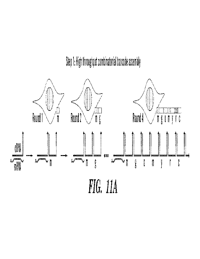
[00039] FIG. 11A-11B shows an assembly of barcodes for a cell or other region
of interest.
(FIG. 11A) Iterative formation of a concatemer upon nucleic acids localized in
situ (e.g.. cDNA
sequences) results in the formation of a specific barcode for reads from that
cell (e.g.. m-g-o-m-y-
r-c). Orientation shown for 3' barcoding of cDNA, although 5' barcoding may
also be performed
(see e.g.. FIG. 18 and FIG. 19). (FIG. 11B) Cross-junction synthesis and PCR
are used to prepare
records for sequencing.
[00040] FIG. 12 shows application of the methods and compositions provided
herein.
[00041] FIG. 13 shows a workflow for dissociative split-pool
barcoding. Iterative splitting
of cells or otherwise associated biomolecules (e.g.. hydrogel pieces) into
tubes, barcoding of
nucleic acids e.g.. with the light-directed concatemer formation depicted
elsewhere, and then re-
pooling enables unique barcode sequences to be associated with each separate
cell/component.
Split-pool strategies have previously been used for single-cell barcoding
through multiple
CA 03161183 2022- 6-8
WO 2021/119402
PCT/US2020/064463
expensive enzymatic ligation steps but using the concatemer-based barcoding
strategy dramatically
lowers costs as each barcoding step can be performed without the need for
expensive enzymes or
other reagents. Sequences can be extracted similarly to when they're on a
surface: with cross-
junction synthesis and PCR of records.
[00042] FIG. 14A-14C shows an embodiment of the spatial barcoding. (FIG. 14A)
Barcodes
are crosslinked typically through the use of a CNVK modification, and
crosslinking is activated
with UV light. (FIG. 14B) By spatially addressing UV light illumination
profiles, barcodes may
be crosslinked to dock sequences only in desired positions, and after a
stringent wash step (e.g.. a
formamide-containing buffer) all non-crosslinked barcode strands can be washed
away. (FIG.
14C) Iterative steps of binding, crosslinking specific regions, and stringent
washing enables the
iterative construction of barcodes associated with those specific regions.
[00043] FIG. 15A demonstrates linear barcoding of N regions (e.g.. N
distinct cells) is
performed such that a single of N barcodes is assigned to each position or (or
positions) of interest.
Sequencing results may then be extracted together in bulk, and reads may be
mapped back to their
original corresponding positions based on the barcode sequences in the reads.
FIG. 15B
demonstrates a method of combinatorial barcoding, a concatenated barcode is
iteratively
constructed, such that each region (e.g. cell) that for which reads should be
attributed to receives
a unique barcode (see e.g.. FIG. 1 8). For example, for N rounds of M
barcodes, MAN unique
barcodes could feasibly be assigned.
[00044] FIG. 16 shows an embodiment of the workflow for combined imaging and
RNA
sequencing data for a sample. In general, extra imaging steps and other assays
may be added
before or after barcoding, and the A-tailing step may optionally occur before
or after barcoding. A
different tailing (e.g.. a T-tailing, C-tailing, G-tailing, or any other type
of tailing with terminal
transferase or other enzyme may be utilized) may instead be utilized. For
targeted approaches, the
workflow is very similar, except that probes may already contain the 5' and 3'
tails, so both RT
and A-tailing steps can be skipped. Any domain (e.g., 1-letter, 2-letter, 3-
letter, or 4-letter) may be
utilized for the 3 tail sequence.
[00045] FIG. 17 shows experimental validation of UV power and illumination
conditions.
A set of experiments to optimize the UV power and illumination conditions for
barcoding FISH
probes bound to rRNA transcripts in HeLa cells. A checkerboard pattern was
rastered across a well
with each separate region testing a different UV power and illumination time
condition.
[00046] FIG. 18 shows a strand diagram of 5' light-directed barcoding strategy
with UMI
on cross-junction synthesis primer. A primer with an overhanging 5' domain
(e.g.. with random
N bases on the end) is localized to RNA's (e.g.. mRNA, non-coding RNAs) and
creates cDNA
sequences. The cDNA sequences may then be appended with bases on the 3' end,
such as with a
11
CA 03161183 2022- 6-8
WO 2021/119402
PCT/US2020/064463
polyA tail with the use of terminal transferase and dATP. Subsequently,
combinatorial barcodes
are assembled iteratively directly onto the 5' overhang of the cDNA or other
in situ localized
sequence, through binding, UV crosslinking, and wash steps. (The A tailing
step may be included
before or after barcoding). Optionally, RNaseH displacement of barcodes from
RNA may be
performed before or simultaneously with cross-junction synthesis. After cross-
junction synthesis,
full records are formed via PCR amplification.
[00047] FIG. 19 shows a strand diagram of 5' light-directed barcoding strategy
with UMI
on barcode capping strand. A primer with an overhanging 5' domain (e.g.. with
random N bases
on the end) is localized to RNA's (e.g.. mRNA, non-coding RNAs) and creates
cDNA sequences.
The cDNA sequences may then be appended with bases on the 3' end, such as with
a polyA tail
with the use of terminal transferase and dATP. Subsequently, combinatorial
barcodes are
assembled iteratively directly onto the 5' overhang of the cDNA or other in
situ /ocalized sequence,
through binding, UV crosslinking, and wash steps. Optionally, RNaseH
displacement of barcodes
from RNA may be performed before or simultaneously with cross-junction
synthesis. After cross-
junction synthesis, full records are formed via PCR amplification.
[00048] FIG. 20 shows experimental validation of primer sets for cDNA library
generation. (top) A table of primers and concentrations used for reverse
transcription (RT). Well
labels (Al -B4) match the orientation of images shown in bottom. Well B1-B4
have a combination
of primers as well as anon-reverse transcribed negative control. (bottom)
Images of the localization
of the cDNA library after reverse transcription using a Cy5 labeled primer. A
Cy3 CNVK barcode
was then added and crosslinked in a checkerboard pattern using a DIVED and a
10x objective and
imaged in Cy3.
[00049] FIG. 21 shows sequencing results for different RT primers. In situ
reverse
transcription in fixed HeLa cells was performed with different primers
containing 5' barcoding
domains along with NNNNININN (7N's, experiment Al), NNNNNGGG (5N's and 3G' s,
experiment A2), or NNNNNCCC (5N's and 3C's, experiment A3) on the 3' end.
After barcoding,
cross-junction synthesis, and PCR according to the strategy depicted in Fig.
18, PCR ampl icons
were purified with Ampure XP beads and sent for sequencing (250bp paired end).
Examples of
several expected read results are shown for each of these primers, and the
highlighted cDNA
sequences (blue) map to known homo sapiens sequences as expected. These data
verify the success
of the general strategy and that each primer may be used to successfully
produce transcriptomic
records.
[00050] FIG. 22A shows the sequence structure for barcoding a 5' sequence
(e.g.. a 5' tail
on cDNA, FISH probe, etc.). A concatemer formed with a Reverse (Rev) primer
capping strand,
zero or more barcode strands, and a cDNA, FISH, or other probe sequence with a
polyA tail can
12
CA 03161183 2022- 6- 8
WO 2021/119402
PCT/US2020/064463
be effectively copied with a cross-junction synthesis primer containing a
Forward (For) primer and
polyT 3' end to form a PCR amplifiable record that can be sequenced. In this
case, two different
orientations of barcode sequences (W/X domains, and Y/Z domains) are utilized,
though more
distinct barcode sequences may be utilized as well. Strands may be purified or
unpurified and may
contain extra bases on the 3' or 5' ends (e.g.. T linkers, fluorophores,
modifications to prevent
extension or degradation). FIG. 22B shows an embodiment of a binding domain
barcode sequences
used for the demonstrations in the next several figures are shown, colored
according to their
domains. An arbitrary number of barcode strands with different (Barcode)
domain sequences may
be utilized for barcoding. FIG. 22C shows complete sequence information for
the experiments
reported in the all subsequent figures are shown. PCR Primer sequences are
based on the Smart
Seq3 protocol. All other sequences and particularly those for barcoding have
been specifically
designed and experimentally for this barcoding application, after modeling and
extensive testing
of dozens of cross-junction synthesis reactions. See also, Tables 1-3 in the
working examples.
[00051] FIG. 23A-23E shows validation of iterative barcode assembly on a
streptavidin-
coated surface (glass slide). FIG. 23A shows a schematic of iterative barcode
assembly of
fluorescently labeled DNA barcode strands, followed by cross-junction
synthesis and PCR. FIG.
23B shows schematics of concatenated barcodes with 2 to 7 junctions,
containing 1 to 6 barcodes,
respectively. FIG. 23C shows distribution of DNA barcode lengths expected in
distinct wells (top).
Top left well in an 8-well chamber contains DNA barcodes of length 6 and will
display the highest
amount of fluorescent signal. Followed by 5 and 4 etc. Scan of the 8-well
chamber in the Cy3
Fluorescent channel (bottom). FIG. 230 shows complete sequence design for the
7-junction
concatemer and amplicons based on sequences presented in FIG. 22A-22C. FIG.
23E shows that
after extraction, PCR, and purification with a MinElute PCR Purification
column, amplicons from
the top left well (6-junction) were sequenced (250bp paired end sequencing).
Example sequencing
results are shown, both for full length (6-barcode containing reads) as well
as truncated reads (e.g..
containing 2 or 4 barcodes). Truncated reads are expected in addition to full
length reads due to
some inefficiencies in the concatemer formation step.
[00052] FIG. 24 shows sequencing results for several different fixation,
permeabilization,
RT, and barcoding conditions following the strategy depicted in FIG. 19. (top)
Several
sequences that were acquired for each of several fixation/permeabilization
conditions (experiments
B1 through B8) and match the expected sequence format after two rounds of
barcoding are shown.
These sequences show the expected barcode sequences in each case and examples
of different
HMI's, and sequence lengths, that occur. (bottom) While keeping the fixation
and permeabilization
constant, several variations to the RT step were tested along with some
controls. For each of
experiments Cl through C4, one barcode was first introduced but not
crosslinked prior to stringent
13
CA 03161183 2022- 6-8
WO 2021/119402
PCT/US2020/064463
washing (Exchange 1), and then a second barcode was introduced that was
crosslinked with UV
and should have shown up in the sequencing reads (Exchange 2). As expected, in
all conditions
except the control that contained RNase A during RT, the correct barcode that
was crosslinked
shows up in the majority of reads (>1,500 of 2,000 reads examined), and the
incorrect (non-
crosslinked Exchange 1 barcode) barcode showed up extremely rarely (as low as
0 in 2,000 reads).
In all of the conditions (experiments B2 through B8, Cl through C4) except the
no reverse
transcriptase (RT) control (experiment B1), the highlighted cDNA sequences
(blue) map to known
homo sapiens sequences. Exceptions: some conditions having A-tailing take
place after barcoding,
as indicated in the figure, and all conditions having the RNaseH treatment
combined with the cross-
junction synthesis incubation.
[00053] FIG. 25A-25D demonstrates imaging and gel results for experiments B1
through
B8 and Cl through C4. FIG. 25A shows imaging results for experiments B1
through B8 show
distinct fluorescence morphologies after reverse transcription (RT) with a
fluorophore (Alexa 488)-
labeled RT primer. As expected, after displacement, the fluorescence signal
from localized primers
goes significantly down, indicating they have been successfully displaced
during the combined
RNaseH and cross-junction synthesis steps. FIG. 25B shows tor the control
condition containing
RNase A and no RNaseOUT during RT, signal was much higher, and lower contrast
visualization
revealed strong suspected nucleolar signal. FIG. 25C shows imaging results for
experiments Cl,
C3, and C4 are also shown. FIG. 25D shows gel results for all conditions show
the lengths of
records produced after PCR amplification (1% Agarose E-gel with Sybr Gold).
For cases
containing reverse transcription and no RNase A, the typical lengths recovered
range between about
150bp and 1300bp.
[00054] FIG. 26 shows transcriptomic mapping results. Transcriptomic mapping
was
performed with the STAR aligner on sequencing results. (left) An example
output log file is shown
on the left for mapping results for 1,024 transcripts identified with the
expected sequence format
for experiment B7. 40.5% of the reads mapped uniquely, whereas 49% mapped to
multiple loci and
9.5% were too short to map. (right) Gene mapping results were sorted by
frequency of mapped
transcripts and the top of the list is depicted. The most common uniquely
mapped genes correspond
to mitochondrial rRNA.
[00055] FIG. 27 shows automated barcode assignment and iterative barcoding on
a
surface. An example workflow whereby a list of barcodes (BC1, BC2, BC3 etc...)
can be
converted into a series of photomasks (middle panel) with each region of
interest (white squares,
middle panel) assigned a unique barcode. An image was taken after a series of
6 barcoding steps
with fluorescent DNA strands to uniquely tag and barcode an array of 112
regions of interest (right
panel).
14
CA 03161183 2022- 6-8
WO 2021/119402
PCT/US2020/064463
[00056] FIG. 28A-28G shows automated barcoding of biomolecular samples. FIG.
28A
shows a workflow whereby a collection of cells can be detected with a computer
algorithm and
selectively targeted for barcode delivery, resulting in each cell with a
unique barcode assignment.
FIG. 28B shows an image of cells with a fluorescent DNA primer targeting RNA.
FIG. 28E shows
an image of cells after 6 rounds of barcoding with a fluorescent DNA barcode
(green) using the
masks from panel (FIG. 28C, 28F). FIG. 28C and FIG. 28F show an overlay of the
detected
cellular masks (white outlines). FIG. 28D and FIG. 28G show an enlarged image
of the outlined
square from (FIG. 28C) and (FIG. 28F) respectively
DETAILED DESCRIPTION
[00057] The fundamental strategy for nucleic acid barcoding provided herein is
depicted in
FIGs. 1A-9D.
[00058] Generally, the methods provided herein are based in part, on the
discovery of methods
and compositions that allow for high-throughput detection of a target nucleic
acid and the
production of sequence and spatial information. The methods and compositions
provided herein
are useful in many applications, such diagnostics, pathology, and basic
research.
[00059] In particular, the compositions and methods provided herein can be
useful in spatial
mapping, detecting biomolecule localization, identifying various cell types in
a tissue, molecular
coding, data storage, tissue engineering, communication, and biosensing. The
approaches provided
herein can be used to create patterned and barcoded surfaces for
oligonucleotide arrays. For
example, the methods and compositions provided herein can be used for higher
levels of patterning,
masking, and capturing nucleic acid targets (e.g.., biomarkers of interest).
[00060] As another example, the targeted approach provided in the
working examples (e.g..,
Strategy 1), can also be used to bind other nucleic acids immobilized in a
sample or on a surface,
such as DNA-conjugated antibodies bound to protein targets of interest (see
FIG. 1B). In general,
any entity (such as nucleic acids, proteins, peptides, lipids, sugar groups,
small molecules,
nanoparticles, beads, glass surfaces) that can be labeled with or crosslinked
to a strand of interest
can be patterned, barcoded and recorded using the methods provided herein.
[00061] In some embodiments, the barcode composition comprises:
a. a first nucleic acid comprising in a 5' to 3' direction: (i) optionally, a
unique
molecule identifier (UMI) sequence; (ii) a first targeting domain; and (iii) a
first
hybridization domain, and
b. a second nucleic acid comprising in a 5' to 3' direction: (i) a barcode
domain; and
(ii) a second hybridization domain, wherein the second hybridization domain is
CA 03161183 2022- 6-8
WO 2021/119402
PCT/US2020/064463
substantially complementary to the first hybridization domain of the first
nucleic
acid, and
wherein at least one of the first or second hybridization domain comprises a
photoreactive
element.
[00062] In some embodiments, the barcode composition comprises:
a. a first nucleic acid comprising in a 5' to 3' direction: (i) optionally, a
unique
molecule identifier sequence; (ii) a first targeting domain; and (iii) a first
hybridization domain; and
b. a second nucleic acid comprising in a 5' to 3' direction: (i) a second
hybridization domain, wherein the second hybridization domain is substantially
complementary to the first hybridization domain of the first nucleic acid; and
(ii) a first barcode domain, and
wherein at least one of the first or second hybridization domain comprises a
photoreactive
element.
[00063] In some embodiments, the barcode composition comprises:
a. a first nucleic acid comprising in a 5' to 3' direction: (i) optionally, a
unique
molecule identifier sequence; (ii) a first targeting domain; and (iii) a first
hybridization domain; and
b. a second nucleic acid comprising in a 5' to 3' direction: (i) a second
hybridization
domain, wherein the second hybridization domain is substantially complementary
to the first hybridization domain of the first nucleic acid; and (ii) a first
barcode
domain; and (iii) a third hybridization domain, and
wherein at least one of the first or second hybridization domains comprises a
photoreactive
element, and the third hybridization domains optionally comprises a
photoreactive element.
[00064] In some embodiments, the barcode composition further comprises n
additional nucleic
acids, wherein: n optionally is an integer from 1 to 100, and each additional
nucleic acid comprises
in 5' to 3' direction: (i) a first hybridization domain; (ii) a barcode
domain; and (iii) a second
hybridization domain, and wherein the first hybridization domain of nth
nucleic acid is
substantially complementary to the second hybridization domain of (n-/)th
nucleic acid, wherein
the first hybridization domain of n-1 nucleic acid is substantially
complementary to the third
hybridization domain, and wherein at least one of the first or the second
hybridization domain of
each nucleic acid comprises a photoreactive element.
[00065] In some embodiments, the barcode composition further
comprises a first cap
nucleic acid strand comprising in 5' to 3' direction: (i) a first cap
hybridization domain, wherein
the first cap hybridization domain is substantially complementary to the
second hybridization
16
CA 03161183 2022- 6-8
WO 2021/119402
PCT/US2020/064463
domain of nth nucleic acid when n is 1 or more, or the cap hybridization
domain is substantially
complementary to the third hybridization domain when n is 0; and (ii) a second
cap hybridization
domain, wherein the first cap hybridization domain optionally comprises a
photoreactive element.
[00066] In some embodiments, the barcode composition further
comprises a first cap
nucleic acid strand and a second cap nucleic acid strand, the second cap
nucleic acid strand
comprising in 5' to 3' direction: (i) a primer sequence domain; (ii)
optionally, a unique molecular
identifier (UNIT) sequence; and (iii) a hybridization domain, wherein the
hybridization domain is
substantially complementary to the second cap hybridization domain of the
first cap nucleic acid,
and wherein at least one of the second cap hybridization domain and the
hybridization domain of
the second nucleic acid comprises a photoreactive element.
[00067] The nucleic acid strands of the compositions and methods described
herein comprise
one or more domains. Without limitation, each domain can independently
comprise any desired
nucleotide sequence or number of nucleotides. In other words, each domain can
be independently
of any length. Accordingly, each domain can be independently one nucleotide to
thousands of
nucleotides in length. For example, each domain can be independently 1 to
1000, 1 to 500, 1 to
250, 1 to 200, 1 to 150, 1 to 100, 1 to 75, 1 to 50, or 1 to 25 nucleotides in
length. In some
embodiments, each domain can be independently 1, 2, 3, 4, 5, 6, 7, 8, 9, 10,
11, 12, 13, 14, 15, 16,
17, 18, 19, 20, 21, 22, 23, 24, 25 or more nucleotides in length.
[00068] As described herein, hybridization domains of two nucleic strands can
hybridize with
each other to form a double-stranded structure. Without limitations, each
duplex region can
independently comprise any desired number of base-pairs. In other words, each
duplex region can
be independently of any length. Accordingly, each duplex region can be one
base pair to tens of
base pairs in length. In some embodiments, each duplex region can be
independently I to 50, 1 to
45, 1 to 40, 1 to 35, 1 to 30, 1 to 25, 1 to 20 or 1 to 15 nucleotides or base
pairs in length. For
example, each duplex region can be independently 1,2, 3, 4, 5, 6, 7, 8, 9, 10,
11, 12, 13, 14, 15, 16,
17, 18, 19, 20, 21, 22, 23, 24, 25 or more nucleotides or base pairs in
length.
[00069] Each nucleic acid strand can be independently of any length. For
example, each nucleic
acid strand can be few nucleotides to thousands of nucleotides in length. For
example, each nucleic
acid strand can be independently 1 to 50, 1 to 75, 1 to 100, 1 to 150, 1 to
175, 1 to 200, 1 to 250, 1
to 300, 1 to 400, Ito 500, 1 to 750, 1 to 1000 or more nucleotides in length.
[00070] Each domain can independently comprise any desired nucleotide
sequence. Further,
each domain can independently utilize a 1-letter, 2-letter, 3-letter or 4-
letter code. As used herein, a
"I-letter code- means the domain only comprises only one type of nucleobase,
i.e., only one of
adenine, thymine/uracil, guanine, and cytosine, or modified versions thereof
For example, a domain
utilizing a 1-letter code comprises a stretch of nucleotides comprising the
same nucleobase or a
17
CA 03161183 2022- 6-8
WO 2021/119402
PCT/US2020/064463
modified version of the nucleobase. For example, a domain can comprise a
stretch of polyA, polyT,
polyC or polyG. In some embodiments, the hybridization domain of the first
nucleic acid utilizes
a 1-letter code. For example, the hybridization domain of the first nucleic
acid can comprise a
poly(A) sequence.
[00071] A "2-letter code" means the domain only comprises two of the four
nucleobases, i.e.,
only two of adenine, thymine/uracil, guanine, and cytosine, or modified
versions thereof. For
example, a 2-letter code can comprise or consist of nucleobases selected from
the group consisting
of adenine and thymine/uracil, adenine and guanine, adenine and cytosine,
thymine/uracil and
guanine, thymine/uracil and cytosine, and guanine and cytosine.
[00072] A -3-letter code" means the domain comprises only three of
the four nucleobases, i.e.,
only three of adenine, thymine/uracil, guanine, and cytosine, or modified
versions thereof For
example, a 3-letter code can comprise or consists of nucleobases selected from
the group consisting
of adenine, thymine/uracil, and guanine; adenine, thymine/uracil, and
cytosine; adenine, guanine,
and cytosine; and thymine/uracil, guanine, and cytosine.
[00073] In some embodiments, at least one domain comprises same types of
nucleobases. For
example, a domain only comprises purine nucleobases or pyrimidine nucleobases.
[00074] The first nucleic acid strand can be an RNA molecule, e.g.., an RNA
transcript. In one
example, the first nucleic acid is an mR_NA. For example, the first nucleic
strand is an mRNA and
the hybridization domain comprises a polyA sequence.
[00075] As described herein, a nucleic acid strand comprises a
unique molecule identifier
sequence or domain. A unique molecule identifier sequence or domain can be
synthesized by using
a mix of nucleotides during base addition chemical synthesis to create
libraries of random sequences
(degenerate sequences). A unique molecule identifier sequence or domain can
consist of several
such random bases in tandem, with or without known nucleotide sequences
intercalated. In some
embodiments, a unique molecule identifier sequence or domain is excluded from
primers and record
sequences. In some embodiments, the unique molecule identifier sequence or
domain of a nucleic
acid is incorporated into one of the other domains of same nucleic acid.
[00076] As described herein, hybridization domains can comprise a
photoreactive element. As
used herein, the term "photoreactive element" refers to any element (e.g..,
nucleotide, protein, or
antibody) that can permit hybridization to another nucleotide upon
photoirradiation by a light
source. In some embodiments, the photoreactive element is a photoreactive
nucleotide. In some
embodiments, the photoreactive nucleotide is a CNVK or CNVD crosslinking base.
In some
embodiments, the photoreactive element is psoralen.
[00077] In some embodiments of any of the aspects described herein, a nucleic
acid strand can
comprise a nucleic acid modification. For example, at least one of a targeting
domain, a barcode
18
CA 03161183 2022- 6-8
WO 2021/119402
PCT/US2020/064463
domain, a hybridization domain, unique molecule identifier sequence and/or
primer sequence
domain can independently comprise a nucleic acid modification. Exemplary
nucleic acid
modifications include, but are not limited to, nucleobase modifications, sugar
modifications, inter-
sugar linkage modifications, conjugates (e.g.., ligands), and any combinations
thereof Nucleic
acid modifications also include unnatural, or degenerate nucleobases.
[00078]
Exemplary modified nucleobases include, but are not limited to, inosine,
xanthine,
hypoxanthine, nubularine, isoguanisine, tubercidine, and substituted or
modified analogs of
adenine, guanine, cytosine and uracil, such as 2-aminoadenine, 6-methyl and
other alkyl
derivatives of adenine and guanine, 2-propyl and other alkyl derivatives of
adenine and guanine,
5-halouracil and cytosine, 5-propynyl uracil and cytosine, 6-azo uracil,
cytosine and thymine, 5-
uracil (pseudouracil), 4-thiouracil, 5-halouracil, 5-(2-aminopropyl)uracil, 5-
amino allyl uracil, 8-
halo, amino, thiol, thioalkyl, hydroxyl and other 8-substituted adenines and
guanines, 5-
trifluoromethyl and other 5-substituted uracils and cytosines, 7-
methylguanine, 5-substituted
pyrimidines, 6-azapyrimidines and N-2, N-6 and 0-6 substituted purines,
including 2-
aminopropyladenine, 5-propynyluracil and 5-propynylcytosine, dihydrouracil, 3-
deaza-5-
azacytosine, 2 -aminop urine, 5 -alkyluracil, 7-alkylguanine, 5-alkyl cy to
sine,7-deazaadenine, N6,
N6-dimethyladenine, 2,6-diaminopurine, 5-amino-allyl-uracil, N3-methyluracil,
substituted 1,2,4-
triazoles, 2-pyridinone, 5-nitroindole, 3-nitropyrrole, 5-methoxyuracil,
uracil-5-oxy acetic acid, 5-
methoxycarbonylmethyluracil, 5-methyl-2-thiouracil, 5-methoxycarbonylmethy1-2-
thiouracil, 5-
m ethyl am in om ethyl -2-th i ouracil, 3 -(3 -amino-3 c arboxypropyl )uracil,
3-methyl cytosine, 5 -
methylcytosine, N4-acetyl cytosine, 2-thiocytosine, N6-methyladenine, N6-
isopentyladenine, 2-
methylthio-N6-isopentenyladenine, N-methylguanines, or 0-alkylated bases.
Further purines and
pyrimidines include those disclosed in U.S. Pat. No. 3,687,808, those
disclosed in the Concise
Encyclopedia of Polymer Science and Engineering, pages 858-859, Kroschwitz, J.
I., ed. John
Wiley & Sons, 1990, and those disclosed by Englisch et al., Angewandte Chemie,
International
Edition, 1991, 30, 613.
[00079] In some embodiments, a modified nucleobase can be selected from the
group
consisting of: inosine, xanthine, hypoxanthine, nubularine, isoguanisine,
tubercidine, 2-
(halo)adenine, 2-(alkyl)adenine, 2-(propyl)adenine, 2-(amino)adenine, 2-
(aminoalkyll)adenine,
2-(aminopropyl)adenine, 2-(methylthio)-N6-(isopentenyl)adenine,
6-(alkyl)adenine,
6-(methyl)adenine, 7-(deaza)adenine, 8-(alkenyl)adenine, 8-(alkyl)adenine, 8-
(alkynyl)adenine,
8-(amino)adenine, 8-(halo)adenine, 8-(hydroxyl)adenine, 8-(thioalkyl)adenine,
8-(thiol)adenine,
N6-(i sopentyl)adenine, N6-(in ethyl)aden in e,
N6, N6-(di methyl)adeni ne, 2-
(alkyl)guanine,2-(propyl)guanine, 6-(alkyl)guanine, 6-(methyl)guanine, 7-
(alkyl)guanine,
7-(methyl)guanine, 7-(deaza)guanine, 8-(alkyl)guanine, 8-(alkenyl)guanine, 8-
(alkynyl)guanine,
19
CA 03161183 2022- 6-8
WO 2021/119402
PCT/US2020/064463
8-(amino)guanine, 8-(halo)guanine, 8-(hydroxyl)guanine, 8-(thioalkyl)guanine,
8 -(thiol)guanine,
N-(methyl)guanine, 2-(thio)cytosine, 3 -(deaza)-5 -(aza)cyto
sine, 3 -(alkyl)cytosine,
3 -(methyl)cytosine, 5 -(alkyl)cytosine, 5-(alkynyl)cytosine, 5 -
(halo)cytosine, 5 -(methyl)cytosine,
-(propynyl)cytosine,
5 -(propynyl)cytosine, 5 -(trifluoromethyl)cytosine, 6-(azo)cytosine,
N4-(acetyl)cytosine, 3 -(3 -amino-3 -carboxypropyl)uracil,
5 -ethyny1-2' -deoxyuri dine, 2-
(thio)uracil, 5-(methyl)-2-(thio)uracil, 5 -(methylaminomethyl)-2-
(thio)uracil, 4-(thio)uracil,
5 -(methy 1)-44 thio) uracil, 5 -(methylaminomethyl)-4-(thio)uracil, 5 -
(methyl)-2,4-(di thio) uracil,
5 -(methylaminomethyl)-2,4-(dithio)uracil, 5 -(2-aminopropyl)uracil,
5 -(alkyl)uracil, 5 -
(alkynyOuracil, 5 -(allylamino)uracil,
5-(aminoallyl)uracil, 5 -(aminoalkyl)uracil,
5 -(guanidiniumalkyl)uracil, 5-(1,3 -di azol e- 1
-alkyl)uracil, 5 -(cyanoalkyl)uracil, 5 -
(dialkylaminoalkyl)uracil, 5 -(dimethylaminoalkyl)uracil, 5 -(halo)uracil, 5-
(methoxy)uracil,
urac il-5-oxy acetic acid,
5-(methoxycarbonylmethyl)-2-(thio)uracil, 5 -(methoxycarbonyl-
methyl)uracil, 5-(propynyl)uracil, 5 -(propynyl)uracil, 5-
(trifluoromethyl)uracil, 6-(azo)uracil,
dihydrouracil, N3-(methyl)uracil, 5 -uracil
(i.e., pseudouracil),
2-(thio)pseudouraci1,4-(thio)pseudouraci1,2,4-(dithio)psuedouraci1,5-
(alkyl)pseudouracil, 5 -
(methyl)pseudouracil, 5-(alkyl)-2-(thio)pseudouracil, 5 -(methyl)-2-
(thio)pseudouracil, 5 -(alkyl)-
4-(thio)pseudouracil, 5 -(methyl)-4-(thio)pseudouracil, S -(alkyl)-2,4-
(dithio)pseudouracil, 5 -
(methyl)-2,4-(dithio)pseudouracil, 1-substituted pseudouracil, 1-substituted
2(thio)-pseudouracil,
1-substituted 4-(thio)p seudouracil, 1-substituted
2,4-(dithio)pseudouracil,
1 -(aminocarbonylethyleny1)-pseudouracil,
1 -(aminocarbonylethyleny1)-2(thio)-pseudouracil,
1 -(aminocarbonylethyleny1)-4-(thio)pseudouracil,
1 -(aminocarbonylethyleny1)-2,4-
(dithio)pseudouracil, 1 -(aminoalkyl amino carbonylethyleny1)-pseudouracil , 1
-(amino alkylamino-
carbonyl ethyl eny1)-2(thio)-pseudouracil,
1 -(aminoalkylaminocarbonylethyleny1)-
4-(thio)pseudouracil, 1 -(amino al kylamino c arbonyl ethyleny1)-2,4-
(dithio)pseudouracil, 1,3 -
(diaza)-2-(oxo)-phenoxazin- 1 -yl, 1 -(aza)-2-(thio)-3 -(aza)-phenoxazin- 1 -
yl, 1,3 -(diaza)-2-(oxo)-
phenthi azin- 1 -yl, 1 -(aza)-2-(thio)-3 -(aza)-phenthiazin- 1 -yl, 7-
substituted 1,3 -(diaza)-2-(oxo)-
phenoxazin- 1 -yl, 7-substituted 1 -(aza)-2-(thio)-3 -(aza)-phenoxazin- 1 -yl,
7-substituted 1,3 -
(diaza)-2-(oxo)-phenthiazin- 1 -yl, 7-substituted 1 -(aza)-2-(thio)-3 -(aza)-
phenthi azin- 1 -yl, 7-
(aminoalkylhy droxy)-1 ,3 -(cliaza)-2-(oxo)-phenoxazin-l-yl,
7-(aminoalkylhy droxy)- 1 -(aza)-2-
(thio)-3 -(aza)-phenoxazin- 1 -yl, 7-(aminoalkylhydroxy)- 1,3 -(diaz a)-2-
(oxo)-phenthiazin- 1 -yl, 7-
(aminoalkylhy droxy)-1 -(aza)-2-(thio)-3 -(aza)- phenthi azin- 1 -yl, 7-
(guanidiniumalkylhy droxy)-
1,3 -(diaza)-2-(oxo)-phenoxazin- 1 -yl,
7-(guanidiniumalky lhy droxy)- 1 -(az a)-2-(thio)-3 -(aza)-
phenoxazin- 1 -yl, 7-(guanidiniumalkyl-hy droxy)- 1,3 -(diaz a)-2-(oxo)-
phenthiazin- 1 -yl, 7-
(guanidiniumalky lhy droxy)- 1 -(aza)-2-(thio)-3 -(aza)-phenthiazin-l-yl,
1,3,5 -(triaza)-2, 6-(dioxa)-
naphthalene, inosine, xanthine, hypoxanthine, nubularine, tubercidine,
isoguanisine, inosinyl, 2-
CA 03161183 2022- 6-8
WO 2021/119402
PCT/US2020/064463
aza-inosinyl, 7-deaza-inosinyl, nitroimidazolyl, nitropyrazolyl,
nitrobenzimidazolyl,
nitroindazolyl, aminoindolyl, pyrrolopyrimidinyl,
3-(methyl)isocarbostyrilyl, 5-
(methyl)isocarbostyrilyl, 3-(methyl)-7-(propynyl)isocarbostyrilyl, 7-
(aza)indolyl, 6-(methyl)-7-
(aza)indolyl, imidizopyridinyl, 9-(methyl)-imidizopyridinyl, pyrrolopyrizinyl,
isocarbostyrilyl, 7-
(propynyl)isocarbostyrilyl, propyny1-7-(aza)indolyl, 2,4,5-(trimethyl)phenyl,
4-(methyl)indolyl,
4,6-(dimethyl)indolyl, phenyl, napthalenyl, anthracenyl, phenanthracenyl,
pyrenyl, stilbenyl,
tetracenyl, p en lac enyl, difluorotolyl,
4-(fluoro)-6-(methyl)benzimidazole, 4-
(methyl)benzimidazole, 6-(azo)thymine, 2-py ri clinone, 5 -nitroindol e, 3 -
nitropyrrol e, 6-
(aza)pyrimidine, 2-(amino)purine, 2,6-(diamino)purine, 5-substituted
pyrimidines, N2-substituted
purines, 1\16-substituted purines, 06-substituted purines, substituted 1,2,4-
triazoles, and any 0-
alkylated or N-alkylated derivatives thereof.
[00080]
Exemplary sugar modifications include, but are not limited to, 2'-Fluoro,
3'-Fluoro,
2' -0Me, 3' -0Me, 2' -deoxy modifications, and acyclic nucleotides, e.g..,
peptide nucleic acids
(PNA), unlocked nucleic acids (UNA) or glycol nucleic acid (GNA).
[00081]
In some embodiments, a nucleic acid modification can include replacement
or
modification of an inter-sugar linkage. Exemplary inter-sugar linkage
modifications include, but
are not limited to, phosphotri esters, methylphosphonates, phosphoramidate,
phosphorothioates,
methylenemethylimino, thiodiester, thionocarbamate, siloxane, N,N'-
dimethylhydrazine (¨CH2-
N(CH3)-N(CH3)-), amide-3 (31-CE12-C(=0)-N(H)-5') and amide-4 (3'-CH2-N(H)-
C(=0)-5'),
hydroxylamino, siloxane (dialkylsiloxxane), carboxamide, carbonate,
carboxymethyl, carbamate,
carboxyl ate ester, thioether, ethylene oxide linker, sulfide,sulfonate,
sulfonamide, sulfonate ester,
thiothrmacetal (3'-S-CH2-0-5'), thrmacetal (3 '-0-CH2-0-5'), oxime,
methyleneimino,
methykenecarbonylamino, methylenemethylimino (M,MI,
3 '-CH2-N(CI-13)-0- 5'),
methylenehydrazo, methyl enedimethylhydrazo, methyleneoxymethylimino, ethers
(C3'-0-05'),
thio ethers (C3'-S-CS'), thioacetami do (C3' -N(H)-C(=0)-CH2-S-C 5' , C3' -0-
P(0)-0-SS-05' ,
C3' -CH2-NH-NH-05', 3'-NHP(0)(OCH3)-0-5' and 3 '-NHP(0)(OCH3)-0-5' .
[00082]
In some embodiments, nucleic acid modifications can include peptide
nucleic acids
(PNA), bridged nucleic acids (BNA), morpholinos, locked nucleic acids (LNA),
glycol nucleic
acids (GNA), threose nucleic acids (TNA), or any other xeno nucleic acids
(XNA) described in
the art
[00083] In some embodiments of the various aspects described herein, a nucleic
acid can be
independently modified on the 3'- and/or 5'-end. For example, a label,
fluorophore, tag, or a cap
can be added to the 3' and/or 5'-end of a nucleic acid described herein.
[00084] In some embodiments of the various aspects described herein, a nucleic
acid strands
described herein can be modified with a linker or spacer, e.g.., at an
internal position, on the 3'-
21
CA 03161183 2022- 6-8
WO 2021/119402
PCT/US2020/064463
and/or 5'-end. Without wishing to be bound by a theory, the linker or spacer
can be used for linking
the nucleic acid strand with a moiety, such as a solid support or label. In
some embodiments, the
linker or spacer can be selected from the group consisting of photocleavable
linkers, hydrolyzable
linkers, redox cleavable linkers, phosphate -based cleavable linkers, acid
cleavable linkers, ester-
based cleavable linkers, peptide-based cleavable linkers, and any combinations
thereof. In some
embodiments, the cleavable linker can comprise a disulfide bond, a tetrazine-
trans-cyclooctene
group, a s ulfhy dry 1 group, a ni trobenLy1 group, a nitoindoline group, a
bromo hy droxy co umarin
group, a bromo hydroxyquinoline group, a hydroxyphenacyl group, a
dimethozybenzoin group, or
any combinations thereof
[00085] Any art-recognized photocleavable linker can be used. In some
embodiments, the
cleavable linker can comprise a photocleavable linker. Generally,
photocleavable linkers contain
a photolabile functional group that is cleavable upon exposure to a light
source (e.g.., UV light) or
specific wavelength. Non-limiting examples of photocleavable spacers can be
found, for example,
in US Patent Nos. 6,589,736 Bl; 7,622,279 B2; 9,371,348 B2; 7,547,530 B2; and
7,057,031 B2;
and PCT Publication No. W02014200767, contents of all of which are
incorporated herein by
reference in their entirety.
[00086] In some embodiments of the various aspects described herein, the
barcode composition
comprises a detectable label. For example, a nucleic acid strand described
herein can be modified
with a detectable label, e.g,.., at an internal position, on the 3'- and/or 5'-
end. Without wishing to
be bound by a theory, such a detectable label can facilitate detection. As
used herein, the term
"detectable label" refers to a composition capable of producing a detectable
signal indicative of the
presence of a target. Detectable labels include any composition detectable by
spectroscopic,
photochemical, biochemical, immunochemical, electrical, optical or chemical
means. Suitable
labels include fluorescent molecules, radioisotopes, nucleotide chromophores,
enzymes, substrates,
chemiluminescent moieties, bioluminescent moieties, and the like. As such, a
label is any
composition detectable by spectroscopic, photochemical, biochemical,
immunochemical,
electrical, optical or chemical means.
[00087] A wide variety of fluorescent reporter dyes are known in the art.
Typically, the
fluorophore is an aromatic or heteroaromatic compound and can be a pyrene,
anthracene,
naphthalene, acridine, stilbene, indole, benzindole, oxazole, thiazole,
benzothiazole, cyanine,
carbocyanine, salicylate, anthranilate, coumarin, fluorescein, rhodamine or
other like compound.
[00088] Exemplary fluorophores include, but are not limited to, 1,5 IAEDANS;
1,8-ANS ; 4-
Methyl umb elliferone; 5 -carboxy -2,7-di chlorofl uore scein; 5-C arb oxyfl
uores cein (5 -FAM); 5 -
C arboxynapthofl uores cein (pH 10); 5-Carboxytetramethylrhodamine (5-TAMRA);
5-FAM (5-
Carboxyfluorescein); 5-Hydroxy Tryptamine (HAT); 5-ROX (carboxy-X-rhodamine);
5-TAMRA
22
CA 03161183 2022- 6-8
WO 2021/119402
PCT/US2020/064463
(5-Carboxytetramethylrhodamine); 6-Carboxyrhodamine 6G; 6-CR 6G; 6-JOE; 7-
Amino-4-
methylcoumarin; 7-Aminoactinomycin D (7-AAD); 7-Hy droxy-4-methylcoumarin; 9-
Amino-6-
chloro-2-methoxyacridine; ABQ; Acid Fuchsin; ACMA (9-Amino-6-chloro-2-
methoxyacridine);
Acridine Orange; Acridine Red; Acridine Yellow; Acriflavin; Acriflavin Feulgen
SITSA;
Aequorin (Photoprotein); Alexa Fluor 350TM; Alexa Fluor 430TM; Alexa Fluor
488TM; Alexa Fluor
532TM; Alexa Fluor 546TM; Alexa Fluor 568TM; Alexa Fluor 594TM; Alexa Fluor
633TM; Alexa Fluor
647TM; Alexa Fluor 660TM, Alexa Fluor 680TM; Alizarin Complexon, Alizarin Red;
Allophycocyanin (APC); AMC, AMCA-S; AMCA (Aminomethylcoumarin); AMCA-X;
Aminoactinomycin D; Aminocoumarin; Anilin Blue; Anthrocyl stearate; APC-Cy7;
APTS;
Astrazon Brilliant Red 4G; Astrazon Orange R; Astrazon Red 6B; Astrazon Yellow
7 GLL;
Atabrine; ATTO-TAGTm CBQCA; ATTO-TAGTm FQ; Auramine; Aurophosphine G;
Aurophosphine; BAO 9 (Bisaminophenyloxadiazole); BCECF (high pH); BCECF (low
pH);
Berberine Sulphate; Beta Lactamase; BFP blue shifted GFP (Y66H); BG-647;
Bimane;
Bisbenzamide; Blancophor FFG; Blancophor SV; BOBOTM -1; BOBOTM -3; Bodipy
492/515;
Bodipy 493/503; Bodipy 500/510; Bodipy 505/515; Bodipy 530/550; Bodipy
542/563; Bodipy
558/568; Bodipy 564/570; Bodipy 576/589; Bodipy 581/591; Bodipy 630/650-X;
Bodipy 650/665-
X; Bodipy 665/676; Bodipy Fl; Bodipy FL ATP; Bodipy Fl-Ceramide; Bodipy R6G
SE; Bodipy
TMR; Bodipy TMR-X conjugate; Bodipy TMR-X, SE; Bodipy TR; Bodipy TR ATP;
Bodipy TR-
X SE; BOPROTM -1; BOPROTM -3; Brilliant Sulphoflavin FF; Calcein; Calcein
Blue; Calcium
CrimsonTM; Calcium Green; Calcium Green-1 Ca2+ Dye; Calcium Green-2 Ca2+;
Calcium Green-
5N Ca2T; Calcium Green-C18 Ca2T; Calcium Orange; Calcofluor White; Carboxy-X-
rhodamine (5-
ROX); Cascade BlueTM; Cascade Yellow; Catecholamine; CFDA; CFP - Cyan
Fluorescent Protein;
Chlorophyll; Chromomycin A; Chromomycin A; CMFDA; Coelenterazine ;
Coelenterazine cp;
Coelenterazine f; Coelenterazine fcp; Coelenterazine h; Coelenterazine hcp;
Coelenterazine ip;
Coelenterazine 0; Coumarin Phalloidin; CPM Methylcoumarin; CTC; Cy2TM; Cy3.1
8; Cy3.STM;
Cy3TM; Cy5.1 8; Cy5.5T1'; Cy5TM; Cy7TM; Cyan GFP; cyclic AMP Fluorosensor
(FiCRhR); d2;
Dabcyl; Dansyl; Dansyl Amine; Dansyl Cadaverine; Dansyl Chloride; Dansyl DHPE;
Dansyl
fluoride; DAPI; Dapoxyl; Dapoxyl 2; Dapoxyl 3; DCFDA; DCFH
(Dichlorodihydrofluorescein
Diacetate); DDAO; DER (Dihydorhodamine 123); Di-4-ANEPPS; Di-8-ANEPPS (non-
ratio);
DiA (4-Di-16-ASP); DIDS; Dihydorhodamine 123 (DHR); Di0 (Di0C18(3)); DiR; DiR
(DiIC18(7)); Dopamine; DsRed; DTAF; DY-630-NHS; DY-635-NHS; EBFP; ECFP; EGFP;
ELF
97; Eosin; Erythrosin; Eiythrosin ITC; Ethidium homodimer-1 (EthD-1);
Euchrysin; Europium
(III) chloride; Europium; EYFP; Fast Blue; FDA; Feulgen (Pararosaniline);
FITC; FL-645; Flazo
Orange; Fluo-3; Fluo-4; Fluorescein Diacetate; Fluoro-Emerald; Fluoro-Gold
(Hydroxystilbamidine); Fluor-Ruby; FluorX; FM 1-43Tm; FM 4-46; Fura RedTM
(high pH); Fura-
23
CA 03161183 2022- 6-8
WO 2021/119402
PCT/US2020/064463
2, high calcium; Fura-2, low calcium; Genacryl Brilliant Red B; Genacryl
Brilliant Yellow 10GF;
Genacryl Pink 3G; Genacryl Yellow 5GF; GFP (S65T); GFP red shifted (rsGFP);
GFP wild type,
non-UV excitation (wtGFP); GFP wild type, UV excitation (wtGFP); GFPuv;
Gloxalic Acid;
Granular Blue; Haematoporphyrin; Hoechst 33258; Hoechst 33342; Hoechst 34580;
IIPTS;
Hydroxycoumarin; Hydroxystilbamidine (Fluor Gold); Hydroxytryptamine;
Indodicarbocyanine
(DiD); Indotricarbocyanine (DiR); Intrawhite Cf; JC-1; JO-JO-1; JO-PRO-1;
LaserPro; Laurodan;
LDS 751, Le ucophor PAF, Le ucophor SF, Leucophor WS, Lissamine Rhodamine,
Lissamine
Rhodamine B; LOLO-1; LO-PRO-1; Lucifer Yellow; Mag Green; Magdala Red (Phloxin
B);
Magnesium Green; Magnesium Orange; Malachite Green; Marina Blue; Maxilon
Brilliant Flavin
GFF; Maxilon Brilliant Flavin 8 GFF; Merocyanin; Methoxycoumarin; Mitotracker
Green FM;
Mitotracker Orange; Mitotracker Red; Mitramycin; Monobromobimane;
Monobromobimane
(mBBr-GSH); Monochlorobimane; MPS (Methyl Green Pyronine Stilbene); NBD; NBD
Amine;
Nile Red; Nitrobenzoxadidole; Noradrenaline; Nuclear Fast Red; Nuclear Yellow;
Nylosan
Brilliant Iavin E8G; Oregon GreenTM; Oregon Green 488-X; Oregon GreenTM 488;
Oregon
GreenTm 500; Oregon GreenTm 514; Pacific Blue; Pararosaniline (Feulgen); PE-
Cy5; PE-Cy7;
PerCP; PerCP-Cy5.5; PE-TexasRed (Red 613); Phloxin B (Magdala Red); Phorwite
AR; Phorwite
BKL; Phorwite Rev; Phorwite RPA; Phosphine 3R; PhotoResist; Phycoerythrin B
[PE];
Phycoerythrin R [PE]; PKH26 ; PKH67; PMIA; Pontochrome Blue Black; POPO-1;
POPO-3; P0-
PRO-1; PO-PRO-3; Primuline; Procion Yellow; Propidium Iodid (PI); PyMPO;
Pyrene; Pyronine;
Pyronine B; Pyrozal Brilliant Flavin 7GF; QSY 7; Quinacrine Mustard;
Resorufin; RH 414; Rhod-
2; Rhodamine; Rhodamine 110; Rhodamine 123; Rhodamine 5 GLD; Rhodamine 6G;
Rhodamine
B 540; Rhodamine B 200 ; Rhodamine B extra; Rhodamine BB; Rhodamine BG;
Rhodamine
Green; Rhodamine Phallicidine; Rhodamine Phalloidine; Rhodamine Red; Rhodamine
WT; Rose
Bengal; R-phycoerythrin (PE); red shifted GFP (rsGFP, S65T); S65A; S65C; S65L;
S65T;
Sapphire GFP; Serotonin; Sevron Brilliant Red 2B; Sevron Brilliant Red 4G;
Sevron Brilliant Red
B; Sevron Orange; Sevron Yellow L; sgBFPTM; sgBFPTM (super glow BFP); sgGFPTM;
sgGFPTM
(super glow GFP); SITS; SITS (Primuline); SITS (Stilbene Isothiosulphonic
Acid); SPQ (6-
methoxy-N-(3-sulfopropy1)-quinolinium); Stilbene; Sulphorhodamine B can C;
Sulphorhodamine
G Extra; Tetracycline; Tetramethylrhodamine ; Texas RedTM; Texas RedXTM
conjugate;
Thiadicarbocyanine (DiSC3); Thiazine Red R; Thiazole Orange; Thioflavin 5;
Thioflavin S;
Thioflavin TCN; Thiolyte; Thiozole Orange; Tinopol CBS (Calcofluor White);
TMR; TO-PRO-1;
TO-PRO-3; TO-PRO-5; TOTO-1; TOTO-3; TriColor (PE-Cy5); TRITC
(TetramethylRodaminelsoThioCyanate); True Blue; TruRed; Ultralite; Uranine B;
Uvitex SFC; wt
GFP; WW 781; XL665; X-Rhodamine; XRITC; Xylene Orange; Y66F; Y66H; Y66W;
Yellow
24
CA 03161183 2022- 6-8
WO 2021/119402
PCT/US2020/064463
GFP; YFP; YO-PRO-1; YO-PRO-3; YOY0-1; and YOYO-3. Many suitable forms of these
fluorescent compounds are available and can be used.
[00089] Other exemplary detectable labels include luminescent and
bioluminescent markers
(e.g.., biotin, luciferase (e.g.., bacterial, firefly, click beetle and the
like), luciferin, and aequorin),
radiolabels (e.g.., 3H, 1251, 35S, 14C, or 32P), enzymes (e.g..,
galactosidases, glucorinidases,
phosphatases (e.g.., alkaline phosphatase), peroxidases (e.g.., horseradish
peroxidase), and
cholinesterases), and calorimetric labels such as colloidal gold or colored
glass or plastic (e.g..,
polystyrene, polypropylene, and latex) beads. Patents teaching the use of such
labels include U.S.
Pat. Nos. 3,817,837, 3,850,752, 3,939,350, 3,996,345, 4,277,437, 4,275,149,
and 4,366,241, each
of which are incorporated herein by reference in their entireties.
[00090] In some embodiments, the detectable label is selected from the group
consisting of:
fluorescent molecules, nanoparticles, stable isotopes, radioisotopes,
nucleotide chromophores,
enzymes, enzyme substrates, chemiluminescent moieties and bioluminescent
moieties, echogenic
substances, non-metallic isotopes, optical reporters, paramagnetic metal ions,
and ferromagnetic
metals, optionally the detectable label is a fluorophore.
[00091] Means of detecting such labels are well known to those of skill in the
art. Thus, for
example, radiolabels can be detected using photographic film or scintillation
counters, fluorescent
markers can be detected using a photo-detector to detect emitted light.
Enzymatic labels are
typically detected by providing the enzyme with an enzyme substrate and
detecting the reaction
product produced by the action of the enzyme on the enzyme substrate, and
calorimetric labels can
be detected by visualizing the colored label.
[00092] In some embodiments, the detectable label is a fluorophore or a
quantum dot. Without
wishing to be bound by a theory, using a fluorescent reagent can reduce signal-
to-noise in the
imaging/readout, thus maintaining sensitivity.
[00093] In some embodiments, a label can be configured to include a "smart
label", which is
undetectable when conjugated with the barcode composition provided herein.
[00094] Acrydite modifications can also be made to a nucleic acid
strand described herein.
Acrydite modifications can permit the nucleic acid strand to be used in
reactions with nucleophiles
such as thiols (e.g., microarrays) or incorporated into gels (e.g.,
polyacrylamide). Accordingly, in
some embodiments, a nucleic acid strand can comprise one or more acrydite
nucleosides. The
acrydite nucleoside can be at the 3'- end, 5-end, and/or at an internal
position of the nucleic acid
strand.
[00095] In some embodiments of the various aspects described herein, the
barcode composition
further comprises a nanoparticle. For example, a nucleic acid strand described
herein can be
conjugated with a nanoparticle, e.g.., at an internal position, on the 3'-
and/or 5'-end. In some
CA 03161183 2022- 6-8
WO 2021/119402
PCT/US2020/064463
embodiments, the nanoparticle is an up-converting nanoparticle. By way of
example only, the up-
converting nanoparticle can be utilized to perform crosslinking at different
wavelengths.
1000961 In some embodiments, a nucleic acid strand describes herein can
comprise a
modification on the 3' end to inhibit extension by polymerase. For example,
the nucleic acid strand
can comprise a 'tail', such as a series of T bases to prevent extension.
[00097] Any modifications to the nucleic acid strands provided
herein that permit purification,
extraction, quantification of expression, binding, electrophoresis, and the
like, can also be made.
[00098] In some embodiments of the various aspects disclosed herein, the
barcode composition
further comprises primers. As used herein, the term "primer" is used to
describe a sequence of
DNA (or RNA) that is paired with a nucleic acid strand and provides a free 3'-
OH at which a
polymerase starts synthesis of a nucleic acid strand chain. Preferably, the
primer is composed of an
oligonucleotide. The exact lengths of the primers will depend on many factors,
including
temperature and source of primer. For example, depending on the complexity of
the target
sequence, the oligonucleotide primer typically contains 15-25 or more
nucleotides, although it may
contain fewer nucleotides. Short primer molecules generally require cooler
temperatures to form
sufficiently stable hybrid complexes with a template.
1000991 In some embodiments of any of the aspects, the barcode composition
further comprises
nucleotide triphosphates or deoxynucleotide triphosphates.
10001001 In some embodiments of the various aspects disclosed herein, the
barcode composition
further comprises a DNA or RNA polymerase. A "polymerase" refers to an enzyme
that performs
template-directed synthesis of polynucleotides, e.g.., DNA and/or RNA. The
term encompasses
both the full length polypeptide and a domain that has polymerase activity.
DNA polymerases are
well-known to those skilled in the art, including but not limited to DNA
polymerases isolated or
derived from Pyrococcus furiosus. Thennococcus litoralis, and Thermo/0ga
maritime, or
modified versions thereof Additional examples of commercially available
polymerase enzymes
include, but are not limited to: Klenow fragment (New England Biolabs Inc.),
Tag DNA
polymerase (QIAGEN), 90 NTM DNA polymerase (New England Biolabs Inc.), Deep
VentTM
DNA polymerase (New England Biolabs Inc.), Manta DNA polymerase
(EnzymaticsS), Bst
DNA polymerase (New England Biolabs Inc.), and phi29 DNA polymerase (New
England
Biolabs Inc.). Polymerases include both DNA-dependent polymerases and RNA-
dependent
polymerases such as reverse transcriptase. At least five families of DNA-
dependent DNA
polymerases are known, although most fall into families A, B and C. There is
little or no sequence
similarity among the various families. Most family A polymerases are single
chain proteins that
can contain multiple enzymatic functions including polymerase, 3' to 5'
exonuclease activity and
5' to 3' exonuclease activity. Family B polymerases typically have a single
catalytic domain with
26
CA 03161183 2022- 6-8
WO 2021/119402
PCT/US2020/064463
polymerase and 3' to 5' exonuclease activity, as well as accessory factors.
Family C polymerases
are typically multi-subunit proteins with polymerizing and 3' to 5'
exonuclease activity. In E. coli,
three types of DNA polymerases have been found, DNA polymerases I (family A),
II (family B),
and III (family C). In eukaryotic cells, three different family B polymerases,
DNA polymerases a,
6, and e, are implicated in nuclear replication, and a family A polymerase,
polymerase y, is used
for mitochondrial DNA replication. Other types of DNA polymerases include
phage polymerases.
Similarly, RNA polymerases typically include eukaryotic RNA polymerases I, II,
and III, and
bacterial RNA polymerases as well as phage and viral polymerases. RNA
polymerases can be
DNA-dependent and RNA-dependent.
10001011 It is noted that reagents, such as strand displacing DNA or RNA
polymerases, and
methods for synthesizing nucleic acid sequences from nucleic acid templates
are well known in
the art and are amenable to the invention. See, for example, US20050277146A1,
US20100035303A1, and W02006030455A1, contents of all of which are incorporated
herein by
reference in their entirety.
10001021 In some embodiments, the polymerase is a strand-displacing
polymerase.
10001031 In some embodiments of the various aspects, the barcode composition
further
comprises a buffer or salt for nucleic acid synthesis. It is contemplated that
buffer used in the
barcode composition is chosen that permit the stability of the nucleic acids
of the barcode
composition. Methods of choosing such buffers are known in the art and can
also be chosen for
their properties in various conditions including pH or temperature of the
reaction being performed
10001041 In some embodiments, two different domains can comprise identical
nucleotide
sequences. In some embodiments, a nucleic acid strand can comprise a
restriction site. For
example, the restriction site can be used within the binding regions between
bound barcode strands,
and a hairpin that can be ligated to cleaved ends to form a complete record
strand. Alternatively,
strands that bridge across junctions can be bound to the assembly and then
ligated together.
10001051 The barcode composition can also include additional components and
elements. For
example, the barcode composition can comprise a light source for
photocrosslinking and/or or
cleaving, uncrosslinking, removing, or reversing a crosslink. In some
embodiments, the light source
is a UV light source.
10001061 In some embodiments, of the various aspects described herein, the
barcode composition
further comprises a target element. As used herein a "target element" refers
to any molecule,
compound, nucleic acid, polypeptide, lipid, antibody, or virus that can be
detected by the method
provided herein.
10001071 In some embodiments, the target element is immobilized on a substrate
surface. In
some embodiments, the target element is immobilized in a predetermined
pattern. In some
27
CA 03161183 2022- 6-8
WO 2021/119402
PCT/US2020/064463
embodiments, the target element is an mRNA. In some embodiments, the target
element is element
is a nucleic acid, a lipid, a sugar, a small molecule, a microorganism or
fragment thereof, a
polypeptide, and/or a biological material. The biological material can be
selected from tissues,
tissue sections, engineered tissues, cells, patient derived cells, primary
cells, organoids,
extracellular matrix, 3D biological organs, dissociated cells, live cells,
fixed cells, etc... Cells can
be prokaryotic or eukaryotic cells.
10001081 Generally, the targeting domain of the first nucleic acid is
substantially complementary
to a target nucleic acid. Without limitations, the target nucleic acid can be
any nucleic acid. For
example, the target nucleic acid can be naturally occurring nucleic acid or a
synthetic nucleic acid.
It can be only a part of larger nucleic acid molecule.
10001091 Further, the target nucleic acid can be free or it can be conjugated
with a target binding
agent, or the target nucleic acid can be conjugated with a target molecule.
Moreover, the target
nucleic acid can be expressed by a target cell. Alternatively, or in addition,
the target nucleic acid
can be presented on a target molecule or cell, e.g.., directly or indirectly
via chemical crosslinking,
genetic encoding, viral transducti on, transfecti on, conjugation, cell
fusion, cellular uptake,
hybridization, DNA binding proteins or adaptor molecules such as target
binding ligands.
10001101 In some embodiments of the various aspects disclosed herein, the
target nucleic acid is
conjugated with a target binding agent. As used herein a "target binding
agent" means a moiety
that can bind to a target element. Exemplary target binding agents include,
but are not limited to,
amino acids, peptides, proteins, monosaccharides, disaccharides,
trisaccharides, oligosacchari des,
polysaccharides, lipopolysaccharides, lectins, nucleosides, nucleotides,
nucleic acids, vitamins,
steroids, hormones, cofactors, receptors and receptor ligands. In some
embodiments, the target
binding agent is an antibody or an antigen binding fragment thereof.
10001111 In some embodiments, the target nucleic acid and/or a nucleic acid of
the barcode
composition provided herein is conjugated, covalently or non-covalently to a
substrate., e.g.., a
surface of substrate. It is noted that the target nucleic acid and/or a
nucleic acid of the barcode
composition provided herein can be applied to any substrate surface, without
the need for
specialized surface treatment, such as formation of microwells common in
microarray chips.
Surfaces only require functionalization with nucleic acid strands which will
serve as the initial
docking strand of a nascent chain barcode concatemer. Alternatively, the
nucleic acids can form
non-covalent interactions with the substrate.
10001121 As used herein, the terms "substrate" or "substrate surface" are used
interchangeably
to describe a structure upon which one or more nucleic acid barcodes or
concatemers of nucleic
acid barcodes provided herein can be displayed or in contact with for contact
with additional nucleic
28
CA 03161183 2022- 6-8
WO 2021/119402
PCT/US2020/064463
acids and/or labels. The nucleic acid barcodes provided herein can be
conjugated to the substrate
surface.
10001131 As used herein, the term "conjugated to" encompasses association of a
nucleic acid
with a substrate surface, a phase-changing agent or a member of an affinity
pair by covalent
bonding, including but not limited to cross-linking via a cross-linking agent,
or by a strong non-
covalent interaction that is maintained under conditions in which the
conjugate is to be used.
10001141 As used herein, the term "hybridize- refers to the phenomenon of a
single-stranded
nucleic acid or region thereof forming hydrogen-bonded base pair interactions
with either another
single stranded nucleic acid or region thereof (intermolecular hybridization)
or with another single-
stranded region of the same nucleic acid (intramolecular hybridization).
Hybridization is governed
by the base sequences involved, with complementary nucleobases forming
hydrogen bonds, and
the stability of any hybrid being determined by the identity of the base pairs
(e.g.., G:C base pairs
being stronger than A:T base pairs) and the number of contiguous base pairs,
with longer stretches
of complementary bases forming more stable hybrids. For example, hybridization
between
docking strands and nucleic acid barcodes comprising a photo-reactive
nucleobase, e.g.., CNVK
base, permit the light-directed reading and/or visualization of the data
stored on the substrate
surface.
10001151 The substrate surface provided herein can exist in the form of a
biological material
(e.g.., cell, tissue, or fragments thereof), platform, column, filter or
sheet, dish, a microfluidic
capture device, capillary tube, electrochemical responsive platform, scaffold,
cartridge, resin,
matrix, bead, phase changing agent, or another substrate surface known in the
art. Multiple surface
types can be used. Non-limiting examples of substrate surfaces include glass,
transparent polymers,
polystyrene, hydrogels, metal, ceramic, paper, agarose, gelatin, alginate,
dextran, iron oxide,
stainless steel, gold nanobeads or particles, copper, silver chloride,
polycarbonate,
polydimethylsiloxane, polyethylene, acrylonitrile butadiene styrene, cyclo-
olefin polymers or
cyclo-olefin copolymers, streptavidin, SepharoseTM resin, biological materials
(e.g.., cells, tissues,
cell membranes, extracellular matrix proteins, etc.), and combinations thereof
10001161 In some embodiments, the substrate can be a glass or polymer surface.
In some
embodiments, the substrate is a compressible hydrogel.
10001171 In some embodiments, the biological material is selected from the
group consisting of:
a tissue, a cell, an organoid, an engineered tissue; and an extracellular
matrix.
10001181 In some embodiments, the target nucleic acid and/or the barcode
composition provided
herein can be applied to, or embedded within, a compressible hydrogel. In some
embodiments, the
target nucleic acid and/or the barcode composition provided herein represent
special information,
e.g.., digital data and can store any information, including but not limited
to text, images, graphics,
29
CA 03161183 2022- 6-8
WO 2021/119402
PCT/US2020/064463
movies, sequencing data, and/or health records. In some embodiments, the
nucleic acid barcodes
or concatemers of nucleic acid barcodes represent spatial information.
10001191 Methods of surface functionalization of these substrates with nucleic
acid strands is
known in the art and requires few material requirements and minimal
preparation time. A typical
preparation first involves passivating the surface with Bovine Serum Albumin-
biotin (BSA-Biotin).
The BSA binds nonspecifically with the glass surface. Secondly, a streptavidin
protein will bind to
the biotin attachment on the BSA protein. Finally, a biotin labeled nucleic
acid can be introduced
to bind to the other available binding sites on the streptavidin protein,
completing the
fun cti on al i zati on of the glass surface.
10001201 In some embodiments, the barcoding composition is modified with
acrydite. Acrydite
modified nucleic acid strands can be mixed with the substrate or hydrogel
material and be
polymerized along with the substrate or hydrogel material.
10001211 In some embodiments, the substrate is a hydrogel. A hydrogel can be
naturally
occurring, derived from a natural source, or derived from a synthetic source.
A hydrogel can be
any water-swollen and cross-linked polymeric material produced by a reaction
of one or more
monomers. A hydrogel can be a polymeric material that is capable of expanding
to retain a
significant fraction of water within its structure without dissolving into the
aqueous solution. A
hydrogel can also be any shrinkable material, e.g.., heat-shrinkable plastics,
viscoelastic foam,
memory foam.
10001221 Hydrogels can be derived from natural monomeric molecules (e.g..,
glycosaminoglycans), hydrophilic materials (e.g.., methacrylates, electrolyte
complexes,
vinylacetates, acrylamides), or natural polymeric materials (e.g.., peptides,
saccharides). Other
suitable hydrogel compositions are as described in U.S. Patent No. 6,271,278,
issued August 7,
2001, entitled "Hydrogel composites and superporous hydrogel composites having
fast swelling,
high mechanical strength, and superabsorbent properties". Hydrogels can be
comprised of
hydrophobic and/or hydrophilic materials, wherein hydrophobic materials are
not physically
attracted to water and hydrophilic materials are physically attracted to
water.
10001231 In some embodiments, a hydrogel can be a homopolymer-based hydrogel,
wherein the
hydrogel is derived from a single monomeric species or molecule. In some
embodiments, a
hydrogel can be a copolymer-based hydrogel, wherein the hydrogel is derived
from two or more
different monomer species or molecules. In some embodiments, a copolymer-based
hydrogel is
arranged in a random, block, or alternating configuration, optionally along
the backbone of one of
the monomers. In some embodiments, a hydrogel can be a multipolymer
interpenetrating polymer-
based hydrogel, wherein the hydrogel is derived from at least two different,
optionally crosslinked,
polymer subunits. In some embodiments, a multipolymer interpenetrating polymer-
based hydrogel
CA 03161183 2022- 6-8
WO 2021/119402
PCT/US2020/064463
comprises one polymer subunit that is a crosslinked and one polymer that is a
non-crosslinked
polymer subunit.
10001241 A hydrogel may be non-crystalline, semicrystalline, or crystalline. A
hydrogel may or
may not be covalently crosslinked. A hydrogel can be synthesized using
chemical methods (e.g..,
chemical crosslinking) or physical methods (e.g.., hydrophobic interactions).
A hydrogel can be
neutrally charged, net positively charged, or net negatively charged. In some
embodiments, a
hydrogel comprises positively charged groups and negatively charged groups. In
some
embodiments, a hydrogel can be amphoteric or zwitterionic.
10001251 In some embodiments, a hydrogel can be pre-cast into a gel, mold, or
other embedding
materials before encoding with nucleic acids. In some embodiments, a hydrogel
can be cast into a
gel, mold or other embedding materials after encoding with nucleic acids.
10001261 The synthesis of, manipulation of, and/or addition of nucleic acids
or other molecular
species to a hydrogel can be facilitated using external stimuli such as
electric field, magnetic field,
pressure, suction and capillary action. The hydrogels provided herein can be
modified for use as a
biosensor (e.g.., monitoring diseases, treating diseases with controlled drug
release mechanisms,
contact lenses, skin or mucosal tissue engraftments, or microarray disease
detection). Modifications
to hydrogels for use in tissue engraftments and cellular scaffolds are known
in the art.
10001271 In some embodiments, microfluidics can be used to synthesize,
manipulate, or add
nucleic acids or other molecular species to a hydrogel.
10001281 In some embodiments, a hydrogel exists in a compressed state, wherein
the hydrogel is
fully compressed or shrunken and water content of the hydrogel is decreased.
In some
embodiments, a hydrogel exists in an expanded state, wherein the hydrogel is
fully expanded,
enlarged, or swelled and water content of the hydrogel is increased. In some
embodiments, a
hydrogel can exist in an intermediate state between fully compressed and fully
expanded. In some
embodiments, a hydrogel is compressed or expanded in response to changes in
external
environmental conditions. In some embodiments, external environmental
conditions can include
physical and chemical conditions, wherein physical conditions include
temperature, electric
potential, light, pressure, and sound, and wherein chemical conditions include
pH, solvent
composition (e.g.., change in amount water, organic solvents), ionic strength,
and small molecule
solutes.
10001291 In some embodiments, biological materials such as molecules, cell-
free reactions, cells,
tissue sections, organoids and organisms can be immobilized on the substrate
provided herein.
Barcoded surfaces and substrates can be pre-patterned with a known
configuration of spatial
barcodes. Barcoded surfaces can be used as a grid for spatial barcoding of the
biological material.
Substrates can serve as docking sites for various targets in biological
samples, including genomic
31
CA 03161183 2022- 6-8
WO 2021/119402
PCT/US2020/064463
and ribonucleic targets. Docking sites on barcoded substrates can carry
functional groups, including
chemical or protein tags, that can be used to bind to protein, metabolic or
other targets in biological
materials. Optionally, nucleic acid barcodes on the barcoded substrate can be
cleaved off from the
surface, using chemical, enzymatic, or photochemical methods and transferred
to the biological
material through diffusion or electrophoresis, force spectroscopy, or magnetic
fields while
preserving the overall barcode pattern.
10001301 In some embodiments of any of the aspects, the nucleic acids provided
herein can be
conjugated to a solid support. Without limitations, the solid support can
exist in the form of a
platform, column, filter or sheet, dish, a mi croflui di c capture device,
capillary tube, electrochemical
responsive platform, scaffold, cartridge, resin, matrix, bead, or another
solid support known in the
art.
10001311 In some embodiments, the solid support comprises materials that
include, but are not
limited to, a polymer, metal, ceramic, gels, paper, or glass. The materials of
the solid support can
further comprise, as non-limiting examples, polystyrene, agarose, gelatin,
alginate, iron oxide,
stainless steel, gold nanobeads or particles, copper, silver chloride,
polycarbonate,
polydimethylsiloxane, polyethylene, acrylonitrile butadiene styrene, cyclo-
olefin polymers or
cyclo-olefin copolymers, or Sepharose' resin.
10001321 In some embodiments, the solid support can further comprise a
magnetoresponsive
element such as a magnetoresponsive bead. In some embodiments, the
magnetoresponsive element
or bead is in the form of a sphere, cube, rectangle, cylinder, cone, or any
other shape described in
the art.
10001331 In some embodiments, the magnetoresponsive element comprises
magnetite, iron (III)
oxide, samarium-cobalt, terfenol-D, or any other magnetic element described in
the art.
10001341 In some embodiments, the substrate comprises a predetermined pattern
of target
elements or nucleic acids.
10001351 In some embodiments, the substrate does not have a pre-determined
pattern of target
nucleic acids. For example, the spatial information of the target nucleic acid
(e.g.., a biomarker)
may be unknown prior to hybridization with the barcoding composition.
Methods
10001361 Also provided herein are methods for barcoding or detecting a target
element.
10001371 In one aspect, the method comprises: (a) hybridizing a target mRNA (a
first nucleic
acid) with a second nucleic acid, and wherein: (i) the mRNA comprises a first
hybridization domain
comprising a polyA sequence; and (ii) the second nucleic acid comprises in a
5' to 3' direction: (1)
a second hybridization domain, wherein the second hybridization domain is
substantially
32
CA 03161183 2022- 6-8
WO 2021/119402
PCT/US2020/064463
complementary to the first hybridization domain and comprises a photoreactive
element; and (2) a
first barcode domain, and (b) photocrosslinking the mRNA with the second
nucleic acid thereby
forming a probe-primer complex; (c) synthesizing a record nucleic acid from
the probe-primer
complex; and (d) detecting the record nucleic acid.
10001381 In another aspect, the method comprises: (a) hybridizing a target
nucleic acid with a
first nucleic acid and hybridizing a second nucleic acid with the first
nucleic acid, wherein: (i) the
first nucleic acid comprising in a 5' to 3' direction: (1) optionally, a
unique molecule identifier
(UMI) sequence; (2) a first targeting domain substantially complementary to a
nucleic acid of the
target element; and (3) a first hybridization domain; and (ii) the second
nucleic acid comprising in
a 5' to 3' direction: (1) a second hybridization domain, wherein the second
hybridization domain
is substantially complementary to the first hybridization domain; and (2) a
first barcode domain,
and wherein at least one of the first or second hybridization domain comprises
a photoreactive
element; (b) photocrosslinking the first nucleic acid with the second nucleic
acid thereby forming
a probe-primer complex; (c) optionally, denaturing the probe-primer complex
from the target
nucleic acid; (d) synthesizing a record nucleic acid from the probe-primer
complex; and (e)
detecting the record nucleic acid.
10001391 In another aspect, the method comprises: (a) hybridizing a target
mRNA (a first nucleic
acid) with a second nucleic acid, and wherein: (i) the mRNA comprises a first
hybridization domain
comprising a polyA sequence; and (ii) the second nucleic acid comprises in a
5' to 3' direction: (1)
a second hybridization domain, wherein the second hybridization domain is
substantially
complementary to the first hybridization domain of the mRNA and comprises a
photoreactive
element; and (2) a first barcode domain, and (b) photocrosslinking the mRNA
with the second
nucleic acid thereby forming a first complex; (c) hybridizing a third nucleic
acid to the second
nucleic in the first complex thereby forming a probe-primer complex, wherein
the third nucleic
acid comprises a second barcode domain substantially complementary to the
first barcode domain
of the second nucleic acid; (d) synthesizing a record nucleic acid from the
probe-primer complex;
and (e) detecting the record nucleic acid.
10001401 In another aspect, the method comprises: (a) hybridizing a target
nucleic acid with a
first nucleic acid and hybridizing a second nucleic acid to the first nucleic
acid, wherein: (i) the
first nucleic acid comprises in a 5' to 3' direction: (1) optionally, a unique
molecule identifier
(UMI) sequence; (2) a first targeting domain, wherein the first targeting
domain is substantially
complementary to the target nucleic acid; and (3) a first hybridization
domain; and (ii) the second
nucleic acid comprises in a 5' to 3' direction: (1) a second hybridization
domain, wherein the
second hybridization domain is substantially complementary to the first
hybridization domain of
the first nucleic acid; and (2) a first barcode domain, and wherein at least
one of the first or second
33
CA 03161183 2022- 6-8
WO 2021/119402
PCT/US2020/064463
hybridization domain comprises a photoreactive element; and
(b)photocrosslinking the first nucleic
acid with the second nucleic acid thereby forming a first complex; (c)
optionally, denaturing the
first complex from the target nucleic acid; (d) hybridizing a third nucleic
acid to the second nucleic
acid in the first complex thereby forming a probe-primer complex, wherein the
third nucleic acid
comprises a second barcode domain substantially complementary to the first
barcode domain of
the second nucleic acid; (e) synthesizing a record nucleic acid from the probe-
primer complex; and
(f) detecting the record nucleic acid.
[000141] In another aspect, the method comprises: (a) hybridizing a target
nucleic acid with a
first nucleic acid, wherein: (i) the first nucleic acid comprises in a 5' to
3' direction: (1) optionally,
a unique molecule identifier (HMI) sequence; (2) a first targeting domain,
wherein the first
targeting domain is substantially complementary to the target nucleic acid;
and (3) a first
hybridization domain; (b) preparing a concatemer by hybridizing 11 additional
nucleic acids and
photocrosslinking the additional nucleic acids with the first complex, wherein
n optionally is an
integer from 1 to 100, and wherein each additional nucleic acid comprises in
5' to 3' direction: (i)
a first hybridization domain; (ii) a barcode domain; and (iii) a second
hybridization domain, and
wherein the first hybridization domain of nth nucleic acid is substantially
complementary to the
second hybridization domain of (n-/)th nucleic acid, wherein the first
hybridization domain of n=1
nucleic acid is substantially complementary to the first hybridization domain
of the first nucleic
acid, and wherein at least one of the first or second hybridization domain of
each nucleic acid
comprises a photoreactive element; (c) hybridizing a first cap nucleic acid
with the concatemer
thereby forming a capped concatemer, wherein the first cap nucleic acid
comprises: (i) a first cap
hybridization domain, wherein the first cap hybridization domain is
substantially complementary
to the second hybridization domain of nth nucleic acid; and (ii) a second cap
hybridization domain;
(d) hybridizing a second cap nucleic acid to the capped concatemer thereby
forming a concatemer-
primer complex, wherein the second cap nucleic acid comprises in a 5' to 3'
direction: (i) a primer
sequence domain; (ii) optionally, a unique molecular identifier (UNIT)
sequence; and (iii) a
hybridization domain, wherein the hybridization domain is substantially
complementary to the
second cap hybridization domain of the first cap nucleic acid, and wherein at
least one of the second
hybridization domain of the first cap hybridization domain of the second cap
nucleic acid comprises
a photoreactive element; and (e) detecting the concatemer-primer complex or
synthesizing a record
nucleic acid from the concatemer-primer complex and detecting the record
nucleic acid.
[000142] In another aspect, the method comprises: (a) hybridizing a target
nucleic acid strand in
each member the plurality of targets with a first nucleic acid strand, wherein
the target nucleic acid
strand is different in each member the plurality of targets, wherein the
target nucleic acid strand is
comprised within another nucleic acid molecule, or the target nucleic acid
strand is conjugated with
34
CA 03161183 2022- 6-8
WO 2021/119402
PCT/US2020/064463
a member of the plurality of targets, or the target nucleic acid strand is
expressed by a cell, or the
target nucleic acid strand is presented on a target or cell directly or
indirectly via chemical
crosslinking, genetic encoding, viral transduction, transfection, conjugation,
cell fusion, cellular
uptake, hybridization, DNA binding proteins or a target binding agent/ligand,
and wherein: (i) the
first nucleic acid strand comprises in a 5' to 3' direction: (1) optionally, a
unique molecule identifier
(UMI) sequence; (2) a first targeting domain, wherein the first targeting
domain is substantially
complementary to the target nucleic acid, and (3) a first hybridization
domain, (b) preparing a
concatemer by hybridizing in a stepwise manner one or more additional nucleic
acid strand and
photocrosslinking the additional nucleic acid strands with the first complex,
wherein said
photocrosslinking comprises selecting predetermined regions of the sample and
exposing the
predetermined regions to light after hybridizing each additional nucleic acid
strand thereby cross-
linking the complementary hybridization domains, and removing any non-
crosslinked additional
nucleic acid strands after exposure to light and prior to hybridization a next
additional nucleic acid
strand, and wherein each additional nucleic acid strand comprises in 5' to 3'
direction: (i) a first
hybridization domain; (ii) a barcode domain; and (iii) a second hybridization
domain, and wherein
the first hybridization domain of nth additional nucleic acid strand is
substantially complementary
to the second hybridization domain of (n-1)th additional nucleic acid strand,
wherein the first
hybridization domain of the first additional nucleic acid strand is
substantially complementary to
the first hybridization domain of the first nucleic acid strand, and wherein
at least one of the first
or second hybridization domain of each nucleic acid strand comprises a
photoreactive element; and
(c) detecting the concatemer and/or synthesizing a record nucleic acid from
the concatemer and
detecting the record nucleic acid.
10001431 In various embodiments of the aspects provided herein, the methods
comprise
preparing a biological sample. Sample preparation can include obtaining a
biological sample from
a subject. Sample preparation can also include culturing cells, tissues, and
organoids by methods
known in the art. In some embodiments, the sample is imaged. In some
embodiments, the sample
undergoes live cell imaging. In some embodiments, the sample is fixed and
permeabilized for
imaging. The amount of time that a sample is prepared can be determined by the
skilled artisan.
10001441 In various embodiments of the aspects provided herein, the methods
comprise imaging
and barcoding a target nucleic acid in a sample. The sample provided herein
can undergo in situ
reverse transcription, A-tailing, and optionally, in situ hybridization (ISH),
immunofluorescence
(IF), or other immunohi sto ch em c al methods.
10001451 In various embodiments of the aspects provided herein, the methods
comprise
photocrosslinking two or more nucleic acid strands. The photocrosslinking can
be performed under
CA 03161183 2022- 6-8
WO 2021/119402
PCT/US2020/064463
any needed conditions. In some embodiments, photocrosslinking can be performed
in aqueous
solution.
10001461 The light used for photocrosslinking will be dependent on the
photoreactive elements.
Generally, photocrosslinking is using a 350-400 nm wavelength of light.
Preferably,
photocrosslinking is using a light source with a wavelength of about 365 nm.
10001471 In some embodiments, the methods further comprise one or more wash
steps, e.g.., to
wash away any remaining reagent and/or nucleic acid strands.
10001481 In some embodiments of the various methods described herein, the
target element, e.g..,
the target nucleic acid can be conjugated with a target binding ligand. For
example, the target
nucleic acid can be conjugated with a target binding element for binding to
the actual target element
to be barcoded and/or detected.
10001491 In some embodiments of the various methods described herein, the
target nucleic acid
is comprised in a biological material. For example, the target nucleic acid
can be expressed by a
target cell, the target nucleic acid can be presented on a target molecule or
cell, e.g.., directly or
indirectly via chemical crosslinking, genetic encoding, viral transduction,
transfection,
conjugation, cell fusion, cellular uptake, hybridization, DNA binding proteins
or adaptor molecules
such as target binding ligands.
10001501 In some embodiments of the various methods described herein, the
target element, e.g..,
the target nucleic acid is immobilized on a substrate surface. The target
element, e.g.., the target
nucleic acid can be immobilized on the substrate surface in a predetermined
pattern.
10001511 In some embodiments, the methods further comprise selecting one or
more specific
regions of interest for illumination or detection. The selection can be manual
or computer aided.
Generally, the selection is based on one or more phenotypic markers. Exemplary
phenotypic
markers for selecting one or more specific regions of interest for
illumination or detection include,
but are not limited to fluorescence, shape, or morphology. In some
embodiments, the phenotypic
marker is fluorescence, shape, intensity, histological stains, antibody
staining, or morphology.
10001521 Some embodiments of the various aspects described herein further
comprise software
for automatically detecting and processing one or more regions of interest for
spatial illumination
or detection.
10001531 In various embodiments of the aspects provided herein, the methods
comprise record
strand extraction and sequencing. The record extraction can be performed by
RNase H
displacement and/or in situ or in vitro hopPER synthesis. In some embodiments,
the strands can be
purified by column or bead-based purification methods known in the art. The
strands can then be
amplified for detection and/or sequencing by PCR. Optionally, amplicons can be
purified along
36
CA 03161183 2022- 6-8
WO 2021/119402
PCT/US2020/064463
with secondary amplification steps and/or adaptor ligation for library
preparation. Optionally,
rRNA can also be reduced by methods known in the art.
10001541 In some embodiments of any of the aspects, the method can be applied
to the 5' end of
a synthesized cDNA library.
10001551 In some embodiments, the method can utilize a photoreactive agent to
serve as a
blocking domain. In some embodiments the photoreactive agent is CNVK.
10001561 Exemplary methods for detecting the record strand include, but are
not limited to
sequencing the record nucleic acid, light microscopy, high throughput scanner,
confocal
microscopy, light sheet microscopy, electron microscopy, atomic force
microscopy, and/or the
unaided eye.
10001571 In some embodiments of any of the aspects, the method
further comprises
amplifying the record strand, e.g.., prior to detection. As used herein, the
term -amplifying" refers
to a step of submitting a nucleic acid sequence to conditions sufficient to
allow for amplification
of a polynucleotide if all of the components of the reaction are intact.
Components of an
amplification reaction include, e.g.., primers, a polynucleotide template,
polymerase, nucleotides,
and the like. The term "amplifying" typically refers to an "exponential"
increase in target nucleic
acid. However, "amplifying" as used herein can also refer to linear increases
in the numbers of a
select target sequence of nucleic acid, such as is obtained with cycle
sequencing. Methods of
amplifying and synthesizing nucleic acid sequences are known in the art. For
example, see US
Patent Nos. 7,906.282, 8,367,328, 5,518,900, 7,378,262, 5,476,774, and
6,638,722, contents of all
of which are incorporated by reference herein in their entirety.
10001581 In some embodiments, amplifying the record strand
comprises a polymerase chain
reaction (PCR). PCR is well known to those of skill in the art; see, e.g..,
U.S. Patent Nos. 4,683,195
and 4,683,202; and PCR Protocols: A Guide to Methods and Applications, Innis
et al., eds, 1990,
contents of all which are incorporated herein by reference in their entirety.
Exemplary PCR
reaction conditions typically comprise either two or three step cycles. Two
step cycles have a
denaturation step followed by a hybridization/elongation step. Three step
cycles comprise a
denaturation step followed by a hybridization step followed by a separate
elongation step.
10001591 In some embodiments, the amplification step includes
additional polynucleotide
sequences or templates with hairpins that are orthogonal the amplification
step. Without wishing
to be bound by a theory, such additional DNA hairpins can reduce or correct
for off-target reactions.
For example, when a three-letter code is used, these additional hairpin
comprising sequences or
templates can serve to soak up the trace amounts of unwanted nucleotide that
can be present in
some samples.
37
CA 03161183 2022- 6-8
WO 2021/119402
PCT/US2020/064463
10001601 In some embodiments, a photocrosslink linking two nucleic
acid strands can be
cleaved, uncrosslinked, removed or reversed prior to amplifying and/or
sequencing the record
strand. The photocrosslink can be cleaved, uncrosslinked, removed or reversed
using a light using
a light source with a wavelength of about 315 nm.
10001611 A record strand can be read using a nucleic acid sequencing
technology. In some
embodiments, the sequence of the record strand can be determined through the
use of
complementary sequences labeled with detectable moieties such as fluorophores,
quantum dots,
peptide tags, beads (e.g.., agarose, latex, magnetoresponsive, chromatic),
polymer dots,
n an op arti cl es, additional docking sites, tags such as biotin, or
functional groups such that their
presence may be detected e.g.., by fluorescence microscopy, fluorescent
scanners, optical scanners
and the like.
10001621 In some embodiments of any of the aspects provided herein, the method
comprises
barcoding biomolecules in pre-defined regions of interest. For example, whole
tissues, tissue
regions, collection of cells, single cells, subcellular regions, microbes, and
surfaces. In order to tag
each region for multimodal integrated analysis, imaging based methods and/or
sequencing can be
used as described above.
10001631 In some embodiments of any of the aspects provided herein, the method
comprises
barcoding biomolecules to create spatial tags that relate sequencing reads
back to spatial positions
for multimodal integrated analysis of selected regions of interest.
10001641 The methods provided herein can be used for screening libraries of
candidate treatments
for various diseases and disorders (e.g.., small molecule drugs, biologics,
therapeutic nucleic acids,
gene or cell therapies, siRNAs, gRNAs, plasmids, phages, viruses, peptides,
proteins, antibodies,
metabolites, hormones, DNA encoded 1 i brari es). In some embodiments,
phenotypic outcomes are
identified by imaging. Selected regions are can be barcoded by light exposure
for sequencing based
analysis using the method provided herein.
10001651 The method provided herein can be used to identify novel therapies
and diagnostics for
various diseases and disorders. Small molecule drugs, biologics, therapeutic
nucleic acids, gene or
cell therapies, siRNAs, gRNAs, peptides, proteins, antibodies, metabolites,
hormones, DNA
encoded libraries can be screened to identify drug targets and/or biomarkers.
Non-limiting
examples of applications for the methods provided herein include drug
screening, biomarker
identification, profiling, characterization of phenotypic to genotypic cell
state, generation of new
disease models, characterization of cells and disease models, characterization
of differentiation
status and cell state, tissue mapping, multi-dimensional analysis, high
content screening, machine-
learning based clustering or classification, cell therapy development, CAR-T
therapy development,
antibody screening, personalized medicine, and cell enrichment.
38
CA 03161183 2022- 6-8
WO 2021/119402
PCT/US2020/064463
Devices
[000166] The methods described herein can be performed on a device. For
example, a method
described herein can be performed on a device comprising a light source and a
sample holder. In
some embodiments, a method described herein can be performed on a device
comprising a light
source, an optical mask or digital micromirror device and a sample holder, and
optionally one or
more lenses for focusing light. In some embodiments, a method described herein
can be performed
on a device comprising a light source, an optical mask or digital micromirror
device, a sample
holder and a fluidic or microfluidic system, wherein the device is configured
for automation. In
some embodiments, a method described herein can be performed on a device
comprising a fluidic
system configured to deliver the barcode composition onto a sample in
predefined steps. In some
embodiments, a method described herein can be performed on a device comprising
a light source,
an optical mask or digital micromirror device, a camera, a fluidic or
microfluidic system and a set
of software tools, wherein the device is configured for automatically
identifying cells and/or
barcode assignments.
[000167] In some embodiments, a method described herein can be performed on a
device
comprising a sensor, wherein the device is configured to respond to a signal
from a method
described herein and adjust/modulate delivery of the barcode composition. In
some embodiments,
a method described herein can be performed on a device comprising a sensor and
a fluidic device,
wherein the device is configured to respond to external input from one or more
acquired images
and/or a signal from a method described herein and adjust/modulate delivery of
the barcode
composition.
[000168] It is noted that barcode composition described herein can be included
in device. For
example, a device can comprise a barcode composition described herein and the
device comprises
a delivery mechanism for the barcode composition onto a sample in predefined
steps for
automation. In some embodiments, a device described herein comprises a sample
holder, where
the sample holder is configured for automated delivery of a barcode
composition described herein.
In some embodiments, a device described herein comprises a sample holder,
where the sample
holder is configured for securing a barcode composition described herein. A
device comprising a
barcode composition described herein can be configured for attaching to and/or
augmenting
existing devices and workflows.
[000169] In some embodiments, a device can comprise a reservoir for holding
one or more
components of a barcode composition described herein. For example, the device
can comprise a
reservoir for holding a nucleic acid strand comprising a photoreactive
element, e.g-.., a CNVK-
modifi ed barcoding strand.
39
CA 03161183 2022- 6-8
WO 2021/119402
PCT/US2020/064463
10001701 In another aspect, provided herein is a device for use in a method
provided herein,
wherein the device comprises a light source and a sample holder. In some
embodiments, the device
comprises a barcode composition provided herein in the sample holder.
10001711 In some embodiments, the device further comprises an optical mask or
Digital
micromirror device. In some embodiments, the device further comprises at least
one lens for
focusing light. In some embodiments of any of the aspect, the light source
provided herein the light
source is a UV light source, a lamp, a LED, at least one laser or a two photon
laser with or without
modulation through a lens system, a photomask, a digital micromirror device, a
pinhole and/or a
structured illumination.
10001721 In some embodiments, the device comprises a housing. In some
embodiments, the
device further comprises a fluidic or microfluidic system. In some
embodiments, the device
comprises a fluidic or microfluidic system for delivering a composition
provided herein to the
sample holder in predefined steps. Microfluidic systems are known in the art
and are described,
e.g.., in U.S. Application Nos. 16/125,433; 16/134,746; U.S. Patent Nos.
9,694,361 B2; 5,876,675
A; 6,991,713 B2; and W02001/045843A2, which are incorporated herein by
reference in their
entireties.
10001731 In some embodiments, the device further comprises a detector. In some
embodiments,
the device further comprises a camera.
10001741 In some embodiments, the device comprises components for processing
the barcodes
detected by the methods provided herein. In some embodiments, the device
comprises software for
automatically identifying cells and/or barcode assignments.
10001751 In some embodiments, the device comprises a reservoir containing a
crosslinkable
strand. In some embodiments, the device comprises a reservoir containing CNVK-
modified
barcoding strands.
10001761 In some embodiments, the device provided herein has automated
features that permit
the delivery of the compositions provided herein.
10001771 In some embodiments, the device comprises a sample holder designed to
secure the
compositions provided herein.
10001781 In some embodiments, the device comprises a sensor. In some
embodiments, the device
comprises a sensor, a fluidic device that responds to external input from
acquired images, detected
signal provided herein and adjusts delivery of the compositions provided
herein.
10001791 In some embodiments, the device is attached to a microscope and/or a
computer system.
Definitions:
CA 03161183 2022- 6-8
WO 2021/119402
PCT/US2020/064463
10001801 For convenience, the meaning of some terms and phrases used in the
specification,
examples, and appended claims, are provided below. Unless stated otherwise, or
implicit from
context, the following terms and phrases include the meanings provided below.
Unless explicitly
stated otherwise, or apparent from context, the terms and phrases below do not
exclude the meaning
that the term or phrase has acquired in the art to which it pertains. The
definitions are provided to
aid in describing particular embodiments of the aspects provided herein, and
are not intended to
limit the claimed invention, because the scope of the invention is limited
only by the claims.
Further, unless otherwise required by context, singular terms shall include
pluralities and plural
terms shall include the singular.
10001811 Definitions of common terms in immunology and molecular biology can
be found in
The Merck Manual of Diagnosis and Therapy, 19th Edition, published by Merck
Sharp & Dohme
Corp., 2011 (ISBN 978-0-911910-19-3); Robert S. Porter et al. (eds.), The
Encyclopedia of
Molecular Cell Biology and Molecular Medicine, published by Blackwell Science
Ltd., 1999-2012
(ISBN 9783527600908); and Robert A. Meyers (ed.), Molecular Biology and
Biotechnology: a
Comprehensive Desk Reference, published by VCH Publishers, Inc., 1995 (ISBN 1-
56081-569-8);
Immunology by Werner Luttmann, published by Elsevier, 2006; Janeway's
Immunobiology,
Kenneth Murphy, Allan Mowat, Casey Weaver (eds.), Taylor & Francis Limited,
2014 (ISBN
0815345305, 9780815345305); Lewin's Genes XI, published by Jones & Bartlett
Publishers, 2014
(ISBN-1449659055); Michael Richard Green and Joseph Sambrook, Molecular
Cloning: A
Laboratory Manual, 4th ed., Cold Spring Harbor Laboratory Press, Cold Spring
Harbor, N.Y., USA
(2012) (ISBN 1936113414); Davis etal., Basic Methods in Molecular Biology,
Elsevier Science
Publishing, Inc., New York, USA (2012) (ISBN 044460149X); Laboratory Methods
in
Enzymology: DNA, Jon Lorsch (ed.) Elsevier, 2013 (ISBN 0124199542); Current
Protocols in
Molecular Biology (CPMB), Frederick M. Ausubel (ed.), John Wiley and Sons,
2014 (ISBN
047150338X, 9780471503385), Current Protocols in Protein Science (CPPS), John
E. Coligan
(ed.), John Wiley and Sons, Inc., 2005; and Current Protocols in Immunology
(CPI) (John E.
Coligan, ADA M Kruisbeek, David H Margulies, Ethan M Shevach, Warren Strobe,
(eds.) John
Wiley and Sons, Inc., 2003 (ISBN 0471142735, 9780471142737), the contents of
which are all
incorporated by reference herein in their entireties.
10001821 As used herein, "nucleic acid" means DNA, RNA, single-stranded,
double-stranded, or
more highly aggregated hybridization motifs, and any chemical modifications
thereof
10001831 The term "statistically significant" or "significantly" refers to
statistical significance
and generally means a two standard deviation (2SD) or greater difference.
41
CA 03161183 2022- 6-8
WO 2021/119402
PCT/US2020/064463
10001841 As used herein the term "comprising" or "comprises" is used in
reference to
compositions, methods, and respective component(s) thereof, that are essential
to the method or
composition, yet open to the inclusion of unspecified elements, whether
essential or not.
10001851 As used herein the term "consisting essentially or refers to those
elements required for
a given embodiment. The term permits the presence of additional elements that
do not materially
affect the basic and novel or functional characteristic(s) of that embodiment
of the invention.
10001861 The singular terms "a," "an," and "the" include plural referents
unless context clearly
indicates otherwise. Similarly, the word "or" is intended to include "and"
unless the context clearly
indicates otherwise. Although methods and materials similar or equivalent to
those provided herein
can be used in the practice or testing of this disclosure, suitable methods
and materials are described
below. The abbreviation, "e.g.." is derived from the Latin exempli gratia, and
is used herein to
indicate a non-limiting example. Thus, the abbreviation "e.g.." is synonymous
with the term "for
example."
10001871 Further, unless otherwise required by context, singular terms shall
include pluralities
and plural terms shall include the singular.
10001881 Other than in the operating examples, or where otherwise indicated,
all numbers
expressing quantities of ingredients or reaction conditions used herein should
be understood as
modified in all instances by the term "about." The term "about" when used in
connection with
percentages can mean +1%.
10001891 The term "substantially identical- means two or more nucleotide
sequences have at
least 65%, 70%, 80%, 85%, 90%, 95%, or 97% identical nucleotides. In some
embodiments,
"substantially identical" means two or more nucleotide sequences have the same
identical
nucleotides.
10001901 As used herein the term "complementary" generally refers to the
potential for a
hybridized pairing or binding interaction between two sets of nucleic acids.
Complementary
nucleic acids are capable of binding to one another through hydrogen bond
pairing according to
canonical Watson-Crick base pairing and non-Watson-Crick base pairing (e.g..,
Wobble base
pairing and Hoogsteen base pairing). In some embodiments, two sets of nucleic
acids may be 100%
complementary to one another. In other embodiments, two sets of nucleic acids
may comprise 1,
2, 3, 4, 5, 6, 7, 8, 9, 10, or more nucleotides that are not complementary. In
other embodiments,
two sets of nucleic acids may be at least 50%, at least 60%, at least 70%, at
least 80%, or at least
90% complementary. In some embodiments, two sets of nucleic acids are
complementary so long
as they are capable of forming a stable or transient complex.
"Complementary" sequences, as
used herein, may also include, or be formed entirely from, non-Watson-Crick
base pairs and/or
base pairs formed from non-natural and modified nucleotides, in as far as the
above requirements
42
CA 03161183 2022- 6-8
WO 2021/119402
PCT/US2020/064463
with respect to their ability to hybridize are fulfilled. Such non-Watson-
Crick base pairs includes,
but not limited to, GU Wobble or Hoogsteen base pairing.
10001911 As used herein, the term "hybridization domain(s)" generally refers
to either a portion
of a first nucleic acid or a second nucleic acid, wherein the second
hybridization domain of the
second nucleic acid is substantially complementary to the first hybridization
domain of the first
nucleic acid. In some embodiments, a hybridization domain is a photoreactive
strand, as defined
herein. In some embodiments, a hybridization domain is a complementary strand,
as defined
herein. In some embodiments, two alternating hybridization domains refer to a
single crosslinking
strand and a single complementary strand.
10001921 As used herein, the term "probe domain" or "targeting domain"
generally refers to a
portion of the first nucleic acid that is complementary to the target element.
10001931 As used herein, an "attachment nucleic acid strand" refers to any
nucleic acid that
allows for the nucleic acids provided herein to associate with, crosslink to,
embed into, or tether to,
covalently or non-covalently interact with the another nucleic acid or a
substrate provided herein.
In some embodiments, the attachment nucleic acid strand comprises a barcode
domain and a
hybridization domain, wherein the hybridization domain optionally comprises a
photoreactive
element. In some embodiments, the attachment nucleic acid strand is
substantially complementary
to at least part of the first nucleic acid.
10001941 As used herein, a "barcode domain," refers to the part of the barcode
strand that
comprises a nucleic acid sequence that represents spatial, sequencing
information, and/or and
encodes data. The barcode domain sequence can be predetermined by a barcode
library. The
barcode domain can be a sequence comprising DNA, RNA, synthetic nucleobases,
or any
combination thereof. A barcode domain can be assigned a bit value. For
example, each barcode
domain can be independently assigned a bit value. It is noted that bit values
are not limited to 0
and 1. A nucleic acid strand comprising a barcode domain can also be referred
to as a barcode
strand herein.
10001951 As used herein, the term "barcode library- is a collection of stored
nucleic acid
sequences with associated information. Each sequence and the associated
information are stored in
a database with information such as the sequence, pattern, structure, and
label. The barcode library
can be used to decipher or read the special information contained in each
barcode strand. The
barcode library can also be used to pre-determine the concatemer pattern for
data storage, writing,
and reading of the concatemers. In some embodiments, the barcode domain of the
first and/or
second nucleic acid is selected from a barcode library having a minimum
Hamming distance of 4.
10001961 As used herein, the term "nucleic acid concatemer" generally refers
to a nucleic acid
that comprises at least three nucleic acid barcodes. A nucleic acid concatemer
may comprise
43
CA 03161183 2022- 6-8
WO 2021/119402
PCT/US2020/064463
nucleic acid barcodes that are covalently linked to one another via
photoreactive nucleotides. In
some embodiments, a nucleic acid concatemer may comprise at least 1, at least
2, at least 3, at least
4, at least 5, or at least 10 nucleic acid barcodes. In some embodiments, a
nucleic acid concatemer
may comprise at least 1, at least 2, at least 3, at least 4, at least 5, or at
least 10 barcode strands that
each incorporate data, e.g.., each barcode strand may uniquely/independently
be assigned spatial
or sequencing information.
10001971 As used herein, the term "spatial information" is any information,
coordinates, markers
in a biological tissue or matrix, that can be stored in the barcode. The
spatial information can inform
one of skill in the art where on the substrate a particular marker, barcode,
or pattern is located. For
example, spatial information may be useful in creating an image or QR code
with the nucleic acid
barcodes. Spatial information can also be useful in the detection of a
specific nucleic acid target.
10001981 As used herein, the term "agent" refers to any substance, chemical
constituent, chemical
molecule of synthetic or biological origin.
10001991 It should be understood that this disclosure is not limited to the
particular methodology,
protocols, and reagents, etc., provided herein and as such may vary. The
terminology used herein
is for the purpose of describing particular embodiments only, and is not
intended to limit the scope
of the present disclosure, which is defined solely by the claims. 'rhe
invention is further illustrated
by the following example, which should not be construed as further limiting.
EXAMPLES
EXAMPLE 1: LIGHT-DIRECTED BIOMOLECULAR BARCODING SUMMARY
10002001 Single-cell sequencing has revealed critical new understandings of
biology by
providing quantitative cell-level transcriptomics information. But multi-scale
spatial information,
both at the sub-cellular level and the level of cells positioned within a
tissue, is lost in the process
of dissociating cells for cell level sequencing. Provided herein is a method
for light-directed
barcoding followed by sequencing, that allows for programmable labeling of
immobilized
biomolecules across length scales (sub-cellular to large tissues) with barcode
sequences that attach
to immobilized sequences in situ. The concatenated barcode and in situ
sequences can be read out
with next-generation sequencing platforms to provide combined sequence and
spatial information.
10002011 To understand how cells function, differentiate and respond to
environmental factors,
high-throughput methods that enable profiling molecular states of single cells
in their native
environment are necessary. Next generation sequencing methods allow
characterizing the cell
diversity by simultaneous detection of thousands of distinct transcripts from
cell populations. More
recently, these approaches have been further extended for transcriptomic
profiling of individual
cells by single-cell RNA-Seq (scRNA-Seq) methods like Drop-Seq that rely on
tracing the
44
CA 03161183 2022- 6-8
WO 2021/119402
PCT/US2020/064463
transcript information back to isolated cells or nuclei. The sequencing
readouts can then be used to
define cell types and states by clustering of read profiles. These methods,
however, require special
instruments like cell sorters, microwells or custom microfluidics, and offer
limited throughput.
More importantly, the reads obtained inherently lack the spatial information
that would allow
linking the molecular profiles to the original location of the individual
cells in the tissue, as well as
subcellular localization of the molecules of interest within these cells.
10002021 Direct imaging of samples with microscopy as in single-molecule FISH
(smFISH)
offers to reconcile sequence information with spatial context. However, FISH
approaches suffer
from low signal to background and low multiplexing. To improve the sign al
level for reliable
detection of RNAs in tissue samples with high autofluorescence, and
scattering, several studies
integrated FISH with signal amplification that improves the fluorescence per
spot but localizing
multitudes of fluorescent oligonucleotides on the same target using approaches
like rolling circle
amplification (RCA), hybridization chain reaction (HCR), branched DNA assays
(bDNA), signal
amplification by exchange reaction (SABER) or clampFISH.
10002031 Due to spectral overlap multiplexed analysis of the same sample is
also quite limited,
allowing only low-plex (3-4 targets at a time) investigations. Multiplexing
limits have been
overcome via iterative exchange rounds of fluorophores or probes,
combinatorial fluorescence
barcoding or in situ sequencing. Whereas exchange-imaging methods are time-
consuming to scale-
up, methods that rely on combinatorial fluorescence labeling or in situ
sequencing require the
targets to be spatially separated and resolvable as unique puncta, hence
generally perform more
reliably for low abundance transcripts. This places an upper limit on the
number of reads obtainable
per cell, and leads to poor detection sensitivity, especially when the noise
and bias coming from in
situ enzymatic reactions, and limitations of in situ sequencing related to
read-depth, read-length
and base-calling errors are taken into account. Even with the most recent
improvements, the
detection efficiency of these methods have been <50% of smFISH. While pairing
the combinatorial
labeling methods with super-resolution approaches like localization microscopy
and expansion
microscopy further provide super-resolution information, data acquisition
becomes inhibitively
slow as imaging times are long and scale with volume. Furthermore, as optical
elements have a
strong influence on the final result, setup to setup variation of the optical
elements like cameras,
objectives, pinholes, light sources as well as use of different fluorophores
for imaging assays
change aspects like light collection, noise, chromatic aberration, flatness of
the illumination field,
out-of-focus fluorescence, spectral bleed-through, photobleaching, quenching.
10002041 An emerging strategy for combining spatial information with single-
cell sequencing
techniques is to utilize oligonucleotide capture arrays or surfaces pre-
barcoded via printing or
linking unique DNA sequences (i.e. DNA barcodes) per spatial position. These
DNA barcodes are
CA 03161183 2022- 6-8
WO 2021/119402
PCT/US2020/064463
then associated with the molecules of interest in the vicinity of each
barcoded spatial position, and
are finally sequenced to retrieve and map the spatial information for each
captured target. Other
recent advancements allow a partial retrieval of the subcellular distribution
information of
transcripts based on proximity to molecular landmarks like organelles,
differential permeabilization
of cellular membranes, or processing stages of RNAs. RNA transcript and
genomic reads can also
be grouped by proximity to each other, using methods that physically link
nearby sequences
together.
[000205] To address all these limitations collectively, a light-based spatial
barcoding and high-
throughput sequencing strategy was developed that encodes the spatial
information directly on each
target molecule in situ without the need for pre-patterned capture arrays and
without destruction of
the sample. Provided herein is a method of DNA photolithography used to
selectively crosslink
barcode strands to target molecules in specified spatial positions.
[000206] The method provided herein reconciles the power of high-throughput
and highly
multiplexed next generation sequencing with the detection sensitivity and
sampling efficiency of
FISH in a scalable manner, while preserving the absolute spatial information
with subcellular
resolution for each target molecule. It complements existing single-cell
sequencing methods and
allows probing of the samples at desired levels of resolution with the
possibility to further define
areas of interest based on markers. This additional flexibility can also be
used to achieve a F ACS-
like sorting in situ without dissociation of the cells or proximity-based
labeling of subsets of
molecules in close vicinity of functional or spatial markers.
[000207] The Method: The fundamental strategy for the light directed
biomolecular barcoding
methods provided herein leverage fast DNA crosslinking chemistry and spatially
confined light
patterns to spatially address and print DNA barcodes in a massively
parallelized fashion. This
crosslinking design is sequence specific and reversible, which enables unique
crosslinking
geometries that can be engineered for barcode retrieval.
EXAMPLE 2: REACTION CHEMISTRIES FOR BARCODING
[000208] Strategy 1: Dual light-directed barcoding:
[000209] The first strategy utilizes two wavelengths of light to crosslink (-
365 nm) primers to
probes/transcripts of interest, followed later by a crosslinking reversal step
(-312 nm), see FIG.
1A-1D. In a targeted approach, probes designed to be complementary to genomic,
transcriptomic
sequences, or other sequences of interest are hybridized in situ (Fig. FIG.
1A). A secondary
hybridization step binds a primer that contains a CNVK modification in the
region complementary
to the probe, in addition to additional domains on the 5' end including a
forward primer (For),
optionally a unique molecular identifier (UM!), and a barcode sequence (in
purple). Upon
46
CA 03161183 2022- 6-8
WO 2021/119402
PCT/US2020/064463
illumination under UV light (approximately 365 nm), the primer becomes
covalently linked
(crosslinked) to the probe sequence, and a polymerase is used to copy the full
record strand. This
may be done after the probe-primer complex is denatured from the sample, or a
strand displacing
polymerase may be used to displace the record strands in situ. Crosslinking is
reversed using UV
light at approximately 312 nm. Records strands may be PCR amplified before
ultimately being
sequenced to recover combined barcode/UMI and probe sequence/identity
information.
10002101 The targeted approach can also be used to bind other nucleic acids
immobilized in a
sample or on a surface, such as DNA-conjugated antibodies bound to protein
targets of interest
(FIG. 1B). In general, any entity that can be labeled with or crosslinked to a
strand of interest can
be recorded with this strategy.
10002111 In a non-targeted approach, primers are bound to conserved or
abundant sequences in
targets of interest. For example, mRNAs with polyA sequences on their 3' ends
may be bound to
barcode-containing primers via a complementary CNVK-containing sequence domain
comprising
one or more polyT sequences (FIG. IC). The primer contains a primer domain
(For), optionally a
unique molecular identifier (UMI) domain, a barcode domain (Barcode/Bar), in
addition to the
CNVK-containing domain. A reverse transcriptase enzyme can then be used to
extend the primer to
copy the mRNA sequence before or after crosslinking is reversed. Record
sequences containing
combined barcode and mRNA sequence information are then prepared for
sequencing with standard
methods, for example by utilizing a template switching oligo (TSO) that
appends a primer on the 3'
end of the record strand to enable PCR amplification. Sequencing of records is
used to recover
combined RNA transcript and barcode sequence data. Other types and/or portions
of RNA and DNA
molecules can be examined through the use of primer libraries and/or primers
with random
sequences.
10002121 Strategy 2: Light-directed barcoding with bridge sequences:
10002131 The second strategy uses only a single wavelength of light (-365 nm)
for crosslinking
of CNVK-containing sequences to semi- or fully- complementary sequences and a
bridge sequence
to avoid the need for crosslinking reversal, see FIG. 2A-2D.
10002141 In a targeted approach, probes designed to be complementary to
genomic or
transcriptomic sequences of interest are hybridized in situ (FIG. 2A-2B). A
secondary
hybridization step binds a bridge sequence that contains a CNVK modification
in the region
complementary to the probe, in addition to a barcode domain (Barcode). Upon
illumination under
UV light (approximately 365 nm), the bridge becomes covalently linked
(crosslinked) to the probe
sequence. After denaturing of probe-bridge complexes, a primer is hybridized.
This primer contains
a forward primer sequence (For), optionally a unique molecular identifier
(UMI), a barcode
sequence (Barcode) complementary to the bridge, and a short 3' overhang
complementary to the
47
CA 03161183 2022- 6-8
WO 2021/119402
PCT/US2020/064463
probe overhang to allow it to reach across the probe-bridge junction (FIG.
2A). A polymerase is
used to copy the full record strand. Records strands may be PCR amplified with
the forward (For)
and reverse (Rev) primers before ultimately being sequenced to recover
combined barcode/UMI
and probe sequence/identity information. If a strand displacing polymerase is
used to copy the
record, then the denaturing step can be skipped, and the primer may be
hybridized to the probe-
bridge complex directly in situ (FIG. 2B).
10002151 In a non-targeted approach, bridges are bound to conserved or
abundant sequences in
targets of interest. For example, mRNAs with polyA sequences on their 3' ends
may be bound to
barcode-containing bridges via a complementary CNVK-containing sequence domain
comprising
one or more polyT sequences (FIG. 2C-2D). The primer contains a primer domain
(For), optionally
a unique molecular identifier (IJ1VII) domain, and a barcode domain (Barcode)
that binds the
barcode domain on the bridge strand (Barcode*). A reverse transcriptase enzyme
can then be used
to extend the primer to copy the mRNA sequence before or after crosslinking is
reversed. Record
sequences containing combined barcode and mRNA sequence information are then
prepared for
sequencing with standard methods, for example by utilizing a template
switching oligo (TSO) that
appends a primer on the 3' end of the record strand to enable PCR
amplification. Sequencing of
records is used to recover combined RNA transcript and barcode sequence data.
Other types and/or
portions of RNA and DNA molecules can be examined through the use of primer
libraries and/or
primers with random sequences.
10002161 Strategy 3: Light-directed barcading with concatemer assembly:
10002171 The third strategy again uses only a single wavelength of light (-365
nm) for
crosslinking of CNVK-containing sequences to semi- or fully- complementary
sequences. This
strategy utilizes multiple rounds of crosslinking are performed on the same
regions or sequences,
so that a multi-strand complex (concatemer) is assembled, see FIG. 3A-3C. The
chain of barcode
sequences on a concatemer can then be copied into a sequenceable record strand
using cross-
junction synthesis.
10002181 In a targeted approach, probes designed to be complementary to
genomic or
transcriptomic sequences of interest are hybridized in situ (FIG. 3A-3C). A
secondary
hybridization step binds a barcode sequence that contains a CNVK modification
in the region
complementary to an overhang on the probe on one end of the strand, a barcode
sequence domain
in the middle of the strand, and a region complementary to another CNVK-
containing barcode
strand on its other end. Upon illumination under UV light (approximately 365
nm), the first barcode
becomes covalently linked (crosslinked) to the probe sequence. A second
barcode strand can
subsequently be hybridized to the concatemer and crosslinked. Further strands
may be crosslinked
to iteratively assemble a concatemer sequence. The last concatemer barcode
strand (the 'capping'
48
CA 03161183 2022- 6-8
WO 2021/119402
PCT/US2020/064463
barcode strand) bound contains a binding site for a 'capping' primer and may
or may not be
crosslinked to the concatemer assembly.
10002191 The final strand introduced is a 'capping' primer, which contains a
forward primer
sequence (For), optionally a unique molecular identifier (UMI), and the primer
sequence
complementary to the 'capping' barcode strand. A strand-displacing polymerase
can then be used
to copy the full record strand through a cross-junction synthesis reaction,
which can be done either
before (FIG. 3B) or after (FIG. 3C) denaturation from the substrate. Record
strands may be PCR
amplified with the forward (For) and reverse (Rev) primers before ultimately
being sequenced to
recover combined barcode/UMI and probe sequence/identity information. The
concatemer
assembly is depicted on the 3' overhang of the probe sequence but may also
alternatively be
performed on the 5' overhang, so that the cross-junction synthesis happens
after the probe sequence
is copied. This strategy also allows the re-use of the same barcode sequences
throughout the
concatemer and can be thought of as a combinatorial assembly method.
10002201 The targeted approach may also be used to bind other nucleic acids
immobilized in a
sample or on a surface, such as DNA-conjugated antibodies bound to protein
targets of interest (see
FIG. 3B). In general, any entity that can be labeled with or crosslinked to a
strand of interest can
be recorded with this strategy.
10002211 Concatemer assembly may also be paired with a non-targeted approach,
either by
assembling the concatemer on an overhang on the binding domain of a barcode
strand (e.g.. see
FIG. 3A-3C) similar to the methods described in Strategies 1 and 2. The
concatemer may also be
formed on a 5' overhang of a template switching oligo (TSO).
Notes on variations:
10002221 Barcode domains may be 0 - 100 nucleotides in length, or longer and
may use 1-, 2-,
3-, or 4-letter code sequences. They may also contain modifications,
unnatural, or degenerate bases.
10002231 UMI domains may optionally be included in barcode strands and/or
probe strands.
10002241 UMI domains may be synthesized by using a mix of nucleotides during
base addition
chemical synthesis to create libraries of random sequences (degenerate
sequences). They may consist
of several such random bases in tandem, with or without known nucleotide
sequences intercalated.
10002251 All domains in all strands can be 1-, 2-, 3-, or 4-letter code
sequences. They can also
comprise modifications, unnatural, or degenerate bases.
10002261 The approaches presented can be used to create patterned and barcoded
surfaces which
can optionally be utilized as oligonucleotide arrays for higher levels of
patterning, masking, and
capturing.
10002271 The targeted approach may also be used to bind other nucleic acids
immobilized in a
sample or on a surface, such as DNA-conjugated antibodies bound to protein
targets of interest (see
49
CA 03161183 2022- 6-8
WO 2021/119402
PCT/US2020/064463
FIG. 1B). In general, any entity (such as nucleic acids, proteins, peptides,
lipids, sugar groups,
small molecules, nanoparticles, beads, glass surfaces) that can be labeled
with or crosslinked to a
strand of interest can be patterned, barcoded and recorded with this strategy.
10002281 Crosslinking reversal (Strategy 1) may be performed before or after
record synthesis
with a polymerase.
10002291 Crosslinking reversal (Strategy 1) can be performed under chaotropic
or denaturing
conditions such as in urea, guanidinium chloride, or formamide-containing
buffers or under low
salt conditions.
10002301 Crosslinking reversal (Strategy 1) can be performed under high
temperature conditions.
10002311 Crosslinking reversal (Strategy 1) may be performed in the presence
of strand
displacing polymerase.
10002321 The barcode domain may be 5' or 3' of the binding domain (e.g.. the
domain binding a
polyA tail of an mRNA) for Strategy 2.
10002331 In the concatemer assembly approach (Strategy 3), an arbitrary number
of rounds can
be used to produce arbitrary length concatemers (e.g.. comprising 1, 2, 3, or
up to 500 strands or
more).
10002341 In the concatemer assembly approach, anywhere from 2 to 100 or more
distinct barcode
sequences per round.
10002351 PCR can be performed before sequencing of records. Records may also
be further
processed to prepare for next-generation sequencing.
10002361 HMIs can optionally be excluded from primers and record sequences.
10002371 Barcode strands can comprise a modification on the 3' end to inhibit
extension by
polymerase. They may alternatively contain a 'tail', such as a series ef T
bases to prevent extension.
They may also not be prevented from extension by a polymerase.
10002381 In some variations, the primers on either side of an amplicon (e.g..
For and Rev
domains) may be identical.
10002391 An alternative to crosslinking utilizing a CNVK base is to use a
photocleavable spacer
on the 5' end of a barcode strand that allows ligation of the barcode strand
to the 3' end of a probe
or other sequence. Strands that are not cleaved would not be covalently linked
to the probe/target
and could be washed away before subsequent barcoding rounds.
10002401 Crosslinking can be performed at UV (300-400nm) or near UV
wavelengths (400 - 500
nm), or at higher wavelengths by using 2-photon illumination.
10002411 Wavelengths for reversal of crosslinking can be performed at UV and
near UV
wavelengths (300-405 nm).
CA 03161183 2022- 6-8
WO 2021/119402
PCT/US2020/064463
10002421 Up-converting nanoparticles can be utilized to perform crosslinking
at different
wavelengths.
10002431 Other methods can be used to convert crosslinked assemblies to
sequenceable records.
For example, a restriction site may be used within the binding regions between
bound barcode
strands, and a hairpin may be ligated to cleaved ends to form a complete
record strand. Alternatively
strands that bridge across junctions may be bound to the assembly and then
ligated together,
possibly after or during a gap-filling step with a polymerase.
10002441 Other methods can be used to observe or validate the barcoding
process such as use of
fluorophores or nanoparticles for microscopic observation.
10002451 An alternative to directly assembling barcodes on biomolecules of
interest, the barcodes
can be formed on molecules nearby, such as on strands that are covalently
linked to a hydrogel
matrix. These nearby assemblies may then be converted to records by either
reaching across to
other molecules and copying sequence information, or through ligation or
otherwise physical
linking of proximal sequences (e.g.. with strategies from Hi-C or DNA
microscopy).
10002461 With the targeted approach, the reverse primer site (Rev) may instead
be moved to
the other overhang strand (on the 3' end of the probe sequence) with a probe-
identifying domain
3' between the Rev domain and the domain that binds barcode strands. This
probe-identifying
domain may be 0, 1, 2, up to 50 or more bases in length and could serve as an
index to identify
what probe sequence was bound without actually requiring the probe binding
sequence itself to
be sequenced.
10002471 Barcoded biomolecules are also compatible with downstream assays. For
example,
proteins might be non-specifically labeled (conjugated to) a nucleic acid
strand which is
subsequently barcoded. After barcoding, the proteins may be purified from a
sample and applied
to a protein or antibody micro-array to reveal the identity of the protein,
which can also be barcoded
onto the target (e.g.. by assembling a larger barcode concatemer). In general,
any downstream assay
that physically separates or sorts the molecules in some way (e.g.. gels,
western blots, FACS, size
exclusion columns) can utilize subsequent barcoding steps to encode additional
information about
the target/transcript in the assembled barcode sequence.
10002481 Secondary assays can follow the barcoding for further analyses. These
may include
qPCR, microscopy, pull-downs, DNA/RNA microarrays, protein microarrays,
antibody arrays,
electrophoresis gels, western blots, cell sorting, FACS, Droplet or
microfluidic based methods,
mass spectrometry, mass spectrometry imaging, laser microdissection.
EXAMPLE 3: SPATIAL PATTERNING WITH ITERATIVE LIGHT CROSSLINKING.
51
CA 03161183 2022- 6- 8
WO 2021/119402
PCT/US2020/064463
10002491 Any light-directed barcoding strategy (e.g.. Strategies 1-3 above)
may be paired with
iterative rounds of spatially patterned illumination to achieve higher levels
of multiplexed
sequencing readouts. The basic crosslinking reaction is depicted in FIG. 4A. A
sequence containing
a CNVK modification binds to a partially or fully complementary sequence, and
a covalent bond
is formed upon UV illumination. By spatially confining the area or volume of
light illumination to
a specific region or set of regions, the crosslinking can be made to occur
only within the illuminated
area(s) (FIG. 4B). After washing away non-crosslinked strands, only the
region(s) will remain
bound to the crosslinked strand.
10002501 Distinct barcode sequences are assembled at different positions in
situ by utilizing
iterative rounds of hybridization and crosslinking using the chosen light-
directed barcoding
strategy and can be pooled together in the same sequencing run following the
barcoding procedures
described in the previous section. Upon sequencing, barcode sequences are used
to map the
sequencing data to the original specified (illuminated) position(s) during the
barcoding round
associated with the barcode sequence. This sequencing data may optionally be
further paired with
microscopy or other types of analysis of the sample or surface of interest to
provide even higher
dimensional data. Figures below are shown for patterned illumination utilizing
a Digital
Micromirror Device (DMD), but any device capable of programmable light
illumination (such as
Point Scanning Confocals, Spinning Disk Confocals, Light Sheet Microscopes,
High Throughput
Scanners, Structured Illumination Microscopes, Stimulated Emission Depletion
Microscopes) can
be combined with the barcoding chemistries.
10002511 In some experiments, multiple regions may receive the same barcode
sequence(s)
during the same round, which may represent a property other than spatial
positioning. For example,
if all cells with the same marker gene or other shared property (e.g.. same
cell state) are labeled
with the same barcode sequence, then their sequencing reads can later be
grouped together. In some
experiments illumination may be done at a sub-cellular level, on just the
nucleus region, at the
whole cell level, or at a level larger than a cell. Illumination may be
performed in fixed cell or
tissue samples, or also directly onto a functional ized surface_
10002521 Approach: Spatial patterning with iterative light crosslinking using
dual wavelengths
(Strategy 1). An example of iterative light crosslinking enabling multiple (n)
regions to be labeled
with unique barcode sequences (B1 through Bn) utilizing the first strategy
described for light-
directed barcoding is depicted in FIG. 4C. Each round would consist of a
hybridization step where
barcode strands are bound to all regions, a crosslinking step where
illumination is confined to a
specific programmed region (or regions), and a wash step that dissociates all
non-crosslinked
barcode strands from the sample/substrate. Optionally, the crosslinking can
also be performed
during the hybridization step. Specified regions can each receive a barcode
strand with a unique
52
CA 03161183 2022- 6-8
WO 2021/119402
PCT/US2020/064463
barcode sequence (B1 through Bn), which is later recovered during sequencing
to allow the
probe/transcript sequence information to be mapped back to the illuminated
region(s).
10002531 Approach: Spatial patterning with iterative light crosslinking using
bridge sequences
(Strategy 2). An example of iterative light crosslinking enabling multiple (n)
regions to be labeled
with unique barcode sequences (B1 through Bn) utilizing the second strategy
described for light-
directed barcoding is depicted in FIG. 4D. Each round would consist of a
hybridization step where
barcode strands are bound to all regions, a crosslinking step where
illumination is confined to a
specific programmed region (or regions), and a wash step that dissociates all
non-crosslinked
barcode strands from the sample/substrate. Optionally, the crosslinking can al
so be performed
during the hybridization step. Specified regions can each receive a barcode
strand with a unique
barcode sequence (B1 through Bn), which is later recovered during sequencing
to allow the
probe/transcript sequence information to be mapped back to the illuminated
region(s).
10002541 Approach: Spatial patterning with iterative light crosslinking and
concatemer
assembly to create combinatorial barcodes (Strategy 3).
10002551 The strategy for massively-multiplexed barcode is depicted in FIG. 5A-
5C. The strategy
is divided into two parts. In the first phase DNA barcodes are iteratively
photo-crosslinked to a
growing strand in a unique crosslinking geometry which will serve as a
template for enzymatic
copying in the second phase (FIG. 5A). The second phase utilizes a strand-
displacing DNA
polymerase to copy across the assembled chain of crosslinked barcodes to copy
the barcode
information into a single contiguous DNA strand, the information of which can
then be retrieved
through sequencing (FIG. 5B).
10002561 The following steps would take place for each barcode strand in each
round: a
hybridization step where barcode strands are bound to all regions, a
crosslinking step where
illumination is confined to a specific programmed region (or regions), and a
wash step that
dissociates all non-crosslinked barcode strands from the sample/substrate.
Optionally, the
crosslinking can also be performed during the hybridization step. Each round
consists of multiple
barcode strands undergoing this process. If m barcode strands are used in each
of n rounds to
construct concatemers containing n barcode sequences, for example, then there
are m"n possible
concatemer sequences that can be programmatically assembled. In FIG. 5A, an
example of m=2 is
shown, so there would be 2"n possible programmable concatemer sequences in n
rounds.
10002571 Experimental validation
10002581 Spatially patterned illumination was validated on fixed EY.T4 cells.
Cells were fixed
as a monolayer using 4% PFA to well chambers on a coverslip. Subsequently,
several washes as
well as a 10 minute incubation in 1xPBS with 0.5% (vol/vol) Triton X-100 to
permeabilize the cells
were performed, and a probe targeting ribosomal RNA (rRNA) was hybridized in
situ overnight at
53
CA 03161183 2022- 6-8
WO 2021/119402
PCT/US2020/064463
37C in a buffer comprising 2xSSCT, 50% formamide, 10% dextran, 0.1% Tween-20,
and ¨67 nM
probe sequence after a 3 minute incubation at 60C following standard
protocols. The probe
sequence contained a 3' overhang to which the first barcode strand could bind.
For validation the
barcode strand carried a Cy3b fluorophore on the 5'end. Cell samples were
incubated for 10 min
with 50 nM of the first barcode strand in PBS. Unbound strands were washed
with PBS for 3x1
mm. A chosen area was then exposed to a 365 nm UV laser (5 with a power
density of 10 w/cm"2
out of the fiber) for 2 sec to induce crosslinking using a DMD with a 4X
objective. Uncrosslinked
strands were washed with 50% formamide in PBS for 2 x 2.5 mm. After a 1 mm
wash with PBS,
nuclei were labeled with DAPI and imaged at 20X with a wide-field microscope
(FIG. 6A-6F).
10002591 Iterative crosslinking for biomolecular barcoding was also tested
using the same type of
rRNA-targeting sample. In this instance, the entire sample was illuminated at
each step with a hand
held UV gun that outputs light at 365 nm with a power density of 2 w/cm"2, and
concatemers
containing up to three barcode strands were assembled sequentially. In each
round 50 nM of Cy3b-
labeled barcode strands were applied onto cells for 10 min in PBS, followed by
removal of unbound
strands by for 3x1 mm PBS washes, UV exposure, and removal of uncrosslinked
strands with 2x3min
washes with 50% formamide in PBS. At the final round the Cy5-labeled primer
strand (primer
capping) that was applied and used for cross-junction DNA synthesis (FIG. 7A).
After cross-junction
synthesis and PCR amplification, strands of the correct length for one- and
three-junction assemblies
were visualized on a 15% TBE-Urea PAGE denaturing gel (FIG. 7B, Experiment 2).
10002601 Another sample with primarily single-junction assemblies
(corresponding to the sample
in FIG 6A-6F, which contained only small regions patterned to the longer
assemblies in the whole
sample) was also visualized after cross-junction synthesis and PCR (Experiment
1). Finally, a control
sample with no underlying probe (no probe during in situ hybridization), but
receiving all
subsequent barcode and crosslinking treatments, was run through the protocol
and did not produce
strands of record lengths as expected.
EXAMPLE 4: VALIDATION OF SPATIAL LABELING AND SEQUENCING
10002611 FIG. 8A-8C shows experimental validation of cell-level spatial
labeling. Multiple
regions of interest (yellow, blue, green, red outlines) pre-selected for
crosslinking are drawn around
the cells displaying GFP signal (FIG. 8B).
10002621 FIG 9A-9D shows sequencing results. Utilizing a variant of Strategy
2, with UMIs on
both ends of the amplicon, three distinct spatially separated regions were
serially barcoded using
patterned illumination on fixed HeLa cells. FIG. 9A demonstrates that 6
distinct probe sequences
(two targeting ribosomal RNA and four targeting the Xist RNA) were bound to
their target RNA
sequences with FISH. This was followed by iterative barcoding, binding of
barcode-containing
54
CA 03161183 2022- 6-8
WO 2021/119402
PCT/US2020/064463
primers, synthesis, and amplification of records. Amplicons were prepared for
Next Generation
Sequencing (HiSeq) using a Collibri sequencing prep kit. FIG. 9B-9C show reads
of the anticipated
format were recovered with high percentage following alignment. FIG. 9D shows
read
distributions for a large subset of the data are shown for each probe-region
pair.
EXAMPLE 5: BARCODING METHODS
10002631 Targeted barcoding can be performed on cDNA sequences, FISH probe
sequences,
nucleic acids conjugated to antibodies, or any other nucleic acids localized
in situ to biomolecules
of interest via affinity reagents. Alternatively, non-targeted approaches such
as the generation of
cDNA sequences using random primers for transcriptome-wide profiling, may act
as substrates for
barcoding that can be performed on any pre-existing RNA or DNA sequences or
other nucleic acid
polymers with modified backbones such as LNA or PNA or nucleic acid analogues
or modified
monomers, or other reaction products in situ generated by the action of
polymerases, ligases,
restriction enzymes, nucleases, telomerases, terminal transferases,
recombinases or transposases
such as those of proximity ligation assay, primer exchange reaction,
autocyclic proximity
recording, or tagmentation (Fig. 10). Barcoding can be performed iteratively
to form known
permutations of barcodes arranged in multi-junction concatemers for reads
extracted from specific
regions (e.g.. a single cell, FIG. 11A). Cross-junction synthesis and PCR can
be used to extract
sequenceable reads from these concatemers. This type of in situ combinatorial
barcode construction
on biomolecul es has a number of possible applications, including single cell
split-pool barcoding
(FIG. 13), assembly of spatial barcodes on individual cells or sub- and super-
cellular regions of
interest (FIG. 14), and specific barcoding of cells with certain phenotyping
e.g.. for drug discovery
(FIG. 12).
10002641 Barcoding may be performed in a linear fashion, where each barcoded
region receives
a single unique barcode (FIG. 15A). Alternatively, junction concatemers may be
formed in a
combinatorial manner, whereby N rounds with M barcodes each can generate MAN
unique barcode
permutations (FIG. 15B).
10002651 In general, the barcoding can be used to link morphological imaging
based datasets
directly with sequencing datasets associated with the exact same samples or
regions of interest. The
general workflow for combining RNA sequencing with imaging data is described
in FIG. 16. Cells,
tissues, or organoids can all be barcoded after fixation and permeabilization.
For transcriptomic
analysis, in situ reverse transcribed cDNA sequences and/or FISH-based probes
may be substrates
for barcoding. For proteomics and other types of -omics analysis, nucleic
acids conjugated to
antibodies, proteins, nanobodies, or other affinity reagents may act as
targets or barcoding
substrates. In some cases a tailing step (e.g.. -A-tailing") may be required
to add a 3' overhang to
CA 03161183 2022- 6-8
WO 2021/119402
PCT/US2020/064463
the nucleic acid being barcoded (e.g.. a cDNA sequence). After the desired
imaging assays are
performed, cells and/or sub- or super-cellular regions are then barcoded
through iterative
construction of junction concatemers. Concatemers bound to RNA may be
displaced using an
enzyme that specifically cleaves RNA (e.g.. RNaseH), which may optionally
happen with the
subsequent synthesis step. Cross-junction synthesis may be performed directly
in situ, or
during/after displacement (if applicable). After PCR amplification of complete
records, amplicons
are prepared for sequencing (e.g.. purified, analyzed through gel
electrophoresis, library
preparation) and then sequenced. Barcodes are extracted from sequencing reads
in order to map
those sequencing reads back to the specific regions that were assigned those
barcodes.
10002661 Tailing (e.g.. "A-tailing") may be achieved through the use of a
terminal transferase
enzyme and dATP. ddATP or another terminating nucleotide may optionally be
included at a low
concentration to randomly terminate the 3' end so that it is protected from
subsequent extension
during the cross-junction synthesis step. Tailing may instead be performed
with a different
nucleotide, e.g.. deTP, dGTP, or dTTP, or a mix of nucleotides. Other
strategies may also be used
to add a 3' overhang, e.g.., ligation.
10002671 Different UV power and illumination time conditions were tested on
prepared HeLa
cells. A FISH probe targeting rRNA was hybridized in situ and acted as a
barcoding substrate via
its 5' overhanging domain (FIG. 17). A control macro was created to
automatically raster the
sample across multiple fields of view, illuminate an area with a checkerboard
pattern, and adjust
the UV power and illumination time accordingly. An optimal UV condition for
each particular light
source maximizes crosslinking efficiency and minimizes off-target crossl
inking. Depending on the
light source, wavelength, power, distance, magnification, focus, and other
constraints, this amount
of illumination time may vary widely, e.g.. between lms and several minutes or
more. For example,
this illumination might be lms, 5 ms, 10 ms, 100 ms, 1000 ms, 10000ms, 100000
ms, 1000000ms,
etc. with a power of 1%, 2%, 5%, 10%, 100%, etc.
10002681 A couple variations of strand diagrams for barcoding of 5' overhangs
of in situ localized
nucleic acids are shown in FIG. 18 and FIG. 19.
10002691 Several different Cy5 labeled primer designs were tested for cDNA
library generation
(FIG. 20A). HeLa cells were prepared on an Ibidi 8-well chamber and fixed at
1% PFA and
permeabilized with 200u1 70% Methanol and 30% PBS buffer supplemented with
0.1% Tween-20.
An identical reverse transcription (RT) protocol was performed on all wells
except for the negative
controls. Afterwards the primers were imaged in the Cy5 channel to assay their
localization (FIG.
20B). Certain primer designs favored the cytoplasmic area indicating that
different primers may be
accessing and copying different types of RNA species during the RT step.
Barcoding of all primer
56
CA 03161183 2022- 6- 8
WO 2021/119402
PCT/US2020/064463
designs was then validated by crosslinking a Cy3 labeled CNVK barcode to the
same cells with a
checkerboard pattern. Sequencing results for several of these primers are
shown in FIG. 21.
10002701 The general sequence design strategy for barcoding of 5' overhangs of
in situ localized
nucleic acids is depicted in FIG. 22A and Table 1 below.
10002711 Table 1. General structures of barcoding strands for two-orientation
setup (refer also
to Fig. 22A. Barcoding is done by constructing a concatemer comprising a rev
capping barcode
strand, zero or more barcode strands, and a dock strand (e.g.. a RT primer
that has been extended
to create a cDNA sequence on RNA, a FISH or other targeted probe, or a strand
that has otherwise
been localized in situ to biomolecules via some affinity relationship). In
this case, there are two
orientations of sequences, with the orientations alternating every other round
of barcoding. More
orientations may also be utilized. Asterisk indicates complementary or largely
complementary
domain e.g.. (Binding domain X) hybridizes to (Binding domain X)*.
Purpose Sequence Structure
Rev capping (Optional mod/tail) (Rev primer) (UMI) (Binding
domain W)* (Binding
barcode strand domain W) (CNVK sequence) (Binding domain X)
(Optional T linker,
(orientation 1) Optional mod/tail)
Rev capping (Optional mod/tail) (Rev primer) (UMI) (Binding
domain Y)* (Binding
barcode strand domain Y) (CNVK sequence) (Binding domain Z)(Optional
T linker,
(orientation 2) Optional mod/tail)
(Optional mod/tail) (Binding domain Z)* (CNVK sequence)* (Binding
Barcode strand domain Y)* (Barcode) (Binding domain W)* (Binding
domain W) (CNVK
(orientation 1) sequence) (Binding domain X) (Optional T linker,
Optional mod/tail)
(Optional mod/tail) (Binding domain X)* (CNVK sequence)* (Binding
Barcode strand domain W)* (Barcode) (Binding domain Y)* (Binding
domain Y) (CNVK
(orientation 2) sequence) (Binding domain Z)(Optional T linker,
Optional mod/tail)
Cross-junction
synthesis primer (Optional mod/tail) (For primer) (Optional UMI) (polyT)
Reverse
transcription
(RT) primer (Optional mod/tail) (Binding domain Z)* (CNVK
sequence)* (Binding
(orientation 1) domain Y)* (Optional barcode) (Optional UMI) (Random
primer)
Reverse
transcription
(RT) primer (Optional mod/tail) (Binding domain X)* (CNVK
sequence)* (Binding
(orientation 2) domain W)* (Optional barcode) (Optional UMI) (Random
primer)
FISH or other (Optional mod/tail) (Binding domain Z)* (CNVK
sequence)* (Binding
targeted probe domain Y)* (Optional Optional) (Optional UMI) (Probe
sequence)
(orientation 1) (Optional polyA) (Optional T linker, Optional
mod/tail)
FISH or other (Optional mod/tail) (Binding domain X)* (CNVK
sequence)* (Binding
targeted probe domain W)* (Optional barcode) (Optional UMI) (Probe
sequence)
(orientation 2) (Optional polyA) (Optional T linker, Optional
mod/tail)
10002721 The specific binding domain sequences used in subsequent figures are
depicted in
FIG. 22B and Table 2. In this example, an A base followed by the CNVK
modification is paired
57
CA 03161183 2022- 6-8
WO 2021/119402
PCT/US2020/064463
across from two T nucleotides. It was found that after crosslinking, the CNVK
is highly effective
at acting as a blocker for polymerase extension, so that it could be used
directly as the blocking
domain during cross-junction synthesis even when all four nucleotides were
present (dATP,
dTTP, dCTP, dGTP).
10002731 Table 2. Specific structure of sequences with the dO and dl binding
domains (refer
also to Fig. 22B). A specific set of barcoding binding domains that have been
experimentally
validated are described (d0 = (Binding domain W) described in Table 1 and dl =
(Binding
domain Y) from Table 1). The binding domains must be designed to be short
enough so that non-
crosslinked barcode strands may be washed away without disrupting the
underlying affinity or
binding of the docking sequence(s) (e.g.. a cDNA sequence or localized FISH or
targeted probe).
Purpose Sequence Structure (X = CNVK internal
modification)
Rev capping barcode
strand (d0 (Optional mod/tail) (Rev primer) (IJMI)
TTGATGAATTCATCA AX
orientation) GTTAAGTTG (Optional T linker, Optional mod/tail)
Rev capping barcode
strand (dl (Optional mod/tail) (Rev primer) (UMI)
TTAGGTTTAAACCTA AX
orientation) ATGATGATG (Optional T linker, Optional mod/tail)
(Optional mod/tail) CATCATCAT TT TAGGTTT (Barcode)
Barcode strand (dl* TTGATGAATTCATCA AX GTTAAGTTG (Optional T linker,
-> dO orientation) Optional mod/tail)
(Optional mod/tail) CAACTTAAC TT TGATGAA (Barcode)
Barcode strand (d0*- TTAGGTTTAAACCTA AX ATGATGATG (Optional T linker,
>d1 orientation) Optional mod/tail)
Reverse
transcription (RT) (Optional mod/tail) CATCATCAT TT TAGGTTT (Optional
barcode)
primer (dl* (Optional UMI) (Random primer, e.g.. NNNNNGGG,
orientation) NNNNNCCC, polyT, polyT+VN)
Reverse
transcription (RT) (Optional mod/tail) CAACTTAAC TT TGATGAA (Optional
barcode)
primer (d0* (Optional UMI) (Random primer, e.g.. NNNNNGGG,
orientation) NNNNNCCC, polyT, polyT+VN)
FISH or other (Optional mod/tail) CATCATCAT TT TAGGTTT (Optional
Optional)
targeted probe (dl* (Optional U1\41) (Probe sequence) (Optional polyA)
(Optional T linker,
orientation) Optional mod/tail)
FISH or other (Optional mod/tail) CAACTTAAC TT TGATGAA (Optional
barcode)
targeted probe (d0* (Optional U1\41) (Probe sequence) (Optional polyA)
(Optional T linker,
orientation) Optional mod/tail)
10002741 FIG. 22C and Table 3 show the exact barcoding and primer sequences
used in
subsequent figures.
10002751 Table 3. Experimentally validated sequences (refer also to FIG. 22C
and data
figures). Barcode sequences validated with full barcoding workflow, including
sequencing.
58
CA 03161183 2022- 6-8
WO 2021/119402
PCT/US2020/064463
Sequence used (X = CNVK
Name of
Purpose internal modification, Bolded SEQ ID
NO:
sequence
underline = barcode domain)
ACGAGCATCAGCAGCATA
Rev capping barcode
CGANNNNNNNNTTGATGA
strand (d0 SEQ ID
NO: 1
ATTCATCAAXGTTAAGTT
orientation)
GT(Cy3) rev.N8.d0
ACGAGCATCAGCAGCATA
Rev capping barcode
CGANNNNNNNNTTAGGTT
strand (dl SEQ ID
NO: 2
TAAACCTAAXATGATGAT
orientation)
GT(Cy3) rev.N8.d1
Barcode strand (dl* -
> dO orientation, CATCATCATTTTAGGTTTT
SEQ ID NO: 3
barcode 0 = GTGGTTTGATGAATTCAT
TGTGGT) CAAXGTTAAGTTGT(Cy3) dl*.b0.d0
Barcode strand (d0*
-
>d1 orientation, CAACTTAACTTTGATGAA
SEQ ID NO: 4
barcode 0= TGTGGTTTAGGTTTA A AC
TGTGGT) CTAAXATGATGATGT(Cy3) d0*.b0.d1
Barcode strand (dl* -
> dO orientation, CATCATCATTTTAGGTTTA
SEQ ID NO: 5
barcode 1 = ATAAGTTGATGAATTCAT
AATAAG) CAAXGTTAAGTTGT(Cy3) dl*.b1.d0
Barcode strand (d0*
-
>d1 orientation, CAACTTAACTTTGATGAA
SEQ ID NO: 6
barcode 2 = GATTTTTTAGGTTTAAAC
GATTTT) CTAAXATGATGATGT(Cy3) d0*.b2.d1
Barcode strand (dl* -
> dO orientation, CATCATCATTTTAGGTTTG
SEQ ID NO: 7
barcode 3 = TTAGATTGATGAATTCAT
GTTAGA) CAAXGTTAAGTTGT(Cy3) dl*.b3.d0
Barcode strand (d0*
-
>d1 orientation, CAACTTAACTTTGATGAA
SEQ ID NO: 8
barcode 6 = GAGGAATTAGGTTTAAAC
GAGGAA) CTAAXATGATGATGT(Cy3) d0*.b6.d1
Cross-junction (Cy5)AGAGACAGATTGCG
synthesis primer CAATGTTTTTTTTTTTTTTT SEQ ID
NO: 9
TTTTT for.20T.fp
Surface strand (d0* CAACTTAACTTTGATGAA
orientation, ATC ATCAAAAAAAAAAAAAA HX6.d0*.A SEQ ID
NO: 10
internal, polyA tail) AAAAAAAAAA(Biotin) TC.24A.bio
10002761 These sequences were tested through the concatenation of up to 8
strands together (to
form 7 junctions) via iterative barcoding of a biotinylated strand bound to a
streptavidin coated
glass slide (FIGs. 23A-23B). In each of 6 wells, a different number of
barcodes was introduced to
create between 2 and 7 junctions for cross-junction synthesis (FIG. 23C). The
complete expected
junction and amplicon sequence design is depicted in FIG. 23D, with the
expected crosslinking
sites indicated from the CNVK ("X-) modification. Some full sequences with the
expected six-
59
CA 03161183 2022- 6-8
WO 2021/119402
PCT/US2020/064463
barcodes were identified after sequencing, as well as a majority of truncated
four-, two-, and zero-
barcode sequences indicating imperfect assembly efficiency. The vast majority
of reads showed
barcodes in the correct order of introduction, indicating that the barcode
records do indeed reflect
the temporal introduction of specific barcode sequences.
10002771 These sequences were then applied for barcoding cDNA sequences in
fixed HeLa cells
following the workflow described in FIG. 19. A variety of fixation,
permeabilization, RT, A-tailing,
and barcoding conditions were tested together and all conditions shown
produced some expected
sequencing results (FIG. 24) In the sequencing results shown, except for the
control missing the
RT enzyme, the highlighted cDNA reads (blue) mapped to known homo sapiens
sequences.
Together, these data indicate the robustness of the technique under a wide
range of RT,
permeabilization, and fixation conditions. The results from experiments Cl
through C4 further
show that background from barcodes introduced in previous rounds but not
crosslinked is very
little, as very few reads are seen for those earlier barcodes. This indicates
that the chosen stringent
wash condition (40% formamide in either 1xPBS or 1xPBS-Tween) is sufficient to
wash away
bound, but non-crosslinked, barcode strands. Imaging and gel results for these
same experiments
(B1 through B8 and Cl through C4) are shown in FIG. 25A-25D. An example of
successful gene
sequence mapping results for 1,024 parsed reads of the correct sequence format
from one of these
experiments (B7) is shown in FIG. 26.
10002781 An experimental test of the combinatorial barcoding strategy was
performed using a
set of six DNA barcodes and integrated with an automated fluidic exchange unit
as well as a control
macro to adjust photomasks per barcoding round (FIG. 27). A total of112 square
sized regions of
interest were assigned a unique DNA barcode sequence. Our encoding strategy
leverages a trinary
encoding scheme (0,1,2). A total of six barcoding rounds were performed
followed by a final
capping round adding a rev primer strand. Each barcode round is assigned a
unique photomask to
parallelize barcode assignments within the field of view. Following the final
capping strand
addition, an image was taken in the Cy3 channel to visualize successful
barcode incorporation.
10002791 An experimental test of an integrated automated cell detection,
photomasking and
barcoding workflow (FIG. 28A). HeLa cells were seeded in an Ibidi flow chamber
and fixed with
4% PFA and permeabilized in lx PBS supplemented with 0.25% Triton-X. A 5N.3G
(see FIG.
22A-22C) primer was used for reverse transcription and imaged in the Cy5
channel (FIG. 28B).
An algorithm was used to identify cells based on the Cy5 signal and the
outlines of the detected
cells are overlaid on the Cy5 signal (FIG. 28C). Each cell outline functions
as a region of interest,
and is assigned its own unique barcode sequence. Automated barcoding and DNA
barcode
exchange was performed on the selected cells using a Cy3 labeled CNVK DNA
barcode.
CA 03161183 2022- 6-8
WO 2021/119402
PCT/US2020/064463
Afterwards cells were imaged in the Cy3 channel (FIG. 28E) to confirm
successful barcode
delivery.
10002801 The workflow provided in FIG. 16 can be used to barcode biomolecules
in pre-defined
regions of interest (such as whole tissues, tissue regions, collection of
cells, single cells, subcellular
regions, microbes, surfaces) in order to tag them for multimodal integrated
analysis by both
imaging based methods and by sequencing-based methods. In this case, the
methods provided
herein are used to create spatial tags that relate sequencing reads back to
spatial positions that they
originate from. Hence the barcoding methods allow screening libraries of
candidate treatments
(small molecule drugs, therapeutic nucleic acids, gene or cell therapies,
peptides, proteins,
antibodies, metabolites, hormones, DNA encoded libraries) where phenotypic
outcomes are
identified by imaging and selected regions are barcoded by light exposure for
sequencing based
analysis using the methods provided herein. The applications of the method
include and are not
limited to screening of therapies (small molecule drugs, biologics,
therapeutic nucleic acids, gene
or cell therapies, siRNAs, gRNAs, peptides, proteins, antibodies, metabolites,
hormones, DNA
encoded libraries), identification of drug targets, identification of
biomarkers, profiling,
characterization of phenotypic to genotypic cell state, generation of new
disease models,
characterization of cells and disease models, characterization of
differentiation status and cell state,
tissue mapping, multi-dimensional analysis, high content screening, machine-
learning based
clustering or classification, cell therapy development, CAR-T therapy
development, antibody
screening, personalized medicine, cell enrichment.
10002811 The method can be applied to any pre-existing target nucleic acid and
other
biomolecules that are either directly conjugated to a nucleic acid or
indirectly bound to a nucleic
acid via adaptors such as affinity binders, antibodies, nanobodies aptamers,
affibodies, tags, fusion
proteins, linkers. In this case potential target molecules includes and are
not limited to DNA
encoded libraries of small molecules, peptides, proteins, antibodies, ligands,
plasmids, siRNAs,
guide (gRNAs), plasmids, phages, viruses, metabolites, hormones, and DNA-
barcoded surfaces,
subcellular structures or whole cells or microorganisms.
10002821 The method provided herein can be used to linearly or combinatorially
barcode
biomolecules with crosslinked DNA strands by using any of the compositions
provided herein and
exposing the molecules in pre-defined regions of interest to light.
10002831 For example, the method can be used to barcode biomolecules in pre-
defined regions
of interest (whole tissues, tissue regions, collection of cells, single cells,
subcellular regions,
microbes, surfaces) in order to tag them for multimodal integrated analysis by
both imaging based
methods and by sequencing-based methods.
61
CA 03161183 2022- 6-8
WO 2021/119402
PCT/US2020/064463
10002841 Furthermore, barcoding biomolecules to create spatial tags that
relate sequencing reads
back to spatial positions can be achieved for multimodal integrated analysis
of selected regions of
interest both imaging based methods and by sequencing-based methods
10002851 The workflow in FIG. 16 can also be used for screening libraries of
candidate
treatments for various diseases. For example, screening of small molecule
drugs, biologics,
therapeutic nucleic acids, gene or cell therapies, siRNAs, gRNAs, plasmids,
phages, viruses,
peptides, proteins, antibodies, metabolites, hormones, and DNA encoded
libraries. The phenotypic
outcomes are identified by imaging and selected regions that are barcoded by
light exposure for
sequencing based analysis using the methods provided herein.
10002861 The methods provided herein can be advantageous for the various
applications
including but not limited to the identification of drug targets,
identification of biomarkers, profiling,
characterization of phenotypic to genotypic cell state, generation of new
disease models,
characterization of cells and disease models, characterization of
differentiation status and cell state,
tissue mapping, multi-dimensional analysis, high content screening, machine-
learning based
clustering or classification, cell therapy development, CAR-T therapy
development, antibody
screening, personalized medicine, and cell enrichment.
References
1) S. Picelli et al., Nat. Methods 10, 1096-1098 (2013).
2) T. Hashimshony, F. Wagner, N. Sher, I. Yanai, Cell Reports 2, 666-673
(2012).
3) D A Jaitin etal., Science 343, 776-779 (2014)
4) Z. Macosko et al., Cell 161, 1202-1214 (2015).
5) M. Klein et al, Cell 161, 1187-1201 (2015).
6) G. X. Y. Zheng et al , Nat. Commun. 8, 14049 (2017).
7) P. L. Stahl et al., Science 353, 78-82 (2016).
8) Rodrigues, S. G., Stickels, R. R., Goeva, A., Martin, C. A., Murray, E.,
Vanderburg,
C. R., ... & Macosko, E. Z. (2019). Slide-seq: A scalable technology for
measuring
genome-wide expression at high spatial resolution. Science, 363(6434), 1463-
1467.
9) Rosenberg, A. B., Roco, C. M., Muscat, R. A., Kuchina, A., Sample, P.,
Yao, Z., ... &
Pun, S. H. (2018). Single-cell profiling of the developing mouse brain and
spinal
cord with split-pool barcoding. Science, 360(6385), 176-182.
10) Vickovic, S., Eraslan, G., Klughammer, J., Stenbeck, L., Salmen, F.,
Aijo, T., ... &
Frisen, J. (2019). High-density spatial transcriptomics arrays for in situ
tissue
profiling. bioRxiv, 563338.
11) Fazal, Eurgan M., Shuo Han, Kevin R. Parker, Pomchai Kaewsapsalc, Jin
Xu, Alistair
N. Boettiger, Howard Y. Chang, and Alice Y. Ting. "Atlas of subcellular RNA
localization revealed by APEX-seq." Cell (2019).
12) Sundah, Noah R., Nicholas RY Ho, Geok Soon Lim, Auginia Natalia,
Xianguang
Ding, Yu Liu, Ju, Ee Seet, Ching Wan Chan, Tze Ping Loh, and Huilin Shao.
"Barcoded DNA nanostructures for the multiplexed profiling of subcellular
protein
distribution." Nature biomedical engineering (2019): 1-11.
13) Femino, A.M., Fay, F.S., Fogarty, K. & Singer, R.H. Visualization of
single RNA
transcripts in situ. Science 280, 585-590 (1998).
62
CA 03161183 2022- 6-8
WO 2021/119402
PCT/US2020/064463
14) Raj, A., van den Bogaard, P., Rifkin, S. A., van Oudenaarden, A. &
Tyagi, S.
Imaging individual mRNA molecules using multiple singly labeled probes. Nat.
Methods 5, 877-879 (2008).
15) Kishi, J.Y., Lapan, S.W., Beliveau, B.J., West, ER., Zhu, A., Sasaki,
H.M., Saka,
S.K., Wang, Y., Cepko, C.L. and Yin, P., 2019. SABER amplifies FISH: enhanced
multiplexed imaging of RNA and DNA in cells and tissues. Nature methods,
16(6),
p.533 (2019)
16) Lubeck, E. & Cai, L. Single-cell systems biology by super-resolution
imaging and
combinatorial labeling. Nat. Methods 9, 743-748 (2012).
17) Lubeck, E., Coskun, A. F., Zhiyentayev, T., Ahmad, M. & Cai, L. Single-
cell in situ
Profiling the transcriptome with RNA SPOTs. Nat. Methods -14, 1153-1155
(2017).
18) Eng, C. L., Shah, S., Thomassie, J. & Cai, L. Shah, S. etal. Dynamics
and spatial
genomics of the nascent transcriptome by intron seqFISH. Cell 174, 363-376.e16
(2018).
19) Eng, C. H. L., Lawson, M., Zhu, Q., Dries, R., Koulena, N., Takei, Y.,
... & Cai, L.
(2019). Transcriptome-scale super-resolved imaging in tissues by RNA seqFISH-
F.
Nature, 568(7751), 235.
20) Kerstens, H. M., Podclighe, P. J. & Hanselaar, A. G. A novel in situ
hybridization
signal amplification method based on the deposition of biotinylated tyramine.
Histochem. Cytochem. 43, 347-352 (1995).
21) Dirks, R M. & Pierce, N. A. Triggered amplification by hybridization
chain reaction.
Proc. Natl Acad Sci. USA 101, 15275-15278 (2004).
22) Choi, H. M. T. et al. Programmable in situ amplification for
multiplexed imaging of
mRNA expression. Nat. Biotechnol. 28, 1208-1212 (2010).
23) Choi, H. M., Beck, V. A. & Pierce, N. A. Next-generation in situ
hybridization chain
reaction: higher gain, lower cost, greater durability. ACS Nano 8, 4284-4294
(2014).
24) Rouhanifard, S. H. etal. ClampFISH detects individual nucleic acid
molecules using
click chemistty¨based amplification. Nat. Biotechnot 37, 84-89 (2018).
25) Nagendran, M., Riordan, D.P., Harbury, P.B. & Desai, Ti Automated cell-
type
classification in intact tissues by single-cell molecular profiling. Elife 7
(2018).
26) Player, A. N., Shen, S. P., Kenny, D., Antao, V. P. & Kolberg, J. A.
Single-copy gene
detection using branched DNA (bDNA) in situ hybridization. .1 Hi stochent
Cytochem.
49, 603-611 (2001).
27) Xia, C., Babcock, H.P., Moffitt, J.R. and Zhuang, X., 2019. Multiplexed
detection of RNA
using 1VIERFISH and branched DNA amplification. Scientific reports, 9(1),
p.7721.
28) Wang, F. etal. RNAscope: a novel in situ RNA analysis platform for
formalin-fixed,
paraffin-embedded tissues. I Mot Diagn. 14, 22-29 (2012).
29) Shah, S. et al. Single-molecule RNA detection at depth via
hybridization chain
reaction and tissue hydrogel embedding and clearing. Development 92, 2862-2867
(2016).
30) Chen, F., Tillberg, P. W. & Boyden, E. S. Expansion microscopy. Science
347, 543-
548 (2015).
31) Wang... Zhuang et al, Scientific Reports (2018)
32) Chen et al, Science (2015)
33) Ke, R. et al. In situ sequencing for RNA analysis in preserved tissue
and cells. Nat.
Methods 10, 857-860 (2013).
34) Lee, J. H. etal. Highly multiplexed subcellular RNA sequencing in situ.
Science (80-.).
343, 1360-1363 (2014).
35) Iyer, Eswar Prasad Ramachandran, et al. "Barcoded oligonucleotides
ligated on
RNA amplified for multiplex and parallel in-situ analyses." bioRxiv (2018):
281121.
36) Wang... Deisseroth eral., Science (2018)
63
CA 03161183 2022- 6-8
WO 2021/119402
PCT/US2020/064463
37) Liu N., Dai M., Saka S.K., Yin P. Super-resolution labelling with
Action-PAINT.
Nature Chemistry (2019), in press.
38) Kim, S. H., Liu, Y., Hoelzel, C., Zhang, X., & Lee, T. H. (2019). Super-
Resolution
Optical Lithography with DNA. Nano letters.
39) Lieberman-aiden, E. et al. Comprehensive Mapping of Long-Range
Interactions
Reveals Folding Principles of the Human Genome. Science (80-.). 326, 289-293
(2009).
40) Schaus, T. E., Woo, S., Xuan, F., Chen, X., & Yin, P. (2017). A DNA
nanoscope
via auto-cycling proximity recording. Nature communications, 8(1), 696.
41) Boulgakov, A. A., Xiong, E., Bliadra, S., Ellington, A. D., & Marcotte,
E. M. (2018).
From Space to Sequence and Back Again: Iterative DNA Proximity Ligation and
its
Applications to DNA-Based Imaging. BioRxiv, 470211.
42) Weinstein, J. A., Regev, A., & Zhang, F. (2019). DNA microscopy: Optics-
free
spatio-genetic imaging by a stand-alone chemical reaction. Cell.
43) Zhu YY, Machleder EM, el al. (2001) Reverse transcriptase template
switching: a
SMART approach for full-length cDNA library construction Biotechniques,
30(4): 892-897.
44) Chu, H., Zhao, J., Mi, Y., Zhao, Y., & Li, L. (2019). Near-infrared
Light-Initiated
Hybridization Chain Reaction for Spatially-and Temporally-Resolved Signal
Amplification. Angewandte Chemie International Edition.
45) Singh-Gasson, S., Green, R. D., Yue, Y., Nelson, C., Blattner, F.,
Sussman, M. R., &
Cerrina, F. (1999). Maskless fabrication of light-directed oligonucleotide
microarrays
using a digital micromirror array. Nature biotechnology, /7(10), 974.
46) Rosenberg, Alexander B., et al "Single-cell profiling of the developing
mouse brain
and spinal cord with split-pool barcoding." Science 360.6385 (2018): 176-182.
47) Hagemann-Jensen, Michael, et al. "Single-cell RNA counting at allele
and isoform
resolution using Smart-seq3." Nature Biotechnology 38.6 (2020): 708-714.
48) Dobin, Alexander, et al "STAR: ultrafast universal RNA-seq aligner."
Bioinformatics
29.1 (2013): 15-21.
64
CA 03161183 2022- 6-8