Note: Descriptions are shown in the official language in which they were submitted.
WO 2021/123893
PCT/IB2019/061131
PHOTOACOUSTIC REMOTE SENSING (PARS), .AND RELA ________________ I ED METHODS
OF
USE
FIELD
[0001] This application relates to the field of biomedical
optics imaging and, in
particular, to a laser and ultrasound-based method and system for in vivo or
ex -vivo, non-
contact imaging of biological tissue.
BACKGROUND
[0002] Photoacoustic imaging is an emerging hybrid imaging
technology providing
optical contrast with high spatial resolution. Nanosecond or picosecond laser
pulses fired into
tissue launch then-no-elastic-induced acoustic waves which are detected and
reconstructed to
form high-resolution images. Photoacoustic imaging has been developed into
multiple
embodiments, including photoacoustic tomography (PAT), photoacoustic
microscopy
(PAM), optical-resolution photoacoustic microscopy (OR-PAM), and array-based
PA
imaging (array-PAI). in photoacoustic tomography (PAT), sigrols are collected
from multiple
transducer locations and reconstructed to form a tomographic image in a way
similar to X-ray:
CT. in PAM, typically, a single element focused high-frequency ultrasound
transducer is
used to collect photoacoustic signals. A photoacoustic signal as a function of
time (depth) is
recorded for each position in a mechanically scanned trajectory to form a 3-1)
photoacoustic
image. The maximum amplitude as 3 function of depth can be determined at each
x-y scan
position to form a maximum amplitude projection (MAP) C-scan image.
Photoacoustic
microscopy has shown significant potential for imaging vascular structures
from macro-
vessels all the way down to micro-vessels. It has also shown great promise for
fitnctional and
molecular imaging, including imaging of nanopartiele contrast agents and
imaging of gene
expression. Multi-wavelength photoacoustic imaging has been used for imaging
of blood
oxygen saturation, by using known ory- and deoxy-hemoglobin molar extinction
spectra.
[0003] in traditional photoacoustic imaging, spatial resolution
is due to ultrasonic
focusing and can provide a depth-to-resolution ratio greater than 100. In OR-
PAM,
penetration depth is limited to 1mm in tissue (due to fundamental limitations
of light
transport) but resolution is micron-scale due to optical focusing. OR-PAM can
provide
micron-scale images of optical absorption in reflection-mode, in vivo,
something that no
other technique can provide. OR-PAM is capable of imaging blood vessels down
to capillary
size roninvasively. Capillaries are the smallest vessels in the body and much
crucial biology
CA 03161943 2022- 6- 14
WO 2021/123893
PCT/I132019/061131
occurs at this level,. including oxygen and nutrient transport. Much can go.
wrong at the
. capillary level too hi cancers, cells have an insatiable appetite for oxygen
and nutrients to
Support .their uncontrolled growth. They invoke a range .of Si=ding pathways
to spawn new
vessels in a process known as angiogenesis and these vessels typically form
abnormally.
Tumors are often highly heterogeneous and have regions Of hypoxia.-
Photoaconstic imaging
has demonstrated the ability to image blood oxygen saturation (S0.2) and tumor
hypoxia in
[0004] In most photoacoustic and ultrasound imaging systems,
piezoelectric transducers
have been employed, in which an ultrasound coupling medium such as water or
ultrasound.
gel is required.. However, for many clinical applications such as wound
healing, 'bum
diagnostics, surgery, and many endoscopic procedures, physical contact,
coupling, or
immersion is undesirable or impractical.
[0005] The detection of ultrasound in photoacoustic imaging has,
until recently, relied on
ultrasonic .transducers in contact with the biological tissue or an ultrasonic
coupling agent
both of which have major drawbacks as described above. Some detection
strategies to solving
the non-contact optical interferometric sensing problems associated with
photoacoustic
imaging have been reported.
[0006] Optical means of detecting ultrasound and photoacoustic
signals have been
investigated over a number of years; however, to date, no technique has
demonstrated.
practical non-contact in vivo microscopy in reflection mode with confocal
resolution and
optical absorption as the contrast mechanism.
[0007] Most previous approaches detected surface oscillations
with interferometric
methods. Others used interferometry to observe photoacoustic stresses,
including optical
coherence tomography (OCT) methods. These methods offer potential sensitivity
to the
scattered probe beam phase modulations associated with motion of scatterers,
subsurface and
surface oscillations, as well as unwanted vibrations_ They are also sensitive
to complex
amplitude reflectivity modulations.
[0008] One example of a low-coherence interferometty method for
sensing photoacoustic
signals was proposed in U.S. pregram publication no. 2014/0185055 to be
combined with an
optical coherence tomography (OCT) system, resulting in 30inn lateral
resolution.
[0009] Another prior art system is described in U.S. pregrant
publication no.
2012/0200845 entitled "Biological Tissue Inspection Method and System", which
describes a.
nt3ncontact photoacoustic imaging system for in vivo or ex vivo, non-contact
imaging of
biological tissue without the need for a coupling agent..
CA 03161943 2022- 6- 14
WO 2021/123893
PCT/IB2019/061131
[0010] Other systems use a fiber based interferometer With
optical amplification to detect
photoacoustic signals and fbrin photoacoustic images of phantoms with acoustic
(not 'optical).
resolution. However', these systems suffer from a poor .si=al-to-
noise'ratio,.whereas,:other
contact-based photoacoustic systems offer significantly improved detection
capabilities.
Furthermore, in vivo imaging Was not demonstrated, and Optical-resolution
excitation was not
demonstrated.
[0011] Industrial laser ultrasonics has used interferometry to
detect acoustic signatures
due to optical excitation of inanimate objects for non-destructive testing.
This approach has
been adapted to detect ultrasound ex vivo in chicken breast and calf brain
specimens,
however, optical-resolution focusing of the excitation light was not examined.
[0012] Laser Doppler vibmmehy has been a powerful non-contact
vibration sensing
methodology, however, weak signal-to-noise and poor image quality have proven
to be a
limitation when sensing deep-tissue signals from broad-beam photoacoustic
excitation.
[0013] Similarly, Mach Zehnder interferometty and two-wave
mixing imerferometry
have been used previously for sensing photoacoustic signals. However, 1111411N
such techniques
still require direct contact or fluid coupling; they have not offered in vivo
studies or optical
resolution for phantom studies.
[0014] The photoacoustic remote sensing (PARS) (including the
non-interferometric
photoacoustic remote sensing (NI-PARS)) systems described herein are
fundamentally
different from other approaches for detection ultrasoundlihotoacoustic
signals. The PARS
takes advantage of a excitation beam co-focused and co-scanned with an
interrogation beam.
Specifically, the PARS uses nj-scale pulse energies focused to near
diffraction-limited spots,
and not the conventional broad excitation beams delivered over broad areas,
Furthermore, in
the NI-PARS, the detection mechanism is based on a non-interferometric
sensing, Rather
than detecting surface oscillations, pressure-induced refractive-index
modulation resulting
from initial pressure fronts can be sampled right at their subsurface origin
where acoustic
pressures are large. The non-interferomehic nature of detection along with the
short-
coherence lengths of the interrogation laser preclude detection of surface and
sub-surface
oscillations to provide only the initial pressure simals,
SUMMARY
[0015] According to an example, a photoacoustic remote sensing
system (PARS) for
imaging a subsurface structure in a sample may comprise one or more laser
sources
configured to generate a plurality of excitation beams configured to generate
pressure signals
3
CA 03161943 2022- 6- 14
WO 2021/123893
PCT/I132019/061131
in the sample at an excitation location,-. and a plurality.of interrogation
beams incident on the
sample at the excitation location, a portion of the plurality of interrogation
beams returning
from the .sample that .is indicative of the generated pressure signals. The
PARS may further
comprise an optical system. configured to focus the plurality of excitation
beams at, a -first
focal point and the plurality of interrogation beams at. a second focal point,
the first and
second .focal points being below the surface of the sample, and a plurality of
detectors each.
configured to detect a returning portion of at least one of the plurality of
interrogation beams.
The one or more laser sources may be a plurality of laser sources. Each of
the. plurality of
excitation beams may have a different wavelength. The plurality of excitation
beams may
include a near-infrared beam, a short-wave infrared beam, a. -VW- beam, a L1VB
beam, a
LIVA beam, and visible light The plurality of excitation beams may be
configured to be
delivered sequentially onto the sample, or the plurality of excitation beams
may be
configured to be delivered simultaneously onto the sample. The first and
second focal points
may be at a depth below the surface of the sample that is less than Ipm.
[0016] In another example, a photoacoustic remote sensing system
(PARS) for imaging a
subsurface stnicture in a sample may comprise one or more laser sources
configured to
generate at least one excitation beam configured to generate ultrasonic
signals in the sample
at an excitation location, wherein the at least. one excitation beam is
directed to the sample.
along a first path, and at least one interrogation beam incident on the sample
at the excitation
location and directed to the sample along a second path that is offset from
the first path, at
least one portion of the at least one interrogation beam returning from the
sample that is
indicative of the generated ultrasonic signals, wherein the returning portion
of the at least one
interrogation beam returns along a. third path that is offset from each of the
first path and the
second path. The PARS may further include a first optical system configured to
focus the at
least one excitation beam at a first focal point, a second optical system
configured to focus
the at least one interrogation beam at. a second focal point, the first and
second focal points
being below the surface of the sample, and at least one detector configured to
detect at least.
one returning portion of the at least one interrogation beam. The angle
between the first path
and second path may be substantially similar to all angle between the second
path and the
third path. The angle between the first path and the third path may be
substantially similar to
an angle between the first path and the third path..
[0017] In another example, =photoaconstic remote sensing system
(PARS) for imaging a.
subsurface structure in a sample may comprise one or more laser sources
configured to
generate at least one excitation beam configured to generate ultrasonic
signals in the sample
4
CA 03161943 2022- 6- 14
WO 2021/123893
PCT/IB2019/061131
at an excitation location,. and at least one interrogation beam incident on
the sample at the
eIttitation locarion,.at least one portion of the at least one interrogation
beam retaining from
the sample that is indicattVe::Of the :generated ultrasonic signals. The PARS
may further
comprise an optical system. configured to focus the at least one excitation
beam at a. first focal
point and the at least one interrOgation beam at a second focal point, the
first and second focal
points being below the surface of the sample, and a polarizing modulation
detector
configured to detect a polarization modulation of the at least one returning
portion.
[001S] According to another example, a photoacoustic remote
sensing system (PARS) for
imaging a subsurface structure in a sample may comprise one or more laser
sources
configured to generate at least one excitation beam configured to generate
Ultrasonic signals
in the sample at an excitation location, and at least one interrogation beam
incident on the
sample at the excitation location, at least one portion of the at least one
interrogation beam
returning from the sample that is indicative of the generated ultrasonic
signals, The PARS
may further comprise an optical system configured .to focus the at least one
excitation beam
at a first focal point and the at least one interrogation beam at a second
focal point, the first
and second focal points being below the surface of the sample, and a phase
modulation
detector configured to detect a phase modulation of the at least one returning
portion.
[0019] In another example, a photoacoustic remote sensing system
(PARS) for imaging a
structure in a sample may comprise one: or more laser sources configured to
generate at least
one excitation beam configured to generate pressure in the sample at an
excitation location,
wherein the one or more laser sources also are configured to generate at least
one
interrogation beam incident on the sample at the excitation location, at least
one portion of
the at least one interrogation beam returning from the sample that is
indicative of the
generated ultrasonic/pressure signals, and a detector configured to detect at
least one light
property of the at least one returning portion. The at least one light
property may include
polarization, phase, amplitude, scattering, auto-fluorescence.. and second
harmonic
generation. The at least one light property may include a plurality of light
properties, and the
detector may be configured to detect the plurality of light properties
simultaneously or
separately. The PARS may be configured to image the structure of the sample
through a glass
window holding the sample. The PARS may comprise a plurality of laser sources
configured
to generate a plurality of excitation beams simultaneously, a plurality of
interrogation beams
simultaneously, or at least one excitation beam and at least one interrogation
beam
simultaneously. The PARS may include an endoscope. Furthermore, the PARS may
further
include an optical system configured to focus the at least one excitation beam
at a first focal
CA 03161943 2022- 6- 14
WO 2021/123893
PCT/I132019/061131
-paint and the at least one interrogation beam at a second focal point,
Wherein the PARS is
configured to scan the optical system while the sample remains stationary_
[0020] The above-mentioned PARS examples may be used in one or
more of the
following applications: imaging histological samples; imaging cell nuclei;
imaging proteins;
imaging cytochromes; imaging DNA; imaging RNA: imaging lipids imaging Of blood
oxygen saturation; imaging of tumor hypoxia; imaging of wound healing, burn
diagnostics, or
surgery; imaging of microcirculation; blood oxygenation parameter imaging;
estimating
blood flow in vessels flowing into and out of a region of tissue; imaging of
molecularly-
specific targets; imaging angiogenesis for pre-clinical tumor models; clinical
imaging of
micro- and macro-circulation and pigmented cells; imaging of the eye;
augmenting or
replacing fluorescein angiography; imaging dermatological lesions; imaging
melanoma;
imaging basal cell carcinoma; imaging hemangionia; imaging psoriasis; imaging
eczema;
imaging dermatitis; imaging Mohs surgery; imaging to verify rumor margin
resections;
imaging peripheral vascular disease; imaging diabetic and/or pressure ulcers;
burn imaging;
plastic surgery; microsurgery; imaging of circulating tumor cells; imaging
melanoma cells;
imaging lymph node angiogenesis; imaging response to photodynamic therapies;
imaging
response to photodynamic therapies having vascular ablative mechanisms;
imaging response
to chemotherapeutics; imaging response to an ti-angiogenic drugs; imaging
response to
radiotherapy; estimating oxygen saturation using multi-wavelength
photoacoustic excitation;
estimating venous oxygen saturation where pulse oximetry cannot be used;
estimating
cerebrovenous oxygen saturation and/or central venous oxygen saturation;
estimating oxygen
flux and/or oxygen consumption; imaging vascular beds and depth of invasion in
Barrett's
esophagus and/or colorectal cancers; functional imaging during brain surgery;
assessment of
internal bleeding, and/or cauterization verification; imaging perfusion
sufficiency of organs
and/or organ transplants; imaging angiogenesis around islet transplants;
imaging of skin-
grafts; imaging of tissue scaffolds and/or biomaterials to evaluate
vascularization and/or
immune, rejection: imaging to aid microsurgery: guidance to avoid cutting
blood vessels
and/or nerves; imaging of contrast agents in clinical or pre-clinical
applications; identification
of sentinel lymph nodes; non- or minimally-invasive identification of tumors
in lymph nodes;
imaging of genetically-encoded reporters, wherein the genetically-encoded
reporters include
tyrosinase, chromoproteins, andlor fluorescent proteins for pre-clinical or
clinical molecular
imaging, applications; imaging- actively or passively targeted optically
absorbing
nanoparticles for molecular imaging; imaging of blood clots; or staging an age
of blood clots.
[0021] The various embodiments described above are not limited
to a particular
6
CA 03161943 2022- 6- 14
WO 2021/123893
PCT/I132019/061131
photoacoastic remote sensing (PARS) system. Rather, they may be applied to the
Various
PARS .$ysterus described herein and inU,S,. Patent 10J l7583 B2, US. Patent
.10,32:7,60.
.B2, LIS, Patent Publication No. 2019/0104944 Al, U.S. .Patent Publication
No..
2019/0320908 Al, U.S. Patent _Publication No. 2018/0275046 Al, and
International PCT
PublicatiOnNo. W02019/145764, all of which .are incorporated by reference
herein M their
entireties.
[00.2.2] Other aspects will be apparent from the description and
claims below.
BRIEF DESCRIPTION OF THE DRAWINGS
[0023] These and other features will become more apparent from
the following
description in which reference is made to the appended drawings, the drawings
are for the
purpose of illustration only and are not intended to be in any way limiting,
wherein:
[0024] FIGS. 1A-IC are block diawams of a photoacoustic remote
sensing (PARS)
microscopy system, according to various embodiments_
[0025] FIG. 2A is a block diagram of a PARS, according to other
embodiments.
[0026] FIGS. .2B and 2C are illustrations of excitation and
detection beams on a sample.
[0027] FIG. 2D is a three-dimensional illustration showing
excitation and detection
beams applied to a sample, along with a returning portion of the detection
beam.
[00.28] FIGS.. 3A-3B are block diagrams of a PARS, according to
other embodiments.
[0029] FIG. 4 is a block diagram of a PARS, according to another
embodiment.
[0030] FIGS. 5A-51 are representative drawings of different
overlaps between the
excitation and interrogation beams on a sample.
[0031] FIGS. 6A ¨ 6C are block diagrams of sensing systems in an
endoscopy
configuration, according to various embodiments.
[0032] FIG. 7 is a block diagram of a sensing system integrated
with another optical
imaging system,
DESCRIPTION
[0033] Reference will now be made in detail to examples of the
present disclosure, which
are illustrated in the accompanying drawings. Wherever possible, the same
reference
numbers will be used throughout the drawings to refer to the same or like
parts. In the
discussion that follows, relative terms such as "about,' "substantially,"
"approximately," etc,
are used to indicate a possible variation of I0% in a stated numeric value.
[0034] Photoaconstic imaging is a biomedical imaging modality
that uses laser light to
CA 03161943 2022- 6- 14
WO 2021/123893
PCT/I132019/061131
excitetissues..Energy absorbed by chromophores, or any other absorber, is
converted to
acoustic waves due to therino-elastic expansion These acoustit signals are
detected and
reconstructed to-1mill Mirages With optical absorption .contrast.
Phototicousaic imaging (PA).
has been shown to provide exquisite images of microvessels and is capable of
imaging blood
oxygen saturation, gene expression, and contrast agents,. among Other uses. In
most PA and
Ultrasound imaging systems, piezoelectric transducers have been employed, in
which an
ultrasound coupling medium such as water or ultrasound gel is required.
However, .for many
clinical applications such as wound healing, burn diagnostics, surgery, and
many endoscopic
procedures, physical contact, coupling, or immersion is undesirable or
impractical. The
systems described herein are capable of in vivo optical-resolution
photoacoustic microscopy
using non-contact non-interferomenic sensing without use of any ultrasound
medium.
[0035] The systems described herein, i.e., photoacoustic remote
sensing (PARS)
microscopy systems, are based on the idea of focusing excitation light to an
excitation spot,
e.g., an aperture-limited diffraction-limited spot, which is larger than the
absolute diffraction-
limited spot, and detecting photoacoustic signals using a confocal
intenogation beam co-
focused with the excitation spot. While previous approaches use a. broad
excitation beam with
powerful lasers delivering int-J of pulse energy over a broad area, the PARS
microscopy
technique described herein uses III- or pico-joules scale pulse energies
focused to excitation
spots, e.g., near diffraction-limited spots. It is noted that larger pulse
energies may be
delivered depending on the size of the excitation spots. Excitation spot
sizes, i.e., the
diameter of the spots, are not particularly limited. In some examples,
excitation spot sizes
may be less than 30 inn, less than 20 pm, less than 10 pm, or less than 1 um.
Larger pulse
energies may also be appropriate in instances in which the excitation is
significantly larger
than the diffraction limit When focusing into tissue, the surface fluence can
be maintained
below present ANSI limits for laser exposure but the ballistically-focused
light beneath the
tissue can create fluences transiently far above the ANSI limits (as is done
in other
microscopy methods). In PARS, this means that very large local fiuences ¨1-
1cin2 are created
within a micron-scale spot, generating large initial acoustic pressures. For
example, at 532-
urn excitation wavelength, imaging a capillary with 500m1/cm2 local fiuence
would result in
an initial pressure on the order of I OOMPa locally. In the PARS approach,
large optically-
focused photoacoustic signals are detected as close to the photoacoustic
source as possible,
which is done optically by co-focusing an interrogation beam with the
excitation spot.
[0036] Some examples of interferomenic PARS systems, eg,.,
coherence gated
photoacoustic remote sensing (CO-PARS) systems, may perfonn optical depth
scanning of
8
CA 03161943 2022- 6- 14
WO 2021/123893
PCT/IB2019/061131
samples. CO-PARS- and other PARS systems may be 0pt1m17ed in Order to take
advantage of
a multi-focus design for improving the depth-of-the-us of 2D and 3D optical
resolution (OR)
PARS imaging. The chromatic .aberration in a collimating and objective lens
pair may be
harnessed to refocts light from a fiber into the object so that each
wavelength is focused at a
slightly different depth location. Using these wavelengths simultaneously May
be used to
improve the -depth of field and signal to noise ratio (SNR) of PARS images.
During PARS
imaging, depth scanning by wavelength tuning may be performed_
[0037] Other examples of PARS systems may not perform -optical
depth scanning. Since
depth scanning is not performed with certain embodiments of NI-PARS, NI-PARS
can
perform in near real time using a high pulse repetition laser and fast
scanning mirrors.
However, most previous non-contact photoacoustic detection methods have not
shown real-
time imaging capability and optical resolution was not demonstrated.
Embodiments of the
disclosure optically focus a pulsed excitation laser into superficial tissues
to generate high
micro-scale initial pressures. Then, these large optically-focused
photoacoustic signals are
harvested as close to the photoacoustic source as possible. This is done by
detecting
photoacoustic signals using a conthcal interrogation beam co-focused and co-
scanned with
the excitation spot. Local initial pressures are very large when optical
focusing and thermal
confinement conditions are applied. These large initial pressures can cause
significant
refractive index mismatch regions which are measured by the N1-PARS as -
changes in
reflected light.
[0038] Furthermore. PARS is not limited to the application of a
singular excitation beam
and/or a singular detection/interrogation beam. For example, a PARS may focus
a plurality of
excitation beams to a spot, e.g., aperture-limited diffraction-limited spot,
near diffraction-
limited spot., and/or a plurally of interrogation beams at the -excitation
spot. As discussed
above, the size of the excitation spot is not particularly limited, and may be
less than 30 um,
less than 20 prn, less than 10 um, or less than 1 am. PARS may further include
a plurality of
detectors configured to detect the returning photoaeoustic signals. Such
systems may provide
additional advantages and benefits, including flexibility and sequential
sample intenogationõ
[0039] Embodiments of the present disclosure are related to an
ultrasoundiphotoacoustic
imaging detection mechanism based on pressure-induced refractive-index
modulation as well
as real-time non-contact detection, This approach contemplates interrogating
subsurface
absorption with optical resolution using a non-contact system. The range of
subsurface depth
is not particularly limited, and in some examples, may range from about 50 nm
to 8 mm,
Thus. subsurface absorption depths in some examples may be very small such as
in, e.g.., skin
9
CA 03161943 2022- 6- 14
WO 2021/123893
PCT/I132019/061131
sampleS,:cir histOlOgy-gIags .slides. In such instaneessoine portion *gõ,
half) of the excitation
spot may be inside the sample while another portion (e.g., the other halt) may
be outside of
the sample.
[0040] The .high sensitivity and the fine resolution of the
proposed.Syglein offer.
performance comparable to Other in vivo optical resolution
photoaccinsticmicroscOpy
systems, but in a non-contact reflection mode suitable for many clinical and
pre-clinical
applications.
[0041] Various embodiments of photoacoustic remote sensing
microscopy systems
(PARS) are depicted through FIGS 1A-4, Variations to the depicted systems will
be apparent
to those Skilled in the art.
[0042] Referring to FIG. 1A, a block diagram of an embodiment of
a PARS IO-a. A multi-
wavelength fiber excitation laser 12 is used in multi focus form to generate
photoacoustic
signals. Excitation laser 12 may operate in the visible, ultraviolet or near-
infrared spectrnm,
although the particular wavelength may be selected according to the
requirements of the
particular application. An excitation beam 17 passes through a multi-
wavelength unit 40, and
both excitation beam 17 and an interrogation beam 16 pass through a lens
system 42 to adjust
their focus on a sample 18. Excitation beam 17 and interrogation beam 16, the
paths of which
are diametrically across from one another, will be combined using a beam
combiner 30. The
acoustic signatures are interrogated using either a short or long-coherence
length probe beam
16 from a detection laser 14 that is co-focused and co-aligned with the
excitation spots on
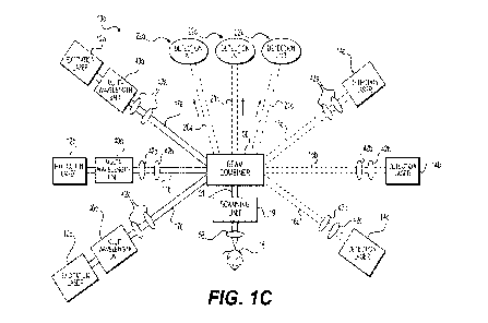
sample IS. Interrogation/probe beam 16 passes through a polarizing beam
splitter 44 and
quarter wave plate 56 to guide the reflected light 20 from sample 18 to the
photodiode 46.
However, PARS 10a is not limited to including polarizing beam splitter 44 and
quarter wave
plate 56. The aforementioned components may be substituted for fiber-based,
equivalent
components, e.g., a circulator, coupler, \VDNI, andlor double-clad fiber, that
are non-
reciprocal elements. Such elements may receive light from a first path, but
then redirect said
light to a second path. A combined beam 21 of excitation beam 17 and
interrogation beam 16
will be scanned by scanning unit 19. The scanned combined beam 21 will pass
through an
objective lens 58 and focus on the sample 18. The reflected beam 20 returns
along the same
path and is analyzed by detection unit 22. Unit 22 includes amplifier 48,
fast. data acquisition
card 50 and computer 52.
[0043] FIG I R shows another embodiment of a PARS lob, in which
scanning unit 19.
(shown in FIG. 1A) is replaced by scanning unit 11 in order to scan (move) the
sample 18 in.
relation to the fixed combined beams 21.. In some other embodiments. PARS
systems may
CA 03161943 2022- 6- 14
WO 2021/123893
PCT/I132019/061131
include both scanning unit 11 and Scanning unit 19õ thereby, having scanning
units on
opposite ends of combined beam 21,
[0044] FIG. IC is another block diagrarn of an embodiment of a
PARS 10c. .PARS IOC
includes three excitation lasers 12a-12c configured to provide three
excitation beams 17a-
1=7c, three detection lasers 14a,14c .configured to provide three
interrogation beams 16a-16c,
and three detection units .22a-'22c to receive and analyze reflected beams 20a-
20c. It is noted,
however, that the number of excitation lasers, detection lasers, and detectors
is not
particularly limited, and any suitable number of lasers and configurations
thereof may be.
used, such as, for example, two, four, five, or more. Similar to PARS 10a,
excitation beams
17a-17c and inteirogation beams 16a-16c combine via beam combiner 30 to focus
combined
beam 21, passing through objective lens 58, onto sample 18. Reflected beams
20a-20c reflect
in directions opposite of combined beam 21, and are received by detection
units 22a.-22c.
Beam combiner 30 may serve additional functions in PARS 10c, including serving
as a
polarizing beam splitter of interrogation beams 16a.-16c and as a guide for re-
directing
reflected beams 20a-20c toward detection units 22a-22c. Detection units 22a-
22c may
respectively include an amplifier (not shown), a fast data acquisition card
(not shown), and a.
computer (not shown), such as amplifier 4-8, fast data acquisition card 50,
and computer 5.2
set forth above with respect to FIG. IA
[0045] PARS 1,0c, including a plurality of excitation beams
and/or interrogation beams
and or detectors, may provide users with the option of applying beams of
varying properties,
e.g., wavelength, for various aims. For example, to image deep-inside
biological tissues, it
may be desirable to use a deeply-penetrating (long transport mean-free-path)
optical
wavelength such as a short-wave infrared wavelength. An example of a deeply-
penetrating
wavelength is 1310 mil, which is typically used in. PARS for deep imaging..
Altematively.
when imaging superficial targets, there may be geometric benefits (in terms of
a smaller focal
spot size) and sensitivity benefits (in terms of increased scattering) to
using a_ shorter, visible
wavelength, such as 630 inn. The combination of such geometric and sensitivity
benefits can
result in several orders of magnitude difference in the amount of returned
light from an
imaged sample. For instance, the focal spot area for 500 nm light will be
roughly 9 times
smaller than that of 1500 nm light for the same focusing optics. Likewise, for
biological
tissues, the scattering at 500 nm can be 3 to 4 times stronger than at 1500
mu, for example.
Thus, such benefits from using a wavelength of 500 Inn, as opposed to a
wavelength of 1500
nm, may ultimately result in a 30 to 40-fold detection sensitivity improvement
at superficial
depths. It is noted that excitation wavelengths are not particularly limited
to the
11
CA 03161943 2022- 6- 14
WO 2021/123893
PCT/I132019/061131
aforementioned example values, and may be any wavelengths suitable for the
intended
purpose. The two properties Of deep sample penetration and improved
superficial
performance- may also be desirable for use at the same time, oras Atwitchable
option
depending on the desired outcoine of an imaging session. For example, both
beams may be
used at the same lime if imaging near-surfate capillary vessels followed by
deeper vessels
with a single volumetric scan. The superficial structures may benefit from the
improved
resolution and sensitivity of the shorter detection wavelength, whereas the
deeper structures
may only be recovered by using the infrared wavelengths. However, the use of
two beams at
the same time may provide too much exposure to optical radiation, and thus a
switching
approach may be taken Where the shorter detection wavelength is traded for the
longer
wavelength detection at an appropriate depth in the sample. Thus, PARS having
a plurality of
excitation beams andior interrogation beams andlor detectors may allow a user
to implement
two or more detections in the same system, thereby allowing the user to
examine the
effectiveness of each detection on a sample. Some samples may provide
specific. improved
contrast for a given detection wavelength over others, due to the nature of
light scattering and
extinction at particular wavelengths. Multiple detection paths may also be
combined using
free-space optical beam combiners such as a dichroic or beamspliders or using
fiber-based
devices such as couplers or wavelength division multiplexers.
[0046] A plurality of excitation wavelengths may also be used
sequentially while
acquiring muItiplexedllinactional information from a single sample, such as
imaging oxy- and
deoxyhemoglobin for visualization of blood oxygenation, or targeting DNA and
cytachrome
absorption peak to extract histological information from a tissue sample. To
facilitate rapid
and consistent imaging, which may minimize the potential for motion artifacts
and may allow
for video-rate real-time multiplexed/functional imaging, the plurality of
excitation
wavelengths may be used in close succession to one another, for example, up to
MHz-range
repetition rates, so that the plurality of excitation beam sources are set-up
and active
simultaneously in the same -PARS. Multiplexed/functional information may also
be extracted
from a sample using variations in pulse-widths. These widths are not
particularly limited, and
may vary from the thermal and stress confinement conditions in the hundreds of
nanoseconds, or down to the femtosecond range. For example, oxygenated and
deoxygenated
hemoglobin can be separated using two 532 urn sources, one which provides
picosecond-
scale puke widths and the other operating in the nanosecond regime (provides
nanosecond-
scale pulse widths). In general, PARS excitation paths may include any
combination of
wavelengths, pulse widths, repetition rates, and pulse energies, which provide
various
1.?
CA 03161943 2022- 6- 14
WO 2021/123893
PCT/I132019/061131
benefits in terms of sample exposure, imaging. sensitivity, imaging
specificity, and
.chromophore The multiple excitation beam paths may be
combined using free-
space optical. beam. 'combiners Such as a .dichroic or beam-Splitters.-or
using-fiber-based
device's such as couplers or wavelength division multiplexers.
[0047] Thus, a PARS including a combination of multiple
detectiOnlinterrogation beams
and excitation beams may provide highly tamable imaging parameters, As
discussed above,
such a system may be configured to image deeply in scattering tissue to target
near-infrared
blood absorption. The same system may be configured to use a short-wave
infrared detection
providing. penetration depths approaching 3 mm for optical resolutions that
are less than 2
um, and beyond this depth with decreased resolving powers. This may be done
sequentially
or simultaneously within the same PARS. The same system may also .use a ITVC
excitation.,
having wavelengths of 200 to 280 urn, to target DNA absorption, and use UVA
detection,
having. wavelengths of 315 to 400 rim, to provide superficial imaging
performance with
resolutions on the order of several hundred nanometers_UNIB beams also may be
utilized for
excitation/detection.
[0048] FIG. 2A shows an embodiment of PARS 1.0d, which includes
individual optical
systems, adjacent to one another, winch are separately configured to focus
excitation beam
16, interrogation beam 17, and receive reflected beam 2.0, respectively. In
PARS 10d,
excitation beam 16 and interrogation beam 17 are not combined via a beam
combiner, and
co-focus on sample 18, via separate focusing optics, i.e., 58a and 58b.
Focusing optics 58a
and 58b may include any device(s) used to converge the beam of light, such as
an objective
lens or curved mirror. It is noted that the central axes of excitation beam 16
and interrogation
beam 17 are angled and offset relative to each other, but that the angle is
not particularly
limited. Reflected beam .20 reMms along a different path that is angled and
offset to the axis
of interrogation beam 16, and reflects back towards focusing optics 58c, which
guides
reflected beam 20 to detection unit 22.
[0049] Similar to PARS 10d, FIGS. 2B and 2C further show
excitation beam 17 and
interrogation beam 16 being directed to sample 18 at an angle relative to one
another.
However, unlike system 1.0d, FIGS, 2B-2C illustrate the use of refractive
optics, as opposed
to reflective optics, such as, e.g., MITTOtS. In FIG. 2B, both excitation beam
17 and
interrogation beam 16 pass through a single objective lens 58, which co-
focuses beams 16
and 17 on sample 18 from two distinct ,angles.. While the refracted portions
of beam 16 and
17 are off their respective longitudinal axes (of beams 16 and 17 prior to
passing through lens
58), the refracted beams are still parallel to said axes such that beams 16
and 17 are able to
13
CA 03161943 2022- 6- 14
WO 2021/123893
PCT/I132019/061131
. co-focus onto the same spot .of sample 18. Because there is a single
objective letts..:58 in FIG.
2B, the angle between beams 16. and 1-7 may be relatively shallow in
coMparfsdii to Systeins.
in which twO lenses may be used. 'However, the use of single objective lens 58
may also
allow for relatively easier co-alignment of beams 16 and 17 onto sample I&
[0050] hi ctuitrast, in FIG. 2C, excitation beam 16 and
interrogation beam 17 each pass
through their respective objective lens, Le., objective lens 58b and objective
lens 58a.
Moreover, beams 16 and 17 remain centered along their respective longitudinal
axes to co-
focus onto the same spot of sample 18. Because there are separate objective
lenses 58b and
58a for beams 16 and 17, respectively, the range of the angle between beams 16
and 17 may
be more flexible and larger angles than the embodiment shown in FIG. 2B. Such
a
configuration may also enable higher levels of polarization. However, the
embodiment shown
in FIG. 2C requires co-alignment of objective lenses 58b and 58a so that beams
16 and 17
may co-focus onto sample 18. Other PARS embodiments, may also include
additional
individual optical systems, andior may be in different configurations or
anangements relative
to one another..
[0051] The configuration of PARS 10d, and the beam
configurations shown in FIGS. 2A-
2C may provide added spatial rejection of undesired randomly scattered
photons, and detect
only photons that have been modulated by excitation laser 12. Since the PARS
imaging
region is defined by the overlap of excitation beam 16,
.detectionlintenugation beam 17, and
backwards detection/reflected beam path 20, if these paths are all co-aligned,
the interrogated
region on sample 18 may be defined by a radial distribution which is commonly
shorter than
the axial distribution. This may cause the axial resolution of such imaging
systems to be.
larger, and thus, worse than the lateral resolution_ By angling excitation
beam 16 and
interrogation beam 17 relative to each other, as shown in PARS 10d and the
beams shown in
FIGS. .2A-2C, the overlap may now be defined between the combination of two or
three
radial distributions. This allows for the lateral resolution of one of the
beams to improve upon
the axial performance provided by the other beam.. To maximize this effect, it
may be most
advantageous to have the three beams evenly distributed in the azimuth and
with around 45
degrees each to the sample surface_ This is shown in FIG_ 2C, which
illustrates sample 18 on
a plane, and excitation beam 17, interrogation beam 16, and reflected
interrogation beam 20
having beam paths, originating from sample 18, of congruent azimuth angles
26a, i.e., 120.
The beam paths also have congruent altitude angles .26h, which may range from
20-90'.
However, in other embodiments the altitude angles may vary amongst. the beam
paths.
Decreasing internal angles between beams 16, 17, and 20 may simply begin to
approach the
14
CA 03161943 2022- 6- 14
WO 2021/123893
PCT/I132019/061131
performance Of non-angled PARS for decreasing internal angles, and become-
impractical as
angles approach 180 degree's since samples are generally fiat,
[0052] As shown in FIGS. .2A-2Cõ the angling of the -focused
paths of excitation beam 16
and interrogation 'beam 17 may be achieved through angling of the input beams
into a single.
focusing element, Leo Objective lens. 58 shown in FIG. 2B, or by constructing
a System with
multiple focusing elements which are angled to each other, i.e., objective
lens 58 and .15
shown in FIG. 2C, or some combination of the RVO. As a result, the axes of
excitation beam
16 and interrogation beam 17 may be angled relative to one another,
[0053] PARS including an excitation source, a detection source,
a. beam combiner
combining excitation beam(s) and interrogation beam(s), focusing optics, and a
detector,
similar to the embodiment in FIG. IA, capture intensity modulations in the
collected
light/reflected beam from the sample. This may be done by sensing the change
in scattering
from the sample, Other -non-PARS or devices that may -pertbrin such a function
include
scattering microscopes, which may include a detection beam from a detection
source passing
through a combinerisplitter to focusing optics, which focus the beam onto a
sample, and an
intensity detector configured to receive reflected interrogation/detection
beams (with no
excitation beam).
[0054] However, the reflected interrogation beam also contains
information regarding its
polarization state and its phase, and there are conventional, non-PARS or
devices that may
capture polarization and phase accumulation. One such device may be a
polarization-based.
microscope, which is similar to the above described scattering microscope,
except a
polarization detector is used in place of an intensity detector. Another such
device may be a
conventional phase microscope, which may include a detection beam from a
detection source
passing through an interferometer to focusing optics, which focus the beam
onto a sample,
and a phase detector configured to receive reflected interrogation/detection
beams that return
through the interferometer, Thus, PARS of the present disclosure modulate the
scattering
properties of reflected beam 20 and also respectively modulate the apparent
polarization and
phase accumulation within a sample, Such PARS are thither discussed below,
referring to
FIGS.. 3A and 3B,
[0055] FIG. 3A shows another block diagram of an embodiment of
PARS log. PARS 10f
includes excitation laser 12 configured to provide excitation beam 17, and
detection laser 14
contil_:,7ured to provide interrogation beam 16. However., as previously
disctissed, the number
of excitation lasers and detection lasers is not particularly limited, and any
suitable number of
lasers and configurations thereof may be used. Similar to PARS 10a, excitation
beam 17 and
CA 03161943 2022- 6- 14
WO 2021/123893
PCT/I132019/061131
interrogation. beam 16 combine via beam Combiner 30 to focus combined beam 2-
1, .passina.
through objective lens 58 onto sample 18, Furthermore,. in this embodiMent,
beam combiner
30 may also -serve the ..functitinatfla.polarizing beam splitter of
interrogation beam 16
However, PARS 10!' does not include the detection unit 22 shown in FIG. 1A,
Instead,
reflected beam 20 is reflected back throngli beam combiner 30,. Which guides
reflected beam
20 to a polarization modulation detector 23. It is noted that a quarter -
waveplate is not used in
PARS 10g., so that the polarization state of reflected beam 20 may be
maintained when
guided toward polarization modulation detector 23.
[0056] More specifically, to capture polarization modulation,
interrogation beam 16 with
a controlled polarization is fed into sample 18, where reflected light 20 is
now separated
based on its polarization content. The means by which polarization is
controlled in not
particularly limited, and can be, e.g., a conventional polarization
controller, and in some
embodiments, beam 16 may already be polarized when emitted from laser 14. For
example,
vertically polarized light may be directed to one photodetector within
detector 23 and
horizontally polarized light may be directed to another photodetector within
detector 23.
Different aspects of polarization could be used such as linear direction,
handedness of
circular polarized states, and higher-dimensional polarization distributions,
such as radially
and azimuthally polarized states. Separation and Characterization of these
states may be
accomplished with polarization sensitive detectors, i.e., polarization
modulation detector 23,
quarter wave plate 56, and polarization-sensitive splitters (not shown). This
may allow for
precise characterization of the polarization shift, as the modulated value
could be directly
compared with the un-modulated value at the same sample location.
[0057] FIG. 3B shows an embodiment of PARS 1011 also including
excitation laser 12
configured to provide excitation beam 17, and detection laser 14 configured to
provide
interrogation beam IS. PARS lOg includes an interferometer .24 and a phase
modulation.
detector 25. PARS lOg may be arranged so that interrogation beam 17 passe.s
through
interferometer 24 and is guided to beam combiner 30, at which interrogation
beam 17
combines with excitation beam 16. Reflected beam 20 from sample 18 returns
along the same
path of interrogation beam 17 up until interferometer 24, at which reflected
beam 20 is then
guided towards and received by phase modulation detector 25.
[0058] To capture phase shifting, a phase sensitive detector,
i.e., phase modulation
detector 25, is implemented. This may be done with heterodyne and homodyne
interferometryõ which may capture a component of or the full quadrature of
returning light 20
from sample 18. This would allow for precise characterization of the phase
shift, as the
16
CA 03161943 2022- 6- 14
WO 2021/123893
PCT/I132019/061131
modulated value could be directly Compared with the un-modulated value- at the
same sample
location_
[0059] Any cOmbination of these six light properties
(e4,..scatteting, polarization, phase,.
and their respective -modulations) may.-be captured and analyzed in a PARS via
any suitable -
mechanismõ phase Modulation detector 2=5 for phase, where the
contrast mechanisms,
may provide 'unique and complementary information. For example, PARS may
generate
strong second harmonic signals, and auto-fluorescence from the sample due to
the PARS
effect. For example, there may be poor scattering contrast, but strong
polarization contrast.
from sample 18. While conventional imaging systems may not be configured to
find such a.
signal, polarization-sensitive detection via polarization modulation detector
23 may provide
improved results. By using the additional information contained within the
polarization and
phase of reflected beam 20, added sensitivity may be achieved by averaging
across shifts,
resulting in lower required optical exposure_ Complementary information may be
found
between these shifts which give optical absorption information, and the
unshitted values may
yield scattering, polarization, and phase in their own right. Such wealth of
information may
be used to drastically improve specificity, since given targets will provide
unique signatures
across these six modalities (e.g., conventional scattering microscope,
conventional
polariza-tion-bas,ed microscope_ conventional phase microscope, a PARS
microscope, and the
microscopes shown in FIGS. 3A and 313), allowing for improved multiplexing
capabilities.
[0060] FIG. 4 shows another embodiment of PARS 10i, in which
excitation beam 17 and
interrogation beam 16 have separated paths, and are not combined. In this
embodiment',
interrogation beam 16 is focused, using another objective lens 15. to sample
18. In other
embodiments, PARS 10i may be similar to aspects of both PARS 10c and 10d,
shown in
FIGS. 1C. and .2A. Similar to PARS 10c, PARS 10i may have multiple excitation
lasers,
detection lasers, and detection units, the number of which are not
particularly limited.
[0061] In some embodiments, both beams may be scanned together.
Alternatively, one
beam may be fixed while the other beam may be scanned. In other embodiments,
sample 18
may be scanned while both beams are fixed. Sample 18 may also be scanned While
both.
beams are scanning. Sample 18 may also be scanned while one beam is fixed and
the other is
scanning.
[0062] It will be apparent to one of ordinary skill in the art
that other PARS embodiments
may be designed with different components to achieve similar results_ For
example, other
embodiments may include all-fiber architectures where circulators replace beam-
splitteis
similar to optical-coherence tomography architectures. Other alternatives may
include various
17
CA 03161943 2022- 6- 14
WO 2021/123893
PCT/I132019/061131
. coherence length sot-trees, use :of balanced photadetectorsi, interrogation-
beam modttlation,.
incorporation of optical:amplifiers in the return signal path,
[0063] The 'PARS takes advantage of two focused laser beams on
the 'saniple 'which May
simulate a confocal PAM configuration
[0064] .PARS = also takes. advantage Of optical excitation and
detection Which may help
dramatically reduce the footprint of the system. The absence of a bulky
ultrasound transducer
makes this system suitable for integrating with other optical imaging systems.
Unlike many
previous non-contact photoacoustic imaging systems. the PARS is capable of in
vivo
imaging. It relies on a much simpler setup and takes advantage of recording
the large initial
ultrasound pressures without appreciable acoustic losses.
[0065] During in vivo imaging experiments, no agent or
ultrasound coupling medium are
required. However, the target may be prepared with water or any liquid such as
oil before a
non-contact imaging session. PARS does not require a floating table unlike
many other
interferometric sensors. No special bolder or immobilization is required to
hold the target
during imaging sessions. However, a cover slip may be implemented to flatten
the target_ In
some instances, glass windows for the targets, e.g.., resect.ed tissue, to sit
on may be
necessary, and imaging may be performed through said glass windows. This may
help image
flat surfaces of the target.
[0066] Other advantages that are inherent to the structure will
be apparent to those skilled'
in the art. The embodiments described herein are illustrative and .not
intended to limit the.
scope of the claims, which are to be interpreted in light of the.
specification .as.a.=whole.
[0067] In PARS, a pulse laser is used to generatephotoacoustie
signals and the acoustic
signatures are interrogated using either a tong-coherence or short-coherence
length probe
beam co-focused with the excitation spots. The PARS may be utilized to
remotely record the
large local initial pressures from chromophores and without appreciable
acoustic losses due
to diffraction, propagation and attenuation.
[0068] The excitation beam may he any pulsed or.modulated source
of electromagnetic
radiation including lasers or other optical sources.. In one example, a
nanosecond-pulsed laser
may be used. The excitation beam may be set to any wavelength suitable for
taking advantage
of optical (or other electromagnetic) absorption of the sample. The source may
be
monochromatic or polychromatic,
[0069] The interrogation beam may be any pulsed, conlinuot is,
or modulated source of
electromagnetic radiation including lasers or other optical sources. Any
wavelength may be
used for interrogation purpose depending on the application.
18
CA 03161943 2022- 6- 14
WO 2021/123893
PCT/I132019/061131
[0070] The chromatic aberration in the :collimating
and.oblectiVe lens pair may be
harnessed to refOcus light from a fiber into the object so that each
wavelength is focused-at a.
slightly different depth location. Using these wavelengths simultaneously may
improve the
depth of field and SNR for structural imaging of inicrovaseubiture with OR-
PAM.
[0071] Since a NI-PARS is not .interferometric, the
probeireceiveninterrogation beam Of
NI-PARS, may be a long-coherence or a short-coherence length probe beam,
.without need of
any reference beam or reference arm. Using a short-coherence length, however,
may ensure
preclusion of interference from reflections in the system or sample to avoid
unwanted signals
and to extract only photoacoustic initial pressures.
[0072] Unlike optical coherence tomography (OCT) or
interferomeny detection of
photoacoustic signal, the N1-PARS detects the changes in the amount of the
reflected light
from sample due to acoustic. pressure and no interferomeny design such as,
reference beam,
reference arm or axial scanning of reference beam are needed.
[0073] Various PARS systems (including, but not limited to PARS,
NI-PARS, CG-
PARS, C-PARS, and SS-PARS) may be integrated with OCT to provide a complete
set of
information offered by both photoacoustic and OCT systems.
[0074] Furthermore, the various PARS with short or long-
coherence beams may be used
fir either optical resolution photoacoustic microscopy (OR-PAM) or conunon
photoacoustic
microscopy (PAI41), or may be combined with 2nd or 3rd harmonic, fluorescent,
multiplioton,
Raman, andlor other, microscopes.
[0075] In one example, both excitation and receiver beam may be
combined and scanned.
In this way, photoacoustic excitations may be sensed in the same area as they
are generated
and where they are the largest. Other arrangements may also be used,
including, keeping the
receiver beam fixed while scanning the excitation beam or vice versa, and
scanning the optics
mechanically while the sample remains stationary, such as, for example, in a
surgical.
microscope where the patient must remain stationary. Galvanometers. MEMS
mirrors and
stepper/DC motors may be used as a means of scanning the excitation beam,
probe/receiver
beam or both.
[0076] The configurations shown in FIGS.. SA ¨ 5D may be used to
perform PARS and
N1-PARS imaging. In the depicted embodiments, excitation beams 502 are
depicted with a.
larger radius of curvature, and receiver/detection beams 504 are depicted with
a smaller
radius of curvature. FIG. 5.A shows an embodiment of a confocal photoacoustic
system where
excitation beam 502 and probing receive beam 504 are focused on the same spot,
which can
be on a micron- or sub-micron scale. In FIG. 5a the optical resolution may be
provided by
19
CA 03161943 2022- 6- 14
WO 2021/123893
PCT/IB2019/061131
receiver beam 504, -rather than excitation beam 502. FIG. SC shows excitation
beam 502 and
receiver beam 504 fficused On different spOs, and takes advantage Of
ultrasound time cif
flight in order to locate excitation beams 502 and receiver beams 504 at
different positions. In
FIG. 51), optical resolution may be provided by excitation beam 502..
Preferably, the -focus of
either Or both of excitation beam 502 and detection beam 504 is less than 30
Am, less than 10
pm, less than 1 pm, or to the diffraction limit of light A fighter focus may
result in a higher.
possible resolution and a better signal to noise ratio in the reflected beam
that is detected. As
used herein, the term "focus- is intended to refer to the focal zone of the
beam, or the point at
which the beam spot size is at the tightest size, and where the diameter of
the focal zone is
30% greater than the diameter of the beam spot size. Also preferably, the
excitation and
detection beams 502 and 504 are focused on the same position, although there
may be some
spacing between the respective focuses as shown in FIG. 5C. hi FIG. 5C., the
beams may be
focused at different locations, but preferably within I mm, 0,5111111, 100 an
or within the
range of the largest focus of the beam. In FIGS. 5A, 513, and 51), the beams
may be confocal,
or may overlap within the focus of the beam with the largest focus. For
example, in FIG. 5A,
excitation beam 502 is larger than detection beam 504, and detection beam 504
is directed at.
a location within the focus of excitation beam 502. By moving detection beam
504, the area
within excitation beam 502 may be imaged. By haying confocal beams, both beams
may be
moved to image the sample.
[0077] One or both of the beams are preferably focused below the
surface of the sample.
Generally speaking, the beams may be effectively focused up to 8 mm (or more)
below the.
surface of the sample. The beams may be focused at least 50 nm (or even less)
below the
surface, or focused such that focal point of the beam is at least the distance
of focal zone of
the beam below the surface of the sample. It will be understood that, while
both beams are
preferably focused below the surface, in some em.bodiments either the
excitation beam or the
interrogation beam may be focused below the surface. with the other focused
on. for
example, the surface of the sample. In cases where only one beam is focused
below the
surface of the sample, the separation between the beams discussed previously
will be: a lateral
separation, he, in the plane of the sample and orthogonal to the depth of the
sample.
[0078] The relationship between excitation beams and detection
beams, specifically, their
focal planes, subsurface of a sample is further illustiated in FIGS. 5E-5I.
For example, FIG.
5F... illustrates a confocal photoaconstic system including excitation beam
502 and detection
beam 504, where an excitation focal plane 506 and a. detection focal plane 508
are focused at
the same depth, thereby exhibiting a co-alignment condition. This is similarly
illustrated in
CA 03161943 2022- 6- 14
WO 2021/123893
PCT/I132019/061131
FIG. 5F,. except FIG. 5F further illustrates that the co-aligned focal planes
506 and 508 are
below a glass wincloaV=510õ. Thus, in this instance, co-alignment takes place
through' window
.510. The distance between glass windOw 510 and focal planes 506 and 508 is
not particularly
limited. FIG. 5G again illustrates 'co-alignment between focal planes 506 and
.508. 'However,
FIG. 5G shOws that 'focal planes 506 and 508 are sabaurface of sample 512, by
a depth
defined by adistance 514. Thus, FIG. 5G illustrates excitation beam 502 and
detection bean'
504 co-focusing on a spot below the surface of sample 512. The depth of focal
planes 506
and 508 below the surface 512 is not particularly limited, and and in some
instances. may
range from 100 nm to litm, FIG. 5H illustrates an instance in which excitation
beam 502 is
focused, relative to detection beam 504, so that excitation focal plane 506 is
above detection
focal plane 508. In contrast, FIG, 51 illustrates an instance when excitation
focal plane 506 is
below detection focal plane 508. Thus, FIGS. 511-51 illustrate that focal
planes 506 and 508
may be out of alignment. An example of when focal planes 506 and 508 are
misaligned may
be when a. PARS system is aligned for imaging near the surface of a sample,
and a user of
said PARS system attempts to fbcus deeper in the sample without any
adjustments. This
results in chromatic aberrations, which cause the detection and excitation
focal planes to shift
away from one another. Focal planes 506 and 508 may be misaligned by 10 tm, 20
pm, 30
pan, etc. However, the distance between the focal planes is not particularly
limited, and may
be any suitable distances. Furthermore, it may be pretbrable to minimize the
distance between
focal planes 506 and 508 for optimal sensitivity..
[0079] The excitation beam and detectionlreceiver beam may be
combined using dichroic
mirrors, prisms, beamsplitters, polarizing beamsplittm etc. They may also be
focused using
different optical paths.
[poso] The reflected light may be collected by photodiodes,
avalanche photodiodes,
phototubes, photomultipliers. CMOS cameras, CCD cameras (including EM-CCD,
intensified-CCDsõ back-thinned and cooled CCDs), spectrometers, etc. The
detected light
may be amplified by an RF amplifier, lock-in amplifier, trans-impedance
amplifier, or other
amplifier configuration. Also different methods may be used in order to filter
the excitation
beam from the receiver beam before detection. PARS may use optical amplifiers
to amplify
detected light.
[0081] PARS may be used in many form factors, such as table top,
handheld, surgical
microscope., and endoscopy. Examples of endoscopy PARS are shown in FIGS_ 6A,
6F1 and
SC' with various amangements of PARS excitation units 1102, PARS detection
units 1104,
fibre optics 1106 such as image-guide fibers, and lenses 1108 that focus the
respective beams
21
CA 03161943 2022- 6- 14
WO 2021/123893
PCT/I132019/061131
onto 'sample 18. When excitation and detection units-1102 and 1104 are
separated, there may
be a separate fiber 1110 provided, such as a-single mode fiber.
[0082] A table top and 'handheld PARS may he constructed based
on principles known in
the .art. The .proposed PARS hikes advantage of optical excitation and
detection which can
help to =dramatically reduce the footprint of the system. The footprint of
previous systems has
been much too large to use the system in all but body surfaces. For endoscopic
applications,
-the footprint of the ultrasound detector mast be minimized to make the
imaging catheter
small and flexible enough to navigate through small orifices and vessels. The
piezoelectric
receivers are not ideal candidates for endoscopic applications as there is
trade-off between the
sensitivity and the size of the receiver. On the other hand for many invasive
applications
sterilisable or disposable catheters and a non-contact approach are necessary.
The system
may also be used as PARS endoscopy system with a potential footprint the size
of an optical
fiber, as both excitation and PARS beam can be coupled into a. single mode
fiber or image
guide fiber.
[0083] Image-guide fibers (miniattnized fiber- bundles with as
ninny as 100,000 or more
individual micrometer-sized strands in a single optical fiber with diameters
ranging from 200
p.m. to 2 mm) may he used to transmit both focused light spots. The excitation
beam may be
scanned either at the distal end or proximal end or the fiber using one of the
scanning
methods mentioned before. However, the receiver beam may be scanned or be
fixed. The
scanned spot is transmitted via the image-guide fiber 1106 to the output end.
Therefore, it
may be used to directly contact the sample, or re-focused using an attached
millianne GRIN
lens 1108. In one example. C-scan photoacoustic images were obtained from the
fiber image-
guides using an external ultrasound transducer to collect .photoaconsfic
signals. Using an
edge-spread and Gaussian function, a resolution of approximately 7 pm was
obtained using
the image-guide fiber 1106. It is believed that a higher resolution may also
be obtained with
appropriate improvements to the. setup and equipment used. This may be one
possible
embodiment of an endoscopic PARS device.
[0084] Endoscopic embodiments may also be constructed using
single-mode fibers if. for
example, the excitation and detection wavelengths are sufficiently close to
each other, such as
532 am and 637 mu. This would allow both wavelengths to propagate in single-
modes in a.
highly compact probe when the fibers are, for example, only 250 microns in
diameter.
[poss] Furioscopic PARS device embodiments may also he assembled
rising double-clad
fibers. These fibers feature a single-mode core surrounded with a multi-mode
core. This
allows for highly dissimilar wavelengths, such as 532 mu and 1310 nun. to be
combined into a
CA 03161943 2022- 6- 14
WO 2021/123893
PCT/I132019/061131
single fiber while maintaining Single-mode propagatitan for at least one of
the- wavelengths.
As well, the -double-clad fiber's Mullin-Lode outer core may be used for
increased return light
collection as a Means of directing collected light towards the optical
deteetio.n components..
[0086] Various PARS embodiments may be combined with other
imaging Modalities
such as fluorescence Microscopy, two-photon and confocal fluorescence
microscopy,
Coherent-Anti-Rainan-Stokes microscopy, Raman microscopy, Optical coherence
tomography, other photoacoustic and ultrasound systems, etc. This may permit
imaging of
the microcirculEttion, blood oxygenation parameter imaging, and imaging of
other
molecularly-specific targets simultaneously, a potentially important task that
is difficult to
implement with only fluorescence based microscopy methods. An example of this
is .shown
in FIG. 7. in which a PARS 10 is integrated with another optical imaging
system 1202, where
PARS 10 and the other optical imaging system 1202 are both connected to sample
18 by a
combiner 1204.
[0087] Interfemmetric designs, such as common path
interferometer (using specially
designed interferometer objective lenses), Michelson interferometer, Fizeatt
interferometer,
Ramsey interferometer, Sagnac interferometer, Fabry-Perot interferometer and
Mach¨
Zelinder interferometer, may also be integrated with various embodiments of
the disclosure.
[0088] A multi-wavelength visible laser source may also be
implemented to generate
photoacoustic signals for functional or structural imaging.
[0089] Polarization analyzers may be used to decompose detected
light into respective
polarization states, The light detected in each polarization state may provide
information
about ultrasound-tissue interaction.
[0090] APPLICATIONS
E0091] It will be understood that the system described herein
may be used in various
ways, such as those purposes described in the prior art, and also may be used
in other ways to
take advantage of the aspects described above. A non-exhaustive list of
applications is
discussed below.
[0092] The system may be used for imaging angiogenesis for
different pre-clinical tumor
models.
[0093] The system may be used to image: (1) histological
samples: (2) cell nuclei; (3)
proteins; (4) cytochromes; (5) DNA; (6) RNA; and (7) lipids.
[0094] The system may also be used thr clinical imaging of micro-
and macm-circulation
and pigmented cells, which may find use for applications such as in (1) the
eye, potentially
augmenting or replacing fluorescein angiography; (2) imaging dermatological
lesions
23
CA 03161943 2022- 6- 14
WO 2021/123893
PCT/I132019/061131
including inOeinoina, basal cell carcinoma, hemangiorna, pSoriasis, eczema,
dermatitis,.
imaging Molus surgery, imaging to verify tumor margin. resections;: (3)
peripheral vascular
disease; (4) diabetic and pressure ulcers; (5) burn imaging: (ft) plastic
surgery and
microsurgery; (7) imaging of cite-gating tumor cells, especially melanoma
cells; (8) imaging
lymph node angiergenesis (9) imaging response to photOdynamic therapies
including: those
with vascular ablative mechanisms; (10) imaging response to chemotherapentics
including
anti-angiogenic drugs; (11) imaging response to radiotherapy.
[0095] The system may be useful in estimating oxygen saturation
using multi-wavelength
photoacoustic excitation and PARS detection and applications including: (1)
estimating
venous oxygen sanitation where pulse oximetry cannot be used including
estimating
cerebrovenous oxygen saturation and central venous oxygen saturation. This
could
potentially replace catheterization procedures which can be risky, especially
in small children
and infants.
[0096] Oxygen flux and oxygen consumption may also be estimated
by using PARS
imaging to estimate oxygen saturation, and an auxiliary method to estimate
blood flow in
vessels flowing into and out of a region of tissue.
[0097] The system may also have some gastroenterological
applications, such as imaging
vascular beds and depth of invasion in Barrett's esophagus and colorectal
cancers. Depth of
invasion is key to prognosis and metabolic potential. Gastroenterological
applications may be
combined or piggy-backed off of a clinical endoscope and the miniaturized PARS
may be
designed either as a standalone endoscope or fit within the accessory channel
of a clinical
endoscope.
[009] The system may have some surgical applications, such as
functional imaging
during brain surgery, use for assessment of internal bleeding and
cauterization verification,
imaging perfusion sufficiency of organs and organ transplants, imaging
angiogenesis around
islet transplants, imaging of skin-grails, imaging of tissue scaffolds and
biomaterials to
evaluate vascularization and immune rejection, imaging to aid microsurgery,
guidance to
avoid cutting critical blood vessels and nerves.
[0099] Other examples of applications may include PARS imaging
of contrast agents in
clinical or pre-clinical applications; identification of sentinel lymph nodes;
non- or
minimally-invasive identification of tumors in lymph nodes; imaging of
genetically-encoded
reporters such as tyrosinase, chromoproteins, fhtorescent proteins for pre-
clinical or clinical
molecular imaging applications: imaging actively or passively targeted
optically absorbing
nanoparticles for molecular imaging; and imaging of blood clots and
potentially staging the
24
CA 03161943 2022- 6- 14
WO 2021/123893
PCT/I132019/061131
age of the. clots,
[0.0100] 'hi this patent document, the ward. "comprising .ased in itS
sense to
mean that items following the word are included, but items not Specifically
mentioned are not:
excluded. A reference to an element by the indefinite article 'a" does not
exclude the
possibility that More than one Of the elements is present, unless the 'context
clearly requires
that there be one and only one of the elements.
[0010.1] The scope of the following claims should not be limited by the
preferred
embodiments set forth in the examples above and in the drawingsõ but should be
given the
broadest interpretation consistent with the description as a. whole.
CA 03161943 2022- 6- 14