Note: Descriptions are shown in the official language in which they were submitted.
WO 2021/141921
PCT/US2021/012218
SYSTEMS, METHODS, AND DEVICES FOR MEDICAL IMAGE ANALYSIS,
DIAGNOSIS, RISK STRATIFICATION, DECISION MAKING AND/OR DISEASE
TRACKING
CROSS-REFERENCE TO RELATED APPLICATIONS
[0001] The present application claims the benefit of U.S.
Provisional Patent
Application No. 62/958,032, filed January 7, 2020, and titled SYSTEMS,
METHODS, AND
DEVICES FOR CARDIOVASCULAR IMAGE ANALYSIS, DIAGNOSIS, RISK
STRATIFICATION, DECISION MAKING AND/OR DISEASE TRACKING, which is
incorporated herein by reference in its entirety under 37 C.F.R. 1.57. Any
and all
applications for which a foreign or domestic priority claim is identified in
the Application
Data Sheet as filed with the present application are hereby incorporated by
reference under
37 C.F.R. 1.57.
BACKGROUND
Field
[0002] The present application relates to systems,
methods, and devices for
medical image analysis, diagnosis, risk stratification, decision making and/or
disease
tracking.
Description
[0003] Coronary heart disease affects over 17.6 million
Americans. The current
trend in treating cardiovascular health issues is generally two-fold. First,
physicians
generally review a patient's cardiovascular health from a macro level, for
example, by
analyzing the biochemistry or blood content or biomarkers of a patient to
determine whether
there are high levels of cholesterol elements in the bloodstream of a patient.
In response to
high levels of cholesterol, some physicians will prescribe one or more drugs,
such as statins,
as part of a treatment plan in order to decrease what is perceived as high
levels of cholesterol
elements in the bloodstream of the patient.
-1-
CA 03162872 2022- 6- 22
SUBSTITUTE SHEET (RULE 26)
WO 2021/141921
PCT/US2021/012218
[0004] The second general trend for currently treating
cardiovascular health
issues involves physicians evaluating a patient's cardiovascular health
through the use of
angiography to identify large blockages in various arteries of a patient. In
response to
finding large blockages in various arteries, physicians in some cases will
perform an
angioplasty procedure wherein a balloon catheter is guided to the point of
narrowing in the
vessel. After properly positioned, the balloon is inflated to compress or
flatten the plaque or
fatty matter into the artery wall and/or to stretch the artery open to
increase the flow of blood
through the vessel and/or to the heart. In some cases, the balloon is used to
position and
expand a stent within the vessel to compress the plaque and/or maintain the
opening of the
vessel to allow more blood to flow. About 500,000 heart stent procedures are
performed
each year in the United States.
[0005] However, a recent federally funded $100 million
study calls into question
whether the current trends in treating cardiovascular disease are the most
effective treatment
for all types of patients. The recent study involved over 5,000 patients with
moderate to
severe stable heart disease from 320 sites in 37 countries and provided new
evidence
showing that stents and bypass surgical procedures arc likely no more
effective than drugs
combined with lifestyle changes for people with stable heart disease.
Accordingly, it may be
more advantageous for patients with stable heart disease to forgo invasive
surgical
procedures, such as angioplasty and/or heart bypass, and instead be prescribed
heart
medicines, such as statins, and certain lifestyle changes, such as regular
exercise. This new
treatment regimen could affect thousands of patients worldwide. Of the
estimated 500,000
heart stent procedures performed annually in the United States, it is
estimated that a fifth of
those are for people with stable heart disease. It is further estimated that
25% of the
estimated 100,000 people with stable heart disease, or roughly 23.000 people,
are individuals
that do not experience any chest pain. Accordingly, over 20,000 patients
annually could
potentially forgo invasive surgical procedures or the complications resulting
from such
procedures.
[0006] To determine whether a patient should forego
invasive surgical procedures
and opt instead for a drug regimen, it can be important to more fully
understand the
cardiovascular disease of a patient. Specifically, it can be advantageous to
better understand
the arterial vessel health of a patient.
_
CA 03162872 2022- 6- 22
SUBSTITUTE SHEET (RULE 26)
WO 2021/141921
PCT/ITS2021/012218
SUMMARY
[0007] Various embodiments described herein relate to
systems, methods, and
devices for medical image analysis, diagnosis, risk stratification, decision
making and/or
disease tracking.
[0008] In particular, in some embodiments, the systems,
devices, and methods
described herein are configured to utilize non-invasive medical imaging
technologies, such as
a CT image for example, which can be inputted into a computer system
configured to
automatically and/or dynamically analyze the medical image to identify one or
more
coronary arteries and/or plaque within the same. For example, in some
embodiments, the
system can be configured to utilize one or more machine learning and/or
artificial
intelligence algorithms to automatically and/or dynamically analyze a medical
image to
identify, quantify, and/or classify one or more coronary arteries and/or
plaque. In some
embodiments, the system can be further configured to utilize the identified,
quantified, and/or
classified one or more coronary arteries and/or plaque to generate a treatment
plan, track
disease progression, and/or a patient-specific medical report, for example
using one or more
artificial intelligence and/or machine learning algorithms. In some
embodiments, the system
can be further configured to dynamically and/or automatically generate a
visualization of the
identified, quantified, and/or classified one or more coronary arteries and/or
plaque, for
example in the form of a graphical user interface. Further, in some
embodiments, to calibrate
medical images obtained from different medical imaging scanners and/or
different scan
parameters or environments, the system can he configured to utilize a
normalization device
comprising one or more compartments of one or more materials.
[0009] In some embodiments, a normalization device
configured to facilitate
normalization of medical images of a coronary region of a subject for an
algorithm-based
medical imaging analysis is provided, wherein the normalization device
comprises: a
substrate having a width, a length, and a depth dimension, the substrate
having a proximal
surface and a distal surface, the proximal surface adapted to be placed
adjacent to a surface
of a body portion of the subject; a plurality of compartments positioned
within the substrate,
each of the plurality of compartments configured to hold a sample of a known
material,
wherein: a first subset of the plurality of compartments hold at least one
sample of a contrast
material, a second subset of the plurality of compartments hold samples of
materials
-3-
CA 03162872 2022- 6- 22
SUBSTITUTE SHEET (RULE 26)
WO 2021/141921
PCT/ITS2021/012218
representative of materials to be analyzed by the algorithm-based medical
imaging analysis,
wherein the samples of materials representative of materials comprise at least
two of calcium
1000 HU, calcium 220 HU, calcium 150 HU, calcium 130 HU, and a low attenuation
material of 30 HU, and a third subset of the plurality of compartments hold at
least one
sample of phantom material; and an adhesive on the proximal surface of the
substrate and
configured to adhere the normalization device to the body portion patient.
[0010] In some embodiments of the normalization device,
wherein samples of
materials representative of materials to be analyzed comprise calcium 1000 HU,
calcium 220
HU, calcium 150 HU, calcium 130 HU, and a low attenuation material of 30 HU.
In some
embodiments of the normalization device, the at least one contrast material
comprises one or
more of iodine, Gad, Tantalum, Tungsten, Gold, Bismuth, or Ytterbium; and the
at least one
sample of phantom material comprise one or more of water, fat, calcium, uric
acid, air, iron,
or blood.
[0011] In some embodiments of the normalization device,
the substrate
comprises: a first layer, and at least some of the plurality of compartments
are positioned in
the first layer in a first arrangement; and a second layer positioned above
the first layer, and
at least some of the plurality of compartments are positioned in the second
layer including in
a second arrangement. In some embodiments of the normalization device, at
least one of the
compartments is configured to be self-sealing such that the sample can be
injected into the
self-sealing compartment and the compartment seals to contain the injected
material.
[0012] In some embodiments, a computer-implemented method
for normalizing
medical images for an algorithm-based medical imaging analysis using a
normalization
device is provided, wherein normalization of the medical images improves
accuracy of the
algorithm-based medical imaging analysis, the method comprising: accessing, by
a computer
system, a first medical image of a coronary region of a subject and the
normalization device,
wherein the first medical image is obtained non-invasively; accessing, by the
computer
system, a second medical image of a coronary region of a subject and the
normalization
device, wherein the second medical image is obtained non-invasively, and
wherein the first
medical image and the second medical image comprise at least one of the
following: one or
more first variable acquisition parameters associated with capture of the
first medical image
differ from a corresponding one Or more second variable acquisition parameters
associated
-4-
CA 03162872 2022- 6- 22
SUBSTITUTE SHEET (RULE 26)
WO 2021/141921
PCT/ITS2021/012218
with capture of the second medical image, a first image capture technology
used to capture
the first medical image differs from a second image capture technology used to
capture the
second medical image, and a first contrast agent used during the capture of
the first medical
image differs from a second contrast agent used during the capture of the
second medical
image; identifying, by the computer system, first image parameters of the
normalization
device within the first medical image; generating a normalized first medical
image for the
algorithm-based medical imaging analysis based in part on the first identified
image
parameters of the normalization device within the first medical image;
identifying, by the
computer system, second image parameters of the normalization device within
the second
medical image; and generating a normalized second medical image for the
algorithm-based
medical imaging analysis based in part on the second identified image
parameters of the
normalization device within the second medical image, wherein the computer
system
comprises a computer processor and an electronic storage medium. In some
embodiments of
a computer-implemented method for normalizing medical images for an algorithm-
based
medical imaging analysis using a normalization device, the algorithm-based
medical imaging
analysis comprises an artificial intelligence or machine learning imaging
analysis algorithm,
wherein the artificial intelligence or machine learning imaging analysis
algorithm was trained
using images that included the normalization device.
[0013] In some embodiments, a computer-implemented method
of quantifying
and classifying coronary plaque within a coronary region of a subject based on
non-invasive
medical image analysis using a computer-implemented method for normalizing
medical
images is provided, the method comprising: accessing, by the computer system,
the first
normalized medical image; identifying, by the computer system utilizing a
coronary artery
identification algorithm, one or more coronary arteries within the first
normalized medical
image, wherein the coronary artery identification algorithm is configured to
utilize raw
medical images as input; identifying, by the computer system utilizing a
plaque identification
algorithm, one or more regions of plaque within the one or more coronary
arteries identified
from the first normalized medical image, wherein the plaque identification
algorithm is
configured to utilize raw medical images as input; determining, by the
computer system, one
or more vascular morphology parameters and a set of quantified plaque
parameters of the one
Or more identified regions of plaque from the first normalized medical image,
wherein the set
-5-
CA 03162872 2022- 6- 22
SUBSTITUTE SHEET (RULE 26)
WO 2021/141921
PCT/ITS2021/012218
of quantified plaque parameters comprises a ratio or function of volume to
surface area,
heterogeneity index, geometry, and radiodensity of the one or more regions of
plaque within
the first normalized medical image; generating, by the computer system, a
weighted measure
of the determined one or more vascular morphology parameters and the set of
quantified
plaque parameters of the one or more regions of plaque; and classifying, by
the computer
system, the one or more regions of plaque within the first normalized medical
image as stable
plaque or unstable plaque based at least in part on the generated weighted
measure of the
determined one or more vascular morphology parameters and the determined set
of
quantified plaque parameters.
[0014] In some embodiments of a computer-implemented
method of quantifying
and classifying coronary plaque within a coronary region of a subject based on
non-invasive
medical image analysis, a ratio of volume to surface area of the one or more
regions of
plaque below a predetermined threshold is indicative of stable plaque. In some
embodiments
of a computer-implemented method of quantifying and classifying coronary
plaque within a
coronary region of a subject based on non-invasive medical image analysis, a
heterogeneity
of the one or more regions of plaque below a predetermined threshold is
indicative of stable
plaque. In some embodiments of a computer-implemented method of quantifying
and
classifying coronary plaque within a coronary region of a subject based on non-
invasive
medical image analysis, the heterogeneity index of one or more regions of
plaque is
determined by generating spatial mapping of radiodensity values across the one
or more
regions of plaque.
[0015] In some embodiments of a computer-implemented
method of quantifying
and classifying coronary plaque within a coronary region of a subject based on
non-invasive
medical image analysis, the method further comprises generating, by the
computer system,
an assessment of the subject for one or more of atherosclerosis, stenosis, or
ischemia based at
least in part on the classified one or more regions of plaque. In some
embodiments of a
computer-implemented method of quantifying and classifying coronary plaque
within a
coronary region of a subject based on non-invasive medical image analysis, the
medical
image is obtained using an imaging technique comprising one or more of CT, x-
ray,
ultrasound, echocardiography, intravascular ultrasound (IVUS), MR imaging,
optical
coherence tomography (OCT), nuclear medicine imaging, positron-emission
tomography
-6-
CA 03162872 2022- 6- 22
SUBSTITUTE SHEET (RULE 26)
WO 2021/141921
PCT/US2021/012218
(PET), single photon emission computed tomography (SPECT), or near-field
infrared
spectroscopy (NIRS). In some embodiments of a computer-implemented method of
quantifying and classifying coronary plaque within a coronary region of a
subject based on
non-invasive medical image analysis, the one or more vascular morphology
parameters
comprises a classification of arterial remodeling.
[0016] In some embodiments, a method for analyzing CT
images and
corresponding information using a computer-implemented method for normalizing
medical
images is provided, the method comprising: storing computer-executable
instructions, the
first normalized medical image comprising a set of computed tomography (CT)
images of a
patient's subject's coronary vessels, vessel labels, and artery information
associated with the
set of CT images including information indicative of stenosis and plaque of
segments of the
coronary vessels, and information indicative of locations of the coronary
vessels; generating
and displaying in a user interface a first panel including an artery tree
comprising a three-
dimensional (3D) representation of coronary vessels based on the CT images and
depicting
coronary vessels identified in the CT images, and depicting segment labels,
the artery tree not
including heart tissue between branches of the artery tree; receiving a first
input indicating a
selection of a coronary vessel in the artery tree in the first panel; in
response to the first input,
generating and displaying on the user interface a second panel illustrating at
least a portion of
the selected coronary vessel in at least one straightened multiplanar vessel
(SMPR) view;
generating and displaying on the user interface a third panel showing a cross-
sectional view
of the selected coronary vessel, the cross-sectional view generated using one
of the set of CT
images of the selected coronary vessel, wherein locations along the at least
one SMPR view
are each associated with one of the CT images in the set of CT images such
that a selection
of a particular location along the coronary vessel in the at least one SMPR
view displays the
associated CT image in the cross-sectional view in the third panel; generating
and displaying
on the user interface a fourth panel showing at least one anatomical plane
view of the
selected coronary vessel based on the set of stored CT images, wherein the
method is
performed by one or more computer hardware processors executing computer-
executable
instructions stored on one or more non-transitory computer storage mediums.
[0017] In some embodiments of a method for analyzing CT
images and
corresponding information, one or more anatomical plane views include an axial
plane view,
-7-
CA 03162872 2022- 6- 22
SUBSTITUTE SHEET (RULE 26)
WO 2021/141921
PCT/ITS2021/012218
a coronal plane view, and a sagittal plane view each corresponding to the
selected coronary
vessel. In some embodiments of a method for analyzing CT images and
corresponding
information, the method further comprises receiving a second input on the
second panel of
the user interface indicating a first location along the selected coronary
vessel in the at least
one SMPR view, and in response to the second input, generating and displaying
in the cross-
sectional view in the third panel a CT image associated with the first
location of the selected
coronary vessel, and generating and displaying in the fourth panel an axial
plane view, a
coronal plane view, and a sagittal plane view of the selected coronary vessel
that correspond
to the selected coronary vessel at the first location. In some embodiments of
a method for
analyzing CT images and corresponding information, the method further
comprises receiving
a third input on the second panel pf the user interface indicating a second
location along the
selected coronary vessel in the at least one SMPR view, and in response to the
third input,
generating and displaying in the cross-sectional view in the third panel a CT
image
associated with the second location of the selected coronary vessel, and
generating and
displaying in the fourth panel an axial plane view, a coronal plane view, and
a sagittal plane
view of the selected coronary vessel that correspond to the selected coronary
vessel at the
second location.
[0018] In some embodiments of a method for analyzing CT
images and
corresponding information, the method further comprises generating and
displaying segment
name labels, proximal to a respective segment on the artery tree, indicative
of the name of the
segment, using the artery information, and in response to an input selection
of a first segment
name label displayed on the user interface, generating and displaying on the
user interface a
panel having a list of vessel segment names and indicating the current name of
the selected
vessel segment, and in response to an input selection of a second segment name
label on the
list, replacing the first segment name label with the second segment name
label of the
displayed artery tree in the user interface. In some embodiments of a method
for analyzing
CT images and corresponding information, the method further comprises
generating and
displaying on the user interface in a cartoon artery tree, the cartoon artery
tree comprising a
non-patient specific graphical representation of a coronary artery tree, and
wherein in
response to a selection of a vessel segment in the cartoon artery tree, a view
of the selected
vessel segment is displayed in the user interface in a SMPR view, and upon
selection of a
-8-
CA 03162872 2022- 6- 22
SUBSTITUTE SHEET (RULE 26)
WO 2021/141921
PCT/US2021/012218
location of the vessel segment displayed in the SMPR view, generating and
displaying in the
user interface a panel that displays information related to stenosis or plaque
of the selected
vessel segment at the selected location. In some embodiments of a method for
analyzing CT
images and corresponding information, the method further comprises generating
and
displaying a tool bar on a the user interface, the tool bar comprising at
least one of the
following tools: a lumen wall tool, a snap to vessel wall tool, a snap to
lumen wall tool,
vessel wall tool, a segment tool, a stenosis tool, a plaque overlay tool a
snap to centerline tool,
chronic total occlusion tool, stent tool, an exclude tool, a tracker tool, or
a distance
measurement tool.
[0019] For purposes of this summary, certain aspects,
advantages, and novel
features of the invention are described herein. It is to be understood that
not necessarily all
such advantages may be achieved in accordance with any particular embodiment
of the
invention. Thus, for example, those skilled in the art will recognize that the
invention may
be embodied or carried out in a manner that achieves one advantage or group of
advantages
as taught herein without necessarily achieving other advantages as may be
taught or
suggested herein.
[0020] All of these embodiments are intended to be within
the scope of the
invention herein disclosed. These and other embodiments will become readily
apparent to
those skilled in the art from the following detailed description having
reference to the
attached figures, the invention not being limited to any particular disclosed
embodiment(s).
BRIEF DESCRIPTION OF THE DRAWINGS
[0021] The disclosed aspects will hereinafter be described
in conjunction with the
accompanying drawings, which are incorporated in and constitute a part of this
specification,
and are provided to illustrate and provide a further understanding of example
embodiments,
and not to limit the disclosed aspects. In the drawings, like designations
denote like elements
unless otherwise stated.
[0022] Figure 1 is a flowchart illustrating an overview of
an example
embodiment(s) of a method for medical image analysis, visualization, risk
assessment,
disease tracking, treatment generation, and/or patient report generation.
-9-
CA 03162872 2022- 6- 22
SUBSTITUTE SHEET (RULE 26)
WO 2021/141921
PCT/ITS2021/012218
[0023] Figure 2A is a flowchart illustrating an overview
of an example
embodiment(s) of a method for analysis and classification of plaque from a
medical image.
[0024] Figure 2B is a flowchart illustrating an overview
of an example
embodiment(s) of a method for determination of non-calcified plaque from a non-
contrast CT
image(s).
[0025] Figure 3A is a flowchart illustrating an overview
of an example
embodiment(s) of a method for risk assessment based on medical image analysis.
[0026] Figure 3B is a flowchart illustrating an overview
of an example
embodiment(s) of a method for quantification of atherosclerosis based on
medical image
analysis.
[0027] Figure 3C is a flowchart illustrating an overview
of an example
embodiment(s) of a method for quantification of stenosis and generation of a
CAD-RADS
score based on medical image analysis.
[0028] Figure 3D is a flowchart illustrating an overview
of an example
embodiment(s) of a method for disease tracking based on medical image
analysis.
[0029] Figure 3E is a flowchart illustrating an overview
of an example
embodiment(s) of a method for determination of cause of change in calcium
score based on
medical image analysis.
[0030] Figure 4A is a flowchart illustrating an overview
of an example
embodiment(s) of a method for prognosis of a cardiovascular event based on
medical image
analysis.
[0031] Figure 4B is a flowchart illustrating an overview
of an example
embodiment(s) of a method for determination of patient-specific stent
parameters based on
medical image analysis.
[0032] Figure 4B is a flowchart illustrating an overview
of an example
embodiment(s) of a method for determination of patient-specific stent
parameters based on
medical image analysis.
[0033] Figure 5A is a flowchart illustrating an overview
of an example
embodiment(s) of a method for generation of a patient-specific medical report
based on
medical image analysis.
-10-
CA 03162872 2022- 6- 22
SUBSTITUTE SHEET (RULE 26)
WO 2021/141921
PCT/ITS2021/012218
[0034] Figures 513-5I illustrate example embodiment(s) of
a patient-specific
medical report generated based on medical image analysis.
[0035] Figure 6A illustrates an example of a user
interface that can be generated
and displayed on the system, the user interface having multiple panels (views)
that can show
various corresponding views of a patient's arteries.
[0036] Figure 6B illustrates an example of a user
interface that can be generated
and displayed on the system, the user interface having multiple panels that
can show various
corresponding views of a patient's arteries.
[0037] Figures 6C, 6D, and 6E illustrate certain details
of a multiplanar reformat
(MPR) vessel view in the second panel, and certain functionality associated
with this view.
[0038] Figure 6F illustrates an example of a three-
dimensional (3D) rendering of
a coronary artery tree that allows a user to view the vessels and modify the
labels of a vessel.
[0039] Figure 6G illustrates an example of a panel of the
user interface that
provides shortcut commands that a user may employ while analyzing information
in the user
interface in a coronary artery tree view, an axial view, a sagittal view, and
a coronal view.
[0040] Figure 6H illustrates examples of panels of the
user interface for viewing
DICOM images in three anatomical planes: axial, coronal, and sagittal.
[0041] Figure 61 illustrates an example of a panel of the
user interface showing a
cross-sectional view of a vessel, in the graphical overlay of an extracted
feature of the vessel.
[0042] Figure 6J illustrates an example of a toolbar that
allows a user to select
different vessels for review and analysis.
[0043] Figure 6K illustrates an example of a series
selection panel of the user
interface in an expanded view of the toolbar illustrated in Figure 6J, which
allows a user to
expand the menu to view all the series (set of images) that arc available for
review and
analysis for a particular patient.
[0044] Figure 6L illustrates an example of a selection
panel that can be displayed
on the user interface that may be uses to select a vessel segment for
analysis.
[0045] Figure 6M illustrates an example of a panel that
can be displayed on the
user interface to add a new vessel on the image.
[0046] Figure 6N illustrates examples of two panels that
can be displayed on the
user interface to name, or to rename, a vessel in the 3-D artery tree view.
-11-
CA 03162872 2022- 6- 22
SUBSTITUTE SHEET (RULE 26)
WO 2021/141921
PCT/ITS2021/012218
[0047] Figure 7A illustrates an example of an editing
toolbar which allows users
to modify and improve the accuracy of the findings resulting from processing
CT scans with
a machine learning algorithm and then by an analyst.
[0048] Figures 7B and 7C illustrate examples of certain
functionality of the
tracker tool.
[0049] Figures 7D and 7E illustrate certain functionality
of the vessel and lumen
wall tools, which are used to modify the lumen and vessel wall contours.
[0050] Figure 7F illustrates the lumen snap tool button
(left) in the vessel snap
tool button (right) on a user interface which can be used to activate these
tools.
[0051] Figure 7G illustrates an example of a panel that
can be displayed on the
user interface while using the lumen snap tool in the vessel snap tool.
[0052] Figure 7H illustrates an example of a panel of the
user interface that can
be displayed while using the segment tool which allows for marking the
boundaries between
individual coronary segments on the MPR.
[0053] Figure 71 illustrates an example of a panel of the
user interface that allows
a different name to be selected for a segment.
[0054] Figure 7J illustrates an example of a panel of the
user interface that can be
displayed while using the stenosis tool, which allows a user to indicate
markers to mark areas
of stenosis on a vessel.
[0055] Figure 7K illustrates an example of a stenosis
button of the user interface
which can be used to drop five evenly spaced stenosis markers.
[0056] Figure 7L illustrates an example of a stenosis
button of the user interface
which can be used to drop stenosis markers based on the user edited lumen and
vessel wall
contours.
[0057] Figure 7M illustrates the stenosis markers on
segments on a curved
multiplanar vessel (CMPR) view.
[0058] Figure 7N illustrates an example of a panel of the
user interface that can
be displayed while using the plaque overlay tool.
[0059] Figures 70 and 7P illustrate a button on the user
interface that can be
selected to the plaque thresholds.
-12-
CA 03162872 2022- 6- 22
SUBSTITUTE SHEET (RULE 26)
WO 2021/141921
PCT/ITS2021/012218
[00601 Figure 7Q illustrates a panel of the user interface
which can receive a user
input to adjust plaque threshold levels for low-density plaque, non-calcified
plaque, and
calcified plaque.
[0061] Figure 7R illustrates a cross-sectional view of a
vessel indicating areas of
plaque which are displayed in the user interface in accordance with the plaque
thresholds.
[0062] Figure 7S illustrates a panel can be displayed
showing plaque thresholds
in a vessel statistics panel that includes information on the vessel being
viewed.
[0063] Figure 7T illustrates a panel showing a cross-
sectional view of a vessel
that can be displayed while using the centerline tool, which allows adjustment
of the center
of the lumen.
[0064] Figures 7U, 7V, 7W illustrate examples of panels
showing other views of
a vessel that can be displayed when using the centerline tool. Figure 7U is an
example of a
view that can be displayed when extending the centerline of a vessel. Figure
7V illustrates an
example of a view that can be displayed when saving or canceling centerline
edits. Figure
7W is an example of a CMPR view that can be displayed when editing the vessel
centerline.
[0065] Figure 7X illustrates an example of a panel that
can be displayed while
using the chronic total occlusion (CTO) tool, which is used to indicate a
portion of artery
with 100% stenosis and no detectable blood flow.
[0066] Figure 7Y illustrates an example of a panel that
can be displayed while
using the stent tool, which allows a user to mark the extent of a stent in a
vessel.
[0067] Figures 7Z and 7AA illustrates examples of panels
that can be displayed
while using the exclude tool, which allows a portion of the vessel to be
excluded from the
analysis, for example, due to image aberrations. A row
[0068] Figures 7AB and 7AC illustrate examples of
additional panels that can be
displayed while using the exclude tool. Figure 7 AB illustrates a panel that
can be used to add
a new exclusion. Figure 7AC illustrates a panel that can be used to add a
reason for the
exclusion.
[0069] Figures 7AD, 7AE, 7AF, and 7AG illustrate examples
of panels that can
be displayed while using the distance tool, which can be used to measure the
distance
between two points on an image. For example, Figure 7AD illustrates the
distance tool being
used to measure a distance on an SMPR view. Figure 7AE illustrates the
distance tool being
-11-
CA 03162872 2022- 6- 22
SUBSTITUTE SHEET (RULE 26)
WO 2021/141921
PCT/ITS2021/012218
used to measure a distance on an CMPR view. Figure 7AF illustrates the
distance will be
used to measure a distance on a cross-sectional view of the vessel. Figure 7AG
illustrates the
distance tool being used to measure a distance on an axial view.
[0070] Figure 7AH illustrates a "vessel statistics"
portion (button) of a panel
which can be selected to display the vessel statistics tab.
[0071] Figure 7A1 illustrates the vessel statistics tab.
[0072] Figure 7AJ illustrates functionality on the vessel
statistics tab that allows a
user to click through the details of multiple lesions.
[0073] Figure 7AK further illustrates an example of the
vessel panel which the
user can use to toggle between vessels.
[0074] Figure 8A illustrates an example of a panel of the
user interface that shows
stenosis, atherosclerosis, and CAD-RADS results of the analysis.
[0075] Figure 8B illustrates an example of a portion of a
panel displayed on the
user interface that allows selection of a territory or combination of
territories (e.g., left main
artery (LM), left anterior descending artery (LAD), left circumflex artery
(LCx), right
coronary artery (RCA), according to various embodiments.
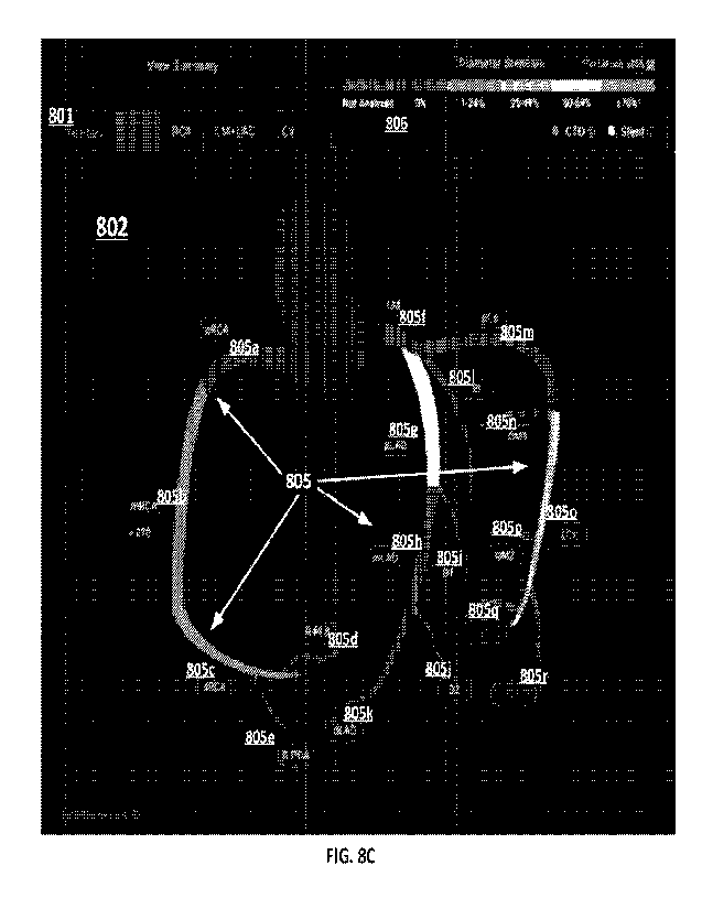
[0076] Figure 8C illustrates an example of a panel that
can be displayed on the
user interface showing a cartoon representation of a coronary artery tree
("cartoon artery
tree").
[0077] Figure 8D illustrates an example of a panel that
can be displayed on the
user interface illustrating territory selection using the cartoon artery tree.
[0078] Figure 8E illustrates an example panel that can be
displayed on the user
interface showing per-territory summaries.
[0079] Figure 8F illustrates an example panel that can be
displayed on the user
interface showing a SMPR view of a selected vessel, and corresponding
statistics of the
selected vessel.
[0080] Figure 8G illustrates an example of a portion of a
panel that can be
displayed in the user interface indicating the presence of a stent, which is
displayed at the
segment level.
[0081] Figure 8H illustrates an example of a portion of a
panel that can be
displayed in the user interface indicating CTO presence at the segment level.
-14-
CA 03162872 2022- 6- 22
SUBSTITUTE SHEET (RULE 26)
WO 2021/141921
PCT/ITS2021/012218
[0082] Figure 81 illustrates an example of a portion of a
panel that can be
displayed in the user interface indicating left or right dominance of the
patient.
[0083] Figure 8J illustrates an example of a panel that
can be displayed on the
user interface showing cartoon artery tree with indications of anomalies that
were found.
[0084] Figure 8K illustrates an example of a portion of a
panel that can be
displayed on the panel of Figure 8J that can be selected to show details of an
anomaly.
[0085] Figure 9A illustrates an example of an
atherosclerosis panel that can be
displayed on the user interface which displays a summary of atherosclerosis
information
based on the analysis.
[0086] Figure 9B illustrates an example of a vessel
selection panel which can be
used to select a vessel such that the summary of atherosclerosis information
is displayed on a
per segment basis.
[0087] Figure 9C illustrates an example of a panel that
can be displayed on the
user interface which shows per segment atherosclerosis information.
[0088] Figure 9D illustrates an example of a panel that
can be displayed on the
user interface that contains stenosis per patient data.
[0089] Figure 9E illustrates an example of a portion of a
panel that can be
displayed on the user interface that when a count is selected (e.g., by
hovering over the
number) segment details are displayed.
[0090] Figure 9F illustrates an example of a portion of a
panel that can be
displayed on the user interface that shows stenosis per segment in a graphical
format, for
example, in a stenosis per segment bar graph.
[0091] Figure 9G illustrates another example of a panel
that can be displayed on
the user interface showing information of the vessel, for example, diameter
stenosis and
minimum luminal diameter.
[0092] Figure 9H illustrates an example of a portion of a
panel that can be
displayed on the user interface indicating a diameter stenosis legend.
[0093] Figure 91 illustrates an example of a panel that
can be displayed on the
user interface indicating minimum and reference lumen diameters.
-15-
CA 03162872 2022- 6- 22
SUBSTITUTE SHEET (RULE 26)
WO 2021/141921
PCT/ITS2021/012218
[0094] Figure 9J illustrates a portion of the panel shown
in Figure 91, and shows
how specific minimum lumen diameter details can be quickly and efficiently
displayed by
selecting (e.g., by hovering over) a desired graphic of a lumen.
[0095] Figure 9K illustrates an example of a panel that
can be displayed in user
interface indicating CADS-RADS score selection.
[0096] Figure 9L illustrates an example of a panel that
can be displayed in the
user interface showing further CAD-RADS details generated in the analysis.
[0097] Figure 9M illustrates an example of a panel that
can be displayed in the
user interface showing a table indicating quantitative stenosis and vessel
outputs which are
determined during the analysis.
[0098] Figure 9N illustrates an example of a panel that
can be displayed in the
user interface showing a table indicating quantitative plaque outputs.
[0099] Figure 10 is a flowchart illustrating a process
1000 for analyzing and
displaying CT images and conesponding information.
[0100] Figures 11A and 11B are example CT images
illustrating how plaque can
appear differently depending on the image acquisition parameters used to
capture the CT
images. Figure 11A illustrates a CT image reconstructed using filtered back
projection,
while Figure 11B illustrates the same CT image reconstructed using iterative
reconstruction.
[0101] Figures 11C and 11D provide another example that
illustrates that plaque
can appear differently in CT images depending on the image acquisition
parameters used to
capture the CT images. Figure 11C illustrates a CT image reconstructed by
using iterative
reconstruction, while Figure 11D illustrates the same image reconstructed
using machine
learning.
[01021 Figure 12A is a block diagram representative of an
embodiment of a
normalization device that can be configured to normalize medical images for
use with the
methods and systems described herein.
[0103] Figure 12B is a perspective view of an embodiment
of a normalization
device including a multilayer substrate.
[0104] Figure 12C is a cross-sectional view of the
normalization device of Figure
12B illustrating various compartments positioned therein for holding samples
of known
materials for use during normalization.
-16-
CA 03162872 2022- 6- 22
SUBSTITUTE SHEET (RULE 26)
WO 2021/141921
PCT/ITS2021/012218
[0105] Figure 12D illustrates a top down view of an
example arrangement of a
plurality of compartments within a normalization device. In the illustrated
embodiment, the
plurality of compartments are arranged in a rectangular or grid-like pattern.
[0106] Figure 12E illustrates a top down view of another
example arrangement of
a plurality of compartments within a normalization device. In the illustrated
embodiment,
the plurality of compartments are arranged in a circular pattern.
[0107] Figure 12F is a cross-sectional view of another
embodiment of a
normalization device illustrating various features thereof, including
adjacently arranged
compartments, self-sealing fillable compartments, and compartments of various
sizes.
[0108] Figure 12G is a perspective view illustrating an
embodiment of an
attachment mechanism for a normalization device that uses hook and loop
fasteners to secure
a substrate of the normalization device to a fastener of the normalization
device.
[0109] Figures 12H and 121 illustrate an embodiment of a
normalization device
that includes an indicator configured to indicate an expiration status of the
normalization
device.
[0110] Figure 12J is a flowchart illustrating an example
method for normalizing
medical images for an algorithm-based medical imaging analysis, wherein
normalization of
the medical images improves accuracy of the algorithm-based medical imaging
analysis.
[0111] Figure 13 is a block diagram depicting an
embodiment(s) of a system for
medical image analysis, visualization, risk assessment, disease tracking,
treatment
generation, and/or patient report generation.
[0112] Figure 14 is a block diagram depicting an
embodiment(s) of a computer
hardware system configured to run software for implementing one or more
embodiments of a
system for medical image analysis, visualization, risk assessment, disease
tracking, treatment
generation, and/or patient report generation.
DETAILED DESCRIPTION
[0113] Although several embodiments, examples, and
illustrations are disclosed
below, it will be understood by those of ordinary skill in the art that the
inventions described
herein extend beyond the specifically disclosed embodiments, examples, and
illustrations and
includes other uses of the inventions and obvious modifications and
equivalents thereof.
-17-
CA 03162872 2022- 6- 22
SUBSTITUTE SHEET (RULE 26)
WO 2021/141921
PCT/ITS2021/012218
Embodiments of the inventions are described with reference to the accompanying
figures,
wherein like numerals refer to like elements throughout. The terminology used
in the
description presented herein is not intended to be interpreted in any limited
or restrictive
manner simply because it is being used in conjunction with a detailed
description of certain
specific embodiments of the inventions. In addition, embodiments of the
inventions can
comprise several novel features and no single feature is solely responsible
for its desirable
attributes or is essential to practicing the inventions herein described.
Introduction
[0114]
Disclosed herein are systems, methods, and devices for medical image
analysis, diagnosis, risk stratification, decision making and/or disease
tracking. Coronary
heart disease affects over 17.6 million Americans.
The current trend in treating
cardiovascular health issues is generally two-fold. First, physicians
generally review a
patient's cardiovascular health from a macro level, for example, by analyzing
the
biochemistry or blood content or biomarkers of a patient to determine whether
there are high
levels of cholesterol elements in the bloodstream of a patient. In response to
high levels of
cholesterol, some physicians will prescribe one or more drugs, such as
statins, as part of a
treatment plan in order to decrease what is perceived as high levels of
cholesterol elements in
the bloodstream of the patient.
[0115]
The second general trend for currently treating cardiovascular health
issues involves physicians evaluating a patient's cardiovascular health
through the use of
angiography to identify large blockages in various arteries of a patient. In
response to
finding large blockages in various arteries, physicians in some cases will
perform an
angioplasty procedure wherein a balloon catheter is guided to the point of
narrowing in the
vessel. After properly positioned, the balloon is inflated to compress or
flatten the plaque or
fatty matter into the artery wall and/or to stretch the artery open to
increase the flow of blood
through the vessel and/or to the heart. In some cases, the balloon is used to
position and
expand a stent within the vessel to compress the plaque and/or maintain the
opening of the
vessel to allow more blood to flow. About 500,000 heart stent procedures are
performed
each year in the United States.
[0116]
However, a recent federally funded $100 million study calls into
question
whether the current trends in treating cardiovascular disease are the most
effective treatment
-18-
CA 03162872 2022- 6- 22
SUBSTITUTE SHEET (RULE 26)
WO 2021/141921
PCT/ITS2021/012218
for all types of patients. The recent study involved over 5,000 patients with
moderate to
severe stable heart disease from 320 sites in 37 countries and provided new
evidence
showing that stents and bypass surgical procedures are likely no more
effective than drugs
combined with lifestyle changes for people with stable heart disease.
Accordingly, it may be
more advantageous for patients with stable heart disease to forgo invasive
surgical
procedures, such as angioplasty and/or heart bypass, and instead be prescribed
heart
medicines, such as statins, and certain lifestyle changes, such as regular
exercise. This new
treatment regimen could affect thousands of patients worldwide. Of the
estimated 500,000
heart stent procedures performed annually in the United States, it is
estimated that a fifth of
those are for people with stable heart disease. It is further estimated that
25% of the
estimated 100,000 people with stable heart disease, or roughly 23,000 people,
are individuals
that do not experience any chest pain. Accordingly, over 20,000 patients
annually could
potentially forgo invasive surgical procedures or the complications resulting
from such
procedures.
[0117] To determine whether a patient should forego
invasive surgical procedures
and opt instead for a drug regimen and/or to generate a more effective
treatment plan, it can
be important to more fully understand the cardiovascular disease of a patient.
Specifically, it
can be advantageous to better understand the arterial vessel health of a
patient. For example,
it is helpful to understand whether plaque build-up in a patient is mostly
fatty matter build-up
or mostly calcified matter build-up, because the former situation may warrant
treatment with
heart medicines, such as statins, whereas in the latter situation a patient
should be subject to
further periodic monitoring without prescribing heart medicine or implanting
any stents.
However, if the plaque build-up is significant enough to cause severe stenosis
or narrowing
of the arterial vessel such that blood flow to heart muscle might be blocked,
then an invasive
angioplasty procedure to implant a stent may likely be required because heart
attack or
sudden cardiac death (SCD) could occur in such patients without the
implantation of a stent
to enlarge the vessel opening. Sudden cardiac death is one of the largest
causes of natural
death in the United States, accounting for approximately 325,000 adult deaths
per year and
responsible for nearly half of all deaths from cardiovascular disease. For
males, SCD is
twice as common as compared to females. In general, SCD strikes people in the
mid-30 to
mid-40 age range. In over 50% of cases, sudden cardiac arrest occurs with no
warning signs.
-19-
CA 03162872 2022- 6- 22
SUBSTITUTE SHEET (RULE 26)
WO 2021/141921
PCT/ITS2021/012218
[0118] With respect to the millions suffering from heart
disease, there is a need to
better understand the overall health of the artery vessels within a patient
beyond just knowing
the blood chemistry or content of the blood flowing through such artery
vessels. For
example, in some embodiments of systems, devices, and methods disclosed
herein, arteries
with "good- or stable plaque or plaque comprising hardened calcified content
are considered
non-life threatening to patients whereas arteries containing "bad" or unstable
plaque or
plaque comprising fatty material are considered more life threatening because
such bad
plaque may rupture within arteries thereby releasing such fatty material into
the arteries.
Such a fatty material release in the blood stream can cause inflammation that
may result in a
blood clot. A blood clot within an artery can prevent blood from traveling to
heart muscle
thereby causing a heart attack or other cardiac event. Further, in some
instances, it is
generally more difficult for blood to flow through fatty plaque buildup than
it is for blood to
flow through calcified plaque build-up. Therefore, there is a need for better
understanding
and analysis of the arterial vessel walls of a patient.
[0119] Further, while blood tests and drug treatment
regimens are helpful in
reducing cardiovascular health issues and mitigating against cardiovascular
events (for
example, heart attacks), such treatment methodologies are not complete or
perfect in that
such treatments can misidentify and/or fail to pinpoint or diagnose
significant cardiovascular
risk areas. For example, the mere analysis of the blood chemistry of a patient
will not likely
identify that a patient has artery vessels having significant amounts of fatty
deposit material
had plaque buildup along a vessel wall. Similarly, an angiogram, while helpful
in identifying
areas of stenosis or vessel narrowing, may not he able to clearly identify
areas of the artery
vessel wall where there is significant buildup of bad plaque. Such areas of
buildup of bad
plaque within an artery vessel wall can be indicators of a patient at high
risk of suffering a
cardiovascular event, such as a heart attack. In certain circumstances, areas
where there exist
areas of bad plaque can lead to a rupture wherein there is a release of the
fatty materials into
the bloodstream of the artery, which in turn can cause a clot to develop in
the artery. A blood
clot in the artery can cause a stoppage of blood flow to the heart tissue,
which can result in a
heart attack. Accordingly, there is a need for new technology for analyzing
artery vessel
walls and/or identifying areas within artery vessel walls that comprise a
buildup of plaque
whether it be bad or otherwise.
-20-
CA 03162872 2022- 6- 22
SUBSTITUTE SHEET (RULE 26)
WO 2021/141921
PCT/ITS2021/012218
[0120] Various systems, methods, and devices disclosed
herein are directed to
embodiments for addressing the foregoing issues. In particular, various
embodiments
described herein relate to systems, methods, and devices for medical image
analysis,
diagnosis, risk stratification, decision making and/or disease tracking. In
some embodiments,
the systems, devices, and methods described herein are configured to utilize
non-invasive
medical imaging technologies, such as a CT image for example, which can be
inputted into a
computer system configured to automatically and/or dynamically analyze the
medical image
to identify one or more coronary arteries and/or plaque within the same. For
example, in
some embodiments, the system can be configured to utilize one or more machine
learning
and/or artificial intelligence algorithms to automatically and/or dynamically
analyze a
medical image to identify, quantify, and/or classify one or more coronary
arteries and/or
plaque. In some embodiments, the system can be further configured to utilize
the identified,
quantified, and/or classified one or more coronary arteries and/or plaque to
generate a
treatment plan, track disease progression, and/or a patient-specific medical
report, for
example using one or more artificial intelligence and/or machine learning
algorithms. In
some embodiments, the system can be further configured to dynamically and/or
automatically generate a visualization of the identified, quantified, and/or
classified one or
more coronary arteries and/or plaque, for example in the form of a graphical
user interface.
Further, in some embodiments, to calibrate medical images obtained from
different medical
imaging scanners and/or different scan parameters or environments, the system
can be
configured to utilize a normalization device comprising one or more
compartments of one or
more materials.
[0121] As will be discussed in further detail, the
systems, devices, and methods
described herein allow for automatic and/or dynamic quantified analysis of
various
parameters relating to plaque, cardiovascular arteries, and/or other
structures. More
specifically, in some embodiments described herein, a medical image of a
patient, such as a
coronary CT image, can be taken at a medical facility. Rather than having a
physician
eyeball or make a general assessment of the patient, the medical image is
transmitted to a
backend main server in some embodiments that is configured to conduct one or
more
analyses thereof in a reproducible manner. As such, in some embodiments, the
systems,
methods, and devices described herein can provide a quantified measurement of
one or more
-21 -
CA 03162872 2022- 6- 22
SUBSTITUTE SHEET (RULE 26)
WO 2021/141921
PCT/ITS2021/012218
features of a coronary CT image using automated and/or dynamic processes. For
example, in
some embodiments, the main server system can be configured to identify one or
more
vessels, plaque, and/or fat from a medical image. Based on the identified
features, in some
embodiments, the system can be configured to generate one or more quantified
measurements from a raw medical image, such as for example radiodensity of one
or more
regions of plaque, identification of stable plaque and/or unstable plaque,
volumes thereof,
surface areas thereof, geometric shapes, heterogeneity thereof, and/or the
like. In some
embodiments, the system can also generate one or more quantified measurements
of vessels
from the raw medical image, such as for example diameter, volume, morphology,
and/or the
like. Based on the identified features and/or quantified
measurements, in some
embodiments, the system can be configured to generate a risk assessment and/or
track the
progression of a plaque-based disease or condition, such as for example
atherosclerosis,
stenosis, and/or ischemia, using raw medical images. Further, in some
embodiments, the
system can be configured to generate a visualization of GUI of one or more
identified
features and/or quantified measurements, such as a quantized color mapping of
different
features. In some embodiments, the systems, devices, and methods described
herein arc
configured to utilize medical image-based processing to assess for a subject
his or her risk of
a cardiovascular event, major adverse cardiovascular event (MACE), rapid
plaque
progression, and/or non-response to medication. In particular, in some
embodiments, the
system can be configured to automatically and/or dynamically assess such
health risk of a
subject by analyzing only non-invasively obtained medical images. In some
embodiments,
one or more of the processes can be automated using an AT and/or ML algorithm.
In some
embodiments, one or more of the processes described herein can be performed
within
minutes in a reproducible manner. This is stark contrast to existing measures
today which do
not produce reproducible prognosis or assessment, take extensive amounts of
time, and/or
require invasive procedures.
[0122] As such, in some embodiments, the systems, devices,
and methods
described herein are able to provide physicians and/or patients specific
quantified and/or
measured data relating to a patient's plaque that do not exist today. For
example, in some
embodiments, the system can provide a specific numerical value for the volume
of stable
and/or unstable plaque, the ratio thereof against the total vessel volume,
percentage of
CA 03162872 2022- 6- 22
SUBSTITUTE SHEET (RULE 26)
WO 2021/141921
PCT/ITS2021/012218
stenosis, and/or the like, using for example radiodensity values of pixels
and/or regions
within a medical image. In some embodiments, such detailed level of quantified
plaque
parameters from image processing and downstream analytical results can provide
more
accurate and useful tools for assessing the health and/or risk of patients in
completely novel
ways.
General Overview
[0123] In some embodiments, the systems, devices, and
methods described herein
are configured to automatically and/or dynamically perform medical image
analysis,
diagnosis, risk stratification, decision making and/or disease tracking.
Figure 1 is a flowchart
illustrating an overview of an example embodiment(s) of a method for medical
image
analysis, visualization, risk assessment, disease tracking, treatment
generation, and/or patient
report generation. As illustrated in Figure 1, in some embodiments, the system
is configured
to access and/or analyze one or more medical images of a subject, such as for
example a
medical image of a coronary region of a subject or patient.
[0124] In some embodiments, before obtaining the medical
image, a
normalization device is attached to the subject and/or is placed within a
field of view of a
medical imaging scanner at block 102. For example, in some embodiments, the
normalization device can comprise one or more compartments comprising one or
more
materials, such as water, calcium, and/or the like. Additional detail
regarding the
normalization device is provided below. Medical imaging scanners may produce
images
with different scalable radiodensities for the same object. This, for example,
can depend not
only on the type of medical imaging scanner or equipment used but also on the
scan
parameters and/or environment of the particular day and/or time when the scan
was taken.
As a result, even if two different scans were taken of the same subject, the
brightness and/or
darkness of the resulting medical image may be different, which can result in
less than
accurate analysis results processed from that image. To account for such
differences, in
some embodiments, a normalization device comprising one or more known elements
is
scanned together with the subject, and the resulting image of the one or more
known
elements can be used as a basis for translating, converting, and/or
normalizing the resulting
image. As such, in some embodiments, a normalization device is attached to the
subject
and/or placed within the field of view of a medical imaging scan at a medical
facility.
-23-
CA 03162872 2022- 6- 22
SUBSTITUTE SHEET (RULE 26)
WO 2021/141921
PCT/ITS2021/012218
[0125] In some embodiments, at block 104, the medical
facility then obtains one
or more medical images of the subject. For example, the medical image can be
of the
coronary region of the subject or patient. In some embodiments, the systems
disclosed herein
can be configured to take in CT data from the image domain or the projection
domain as raw
scanned data or any other medical data, such as but not limited to: x-ray;
Dual-Energy
Computed Tomography (DECT), Spectral CT, photon-counting detector CT,
ultrasound,
such as echocardiography or intravascular ultrasound (IVUS); magnetic
resonance (MR)
imaging; optical coherence tomography (OCT); nuclear medicine imaging,
including
positron-emission tomography (PET) and single photon emission computed
tomography
(SPECT); near-field infrared spectroscopy (NIRS); and/or the like. As used
herein, the term
CT image data or CT scanned data can be substituted with any of the foregoing
medical
scanning modalities and process such data through an artificial intelligence
(Al) algorithm
system in order to generate processed CT image data. In some embodiments, the
data from
these imaging modalities enables determination of cardiovascular phenotype,
and can include
the image domain data, the projection domain data, and/or a combination of
both.
[0126] In some embodiments, at block 106, the medical
facility can also obtain
non-imaging data from the subject. For example, this can include blood tests,
biomarkers,
panomics and/or the like. In some embodiments, at block 108, the medical
facility can
transmit the one or more medical images and/or other non-imaging data at block
108 to a
main server system. In some embodiments, the main server system can be
configured to
receive and/or otherwise access the medical image and/or other non-imaging
data at block
110.
[0127] In some embodiments, at block 112, the system can
be configured to
automatically and/or dynamically analyze the one or more medical images which
can be
stored and/or accessed from a medical image database 100. For example, in some
embodiments, the system can be configured to take in raw CT image data and
apply an
artificial intelligence (Al) algorithm, machine learning (ML) algorithm,
and/or other physics-
based algorithm to the raw CT data in order to identify, measure, and/or
analyze various
aspects of the identified arteries within the CT data. In some embodiments,
the inputting of
the raw medical image data involves uploading the raw medical image data into
cloud-based
data repository system. In some embodiments, the processing of the medical
image data
-24-
CA 03162872 2022- 6- 22
SUBSTITUTE SHEET (RULE 26)
WO 2021/141921
PCT/ITS2021/012218
involves processing the data in a cloud-based computing system using an Al
and/or ML
algorithm. In some embodiments, the system can be configured to analyze the
raw CT data
in about 1 minute, about 2 minutes, about 3 minutes, about 4 minutes, about 5
minutes, about
6 minutes, about 7 minutes, about 8 minutes, about 9 minutes, about 10
minutes, about 15
minutes, about 20 minutes, about 30 minutes, about 35 minutes, about 40
minutes, about 45
minutes, about 50 minutes, about 55 minutes, about 60 minutes, and/or within a
range
defined by two of the aforementioned values.
[0128] In some embodiments, the system can be configured
to utilize a vessel
identification algorithm to identify and/or analyze one or more vessels within
the medical
image. In some embodiments, the system can he configured to utilize a coronary
artery
identification algorithm to identify and/or analyze one or more coronary
arteries within the
medical image. In some embodiments, the system can be configured to utilize a
plaque
identification algorithm to identify and/or analyze one or more regions of
plaque within the
medical image. In some embodiments, the vessel identification algorithm,
coronary artery
identification algorithm, and/or plaque identification algorithm comprises an
AT and/or ML
algorithm. For example, in some embodiments, the vessel identification
algorithm, coronary
artery identification algorithm, and/or plaque identification algorithm can be
trained on a
plurality of medical images wherein one or more vessels, coronary arteries,
and/or regions of
plaque are pre-identified. Based on such training, for example by use of a
Convolutional
Neural Network in some embodiments, the system can be configured to
automatically and/or
dynamically identify from raw medical images the presence and/or parameters of
vessels,
coronary arteries, and/or plaque.
[0129] As such, in some embodiments, the processing of the
medical image or
raw CT scan data can comprise analysis of the medical image or CT data in
order to
determine and/or identify the existence and/or nonexistence of certain artery
vessels in a
patient. As a natural occurring phenomenon, certain arteries may be present in
certain
patients whereas such certain arteries may not exist in other patients.
[0130] In some embodiments, at block 112, the system can
be further configured
to analyze the identified vessels, coronary arteries, and/or plaque, for
example using an AT
and/or ML algorithm. In particular, in some embodiments, the system can be
configured to
determine one Or more vascular morphology parameters, such as for example
arterial
-25-
CA 03162872 2022- 6- 22
SUBSTITUTE SHEET (RULE 26)
WO 2021/141921
PCT/ITS2021/012218
remodeling, curvature, volume, width, diameter, length, and/or the like.
In some
embodiments, the system can be configured to determine one or more plaque
parameters,
such as for example volume, surface area, geometry, radiodensity, ratio or
function of
volume to surface area, heterogeneity index, and/or the like of one or more
regions of plaque
shown within the medical image. -Radiodensity" as used herein is a broad term
that refers to
the relative inability of electromagnetic relation (e.g., X-rays) to pass
through a material. In
reference to an image, radiodensity values refer to values indicting a density
in image data
(e.g., film, print, or in an electronic format) where the radiodensity values
in the image
corresponds to the density of material depicted in the image.
[0131]
In some embodiments, at block 114, the system can be configured to
utilize the identified and/or analyzed vessels, coronary arteries, and/or
plaque from the
medical image to perform a point-in-time analysis of the subject. In some
embodiments, the
system can be configured to use automatic and/or dynamic image processing of
one or more
medical images taken from one point in time to identify and/or analyze one or
more vessels,
coronary arteries, and/or plaque and derive one or more parameters and/or
classifications
thereof. For example, as will be described in more detail herein, in some
embodiments, the
system can be configured to generate one or more quantification metrics of
plaque and/or
classify the identified regions of plaque as good or bad plaque. Further, in
some
embodiments, at block 114, the system can be configured to generate one or
more treatment
plans for the subject based on the analysis results. In some embodiments, the
system can be
configured to utilize one or more Al and/or ML algorithms to identify and/or
analyze vessels
or plaque, derive one or more quantification metrics and/or classifications,
and/or generate a
treatment plan.
[0132]
In some embodiments, if a previous scan or medical image of the subject
exists, the system can be configured to perform at block 126 one or more time-
based
analyses, such as disease tracking. For example, in some embodiments, if the
system has
access to one or more quantified parameters or classifications derived from
previous scans or
medical images of the subject, the system can be configured to compare the
same with one or
more quantified parameters or classifications derived from a current scan or
medical image to
determine the progression of disease and/or state of the subject.
-26-
CA 03162872 2022- 6- 22
SUBSTITUTE SHEET (RULE 26)
WO 2021/141921
PCT/ITS2021/012218
[0133] In some embodiments, at block 116, the system is
configured to
automatically and/or dynamically generate a Graphical User Interface (GUI) or
other
visualization of the analysis results at block 116, which can include for
example identified
vessels, regions of plaque, coronary arteries, quantified metrics or
parameters, risk
assessment, proposed treatment plan, and/or any other analysis result
discussed herein. In
some embodiments, the system is configured to analyze arteries present in the
CT scan data
and display various views of the arteries present in the patient, for example
within 10-15
minutes or less. In contrast, as an example, conducting a visual assessment of
a CT to
identify stenosis alone, without consideration of good or bad plaque or any
other factor, can
take anywhere between 15 minutes to more than an hour depending on the skill
level, and
can also have substantial variability across radiologists and/or cardiac
imagers.
[0134] In some embodiments, at block 118, the system can
be configured to
transmit the generated GUI or other visualization, analysis results, and/or
treatment to the
medical facility. In some embodiments, at block 120, a physician at the
medical facility can
then review and/or confirm and/or revise the generated GUI or other
visualization, analysis
results, and/or treatment.
[0135] In some embodiments, at block 122, the system can
be configured to
further generate and transmit a patient-specific medical report to a patient,
who can receive
the same at block 124. In some embodiments, the patient-specific medical
report can be
dynamically generated based on the analysis results derived from and/or other
generated
from the medical image processing and analytics. For example, the patient-
specific report
can include identified vessels, regions of plaque, coronary arteries,
quantified metrics or
parameters, risk assessment, proposed treatment plan, and/or any other
analysis result
discussed herein.
[0136] In some embodiments, one or more of the process
illustrated in Figure 1
can be repeated, for example for the same patient at a different time to track
progression of a
disease and/or the state of the patient.
Image Processing-Based Classification of Good v. Bad Plaque
[0137] As discussed, in some embodiments, the systems,
methods, and devices
described herein are configured to automatically and/or dynamically identify
and/or classify
good v. had plaque or stable v. unstable plaque based on medical image
analysis and/or
-27-
CA 03162872 2022- 6- 22
SUBSTITUTE SHEET (RULE 26)
WO 2021/141921
PCT/ITS2021/012218
processing. For example, in some embodiments, the system can be configured to
utilize an
AT and/or ML algorithm to identify areas in an artery that exhibit plaque
buildup within,
along, inside and/or outside the arteries. In some embodiments, the system can
be configured
to identify the outline or boundary of plaque buildup associated with an
artery vessel wall. In
some embodiments, the system can be configured to draw or generate a line that
outlines the
shape and configuration of the plaque buildup associated with the artery. In
some
embodiments, the system can be configured to identify whether the plaque
buildup is a
certain kind of plaque and/or the composition or characterization of a
particular plaque
buildup. In some embodiments, the system can be configured to characterize
plaque binarily,
ordinally and/or continuously. In some embodiments, the system can be
configured to
determine that the kind of plaque buildup identified is a "bad" kind of plaque
due to the dark
color or dark gray scale nature of the image corresponding to the plaque area,
and/or by
determination of its attenuation density (e.g., using a Hounsfield unit scale
or other). For
example, in some embodiments, the system can be configured to identify certain
plaque as
"bad" plaque if the brightness of the plaque is darker than a pre-determined
level. In some
embodiments, the system can be configured to identify good plaque areas based
on the white
coloration and/or the light gray scale nature of the area corresponding to the
plaque buildup.
For example, in some embodiments, the system can be configured to identify
certain plaque
as "good" plaque if the brightness of the plaque is lighter than a pre-
determined level. In
some embodiments, the system can be configured to determine that dark areas in
the CT scan
are related to "bad" plaque, whereas the system can be configured to identify
good plaque
areas corresponding to white areas. In some embodiments, the system can be
configured to
identify and determine the total area and/or volume of total plaque, good
plaque, and/or bad
plaque identified within an artery vessel or plurality of vessels. In some
embodiments, the
system can be configured to determine the length of the total plaque area,
good plaque area,
and/or bad plaque area identified. In some embodiments, the system can be
configured to
determine the width of the total plaque area, good plaque area, and/or bad
plaque area
identified. The "good" plaque may be considered as such because it is less
likely to cause
heart attack, less likely to exhibit significant plaque progression, and/or
less likely to be
ischemia, amongst others. Conversely, the "bad" plaque be considered as such
because it is
more likely to cause heart attack, more likely to exhibit significant plaque
progression, and/or
-28-
CA 03162872 2022- 6- 22
SUBSTITUTE SHEET (RULE 26)
WO 2021/141921
PCT/ITS2021/012218
more likely to be ischemia, amongst others. In some embodiments, the -good"
plaque may be
considered as such because it is less likely to result in the no-reflow
phenomenon at the time
of coronary revascularization. Conversely, the "bad" plaque may be considered
as such
because it is more likely to cause the no-reflow phenomenon at the time of
coronary
revascularization.
[0138] Figure 2A is a flowchart illustrating an overview
of an example
embodiment(s) of a method for analysis and classification of plaque from a
medical image,
which can be obtained non-invasively. As illustrated in Figure 2A, at block
202, in some
embodiments, the system can be configured to access a medical image, which can
include a
coronary region of a subject and/or be stored in a medical image database 100.
The medical
image database 100 can be locally accessible by the system and/or can be
located remotely
and accessible through a network connection. The medical image can comprise an
image
obtain using one or more modalities such as for example, CT, Dual-Energy
Computed
Tomography (DECT), Spectral CT, photon-counting CT, x-ray, ultrasound,
echocardiography, intravascular ultrasound (BTUS), Magnetic Resonance (MR)
imaging,
optical coherence tomography (OCT), nuclear medicine imaging, positron-
emission
tomography (PET), single photon emission computed tomography (SPECT), or near-
field
infrared spectroscopy (NIRS). In some embodiments, the medical image comprises
one or
more of a contrast-enhanced CT image, non-contrast CT image, MR image, and/or
an image
obtained using any of the modalities described above.
[0139] In some embodiments, the system can be configured
to automatically
and/or dynamically perform one or more analyses of the medical image as
discussed herein.
For example, in some embodiments, at block 204, the system can be configured
to identify
one or more arteries. The one or more arteries can include coronary arteries,
carotid arteries,
aorta, renal artery, lower extremity artery, upper extremity artery, and/or
cerebral artery,
amongst others. In some embodiments, the system can be configured to utilize
one or more
AT and/or ML algorithms to automatically and/or dynamically identify one or
more arteries
or coronary arteries using image processing. For example, in some embodiments,
the one or
more Al and/or ML algorithms can be trained using a Convolutional Neural
Network (CNN)
on a set of medical images on which arteries or coronary arteries have been
identified,
thereby allowing the AT and/or ML algorithm automatically identify arteries or
coronary
-29-
CA 03162872 2022- 6- 22
SUBSTITUTE SHEET (RULE 26)
WO 2021/141921
PCT/US2021/012218
arteries directly from a medical image. In some embodiments, the arteries or
coronary
arteries are identified by size and/or location.
[0140] In some embodiments, at block 206, the system can
be configured to
identify one or more regions of plaque in the medical image. In some
embodiments, the
system can be configured to utilize one or more AT and/or ML algorithms to
automatically
and/or dynamically identify one or more regions of plaque using image
processing. For
example, in some embodiments, the one or more AT and/or ML algorithms can be
trained
using a Convolutional Neural Network (CNN) on a set of medical images on which
regions
of plaque have been identified, thereby allowing the AT and/or ML algorithm
automatically
identify regions of plaque directly from a medical image. In some embodiments,
the system
can be configured to identify a vessel wall and a lumen wall for each of the
identified
coronary arteries in the medical image. In some embodiments, the system is
then configured
to determine the volume in between the vessel wall and the lumen wall as
plaque. In some
embodiments, the system can be configured to identify regions of plaque based
on the
radiodensity values typically associated with plaque, for example by setting a
predetermined
threshold or range of radiodensity values that arc typically associated with
plaque with or
without normalizing using a normalization device.
[0141] In some embodiments, the system is configured to
automatically and/or
dynamically determine one or more vascular morphology parameters and/or plaque
parameters at block 208 from the medical image. In some embodiments, the one
or more
vascular morphology parameters and/or plaque parameters can comprise
quantified
parameters derived from the medical image. For example, in some embodiments,
the system
can be configured to utilize an Al and/or ML algorithm or other algorithm to
determine one
or more vascular morphology parameters and/or plaque parameters. As another
example, in
some embodiments, the system can be configured to determine one or more
vascular
morphology parameters, such as classification of arterial remodeling due to
plaque, which
can further include positive arterial remodeling, negative arterial
remodeling, and/or
intermediate arterial remodeling. In some embodiments, the classification of
arterial
remodeling is determined based on a ratio of the largest vessel diameter at a
region of plaque
to a normal reference vessel diameter of the same region which can be
retrieved from a
normal database. In some embodiments, the system can be configured to classify
arterial
-30-
CA 03162872 2022- 6- 22
SUBSTITUTE SHEET (RULE 26)
WO 2021/141921
PCT/ITS2021/012218
remodeling as positive when the ratio of the largest vessel diameter at a
region of plaque to a
normal reference vessel diameter of the same region is more than 1.1. In some
embodiments,
the system can be configured to classify arterial remodeling as negative when
the ratio of the
largest vessel diameter at a region of plaque to a normal reference vessel
diameter is less than
0.95. In some embodiments, the system can be configured to classify arterial
remodeling as
intermediate when the ratio of the largest vessel diameter at a region of
plaque to a normal
reference vessel diameter is between 0.95 and 1.1.
[0142] Further, as part of block 208, in some embodiments,
the system can be
configured to determine a geometry and/or volume of one or more regions of
plaque and/or
one or more vessels or arteries at block 201. For example, the system can be
configured to
determine if the geometry of a particular region of plaque is round or oblong
or other shape.
In some embodiments, the geometry of a region of plaque can be a factor in
assessing the
stability of the plaque. As another example, in some embodiments, the system
can be
configured to determine the curvature, diameter, length, volume, and/or any
other parameters
of a vessel or artery from the medical image.
[0143] In some embodiments, as part of block 208. the
system can be configured
to determine a volume and/or surface area of a region of plaque and/or a ratio
or other
function of volume to surface area of a region of plaque at block 203, such as
for example a
diameter, radius, and/or thickness of a region of plaque. In some embodiments,
a plaque
having a low ratio of volume to surface area can indicate that the plaque is
stable. As such,
in some embodiments, the system can be configured to determine that a ratio of
volume to
surface area of a region of plaque below a predetermined threshold is
indicative of stable
plaque.
[01441 In some embodiments, as part of block 208, the
system can be configured
to determine a heterogeneity index of a region of plaque at block 205. For
instance, in some
embodiments, a plaque having a low heterogeneity or high homogeneity can
indicate that the
plaque is stable. As such, in some embodiments, the system can be configured
to determine
that a heterogeneity of a region of plaque below a predetermined threshold is
indicative of
stable plaque. In some embodiments, heterogeneity or homogeneity of a region
of plaque
can be determined based on the heterogeneity or homogeneity of radiodensity
values within
the region of plaque. As such, in some embodiments, the system can be
configured to
-11-
CA 03162872 2022- 6- 22
SUBSTITUTE SHEET (RULE 26)
WO 2021/141921
PCT/ITS2021/012218
determine a heterogeneity index of plaque by generating spatial mapping, such
as a three-
dimensional histogram, of radiodensity values within or across a geometric
shape or region
of plaque. In some embodiments, if a gradient or change in radiodensity values
across the
spatial mapping is above a certain threshold, the system can be configured to
assign a high
heterogeneity index. Conversely, in some embodiments, if a gradient or change
in
radiodensity values across the spatial mapping is below a certain threshold,
the system can be
configured to assign a low heterogeneity index.
[0145] In some embodiments, as part of block 208, the
system can be configured
to determine a radiodensity of plaque and/or a composition thereof at block
207. For
example, a high radiodensity value can indicate that a plaque is highly
calcified or stable,
whereas a low radiodensity value can indicate that a plaque is less calcified
or unstable. As
such, in some embodiments, the system can be configured to determine that a
radiodensity of
a region of plaque above a predetermined threshold is indicative of stable
stabilized plaque.
In addition, different areas within a region of plaque can be calcified at
different levels and
thereby show different radiodensity values. As such, in some embodiments, the
system can
be configured to determine the radiodensity values of a region of plaque
and/or a
composition or percentage or change of radiodensity values within a region of
plaque. For
instance, in some embodiments, the system can be configured to determine how
much or
what percentage of plaque within a region of plaque shows a radiodensity value
within a low
range, medium range, high range, and/or any other classification.
[0146] Similarly, in some embodiments, as part of block
208, the system can be
configured to determine a ratio of radiodensity value of plaque to a volume of
plaque at block
209. For instance, it can be important to assess whether a large or small
region of plaque is
showing a high or low radiodensity value. As such, in some embodiments, the
system can be
configured to determine a percentage composition of plaque comprising
different
radiodensity values as a function or ratio of volume of plaque.
[0147] In some embodiments, as part of block 208, the
system can be configured
to determine the diffusivity and/or assign a diffusivity index to a region of
plaque at block
211. For example, in some embodiments, the diffusivity of a plaque can depend
on the
radiodensity value of plaque, in which a high radiodensity value can indicate
low diffusivity
Or stability of the plaque.
-12-
CA 03162872 2022- 6- 22
SUBSTITUTE SHEET (RULE 26)
WO 2021/141921
PCT/ITS2021/012218
[0148] In some embodiments, at block 210, the system can
be configured to
classify one or regions of plaque identified from the medical image as stable
v. unstable or
good v. bad based on the one or more vascular morphology parameters and/or
quantified
plaque parameters determined and/or derived from raw medical images. In
particular, in
some embodiments, the system can be configured to generate a weighted measure
of one or
more vascular morphology parameters and/or quantified plaque parameters
determined
and/or derived from raw medical images. For example. in some embodiments, the
system
can be configured weight one or more vascular morphology parameters and/or
quantified
plaque parameters equally. In some embodiments, the system can be configured
weight one
or more vascular morphology parameters and/or quantified plaque parameters
differently. In
some embodiments, the system can be configured weight one or more vascular
morphology
parameters and/or quantified plaque parameters logarithmically, algebraically,
and/or
utilizing another mathematical transform. In some embodiments, the system is
configured to
classify one or more regions of plaque at block 210 using the generated
weighted measure
and/or using only some of the vascular morphology parameters and/or quantified
plaque
parameters.
[0149] In some embodiments, at block 212, the system is
configured to generate a
quantized color mapping based on the analyzed and/or determined parameters.
For example,
in some embodiments, the system is configured to generate a visualization of
the analyzed
medical image by generating a quantized color mapping of calcified plaque, non-
calcified
plaque, good plaque, had plaque, stable plaque, and/or unstable plaque as
determined using
any of the analytical techniques described herein. Further, in some
embodiments, the
quantified color mapping can also include arteries and/or epicardial fat,
which can also be
determined by the system, for example by utilizing one or more Al and/or ML
algorithms.
[0150] In some embodiments, at block 214, the system is
configured to generate a
proposed treatment plan for the subject based on the analysis, such as for
example the
classification of plaque derived automatically from a raw medical image. In
particular, in
some embodiments, the system can be configured to assess or predict the risk
of
atherosclerosis, stenosis, and/or ischemia of the subject based on a raw
medical image and
automated image processing thereof.
-11-
CA 03162872 2022- 6- 22
SUBSTITUTE SHEET (RULE 26)
WO 2021/141921
PCT/ITS2021/012218
[0151] In some embodiments, one or more processes
described herein in
connection with Figure 2A can be repeated. For example, if a medical image of
the same
subject is taken again at a later point in time, one or more processes
described herein can be
repeated and the analytical results thereof can be used for disease tracking
and/or other
purposes.
Determination of Non-Calcified Plaque from a Non-Contrast CT Image(s)
[0152] As discussed herein, in some embodiments, the
system can be configured
to utilize a CT or other medical image of a subject as input for performing
one or more image
analysis techniques to assess a subject, including for example risk of a
cardiovascular event.
In some embodiments, such CT image can comprise a contrast-enhanced CT image,
in which
case some of the analysis techniques described herein can be directly applied,
for example to
identify or classify plaque. However, in some embodiments, such CT image can
comprise a
non-contrast CT image, in which case it can be more difficult to identify
and/or determine
non-calcified plaque due to its low radiodensity value and overlap with other
low
radiodensity values components, such as blood for example. As such, in some
embodiments,
the systems, devices, and methods described herein provide a novel approach to
determining
non-calcified plaque from a non-contrast CT image, which can be more widely
available.
[0153] Also, in some embodiments, in addition to or
instead of analyzing a
contrast-enhanced CT scan, the system can also be configured to examine the
attenuation
densities within the arteries that are lower than the attenuation density of
the blood flowing
within them in a non-contrast CT scan. In some embodiments, these "low
attenuation"
plaques may be differentiated between the blood attenuation density and the
fat that
sometimes surrounds the coronary artery and/or may represent non-calcified
plaques of
different materials. In some embodiments, the presence of these non-calcified
plaques may
offer incremental prediction for whether a previously calcified plaque is
stabilizing or
worsening or progressing or regressing. These findings that are measurable
through these
embodiments may be linked to the prognosis of a patient, wherein calcium
stabilization (that
is, higher attenuation densities) and lack of non-calcified plaque by may
associated with a
favorable prognosis, while lack of calcium stabilization (that is, no increase
in attenuation
densities), or significant progression or new calcium formation may be
associated with a
-14-
CA 03162872 2022- 6- 22
SUBSTITUTE SHEET (RULE 26)
WO 2021/141921
PCT/ITS2021/012218
poorer prognosis, including risk of rapid progression of disease, heart attack
or other major
adverse cardiovascular event.
[0154] Figure 2B is a flowchart illustrating an overview
of an example
embodiment(s) of a method for determination of non-calcified and/or low-
attenuated plaque
from a medical image, such as a non-contrast CT image. As discussed herein and
as
illustrated in Figure 2B, in some embodiments, the system can be configured to
determine
non-calcified and/or low-attenuated plaque from a medical image. In some
embodiments, the
medical image can be of the coronary region of the subject or patient. In some
embodiments,
the medical image can be obtained using one or more modalities such as CT,
Dual-Energy
Computed Tomography (DECT), Spectral CT, x-ray, ultrasound, echocardiography,
BTUS,
MR, OCT, nuclear medicine imaging, PET, SPECT, NIRS, and/or the like. In some
embodiments, the system can be configured to access one or more medical images
at block
202, for example from a medical image database 100.
[0155] In some embodiments, in order to determine non-
calcified and/or low-
attenuated plaque from the medical image or non-contrast CT image, the system
can be
configured to utilize a stepwise approach to first identify areas within the
medical image that
are clearly non-calcified plaque. In some embodiments, the system can then
conduct a more
detailed analysis of the remaining areas in the image to identify other
regions of non-calcified
and/or low-attenuated plaque. By utilizing such compartmentalized or a
stepwise approach,
in some embodiments, the system can identify or determine non-calcified and/or
low-
attenuated plaque from the medical image or non-contrast CT image with a
faster turnaround
rather than having to apply a more complicated analysis to every region or
pixel of the
image.
[0156] In particular, in some embodiments, at block 224,
the system can be
configured to identify epicardial fat from the medical image. In some
embodiments, the
system can be configured to identify epicardial fat by determining every pixel
or region
within the image that has a radiodensity value below a predetermined threshold
and/or within
a predetermined range. The exact predetermined threshold value or range of
radiodensity for
identifying epicardial fat can depend on the medical image, scanner type, scan
parameters,
and/or the like, which is why a normalization device can be used in some
instances to
normalize the medical image. For example, in some embodiments, the system can
be
-15-
CA 03162872 2022- 6- 22
SUBSTITUTE SHEET (RULE 26)
WO 2021/141921
PCT/ITS2021/012218
configured to identify as epicardial fat pixels and/or regions within the
medical image or non-
contrast CT image with a radiodensity value that is around -100 Hounsfield
units and/or
within a range that includes -100 Hounsfield units. In particular, in some
embodiments, the
system can be configured to identify as epicardial fat pixels and/or regions
within the medical
image or non-contrast CT image with a radiodensity value that is within a
range with a lower
limit of about -100 Hounsfield units, about -110 Hounsfield units, about -120
Hounsfield
units, about -130 Hounsfield units, about -140 Hounsfield units, about -150
Hounsfield units,
about -160 Hounsfield units, about -170 Hounsfield units, about -180
Hounsfield units, about
-190 Hounsfield units, or about -200 Hounsfield units, and an upper limit of
about 30
Hounsfield units, about 20 Hounsfield units, about 10 Hounsfield units, about
0 Hounsfield
units, about -10 Hounsfield units, about -20 Hounsfield units, about -30
Hounsfield units,
about -40 Hounsfield units, about -50 Hounsfield units, about -60 Hounsfield
units, about -70
Hounsfield units, about -80 Hounsfield units, or about -90 Hounsfield units.
[0157] In some embodiments, the system can be configured
to identify and/or
segment arteries on the medical image or non-contrast CT image using the
identified
epicardial fat as outer boundaries of the arteries. For example, the system
can be configured
to first identify regions of epicardial fat on the medical image and assign a
volume in
between epicardial fat as an artery, such as a coronary artery.
[0158] In some embodiments, at block 226, the system can
be configured to
identify a first set of pixels or regions within the medical image, such as
within the identified
arteries, as non-calcified or low-attenuated plaque. More specifically, in
some embodiments,
the system can be configured to identify as an initial set low-attenuated or
non-calcified
plaque by identifying pixels or regions with a radiodensity value that is
below a
predetermined threshold or within a predetermined range. For example, the
predetermined
threshold or predetermined range can be set such that the resulting pixels can
be confidently
marked as low-attenuated or non-calcified plaque without likelihood of
confusion with
another matter such as blood. In particular, in some embodiments, the system
can be
configured to identify the initial set of low-attenuated or non-calcified
plaque by identifying
pixels or regions with a radiodensity value below around 30 Hounsfield units.
In some
embodiments, the system can be configured to identify the initial set of low-
attenuated or
non-calcified plaque by identifying pixels Or regions with a radiodensity
value at Or below
-36-
CA 03162872 2022- 6- 22
SUBSTITUTE SHEET (RULE 26)
WO 2021/141921
PCT/ITS2021/012218
around 60 Hounsfield units, around 55 Hounsfield units, around 50 Hounsfield
units, around
45 Hounsfield units, around 40 Hounsfield units, around 35 Hounsfield units,
around 30
Hounsfield units, around 25 Hounsfield units, around 20 Hounsfield units,
around 15
Hounsfield units, around 10 Hounsfield units, around 5 Hounsfield units,
and/or with a
radiodensity value at or above around 0 Hounsfield units, around 5 Hounsfield
units, around
Hounsfield units, around 15 Hounsfield units, around 20 Hounsfield units,
around 25
Hounsfield units, and/or around 30 Hounsfield units. In some embodiments, the
system can
be configured classify pixels or regions that fall within or below this
predetermined range of
radiodensity values as a first set of identified non-calcified or low-
attenuated plaque at block
238.
[0159] In some embodiments, the system at block 228 can be
configured to
identify a second set of pixels or regions within the medical image, such as
within the
identified arteries, that may or may not represent low-attenuated or non-
calcified plaque. As
discussed, in some embodiments, this second set of candidates of pixels or
regions may
require additional analysis to confirm that they represent plaque. In
particular, in some
embodiments, the system can be configured to identify this second set of
pixels or regions
that may potentially be low-attenuated or non-calcified plaque by identifying
pixels or
regions of the image with a radiodensity value within a predetermined range.
In some
embodiments, the predetermined range for identifying this second set of pixels
or regions can
be between around 30 Hounsfield units and 100 Hounsfield units. In some
embodiments, the
predetermined range for identifying this second set of pixels or regions can
have a lower
limit of around 0 Hounsfield units. 5 Hounsfield units, 10 Hounsfield units,
15 Hounsfield
units, 20 Hounsfield units, 25 Hounsfield units, 30 Hounsfield units, 35
Hounsfield units, 40
Hounsfield units, 45 Hounsfield units, 50 Hounsfield units, and/or an upper
limit of around
55 Hounsfield units, 60 Hounsfield units, 65 Hounsfield units, 70 Hounsfield
units, 75
Hounsfield units, 80 Hounsfield units, 85 Hounsfield units, 90 Hounsfield
units, 95
Hounsfield units, 100 Hounsfield units, 110 Hounsfield units. 120 Hounsfield
units. 130
Hounsfield units, 140 Hounsfield units, 150 Hounsfield units.
[0160] In some embodiments, at block 230, the system can
be configured conduct
an analysis of the heterogeneity of the identified second set of pixels or
regions. For
example, depending on the range of radiodensity values used to identify the
second set of
-17-
CA 03162872 2022- 6- 22
SUBSTITUTE SHEET (RULE 26)
WO 2021/141921
PCT/ITS2021/012218
pixels, in some embodiments, the second set of pixels or regions may include
blood and/or
plaque. Blood can typically show a more homogeneous gradient of radiodensity
values
compared to plaque. As such, in some embodiments, by analyzing the homogeneity
or
heterogeneity of the pixels or regions identified as part of the second set,
the system can be
able to distinguish between blood and non-calcified or low attenuated plaque.
As such, in
some embodiments, the system can be configured to determine a heterogeneity
index of the
second set of regions of pixels identified from the medical image by
generating spatial
mapping, such as a three-dimensional histogram, of radiodensity values within
or across a
geometric shape or region of plaque. In some embodiments, if a gradient or
change in
radiodensity values across the spatial mapping is above a certain threshold,
the system can be
configured to assign a high heterogeneity index and/or classify as plaque.
Conversely, in
some embodiments, if a gradient or change in radiodensity values across the
spatial mapping
is below a certain threshold, the system can be configured to assign a low
heterogeneity
index and/or classify as blood.
[0161] In some embodiments, at block 240, the system can
be configured to
identify a subset of the second set of regions of pixels identified from the
medical image as
plaque or non-calcified or low-attenuated plaque. In some embodiments, at
block 242, the
system can be configured to combine the first set of identified non-calcified
or low-
attenuated plaque from block 238 and the second set of identified non-
calcified or low-
attenuated plaque from block 240. As such, even using non-contrast CT images,
in some
embodiments, the system can be configured to identify low-attenuated or non-
calcified
plaque which can be more difficult to identify compared to calcified or high-
attenuated
plaque due to possible overlap with other matter such as blood.
[0162] In some embodiments, the system can also be
configured to determine
calcified or high-attenuated plaque from the medical image at block 232. This
process can be
more straightforward compared to identifying low-attenuated or non-calcified
plaque from
the medical image or non-contrast CT image. In particular, in some
embodiments, the
system can be configured to identify calcified or high-attenuated plaque from
the medical
image or non-contrast CT image by identifying pixels or regions within the
image that have a
radiodensity value above a predetermined threshold and/or within a
predetermined range.
For example, in some embodiments, the system can be configured to identify as
calcified Or
-38-
CA 03162872 2022- 6- 22
SUBSTITUTE SHEET (RULE 26)
WO 2021/141921
PCT/ITS2021/012218
high-attenuated plaque regions or pixels from the medical image or non-
contrast CT image
having a radiodensity value above around 100 Hounsfield units, around 150
Hounsfield units,
around 200 Hounsfield units, around 250 Hounsfield units, around 300
Hounsfield units,
around 350 Hounsfield units, around 400 Hounsfield units, around 450
Hounsfield units,
around 500 Hounsfield units, around 600 Hounsfield units, around 700
Hounsfield units,
around 800 Hounsfield units, around 900 Hounsfield units, around 1000
Hounsfield units,
around 1100 Hounsfield units, around 1200 Hounsfield units, around 1300
Hounsfield units,
around 1400 Hounsfield units, around 1500 Hounsfield units, around 1600
Hounsfield units,
around 1700 Hounsfield units, around 1800 Hounsfield units, around 1900
Hounsfield units,
around 2000 Hounsfield units, around 2500 Hounsfield units, around 3000
Hounsfield units,
and/or any other minimum threshold.
[0163] In some embodiments, at block 234, the system can
be configured to
generate a quantized color mapping of one or more identified matters from the
medical
image. For example, in some embodiments, the system can be configured assign
different
colors to each of the different regions associated with different matters,
such as non-calcified
or low-attenuated plaque, calcified or high-attenuated plaque, all plaque,
arteries, cpicardial
fat, and/or the like. In some embodiments, the system can be configured to
generate a
visualization of the quantized color map and/or present the same to a medical
personnel or
patient via a GUI. In some embodiments, at block 236, the system can be
configured to
generate a proposed treatment plan for a disease based on one or more of the
identified non-
calcified or low-attenuated plaque, calcified or high-attenuated plaque, all
plaque, arteries,
epicardial fat, and/or the like. For example, in some embodiments, the system
can be
configured to generate a treatment plan for an arterial disease, renal artery
disease, abdominal
atherosclerosis, carotid atherosclerosis, and/or the like, and the medical
image being analyzed
can be taken from any one or more regions of the subject for such disease
analysis.
[0164] In some embodiments, one or more processes
described herein in
connection with Figure 2B can be repeated. For example, if a medical image of
the same
subject is taken again at a later point in time, one or more processes
described herein can be
repeated and the analytical results thereof can be used for disease tracking
and/or other
purposes.
-39-
CA 03162872 2022- 6- 22
SUBSTITUTE SHEET (RULE 26)
WO 2021/141921
PCT/ITS2021/012218
[0165] Further, in some embodiments, the system can be
configured to identify
and/or determine non-calcified plaque from a DECT or spectral CT image.
Similar to the
processes described above, in some embodiments, the system can be configured
to access a
DECT or spectral CT image, identify epicardial fat on the DECT image or
spectral CT and/or
segment one or more arteries on the DECT image or spectral CT, identify and/or
classify a
first set of pixels or regions within the arteries as a first set of low-
attenuated or non-calcified
plaque, and/or identify a second set of pixels or regions within the arteries
as a second set of
low-attenuated or non-calcified plaque. However, unlike the techniques
described above, in
some embodiments, such as for example where a DECT or spectral CT image is
being
analyzed, the system can be configured to identify a subset of those second
set of pixels
without having to perform a heterogeneity and/or homogeneity analysis of the
second set of
pixels. Rather, in some embodiments, the system can be configured to
distinguish between
blood and low-attenuated or non-calcified plaque directly from the image, for
example by
utilizing the dual or multispectral aspect of a DECT or spectral CT image. In
some
embodiments, the system can be configured to combine the first set of
identified pixels or
regions and the subset of the second set of pixels or regions identified as
low-attenuated or
non-calcified plaque to identify a whole set of the same on the medical image.
In some
embodiments, even if analyzing a DECT or spectral CT image, the system can be
configured
to further analyze the second set of pixels or regions by performing a
heterogeneity or
homogeneity analysis, similar to that described above in relation to block
230. For example,
even if analyzing a DECT or spectral CT image, in some embodiments, the
distinction
between certain areas of blood and/or low-attenuated or non-calcified plaque
may not be
complete and/or accurate.
Imaging Analysis-Based Risk Assessment
[0166] In some embodiments, the systems, devices, and
methods described herein
are configured to utilize medical image-based processing to assess for a
subject his or her
risk of a cardiovascular event, major adverse cardiovascular event (MACE),
rapid plaque
progression, and/or non-response to medication. In particular, in some
embodiments, the
system can be configured to automatically and/or dynamically assess such
health risk of a
subject by analyzing only non-invasively obtained medical images, for example
using Al
and/or ML algorithms, to provide a full image-based analysis report within
minutes.
-40-
CA 03162872 2022- 6- 22
SUBSTITUTE SHEET (RULE 26)
WO 2021/141921
PCT/ITS2021/012218
[0167]
In particular, in some embodiments, the system can be configured to
calculate the total amount of plaque (and/or amounts of specific types of
plaque) within a
specific artery and/or within all the arteries of a patient. In some
embodiments, the system
can be configured to determine the total amount of bad plaque in a particular
artery and/or
within a total artery area across some or all of the arteries of the patient.
In some
embodiments, the system can be configured to determine a risk factor and/or a
diagnosis for a
particular patient to suffer a heart attack or other cardiac event based on
the total amount of
plaque in a particular artery and/or a total artery area across some or all of
the arteries of a
patient. Other risk factors that can be determined from the amount of "bad"
plaque, or the
relative amount of "bad" versus "good" plaque, can include the rate of disease
progression
and/or the likelihood of ischemia. In some embodiments, plaques can be
measured by total
volume (or area on cross-sectional imaging) as well as by relative amount when
normalized
to the total vessel volumes, total vessel lengths or subtended myocardium.
[01681
In some embodiments, the imaging data of the coronary arteries can
include measures of atherosclerosis, stenosis and vascular morphology.
In some
embodiments, this information can be combined with other cardiovascular
disease
phenotyping by quantitative characterization of left and right ventricles,
left and right atria;
aortic, mitral, tricuspid and pulmonic valves; aorta, pulmonary artery,
pulmonary vein,
coronary sinus and inferior and superior vena cava; epicardial or pericoronary
fat; lung
densities; bone densities; pericardium and others. As an example, in some
embodiments, the
imaging data for the coronary arteries may be integrated with the left
ventricular mass, which
can be segmented according to the amount and location of the artery it is
subtended by. This
combination of left ventricular fractional myocardial mass to coronary artery
information
may enhance the prediction of whether a future heart attack will be a large
one or a small
one. As another example, in some embodiments, the vessel volume of the
coronary arteries
can be related to the left ventricular mass as a measure of left ventricular
hypertrophy, which
can be a common finding in patients with hypertension. Increased left
ventricular mass
(relative or absolute) may indicate disease worsening or uncontrolled
hypertension. As
another example, in some embodiments. the onset, progression, and/or worsening
of atrial
fibrillation may be predicted by the atrial size, volume, atrial free wall
mass and thickness,
-41-
CA 03162872 2022- 6- 22
SUBSTITUTE SHEET (RULE 26)
WO 2021/141921
PCT/US2021/012218
atrial function and fat surrounding the atrium. In some embodiments, these
predictions may
be done with a ML or Al algorithm or other algorithm type.
[0169]
Sequentially, in some embodiments, the algorithms that allow for
segmentation of atherosclerosis, stenosis and vascular morphology
__________________ along with those that
allow for segmentation of other cardiovascular structures, and thoracic
structures¨may serve
as the inputs for the prognostic algorithms. In some embodiments, the outputs
of the
prognostic algorithms, or those that allow for image segmentation. may be
leveraged as
inputs to other algorithms that may then guide clinical decision making by
predicting future
events. As an example, in some embodiments, the integrated scoring of
atherosclerosis,
stenosis, and/or vascular morphology may identify patients who may benefit
from coronary
revascularization, that is, those who will achieve symptom benefit, reduced
risk of heart
attack and death. As another example, in some embodiments, the integrated
scoring of
atherosclerosis, stenosis and vascular morphology may identify individuals who
may benefit
from specific types of medications, such as lipid lowering medications (such
as statin
medications, PCSK-9 inhibitors, icosopent ethyl, and others); Lp(a) lowering
medications;
anti-thrombotic medications (such as clopidogrel, rivoroxaban and others). In
some
embodiments, the benefit that is predicted by these algorithms may be for
reduced
progression, determination of type of plaque progression (progression,
regression or mixed
response), stabilization due to the medical therapy, and/or need for
heightened intensified
therapy. In some embodiments, the imaging data may be combined with other data
to
identify areas within a coronary vessel that are normal and without plaque now
but may be at
higher likelihood of future plaque formation.
[0170]
In some embodiments, an automated or manual co-registration method can
be combined with the imaging segmentation data to compare two or more images
over time.
In some embodiments, the comparison of these images can allow for
determination of
differences in coronary artery atherosclerosis, stenosis and vascular
morphology over time,
and can be used as an input variable for risk prediction.
[0171]
In some embodiments, the imaging data of the coronary arteries for
atherosclerosis, stenosis, and vascular morphology¨coupled or not coupled to
thoracic and
cardiovascular disease measurements¨can be integrated into an algorithm that
determines
-42-
CA 03162872 2022- 6- 22
SUBSTITUTE SHEET (RULE 26)
WO 2021/141921
PCT/ITS2021/012218
whether a coronary vessel is ischemia, or exhibits reduced blood flow or
pressure (either at
rest or hyperemic states).
[0172] In some embodiments, the algorithms for coronary
atherosclerosis,
stenosis and ischemia can be modified by a computer system and/or other to
remove plaque
or -seal" plaque. In some embodiments, a comparison can be made before or
after the
system has removed or sealed the plaque to determine whether any changes have
occurred.
For example, in some embodiments, the system can be configured to determine
whether
coronary ischemia is removed with the plaque sealing.
[0173] In some embodiments, the characterization of
coronary atherosclerosis,
stenosis and/or vascular morphology can enable relating a patient's biological
age to their
vascular age, when compared to a population-based cohort of patients who have
undergone
similar scanning. As an example, a 60-year old patient may have X units of
plaque in their
coronary arteries that is equivalent to the average 70-year old patient in the
population-based
cohort. In this case, the patient's vascular age may be 10 years older than
the patient's
biological age.
[0174] In some embodiments, the risk assessment enabled by
the image
segmentation prediction algorithms can allow for refined measures of disease
or death
likelihood in people being considered for disability or life insurance. In
this scenario, the
risk assessment may replace or augment traditional actuarial algorithms.
[0175] In some embodiments, imaging data may be combined
with other data to
augment risk assessment for future adverse events, such as heart attacks,
strokes, death, rapid
progression, non-response to medical therapy, no-reflow phenomenon and others.
In some
embodiments, other data may include a multi-omic approach wherein an algorithm
integrates
the imaging phenotype data with genotype data, proteomic data, transcriptomic
data,
metabolomic data, microbiomic data and/or activity and lifestyle data as
measured by a smart
phone or similar device.
[0176] Figure 3A is a flowchart illustrating an overview
of an example
embodiment(s) of a method for risk assessment based on medical image analysis.
As
illustrated in Figure 3A, in some embodiments, the system can be configured to
access a
medical image at block 202. Further, in some embodiments, the system can be
configured to
identify one Or more arteries at block 204 and/or one Or more regions of
plaque at block 206.
-43-
CA 03162872 2022- 6- 22
SUBSTITUTE SHEET (RULE 26)
WO 2021/141921
PCT/ITS2021/012218
In addition, in some embodiments, the system can be configured to determine
one or more
vascular morphology and/or quantified plaque parameters at block 208 and/or
classify stable
or unstable plaque based on the determined one or more vascular morphology
and/or
quantified plaque parameters and/or a weighted measure thereof at block 210.
Additional
detail regarding the processes and techniques represented in blocks 202, 204,
206, 208, and
210 can be found in the description above in relation to Figure 2A.
[0177] In some embodiments, the system can automatically
and/or dynamically
determine and/or generate a risk of cardiovascular event for the subject at
block 302, for
example using the classified stable and/or unstable regions of plaque. More
specifically, in
some embodiments, the system can utilize an AT, ML, or other algorithm to
generate a risk of
cardiovascular event, MACE, rapid plaque progression, and/or non-response to
medication at
block 302 based on the image analysis.
[0178] In some embodiments, at block 304, the system can
be configured to
compare the determined one or more vascular morphology parameters, quantified
plaque
parameters, and/or classified stable v. unstable plaque and/or values thereof,
such as volume,
ratio, and/or the like, to one or more known datasets of coronary values
derived from one or
more other subjects. The one or more known datasets can comprise one or more
vascular
morphology parameters, quantified plaque parameters, and/or classified stable
v. unstable
plaque and/or values thereof, such as volume, ratio, and/or the like, derived
from medical
images taken from other subjects, including healthy subjects and/or subjects
with varying
levels of risk. For example, the one or more known datasets of coronary values
can be stored
in a coronary values database 306 that can be locally accessible by the system
and/or
remotely accessible via a network connection by the system.
[0179] In some embodiments, at block 308, the system can
be configured to
update the risk of cardiovascular event for the subject based on the
comparison to the one or
more known datasets. For example, based on the comparison, the system may
increase or
decrease the previously generated risk assessment. In some embodiments, the
system may
maintain the previously generated risk assessment even after comparison. In
some
embodiments, the system can be configured to generate a proposed treatment for
the subject
based on the generated and/or updated risk assessment after comparison with
the known
datasets of coronary values.
-44-
CA 03162872 2022- 6- 22
SUBSTITUTE SHEET (RULE 26)
WO 2021/141921
PCT/ITS2021/012218
[0180] In some embodiments, at block 310, the system can
be configured to
further identify one or more other cardiovascular structures from the medical
image and/or
determine one or more parameters associated with the same. For example, the
one or more
additional cardiovascular structures can include the left ventricle, right
ventricle, left atrium,
right atrium, aortic valve, mitral valve, tricuspid valve, pulmonic valve,
aorta, pulmonary
artery, inferior and superior vena cava, epicardial fat, and/or pericardium.
[0181] In some embodiments, parameters associated with the
left ventricle can
include size, mass, volume, shape, eccentricity, surface area, thickness,
and/or the like.
Similarly, in some embodiments, parameters associated with the right ventricle
can include
size, mass, volume, shape, eccentricity, surface area, thickness, and/or the
like. In some
embodiments, parameters associated with the left atrium can include size,
mass, volume,
shape, eccentricity, surface area, thickness, pulmonary vein angulation,
atrial appendage
morphology, and/or the like. In some embodiments, parameters associated with
the right
atrium can include size, mass, volume, shape, eccentricity, surface area,
thickness, and/or the
like.
[0182] Further, in some embodiments, parameters associated
with the aortic valve
can include thickness, volume, mass, calcifications, three-dimensional map of
calcifications
and density, eccentricity of calcification, classification by individual
leaflet, and/or the like.
In some embodiments, parameters associated with the mitral valve can include
thickness,
volume, mass, calcifications, three-dimensional map of calcifications and
density,
eccentricity of calcification, classification by individual leaflet, and/or
the like. In some
embodiments, parameters associated with the tricuspid valve can include
thickness, volume,
mass, calcifications, three-dimensional map of calcifications and density,
eccentricity of
calcification, classification by individual leaflet, and/or the like. In some
embodiments,
parameters associated with the pulmonic valve can include thickness, volume,
mass,
calcifications, three-dimensional map of calcifications and density,
eccentricity of
calcification, classification by individual leaflet, and/or the like.
[0183] In some embodiments, parameters associated with the
aorta can include
dimensions, volume, diameter, area, enlargement, outpouching, and/or the like.
In some
embodiments, parameters associated with the pulmonary artery can include
dimensions,
volume, diameter, area, enlargement, outpouching, and/or the like. In some
embodiments,
-45-
CA 03162872 2022- 6- 22
SUBSTITUTE SHEET (RULE 26)
WO 2021/141921
PCT/ITS2021/012218
parameters associated with the inferior and superior vena cava can include
dimensions,
volume, diameter, area, enlargement, outpouching, and/or the like.
[0184] In some embodiments, parameters associated with
epicardial fat can
include volume, density, density in three dimensions, and/or the like. In some
embodiments,
parameters associated with the pericardium can include thickness, mass, and/or
the like.
[0185] In some embodiments, at block 312, the system can
be configured to
classify one or more of the other identified cardiovascular structures, for
example using the
one or more determined parameters thereof. In some embodiments, for one or
more of the
other identified cardiovascular structures, the system can be configured to
classify each as
normal v. abnormal, increased or decreased, and/or static or dynamic over
time.
[0186] In some embodiments, at block 314, the system can
be configured to
compare the determined one or more parameters of other cardiovascular
structures to one or
more known datasets of cardiovascular structure parameters derived from one or
more other
subjects. The one or more known datasets of cardiovascular structure
parameters can include
any one or more of the parameters mentioned above associated with the other
cardiovascular
structures. In some embodiments, the cardiovascular structure parameters of
the one or more
known datasets can be derived from medical images taken from other subjects,
including
healthy subjects and/or subjects with varying levels of risk. In some
embodiments, the one
or more known datasets of cardiovascular structure parameters can be stored in
a
cardiovascular structure values or cardiovascular disease (CVD) database 316
that can be
locally accessible by the system and/or remotely accessible via a network
connection by the
system.
[0187] In some embodiments, at block 318, the system can
be configured to
update the risk of cardiovascular event for the subject based on the
comparison to the one or
more known datasets of cardiovascular structure parameters. For example, based
on the
comparison, the system may increase or decrease the previously generated risk
assessment.
In some embodiments, the system may maintain the previously generated risk
assessment
even after comparison.
[0188] In some embodiments, at block 320, the system can
be configured to
generate a quantified color map, which can include color coding for one or
more other
cardiovascular structures identified from the medical image, stable plaque,
unstable plaque,
-46-
CA 03162872 2022- 6- 22
SUBSTITUTE SHEET (RULE 26)
WO 2021/141921
PCT/ITS2021/012218
arteries, and/or the like. In some embodiments, at block 322, the system can
be configured to
generate a proposed treatment for the subject based on the generated and/or
updated risk
assessment after comparison with the known datasets of cardiovascular
structure parameters.
[0189] In some embodiments, at block 324, the system can
be configured to
further identify one or more non-cardiovascular structures from the medical
image and/or
determine one or more parameters associated with the same. For example, the
medical image
can include one or more non-cardiovascular structures that are in the field of
view. In
particular, the one or more non-cardiovascular structures can include the
lungs, bones, liver,
and/or the like.
[0190] In some embodiments, parameters associated with the
non-cardiovascular
structures can include volume, surface area, ratio or function of volume to
surface area,
heterogeneity of radiodensity values, radiodensity values, geometry (such as
oblong,
spherical, and/or the like), spatial radiodensity, spatial scarring, and/or
the like. In addition,
in some embodiments. parameters associated with the lungs can include density,
scarring,
and/or the like. For example, in some embodiments, the system can be
configured to
associate a low Hounsfield unit of a region of the lungs with emphysema. In
some
embodiments, parameters associated with bones, such as the spine and/or ribs,
can include
radiodensity, presence and/or extent of fractures, and/or the like. For
example, in some
embodiments, the system can be configured to associate a low Hounsfield unit
of a region of
bones with osteoporosis. In some embodiments, parameters associated with the
liver can
include density for non-alcoholic fatty liver disease which can be assessed by
the system by
analyzing and/or comparing to the Hounsfield unit density of the liver.
[0191] In some embodiments, at block 326, the system can
be configured to
classify one or more of the identified non-cardiovascular structures, for
example using the
one or more determined parameters thereof. In some embodiments, for one or
more of the
identified non-cardiovascular structures, the system can be configured to
classify each as
normal v. abnormal, increased or decreased, and/or static or dynamic over
time.
[0192] In some embodiments, at block 328, the system can
be configured to
compare the determined one or more parameters of non-cardiovascular structures
to one or
more known datasets of non-cardiovascular structure parameters or non-CVD
values derived
from one Or more other subjects. The one Or more known datasets of non-
cardiovascular
-47-
CA 03162872 2022- 6- 22
SUBSTITUTE SHEET (RULE 26)
WO 2021/141921
PCT/ITS2021/012218
structure parameters or non-CVD values can include any one or more of the
parameters
mentioned above associated with non-cardiovascular structures. In some
embodiments, the
non-cardiovascular structure parameters or non-CVD values of the one or more
known
datasets can be derived from medical images taken from other subjects,
including healthy
subjects and/or subjects with varying levels of risk. In some embodiments, the
one or more
known datasets of non-cardiovascular structure parameters or non-CVD values
can be stored
in a non-cardiovascular structure values or non-CVD database 330 that can be
locally
accessible by the system and/or remotely accessible via a network connection
by the system.
[0193] In some embodiments, at block 332, the system can
be configured to
update the risk of cardiovascular event for the subject based on the
comparison to the one or
more known datasets of non-cardiovascular structure parameters or non-CVD
values. For
example, based on the comparison, the system may increase or decrease the
previously
generated risk assessment. In some embodiments, the system may maintain the
previously
generated risk assessment even after comparison.
[0194] In some embodiments, at block 334, the system can
be configured to
generate a quantified color map, which can include color coding for one or
more non-
cardiovascular structures identified from the medical image, as well as for
the other
cardiovascular structures identified from the medical image, stable plaque,
unstable plaque,
arteries, and/or the like. In some embodiments, at block 336, the system can
be configured to
generate a proposed treatment for the subject based on the generated and/or
updated risk
assessment after comparison with the known datasets of non-cardiovascular
structure
parameters or non-CVD values.
[0195] In some embodiments, one or more processes
described herein in
connection with Figure 3A can be repeated. For example, if a medical image of
the same
subject is taken again at a later point in time, one or more processes
described herein can be
repeated and the analytical results thereof can be used for tracking of risk
assessment of the
subject based on image processing and/or other purposes.
Quantification of Atherosclerosis
[0196] In some embodiments, the system is configured to
analyze one or more
arteries present in a medical image, such as CT scan data, to automatically
and/or
dynamically quantify atherosclerosis. In some embodiments, the system is
configured to
-48-
CA 03162872 2022- 6- 22
SUBSTITUTE SHEET (RULE 26)
WO 2021/141921
PCT/ITS2021/012218
quantify atherosclerosis as the primary disease process, while stenosis and/or
ischemia can be
considered surrogates thereof. Prior to the embodiments described herein, it
was not feasible
to quantify the primary disease due to the lengthy manual process and manpower
needed to
do so, which could take anywhere from 4 to 8 or more hours. In contrast, in
some
embodiments, the system is configured to quantify atherosclerosis based on
analysis of a
medical image and/or CT scan using one or more Al, ML, and/or other algorithms
that can
segment, identify, and/or quantify atherosclerosis in less than about 1
minute, about 2
minutes, about 3 minutes, about 4 minutes, about 5 minutes, about 6 minutes,
about 7
minutes, about 8 minutes, about 9 minutes, about 10 minutes, about 11 minutes,
about 12
minutes, about 13 minutes, about 14 minutes, about 15 minutes, about 20
minutes, about 25
minutes, about 30 minutes, about 40 minutes, about 50 minutes, and/or about 60
minutes. In
some embodiments, the system is configured to quantify atherosclerosis within
a time frame
defined by two of the aforementioned values. In some embodiments, the system
is
configured to calculate stenosis rather than simply eyeballing, thereby
allowing users to
better understand whole heart atherosclerosis and/or guaranteeing the same
calculated
stenosis result if the same medical image is used for analysis. Importantly,
the type of
atherosclerosis can also be quantified and/or classified by this method. Types
of
atherosclerosis can be determined binarily (calcified vs. non-calcified
plaque), ordinally
(dense calcified plaque, calcified plaque, fibrous plaque, fibrofatty plaque,
necrotic core, or
admixtures of plaque types), or continuously (by attenuation density on a
Hounsfield unit
scale or similar).
[0197] Figure 3B is a flowchart illustrating an overview
of an example
embodiment(s) of a method for quantification and/or classification of
atherosclerosis based
on medical image analysis. As illustrated in Figure 3B, in some embodiments,
the system
can be configured to access a medical image at block 202, such as a CT scan of
a coronary
region of a subject. Further, in some embodiments, the system can be
configured to identify
one or more arteries at block 204 and/or one or more regions of plaque at
block 206. In
addition, in some embodiments, the system can be configured to determine one
or more
vascular morphology and/or quantified plaque parameters at block 208. For
example, in
some embodiments, the system can be configured to determine a geometry and/or
volume of
a region of plaque and/or a vessel at block 201, a ratio Or function of volume
to surface area
-49-
CA 03162872 2022- 6- 22
SUBSTITUTE SHEET (RULE 26)
WO 2021/141921
PCT/ITS2021/012218
of a region of plaque at block 203, a heterogeneity or homogeneity index of a
region of
plaque at block 205, radiodensity of a region of plaque and/or a composition
thereof by
ranges of radiodensity values at block 207, a ratio of radiodensity to volume
of a region of
plaque at block 209, and/or a diffusivity of a region of plaque at block 211.
Additional detail
regarding the processes and techniques represented in blocks 202, 204, 206,
208, 201, 203,
205, 207, 209, and 211 can be found in the description above in relation to
Figure 2A.
[0198] In some embodiments, the system can be configured
quantify and/or
classify atherosclerosis at block 340 based on the determined one or more
vascular
morphology and/or quantified plaque parameters. In some embodiments, the
system can be
configured to generate a weighted measure of one or more vascular morphology
parameters
and/or quantified plaque parameters determined and/or derived from raw medical
images.
For example, in some embodiments, the system can be configured to weight one
or more
vascular morphology parameters and/or quantified plaque parameters equally. In
some
embodiments, the system can be configured weight one or more vascular
morphology
parameters and/or quantified plaque parameters differently. In some
embodiments, the
system can be configured weight one or more vascular morphology parameters
and/or
quantified plaque parameters logarithmically, algebraically, and/or utilizing
another
mathematical transform. In some embodiments, the system is configured to
quantify and/or
classify atherosclerosis at block 340 using the weighted measure and/or using
only some of
the vascular morphology parameters and/or quantified plaque parameters.
[0199] In some embodiments, the system is configured to
generate a weighted
measure of the one or more vascular morphology parameters and/or quantified
plaque
parameters by comparing the same to one or more known vascular morphology
parameters
and/or quantified plaque parameters that are derived from medical images of
other subjects.
For example, the one or more known vascular morphology parameters and/or
quantified
plaque parameters can be derived from one or more healthy subjects and/or
subjects at risk of
coronary vascular disease.
[0200] In some embodiments, the system is configured to
classify atherosclerosis
of a subject based on the quantified atherosclerosis as one or more of high
risk, medium risk,
or low risk. In some embodiments, the system is configured to classify
atherosclerosis of a
subject based on the quantified atherosclerosis using an Al, ML, and/or other
algorithm. In
-50-
CA 03162872 2022- 6- 22
SUBSTITUTE SHEET (RULE 26)
WO 2021/141921
PCT/ITS2021/012218
some embodiments, the system is configured to classify atherosclerosis of a
subject by
combining and/or weighting one or more of a ratio of volume of surface area,
volume,
heterogeneity index, and radiodensity of the one or more regions of plaque.
[0201] In some embodiments, a plaque having a low ratio of
volume to surface
area or a low absolute volume itself can indicate that the plaque is stable.
As such, in some
embodiments, the system can be configured to determine that a ratio of volume
to surface
area of a region of plaque below a predetermined threshold is indicative of a
low risk
atherosclerosis. Thus, in some embodiments, the system can be configured to
take into
account the number and/or sides of a plaque. For example, if there are a
higher number of
plaques with smaller sides, then that can be associated with a higher surface
area or more
irregularity, which in turn can be associated with a higher surface area to
volume ratio. In
contrast, if there are fewer number of plaques with larger sides or more
regularity, then that
can be associated with a lower surface area to volume ratio or a higher volume
to surface
area ratio. In some embodiments, a high radiodensity value can indicate that a
plaque is
highly calcified or stable, whereas a low radiodensity value can indicate that
a plaque is less
calcified or unstable. As such, in some embodiments, the system can be
configured to
determine that a radiodensity of a region of plaque above a predetermined
threshold is
indicative of a low risk atherosclerosis. In some embodiments, a plaque having
a low
heterogeneity or high homogeneity can indicate that the plaque is stable. As
such, in some
embodiments, the system can be configured to determine that a heterogeneity of
a region of
plaque below a predetermined threshold is indicative of a low risk
atherosclerosis.
[0202] In some embodiments, at block 342, the system is
configured to calculate
or determine a numerical calculation or representation of coronary stenosis
based on the
quantified and/or classified atherosclerosis derived from the medical image.
In some
embodiments, the system is configured to calculate stenosis using the one or
more vascular
morphology parameters and/or quantified plaque parameters derived from the
medical image
of a coronary region of the subject.
[0203] In some embodiments, at block 344, the system is
configured to predict a
risk of ischemia for the subject based on the quantified and/or classified
atherosclerosis
derived from the medical image. In some embodiments, the system is configured
to calculate
-51 -
CA 03162872 2022- 6- 22
SUBSTITUTE SHEET (RULE 26)
WO 2021/141921
PCT/ITS2021/012218
a risk of ischemia using the one or more vascular morphology parameters and/or
quantified
plaque parameters derived from the medical image of a coronary region of the
subject.
[0204] In some embodiments, the system is configured to
generate a proposed
treatment for the subject based on the quantified and/or classified
atherosclerosis, stenosis,
and/or risk of ischemia, wherein all of the foregoing are derived
automatically and/or
dynamically from a raw medical image using image processing algorithms and
techniques.
[0205] In some embodiments, one or more processes
described herein in
connection with Figure 3A can be repeated. For example, if a medical image of
the same
subject is taken again at a later point in time, one or more processes
described herein can be
repeated and the analytical results thereof can be used for tracking of
quantified
atherosclerosis for a subject and/or other purposes.
Quantification of Plaque. Stenosis, and/or CAD-RADS Score
[0206] As discussed herein, in some embodiments, the
system is configured to
take the guesswork out of interpretation of medical images and provide
substantially exact
and/or substantially accurate calculations or estimates of stenosis
percentage, atherosclerosis,
and/or Coronary Artery Disease ¨ Reporting and Data System (CAD-RADS) score as
derived from a medical image. As such, in some embodiments, the system can
enhance the
reads of the imagers by providing comprehensive quantitative analyses that can
improve
efficiency, accuracy, and/or reproducibility.
[0207] Figure 3C is a flowchart illustrating an overview
of an example
embodiment(s) of a method for quantification of stenosis and generation of a
CAD-RADS
score based on medical image analysis. As illustrated in Figure 3A, in some
embodiments,
the system can be configured to access a medical image at block 202.
Additional detail
regarding the types of medical images and other processes and techniques
represented in
block 202 can be found in the description above in relation to Figure 2A.
[0208] In some embodiments, at block 354, the system is
configured to identify
one or more arteries, plaque, and/or fat in the medical image, for example
using Al, ML,
and/or other algorithms. The processes and techniques for identifying one or
more arteries,
plaque, and/or fat can include one or more of the same features as described
above in relation
to blocks 204 and 206. In particular, in some embodiments, the system can be
configured to
utilize one or more AT and/or ML algorithms to automatically and/or
dynamically identify
-52-
CA 03162872 2022- 6- 22
SUBSTITUTE SHEET (RULE 26)
WO 2021/141921
PCT/ITS2021/012218
one or more arteries, including for example coronary arteries, carotid
arteries, aorta, renal
artery, lower extremity artery, and/or cerebral artery. In some embodiments,
one or more AT
and/or ML algorithms can be trained using a Convolutional Neural Network (CNN)
on a set
of medical images on which arteries have been identified, thereby allowing the
AT and/or ML
algorithm automatically identify arteries directly from a medical image. In
some
embodiments, the arteries are identified by size and/or location.
[0209] Further, in some embodiments, the system can be
configured to identify
one or more regions of plaque in the medical image, for example using one or
more Al
and/or ML algorithms to automatically and/or dynamically identify one or more
regions of
plaque. In some embodiments, the one or more Al and/or ML algorithms can be
trained
using a Convolutional Neural Network (CNN) on a set of medical images on which
regions
of plaque have been identified, thereby allowing the Al and/or ML algorithm
automatically
identify regions of plaque directly from a medical image. In some embodiments,
the system
can be configured to identify a vessel wall and a lumen wall for each of the
identified
coronary arteries in the medical image. In some embodiments, the system is
then configured
to determine the volume in between the vessel wall and the lumen wall as
plaque. In some
embodiments, the system can be configured to identify regions of plaque based
on the
radiodensity values typically associated with plaque, for example by setting a
predetermined
threshold or range of radiodensity values that are typically associated with
plaque with or
without normalizing using a normalization device.
[0210] Similarly, in some embodiments, the system can be
configured to identify
one or more regions of fat, such as epicardial fat, in the medical image, for
example using
one or more Al and/or ML algorithms to automatically and/or dynamically
identify one or
more regions of fat. In some embodiments, the one or more Al and/or ML
algorithms can be
trained using a Convolutional Neural Network (CNN) on a set of medical images
on which
regions of fat have been identified, thereby allowing the AT and/or ML
algorithm
automatically identify regions of fat directly from a medical image. In some
embodiments,
the system can be configured to identify regions of fat based on the
radiodensity values
typically associated with fat, for example by setting a predetermined
threshold or range of
radiodensity values that are typically associated with fat with or without
normalizing using a
normalization device.
-51-
CA 03162872 2022- 6- 22
SUBSTITUTE SHEET (RULE 26)
WO 2021/141921
PCT/ITS2021/012218
[0211] In some embodiments, the system can be configured
to determine one or
more vascular morphology and/or quantified plaque parameters at block 208. For
example,
in some embodiments, the system can be configured to determine a geometry
and/or volume
of a region of plaque and/or a vessel at block 201, a ratio or function of
volume to surface
area of a region of plaque at block 203, a heterogeneity or homogeneity index
of a region of
plaque at block 205, radiodensity of a region of plaque and/or a composition
thereof by
ranges of radiodensity values at block 207, a ratio of radiodensity to volume
of a region of
plaque at block 209, and/or a diffusivity of a region of plaque at block 211.
Additional detail
regarding the processes and techniques represented in blocks 208, 201, 203,
205, 207, 209,
and 211 can be found in the description above in relation to Figure 2A.
[0212] In some embodiments, at block 358, the system is
configured to calculate
or determine a numerical calculation or representation of coronary stenosis
based on the one
or more vascular morphology parameters and/or quantified plaque parameters
derived from
the medical image of a coronary region of the subject. In some embodiments,
the system can
be configured to generate a weighted measure of one or more vascular
morphology
parameters and/or quantified plaque parameters determined and/or derived from
raw medical
images. For example, in some embodiments, the system can be configured weight
one or
more vascular morphology parameters and/or quantified plaque parameters
equally. In some
embodiments, the system can be configured to weight one or more vascular
morphology
parameters and/or quantified plaque parameters differently. In some
embodiments, the
system can be configured weight one or more vascular morphology parameters
and/or
quantified plaque parameters logarithmically, algebraically, and/or utilizing
another
mathematical transform. In some embodiments, the system is configured to
calculate
stenosis at block 358 using the weighted measure and/or using only some of the
vascular
morphology parameters and/or quantified plaque parameters. In some
embodiments, the
system can be configured to calculate stenosis on a vessel-by-vessel basis or
a region-by-
region basis.
[0213] In some embodiments, based on the calculated
stenosis, the system is
configured to deteimine a CAD-RADS score at block 360. This is in contrast to
preexisting
methods of determining a CAD-RADS based on eyeballing or general assessment of
a
medical image by a physician, which can result in unreproducible results. In
some
-54-
CA 03162872 2022- 6- 22
SUBSTITUTE SHEET (RULE 26)
WO 2021/141921
PCT/ITS2021/012218
embodiments described herein, however, the system can be configured to
generate a
reproducible and/or objective calculated CAD-RADS score based on automatic
and/or
dynamic image processing of a raw medical image.
[0214] In some embodiments, at block 362, the system can
be configured to
determine a presence or risk of ischemia based on the calculated stenosis, one
or more
quantified plaque parameters and/or vascular morphology parameters derived
from the
medical image. For example, in some embodiments, the system can be configured
to
determine a presence or risk of ischemia by combining one or more of the
foregoing
parameters, either weighted or not, or by using some or all of these
parameters on an
individual basis. In some embodiments, the system can be configured to
determine a
presence of risk of ischemia by comparing one or more of the calculated
stenosis, one or
more quantified plaque parameters and/or vascular morphology parameters to a
database of
known such parameters derived from medical images of other subjects, including
for
example healthy subjects and/or subjects at risk of a cardiovascular event. In
some
embodiments, the system can be configured to calculate presence or risk of
ischemia on a
vessel-by-vessel basis or a region-by-region basis.
[0215] In some embodiments, at block 364, the system can
be configured to
determine one or more quantified parameters of fat for one or more regions of
fat identified
from the medical image. For example, in some embodiments, the system can
utilize any of
the processes and/or techniques discussed herein in relation to deriving
quantified parameters
of plaque, such as those described in connection with blocks 208, 201, 203,
205, 207, 209,
and 211. In particular, in some embodiments, the system can be configured to
determine one
or more parameters of fat, including volume, geometry, radiodensity, and/or
the like of one
or more regions of fat within the medical image.
[0216] In some embodiments, at block 366, the system can
be configured to
generate a risk assessment of cardiovascular disease or event for the subject.
In some
embodiments, the generated risk assessment can comprise a risk score
indicating a risk of
coronary disease for the subject. In some embodiments, the system can generate
a risk
assessment based on an analysis of one or more vascular morphology parameters,
one or
more quantified plaque parameters, one or more quantified fat parameters,
calculated
stenosis, risk of ischemia, CAD-RADS score, and/or the like. In some
embodiments, the
-55-
CA 03162872 2022- 6- 22
SUBSTITUTE SHEET (RULE 26)
WO 2021/141921
PCT/ITS2021/012218
system can be configured to generate a weighted measure of one or more
vascular
morphology parameters, one or more quantified plaque parameters, one or more
quantified
fat parameters, calculated stenosis, risk of ischemia, and/or CAD-RADS score
of the subject.
For example, in some embodiments, the system can be configured weight one or
more of the
foregoing parameters equally. In some embodiments, the system can be
configured weight
one or more of these parameters differently. In some embodiments, the system
can be
configured weight one or more of these parameters logarithmically,
algebraically, and/or
utilizing another mathematical transform. In some embodiments, the system is
configured to
generate a risk assessment of coronary disease or cardiovascular event for the
subject at
block 366 using the weighted measure and/or using only some of these
parameters.
[0217] In some embodiments, the system can be configured
to generate a risk
assessment of coronary disease or cardiovascular event for the subject by
combining one or
more of the foregoing parameters, either weighted or not, or by using some or
all of these
parameters on an individual basis. In some embodiments, the system can be
configured to
generate a risk assessment of coronary disease or cardiovascular event by
comparing one or
more vascular morphology parameters, one or more quantified plaque parameters,
one or
more quantified fat parameters, calculated stenosis, risk of ischemia, and/or
CAD-RADS
score of the subject to a database of known such parameters derived from
medical images of
other subjects, including for example healthy subjects and/or subjects at risk
of a
cardiovascular event.
[0218] Further, in some embodiments, the system can be
configured to
automatically and/or dynamically generate a CAD-RADS modifier based on one or
more of
the determined one or more vascular morphology parameters, the set of
quantified plaque
parameters of the one or more regions of plaque, the quantified coronary
stenosis, the
determined presence or risk of ischemia, and/or the determined set of
quantified fat
parameters. In particular, in some embodiments, the system can be configured
to
automatically and/or dynamically generate one or more applicable CAD-RADS
modifiers for
the subject, including for example one or more of nondiagnostic (N), stent
(S), graft (G), or
vulnerability (V), as defined by and used by CAD-RADS. For example. N can
indicate that a
study is non-diagnostic, S can indicate the presence of a stent, G can
indicate the presence of
-56-
CA 03162872 2022- 6- 22
SUBSTITUTE SHEET (RULE 26)
WO 2021/141921
PCT/ITS2021/012218
a coronary artery bypass graft, and V can indicate the presence of vulnerable
plaque, for
example showing a low radiodensity value.
[0219] In some embodiments, the system can be configured
to generate a
proposed treatment for the subject based on the generated risk assessment of
coronary
disease, one or more vascular morphology parameters, one or more quantified
plaque
parameters, one or more quantified fat parameters, calculated stenosis, risk
of ischemia,
CAD-RADS score, and/or CAD-RADS modifier derived from the raw medical image
using
image processing.
[0220] In some embodiments, one or more processes
described herein in
connection with Figure 3B can be repeated. For example, if a medical image of
the same
subject is taken again at a later point in time, one or more processes
described herein can be
repeated and the analytical results thereof can be used for tracking of
quantified plaque,
calculated stenosis, CAD-RADS score and/or modifier derived from a medical
image(s), risk
determined risk of ischemia, quantified fat parameters, generated risk
assessment of coronary
disease for a subject, and/or other purposes.
Disease Tracking
[0221] In some embodiments, the systems, methods, and
devices described herein
can be configured to track the progression and/or regression of an arterial
and/or plaque-
based disease, such as a coronary disease. For example, in some embodiments,
the system
can be configured to track the progression and/or regression of a disease by
automatically
and/or dynamically analyzing a plurality of medical images obtained from
different times
using one or more techniques discussed herein and comparing different
parameters derived
therefrom. As such, in some embodiments, the system can provide an automated
disease
tracking tool using non-invasive raw medical images as an input, which does
not rely on
subjective assessment.
[0222] In particular, in some embodiments, the system can
be configured to
utilize a four-category system to determine whether plaque stabilization or
worsening is
occurring in a subject. For example, in some embodiments, these categories can
include: (1)
-plaque progression" or "rapid plaque progression"; (2) "mixed response -
calcium
dominant" or "non-rapid calcium dominant mixed response"; (3) "mixed response -
non-
-57-
CA 03162872 2022- 6- 22
SUBSTITUTE SHEET (RULE 26)
WO 2021/141921
PCT/ITS2021/012218
calcium dominant" or -non-rapid non-calcium dominant mixed response"; or (4)
"plaque
regression."
[0223] In some embodiments, in "plaque progression" or
"rapid plaque
progression," the overall volume or relative volume of plaque increases. In
some
embodiments, in -mixed response - calcium dominant" or -non-rapid calcium
dominant
mixed response," the plaque volume remains relatively constant or does not
increase to the
threshold level of "rapid plaque progression" but there is a general
progression of calcified
plaque and a general regression of non-calcified plaque. In some embodiments,
in "mixed
response - non-calcium dominant" or "non-rapid non-calcium dominant mixed
response,"
the plaque volume remains relatively constant but there is a general
progression of non-
calcified plaque and a general regression of calcified plaque. In some
embodiments, in
-plaque regression," the overall volume or relative volume of plaque
decreases.
[0224] In some embodiments, these 4 categories can be
expanded to be more
granular, for example including for higher vs. lower density calcium plaques
(e.g., for those
> vs. <1000 Hounsfield units) and/or to categorize more specifically in
calcium-dominant
and non-calcified plaque-dominant mixed response. For example, for the non-
calcified
plaque-dominant mixed response, the non-calcified plaque can further include
necrotic core,
fibrofatty plaque and/or fibrous plaque as separate categories within the
overall umbrella of
non-calcified plaque. Similarly, calcified plaques can be categorized as lower
density
calcified plaques, medium density calcified plaques and high density calcified
plaques.
[0225] Figure 3D is a flowchart illustrating an overview
of an example
embodiment(s) of a method for disease tracking based on medical image
analysis. For
example, in some embodiments, the system can be configured to track the
progression and/or
regression of a plaque-based disease or condition, such as a coronary disease
relating to or
involving atherosclerosis, stenosis, ischemia, and/or the like, by analyzing
one or more
medical images obtained non-invasively.
[0226] As illustrated in Figure 3D, in some embodiments,
the system at block 372
is configured to access a first set of plaque parameters derived from a
medical image of a
subject at a first point in time. In some embodiments, the medical image can
be stored in a
medical image database 100 and can include any of the types of medical images
described
above, including for example CT, non-contrast CT, contrast-enhanced CT, MR,
DECT,
-58-
CA 03162872 2022- 6- 22
SUBSTITUTE SHEET (RULE 26)
WO 2021/141921
PCT/ITS2021/012218
Spectral CT, and/or the like. In some embodiments, the medical image of the
subject can
comprise the coronary region, coronary arteries, carotid arteries, renal
arteries, abdominal
aorta, cerebral arteries, lower extremities, and/or upper extremities of the
subject. In some
embodiments, the set of plaque parameters can be stored in a plaque parameter
database 370,
which can include any of the quantified plaque parameters discussed above in
relation to
blocks 208, 201, 203, 205, 207, 209, and/or 211.
[0227] In some embodiments, the system can be configured
to directly access the
first set of plaque parameters that were previously derived from a medical
image(s) and/or
stored in a plaque parameter database 370. In some embodiments, the plaque
parameter
database 370 can be locally accessible and/or remotely accessible by the
system via a
network connection. In some embodiments, the system can be configured to
dynamically
and/or automatically derive the first set of plaque parameters from a medical
image taken
from a first point in time.
[0228] In some embodiments, at block 374. the system can
be configured to
access a second medical image(s) of the subject, which can be obtained from
the subject at a
later point in time than the medical image from which the first set of plaque
parameters were
derived. In some embodiments, the medical image can be stored in a medical
image database
100 and can include any of the types of medical images described above,
including for
example CT, non-contrast CT, contrast-enhanced CT, MR, DECT, Spectral CT,
and/or the
like.
[0229] In some embodiments, at block 376, the system can
be configured to
dynamically and/or automatically derive a second set of plaque parameters from
the second
medical image taken from the second point in time. in some embodiments, the
second set of
plaque parameters can include any of the quantified plaque parameters
discussed above in
relation to blocks 208, 201, 203, 205, 207, 209, and/or 211. In some
embodiments, the
system can be configured to store the derived or determined second set of
plaque parameters
in the plaque parameter database 370.
[0230] In some embodiments, at block 378, the system can
be configured to
analyze changes in one or more plaque parameters between the first set derived
from a
medical image taken at a first point in time to the second set derived from a
medical image
taken at a later point in time. For example, in some embodiments, the system
can be
-59-
CA 03162872 2022- 6- 22
SUBSTITUTE SHEET (RULE 26)
WO 2021/141921
PCT/ITS2021/012218
configured to compare a quantified plaque parameter between the two scans,
such as for
example radiodensity, volume, geometry, location, ratio or function of volume
to surface
area, heterogeneity index, radiodensity composition, radiodensity composition
as a function
of volume, ratio of radiodensity to volume, diffusivity, any combinations or
relations thereof,
and/or the like of one or more regions of plaque. In some embodiments, the
system can be
configured to determine the heterogeneity index of one or more regions of
plaque by
generating a spatial mapping or a three-dimensional histogram of radiodensity
values across
a geometric shape of one or more regions of plaque. In some embodiments, the
system is
configured to analyze changes in one or more non-image based metrics, such as
for example
serum biomarkers, genetics, omics, transcriptomics, microbiomics, and/or
metabolomics.
[0231] In some embodiments, the system is configured to
determine a change in
plaque composition in terms of radiodensity or stable v. unstable plaque
between the two
scans. For example, in some embodiments, the system is configured to determine
a change
in percentage of higher radiodensity or stable plaques v. lower radiodensity
or unstable
plaques between the two scans. In some embodiments, the system can be
configured to track
a change in higher radiodensity plaques v. lower radiodensity plaques between
the two scans.
In some embodiments, the system can be configured to define higher
radiodensity plaques as
those with a Hounsfield unit of above 1000 and lower radiodensity plaques as
those with a
Hounsfield unit of below 1000.
[0232] In some embodiments, at block 380, the system can
be configured to
determine the progression or regression of plaque and/or any other related
measurement,
condition, assessment, or related disease based on the comparison of the one
or more
parameters derived from two or more scans and/or change in one or more non-
image based
metrics, such as scrum biomarkers, genetics, omics, transcriptomics,
microbiomics, and/or
metabolomics. For example, in some embodiments, the system can be configured
to
determine the progression and/or regression of plaque in general,
atherosclerosis, stenosis,
risk or presence of ischemia, and/or the like. Further, in some embodiments,
the system can
be configured to automatically and/or dynamically generate a CAD-RADS score of
the
subject based on the quantified or calculated stenosis, as derived from the
two medical
images. Additional detail regarding generating a CAD-RADS score is described
herein in
relation to Figure 3C. In some embodiments, the system can be configured to
determine a
-60-
CA 03162872 2022- 6- 22
SUBSTITUTE SHEET (RULE 26)
WO 2021/141921
PCT/ITS2021/012218
progression or regression in the CAD-RADS score of the subject. In some
embodiments, the
system can be configured to compare the plaque parameters individually and/or
combining
one or more of them as a weighted measure. For example, in some embodiments,
the system
can be configured to weight the plaque parameters equally, differently,
logarithmically,
algebraically, and/or utilizing another mathematical transform. In some
embodiments, the
system can be configured to utilize only some or all of the quantified plaque
parameters.
[0233] In some embodiments, the state of plaque
progression as determined by
the system can include one of four categories, including rapid plaque
progression, non-rapid
calcium dominant mixed response, non-rapid non-calcium dominant mixed
response, or
plaque regression. In some embodiments, the system is configured to classify
the state of
plaque progression as rapid plaque progression when a percent atheroma volume
increase of
the subject is more than 1% per year. In some embodiments, the system is
configured to
classify the state of plaque progression as non-rapid calcium dominant mixed
response when
a percent atheroma volume increase of the subject is less than 1% per year and
calcified
plaque represents more than 50% of total new plaque formation. In some
embodiments, the
system is configured to classify the state of plaque progression as non-rapid
non-calcium
dominant mixed response when a percent atheroma volume increase of the subject
is less
than 1% per year and non-calcified plaque represents more than 50% of total
new plaque
formation. In some embodiments, the system is configured to classify the state
of plaque
progression as plaque regression when a decrease in total percent atheroma
volume is
present.
[0234] In some embodiments, at block 382, the system can
be configured to
generate a proposed treatment plan for the subject. For example, in some
embodiments, the
system can be configured to generate a proposed treatment plan for the subject
based on the
determined progression or regression of plaque and/or any other related
measurement,
condition, assessment, or related disease based on the comparison of the one
or more
parameters derived from two or more scans.
[0235] In some embodiments, one or more processes
described herein in
connection with Figure 3D can be repeated. For example, one or more processes
described
herein can be repeated and the analytical results thereof can be used for
continued tracking of
a plaque-based disease and/or other purposes.
-61 -
CA 03162872 2022- 6- 22
SUBSTITUTE SHEET (RULE 26)
WO 2021/141921
PCT/ITS2021/012218
Determination of Cause of Change in Calcium Score
[0236] In some embodiments, the systems, methods and
devices disclosed herein
can be configured to generate analysis and/or reports that can determine the
likely cause of
an increased calcium score. A high or increased calcium score alone is not
representative of
any specific cause, either positive or negative. Rather, in general, there can
be various
possible causes for a high or increased calcium score. For example, in some
cases, a high or
increased calcium score can be an indicator of significant heart disease
and/or that the patient
is at increased risk of a heart attack. Also, in some cases, a high or
increased calcium score
can be an indicator that the patient is increasing the amount of exercise
performed, because
exercise can convert fatty material plaque within the artery vessel. In some
cases, a high or
increased calcium score can be an indicator of the patient beginning a statin
regimen wherein
the statin is converting the fatty material plaque into calcium.
Unfortunately, a blood test
alone cannot be used to determine which of the foregoing reasons is the likely
cause of an
increased calcium score. In some embodiments, by utilizing one or more
techniques
described herein, the system can be configured to determine the cause of an
increased or high
calcium score.
[0237] More specifically, in some embodiments, the system
can be configured to
track a particular segment of an artery wall vessel of a patient in such a way
to monitor the
conversion of a fatty deposit material plaque lesion to a mostly calcified
plaque deposit,
which can be helpful in determining the cause of an increase calcium score,
such as one or
more of the causes identified above. In addition, in some embodiments, the
system can be
configured to determine and/or use the location, size, shape, diffusivity
and/or the attenuation
radiodensity of one or more regions of calcified plaque to determine the cause
of an increase
in calcium score. As a non-limiting example, if a calcium plaque increases in
density, this
may represent a stabilization of plaque by treatment or lifestyle, whereas if
a new calcium
plaque forms where one was not there before (particularly with a lower
attenuation density),
this may represent an adverse finding of disease progression rather than
stabilization. In
some embodiments, one or more processes and techniques described herein may be
applied
for non-contrast CT scans (such as an ECG gated coronary artery calcium score
or non-ECG
gated chest CT) as well as contrast-enhanced CT scans (such as a coronary CT
angiogram).
-62-
CA 03162872 2022- 6- 22
SUBSTITUTE SHEET (RULE 26)
WO 2021/141921
PCT/US2021/012218
[0238] As another non-limiting example, the CT scan image
acquisition
parameters can be altered to improve understanding of calcium changes over
time. As an
example, traditional coronary artery calcium imaging is done using a 2.5-3.0
mm slice
thickness and detecting voxels/pixels that are 130 Hounsfield units or
greater. An alternative
may be to do -thin" slice imaging with 0.5 mm slice thickness or similar; and
detecting all
Hounsfield units densities below 130 and above a certain threshold (e.g., 100)
that may
identify less dense calcium that may be missed by an arbitrary 130 Hounsfield
unit threshold.
[0239] Figure 3E is a flowchart illustrating an overview
of an example
embodiment(s) of a method for determination of cause of change in calcium
score, whether
an increase or decrease, based on medical image analysis.
[0240] As illustrated in Figure 3E, in some embodiments,
the system can be
configured to access a first calcium score and/or a first set of plaque
parameters of a subject
at block 384. The first calcium score and/or a first set of plaque parameters
can be derived
from a medical image of a subject and/or from a blood test at a first point in
time. In some
embodiments, the medical image can be stored in a medical image database 100
and can
include any of the types of medical images described above, including for
example CT, non-
contrast CT, contrast-enhanced CT, MR, DECT, Spectral CT, and/or the like. In
some
embodiments, the medical image of the subject can comprise the coronary
region, coronary
arteries, carotid arteries, renal arteries, abdominal aorta, cerebral
arteries, lower extremities,
and/or upper extremities of the subject. In some embodiments, the set of
plaque parameters
can be stored in a plaque parameter database 370, which can include any of the
quantified
plaque parameters discussed above in relation to blocks 208, 201, 203, 205,
207. 209, and/or
211.
[02411 In some embodiments, the system can be configured
to directly access
and/or retrieve the first calcium score and/or first set of plaque parameters
that are stored in a
calcium score database 398 and/or plaque parameter database 370 respectively.
In some
embodiments, the plaque parameter database 370 and/or calcium score database
298 can be
locally accessible and/or remotely accessible by the system via a network
connection. In
some embodiments, the system can be configured to dynamically and/or
automatically derive
the first set of plaque parameters and/or calcium score from a medical image
and/or blood
test of the subject taken from a first point in time.
-63-
CA 03162872 2022- 6- 22
SUBSTITUTE SHEET (RULE 26)
WO 2021/141921
PCT/ITS2021/012218
[0242] In some embodiments, at block 386, the system can
be configured to
access a second calcium score and/or second medical image(s) of the subject,
which can be
obtained from the subject at a later point in time than the first calcium
score and/or medical
image from which the first set of plaque parameters were derived. For example,
in some
embodiments, the second calcium score can be derived from the second medical
image
and/or a second blood test taken of the subject at a second point in time. In
some
embodiments, the second calcium score can be stored in the calcium score
database 398. In
some embodiments, the medical image can be stored in a medical image database
100 and
can include any of the types of medical images described above, including for
example CT,
non-contrast CT. contrast-enhanced CT, MR, DECT, Spectral CT, and/or the like.
[0243] In some embodiments, at block 388, the system can
be configured to
compare the first calcium score to the second calcium score and determine a
change in the
calcium score. However, as discussed above, this alone typically does not
provide insight as
to the cause of the change in calcium score, if any. In some embodiments, if
there is no
statistically significant change in calcium score between the two readings,
for example if any
difference is below a predetermined threshold value, then the system can be
configured to
end the analysis of the change in calcium score. In some embodiments, if there
is a
statistically significant change in calcium score between the two readings,
for example if the
difference is above a predetermined threshold value, then the system can be
configured to
continue its analysis.
[0244] In particular, in some embodiments, at block 390,
the system can be
configured to dynamically and/or automatically derive a second set of plaque
parameters
from the second medical image taken from the second point in time. In some
embodiments,
the second set of plaque parameters can include any of the quantified plaque
parameters
discussed above in relation to blocks 208, 201, 203, 205, 207, 209. and/or
211. In some
embodiments, the system can be configured to store the derived or determined
second set of
plaque parameters in the plaque parameter database 370.
[0245] In some embodiments, at block 392, the system can
be configured to
analyze changes in one or more plaque parameters between the first set derived
from a
medical image taken at a first point in time to the second set derived from a
medical image
taken at a later point in time. For example, in some embodiments, the system
can be
-64-
CA 03162872 2022- 6- 22
SUBSTITUTE SHEET (RULE 26)
WO 2021/141921
PCT/ITS2021/012218
configured to compare a quantified plaque parameter between the two scans,
such as for
example radiodensity, volume, geometry, location, ratio or function of volume
to surface
area, heterogeneity index, radiodensity composition, radiodensity composition
as a function
of volume, ratio of radiodensity to volume, diffusivity, any combinations or
relations thereof,
and/or the like of one or more regions of plaque and/or one or more regions
surrounding
plaque. In some embodiments, the system can be configured to determine the
heterogeneity
index of one or more regions of plaque by generating a spatial mapping or a
three-
dimensional histogram of radiodensity values across a geometric shape of one
or more
regions of plaque. In some embodiments, the system is configured to analyze
changes in one
or more non-image based metrics, such as for example serum biomarkers,
genetics, omics,
trans criptomic s microbiomics, and/or metabolomics .
[0246] In some embodiments, the system is configured to
determine a change in
plaque composition in terms of radiodensity or stable v. unstable plaque
between the two
scans. For example, in some embodiments, the system is configured to determine
a change
in percentage of higher radiodensity or stable plaques v. lower radiodensity
or unstable
plaques between the two scans. In some embodiments, the system can be
configured to track
a change in higher radiodensity plaques v. lower radiodensity plaques between
the two scans.
In some embodiments, the system can be configured to define higher
radiodensity plaques as
those with a Hounsfield unit of above 1000 and lower radiodensity plaques as
those with a
Hounsfield unit of below 1000.
[0247] In some embodiments, the system can be configured
to compare the
plaque parameters individually and/or combining one or more of them as a
weighted
measure. For example, in some embodiments, the system can be configured to
weight the
plaque parameters equally, differently, logarithmically, algebraically, and/or
utilizing another
mathematical transform. In some embodiments, the system can be configured to
utilize only
some or all of the quantified plaque parameters.
[0248] In some embodiments, at block 394, the system can
be configured to
characterize the change in calcium score of the subject based on the
comparison of the one or
more plaque parameters, whether individually and/or combined or weighted. In
some
embodiments, the system can be configured to characterize the change in
calcium score as
positive, neutral, Or negative. For example, in some embodiments, if the
comparison of one
-65-
CA 03162872 2022- 6- 22
SUBSTITUTE SHEET (RULE 26)
WO 2021/141921
PCT/ITS2021/012218
or more plaque parameters reveals that plaque is stabilizing or showing high
radiodensity
values as a whole for the subject without generation of any new plaque, then
the system can
report that the change in calcium score is positive. In contrast, if the
comparison of one or
more plaque parameters reveals that plaque is destabilizing as a whole for the
subject, for
example due to generation of new unstable regions of plaque with low
radiodensity values,
without generation of any new plaque, then the system can report that the
change in calcium
score is negative. In some embodiments, the system can be configured to
utilize any or all
techniques of plaque quantification and/or tracking of plaque-based disease
analysis
discussed herein, include those discussed in connection with Figures 3A, 3B,
3C, and 3D.
[0249] As a non-limiting example, in some embodiments, the
system can be
configured to characterize the cause of a change in calcium score based on
determining and
comparing a change in ratio between volume and radiodensity of one or more
regions of
plaque between the two scans. Similarly, in some embodiments, the system can
be
configured to characterize the cause of a change in calcium score based on
determining and
comparing a change in diffusivity and/or radiodensity of one or more regions
of plaque
between the two scans. For example, if the radiodensity of a region of plaque
has increased,
the system can be configured to characterize the change or increase in calcium
score as
positive. In some embodiments, if the system identifies one or more new
regions of plaque
in the second image that were not present in the first image, the system can
be configured to
characterize the change in calcium score as negative. In some embodiments, if
the system
determines that the volume to surface area ratio of one or more regions of
plaque has
decreased between the two scans, the system can be configured to characterize
the change in
calcium score as positive. In some embodiments, if the system determines that
a
heterogeneity or heterogeneity index of a region is plaque has decreased
between the two
scans, for example by generating and/or analyzing spatial mapping of
radiodensity values,
then the system can be configured to characterize the change in calcium score
as positive.
[0250] In some embodiments, the system is configured to
utilize an Al, ML,
and/or other algorithm to characterize the change in calcium score based on
one or more
plaque parameters derived from a medical image. For example, in some
embodiments, the
system can be configured to utilize an AT and/or ML algorithm that is trained
using a CNN
and/or using a dataset of known medical images with identified plaque
parameters combined
-66-
CA 03162872 2022- 6- 22
SUBSTITUTE SHEET (RULE 26)
WO 2021/141921
PCT/ITS2021/012218
with calcium scores. In some embodiments, the system can be configured to
characterize a
change in calcium score by accessing known datasets of the same stored in a
database. For
example, the known dataset may include datasets of changes in calcium scores
and/or
medical images and/or plaque parameters derived therefrom of other subjects in
the past. In
some embodiments, the system can be configured to characterize a change in
calcium score
and/or determine a cause thereof on a vessel-by-vessel basis, segment-by-
segment basis,
plaque-by-plaque basis, and/or a subject basis.
[0251] In some embodiments, at block 396, the system can
be configured to
generate a proposed treatment plan for the subject. For example, in some
embodiments, the
system can be configured to generate a proposed treatment plan for the subject
based on the
change in calcium score and/or characterization thereof for the subject.
[0252] In some embodiments, one or more processes
described herein in
connection with Figure 3E can be repeated. For example, one or more processes
described
herein can be repeated and the analytical results thereof can be used for
continued tracking
and/or characterization of changes in calcium score for a subject and/or other
purposes.
Prognosis of Cardiovascular Event
[0253] In some embodiments, the systems, devices, and
methods described herein
are configured to generate a prognosis of a cardiovascular event for a subject
based on one or
more of the medical image-based analysis techniques described herein. For
example, in
some embodiments, the system is configured to determine whether a patient is
at risk for a
cardiovascular event based on the amount of bad plaque buildup in the
patient's artery
vessels. For this purpose, a cardiovascular event can include clinical major
cardiovascular
events, such as heart attack, stroke or death, as well as disease progression
and/or ischemia.
[0254] In some embodiments, the system can identify the
risk of a cardiovascular
event based on a ratio of the amount and/or volume of bad plaque buildup
versus the total
surface area and/or volume of some or all of the artery vessels in a patient.
In some
embodiments, if the foregoing ratio exceeds a certain threshold, the system
can be configured
to output a certain risk factor and/or number and/or level associated with the
patient. In some
embodiments, the system is configured to determine whether a patient is at
risk for a
cardiovascular event based on an absolute amount or volume or a ratio of the
amount or
volume had plaque buildup in the patient's artery vessels compared to the
total volume of
-67-
CA 03162872 2022- 6- 22
SUBSTITUTE SHEET (RULE 26)
WO 2021/141921
PCT/ITS2021/012218
some or all of the artery vessels. In some embodiments, the system is
configured to
determine whether a patient is at risk for a cardiovascular event based on
results from blood
chemistry or biomarker tests of the patient, for example whether certain blood
chemistry or
biomarker tests of the patient exceed certain threshold levels. In some
embodiments, the
system is configured to receive as input from the user or other systems and/or
access blood
chemistry or biomarker tests data of the patient from a database system. In
some
embodiments, the system can be configured to utilize not only artery
information related to
plaque, vessel morphology, and/or stenosis but also input from other imaging
data about the
non-coronary cardiovascular system, such as subtended left ventricular mass,
chamber
volumes and size, valvular morphology, vessel (e.g., aorta, pulmonary artery)
morphology,
fat, and/or lung and/or bone health. In some embodiments, the system can
utilize the
outputted risk factor to generate a treatment plan proposal. For example, the
system can be
configured to output a treatment plan that involves the administration of
cholesterol reducing
drugs, such as statins, in order to transform the soft bad plaque into hard
plaque that is safer
and more stable for a patient. In general, hard plaque that is largely
calcified can have a
significant lower risk of rupturing into the artery vessel thereby decreasing
the chances of a
clot forming in the artery vessel which can decrease a patient's risk of a
heart attack or other
cardiac event.
[0255] Figure 4A is a flowchart illustrating an overview
of an example
embodiment(s) of a method for prognosis of a cardiovascular event based on
and/or derived
from medical image analysis.
[0256] As illustrated in Figure 4A, in some embodiments,
the system can be
configured to access a medical image at block 202, such as a CT scan of a
coronary region of
a subject, which can be stored in a medical image database 100. Further, in
some
embodiments, the system can be configured to identify one or more arteries at
block 204
and/or one or more regions of plaque at block 206. In addition, in some
embodiments, the
system can be configured to determine one or more vascular morphology and/or
quantified
plaque parameters at block 208. For example, in some embodiments, the system
can be
configured to determine a geometry and/or volume of a region of plaque and/or
a vessel, a
ratio or function of volume to surface area of a region of plaque, a
heterogeneity or
homogeneity index of a region of plaque, radiodensity of a region of plaque
and/or a
-68-
CA 03162872 2022- 6- 22
SUBSTITUTE SHEET (RULE 26)
WO 2021/141921
PCT/ITS2021/012218
composition thereof by ranges of radiodensity values, a ratio of radiodensity
to volume of a
region of plaque, and/or a diffusivity of a region of plaque. In addition, in
some
embodiments, at block 210, the system can be configured to classify one or
more regions of
plaque as stable v. unstable or good v. bad based on the one or more vascular
morphology
parameters and/or quantified plaque parameters determined and/or derived from
raw medical
images. Additional detail regarding the processes and techniques represented
in blocks 202,
204, 206, 208, and 210 can be found in the description above in relation to
Figure 2A.
[0257] In some embodiments, the system at block 412 is
configured to generate a
ratio of bad plaque to the vessel on which the bad plaque appears. More
specifically, in some
embodiments, the system can be configured to determine a total surface area of
a vessel
identified on a medical image and a surface area of all regions of bad or
unstable plaque
within that vessel. Based on the foregoing, in some embodiments, the system
can be
configured to generate a ratio of surface area of all bad plaque within a
particular vessel to
the surface area of the entire vessel or a portion thereof shown in a medical
image. Similarly,
in some embodiments, the system can be configured to determine a total volume
of a vessel
identified on a medical image and a volume of all regions of bad or unstable
plaque within
that vessel. Based on the foregoing, in some embodiments, the system can be
configured to
generate a ratio of volume of all bad plaque within a particular vessel to the
volume of the
entire vessel or a portion thereof shown in a medical image.
[0258] In some embodiments, at block 414, the system is
further configured to
determine a total absolute volume and/or surface area of all had or unstable
plaque identified
in a medical image. Also, in some embodiments, at block 416, the system is
configured to
determine a total absolute volume of all plaque, including good plaque and bad
plaque,
identified in a medical image. Further, in some embodiments, at block 418, the
system can
be configured to access or retrieve results from a blood chemistry and/or
biomarker test of
the patient and/or other non-imaging test results. Furthermore, in some
embodiments, at
block 422, the system can be configured to access and/or analyze one or more
non-coronary
cardiovascular system medical images.
[0259] In some embodiments, at block 420, the system can
be configured to
analyze one or more of the generated ratio of bad plaque to a vessel, whether
by surface area
Or volume, total absolute volume of bad plaque, total absolute volume of
plaque, blood
-69-
CA 03162872 2022- 6- 22
SUBSTITUTE SHEET (RULE 26)
WO 2021/141921
PCT/ITS2021/012218
chemistry and/or biomarker test results, and/or analysis results of one or
more non-coronary
cardiovascular system medical images to determine whether one or more of these
parameters,
either individually and/or combined, is above a predetermined threshold. For
example, in
some embodiments, the system can be configured to analyze one or more of the
foregoing
parameters individually by comparing them to one or more reference values of
healthy
subjects and/or subjects at risk of a cardiovascular event. In some
embodiments, the system
can be configured to analyze a combination, such as a weighted measure, of one
or more of
the foregoing parameters by comparing the combined or weighted measure thereof
to one or
more reference values of healthy subjects and/or subjects at risk of a
cardiovascular event. In
some embodiments, the system can be configured to weight one or more of these
parameters
equally. In some embodiments, the system can be configured to weight one or
more of these
parameters differently. In some embodiments, the system can be configured to
weight one or
more of these parameters logarithmically, algebraically, and/or utilizing
another
mathematical transform. In some embodiments. the system can be configured to
utilize only
some of the aforementioned parameters, either individually, combined, and/or
as part of a
weighted measure.
[0260] In some embodiments, at block 424, the system is
configured to generate a
prognosis for a cardiovascular event for the subject. In particular, in some
embodiments, the
system is configured to generate a prognosis for cardiovascular event based on
one or more
of the analysis results of the generated ratio of bad plaque to a vessel,
whether by surface
area or volume, total absolute volume of had plaque, total absolute volume of
plaque, blood
chemistry and/or biomarker test results, and/or analysis results of one or
more non-coronary
cardiovascular system medical images. In some embodiments, the system is
configured to
generate the prognosis utilizing an Al, ML, and/or other algorithm. In some
embodiments,
the generated prognosis comprises a risk score or risk assessment of a
cardiovascular event
for the subject. In some embodiments, the cardiovascular event can include one
or more of
atherosclerosis, stenosis, ischemia, heart attack, and/or the like.
[0261] In some embodiments, at block 426, the system can
be configured to
generate a proposed treatment plan for the subject. For example, in some
embodiments, the
system can be configured to generate a proposed treatment plan for the subject
based on the
change in calcium score and/or characterization thereof for the subject. In
some
-70-
CA 03162872 2022- 6- 22
SUBSTITUTE SHEET (RULE 26)
WO 2021/141921
PCT/US2021/012218
embodiments, the generated treatment plan can include use of statins,
lifestyle changes,
and/or surgery.
[0262] In some embodiments, one or more processes
described herein in
connection with Figure 4A can be repeated. For example, one or more processes
described
herein can be repeated and the analytical results thereof can be used for
continued prognosis
of a cardiovascular event for a subject and/or other purposes.
Patient-Specific Stent Determination
[0263] In some embodiments, the systems, methods, and
devices described herein
can be used to determine and/or generate one or more parameters for a patient-
specific stent
and/or selection or guidance for implantation thereof. In particular, in some
embodiments,
the systems disclosed herein can be used to dynamically and automatically
determine the
necessary stent type, length, diameter, gauge, strength, and/or any other
stent parameter for a
particular patient based on processing of the medical image data, for example
using AT, ML,
and/or other algorithms.
[0264] In some embodiments, by determining one or more
patient-specific stent
parameters that are best suited for a particular artery area, the system can
reduce the risk of
patient complications and/or insurance risks because if too large of a stent
is implanted, then
the artery wall can be stretched too thin resulting in a possible rupture, or
undesirable high
flow, or other issues. On the other hand, if too small of a stent is
implanted, then the artery
wall might not be stretched open enough resulting in too little blood flow or
other issues.
[0265] In some embodiments, the system is configured to
dynamically identify an
area of stenosis within an artery, dynamically determine a proper diameter of
the identified
area of the artery, and/or automatically select a stent from a plurality of
available stent
options. In some embodiments, the selected stent can be configured to prop
open the artery
area after implantation to the determined proper artery diameter. In some
embodiments, the
proper artery diameter is determined to be equivalent or substantially
equivalent to what the
diameter would naturally be without stenosis. In some embodiments, the system
can be
configured to dynamically generate a patient-specific surgical plan fur
implanting the
selected stent in the identified artery area. For example, the system can be
configured to
determine whether a bifurcation of the artery is near the identified artery
area and generate a
-71 -
CA 03162872 2022- 6- 22
SUBSTITUTE SHEET (RULE 26)
WO 2021/141921
PCT/ITS2021/012218
patient-specific surgical plan for inserting two guidewires for handling the
bifurcation and/or
determining the position for jailing and inserting a second stent into the
bifurcation.
[0266] Figure 4B is a flowchart illustrating an overview
of an example
embodiment(s) of a method for determination of patient-specific stent
parameters based on
medical image analysis.
[0267] As illustrated in Figure 4B, in some embodiments,
the system can be
configured to access a medical image at block 202, such as a CT scan of a
coronary region of
a subject. Further, in some embodiments, the system can be configured to
identify one or
more arteries at block 204 and/or one or more regions of plaque at block 206.
In addition, in
some embodiments, the system can be configured to determine one or more
vascular
morphology and/or quantified plaque parameters at block 208. For example, in
some
embodiments, the system can be configured to determine a geometry and/or
volume of a
region of plaque and/or a vessel at block 201, a ratio or function of volume
to surface area of
a region of plaque at block 203, a heterogeneity or homogeneity index of a
region of plaque
at block 205, radiodensity of a region of plaque and/or a composition thereof
by ranges of
radiodensity values at block 207, a ratio of radiodensity to volume of a
region of plaque at
block 209, and/or a diffusivity of a region of plaque at block 211. Additional
detail regarding
the processes and techniques represented in blocks 202, 204, 206, 208, 201,
203, 205, 207,
209, and 211 can be found in the description above in relation to Figure 2A.
[0268] In some embodiments, at block 440, the system can
be configured to
analyze the medical image to determine one or more vessel parameters, such as
the diameter,
curvature, vascular morphology, vessel wall, lumen wall, and/or the like. In
some
embodiments, the system can be configured to determine or derive from the
medical image
one or more vessel parameters as shown in the medical image, for example with
stenosis at
certain regions along the vessel. In some embodiments, the system can be
configured to
determine one or more vessel parameters without stenosis. For example, in some
embodiments, the system can be configured to graphically and/or hypothetically
remove
stenosis or plaque from a vessel to determine the diameter, curvature, and/or
the like of the
vessel if stenosis did not exist.
[0269] In some embodiments, at block 442, the system can
be configured to
determine whether a stent is recommended for the subject and, if so, one Of
more
-72-
CA 03162872 2022- 6- 22
SUBSTITUTE SHEET (RULE 26)
WO 2021/141921
PCT/ITS2021/012218
recommended parameters of a stent specific for that patient based on the
medical analysis.
For example, in some embodiments, the system can be configured to analyze one
or more of
the identified vascular morphology parameters, quantified plaque parameters,
and/or vessel
parameters. In some embodiments, the system can be configured to utilize an
AT, ML, and/or
other algorithm. In some embodiments, the system is configured to analyze one
or more of
the aforementioned parameters individually, combined, and/or as a weighted
measure. In
some embodiments, one or more of these parameters derived from a medical
image, either
individually or combined, can be compared to one or more reference values
derived or
collected from other subjects, including those who had a stent implanted and
those who did
not. In some embodiments, based on the determined parameters of a patient-
specific stent,
the system can be configured to determine a selection of a preexisting stent
that matches
those parameters and/or generate manufacturing instructions to manufacture a
patient-
specific stent with stent parameters derived from a medical image. In some
embodiments,
the system can be configured to recommend a diameter of a stent that is less
than or
substantially equal to the diameter of an artery if stenosis did not exist.
[0270] In some embodiments, at block 444, the system can
be configured to
generate a recommended surgical plan for stent implantation based on the
analyzed medical
image. For example, in some embodiments, the system can be configured to
determine
whether a bifurcation exists based on the medical image and/or generate
guidelines for the
positioning of guidewires and/or stent for the patient prior to surgery. As
such, in some
embodiments, the system can be configured to generate a detailed surgical plan
that is
specific to a particular patient based on medical image analysis of plaque
and/or other
parameters.
[0271] In some embodiments, at block 446, the system is
configured to access or
retrieve one or more medical images after stent implantation. In some
embodiments, at block
448, the system can be configured to analyze the accessed medical image to
perform post-
implantation analysis. For example, in some embodiments, the system can be
configured to
derive one or more vascular morphology and/or plaque parameters, including any
of those
discussed herein in relation to block 208, after stent implantation. Based on
analysis of the
foregoing, in some embodiments, the system can generate further proposed
treatment in some
-71-
CA 03162872 2022- 6- 22
SUBSTITUTE SHEET (RULE 26)
WO 2021/141921
PCT/ITS2021/012218
embodiments, such as for example recommended use of statins or other
medications, lifestyle
change, further surgery or stent implantation, and/or the like.
[0272] In some embodiments, one or more processes
described herein in
connection with Figure 4B can be repeated. For example, one or more processes
described
herein can be repeated and the analytical results thereof can be used to
determine the need for
and/or parameters of an additional patient-specific stent for a patient and/or
other purposes.
Patient-Specific Report
[0273] In some embodiments, the system is configured to
dynamically generate a
patient-specific report based on the analysis of the processed data generated
from the raw CT
scan data. In some embodiments, the patient specific report is dynamically
generated based
on the processed data. In some embodiments, the written report is dynamically
generated
based on selecting and/or combining certain phrases from a database, wherein
certain words,
terms, and/or phrases are altered to be specific to the patient and the
identified medical issues
of the patient. In some embodiments, the system is configured to dynamically
select one or
more images from the image scanning data and/or the system generated image
views
described herein, wherein the selected one or more images are dynamically
inserted into the
written report in order to generate a patient-specific report based on the
analysis of the
processed data.
[0274] In some embodiments, the system is configured to
dynamically annotate
the selected one or more images for insertion into the patient specific
report, wherein the
annotations are specific to patient and/or are annotations based on the data
processing
performed by the devices, methods, and systems disclosed herein, for example,
annotating
the one or more images to include markings or other indicators to show where
along the
artery there exists bad plaque buildup that is significant.
[0275] In some embodiments, the system is configured to
dynamically generate a
report based on past and/or present medical data. For example, in some
embodiments, the
system can be configured to show how a patient' s cardiovascular health has
changed over a
period. In some embodiments, the system is configured to dynamically generate
phrases
and/or select phrases from a database to specifically describe the
cardiovascular health of the
patient and/or how the cardiovascular disease has changed within a patient.
-74-
CA 03162872 2022- 6- 22
SUBSTITUTE SHEET (RULE 26)
WO 2021/141921
PCT/ITS2021/012218
[0276] In some embodiments, the system is configured to
dynamically select one
or more medical images from prior medical scanning and/or current medical
scanning for
insertion into the medical report in order to show how the cardiovascular
disease has changed
over time in a patient, for example, showing past and present images
juxtaposed to each
other, or for example, showing past images that are superimposed on present
images thereby
allowing a user to move or fade or toggle between past and present images.
[0277] In some embodiments, the patient-specific report is
an interactive report
that allows a user to interact with certain images, videos, animations,
augmented reality
(AR), virtual reality (VR), and/or features of the report. In some
embodiments, the system is
configured to insert into the patient-specific report dynamically generated
illustrations or
images of patient artery vessels in order to highlight specific vessels and/or
portions of
vessels that contain or are likely to contain vascular disease that require
review or further
analysis. In some embodiments, the dynamically generated patient-specific
report is
configured to show a user the vessel walls using AR and/or YR.
[0278] In some embodiments, the system is configured to
insert into the
dynamically generated report any ratios and/or dynamically generated data
using the
methods, systems, and devices disclosed herein. In some embodiments, the
dynamically
generated report comprises a radiology report. In some embodiments, the
dynamically
generated report is in an editable document, such as Microsoft Word , in order
to allow the
physician to make edits to the report. In some embodiments, the dynamically
generated
report is saved into a PACS (Picture Archiving and Communication System) or
other EMR
(electronic medical records) system.
[0279] In some embodiments, the system is configured to
transform and/or
translate data from the imaging into drawings or infographics in a video
format, with or
without audio, in order to transmit accurately the information in a way that
is better
understandable to any patient to improve literacy. In some embodiments, this
method of
improving literacy is coupled to a risk stratification tool that defines a
lower risk with higher
literacy, and a higher risk with lower literacy. In some embodiments, these
report outputs
may be patient-derived and/or patient-specific. In some embodiments, real
patient imaging
data (for example, from their CT) can be coupled to graphics from their CT
and/or drawings
from the CT to explain the findings further. In some embodiments, real patient
imaging data,
-75-
CA 03162872 2022- 6- 22
SUBSTITUTE SHEET (RULE 26)
WO 2021/141921
PCT/ITS2021/012218
graphics data and/or drawings data can be coupled to an explaining graphic
that is not from
the patient but that can help the patient better understand (for example, a
video about lipid-
rich plaque).
[0280] In some embodiments, these patient reports can be
imported into an
application that allows for following disease over time in relation to control
of heart disease
risk factors, such as diabetes or hypertension. In some embodiments, an app
and/or user
interface can allow for following of blood glucose and blood pressure over
time and/or relate
the changes of the image over time in a way that augments risk prediction.
[0281] In some embodiments, the system can be configured
to generate a video
report that is specific to the patient based on the processed data generated
from the raw CT
data. In some embodiments, the system is configured to generate and/or provide
a
personalized cinematic viewing experience for a user, which can be programmed
to
automatically and dynamically change content based upon imaging findings,
associated auto-
calculated diagnoses, and/or prognosis algorithms. In some embodiments, the
method of
viewing, unlike traditional reporting, is through a movie experience which can
be in the form
of a regular 2D movie and/or through a mixed reality movie experience through
AR or YR.
In some embodiments, in the case of both 2D and mixed reality, the
personalized cinematic
experience can be interactive with the patient to predict their prognosis,
such as risk of heart
attack, rate of disease progression, and/or i schemi a.
[0282] In some embodiments, the system can be configured
to dynamically
generate a video report that comprises both cartoon images and/or animation
along with
audio content in combination with actual CT image data from the patient. In
some
embodiments, the dynamically generated video medical report is dynamically
narrated based
on selecting phrases, terms and/or other content from a database such that a
voice synthesizer
or pre-made voice content can be used for playback during the video report. In
some
embodiments, the dynamically generated video medical report is configured to
comprise any
of the images disclosed herein. In some embodiments, the dynamically generated
video
medical report can be configured to dynamically select one or more medical
images from
prior medical scanning and/or current medical scanning for insertion into the
video medical
report in order to show how the cardiovascular disease has changed over time
in a patient.
For example, in some embodiments, the report can show past and present images
juxtaposed
-76-
CA 03162872 2022- 6- 22
SUBSTITUTE SHEET (RULE 26)
WO 2021/141921
PCT/ITS2021/012218
next to each other. In some embodiments, the repot can show past images that
are
superimposed on present images thereby allowing a user to toggle or move or
fade between
past and present images. In some embodiments, the dynamically generated video
medical
report can be configured to show actual medical images, such as a CT medical
image, in the
video report and then transition to an illustrative view or cartoon view
(partial or entirely an
illustrative or cartoon view) of the actual medical images, thereby
highlighting certain
features of the patient's arteries. In some embodiments, the dynamically
generated video
medical report is configured to show a user the vessel walls using AR and/or
VR.
[0283] Figure 5A is a flowchart illustrating an overview
of an example
embodiment(s) of a method for generation of a patient-specific medical report
based on
medical image analysis. As illustrated in Figure 5A, in some embodiments, the
system can
be configured to access a medical image at block 202. In some embodiments, the
medical
image can be stored in a medical image database 100. Additional detail
regarding the types
of medical images and other processes and techniques represented in block 202
can be found
in the description above in relation to Figure 2A.
[0284] In some embodiments, at block 354, the system is
configured to identify
one or more arteries, plaque, and/or fat in the medical image, for example
using Al, ML,
and/or other algorithms. Additional detail regarding the types of medical
images and other
processes and techniques represented in block 354 can be found in the
description above in
relation to Figure 3C.
[0285] In some embodiments, at block 208, the system can
be configured to
determine one or more vascular morphology and/or quantified plaque parameters.
For
example, in some embodiments, the system can be configured to determine a
geometry
and/or volume of a region of plaque and/or a vessel at block 201, a ratio or
function of
volume to surface area of a region of plaque at block 203, a heterogeneity or
homogeneity
index of a region of plaque at block 205, radiodensity of a region of plaque
and/or a
composition thereof by ranges of radiodensity values at block 207, a ratio of
radiodensity to
volume of a region of plaque at block 209, and/or a diffusivity of a region of
plaque at block
211. Additional detail regarding the processes and techniques represented in
blocks 208,
201, 203, 205, 207, 209, and 211 can be found in the description above in
relation to Figure
2A.
-77-
CA 03162872 2022- 6- 22
SUBSTITUTE SHEET (RULE 26)
WO 2021/141921
PCT/ITS2021/012218
[0286] In some embodiments, at block 508, the system can
be configured to
determine and/or quantify stenosis, atherosclerosis, risk of ischemia, risk of
cardiovascular
event or disease, and/or the like. The system can be configured to utilize any
techniques
and/or algorithms described herein, including but not limited to those
described above in
connection with block 358 and block 366 of Figure 3C.
[0287] In some embodiments, at block 510, the system can
be configured to
generate an annotated medical image and/or quantized color map using the
analysis results
derived from the medical image. For example, in some embodiments, the system
can be
configured to generate a quantized map showing one or more arteries, plaque,
fat, good
plaque, had plaque, vascular morphologies, and/or the like.
[0288] In some embodiments, at block 512, the system can
be configured to
determine a progression of plaque and/or disease of the patient, for example
based on
analysis of previously obtained medical images of the subject. In some
embodiments, the
system can be configured to utilize any algorithms or techniques described
herein in relation
to disease tracking, including but not limited to those described in
connection with block 380
and/or Figure 3D generally.
[0289] In some embodiments, at block 514, the system can
be configured to
generate a proposed treatment plan for the patient based on the determined
progression of
plaque and/or disease. In some embodiments, the system can be configured to
utilize any
algorithms or techniques described herein in relation to disease tracking and
treatment
generation, including but not limited to those described in connection with
block 382 and/or
Figure 3D generally.
[0290] In some embodiments, at block 516, the system can
be configured to
generate a patient-specific report. The patient-specific report can include
one or more
medical images of the patient and/or derived graphics thereof. For example, in
some
embodiments, the patient report can include one or more annotated medical
images and/or
quantized color maps. In some embodiments, the patient-specific report can
include one or
more vascular morphology and/or quantified plaque parameters derived from the
medical
image. In some embodiments, the patient-specific report can include quantified
stenosis,
atherosclerosis, ischemia, risk of cardiovascular event or disease, CAD-RADS
score, and/or
-78-
CA 03162872 2022- 6- 22
SUBSTITUTE SHEET (RULE 26)
WO 2021/141921
PCT/ITS2021/012218
progression or tracking of any of the foregoing. In some embodiments, the
patient-specific
report can include a proposed treatment, such as statins, lifestyle changes,
and/or surgery.
[0291] In some embodiments, the system can be configured
to access and/or
retrieve from a patient report database 500 one or more phrases,
characterizations, graphics,
videos, audio files, and/or the like that are applicable and/or can be used to
generate the
patient-specific report. In generating the patient-specific report, in some
embodiments, the
system can be configured to compare one or more parameters, such as those
mentioned
above and/or derived from a medical image of the patient, with one or more
parameters
previously derived from other patients. For example, in some embodiments, the
system can
he configured to compare one or more quantified plaque parameters derived from
the
medical image of the patient with one or more quantified plaque parameters
derived from
medical images of other patients in the similar or same age group. Based on
the comparison,
in some embodiments, the system can be configured to determine which phrases,
characterizations, graphics, videos, audio files, and/or the like to include
in the patient-
specific report, for example by identifying similar previous cases. In some
embodiments, the
system can be configured to utilize an AT and/or ML algorithm to generate the
patient-
specific report. In some embodiments, the patient-specific report can include
a document,
AR experience, VR experience, video, and/or audio component.
[0292] Figures 5B-5I illustrate example embodiment(s) of a
patient-specific
medical report generated based on medical image analysis. In particular,
Figure 5B
illustrates an example cover page of a patient-specific report.
[0293] Figures 5C-51 illustrate portions of an example
patient-specific report(s).
In some embodiments, a patient-specific report generated by the system may
include only
some or all of these illustrated portions. As illustrated in Figures 5C-5I, in
some
embodiments, the patient-specific report includes a visualization of one or
more arteries
and/or portions thereof, such as for example, the Right Coronary Artery (RCA),
R-Posterior
Descending Artery (R-PDA), R-Posterolateral Branch (R-PLB), Left Main (LM) and
Left
Anterior Descending (LAD) Artery, 1st Diagonal (D1) Artery, 2nd Diagonal (D2)
Artery,
Circumflex (Cx) Artery, 1st Obtuse Marginal Branch (0M1), 2nd Obtuse Marginal
Branch
(0M2), Ramus Intcrmedius (RI), and/or the like. In some embodiments, for each
of the
arteries included in the report, the system is configured to generate a
straightened view for
-79-
CA 03162872 2022- 6- 22
SUBSTITUTE SHEET (RULE 26)
WO 2021/141921
PCT/US2021/012218
easy tracking along the length of the vessel, such as for example at the
proximal, mid, and/or
distal portions of an artery.
[0294] In some embodiments, a patient-specific report
generated by the system
includes a quantified measure of various plaque and/or vascular morphology-
related
parameters shown within the vessel. In some embodiments, for each or some of
the arteries
included in the report, the system is configured to generate and/or derive
from a medical
image of the patient and include in a patient-specific report a quantified
measure of the total
plaque volume, total low-density or non-calcified plaque volume, total non-
calcified plaque
value, and/or total calcified plaque volume. Further, in some embodiments, for
each or some
of the arteries included in the report, the system is configured to generate
and/or derive from
a medical image of the patient and include in a patient-specific report a
quantified measure of
stenosis severity, such as for example a percentage of the greatest diameter
stenosis within
the artery. In some embodiments, for each or some of the arteries included in
the patient-
specific report, the system is configured to generate and/or derive from a
medical image of
the patient and include in a patient-specific report a quantified measure of
vascular
remodeling, such as for example the highest remodeling index.
Visualization / GUI
[0295] Atherosclerosis, the buildup of fats, cholesterol
and other substances in
and on your artery walls (e.g., plaque), which can restrict blood flow. The
plaque can burst,
triggering a blood clot. Although atherosclerosis is often considered a heart
problem, it can
affect arteries anywhere in the body. However, determining information about
plaque in
coronary arteries can be difficult due in part to imperfect imaging data,
aberrations that can
be present in coronary artery images (e.g., due to movement of the patient),
and differences
in the manifestation of plaque in different patients. Accordingly, neither
calculated
information derived from CT images, or visual inspection of the CT images,
alone provide
sufficient information to determine conditions that exist in the patient's
coronary arteries.
Portions of this disclosure describe information they can be determined from
CT images
using automatic or semiautomatic processes. For example, using a machine
learning process
has been trained on thousands of CT scans determine information depicted in
the CT images,
and/or utilizing analyst to review and enhance the results of the machine
learning process,
and the example user interfaces described herein can provide the determined
information to
-80-
CA 03162872 2022- 6- 22
SUBSTITUTE SHEET (RULE 26)
WO 2021/141921
PCT/ITS2021/012218
another analyst or a medical practitioner. While the information determined
from the CT
images is invaluable in assessing the condition of a patient's coronary
arteries, visual analysis
of the coronary arteries by skilled medical practitioner, with the information
determined from
the CT images in-hand, allows a more comprehensive assessment of the patient's
coronary
arteries. As indicated herein, embodiments of the system facilitate the
analysis and
visualization of vessel lumens, vessel walls, plaque and stenosis in and
around coronary
vessels. This system can display vessels in multi-planar formats, cross-
sectional views, 3D
coronary artery tree view, axial, sagittal, and coronal views based on a set
of computerized
tomography (CT) images, e.g., generated by a CT scan of a patient's vessels.
The CT images
can be Digital Imaging and Communications in Medicine (DICOM) images, a
standard for
the communication and management of medical imaging information and related
data. CT
images, or CT scans, as used herein, is a broad term that refers to pictures
of structures within
the body created by computer controlled scanner. For example, by a scanner
that uses an X-
ray beam. However, it is appreciated that other radiation sources and/or
imaging systems
may produce a set of CT-like images. Accordingly, the use of the term "CT
images" herein
may refer to any type of imaging system having any type of imaging source that
produces a
set of images depicting "slices" of structures within a body, unless otherwise
indicated. One
key aspect of the user interface described herein is the precise correlation
of the views and
information that is displayed of the CT images. Locations in the CT images
displayed on
portions (or "panels") of the user interface are correlated precisely by the
system such that
the same locations are displayed concurrently in a different views. By
simultaneously
displaying a portion of the coronary vessel in, for example, two, three, four,
five or six views
simultaneously, and allowing a practitioner to explore particular locations of
a coronary
vessel in one view while the other 2-6 views correspondingly show the exact
same location
provides an enormous amount of insight into the condition of the vessel and
allows the
practitioner/analyst to quickly and easily visually integrate the presented
information to gain
a comprehensive and accurate understanding of the condition of the coronary
vessel being
examined.
[0296] Advantageously, the present disclosure allows CT
images and data to be
analyzed in a more useful and accurate way, for users to interact and analyze
images and
data in a more analytically useful way and/or for computation analysis to be
performed in a
-81 -
CA 03162872 2022- 6- 22
SUBSTITUTE SHEET (RULE 26)
WO 2021/141921
PCT/ITS2021/012218
more useful way, for example to detect conditions requiring attention. The
graphical user
interfaces in the processing described herein allow a user to visualize
otherwise difficult to
define relationships between different information and views of coronary
arteries. In an
example, displaying a portion of a coronary artery simultaneously in a CMPR
view, a SMPR
view, and a cross-sectional view can provide insight to an analyst of plaque
or stenosis
associated with the coronary artery that may not otherwise be perceivable
using a fewer
number of views. Similarly, displaying the portion of the coronary artery in
an axial view, a
sagittal view, and a coronal view, in addition to the CMPR view, the SMPR
view, and the
cross-sectional view can provide further information to the analyst that would
not otherwise
he perceivable with a fewer number of views of the coronary artery. In various
embodiments,
any of the information described or illustrated herein, determined by the
system or an analyst
interacting with the system, and other information (for example, from another
outside source,
e.g., an analyst) that relates to coronary arteries/vessels associated with
the set of CT images
("artery information") including information indicative of stenosis and plaque
of segments of
the coronary vessels in the set of CT images, and information indicative of
identification and
location of the coronary vessels in the set of CT images, can be stored on the
system and
presented in various panels of the user interface and in reports. The present
disclosure allows
for easier and quicker analysis of a patient's coronary arteries and features
associate with
coronary arteries. The present disclosure also allows faster analysis of
coronary artery data
by allowing quick and accurate access to selected portions of coronary artery
data. Without
using the present system and methods of the disclosure, quickly selecting,
displaying, and
analyzing CT images and coronary artery information, can be cumbersome and
inefficient,
and may lead to analyst missing critical information in their analysis of a
patient's coronary
arteries, which may lead to inaccurate evaluation of a patient's condition.
[0297] In various embodiments, the system can identify a
patient's coronary
arteries either automatically (e.g., using a machine learning algorithm during
the
preprocessing step of set of CT images associated with a patient), or
interactively (e.g., by
receiving at least some input form a user) by an analyst or practitioner using
the system. As
described herein, in some embodiments, the processing of the raw CT scan data
can comprise
analysis of the CT data in order to determine and/or identify the existence
and/or
nonexistence of certain artery vessels in a patient. As a natural occurring
phenomenon,
-82-
CA 03162872 2022- 6- 22
SUBSTITUTE SHEET (RULE 26)
WO 2021/141921
PCT/ITS2021/012218
certain arteries may be present in certain patients whereas such certain
arteries may not exist
in other patients. In some embodiments, the system can be configured to
identify and label
the artery vessels detected in the scan data. In certain embodiments, the
system can be
configured to allow a user to click upon a label of an identified artery
within the patient, and
thereby allowing that artery to be highlighted in an electronic representation
of a plurality of
artery vessels existing in the patient. In some embodiments, the system is
configured to
analyze arteries present in the CT scan data and display various views of the
arteries present
in the patient, for example within 10-15 minutes or less. In contrast, as an
example,
conducting a visual assessment of a CT to identify stenosis alone, without
consideration of
good or had plaque or any other factor, can take anywhere between 15 minutes
to more than
an hour depending on the skill level, and can also have substantial
variability across
radiologists and/or cardiac imagers.
[0298] Although some systems may allow an analyst to view
the CT images
associated with a patient, they lack the ability to display all of the
necessary views, in real or
near real-time, with correspondence between 3-D artery tree views of coronary
arteries
specific to a patient, multiple SMPR views, and a cross-sectional, as well as
an axial view, a
sagittal view, and/or the coronal view. Embodiments of the system can be
configured this
display one or more of the use, or all of the use, which provides unparalleled
visibility of a
patient's coronary arteries, and allows an analyst or practitioner to perceive
features and
information that is simply may not be perceivable without these views. That
is, a user
interface configured to show all of these views, as well as information
related to the
displayed coronary vessel, allows an analyst or practitioner to use their own
experience in
conjunction with the information that the system is providing, to better
identify conditions of
the arteries which can help them make a determination on treatments for the
patient. In
addition, the information that is determined by the system and displayed by
the user interface
that cannot be perceived by an analyst or practitioner is presented in such a
manner that is
easy to understand and quick to assimilate. As an example, the knowledge of
actual
radiodensity values of plaque is not something that analyst and determine
simply by looking
at the CT image, but the system can and present a full analysis of all plaque
is found.
[0299] In general, arteries vessels are curvilinear in
nature. Accordingly, the
system can be configured to straighten out such curvilinear artery vessels
into a substantially
-83-
CA 03162872 2022- 6- 22
SUBSTITUTE SHEET (RULE 26)
WO 2021/141921
PCT/ITS2021/012218
straight-line view of the artery, and in some embodiments, the foregoing is
referred to as a
straight multiplanar reformation (MPR) view. In some embodiments, the system
is
configured to show a dashboard view with a plurality of artery vessels showing
in a straight
multiplanar reformation view. In some embodiments, the linear view of the
artery vessels
shows a cross-sectional view along a longitudinal axis (or the length of the
vessel or a long
axis) of the artery vessel. In some embodiments, the system can be configured
to allow the
user to rotate in a 3600 fashion about the longitudinal axis of the
substantially linear artery
vessels in order for the user to review the vessel walls from various views
and angles. In
some embodiments, the system is configured to not only show the narrowing of
the inner
vessel diameter but also characteristics of the inner and/or outer vessel wall
itself. In some
embodiments, the system can be configured to display the plurality of artery
vessels in a
multiple linear views, e.g., in an SMPR view.
[03001 In some embodiments, the system can be configured
to display the
plurality of artery vessels in a perspective view in order to better show the
user the curvatures
of the artery vessels. In some embodiments, the perspective view is referred
to as a curved
multiplanar reformation view. In sonic embodiments, the perspective view
comprises the CT
image of the heart and the vessels, for example, in an artery tree view. In
some embodiments,
the perspective view comprises a modified CT image showing the artery vessels
without the
heart tissue displayed in order to better highlight the vessels of the heart.
In some
embodiments, the system can be configured to allow the user to rotate the
perspective view in
order to display the various arteries of the patient from different
perspectives. In some
embodiments, the system can be configured to show a cross-sectional view of an
artery
vessel along a latitudinal axis (or the width of the vessel or short axis). In
contrast to the
cross-sectional view along a longitudinal axis, in some embodiments, the
system can allow a
user to more clearly see the stenosis or vessel wall narrowing by viewing the
artery vessel
from a cross-sectional view across a latitudinal axis.
[0301] In some embodiments, the system is configured to
display the plurality of
artery vessels in an illustrative view or cartoon view. In the illustrative
view of the artery
vessels, in some embodiments, the system can utilize solid coloring or grey
scaling of the
specific artery vessels or sections of specific artery vessels to indicate
varying degrees of risk
for a cardiovascular event to occur in a particular artery vessel or section
of artery vessel.
-84-
CA 03162872 2022- 6- 22
SUBSTITUTE SHEET (RULE 26)
WO 2021/141921
PCT/US2021/012218
For example, the system can be configured to display a first artery vessel in
yellow to
indicate a medium risk of a cardiovascular event occurring in the first artery
vessel while
displaying a second artery vessel in red to indicate a high risk of a
cardiovascular event
occurring in the second artery vessel. In some embodiments, the system can be
configured to
allow the user to interact with the various artery vessels and/or sections of
artery vessels in
order to better understand the designated risk associated with the artery
vessel or section of
artery vessel. In some embodiments. the system can allow the user to switch
from the
illustrative view to a CT view of the arteries of the patient.
[0302] In some embodiments, the system can be configured
to display in a single
dashboard view all or some of the various views described herein. For example,
the system
can be configured to display the linear view with the perspective view. In
another example,
the system can be configured to display the linear view with the illustrative
view.
[0303] In some embodiments, the processed CT image data
can result in allowing
the system to utilize such processed data to display to a user various
arteries of a patient. As
described above, the system can be configured to utilize the processed CT data
in order to
generate a linear view of the plurality of artery vessels of a patient. In
some embodiments,
the linear view displays the arteries of a patient as in a linear fashion to
resemble a
substantially straight line. In some embodiments, the generating of the linear
view requires
the stretching of the image of one or more naturally occurring curvilinear
artery vessels. In
some embodiments, the system can be configured to utilize such processed data
to allow a
user to rotate a displayed linear view of an artery in a 360 rotatable
fashion. In some
embodiments, the processed CT image data can visualize and compare the artery
morphologies over time, i.e., throughout the cardiac cycle. The dilation of
the arteries, or lack
thereof, may represent a healthy versus sick artery that is not capable of
vasodilation. In
some embodiments, a prediction algorithm can be made to determine the ability
of the artery
to dilate or not, by simply examining a single point in time.
[0304] As mentioned above, aspects of the system can help
to visualize a
patient's coronary arteries. In some embodiments, the system can be configured
to utilize the
processed data from the raw CT scans in order to dynamically generate a
visualization
interface for a user to interact with and/or analyze the data for a particular
patient. The
visualization system can display multiple arteries associated with a patient's
heart. The
-85-
CA 03162872 2022- 6- 22
SUBSTITUTE SHEET (RULE 26)
WO 2021/141921
PCT/ITS2021/012218
system can be configured to display multiple arteries in a substantially
linear fashion even
though the arteries are not linear within the body of the patient. In some
embodiments, the
system can be configured to allow the user to scroll up and down or left to
right along the
length of the artery in order to visualize different areas of the artery. In
some embodiments,
the system can be configured to allow a user to rotate in a 3600 fashion an
artery in order to
allow the user to see different portions of the artery at different angles.
[0305] Advantageously, the system can be configured to
comprise or generate
markings in areas where there is an amount of plaque buildup that exceeds a
threshold level.
In some embodiments, the system can be configured to allow the user to target
a particular
area of the artery for further examination. The system can be configured to
allow the user to
click on one or more marked areas of the artery in order to display the
underlying data
associated with the artery at a particular point along the length of the
artery. In some
embodiments, the system can be configured to generate a cartoon rendition of
the patient's
arteries. In some embodiments, the cartoon or computer-generated
representation of the
arteries can comprise a color-coded scheme for highlighting certain areas of
the patient's
arteries for the user to examine further. In some embodiments, the system can
be configured
to generate a cartoon or computer-generated image of the arteries using a red
color, or any
other graphical representation, to signify arteries that require further
analysis by the user. In
some embodiments, the system can label the cartoon representation of the
arteries, and the
3D representation of the arteries described above, with stored coronary vessel
labels
according to the labeling scheme. If a user desires, the labeling scheme can
be changed or
refined and preferred labels may be stored and used label coronary arteries.
[03061 In some embodiments, the system can be configured
to identify areas in
the artery where ischemia is likely to be found. In some embodiments, the
system can be
configured to identify the areas of plaque in which bad plaque exists. In some
embodiments,
the system can be configured to identify bad plaque areas by determining
whether the
coloration and/or the gray scale level of the area within the artery exceeds a
threshold level.
In an example, the system can be configured to identify areas of plaque where
the image of a
plaque area is black or substantially black or dark gray. In an example, the
system can be
configured to identify areas of "good" plaque by the designation of whiteness
or light grey in
a plaque area within the artery.
-86-
CA 03162872 2022- 6- 22
SUBSTITUTE SHEET (RULE 26)
WO 2021/141921
PCT/ITS2021/012218
[0307] In some embodiments, the system is configured to
identify portions of an
artery vessel where there is high risk for a cardiac event and/or draw an
outline following the
vessel wall or profiles of plaque build-up along the vessel wall. In some
embodiments, the
system is further configured to display this information to a user and/or
provide editing tools
for the user to change the identified portions or the outline designations if
the user thinks that
the Al algorithm incorrectly drew the outline designations. In some
embodiments, the
system comprises an editing tool referred to as "snap-to-lumen," wherein the
user selects a
region of interest by drawing a box around a particular area of the vessel and
selecting the
snap-to-lumen option and the system automatically redraws the outline
designation to more
closely track the boundaries of the vessel wall and/or the plaque build-up,
wherein the system
is using image processing techniques, such as but not limited to edge
detection. In some
embodiments, the Al algorithm does not process the medical image data with
complete
accuracy and therefore editing tools are necessary to complete the analysis of
the medical
image data. In some embodiments, the final user editing of the medical image
data allows
for faster processing of the medical image data than using solely Al
algorithms to process the
medical image data.
[0308] In some embodiments, the system is configured to
replicate images from
higher resolution imaging. As an example, in CT, partial volume artifacts from
calcium are a
known artifact of CT that results in overestimation of the volume of calcium
and the
narrowing of an artery. By training and validating a CT artery appearance to
that of
intravascular ultrasound or optical coherence tomography or histopathology, in
some
embodiments, the CT artery appearance may be replicated to be similar to that
of IVUS or
OCT and, in this way, de-bloom the coronary calcium artifacts to improve the
accuracy of
the CT image.
[0309] In some embodiments, the system is configured to
provide a graphical user
interface for displaying a vessel from a beginning portion to an ending
portion and/or the
tapering of the vessel over the course of the vessel length. Many examples of
panels that can
be displayed in a graphical user interface are illustrated and described in
reference to Figures
6A-9N. In some embodiments, portions of the user interface, panels, buttons,
or information
displayed on the user interface be arranged differently than what is described
herein and
-87-
CA 03162872 2022- 6- 22
SUBSTITUTE SHEET (RULE 26)
WO 2021/141921
PCT/ITS2021/012218
illustrated in the Figures. For example, a user may have a preference for
arranging different
views of the arteries in different portions of the user interface.
[0310] In some embodiments, the graphical user interface
is configured to
annotate the displayed vessel view with plaque build-up data obtained from the
Al algorithm
analysis in order to show the stenosis of the vessel or a stenosis view. In
some embodiments,
the graphical user interface system is configured to annotate the displayed
vessel view with
colored markings or other markings to show areas of high risk or further
analysis, areas of
medium risk, and/or areas of low risk. For example, the graphical user
interface system can
be configured to annotate certain areas along the vessel length in red
markings, or other
graphical marking, to indicate that there is significant had fatty plaque
build-up and/or
stenosis. In some embodiments, the annotated markings along the vessel length
are based on
one or more variable such as but not limited to stenosis, biochemistry tests,
biomarker tests,
AT algorithm analysis of the medical image data, and/or the like. In some
embodiments, the
graphical user interface system is configured to annotate the vessel view with
an
arthrosclerosis view. In some embodiments, the graphical user interface system
is configured
to annotate the vessel view with an ischemia view. In some embodiments, the
graphical user
interface is configured to allow the user to rotate the vessel 180 degrees or
360 degrees in
order to display the vessel and the annotated plaque build-up views from
different angles.
From this view, the user can manually determine the stent length and diameter
for addressing
the stenosis, and in some embodiments, the system is configured to analyze the
medical
image information to determine the recommended stent length and diameter, and
display the
proposed stent for implantation in the graphical user interface to illustrate
to the user how the
stent would address the stenosis within the identified area of the vessel. In
some
embodiments, the systems, methods, and devices disclosed herein can be applied
to other
areas of the body and/or other vessels and/or organs of a subject, whether the
subject is
human or other mammal.
Illustrative Example
[0311] One of the main uses of such systems can be to
determine the presence of
plaque in vessels, for example but not limited to coronary vessels. Plaque
type can be
visualized based on Hounsfield Unit density for enhanced readability for the
user.
-88-
CA 03162872 2022- 6- 22
SUBSTITUTE SHEET (RULE 26)
WO 2021/141921
PCT/ITS2021/012218
Embodiments of the system also provide quantification of variables related to
stenosis and
plaque composition at both the vessel and lesion levels for the segmented
coronary artery.
[0312] In some embodiments, the system is configured as a
web-based software
application that is intended to be used by trained medical professionals as an
interactive tool
for viewing and analyzing cardiac CT data for determining the presence and
extent of
coronary plaques (i.e., atherosclerosis) and stenosis in patients who
underwent Coronary
Computed Tomography Angiography (CCTA) for evaluation of coronary artery
disease
(CAD), or suspected CAD. This system post processes CT images obtained using a
CT
scanner. The system is configured to generate a user interface that provides
tools and
functionality for the characterization, measurement, and visualization of
features of the
coronary arteries.
[0313] Features of embodiments of the system can include,
for example,
centerline and lumen/vessel extraction, plaque composition overlay, user
identification of
stenosis, vessel statistics calculated in real time, including vessel length,
lesion length, vessel
volume, lumen volume, plaque volume (non-calcified, calcified, low-density¨non-
calcified
plaque and total), maximum remodeling index, and area/diameter stcnosis (e.g.,
a
percentage), two dimensional (2D) visualization of multi-planar reformatted
vessel and
cross-sectional views, interactive three dimensional (3D) rendered coronary
artery tree,
visualization of a cartoon artery tree that corresponds to actual vessels that
appear in the CT
images, semi-automatic vessel segmentation that is user modifiable, and user
identification of
stents and Chronic Total Occlusion (CTO).
[0314] In an embodiment, the system uses 18 coronary
segments within the
coronary vascular tree (e.g., in accordance with the guidelines of the Society
of
Cardiovascular Computed Tomography). The coronary segment labels include:
[0315] pRCA - proximal right coronary artery
[0316] mRCA - mid right coronary artery
[0317] dRCA - distal right coronary artery
[0318] R-PDA- right posterior descending artery
[0319] LM - left main artery
[0320] pLAD - proximal left descending artery
[0321] mLAD - mid left anterior descending artery
-89-
CA 03162872 2022- 6- 22
SUBSTITUTE SHEET (RULE 26)
WO 2021/141921
PCT/ITS2021/012218
[0322] dLAD - distal left anterior descending artery
[0323] DI - first diagonal
[0324] D2 - second diagonal
[0325] pCx - proximal left circumflex artery
[0326] 0M1 - first obtuse marginal
[0327] LCx - distal left circumflex
[0328] 0M2 - second obtuse marginal
[0329] L-PDA - left posterior descending artery
[0330] R-PLB - right posterior lateral branch
[0331] RI - ramus intermedius artery
[0332] L-PLB - left posterior lateral branch
[0333] Other embodiments can include more, or fewer,
coronary segment labels.
The coronary segments present in an individual patient are dependent on
whether they are
right or left coronary dominant. Some segments are only present when there is
right coronary
dominance, and some only when there is a left coronary dominance. Therefore,
in many, if
not all instances, no single patient may have all 18 segments. The system will
account for
most known variants.
[0334] In one example of performance of the system. CT
scans were processed by
the system, and the resulting data was compared to ground truth results
produced by expert
readers. Pearson Correlation Coefficients and Bland-Altman Agreements between
the
systems results and the expert reader results is shown in the table below:
Output Pearson Correlation Bland-Altman
Agreement
Lumen Volume 0.91 96%
Vessel Volume 0.93 97%
Total Plaque Volume 0.85 95%
Calcified Plaque Volume 0.94 95%
Non-Calcified Plaque Volume 0.74 95%
Low-Density-Non-Calcified 0.53 97%
Plaque Volume
-90-
CA 03162872 2022- 6- 22
SUBSTITUTE SHEET (RULE 26)
WO 2021/141921
PCT/ITS2021/012218
[0335] Figures 6A - 9N illustrate an embodiment of the
user interface of the
system, and show examples of panels, graphics, tools, representations of CT
images, and
characteristics, structure, and statistics related to coronary vessels found
in a set of CT
images. In various embodiments, the user interface is flexible and that it can
be configured
to show various arrangements of the panels, images, graphics representations
of CT images,
and characteristics, structure, and statistics. For example, based on an
analyst's preference.
The system has multiple menus and navigational tools to assist in visualizing
the coronary
arteries. Keyboard and mouse shortcuts can also be used to navigate through
the images and
information associated with a set of CT images for patient.
[0336] Figure 6A illustrates an example of a user
interface 600 that can be
generated and displayed on a CT image analysis system described herein, the
user interface
600 having multiple panels (views) that can show various corresponding views
of a patient's
arteries and information about the arteries. In an embodiment, the user
interface 600 shown
in Figure 6A can be a starting point for analysis of the patient's coronary
arteries, and is
sometimes referred to herein as the "Study Page" (or the Study Page 600). In
some
embodiments, the Study Page can include a number of panels that can be
arranged in
different positions on the user interface 600, for example, based on the
preference the
analyst. In various instances of the user interface 600, certain panels of the
possible panels
that may be displayed can be selected to be displayed (e.g., based on a user
input).
[0337] The example of the Study Page 600 shown in Figure
6A includes a first
panel 601 (also shown in the circled "2") including an artery tree 602
comprising a three-
dimensional (3D) representation of coronary vessels based on the CT images and
depicting
coronary vessels identified in the CT images, and further depicting respective
segment labels.
While processing the CT images, the system can determine the extent of the
coronary vessels
are determined and the artery tree is generated. Structure that is not part of
the coronary
vessels (e.g., heart tissue and other tissue around the coronary vessels) are
not included in the
artery tree 602. Accordingly, the artery tree 602 in Figure 6A does not
include any heart
tissue between the branches (vessels) 603 of the artery tree 602 allowing
visualization of all
portions of the artery tree 602 without them being obscured by heart tissue.
[0338] This Study Page 600 example also includes a second
panel 604 (also
shown in the circled "la") illustrating at least a portion of the selected
coronary vessel in at
-91-
CA 03162872 2022- 6- 22
SUBSTITUTE SHEET (RULE 26)
WO 2021/141921
PCT/ITS2021/012218
least one straightened multiplanar reformat (SMPR) vessel view. A SMPR view is
an
elevation view of a vessel at a certain rotational aspect. When multiple SMPR
views are
displayed in the second panel 604 each view can be at a different rotational
aspect. For
example, at any whole degree, or at a half degree, from 00 to 259.5 , where
360 is the same
view as 00. In this example, the second panel 604 includes four straightened
multiplanar
vessels 604a-d displayed in elevation views at a relative rotation of 0 , 22.5
. 45 , and 67.5 ,
the rotation indicated that the upper portion of the straightened multiplanar
vessel. In some
embodiments, the rotation of each view can be selected by the user, for
example, at the
different relative rotation interval. The user interface includes the rotation
tool 605 that is
configured to receive an input from a user, and can be used to adjust rotation
of a SMPR
view (e.g., by one or more degrees). One or more graphics related to the
vessel shown in the
SMPR view can also be displayed. For example, a graphic representing the lumen
of the
vessel, a graphic representing the vessel wall, and/or a graphic representing
plaque.
[03391 This Study Page 600 example also includes the third
panel 606 (also
indicated by the circled "lc"), which is configured to show a cross-sectional
view of a vessel
606a generated based on a CT image in the set of CT images of the patient. The
cross-
sectional view corresponds to the vessel shown in the SMPR view. The cross-
sectional view
also corresponds to a location indicated by a user (e.g., with a pointing
device) on a vessel in
the SMPR view. The user interfaces configured such that a selection of a
particular location
along the coronary vessel in the second panel 604 displays the associated CT
image in a
cross-sectional view in the third panel 606. In this example, a graphic 607 is
displayed on the
second panel 604 and the third panel 606 indicating the extent of plaque in
the vessel.
[0340] This Study Page 600 example also includes a fourth
panel 608 that
includes anatomical plane views of the selected coronary vessel. In this
embodiment, the
Study Page 600 includes an axial plane view 608a (also indicated by the
circled "3a"), a
coronal plane view 608b (also indicated by the circled "3b"), and a sagittal
plane view 608c
(also indicated by the circled -3c"). The axial plane view is a transverse or
"top" view. The
coronal plane view is a front view. The sagittal plane view is a side view.
The user interface
is configured to display corresponding views of the selected coronary vessel.
For example,
views of the selected coronary vessel at a location on the coronary vessel
selected by the user
(e.g., on one of the SMPR views in the second panel 604.
-92-
CA 03162872 2022- 6- 22
SUBSTITUTE SHEET (RULE 26)
WO 2021/141921
PCT/ITS2021/012218
[0341] Figure 6B illustrates another example of the Study
Page (user interface)
600 that can be generated and displayed on the system, the user interface 600
having multiple
panels that can show various corresponding views of a patient's arteries. In
this example, the
user interface 600 displays an 3D artery tree in the first panel 601, the
cross-sectional view in
the third panel 606, and axial, coronal, and sagittal plane views in the
fourth panel 608.
Instead of the second panel 604 shown in Figure 6A, the user interface 600
includes a fifth
panel 609 showing curved multiplanar reformat (CMPR) vessel views of a
selected coronary
vessel. The fifth panel 609 can be configured to show one or more CMPR views.
In this
example, two CMPR views were generated and are displayed, a first CMPR view
609a at 00
and a second CMPR view 609b at 90 . The CMPR views can be generated and
displayed at
various relative rotations, for example, from 0 to 259.5 . The coronary
vessel shown in the
CMPR view corresponds to the selected vessel, and corresponds to the vessel
displayed in
the other panels. When a location on the vessel in one panel is selected
(e.g., the CMPR
view), the views in the other panels (e.g., the cross-section, axial,
sagittal, and coronal views)
can be automatically updated to also show the vessel at that the selected
location in the
respective views, thus greatly enhancing the information presented to a user
and increasing
the efficiency of the analysis.
[0342] Figures 6C, 6D, and 6E illustrate certain details
of a multiplanar reformat
(MPR) vessel view in the second panel, and certain functionality associated
with this view.
After a user verifies the accuracy of the segmentation of the coronary artery
tree in panel
602, they can proceed to interact with the MPR views where edits can be made
to the
individual vessel segments (e.g., the vessel walls, the lumen, etc.) In the
SMPR and CMPR
views, the vessel can be rotated in increments (e.g., 22.5') by using the
arrow icon 605,
illustrated in Figures 6C and 6D. Alternatively, the vessel can be rotated
continuously by 1
degree increments in 360 degrees by using the rotation command 610, as
illustrated in Figure
6E. The vessels can also be rotated by pressing the COMMAND or CTRL button and
left
clicking + dragging the mouse on the user interface 600.
[0343] Figure 6F illustrates additional information of the
three-dimensional (3D)
rendering of the coronary artery tree 602 on the first panel 601 that allows a
user to view the
vessels and modify the labels of a vessel. Figure 6G illustrates shortcut
commands for the
coronary artery tree 602, axial view 608a, sagittal view 608b, and coronal
view 608c. In
-93-
CA 03162872 2022- 6- 22
SUBSTITUTE SHEET (RULE 26)
WO 2021/141921
PCT/ITS2021/012218
panel 601 shown in Figure 6F, a user can rotate the artery tree as well as
zoom in and out of
the 3D rendering using commands selected in the user interface illustrated in
Figure 6G.
Clicking on a vessel will turn it yellow which indicates that is the vessel
that is currently
being reviewed. In this view, users can rename or delete a vessel by right-
clicking on the
vessel name which opens panel 611, which is configured to receive an input
from a user to
rename the vessel. Panel 601 also includes a control that can be activated to
turn the
displayed labels "on" or -off." Figure 6H further illustrates panel 608 of the
user interface for
viewing DICOM images in three anatomical planes: axial, coronal, and sagittal.
Figure 61
illustrates panel 606 showing a cross-sectional view of a vessel. The scroll,
zoom in/out, and
pan commands can also be used on these views.
[0344] Figure 6J and 6K illustrate certain aspects of the
toolbar 612 and menu
navigation functionality of the user interface 600. Figure 6J illustrates a
toolbar of the user
interface for navigating the vessels. The toolbar 612 includes a button 612a,
612b etc. for
each of the vessels displayed on the screen. The user interface 600 is
configured to display
the buttons 612a-n to indicate various information to the user. In an example,
when a vessel
is selected, the corresponding button is highlighted (e.g., displayed in
yellow), for example,
button 612c. In another example, a button being dark gray with white lettering
indicates that
a vessel is available for analysis. In an example, a button 612d that is
shaded black means a
vessel could not be analyzed by the software because they are either not
anatomically present
or there are too many artifacts. A button 612e that is displayed as gray with
check mark
indicates that the vessel has been reviewed.
[0345] Figure 6K illustrates a view of the user interface
600 with an expanded
menu to view all the series (of images) that are available for review and
analysis. If the
system has provided more than one of the same vessel segment from different
series of
images for analysis, the user interface is configured to receive a user input
to selected the
desired series for analysis. In an example, an input can be received
indicating a series for
review by a selection on one of the radio buttons 613 from the series of
interest. The radio
buttons will change from gray to purple when it is selected for review. In an
embodiment, the
software, by default, selects the two series of highest diagnostic quality for
analysis however,
all series are available for review. The user can use clinical judgment to
determine if the
series selected by the system is of diagnostic quality that is required for
the analysis, and
-94-
CA 03162872 2022- 6- 22
SUBSTITUTE SHEET (RULE 26)
WO 2021/141921
PCT/ITS2021/012218
should select a different series for analysis if desired. The series selected
by the system is
intended to improve workflow by prioritizing diagnostic quality images. The
system is not
intended to replace the user's review of all series and selection of a
diagnostic quality image
within a study. Users can send any series illustrated in Figure 6K for the
system to suggest
vessel segmentations by hovering the mouse over the series and select an
"Analyze" button
614 as illustrated in Figure 6L.
[0346] Figure 6M illustrates a panel that can be displayed
on the user interface
600 to add a new vessel on the image, according to one embodiment. To add a
new vessel on
the image, the user interface 600 can receive a user input via a "-FAdd
Vessel" button on the
toolbar 612. The user interface will display a "create Mode" 615 button appear
in the fourth
panel 608 on the axial, coronal and sagittal view. Then the vessel can be
added on the image
by scrolling and clicking the left mouse button to create multiple dots (e.g.,
green dots). As
the new vessel is being added, it will preview as a new vessel in the MPR,
cross-section, and
3D artery tree view. The user interface is configured to receive a "Done"
command to
indicate adding the vessel has been completed. Then, to segment the vessels
utilizing the
system's semi-automatic segmentation tool, click "Analyze" on the tool bar and
the user
interface displays suggested segmentation for review and modification. The
name of the
vessel can be chosen by selecting "New" in the 3D artery tree view in the
first panel 601,
which activates the name panel 611 and the name of the vessel can be selected
from panel
611, which then stores the new vessel and its name. In an embodiment, if the
software is
unable to identify the vessel which has been added by the user, it will return
straight vessel
lines connecting the user-added green dots, and the user can adjust the
centerline. The pop-up
menu 611 of the user interface allows new vessels to be identified and named
according to a
standard format quickly and consistently.
[0347] Figure 7A illustrates an example of an editing
toolbar 714 that includes
editing tools which allow users to modify and improve the accuracy of the
findings resulting
from processing CT scans with a machine learning algorithm, and then
processing the CT
scans, and information generated by the machine learning algorithm, by an
analyst. In some
embodiments, the user interface includes editing tools that can be used to
modify and
improve the accuracy of the findings. In some embodiments, the editing tools
are located on
the left-hand side of the user interface, as shown in Figure 7A. The following
is a listing and
-95-
CA 03162872 2022- 6- 22
SUBSTITUTE SHEET (RULE 26)
WO 2021/141921
PCT/US2021/012218
description of the available editing tools. Hovering over each button (icon)
will display the
name of each tool. These tools can be activated and deactivated by clicking on
it. If the color
of the tool is gray, it is deactivated. If the software has identified any of
these characteristics
in the vessel, the annotations will already be on the image when the tool is
activated. The
editing tools in the toolbar can include one or more of the following tools:
Lumen Wall 701,
Snap to Vessel Wall 702, Vessel Wall 703, Snap to Lumen Wall 704, Segments
705,
Stenosis 706, Plaque Overlay 707. Centerline 708, Chronic Total Occlusion
(CTO) 709,
Stent 710, Exclude By 711, Tracker 712, and Distance 713. The user interface
600 is
configured to activate each of these tools by receiving a user selection on
the respective toll
icon (shown in the table below and in Figure 7A) and are configured to provide
functionality
described in the Editing Tools Description Table below:
-96-
CA 03162872 2022- 6- 22
SUBSTITUTE SHEET (RULE 26)
9
a
.9
8
,..,
T
0
, . Lumen wall Users can adjust or draw new lumen wall contours to improve
the accuracy of the location and t.)
=
measurements of the lumen
t.)
,
r.
, Snap to Users can drag a shaded area and release it in order to snap
the lumen wall to the vessel W all for healthy
Vessel Wall vessels areas
t.)
Vessel wall Users can adjust or draw new vessel wall contours to refine the
exterior of the vessel wall
, Snap to Users can drag a shaded area and release it in order to snap
the vessel wall to the lumen all for healthy
VI 4 4" Lumen Wall vessels areas
C
OJ Segments Users can add segment markers to define
the boundaries of each of the 18 coronary segments. New or
Crt CTI 1":"
already existing markers can be dragged up and down to adjust to the exact
segment boundaries.
¨I 5' Stenosis .. This tool consists of 5
markers that allow users to mark regions of stenosis on the vessel. Users can
add
C cm t 4
¨I *3 new stenosis markers and new or
already existing markers can be dragged up/down.
M o
o
7 Plaque This tool overlays the SMPR and the cross
section views . with colorized areas of plaque based upon the
2 w Overlay plaques Hounsfield attenuation
M , v)
n M Centerline Users can adjust the centerline of
the vessel in the CMPIZ or cross-section view. Adjustments will be
Z. :
¨I la propagated to the SMPR view.
'V
X = õ = CTO Chronic Total Occlusion tool consists of
two markers that identify the start and end of a section of an
C =3 Pt artery that is totally occluded. Multiple
CTOs can be added and dragged to the area of interest.
.
M r
71:7 Stent The stent tool allow users to identify
the presence of stent(s) in the coronary arteries. Users can add stent
`.=%, markers and drag existing markers up or down to the exact stent
boundaries.
0)
= Exclude By using this tool, sections
of a vessel can be removed from the final calculations/analysis. Removal of
i these sections is often due to the presence of artifacts, usually
due to motion or misalignment issues,
-0
among others.
n
Tracker The tracker orients and allows users to
correlate the MPR, cross-section, axial, coronal, sagittal, and 3D ;=-1
--f/
cp
artery tree views
t.)
= =
ts..)
Distance The tool is used on the MPR, cross-
section, axial, coronal, or sagittal views to measure distances between .
points. The tool provides accurate readings in millimeters allowing for quick
review and estimation on
areas of interest.
t.)
aO
WO 2021/141921
PCT/ITS2021/012218
[0348] Figures 7B and 7C illustrate certain functionality
of the Tracker tool. The
Tracker tool 712 orients and allows user to correlate the views shown in the
various panels of
the user interface 600, for example, in the SMPR. CMPR, cross-section, axial,
coronal,
sagittal, and the 3D artery tree views. To activate, the tracker icon is
selected on the editing
toolbar. When the Tracker tool 712 is activated, the user interface generates
and displays a
line 616 (e.g., a red line) on the SMPR or CMPR view. The system generates on
the user
interface a corresponding (red) disc 617 which is displayed on the 3D artery
tree in the first
panel 601 in a corresponding location as the line 616. The system generates on
the user
interface a corresponding (red) dot which his displayed on the axial, sagittal
and coronal
views in the fourth panel 608 in a corresponding location as the line 616. The
line 616, disc
617, and dots 618 are location indicators all referencing the same location in
the different
views, such that scrolling any of the trackers up and down will also result in
the same
movement of the location indicator in other views. Also, the user interface
600 displays the
cross-sectional image in panel 606 corresponding to the location indicated by
the location
indicators.
[0349] Figures 7D and 7E illustrate certain functionality
of the vessel and lumen
wall tools, which are used to modify the lumen and vessel wall contours. The
Lumen Wall
tool 701 and the Vessel Wall tool 703 are configured to modify the lumen and
vessel walls
(also referred to herein as contours, boundaries, or features) that were
previously determined
for a vessel (e.g., determined by processing the CT images using a machine
learning process.
These tool are used by the system for determining measurements that are output
or displayed.
By interacting with the contours generated by the system with these tools, a
user can refine
the accuracy of the location of the contours, and any measurements that are
derived from
those contours. These tools can be used in the SMPR and cross-section view.
The tools are
activated by selecting the vessel and lumen icons 701, 703 on the editing
toolbar. The vessel
wall 619 will be displayed in the MPR view and the cross-section view in a
graphical "trace"
overlay in a color (e.g., yellow). The lumen wall 629 will be displayed in a
graphical -trace"
overly in a different color (e.g., purple). In an embodiment, the user
interface is configured to
refine the contours through interactions with a user. For example, to refine
the contours, the
user can hover above the contour with a pointing device (e.g., mouse, stylus,
finger) so it
highlights the contour, click on the contour for the desired vessel or lumen
wall and drag the
-98-
CA 03162872 2022- 6- 22
SUBSTITUTE SHEET (RULE 26)
WO 2021/141921
PCT/ITS2021/012218
displayed trace to a different location setting a new boundary. The user
interface 600 is
configured to automatically save any changes to these tracings. The system re-
calculates any
measurements derived from the changes contours in real time, or near real
time. Also, the
changes made in one panel on one view are displayed correspondingly in the
other views /
panels.
[0350] Figure 7F illustrates the lumen wall button 701 and
the snap to vessel wall
button 702 (left) and the vessel wall button 703 and the snap to lumen wall
button 704 (right)
of the user interface 600 which can be used to activate the Lumen Wall/Snap to
Vessel tools
701, 702, and the Vessel Wall/Snap to Lumen Wall 703, 704 tools, respectively.
The user
interface provides these tools to modify lumen and vessel wall contours that
were previously
determined. The Snap to Vessel/Lumen Wall tools are used to easily and quickly
close the
gap between lumen and vessel wall contours, that is, move a trace of the lumen
contour and a
trace of the vessel contour to be the same, or substantially the same, saving
interactive
editing time. The user interface 600 is configured to activate these tools
when a user hovers
of the tools with a pointing device, which reveals the snap to buttons. For
example, hovering
over the Lumen Wall button 701 reveals the Snap to Vessel button 702 to the
right-side of
the Lumen wall button, and hovering over the Vessel Wall button 703 reveals
the Snap to
Lumen Wall button 704 beside the Vessel Wall button 703. A button is selected
to activate
the desired tool. In reference to Figure G. a pointing device can be used to
click at a first
point 620 and drag along the intended part of the vessel to edit to a second
point 621, and an
area 622 will appear indicating where the tool will run. Once the end of the
desired area 622
is drawn, releasing the selection will snap the lumen and vessel walls
together.
[0351] Figure 7H illustrates an example of the second
panel 602 that can be
displayed while using the Segment tool 705 which allows for marking the
boundaries
between individual coronary segments on the MPR. The user interface 600 is
configured
such that when the Segment tool 705 is selected, lines (e.g., lines 623, 624)
appear on the
vessel image in the second panel 602 on the vessels in the SMPR view. The
lines indicate
segment boundaries that were determined by the system. The names are displayed
in icons
625, 626 adjacent to the respective line 623, 624. To edit the name of the
segment, click on
an icon 625, 626 and label appropriately using the name panel 611, illustrated
in Figure 71. A
segment can also be deleted, for example, by selecting a trashcan icon. The
lines 623, 624
-99-
CA 03162872 2022- 6- 22
SUBSTITUTE SHEET (RULE 26)
WO 2021/141921
PCT/ITS2021/012218
can be moved up and down to define the segment of interest. If a segment is
missing, the user
can add a new segment using a segment addition button, and labeled using the
labeling
feature in the segment labeling pop-up menu 611.
[0352] Figures 7J ¨ 7M illustrate an example of using the
stenosis tool 706 on the
user interface 600. For example, Figure 7L illustrates a stenosis button which
can be used to
drop stenosis markers based on the user edited lumen and vessel wall contours.
Figure 7M
illustrates the stenosis markers on segments on a curved multiplanar vessel
(CMPR) view.
The second panel 604 can be displayed while using the stenosis tool 706 which
allows a user
to indicate markers to mark areas of stenosis on a vessel. In an embodiment,
the stenosis tool
contains a set of five markers that are used to mark areas of stenosis on the
vessel. These
markers are defined as:
[0353] R1: Nearest proximal normal slice to the
stenosis/lesion
[0354] P: Most proximal abnormal slice of the
stenosis/lesion
[0355] 0: Slice with the maximum occlusion
[0356] D: Most distal abnormal slice of the
stenosis/lesion
[0357] R2: Nearest distal normal slice to the
stenosis/lesion
[0358] In an embodiment, there are two ways to add
stenosis markers to the
multiplanar view (straightened and curved). After selecting the stenosis tool
706, a stenosis
can be added by activating the stenosis button shown in Figure 7K or Figure
7L: to drop 5
evenly spaced stenosis markers (i) click on the Stenosis "+" button (Figure
7K); (ii) a series
of 5 evenly spaced yellow lines will appear on the vessel; the user must edit
these markers to
the applicable position; (iii) move all 5 markers at the same time by clicking
inside the
highlighted area encompassed by the markers and dragging them up/down; (iv)
move the
individual markers by clicking on the individual yellow lines or tags and move
up and down;
(v) to delete a stenosis, click on the red trashcan icon. To drop stenosis
markers based on the
user-edited lumen and vessel wall contours, click on the stenosis .60. button
(see Figure 7L).
A series of 5 yellow lines will appear on the vessel. The positions are based
on the user-
edited contours. The user interface 600 provides functionality for a user to
edit the stenosis
markers, e.g., can move the stenosis markers Figure 7J illustrates the
stenosis markers R1, P,
0, D, and R2 placed on vessels in a SMPR view. Figure 7M illustrates the
markers R1, P, 0,
D, and R2 placed on vessels in a CMPR view.
-100-
CA 03162872 2022- 6- 22
SUBSTITUTE SHEET (RULE 26)
WO 2021/141921
PCT/ITS2021/012218
[0359] Figure 7N illustrates an example of a panel that
can be displayed while
using the Plaque Overlay tool 707 of the user interface. In an embodiment and
in reference to
Figure 7N, "Plaque" is categorized as: low-density-non-calcified plaque (LD-
NCP) 701, non-
calcified plaque (NCP) 632, or calcified plaque (CP) 633. Selecting the Plaque
Overlay tool
707 on the editing toolbar activates the tool. When activated, the Plaque
Overlay tool 707
overlays different colors on vessels in the SMPR view in the second panel 604,
and in the
cross-section the SMPR, and cross-section view in the third panel 606 (see for
example.
Figure 7R) with areas of plaque based on Hounsfield Unit (HU) density. In
addition, a legend
opens in the cross-section view corresponding to plaque type to plaque overlay
color as
illustrated in Figures 70 and 7Q. Users can select different HU ranges for the
three different
types of plaque by clicking on the "Edit Thresholds" button located in the top
right corner of
the cross-section view as illustrated in Figure 7P. In one embodiment, plaque
thresholds
default to the values shown in the table below:
Plaque Type Hounsfield Unit (HU)
LD-NCP -189. to 30
NCP -189 to ;35tt
CP 35.0 to 25.00
[03601 The default values can be revised, if desired, for
example, using the
Plaque Threshold interface shown in Figure 7Q. Although default values are
provided, users
can select different plaque thresholds based on their clinical judgment. Users
can use the
cross-section view of the third panel 606, illustrated in Figure 7R, to
further examine areas of
interest. Users can also view the selected plaque thresholds in a vessel
statistics panel of the
user interface 600, illustrated in Figure 7S.
[0361] The Centerline tool 708 allows users to adjust the
center of the lumen.
Changing a center point (of the centerline) may change the lumen and vessel
wall and the
plaque quantification. if present. The Centerline tool 708 is activated by
selecting it on the
user interface 600. A line 635 (e.g., a yellow line) will appear on the CMPR
view 609 and a
point 634 (e.g., a yellow point) will appear in the cross-section view on the
third panel 606.
The centerline can be adjusted as necessary by clicking and dragging the
line/point. Any
-101-
CA 03162872 2022- 6- 22
SUBSTITUTE SHEET (RULE 26)
WO 2021/141921
PCT/ITS2021/012218
changes made in the CMPR view will be reflected in the cross-section view, and
vice-versa.
The user interface 600 provides for several ways to extend the centerline of
an existing
vessel. For example, a user can extend the centerline by: (1) right-clicking
on the dot 634
delineated vessel on the axial, coronal, or sagittal view (see Figure 7U); (2)
select "Extend
from Start" or -Extend from End" (see Figure 7U), the view will jump to the
start or end of
the vessel; (3) add (green) dots to extend the vessel (see Figure 7V); (4)
when finished, select
the (blue) check mark button, to cancel the extension, select the (red) "x"
button (see for
example, Figure 7V). The user interface then extends the vessel according to
the changes
made by the user. A user can then manually edit the lumen and vessel walls on
the SMPR or
cross-section views (see for example. Figure 7W). If the user interface is
unable to identify
the vessel section which has been added by the user, it will return straight
vessel lines
connecting the user-added dots. The user can then adjust the centerline.
[0362] The user interface 600 also provides a Chronic
Total Occlusion (CTO)
tool 709 to identify portions of an artery with a chronic total occlusion
(CTO), that is, a
portion of artery with 100% stenosis and no detectable blood flow. Since it is
likely to
contain a large amount of thrombus, the plaque within the CTO is not included
in overall
plaque quantification. To activate, click on the CTO tool 709 on the editing
toolbar 612. To
add a CTO, click on the CTO "+" button on the user interface. Two lines
(markers) 636, 637
will appear on the M PR view in the second panel 604, as illustrated in Figure
7X indicating a
portion of the vessel of the CTO. The markers 636, 637 can be moved to adjust
the extent of
the CTO. If more than one CTO is present, additional CTO' s can be added by
again
activating the CTO "+" button on the user interface. A CTO can also be
deleted, if necessary.
The location of the CTO is stored. In addition, portions of the vessel that
are within the
designated CTO are not included in the overall plaque calculation, and the
plaque
quantification determination is re-calculated as necessary after CTO's are
identified.
[0363] The user interface 600 also provides a Stent tool
710 to indicate where in
vessel a stent exists. The Stent tool is activated by a user selection of the
Stent tool 710 on
the toolbar 612. To add a stent, click on the Stent "+" button provided on the
user interface.
Two lines 638, 639 (e.g., purple lines) will appear on of the MPR view as
illustrated in
Figure 7Y, and the lines 638, 639 can be moved to indicate the extend of the
stent by clicking
on the individual lines 638, 639 and moving them up and down along the vessel
to the ends
-102-
CA 03162872 2022- 6- 22
SUBSTITUTE SHEET (RULE 26)
WO 2021/141921
PCT/ITS2021/012218
of the stent. Overlapping with the stent (or the CTO/Exclusion/Stenosis)
markers is not
permitted by the user interface 600. A stent can also be deleted.
[0364] The user interface 600 also provides an Exclude
tool 711 that is
configured to indicate a portion of a vessel to exclude from the analysis due
to blurring
caused by motion, contrast, misalignment, or other reasons. Excluding poor
quality images
will improve the overall quality of the results of the analysis for the non-
excluded portions of
the vessels. To exclude the top or bottom portion of a vessel, activate the
segment tool 705
and the exclude tool 711 in the editing toolbar 612. Figure 77 illustrates the
use of the
exclusion tool to exclude a portion from the top of the vessel. Figure 7AA
illustrates the use
of the exclusion tool to exclude a bottom portion of the vessel. A first
segment marker acts as
the exclusion marker for the top portion of the vessel. The area enclosed by
exclusion
markers is excluded from all vessel statistic calculations. An area can be
excluded by
dragging the top segment marker to the bottom of the desired area of
exclusion. The excluded
area will be highlighted. Or the "End" marker can be dragged to the top of the
desired area of
exclusion. The excluded area will be highlighted, and a user can enter the
reason for an
exclusion in the user interface (see Figure 7AC). To add a new exclusion to
the center of the
vessel, activate the exclude tool 711 on the editing toolbar 612. Click on the
Exclusion "+"
button. A pop-up window on the user interface will appear for the reason of
the exclusion
(Figure 7AC), and the reason can be entered and it is stored in reference to
the indicated
excluded area. Two markers 640, 641 will appear on the MPR as shown in Figure
7AB.
Move both markers at the same time by clicking inside the highlighted area.
The user can
move the individual markers by clicking and dragging the lines 640, 641. The
user interface
600 tracks the locations of the of the exclusion marker lines 640, 641 (and
previously defined
features) and prohibits overlap of the area defined by the exclusion lines
640, 641 with any
previously indicated portions of the vessel having a CTO, stent or stenosis.
The user interface
600 also is configured to delete a designated exclusion.
[0365] Now referring to Figures 7AD-7AG, the user
interface 600 also provides a
Distance tool 713, which is used to measure the distance between two points on
an image. It
is a drag and drop ruler that captures precise measurements. The Distance tool
works in the
MPR, cross-section, axial, coronal, and sagittal views. To activate, click on
the distance tool
713 on the editing toolbar 612. Then, click and drag between the desired two
points. A line
-103-
CA 03162872 2022- 6- 22
SUBSTITUTE SHEET (RULE 26)
WO 2021/141921
PCT/ITS2021/012218
642 and measurement 643 will appear on the image displayed on the user
interface 600.
Delete the measurement by right-clicking on the distance line 642 or
measurement 643 and
selecting "Remove the Distance" button 644 on the user interface 600 (see
Figure 7AF).
Figure 7AD illustrates an example of measuring a distance of a straightened
multiplanar
vessel (SMPR). Figure 7AE illustrates an example of measuring the distance 642
of a curved
multiplanar vessel (CMPR). Figure 7AF illustrates an example of measuring a
distance 642
of a cross-section of the vessel. Figure 7AG illustrates an example of
measuring the distance
642 on an Axial View of a patient's anatomy.
[0366] An example of a vessel statistics panel of the user
interface 600 is
described in reference to Figures 7AH - 7AK. Figure 7AH illustrates a "vessel
statistics"
portion 645 of the user interface 600 (e.g., a button) of a panel which can be
selected to
display the vessel statistics panel 646 (or "tab"), illustrated in Figure 7AI.
Figure 7AJ
illustrates certain functionality on the vessel statistics tab that allows a
user to click through
the details of multiple lesions. Figure 7AK further illustrates the vessel
panel which the user
can use to toggle between vessels. For example, Users can hide the panel by
clicking on the
-X" on the top right hand side of the panel, illustrated in Figure 7AI.
Statistics arc shown at
the per-vessel and per-lesion (if present) level, as indicated in Figure 7AJ.
[0367] If more than one lesion is marked by the user, the
user can click through
each lesion's details. To view the statistics for each vessel, the users can
toggle between
vessels on the vessel panel illustrated in Figure 7AK.
[0368] General information pertaining to the length and
volume are presented for
the vessel and lesion (if present) in the vessel statistics panel 646, along
with the plaque and
stenosis information on a per-vessel and per-lesion level. Users may exclude
artifacts from
the image they do not want to be considered in the calculations by using the
exclusion tool.
The following tables indicate certain statistics that are available for
vessels, lesions, plaque,
and stenosis.
Vessel
Term Definition
Vessel Length (mm) Length of a linear coronary vessel
Total Vessel Volume (mm3) The volume of consecutive slices of
vessel contours.
Total Lumen Volume (mm3) The volume of consecutive slices of lumen contours
-104-
CA 03162872 2022- 6- 22
SUBSTITUTE SHEET (RULE 26)
WO 2021/141921
PCT/ITS2021/012218
Lesion
Term Definition
Lesion Length (mm) Linear distance from the start of a
coronary
lesion to the end of a coronary lesion.
Vessel Volume (mm3) The volume of consecutive slices of
vessel
contours.
Lumen Volume (mm3) The volume of consecutive slices of
lumen
contours.
Plaque
Term Definition
Total Calcified Plaque Volume (mm3) Calcified plaque is defined as
plaque in
between the lumen and vessel wall with an
attenuation of greater than 350 HU, or as
defined by the user, and is reported in
absolute measures by plaque volume.
Calcified plaques are identified in each
coronary artery >1.5 mm in mean vessel
diameter.
Total Non-Calcified Plaque Volume (mm3) Non-calcified plaque is defined
as plaque in
between the lumen and vessel wall with an
attenuation of less than or equal to 350, or as
defined by the user, HU and is reported in
absolute measures by plaque volume. The
total non-calcified plaque volume is the sum
total of all non-calcified plaques identified in
each coronary artery >1.5 mm in mean vessel
diameter. Non-calcified plaque data reported
is further broken down into low-density
plaque, based on HU density thresholds.
-105-
CA 03162872 2022- 6- 22
SUBSTITUTE SHEET (RULE 26)
WO 2021/141921
PCT/ITS2021/012218
Low-Density Non-Calcified Plaque Volume Low-Density--Non-Calcified
Plaque is
(mm3) defined as plaque in between the lumen and
vessel wall with an attenuation of less than or
equal to 30 HU or as defined by the user and
is reported in absolute measures by plaque
volume.
Total Plaque Volume (mm3) Plaque volume is defined as plaque in
between the lumen and vessel wall reported
in absolute measures. The total plaque
volume is the sum total of all plaque
identified in each coronary artery >1.5 mm in
mean vessel diameter or wherever the user
places the "End" marker.
Stenosis
Term Definition
Remodeling Index
Remodeling Index is defined as the mean
vessel diameter at a denoted slice divided by
the mean vessel diameter at a reference slice.
Greatest Diameter Stenosis (%)
The deviation of the mean lumen diameter at
the denoted slice from a reference slice,
expressed in percentage.
Greatest Area Stenosis (%)
The deviation of the lumen area at the
denoted slice to a reference area, expressed
in percentage
[0369]
A quantitative variable that is used in the system and displayed on
various
portions of the user interface 600, for example, in reference to low-density
non-calcified
plaque, non-calcified plaque, and calcified plaque, is the I lounsfield unit
(I IU). As is known,
a Hounsfield Unit scale is a quantitative scale for describing radiation, and
is frequently used
in reference to CT scans as a way to characterize radiation attenuation and
thus making it
easier to define what a given finding may represent. A Hounsfield Unit
measurement is
-106-
CA 03162872 2022- 6- 22
SUBSTITUTE SHEET (RULE 26)
WO 2021/141921
PCT/ITS2021/012218
presented in reference to a quantitative scale. Examples of Hounsfield Unit
measurements of
certain materials are shown in the following table:
Material HU
Air -1000
Fat -50
Distilled Water 0
Soft Tissue +40
Blood +40 to 80
Calcified Plaques 350-1000+
Bone +1000
[0370] In an embodiment, information that the system
determines relating to
stenosis, atherosclerosis, and CAD-RADS details are included on panel 800 of
the user
interface 600, as illustrated in Figure 8A. By default, the CAD-RADS score may
be
unselected and requires the user to manually select the score on the CAD-RADS
page.
Hovering over the "#" icons causes the user interface 600 to provide more
information about
the selected output. To view more details about the stenosis, atherosclerosis,
and CAD-
RADS outputs, click the "View Details" button in the upper right of panel 800 -
this will
navigate to the applicable details page. In an embodiment, in the center of a
centerpiece page
view of the user interface 600 there is a non-patient specific rendition of a
coronary artery
tree 805 (a "cartoon artery tree" 805) broken into segments 805a-855r based on
the SCCT
coronary segmentation, as illustrated in panel 802 in Figure 8C. All analyzed
vessels are
displayed in color according to the legend 806 based on the highest diameter
stenosis within
that vessel. Greyed out segments/vessels in the cartoon artery tree 805, for
example, segment
805q and 805r, were not anatomically available or not analyzed in the system
(all segments
may not exist in all patients). Per-territory and per-segment information can
be viewed by
clicking the territory above the tree (RCA, LM+LAD, etc.) using, for example,
the user
interface 600 selection buttons in panel 801, as illustrated in Figure 8B and
8C. Or my
selecting a segment 805a-805r within the cartoon coronary tree 805.
[0371] Stenosis and atherosclerosis data displayed on the
user interface in panel
807 will update accordingly as various segments are selected, as illustrated
in Figure 8D.
-107-
CA 03162872 2022- 6- 22
SUBSTITUTE SHEET (RULE 26)
WO 2021/141921
PCT/ITS2021/012218
Figure 8E illustrates an example of a portion of the per-territory summary
panel 807 of the
user interface. Figure 8F also illustrates an example of portion of panel 807
showing the
SMPR of a selected vessel and its associated statistics along the vessel at
indicated locations
(e.g., at locations indicated by a pointing device as it is moved along the
SMPR
visualization). That is, the user interface 600 is configured to provide
plaque details and
stenosis details in an SMPR visualization in panel 809 and a pop-up panel 810
that displays
information as the user interface receives location information long the
displayed vessel from
the user, e.g., via a pointing device. The presence of a chronic total
occlusion (CT)) and/or a
stent are indicated at the vessel segment level. For example, Figure 8G
illustrates the
presence of a stent in the D1 segment. Figure 8H indicates the presence of a
CTO in the
mRCA segment. Coronary dominance and any anomalies can be displayed below the
coronary artery tree as illustrated in Figure 81. The anomalies that were
selected in the
analysis can be displayed, for example, by "hovering" with a pointing device
over the
"details" button. If plaque thresholds were changed in the analysis, an alert
can be displayed
on the user interface, or on a generated report, that indicates the plaque
thresholds were
changed. When anomalies are present, the coronary vessel segment 805
associated with each
anomaly will appear detached from the aorta as illustrated in Figure 8J. In an
embodiment, a
textual summary of the analysis can also be displayed below the coronary tree,
for example,
as illustrated in the panel 811 in Figure 8K.
[0372] Figure 9A illustrates an atherosclerosis panel 900
that can be displayed on
the user interface, which displays a summary of atherosclerosis information
based on the
analysis. Figure 9B illustrates the vessel selection panel which can be used
to select a vessel
such that the summary of atherosclerosis information is displayed on a per
segment basis.
The top section of the atherosclerosis panel 900 contains per-patient data, as
illustrated in
Figure 9A. When a user "hovers' over the "Segments with Calcified Plaque" on
panel 901, or
hovers over the "Segments with Non-Calcified Plaque" in panel 902, the segment
names with
the applicable plaque are displayed. Below the patient specific data, users
may access per-
vessel and per-segment atherosclerosis data by clicking on one of the vessel
buttons,
illustrated in Figure 9B.
[0373] Figure 9C illustrates a panel 903, that can be
generated and displayed on
the user interface, which shows atherosclerosis information determined by the
system on a
-108-
CA 03162872 2022- 6- 22
SUBSTITUTE SHEET (RULE 26)
WO 2021/141921
PCT/ITS2021/012218
per segment basis. The presence of positive remodeling, the highest remodeling
index, and
the presence of Low-Density¨Non-Calcified Plaque are reported for each segment
in the
panel 903 illustrated in Figure 9C. For example, plaque data can be displayed
below on a
per-segment basis, and plaque composition volumes can be displayed on a per-
segment in the
panel 903 illustrated in Figure 9C.
[0374] Figure 9D illustrates a panel 904 that can be
displayed on the user
interface that contains stenosis per patient data. The top section of the
stenosis panel 904
contains per-patient data. Further details about each count can be displayed
by hovering with
a pointing device over the numbers, as illustrated in Figure 9E. Vessels
included in each
territory are shown in the table below:
Vessel Territory Segment Name
LM (Left Main Artery) LM
LAD (Left Anterior Descending) pLAD
mLAD
dLAD
DI
D2
RI
LCx (Left Circumflex Artery) pCx
LCx
OM1
0M2
L-PLB
L-PDA
RCA (Right Coronary Artery) pRCA
mRCA
dRCA
R-PLB
R-PDA
-109-
CA 03162872 2022- 6- 22
SUBSTITUTE SHEET (RULE 26)
WO 2021/141921
PCT/ITS2021/012218
[0375] In an embodiment, a percentage Diameter Stenosis
bar graph 906 can be
generated and displayed in a panel 905 of the user interface, as illustrated
in Figure 9F. The
percentage Diameter Stenosis bar graph 906 displays the greatest diameter
stenosis in each
segment. If a CTO has been marked on the segment, it will display as a 100%
diameter
stenosis. If more than one stenosis has been marked on a segment, the highest
value outputs
are displayed by default and the user can click into each stenosis bar to view
stenosis details
and interrogate smaller stenosis (if present) within that segment. The user
can also scroll
through each cross-section by dragging the grey button in the center of a SMPR
view of the
vessel, and view the lumen diameter and % diameter stenosis at each cross-
section at any
selected location, as illustrated in Figure 9G.
[0376] Figure 9H illustrates a panel showing categories of
the one or more
stenosis marked on the SMPR based on the analysis. Color can be used to
enhance the
displayed information. In an example, stenosis in the LM >= 50% diameter
stenosis are
marked in red. As illustrated in a panel 907 of the user interface in Figure
91, for each
segment's greatest percentage diameter stenosis the minimum luminal diameter
and lumen
diameter at the reference can be displayed when a pointing device is "hovered"
above the
graphical vessel cross-section representation, as illustrated in Figure 9J. If
a segment was not
analyzed or is not anatomically present, the segment will be greyed out and
will display "Not
Analyzed". If a segment was analyzed but did not have any stenosis marked, the
value will
display "N/A".
[0377] Figure 9K illustrates a panel 908 of the user
interface that indicates
CADS-RADS score selection. The CAD-RADS panel displays the definitions of CAD-
RADS as defined by "Coronary Artery Disease - Reporting and Data System (CAD-
RADS)
An Expert Consensus Document of SCCT, ACR and NASCI: Endorsed by the ACC". The
user is in full control of selecting the CAD-RADS score. In an embodiment, no
score will be
suggested by the system. In another embodiment, a CAD-RADS score can be
suggested.
Once a CAD-RADS score is selected on this page, the score will display in both
certain user
interface panels and full text report pages. Once a CAD-RADS score is
selected, the user has
the option of selecting modifiers and the presentation of symptoms. Once a
presentation is
selected, the interpretation, further cardiac investigation and management
guidelines can be
displayed to the user on the user interface, for example, as illustrated in
the panel 909
-110-
CA 03162872 2022- 6- 22
SUBSTITUTE SHEET (RULE 26)
WO 2021/141921
PCT/ITS2021/012218
illustrated in Figure 9L. These guidelines reproduce the guidelines found in -
Coronary
Artery Disease - Reporting and Data System (CAD-RADS) An Expert Consensus
Document
of SCCT, ACR and NASCI: Endorsed by the ACC."
[0378] Figures 9M and 9N illustrate tables that can be
generated and displayed on
a panel of the user interface, and/or included in a report. Figure 9M
illustrates quantitative
stenosis and vessel outputs. Figure 9N illustrates quantitative plaque
outputs. In these
quantitative tables, a user can view quantitative per-segment stenosis and
atherosclerosis
outputs from the system analysis. The quantitative stenosis and vessel outputs
table (Figure
9M) includes information for the evaluated arteries and segments. Totals are
given for each
vessel territory. Information can include, for example, length, vessel volume,
lumen volume,
total plaque volume, maximum diameter stenosis, maximum area stenosis, and
highest
remodeling index. The quantitative plaque outputs table (Figure 9N) includes
information for
the evaluated arteries and segments. Information can include, for example,
total plaque
volume, total calcified plaque volume, non-calcified plaque volume, low-
density non-
calcified plaque volume, and total non-calcified plaque volume. The user is
also able to
download a PDF or CSV file of the quantitative outputs is a full text Report.
The full text
Report presents a textual summary of the atherosclerosis, stenosis, and CAD-
RADS
measures. The user can edit the report, as desired. Once the user chooses to
edit the report,
the report will not update the CAD-RADS selection automatically.
[0379] Figure 10 is a flowchart illustrating a process
1000 for analyzing and
displaying CT images and corresponding information. At block 1005, the process
1000 stores
computer-executable instructions, a set of CT images of a patient's coronary
vessels, vessel
labels, and artery information associated with the set of CT images including
information of
stenosis, plaque, and locations of segments of the coronary vessels. All of
the steps of the
process can be performed by embodiments of the system described herein, for
example, on
embodiments of the systems described in Figure 13. For example, by one or more
computer
hardware processors in communication with the one or more non-transitory
computer storage
mediums, executing the computer-executable instructions stored on one or more
non-
transitory computer storage mediums. In various embodiments, the user
interface can include
one or more portions, or panels, that are configured to display one or more of
images, in
various views (e.g., SMPR, CMPR, cross-sectional, axial, sagittal, coronal,
etc.) related to the
-111-
CA 03162872 2022- 6- 22
SUBSTITUTE SHEET (RULE 26)
WO 2021/141921
PCT/ITS2021/012218
CT images of a patient's coronary arteries, a graphical representation of
coronary arteries,
features (e.g., a vessel wall, the lumen, the centerline, the stenosis,
plaque, etc.) that have
been extracted or revised by machine learning algorithm or by an analyst, and
information
relating to the CT images that has been determined by the system, by an
analyst, or by an
analyst interacting with the system (e.g., measurements of features in the CT
images. In
various embodiments, panels of the user interface can be arranged differently
than what is
described herein and what is illustrated in the corresponding figures. A user
can make an
input to the user interface using a pointing device or a user's finger on a
touchscreen. In an
embodiment, the user interface can receive input by determining the selection
of a
button/icon/portion of the user interface. In an embodiment, the user
interface can receive an
input in a defined field of the user interface.
[0380] At block 1010, the process 1000 can generate and
display in a user
interface a first panel including an artery tree comprising a three-
dimensional (3D)
representation of coronary vessels based on the CT images and depicting
coronary vessels
identified in the CT images, and depicting segment labels, the artery tree not
including heart
tissue between branches of the artery tree. An example of such an artery tree
602 is shown in
panel 601 in Figure 6A. In various embodiments, panel 601 can be positioned in
locations of
the user interface 600 other than what is shown in Figure 6A.
[0381] At block 1015, the process 1000 can receive a first
input indicating a
selection of a coronary vessel in the artery tree in the first panel. For
example, the first input
can be received by the user interface 600 of a vessel in the artery tree 602
in panel 601. At
block 1020, in response to the first input, the process 1000 can generate and
display on the
user interface a second panel illustrating at least a portion of the selected
coronary vessel in
at least one straightened multiplanar vessel (SMPR) view. In an example, the
SMPR view is
displayed in panel 604 of Figure 6A.
[0382] At block 1025, the process 1000 can generate and
display on the user
interface a third panel showing a cross-sectional view of the selected
coronary vessel, the
cross-sectional view generated using one of the set of CT images of the
selected coronary
vessel. Locations along the at least one SMPR view are each associated with
one of the CT
images in the set of CT images such that a selection of a particular location
along the
coronary vessel in the at least one SMPR view displays the associated CT image
in the Cross-
-112-
CA 03162872 2022- 6- 22
SUBSTITUTE SHEET (RULE 26)
WO 2021/141921
PCT/ITS2021/012218
sectional view in the third panel. In an example, the cross-sectional view can
be displayed in
panel 606 as illustrated in Figure 6A. At block 1030, the process 1000 can
receive a second
input on the user interface indicating a first location along the selected
coronary artery in the
at least one SMPR view. In an example, user may use a pointing device to
select a different
portion of the vessel shown in the SMPR view in panel 604. At block 1030, the
process 1000,
in response to the second input, displays the associated CT scan associated in
the cross-
sectional view in the third panel, panel 606. That is, the cross-sectional
view that correspond
to the first input is replaced by the cross-sectional view that corresponds to
the second input
on the SMPR view.
Normalization Device
[0383] In some instances, medical images processed and/or
analyzed as described
throughout this application can be normalized using a normalization device. As
will be
described in more detail in this section, the normalization device may
comprise a device
including a plurality of samples of known substances that can be placed in the
medical image
field of view so as to provide images of the known substances, which can serve
as the basis
for normalizing the medical images. In some instances, the normalization
device allows for
direct within image comparisons between patient tissue and/or other substances
(e.g., plaque)
within the image and known substances within the normalization device.
[0384] As mentioned briefly above, in some instances,
medical imaging scanners
may produce images with different scalable radiodensities for the same object.
This, for
example, can depend not only on the type of medical imaging scanner or
equipment used but
also on the scan parameters and/or environment of the particular day and/or
time when the
scan was taken. As a result, even if two different scans were taken of the
same subject, the
brightness and/or darkness of the resulting medical image may be different,
which can result
in less than accurate analysis results processed from that image. To account
for such
differences, in some embodiments, the normalization device comprising one or
more known
samples of known materials can be scanned together with the subject, and the
resulting image
of the one or more known elements can be used as a basis for translating,
converting, and/or
normalizing the resulting image.
[0385] Normalizing the medical images that will be
analyzed can be beneficial
for several reasons. For example, medical images can be captured under a wide
variety of
-113-
CA 03162872 2022- 6- 22
SUBSTITUTE SHEET (RULE 26)
WO 2021/141921
PCT/ITS2021/012218
conditions, all of which can affect the resulting medical images. In instances
where the
medical imager comprises a CT scanner, a number of different variables can
affect the
resulting image. Variable image acquisition parameters, for example, can
affect the resulting
image. Variable image acquisition parameters can comprise one or more of a
kilovoltage
(kV), kilovoltage peak (kVp), a milliamperage (mA), or a method of gating,
among others. In
some embodiments, methods of gating can include prospective axial triggering,
retrospective
ECG helical gating, and fast pitch helical, among others. Varying any of these
parameters,
may produce slight differences in the resulting medical images, even if the
same subject is
scanned.
[0386] Additionally, the type of reconstruction used to
prepare the image after the
scan may provide differences in medical images. Example types of
reconstruction can
include iterative reconstruction, non-iterative reconstruction, machine
learning-based
reconstruction, and other types of physics-based reconstruction among others.
Figures 11A-
11D illustrate different images reconstructed using different reconstruction
techniques. In
particular, Figure 11A illustrates a CT image reconstructed using filtered
back projection,
while Figure 11B illustrates the same CT image reconstructed using iterative
reconstruction.
As shown, the two images appear slightly different. The normalization device
described
below can be used to help account for these differences by providing a method
for
normalizing between the two. Figure 11C illustrates a CT image reconstructed
by using
iterative reconstruction, while Figure 11D illustrates the same image
reconstructed using
machine learning. Again, one can see that the images include slight
differences, and the
normalization device described herein can advantageously be useful in
normalizing the
images to account for the two differences.
[0387] As another example, various types of image capture
technologies can be
used to capture the medical images. In instances where the medical imager
comprises a CT
scanner, such image capture technologies may include a dual source scanner, a
single source
scanner, dual energy, monochromatic energy, spectral CT, photon counting, and
different
detector materials, among others. As before, images captured using difference
parameters
may appear slightly different, even if the same subject is scanned. In
addition to CT
scanners, other types of medical imagers can also be used to capture medical
images. These
can include, for example, x-ray, ultrasound, echocardiography, intravascular
ultrasound
-114-
CA 03162872 2022- 6- 22
SUBSTITUTE SHEET (RULE 26)
WO 2021/141921
PCT/ITS2021/012218
(IVUS), MR imaging, optical coherence tomography (OCT), nuclear medicine
imaging,
positron-emission tomography (PET), single photon emission computed tomography
(SPECT), or near-field infrared spectroscopy (MRS). Use of the normalization
device can
facilitate normalization of images such that images captured on these
different imaging
devices can be used in the methods and systems described herein.
[0388] Additionally, new types of medical imaging
technologies are currently
being developed. Use of the normalization device can allow the methods and
systems
described herein to be used even with medical imaging technologies that are
currently being
developed or that will be developed in the future. Use of different or
emerging medical
imaging technologies can also cause slight differences between images.
[0389] Another factor that can cause differences in
medical images that can be
accounted for using the normalization device can be use of different contrast
agents during
medical imaging. Various contrast agents currently exist, and still others are
under
development. Use of the normalization device can facilitate normalization of
medical images
regardless of the type of contrast agent used and even in instances where no
contrast agent is
used.
[0390] These slight differences can, in some instances,
negatively impact analysis
of the image, especially where analysis of the image is performed by
artificial intelligence or
machine learning algorithms that were trained or developed using medical
images captured
under different conditions. In some embodiments, the methods and systems
described
throughout this application for analyzing medical images can include the use
of artificial
intelligence and/or machine learning algorithms. Such algorithms can be
trained using
medical images. In some embodiments, the medical images that are used to train
these
algorithms can include the normalization device such that the algorithms are
trained based on
normalized images. Then, by normalizing subsequent images by also including
the
normalization device in those images, the machine learning algorithms can be
used to
analyze medical images captured under a wide variety of parameters, such as
those described
above.
[0391] In some embodiments, the normalization device
described herein is
distinguishable from a conventional phantom. In some instances, conventional
phantoms can
be used to verify if a CT machine is operating in a correct manner. These
conventional
-115-
CA 03162872 2022- 6- 22
SUBSTITUTE SHEET (RULE 26)
WO 2021/141921
PCT/ITS2021/012218
phantoms can be used periodically to verify the calibration of the CT machine.
For example,
in some instances, conventional phantoms can be used prior to each scan,
weekly, monthly,
yearly, or after maintenance on the CT machine to ensure proper functioning
and calibration.
Notably, however, the conventional phantoms do not provide a normalization
function that
allows for normalization of the resulting medical images across different
machines, different
parameters, different patients, etc.
[0392] In some embodiments, the normalization device
described herein can
provide this functionality. The normalization device can allow for the
normalization of CT
data or other medical imaging data generated by various machine types and/or
for
normalization across different patients. For example, different CT devices
manufactured by
various manufacturers, can produce different coloration and/or different gray
scale images.
In another example, some CT scanning devices can produce different coloration
and/or
different gray scale images as the CT scanning device ages or as the CT
scanning device is
used or based on the environmental conditions surrounding the device during
the scanning.
In another example, patient tissue types or the like can cause different
coloration and/or gray
scale levels to appear differently in medical image scan data. Normalization
of CT scan data
can be important in order to ensure that processing of the CT scan data or
other medical
imaging data is consistent across various data sets generated by various
machines or the same
machines used at different times and/or across different patients. In some
embodiments, the
normalization device needs to be used each time a medical image scan is
peifonried because
scanning equipment can change over time and/or patients are different with
each scan. In
some embodiments, the normalization device is used in performing each and
every scan of
patient in order to normalize the medical image data of each patient for the
Al algorithm(s)
used to analyze the medical image data of the patient. In other words, in some
embodiments,
the normalization device is used to normalize to each patient as opposed to
each scanner. In
some embodiments, the normalization device may have different known materials
with
different densities adjacent to each other (e.g., as described with reference
to Figure 12F).
This configuration may address an issue present in some CT images where the
density of a
pixel influences the density of the adjacent pixels and that influence changes
with the density
of each of the individual pixel. One example of such an embodiment can include
different
contrast densities in the coronary lumen influencing the density of the plaque
pixels. The
-116-
CA 03162872 2022- 6- 22
SUBSTITUTE SHEET (RULE 26)
WO 2021/141921
PCT/ITS2021/012218
normalization device can address this issue by having known volumes of known
substances
to help to correctly evaluate volumes of materials/lesions within the image
correcting in
some way the influence of the blooming artifact on quantitative CT image
analysis/measures.
In some instances, the normalization device might have moving known materials
with known
volume and known and controllable motion. This may allow to exclude or reduce
the effect
of motion on quantitative CT image analysis/measures.
[0393] Accordingly, the normalization device. in some
embodiments, is not a
phantom in the traditional sense because the normalization device is not just
calibrating to a
particular scanner but is also normalizing for a specific patient at a
particular time in a
particular environment for a particular scan, for particular scan image
acquisition parameters,
and/or for specific contrast protocols. Accordingly, in some embodiments, the
normalization
device can be considered a reverse phantom. This can be because, rather than
providing a
mechanism for validating a particular medical imager as a conventional phantom
would, the
normalization device can provide a mechanism for normalizing or validating a
resulting
medical image such that it can be compared with other medical images taken
under different
conditions. In some embodiments, the normalization device is configured to
normalize the
medical image data being examined with the medical image data used to train,
test, and/or
validate the AT algorithms used for analyzing the to be examined medical image
data.
[0394] In some embodiments, the normalization of medical
scanning data can be
necessary for the AT processing methods disclosed herein because in some
instances AT
processing methods can only properly process medical scanning data when the
medical
scanning data is consistent across all medical scanning data being processed.
For example, in
situations where a first medical scanner produces medical images showing fatty
material as
dark gray or black, whereas a second medical scanner produces medical image
showing the
same fatty material as medium or light gray, then the Al processing
methodologies of the
systems, methods, and devices disclosed herein may misidentify and/or not
fully identify the
fatty materials in one set or both sets of the medical images produced by the
first and second
medical scanners. This can be even more problematic as the relationship of
specific material
densities may not be not constant, and even may change in an non linear way
depending on
the material and on the scanning parameters. In some embodiments, the
normalization
device enables the use of AT algorithms trained on certain medical scanner
devices to be used
-117-
CA 03162872 2022- 6- 22
SUBSTITUTE SHEET (RULE 26)
WO 2021/141921
PCT/ITS2021/012218
on medical images generated by next-generation medical scanner devices that
may have not
yet even been developed.
[0395] Figure 12A is a block diagram representative of an
embodiment of a
normalization device 1200 that can be configured to normalize medical images
for use with
the methods and systems described herein. In the illustrated embodiment, the
normalization
device 1200 can include a substrate 1202. The substrate 1202 can provide the
body or
structure for the normalization device 1200. In some embodiments. the
normalization device
1200 can comprise a square or rectangular or cube shape, although other shapes
are possible.
In some embodiments, the normalization device 1200 is configured to be
bendable and/or be
self-supporting. For example, the substrate 1202 can be bendable and/or self-
supporting. A
bendable substrate 1202 can allow the normalization device to fit to the
contours of a
patient's body. In some embodiments, the substrate 1202 can comprise one or
more fiducials
1203. The fiducials 1203 can be configured to facilitate determination of the
alignment of
the normalization device 1200 in an image of the normalization device such
that the position
in the image of each of the one or more compartments holding samples of known
materials
can be determined.
[0396] The substrate 1202 can also include a plurality of
compartments (not
shown in Figure 12A, but see, for example, compartments 1216 of FIGs. 12C-
12F). The
compartments 1216 can be configured to hold samples of known materials, such
as contrast
samples 1204, studied variable samples 1206, and phantom samples 1208. In some
embodiments, the contrast samples 1204 comprise samples of contrast materials
used during
capture of the medical image. In some embodiments, the samples of the contrast
materials
1204 comprise one or more of iodine, Gad, Tantalum, Tungsten, Gold, Bismuth,
or
Ytterbium. These samples can be provided within the compartments 1216 of the
normalization device 1200 at various concentrations. The studied variable
samples 1206 can
includes samples of materials representative of materials to be analyzed
systems and methods
described herein. In some examples, the studied variable samples 1206 comprise
one or
more of calcium 1000HU, calcium 220HU, calcium 150HU, calcium 130HU, and a low
attenuation (e.g., 30 HU) material. Other studied variable samples 1206
provided at different
concentrations can also be included. In general, the studied variable samples
1206 can
correspond to the materials for which the medical image is being analyzed. The
phantom
-118-
CA 03162872 2022- 6- 22
SUBSTITUTE SHEET (RULE 26)
WO 2021/141921
PCT/ITS2021/012218
samples 1208 can comprise samples of one or more phantom materials. In some
examples,
the phantom samples 1208 comprise one or more of water, fat, calcium, uric
acid, air, iron, or
blood. Other phantom samples 1208 can also be used.
[0397]
In some embodiments, the more materials contained in the normalization
device 1200, or the more compartments 1216 with different materials in the
normalization
device 1200, the better the normalization of the data produced by the medical
scanner. In
some embodiments, the normalization device 1200 or the substrate 1202 thereof
is
manufactured from flexible and/or bendable plastic.
In some embodiments, the
normalization device 1200 is adapted to be positioned within or under the
coils of an MR
scanning device. In some embodiments, the normalization device 1200 or the
substrate 1202
thereof is manufactured from rigid plastic.
[0398]
In the illustrated embodiment of Figure 12A, the normalization device
1200 also includes an attachment mechanism 1210. The attachment mechanism 1210
can be
used to attach the normalization device 1200 to the patient. For example, in
some
embodiments, the normalization device 1200 is attached to the patient near the
coronary
region to be imaged prior to image acquisition. In some embodiments, the
normalization
device 1200 can be adhered to the skin of a patient using an adhesive or
Velcro or some other
fastener or glue. In some embodiments, the normalization device 1200 can be
applied to a
patient like a bandage. For example, in some embodiments, a removable Band-Aid
or sticker
is applied to the skin of the patient, wherein the Band-Aid can comprise a
Velcro outward
facing portion that allows the normalization device having a corresponding
Velcro mating
portion to adhere to the Band-Aid or sticker that is affixed to the skin of
the patient (see, for
example, the normalization device of Figure 12G, described below).
[0399]
In some embodiments, the attachment mechanism 1210 can be omitted,
such that the normalization device 1200 need not be affixed to the patient.
Rather, in some
embodiments, the normalization device can be placed in a medical scanner with
or without a
patient. In some embodiments, the normalization device can be configured to be
placed
alongside a patient within a medical scanner.
[0400]
In some embodiments, the normalization device 1200 can be a reusable
device or be a disposable one-time use device. In some embodiments, the
normalization
device 1200 comprises an expiration date, for example, the device can comprise
a material
-119-
CA 03162872 2022- 6- 22
SUBSTITUTE SHEET (RULE 26)
WO 2021/141921
PCT/US2021/012218
that changes color to indicate expiration of the device, wherein the color
changes over time
and/or after a certain number of scans or an amount of radiation exposure
(see, for example,
Figures 12H and 121, described below). In some embodiments, the normalization
device
1200 requires refrigeration between uses, for example, to preserve one or more
of the
samples contained therein. In some embodiments, the normalization device 1200
can
comprise an indicator, such as a color change indicator, that notifies the
user that the device
has expired due to heat exposure or failure to refrigerate.
[0401] In certain embodiments, the normalization device
1200 comprises a
material that allows for heat transfer from the skin of the patient in order
for the materials
within the normalization device 1200 to reach the same or substantially the
same temperature
of the skin of the patient because in some cases the temperature of the
materials can affect
the resulting coloration or gray-scale of the materials produced by the image
scanning device.
For example, the substrate 1202 can comprise a material with a relatively high
heat transfer
coefficient to facilitate heat transfer from the patient to the samples within
the substrate 1202.
In some embodiments, the normalization device 1200 can be removably coupled to
a
patient's skin by using an adhesive that can allow the device to adhere to the
skin of a
patient.
[0402] In some embodiments, the normalization device 1200
can be used in the
imaging field of view or not in the imaging field of view. In some
embodiments, the
normalization device 1200 can be imaged simultaneously with the patient image
acquisition
or sequentially. Sequential use can comprise first imaging the normalization
device 1200 and
the imaging the patient shortly thereafter using the same imaging parameters
(or vice versa).
In some embodiments, the normalization device 1200 can be static or programmed
to be in
motion or movement in sync with the image acquisition or the patient's heart
or respiratory
motion. In some embodiments, the normalization device 1200 can utilize
comparison to
image domain-based data or projection domain-based data. In some embodiments,
the
normalization device 1200 can be a 2D (area), or 3D (volume), or 4D (changes
with time)
device. In some embodiments, two or more normalization devices 1200 can be
affixed to
and/or positioned alongside a patient during medical image scanning in order
to account for
changes in coloration and/or gray scale levels at different depths within the
scanner and/or
different locations within the scanner.
-120-
CA 03162872 2022- 6- 22
SUBSTITUTE SHEET (RULE 26)
WO 2021/141921
PCT/ITS2021/012218
[0403] In some embodiments, the normalization device 1200
can comprise one or
more layers, wherein each layer comprises compartments for holding the same or
different
materials as other layers of the device. Figure 12B, for example, illustrates
a perspective
view of an embodiment of a normalization device 1200 including a multilayer
substrate
1202. in the illustrated embodiment, the substrate 1202 comprises a first
layer 1212 and a
second layer 1214. The second layer 1214 can be positioned above the first
layer 1212. In
other embodiments, one or more additional layers may be positioned above the
second layer
1214. Each of the layers 1212, 1214 can be configured with compartments for
holding the
various known samples, as shown in FIG. 12C. In some embodiments, the various
layers
1212, 1214 of the normalization device 1200 allow for normalization at various
depth levels
for various scanning machines that perform three-dimensional scanning, such as
MR and
ultrasound. In some embodiments, the system can be configured to normalize by
averaging
of coloration and/or gray scale level changes in imaging characteristics due
to changes in
depth.
[0404] Figure 12C is a cross-sectional view of the
normalization device 1200 of
Figure 12B illustrating various compartments 1216 positioned therein for
holding samples of
known materials for use during normalization. The compartments 1216 can be
configured to
hold, for example, the contrast samples 1204, the studied variable samples
1206, and the
phantom samples 1208 illustrated in FIG. 12A. The compartments 1216 may
comprise
spaces, pouches, cubes, spheres, areas, or the like, and within each
compartment 1216 there
is contained one or more compounds, fluids, substances, elements, materials,
and the like. In
some embodiments, each of the compartments 1216 can comprise a different
substance or
material. In some embodiments, each compartment 1216 is air-tight and sealed
to prevent
the sample, which may be a liquid, from leaking out.
[0405] Within each layer 1212, 1214, or within the
substrate 1202, the
normalization device 1200 may include different arrangements for the
compartments 1216.
Figure 12D illustrates a top down view of an example arrangement of a
plurality of
compartments 1216 within the normalization device 1200. In the illustrated
embodiment, the
plurality of compartments 1216 are arranged in a rectangular or grid-like
pattern. Figure 12E
illustrates a top down view of another example arrangement of a plurality of
compartments
-121-
CA 03162872 2022- 6- 22
SUBSTITUTE SHEET (RULE 26)
WO 2021/141921
PCT/ITS2021/012218
1216 within a normalization device 1200. In the illustrated embodiment, the
plurality of
compartments 1216 are arranged in a circular pattern. Other arrangements are
also possible.
[0406] Figure 12F is a cross-sectional view of another
embodiment of a
normalization device 1200 illustrating various features thereof, including
adjacently arranged
compartments 1216A, self-sealing fillable compartments 1216B, and compartments
of
various sizes and shapes 1216C. As shown in Figure 12F, one or more of the
compartments
1216A can be arranged so as to be adjacent to each other so that materials
within the
compartments 1216A can be in contact with and/or in close proximity to the
materials within
the adjacent compartments 1216A. In some embodiments, the normalization device
1200
comprises high density materials juxtaposed to low density materials in order
to determine
how a particular scanning device displays certain materials, thereby allowing
normalization
across multiple scanning devices. In some embodiments, certain materials are
positioned
adjacent or near other materials because during scanning certain materials can
influence each
other. Examples of materials that can be placed in adjacently positioned
compartments
1216A can include iodine, air, fat material, tissue, radioactive contrast
agent, gold, iron, other
metals, distilled water, and/or water, among others.
[0407] In some embodiments, the normalization device 1200
is configured
receive material and/or fluid such that the normalization device is self-
sealing. Accordingly,
Figure 12F illustrates compartments 1216B that are self-sealing. These can
allow a material
to be injected into the compartment 1216B and then sealed therein. For
example, a
radioactive contrast agent can be injected in a self-sealing manner into a
compartment 1216B
of the normalization device 1200, such that the medical image data generated
from the
scanning device can be normalized over time as the radioactive contrast agent
decays over
time during the scanning procedure. In some embodiments, the normalization
device can be
configured to contain materials specific for a patient and/or a type of tissue
being analyzed
and/or a disease type and/or a scanner machine type.
[0408] In some embodiments, the normalization device 1200
can be configured
measure scanner resolution and type of resolution by configuring the
normalization device
1200 with a plurality of shapes, such as a circle. Accordingly, the
compartments 1216C can
be provided with different shapes and sizes. Figures 12F illustrates an
example wherein
-122-
CA 03162872 2022- 6- 22
SUBSTITUTE SHEET (RULE 26)
WO 2021/141921
PCT/ITS2021/012218
compartments 1216C are provided with different shapes (cubic and spherical)
and different
sizes. In some embodiments, all compartments 1216 can be the same shape and
size.
[0409] In some embodiments, the size of one or more
compartment 1216 of the
normalization device 1200 can be configured or selected to correspond to the
resolution of
the medical image scanner. For example, in some embodiments, if the spatial
resolution of a
medical image scanner is 0.5 mm X 0.5 mm X 0.5 mm, then the dimension of the
compartments of the normalization device can also be 0.5 mm X 0.5 mm X 0.5 mm.
In some
embodiments, the sizes of the compartments range from 0.5 mm to 0.75 mm. In
some
embodiments, the width of the compartments of the normalization device can be
about 0.1
mm, about 0.15 mm, about 0.2 mm, about 0.25 mm. about 0.3 mm, about 0.35 mm,
about 0.4
mm, about 0.45 mm, about 0.5 mm, about 0.55 mm, about 0.6 mm, about 0.65 mm,
about 0.7
mm, about 0.75 mm, about 0.8 mm, about 0.85 mm. about 0.9 mm, about 0.95 mm,
about 1.0
mm, and/or within a range defined by two of the aforementioned values. In some
embodiments, the length of the compartments of the normalization device can be
about 0.1
mm, about 0.15 mm, about 0.2 mm, about 0.25 mm. about 0.3 mm, about 0.35 mm,
about 0.4
mm, about 0.45 mm, about 0.5 mm, about 0.55 mm. about 0.6 mm, about 0.65 mm,
about 0.7
mm, about 0.75 mm, about 0.8 mm, about 0.85 mm. about 0.9 mm, about 0.95 mm,
about 1.0
mm, and/or within a range defined by two of the aforementioned values. In some
embodiments, the height of the compartments of the normalization device can be
about 0.1
mm, about 0.15 mm, about 0.2 mm, about 0.25 mm. about 0.3 mm, about 0.35 mm,
about 0.4
mm, about 0.45 mm, about 0.5 mm, about 0.55 mm. about 0.6 mm, about 0.65 mm,
about 0.7
mm, about 0.75 mm, about 0.8 mm, about 0.85 mm. about 0.9 mm, about 0.95 mm,
about 1.0
mm, and/or within a range defined by two of the aforementioned values.
[0410] In some embodiments, the dimensions of each of the
compartments 1216
in the normalization device 1200 are the same or substantially the same for
all of the
compartments 1216. In some embodiments, the dimensions of some or all of the
compartments 1216 in the normalization device 1200 can be different from each
other in
order for a single normalization device 1200 to have a plurality of
compartments having
different dimensions such that the normalization device 1200 can be used in
various medical
image scanning devices having different resolution capabilities (for example,
as illustrated in
Figure 12F). In some embodiments, a normalization device 1200 having a
plurality of
-121-
CA 03162872 2022- 6- 22
SUBSTITUTE SHEET (RULE 26)
WO 2021/141921
PCT/ITS2021/012218
compartments 1216 with differing dimensions enable the normalization device to
be used to
determine the actual resolution capability of the scanning device. In some
embodiments, the
size of each compartment 1216 may extend up to 10 mm, and the sizes of each
compartment
may be variable depending upon the material contained within.
[0411] In the illustrated embodiment of Figures 12C and
12F, the normalization
device 1200 includes an attachment mechanism 1210 which includes an adhesive
surface
1218. The adhesive surface 1218 can be configured to affix (e.g., removably
affix) the
normalization device 1200 to the skin of the patient. Figure 12G is a
perspective view
illustrating an embodiment of an attachment mechanism 1210 for a normalization
device
1200 that uses hook and loop fasteners 1220 to secure a substrate of the
normalization device
to a fastener of the normalization device 1200. In the illustrated embodiment,
an adhesive
surface 1218 can be configured to be affixed to the patient. The adhesive
surface 1218 can
include a first hook and loop fastener 1220. A corresponding hook and loop
fastener 1220
can be provided on a lower surface of the substrate 1202 and used to removably
attach the
substrate 1202 to the adhesive surface 1218 via the hook and loop fasteners
1220.
[0412] Figures 12H and 121 illustrate an embodiment of a
normalization device
1200 that includes an indicator 1222 configured to indicate an expiration
status of the
normalization device 1200. The indicator 1222 can comprise a material that
changes color or
reveals a word to indicate expiration of the device, wherein the color or text
changes or
appears over time and/or after a certain number of scans or an amount of
radiation exposure.
Figure 12H illustrates the indicator 1222 in a first state representative of a
non-expired state,
and Figure 121 illustrates the indicator 1222 in a second state representative
of an expired
state. In some embodiments, the normalization device 1200 requires
refrigeration between
uses. In some embodiments, the indicator 1222, such as a color change
indicator, can notify
the user that the device has expired due to heat exposure or failure to
refrigerate.
[0413] In some embodiments, the normalization device 1200
can be used with a
system configured to set distilled water to a gray scale value of zero, such
that if a particular
medical image scanning device registers the compartment of the normalization
device 1200
comprising distilled water as having a gray scale value of some value other
than zero, then
the system can utilize an algorithm to transpose or transform the registered
value to zero. In
some embodiments, the system is configured to generate a normalization
algorithm based on
-124-
CA 03162872 2022- 6- 22
SUBSTITUTE SHEET (RULE 26)
WO 2021/141921
PCT/ITS2021/012218
known values established for particular substances in the compartments of the
normalization
device 1200, and on the detected/generated values by a medical image scanning
device for
the same substances in the compartments 1216 of the normalization device 1200.
In some
embodiments, the normalization device 1200 can be configured to generate a
normalization
algorithm based on a linear regression model to normalize medical image data
to be
analyzed. In some embodiments, the normalization device 1200 can be configured
to
generate a normalization algorithm based on a non-linear regression model to
normalize
medical image data to be analyzed. In some embodiments, the normalization
device 1200
can be configured to generate a normalization algorithm based on any type of
model or
models, such as an exponential, logarithmic, polynomial, power, moving
average, and/or the
like, to normalize medical image data to be analyzed. In some embodiments, the
normalization algorithm can comprise a two-dimensional transformation. In some
embodiments, the normalization algorithm can comprise a three-dimensional
transformation
to account for other factors such as depth, time, and/or the like.
[0414] By using the normalization device 1200 to scan
known substances using
different machines or the same machine at different times, the system can
normalize CT scan
data across various scanning machines and/or the same scanning machine at
different times.
In some embodiments, the normalization device 1200 disclosed herein can be
used with any
scanning modality including but not limited to x-ray, ultrasound,
echocardiogram, magnetic
resonance (MR), optical coherence tomography (OCT), intravascular ultrasound
(IVUS)
and/or nuclear medicine imaging, including positron-emission tomography (PET)
and single
photon emission computed tomography (SPECT).
[0415] In some embodiments, the normalization device 1200
contains one or
more materials that form plaque (e.g., studied variable samples 1206) and one
or more
materials that are used in the contrast that is given to the patient through a
vein during
examination (e.g., contrast samples 1204). In some embodiments, the materials
within the
compartments 1216 include iodine of varying concentrations, calcium of varying
densities,
non-calcified plaque materials or equivalents of varying densities, water,
fat, blood or
equivalent density material, iron, uric acid, air, gadolinium, tantalum,
tungsten, gold,
bismuth, ytterbium, and/or other material. In some embodiments, the training
of the AT
algorithm can be based at least in part on data relating to the density in the
images of the
-125-
CA 03162872 2022- 6- 22
SUBSTITUTE SHEET (RULE 26)
WO 2021/141921
PCT/ITS2021/012218
normalization device 1200. As such, in some embodiments, the system can have
access to
and/or have stored pre-existing data on how the normalization device 1200
behaved or was
shown in one or more images during the training of the AT algorithm. In some
embodiments,
the system can use such prior data as a baseline to determine the difference
with how the
normalization device 1200 behaves in the new or current CT scan to which the
Al algorithm
is applied to. In some embodiments, the determined difference can be used to
calibrate,
normalize, and/or map one or more densities in recently acquired image(s) to
one or more
images that were obtained and/or used during training of the AT algorithm.
[0416] As a non-limiting example, in some embodiments, the
normalization
device 1200 comprises calcium. If, for example, the calcium in the CT or
normalization
device 1200 that was used to train the AT algorithm(s) showed a density of 300
Hounsfield
Units (HU), and if the same calcium showed a density of 600 HU in one or more
images of a
new scan, then the system, in some embodiments, may be configured to
automatically divide
all calcium densities in half to normalize or transform the new CT image(s) to
be equivalent
to the old CT image(s) used to train the AT algorithm.
[0417] In some embodiments, as discussed above, the
normalization device 1200
comprises a plurality or all materials that may be relevant, which can be
advantageous as
different materials can change densities in different amounts across scans.
For example, if
the density of calcium changes 2X across scans, the density of fat may change
around 10%
across the same scans. As such, it can be advantageous for the normalization
device 1200 to
comprise a plurality of materials, such as for example one or more materials
that make up
plaque, blood, contrast, and/or the like.
[0418] As described above, in some embodiments, the system
can be configured
to normalize, map, and/or calibrate density readings and/or CT images obtained
from a
particular scanner and/or subject proportionally according to changes or
differences in
density readings and/or CT images obtained from one or more materials of a
normalization
device 1200 using a baseline scanner compared to density readings and/or CT
images
obtained from one or more same materials of a normalization device 1200 using
the
particular scanner and/or subject. As a non-limiting example, for embodiments
in which the
normalization device 1200 comprises calcium, the system can be configured to
apply the
same change in density of known calcium between the baseline scan and the new
scan, for
-126-
CA 03162872 2022- 6- 22
SUBSTITUTE SHEET (RULE 26)
WO 2021/141921
PCT/US2021/012218
example 2X, to all other calcium readings of the new scan to calibrate and/or
normalize the
readings.
[0419] In some embodiments, the system can be configured
to normalize, map,
and/or calibrate density readings and/or CT images obtained from a particular
scanner and/or
subject by averaging changes or differences between density readings and/or CT
images
obtained from one or more materials of a normalization device 1200 using a
baseline scanner
compared to density readings and/or CT images obtained from one or more
materials or areas
of a subject using the same baseline scanner. As a non-limiting example, for
embodiments in
which the normalization device 1200 comprises calcium, the system can be
configured to
determine a difference, or a ratio thereof, in density readings between
calcium in the
normalization device 1200 and other areas of calcium in the subject during the
baseline scan.
In some embodiments, the system can be configured to similarly determine a
difference, or a
ratio thereof, in density readings between calcium in the normalization device
1200 and other
areas of calcium in the subject during the new scan; dividing the value of
calcium from the
device to the value of calcium anywhere else in the image can cancel out any
change as the
difference in conditions can affect the same material in the same manner.
[0420] In some embodiments, the device will account for
scan parameters (such
as mA or kVp), type and number of x-ray sources within a scanner (such as
single source or
dual source), temporal resolution of a scanner, spatial resolution of scanner
or image, image
reconstruction method (such as adaptive statistical iterative reconstruction,
model-based
iterative reconstruction, machine learning-based iterative reconstruction or
similar); image
reconstruction method (such as from different types of kernels, overlapping
slices from
retrospective ECG-helical studies, non-overlapping slices from prospective
axial triggered
studies, fast pitch helical studies, or half vs. full scan integral
reconstruction); contrast
density accounting for internal factors (such as oxygen, blood, temperature,
and others);
contrast density accounting for external factors (such as contrast density,
concentration,
osmolality and temporal change during the scan); detection technology (such as
material,
collimation and filtering); spectral imaging (such as polychromatic,
monochromatic and
spectral imaging along with material basis decomposition and single energy
imaging);
photon counting; and/or scanner brand and model.
-127-
CA 03162872 2022- 6- 22
SUBSTITUTE SHEET (RULE 26)
WO 2021/141921
PCT/ITS2021/012218
[0421] In some embodiments, the normalization device 1200
can be applied to
MRI studies, and account for one or more of: type of coil; place of
positioning, number of
antennas; depth from coil elements; image acquisition type; pulse sequence
type and
characteristics; field strength, gradient strength, slew rate and other
hardware characteristics;
magnet vendor, brand and type; imaging characteristics (thickness, matrix
size, field of view,
acceleration factor, reconstruction methods and characteristics, 2D, 3D, 4D
[cine imaging,
any change over time], temporal resolution, number of acquisitions, diffusion
coefficients,
method of populating k-space); contrast (intrinsic [oxygen, blood,
temperature, etc.] and
extrinsic types, volume, temporal change after administration); static or
moving materials;
quantitative imaging (including Ti T2 mapping, ADC, diffusion, phase contrast,
and others);
and/or administration of pharmaceuticals during image acquisition.
[0422] In some embodiments, the normalization device 1200
can be applied to
ultrasound studies, and account for one or more of: type and machine brands;
transducer type
and frequency; greyscale, color, and pulsed wave doppler; B- or M-mode doppler
type;
contrast agent; field of view; depth from transducer; pulsed wave deformity
(including
elastography), angle; imaging characteristics (thickness, matrix size, field
of view,
acceleration factor, reconstruction methods and characteristics, 2D, 3D, 4D
[cine imaging,
any change over time]; temporal resolution; number of acquisitions; gain,
and/or focus
number and places, amongst others.
[0423] In some embodiments, the normalization device 1200
can be applied to
nuclear medicine studies, such as PET or SPECT and account for one or more of:
type and
machine brands; for PET/CT all CT applies; for PET/MR all MR applies; contrast
(radiopharmaceutical agent types, volume, temporal change after
administration); imaging
characteristics (thickness, matrix size, field of view, acceleration factor,
reconstruction
methods and characteristics, 2D, 3D, 4D [cine imaging, any change over time];
temporal
resolution; number of acquisitions; gain, and/or focus number and places,
amongst others.
[0424] In some embodiments, the normalization device may
have different known
materials with different densities adjacent to each other. This may address
any issue present
in some CT images where the density of a pixel influences the density of the
adjacent pixels
and that influence changes with the density of each of the individual pixel.
One example of
this embodiment being different contrast densities in the coronary lumen
influencing the
-128-
CA 03162872 2022- 6- 22
SUBSTITUTE SHEET (RULE 26)
WO 2021/141921
PCT/ITS2021/012218
density of the plaque pixels. In some embodiments, the normalization device
may include
known volumes of known substances to help to correctly evaluate volumes of
materials/lesions within the image in order to correct the influence of the
blooming artifact
on quantitative CT image analysis/measures. In some embodiments, the
normalization
device might have moving known materials with known volume and known and
controllable
motion. This would allow to exclude or reduce the effect of motion on
quantitative CT image
analysis/measures.
[0425] In some embodiments, having a known material on the
image in the
normalization device might also be helpful for material specific
reconstructions from the
same image. For example, it can be possible to use only one set of images to
display only
known materials, not needing multiple kV/spectral image hardware.
[0426] Figure 12J is a flowchart illustrating an example
method 1250 for
normalizing medical images for an algorithm-based medical imaging analysis
such as the
analyses described herein. Use of the normalization device can improve
accuracy of the
algorithm-based medical imaging analysis. The method 1250 can be a computer-
implemented method, implemented on a system that comprises a processor and an
electronic
storage medium. The method 1250 illustrates that the normalization device can
be used to
normalize medical images captured under different conditions. For example, at
block 1252, a
first medical image of a coronary region of a subject and the normalization
device is
accessed. The first medical image can be obtained non-invasively. The
normalization device
can comprise a substrate comprising a plurality of compartments, each of the
plurality of
compartments holding a sample of a known material, for example as described
above. At
block 1254, a second medical image of a coronary region of a subject and the
normalization
device is captured. The second medical image can be obtained non-invasively.
Although the
method 1250 is described with reference to a coronary region of a patient, the
method is also
applicable to all body parts and not only the vessels as the same principles
apply to all body
parts, all time points and all imaging devices. This can even include "live"
type of images
such as fluoroscopy or MR real time image.
[0427] As illustrated by the portion within the dotted
lines, the first medical
image and the second medical image can comprise at least one of the following:
(1) one or
more first variable acquisition parameters associated with capture of the
first medical image
-129-
CA 03162872 2022- 6- 22
SUBSTITUTE SHEET (RULE 26)
WO 2021/141921
PCT/ITS2021/012218
differ from a corresponding one or more second variable acquisition parameters
associated
with capture of the second medical image, (2) a first image capture technology
used to
capture the first medical image differs from a second image capture technology
used to
capture the second medical image, and (3) a first contrast agent used during
the capture of the
first medical image differs from a second contrast agent used during the
capture of the
second medical image.
[0428] In some embodiments, the first medical image and
the second medical
image each comprise a CT image and the one or more first variable acquisition
parameters
and the one or more second variable acquisition parameters comprise one or
more of a
kilovoltage (kV), kilovoltage peak (kVp), a milliamperage (mA), or a method of
gating. In
some embodiments, the method of gating comprises one of prospective axial
triggering,
retrospective ECG helical gating, and fast pitch helical. In some embodiments,
the first
image capture technology and the second image capture technology each comprise
one of a
dual source scanner, a single source scanner, dual energy, monochromatic
energy, spectral
CT, photon counting, and different detector materials. In some embodiments,
the first
contrast agent and the second contrast agent each comprise one of an iodine
contrast of
varying concentration or a non-iodine contrast agent. In some embodiments, the
first image
capture technology and the second image capture technology each comprise one
of CT, x-
ray, ultrasound, echocardiography, intravascular ultrasound (IVUS), MR
imaging, optical
coherence tomography (OCT), nuclear medicine imaging, positron-emission
tomography
(PET), single photon emission computed tomography (SPECT), or near-field
infrared
spectroscopy (MRS).
[0429] In some embodiments, a first medical imager that
captures the first
medical imager is different than a second medical image that capture the
second medical
image. In some embodiments, the subject of the first medical image is
different than the
subject of the first medical image. In some embodiments, wherein the subject
of the first
medical image is the same as the subject of the second medical image. In some
embodiments,
wherein the subject of the first medical image is different than the subject
of the second
medical image. In some embodiments, wherein the capture of the first medical
image is
separated from the capture of the second medical image by at least one day. In
some
embodiments, wherein the capture of the first medical image is separated from
the capture of
-130-
CA 03162872 2022- 6- 22
SUBSTITUTE SHEET (RULE 26)
WO 2021/141921
PCT/ITS2021/012218
the second medical image by at least one day. In some embodiments, wherein a
location of
the capture of the first medical image is geographically separated from a
location of the
capture of the second medical image.
[0430] Accordingly, it is apparent that the first and
second medical images can be
acquired under different conditions that can cause differences between the two
images, even
if the subject of each image is the same. The normalization device can help to
normalize and
account for these differences.
[0431] The method 1250 then moves to blocks 1262 and 1264,
at which image
parameters of the normalization device within the first medical image and
which image
parameters of the normalization device within the second medical image are
identified,
respectively. Due to different circumstances under which the first and second
medical
images were captured, the normalization device may appear differently in each
image, even
though the normalization device includes the same known samples.
[0432] Next, at blocks 1266 and 1268, the method generates
a normalized first
medical image for the algorithm-based medical imaging analysis based in part
on the first
identified image parameters of the normalization device within the first
medical image and
generates a normalized second medical image for the algorithm-based medical
imaging
analysis based in part on the second identified image parameters of the
normalization device
within the second medical image, respectively. In these blocks, each image is
normalized
based on the appearance or determined parameters of the normalization device
in each image.
[0433] In some embodiments, the algorithm-based medical
imaging analysis
comprises an artificial intelligence or machine learning imaging analysis
algorithm, and the
artificial intelligence or machine learning imaging analysis algorithm was
trained using
images that included the normalization device.
System Overview
[0434] In some embodiments, the systems, devices, and
methods described herein
are implemented using a network of one or more computer systems, such as the
one
illustrated in Figure 13. Figure 13 is a block diagram depicting an
enabodiment(s) of a
system for medical image analysis, visualization, risk assessment, disease
tracking, treatment
generation, and/or patient report generation.
-131-
CA 03162872 2022- 6- 22
SUBSTITUTE SHEET (RULE 26)
WO 2021/141921
PCT/ITS2021/012218
[0435]
As illustrated in Figure 13, in some embodiments, a main server system
1302 is configured to perform one or more processes, analytics, and/or
techniques described
herein, some of which relating to medical image analysis, visualization, risk
assessment,
disease tracking, treatment generation, and/or patient report generation.
In some
embodiments, the main server system 1302 is connected via an electronic
communications
network 1308 to one or more medical facility client systems 1304 and/or one or
more user
access point systems 1306. For example, in some embodiments. one or more
medical facility
client systems 1304 can be configured to access a medical image taken at the
medical facility
of a subject, which can then be transmitted to the main server system 1302 via
the network
1308 for further analysis. After analysis, in some embodiments, the analysis
results, such as
for example quantified plaque parameters, assessed risk of a cardiovascular
event, generated
report, annotated and/or derived medical images, and/or the like, can be
transmitted back to
the medical facility client system 1304 via the network 1308. In some
embodiments, the
analysis results, such as for example quantified plaque parameters, assessed
risk of a
cardiovascular event, generated report, annotated and/or derived medical
images, and/or the
like, can be transmitted also to a user access point system 1306, such as a
smartphone or
other computing device of the patient or subject. As such, in some
embodiments, a patient
can be allowed to view and/or access a patient-specific report and/or other
analyses generated
and/or derived by the system from the medical image on the patient's computing
device.
[0436]
In some embodiments, the main server system 1302 can comprise and/or
be configured to access one or more modules and/or databases for performing
the one or
more processes, analytics, and/or techniques described herein. For example, in
some
embodiments, the main server system 1302 can comprise an image analysis module
1310, a
plaque quantification module 1312, a fat quantification module 1314, an
atherosclerosis,
stenosis, and/or ischemia analysis module 1316, a visualization/GUI module
1318, a risk
assessment module 1320, a disease tracking module 1322, a normalization module
1324, a
medical image database 1326, a parameter database 1328, a treatment database
1330, a
patient report database 1332, a normalization device database 1334, and/or the
like.
[0437]
In some embodiments, the image analysis module 1310 can be configured
to perform one or more processes described herein relating to image analysis,
such as for
example vessel and/or plaque identification from a raw medical image. In some
-132-
CA 03162872 2022- 6- 22
SUBSTITUTE SHEET (RULE 26)
WO 2021/141921
PCT/ITS2021/012218
embodiments, the plaque quantification module 1312 can be configured to
perform one or
more processes described herein relating to deriving or generating quantified
plaque
parameters, such as for example radiodensity, volume, heterogeneity, and/or
the like of
plaque from a raw medical image. In some embodiments, the fat quantification
module 1314
can be configured to perform one or more processes described herein relating
to deriving or
generating quantified fat parameters, such as for example radiodensity,
volume,
heterogeneity. and/or the like of fat from a raw medical image. In some
embodiments, the
atherosclerosis, stenosis, and/or ischemia analysis module 1316 can be
configured to perform
one or more processes described herein relating to analyzing and/or generating
an assessment
or quantification of atherosclerosis, stenosis, and/or ischemia from a raw
medical image. In
some embodiments, the visualization / GUI module 1318 can be configured to
perform one
or more processes described herein relating to deriving or generating one or
more
visualizations and/or GUIs, such as for example a straightened view of a
vessel identifying
areas of good and/or bad plaque from a raw medical image. In some embodiments,
the risk
assessment module 1320 can be configured to perform one or more processes
described
herein relating to deriving or generating risk assessment, such as for example
of a
cardiovascular event or disease from a raw medical image. In some embodiments,
the
disease tracking module 1322 can be configured to perform one or more
processes described
herein relating to tracking a plaque-based disease, such as for example
atherosclerosis,
stenosis, ischemia, and/or the like from a raw medical image. In some
embodiments, the
normalization module 1324 can be configured to perform one or more processes
described
herein relating to normalizing and/or translating a medical image, for example
based on a
medical image of a normalization device comprising known materials, for
further processing
and/or analysis.
[0438] In some embodiments, the medical image database
1326 can comprise one
or more medical images that are used for one or more of the various analysis
techniques and
processes described herein. In some embodiments, the parameter database 1328
can
comprise one or more parameters derived from raw medical images by the system,
such as
for example one or more vessel morphology parameters, quantified plaque
parameters,
quantified fat parameters, and/or the like. In some embodiments, the treatment
database
1328 can comprise one Or more recommended treatments derived from raw medical
images
-131-
CA 03162872 2022- 6- 22
SUBSTITUTE SHEET (RULE 26)
WO 2021/141921
PCT/US2021/012218
by the system. In some embodiments, the patient report database 1332 can
comprise one or
more patient-specific reports derived from raw medical images by the system
and/or one or
more components thereof that can be used to generate a patient-specific report
based on
medical image analysis results. In some embodiments, the normalization
database 1334 can
comprise one or more historical data points and/or datasets of normalizing
various medical
images and/or the specific types of medical imaging scanners and/or specific
scan parameters
used to obtain those images, as well as previously used normalization
variables and/or
translations for different medical images.
Computer System
[0439] In some embodiments, the systems, processes, and
methods described
herein are implemented using a computing system, such as the one illustrated
in Figure 14.
The example computer system 1402 is in communication with one or more
computing
systems 1420 and/or one or more data sources 1422 via one or more networks
1418. While
Figure 14 illustrates an embodiment of a computing system 1402, it is
recognized that the
functionality provided for in the components and modules of computer system
1402 may be
combined into fewer components and modules, or further separated into
additional
components and modules.
[0440] The computer system 1402 can comprise a Medical
Analysis, Risk
Assessment, and Tracking Module 1414 that carries out the functions, methods,
acts, and/or
processes described herein. The Medical Analysis, Risk Assessment, and
Tracking Module
1414 is executed on the computer system 1402 by a central processing unit 1406
discussed
further below.
[0441] In general the word "module," as used herein,
refers to logic embodied in
hardware or firmware or to a collection of software instructions, having entry
and exit points.
Modules are written in a program language, such as JAVA, C or C++, PYPHON or
the like.
Software modules may be compiled or linked into an executable program,
installed in a
dynamic link library, or may be written in an interpreted language such as
BASIC, PERL,
LUA, or Python. Software modules may be called from other modules or from
themselves,
and/or may be invoked in response to detected events or interruptions. Modules
implemented in hardware include connected logic units such as gates and flip-
flops, and/or
may include programmable units, such as programmable gate arrays or
processors.
-134-
CA 03162872 2022- 6- 22
SUBSTITUTE SHEET (RULE 26)
WO 2021/141921
PCT/ITS2021/012218
[0442] Generally, the modules described herein refer to
logical modules that may
be combined with other modules or divided into sub-modules despite their
physical
organization or storage. The modules are executed by one or more computing
systems, and
may be stored on or within any suitable computer readable medium, or
implemented in-
whole or in-part within special designed hardware or firmware. Not all
calculations,
analysis, and/or optimization require the use of computer systems, though any
of the above-
described methods, calculations, processes, or analyses may be facilitated
through the use of
computers. Further, in some embodiments, process blocks described herein may
be altered,
rearranged, combined, and/or omitted.
[0443] The computer system 1402 includes one or more
processing units (CPU)
1406, which may comprise a microprocessor. The computer system 1402 further
includes a
physical memory 1410, such as random access memory (RAM) for temporary storage
of
information, a read only memory (ROM) for permanent storage of information,
and a mass
storage device 1404, such as a backing store, hard drive, rotating magnetic
disks, solid state
disks (SSD), flash memory, phase-change memory (PCM), 3D XPoint memory,
diskette, or
optical media storage device. Alternatively, the mass storage device may be
implemented in
an array of servers. Typically, the components of the computer system 1402 are
connected to
the computer using a standards based bus system. The bus system can be
implemented using
various protocols, such as Peripheral Component Interconnect (PCI), Micro
Channel, SCSI,
Industrial Standard Architecture (IS A) and Extended ISA (EISA) architectures.
[0444] The computer system 1402 includes one or more
input/output (I/O)
devices and interfaces 1412, such as a keyboard, mouse, touch pad, and
printer. The I/O
devices and interfaces 1412 can include one or more display devices, such as a
monitor, that
allows the visual presentation of data to a user. More particularly, a display
device provides
for the presentation of GUIs as application software data, and multi-media
presentations, for
example. The I/0 devices and interfaces 1412 can also provide a communications
interface
to various external devices. The computer system 1402 may comprise one or more
multi-
media devices 1408, such as speakers, video cards, graphics accelerators, and
microphones,
for example.
[0445] The computer system 1402 may run on a variety of
computing devices,
such as a server, a Windows server, a Structure Query Language server, a Unix
Server, a
-135-
CA 03162872 2022- 6- 22
SUBSTITUTE SHEET (RULE 26)
WO 2021/141921
PCT/ITS2021/012218
personal computer, a laptop computer, and so forth. In other embodiments, the
computer
system 1402 may run on a cluster computer system, a mainframe computer system
and/or
other computing system suitable for controlling and/or communicating with
large databases,
performing high volume transaction processing, and generating reports from
large databases.
The computing system 1402 is generally controlled and coordinated by an
operating system
software, such as z/OS, Windows, Linux, UNIX, BSD, SunOS, Solaris, MacOS, or
other
compatible operating systems, including proprietary operating systems.
Operating systems
control and schedule computer processes for execution, perform memory
management,
provide file system, networking, and 1/0 services, and provide a user
interface, such as a
graphical user interface (GUI), among other things.
[0446] The computer system 1402 illustrated in Figure 14
is coupled to a network
1418, such as a LAN, WAN, or the Internet via a communication link 1416
(wired, wireless,
or a combination thereof). Network 1418 communicates with various computing
devices
and/or other electronic devices. Network 1418 is communicating with one or
more
computing systems 1420 and one or more data sources 1422. The Medical
Analysis, Risk
Assessment, and Tracking Module 1414 may access or may be accessed by
computing
systems 1420 and/or data sources 1422 through a web-enabled user access point.
Connections may be a direct physical connection, a virtual connection, and
other connection
type. The web-enabled user access point may comprise a browser module that
uses text,
graphics, audio, video, and other media to present data and to allow
interaction with data via
the network 1418.
[0447] Access to the Medical Analysis, Risk Assessment.
and Tracking Module
1414 of the computer system 1402 by computing systems 1420 and/or by data
sources 1422
may be through a web-enabled user access point such as the computing systems'
1420 or
data source's 1422 personal computer, cellular phone, smartphone, laptop,
tablet computer,
e-reader device, audio player, or other device capable of connecting to the
network 1418.
Such a device may have a browser module that is implemented as a module that
uses text,
graphics, audio, video, and other media to present data and to allow
interaction with data via
the network 1418.
[0448] The output module may be implemented as a
combination of an all-points
addressable display such as a cathode ray tube (CRT), a liquid crystal display
(LCD), a
-136-
CA 03162872 2022- 6- 22
SUBSTITUTE SHEET (RULE 26)
WO 2021/141921
PCT/ITS2021/012218
plasma display, or other types and/or combinations of displays. The output
module may be
implemented to communicate with input devices 1412 and they also include
software with
the appropriate interfaces which allow a user to access data through the use
of stylized screen
elements, such as menus, windows, dialogue boxes, tool bars, and controls (for
example,
radio buttons, check boxes, sliding scales, and so forth). Furthermore, the
output module
may communicate with a set of input and output devices to receive signals from
the user.
[0449] The input device(s) may comprise a keyboard, roller
ball, pen and stylus,
mouse, trackball, voice recognition system, or pre-designated switches or
buttons. The output
device(s) may comprise a speaker, a display screen, a printer, or a voice
synthesizer. In
addition a touch screen may act as a hybrid input/output device. In another
embodiment, a
user may interact with the system more directly such as through a system
terminal connected
to the score generator without communications over the Internet, a WAN, or
LAN, or similar
network.
[0450] In some embodiments, the system 1402 may comprise a
physical or
logical connection established between a remote microprocessor and a mainframe
host
computer for the express purpose of uploading, downloading, or viewing
interactive data and
databases on-line in real time. The remote microprocessor may be operated by
an entity
operating the computer system 1402, including the client server systems or the
main server
system, and/or may be operated by one or more of the data sources 1422 and/or
one or more
of the computing systems 1420. In some embodiments, terminal emulation
software may be
used on the microprocessor for participating in the micro-mainframe link.
[0451] In some embodiments, computing systems 1420 who are
internal to an
entity operating the computer system 1402 may access the Medical Analysis,
Risk
Assessment, and Tracking Module 1414 internally as an application or process
run by the
CPU 1406.
[0452] The computing system 1402 may include one or more
internal and/or
external data sources (for example, data sources 1422). In some embodiments,
one or more
of the data repositories and the data sources described above may be
implemented using a
relational database, such as DB2, Sybase, Oracle, CodeBase, and Microsoft SQL
Server as
well as other types of databases such as a flat-file database, an entity
relationship database,
and object-oriented database, and/or a record-based database.
-137-
CA 03162872 2022- 6- 22
SUBSTITUTE SHEET (RULE 26)
WO 2021/141921
PCT/ITS2021/012218
[0453] The computer system 1402 may also access one or
more databases 1422.
The databases 1422 may be stored in a database or data repository. The
computer system
1402 may access the one or more databases 1422 through a network 1418 or may
directly
access the database or data repository through I/0 devices and interfaces
1412. The data
repository storing the one or more databases 1422 may reside within the
computer system
1402.
[0454] In some embodiments. one or more features of the
systems, methods, and
devices described herein can utilize a URL and/or cookies, for example for
storing and/or
transmitting data or user information. A Uniform Resource Locator (URL) can
include a
web address and/or a reference to a web resource that is stored on a database
and/or a
server. The URL can specify the location of the resource on a computer and/or
a computer
network. The URL can include a mechanism to retrieve the network resource. The
source of
the network resource can receive a URL, identify the location of the web
resource, and
transmit the web resource back to the requestor. A URL can be converted to an
IP address,
and a Domain Name System (DNS) can look up the URL and its corresponding IP
address.
URLs can be references to web pages, file transfers, emails, database
accesses, and other
applications. The URLs can include a sequence of characters that identify a
path, domain
name, a file extension, a host name, a query, a fragment, scheme, a protocol
identifier, a port
number, a username, a password, a flag, an object, a resource name and/or the
like. The
systems disclosed herein can generate, receive, transmit, apply, parse,
serialize, render,
and/or perform an action on a URL.
[0455] A cookie, also referred to as an HTTP cookie, a web
cookie, an internet
cookie, and a browser cookie, can include data sent from a website and/or
stored on a user's
computer. This data can be stored by a user's web browser while the user is
browsing. The
cookies can include useful information for websites to remember prior browsing
information,
such as a shopping cart on an online store, clicking of buttons, login
information, and/or
records of web pages or network resources visited in the past. Cookies can
also include
information that the user enters, such as names, addresses, passwords, credit
card
information, etc. Cookies can also perform computer functions. For example,
authentication
cookies can be used by applications (for example, a web browser) to identify
whether the
user is already logged in (for example, to a web site). The cookie data can be
encrypted to
-138-
CA 03162872 2022- 6- 22
SUBSTITUTE SHEET (RULE 26)
WO 2021/141921
PCT/ITS2021/012218
provide security for the consumer. Tracking cookies can be used to compile
historical
browsing histories of individuals. Systems disclosed herein can generate and
use cookies to
access data of an individual. Systems can also generate and use JSON web
tokens to store
authenticity information, HTTP authentication as authentication protocols, IP
addresses to
track session or identity information, URLs, and the like.
Example Embodiments
[0456] The following are non-limiting examples of certain
embodiments of
systems and methods of characterizing coronary plaque. Other embodiments may
include
one or more other features, or different features, that are discussed herein.
[0457] Embodiment 1: A computer-implemented method of
quantifying and
classifying coronary plaque within a coronary region of a subject based on non-
invasive
medical image analysis, the method comprising: accessing, by a computer
system, a medical
image of a coronary region of a subject, wherein the medical image of the
coronary region of
the subject is obtained non-invasively; identifying, by the computer system
utilizing a
coronary artery identification algorithm, one or more coronary arteries within
the medical
image of the coronary region of the subject, wherein the coronary artery
identification
algorithm is configured to utilize raw medical images as input; identifying,
by the computer
system utilizing a plaque identification algorithm, one or more regions of
plaque within the
one or more coronary arteries identified from the medical image of the
coronary region of the
subject, wherein the plaque identification algorithm is configured to utilize
raw medical
images as input; determining, by the computer system, one or more vascular
morphology
parameters and a set of quantified plaque parameters of the one or more
identified regions of
plaque from the medical image of the coronary region of the subject, wherein
the set of
quantified plaque parameters comprises a ratio or function of volume to
surface area,
heterogeneity index, geometry, and radiodensity of the one or more regions of
plaque within
the medical image; generating, by the computer system, a weighted measure of
the
determined one or more vascular morphology parameters and the set of
quantified plaque
parameters of the one or more regions of plaque; and classifying, by the
computer system,
the one or more regions of plaque within the medical image as stable plaque or
unstable
plaque based at least in part on the generated weighted measure of the
determined one or
more vascular morphology parameters and the determined set of quantified
plaque
-139-
CA 03162872 2022- 6- 22
SUBSTITUTE SHEET (RULE 26)
WO 2021/141921
PCT/US2021/012218
parameters, wherein the computer system comprises a computer processor and an
electronic
storage medium.
[0458] Embodiment 2: The computer-implemented method of
Embodiment 1,
wherein one or more of the coronary artery identification algorithm or the
plaque
identification algorithm comprises an artificial intelligence or machine
learning algorithm.
[0459] Embodiment 3: The computer-implemented method of
any one of
Embodiments 1 or 2, wherein the plaque identification algorithm is configured
to determine
the one or more regions of plaque by determining a vessel wall and lumen wall
of the one or
more coronary arteries and determining a volume between the vessel wall and
lumen wall as
the one or more regions of plaque.
[0460] Embodiment 4: The computer-implemented method of
any one of
Embodiments 1-3, wherein the one or more coronary arteries are identified by
size.
[0461] Embodiment 5: The computer-implemented method of
any one of
Embodiments 1-4, wherein a ratio of volume to surface area of the one or more
regions of
plaque below a predetermined threshold is indicative of stable plaque.
[0462] Embodiment 6: The computer-implemented method of
any one of
Embodiments 1-5, wherein a radiodensity of the one or more regions of plaque
above a
predetermined threshold is indicative of stable plaque.
[0463] Embodiment 7: The computer-implemented method of
any one of
Embodiments 1-6, wherein a heterogeneity of the one or more regions of plaque
below a
predetermined threshold is indicative of stable plaque.
[0464] Embodiment 8: The computer-implemented method of
any one of
Embodiments 1-7, wherein the set of quantified plaque parameters further
comprises
diffusivity of the one or more regions of plaque.
[0465] Embodiment 9: The computer-implemented method of
any one of
Embodiments 1-8, wherein the set of quantified plaque parameters further
comprises a ratio
of radiodensity to volume of the one or more regions of plaque.
[0466] Embodiment 10: The computer-implemented method of
any one of
Embodiments 1-9, further comprising generating, by the computer system, a
proposed
treatment for the subject based at least in part on the classified one or more
regions of plaque.
-140-
CA 03162872 2022- 6- 22
SUBSTITUTE SHEET (RULE 26)
WO 2021/141921
PCT/ITS2021/012218
[0467] Embodiment 11: The computer-implemented method of
any one of
Embodiments 1-10, further comprising generating, by the computer system, an
assessment of
the subject for one or more of atherosclerosis, stenosis, or ischemia based at
least in part on
the classified one or more regions of plaque.
[0468] Embodiment 12: The computer-implemented method of
any one of
Embodiments 1-11, wherein the medical image comprises a Computed Tomography
(CT)
image.
[0469] Embodiment 13: The computer-implemented method of
Embodiment 12,
wherein the medical image comprises a non-contrast CT image.
[0470] Embodiment 14: The computer-implemented method of
Embodiment 12,
wherein the medical image comprises a contrast-enhanced CT image.
[0471] Embodiment 15: The computer-implemented method of
any one of
Embodiments 1-11, wherein the medical image comprises a Magnetic Resonance
(MR)
image.
[0472] Embodiment 16: The computer-implemented method of
any one of
Embodiments 1-11, wherein the medical image is obtained using an imaging
technique
comprising one or more of CT, x-ray, ultrasound, echocardiography,
intravascular ultrasound
(IVUS), MR imaging, optical coherence tomography (OCT), nuclear medicine
imaging,
positron-emission tomography (PET), single photon emission computed tomography
(SPECT), or near-field infrared spectroscopy (NIRS).
[0473] Embodiment 17: The computer-implemented method of
any one of
Embodiments 1-16, wherein the heterogeneity index of one or more regions of
plaque is
determined by generating a three-dimensional histogram of radiodensity values
across a
geometric shape of the one or more regions of plaque.
[0474] Embodiment 18: The computer-implemented method of
any one of
Embodiments 1-17, wherein the heterogeneity index of one or more regions of
plaque is
determined by generating spatial mapping of radiodensity values across the one
or more
regions of plaque.
[0475] Embodiment 19: The computer-implemented method of
any one of
Embodiments 1-18, wherein the set of quantified plaque parameters comprises a
percentage
composition of plaque comprising different radiodensity values.
-141-
CA 03162872 2022- 6- 22
SUBSTITUTE SHEET (RULE 26)
WO 2021/141921
PCT/ITS2021/012218
[0476] Embodiment 20: The computer-implemented method of
any one of
Embodiments 1-19, wherein the set of quantified plaque parameters comprises a
percentage
composition of plaque comprising different radiodensity values as a function
of volume of
plaque.
[0477] Embodiment 21: The computer-implemented method of
any one of
Embodiments 1-20, wherein the geometry of the one or more regions of plaque
comprises a
round or oblong shape.
[0478] Embodiment 22: The computer-implemented method of
any one of
Embodiments 1-21, wherein the one or more vascular morphology parameters
comprises a
classification of arterial remodeling.
[0479] Embodiment 23: The computer-implemented method of
Embodiment 22,
wherein the classification of arterial remodeling comprises positive arterial
remodeling,
negative arterial remodeling, and intermediate arterial remodeling.
[0480] Embodiment 24: The computer-implemented method of
Embodiment 22,
wherein the classification of arterial remodeling is determined based at least
in part on a ratio
of a largest vessel diameter at the one or more regions of plaque to a normal
reference vessel
diameter.
[0481] Embodiment 25: The computer-implemented method of
Embodiment 23,
wherein the classification of arterial remodeling comprises positive arterial
remodeling,
negative arterial remodeling, and intermediate arterial remodeling, and
wherein positive
arterial remodeling is determined when the ratio of the largest vessel
diameter at the one or
more regions of plaque to the normal reference vessel diameter is more than
1.1, wherein
negative arterial remodeling is determined when the ratio of the largest
vessel diameter at the
one or more regions of plaque to the normal reference vessel diameter is less
than 0.95, and
wherein intermediate arterial remodeling is determined when the ratio of the
largest vessel
diameter at the one or more regions of plaque to the normal reference vessel
diameter is
between 0.95 and 1.1.
[0482] Embodiment 26: The computer-implemented method of
any one of
Embodiments 1-25, wherein the function of volume to surface area of the one or
more
regions of plaque comprises one or more of a thickness or diameter of the one
or more
regions of plaque.
-142-
CA 03162872 2022- 6- 22
SUBSTITUTE SHEET (RULE 26)
WO 2021/141921
PCT/ITS2021/012218
[0483] Embodiment 27: The computer-implemented method of
any one of
Embodiments 1-26, wherein the weighted measure is generated by weighting the
one or more
vascular morphology parameters and the set of quantified plaque parameters of
the one or
more regions of plaque equally.
[0484] Embodiment 28: The computer-implemented method of
any one of
Embodiments 1-26, wherein the weighted measure is generated by weighting the
one or more
vascular morphology parameters and the set of quantified plaque parameters of
the one or
more regions of plaque differently.
[0485] Embodiment 29: The computer-implemented method of
any one of
Embodiments 1-26, wherein the weighted measure is generated by weighting the
one or more
vascular morphology parameters and the set of quantified plaque parameters of
the one or
more regions of plaque logarithmically, algebraically, or utilizing another
mathematical
transform.
[0486] Embodiment 30: A computer-implemented method of
quantifying and
classifying vascular plaque based on non-invasive medical image analysis, the
method
comprising: accessing, by a computer system, a medical image of a subject,
wherein the
medical image of the subject is obtained non-invasively; identifying, by the
computer system
utilizing an artery identification algorithm, one or more arteries within the
medical image of
the subject, wherein the artery identification algorithm is configured to
utilize raw medical
images as input; identifying, by the computer system utilizing a plaque
identification
algorithm, one or more regions of plaque within the one or more arteries
identified from the
medical image of the subject, wherein the plaque identification algorithm is
configured to
utilize raw medical images as input; determining, by the computer system, one
or more
vascular morphology parameters and a set of quantified plaque parameters of
the one or more
identified regions of plaque from the medical image of the subject, wherein
the set of
quantified plaque parameters comprises a ratio or function of volume to
surface area,
heterogeneity index, geometry, and radiodensity of the one or more regions of
plaque from
the medical image; generating, by the computer system, a weighted measure of
the
determined one or more vascular morphology parameters and the set of
quantified plaque
parameters of the one or more regions of plaque; and classifying, by the
computer system,
the one Or more regions of plaque within the medical image as stable plaque Or
unstable
-143-
CA 03162872 2022- 6- 22
SUBSTITUTE SHEET (RULE 26)
WO 2021/141921
PCT/ITS2021/012218
plaque based at least in part on the generated weighted measure of the
determined one or
more vascular morphology and the determined set of quantified plaque
parameters, wherein
the computer system comprises a computer processor and an electronic storage
medium.
[0487] Embodiment 31: The computer-implemented method of
Embodiment 30,
wherein the identified one or more arteries comprise one or more of carotid
arteries, aorta,
renal artery, lower extremity artery, or cerebral artery.
[0488] Embodiment 32: A computer-implemented method of
determining non-
calcified plaque from a non-contrast Computed Tomography (CT) image, the
method
comprising: accessing, by a computer system, a non-contrast CT image of a
coronary region
of a subject; identifying, by the computer system, epicardial fat on the non-
contrast CT
image; segmenting, by the computer system, arteries on the non-contrast CT
image using the
identified epicardial fat as outer boundaries of the arteries; identifying, by
the computer
system, a first set of pixels within the arteries on the non-contrast CT image
comprising a
Hounsfield unit radiodensity value below a predetermined radiodensity
threshold;
classifying, by the computer system, the first set of pixels as a first subset
of non-calcified
plaque; identifying, by the computer system, a second set of pixels within the
arteries on the
non-contrast CT image comprising a Hounsfield unit radiodensity value within a
predetermined radiodensity range; determining, by the computer system, a
heterogeneity
index of the second set of pixels and identifying a subset of the second set
of pixels
comprising a heterogeneity index above a heterogeneity index threshold;
classifying, by the
computer system, the subset of the second set of pixels as a second subset of
non-calcified
plaque; and determining, by the computer system, non-calcified plaque from the
non-contrast
CT image by combining the first subset of non-calcified plaque and the second
subset of non-
calcified plaque, wherein the computer system comprises a computer processor
and an
electronic storage medium.
[0489] Embodiment 33: The computer-implemented method of
Embodiment 32,
wherein the predetermined radiodensity threshold comprises a Hounsfield unit
radiodensity
value of 30.
[0490] Embodiment 34: The computer-implemented method of
any one of
Embodiments 32-33, wherein the predetermined radiodensity range comprises
Hounsfield
unit radiodensity values between 30 and 100.
-144-
CA 03162872 2022- 6- 22
SUBSTITUTE SHEET (RULE 26)
WO 2021/141921
PCT/ITS2021/012218
[0491] Embodiment 35: The computer-implemented method of
any one of
Embodiments 32-34, wherein identifying epicardial fat on the non-contrast CT
image further
comprises: determining a Hounsfield unit radiodensity value of each pixel
within the non-
contrast CT image; and classifying as epicardial fat pixels within the non-
contrast CT image
with a Hounsfield unit radiodensity value within a predetermined epicardial
fat radiodensity
range, wherein the predetermined epicardial fat radiodensity range comprises a
Hounsfield
unit radiodensity value of -100.
[0492] Embodiment 36: The computer-implemented method of
any one of
Embodiments 32-35, wherein the heterogeneity index of the second set of pixels
is
determined by generating spatial mapping of radiodensity values of the second
set of pixels.
[0493] Embodiment 37: The computer-implemented method of
any one of
Embodiments 32-36, wherein the heterogeneity index of the second set of pixels
is
determined by generating a three-dimensional histogram of radiodensity values
across a
geometric region within the second set of pixels.
[0494] Embodiment 38: The computer-implemented method of
any one of
Embodiments 32-37, further comprising classifying, by the computer system, a
subset of the
second set of pixels comprising a heterogeneity index below the heterogeneity
index
threshold as blood.
[0495] Embodiment 39: The computer-implemented method of
any one of
Embodiments 32-38, further comprising generating a quantized color map of the
coronary
region of the subject by assigning a first color to the identified epicardial
fat, assigning a
second color to the segmented arteries, and assigning a third color to the
determined non-
calcified plaque.
[0496] Embodiment 40: The computer-implemented method of
any one of
Embodiments 32-39, further comprising: identifying, by the computer system, a
third set of
pixels within the arteries on the non-contrast CT image comprising a
Hounsfield unit
radiodensity value above a predetermined calcified radiodensity threshold; and
classifying,
by the computer system, the third set of pixels as calcified plaque.
[0497] Embodiment 41: The computer-implemented method of
any one of
Embodiments 32-40, further comprising determining, by the computer system, a
proposed
treatment based at least in part on the determined non-calcified plaque.
-145-
CA 03162872 2022- 6- 22
SUBSTITUTE SHEET (RULE 26)
WO 2021/141921
PCT/ITS2021/012218
[0498] Embodiment 42: A computer-implemented method of
determining low-
attenuated plaque from a medical image of a subject, the method comprising:
accessing, by a
computer system, a medical image of a subject; identifying, by the computer
system,
epicardial fat on the medical image of the subject by: determining a
radiodensity value of
each pixel within the medical image of the subject; and classifying as
epicardial fat pixels
within the medical image of the subject with a radiodensity value within a
predetermined
epicardial fat radiodensity range; segmenting, by the computer system,
arteries on the
medical image of the subject using the identified epicardial fat as outer
boundaries of the
arteries; identifying, by the computer system, a first set of pixels within
the arteries on the
medical image of the subject comprising a radiodensity value below a
predetermined
radiodensity threshold; classifying, by the computer system, the first set of
pixels as a first
subset of low-attenuated plaque; identifying, by the computer system, a second
set of pixels
within the arteries on the non-contrast CT image comprising a radiodensity
value within a
predetermined radiodensity range; determining, by the computer system, a
heterogeneity
index of the second set of pixels and identifying a subset of the second set
of pixels
comprising a heterogeneity index above a heterogeneity index threshold;
classifying, by the
computer system, the subset of the second set of pixels as a second subset of
low-attenuated
plaque; and determining, by the computer system, low-attenuated plaque from
the medical
image of the subject by combining the first subset of low-attenuated plaque
and the second
subset of low-attenuated plaque, wherein the computer system comprises a
computer
processor and an electronic storage medium.
[0499] Embodiment 43: The computer-implemented method of
Embodiment 42,
wherein the medical image comprises a Computed Tomography (CT) image.
[0500] Embodiment 44: The computer-implemented method of
Embodiment 42,
wherein the medical image comprises a Magnetic Resonance (MR) image.
[0501] Embodiment 45: The computer-implemented method of
Embodiment 42,
wherein the medical image comprises an ultrasound image.
[0502] Embodiment 46: The computer-implemented method of
any one of
Embodiments 42-45, wherein the medical image comprises an image of a coronary
region of
the subject.
-146-
CA 03162872 2022- 6- 22
SUBSTITUTE SHEET (RULE 26)
WO 2021/141921
PCT/ITS2021/012218
[0503] Embodiment 47: The computer-implemented method of
any one of
Embodiments 42-46, further comprising determining, by the computer system, a
proposed
treatment for a disease based at least in part on the determined low-
attenuated plaque.
[0504] Embodiment 48: The computer-implemented method of
Embodiment 47,
wherein the disease comprises one or more of arterial disease, renal artery
disease, abdominal
atherosclerosis, or carotid atherosclerosis.
[0505] Embodiment 49: The computer-implemented method of
any one of
Embodiments 42-48, wherein the heterogeneity index of the second set of pixels
is
determined by generating spatial mapping of radiodensity values of the second
set of pixels.
[0506] Embodiment 50: A computer-implemented method of
determining non-
calcified plaque from a Dual-Energy Computed Tomography (DECT) image or
spectral
Computed Tomography (CT) image, the method comprising: accessing, by a
computer
system, a DECT or spectral CT image of a coronary region of a subject;
identifying, by the
computer system, epicardial fat on the DECT image or spectral CT; segmenting,
by the
computer system, arteries on the DECT image or spectral CT; identifying, by
the computer
system, a first set of pixels within the arteries on the DECT or spectral CT
image comprising
a Hounsfield unit radiodensity value below a predetermined radiodensity
threshold;
classifying, by the computer system, the first set of pixels as a first subset
of non-calcified
plaque; identifying, by the computer system, a second set of pixels within the
arteries on the
DECT or spectral CT image comprising a Hounsfield unit radiodensity value
within a
predetermined radiodensity range; classifying, by the computer system, a
subset of the
second set of pixels as a second subset of non-calcified plaque; and
determining, by the
computer system, non-calcified plaque from the DECT image or spectral CT by
combining
the first subset of non-calcified plaque and the second subset of non-
calcified plaque,
wherein the computer system comprises a computer processor and an electronic
storage
medium.
[0507] Embodiment 51: The computer-implemented method of
Embodiment 50,
wherein the subset of the second set of pixels is identified by determining,
by the computer
system, a heterogeneity index of the second set of pixels and identifying the
subset of the
second set of pixels comprising a heterogeneity index above a heterogeneity
index threshold.
-147-
CA 03162872 2022- 6- 22
SUBSTITUTE SHEET (RULE 26)
WO 2021/141921
PCT/ITS2021/012218
[0508] Embodiment 52: A computer-implemented method of
assessing risk of a
cardiovascular event for a subject based on non-invasive medical image
analysis, the method
comprising: accessing, by a computer system, a medical image of a coronary
region of a
subject, wherein the medical image of the coronary region of the subject is
obtained non-
invasively; identifying, by the computer system utilizing a coronary artery
identification
algorithm, one or more coronary arteries within the medical image of the
coronary region of
the subject, wherein the coronary artery identification algorithm is
configured to utilize raw
medical images as input; identifying, by the computer system utilizing a
plaque identification
algorithm, one or more regions of plaque within the one or more coronary
arteries identified
from the medical image of the coronary region of the subject, wherein the
plaque
identification algorithm is configured to utilize raw medical images as input;
determining, by
the computer system, one or more vascular morphology parameters and a set of
quantified
plaque parameters of the one or more identified regions of plaque from the
medical image of
the coronary region of the subject, wherein the set of quantified plaque
parameters comprises
a ratio or function of volume to surface area, heterogeneity index, geometry,
and radiodensity
of the one or more regions of plaque within the medical image; generating, by
the computer
system, a weighted measure of the determined one or more vascular morphology
parameters
and the set of quantified plaque parameters of the one or more regions of
plaque; classifying,
by the computer system, the one or more regions of plaque within the medical
image as
stable plaque or unstable plaque based at least in part on the generated
weighted measure of
the determined one or more vascular morphology parameters and the determined
set of
quantified plaque parameters; generating, by the computer system, a risk of
cardiovascular
event for the subject based at least in part on the one or more regions of
plaque classified as
stable plaque or unstable plaque; accessing, by the computer system, a
coronary values
database comprising one or more known datasets of coronary values derived from
one or
more other subjects and comparing the one or more regions of plaque classified
as stable
plaque or unstable plaque to the one or more known datasets of coronary
values; updating, by
the computer system, the generated risk of cardiovascular event for the
subject based at least
in part on the comparison of the one or more regions of plaque classified as
stable plaque or
unstable plaque to the one or more known datasets of coronary values; and
generating, by the
computer system, a proposed treatment for the subject based at least in part
on the
-148-
CA 03162872 2022- 6- 22
SUBSTITUTE SHEET (RULE 26)
WO 2021/141921
PCT/ITS2021/012218
comparison of the one or more regions of plaque classified as stable plaque or
unstable
plaque to the one or more known datasets of coronary values, wherein the
computer system
comprises a computer processor and an electronic storage medium.
[0509] Embodiment 53: The computer-implemented method of
Embodiment 52,
wherein the cardiovascular event comprises one or more of a Major Adverse
Cardiovascular
Event (MACE), rapid plaque progression, or non-response to medication.
[0510] Embodiment 54: The computer-implemented method of
any one of
Embodiments 52-53, wherein the one or more known datasets of coronary values
comprises
one or more parameters of stable plaque and unstable plaque derived from
medical images of
healthy subjects.
[0511] Embodiment 55: The computer-implemented method of
any one of
Embodiments 52-54, wherein the one or more other subjects are healthy.
[0512] Embodiment 56: The computer-implemented method of
any one of
Embodiments 52-55, wherein the one or more other subjects have a heightened
risk of a
cardiovascular event.
[0513] Embodiment 57: The computer-implemented method of
any one of
Embodiments 52-57, further comprising: identifying, by the computer system,
one or more
additional cardiovascular structures within the medical image, wherein the one
or more
additional cardiovascular structures comprise one or more of the left
ventricle, right ventricle,
left atrium, right atrium, aortic valve, mitral valve, tricuspid valve,
pulmonic valve, aorta,
pulmonary artery, inferior and superior vena cava, epicardial fat, or
pericardium;
determining, by the computer system, one or more parameters associated with
the identified
one or more additional cardiovascular structures; classifying, by the computer
system, the
one or more additional cardiovascular structures based at least in part on the
determined one
or more parameters; accessing, by the computer system, a cardiovascular
structures values
database comprising one or more known datasets of cardiovascular structures
parameters
derived from medical images of one or more other subjects and comparing the
classified one
or more additional cardiovascular structures to the one or more known datasets
of
cardiovascular structures parameters; and updating, by the computer system,
the generated
risk of cardiovascular event for the subject based at least in part on the
comparison of the
-149-
CA 03162872 2022- 6- 22
SUBSTITUTE SHEET (RULE 26)
WO 2021/141921
PCT/ITS2021/012218
classified one or more additional cardiovascular structures to the one or more
known datasets
of cardiovascular structures parameters.
[0514] Embodiment 58: The computer-implemented method of
Embodiment 57,
wherein the one or more additional cardiovascular structures are classified as
normal or
abnormal.
[0515] Embodiment 59: The computer-implemented method of
Embodiment 57,
wherein the one or more additional cardiovascular structures are classified as
increased or
decreased.
[0516] Embodiment 60: The computer-implemented method of
Embodiment 57,
wherein the one or more additional cardiovascular structures are classified as
static or
dynamic over time.
[0517] Embodiment 61: The computer-implemented method of
any one of
Embodiments 57-60, further comprising generating, by the computer system, a
quantized
color map for the additional cardiovascular structures.
[0518] Embodiment 62: The computer-implemented method of
any one of
Embodiments 57-61. further comprising updating, by the computer system, the
proposed
treatment for the subject based at least in part on the comparison of the
classified one or more
additional cardiovascular structures to the one or more known datasets of
cardiovascular
structures parameters.
[0519] Embodiment 63: The computer-implemented method of
any one of
Embodiments 57-62, further comprising: identifying, by the computer system,
one or more
non-cardiovascular structures within the medical image, wherein the one or
more non-
cardiovascular structures comprise one or more of the lungs, bones, or liver;
determining, by
the computer system, one or more parameters associated with the identified one
or more non-
cardiovascular structures; classifying, by the computer system, the one or
more non-
cardiovascular structures based at least in part on the determined one or more
parameters;
accessing, by the computer system, a non-cardiovascular structures values
database
comprising one or more known datasets of non-cardiovascular structures
parameters derived
from medical images of one or more other subjects and comparing the classified
one or more
non-cardiovascular structures to the one or more known datasets of non-
cardiovascular
structures parameters; and updating, by the computer system, the generated
risk of
-150-
CA 03162872 2022- 6- 22
SUBSTITUTE SHEET (RULE 26)
WO 2021/141921
PCT/ITS2021/012218
cardiovascular event for the subject based at least in part on the comparison
of the classified
one or more non-cardiovascular structures to the one or more known datasets of
non-
cardiovascular structures parameters.
[0520] Embodiment 64: The computer-implemented method of
Embodiment 63,
wherein the one or more non-cardiovascular structures are classified as normal
or abnormal.
[0521] Embodiment 65: The computer-implemented method of
Embodiment 63,
wherein the one or more non-cardiovascular structures are classified as
increased or
decreased.
[0522] Embodiment 66: The computer-implemented method of
Embodiment 63,
wherein the one or more non-cardiovascular structures are classified as static
or dynamic
over time.
[0523] Embodiment 67: The computer-implemented method of
any one of
Embodiments 63-66, further comprising generating, by the computer system, a
quantized
color map for the non-cardiovascular structures.
[0524] Embodiment 68: The computer-implemented method of
any one of
Embodiments 63-67, further comprising updating, by the computer system, the
proposed
treatment for the subject based at least in part on the comparison of the
classified one or more
non-cardiovascular structures to the one or more known datasets of non-
cardiovascular
structures parameters.
[0525] Embodiment 69: The computer-implemented method of
any one of
Embodiments Claim 63-68, wherein the one or more parameters associated with
the
identified one or more non-cardiovascular structures comprises one or more of
ratio of
volume to surface area, heterogeneity, radiodensity, or geometry of the
identified one or
more non-cardiovascular structures.
[0526] Embodiment 70: The computer-implemented method of
any one of
Embodiments 52-69, wherein the medical image comprises a Computed Tomography
(CT)
image.
[0527] Embodiment 71: The computer-implemented method of
any one of
Embodiments 52-69, wherein the medical image comprises a Magnetic Resonance
(MR)
image.
-151-
CA 03162872 2022- 6- 22
SUBSTITUTE SHEET (RULE 26)
WO 2021/141921
PCT/ITS2021/012218
[0528] Embodiment 72: A computer-implemented method of
quantifying and
classifying coronary atherosclerosis within a coronary region of a subject
based on non-
invasive medical image analysis, the method comprising: accessing, by a
computer system, a
medical image of a coronary region of a subject, wherein the medical image of
the coronary
region of the subject is obtained non-invasively; identifying, by the computer
system
utilizing a coronary artery identification algorithm, one or more coronary
arteries within the
medical image of the coronary region of the subject, wherein the coronary
artery
identification algorithm is configured to utilize raw medical images as input;
identifying, by
the computer system utilizing a plaque identification algorithm, one or more
regions of
plaque within the one or more coronary arteries identified from the medical
image of the
coronary region of the subject, wherein the plaque identification algorithm is
configured to
utilize raw medical images as input; determining, by the computer system, one
or more
vascular morphology parameters and a set of quantified plaque parameters of
the one or more
identified regions of plaque from the medical image of the coronary region of
the subject,
wherein the set of quantified plaque parameters comprises a ratio or function
of volume to
surface area, heterogeneity index, geometry, and radiodensity of the one or
more regions of
plaque within the medical image; generating, by the computer system, a
weighted measure of
the determined one or more vascular morphology parameters and the set of
quantified plaque
parameters of the one or more regions of plaque; quantifying, by the computer
system,
coronary atherosclerosis of the subject based at least in part on the set of
generated weighted
measure of the determined one or more vascular morphology parameters and the
determined
quantified plaque parameters; and classifying, by the computer system,
coronary
atherosclerosis of the subject as one or more of high risk, medium risk, or
low risk based at
least in part on the quantified coronary atherosclerosis of the subject,
wherein the computer
system comprises a computer processor and an electronic storage medium.
[0529] Embodiment 73: The computer-implemented method of
Embodiment 72,
wherein one or more of the coronary artery identification algorithm or the
plaque
identification algorithm comprises an artificial intelligence or machine
learning algorithm.
[0530] Embodiment 74: The computer-implemented method of
any one of
Embodiments 72 or 73, further comprising determining a numerical calculation
of coronary
stenosis of the subject based at least in part on the one Or more vascular
morphology
-152-
CA 03162872 2022- 6- 22
SUBSTITUTE SHEET (RULE 26)
WO 2021/141921
PCT/ITS2021/012218
parameters and/or set of quantified plaque parameters determined from the
medical image of
the coronary region of the subject.
[0531] Embodiment 75: The computer-implemented method of
any one of
Embodiments 72-74, further comprising assessing a risk of ischemia for the
subject based at
least in part on the one or more vascular morphology parameters and/or set of
quantified
plaque parameters determined from the medical image of the coronary region of
the subject.
[0532] Embodiment 76: The computer-implemented method of
any one of
Embodiments 72-75, wherein the plaque identification algorithm is configured
to determine
the one or more regions of plaque by determining a vessel wall and lumen wall
of the one or
more coronary arteries and determining a volume between the vessel wall and
lumen wall as
the one or more regions of plaque.
[0533] Embodiment 77: The computer-implemented method of
any one of
Embodiments 72-76, wherein the one or more coronary arteries are identified by
size.
[0534] Embodiment 78: The computer-implemented method of
any one of
Embodiments 72-77, wherein a ratio of volume to surface area of the one or
more regions of
plaque below a predetermined threshold is indicative of low risk.
[0535] Embodiment 79: The computer-implemented method of
any one of
Embodiments 72-78, wherein a radiodensity of the one or more regions of plaque
above a
predetermined threshold is indicative of low risk.
[0536] Embodiment 80: The computer-implemented method of
any one of
Embodiments 72-79, wherein a heterogeneity of the one or more regions of
plaque below a
predetermined threshold is indicative of low risk.
[0537] Embodiment 81: The computer-implemented method of
any one of
Embodiments 72-80, wherein the set of quantified plaque parameters further
comprises
diffusivity of the one or more regions of plaque.
[0538] Embodiment 82: The computer-implemented method of
any one of
Embodiments 72-81, wherein the set of quantified plaque parameters further
comprises a
ratio of radiodensity to volume of the one or more regions of plaque.
[0539] Embodiment 83: The computer-implemented method of
any one of
Embodiments 72-82, further comprising generating, by the computer system, a
proposed
treatment for the subject based at least in part on the classified
atherosclerosis.
-151-
CA 03162872 2022- 6- 22
SUBSTITUTE SHEET (RULE 26)
WO 2021/141921
PCT/ITS2021/012218
[0540] Embodiment 84: The computer-implemented method of
any one of
Embodiments 72-83, wherein the coronary atherosclerosis of the subject is
classified by the
computer system using a coronary atherosclerosis classification algorithm,
wherein the
coronary atherosclerosis classification algorithm is configured to utilize a
combination of the
ratio of volume of surface area, volume, heterogeneity index, and radiodensity
of the one or
more regions of plaque as input.
[0541] Embodiment 85: The computer-implemented method of
any one of
Embodiments 72-84, wherein the medical image comprises a Computed Tomography
(CT)
image.
[0542] Embodiment 86: The computer-implemented method of
Embodiment 85,
wherein the medical image comprises a non-contrast CT image.
[0543] Embodiment 87: The computer-implemented method of
Embodiment 85,
wherein the medical image comprises a contrast CT image.
[0544] Embodiment 88: The computer-implemented method of
any one of
Embodiments 72-84, wherein the medical image is obtained using an imaging
technique
comprising one or more of CT, x-ray, ultrasound, echocardiography,
intravascular ultrasound
(IVUS), MR imaging, optical coherence tomography (OCT), nuclear medicine
imaging,
positron-emission tomography (PET), single photon emission computed tomography
(SPECT), or near-field infrared spectroscopy (N IRS).
[0545] Embodiment 89: The computer-implemented method of
any one of
Embodiments 72-88, wherein the heterogeneity index of one or more regions of
plaque is
determined by generating a three-dimensional histogram of radiodensity values
across a
geometric shape of the one or more regions of plaque.
[0546] Embodiment 90: The computer-implemented method of
any one of
Embodiments 72-89, wherein the heterogeneity index of one or more regions of
plaque is
determined by generating spatial mapping of radiodensity values across the one
or more
regions of plaque.
[0547] Embodiment 91: The computer-implemented method of
any one of
Embodiments 72-90, wherein the set of quantified plaque parameters comprises a
percentage
composition of plaque comprising different radiodensity values.
-154-
CA 03162872 2022- 6- 22
SUBSTITUTE SHEET (RULE 26)
WO 2021/141921
PCT/US2021/012218
[0548] Embodiment 92: The computer-implemented method of
any one of
Embodiments 72-91, wherein the set of quantified plaque parameters comprises a
percentage
composition of plaque comprising different radiodensity values as a function
of volume of
plaque.
[0549] Embodiment 93: The computer-implemented method of
any one of
Embodiments 72-92, wherein the weighted measure of the determined one or more
vascular
morphology parameters and the set of quantified plaque parameters of the one
or more
regions of plaque is generated based at least in part by comparing the
determined set of
quantified plaque parameters to one or more predetermined sets of quantified
plaque
parameters.
[0550] Embodiment 94: The computer-implemented method of
Embodiment 93,
wherein the one or more predetermined sets of quantified plaque parameters are
derived from
one or more medical images of other subjects.
[0551] Embodiment 95: The computer-implemented method of
Embodiment 93,
wherein the one or more predetermined sets of quantified plaque parameters are
derived from
one or more medical images of the subject.
[0552] Embodiment 96: The computer-implemented method of
any one of
Embodiments 72-95, wherein the geometry of the one or more regions of plaque
comprises a
round or oblong shape.
[0553] Embodiment 97: The computer-implemented method of
any one of
Embodiments 72-96, wherein the one or more vascular morphology parameters
comprises a
classification of arterial remodeling.
[0554] Embodiment 98: The computer-implemented method of
Embodiment 97,
wherein the classification of arterial remodeling comprises positive arterial
remodeling,
negative arterial remodeling, and intermediate arterial remodeling.
[0555] Embodiment 99: The computer-implemented method of
Embodiment 97,
wherein the classification of arterial remodeling is determined based at least
in part on a ratio
of a largest vessel diameter at the one or more regions of plaque to a normal
reference vessel
diameter.
[0556] Embodiment 100: The computer-implemented method of
Embodiment 99,
wherein the classification of arterial remodeling comprises positive arterial
remodeling,
-155-
CA 03162872 2022- 6- 22
SUBSTITUTE SHEET (RULE 26)
WO 2021/141921
PCT/ITS2021/012218
negative arterial remodeling, and intermediate arterial remodeling, and
wherein positive
arterial remodeling is determined when the ratio of the largest vessel
diameter at the one or
more regions of plaque to the normal reference vessel diameter is more than
1.1, wherein
negative arterial remodeling is determined when the ratio of the largest
vessel diameter at the
one or more regions of plaque to the normal reference vessel diameter is less
than 0.95, and
wherein intermediate arterial remodeling is determined when the ratio of the
largest vessel
diameter at the one or more regions of plaque to the normal reference vessel
diameter is
between 0.95 and 1.1.
[0557] Embodiment 101: The computer-implemented method of
any one of
Embodiments 72-100, wherein the function of volume to surface area of the one
or more
regions of plaque comprises one or more of a thickness or diameter of the one
or more
regions of plaque.
[0558] Embodiment 102: The computer-implemented method of
any one of
Embodiments 72-101, wherein the weighted measure is generated by weighting the
one or
more vascular morphology parameters and the set of quantified plaque
parameters of the one
or more regions of plaque equally.
[0559] Embodiment 103: The computer-implemented method of
any one of
Embodiments 72-101, wherein the weighted measure is generated by weighting the
one or
more vascular morphology parameters and the set of quantified plaque
parameters of the one
or more regions of plaque differently.
[0560] Embodiment 104: The computer-implemented method of
any one of
Embodiments 72-101, wherein the weighted measure is generated by weighting the
one or
more vascular morphology parameters and the set of quantified plaque
parameters of the one
or more regions of plaque logarithmically, algebraically, or utilizing another
mathematical
transform.
[0561] Embodiment 105: A computer-implemented method of
quantifying a state
of coronary artery disease based on quantification of plaque, ischemia, and
fat inflammation
based on non-invasive medical image analysis, the method comprising:
accessing, by a
computer system, a medical image of a coronary region of a subject, wherein
the medical
image of the coronary region of the subject is obtained non-invasively;
identifying, by the
computer system utilizing a coronary artery identification algorithm, one Or
more coronary
-156-
CA 03162872 2022- 6- 22
SUBSTITUTE SHEET (RULE 26)
WO 2021/141921
PCT/ITS2021/012218
arteries within the medical image of the coronary region of the subject,
wherein the coronary
artery identification algorithm is configured to utilize raw medical images as
input;
identifying, by the computer system utilizing a plaque identification
algorithm, one or more
regions of plaque within the one or more coronary arteries identified from the
medical image
of the coronary region of the subject, wherein the plaque identification
algorithm is
configured to utilize raw medical images as input; identifying, by the
computer system
utilizing a fat identification algorithm, one or more regions of fat within
the medical image of
the coronary region of the subject, wherein the fat identification algorithm
is configured to
utilize raw medical images as input; determining, by the computer system, one
or more
vascular morphology parameters and a set of quantified plaque parameters of
the one or more
identified regions of plaque from the medical image of the coronary region of
the subject,
wherein the set of quantified plaque parameters comprises a ratio or function
of volume to
surface area, heterogeneity index, geometry, and radiodensity of the one or
more regions of
plaque within the medical image; quantifying, by the computer system, coronary
stenosis
based at least in part on the set of quantified plaque parameters determined
from the medical
image of the coronary region of the subject; and determining, by the computer
system, a
presence or risk of ischemia based at least in part on the set of quantified
plaque parameters
determined from the medical image of the coronary region of the subject;
determining, by the
computer system, a set of quantified fat parameters of the one or more
identified regions of
fat within the medical image of the coronary region of the subject, wherein
the set of
quantified fat parameters comprises volume, geometry, and radiodensity of the
one or more
regions of fat within the medical image; generating, by the computer system, a
weighted
measure of the determined one or more vascular morphology parameters, the set
of
quantified plaque parameters of the one or more regions of plaque, the
quantified coronary
stenosis, the determined presence or risk of ischemia, and the determined set
of quantified fat
parameters; and generating, by the computer system, a risk assessment of
coronary disease of
the subject based at least in part on the generated weighted measure of the
determined one or
more vascular morphology parameters, the set of quantified plaque parameters
of the one or
more regions of plaque, the quantified coronary stenosis, the determined
presence or risk of
ischemia, and the determined set of quantified fat parameters, wherein the
computer system
comprises a computer processor and an electronic storage medium.
-157-
CA 03162872 2022- 6- 22
SUBSTITUTE SHEET (RULE 26)
WO 2021/141921
PCT/ITS2021/012218
[0562] Embodiment 106: The computer-implemented method of
Embodiment
105, wherein one or more of the coronary artery identification algorithm,
plaque
identification algorithm, or fat identification algorithm comprises an
artificial intelligence or
machine learning algorithm.
[0563] Embodiment 107: The computer-implemented method of
any one of
Embodiments 105 or 106, further comprising automatically generating, by the
computer
system, a Coronary Artery Disease Reporting & Data System (CAD-RADS)
classification
score of the subject based at least in part on the quantified coronary
stenosis.
[0564] Embodiment 108: The computer-implemented method of
any one of
Embodiments 105-107, further comprising automatically generating, by the
computer
system, a CAD-RADS modifier of the subject based at least in part on one or
more of the
determined one or more vascular morphology parameters, the set of quantified
plaque
parameters of the one or more regions of plaque, the quantified coronary
stenosis, the
determined presence or risk of ischemia, and the determined set of quantified
fat parameters,
wherein the CAD-RADS modifier comprises one or more of nondiagnostic (N).
stent (S),
graft (G), or vulnerability (V).
[0565] Embodiment 109: The computer-implemented method of
any one of
Embodiments 105-108, wherein the coronary stenosis is quantified on a vessel-
by-vessel
basis.
[0566] Embodiment 110: The computer-implemented method of
any one of
Embodiments 105-109, wherein the presence or risk of ischemia is determined on
a vessel-
by-vessel basis.
[0567] Embodiment 111: The computer-implemented method of
any one of
Embodiments 105-110, wherein the one or more regions of fat comprises
epicardial fat.
[0568] Embodiment 112: The computer-implemented method of
any one of
Embodiments 105-111, further comprising generating, by the computer system, a
proposed
treatment for the subject based at least in part on the generated risk
assessment of coronary
disease.
[0569] Embodiment 113: The computer-implemented method of
any one of
Embodiments 105-112, wherein the medical image comprises a Computed Tomography
(CT)
image.
-158-
CA 03162872 2022- 6- 22
SUBSTITUTE SHEET (RULE 26)
WO 2021/141921
PCT/ITS2021/012218
[0570] Embodiment 114: The computer-implemented method of
Embodiment
113, wherein the medical image comprises a non-contrast CT image.
[0571] Embodiment 115: The computer-implemented method of
Embodiment
113, wherein the medical image comprises a contrast CT image.
[0572] Embodiment 116: The computer-implemented method of
any one of
Embodiments 113-115, wherein the determined set of plaque parameters comprises
one or
more of a percentage of higher radiodensity calcium plaque or lower
radiodensity calcium
plaque within the one or more regions of plaque, wherein higher radiodensity
calcium plaque
comprises a Hounsfield radiodensity unit of above 1000, and wherein lower
radiodensity
calcium plaque comprises a Hounsfield radiodensity unit of below 1000.
[0573] Embodiment 117: The computer-implemented method of
any one of
Embodiments 105-112, wherein the medical image comprises a Magnetic Resonance
(MR)
image.
[0574] Embodiment 118: The computer-implemented method of
any one of
Embodiments 105-112, wherein the medical image comprises an ultrasound image.
[0575] Embodiment 119: The computer-implemented method of
any one of
Embodiments 105-112, wherein the medical image is obtained using an imaging
technique
comprising one or more of CT, x-ray, ultrasound, echocardiography,
intravascular ultrasound
(IVUS), MR imaging, optical coherence tomography (OCT), nuclear medicine
imaging,
positron-emission tomography (PET), single photon emission computed tomography
(SPECT), or near-field infrared spectroscopy (NIRS).
[0576] Embodiment 120: The computer-implemented method of
any one of
Embodiments 105-119, wherein the heterogeneity index of one or more regions of
plaque is
determined by generating a three-dimensional histogram of radiodensity values
across a
geometric shape of the one or more regions of plaque.
[0577] Embodiment 121: The computer-implemented method of
any one of
Embodiments 105-119, wherein the heterogeneity index of one or more regions of
plaque is
determined by generating spatial mapping of radiodensity values across the one
or more
regions of plaque.
-159-
CA 03162872 2022- 6- 22
SUBSTITUTE SHEET (RULE 26)
WO 2021/141921
PCT/ITS2021/012218
[0578] Embodiment 122: The computer-implemented method of
any one of
Embodiments 105-121, wherein the set of quantified plaque parameters comprises
a
percentage composition of plaque comprising different radiodensity values.
[0579] Embodiment 123: The computer-implemented method of
any one of
Embodiments 105-122, wherein the set of quantified plaque parameters further
comprises
diffusivity of the one or more regions of plaque.
[0580] Embodiment 124: The computer-implemented method of
any one of
Embodiments 105-123, wherein the set of quantified plaque parameters further
comprises a
ratio of radiodensity to volume of the one or more regions of plaque.
[0581] Embodiment 125: The computer-implemented method of
any one of
Embodiments 105-124, wherein the plaque identification algorithm is configured
to
determine the one or more regions of plaque by determining a vessel wall and
lumen wall of
the one or more coronary arteries and determining a volume between the vessel
wall and
lumen wall as the one or more regions of plaque.
[0582] Embodiment 126: The computer-implemented method of
any one of
Embodiments 105-125, wherein the one or more coronary arteries are identified
by size.
[0583] Embodiment 127: The computer-implemented method of
any one of
Embodiments 105-126, wherein the generated risk assessment of coronary disease
of the
subject comprises a risk score.
[0584] Embodiment 128: The computer-implemented method of
any one of
Embodiments 105-127, wherein the geometry of the one or more regions of plaque
comprises
a round or oblong shape.
[0585] Embodiment 129: The computer-implemented method of
any one of
Embodiments 105-128, wherein the one or more vascular morphology parameters
comprises
a classification of arterial remodeling.
[0586] Embodiment 130: The computer-implemented method of
Embodiment
129, wherein the classification of arterial remodeling comprises positive
arterial remodeling,
negative arterial remodeling, and intermediate arterial remodeling.
[0587] Embodiment 131: The computer-implemented method of
Embodiment
129, wherein the classification of arterial remodeling is determined based at
least in part on a
-160-
CA 03162872 2022- 6- 22
SUBSTITUTE SHEET (RULE 26)
WO 2021/141921
PCT/ITS2021/012218
ratio of a largest vessel diameter at the one or more regions of plaque to a
normal reference
vessel diameter.
[0588] Embodiment 132: The computer-implemented method of
Embodiment
131, wherein the classification of arterial remodeling comprises positive
arterial remodeling,
negative arterial remodeling, and intermediate arterial remodeling, and
wherein positive
arterial remodeling is determined when the ratio of the largest vessel
diameter at the one or
more regions of plaque to the normal reference vessel diameter is more than
1.1, wherein
negative arterial remodeling is determined when the ratio of the largest
vessel diameter at the
one or more regions of plaque to the normal reference vessel diameter is less
than 0.95, and
wherein intermediate arterial remodeling is determined when the ratio of the
largest vessel
diameter at the one or more regions of plaque to the normal reference vessel
diameter is
between 0.95 and 1.1.
[0589] Embodiment 133: The computer-implemented method of
any of
Embodiments 105-132, wherein the function of volume to surface area of the one
or more
regions of plaque comprises one or more of a thickness or diameter of the one
or more
regions of plaque.
[0590] Embodiment 134: The computer-implemented method of
any one of
Embodiments 105-133, wherein the weighted measure is generated by weighting
the one or
more vascular morphology parameters, the set of quantified plaque parameters
of the one or
more regions of plaque, the quantified coronary stenosis, the determined
presence or risk of
i schemi a, and the determined set of quantified fat parameters equally.
[0591] Embodiment 135: The computer-implemented method of
any one of
Embodiments 105-133, wherein the weighted measure is generated by weighting
the one or
more vascular morphology parameters, the set of quantified plaque parameters
of the one or
more regions of plaque, the quantified coronary stenosis, the determined
presence or risk of
ischemia, and the determined set of quantified fat parameters differently.
[0592] Embodiment 136: The computer-implemented method of
any one of
Embodiments 105-133, wherein the weighted measure is generated by weighting
the one or
more vascular morphology parameters, the set of quantified plaque parameters
of the one or
more regions of plaque, the quantified coronary stenosis, the determined
presence or risk of
-161-
CA 03162872 2022- 6- 22
SUBSTITUTE SHEET (RULE 26)
WO 2021/141921
PCT/ITS2021/012218
ischemia, and the determined set of quantified fat parameters logarithmically,
algebraically,
or utilizing another mathematical transform.
[0593] Embodiment 137: A computer-implemented method of
tracking a plaque-
based disease based at least in part on determining a state of plaque
progression of a subject
using non-invasive medical image analysis, the method comprising: accessing,
by a computer
system, a first set of plaque parameters associated with a region of a
subject, wherein the first
set of plaque parameters are derived from a first medical image of the
subject, wherein the
first medical image of the subject is obtained non-invasively at a first point
in time;
accessing, by a computer system, a second medical image of the subject,
wherein the second
medical image of the subject is obtained non-invasively at a second point in
time, the second
point in time being later than the first point in time; identifying, by the
computer system, one
or more regions of plaque from the second medical image; determining, by the
computer
system, a second set of plaque parameters associated with the region of the
subject by
analyzing the second medical image and the identified one or more regions of
plaque from
the second medical image; analyzing, by the computer system, a change in one
or more
plaque parameters by comparing one or more of the first set of plaque
parameters against one
or more of the second set of plaque parameters; determining, by the computer
system, a state
of plaque progression associated with a plaque-based disease for the subject
based at least in
part on the analyzed change in the one or more plaque parameters, wherein the
determined
state of plaque progression comprises one or more of rapid plaque progression,
non-rapid
calcium dominant mixed response, non-rapid non-calcium dominant mixed
response, or
plaque regression; and tracking, by the computer system, progression of the
plaque-based
disease based at least in part on the determined state of plaque progression,
wherein the
computer system comprises a computer processor and an electronic storage
medium.
[0594] Embodiment 138: The computer-implemented method of
Embodiment
137, wherein rapid plaque progression is determined when a percent atheroma
volume
increase of the subject is more than 1% per year, wherein non-rapid calcium
dominant mixed
response is determined when a percent atheroma volume increase of the subject
is less than
1% per year and calcified plaque represents more than 50% of total new plaque
formation,
wherein non-rapid non-calcium dominant mixed response is determined when a
percent
atheroma volume increase of the subject is less than 1% per year and non-
calcified plaque
-162-
CA 03162872 2022- 6- 22
SUBSTITUTE SHEET (RULE 26)
WO 2021/141921
PCT/ITS2021/012218
represents more than 50% of total new plaque formation, and wherein plaque
regression is
determined when a decrease in total percent atheroma volume is present.
[0595] Embodiment 139: The computer-implemented method of
any one of
Embodiments 137-138, further comprising generating, by the computer system, a
proposed
treatment for the subject based at least in part on the determined state of
plaque progression
of the plaque-based disease.
[0596] Embodiment 140: The computer-implemented method of
any one of
Embodiments 137-139, wherein the medical image comprises a Computed Tomography
(CT)
image.
[0597] Embodiment 141: The computer-implemented method of
Embodiment
140, wherein the medical image comprises a non-contrast CT image.
[0598] Embodiment 142: The computer-implemented method of
Embodiment
140, wherein the medical image comprises a contrast CT image.
[0599] Embodiment 143: The computer-implemented method of
any one of
Embodiments 140-142, wherein the determined state of plaque progression
further comprises
one or more of a percentage of higher radiodensity plaques or lower
radiodensity plaques,
wherein higher radiodensity plaques comprise a Hounsfield unit of above 1000,
and wherein
lower radiodensity plaques comprise a Hounsfield unit of below 1000.
[0600] Embodiment 144: The computer-implemented method of
any one of
Embodiments 137-139, wherein the medical image comprises a Magnetic Resonance
(MR)
image.
[0601] Embodiment 145: The computer-implemented method of
any one of
Embodiments 137-139, wherein the medical image comprises an ultrasound image.
[0602] Embodiment 146: The computer-implemented method of
any one of
Embodiments 137-145, wherein the region of the subject comprises a coronary
region of the
subject.
[0603] Embodiment 147: The computer-implemented method of
any one of
Embodiments 137-145, wherein the region of the subject comprises one or more
of carotid
arteries, renal arteries, abdominal aorta, cerebral arteries, lower
extremities, or upper
extremities.
-163-
CA 03162872 2022- 6- 22
SUBSTITUTE SHEET (RULE 26)
WO 2021/141921
PCT/US2021/012218
[0604] Embodiment 148: The computer-implemented method of
any one of
Embodiments 137-147, wherein the plaque-based disease comprises one or more of
atherosclerosis, stenosis, or ischemia.
[0605] Embodiment 149: The computer-implemented method of
any one of
Embodiments 137-148, further comprising: determining, by the computer system,
a first
Coronary Artery Disease Reporting & Data System (CAD-RADS) classification
score of the
subject based at least in part on the first set of plaque parameters;
determining, by the
computer system, a second CAD-RADS classification score of the subject based
at least in
part on the second set of plaque parameters; and tracking, by the computer
system,
progression of a CAD-RADS classification score of the subject based on
comparing the first
CAD-RADS classification score and the second CAD-RADS classification score.
[0606] Embodiment 150: The computer-implemented method of
any one of
Embodiments 137-149, wherein the plaque-based disease is further tracked by
the computer
system by analyzing one or more of serum biotnarkers, genetics, omics,
transcriptomics,
microbiomics, or metabolomics .
[0607] Embodiment 151: The computer-implemented method of
any one of
Embodiments 137-150, wherein the first set of plaque parameters comprises one
or more of a
volume, surface area, geometric shape, location, heterogeneity index, and
radiodensity of one
or more regions of plaque within the first medical image.
[0608] Embodiment 152: The computer-implemented method of
any one of
Embodiments 137-151, wherein the second set of plaque parameters comprises one
or more
of a volume, surface area, geometric shape, location, heterogeneity index, and
radiodensity of
one or more regions of plaque within the second medical image.
[0609] Embodiment 153: The computer-implemented method of
any one of
Embodiments 137-152, wherein the first set of plaque parameters and the second
set of
plaque parameters comprise a ratio of radiodensity to volume of one or more
regions of
plaque.
[0610] Embodiment 154: The computer-implemented method of
any one of
Embodiments 137-153, wherein the first set of plaque parameters and the second
set of
plaque parameters comprise a diffusivity of one or more regions of plaque.
-164-
CA 03162872 2022- 6- 22
SUBSTITUTE SHEET (RULE 26)
WO 2021/141921
PCT/ITS2021/012218
[0611] Embodiment 155: The computer-implemented method of
any one of
Embodiments 137-154, wherein the first set of plaque parameters and the second
set of
plaque parameters comprise a volume to surface area ratio of one or more
regions of plaque.
[0612] Embodiment 156: The computer-implemented method of
any one of
Embodiments 137-155, wherein the first set of plaque parameters and the second
set of
plaque parameters comprise a heterogeneity index of one or more regions of
plaque.
[0613] Embodiment 157: The computer-implemented method of
Embodiment
156, wherein the heterogeneity index of one or more regions of plaque is
determined by
generating a three-dimensional histogram of radiodensity values across a
geometric shape of
the one or more regions of plaque.
[0614] Embodiment 158: The computer-implemented method of
Embodiment
156, wherein the heterogeneity index of one or more regions of plaque is
determined by
generating spatial mapping of radiodensity values across the one or more
regions of plaque.
[06151 Embodiment 159: The computer-implemented method of
any one of
Embodiments 137-158, wherein the first set of plaque parameters and the second
set of
plaque parameters comprise a percentage composition of plaque comprising
different
radiodensity values.
[0616] Embodiment 160: The computer-implemented method of
any one of
Embodiments 137-159, wherein the first set of plaque parameters and the second
set of
plaque parameters comprise a percentage composition of plaque comprising
different
radiodensity values as a function of volume of plaque.
[0617] Embodiment 161: A computer-implemented method of
characterizing a
change in coronary calcium score of a subject, the method comprising:
accessing, by the
computer system, a first coronary calcium score of a subject and a first set
of plaque
parameters associated with a coronary region of a subject, the first coronary
calcium score
and the first set of parameters obtained at a first point in time, wherein the
first set of plaque
parameters comprises volume, surface area, geometric shape, location,
heterogeneity index,
and radiodensity for one or more regions of plaque within the coronary region
of the subject;
generating, by the computer system, a first weighted measure of the accessed
first set of
plaque parameters; accessing, by a computer system, a second coronary calcium
score of the
subject and one or more medical images of the coronary region of the subject,
the second
-165-
CA 03162872 2022- 6- 22
SUBSTITUTE SHEET (RULE 26)
WO 2021/141921
PCT/ITS2021/012218
coronary calcium score and the one or more medical images obtained at a second
point in
time, the second point in time being later than the first point in time,
wherein the one or more
medical images of the coronary region of the subject comprises the one or more
regions of
plaque; determining, by the computer system, a change in coronary calcium
score of the
subject by comparing the first coronary calcium score and the second coronary
calcium
score; identifying, by the computer system, the one or more regions of plaque
from the one or
more medical images; determining, by the computer system, a second set of
plaque
parameters associated with the coronary region of the subject by analyzing the
one or more
medical images, wherein the second set of plaque parameters comprises volume,
surface
area, geometric shape, location, heterogeneity index, and radiodensity for the
one or more
regions of plaque; generating, by the computer system, a second weighted
measure of the
determined second set of plaque parameters; analyzing, by the computer system,
a change in
the first weighted measure of the accessed first set of plaque parameters and
the second
weighted measure of the determined second set of plaque parameters; and
characterizing, by
the computer system, the change in coronary calcium score of the subject based
at least in
part on the identified one or more regions of plaque and the analyzed change
in the first
weighted measure of the accessed first set of plaque parameters and the second
weighted
measure of the determined second set of plaque parameters, wherein the change
in coronary
in coronary calcium score is characterized as positive, neutral, or negative,
wherein the
computer system comprises a computer processor and an electronic storage
medium.
[0618] Embodiment 162: The computer-implemented method of
Embodiment
161, wherein radiodensity of the one or more regions of plaque is determined
from the one or
more medical images by analyzing a Hounsfield unit of the identified one or
more regions of
plaque.
[0619] Embodiment 163: The computer-implemented method of
any one of
Embodiments 161-162, further comprising determining a change in ratio between
volume
and radiodensity of the one or more regions of plaque within the coronary
region of the
subject, and wherein the change in coronary calcium score of the subject is
further
characterized based at least in part the determined change in ratio between
volume and
radiodensity of one or more regions of plaque within the coronary region of
the subject.
-166-
CA 03162872 2022- 6- 22
SUBSTITUTE SHEET (RULE 26)
WO 2021/141921
PCT/ITS2021/012218
[0620] Embodiment 164: The computer-implemented method of
any one of
Embodiments 161-163, wherein the change in coronary calcium score of the
subject is
characterized for each vessel.
[0621] Embodiment 165: The computer-implemented method of
any one of
Embodiments 161-164, wherein the change in coronary calcium score of the
subject is
characterized for each segment.
[0622] Embodiment 166: The computer-implemented method of
any one of
Embodiments 161-165, wherein the change in coronary calcium score of the
subject is
characterized for each plaque.
[0623] Embodiment 167: The computer-implemented method of
any one of
Embodiments 161-166, wherein the first set of plaque parameters and the second
set of
plaque parameters further comprise a diffusivity of the one or more regions of
plaque.
[0624] Embodiment 168: The computer-implemented method of
any one of
Embodiments 161-167, wherein the change in coronary calcium score of the
subject is
characterized as positive when the radiodensity of the one or more regions of
plaque is
increased.
[0625] Embodiment 169: The computer-implemented method of
any one of
Embodiments 161-168, wherein the change in coronary calcium score of the
subject is
characterized as negative when one or more new regions of plaque are
identified from the
one or more medical images.
[0626] Embodiment 170: The computer-implemented method of
any one of
Embodiments 161-169, wherein the change in coronary calcium score of the
subject is
characterized as positive when a volume to surface area ratio of the one or
more regions of
plaque is decreased.
[0627] Embodiment 171: The computer-implemented method of
any one of
Embodiments 161-170, wherein the heterogeneity index of the one or more
regions of plaque
is determined by generating a three-dimensional histogram of radiodensity
values across a
geometric shape of the one or more regions of plaque.
[0628] Embodiment 172: The computer-implemented method of
any one of
Embodiments 161-171, wherein the change in coronary calcium score of the
subject is
-167-
CA 03162872 2022- 6- 22
SUBSTITUTE SHEET (RULE 26)
WO 2021/141921
PCT/ITS2021/012218
characterized as positive when the heterogeneity index of the one or more
regions of plaque
is decreased.
[0629] Embodiment 173: The computer-implemented method of
any one of
Embodiments 161-172, wherein the second coronary calcium score of the subject
is
determined by analyzing the one or more medical images of the coronary region
of the
subject.
[0630] Embodiment 174: The computer-implemented method of
any one of
Embodiments 161-172, wherein the second coronary calcium score of the subject
is accessed
from a database.
[0631] Embodiment 175: The computer-implemented method of
any one of
Embodiments 161-174, wherein the one or more medical images of the coronary
region of
the subject comprises an image obtained from a non-contrast Computed
Tomography (CT)
scan.
[0632] Embodiment 176: The computer-implemented method of
any one of
Embodiments 161-174, wherein the one or more medical images of the coronary
region of
the subject comprises an image obtained from a contrast-enhanced CT scan.
[0633] Embodiment 177: The computer-implemented method of
Embodiment
176, wherein the one or more medical images of the coronary region of the
subject comprises
an image obtained from a contrast-enhanced CT angiogram.
[0634] Embodiment 178: The computer-implemented method of
any one of
Embodiments 161-177, wherein a positive characterization of the change in
coronary in
coronary calcium score is indicative of plaque stabilization.
[0635] Embodiment 179: The computer-implemented method of
any one of
Embodiments 161-178, wherein the first set of plaque parameters and the second
set of
plaque parameters further comprise radiodensity of a volume around plaque
[0636] Embodiment 180: The computer-implemented method of
any one of
Embodiments 161-179, wherein the change in coronary calcium score of the
subject is
characterized by a machine learning algorithm utilized by the computer system.
[0637] Embodiment 181: The computer-implemented method of
any one of
Embodiments 161-180, wherein the first weighted measure is generated by
weighting the
accessed first set of plaque parameters equally.
-168-
CA 03162872 2022- 6- 22
SUBSTITUTE SHEET (RULE 26)
WO 2021/141921
PCT/ITS2021/012218
[06381 Embodiment 182: The computer-implemented method of
any one of
Embodiments 161-180, wherein the first weighted measure is generated by
weighting the
accessed first set of plaque parameters differently.
[0639] Embodiment 183: The computer-implemented method of
any one of
Embodiments 161-180, wherein the first weighted measure is generated by
weighting the
accessed first set of plaque parameters logarithmically, algebraically, or
utilizing another
mathematical transform.
[0640] Embodiment 184: A computer-implemented method of
generating
prognosis of a cardiovascular event for a subject based on non-invasive
medical image
analysis, the method comprising: accessing, by a computer system, a medical
image of a
coronary region of a subject, wherein the medical image of the coronary region
of the subject
is obtained non-invasively; identifying, by the computer system utilizing a
coronary artery
identification algorithm, one or more coronary arteries within the medical
image of the
coronary region of the subject, wherein the coronary artery identification
algorithm is
configured to utilize raw medical images as input; identifying, by the
computer system
utilizing a plaque identification algorithm, one or more regions of plaque
within the one or
more coronary arteries identified from the medical image of the coronary
region of the
subject, wherein the plaque identification algorithm is configured to utilize
raw medical
images as input; determining, by the computer system, a set of quantified
plaque parameters
of the one or more identified regions of plaque within the medical image of
the coronary
region of the subject, wherein the set of quantified plaque parameters
comprises volume,
surface area, ratio of volume to surface area, heterogeneity index, geometry,
and radiodensity
of the one or more regions of plaque within the medical image; classifying, by
the computer
system, the one or more regions of plaque within the medical image as stable
plaque or
unstable plaque based at least in part on the determined set of quantified
plaque parameters;
determining, by the computer system, a volume of unstable plaque classified
within the
medical image and a total volume of the one or more coronary arteries within
the medical
image; determining, by the computer system, a ratio of volume of unstable
plaque to the total
volume of the one or more coronary arteries; generating, by the computer
system, a prognosis
of a cardiovascular event for the subject based at least in part on analyzing
the ratio of
volume of unstable plaque to the total volume of the one Or more coronary
arteries, the
-169-
CA 03162872 2022- 6- 22
SUBSTITUTE SHEET (RULE 26)
WO 2021/141921
PCT/US2021/012218
volume of the one or more regions of plaque, and the volume of unstable plaque
classified
within the medical image, wherein the analyzing comprises conducting a
comparison to a
known dataset of one or more ratios of volume of unstable plaque to total
volume of one or
more coronary arteries, volume of one or more regions of plaque, and volume of
unstable
plaque, wherein the known dataset is collected from other subjects; and
generating, by the
computer system, treatment plan for the subject based at least in part on the
generated
prognosis of cardiovascular event for the subject, wherein the computer system
comprises a
computer processor and an electronic storage medium.
[0641] Embodiment 185: The computer-implemented method of
Embodiment
184, further comprising generating, by the computer system, a weighted measure
of the ratio
of volume of unstable plaque to the total volume of the one or more coronary
arteries, the
volume of the one or more regions of plaque. and the volume of unstable plaque
classified
within the medical image, wherein the prognosis of cardiovascular event is
further generated
by comparing the weighted measure to one or more weighted measures derived
from the
known dataset.
[0642] Embodiment 186: The computer-implemented method of
Embodiment
185, wherein the weighted measure is generated by weighting the ratio of
volume of unstable
plaque to the total volume of the one or more coronary arteries, the volume of
the one or
more regions of plaque, and the volume of unstable plaque classified within
the medical
image equally.
[0643] Embodiment 187: The computer-implemented method of
Embodiment
185, wherein the weighted measure is generated by weighting the ratio of
volume of unstable
plaque to the total volume of the one or more coronary arteries, the volume of
the one or
more regions of plaque, and the volume of unstable plaque classified within
the medical
image differently.
[0644] Embodiment 188: The computer-implemented method of
Embodiment
185, wherein the weighted measure is generated by weighting the ratio of
volume of unstable
plaque to the total volume of the one or more coronary arteries, the volume of
the one or
more regions of plaque, and the volume of unstable plaque classified within
the medical
image logarithmically, algebraically, or utilizing another mathematical
transform.
-170-
CA 03162872 2022- 6- 22
SUBSTITUTE SHEET (RULE 26)
WO 2021/141921
PCT/ITS2021/012218
[0645] Embodiment 189: The computer-implemented method of
any one of
Embodiments 184-188, further comprising analyzing, by the computer system, a
medical
image of a non-coronary cardiovascular system of the subject, and wherein the
prognosis of a
cardiovascular event for the subject is further generated based at least in
part on the analyzed
medical image of the non-coronary cardiovascular system of the subject.
[0646] Embodiment 190: The computer-implemented method of
any one of
Embodiments 184-189, further comprising accessing, by the computer system,
results of a
blood chemistry or biomarker test of the subject, and wherein the prognosis of
a
cardiovascular event for the subject is further generated based at least in
part on the results of
the blood chemistry or biomarker test of the subject.
[0647] Embodiment 191: The computer-implemented method of
any one of
Embodiments 184-190, wherein the generated prognosis of a cardiovascular event
for the
subject comprises a risk score of a cardiovascular event for the subject.
[06481 Embodiment 192: The computer-implemented method of
any one of
Embodiments 184-191, wherein the prognosis of a cardiovascular event is
generated by the
computer system utilizing an artificial intelligence or machine learning
algorithm.
[0649] Embodiment 193: The computer-implemented method of
any one of
Embodiments 184-192, wherein the cardiovascular event comprises one or more of
atherosclerosis, stenosis, or ischemia.
[0650] Embodiment 194: The computer-implemented method of
any one of
Embodiments 184-193, wherein the generated treatment plan comprises one or
more of use
of statins, lifestyle changes, or surgery.
[0651] Embodiment 195: The computer-implemented method of
any one of
Embodiments 184-194, wherein one or more of the coronary artery identification
algorithm
or the plaque identification algorithm comprises an artificial intelligence or
machine learning
algorithm.
[0652] Embodiment 196: The computer-implemented method of
any one of
Embodiments 184-195, wherein the plaque identification algorithm is configured
to
determine the one or more regions of plaque by determining a vessel wall and
lumen wall of
the one or more coronary arteries and determining a volume between the vessel
wall and
lumen wall as the one Or more regions of plaque.
-171-
CA 03162872 2022- 6- 22
SUBSTITUTE SHEET (RULE 26)
WO 2021/141921
PCT/ITS2021/012218
[0653] Embodiment 197: The computer-implemented method of
any one of
Embodiments 184-196, wherein the medical image comprises a Computed Tomography
(CT)
image.
[0654] Embodiment 198: The computer-implemented method of
Embodiment
197, wherein the medical image comprises a non-contrast CT image.
[0655] Embodiment 199: The computer-implemented method of
Embodiment
197, wherein the medical image comprises a contrast CT image.
[0656] Embodiment 200: The computer-implemented method of
any one of
Embodiments 184-196, wherein the medical image comprises a Magnetic Resonance
(MR)
image.
[0657] Embodiment 201: The computer-implemented method of
any one of
Embodiments 184-196, wherein the medical image is obtained using an imaging
technique
comprising one or more of CT, x-ray, ultrasound, echocardiography,
intravascular ultrasound
(IVUS), MR imaging, optical coherence tomography (OCT), nuclear medicine
imaging,
positron-emission tomography (PET), single photon emission computed tomography
(SPECT), or near-field infrared spectroscopy (NIRS).
[0658] Embodiment 202: A computer-implemented method of
determining
patient-specific stent parameters and guidance for implantation based on non-
invasive
medical image analysis, the method comprising: accessing, by a computer
system, a medical
image of a coronary region of a patient, wherein the medical image of the
coronary region of
the patient is obtained non-invasively; identifying, by the computer system
utilizing a
coronary artery identification algorithm, one or more coronary arteries within
the medical
image of the coronary region of the patient, wherein the coronary artery
identification
algorithm is configured to utilize raw medical images as input; identifying,
by the computer
system utilizing a plaque identification algorithm, one or more regions of
plaque within the
one or more coronary arteries identified from the medical image of the
coronary region of the
patient, wherein the plaque identification algorithm is configured to utilize
raw medical
images as input; determining, by the computer system, a set of quantified
plaque parameters
of the one or more identified regions of plaque from the medical image of the
coronary
region of the patient, wherein the set of quantified plaque parameters
comprises a ratio or
function of volume to surface area, heterogeneity index, location, geometry,
and radiodensity
-172-
CA 03162872 2022- 6- 22
SUBSTITUTE SHEET (RULE 26)
WO 2021/141921
PCT/US2021/012218
of the one or more regions of plaque within the medical image; determining, by
the computer
system, a set of stenosis vessel parameters of the one or more coronary
arteries within the
medical image of the coronary region of the patient, wherein the set of vessel
parameters
comprises volume, curvature, vessel wall, lumen wall, and diameter of the one
or more
coronary arteries within the medical image in the presence of stenosis;
determining, by the
computer system, a set of normal vessel parameters of the one or more coronary
arteries
within the medical image of the coronary region of the patient, wherein the
set of vessel
parameters comprises volume, curvature, vessel wall, lumen wall, and diameter
of the one or
more coronary arteries within the medical image without stenosis, wherein the
set of normal
vessel parameters are determined by graphically removing from the medical
image of the
coronary region of the patient the identified one or more regions of plaque;
determining, by
the computer system, a predicted effectiveness of stent implantation for the
patient based at
least in part on the set of quantified plaque parameters and the set of vessel
parameters;
generating, by the computer system, patient-specific stent parameters for the
patient when the
predicted effectiveness of stent implantation for the patient is above a
predetermined
threshold, wherein the patient-specific stent parameters are generated based
at least in part on
the set of quantified plaque parameters, the set of vessel parameters, and the
set of normal
vessel parameters; and generating, by the computer system, guidance for
implantation of a
patient-specific stent comprising the patient-specific stent parameters,
wherein the guidance
for implantation of the patient-specific stent is generated based at least in
part on the set of
quantified plaque parameters and the set of vessel parameters, wherein the
generated
guidance for implantation of the patient-specific stent comprises insertion of
guidance wires
and positioning of the patient-specific stent, wherein the computer system
comprises a
computer processor and an electronic storage medium.
[0659] Embodiment 203: The computer-implemented method of
Embodiment
202, further comprising accessing, by the computer system, a post-implantation
medical
image of the coronary region of the patient and performing post-implantation
analysis.
[0660] Embodiment 204: The computer-implemented method of
Embodiment
203, further comprising generating, by the computer system, a treatment plan
for the patient
based at least in part on the post-implantation analysis.
-173-
CA 03162872 2022- 6- 22
SUBSTITUTE SHEET (RULE 26)
WO 2021/141921
PCT/ITS2021/012218
[0661] Embodiment 205: The computer-implemented method of
Embodiment
204, wherein the generated treatment plan comprises one or more of use of
statins, lifestyle
changes, or surgery.
[0662] Embodiment 206: The computer-implemented method of
any one of
Embodiments 202-205, wherein the set of stenosis vessel parameters comprises a
location,
curvature, and diameter of bifurcation of the one or more coronary arteries.
[0663] Embodiment 207: The computer-implemented method of
any one of
Embodiments 202-206, wherein the patient-specific stent parameters comprise a
diameter of
the patient-specific stent.
[0664] Embodiment 208: The computer-implemented method of
Embodiment
207, wherein the diameter of the patient-specific stent is substantially equal
to the diameter
of the one or more coronary arteries without stenosis.
[0665] Embodiment 209: The computer-implemented method of
Embodiment
207, wherein the diameter of the patient-specific stent is less than the
diameter of the one or
more coronary arteries without stenosis.
[0666] Embodiment 210: The computer-implemented method of
any one of
Embodiments 202-209, wherein the predicted effectiveness of stent implantation
for the
patient is determined by the computer system utilizing an artificial
intelligence or machine
learning algorithm.
[0667] Embodiment 211: The computer-implemented method of
any one of
Embodiments 202-210, wherein the patient-specific stent parameters for the
patient are
generated by the computer system utilizing an artificial intelligence or
machine learning
algorithm.
[06681 Embodiment 212: The computer-implemented method of
any one of
Embodiments 202-211, wherein one or more of the coronary artery identification
algorithm
or the plaque identification algorithm comprises an artificial intelligence or
machine learning
algorithm.
[0669] Embodiment 213: The computer-implemented method of
any one of
Embodiments 202-212, wherein the plaque identification algorithm is configured
to
determine the one or more regions of plaque by determining a vessel wall and
lumen wall of
-174-
CA 03162872 2022- 6- 22
SUBSTITUTE SHEET (RULE 26)
WO 2021/141921
PCT/ITS2021/012218
the one or more coronary arteries and determining a volume between the vessel
wall and
lumen wall as the one or more regions of plaque.
[0670] Embodiment 214: The computer-implemented method of
any one of
Embodiments 202-213, wherein the medical image comprises a Computed Tomography
(CT)
image.
[0671] Embodiment 215: The computer-implemented method of
Embodiment
214, wherein the medical image comprises a non-contrast CT image.
[0672] Embodiment 216: The computer-implemented method of
Embodiment
214, wherein the medical image comprises a contrast CT image.
[0673] Embodiment 217: The computer-implemented method of
any one of
Embodiments 202-213, wherein the medical image comprises a Magnetic Resonance
(MR)
image.
[0674] Embodiment 218: The computer-implemented method of
any one of
Embodiments 202-213, wherein the medical image is obtained using an imaging
technique
comprising one or more of CT, x-ray, ultrasound, echocardiography,
intravascular ultrasound
(IVUS), MR imaging, optical coherence tomography (OCT), nuclear medicine
imaging,
positron-emission tomography (PET), single photon emission computed tomography
(SPECT), or near-field infrared spectroscopy (NIRS).
[0675] Embodiment 219: A computer-implemented method of
generating a
patient-specific report on coronary artery disease for a patient based on non-
invasive medical
image analysis, the method comprising: accessing, by a computer system, a
medical image of
a coronary region of a patient, wherein the medical image of the coronary
region of the
patient is obtained non-invasively; identifying, by the computer system
utilizing a coronary
artery identification algorithm, one or more coronary arteries within the
medical image of the
coronary region of the patient, wherein the coronary artery identification
algorithm is
configured to utilize raw medical images as input; identifying, by the
computer system
utilizing a plaque identification algorithm, one or more regions of plaque
within the one or
more coronary arteries identified from the medical image of the coronary
region of the
patient, wherein the plaque identification algorithm is configured to utilize
raw medical
images as input: determining, by the computer system, one or more vascular
morphology
parameters and a set of quantified plaque parameters of the one Or more
identified regions of
-175-
CA 03162872 2022- 6- 22
SUBSTITUTE SHEET (RULE 26)
WO 2021/141921
PCT/US2021/012218
plaque from the medical image of the coronary region of the patient, wherein
the set of
quantified plaque parameters comprises a ratio or function of volume to
surface area,
volume, heterogeneity index, location, geometry, and radiodensity of the one
or more regions
of plaque within the medical image; quantifying, by the computer system,
stenosis and
atherosclerosis of the patient based at least in part on the set of quantified
plaque parameters
determined from the medical image; generating, by the computer system, one or
more
annotated medical images based at least in part on the medical image, the
quantified stenosis
and atherosclerosis of the patient, and the set of quantified plaque
parameters determined
from the medical image; determining, by the computer system, a risk of
coronary artery
disease for the patient based at least in part by comparing the quantified
stenosis and
atherosclerosis of the patient and the set of quantified plaque parameters
determined from the
medical image to a known dataset of one or more quantified stenosis and
atherosclerosis and
one or more quantified plaque parameters derived from one or more medial
images of
healthy subjects within an age group of the patient; dynamically generating,
by the computer
system, a patient-specific report on coronary artery disease for the patient,
wherein the
generated patient-specific report comprises the one or more annotated medical
images, one or
more of the set of quantified plaque parameters, and determined risk of
coronary artery
disease, wherein the computer system comprises a computer processor and an
electronic
storage medium.
[0676] Embodiment 220: The computer-implemented method of
Embodiment
219, wherein the patient-specific report comprises a cinematic report.
[0677] Embodiment 221: The computer-implemented method of
Embodiment
220, wherein the patient-specific report comprises content configured to
provide an
Augmented Reality (AR) or Virtual Reality (VR) experience.
[0678] Embodiment 222: The computer-implemented method of
any one of
Embodiments 219-221, wherein the patient-specific report comprises audio
dynamically
generated for the patient based at least in part on the quantified stenosis
and atherosclerosis
of the patient, the set of quantified plaque parameters determined from the
medical image,
and determined risk of coronary artery disease.
[0679] Embodiment 223: The computer-implemented method of
any one of
Embodiments 219-222, wherein the patient-specific report comprises phrases
dynamically
-176-
CA 03162872 2022- 6- 22
SUBSTITUTE SHEET (RULE 26)
WO 2021/141921
PCT/US2021/012218
generated for the patient based at least in part on the quantified stenosis
and atherosclerosis
of the patient, the set of quantified plaque parameters determined from the
medical image,
and determined risk of coronary artery disease.
[0680] Embodiment 224: The computer-implemented method of
any one of
Embodiments 219-223, further comprising generating, by the computer system, a
treatment
plan for the patient based at least in part on the quantified stenosis and
atherosclerosis of the
patient, the set of quantified plaque parameters determined from the medical
image, and
determined risk of coronary artery disease, wherein the patient-specific
report comprises the
generated treatment plan.
[0681] Embodiment 225: The computer-implemented method of
Embodiment
224, wherein the generated treatment plan comprises one or more of use of
statins, lifestyle
changes, or surgery.
[0682] Embodiment 226: The computer-implemented method of
any one of
Embodiments 219-225, further comprising tracking, by the computer system,
progression of
coronary artery disease for the patient based at least in part on comparing
one or more of the
set of quantified plaque parameters determined from the medical image against
one or more
previous quantified plaque parameters derived from a previous medical image of
the patient,
wherein the patient-specific report comprises the tracked progression of
coronary artery
disease.
[0683] Embodiment 227: The computer-implemented method of
any one of
Embodiments 219-226, wherein one or more of the coronary artery identification
algorithm
or the plaque identification algorithm comprises an artificial intelligence or
machine learning
algorithm.
[0684] Embodiment 228: The computer-implemented method of
any one of
Embodiments 219-227, wherein the plaque identification algorithm is configured
to
determine the one or more regions of plaque by determining a vessel wall and
lumen wall of
the one or more coronary arteries and determining a volume between the vessel
wall and
lumen wall as the one or more regions of plaque.
[0685] Embodiment 229: The computer-implemented method of
any one of
Embodiments 219-228, wherein the medical image comprises a Computed Tomography
(CT)
image.
-177-
CA 03162872 2022- 6- 22
SUBSTITUTE SHEET (RULE 26)
WO 2021/141921
PCT/ITS2021/012218
[06861 Embodiment 230: The computer-implemented method of
Embodiment
229, wherein the medical image comprises a non-contrast CT image.
[0687] Embodiment 231: The computer-implemented method of
Embodiment
229, wherein the medical image comprises a contrast CT image.
[0688] Embodiment 232: The computer-implemented method of
any one of
Embodiments 219-228, wherein the medical image comprises a Magnetic Resonance
(MR)
image.
[0689] Embodiment 233: The computer-implemented method of
any one of
Embodiments 219-228, wherein the medical image is obtained using an imaging
technique
comprising one or more of CT, x-ray, ultrasound, echocardiography,
intravascular ultrasound
(IVUS), MR imaging, optical coherence tomography (OCT), nuclear medicine
imaging,
positron-emission tomography (PET), single photon emission computed tomography
(SPECT), or near-field infrared spectroscopy (NIRS).
[06901 Embodiment 234: A system comprising: at least one
non-transitory
computer storage medium configured to at least store computer-executable
instructions, a set
of computed tomography (CT) images of a patient's coronary vessels, vessel
labels, and
artery information associated with the set of CT images including information
of stenosis,
plaque, and locations of segments of the coronary vessels; one or more
computer hardware
processors in communication with the at least one non-transitory computer
storage medium,
the one or more computer hardware processors configured to execute the
computer-
executable instructions to at least: generate and display a user interface a
first panel including
an artery tree comprising a three-dimensional (3D) representation of coronary
vessels
depicting coronary vessels identified in the CT images, and including segment
labels related
to the artery tree, the artery tree not including heart tissue between
branches of the artery
tree; in response to an input on the user interface indicating the selection
of a coronary vessel
in the artery tree in the first panel, generate and display on the user
interface a second panel
illustrating at least a portion of the selected coronary vessel in at least
one straightened
multiplanar vessel (SMPR) view; generate and display on the user interface a
third panel
showing a cross-sectional view of the selected coronary vessel, the cross-
sectional view
generated using one of the set of CT images of the selected coronary vessel,
wherein
locations along the at least one SMPR view are each associated with one of the
CT images in
-178-
CA 03162872 2022- 6- 22
SUBSTITUTE SHEET (RULE 26)
WO 2021/141921
PCT/ITS2021/012218
the set of CT images such that a selection of a particular location along the
coronary vessel in
the at least one SMPR view displays the associated CT image in the cross-
sectional view in
the third panel; and in response to an input on the third panel indicating a
first location along
the selected coronary artery in the at least one SMPR view, display a cross-
sectional view
associated with the selected coronary artery at the first location in the
third panel.
[0691] Embodiment 235: The system of embodiment 234,
wherein the one or
more computer hardware processors are further configured to execute the
computer-
executable instructions to, in response to an input on the second panel pf the
user interface
indicating a second location along the selected coronary artery in the at
least one SMPR
view, display the associated CT scan associated with the second location in a
cross-sectional
view in the third panel.
[0692] Embodiment 236: The system of embodiment 234,
wherein the one or
more computer hardware processors are further configured to execute the
computer-
executable instructions to: in response to a second input on the user
interface indicating the
selection of a second coronary vessel in the artery tree displayed in the
first panel, generate
and display in the second panel least a portion of the selected second
coronary vessel in at
least one straightened multiplanar vessel (SMPR) view, and generate and
display on the third
panel a cross-sectional view of the selected second coronary vessel, the cross-
sectional view
generated using one of the set of CT images of the selected second coronary
vessel, wherein
locations along the selected second coronary artery in the at least one SMPR
view are each
associated with one of the CT images in the set of CT images such that a
selection of a
particular location along the second coronary vessel in the at least one SMPR
view displays
the associated CT image in the cross-sectional view in the third panel.
[0693] Embodiment 237: The system of embodiment 234,
wherein the one or
more computer hardware processors are further configured to identify the
vessel segments
using a machine learning algorithm that processes the CT images prior to
storing the artery
information on the at least one non-transitory computer storage medium.
[0694] Embodiment 238: The system of embodiment 234,
wherein the one or
more computer hardware processors are further configured to execute the
computer-
executable instructions to generate and display on the user interface in a
fourth panel a
cartoon artery tree, the cartoon artery tree comprising a non-patient specific
graphical
-179-
CA 03162872 2022- 6- 22
SUBSTITUTE SHEET (RULE 26)
WO 2021/141921
PCT/ITS2021/012218
representation of a coronary artery tree, and wherein in response to a
selection of a vessel
segment in the cartoon artery tree, a view of the selected vessel segment is
displayed in a
panel of the user interface in a SMPR view, and upon selection of a location
of the vessel
segment displayed in the SMPR view, generate and display in the user interface
a panel that
displays information about the selected vessel at the selected location.
[0695] Embodiment 239: The system of embodiment 238,
wherein the displayed
information includes information relating to stenosis and plaque of the
selected vessel.
[0696] Embodiment 240: The system of embodiment 234,
wherein the one or
more computer hardware processors are further configured to execute the
computer-
executable instructions to generate and segment name labels, proximal to a
respective
segment on the artery tree, indicative of the name of the segment.
[0697] Embodiment 241: The system of embodiment 240,
wherein the one or
more computer hardware processors are further configured to execute the
computer-
executable instructions to, in response to an input selection of a first
segment name label
displayed on the user interface, generate and display on the user interface a
panel having a
list of vessel segment names and indicating the current name of the selected
vessel segment;
and in response to an input selection of a second segment name label on the
list, replace the
first segment name label with the second segment name label of the displayed
artery tree in
the user interface.
[0698] Embodiment 242: The system of embodiment 234,
wherein the at least
one SMPR view of the selected coronary vessel comprises at least two SMPR
views of the
selected coronary vessel displayed adjacently at a rotational interval.
[0699] Embodiment 243: The system of embodiment 234,
wherein the at least
one SMPR view include four SMPR views displayed at a relative rotation of 0',
22.5 , 45 ,
and 67.5 .
[0700] Embodiment 244: The system of embodiment 234,
wherein the one or
more computer hardware processors are further configured to execute the
computer-
executable instructions to, in response to a user input, rotate the at least
one SMPR view in
increments of 1'.
-180-
CA 03162872 2022- 6- 22
SUBSTITUTE SHEET (RULE 26)
WO 2021/141921
PCT/ITS2021/012218
[0701] Embodiment 245: The system of embodiment 234,
wherein the artery tree,
the at least one SMPR view, and the cross-sectional view are displayed
concurrently on the
user interface.
[0702] Embodiment 246: The system of embodiment 245,
wherein the artery tree
is displayed in a center portion of the user panel, the cross-sectional view
is displayed in a
center portion of the user interface above or below the artery tree, and the
at least one SMPR
view are displayed on one side of the center portion of the user interface.
[0703] Embodiment 247: The system of embodiment 246,
wherein the one or
more computer hardware processors are further configured to generate and
display, on one
side of the center portion of the user interface, one or more anatomical plane
views
corresponding to the selected coronary artery, the anatomical plane views of
the selected
coronary vessel based on the CT images
[0704] Embodiment 248: The system of embodiment 247,
wherein the anatomical
plane views comprise three anatomical plane views.
[0705] Embodiment 249: The system of embodiment 247,
wherein the anatomical
plane views comprise at least one of an axial plane view, a coronal plane
view, or a sagittal
plane view.
[0706] Embodiment 250: The system of embodiment 234,
wherein the one or
more computer hardware processors are further configured to receive a rotation
input on the
user interface, and rotate the at least one SMPR views incrementally based on
the rotation
input.
[0707] Embodiment 251: The system of embodiment 234,
wherein the at least
one non-transitory computer storage medium is further configured to at least
store vessel wall
information including information indicative of the lumen and the vessel walls
of the
coronary artery vessels, and wherein the one or more computer hardware
processors are
further configured to graphically display lumen and vessel wall information
corresponding to
the coronary vessel displayed in the cross-sectional view in the third panel.
[0708] Embodiment 252: The system of embodiment 251,
wherein and one or
more computer hardware processors are further configured to display
information of the
lumen and the vessel wall on the user interface based on the selected portion
of the coronary
vessel in the at least one SMPR view.
-181-
CA 03162872 2022- 6- 22
SUBSTITUTE SHEET (RULE 26)
WO 2021/141921
PCT/ITS2021/012218
[0709] Embodiment 253: The system of embodiment 251,
wherein and one or
more computer hardware processors are further configured to display
information of plaque
based on the selected portion of the coronary vessel in the at least one SMPR
view.
[0710] Embodiment 254: The system of embodiment 251,
wherein and one or
more computer hardware processors are further configured to display
information of stenosis
based on the selected portion of the coronary vessel in the at least one SMPR
view.
[0711] Embodiment 255: The system of embodiment 234,
wherein the one or
more computer hardware processors are further configured to execute the
computer-
executable instructions to generate and display on the user interface a
cartoon artery tree, the
cartoon artery tree being a non-patient specific graphical representation of
an artery tree,
wherein portions of the artery tree are displayed in a color that corresponds
to a risk level.
[0712] Embodiment 256: The system of embodiment 255,
wherein the risk level
is based on stenosis.
[0713] Embodiment 257: The system of embodiment 255,
wherein the risk level
is based on a plaque.
[0714] Embodiment 258: The system of embodiment 255,
wherein the risk level
is based on ischemia.
[0715] Embodiment 259: The system of embodiment 255,
wherein the one or
more computer hardware processors are further configured to execute the
computer-
executable instructions to, in response to selecting a portion of the cartoon
artery tree,
displaying on the second panel a SMPR view of the vessel corresponding to the
selected
portion of the cartoon artery tree, and displaying on the third panel a cross-
sectional view of
corresponding to the selected portion of the cartoon artery tree.
[0716] Embodiment 269: A system comprising: means for
storing computer-
executable instructions, a set of computed tomography (CT) images of a
patient's coronary
vessels, vessel labels, and artery information associated with the set of CT
images including
information of stenosis, plaque, and locations of segments of the coronary
vessels; and means
for executing the computer-executable instructions to at least: generate and
display a user
interface a first panel including an artery tree comprising a three-
dimensional (3D)
representation of coronary vessels based on the CT images and depicting
coronary vessels
identified in the CT images, and depicting segment labels, the artery tree not
including heart
-182-
CA 03162872 2022- 6- 22
SUBSTITUTE SHEET (RULE 26)
WO 2021/141921
PCT/ITS2021/012218
tissue between branches of the artery tree; in response to an input on the
user interface
indicating the selection of a coronary vessel in the artery tree in the first
panel, generate and
display on the user interface a second panel illustrating at least a portion
of the selected
coronary vessel in at least one straightened multiplanar vessel (SMPR) view;
generate and
display on the user interface a third panel showing a cross-sectional view of
the selected
coronary vessel, the cross-sectional view generated using one of the set of CT
images of the
selected coronary vessel, wherein locations along the at least one SMPR view
are each
associated with one of the CT images in the set of CT images such that a
selection of a
particular location along the coronary vessel in the at least one SMPR view
displays the
associated CT image in the cross-sectional view in the third panel; and in
response to an
input on the user interface indicating a first location along the selected
coronary artery in the
at least one SMPR view, display the associated CT scan associated with the in
the cross-
sectional view in the third panel.
[07171 Embodiment 261: A method for analyzing CT images
and corresponding
information, the method comprising: storing computer-executable instructions,
a set of
computed tomography (CT) images of a patient's coronary vessels, vessel
labels, and artery
information associated with the set of CT images including information of
stenosis, plaque,
and locations of segments of the coronary vessels and information indicative
of identification
and location of the coronary vessels in the set of CT images; generating and
displaying in a
user interface a first panel including an artery tree comprising a three-
dimensional (3D)
representation of coronary vessels based on the CT images and depicting
coronary vessels
identified in the CT images, and depicting segment labels, the artery tree not
including heart
tissue between branches of the artery tree; receiving a first input indicating
a selection of a
coronary vessel in the artery tree in the first panel; in response to the
first input, generating
and displaying on the user interface a second panel illustrating at least a
portion of the
selected coronary vessel in at least one straightened multiplanar vessel
(SMPR) view;
generating and displaying on the user interface a third panel showing a cross-
sectional view
of the selected coronary vessel, the cross-sectional view generated using one
of the set of CT
images of the selected coronary vessel, wherein locations along the at least
one SMPR view
are each associated with one of the CT images in the set of CT images such
that a selection
of a particular location along the coronary vessel in the at least one SMPR
view displays the
-183-
CA 03162872 2022- 6- 22
SUBSTITUTE SHEET (RULE 26)
WO 2021/141921
PCT/ITS2021/012218
associated CT image in the cross-sectional view in the third panel; receiving
a second input
on the user interface indicating a first location along the selected coronary
artery in the at
least one SMPR view; and in response to the second input, displaying the
associated CT scan
associated in the cross-sectional view in the third panel, wherein the method
is performed by
one or more computer hardware processors executing computer-executable
instructions in
communication stored on one or more non-transitory computer storage mediums.
[0718] Embodiment 262: The method of embodiment 261,
further comprising, in
response to an input on the second panel pf the user interface indicating a
second location
along the selected coronary artery in the at least one SMPR view, display the
associated CT
scan associated with the second location in a cross-sectional view in the
third panel.
[0719] Embodiment 263: The method of any one of
embodiments 261 and 262,
further comprising: in response to a second input on the user interface
indicating the selection
of a second coronary vessel in the artery tree displayed in the first panel,
generating and
displaying in the second panel least a portion of the selected second coronary
vessel in at
least one straightened multiplanar vessel (SMPR) view, and generating and
displaying on the
third panel a cross-sectional view of the selected second coronary vessel, the
cross-sectional
view generated using one of the set of CT images of the selected second
coronary vessel,
wherein locations along the selected second coronary artery in the at least
one SMPR view
are each associated with one of the CT images in the set of CT images such
that a selection
of a particular location along the second coronary vessel in the at least one
SMPR view
displays the associated CT image in the cross-sectional view in the third
panel.
[0720] Embodiment 264: The method of any one embodiments
261-263, further
comprising generating and displaying on the user interface in a fourth panel a
cartoon artery
tree, the cartoon artery tree comprising a non-patient specific graphical
representation of a
coronary artery tree, and wherein in response to a selection of a vessel
segment in the cartoon
artery tree, a view of the selected vessel segment is displayed in a panel of
the user interface
in a SMPR view, and upon selection of a location of the vessel segment
displayed in the
SMPR view, generating and displaying in the user interface a panel that
displays information
about the selected vessel at the selected location.
[0721] Embodiment 265: The method of embodiment 264,
wherein the displayed
information includes information relating to stenosis and plaque of the
selected vessel.
-184-
CA 03162872 2022- 6- 22
SUBSTITUTE SHEET (RULE 26)
WO 2021/141921
PCT/ITS2021/012218
[0722] Embodiment 266: The method of any one of
embodiments 261-265,
further comprising generating and displaying segment name labels, proximal to
a respective
segment on the artery tree, indicative of the name of the segment, using the
stored artery
information.
[0723] Embodiment 267: The method of any one of
embodiments 261-266,
further comprising, in response to an input selection of a first segment name
label displayed
on the user interface, generating and displaying on the user interface a panel
having a list of
vessel segment names and indicating the current name of the selected vessel
segment, and in
response to an input selection of a second segment name label on the list,
replacing the first
segment name label with the second segment name label of the displayed artery
tree in the
user interface.
[0724] Embodiment 268: The method of any one of
embodiments 261-267,
further comprising generating and displaying a tool bar on a fourth panel of
the user
interface, the tool bar comprising tools to add, delete, or revise artery
information displayed
on the user interface.
[0725] Embodiment 269: The method of embodiment 268,
wherein the tools on
the toolbar include a lumen wall tool, a snap to vessel wall tool, a snap to
lumen wall tool,
vessel wall tool, a segment tool, a stenosis tool, a plaque overlay tool a
snap to centerline
tool, chronic total occlusion tool, stent tool, an exclude tool, a tracker
tool, or a distance
measurement tool.
[0726] Embodiment 270: The method of embodiment 268,
wherein the tools on
the toolbar include a lumen wall tool, a snap to vessel wall tool, a snap to
lumen wall tool,
vessel wall tool, a segment tool, a stenosis tool, a plaque overlay tool a
snap to centerline
tool, chronic total occlusion tool, stent tool, an exclude tool, a tracker
tool, and a distance
measurement tool.
[0727] Embodiment 271: A normalization device configured
to facilitate
normalization of medical images of a coronary region of a subject for an
algorithm-based
medical imaging analysis, the normalization device comprising: a substrate
having a width, a
length, and a depth dimension, the substrate having a proximal surface and a
distal surface,
the proximal surface adapted to be placed adjacent to a surface of a body
portion of a patient;
a plurality of compartments positioned within the substrate, each of the
plurality of
-185-
CA 03162872 2022- 6- 22
SUBSTITUTE SHEET (RULE 26)
WO 2021/141921
PCT/ITS2021/012218
compartments configured to hold a sample of a known material, wherein: a first
subset of the
plurality of compartments hold samples of a contrast material with different
concentrations, a
second subset of the plurality of compartments hold samples of materials
representative of
materials to be analyzed by the algorithm-based medical imaging analysis, and
a third subset
of the plurality of compartments hold samples of phantom materials.
[0728] Embodiment 272: The normalization device of
Embodiment 271, wherein
the contrast material comprises one of iodine, Gad, Tantalum, Tungsten, Gold,
Bismuth, or
Ytterbium.
[0729] Embodiment 273: The normalization device of any of
Embodiments 271-
272, wherein the samples of materials representative of materials to be
analyzed by the
algorithm-based medical imaging analysis comprise at least two of calcium
1000HU, calcium
220HU, calcium 150HU, calcium 130HU, and a low attenuation (e.g.. 30 HU)
material.
[0730] Embodiment 274: The normalization device of any of
Embodiments 271-
273, wherein the samples of phantom materials comprise one more of water, fat,
calcium,
uric acid, air, iron, or blood.
[0731] Embodiment 275: The normalization device of any of
Embodiments 271-
274, further comprising one or more fiducials positioned on or in the
substrate for
determining the alignment of the normalization device in an image of the
normalization
device such that the position in the image of each of the one or more
compartments in the
first arrangement can be determined using the one or more fiducials.
[0732] Embodiment 276: The normalization device of any of
Embodiments 271-
275, wherein the substrate comprises a first layer, and at least some of the
plurality of
compartments are positioned in the first layer in a first arrangement.
[0733] Embodiment 277: The normalization device of
Embodiment 276, wherein
the substrate further comprises a second layer positioned above the first
layer, and at least
some of the plurality of compartments are positioned in the second layer
including in a
second arrangement.
[0734] Embodiment 278: The normalization device of
Embodiment 277, further
comprising one or more additional layers positioned above the second layer,
and at least
some of the plurality of compartments are positioned within the one or more
additional
layers.
-186-
CA 03162872 2022- 6- 22
SUBSTITUTE SHEET (RULE 26)
WO 2021/141921
PCT/ITS2021/012218
[0735] Embodiment 279: The normalization device of any one
of Embodiments
271-278, wherein at least one of the compartments is configured to be self-
sealing such that
the material can be injected into the self-sealing compartment and the
compartment seals to
contain the injected material.
[0736] Embodiment 280: The normalization device of any of
Embodiments 271-
279, further comprising an adhesive on the proximal surface of the substrate
and configured
to adhere the normalization device to the body portion patient.
[0737] Embodiment 281: The normalization device of any of
Embodiments 271-
280, further comprising a heat transfer material designed to transfer heat
from the body
portion of the patient to the material in the one or more compartments.
[0738] Embodiment 282: The normalization device of any of
Embodiments 271-
280, further comprising an adhesive strip having a proximal side and a distal
side, the
proximal side configured to adhere to the body portion, the adhesive strip
including a
fastener configured to removably attach to the proximal surface of the
substrate.
[0739] Embodiment 283: The normalization device of
Embodiment 282, wherein
the fastener comprises a first part of a hook-and-loop fastener, and the first
layer comprises a
corresponding second part of the hook-and-loop fastener.
[0740] Embodiment 284: The normalization device of any of
Embodiments 271-
283, wherein substrate a flexible material to allow the substrate to conform
to the shape of
the body portion.
[0741] Embodiment 285: The normalization device of any of
Embodiments 271-
284, wherein the first arrangement includes a circular-shaped arrangements of
the
compartments.
[0742] Embodiment 286: The normalization device of any of
Embodiments 271-
284, wherein the first arrangement includes a rectangular-shaped arrangements
of the
compartments.
[0743] Embodiment 287: The normalization device of any of
Embodiments 271-
286, wherein the material in at least two compartments is the same.
[0744] Embodiment 288: The normalization device of any of
Embodiments 271-
287, wherein at least one of a length, a width or a depth dimension of a
compartment is less
than 0.5 mm.
-187-
CA 03162872 2022- 6- 22
SUBSTITUTE SHEET (RULE 26)
WO 2021/141921
PCT/ITS2021/012218
[0745] Embodiment 289: The normalization device of any of
Embodiments 271-
287, wherein a width dimension the compartments is between 0.1 mm and 1 mm.
[0746] Embodiment 290: The normalization device of
Embodiment 289, wherein
a length dimension the compartments is between 0.1 mm and 1 mm.
[0747] Embodiment 291: The normalization device of
Embodiment 290, wherein
a depth dimension the compartments is between 0.1 mm and 1 mm.
[0748] Embodiment 292: The normalization device of any of
Embodiments 271-
287, wherein at least one of the length, width or depth dimension of a
compartment is greater
than 1.0 mm.
[0749] Embodiment 293: The normalization device of any of
Embodiments 271-
287, wherein dimensions of some or all of the compartments in the
normalization device are
different from each other allowing a single normalization device to have a
plurality of
compartments having different dimensions such that the normalization device
can be used in
various medical image scanning devices having different resolution
capabilities.
[0750] Embodiment 294: The normalization device of any of
Embodiments 271-
287, wherein the normalization device includes a plurality of compartments
with differing
dimensions such that the normalization device can be used to determine the
actual resolution
capability of the scanning device.
[0751] Embodiment 295: A normalization device, comprising:
a first layer having
a width, length, and depth dimension, the first layer having a proximal
surface and a distal
surface, the proximal surface adapted to be placed adjacent to a surface of a
body portion of a
patient, the first layer including one or more compartments positioned in the
first layer in a
first arrangement, each of the one or more compartments containing a known
material; and
one or more fiducials for determining the alignment of the normalization
device in an image
of the normalization device such that the position in the image of each of the
one or more
compartments in the first arrangement can be determined using the one or more
fiducials.
[0752] Embodiment 296: The normalization device of
Embodiment 295, further
comprising a second layer having a width, length, and depth dimension, the
second layer
having a proximal surface and a distal surface, the proximal surface adjacent
to the distal
surface of the first layer, the second layer including one or more
compartments positioned in
-188-
CA 03162872 2022- 6- 22
SUBSTITUTE SHEET (RULE 26)
WO 2021/141921
PCT/ITS2021/012218
the second layer in a second arrangement, each of the one or more compartments
of the
second layer containing a known material.
[0753] Embodiment 297: The normalization device of
Embodiment 296, further
comprising one or more additional layers each having a width, length, and
depth dimension,
the one or more additional layers having a proximal surface and a distal
surface, the proximal
surface facing the second layer and each of the one or more layers positioned
such that the
second layer is between the first layer and the one or more additional layers,
each of the one
or more additional layers respectively including one or more compartments
positioned in
each respective one or more additional layers layer in a second arrangement,
each of the one
or more compartments of the one or more additional layers containing a known
material.
[0754] Embodiment 298: The normalization device of any one
of Embodiments
295-297, wherein at least one of the compartments is configured to be self-
sealing such that
the material can be injected into the self-sealing compartment and the
compartment seals to
contain the injected material.
[0755] Embodiment 299: The normalization device of
Embodiment 295, further
comprising an adhesive on the proximal surface of the first layer.
[0756] Embodiment 300: The normalization device of
Embodiment 295, further
comprising a heat transfer material designed to transfer heat from the body
portion of the
patient to the material in the one or more compartments.
[0757] Embodiment 301: The normalization device of
Embodiment 295, further
comprising an adhesive strip having a proximal side and a distal side, the
proximal side
configured to adhere to the body portion, the adhesive strip including a
fastener configured to
removably attach to the proximal surface of the first layer.
[0758] Embodiment 302: The normalization device of
Embodiment 301, wherein
the fastener comprises a first part of a hook-and-loop fastener, and the first
layer comprises a
corresponding second part of the hook-and-loop fastener.
[0759] Embodiment 303: The normalization device of
Embodiment 295, wherein
the normalization device comprises a flexible material to allow the
normalization device to
conform to the shape of the body portion.
[0760] Embodiment 304: The normalization device of
Embodiment 295, wherein
the first arrangement includes a circular-shaped arrangements of the
compartments.
-189-
CA 03162872 2022- 6- 22
SUBSTITUTE SHEET (RULE 26)
WO 2021/141921
PCT/ITS2021/012218
[0761] Embodiment 305: The normalization device of
Embodiment 295, wherein
the first arrangement includes a rectangular-shaped arrangements of the
compartments.
[0762] Embodiment 306: The normalization device of
Embodiment 295, wherein
the material in at least two compartments of the first layer is the same.
[0763] Embodiment 307: The normalization device of any of
Embodiments 296
or 297, wherein the material in at least two compartments of any of the layers
is the same.
[0764] Embodiment 308: The normalization device of
Embodiment 295, wherein
at least one of the one or more compartments include a contrast material.
[0765] Embodiment 309: The normalization device of
Embodiment 308, wherein
the contrast material comprises one of iodine, Gad, Tantalum, Tungsten, Gold,
Bismuth, or
Ytterbium.
[0766] Embodiment 310: The normalization device of
Embodiment 295, wherein
at least one of the one or more compartments include a material representative
of a studied
variable.
[0767] Embodiment 311: The normalization device of
Embodiment 309, wherein
the studied variable is representative of calcium 1000HU, calcium 220HU,
calcium 150HU,
calcium 130HU, or a low attenuation (e.g., 30 HU) material.
[0768] Embodiment 312: The normalization device of
Embodiment 295, wherein
at least one of the one or more compartments include a phantom.
[0769] Embodiment 313: The normalization device of
Embodiment 312, wherein
the phantom comprises one of water, fat, calcium, uric acid, air, iron, or
blood.
[0770] Embodiment 314: The normalization device of
Embodiment 295, wherein
the first arrangement includes at least one compartment that contains a
contrast agent, at least
one compartment that includes a studied variable and at least one compartment
that includes
a phantom.
[0771] Embodiment 315: The normalization device of
Embodiment 295, wherein
the first arrangement includes at least one compartment that contains a
contrast agent and at
least one compartment that includes a studied variable.
[0772] Embodiment 316: The normalization device of
Embodiment 295, wherein
the first arrangement includes at least one compartment that contains a
contrast agent and at
least one compartment that includes a phantom.
-190-
CA 03162872 2022- 6- 22
SUBSTITUTE SHEET (RULE 26)
WO 2021/141921
PCT/ITS2021/012218
[0773] Embodiment 317: The normalization device of
Embodiment 295, wherein
the first arrangement includes at least one compartment that contains a
studied variable and at
least one compartment that includes a phantom.
[0774] Embodiment 318: The normalization device of
Embodiment 271, wherein
the first arrangement of the first layer includes at least one compartment
that contains a
contrast agent, at least one compartment that includes a studied variable and
at least one
compartment that includes a phantom, and the second arrangement of the second
layer
includes at least one compartment that contains a contrast agent, at least one
compartment
that includes a studied variable and at least one compartment that includes a
phantom.
[0775] Embodiment 319: The normalization device of
Embodiment 295, wherein
at least one of the length, width or depth dimension of a compartment is less
than 0.5 mm.
[0776] Embodiment 320: The normalization device of
Embodiment 295, wherein
the width dimension the compartments is between 0.1 mm and 1 mm.
[0777] Embodiment 321: The normalization device of
Embodiment 295, wherein
the length dimension the compartments is between 0.1 mm and 1 mm.
[0778] Embodiment 322: The normalization device of
Embodiment 295, wherein
the depth (or height) dimension the compartments is between 0.1 mm and 1 mm.
[0779] Embodiment 323: The normalization device of
Embodiment 295, wherein
at least one of the length, width or depth dimension of a compartment is
greater than 1.0 mm.
[0780] Embodiment 324: The normalization device of any one
of Embodiments
295-297, wherein the dimensions of some or all of the compartments in the
normalization
device are different from each other allowing a single normalization device to
have a
plurality of compartments having different dimension such that the
normalization device can
be used in various medical image scanning devices having different resolution
capabilities.
[0781] Embodiment 325: The normalization device of any one
of Embodiments
295-297, wherein the normalization device includes a plurality of compartments
with
differing dimensions such that the normalization device can be used to
determine the actual
resolution capability of the scanning device.
[0782] Embodiment 326: A computer-implemented method for
normalizing
medical images for an algorithm-based medical imaging analysis, wherein
normalization of
the medical images improves accuracy of the algorithm-based medical imaging
analysis, the
-191-
CA 03162872 2022- 6- 22
SUBSTITUTE SHEET (RULE 26)
WO 2021/141921
PCT/US2021/012218
method comprising: accessing, by a computer system, a first medical image of a
region of a
subject and the normalization device, wherein the first medical image is
obtained non-
invasively, and wherein the normalization device comprises a substrate
comprising a
plurality of compartments, each of the plurality of compartments holding a
sample of a
known material; accessing, by the computer system, a second medical image of a
region of a
subject and the normalization device, wherein the second medical image is
obtained non-
invasively, and wherein the first medical image and the second medical image
comprise at
least one of the following: one or more first variable acquisition parameters
associated with
capture of the first medical image differ from a corresponding one or more
second variable
acquisition parameters associated with capture of the second medical image, a
first image
capture technology used to capture the first medical image differs from a
second image
capture technology used to capture the second medical image, and a first
contrast agent used
during the capture of the first medical image differs from a second contrast
agent used during
the capture of the second medical image; identifying, by the computer system,
image
parameters of the normalization device within the first medical image;
generating a
normalized first medical image for the algorithm-based medical imaging
analysis based in
part on the first identified image parameters of the normalization device
within the first
medical image; identifying, by the computer system, image parameters of the
normalization
device within the second medical image; and generating a normalized second
medical image
for the algorithm-based medical imaging analysis based in part on the second
identified
image parameters of the normalization device within the second medical image,
wherein the
computer system comprises a computer processor and an electronic storage
medium.
[0783] Embodiment 327: The computer-implemented method of
Embodiment
326, wherein the algorithm-based medical imaging analysis comprises an
artificial
intelligence or machine learning imaging analysis algorithm, and wherein the
artificial
intelligence or machine learning imaging analysis algorithm was trained using
images that
included the normalization device.
[0784] Embodiment 328: The computer-implemented method of
any of
Embodiments 326-327, wherein the first medical image and the second medical
image each
comprise a CT image and the one or more first variable acquisition parameters
and the one or
-192-
CA 03162872 2022- 6- 22
SUBSTITUTE SHEET (RULE 26)
WO 2021/141921
PCT/ITS2021/012218
more second variable acquisition parameters comprise one or more of a
kilovoltage (kV),
kilovoltage peak (kVp), a milliamperage (mA), or a method of gating.
[0785] Embodiment 329: The computer-implemented method of
Embodiment
328, wherein the method of gating comprises one of prospective axial
triggering,
retrospective ECG helical gating, and fast pitch helical.
[0786] Embodiment 330: The computer-implemented method of
any of
Embodiments 326-329, wherein the first image capture technology and the second
image
capture technology each comprise one of a dual source scanner, a single source
scanner, Dual
source vs. single source scanners dual energy, monochromatic energy, spectral
CT, photon
counting, and different detector materials.
[0787] Embodiment 331: The computer-implemented method of
any of
Embodiments 326-330, wherein the first contrast agent and the second contrast
agent each
comprise one of an iodine contrast of varying concentration or a non-iodine
contrast agent.
[0788] Embodiment 332: The computer-implemented method of
any of
Embodiments 326-327, wherein the first image capture technology and the second
image
capture technology each comprise one of CT, x-ray, ultrasound,
echocardiography,
intravascular ultrasound (IVUS), MR imaging, optical coherence tomography
(OCT), nuclear
medicine imaging, positron-emission tomography (PET), single photon emission
computed
tomography (SPECT), or near-field infrared spectroscopy (NIRS).
[0789] Embodiment 333: The computer-implemented method of
any of
Embodiments 326-332, wherein a first medical imager that captures the first
medical imager
is different than a second medical image that capture the second medical
image.
[0790] Embodiment 334: The computer-implemented method of
any of
Embodiments 326-333, wherein the subject of the first medical image is
different than the
subject of the first medical image.
[0791] Embodiment 335: The computer-implemented method of
any of
Embodiments 326-333, wherein the subject of the first medical image is the
same as the
subject of the second medical image.
[0792] Embodiment 336: The computer-implemented method of
any of
Embodiments 326-333, wherein the subject of the first medical image is
different than the
subject of the second medical image.
-193-
CA 03162872 2022- 6- 22
SUBSTITUTE SHEET (RULE 26)
WO 2021/141921
PCT/ITS2021/012218
[0793] Embodiment 337: The computer-implemented method of
any of
Embodiments 326-336, wherein the capture of the first medical image is
separated from the
capture of the second medical image by at least one day.
[0794] Embodiment 338: The computer-implemented method of
any of
Embodiments 326-337, wherein the capture of the first medical image is
separated from the
capture of the second medical image by at least one day.
[0795] Embodiment 339: The computer-implemented method of
any of
Embodiments 326-338, wherein a location of the capture of the first medical
image is
geographically separated from a location of the capture of the second medical
image.
[0796] Embodiment 340: The computer-implemented method of
any of
Embodiments 326-339, wherein the normalization device comprises the
normalization device
of any of Embodiments 271-325.
[0797] Embodiment 340: The computer-implemented method of
any of
Embodiments 326-339, wherein the normalization device comprises the
normalization device
of any of Embodiments 271-325.
[0798] Embodiment 341: The computer-implemented method of
any of
Embodiments 326-340, wherein the region of the subject comprises a coronary
region of the
subject.
[0799] Embodiment 342: The computer-implemented method of
any of
Embodiments 326-341, wherein the region of the subject comprises one or more
coronary
arteries of the subject.
[0800] Embodiment 343: The computer-implemented method of
any of
Embodiments 326-340, wherein the region of the subject comprises one or more
of carotid
arteries, renal arteries, abdominal aorta, cerebral arteries, lower
extremities, or upper
extremities of the subject.
Other Embodiment(s)
[0801] Although this invention has been disclosed in the
context of certain
embodiments and examples, it will be understood by those skilled in the art
that the invention
extends beyond the specifically disclosed embodiments to other alternative
embodiments
and/or uses of the invention and obvious modifications and equivalents
thereof. In addition,
while several variations of the embodiments of the invention have been shown
and described
-194-
CA 03162872 2022- 6- 22
SUBSTITUTE SHEET (RULE 26)
WO 2021/141921
PCT/ITS2021/012218
in detail, other modifications, which are within the scope of this invention,
will be readily
apparent to those of skill in the art based upon this disclosure. It is also
contemplated that
various combinations or sub-combinations of the specific features and aspects
of the
embodiments may be made and still fall within the scope of the invention. It
should be
understood that various features and aspects of the disclosed embodiments can
be combined
with, or substituted for, one another in order to form varying modes of the
embodiments of
the disclosed invention. Any methods disclosed herein need not be performed in
the order
recited. Thus, it is intended that the scope of the invention herein disclosed
should not be
limited by the particular embodiments described above.
[0802] Conditional language, such as, among others, "can,"
"could," "might," or
"may," unless specifically stated otherwise, or otherwise understood within
the context as
used, is generally intended to convey that certain embodiments include, while
other
embodiments do not include, certain features, elements and/or steps. Thus,
such conditional
language is not generally intended to imply that features, elements and/or
steps are in any
way required for one or more embodiments or that one or more embodiments
necessarily
include logic for deciding, with or without user input or prompting, whether
these features,
elements and/or steps are included or are to be performed in any particular
embodiment. The
headings used herein are for the convenience of the reader only and are not
meant to limit the
scope of the inventions or claims.
[0803] Further, while the methods and devices described
herein may be
susceptible to various modifications and alternative forms, specific examples
thereof have
been shown in the drawings and are herein described in detail. It should be
understood,
however, that the invention is not to be limited to the particular forms or
methods disclosed,
but, to the contrary, the invention is to cover all modifications,
equivalents, and alternatives
falling within the spirit and scope of the various implementations described
and the appended
claims. Further, the disclosure herein of any particular feature, aspect,
method, property,
characteristic, quality, attribute, element, or the like in connection with an
implementation or
embodiment can be used in all other implementations or embodiments set forth
herein. Any
methods disclosed herein need not be performed in the order recited. The
methods disclosed
herein may include certain actions taken by a practitioner; however, the
methods can also
include any third-party instruction of those actions, either expressly Or by
implication. The
-195-
CA 03162872 2022- 6- 22
SUBSTITUTE SHEET (RULE 26)
WO 2021/141921
PCT/US2021/012218
ranges disclosed herein also encompass any and all overlap, sub-ranges, and
combinations
thereof. Language such as -up to," -at least," -greater than," -less than," -
between," and the
like includes the number recited. Numbers preceded by a term such as "about"
or
"approximately" include the recited numbers and should be interpreted based on
the
circumstances (e.g., as accurate as reasonably possible under the
circumstances, for example
5%, 10%, 15%, etc.). For example, "about 3.5 mm" includes "3.5 mm." Phrases
preceded by a term such as "substantially" include the recited phrase and
should be
interpreted based on the circumstances (e.g., as much as reasonably possible
under the
circumstances). For example, "substantially constant" includes "constant."
Unless stated
otherwise, all measurements are at standard conditions including temperature
and pressure.
[0804] As used herein, a phrase referring to "at least one
of' a list of items refers
to any combination of those items, including single members. As an example,
"at least one
of: A, B, or C" is intended to cover: A, B, C, A and B, A and C, B and C, and
A, B, and
C. Conjunctive language such as the phrase "at least one of X, Y and Z,"
unless specifically
stated otherwise, is otherwise understood with the context as used in general
to convey that
an item, term, etc. may be at least one of X, Y or Z. Thus, such conjunctive
language is not
generally intended to imply that certain embodiments require at least one of
X, at least one of
Y, and at least one of Z to each be present.
-196-
CA 03162872 2022- 6- 22
SUBSTITUTE SHEET (RULE 26)