Note: Descriptions are shown in the official language in which they were submitted.
CA 03163184 2022-05-27
WO 2021/110847 PCT/EP2020/084490
1
ELECTROSURGICAL INSTRUMENT, GENERATOR AND APPARATUS
FIELD OF THE INVENTION
The invention relates to an electrosurgical instrument for delivering
electromagnetic (EM) energy and ultrasonic vibrations for treating biological
tissue,
the electrosurgical instrument having a magnetostrictive ultrasound transducer
for
generating said ultrasonic vibrations. The invention further relates to an
electrosurgical generator for generating the EM energy and an electrical
signal for
lo driving said magnetostrictive ultrasound transducer.
BACKGROUND TO THE INVENTION
Electrosurgical instruments and their associated generators are pervasive
throughout hospital operating theatres, for use in open and laparoscopic
procedures,
and are also increasingly present in endoscopy suites. In endoscopic
procedures the
electrosurgical accessory is typically inserted through a lumen inside an
endoscope.
Considered against the equivalent access channel for laparoscopic surgery,
such a
lumen is comparatively narrow in bore and greater in length.
It is known to use radiofrequency (RF) energy to cut biological tissue. The
method of cutting using RF energy operates using the principle that as an
electric
current passes through a tissue matrix (aided by the ionic contents of the
cells and
the intercellular electrolytes), the impedance to the flow of electrons across
the tissue
generates heat. When an RF voltage is applied to the tissue matrix, enough
heat is
generated within the cells to vaporise the water content of the tissue. As a
result of
this increasing desiccation, particularly adjacent to the RF emitting region
of the
instrument (referred to herein as an RF blade) which has the highest current
density
of the entire current path through tissue, the tissue adjacent to the cut pole
of the RF
blade loses direct contact with the blade. The applied voltage then appears
almost
entirely across this void which ionises as a result, forming a plasma, which
has a very
high volume resistivity compared to tissue. This differentiation is important
as it
focusses the applied energy to the plasma that completed the electrical
circuit
between the cut pole of the RF blade and the tissue. Any volatile material
entering
the plasma slowly enough is vaporised and the perception is therefore of a
tissue
dissecting plasma.
GB 2 486 343 discloses a control system for an electrosurgical apparatus
which delivers both RF and microwave energy to treat biological tissue. The
energy
delivery profile of both RF energy and microwave energy delivered to a probe
is set
based on sampled voltage and current information of RF energy conveyed to the
CA 03163184 2022-05-27
WO 2021/110847
PCT/EP2020/084490
2
probe and sampled forward and reflected power information for the microwave
energy conveyed to and from the probe.
Forceps capable of delivering heat energy into grasped biological tissue are
also known. For example, it is known to deliver radiofrequency (RF) energy
from a
bipolar electrode arrangement in the jaws of the forceps. The RF energy may be
used to seal a vessel by thermal denaturation of extracellular matrix proteins
(e.g.
collagen) within the vessel wall. The heat energy may also cauterise the
grasped
tissue and facilitate coagulation.
Such devices typically find application on the end of minimal invasive
surgical
lo laparoscopic tools but can equally find use in other clinical procedural
areas such as
gynaecology, endourology, gastrointestinal surgery, ENT procedures, etc.
Depending on the context of use, these devices can have differing physical
construction, size, scale and complexity.
Current examples of minimally invasive device that are capable of dissecting
body tissue at the same time as achieving haemostasis include the LigaSure
vessel
sealing technology manufactured by Covidien, and the Thunderbeat platform from
Olympus. The LigaSure system is a bipolar forceps arrangement in which current
is
delivered to seal tissue while pressure is applied. The Thunderbeat platform
simultaneously delivers thermal energy generated using an ultrasonic source,
and
bipolar electrical energy.
US 6,585,735 describes an endoscopic bipolar forceps in which the jaws of
the forceps are arranged to conduct bipolar energy through the tissue held
therebetween.
EP 2 233 098 describes microwave forceps for sealing tissue in which the
sealing surfaces of the jaws include one or more microwave antennas for
radiating
microwave energy into tissue grasped between the jaws of the forceps.
WO 2015/097472 describes electrosurgical forceps in which one or more
pairs of non-resonant unbalanced lossy transmission line structure are
arranged on
the inner surface of a pair of jaws.
SUMMARY OF THE INVENTION
At its most general, the invention provides an electrosurgical instrument for
delivering electromagnetic (EM) energy and ultrasound vibrations for treating
biological tissue, wherein the ultrasound vibrations are generated by a
magnetostrictive ultrasound transducer. The EM energy may include
radiofrequency
(RF) EM energy and/or microwave EM energy. The electrosurgical instrument may
include a distal end assembly which delivers the EM energy into biological
tissue for
tissue treatment, and the transducer may generate ultrasonic vibrations around
the
distal end assembly for treatment of biological tissue.
CA 03163184 2022-05-27
WO 2021/110847 PCT/EP2020/084490
3
In this way, a magnetostrictive transducer may be used to generate ultrasonic
vibrations for tissue treatment without the need for additional mechanical
amplifying
means. That is, compared to other types of ultrasonic transducer (e.g.
piezoelectric)
a magnetostrictive transducer can generate stronger ultrasonic vibrations,
meaning
that further amplification means or mechanisms may not be required.
Advantageously, therefore, a magnetostrictive transducer may be positioned
towards
a distal tip of the instrument so as to maximise delivery of ultrasonic
vibrations to
biological tissue surrounding the distal end assembly.
Also, the invention provides an electrosurgical generator capable of supplying
an electrical signal for driving a magnetostrictive transducer to generate
ultrasonic
vibrations. The electrosurgical generator may comprise an electrical signal
supply
unit that is integrated with means for generating EM energy (e.g. microwave
electromagnetic signals and/or radiofrequency electromagnetic signals) for
treatment.
The electrosurgical generator may be configured to deliver different types of
signal
(e.g. RF, microwave, electrical signal for driving a magnetostrictive
ultrasound
transducer) along a common feed cable. A single generator may thus be used as
the
source of energy of different types of treatment. This can be advantageous in
terms
of minimising the equipment needed in a treatment suite. For example,
ultrasonic
vibrations may be used to divide (e.g. cut or dissect) biological tissue, and
EM energy
may be used to ablate and/or coagulate biological tissue.
Also, the magnetostrictive ultrasound transducer is a current controlled
device, that is, the transducer receives an oscillating current signal in
order to induce
an oscillating magnetic field so as to generate an oscillating vibration. When
the
oscillations are at an ultrasound frequency, the ultrasonic vibrations are
generated.
This contrasts with other types of ultrasound transducer, such as,
piezoelectric
ultrasound transducers, which receive an oscillating voltage signal to create
an
oscillating vibration due to the piezoelectric effect.
According to a first aspect of the invention, there is provided an
electrosurgical instrument for delivering electromagnetic (EM) energy and
ultrasonic
vibrations for treating biological tissue, the electrosurgical instrument
comprising: an
instrument shaft arranged to convey EM energy and an electrical signal for
driving an
ultrasound transducer; a distal end assembly arranged at a distal end of the
instrument shaft to receive the EM energy from the instrument shaft and
deliver the
EM energy from the distal end assembly for tissue treatment; and a
magnetostrictive
ultrasound transducer arranged to receive the electrical signal from the
instrument
shaft and generate ultrasonic vibrations around the distal end assembly for
tissue
treatment.
The instrument shaft may include a coaxial transmission line having an inner
conductor, an outer conductor, and a dielectric material separating the inner
conductor from the outer conductor, the coaxial transmission line being
arranged to
CA 03163184 2022-05-27
WO 2021/110847 PCT/EP2020/084490
4
convey the EM energy and the electrical signal. The EM energy may include RF
and/or microwave EM energy. In this example, the RF and microwave energy is
carried along the instrument shaft by a common coaxial transmission line.
Also, the
transducer may include first and second input terminals for receiving the
electrical
signal from the coaxial transmission line, the first input terminal being
connected to
the inner conductor by a first connection means (e.g. conductor, wire, cable
or track),
and the second input terminal being connected to the outer conductor by a
second
connection means (e.g. conductor, wire, cable or track).
In other examples, RF and microwave energy may be transported along
lo separate energy conveying structures. For example, the RF energy can be
conveyed
by a twisted wire pair or two insulated wire assemblies mounted in parallel,
whilst the
microwave energy is carried by a suitable coaxial transmission line. Also, the
electrical signal for driving the magnetostrictive transducer to generate
ultrasonic
vibrations can be delivered in a similar manner, i.e. the electrical signal
can be
delivered with EM energy (e.g. via a coaxial cable) or along a separate
conveying
structure (e.g. a twisted wire pair or two insulated wire assemblies mounted
in
parallel). In any case, the transducer will be coupled to the structure
conveying the
electrical signal by suitable connection means (e.g. one or more conductors,
wires,
cables or tracks).
The magnetostrictive transducer includes a coil of conductive material
wrapped around a magnetostrictive element (e.g. solenoid or rod) made from
magnetostrictive material. Magnetostriction is a property of ferromagnetic
materials
which causes them to expand or contract (i.e. change their physical
dimensions) in
response to a magnetic field (H-field). This effect allows magnetostrictive
materials to
convert electromagnetic energy into mechanical energy. As a magnetic field is
applied to the material, its molecular dipoles and magnetic field boundaries
rotate to
align with the field. This causes the material to strain and elongate. To
effect this
change in physical dimensions, an oscillating electrical signal (e.g. current
signal) is
applied to the coil in order to induce an oscillating magnetic field around
the
magnetostrictive element which, due to its magnetostrictive properties, causes
a
corresponding oscillating change in the physical dimensions of the
magnetostrictive
element (e.g. an oscillating expansion and contraction). The oscillating
electrical
signal (e.g. current signal) can be selected or tuned in order to cause
changes in the
physical dimensions of the magnetostrictive element at an ultrasonic frequency
which
in turn generate ultrasonic vibrations around the magnetostrictive transducer
and
elements to which the transducer is connected (directly) or coupled
(indirectly).
Ultrasonic frequencies are understood to be those in the range of 20kHz to
5MHz. As
such, the electrical signal has a frequency of between 20kHz to 5MHz.
In an embodiment, the electrical signal is a varying or oscillating current
signal. The varying or oscillating current signal may have one of the
following forms:
CA 03163184 2022-05-27
WO 2021/110847 PCT/EP2020/084490
sinusoidal, square, trapezoidal, ramp, exponential. In any case, the
electrical signal is
applied to the coil of the magnetostrictive transducer to induce a varying
magnetic
field around the magnetostrictive element. Due to the magnetostrictive effect,
the
magnetostrictive element changes its physical dimensions with the changes in
5 magnetic field (and the changes in current). Therefore, in order to
generate ultrasonic
vibrations, the electrical signal includes oscillations (e.g. oscillations in
current) at an
ultrasound frequency. In an embodiment, the electrical signal is an
oscillating current
signal which varies by up to 100A between its minimum and maximum (e.g.
between
OA and 100A).
lo In an embodiment, the transducer comprises a magnetostrictive element
made from Terfenol-D. Terfenol-D is advantageous because, relative to other
magnetostrictive materials (e.g. Galfenol and Alfenol), it produces a large
strain (i.e.
change in physical dimension; or output) for a given stress (i.e. variation in
applied
magnetic field; or input).
The magnetostrictive transducer may be mounted on or in the instrument
shaft. For example, the transducer may be positioned towards a distal end of
the
instrument shaft. If the instrument shaft includes a coaxial transmission
line, the
transducer may be positioned at a distal end of the coaxial transmission line,
and at
or near to the point at which the distal end assembly attaches to the coaxial
transmission line. This arrangement may be preferable where the distal end
assembly is small and complex such that it would be difficult to find room to
position
the transducer in the distal end assembly.
Alternatively, the magnetostrictive transducer may be mounted on or in the
distal end assembly. For example, the distal end assembly may be a radiating
tip
portion arranged to radiate an EM field for tissue treatment. This type of
electrosurgical instrument maybe suitable for use in minimally invasive
surgical
techniques that provide, at a very small scale, a localized microwave field
capable of
precisely ablating tissue, for example, in the lungs. This may be done through
suitable selection of geometry and material for a radiating distal tip. The
radiating tip
portion may also be configured to deliver RF energy. The radiating tip portion
may
include: a dielectric tip, a distal conductive portion of the inner conductor,
which
extends longitudinally into the dielectric tip, an intermediate dielectric
element
surrounding a proximal part of the distal conductive portion and separating
the
dielectric material of the coaxial transmission line from the dielectric tip,
and wherein
the transducer is mounted on or in the intermediate dielectric element.
Where the EM energy and electrical signal are conveyed by the coaxial
transmission line, the transducer may be electrically coupled at a first
terminal to the
inner conductor of the coaxial cable and at a second terminal to the outer
conductor
of the coaxial cable. For example, small wires or tracks may be used as
connectors
or couplings. Additionally, the transducer may be partly or completely
embedded
CA 03163184 2022-05-27
WO 2021/110847
PCT/EP2020/084490
6
within a volume of the intermediate dielectric element. Additionally or
alternatively,
the transducer may be positioned on an inner or an outer surface of the
intermediate
dielectric element. In another embodiment, the transducer may be mounted on a
different part of the radiating tip portion, for example, on or in the
dielectric tip.
In an embodiment, the dielectric tip may be formed from a second dielectric
material that has a dielectric constant different to (e.g. greater than) the
dielectric
material of the coaxial transmission line (aka first dielectric material).
In an embodiment, the radiating tip portion is thus a coaxial-based device
with
a dielectric material at its distal end to produce an omnidirectional
radiation pattern to
lo create a controllable spherical zone of ablation or coagulation. The
geometry of the
dielectric radiator determines the shape of the electromagnetic radiation
pattern and
the tissue affects produced. The distal end of the device is designed to
facilitate
efficient microwave energy delivery into biological tissue to achieve a
localized
volume of ablation or coagulation. The resulting localized, thermally induced
zone of
ablation or coagulation occurs as a result of dielectric heating or a
combination of
dielectric and thermal conduction.
The effect of the dielectric tip is to reduce the wavelength of the microwave
energy and the structure of the dielectric tip is modelled, using
electromagnetic field
analysis software to produce better impedance matching and control of the
resultant
ablation profile based on the small geometry constraints imposed by the
dimensions
of blood vessels. For example, the outer diameter of the coaxial cable and
radiating
tip portion may be equal to or less than 1.9 mm, preferably equal to or less
than 1.5
mm or even more preferably less than 1mm. This size enables the instrument to
fit
down a vessel directly or be manipulated by commercially available miniature
scoping device instrument channels. This size also enables the instrument to
be
inserted inside of, and travel within, a blood vessel.
In order to maintain flexibility of the device, the axial length of the
dielectric tip
is equal to or less than 5 mm, preferably equal to or less than 2 mm. This
enables
the second dielectric material to be relatively rigid without adversely
affecting the
flexibility of the instrument, especially at its distal end. In order to
shrink the length of
the tip by a large enough amount, the dielectric constant of the dielectric
may need to
be much greater than unity, i.e. 9 or 100, where the wavelength will be shrunk
by 3
and 10 respectively,
The microwave energy may be a single spot frequency, e.g. 5.8 GHz or it
may be a spot frequency that can be increased or decreased around the spot
frequency, e.g. 5.8GHz +1- 100MHz or 2.45GHz +1- 50MHz. This frequency
variation
can be translated into a change in phase that helps tune or match the
microwave
energy in the tissue load.
The dielectric constant of the second dielectric material may be selected
based on the frequency of the microwave energy such that the axial length of
the
CA 03163184 2022-05-27
WO 2021/110847 PCT/EP2020/084490
7
dielectric tip corresponds to a non-negligible fraction of a wavelength of the
microwave energy when propagating in the dielectric tip. Herein, a non-
negligible
fraction may be equal to or greater than 0.05, preferably more than 0.06. This
can
ensure that the second dielectric material provides a suitable wavelength-
shortening
effect. In one embodiment, the dielectric constant of the second dielectric
material is
equal to or greater than 80. For example, titanium dioxide may be used as the
second dielectric material. PFTE or any other dielectric that is low loss at
the
frequency of the microwave energy may be used for the first dielectric
material.
The radiating tip portion may be arranged to act as an impedance
lo transformer, for example a quarter wave impedance transformer to match
the
effective impedance of the antenna to a tissue load impedance. In other words,
the
geometry of the radiating tip portion is selected so that the effects of the
impedance
mismatch are invisible when looking into the transmission line prior to the
impedance
transformer. This may also be considered as being an impedance matching
network.
The radiating tip portion includes an intermediate dielectric element
surrounding a proximal part of the distal conductive portion and separating
the first
dielectric material from the dielectric tip. The intermediate dielectric
element may be
formed from a third dielectric material that is different from the second
dielectric
material. The third dielectric material may be the same as or different from
the first
dielectric material. The geometry of the intermediate dielectric element can
be
selected, e.g. based on electromagnetic simulations or the like, to facilitate
the
impedance matching function discussed above. Again, this may be considered as
an
impedance matching network.
An embodiment of the instrument may include a handle at the proximal end of
the coaxial cable, e.g. to provide an interface to a suitable electrosurgical
generator.
Also, the instrument shaft may include a closed ended catheter/sheath for
conveying
the coaxial cable and radiating tip portion.
The localized microwave field may be substantially spherical, e.g. around the
radiating tip portion or it may be elongated, e.g. a cylinder of ablation
along the shaft.
One advantage of a spherical field shape is that it is rotation invariant, so
the
orientation of the instrument in the vessel or the instrument channel does not
need to
be controlled.
An outer sheath may be formed over the radiating tip portion, e.g. to prevent
a
sharp tip damaging the wall of a blood vessel or the instrument channel of a
scoping
device and/or protect the instrument. The dielectric tip may have a geometry
that
assists manipulation of the instrument within a blood vessel. For example, the
distal
end of the device may be rounded, e.g. dome-like or hemispherical.
The instrument may further include a temperature sensor at the distal end
thereof. The instrument can therefore provide additional feedback about the
conditions at the distal end of the instrument. The temperature sensor may be
a
CA 03163184 2022-05-27
WO 2021/110847 PCT/EP2020/084490
8
thermocouple mounted on the outer conductor of the coaxial cable or even on
the
radiating tip. There may be a plurality of thermocouples positioned around the
radiating tip. The thermocouple(s) may be located near a tuning stub or a
plurality of
stubs, the stub(s) being arranged to filter out a signal having the same
frequency as
the microwave energy or to force the voltage at or close to the thermocouple
to zero
or close to zero to ensure that the response (in mV/C or V/C) of the
thermocouple is
not affected by the microwave signal. To avoid the microwave energy from
swamping
response signals from the temperature sensor, temperature measurements may
also
be taken when the microwave energy is off, i.e. in an OFF period of the pulsed
operation. Alternatively or additionally, the instrument may include a
filtering
arrangement for removing noise on the response signal from the temperature
sensor
caused by the microwave energy, i.e. post filtering may be used to remove the
microwave signal (noise) from the measurement signal ¨ a half wavelength
filter or a
high frequency operational amplifier with a very high common mode rejection
ratio
(CM RR), e.g. 100dB, may be used to filter out the common mode signal.
The filtering arrangement may include a low pass filter and a common mode
injection instrumentation amplifier arranged to remove higher frequency
components
from the response signal.
As an alternative to the abovementioned radiating tip portion, the distal end
assembly may be a vessel sealer that can seal biological vessels using a
confined
microwave field that can yield a well-defined seal location with low thermal
margin.
Moreover, the vessel sealer may include the magnetostrictive transducer in
order to
provide auxiliary functionality to assist vessel dividing, fine tissue
cutting, and/or
dissection. With these auxiliary functions, fewer device interchanges may be
needed
during a procedure. The vessel sealer may be used in any type of surgical
procedure, but it is expected to find particular utility for non-invasive or
minimally
invasive procedures. For example, the device may be configured to be
introduced to
a treatment site through an instrument channel of a surgical scoping device,
such as
a laparoscope or an endoscope.
Specifically, the distal end assembly includes a pair of jaws that are movable
relative to each other to open and close a gap between opposing inner surfaces
thereof, the pair of jaws comprising an energy delivery structure arranged to
emit the
EM energy (e.g. microwave EM energy) into the gap between the opposing inner
surfaces, wherein the energy delivery structure comprises a microstrip antenna
mounted on the inner surface of one or both of the pair of jaws.
The energy delivery structure may be arranged to confine an emitted
microwave field substantially within a region between the pair of jaws.
Accordingly,
the energy delivery structure in the pair of jaws operates to provide a
localised vessel
seal for a biological vessel gripped between the jaws.
CA 03163184 2022-05-27
WO 2021/110847 PCT/EP2020/084490
9
The distal end assembly may include a blade comprising the transducer for
cutting through biological tissue, the blade being slidably disposed within
the distal
end assembly to be movable through the region between the pair of jaws. In
this way,
the blade is operable to cut through the localised vessel seal formed by the
energy
delivery structure and divide the vessel. The transducer may be connected to a
proximal end portion of the blade.
Also, instead of forming part of a blade, the magnetostrictive transducer may
be housed in or on one of the jaws. For example, the pair of jaws may comprise
a
first (e.g. active) jaw having the energy delivery structure mounted therein,
and a
lo second (e.g. passive) jaw which does not receive an EM energy (e.g. RF
or
microwave EM energy) feed, and wherein the transducer is housed in or on the
second jaw. Alternatively, the transducer may be within a volume of, or on a
surface
of, the first jaw. For example, the transducer may be incorporated into the
microstrip
antenna. Specifically, the microstrip antenna may be a coplanar microstrip
antenna
comprising: a planar dielectric substrate having a top surface that is exposed
at the
gap between the opposing inner surfaces, and an under surface on an opposite
side
of the planar dielectric substrate from the top surface; a ground conductor
layer on
the under surface; a ground conductive strip on the top surface and
electrically
connected to the ground conductor layer; and an active conductive strip on the
top
surface, the active conductive strip being spaced from the ground conductive
strip,
wherein the active conductive strip and the ground conductive strip are
positioned to
have a uniform closest spacing within the region between the pair of jaws, and
wherein the transducer is positioned on the top surface of the planar
dielectric
substrate and in-between the active conductive strip and the ground conductive
strip.
In an embodiment, multiple magnetostrictive transducers may be positioned in-
between the active conductive strip and the ground conductive strip. In this
way, the
combined ultrasonic vibrations provided by multiple smaller transducers
positioned
in-between the active and ground conductive strips may be more comparable to a
larger single transducer positioned in or on a jaw (e.g. within a volume of
the jaw).
Where the transducer is housed in or on one of the jaws, a blade may not be
provided. Alternatively, however, a blade may be present in addition, but the
blade
may provide a different type of cutting mechanism to the ultrasound
transducer, for
example, the blade may comprise a rigid element with a sharp edge adapted to
slice
biological tissue, e.g. a scalpel-type blade or the like.
In use, the vessel sealer may thus perform vessel sealing and vessel dividing.
Vessel sealing is typically the application of pressure to squash the walls of
a
biological vessel together, followed by the application of some form of
thermal
energy. The thermal energy is applied by dielectric heating the gripped tissue
using
the microwave EM energy. The applied electro-mechanical energy
disrupts/denatures the tissue cells and forms an amalgam of collagen
predominant in
CA 03163184 2022-05-27
WO 2021/110847 PCT/EP2020/084490
lo
vessel walls, which effectively bonds the vessel walls together. With time,
post
operatively, cellular recovery and regrowth occurs to reinforce the seal
further.
Vessel dividing is a process of cutting through a continuous biological vessel
to
separate it into two pieces. It is normally performed after a vessel is first
sealed.
Vessel dividing is performed by the magnetostrictive transducer which may be
part of
a blade in-between the jaws or may be housed in or on one of the jaws.
The energy delivery structure may comprise a microwave radiator element
disposed on the inner surface of one or both of the pair of jaws. For example,
the
pair of jaws may comprise an active jaw having the energy delivery structure
lo mounted therein, and a passive jaw which does not receive a microwave EM
energy
feed. Alternatively, each jaw in the pair of jaws may have a respective energy
delivery structure mounted therein. In this scenario, the distal end assembly
may
include a power splitter for dividing the microwave EM energy received from
the
coaxial transmission line between the respective energy delivery structures.
In a
further example, the energy delivery structure may have components that are
divided
between the pair of jaws, so that the pair of jaws in combination provide a
microwave
radiator element.
The microwave radiator element may comprise a coplanar microstrip antenna
mounted on the inner surface of one or both of the pair of jaws. In one
embodiment,
the coplanar microstrip antenna may be mounted on an active jaw and the
opposing
jaw may be a passive jaw. The inner surface of the passive jaw at the gap may
comprise a resilient deformable layer of electrically insulating material,
e.g. silicone
rubber or the like. The layer of electrically insulating material may provide
a thermal
barrier to inhibit propagation of heat beyond the jaws. In some cases, the
deformable layer may assist in providing a substantially constant clamping
force
along the length of the pair of jaws.
The coplanar microstrip antenna may comprise a planar dielectric substrate
having a top surface that is exposed at the gap between the opposing inner
surfaces,
and an under surface on an opposite side of the planar dielectric substrate
from the
top surface. The dielectric substrate may be made from a suitable ceramic. It
may
be mounted, e.g. bonded or otherwise affixed, to the active jaw. A ground
conductor
layer may be provided on the under surface. This may be a layer of
metallisation,
e.g. of copper, silver, gold or the like. On the top surface of the dielectric
substrate,
there may be provided a ground conductive strip that is electrically connected
to the
ground conductor layer, and an active conductive strip that is spaced from the
ground
conductive strip. The ground conductor may be electrically connected to an
outer
conductor of the coaxial transmission line. The active conductive strip may be
connected to an inner conductor of the coaxial transmission line. The active
conductive strip and the ground conductive strip may be positioned to have a
uniform
closest spacing within the region between the pair of jaws. The closest
spacing
CA 03163184 2022-05-27
WO 2021/110847 PCT/EP2020/084490
11
between the active conductive strip and the ground conductive strip is the
region
when the emitted microwave field will be at its strongest. Accordingly, a
geometry for
the active conductive strip and the ground conductive strip can be selected
that
confines the field within the region between the jaws.
In one example, the active conductive strip may be an elongate longitudinally
extending finger electrode. The ground conductive strip comprise one or more
elongate portions that flank the finger electrode whereby the closest spacing
comprises a elongate longitudinally extending portion along the inner surface
of the
pair of jaws. The ground conductive strip may flank both sides of the finger
electrode. In one example, the ground conductive strip may be a U-shaped
element
that flanks both sides of the finger electrode and surrounds its distal end.
In this
example the field may be confined primarily within a region lying inwardly of
the U-
shaped element. Where one or more magnetostrictive ultrasound transducers are
positioned in the coplanar microstrip antenna, the ultrasound transducers may
be
positioned in the gap in-between the finger electrode and the U-shaped
element.
The ground conductive strip may be electrically connected to the ground
conductor layer via through holes formed in the dielectric substrate.
The microwave radiator element need not be limited to a coplanar microstrip
configuration. In other examples it may comprise a travelling wave antenna, or
meandering or interdigitated microstrip arrangement.
The opposing inner surfaces of the pair of jaws may include textured or ridged
portions to retain biological tissue within the gap. This feature may also
permit gas or
vapour generated by the denaturing process at the sealing interface to escape.
The pair of jaws may be pivotable relative to each other about a hinge axis
that lies transverse to a longitudinal axis of the coaxial transmission line.
In one
example, the pair of jaws comprises a static jaw that is fixed relative to the
instrument
shaft, and a movable jaw that is pivotably mounted relative to the static jaw
to open
and close the gap between the opposing inner surfaces. The energy delivery
structure may be disposed on the inner surface of the static jaw. In another
example,
both jaws are arranged to pivot with respect to the instrument shaft, e.g. in
a
symmetrical forceps-type arrangement. Relative movement of the pair of jaws
may
be controlled from a handle at a proximal end of the instrument shaft. A
control rod
or control wires may pass through the instrument shaft to operably couple an
actuation mechanism on the handle to the pair of jaws.
In another example, the pair of jaws may be arranged to move relative to one
another in a manner that maintains the inner surfaces thereof in an aligned,
e.g.
parallel, orientation. This configuration may be desirable for maintaining a
uniform
pressure on grasped tissue along the length of the jaws. One example of such a
closure mechanism is disclosed in WO 2015/097472.
CA 03163184 2022-05-27
WO 2021/110847 PCT/EP2020/084490
12
When present, the blade may be slidable in a longitudinal direction between a
retracted position in which it lies proximal to the pair of jaws and an
extended position
in which it lies within the region between the pair of jaws. It is desirable
for the blade
to slide into the region between the jaws when they are in a tissue gripping
configuration, i.e. at least partially closed. The blade may be slidable along
a
longitudinally extending recessed groove formed in the pair of jaws, i.e. in
each jaw
of the pair of jaws, so that it can contact tissue held in the gap when the
pair of jaws
are closed. The groove may be arranged to act as a guide rail for the cutting
blade,
which may be particular useful where the pair of jaws curve towards their
distal ends.
lo In another example, the blade may be mounted within one of the pair
of jaws,
and may be slidable or otherwise movable in a lateral direction between a
retracted
position in which it lies beneath the inner surface of the jaw and an extended
position
in which it lies within the region between the pair of jaws.
As mentioned above, the blade may comprise a rigid element with a sharp
edge adapted to slice biological tissue, e.g. a scalpel-type blade or the
like. This type
of blade is configured to perform a "cold" cut, which may be preferred because
it
carries a low risk of collateral thermal damage that is associated with other
cutting
techniques. However, the invention need not be limited to a cold cut blade. In
other
examples, the blade may comprise the magnetostrictive transducer, a bipolar
radiofrequency cutting element, and a heatable wire element. Where the
magnetostrictive transducer is not part of the blade, the magnetostrictive
transducer
is positioned elsewhere in the vessel sealer (e.g. in one of the jaws) in
order to
provide an ultrasonic cutting function.
As mentioned above, the vessel sealer may advantageously provide auxiliary
functions in addition to its primary microwave-based vessel sealing function,
and
ultrasonic-based dividing function. For example, the instrument shaft may be
arranged to convey RF EM energy and the distal end assembly may be arranged to
receive the RF EM energy from the instrument shaft. In this example, the
distal end
assembly may further comprise a dissector element arranged to deliver the RF
EM
energy for cutting through biological tissue, wherein the dissector element is
located
outside the region between the pair of jaws.
The dissector element may comprise a bipolar RF structure having an active
electrode and a return electrode. The active electrode (cutting element) may
be an
order of magnitude smaller than the return electrode. The return electrode may
be
formed on an outer surface of the jaw adjacent to the dissector element, so
that it is
in direct contact with the tissue when used in a dry field. The dissector
element may
thus be used for small scale or fine cutting, e.g. to improve access to or
open up a
treatment site.
The cutting region may sit away from (i.e. proud) of the pair of jaws. For
example, the dissector element may comprise a protruding body that presents a
CA 03163184 2022-05-27
WO 2021/110847
PCT/EP2020/084490
13
leading edge for contacting tissue. The active electrode may be provided at
the
leading edge, e.g. to ensure that the RF current density is concentrated in
that
region.
The dissector element may be mounted on an outer surface of the pair of
jaws. For example, the protruding body may be on a distal or side surface of
the pair
of jaws. The protruding body may be formed from a suitable dielectric, with
the
active electrode being a conductive portion fabricated thereon. The return
electrode
may be on the protruding body or on the outer surface of the pair of jaws.
In another example, the dissector element may be mounted on a longitudinal
lo extender, the longitudinal extender being movable longitudinally with
respect to the
pair of jaws. This arrangement can assist visibility of the dissector element
in use,
e.g. by enabling it to be moved into a treatment site before the pair of jaws.
In a preference example, the dissector element may be mounted at a distal
end of the distal end assembly.
The microwave EM energy and RF EM energy may be conveyed along a
common signal pathway through the instrument shaft. For example, a coaxial
transmission line may provide the common signal pathway for conveying both the
microwave EM energy and the RF EM energy. In this arrangement, the distal end
assembly may comprise an inductive filter for blocking the microwave EM energy
from the dissector element, and a capacitive filter for blocking the RF EM
energy
from the energy delivery structure on the pair of jaws. In an alternative
arrangement,
the RF EM energy and microwave EM energy are conveyed along separate
pathways within the instrument shaft, wherein the inductive filter and
capacitive filter
are provided at a proximal end of the instrument shaft, e.g. in a handle.
As mentioned above, the distal end assembly and instrument shaft may be
dimensioned to fit within an instrument channel of a surgical scoping device.
The
surgical scoping device may be a laparoscope or an endoscope. Surgical scoping
devices are typically provided with an insertion tube that is a rigid or
flexible (e.g.
steerable) conduit that is introduced into a patient's body during an invasive
procedure. The insertion tube may include the instrument channel and an
optical
channel (e.g. for transmitting light to illuminate and/or capture images of a
treatment
site at the distal end of the insertion tube. The instrument channel may have
a
diameter suitable for receiving invasive surgical tools. The diameter of the
instrument channel may be equal to or less than 13 mm, preferably equal to or
less
than 10 mm, and more preferably, especially for flexible insertion tubes,
equal to or
less than 5 mm.
The vessel sealer discussed above may find applicability in other tissue
welding techniques. For example, the energy delivery structure may be used as
an
alternative to staples. In some abdominal procedures, staple guns are used to
deliver 50 to 100 small staples that are fired simultaneously between jaws
that can
CA 03163184 2022-05-27
WO 2021/110847 PCT/EP2020/084490
14
have a length of 70 mm or more, or from an annular jawed arrangements with
diameters of 20 to 50 mm. In this type of application multiple antenna
structures such
as those discussed herein may be used to cover the required length. The
antenna
structures may be arranged in any number of array forms to be activated
simultaneously, sequentially or progressively in a suitable manner.
A second aspect of the invention provides an electrosurgical generator
comprising: an electromagnetic (EM) signal supply unit for generating EM
energy; an
electrical signal supply unit for generating an electrical signal for driving
a
magnetostrictive ultrasound transducer (e.g. to generate ultrasonic
vibrations); an
lo output port configured to be connectable to an electrosurgical
instrument for
delivering the EM energy from a distal end thereof, and for generating
ultrasonic
vibrations using the electrical signal; and a feed structure for conveying the
EM
energy from the EM signal supply unit to the output port, and for conveying
the
electrical signal from the electrical signal supply unit to the output port,
wherein the
feed structure has a common signal pathway for conveying the EM energy and the
electrical signal to the output port.
In this arrangement, the same generator can supply RF energy and/or
microwave energy, e.g. for tissue cutting, ablation, haemostasis or other
effects as
well as the electrical signal for driving an ultrasound transducer (e.g. a
magnetostrictive transducer) to generate ultrasonic vibrations in tissue.
Ultrasonic
vibrations can be used to divide, dissect, or cut biological tissue. By
incorporating RF
and/or microwave energy into a common generator, the invention may enable the
same instrument to deliver RF and/or microwave energy as well. This may
provide
more treatment options for the practitioner during a treatment procedure.
As mentioned above, the electrical signal can be a varying or oscillating
current signal. The varying or oscillating current signal may have one of the
following
forms: pulsed, sinusoidal, square, trapezoidal, ramp, exponential. In any
case, the
electrical signal can be applied to the coil of a magnetostrictive transducer
to induce
a varying magnetic field around the transducer's magnetostrictive element. Due
to
the magnetostrictive effect, the magnetostrictive element changes its physical
dimensions with the changes in magnetic field (and the changes in current in
the
electrical signal). Therefore, in order to generate ultrasonic vibrations, the
electrical
signal includes oscillations (e.g. current oscillations) at an ultrasound
frequency (e.g.
20 kHz to 5 MHz). In an embodiment, the electrical signal is an oscillating
current
signal which varies in amplitude by up to 100A between its minimum and maximum
(e.g. between OA and 100A).
The EM signal supply unit may be arranged to supply both RF energy and
microwave energy, either separately or simultaneously. For example, the EM
signal
supply unit may comprise a microwave signal generator for generating microwave
EM radiation having a first frequency, and a radiofrequency (RF) signal
generator for
CA 03163184 2022-05-27
WO 2021/110847 PCT/EP2020/084490
generating RF electromagnetic (EM) radiation having a second frequency that is
lower than the first frequency.
The feed structure may comprise an electrical signal channel for connecting
the output port to the electrical signal supply unit, and a microwave channel
for
5 connecting the output port to the microwave signal generator. The
electrical signal
channel and microwave channel may comprise physically separate signal pathways
from the electrical signal supply unit and microwave signal generator
respectively.
The feed structure may include a first combining circuit having: a first input
connected
to receive the electrical signal from the electrical signal channel, a second
input
lo connected to receive the microwave EM radiation from the microwave
channel, and
an output in communication with the first and second inputs for transferring
the
electrical signal and the microwave EM radiation to the common signal pathway.
The microwave channel may include a first filter arranged to permit the
passage of microwave EM radiation from the microwave signal generator to the
first
15 combining circuit, but prevent (e.g. block) the passage of the
electrical signal from
the first combining circuit to the microwave signal generator. In an
embodiment, the
first filter may be a high-pass filter with a relatively high cut-off
frequency (e.g. about
300MHz) such that it passes microwave frequency energy, but blocks the lower
frequencies of the electrical signal (which has an ultrasound frequency) and
any RF
signal present. For example, a 1pF capacitor may be used.
Also, the electrical signal channel may comprise a second filter arranged to
permit the passage of the electrical signal from the electrical signal supply
unit to the
first combining circuit, but prevent (e.g. block) the passage of the microwave
EM
radiation from the first combining circuit to the electrical signal supply
unit. In an
embodiment, the second filter may be a low-pass filter with a relatively high
cut-off
frequency (e.g. about 300MHz) such that it passes the electrical signal that
has an
ultrasound frequency and any RF signal present, but blocks the higher
frequency
microwave energy. For example, one or more (e.g. three) microwave stubs may be
used, wherein the stubs are arranged to filter out a signal having the same
frequency
as the microwave energy.
The feed structure may comprise an RF channel for connecting the output
port to the RF signal generator. The RF channel and microwave channel may
comprise physically separate signal pathways from the RF signal generator and
microwave signal generator respectively. Also, the RF channel may combine with
the
electrical signal channel. The feed structure may include a second combining
circuit
connected to the electrical signal channel and having: a first input connected
to
receive the electrical signal from the electrical signal supply unit and a
second input
connected to receive the RF EM radiation from the RF channel, and an output in
communication with the first and second inputs for transferring the RF EM
radiation
and the electrical signal to the first combining circuit.
CA 03163184 2022-05-27
WO 2021/110847 PCT/EP2020/084490
16
The electrical signal channel may comprise a third filter arranged to permit
the
passage of the electrical signal from the electrical signal supply unit to the
second
combining circuit, but prevent (e.g. block) the passage of the RF EM radiation
from
the second combining circuit to the electrical signal supply unit. In an
embodiment,
the third filter may be a low-pass filter which has a relatively low cut-off
frequency
(e.g. about 100 kHz) such that it passes the electrical signal having an
ultrasound
frequency, but blocks RF signals. For example, an inductor may be used.
Also, the RF channel may comprise a fourth filter arranged to permit the
passage of the RF EM radiation from the RF signal generator to the second
lo combining circuit, but prevent (e.g. block) the passage of the
electrical signal from
the second combining circuit to the RF signal generator. In an embodiment, the
fourth filter may be a high-pass filter with a relatively low cut-off
frequency (e.g. about
100 kHz) such that it passes RF frequency energy but blocks the lower
ultrasound
frequency of the electrical signal. For example, a 1pF capacitor may be used.
The electrical signal supply unit may include: a first power supply for
outputting a first supply signal; a signal source for outputting a first
control signal; a
first switching circuit having a control input coupled to the signal source
for receiving
the first control signal, a supply input coupled to the first power supply for
receiving
the first supply signal, and an output, wherein the first switching circuit is
operable to
provide at the output at least part of the electrical signal based on the
first supply
signal and the first control signal. In an embodiment, the first switching
circuit
includes a current source which generates the electrical signal (or part
thereof) from
(e.g. using) the first supply signal and in accordance with a characteristic
(e.g. an
oscillation, a frequency, a variation) of the first control signal. For
example, the
current source may be a voltage controlled current source, such as, as an
IGFET or
MOSFET, or a current controlled current source, such as, a BJT.
The first power supply may be a DC power supply. Also, the signal source
provides a varying control or trigger signal which is used to control the
generation of
the electrical signal from (e.g. using) the first supply signal. The signal
source may be
a microcontroller (e.g. an ArduinoTM microcontroller), a Colpitts oscillator,
a Hartley
oscillator or a 555 timer. In an embodiment, the first control signal has an
oscillating
waveform, such as, a pulsed, sinusoidal, square, trapezoidal, ramp, or
exponential
waveform. In an example, the first control signal may oscillate between a LOW
state
(e.g. OV) and a HIGH state (e.g. 5V) at an ultrasound frequency (e.g. 20kHz to
5MHz). The first switching circuit generates the electrical signal (or a part
thereof) by
generating an oscillating electrical (e.g. current) signal from the first
supply signal and
having the oscillations of the first control signal. For example, when the
first control
signal is LOW, the first switching circuit is in an OFF state such that no
electrical
signal is output from the electrical signal supply unit. However, when the
first control
signal is HIGH, the first switching circuit is in an ON state such that an
electrical (e.g.
CA 03163184 2022-05-27
WO 2021/110847 PCT/EP2020/084490
17
current) signal is output from the electric single supply unit. This output
electrical (e.g.
current) signal may be generated by the current source of the first switching
circuit
from the first supply signal. This output electrical (e.g. current) signal may
be
transmitted to the electrosurgical instrument of the first aspect and used to
drive its
magnetostrictive transducer in order to generate ultrasonic vibrations around
the
distal end of the instrument for treating (e.g. dividing, cutting) biological
tissue.
In the above example, the first control signal controls the first switching
circuit
to generate an electrical (e.g. current) signal from (e.g. using) the first
supply signal.
This electrical signal can be positive or negative depending on the
construction of the
lo first switching circuit and the first power supply. For example, where
the first power
supply is a DC power supply connected in a forward configuration and the first
switching circuit comprises a P-channel MOSFET, the output electric current
signal is
a positive oscillating signal. On the other hand, where the first power supply
is a DC
power supply connected in a reverse configuration and the first switching
circuit
comprises an N-channel MOSFET, the output electric current signal is a
negative
oscillating signal. Thus, the signal source with the first power supply and
the first
switching circuit generate the complete electric (e.g. current) signal, which
may be a
positive oscillating current signal or negative oscillating current signal.
However, in another embodiment, a second power supply may be provided
for outputting a second supply signal. In this case, as before, the signal
source may
be arranged to output the first control signal to the first switching circuit
but, this time,
also output a second control signal to drive a second switching circuit. The
first and
second control signals may be out of phase (e.g. 180 degrees) with each other
such
that when the first control signal is at a maximum (e.g. 5V) the second
control signal
is at a minimum (e.g. OV), and when the first control signal is at a minimum
(e.g. OV)
the second control signal is at a maximum (e.g. -5V). In this way, the signal
source,
the first power supply and the first switching circuit can provide one part
(e.g. one
half, or a positive half) of the electrical signal (e.g. variations between a
positive
maximum current and zero), and the signal source, the second power supply and
the
second switching circuit can provide a second (or remaining) part (e.g. the
other half,
or the negative half) of the electrical signal (e.g. variations between zero
and a
negative maximum current). Accordingly, it is possible to generate a compound
electrical (e.g. current) signal with a wider amplitude range (e.g. current
range) which
in turn can be used to generate stronger ultrasonic vibrations. Further, since
the two
parts of the electrical signal are separately controlled by the first and
second control
signals, it is possible to introduce a delay in-between the first and second
parts,
which can be useful in ensuring that the coil of the magnetostrictive
transducer has
time to cool between cycles. In this way, wear and damage to the
magnetostrictive
transducer can be reduced.
CA 03163184 2022-05-27
WO 2021/110847
PCT/EP2020/084490
18
It is to be understood that where the electrical signal supply unit includes
the
first power supply and the first switching circuit for providing the first
part of the
electrical signal, and the second power supply and the second switching
circuit for
providing the second (or remaining) part of the electrical signal, the
electrical signal
supply unit further includes a common signal pathway connected to the outputs
of
both the first and second switching circuits in order to combine the first and
second
parts and form the electrical signal for driving the magnetostrictive
ultrasound
transducer.
In an embodiment, the first and/or second power supply comprises a filtering
lo circuit for blocking an alternating current signal or spike received at
its output. Such
alternating currents or spikes may be generated elsewhere in the generator and
could otherwise enter the power supply at its output and cause damage to the
power
supply. Therefore, the filtering circuit operates to protect the power supply
from such
damage. In an embodiment, the filtering circuit comprises a capacitive
circuit, for
example, two parallel connected capacitors connected between the power supply
output and zero volts. An additional advantage of such a structure is that the
capacitive circuit stores power from the power supply output such that power
is
delivered to the rest of the circuit from the capacitive circuit rather than
directly from
the power supply. This capacitive circuit functions to increase the current
available to
the switching circuit for creating the electrical (e.g. current) signal used
to drive the
magnetostrictive ultrasound transducer.
In an embodiment, the first and/or second switching circuit comprises a signal
conditioner coupled to a switch. The signal conditioner is operable to convert
the
control signal into a driving signal for operating the switch. That is, a
current and/or
voltage of the output from the signal source may not be large enough to charge
up
the inherent capacitances of the switch (e.g. a MOSFET) in order to activate
the
switch at ultrasound frequencies. Therefore, the signal conditioner amplifies
the
control signal from the signal source such that it possess a suitable size
current
and/or voltage for driving the switch. It is noted that the signal conditioner
does not
change the frequency of the control signal since the frequency is specifically
set by
the signal source in order to create the required ultrasonic vibrations via
the
magnetostrictive transducer. In an embodiment, the switch is a switchable
current
source, such as a voltage controlled current source (e.g. a MOSFET or IGFET)
or a
current controlled current source (e.g. a BJT). That is, the switch may
include the
aforementioned current source of the switching circuit.
It is to be understood that the first power supply may have the same physical
construction as the second power supply, but the first power supply may be
connected in an opposite (e.g. reverse) configuration compared to the second
power
supply. Also, the first switching circuit may have the same physical
construction as
the second switching circuit, but the first switching circuit may include a
switch (e.g.
CA 03163184 2022-05-27
WO 2021/110847 PCT/EP2020/084490
19
MOSFET) of the opposite type compared to the second switching circuit (e.g. N-
channel instead of P-channel or vice versa).
According to a third aspect of the invention, there is provided an
electrosurgical apparatus comprising: an electrosurgical instrument according
to the
first aspect, and an electrosurgical generator according to the second aspect,
wherein the output port of the electrosurgical generator is configured to be
connectable to a proximal end of the instrument shaft of the electrosurgical
instrument. In this way, the EM energy (e.g. microwave and/or RF) generated by
the
generator can be provided to the instrument in order to be delivered from the
distal
end assembly for tissue treatment. Also, the electrical signal generated by
the
generator can be provided to the instrument in order to drive the instrument's
magnetostrictive ultrasound transducer to generate ultrasonic vibrations
around the
distal end assembly for tissue treatment.
Herein, the terms "proximal" and "distal" refer to the ends of the energy
conveying structure further from and closer to the treatment site
respectively. Thus,
in use the proximal end is closer to a generator for providing the RF and/or
microwave energy, whereas the distal end is closer to the treatment site, i.e.
the
patient.
The term "oscillate" is used herein to mean both regular and irregular
variations.
The term "conductive" is used herein to mean electrically conductive, unless
the context dictates otherwise.
The term "longitudinal" used below refers to the direction along the
instrument
channel parallel to the axis of the coaxial transmission line. The term
"lateral" refers
to a direction that is perpendicular to the longitudinal direction. The term
"inner"
means radially closer to the centre (e.g. axis) of the instrument channel. The
term
"outer" means radially further from the centre (axis) of the instrument
channel.
The term "electrosurgical" is used in relation an instrument, apparatus or
tool
which is used during surgery and which utilises radiofrequency (RF)
electromagnetic
(EM) energy and/or microwave EM energy. Herein, RF EM energy may mean a
stable fixed frequency in a range 10 kHz to 300 MHz, preferably in a range
from 100
kHz to 5MHz, and more preferably in a range from 360 to 440 kHz. The microwave
EM energy may mean electromagnetic energy having a stable fixed frequency in
the
range 300 MHz to 100 GHz. The RF EM energy should have a frequency high
enough to prevent the energy from causing nerve stimulation. In use, the
magnitude
of the RF EM energy and the duration for which it is applied may be selected
to
prevent the energy from causing tissue blanching or unnecessary thermal margin
or
damage to the tissue structure. Preferred spot frequencies for the RF EM
energy
include any one or more of: 100 kHz, 250 kHz, 400 kHz, 500 kHz, 1 MHz, 5 MHz.
CA 03163184 2022-05-27
WO 2021/110847
PCT/EP2020/084490
Preferred spot frequencies for the microwave EM energy include 915 MHz, 2.45
GHz, 5.8 GHz, 14.5 GHz, 24 GHz. 5.8 GHz may be preferred.
BRIEF DESCRIPTION OF DRAWINGS
5
Examples of the invention are described in more detail below with reference
to the accompanying drawings, in which:
Fig. 1 is a schematic diagram of an electrosurgical generator in accordance
with an embodiment;
10 Fig. 2 is a schematic diagram of an electrical signal supply unit
of the
electrosurgical generator of Fig. 1, in accordance with an embodiment;
Fig. 3 is a schematic diagram of an electrical signal supply unit of the
electrosurgical generator of Fig. 1, in accordance with another embodiment;
Fig. 4 is a schematic diagram of an electrical signal supply unit of the
15 electrosurgical generator of Fig. 1, in accordance with a further
embodiment;
Fig. 5 is a schematic diagram of a feed structure of the electrosurgical
generator of Fig. 1, in accordance with an embodiment;
Fig. 6A is a schematic diagram of a magnetostrictive ultrasound transducer in
accordance with an embodiment;
20 Fig. 6B is a diagram of a magnetic hysteresis loop;
Fig. 7 is a schematic diagram of an electrosurgical apparatus, in accordance
with an embodiment;
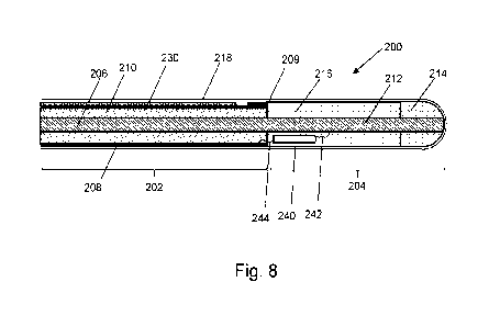
Fig. 8 is a schematic cross section view of an electrosurgical instrument, in
accordance with an embodiment;
Fig. 9 is a schematic perspective view of an electrosurgical instrument, in an
open configuration, in accordance with another embodiment;
Fig. 10 is a schematic perspective view of an underside of the electrosurgical
instrument of Fig. 9;
Fig. 11 is a schematic perspective view of the electrosurgical instrument of
Fig. 9, in a closed configuration;
Fig. 12A and 12B show opposing surfaces of an example coplanar microstrip
antenna that can be used in the electrosurgical instrument of Fig. 9; and,
Fig. 13 shows a top surface of a coplanar microstrip antenna comprising
multiple magnetostrictive ultrasound transducers, an accordance with an
embodiment.
DETAILED DESCRIPTION; FURTHER OPTIONS AND PREFERENCES
Fig. 1 shows a schematic diagram of an electrosurgical apparatus 400. The
apparatus comprises a RF channel, a microwave channel, and an electrical
signal
CA 03163184 2022-05-27
WO 2021/110847 PCT/EP2020/084490
21
channel for conveying an electrical (e.g. current) signal for driving a
magnetostrictive
ultrasound transducer to generate ultrasonic vibrations. The RF channel
contains
components for generating and controlling an RF frequency electromagnetic
signal at
a power level suitable for treating (e.g. cutting or desiccating) biological
tissue. The
microwave channel contains components for generating and controlling a
microwave
frequency electromagnetic signal at a power level suitable for treating (e.g.
coagulating or ablating) biological tissue. The electrical signal channel
contains
components for generating and controlling an electrical (e.g. current) signal
for
driving a magnetostrictive ultrasound transducer for forming ultrasound
vibrations at
a power level suitable for tissue treatment (e.g. dissecting, cutting,
dividing).
The microwave channel has a microwave frequency source 402 followed by a
power splitter 424 (e.g. a 3 dB power splitter), which divides the signal from
the
source 402 into two branches. One branch from the power splitter 424 forms a
microwave channel, which has a power control module comprising a variable
attenuator 404 controlled by controller 406 via control signal Vio and a
signal
modulator 408 controlled by controller 406 via control signal V11, and an
amplifier
module comprising drive amplifier 410 and power amplifier 412 for generating
forward microwave EM radiation for delivery from an instrument (e.g. probe or
pair of
jaws) 420 at a power level suitable for treatment. After the amplifier module,
the
microwave channel continues with a microwave signal coupling module (which
forms
part of a microwave signal detector) comprising a circulator 416 connected to
deliver
microwave EM energy from the source to the instrument along a path between its
first and second ports, a forward coupler 414 at the first port of the
circulator 416, and
a reflected coupler 418 at the third port of the circulator 416. After passing
through
the reflected coupler, the microwave EM energy from the third port is absorbed
in a
power dump load 422. The microwave signal coupling module also includes a
switch
415 operated by the controller 406 via control signal V12 for connecting
either the
forward coupled signal or the reflected coupled signal to a heterodyne
receiver for
detection.
The other branch from the power splitter 424 forms a measurement channel.
The measurement channel bypasses the amplifying line-up on the microwave
channel, and hence is arranged to deliver a low power signal from the
instrument. A
primary channel selection switch 426 controlled by the controller 406 via
control
signal V13 is operable to select a signal from either the microwave channel or
the
measurement channel to deliver to the instrument. A high band pass filter 427
is
connected between the primary channel selection switch 426 and the probe 420
to
protect the microwave signal generator from low frequency RF signals and/or
ultrasound frequency signals (produced by electrical signal supply unit 490).
The high
pass filter 427 is part of a feed structure which is described in more detail
below with
reference to Fig. 5.
CA 03163184 2022-05-27
WO 2021/110847
PCT/EP2020/084490
22
The measurement channel includes components arranged to detect the
phase and magnitude of power reflected from the instrument, which may yield
information about the material e.g. biological tissue present at the distal
end of the
instrument. The measurement channel comprises a circulator 428 connected to
deliver microwave EM energy from the source 402 to the probe along a path
between
its first and second ports. A reflected signal returned from the instrument is
directed
into the third port of the circulator 428. The circulator 428 is used to
provide isolation
between the forward signal and the reflected signal to facilitate accurate
measurement. However, as the circulator does not provide complete isolation
lo between its first and third ports, i.e. some of the forward signal may
break through to
the third port and interfere with the reflected signal, a carrier cancellation
circuit may
be used that injects a portion of the forward signal (from forward coupler
430) back
into the signal coming out of the third port (via injection coupler 432). The
carrier
cancellation circuit include a phase adjustor 434 to ensure that the injected
portion is
180 out of phase with any signal that breaks through into the third port from
the first
port in order to cancel it out. The carrier cancellation circuit also include
a signal
attenuator 436 to ensure that the magnitude of the injected portion is the
same as
any breakthrough signal.
To compensate for any drift in the forward signal, a forward coupler 438 is
provided on the measurement channel. The coupled output of the forward coupler
438 and the reflected signal from the third port of the circulator 428 are
connected to
respective input terminal of a switch 440, which is operated by the controller
406 via
control signal V14 to connect either the coupled forward signal or the
reflected signal
to a heterodyne receiver for detection.
The output of the switch 440 (i.e. the output from the measurement channel)
and the output of the switch 415 (i.e. the output from the microwave channel)
are
connect to a respective input terminal of a secondary channel selection switch
442,
which is operable by the controller 406 via control signal V15 in conjunction
with the
primary channel selection switch to ensure that the output of the measurement
channel is connected to the heterodyne receiver when the measurement channel
is
supplying energy to the instrument and that the output of the microwave
channel is
connected to the heterodyne receiver when the microwave channel is supplying
energy to the instrument.
The heterodyne receiver is used to extract the phase and magnitude
information from the signal output by the secondary channel selection switch
442. A
single heterodyne receiver is shown in this system, but a double heterodyne
receiver
(containing two local oscillators and mixers) to mix the source frequency down
twice
before the signal enters the controller may be used if necessary. The
heterodyne
receiver comprises a local oscillator 444 and a mixer 448 for mixing down the
signal
output by the secondary channel selection switch 442. The frequency of the
local
CA 03163184 2022-05-27
WO 2021/110847 PCT/EP2020/084490
23
oscillator signal is selected so that the output from the mixer 448 is at an
intermediate
frequency suitable to be received in the controller 406. Band pass filters
446, 450
are provided to protect the local oscillator 444 and the controller 406 from
the high
frequency microwave signals.
The controller 406 receives the output of the heterodyne receiver and
determines (e.g. extracts) from it information indicative of phase and
magnitude of
the forward and/or reflected signals on the microwave or measurement channel.
This information can be used to control the delivery of high power microwave
EM
radiation on the microwave channel or high power RF EM radiation on the RF
lo channel. A user may interact with the controller 406 via a user
interface 452, as
discussed above.
The RF channel shown in Fig. 1 comprises an RF frequency source 454
connected to a gate driver 456 that is controlled by the controller 406 via
control
signal V16. The gate driver 456 supplies an operation signal for an RF
amplifier 458,
which is a half-bridge arrangement. The drain voltage of the half-bridge
arrangement is controllable via a variable DC supply 460. An output
transformer 462
transfers the generated RF signal on to a line for delivery to the instrument
420. A
high pass filter 464 is connected on that line to protect the RF signal
generator from
ultrasound frequency signals produced by electrical signal supply unit 490.
The filter
464 also forms part of the feed structure, which is described below with
reference to
Fig. 5.
A current transformer 466 is connected on the RF channel to measure the
current delivered to the tissue load. A potential divider 468 (which may be
tapped off
the output transformer) is used to measure the voltage. The output signals
from the
potential divider 468 and current transformer 466 (i.e. voltage outputs
indicative of
voltage and current) are connected directly to the controller 406 after
conditioning by
respective buffer amplifiers 470, 472 and voltage clamping Zener diodes 474,
476,
478, 480 (shown as signals B and C in Fig. 1).
To derive phase information, the voltage and current signals (B and C) are
also connected to a phase comparator 482 (e.g. an EXOR gate) whose output
voltage is integrated by RC circuit 484 to produce a voltage output (shown as
A in
Fig. 1) that is proportional to the phase difference between the voltage and
current
waveforms. This voltage output (signal A) is connected directly to the
controller 406.
The electrical signal channel is provided in part by the electrical signal
supply
unit 490, an embodiment of which is shown in more detail in Fig. 2. The
electrical
signal supply unit 490 includes a signal source 500 for generating one or more
control signals (e.g. voltage signals). In an embodiment, the signal source
500 is a
microcontroller (e.g. an ArduinoTM microcontroller), a Colpitts oscillator, a
Hartley
oscillator or a 555 timer. The signal source 500 generates control signals in
the form
of low power oscillating (or alternating) signals (e.g. pulsed, square,
sinusoidal, ramp,
CA 03163184 2022-05-27
WO 2021/110847 PCT/EP2020/084490
24
trapezoidal, exponential) which drive the remaining parts of the electrical
signal
supply unit to form an electrical (e.g. current) signal having an ultrasound
frequency
for driving a magnetostrictive ultrasound transducer (e.g. on the instrument
420) so
as to generate ultrasound vibrations for treating biological tissue.
Specifically, the
signal source 500 generates two control signals, a first (e.g. positive)
control signal
which is provided to a control input of a first switching circuit 502, and a
second (e.g.
negative) control signal which is provided to a control input of a second
switching
circuit 504. Example diagrammatic representations of the first and second
control
signals are shown in Fig. 2, wherein the first control signal is a positive
square wave,
and the second control signal is a negative square wave that is 180 or it out
of
phase with the first control signal. The control signals are shown as voltage
signals in
Fig. 2 because the first and second switching circuits 502 and 504 include
voltage
controlled current sources, as is described in more detail below. However, it
is to be
understood that the control signals could be oscillating low power current
signals if,
for example, the first and second switching circuits 502, 504 include current
controlled current sources.
In an embodiment, the first control signal oscillates at an ultrasound
frequency between OV and 5V and with minimal current (e.g. <1mA), and the
second
control signal oscillates at an ultrasound frequency (e.g. the same ultrasound
frequency as the first control signal) between OV and -5V and with minimal
current
(e.g. <1mA). The first switching circuit 502 has a supply input coupled to a
first power
supply 506. Also, the second switching circuit 502 has a supply input coupled
to a
second power supply 508. In an embodiment, the first and second power supply
units
may be DC power supply units. The first switching circuit 502 provides a first
part of
an output electrical (e.g. current) signal of the electrical signal supply
unit 490.
Specifically, the first switching circuit 502 comprises a current source (e.g.
a
MOSFET, BJT, or IGFET) and uses the first supply signal received from the
first
power supply 506 to generate an oscillating current signal based on the
oscillations
of the first control signal. This is diagrammatically shown in Fig. 2 by the
positive
square wave which oscillates between a current +i Amps and 0 Amps (+i Amps may
be 100A in an embodiment). Also, the second switching circuit 504 provides a
second or remaining part of the output electric current signal of the
electrical signal
supply unit 490. Specifically, the second switching circuit 504 comprises a
current
source (e.g. a MOSFET, BJT, or IGFET) and uses the second supply signal
received
from the second power supply 508 to generate an oscillating current signal
based on
the oscillations of the second control signal. This is diagrammatically shown
in Fig. 2
by the negative square wave which oscillates between a current -i Amps and 0
Amps
(-i Amps may be -100 Amps in an embodiment).
As seen on Fig. 2, the output (i.e. the first part of the electrical signal)
from the
first switching circuit 502 and the output (i.e. the second part of the
electrical signal)
CA 03163184 2022-05-27
WO 2021/110847 PCT/EP2020/084490
from the second switching circuit 504 are combined together on a common signal
path to form a combined output electrical (e.g. current) signal from the
electrical
signal supply unit 490. Therefore, the output from the electrical signal
supply unit 490
is an electrical signal which varies at the same ultrasound frequency as the
signal
5 source 500 and between the maximum current values provided by the first
and
second switching circuits (i.e. between +i and -i). That is, the electrical
(e.g. current)
signal output from the electrical signal supply unit 490 is a compound signal
made up
from two portions: a first (e.g. positive) portion provided by the signal
source 500, the
first power supply 506 and the first switching circuit 502; and, a second
(e.g.
10 negative) portion provided by the signal source 500, the second power
supply 508
and the second switching circuit 504. It is to be understood that the positive
portion
will cause a positive H-field in the magnetostrictive ultrasound transducer
and so
cause a change in dimension in one direction, whereas the negative portion
will
cause a negative H-field in the transducer and so cause a change in dimension
in a
15 second (e.g. opposite) direction.
An advantage of having two power supplies 506, 508 and two switching
circuits 502, 504 which provide the first and second parts of the output
electrical (e.g.
current) signal is that the waveform of the output electrical signal can be
adapted to
improve performance of the ultrasound transducer. For example, the first and
second
20 control signals can be adapted to introduce a delay between the two
parts of the
output electrical signal in order to provide time for the transducer (e.g. its
coil and/or
its magnetostrictive element) to cool after being driven in one direction and
before
being driven in the opposite direction. In turn, this may increase the useable
lifespan
of the transducer and/or reduce the chances of it breaking or malfunctioning.
25 In an embodiment, the operation of the signal source may be
controlled by
the controller 406, for example, via a dedicated control signal received
therefrom.
Returning to Fig. 1, a transformer 492 is coupled at one side to the
electrical
signal supply unit 490 and at the other side to a low pass filter 494. The
transformer
492 functions to isolate the instrument 420 (and a patient) from the electric
supply
unit 490. In an embodiment, the transformer 492 may be an opto-isolator. Also,
the
transformer 492 may be absent in some embodiments. In any case, the low pass
filter 464 is connected to the RF channel by signal combiner 496, and the low
pass
filter 494 operates to protect the electrical signal supply unit from RF
signals
generated on the RF channel. The filter 494 and the signal combiner 496 also
form
part of the feed structure, which is described below with reference to Fig. 5.
The microwave/measurement channel is connected to a signal combiner 417.
Also, the signal combiner 417 is connected to the signal combiner 496 via a
low pass
filter 498, which functions to protect both the RF channel and the electrical
signal
channel from microwave energy. Further, the microwave, RF and electrical
signal are
conveyed separately or simultaneously along cable assembly 419 to the
instrument
CA 03163184 2022-05-27
WO 2021/110847
PCT/EP2020/084490
26
420. The instrument 420 delivers (e.g. radiates) microwave and/or RF energy
into the
biological tissue of a patient. Also, the instrument 420 includes a
magnetostrictive
ultrasound transducer, and the electrical signal drives the transducer to
generate
ultrasonic vibrations for tissue treatment.
It is to be understood that in some embodiments, only one of the microwave
and RF channels may be present and so the instrument may deliver only one of
RF
and microwave energy. Also, the measurement channel may be absent in some
embodiments.
Fig. 3 shows a more detailed embodiment of the electrical signal supply unit
600. The electrical signal supply unit 600 is analogous to the electrical
signal supply
unit 490 of Fig. 2, wherein like reference numerals relate to like components.
As is
clear from Fig. 3, the electrical signal supply unit 600 includes the signal
source 500.
However, in Fig. 3, the first switching circuit 502 of Fig. 2 is constructed
from a first
gate driver 602 and a first switch 604 comprising a current source.
Additionally, the
second switching circuit 504 of Fig. 2 is constructed from a second gate
driver 606
and a second switch 608 comprising a current source. In an embodiment, the
first
and second switches 604, 608 are MOSFETs (i.e. voltage controlled current
sources), for example, switch 604 may be a P-channel MOSFET and switch 608 may
be an N-channel MOSFET. Also, the gate drivers 602, 606 function to condition
the
control signals received from the signal source 500 into suitable signals for
driving
the switches 604, 608, respectively. For example, as mentioned previously, the
first
control signal is a low power oscillating positive signal which oscillates at
an
ultrasound frequency between, for example, OV and 5V and with minimal current
(e.g. <1mA). Also, the second control signal is a low power oscillating
negative signal
which oscillates at an ultrasound frequency (e.g. the same ultrasound
frequency as
the first control signal) between, for example, OV and -5V and with minimal
current
(e.g. <1mA). However, switches 604 and 608 (e.g. MOSFETs) may require a higher
voltage and current in order to operate and, therefore, the gate drivers 602
and 606
condition the control signals by increasing their voltage and/or current such
that the
conditioned signals can drive the switches 604, 608. For example, larger
signals may
be required in order to charge-up inherent capacitances of the MOSFETs. In an
embodiment, the gate driver 602 may condition the first control signal into an
oscillating signal which oscillates between OV and 15V and with a current of
1A. Also,
the gate driver 606 may condition the second control signal into an
oscillating signal
which oscillates between OV and -15V and with a current of 1A. The gate
drivers 602,
606 may be referred to as signal conditioners.
Also, in Fig. 3, the first power supply 506 of Fig. 2 includes a capacitive
circuit
C1, 02 which performs two functions. Also, the second power supply 508 of Fig.
2
includes a capacitive circuit 03, 04 which performs the same two functions.
Firstly,
the capacitive circuits (C1, 02 and 03, 04) provide power supply decoupling
that
CA 03163184 2022-05-27
WO 2021/110847 PCT/EP2020/084490
27
prevents alternating current (AC) or spikes from entering the output of the DC
power
supplies, i.e. it takes such AC currents or spikes to ground. This reduces the
chances
that the DC power supply will be damaged by such AC currents or spikes.
Secondly,
the capacitive circuits (Cl, C2 and C3, C4) cause the output electrical signal
from the
electrical signal supply unit 600 to be provided from the capacitive circuits
rather than
directly from the power supplies. In turn, this increases the current
available for
generating the output electrical (e.g. current) signal. In an embodiment,
capacitors
Cl and C3 may each have a value of 100pF, whereas capacitors C2 and C4 may
each have a value of 0.1 pF.
Fig. 4 shows an alternative embodiment of the electrical signal supply unit
700. Specifically, comparing the electrical signal supply unit 700 with the
electrical
signal supply unit 490 of Fig. 2, it is clear that the unit 700 includes a
signal source
702 which generates a single control signal for a single switching circuit
704. Also the
switching circuit 704 receives a supply signal from a single power supply 706.
As
such, the output electrical (e.g. current) signal provided by the electrical
signal supply
unit is either positive (e.g. between 0 and +i) or negative (e.g. between 0
and -i). This
is in contrast to the arrangement of Fig. 2 in which the output electrical
(e.g. current)
signal is a compound signal constructed from a two parts (e.g. a positive part
and a
negative part). Whilst the arrangement of Fig. 4 is simpler and cheaper to
manufacture, it can provide a smaller magnitude range compared to the
arrangement
of Fig. 2, since it tends to generate either a positive or a negative signal.
Additionally,
since the output electrical signal is not a combination of two control
signals, it is not
possible to use the control signals to introduce a delay between the positive
and
negative portions of the output electrical signal.
A further embodiment of the electrical signal supply unit maybe formed as a
variant of the electrical signal supply unit of Fig. 3, wherein a single gate
driver may
receive a single control signal from the signal source 500, and the single
gate driver
may condition that single control signal to drive both the switch 604 and the
switch
608. In this way, advantageously, the output electrical signal may have the
wider
current range of the arrangement of Fig. 2 (e.g. +i to -i). However, as with
the
arrangement of Fig. 4, this variant would be unable to introduce a delay
between the
positive and negative portions of the output electrical signal. That is, the
conditioned
control signal from the single gate driver must switch the switch 604 ON at
the same
time as switching the switch 608 OFF (and vice versa).
Fig. 5 shows a schematic view of a feed structure of the generator in
accordance with the embodiment of Fig. 1. The feed structure receives as
inputs a
microwave EM signal from the microwave channel, an electrical signal for
driving the
ultrasound transducer from the electrical signal channel, and an RF EM signal
from
the RF channel. The feed structure provides as an output one or more of the
three
input signals (separately or simultaneously) for onward transmission to the
CA 03163184 2022-05-27
WO 2021/110847 PCT/EP2020/084490
28
instrument 420 via the feed line 419. The feed structure includes one or more
signal
combiners and one or more filters in order to combine together these different
signals
in such a way as to avoid damage to the separate but interconnected mechanisms
for generating these different signals. Specifically, the feed structure
includes a first
signal combiner 417 which combines the microwave channel and the electrical
signal
channel into a common signal path. Additionally, the feed structure includes a
second
signal combiner 496 for combining the RF channel with the electrical signal
channel.
The RF EM signal and the electrical signal may cause damage to the mechanisms
for generating the microwave EM signal and, therefore, positioned in the
microwave
lo channel and before the signal combiner 417 is the high-pass filter 427
with a
relatively high cut-off frequency (e.g. about 300 MHz). This high pass filer
427
passes microwave EM frequency energy, but blocks the lower frequencies of the
electrical signal (which has an ultrasound frequency) and the RF signal. For
example,
a 1pF capacitor may be used as the filter 427.
Additionally, the microwave EM signal may cause damage to the mechanisms
for generating the electrical signal and the RF EM signal and, therefore,
positioned
in-between the first and second signal combiners 417, 496 is the low-pass
filter 498
with a relatively high cut-off frequency (e.g. about 300 MHz) such that it
passes the
electrical signal that has an ultrasound frequency and the RF EM signal, but
blocks
the higher frequency microwave energy. For example, one or more (e.g. three)
microwave stubs may be used, wherein the stubs are arranged to filter out a
signal
having the same frequency as the microwave energy. The stubs may be as
disclosed
in W02017103209A1.
Additionally, the RF EM signal may cause damage to the mechanisms for
generating the electrical signal and, therefore, positioned in the electrical
signal
channel and before the signal combiner 496 is the low-pass filter 494 which
has a
relatively low cut-off frequency (e.g. about 100 KHz) such that it passes the
electrical
signal having an ultrasound frequency, but blocks RF signals. For example, an
inductor may be used. Also, the electrical signal may cause damage to the
mechanisms for generating the RF EM signal and, therefore, positioned in the
RF
channel and before the signal combiner 496 is the high-pass filter 464 with a
relatively low cut-off frequency (e.g. about 100 kHz) such that it passes RF
frequency
energy but blocks the lower ultrasound frequency of the electrical signal. For
example, a 1pF capacitor may be used as the filter 464.
As such, the feed structure conveys EM energy (e.g. microwave and/or RF)
from respective EM energy supply units and conveys the electrical signal from
the
electrical signal supply unit. Also, the feed structure includes a common
signal
pathway for conveying the EM energy and the electrical signal to the output
port for
onward transmission to the instrument 420. Moreover, the feed structure
includes a
circuit or network of signal combiners and filters which function to ensure
that these
CA 03163184 2022-05-27
WO 2021/110847 PCT/EP2020/084490
29
separate and different input signals do not damage the various separate and
different
mechanisms for producing those signals.
Fig. 6A illustrates a magnetostrictive ultrasound transducer in accordance
with an embodiment. The transducer includes a housing 802 within which is
located a
coiled conductor 804 which is wrapped around an element 806 of
magnetostrictive
material. The element 806 may be substantially elongate or rod shaped, and may
be
referred to as a solenoid. In an embodiment, the element 806 may be
approximately
the same length as the coil, or just longer than the coil, for example, 1cm or
1.2cm.
Also, the element 806 may be substantially cylindrical and have a diameter of
about
lo 0.5cm or 0.6cm. The coiled conductor 804 is connected at a first end to
a first
terminal 808 of the transducer 800, and at a second end to a second terminal
810 of
the transducer 800. In use, the first terminal 808 and the second terminal 810
are
connected to the cable assembly 419 of Fig. 1 in order that the electrical
signal is
provided to the transducer 800 so that the transducer 800 generates ultrasonic
vibrations. Specifically, magnetostriction is a property of ferromagnetic
materials
which causes them to expand or contract (i.e. change their physical
dimensions) in
response to a magnetic field (H-field). This effect allows magnetostrictive
materials to
convert electromagnetic energy into mechanical energy. As a magnetic field is
applied to the material, its molecular dipoles and magnetic field boundaries
rotate to
align with the field. This causes the material to strain and elongate.
Fig. 6B shows an example magnetic hysteresis loop. A magnetic hysteresis
loop is produced when a ferromagnetic material is magnetised in one direction,
until it
reaches a saturation point (e.g. point 824 on Fig. 6B), then the magnetic
field
strength is demagnetised in the opposite direction to the reverse saturation
point
(e.g. point 826 on Fig. 6B). When the magnetic field direction is alternated,
a loop is
formed which goes back and forth from the saturation point and reverse
saturation
point. This loop can be viewed in Fig. 6B. The magnetising field (H) provides
the
required stress to produce the required change in magnetisation (M or B) or
the
strain. The magnetisation (M) of the ferromagnetic material when the magnetic
field
strength (H) is at zero is called remanence. As the magnetisation falls back
to zero,
the magnetic field strength (H) needed to demagnetise after saturation is
called the
coercivity. The point marked 820 can be referred to as the remanence point and
is a
measure of the remaining magnetisation when the driving field is dropped to
zero.
This illustrates that when the driving magnetic field drops to zero, the
ferromagnetic
material retains a considerable degree of magnetisation. The point marked 822
can
be referred to as the coercivity point and is a measure of the reverse field
needed to
drive the magnetisation to zero after being saturated. This illustrates that
the driving
magnetic field must be reversed and then increased to drive the magnetisation
to
zero again. Point 824 indicates a point at which the ferromagnetic material is
magnetised to saturation by alignment of magnetic domains in one direction
(e.g.
CA 03163184 2022-05-27
WO 2021/110847 PCT/EP2020/084490
north pole), whereas the point 826 indicates a point at which the
ferromagnetic
material is magnetised to saturation by alignment of magnetic domains in the
opposite direction (e.g. south pole). A width 828 of the hysteresis loop
indicates if a
material retains a large or small fraction of the saturation field when the
driving field is
5 removed. The width may vary between different magnetostrictive materials.
A narrow
hysteresis loop implies that a small amount of dissipated energy is repeatedly
reversing the magnetisation. Embodiments are directed to the use of
magnetostrictive materials in the production of ultrasound vibrations. Given
the
relatively high frequency of ultrasound vibrations, the switching between
saturation
10 points must occur relatively quickly, e.g. between 20,000 and 5,000,000
times per
second (i.e. 20 kHz to 5 MHz). Therefore, an aim is to keep the hysteresis
loop as
narrow as possible (i.e. to keep width 828 as low as possible) so that the
change (or
switch) of the magnetisation occurs at a much faster rate and so that energy
dissipation and subsequent heating remains small.
15 Terfenol-D has been selected as a particularly suitable
magnetostrictive
material from which to make the element 806. Terfenol-D is a good material to
use
because it possesses a large magnetisation and magnetostriction at room
temperature, which is about 2400ppm, which occurs due to rhombohedral
distortion
of the lattice structure of the Terfenol-D material. That is, Terfenol-D,
relative to other
20 magnetostrictive materials, produces a large strain (i.e. change in
physical
dimension) for a given stress (i.e. applies change in magnetic driving field).
For
example, Galfenol has a magnetostriction of about 400ppm and Alfenol has a
magnetostriction of about 200ppm. However, since Terfenol-D is a relatively
expensive material, the element 806 may be constructed from two portions: a
first
25 portion that is substantially the same length as the coil and is made
from Terfenol-D,
and a second portion that is an extension of the first portion and is made
from a
cheaper material, such as, steel.
The following provides some example calculations to determine a possible
structure of the coil 804 and element 806, in accordance with an embodiment.
30 It is known that the magnetic field strength (H) can be found from
equation (1)
below:
NI
H= ¨ (1)
where N refers to the number of turns of wire on the coil 804, I is the
current
of the electrical signal used to drive the transducer 800, and L is the length
of the coil
804 (labelled "L" in Fig. 6A).
In an embodiment, it may be considered that a magnetic field strength of
2000 Oersted (Oe) will produce a maximum strain of 2400ppm for a certain
Terfenol-
CA 03163184 2022-05-27
WO 2021/110847 PCT/EP2020/084490
31
D magnetostrictive element 806. If 10e is equivalent to 79.58A/m, then 2000 Oe
is
equivalent to 159,155A/m.
From equation (1), if a current of 100A is driven into the coil 804, then N/L
=
1,592. N/L is the ratio of the number of turns of wire on the coil 804 divided
by the
length (L) of the coil 804. If the length of the coil is 1cm (0.01m), then the
number of
turns required will be: (N = H*L)/I = (159155*0.01)/100 = 15.9 turns ¨ i.e.
about 16
turns.
In an embodiment, the coil is made from copper wire with an outer diameter of
0.5mm, and so it is possible to get 20 turns within a 1cm length and a single
layer
winding. Additionally, it is possible to have multi-layer windings. However,
it should
be noted that the number of turns in an opposite direction should be minimised
in
order to avoid having a cancelling-out effect. For example, a double layer
winding for
providing 20 turns could include 11 turns in a first direction (layer 1), a
single return
turn (which would cancel out one of the original turns ¨ i.e. the 11 turns
reduces to 10
turns) (layer 2), followed by 10 more turns in the first direction (layer 3).
Using multi-
layer windings in this manner makes it possible to reduce the coil length
required
and, therefore, reduce the overall size of the transducer 800. For example,
the coil
length could be reduced to 0.5cm or less. Another mechanism for reducing the
coil
length required and, therefore, reduce the overall size of the transducer 800,
is to
increase the current of the driving electrical signal applied to the input
terminals, 808,
810 of the transducer 800. For example, considering the arrangement of Fig. 3,
this
could be done by increasing the size of the power supplies (e.g. +VDD,-VDD)
and the
size of the current sources (e.g. MOSFETs).
Fig. 7 is a schematic diagram of a complete electrosurgery system (or
apparatus) 100 that is capable of supplying RF energy, microwave energy, or an
electrical signal for driving a magnetostrictive ultrasound transducer to
generate
ultrasound vibrations. The system 100 comprises a generator 102 for
controllably
supplying RF energy, microwave energy, and the electrical signal suitable for
driving
a magnetostrictive ultrasound transducer to generate ultrasound vibrations. In
an
embodiment, the generator 102 is the same as generator 400 described above
with
reference to Figs. 1 to 5.
The generator 102 is connected to an interface joint 106 by an interface cable
104. If needed, the interface joint 106 can house an instrument control
mechanism
that is operable by sliding a trigger 110, e.g. to control longitudinal (back
and forth)
movement of one or more control wires or push rods (not shown). If there is a
plurality of control wires, there may be multiple sliding triggers on the
interface joint to
provide full control. The function of the interface joint 106 is to combine
the inputs
from the generator 102 and instrument control mechanism into a single flexible
shaft
112, which extends from the distal end of the interface joint 106.
CA 03163184 2022-05-27
WO 2021/110847 PCT/EP2020/084490
32
The flexible shaft 112 is insertable through the entire length of an
instrument
(working) channel of a surgical scoping device 114, such as an endoscope,
laparoscope, bronchoscope, gastroscope or the like.
The surgical scoping device 114 comprises a body 116 having a number of
input ports and an output port from which an instrument cord 120 extends. The
instrument cord 120 comprises an outer jacket which surrounds a plurality of
lumens.
The plurality of lumens convey various things from the body 116 to a distal
end of the
instrument cord 120. One of the plurality of lumens is an instrument channel.
Other
lumens may include a channel for conveying optical radiation, e.g. to provide
lo illumination at the distal end or to gather images from the distal end.
The body 116
may include an eye piece 122 for viewing the distal end. In order to provide
illumination at the distal end, a light source 124 (e.g. LED or the like) may
be
connected to the body 116 by an illumination input port 126.
The flexible shaft 112 has a distal assembly 118 (not drawn to scale in Fig.
7)
that is shaped to pass through the instrument channel of the surgical scoping
device
114 and protrude (e.g. inside the patient) at the distal end thereof. The
distal end
assembly includes an active tip for delivering microwave energy into
biological tissue
as discussed herein. The distal assembly 118 may be analogous to the
instrument
420 of Fig. 1, and the flexible shaft 112 may be analogous to the feed line
419 of Fig.
1.
The structure of the distal assembly 118 discussed below may be designed to
have a maximum outer diameter equal to or less than 2.0 mm, e.g. less than 1.9
mm
(and more preferably less than 1.5 mm) and the length of the flexible shaft
can be
equal to or greater than 1.2 m.
The body 116 includes a power input port 128 for connecting to the flexible
shaft, which comprises a coaxial cable (e.g. a conventional coaxial cable)
capable of
conveying the microwave energy, RF energy and an electrical signal from the
generator 102 to the distal assembly 118. Alternatively, different means may
be
provided to conveying one or more of these signals. For instance, the
microwave
energy may be conveyed by the coaxial cable, but the electrical signal and/or
the RF
energy may be conveyed by a twisted cable pair or the like. Coaxial cables
that are
physically capable of fitting down the instrument channel of a surgical
scoping device
are available with the following outer diameters: 1.19 mm (0.047"), 1.35 mm
(0.053"),
1.40 mm (0.055"), 1.60 mm (0.063"), 1.78 mm (0.070"). Custom-sized coaxial
cables
(i.e. made to order) may also be used.
As discussed above, it is desirable to be able to control the position of at
least
the distal end of the instrument cord 120. The body 116 may include a control
actuator 130 that is mechanically coupled to the distal end of the instrument
cord 120
by one or more control wires (not shown), which extend through the instrument
cord
120. The control wires may travel within the instrument channel or within
their own
CA 03163184 2022-05-27
WO 2021/110847 PCT/EP2020/084490
33
dedicated channels. The control actuator 130 may be a lever or rotatable knob,
or
any other known catheter manipulation device. The manipulation of the
instrument
cord 120 may be software-assisted, e.g. using a virtual three-dimensional map
assembled from computer tomography (CT) images.
The coaxial cable for delivering the microwave radiation to the target site
should be low-loss, have a small cross-section and be flexible. The cable
should be
low loss to avoid or reduce heating during treatment and so that there is
enough
power at the distal end to produce the desired radiation from the antenna.
If the cable is not separated from the body by the use of a sealed scoping
lo device, catheter or other protective sheath, then the cable should be
made of, or be
coated with, a biologically inert material to avoid unwanted interaction with
the body.
A preferred cable type is a coaxial cable which is made up of an inner
conductor axially surrounded by a dielectric sheath which is in turn axially
surrounded
by an outer conductor. The radiating portion in an antenna produced from such
a
cable may be made up of a section of inner conductor and dielectric sheath
which
protrudes from the end of the outer conductor of the coaxial cable.
In an embodiment, the outer conductor of the coaxial cable may be as
physically thick as possible to increase its thermal mass and heat capacity.
In this
way, all or a majority of the heat generated in the cable due to conveying
microwave
energy can be held within the structure of the cable rather than, for example,
being
leaked inside the patient. In an embodiment, the outer conductor may be 0.5 mm
thick.
The invention also seeks to provide an antenna with a well-defined radiation
pattern. It is desirable that a practitioner would be able to select an
instrument for the
treatment of a specific area of tissue, such that the radiation of target
tissue is
maximised and the radiation of healthy tissue is minimised. For example, in
some
circumstances it can be desirable to produce a generally spherically symmetric
radiation pattern with a substantially uniform power absorption distribution,
so that
the amount of radiation received by an area of tissue can be more easily
controlled
by the practitioner.
It is also preferable that the instrument can be operated alongside other
instruments to enable practitioners to receive information from the target
site. For
example, a scoping device may aid the steering of the instruments around
obstacles
within a patient's body. Other instruments may include a thermometer or
camera.
In the following description, unless stated otherwise, the length of a
component refers to its dimension in the direction parallel to the
longitudinal axis of
the coaxial cable.
Fig. 8 is a cross-sectional view of the distal end of an electrosurgical
instrument 200 that is an embodiment of the invention. The electrosurgical
instrument
200 may include the distal assembly 118 of Fig. 7, or the instrument 420 of
Fig. 1.
CA 03163184 2022-05-27
WO 2021/110847
PCT/EP2020/084490
34
The electrosurgical instrument 200 may therefore be used to deliver microwave
energy and/or RF energy into biological tissue for tissue treatment. Also, the
instrument 200 includes a magnetostrictive ultrasound transducer for
converting an
electrical (e.g. current) signal into ultrasonic vibrations in biological
tissue for tissue
treatment. The electrosurgical instrument 200 comprises a coaxial cable 202
that is
connected at its proximal end to an electrosurgical generator (e.g. generator
400 of
Fig. 1 or generator 102 of Fig. 7) in order to convey microwave energy. The
coaxial
cable 202 comprises an inner conductor 206, which is separated from an outer
conductor 208 by a first dielectric material 210. The coaxial cable 202 is
preferably
low loss for microwave energy. A choke (not shown) may be provided on the
coaxial
cable to inhibit back propagation of microwave energy reflected from the
distal end
and therefore limit backward heating along the device.
The device may include a temperature sensor at the distal end. For example,
in Fig. 8 a thermocouple 230 is mounted on the outer conductor to transmit a
signal
back to the proximal end that is indicative of temperature at the distal end
of the
instrument.
Other techniques for temperature monitoring can be used. For example, one
or more micromechanical structures whose physical configuration is sensitive
to
temperature may be mounted in the distal portion of the device, e.g. in or on
the
outer sheath discussed below. These structures can be interfaced with an
optical
fibre, whereby changes in a reflected signal caused by movement of the
structure
can be indicative of temperature changes.
The coaxial cable 202 terminates at its distal end with a radiating tip
section
204. In this embodiment, the radiating tip section 204 comprises a distal
conductive
section 212 of the inner conductor 206 that extends beyond a distal end 209 of
the
outer conductor 208. The distal conductive section 212 is surrounded at its
distal
end by a dielectric tip 214 formed from a second dielectric material, which is
different
from the first dielectric material 210. The length of the dielectric tip 214
is shorter
than the length of the distal conductive section 212. An intermediate
dielectric sleeve
216 surrounds the distal conductive section 212 between the distal end of the
coaxial
cable 202 and the proximal end of the dielectric tip 214. The intermediate
dielectric
sleeve 216 is formed from a third dielectric material, which is different from
the
second dielectric material but which may be the same as the first dielectric
material
210.
In this embodiment, the coaxial cable 202 and radiating tip section 204 have
an outer sheath 218 formed over their outermost surfaces. The outer sheath 218
may be formed from a biocompatible material. The outer sheath 218 has a
thickness
that is small enough to ensure that it does not significantly interfere with
the
microwave energy radiated by the radiating tip section 204 (i.e. radiating
pattern and
return loss). In an embodiment, the sheath is made from PTFE, although other
CA 03163184 2022-05-27
WO 2021/110847 PCT/EP2020/084490
materials are also appropriate. The thickness of the wall of the sheath is
selected to
withstand breakdown voltages equal to or greater than 200 kV/m.
The purpose of the dielectric tip 214 is to alter the shape of the radiated
energy. The second dielectric material is selected to reduce the wavelength of
the
5 microwave energy, which results in the radiated energy exhibiting a more
spherical
radiation pattern. To do this, the second dielectric material preferably has a
large
dielectric constant (relative permittivity Er). The dielectric constant of the
second
dielectric material is preferably chosen to enable the length of the
dielectric tip 214 to
be minimised whilst still constituting a non-negligible portion of a
wavelength of the
10 microwave energy when it propagates through the second dielectric
material. It is
desirable for the dielectric tip 214 to be as short as possible in order to
retain
flexibility in the device, especially if the second dielectric material is
rigid. In an
embodiment, the dielectric tip 214 may have a length equal to or less than 2
mm.
The dielectric constant of the second dielectric material may be greater than
80, and
15 is preferably 100 or more at the frequency of the microwave energy. The
second
dielectric material may be TiO2 (titanium dioxide).
The wavelength of radiation in a material becomes shorter as the dielectric
constant of the material increases. Therefore a dielectric tip 214 with a
greater
dielectric constant will have a greater effect on the radiation pattern. The
larger the
20 dielectric constant, the smaller the dielectric tip 214 can be while
still having a
substantial effect on the shape of the radiation pattern. Using a dielectric
tip 214 with
a large dielectric constant means that the antenna can be made small and so
the
instrument can remain flexible. For example the dielectric constant in TiO2 is
around
100. The wavelength of microwave radiation having a frequency of 5.8 GHz is
about
25 6 mm in TiO2 compared to around 36 mm in PTFE (which may be the material
used
for the first and/or third dielectric materials). A noticeable effect on the
shape of the
radiation pattern can be produced in this arrangement with a dielectric tip
214 of
approximately 1 mm. As the dielectric tip 214 is short, it can be made from a
rigid
material whilst still maintaining flexibility of the antenna as a whole.
30 The dielectric tip 214 may have any suitable distal shape. In Fig. 8
it has a
dome shape, but this is not necessarily essential. For example, it may be
cylindrical,
conical, etc. However, a smooth dome shape may be preferred because it
increases
the mobility of the antenna as it is manoeuvred through small channels (e.g.
inside
blood vessels). The dielectric tip 214 may be coated with a non-stick material
such as
35 Parylene C or Parylene D, or PFTE to prevent the tissue from sticking to
the
instrument. The whole instrument can be coated in this way.
The properties of the intermediate dielectric sleeve 216 are preferably chosen
(e.g. through simulation or the like) so that the radiating tip section 204
forms a
quarter wave impedance transformer for matching the input impedance of the
generator into a biological tissue load in contact with the radiating tip
section 204.
CA 03163184 2022-05-27
WO 2021/110847 PCT/EP2020/084490
36
During treatment, the surrounding tissue absorbs the radiated energy. The
volume of tissue into which the energy is delivered depends on the frequency
of the
microwave energy.
As seen on Fig. 8, the radiating tip portion 204 includes magnetostrictive
transducer 240 which is coupled to the inner conductor 206 (e.g. directly or
via distal
conductive section 212) by a first connector 242 and which is coupled to the
outer
conductor 208 by a second connector 244. In the embodiment of Fig. 8, the
transducer 240 is encased (e.g. partly or completely) in the intermediate
dielectric
sleeve 216; however, it is to be understood that the transducer 240 may
instead be
lo positioned on an inner or outer surface of the intermediate dielectric
sleeve 216.
Further, the transducer 240 may alternatively be positioned elsewhere on the
radiating tip portion, for example, in or on the radiating tip 214. In an
embodiment, the
transducer 240 has the same or similar construction to the transducer 800 of
Fig. 6A.
In use, an electrical (e.g. current) signal for driving the transducer 240 may
be
introduced into the coaxial cable 202 at its proximal end (e.g. by generator
102, or
generator 400). As described above with reference to Figs. 1 to 5, the
electrical
signal may oscillate at an ultrasound frequency. On receiving this electrical
signal,
the coil of the transducer 240 induces an oscillating magnetic field around
the
magnetostrictive element the transducer 240 such that the magnetostrictive
effect
causes the magnetostrictive element to rapidly expand and contract at the
ultrasonic
frequency thereby generating ultrasonic vibrations. Since the transducer 240
is
coupled to the radiating tip portion 204, these ultrasonic vibrations travel
into the
radiating tip portion and are then radiated out of the instrument 200 and into
the
surrounding biological tissue. The vibrations produce mechanical friction that
generates thermal energy thereby resulting in extracellular heating followed
subsequently by intracellular heating. In this way, the ultrasound vibrations
can be
used to treat (e.g. cut or coagulate) the biological tissue.
A further embodiment of an electrosurgical instrument will now be described
with reference to Figs. 9 to 12B, wherein the instrument comprises an
electrosurgical
vessel sealer device capable of delivering microwave energy, RF energy and
ultrasonic vibrations to seal blood vessels. The electrosurgical vessel sealer
may
include the distal assembly 118 of Fig. 7, or the instrument 420 of Fig. 1.
The
electrosurgical vessel sealer may be used in open surgery, but may find
particular
use in procedures where there is restricted access to the treatment site. For
example, the electrosurgical vessel sealer may be adapted to fit within the
instrument
channel of a surgical scoping device i.e. laparoscope, endoscope, or the like,
as
described above with reference to the scoping device of Fig. 7.
Fig. 9 shows a schematic perspective view of a distal end assembly 300 of an
electrosurgical instrument that is an embodiment of the invention. The distal
end
assembly 300 is connected to an instrument shaft 302 which is dimensioned to
fit
CA 03163184 2022-05-27
WO 2021/110847
PCT/EP2020/084490
37
within the instrument channel of a laparoscope or other surgical scoping
device. The
instrument shaft 302 comprises a tubular sheath that conveys a coaxial cable
for
carrying EM power (e.g. microwave and/or RF) to the distal end assembly
together
with various control wires or rods that are arranged to control physical
manipulation
of the distal end assembly, as discussed below.
In this example, the distal end assembly 300 comprises a pair of jaws 308,
310. The jaws 308, 310 are operably coupled to a collar 304 that is mounted on
a
distal end of the instrument shaft 302. In this example, the pair of jaws 308,
310
comprise a movable jaw 308 which is pivotal around a laterally extending pin
306 in
the collar 304 to enable a gap between opposing inner surfaces of the jaws
308, 310
to be opened and closed. Although there is only one movable jaw in this
example, in
other embodiments, both jaws may be arranged to pivot relative to the collar
304.
The collar 304 may be arranged to ensure that the jaws remain laterally
aligned as
they are moved together.
In the example shown in Fig. 9, the pair of jaws 308, 310 comprises a static
jaw 310 that has an energy delivery structure 312 on its top surface, i.e. the
surface
that opposes a corresponding surface on the movable jaw 308. In use, the
distal end
assembly 308 is intended to grip biological tissues (and in particular a blood
vessel)
between the pair of jaws 308, 310. The pair of jaws 308, 310 are arranged to
apply
pressure to the biological tissue between the opposed surfaces and deliver
energy
(preferably microwave electromagnetic energy) into the tissue from the energy
delivery structure 312.
In this embodiment, the energy delivery structure is present only on the
static
jaw 310. However, in other arrangements, there may be an energy delivery
structure
on both jaws, or only on a single movable jaw.
In this example, the energy delivery structure 312 comprises a coplanar
microstrip antenna fabricated in the top surface of the status jaw 310. The
coplanar
microstrip antenna comprises a substrate 320 made of nonconductive dielectric
material, e.g. ceramic or the like. The dielectric substrate 320 has a
conductive layer
fabricated on its underside (not visible in Fig. 9). On its top surface (i.e.
the surface
opposite the underside) the dielectric substrate 320 has a first conductive
region in
the form of a longitudinally extending finger electrode 314 disposed centrally
thereon.
A U-shaped second conductive region 316 is disposed on the top surface of the
dielectric substrate 320 around the finger electrode 314 with a gap of exposed
dielectric 315 separating the finger electrode 314 from the U-shaped region
316. A
plurality of through holes 318 are formed, e.g. machined, through the U-shaped
region 316 and dielectric substrate 315. The through holes 318 are filled with
conductive material to electrically connect the conductive layer on the
underside of
the dielectric substrate 320 with the U-shaped conductive region 316. The
finger
electrode 314 has a contact pad 317 at a proximal end thereof. The inner
conductor
CA 03163184 2022-05-27
WO 2021/110847 PCT/EP2020/084490
38
of the coaxial cable conveyed by the instrument shaft 302 is electrically
coupled to
the contact pad 317, e.g. by extending from the instrument shaft 302 to
physically
contact the contact pad 317. The finger electrode 314 provides an active
region for
the coplanar microstrip antenna. The conductive layer on the underside of the
dielectric substrate 320 is electrically connected to an outer conductor of
the coaxial
cable conveyed by the instrument shaft 302. In conjunction with the conductive
communication through the through holes 318, the U-shaped conductive region
310
forms a ground electrode for the coplanar microstrip antenna.
The configuration of the coplanar microstrip antenna shown in Fig. 9 is
lo particularly advantageous because it confines the emitted field within
the region
defined by the pair of jaws 308, 310. As discussed below, very little energy
is
delivered to a region outside the pair of opposing surfaces. Moreover, by
arranging
the U-shaped conductive region 316 to extend around a distal end of the finger
electrode 314, the coplanar microstrip antenna structure can prevent energy
from
escaping in the longitudinal direction distal to assembly 300.
The conductive layers mentioned above may be made from any suitable
conductive material. Silver and gold are preferred because of their high
conductivity
and biocompatibility. Copper may also be used, although it is preferably
plated with
silver or gold in regions likely to contact biological tissue.
The coplanar microstrip antenna structure may be fabricated independently of
the static jaw 310, e.g. using thin film deposition techniques. This
construction of the
coplanar microstrip antenna ensures two important performance features.
Firstly, it
ensures that the projected energy applied to the biological tissue of the
gripped
vessel is focused inwardly within the grasp of the instrument jaws. This
provides a
localised energy delivery effect, whereby the applied energy is efficiently
delivered to
a desired region of tissue.
Moreover, the use of thin film conductive layers means that the thermal mass
of the conductive lines is minimal. In combination with the effective thermal
barrier
provided by the dielectric substrate 320, this means that any residual heat
within the
conductive lines quickly dissipates. The effect can be further enhanced by
providing
a layer on the surface opposing the coplanar microstrip antenna that also acts
as a
thermal barrier. In the embodiments shown in Fig. 9, the moveable jaw 308 has
a
layer of resiliently deformable material 322 formed on its inner surface. The
layer
322 may be formed from silicone rubber or other compliant polymer material
that can
withstand the temperatures that occur during treatment and are biocompatible.
They
may be fabricated from an elastomeric thermoplastic polymer, for example. This
layer both assists in efficient delivery of energy to gripped biological
tissue, but also
facilitates retaining the biological tissue within the jaws.
Alternatively or additionally, a coating may be applied to the surface of the
coplanar microstrip antenna itself. This may be a coating applied only to the
CA 03163184 2022-05-27
WO 2021/110847 PCT/EP2020/084490
39
conductive regions, e.g. to minimise tissue sticking. In embodiments arranged
to
deliver microwave energy, it may not be necessary for the inner surfaces of
the jaws
to make direct electrical conductive contact with tissue. Accordingly, the
coating may
be a thin high temperature polymeric material, e.g. applied across the whole
face of
the antenna. The specific material may be chosen to exhibit high loss and
appear
transparent to the microwave energy.
The coating may conform to the shape of the jaws. It may comprise a
silicone-based passivation material similar to that used as a protective
coating on
printed circuit boards. Other examples include polyimide, PTFE or FEP type
materials.
As shown in Fig. 9, the layer 322 has a plurality of ridges moulded into it.
It
therefore presents a textured or toothed surface with which to contact
biological
tissue. A similar ridged or textured grip may be provided around the periphery
of the
coplanar microstrip antenna. As mentioned above, these textured surfaces can
aid
the release of gas during the vessel sealing operation.
The coplanar microstrip antenna has a size suitable for receiving and sealing
biological vessels. For example, the coplanar microstrip antenna may be
arranged to
provide an effective treatment area having a width (i.e. dimension extending
laterally
with respect to the axis of the coaxial cable) of 2 to 5 mm and a length
(along the axis
of the device) of 15 to 26 mm.
Operation of the instrument 300 may be controlled by an actuation
mechanism (e.g. the trigger 110 of Fig. 7), which may take the form of a
scissor-type
handle, slider, rotatable dial, level, trigger or the like. The actuation
mechanism can
be operably coupled to the instrument 300 via one or more control wires that
extend
along the instrument shaft 302, e.g. within the instrument channel of a
scoping
device. In one example, the actuation mechanism may include a force limiter
arranged to limit the maximum actuation force that can be supplied to the
instrument.
Limiting the maximum actuation force may assist in preventing damage to
delicate
components in the instrument 300, and can ensure that the force applied to
tissue
remains within desired parameters. The force limited may comprise a
compression
spring or ratchet mechanism as part of the actuation mechanism. In some
examples
it may be desirable to vary the maximum actuation force, e.g. by provide a
dial or
switch on the interface joint 106 that adjusts the maximum actuation force
associated
with the actuation mechanism.
The pair of jaws may include a stand-off (not shown) that ensures that the
jaws remain separated by a minimum distance irrespective of the closure force
applied by an associated actuation mechanism located at the proximal end of
the
instrument shaft 302. The stand-off may be a physical projection on one or
both jaws
that engages the inner surface of the opposite jaw.
CA 03163184 2022-05-27
WO 2021/110847 PCT/EP2020/084490
It is desirable for the pressure applied by jaws to tissue held therebetween
to
be uniform in a longitudinal direction along the inner surfaces of the jaws.
In a
development of the structure shown in Fig. 9, the movable jaw 308 may comprise
an
engagement plate at its inner surface that is capable of articulating back
into the jaw
5 308 about a pivot point located at a distal end of the jaw 308. A
resiliently
deformable support element may be mounted in the jaw 308 behind the engagement
plate to urge it outwardly. With this arrangement, tissue in the region
between the
jaws is grasped between the inner surface of the static jaw and the engagement
plate
of the movable jaw. As the jaws are closed, the pressure applied along the
jaws is
lo generated by a combination of the pivoting action of the jaws and the
articulation of
the engagement plate. The location of the pivot point and properties of the
resiliently
deformable support element can be selected so that the non-uniformity in
applied
force that arises changing mechanical advantage along the jaws away from the
pivot
is balanced by a cooperating non-uniformity arising from the pivotable
articulation of
15 the engagement plate.
The energy delivery structure 312 described with respect to Fig. 9 is a
coplanar microstrip antenna. The configuration of that antenna may be as shown
in
Fig. 9, however, alterative microwave radiator structures can be used. For
example
the top surface of the static jaw 310 may be provided with other microstrip-
based
20 energy delivery configurations, e.g. meandering or interdigitated
microstrip lines. In
another embodiment, the energy delivery structure may be a travelling wave
antenna.
In addition to the function of the vessel sealing, the electrosurgical
instrument
of the present invention also functions as a vessel divider, e.g. to cut
through and
25 separate a sealed section of a blood vessel. In one embodiment, the
vessel sealer
may be provided with a blade 326 that is slidably mounted with respect to the
pair of
jaws 308, 310 to cut through biological tissue held between the jaws. In Fig.
9, the
blade 326 is shown as protruding into the region between the open jaws in Fig.
9.
However, in practice, it is desirable for the instrument to prevent forward
movement
30 of the blade until after the jaws are closed and microwave energy is
applied.
In the embodiment shown in Fig. 9, the blade 326 is movable in a longitudinal
direction, e.g. along the axis of the device. The opposed surfaces of the jaws
308,
310 contain respective recess or guide grooves 328, 324 for receiving the
blade as it
travels. The guide groove 324 in the static jaw 310 is formed within the
finger
35 electrode 314 so that it moves through the centre of the applied field.
In other embodiments, the blade may be mounted within one of the jaws and
arranged to move laterally with respect to the longitudinal direction, i.e. to
extend out
of one of the opposed surfaces into gripped tissue. The sharp edge of the
blade may
lie below the opposed surface during the vessel gripping and sealing
operation.
CA 03163184 2022-05-27
WO 2021/110847 PCT/EP2020/084490
41
In one embodiment, the cutting functionality of the blade is provided or
enhanced by a magnetostrictive ultrasound transducer (not visible in Fig. 9).
The
transducer may have the same construction as the transducer 800 of Fig. 6A.
The
location of the transducer may vary between embodiments, but generally
speaking it
is located towards a proximal end of the blade 326 so that when the transducer
generates ultrasonic vibrations, those vibrations travel along the blade 326
to enable
or assist in the cutting action of the blade 326. In an embodiment, the
transducer is
connected to a proximal end portion of the blade 326. Where the electric drive
signal
for the transducer is conveyed by the coaxial cable in the instrument shaft
302, the
transducer has a first input terminal coupled to one conductor (e.g. inner
conductor)
of the coaxial cable and a second input terminal coupled to the other
conductor (e.g.
outer conductor) of the coaxial cable. For example, first and second
connectors (e.g.
wires, tracks, cables, and conductors) may couple each terminal to its
respective
conductor of the coaxial cable. In this way, an electrical (e.g. current)
signal for
driving the magnetostrictive ultrasound transducer may be conveyed by the
coaxial
cable, and delivered to the transducer at the proximal end of the blade 326.
In
operation, the transducer may generate ultrasonic vibrations which enable or
enhance a cutting action of the blade 326. Specifically, as described above
with
reference to Figs. 1 to 5, the electrical signal may oscillate at an
ultrasound
frequency. On receiving this electrical signal, the coil of the transducer
induces an
oscillating magnetic field around the magnetostrictive element the transducer
such
that the magnetostrictive effect causes the magnetostrictive element to
rapidly
expand and contract at the ultrasonic frequency thereby generating ultrasonic
vibrations. Since the transducer is coupled to the blade 326, these ultrasonic
vibrations travel into the blade 326 and are then radiated into the tissue
surrounding
the blade 326. The vibrations produce mechanical friction that generates
thermal
energy thereby resulting in extracellular heating followed subsequently by
intracellular heating. In this way, the ultrasound vibrations can be used to
treat (i.e.
cut) the biological tissue.
The distal end assembly may be configured to perform functions in addition to
vessel sealing. For example, the distal end assembly may have an auxiliary
radiofrequency (RF) cutting blade mounted on a distal tip thereon. In the
example
shown in Fig. 9, an RF dissector element 330 is mounted on the distal end of
the
static jaw 310. The RF dissector element 330 is a bipolar structure that
comprises an
active electrode mounted on a protruding body, and a return electrode, which
may be
fabricated on or integrated with the static jaw 310 in the vicinity of the
protruding
body.
Fig. 10 shows the underside of the distal end assembly 300, where the RF
dissector element 330 can be seen in more detail. The RF dissector element 330
can be used for fine bloodless tissue cutting and tissue dissection. In the
CA 03163184 2022-05-27
WO 2021/110847 PCT/EP2020/084490
42
arrangement shown in Figs. 9 and 10, the RF dissector element 330 presents a
leading edge that sits proud of the distal end of the static jaw 310 This
position can
enable both side and end-on dissection to be performed. In dry field treatment
scenarios (i.e. in the absence of saline or other electrically conductive
fluid) it is
desirable for the return electrode to be in close proximity to the active
electrode that
is on the RF dissector element 330. The ratio of the exposed tissue contacting
electrode areas is also important to ensure that current flow occurs in a
desired
manner that causes maximum current density to occur on the leading edge of the
RF
dissector element 330.
Although the RF dissector element 330 is shown at the distal end of the static
jaw in Figs. 9 and 10, it can be mounted in a variety of orientations or
locations on
the distal end assembly, e.g. vertically, horizontally, at an angle, on one
side, and on
either jaw.
The pair of jaws may have any suitable shape. For example, the jaws may be
tapered along their length towards the distal tip, or may be bent or hooked if
desired
for any particular treatment scenario.
Opening and closing of the jaws 308, 310 may be controlled by an actuation
mechanism that is operable by a user at an external handle of the surgical
scoping
device, i.e. at a proximal end of the instrument shaft 302 (e.g. trigger 110
of interface
joint 106). The actuation mechanism may include a pressure control device
arranged
to enable a user to control closure of the pair of jaws based on an amount of
pressure applied to the biological tissue that is captured between the jaws.
In one
example, a user may select a desired (e.g. maximum) closure pressure for the
jaws,
and the actuation mechanism may be arranged to inhibit further movement of the
jaws towards each other once the desired pressure is reached.
As mentioned above, in some embodiments, both of the jaws may be active
in the sense that they are electrically connected to a coaxial cable within
the
instrument shaft. In one example the pair of jaws comprise different elements
of a
single microwave energy delivery device. For example, one of the jaws may
comprise a ground electrode, and the other may comprise an active electrode
for an
antenna structure. In another example, each jaw may comprise its own
independent
microwave energy delivery structure, e.g. corresponding to the coplanar
microstrip
antenna described above.
If both of the jaws are active, they may be fed from a common coaxial
transmission line within the instrument shaft by providing a microwave power
divider
or splitter at the distal end of the coaxial transmission line, e.g. at the
distal end of the
instrument shaft, or within the collar 304. The microwave power splitter may
be
implemented in any known manner. For example, the power splitter could be
implemented as a Wilkinson power splitter, as two quarter wavelength (or odd
multiple thereof) impedance transformers or as a half wavelength balun
arrangement,
CA 03163184 2022-05-27
WO 2021/110847 PCT/EP2020/084490
43
where the distal end of the coaxial line forms an unbalanced feed that is
input to the
first jaw, and where the second jaw is fed from a point that is half an
electrical
wavelength away from the feed. Alternatively, the power splitter may be
implemented as half electrical wavelength impedance transformers that are
fabricated using flexible substrate materials, which are able to flex to allow
for moving
one or both jaws.
In arrangements where the distal end assembly also includes an auxiliary
device for delivering RF energy, the instrument may be arranged to receive the
RF
energy for the auxiliary device and the microwave energy for delivery from the
jaws
along a common energy delivery pathway, which may be a coaxial transmission
line
within the instrument shaft. In one example, RF energy may be delivered at 400
kHz,
whereas the microwave energy may be delivered at 5.8 GHz. In order to prevent
the
microwave energy from entering the auxiliary device an inductive blocking or
filtering
component may be mounted within the distal end assembly. The inductive block
may be a wire-wound inductor, which permits RF energy to pass through the use
of
parasitic effects, but blocks microwave energy. Alternatively, the inductive
block may
be provided by one or more quarter wavelength open stubs located at half
wavelength intervals along a transmission line between the coaxial cable and
the
auxiliary RF device. In order to prevent RF energy from entering the microwave
energy delivery structure in the jaws, a capacitive block or filter element
may be
mounted between the coaxial cable and the microwave energy delivery structure.
The capacitive filter element may be a parallel plate capacitor that operates
at
microwave frequencies, or a waveguide cavity or coupled microstrip line where
an
insulating dielectric breaks the conductive path in the manner that blocks
flow of RF
energy.
Similar blocks or filters may be used at the generator to prevent RF energy
from entering the microwave source and microwave energy from entering the RF
source. For example one or more chokes may be provided to prevent microwave
energy from radiating into the RF source.
In the example above, the RF and microwave energy is carried along the
instrument shaft by a common coaxial transmission line. In other examples, the
separation of the RF and microwave energy may occur before they are delivered
into
the instrument shaft. In this arrangement, separate energy conveying
structures are
provided for the RF energy and microwave energy respectively. For example, the
RF
energy can be conveyed by a twisted wire pair or two insulated wire assemblies
mounted in parallel, whilst the microwave energy is carried by a suitable
coaxial
transmission line. Power for the ultrasound transducer can be delivered in a
similar
manner, e.g. in a coaxial cable with the microwave EM energy (and RF EM
energy),
or separately from the coaxial cable in a twisted wire pair (e.g. separately
or with the
RF EM energy).
CA 03163184 2022-05-27
WO 2021/110847 PCT/EP2020/084490
44
Fig. 11 shows a view of the underside of the distal end assembly when the
jaws 308, 310 are closed. This is a configuration in which the instrument may
be
introduced to an instrument channel of a laparoscope.
Figs. 12A and 12B show in more detail a first example of a coplanar
microstrip antenna that can be used as an energy delivery structure 312 in an
embodiment of the invention. The coplanar microstrip antenna comprises a
dielectric
substrate 320 which has a conductive ground layer 336 on its under surface
(see Fig.
12B) and a pair of conductor lines 314, 316 on its upper surface. The ground
layer
336 and the conductor lines 314, 316 may be formed on the substrate using any
suitable technique, e.g. metallisation, thin film deposition and patterning
(etching),
etc.
As discussed above, the pair of conductive lines 314, 316 in this example
comprise a finger electrode 314 that is surrounded along its length and around
its
distal end by a U-shaped conductive region 316. The U-shaped conductive region
316 is electrically connected to the ground layer 336 via through holes 318,
338
which are filled with conductive material to provide an electrical connection.
The
finger electrode 314 and u-shaped conductive region 316 are separated by a gap
315 across which the microwave field is concentrated in use. The ground
conductor
336 is in electrical communication with an outer conductor of a coaxial
feedline,
whereas the finger electrode 314 is electrically connected to an inner
conductor of
the coaxial feedline.
In a variant of the embodiment of Figs. 9 to 12B, the blade 326 or blade
mechanism may not include a magnetostrictive ultrasound transducer and,
instead,
the blade 326 may provide a "cold" cut, and comprise a sharp scalpel-type
structure,
made of steel or other hard material. However, cutting functionality could
additionally
or alternatively be provided or enhanced by thermal means other than
ultrasonic
vibration, e.g. a radiofrequency (RF) monopolar or bipolar energy delivery
structure.
An arrangement for delivering auxiliary power down the instrument shaft, e.g.
for an
RF cutting blade, is discussed above. In such cases, the magnetostrictive
ultrasound
transducer may be located elsewhere in the distal tip assembly. Three
different
embodiments will now be described.
Firstly, the magnetostrictive ultrasound transducer may be located in or on
one of the jaws. Where the transducer is "in" one of the jaws, the transducer
may be
completely inside the jaw's volume, and where the transducer is "on" one of
the jaws,
the transducer may be on a surface of the jaw and, possibly, partly inside the
jaw's
volume. As such, when the transducer generates ultrasonic vibrations the whole
jaw
vibrates to assist in treating (e.g. dividing) tissue positioned between the
jaws. In the
case where the jaw 308 does not include a microwave or RF delivery structure,
the
jaw 308 may contain the transducer encased (partly or completely) within its
volume.
As such, the jaw 310 may function to deliver microwave EM energy into tissue
CA 03163184 2022-05-27
WO 2021/110847 PCT/EP2020/084490
positioned between the jaws, and the jaw 308 may function to deliver
ultrasonic
vibrations into tissue positioned between the jaws. It is to be understood,
that in this
embodiment the transducer is electrically coupled to the structure (e.g.
coaxial cable,
twisted wire pair) for conveying the transducer's electrical drive signal
through the
5 instrument shaft 302.
Secondly, in another embodiment in which one of the jaws includes the
magnetostrictive ultrasound transducer for delivering ultrasonic energy to
perform
cutting, the transducer may be mounted on an independently slidable member
that
can be longitudinally extended and retracted with respect to the instrument
shaft 302.
10 This can assist in improving visibility of fine treatment using the
transducer, as it can
be extended into the field of view of the surgical scoping device
independently of the
rest of the distal end assembly 300. In one embodiment, the independently
slidable
member may be the static jaw 310, which can be dislocated from the collar 304
to
enable it to slide longitudinally. The static jaw may be either retractable
proximally
15 away from its normal hinged location, or may be extendable distally away
from its
normal hinged location. In the latter scenario, the transducer may be located
on the
static jaw, so that it is can be moved into a distalmost position. In the
former
scenario, transducer may be located on the opposing jaw so that is occupies a
distalmost position having good visibility when the static jaw is retracted.
20 Thirdly, in a further embodiment, a single jaw (e.g. jaw 310) may
deliver both
microwave EM energy and ultrasonic vibrations into tissue positioned in-
between the
jaws. For example, considering the coplanar microwave antenna structure of
Figs.
12A and 12B, one or more magnetostrictive ultrasound transducers may be
positioned within the coplanar microwave structure, e.g. in-between the active
25 conductive strip and the ground conductive strip. For example, the one
or more
transducers may be arranged in¨between the finger electrode 314 and the U-
shaped
electrode 316. Since space is restricted in this location, it may be necessary
to
include multiple transducers because the maximum size for each individual
transducer may be smaller compared to the aforementioned embodiments in which
30 the transducer positioned either at the proximal end of the blade 326 or
in/on one of
the jaws. Therefore, the combined magnitude of the ultrasonic vibrations
generated
by multiple smaller transducers positioned in-between the electrodes 314 and
316
may be comparable to the magnitude of the ultrasonic vibrations generated by a
single larger transducer positioned either at the proximal end of the blade
326 or
35 in/on one of the jaws. Furthermore, each transducer positioned in-
between the
electrodes 314 and 316 may be located at or near a top surface of the
dielectric
substrate 320 such that the source of the ultrasonic vibrations is as close as
possible
to the tissue being treated, in order to minimise attenuation of the
vibrations before
they reach the tissue. In an embodiment, two, three, five, ten or more
transducers
40 may be positioned in-between the electrodes 314 and 316. An embodiment
with eight
CA 03163184 2022-05-27
WO 2021/110847 PCT/EP2020/084490
46
such transducers 350a-h is shown in Fig. 13. Though connection lines are not
visible
in Fig. 13, it is to be understood that each transducer is electrically
coupled to the
structure for conveying the transducer's electrical drive signal through the
instrument
shaft 302 (e.g. coaxial cable, twisted wire pair). For example, each
transducer 350a-h
may have a first terminal coupled to ground layer 336 (e.g. via electrode 316)
and a
second terminal coupled to the electrode 314. Also, each transducer 350a-h may
be
driven by the same or separate electrical drive signals.
In the aforementioned description with respect to Figs. 8 to 13, various
embodiments have been described in which one or more magnetostrictive
ultrasound
transducers are positioned on a distal end assembly of an electrosurgical
instrument.
However, in a further embodiment, the distal end assembly may not include any
magnetostrictive ultrasound transducers and, instead, the instrument shaft
(e.g. a
distal end) may include one or more magnetostrictive ultrasound transducers.
In this
way, the or each transducer can be easily coupled to the structure within the
shaft
which conveys the electrical signal for driving the or each transducer. Also,
the
ultrasonic vibrations generated by the or each transducer are generated at the
distal
end of the shaft can travel through the distal end assembly of the instrument
to reach
biological tissue for tissue treatment. An advantage of this arrangement is
that finding
a suitable space for housing the transducer and providing connections between
the
shaft and transducer may be easier.
The features disclosed in the foregoing description, or in the following
claims,
or in the accompanying drawings, expressed in their specific forms or in terms
of a
means for performing the disclosed function, or a method or process for
obtaining the
disclosed results, as appropriate, may, separately, or in any combination of
such
features, be utilised for realising the invention in diverse forms thereof.
While the invention has been described in conjunction with the exemplary
embodiments described above, many equivalent modifications and variations will
be
apparent to those skilled in the art when given this disclosure. Accordingly,
the
exemplary embodiments of the invention set forth above are considered to be
illustrative and not limiting. Various changes to the described embodiments
may be
made without departing from the spirit and scope of the invention.
For the avoidance of any doubt, any theoretical explanations provided herein
are provided for the purposes of improving the understanding of a reader. The
inventors do not wish to be bound by any of these theoretical explanations.
Throughout this specification, including the claims which follow, unless the
context requires otherwise, the words "have", "comprise", and "include", and
variations such as "having", "comprises", "comprising", and "including" will
be
understood to imply the inclusion of a stated integer or step or group of
integers or
steps but not the exclusion of any other integer or step or group of integers
or steps.
CA 03163184 2022-05-27
WO 2021/110847
PCT/EP2020/084490
47
It must be noted that, as used in the specification and the appended claims,
the singular forms "a," "an," and "the" include plural referents unless the
context
clearly dictates otherwise. Ranges may be expressed herein as from "about" one
particular value, and/or to "about" another particular value. When such a
range is
expressed, another embodiment includes from the one particular value and/or to
the
other particular value. Similarly, when values are expressed as
approximations, by
the use of the antecedent "about," it will be understood that the particular
value forms
another embodiment. The term "about" in relation to a numerical value is
optional
and means, for example, +/- 10%.
The words "preferred" and "preferably" are used herein refer to embodiments
of the invention that may provide certain benefits under some circumstances.
It is to
be appreciated, however, that other embodiments may also be preferred under
the
same or different circumstances. The recitation of one or more preferred
embodiments therefore does not mean or imply that other embodiments are not
useful, and is not intended to exclude other embodiments from the scope of the
disclosure, or from the scope of the claims.