Note: Descriptions are shown in the official language in which they were submitted.
WO 2021/134133
PCT/CA2020/051811
ELECTROCHEMICAL SENSING METHODS AND APPARATUS FOR DETERMINING
DRUG UPTAKE AND RETENTION IN CELLS
CROSS REFERENCE TO RELATED APPLICATION
This application claims the benefit of U.S. Provisional Patent Application
Serial No.
62/957,109 filed on 3 January 2020, entitled "ELECTROCHEMICAL SENSING METHODS
AND APPARATUS FOR DETERMINING DRUG UPTAKE AND RETENTION IN CELLS".
FIELD OF THE INVENTION
This invention relates to electrochemical sensing of antibiotics and anti-
cancer drugs
to evaluate membrane permeability of a target cell to the drug. More
particularly, the
invention relates to the determination of drug resistance in a cell from a
biological sample
using electrochemistry.
BACKGROUND
The rapid spread of drug resistance in bacteria as well as cancer has
developed into a
significant threat to the global public health.1 According to the World Health
Organization
(WHO), antibiotic resistance is present in every country,2 and various
national and
international health organizations, including the United Nations, the
Infectious Diseases
Society of America, as well as the Public Health Agency of Canada, have called
for the urgent
development of new treatment and diagnostic strategies.3,4 The Centers for
Disease Control
and Prevention reports approximately 10 million deaths worldwide each year in
connection
with antibiotic resistance.5 Similarly, drug resistance in cancer is believed
to be responsible
for treatment failure in up to 90% of metastatic cancer patients.6 Cellular
resistance
mechanisms in both bacteria and cancer include cell membrane protein
modifications,
intracellular drug target alterations, and the over expression of efflux
pumps.7-9 The over-
expression of efflux pump proteins enable cells to expel drugs rapidly from
the cell interior,
before these compounds can take effective action.5 New methodologies to
understand and
detect drug resistance in both bacteria and cancer by electrochemistry are
under
development.
1
CA 03163227 2022- 6- 28
WO 2021/134133
PCT/CA2020/051811
In recent years, the innovation of electrochemical sensors has attracted
immense
attention, due to their high sensitivity, rapid analysis and ability to
analyze complex samples,
such as urine and blood. Although no sensor for the point-of-care detection of
antibiotic
resistance has been proposed so far, electrochemical sensors have become a
powerful tool
in various fields, such as environmental monitoringlm, biotechnologyil, and
industrial
process contro11-2,13. Electrochemical sensors are fast, sensitive, cost
effective, and allow for
direct in vitro analysis of analytes in biological samples without much
preparation.
Accordingly, electrochemistry is very attractive for its use in medical
applications14 and a
number of research articles have emerged over the last decade that represent
attempts at
the analysis of a drug by electrochemistry. For example, electrochemistry was
used for the
assessment of curcumin on the viability of human glioblastoma cells by
measuring the
electrochemical signals (Epc = -0.05 V vs. Ag/AgCI), obtained with cyclic
voltammetry43,
whereby the electrochemical signal is not attributed to cell membrane
permeability, but
shows that the electrochemical signal decreases when the cells die and no
electrochemistry
of curcumin is shown in the paper.
Simultaneous electrochemical detection of both anti-cancer drugs ifosfamide
(IFO)
and etoposide (ETO) by cyclic voltammetry (CV) and differential pulse
voltammetry (DPV)42
was shown, whereby the authors modify electrodes to detect Ifosfamide and
Etoposide
simultaneously. However, at no time were living biological samples tested
(i.e. no cells nor
bacteria) and drugs were measured in solution (i.e. urine and blood serum) or
immobilized
at electrodes. Nevertheless, these experiments showed that the electrodes had
low detection
limits and could detect drug concentration changes.
Similarly, electrochemical sensing of Oxaliplatin was undertaken in biological
samples. The authors modify electrodes to reach very low detection limits of
Oxaliplatin and
tested for interference by other drugs, but no cell studies were carried
out44.
SUMMARY OF THE INVENTION
This invention relates to antibiotics and anti-cancer drugs for use evaluating
membrane permeability of a target cell to the drug. Alternatively, the drugs
may be electro-
active antibiotics and electro-active anti-cancer drugs. In order to detect
drug resistance by
2
CA 03163227 2022- 6- 28
WO 2021/134133
PCT/CA2020/051811
electrochemistry, suitable target analytes have been identified and their
interaction with
biological cells have been characterized. In particular, characterization of
antibiotics and
anti-cancer drugs that are electro-active are useful in identifying
antibiotics and anti-cancer
drugs that are most suitable for administration to a target cell, whereby
electrochemical
analysis of a target cell can provide useful information about possible drug
resistance based
on drug permeability measurements (i.e. influx and efflux). However, non-
electro-active
antibiotics and non-electro-active anti-cancer drugs may also be detected
using
electrochemical analyses. Furthermore, such analysis may also useful for the
design and
development of novel pharmaceare is based on the surprising discovery that
quantitative
electrochemical measurements of antibiotics and anti-cancer drugs in vitro can
reliably
predict drug resistance by a target cell as a representation of the drugs
permeability of the
target cell.
In a first aspect, there is provided a method of determining membrane
permeability
of a cell to a drug, the method including: (a) obtaining a biological sample;
(b) dispersing at
least one cell from the biological sample to a discrete location or attached
to a discrete
substrate; (c) exposing the at least one cell to one member of a drug panel in
a drug
solution, wherein the drug panel is composed of drugs of a given
concentration; (d)
incubating the at least one cell from the biological sample in the drug for a
given time; (e)
obtaining at least one electro-analytical measurement of the discrete location
adjacent the
at least one cell.
The method may further include exchanging the drug solution for a drug-less
solution. The method may further include further incubating the at least one
cell from the
biological sample in the drug-less solution for a given time. The method may
further
include at least one further electro-analytical measurement of the discrete
location
adjacent to the at least one cell. The drug may be an electro-active drug. The
drug may be
selected from: an antibiotic drug and an anticancer drug. The drug may be
selected from:
an electro-active antibiotic drug and an electro-active anticancer drug. The
antibiotic drug
may be selected from one or more of the following: ampicillin; penicillin;
amoxicillin;
neomycin; tobramycin; ciprofloxacin; levofloxacin; norfloxacin; enrofloxacin;
ofloxacin;
linezolid; tetracycline; and azithromycin. Alternatively, the antibiotic may
be a
Tobramycin-Ciprofloxacin hybrid compound. The anticancer drug may be selected
from
3
CA 03163227 2022- 6- 28
WO 2021/134133
PCT/CA2020/051811
one or more of the following: 6-mercaptopurine; 5-fluorouracil; gemcitabine;
doxorubicin;
mitoxantrone; epirubicin; daunorubicin; valrubicin; cisplatin; temodal;
oxaliplatin;
carboplatin; etoposide; ifosfamide; erlotinib; irinotecan; and roscovitine.
The drug panel
may be comprised of multiple drugs each at a variety of concentrations or
combinations of
drugs each combination at a variety of concentrations. The drug panel may be
comprised
of multiple electro-active drugs each at a variety of concentrations or
combinations of
electro-active drugs each combination at a variety of concentrations. The
biological sample
may include bacteria isolated from a patient. The biological sample may
include a cancer
biopsy from a patient. The electro-analytical measurement may be made by one
or more of
the following: linear sweep voltammetry (LSV); cyclic voltammetry (CV);
differential pulse
voltammetry (DPV); differential pulse anodic stripping voltammetry (DPASV);
square wave
voltammetry (SWV); adsorptive stripping linear sweep voltammetry (AdSLSV);
electrochemical impedance spectroscopy (EIS); chronoamperometry (CA);
chronopotentiometry (CP); chronocoulometry (CC); impact chemistry (IC);
scanning ion
conductance microscopy (SICM); scanning electrochemical cell microscopy
(SECCM);
scanning photoelectrochemical microscopy (SPECM); and scanning electrochemical
microscopy (SECM). The electro-analytical measurement may be made by one or
more of
the following: cyclic voltammetry (CV); electrochemical impedance spectroscopy
(EIS);
impact chemistry (IC); and scanning electrochemical microscopy (SECM). The
electro-
analytical measurement may be made by one or more of the following: linear
sweep
voltammetry (LSV); cyclic voltammetry (CV); differential pulse voltammetry
(DPV);
differential pulse anodic stripping voltammetry (DPASV); square wave
voltammetry
(SWV); adsorptive stripping linear sweep voltammetry (AdSLSV); electrochemical
impedance spectroscopy (EIS); chronoamperometry (CA); chronopotentiometry
(CP);
chronocoulometry (CC); and impact chemistry (IC). The electro-analytical
measurement
may be made by cyclic voltammetry (CV). The electro-analytical measurement,
may be
made by impact chemistry (IC). The electro-analytical measurement, may be made
by
scanning electrochemical microscopy (SECM). The electrode, may be optimized
for the
electro-active drug or electro-active drugs at the discrete location.
In a further aspect, there is provided, an apparatus, the apparatus including
(a) cell
retention array having a plurality of array locations; and (b) a corresponding
electrode
4
CA 03163227 2022- 6- 28
WO 2021/134133
PCT/CA2020/051811
array, wherein each electrode corresponds to each array location or a group of
electrode
locations and wherein the electrode is selected to be operable for a
corresponding drug
solution.
In a further aspect, there is provided a microfluidic device, the microfluidic
device
including (a) a plurality of cell retention locations; and (b) a corresponding
electrode for
each cell retention location or locations and wherein the electrodes are
selected to be
operable for a corresponding drug solution which might be delivered to the
retention
location or locations. The microfluidic device may further include a system
for fluid
exchange at one or more of the retention locations.
In a further aspect, there is provided an apparatus, the apparatus including
(a) a
plurality of cell retention substrates; and (b) a corresponding electrode
associated with
each cell retention substrate, wherein the electrode is selected to be
operable for a
corresponding drug solution. The cell retention substrates may be beads.
In an alternative embodiment, the bacterial cells or other cells, may be made
to
collide with a metal wire electrode or other electrode in impact chemistry
(IC), whereby
the electrode provides an electro-analytical measurement of the bacterial
cells or other
cells with which the metal wire electrode or other electrode collides.
In an alternative embodiment, an apparatus is provided that includes a cell
retention array having a plurality of array locations; wherein each cell
retention array
location corresponds to an electrode, and wherein each electrode is suitable
for deposition
of a cell on the surface of the electrode.
In an alternative embodiment, an apparatus is provided that includes an array
of
electrodes operable to retain at least one cell on the surface of the
electrode, wherein the
electrodes are distributed at a plurality of electrode array locations.
Each electrode may be operable to retain the cell on the electrode by
dropcasting.
Furthermore, each electrode may be operable to receive a drug solution.
Dropcasting is the
result of depositing of an aqueous cell solution, containing a cell, on an
electrode, whereby
when the aqueous cell solution evaporates, it leaves the cells "sticking" to
the electrode
without killing the cell, such that the cells' internal composition and
osmotic pressure is not
compromised.
CA 03163227 2022- 6- 28
WO 2021/134133
PCT/CA2020/051811
In an alternative embodiment, a method is provided for determining membrane
permeability of a cell to a drug, the method including: (a) obtaining a
biological sample; (b)
dispersing at least one cell from the biological sample to a surface or
attached to a surface;
(c) exposing the at least one cell to a drug solution, wherein the drug
solution has a given
drug concentration; (d) incubating the at least one cell from the biological
sample in the
drug solution for a given time; (e) obtaining at least one electro-analytical
measurement of
the at least one cell by impact chemistry (IC), whereby the at least one cell
from the
biological sample is made to collide with an electrode. The electrode may be a
metal wire
electrode.
BRIEF DESCRIPTION OF THE DRAWINGS
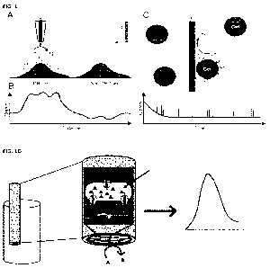
FIGURE 1 shows electrochemical quantification of CP efflux from living cells.
(A) When
expelled from the cell, CP is oxidized at the electrode during SECM. The
resulting current
signal increased with increasing CP efflux (B). (C) During impact chemistry
cells in solution
collide with a wire electrode whereby the CP diffusion layer around the cells
(pink) is
oxidized at the electrode. This will provide statistical data over populations
of cells. (D)
shows a schematic for electrochemical detection of drug efflux from a
pseudomonas
bacteria.
FIGURE 2 shows electrochemical characterization of carboplatin (CP), where in
(A)
CP exhibits an oxidation peak at 0.8 V vs Ag/AgC1 reference electrode; (B) at
unmodified platinum electrodes a limit of detection (LOD) of 50 pM was found;
(C) a
pH dependency study revealed that CP can be detected at a pH range of 1 to
7.5; and
in (D) shows a schematic of electrochemical drug efflux studies in Pseudomonas
bacteria. Bacteria (diagonal arrow) are drop-casted onto a macro-electrode.
When
expelling ciprofloxacin, the antibiotic is electrochemically oxidized at the
electrode,
resulting in a current increase seen as peak during DPV.
FIGURE 3 shows a schematic representation of DR in bacteria, wherein the
membrane protein modification, drug target alteration, drug inactivation by
intracellular enzymes, and membrane efflux pumps can prevent drugs to enter
and/or affect the cell.
6
CA 03163227 2022- 6- 28
WO 2021/134133
PCT/CA2020/051811
FIGURE 4 shows antibiotic hybrids for electrochemical investigations, with (A)
Structure of the tobramycin-ciprofloxacin hybrid, containing a 12-carbon-long
aliphatic (C12) hydrocarbon linker and (B) Cyclic voltammetry of 2 mM
tobramycin-
ciprofloxacin hybrid at various scan rates.
FIGURE 5 shows a schematic representation of SECM for biological applications.
(A)
Instrumental design, including Z-axis positioner (I), constant distance
controller (II),
light source (III), electrochemical cell (IV), as well as working electrode
(WE), counter
electrode (CE) and reference electrode (RE). (B) Example of a microelectrode
and its
size comparison (C) as well as top view (D) of the same electrode. (E)
Representation
of the low current bi-potentiostat, connected to all three electrodes.
FIGURE 6 shows a schematic representation of electrochemical measurements on
living bacteria. (A) Bacteria dropcasted onto a macroelectrode and exposed to
an
antibiotic (A), which is expelled by efflux pump from the organism. The
antibiotic is
then electrochemically converted at the electrode. (B) SECM electrode scanning
across small islands of bacteria, crossing DR bacteria, as well as non-
resistant entities.
(C) Schematic of an expected current profile of lateral scan across living
bacteria
FIGURE 7 shows cell patterning of HeLa cells using elastomeric through-hole
membranes. (A) Photograph of a through-hole membrane and its middle part (B).
Insets showing SEM images of a top (A) and side view (B). Scale bars: 500 lim.
(C, D,
E) Cell patterns achieved for HeLa in island sizes of 400 iim (C), 200 iim (D)
and 50
um (E). Scale bars: 100 lim. F) Optical micrograph of E. coil patterns in
201.tm islands.
FIGURE 8 shows a schematic representation of resistance adaptation monitored
by
SECM. (A) Fluorescently labelled DR and non-DR bacteria immobilized in co-
culture
will be imaged by an SECM microelectrode, resulting in a 3D current intensity
map
(B).
FIGURE 9 shows he peak current recorded at various scan velocities of the
microelectrode, wherein the initial electrochemical response recorded prior to
carboplatin exposure for both carboplatin-susceptible (A2780-s) and
carboplatin-
resistant (A2780-cp) ovarian cancer cells and the slope of the linear
regression was
shows the cells' ability to regenerate FcCH2OH through the cellular export of
glutathione, to indicate stress experienced by the cells due to carboplatin.
7
CA 03163227 2022- 6- 28
WO 2021/134133
PCT/CA2020/051811
FIGURE 10 shows Ciprofloxacin (1 mM) uptake quantification in both resistant
and
sensitive Pseudomonas aeruginosa bacterial strains using differential pulse
voltammetry (DPV).
FIGURE 11 shows Tobramycin (2 mM) uptake quantification in both resistant and
sensitive Pseudomonas aeruginosa bacterial strains using differential pulse
voltammetry (DPV).
FIGURE 12 shows Ciprofloxacin-Tobramycin (Cip-Tob) hybrid influx
quantification in
P. aeruginosa by DPV.
FIGURE 13 shows electrochemical detection of ciprofloxacin export from
Pseudomonas bacteria.
FIGURE 14 shows electrochemical detection of ciprofloxacin export in PA01 and
PA262 Pseudomonas bacterial strains.
DETAILED DESCRIPTION OF THE INVENTION
The following detailed description, will be better understood when read in
conjunction with the appended figures. For the purpose, of describing the
invention,
the figures demonstrate embodiments of the present invention. However, the
invention
is not limited to the precise arrangements and examples shown.
As used herein a "drug" refers to any therapeutic moiety, which includes small
molecules and biological agents (for example, proteins, peptides, nucleic
acids). As
used herein, the term drug may in certain embodiments include any therapeutic
moiety, or a subset of therapeutic moieties. For example, but not limited to
one or more
of the potentially overlapping subsets and one or more drugs, as follows:
antibiotic
drugs; and anticancer drugs.
As used herein an "electro-active drug" refers to any molecule that can
produce
detectable electro-activity, and which, also has therapeutic activity. The
molecular
structure is the primary determinant of a compound's electro-activity, whereby
the
presence of particular functional groups (for example, phenol, aromatic amine,
thiol,
8
CA 03163227 2022- 6- 28
WO 2021/134133
PCT/CA2020/051811
nitro, nitrophenol and quinone groups) and/or whether the structure permits
for
delocalization of a positively or negatively changed group. In particular, the
electro-
activity of a given drug compound may be based on the oxidation-reduction
(redox)
potential of the compound, or whether the compound is prone to undergo an
oxidation-
reduction reaction by gaining or losing an electron.
As used herein "membrane permeability" refers to the influx and/or efflux of
an
electro-active drug into or out of a cell. Depending on the electro-active
drug and the
cell, there may be by passive diffusion, facilitated passive diffusion, active
transport,
and pinocytosis. Similarly, once a drug is within a given cell, the drug may
be removed
from the cell by an efflux pump or other cell transport mechanism.
As used herein a "drug panel" refers a panel of drugs or electro-active drugs
of
various concentrations selected based on the target cell or cells being
tested. For
example, where the target cell is a cancer cell, then the panel would be made
of anti-
cancer drugs and these drugs may be tested at a variety of concentrations,
such that an
at least one cell deposited at a discrete location may be incubated with a
member of the
drug panel. Similarly, where the target cell is a bacterial cell, then the
panel would be
made of antibacterial drugs and these drugs may be tested at a variety of
concentrations, such that an at least one cell deposited at a discrete
location may be
incubated with a member of the drug panel. When we look at impact chemistry
cells
(both cancer and bacteria) the cells may be in solution, where the cells are
governed by
Brownian motion colliding with an electrode. For example, this could be also
implemented in a microfluidic device with a solution that may be exchanged or
added
to, leaving the at least one cell at the discrete location.
Anti-cancer drugs may be categorized as alkylating agents (hi and mono-
functional), anthracyclines, cytoskeletal disruptors, epothilone,
topoisomerase
inhibitors (1 and II), kinase inhibitors, nucleotide analogs and precursor
analogs,
peptide antibiotics, platinum-based agents, vinka alkaloids, and retinoids.
Alkylating
agents, may be bifunctional alkylators (for example, Cyclophosphamide,
Mechlorethamine, Chlorambucil and Melphalan) or monofunctional alkylators (for
9
CA 03163227 2022- 6- 28
WO 2021/134133
PCT/CA2020/051811
example, Dacarbazine(DTIC), Nitrosoureas and Temozolomide). Examples of
anthracyclines are Daunorubicin, Doxorubicin, Epirubicin, Idarubicin,
Mitoxantrone,
and Valrubicin. Cytoskeletal disruptors or taxanes are Paclitaxel, Docetaxel,
Abraxane
and Taxotere. Epothilones may be epothilone or related analogs. Histone
deacetylase
inhibitors may be Vorinostat or Romidepsin. Inhibitors of topoisomerase I may
include
lrinotecan and Topotecan. Inhibitors of topoisomerase II may include
Etoposide,
Teniposide or Tafluposide. Kinase inhibitors may be selected from Bortezomib,
Erlotinib, Gefitinib, Imatinib, Vemurafenib or Vismodegib. Nucleotide analogs
and
precursor analogs may be selected from Azacitidine, Azathioprine,
Capecitabine,
Cytarabine, Doxifluridine, Fluorouracil, Gemcitabine, Hydroxyurea,
Mercaptopurine,
Methotrexate or Tioguanine/Thioguanine. Peptide antibiotics like Bleomycin or
Actinomycin. Platinum-based agents may be selected from Carboplatin, Cisplatin
or
Oxaliplatin. Retinoids may be Tretinoin, Alitretinoin or Bexarotene. The Vinca
alkaloids and derivatives may be selected from Vinblastine, Vincristine,
Vindesine and
Vinorelbine. Alternatively, an electro-active anti-cancer drug may be selected
from
TABLE 1.
TABLE 1: Anti-cancer drug detection by electrochemistry.
Classes Drug Electrode Method of LOD
[M]
modification analysis
Antimetabolite 6- MWCNT Paste LSV 1
x10-7
Mercaptopurine electrode
Antimetabolite 6- [Co (phen)3] 3+-GO- DPV
1.5x10⁻⁸
Mercaptopurine dsDNA/GCE
Antimetabolite 6- N-HCNS-Pd-MIP/IL- DPASV
7.2x 10⁻¹⁸
Mercaptopurine PGE
Antimetabolite 5-Fluorouracil Glucose/CPE CV, DPV
5.17x10⁻⁹
Antimetabolite 5-Fluorouracil BMPA/Flexible AuE CV, SWV
3.4x10⁷
Antimetabolite 5-Fluorouracil Reduced GO-CS/GCE CV, SCV,
1.24x10⁻⁹
SWV
Antimetabolite 5-Fluorouracil AuNP-MWCNT- CV, DPV
2.0x 10⁻⁸
CS/GCE
Antimetabolite 5-Fluorouracil AuNP-PFR/CPE CV, DPV
6.70 x10⁻⁷
Antimetabolite 5-Fluorouracil PANINT-AgNP/PGE DPV 6
x 10-8
Antimetabolite 5-Fluorouracil IL/CPE CV, DPV
1.3 x 10⁻⁸
Antimetabolite 5-Fluorouracil GO-MWCNT/GCE and CV, SWV
1.6x10⁸
SPCE
CA 03163227 2022- 6- 28
WO 2021/134133
PCT/CA2020/051811
Antimetabolite 5-Fluorouracil CuSAE CV, AdSLSV
1.2x10⁹
Antimetabolite 5-Fluorouracil MTB/CPE CV, DPV
2.04x10⁹
Antimetabolite Gemcitabine AuE DPV
6x10⁸
Antimetabolite Gemcitabine MMOF-AuNP/AuE LSV 3
x10-15
Cytotoxic Doxorubicin MAb-AuNP-TBSol- EIS
1.7x10'
antibiotic Gel/AuE
Cytotoxic Doxorubicin Mab-AuN P- EIS
3.1 x 10⁻¹²
antibiotic APTES/SSE
Cytotoxic Doxorubicin Pd@PtNP-MWCNT/ AdSSWV 8.6x10⁻¹
antibiotic GCE
Cytotoxic Mitoxantrone dsDNA-MWCNT- DPV
1.3 x 10⁻⁸
antibiotic AgNP-PTP/GCE
Cytotoxic Epirubicin Ag-MWCNT/GCE SWV, CV
1.0x10⁻⁹
antibiotic
Cytotoxic Daunorubicin N-rGO-SWCNT- DPV
5.7x10⁻⁹
antibiotic PtNP/GCE
Cytotoxic Valrubicin AuNP-EDA- CV
1.8 x 10⁻⁸
antibiotic MWCNT/AuE
Alkylating agents Cisplatin GST/CPE CV, SWV
8.8 x 10⁻⁶
Alkylating agents Cisplatin MWCNT/SPCE CV, DPV
4.6x 10⁻⁶
Alkylating agents Temodal dsDNA-AuNP/PGE DPV
1.0x10⁻⁹
Inhibitors Etoposide Au-Pd@rGO-L- DPV
7.18 x10⁻¹
Ifosfamide Cysteine/PGE
Inhibitors Erlotinib MWCNT-PUFIX- DPV
2x10⁸
PPHF/PGE
Inhibitors Irinotecan GCE CV
1.12x10¹⁰
Inhibitors Roscovitine PGE or SPCE SWV
PGE: 1.96x10-
7
SPCE:
1.53 x10⁻⁷
([Co(phen)3]3+ = cobalt (111) trisphenanthroline complex; BMPA = biopolymer
from
babassu mesocarp modified with phthalic anhydride; PFR = porphyran; PAN1NT =
polyaniline nanotube; CuSAE = Copper solid amalgam electrode; AdSLSV =
adsorptive
stripping linear sweep voltammetry; Pd@PtNP = mesoporous Palladium and
Platinum
Core shell nanoparticles; AdSSWV = adsorptive stripping square wave
voltammetry;
PTP = polythiophene; N-rGO = nitrogen-doped reduced graphene oxide; GST =
Glutathione-s-transferase; Au-PdgrGO = gold, palladium and reduced graphene
oxide
nanocomposite; PUFIX = polyurethane; PPHF = polypropylene hollow fiber).
An anti-cancer drug that may be used as described herein, may be selected from
one or more of: Actinomycin; All-trans retinoic acid; Azacitidine;
Azathioprine;
11
CA 03163227 2022- 6- 28
WO 2021/134133
PCT/CA2020/051811
Bleomycin; Bortezomib; Carboplatin; Capecitabine; Cisplatin; Chlorambucil;
Cyclophosphamide; Cytarabine; Daunorubicin; Docetaxel; Doxifluridine;
Doxorubicin;
Epirubicin; Epothilone; Etoposide; Fluorouracil; Gemcitabine; Hydroxyurea;
Idarubicin; Imatinib; Irinotecan; Mechlorethamine; Mercaptopurine;
Methotrexate;
Mitoxantrone; Oxaliplatin; Paclitaxel; Pemetrexed; Teniposide; Tioguanine;
Topotecan; Valrubicin; Vemurafenib; Vinblastine; Vincristine; Vindesine; and
Vinorelbine. Alternatively, the anti-cancer drug may be a biological agent and
may be
selected from Herceptin (Trastuzumab), Ado-trastuzumab, Lapatinib, Neratinib,
Pertuzumab, Avastin, Erbitux or radiolabelled antibodies or targeted
radiotherapies
such as PSMA-radioligands. The anti-cancer drug may be an Androgen Receptor,
an
Estrogen Receptor, epidermal growth factor receptor (EGFR) antagonists, or
tyrosine
kinase inhibitor (TKI). An anti-angiogenesis agent may be selected from
avastin, an
epidermal growth factor receptor (EGFR) antagonists or tyrosine kinase
inhibitor
(TKI). An Immune modulator such as Bacillus Calmette-Guerin (BCG).
Alternatively,
an anti-cancer drug may include hybrids of two or more of the preceding anti-
cancer
drugs.
Alternatively, an electro-active antibiotic drug may be selected from TABLE 2.
TABLE 2: Antibiotic drug detection by electrochemistry.
Classes Drug Method of LOD [M]
Electrode
analysis
modification
B-Lactams Ampicillin DPV 3.2x10⁻¹-1-
dsDNA/AMP
aptamer
B-Lactams penicillin CV 8x10⁻¹-6 RGO/AuNP
B-Lactams penicillin CV 1.05x10⁻⁵
multisegment
nanoparticles
B-Lactams Amoxicillin CV 6 x10⁻⁷ POT(SDS)
B-Lactams Amoxicillin CV 5x10⁻⁶ Ni/CR
B-Lactams Amoxicillin SWV 9x10⁻⁶ AuNP-
PdNP-RGO
B-Lactams Amoxicillin SWV 1.2x10⁻⁷ CB DHP
B-Lactams Amoxicillin CV 1.87x10⁻⁹ poly
acridine
orange
Aminoglycosid Neomycin SWV 1.07x10⁻⁶
Polyamic acid/GO
es
12
CA 03163227 2022- 6- 28
WO 2021/134133
PCT/CA2020/051811
Aminoglycosid Tobramycin CV 1.4x10⁻¹⁰
Polypyrrole
es
Quinolones Ciprofloxacin CV, ASV 5.9x10⁻⁸ Graphene
Quinolones Ciprofloxacin CV, DPV 5x10⁻⁸ CTAB
Quinolones Ciprofloxacin CV 1.2x10⁻⁸ MgFe204-
MWCNT
Quinolones Ciprofloxacin CV, LSV 9x10⁻⁷ MWCNT
Quinolones Ciprofloxacin CV GO
Quinolones Ciprofloxacin CV 3.3x10⁻⁶ BDD
Quinolones Ciprofloxacin SWV 3.3x10⁻⁸ GCP
Quinolones Ciprofloxacin CV 6x10⁻⁶ MWCNT
Quinolones Levofloxacin DPV 1x10⁻⁶
PoAP/MWCNT
Quinolones Levofloxacin CV, SWV 1.4x10⁻⁸ AgNPs-CB-
PEDOT:PSS
Quinolones Levofloxacin CV, DPV 5.3x10⁻⁷ MIP/G-
AuNPs
Quinolones Levofloxacin CV, SWV 2.88x10⁻⁶ BDD
Quinolones Levofloxacin CV 1x10⁻⁸ AgNP
Quinolones Norfloxacin SWV 3.4x10⁻⁸ Polyamic
acid/GO
Quinolones Norfloxacin LSV 5x10⁻⁸ MWCNT
Quinolones Enrofloxacin LSV 5x10⁻⁷ MWCNT
Quinolones Ofloxacin CV 1.8X10⁻¹⁰ MWCNT
SW-AdAsV 2.4X10⁻¹⁰
Quinolones Ofloxacin CV, DPV 1x10⁻⁹
AuNPs/ATP/ABA
RGO = reduced graphine oxide; POT (SDS) = poly(o-toluidine) (sodium dodecyl
sulphate); C13 = carno black; DHP = dihexadecylphosphate; CTAB =
cetyltrimethylammonium bromide; BDD = boron doped diamond; GCP = glassy carbon
paste; PoAP = poly(o- aminophenol); PEDOT:PSS = poly(3,4-
ethylenedioxythiophene)-
poly(styrenesulfonate); G = graphene; ATP = 4-aminothiophenol; ABA = 4-
aminobenzoic acid (4-ABA); 1L-G = ionic liquid- graphene; ZSM = mesoporous
zeolitic
material.
An antibiotic drug that may be used as described herein, may be selected from
one
or more of: Amikacin; Gentamicin; Kanamycin; Neomycin; Netilmicin; Tobramycin;
Paromomycin; Streptomycin; Spectinomycin(Bs); Geldanamycin; Herbimycin;
Rifaximin;
Carbacephem; Loracarbef; Carbapenems; Ertapenem; Doripenem;
Imipenem/Cilastatin;
Meropenem; Cefadroxil; Cefazolin; Cephradine; Cephapirin; Cephalothin;
Cefalexin;
Cefaclor; Cefoxitin; Cefotetan; Cefamandole; Cefmetazole; Cefonicid;
Loracarbef; Cefprozil;
Cefuroxime; Cefixime; Cefdinir; Cefditoren; Cefoperazone; Cefotaxime;
Cefpodoxime;
Ceftazidime; Ceftibuten; Ceftizoxime; Moxalactam; Ceftriaxone; Cefepime;
Ceftaroline
13
CA 03163227 2022- 6- 28
WO 2021/134133
PCT/CA2020/051811
fosamil; Ceftobiprole; Teicoplanin; Vancomycin; Telavancin; Dalbavancin;
Oritavancin;
Clindamycin; Lincomycin; Lipopeptide; Daptomycin; Clarithromycin;
Erythromycin;
Roxithromycin; Telithromycin; Spiramycin; Fidaxomicin; Aztreonam; Nitrofurans;
Furazolidone; Nitrofurantoin(Bs); Linezolid; Posizolid; Radezolid; Torezolid;
Amoxicillin;
Ampicillin; Azlocillin; Dicloxacillin; Flucloxacillin; Mezlocillin;
Methicillin; Nafcillin;
Oxacillin; Penicillin G; Penicillin V; Piperacillin; Penicillin G; Temocillin;
Ticarcillin;
Amoxicillin/clavulanate; Ampicillin/sulbactam; Piperacillin/tazobactam;
Ticarcillin/clavulanate; Bacitracin; Colistin; Polymyxin B; Enoxacin;
Gatifloxacin;
Gemifloxacin; Levofloxacin; Lomefloxacin; Moxifloxacin; Nadifloxacin;
Nalidixic acid;
Norfloxacin; Ofloxacin; Trovafloxacin; Grepafloxacin; Sparfloxacin;
Temafloxacin;
Sulfacetamide; Sulfadiazine; Silver sulfadiazine; Sulfadimethoxine;
Sulfamethizole;
Sulfamethoxazole; Sulfanilimide; Sulfasalazine; Sulfisoxazole; Trimethoprim-
Sulfamethoxazole (Co-trimoxazole) (TMP-SMX);Sulfonamidochrysoidine;
Demeclocycline;
Doxycycline; Metacycline; Minocycline; Oxytetracycline; Tetracycline;
Clofazimine;
Dapsone; Capreomycin; Cycloserine; Ethambutol(Bs); Ethionamide; Isoniazid;
Pyrazinamide; Rifampicin; Rifabutin; Rifapentine; Streptomycin; Arsphenamine;
Chloramphenicol(Bs); Fosfomycin; Fusidic acid; Metronidazole; Mupirocin;
Platensimycin;
Quinupristin/Dalfopristin; Thiamphenicol; Tigecycline(Bs); Tinidazole; and
Trimethoprim(Bs). Alternatively, an antibiotic drug may include hybrids of two
or more of
the preceding antibiotic drugs. For example, an antibiotic hybrid molecule
described
herein is tobramycin-ciprofloxacin (Tob-Cip).
As used herein "electro-analytical measurement" may be obtained by one or more
of
the following: linear sweep voltammetry (LSV); cyclic voltammetry (CV);
differential pulse
voltammetry (DPV); differential pulse anodic stripping voltammetry (DPASV);
square wave
voltammetry (SWV); adsorptive stripping linear sweep voltammetry (AdSLSV);
electrochemical impedance spectroscopy (EIS); chronoamperometry (CA);
chronopotentiometry (CP); chronocoulometry (CC); impact chemistry (IC);
scanning ion
conductance microscopy (SICM); scanning electrochemical cell microscopy
(SECCM);
scanning photoelectrochemical microscopy (SPEC M); and scanning
electrochemical
microscopy (SECM).
14
CA 03163227 2022- 6- 28
WO 2021/134133
PCT/CA2020/051811
As used herein "dropcasting" is meant to describe the pipetting of or
otherwise
depositing of an aqueous cell solution, such as bacteria or cancer cell, on an
electrode,
whereby when the aqueous cell solution evaporates, it leaves the cells
"sticking" to the
electrode without killing the cell, such that the cells' internal composition
and osmotic
pressure is not compromised. Accordingly, dropcasting is alternative method
for putting a
functional cell in contact with an electrode, whereby the cells are not
actually dried, just the
aqueous cell solution surrounding the cells that is being evaporated.
MATERIALS AND METHODS
Electrochemistry
Standard electrochemical methods, such as cyclic voltammetry (CV), and
scanning electrochemical microscopy (SECM), may be used. However,
the
methodology could be adapted to use other methods such as linear sweep
voltammetry
(LSV), differential pulse voltammetry (DPV), differential pulse anodic
stripping
voltammetry (DPASV), square wave voltammetry (SVVV), adsorptive stripping
linear
sweep voltammetry (AdSLSV), electrochemical impedance spectroscopy (EIS),
chronoamperometry (CA), chronopotentiometry (CP), chronocoulometry (CC),
impact
chemistry (IC), scanning ion conductance microscopy (SICM) or scanning
electrochemical cell microscopy (SECCM). SECM has been successfully utilized
to study
drug resistance in mammalian cancer cells15-18. In particular, applying SECM
to drug
resistant cancer cells as compared to non- drug resistant cells, showed
different
electrochemical behaviours15. Furthermore, use of cell permeable and
impermeable
redox mediators that allowed the extraction of kinetic information from
experimental
SECM data, which resulted in the quantification of drug resistance on the
single cell
level by mathematical and numerical models16. In addition, it has been
demonstrated
that SECM may be used to assess cancer cells, exposed to antioxidants, and
their
electrochemical response over time may be quickly acquired. Also, it was
possible to
determine a samples' apparent heterogeneous rate constant, independent from
their
topography, which until this point remained a challenge to the SECM
community17-18.
These successful studies on cancer cells present an ideal basis for the
investigation of antibiotic resistance in bacteria. Similar to cancer cells,
the
CA 03163227 2022- 6- 28
WO 2021/134133
PCT/CA2020/051811
electrochemical response of bacteria is based on the expression of membrane
efflux
pumps, which affect drug take-up and release from the organisms and subsequent
local
and transported concentration of electroactive material to the electrode.
Scanning
electrochemical microscopy (SECM) is an electroanalytical technique, employing
a
micro- or nanoscale electrode, which is rastered across a surface to analyse
its
electrochemical activity. As shown in FIGURE 5, a microelectrode (FIGURE 5A
orange
wire, B, C, and D) consists of a metal wire, which is sealed into a quartz
capillary and is
connected to a potentiostat19. This electrode functions as working electrode
(WE) and
is mounted onto a motor station, moving in the z-direction above an
electrochemical
cell, which is mounted onto an X and Y axis positioner20. An incorporated
light source
and microscope, equipped with a camera, allows the monitoring of any sample
during
electrode positioning prior to the data acquisition, as well as during SECM
measurements21. Sample observation becomes especially important when working
with biological samples, such as living bacteria or tissue cells, as the
target's
morphology can be observed during the experiment. Furthermore, the SECM
apparatus
is placed on a vibration isolation table inside a Faraday cage to avoid
interference from
external electric noise.
In the presence of a redox active species in solution, a potential, far
exceeding
the standard potential of the dissolved redox mediator, can be applied at the
WE to
drive the oxidation or reduction of a redox species at the surface of the
electrode. A
potentiostat compares the potential difference between WE and a reference
electrode
(RE) to a computer defined value and adjusts a power source between WE and a
counter electrode (CE) accordingly (FIGURE 5E). Thereby, a current commonly in
the
fA to [tA range is measured at the WE. In fact, some redox mediators have been
shown
to interact with biological entities (e.g. living bacteria or tissue cells)22-
23, and many
biological samples have been successfully analysed in the past by SECM24.
Cyclic Voltammetry (CV) is an electrochemical technique in which an applied
potential is swept linearly between two limiting potentials, driving a
chemical reaction
at macro-, micro- or even nanoelectrodes25. The overall CV shape of a redox
reaction
at electrodes is determined by, and provides information about, redox
16
CA 03163227 2022- 6- 28
WO 2021/134133
PCT/CA2020/051811
thermodynamics, electron transfer kinetics, diffusion processes of molecules
in
solution and towards the electrode, and possible decay reactions28.
Bioelectrochemical sensors have attracted immense attention, whereby
electrode materials and electrode modifications have emerged for the design of
highly
sensitive and selective sensors.26 Electroanalytical techniques are cost
effective,
sensitive and the transparency of a solid or liquid sample is irrelevant,
allowing direct
in vitro analysis of food, beverage, blood, urine, and saliva samples and
tissue samples
with minimal preparation using electroanalytical methods, such as cyclic
voltammetry
(CV), chronoamperometry (CA), impact chemistry and scanning electrochemical
microscopy (SECM), to quantify electro-active drug compounds released from
cells.
The bioelectrochemical studies of Pseudomonas aeruginosa as shown in FIGURE
utilized differential pulse voltammetry (DPV), and four (4) glass vials were
prepared, holding a PBS solution containing 1 mM ciprofloxacin. Vial 1 was
used as a
control; Vial 2 was a second control with ciprofloxacin after 25 minutes of
incubation
at 37 C. These two controls demonstrate that the incubation at 37 C does not
lead to
a degradation of ciprofloxacin in solution; Vials 3 and 4 each contain P.
aeruginosa cells
at a cell number range of 106 to 108 per ml held for 25 minutes at 37 C. DPV
measurements of 1mM Cip were taken and after incubation, centrifugation was
performed at 4000rpm for 10min and the supernatant was collected. Any
ciprofloxacin
taken up by the bacteria during the incubation time was hence removed from the
solution. DPV measurements were then performed on the supernatant as an
indication
of ciprofloxacin uptake by the bacteria.
Similarly uptake of Tobramycin (2 mM), was analyzed in Pseudomonas
aeruginosa also using DPV. The control solutions were tested before and after
incubation at 37 C and with different incubation times at 37 C (i.e. 15
minutes; 30
minutes and 60 minutes). Two Pseudomonas aeruginosa bacterial strains were
analyzed for drug uptake (PA01 strain - a wild type strain, containing efflux
pumps on
the bacterial membranes; and PA0750 - a hyper- susceptible strain where efflux
pumps
have been deleted). Integration underneath the curves provides the charge.
This
charge is proportional to the number of molecules transferred during DPV
recordings
(see FIGURES 11-14).
17
CA 03163227 2022- 6- 28
WO 2021/134133
PCT/CA2020/051811
Ovarian Cancer Cell Studies
Carboplatin-susceptible (A2 780-s) and carboplatin-resistant (A2780-cp)
ovarian cancer cells were grown in petri dishes at 37 C until a confluence of
approximately 60 to 70% was reached. Cells were exposed to a PBS solution
containing
1 mM ferrocenemethanol (FcCH2OH) at 37 C for 45 minutes. Following this
incubation,
the petri dish was inserted into a scanning electrochemical microscope (SECM),
equipped with a camera, fluorescence unit and a heating stage. Target cells
were
identified using an approach curve and a horizontal line scan was carried out
across
the cells every 5 to 10 minutes. The solution in the petri dish was exchanged
for a PBS
solution containing 2 mM carboplatin and 1 mM ferrocenemethanol (FcCH2OH).
Horizontal line scans were carried out every 5 to 10 minutes to record the
electrochemical response by the cells.
Electrochemical response was recorded prior to drug exposure (i.e.
carboplatin) for both carboplatin-susceptible (A2 780-s) and carboplatin-
resistant
(A2780-cp) ovarian cancer cells and the slope of the linear regression was
shown to be
related to the cells' ability to regenerate FcCH2OH through the cellular
export of
glutathione, wherein the extend of FcCH2OH regeneration by the cells can be
expressed
as apparent heterogeneous rate constant17, 46.
Electrodes
A three-electrode setup may be used for cyclic voltammetry (CV) and scanning
electrochemical microscopy (SECM) or other electrochemical analyses.
Electrodes
may have a 25 micrometer platinum (Pt) diameter or laser pulled Pt working
electrodes, an Ag/AgC1 (3 M NaC1) pseudo-reference electrode (calibrated in
FcCH2OH)
and 0.5 mm Pt auxiliary. The preparation of conventional 25 micrometer Pt
microelectrodes followed a well-established fabrication protoco140 while
polished;
needle-like, disk-shaped nanoelectrodes were fabricated using a similar to the
procedures described41. The fabrication procedure specifically produces disk
shaped
Pt microelectrode sealed in a quartz capillary and laser pulled until a
dimensionless
radius of glass (RG) inferior to 10 is obtained. In brief, 25 um annealed Pt
wires were
18
CA 03163227 2022- 6- 28
WO 2021/134133
PCT/CA2020/051811
pulled into quartz glass capillaries (length of 150 mm, an outer diameter of 1
mm, and
an inner diameter of 0.3 mm) under vacuum with the help of a P-2000 laser
pipet puller
(Sutter InstrumentsTM, CA, USA). The pulling program results in the formation
of a long
and sharp microelectrode with a thin glass sheath, which facilitates membrane
penetration. The effective radius was evaluated from steady-state voltammetry.
Electrodes with diameters < and/or > 25 um may be used. For example, 10 um, 5
um,
1 um in diameter or even on the nanoscale). Marcoeletrodes (diameter 1 mm) may
be used for voltammetric measurements. Alternatively, a metal wire may be used
as
an electrode (for example, in impact chemistry).
Statistical analysis
All values were measured in triplicates and subsequently statistically
evaluated.
Based on a student's t-distribution, errors were calculated applying a two-
tailed test
with n=3, a=0.025 and therefore a confidence level (CL) of 95% is given.
EXAMPLES
EXAMPLE 1. Electrochemical Analysis of Anticancer Drug Carboplatin (CP)
Cyclic voltammetry (CV) is an electrochemical technique in which an applied
potential is swept linearly between two limiting potentials, driving a
chemical reaction
at macro-, micro- or even nano-electrodes.
The overall CV shape of a
reduction/oxidation (redox) reaction at the electrodes is determined by, and
provides
information about, redox thermodynamics, electron transfer kinetics, diffusion
processes of molecules in solution and towards the electrode, and possible
decay
reactions.25
Furthermore, experiments have determined an oxidation peak of
carboplatin at 0.8 V vs Ag/AgC1 reference electrode (FIGURE 2A). The oxidation
of
carboplatin (CP) was found to be irreversible and no electrode blockage by its
oxidation reaction products was observed. A detection limit (LOD) of SO uM at
unmodified platinum electrodes was identified (FIGURE 2B), whereby CP can be
detected at a pH range of 1 to 7.5 (FIGURE 2C). This characterization shows
that CP
can be recognized electrochemically at low concentrations and its diffusion in
solution
is understood.
19
CA 03163227 2022- 6- 28
WO 2021/134133
PCT/CA2020/051811
In the literature, nanoparticles have been successfully used to increase the
surface area of
electrodes to ultimately lowering the necessary overpotential applied to drive
the
oxidation/reduction reaction.26 To increase the sensitivity of CP detection at
platinum
(Pt) electrodes, platinum nanoparticles (PtNPs) may be drop-casted onto
electrodes as
a first approach. The current recorded at the electrode is expected to rise
with
increasing concentration of PtNPs. An optimal concentration of PtNPs, may be
determined to enable the detection of CP or another anti-cancer electroactive
drug at
sub-jun concentrations. However, during live-cell imaging using the scanning
probe
technology SECM, dropcasted PtNPs are unlikely to be stable at the electrode
surface
during scans. Hence, a mixture of PtNPs and pyrrole will be polymerized at the
electrode surface using CV. This may result in a stable conductive polymer
layer,
capturing PtNPs. The porous nature of polypyrrol may allow for efficient
electron
transfer and diffusion of CP towards the electrode. The thickness of the
polymer layer
can be controlled by adjusting the duration of polymerization.
Electrochemical quantification of carboplatin efflux from A2780 endometrioid
EOC cell lines was tested. Paired, syngeneic A2780 EOC cells that are
chemosensitive
(A2780-s) or chemosresistant (A2780-cp). Cells may be patterned in defined
areas on
the surface of plastic substrates using elastomeric through-hole membranes.16
The
applicability of these substrates has been demonstrated in the past for HeLa
cells. Cell
patterning is useful for Bio-SECM studies, as target cells can be easily
located through
the SECM-integrated optical microscope and cells will not be able to "crawl"
away
during repeated measurements. When cells are patterned on plastic, a
microelectrode
may be brought in close proximity to the A2780 cells using the SECM setup to
maximize
the recorded current response. For this purpose, an approach curve over or
next to a
monolayer of cells will be carried out in the presence of a redox mediator,
which is cell
impermeable and will have no influence on the biological sample of interest.
Hexaammineruthinium (III) chloride is the substance of choice based on
literaturel6 to
carry out such an approach curve. The probe may be retracted to any desired
distance
above the cells (for example, 10 pm). A2780 cells may be grown at 37 C in cell
growth
medium containing CP. Concentration and time of incubation may be optimized.
CP
CA 03163227 2022- 6- 28
WO 2021/134133
PCT/CA2020/051811
may be removed by exchanging the solution to fresh growth medium without CP.
The
SECM electrode may be moved horizontally across an island or single cells
while
recording the electrochemical current, resulting from the oxidation of CP that
is
expelled by the cells (FIGURE 1A). This allows for a comparison of cells
positioned
side-by-side of different resistance phenotypes at the same time and under the
same
conditions. Cells of higher magnitude of drug resistance (higher efflux rate)
are
expected to result in higher current values during SECM measurements (FIGURE
1B).
Due to the sensitivity of the SECM methodology it may be possible to tell the
exact
number of CP molecules exported from a single cell per second. This will
provide a
numerical measure for the drug resistant phenotype for any cell of interest.
To obtain
statistical data on a large number of cells, impact chemistry may be used.
Impact
chemistry is a powerful technique for the detection of single biological
entities in large
numbers.38, 39 Impact chemistry is based on faradaic charge transfer,
following the
collision of redox active entities on the nano- or micrometer scale with an
electrode.
Cells pre-exposed to CP may be put into fresh cell medium lacking serum.
Governed by
Brownian motion, single cells would collide with the electrode, which may be
held at
an oxidizing or reducing potential. Collision events will result in the
oxidation CP
released from single cells, revealing a short current burst ("spike") every
time a cell
passes the electrode, whereby the spike intensity is related to the drug
efflux rate.
While A2780-s and A2780-cp cells may be employed, different EOC cell lines
representing most histotypes, as well as EOC patient cell samples may also be
tested.
Cells will be measured and compared for their resistance phenotype by SECM.
Patient
samples may be used to determine the cells susceptibility against the panel of
electroactive anti-cancer drugs and at various concentrations. Testing patient
samples
may allow for personalized clinical management.
Further work was done by the inventors45 to evaluate the electrochemical
detection of chemotherapeutic uptake by ovarian cancer cells, whereby an
electrochemical characterization of carboplatin was evaluated for detection
limits and
pH dependence. Furthermore, bioelectrochemical studies quantified carboplatin
uptake by ovarian cancer cells. Voltammetric drug uptake studies demonstrated
the
21
CA 03163227 2022- 6- 28
WO 2021/134133
PCT/CA2020/051811
detection of carboplatin uptake in one carboplatin-susceptible and one
carboplatin-
resistant ovarian cancer cell line.
The electrochemical response of ovarian cancer cells to carboplatin was
assessed by scanning electrochemical microscopy. In A2780-cp cells, an
increase in
slope right after carboplatin exposure was observed and the increase relaxes
back to
its initial value within 5 to 10 minutes. It is hypothesized that this
electrochemical
response indicates the ability of resistant cells to cope with the exposure to
carboplatin
by temporarily increasing the rate of glutathione efflux, transporting not
only
glutathione, but also carboplatin to the cell exterior (FIGURE 9). A clear
difference
between carboplatin-susceptible and carboplatin-resistant cells was observed
by
SECM measurements.
EXAMPLE 2. Electrochemical Analysis of Anti-biotic Drugs
The ability of biological entities, such as bacteria, to remain unaffected by
at
least one antimicrobial agent is referred to as drug resistance (DR), whereby
the non-
susceptibility to agent in antimicrobial categories is called
multidrug resistance.
DR can be due to the acquisition of genes encoding for defence mechanisms to a
specific
agent or to overexpression of efflux pumps, which can rapidly expel drugs from
the cell
interior. Membrane protein modification, drug target alteration, drug
inactivation by
bacterial enzymes and bacterial efflux pumps are successful antibiotic defence
strategies in bacteria is shown in FIGURE 329. The increase of resistance in
Gram-
negative bacteria in particular is a major cause for concern39-31, as many
Gram-
negatives cause serious infections, such as pneumonia. Only a few antibiotics
effective
against Gram-negatives have been developed due to their innate defence
mechanisms
including low outer membrane permeability and high number of efflux pumps.
Thus,
with the rise of DR, many infections caused by Gram-negative bacteria have
become
untreatable32. Strategies that are able to quantify the efflux of agents from
bacterial
cells, especially Gram-negatives, for the assessment of potential new and
reliable
antimicrobial candidates would be very useful.
22
CA 03163227 2022- 6- 28
WO 2021/134133
PCT/CA2020/051811
Most recently we have collected preliminary data about the electrochemical
behavior of the antibiotics tobramycin and ciprofloxacin, which are in
agreement with
literature and show that both species can serve as potential efflux
indicators. In
addition, we have conducted first experiments on the antibiotic hybrid
molecule
tobramycin-ciprofloxacin (Tob-Cip, FIGURE 4A). CVs show two irreversible peaks
that can be assigned to the individual ciprofloxacin and tobramycin components
at a
potential of about 1.1 V and 1.3 V vs standard calomel reference electrode
(FIGURE
4B). As the ciprofloxacin peak is partially covered by the electrochemical
response of
tobramycin, we are currently working on electrode modifications using
nanomaterials
to separate the individual peaks more prominently. Nanoparticles have been
successfully used in the literature to increase the surface area of electrodes
to
ultimately lowering the necessary overpotential applied to drive the
oxidation/reduction reaction26. These are encouraging first results, because
it
demonstrates that we can recognize and quantify antibiotic hybrids at
electrodes.
Other alternative experimental hybrid antibiotics, are to be tested. In
addition,
conventional electroactive antimicrobial agents that are known to be expelled
by E. co/i,
such as ampicillin, and amoxici11in28, will be characterized, resulting in a
broad library
of electroactive antibiotic substrates. Next to diffusional and thermodynamic
parameters, the reversibility of the antibiotic redox reactions and whether
the oxidized
and reduced forms are stable may be assessed, as well as the possible
occurrence of
electrode fouling over multiple cycles. The term electrode fouling describes
the
blockage of the electroactive surface area of the electrode due to the
absorption of
solution species. These decomposition products can be characterized post-
experimentally by X-ray photoelectron (XPS) spectroscopy or scanning electron
microscopy (SEM). To avoid electrode fouling, testing may be restricted (e.g.
avoiding
sweeping over several electron-transfer reaction steps), or oxidative cleaning
may be
applied in between measurements to remove adsorbed material from the electrode
surface. Also, thorough polishing of the electrode after each measurement
using a
water-alumina mix on micro-cloth polishing pads will assure the complete
removal of
possible reaction products may be useful.
23
CA 03163227 2022- 6- 28
WO 2021/134133
PCT/CA2020/051811
The CV shape may be studied by finite element modelling, for example using a
known approach27, the physical processes may be described by a mass transport
equation, the Butler-Volmer surface electron transfer kinetics, and chemical
reaction
kinetics in solution in a one-dimensional system. The output of this
simulation is a CV
current response, which may be fitted to the experimental CV. Thusly, redox
reactions
may be simulated to determine the Butler-Volmer kinetic parameters of
antibiotics
oxidation and reduction at the macro-electrode and a fitting of the
concentration
independent heterogeneous standard electrochemical rate constant as well as
the
standard electrode potential may be conducted. Accordingly, the reaction
parameters
for non-trivial redox systems may be determined, i.e. those that exhibit slow
or
asymmetric electron transfer kinetics, or irreversible side reactions. Such
determined
electrochemical reaction parameters might be useful for the drug efflux
quantification,
by choosing electrode potential, concentration range, and electrode material.
Bacteria, such as E. coil or P. aeruginosa would be useful as model organisms,
and may be used in a buffer solution or may be drop-casted onto a macro- or
microelectrode23. These organisms have been shown to exhibit drug resistance
associated with efflux pumps33 and are relatively easy to handle with high
proliferation
rates. In addition, the importance of E. coil as contaminant in the food
industry, as well
as both bacteria types' impact in the medical sector make these organisms
interesting
targets. In both cases, bacteria may be exposed to electroactive antimicrobial
agents
that are known to be expelled by E. coil, such as amoxici11in34,36, followed
by an
incubation period during which the agent may be taken up by the bacteria.
Exact drug
concentrations and incubation times would be evaluated. The exchange of
solution to
a fresh, drug-free buffer, would then allow for measurement of expelled drug
molecules
from the bacteria. For this purpose, the electrode may be biased at a
potential far
exceeding the formal potential of the antibiotic to drive the chemical
reaction at the
electrode surface (FIGURE 6A). The conversion of any trace amounts of drugs,
released by the bacteria, may be recorded through the potentiostat. Bacterial
strains,
which are non-resistant, DR, and deficient in efflux pump expression, such as
P.
aeruginosa (i.e. PAO 200 and PAO 750) are useful control organisms to assess
the
quantification method. As current approaches rely on complicated and costly
24
CA 03163227 2022- 6- 28
WO 2021/134133
PCT/CA2020/051811
methodologies35, such as synchrotron based spectromicroscopy, the recognition
of
drug efflux by electrochemistry would allow for simple and direct measurement
of
antibiotic mass flux at low cost.
Target bacteria may be patterned in discrete locations on a substrate surface,
for example using elastomeric through-hole membranes16. The applicability of
these
substrates has been demonstrated in the past for mammalian cancer cells
(FIGURES
7A-E)16 and most recent preliminary data shows that this approach can be
transferred
to bacteria (FIGURE 7F). For example, membranes for bacteria attachment may be
an
elastomeric polymer synthesised and masked into the defined membranes as shown
in
FIGURES 7A and B. Thereby, the hole-shape and -size may be modified and
prepared
according the bacteria to be immobilization (for example, 20 i_tm to 50 i_tm).
The precise
positioning of target bacteria onto plastic or glass substrates may be
achieved by
oxygen plasma treatment of the membranes placed on plastic or glass surfaces.
The
exposure to oxygen plasma renders the surface hydrophilic, promoting cell
adhesion
and will thereby allow for SECM studies on small islands of bacteria cultures
or even
single cells, whereby the drug resistant phenotype of these target cultures
will be
quantified. When bacteria are patterned on glass, a microelectrode may be
brought in
close proximity to the bacteria using the SECM setup to maximize the recorded
current
response. For this purpose, an approach curve over or next to a monolayer of
bacteria
may be carried out in the presence of a redox mediator, which is cell
impermeable and
will have no influence on the biological sample of interest.
Hexaammineruthinium(III)chloride (Ru(NH3)6C13) is used in the literature16 for
an
approach curve. During an approach curve, while applying a reductive potential
for
Ru(NH3)6C13, the microelectrode moves vertically towards the bacteria while
recording
the current. When the diffusion of the redox species gets hindered by the
presence of
the bacteria, the current value decreases and the motion of the electrode is
stopped
when it comes into contact with the bacteria. The probe may then be retracted
to any
desired distance above the bacteria (for example,5 pm). Cell patterning is
significant
for Bio-SECM studies, as target bacteria can be easily located through the
SECM-
integrated optical microscope and bacteria will not be able to "crawl" away
during
repeated measurements. The SECM electrode may be moved horizontally across an
CA 03163227 2022- 6- 28
WO 2021/134133
PCT/CA2020/051811
island or single bacteria while recording the electrochemical current,
resulting from
the oxidation or reduction of a selected antimicrobial agent, exposed to the
bacteria
previously (FIGURES 6B and C). This allows for comparisons of bacteria of
different
resistance phenotypes, patterned in co-culture, at the same time and under the
same
conditions. Organisms of higher magnitude of DR (higher efflux rate) are
thereby
expected to result in higher current values during SECM measurements.
Due to the sensitivity of the SECM methodology it may be possible to tell the
exact number of drug molecules exported from a single cell per second and per
single
cell. This would provide a numerical measure for the DR phenotype for any
bacteria
strain of interest. Although, E. coli may be employed as model organism the
methods
may be adapted to many different bacterial strains, such as Pseudomonas, will
be
measured and compared for their resistant phenotype by SECM. Drug efflux pump
inhibitors, such as 3-(3',4/5'-trimethoxypheny1)-4,5,6-trimethoxyindanone-111,
may
be employed to analyse the sensitivity of the proposed method.
It is known that some DR bacteria have the ability to pass on their resistance
to
neighbouring bacteria37. Understanding this phenomenon is key for developing
models of DR progression across populations and may be investigated by
electrochemistry, and specifically by SECM. DR and non-resistant bacteria may
be
patterned in close proximity to each other and the electrochemical current
response to
antibiotic treatment may be monitored in both populations over time. DR
bacteria will
be co-patterned in direct contact or any desired distance with non-resistant
bacteria,
employing the elastomeric through-hole membranes described above. Parts of the
oxygen plasma treated surface may be covered by a commercially available
elastomeric
polymer (for example, polydimethylsiloxane (PDMF)), during cell exposure. The
PDMF
layer may then be removed and a second bacterial strain may be added.
Bacterial
strains may then be distinguished in co-cultures by fluorescently labelling
their
cytoplasmic membranes using different dye solutions. Combined fluorescent
imaging
and SECM would allow for the identification of various cell populations, even
during
cell proliferation or cell movement. The electrochemical current response to
antibiotic
treatment may be monitored simultaneously in populations and recorded over
time for
all bacteria. A change in current, as schematically shown in FIGURE 8, may
indicate an
26
CA 03163227 2022- 6- 28
WO 2021/134133
PCT/CA2020/051811
adaptation of non-resistant bacteria to the antibiotic in the presence of DR
organisms.
How quickly various bacteria strains can adopt antibiotic resistance depending
on
dosage, exposure time and nature of an antibiotic may be tested using this
methodology. Different genetic models of bacteria may be monitored across
populations and bacterial strains. Furthermore, the methods described herein
may be
used to test new antibiotic candidates, such as the Tob-Cip hybrid, and DR
inhibitors
may be tested and their local effect on a fraction of a population, as well as
its influence
on organisms within the same population, but in locally different areas.
Quantitative
measurements of the adaptation/transfer of DR properties between populations,
may
be subsequently used to establish models of DR progression. Monitoring DR
initiation
and progression quantitatively by electrochemistry may enable the
establishment of
DR population models based on reliable empirical data. Ultimately, gaining
understanding of the development and spread of DR across organisms would
greatly
support efforts at developing strategies against this exceptional medical
challenge.
Ciprofloxacin resistance is increasingly spread among infections and various
pathogens exhibit resistance against this antibiotic. Pseudomonas aeruginosa
cultures
were analyzed to demonstrate the quantification of drug uptake in bacteria by
electrochemistry. Two bacterial strains, one ciprofloxacin-susceptible (PA01)
and one
ciprofloxacin-resistant (PA262) strains were used for the experiments. The
PA262
strain exhibits an overexpression of efflux systems, which expel antibiotics
from the
cell's cytosol to the exterior environment. This mechanism causes a decreased
susceptibility against ciprofloxacin and makes these cultures resistant to the
antibiotic.
Electrochemical detection by differential pulse voltammetry (DPV) of
antibiotic
uptake by Pseudomonas aeruginosa is shown for Ciprofloxacin (Cip) in FIGURE
10.
There were two controls, containing PBS solution containing 1 mM
ciprofloxacin: Vial
1 (black) shows a peak current of approximately 55 A prior to incubation; and
Vial 2
(broken black) shows the current response of ciprofloxacin after 25 minutes of
incubation at 37 C. These two controls demonstrate that the incubation at 37
C does
not lead to a degradation of ciprofloxacin in solution. Vials 3 and 4 (grey
and broken
grey) each contain P. aeruginosa cells in 1mM Ciprofloxacin, where DPV
measurements
of the collected supernatant, whereby any ciprofloxacin taken up by the
bacteria during
27
CA 03163227 2022- 6- 28
WO 2021/134133
PCT/CA2020/051811
the incubation time would be removed from the solution. Importantly, DPV
measurements performed in the supernatant result in a lower current signal
(FIGURE
10, grey and broken grey) compared to the controls, indicating the uptake of
ciprofloxa cin by the bacteria. This preliminary data demonstrates
that
electrochemistry can detect drug uptake in bacteria. No statistically
significant
difference was observed between PA01 and PA262 bacteria strains. This
indicated that
the resistance mechanism towards ciprofloxacin in these cultures is not due to
an
inhibited drug uptake.
The uptake of another antibiotic, Tobramycin (2 mM) was analyzed in
Pseudomonas aeruginosa using DPV. As shown in FIGURE 11, currents recorded in
control solutions (B and C) before and after incubation at 37 C do not vary,
indicating
stable 2 mM Tobramycin concentrations in both control samples. As summarized
in
TABLE 3, the concentration of the control remains stable over different times
of
incubation at 37 C. As shown in TABLE 3, the wild type strain PA01 takes up
approximately 20% of Tobramycin from the solution at incubation times of 15
and 30
min, indicating a rapid (<15 min) establishment of equilibrium of
intracellular and
extracellular Tobramycin. At 15 min, the hyper-susceptible PA0750 removes
about
26% of Tobramycin, which is slightly more than the wild type. This may be due
to the
absence of efflux pumps on the cell membranes, so that bacteria do not have
the
opportunity to expel parts of the internalized Tobramycin back into solution.
At 30 min,
we see that the uptake is failing, probably due to cell lysis of PA0750. A
concentration
o12 mM Tobramycin appears to have been too high for the cells to withstand. A
similar
effect is seen in PA01 at an incubation time of 60 min. As both of these
strains are not
resistant to Tobramycin, cell lysis at prolonged incubation times was
expected.
28
CA 03163227 2022- 6- 28
WO 2021/134133
PCT/CA2020/051811
TABLE 3. Percentage of tobramycin left in the supernatant after various
incubations.
Incubation Percentage of Tobramycin in
Time Supernatanta, b
(Minutes) Controlc PA01 PA0750
15 99% 80% 74%
30 99% 81% 95%
60 95% 99% 99%
aCells were incubated with 2 mM Tobramycin in 1X PBS, 0.4% glucose at 37 C
with aeration; bRemaining Tobramycin was measured using DPV on GCE v. Ag/AgCI
at
pH 3; cControl was 2 mM Tobramycin in 1X PBS, 0.4% glucose without bacterial
cells.
The ability of cells to take up a newly developed antibiotic hybrid was also
evaluated (see FIGURE 12). A Ciprofloxacin-Tobramycin (Cip-Tob) hybrid influx
was
monitored in P. aeruginosa by DPV. This hybrid was specifically developed to
overcome
resistance against ciprofloxacin in pathogens. The tobramycin moiety
facilitates the
uptake of the molecule, whereas the ciprofloxacin moiety, once inside the
bacteria is
expected to kill the pathogenic bacteria. A recent publication by the
inventors further
characterizes this hybrid antibiotic by electrochemistry47.
FIGURE 12 shows DPV measurements of the Cip-Tob hybrid prior to exposure
to bacteria (black) and after incubation with PA01 (broken black) and PA0750
(grey).
Two pronounced peaks can be observed, representing the ciprofloxacin and
tobramycin molecules as shown. Looking at the Tobramycin peak, the uptake of
the
drug by the bacteria becomes obvious. As expected, no significant difference
between
the bacterial strains was observed, as both strains are not resistant to
Tobramycin and
the resistance mechanism is based on the efflux of drugs in these bacteria.
To test the efflux of ciprofloxacin, Pseudomonas bacteria were incubated in a
solution of PBS and ciprofloxacin. After 25 min of incubation, the bacteria
suspension
is centrifuged, cells are resuspended in PBS and drop-casted onto a 3-mm
glassy carbon
macroelectrode (see FIGURE 1D). Other macroelectrode materials, such as gold,
29
CA 03163227 2022- 6- 28
WO 2021/134133
PCT/CA2020/051811
platinum, etc. could be employed in the same way. The electrode is then placed
in a KC1
solution and DPV is performed at a potential rage of zero to 1.0V. This
potential range
would be adjusted depending on the drug of interest. Ciprofloxacin oxidizes at
a
potential of approximately 0.7V vs Ag/AgC1 reference electrode. A current
increase is
only expected, if the bacteria are exporting ciprofloxacin, which is then
oxidized at the
electrode.
FIGURE 13 shows DPV measurements in the absence and presence of bacteria
at the electrode. Two controls are shown. A blank (black) demonstrates the
current
profile in the absence of both bacteria and ciprofloxacin. No current peak is
observed,
as there is no ciprofloxacin present in solution. A second control (grey)
shows drop-
casted bacteria do not result in a current increase, if they were not
incubated in
ciprofloxacin. The error bar indicates the experimental error and variations
in the
controls. After incubation with ciprofloxacin, drop-casted bacteria result in
a
significant increase in current due to the export of ciprofloxacin from the
bacteria, as
shown in the various dotted and dashed curves in FIGURE 13. When the electrode
is
placed in the KCI solution, various time intervals were applied before driving
the
oxidation reaction at the electrode. This gives the bacteria different time
intervals to
export ciprofloxacin to the cell exterior and demonstrate that a longer wait
time results
in a higher current peak.
To differentiate between ciprofloxacin-resistant and -susceptible bacteria,
PA01 and PA262 Pseudomonas strains were drop-casted individually at macro-
electrodes. As shown in FIGURE 14, a significant current increase is observed
with
both species, whereby the resistant strain appears to result in a higher
current than the
susceptible strain, probably due to an enhances efflux of ciprofloxacin.
The
experiments shown herein suggest that electrochemistry is able to detect the
uptake
and efflux of antibiotics and chemotherapeutics.
Although various embodiments of the invention are disclosed herein, many
adaptations and modifications may be made within the scope of the invention in
accordance with the common general knowledge of those skilled in this art.
Such
modifications include the substitution of known equivalents for any aspect of
the
invention in order to achieve the same result in substantially the same way.
Numeric
CA 03163227 2022- 6- 28
WO 2021/134133
PCT/CA2020/051811
ranges are inclusive of the numbers defining the range. The word "comprising"
is used
herein as an open-ended term, substantially equivalent to the phrase
"including, but
not limited to", and the word "comprises" has a corresponding meaning. As used
herein, the singular forms "a", "an" and "the" include plural referents unless
the context
clearly dictates otherwise. Thus, for example, reference to "a thing" includes
more than
one such thing. Citation of references herein is not an admission that such
references
are prior art to an embodiment of the present invention. The invention
includes all
embodiments and variations substantially as hereinbefore described and with
reference to the examples and drawings.
31
CA 03163227 2022- 6- 28
WO 2021/134133
PCT/CA2020/051811
REFERENCES
1 X. Lv, W. Ge, Q. Li, Y. Wu, H. Jiang, and X. Wang, ACS AppL
Mater. Interfaces, 6, 11025-
11031 (2014).
2 World Health Organization. Antimicrobial Resistance - Fact
Sheet; (2018).
https://www.who.int/news-room/fact-sheets/detail/antimicrobial-resistance.
Accessed
Nov 9th, 2019.
3 World Health Organization. Global action plan on antimicrobial
resistance; (2015).
https://www.who.int/antimicrobial-resistance/global-action-plan/en/. Accessed
Nov 9th,
2019.
4 Government of Canada. Government of Canada's response to
antimicrobial
resistance; (2018). https://www.canada.ca/en/public-health/services/antibiotic-
antimicrobial-resistance/government-canada-response-antimicrobial-
resistance.html.
Accessed Nov 9th, 2019.
J. Sun, A. R. Warden, J. Huang, W. Wang, and X. Ding, Anal. Chem., 91, 7524-
7530
(2019).
6 R. Articlej PathoL, 205, 275-292 (2005).
7 H. Nikaido, Annu. Rev. Biochem., 78, 119-146 (2009).
8 S. N. Aleksakhina, A. Kashyap, and E. N. Imyanitov, Biochim.
Biophys. Acta - Rev.
Cancer, 1872, 188310 (2019).
9 G. Housman, S. Byler, S. Heerboth, K. Lapinska, M. Longacre, N.
Snyder, and S. Sarkar,
Cancers (Basel)., 6, 1769-1792 (2014).
G. Divyapriya, P. Thangadurai, and I. Nambi, ACS Sustain. Chem. Eng., 6, 3453-
3462
(2018).
11 A. A. Ensafi, A. R. Allafchian, and R. Mohammadzadeh, Anal.
Sci., 28, 705-710 (2012).
12 H. Yi and C. Li, Russ. J. Electrochem., 43, 1377-1381 (2007).
13 L. Fotouhi and M. Alahyari, Colloids Surfaces B Biointerfaces,
81, 110-114 (2010).
14 C. E. Banks, T. Killard, and B. J. Venton, Anal. Methods, 11,
2736-2737 (2019).
S. Kuss, R. Cornut,I. Beaulieu, M.A. Mezour, B. Annabi, J. Mauzeroll,
Assessing
multidrug resistance protein 1-mediated function in cancer cell multidrug
resistance by
scanning electrochemical microscopy and flow cytometry, Bioelectrochemistry.
82 (2011)
29-37.
32
CA 03163227 2022- 6- 28
WO 2021/134133
PCT/CA2020/051811
16 S. Kuss, D. Polcari, M. Geissler, D. Brassard, J. Mauzeroll,
Assessment of multidrug
resistance on cell coculture patterns using scanning electrochemical
microscopy, Proc. Natl.
Acad. Sc!. U.S. A. 110 (2013) 9249-9254.
17 S. Kuss, D. Trinh, L. Danis, J. Mauzeroll, High-Speed Scanning
Electrochemical
Microscopy Method for Substrate Kinetic Determination: Method and Theory,
Anal. Chem.
87 (2015) 8096-
8101.
18 S. Kuss, D. Trinh, J. Mauzeroll, High-Speed Scanning
Electrochemical Microscopy
Method for Substrate Kinetic Determination: Application to Live Cell Imaging
in Human
Cancer, Anal. Chem. 87 (2015) 8102-8106.
19 L. Danis, D. Polcari, A. Kwan, S.M. Gateman, J. Mauzeroll,
Fabrication of Carbon, Gold,
Platinum, Silver and Mercury Ultramicroelectrodes with Controlled Geometry,
Anal. Chem.
87 (2015) 2565-2569.
20 A.J. Bard, M. V Mirkin, Scanning Electrochemical Microscopy,
Second, Marcel Decker
Inc., USA: New York, 2001.
21 J. Mauzeroll, S.B. Schougaard, Electrochemical Microscopy of
Living Cells, Scanning
Electrochem. Microsc. (2012) 379.
22 S. Bergner, J. Wegener, F.-M. Matysik, Monitoring passive
transport of redox
mediators across a confluent cell monolayer with single-cell resolution by
means of
scanning electrochemical microscopy, Anal. Methods. 4 (2012) 623-629.
23 S. Kuss, E.E.L. Tanner, M. Ordovas-Montanes, R.G. Compton,
Electrochemical
recognition and quantification of cytochrome c expression in: Bacillus
subtilis and
aerobe/anaerobe Escherichia coil using N, N, N1, N1-tetramethyl- para -
phenylene-diamine
(TMPD), Chem. Sci. 8 (2017).
24 I. Beaulieu, S. Kuss, J. Mauzeroll, M. Geissler, Biological
scanning electrochemical
microscopy and its application to live cell studies, Anal. Chem. 83 (2011)
1485-1492.
25 R.G. Compton, C.E. Banks, Understanding voltammetry, 2nd
edition, 2010.
26 X. Luo, A. Morrin, A.J. Killard, M.R. Smyth, Nanoparticles in
Electrochemical
Biosensors, in: H.S. Nalwa (Ed.), Encycl. Nanosci. Nanotechnol., American
Scientific
Publishers, 2011.
33
CA 03163227 2022- 6- 28
WO 2021/134133
PCT/CA2020/051811
27 S. Kuss, R.G. Compton, Electrocatalytic detection of ascorbic
acid using N,N,N',N'
tetramethylpara-phenylene-diamine (TMPD) mediated oxidation at unmodified gold
electrodes; reaction mechanism and analytical application, Electrochim. Acta.
242 (2017).
28 J.A. Delmar, C.-C. Su, E.W. Yu, Bacterial Mu ltidrug Efflux
Transporters, Annu. Rev.
Biophys. 43 (2014) 93-117.
29 H. Nikaido, Multidrug resistance in bacteria, Annu. Rev.
Biochem. 78 (2009) 119-
146.
30 L.L. Silver, A Gestalt approach to Gram-negative entry, Bioorg.
Med. Chem. 24 (2016)
6379-6389.
31 1. Karaiskos, H. Giamarellou, Multidrug-resistant and
extensively drug-resistant
Gram-negative pathogens: Current and emerging therapeutic approaches, Expert
Opin.
Pharmacother. 15 (2014) 1351-1370.
32 A. Rodrigo-Troyano, 0. Sibila, The respiratory threat posed by
multidrug resistant
Gram negative bacteria, Respirology. 22 (2017) 1288-1299.
33 H.W. Boucher, G.H. Talbot, J.S. Bradley, J.E. Edwards, D.
Gilbert, L.B. Rice, M. Scheid,
B. Spellberg, J. Bartlett, Bad Bugs, No Drugs: No ESKAPE! An Update from the
Infectious
Diseases Society of America, Clin. Infect. Dis. 48 (2009) 1-12.
34 F.O. Olorunmola, D.O. Kolawole, A. Lamikanra, Antibiotic
Resistance and Virulence
Properties in Escherichia Coli Strains from Cases of Urinary Tract Infections,
African J.
Infect. Dis. 7 (2013) 1-7.
35 B. Cinquin, L. Maigre, E. Pinet, J. Chevalier, R.A. Stayenger,
S. Mills, M. Refregiers, J.-M.
Pages, Microspectrometric insights on the uptake of antibiotics at the single
bacterial cell
level, Sci. Rep. 5 (2015) 17968.
36 M. Fouladgar, M.R. Hadjmohammadi, M.A. Khalilzadeh, P. Biparva,
N. Teymoori, H.
Beitollah, Voltammetric Determination of Amoxicillin at the Electrochemical
Sensor
Ferrocenedicarboxylic Acid Multi Wall Carbon Nanotubes Paste Electrode, Int.
J.
Electrochem. Sci. 6 (2011) 1355-1366.
37 Centers for Disease Control and Prevention, Antibiotic /
Antimicrobial Resistance,
(2017). https://www.cdc.gov/drugresistance/about.html (accessed October 11,
2017).
38 Albrecht, T. et al. Curr Opin Electrochem 2018. 7, 138.
39 Stevenson, K.J. et al. Curr Opin Electrochem 2017. 6(1), 38.
34
CA 03163227 2022- 6- 28
WO 2021/134133
PCT/CA2020/051811
40 F.-R.F. Fan, J. Fernandez, B. Liu, J. Mauzeroll, C.G. Zoski,
Platinum and gold inlaid
disks5 mdiameter, in: C.G. Zoski (Ed.), Handbook of Electrochemistry,
Elsevier B.V,
Amsterdam, 2007, pp. 189-199.
41 J. Mauzeroll, R.J. LeSuer, Laser-pulled ultramicroelectrodes,
in: C.G. Zoski (Ed.),
Handbook of Electrochemistry, Elsevier B.V, Amsterdam, 2007, pp. 199-211.
42 Hatamluyi B. et al. Biosens Bioelectron. (2018) "Au/Pd@rGO
nanocomposite
decorated with poly (L-Cysteine) as a probe for simultaneous sensitive
electrochemical
determination of anticancer drugs, Ifosfamide and Etoposide" 120:22-29.
43 Suhito, I. R. et al. Sensors and Actuators B: Chemical (2019)
"Rapid and sensitive
electrochemical detection of anticancer effects of curcumin on human
glioblastoma cells"
288: 527-534.
44 Hatamluyi B. etal. Sensors and Actuators B: Chemical (2020 -
available online 20
December 2019) "A novel molecularly imprinted polymer decorated by CQDs@HBNNS
nanocomposite and U10-66-NH2 for ultra-selective electrochemical sensing of
Oxaliplatin
in biological samples" 307:127614.
45 Luu etal. Journal of Electroanalytical Chemistry Electrochemical
characterization of
carboplatin at unmodified platinum electrodes and its application to drug
consumption
studies in ovarian cancer ce11s872 (2020) 114253.
46. Kuss S. et al. Forced Convection during Scanning Electrochemical
Microscopy
Imaging over living cells: Effect of Topographies and Kinetics on the
Microelectrode
Current. Electrochimica Acta, 2013,110, 42. DO!:
10.1016/j.electacta.2013.03.149.
47. Islam M.R. et al. Electrochemical Characterization of the Antibiotic
Hybrid
Ciprofloxacin-Tobramycin. Electrochemistry Communications 2020, 119C, 106825.
DO!:
10.1016/j.elecom.2020.106825.
CA 03163227 2022- 6- 28