Note: Descriptions are shown in the official language in which they were submitted.
WO 2021/138364
PCT/US2020/067368
KINASES AS BIOMARKERS FOR NEURODEGENERATIVE CONDITIONS
REFERENCE TO RELATED APPLICATIONS
[0001] This application claims the benefit of the priority date
of U.S. Provisional application
62/956,029, filed December 31, 2019, the contents of which are incorporated
herein in their
entirety.
BACKGROUND
[0002] Neurodegenerative diseases are characterized by
degenerative changes in the brain
including loss of function and death of neurons. Neurodegenerative diseases
include, without
limitation, Parkinson's disease, Alzheimer's disease, Huntington's disease,
amyotrophic lateral
sclerosis and Lewy Body dementia.
[0003] Various signaling kinases have been implicated in
neurodegenerative diseases.
See, for example, Mehdi, S.J. et al., "Protein Kinases and Parkinson's
Disease," Int J Mol Sci.
2016 Sep; 17(9): 1585 (doi: 10.3390/ijms17091585); Martin, L. et al., "Tau
protein kinases:
Involvement in Alzheimer's disease," Ageing Research Reviews, Volume 12, Issue
1, January
2013, Pages 289-309 (doi.org/10.1016/j.arr.2012.06.003); and Bowles, K. R. et
al., "Kinase
Signaling in Huntington's Disease," Journal of Huntington's Disease 3 (2014) 9-
123 (DOI
10.3233/JHD-140106).
[0004] Many neurodegenerative diseases are characterized by the
aberrant accumulation of
oligomeric forms of proteins. It is believed that these oligomeric forms
contribute to neuronal
degeneration and death. In particular, Parkinson's Disease is characterized by
accumulation of
oligomeric forms of alpha synuclein. It has further been found that alpha
synuclein can
aggregate to form co-polymers with other proteins, such as tau and amyloid
beta.
SUMMARY OF THE DISCLOSURE
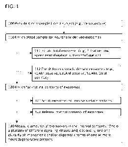
[0005] Referring to FIG. 1, assays for kinases include the
following operations: A body fluid
sample, such as a blood or saliva sample from a subject is obtained (100). The
blood sample
may be treated to provide a blood fraction, e.g., a plasma sample. The blood
sample is enriched
for neuronally-derived microparticles, e.g., exosomes (e.g., neuronally
derived exosomes are
isolated from the blood sample) (110). This can be a two-step operation that
involves, first,
isolating total exosomes (111) and, second, enriching neuronally derived
exosomes from the
total exosomes (112). Isolated exosomes are enriched for their internal
contents (120). This
can involve scrubbing to remove proteins attached to their surfaces (121). The
internally
enriched contents of the exosomes are released for analysis (122). Analysis
involves
measuring in the sample biomarkers selected from either (1) amounts of at
least one signaling
1
CA 03163308 2022- 6- 28
WO 2021/138364
PCT/US2020/067368
kinase and, optionally, at least one oligomeric form of a neurodegenerative
protein (e.g.,
oligomeric alpha synuclein), or (2) amounts of (e.g., activity of) each of a
plurality of different
signaling kinases. Optionally, amounts of one or more forms of
neurodegenerative proteins,
e.g., monomeric a synuclein and/or oligomeric a synuclein and tau or amyloid
beta, also can be
measured. Measurement of kinases, in combination with oligomeric forms, can
reduce false
positive classifications.
[0006] Measures of these bionnarkers, can be used in diagnostic
testing to determine
presence or absence of a particular neurodegenerative condition (e.g., a
syncucleinopathic
condition) or of its cumulative severity or current rate of progression, or to
determine efficacy of
a drug to alter amounts or relative amounts of one or more biomarker proteins
described herein
toward normal amounts.
[0007] Disclosed herein are, among other things, biomarker
profiles for neurodegenerative
conditions, such as syncucleinopathic conditions, amyloidopathic conditions,
tauopathies and
Huntington's disease, and the neurodegeneration associated therewith. In
certain
embodiments, the biomarker profiles comprise measures of a set of biomarkers
that include at
least one signaling kinase and that can be selected from (1) at least one
signaling kinase and,
optionally, at least one oligomeric form of a neurodegenerative protein, or
(2) each of one or a
plurality of different signaling kinases. Biomarker profiles can comprise
measures of one or
more oligomeric forms of neurodegenerative proteins, such as alpha-synuclein,
amyloid beta,
tau or huntingtin.
[0008] Signaling kinases measured can be one or a plurality of
kinases. They can be
selected from the same signaling pathway, such as the mTOR pathway, or from
different
signaling pathways.
[0009] Oligomeric forms of neurodegenerative proteins measured
can be a collection of
forms, such as total oligomeric alpha synuclein, or individual oligomeric
forms, such as a
tetramer of alpha synuclein, or a plurality of forms, such as alpha synuclein
dimers, turners and
tetramers. Monomeric forms of the neurodegenerative protein also can be
measured. So, for
example, the biomarker profile can comprise measures of each of one or a
plurality of
neurodegenerative protein forms selected from: (I) at least one oligomeric
form; (II) a plurality
(e.g., pattern) of oligomeric forms; (Ill) at least one oligomeric form and at
least one monomeric
form; (IV) a plurality of oligomeric forms and at least one monomeric form;
(V) at least one
oligomeric form and a plurality of monomeric forms; and (VI) a plurality of
oligomeric forms and
a plurality of monomeric forms.
[0010] Further disclosed herein are methods of developing
pharmaceuticals for treatment of
neurodegenerative conditions, such as syncucleinopathic conditions,
amyloidopathic conditions,
tauopathic conditions, and Huntington's disease. The methods involve using a
biomarker profile
2
CA 03163308 2022- 6- 28
WO 2021/138364
PCT/US2020/067368
to determine the effect of a candidate pharmaceutical on the condition. The
biomarker profile
includes measures of a biomarker set including biomarkers selected from (1) at
least one
signaling kinase and, optionally, at least one oligomeric form of a
neurodegenerative protein, or
(2) each of one or a plurality of different signaling kinases. Biomarker
proteins can be quantified
from, e.g., neuronally derived microparticles, e.g., exosomes, from the blood
of a subject.
[0011] In certain embodiments, the protein species are measured
from neuronally derived
extracellular vesicles, hereinafter termed exosomes, isolated, e.g., from
blood, saliva, or urine.
The species examined can derive from an internal compartment of the exosome,
e.g., from
exosomes from which surface proteins have been removed. The biomarker
profiles, measured
in this way, represent a relatively simple and non-invasive means for
measurement.
[0012] As such, methods of this disclosure for measuring a
biomarker profile for a
neurodegenerative condition are useful in drug development for testing
neuroprotective efficacy
of a drug candidate, sometimes referred to herein as a putative
neuroprotective agent. For
example, the methods described herein can be used to further understand the
downstream
effects of kinase activity, and to accelerate the development of effective
therapeutic strategies.
Such methods also are useful for identifying subjects for enrollment in
clinical trials and for
determining a diagnosis, prognosis, progression or risk of developing a
syncucleinopathic
condition. Further provided herein are novel methods of treating a subject
determined, by the
methods of this disclosure, to have or to be at risk of developing
neurodegeneration associated
with syncucleinopathic conditions, in particular, a neuroprotective treatment.
[0013] In one embodiment provided herein is a method comprising:
a) enriching each
biological sample in a collection of biological samples for neuronally derived
nnicroparticles, e.g.,
exosomes, wherein: (i) the collection of biological samples is from subjects
in a cohort of
subjects, wherein the cohort comprises subjects including: (1) a plurality of
subjects diagnosed
with a neurodegenerative condition at each of a plurality of different disease
stages, wherein
each of the diagnosed subjects has received a putative neuroprotective agent,
and/or (2) a
plurality of healthy control subjects, wherein the biological samples were
collected before and
again at one or more times during and, optionally, after administration of the
putative
neuroprotective agent; b) isolating protein contents from an internal
compartment of the
nnicroparticles, e.g., exosomes, to produce a biomarker sample; c) measuring,
in the biomarker
sample, a set of bionnarkers to create a dataset, wherein the set of
bionnarkers includes: (i) at
least one signaling kinase and, optionally, at least one oligomeric form of a
neurodegenerative
protein; or (ii) a plurality of different signaling kinases; and d) performing
statistical analysis on
the dataset to compare differences in the biomarker sets: (i) in individual
subjects over time to
determine a diagnostic algorithm that predicts rates of disease progression or
degree of
response to the putative neuroprotective agent; or (ii) between different
subjects to determine a
diagnostic algorithm that (1) makes a pathogenic diagnosis, (2) separates
clinically similar but
3
CA 03163308 2022- 6- 28
WO 2021/138364
PCT/US2020/067368
etiologically different neurodegenerative disorder subgroups, or (3) predicts
whether or the
degree to which a subject is likely to respond to the putative neuroprotective
agent. In one
embodiment the method further comprises, before enriching: I) providing a
cohort of subjects,
wherein the cohort comprises subjects including: (i) a plurality of subjects
diagnosed with a
neurodegenerative condition at each of a plurality of different disease
stages, and/or (ii) a
plurality of healthy control subjects; II) administering to each of the
diagnosed subjects a
putative neuroprotective agent; Ill) before and again at one or more times
during and, optionally,
after administration of the putative neuroprotective agent, collecting a
biological sample from
each of the subjects in the cohort. In another embodiment at least one of the
signaling kinases
is a kinase of the PI3K-Akt-mTOR signaling pathway. In another embodiment
wherein at least
one of the signaling kinases is selected from mitogen-activated protein kinase
(MARK or MEK),
extracellular signal-regulated kinases (ERK), glycogen synthase kinase 3 beta
(GSK3B), AKT
kinase and beclin. In another embodiment wherein the neurodegenerative protein
selected from
alpha synuclein, amyloid beta, tau, or huntingtin. In another embodiment the
oligomeric form of
the neurodegenerative protein is a collection of oligomeric forms, e.g.,
oligomers of alpha
synuclein, e.g., alpha synuclein 2-50, e.g., alpha synuclein 4-30, e.g., alpha
synuclein 4-20. In
another embodiment the method further comprises: e) validating one or more of
the diagnostic
algorithms against standard clinical measures. In another embodiment wherein
the statistical
analysis comprises: correlational, Pearson correlation, Spearman correlation,
chi-square,
comparison of means (e.g., paired T-test, independent T-test, ANOVA)
regression analysis
(e.g., simple regression, multiple regression, linear regression, non-linear
regression, logistic
regression, polynomial regression. stepwise regression, ridge regression,
lasso regression,
elasticnet regression) or non-parametric analysis (e.g., Wilcoxon rank-sum
test, Wilcoxon sign-
rank test, sign test). In another embodiment the statistical analysis is
executed by computer. In
another embodiment wherein the statistical analysis comprises machine
learning. In another
embodiment the subjects are humans. In another embodiment the
neurodegenerative condition
is a syncucleinopathic disorder. In another embodiment the syncucleinopathic
disorder is
Parkinson's disease. In another embodiment the syncucleinopathic disorder is
Lewy body
dementia. In another embodiment the standard clinical measures are selected
from UPDRS
scores, CGI scores and radiologic findings. In another embodiment the
neurodegenerative
condition is an amyloidopathy, a tauopathy or Huntington's disease. In another
embodiment the
biological sample comprises a venous blood sample. In another embodiment the
different
disease stages comprise one or more of suspected, early, middle, and
clinically advanced. In
another embodiment the times during or after administration are selected from
1, 2, 3 or more
months after treatment. In another embodiment enriching comprises using one or
more brain-
specific protein markers. In another embodiment at least one of the brain-
specific markers
comprises K1cam. In another embodiment isolating comprises washing the
exosomes in each
enriched sample to remove surface membrane-bound proteins. In another
embodiment the
4
CA 03163308 2022- 6- 28
WO 2021/138364
PCT/US2020/067368
exosomes are washed with PBS. In another embodiment the forms of the
neurodegenerative
protein are measured by gel electrophoresis, Western blot or fluorescence
techniques.
[0014]
In another aspect provided herein is a method comprising: a) enriching a
biological
sample from a subject for neuronally derived microparticles, e.g., exosomes;
b) isolating protein
contents from an internal compartment of the microparticles, e.g., exosomes,
to produce a
biomarker sample; c) measuring, in the biomarker sample, a set of biomarkers
to create a
dataset, wherein the set of bionnarkers includes: (1) at least one signaling
kinase and, optionally,
at least one oligomeric form of a neurodegenerative protein; or (2) a
plurality of different
signaling kinases; and d) using the dataset to perform one of the following:
(1) make a
pathogenic diagnosis, (2) classify the subject into one of a plurality of
clinically similar but
etiologically different neurodegenerative disorder subgroups, or (3) predict
whether or the
degree to which the subject is likely to respond to the putative
neuroprotective agent. In another
embodiment using comprises executing a diagnostic algorithm as described
herein, on the
dataset. In another embodiment at least one of the signaling kinases is a
kinase of the PI3K-
Akt-mTOR signaling pathway. In another embodiment wherein at least one of the
signaling
kinases is selected from mitogen-activated protein kinase (MAPK or MEK),
extracellular signal-
regulated kinases (ERK), glycogen synthase kinase 3 beta (GSK3B), AKT kinase
and beclin. In
another embodiment the neurodegenerative protein selected from alpha
synuclein, amyloid
beta, tau, or huntingtin. In another embodiment the oligomeric form of the
neurodegenerative
protein is a collection of oligomeric forms, e.g., oligomers of alpha
synuclein, e.g., alpha
synuclein 2-50, e.g., alpha synuclein 4-30, e.g., alpha synuclein 4-20. In
another embodiment
isolating neuronally derived exosomes comprises: (i) ultra-centrifugation;
(ii) density gradient
centrifugation; or (iii) size exclusion chromatography. In another embodiment
isolating
neuronally derived exosomes comprises capturing the neuronally derived
exosomes using a
binding moiety that binds to brain-specific protein. In another embodiment the
brain-specific
protein is Li CAM. In another embodiment removing proteins from the surface of
the isolated
exosomes comprises washing the isolated exosomes with an aqueous solution
(e.g., phosphate
buffered saline ("PBS")). In another embodiment determining amounts of a
neurodegenerative
protein comprises: i) separating species of oligomeric a-synuclein into a
plurality of fractions; ii)
measuring each of one or a plurality of the separated oligomeric a-synuclein
species and,
optionally, one or a plurality of species selected from: monomeric a-
synuclein, tau-synuclein co-
polymers, amyloid beta-synuclein co-polymers and tau-amyloid beta-synuclein co-
polymers. In
another embodiment separating species into a plurality of fractions comprises
separating by
electrophoresis. In another embodiment separating species into a plurality of
fractions
comprises separating by chromatography. In another embodiment determining
among the
separated species, at least one oligomeric form of a-synuclein selected from
forms having
between 2 and about 100 monomeric units, between 4 and 16 monomeric units and
no more
than about 30 monomeric units. In another embodiment determining among the
separated
5
CA 03163308 2022- 6- 28
WO 2021/138364
PCT/US2020/067368
species, a quantitative measure of monomeric a-synuclein. In another
embodiment measuring
among the separated species, a plurality of different oligomeric a-synuclein
species. In another
embodiment measuring among the separated species a co-polymer comprising a-
synuclein and
tau. In another embodiment determining among the separated species, a
quantitative measure
of a co-polymer comprising a-synuclein and amyloid beta. In another embodiment
measuring
the separated species comprises detecting one or a plurality of separated
species by
immunoassay. In another embodiment the immunoassay comprises immunoblotting.
In another
embodiment the immunoassay comprises Western blot. In another embodiment the
immunoassay uses an antibody coupled to a direct label. In another embodiment
the
immunoassay uses an antibody coupled to an indirect label. In another
embodiment the method
further comprises: I) measuring the biomarkers in the subject before and after
administration of
a putative neuroprotective agent; and II) determining changes in amounts of
proteins or patterns
of biomarkers, wherein changes toward normal amounts or patterns indicate
efficacy of the
neuroprotective agent. In another embodiment the method further comprises:
measuring the
biomarkers in the subject at two different times; and determining changes in
amounts of proteins
or patterns of biomarkers, wherein changes indicate a change in a
neurodegenerative state. In
another embodiment the method comprises collecting a plurality of biological
samples from the
subject over a time period, optionally wherein the subject is receiving a
putative or known
neuroprotective agent during the time period, wherein the diagnostic algorithm
predicts rates of
disease progression or degree of response to the putative neuroprotective
agent.
[0015] In another aspect provided herein is a method comprising:
a) providing a dataset
comprising, for each of a plurality of subjects, values indicating (1) state
of a neurodegenerative
condition, and (2) measures of a set of biomarkers, wherein the set of
biomarkers includes: (i) at
least one signaling kinase and, optionally, at least one oligomeric form of a
neurodegenerative
protein; or (ii) a plurality of different signaling kinases; and b) performing
a statistical analysis on
the dataset to develop a model that infers the state of the neurodegenerative
condition in an
individual. In one embodiment at least one of the signaling kinases is a
kinase of the PI3K-Akt-
mTOR signaling pathway. In another embodiment at least one of the signaling
kinases is
selected from mitogen-activated protein kinase (MAPK or MEK), extracellular
signal-regulated
kinases (ERK), glycogen synthase kinase 3 beta (GSK3B), AKT kinase and beclin.
In another
embodiment the neurodegenerative protein selected from alpha synuclein,
amyloid beta, tau, or
huntingtin. In another embodiment the oligomeric form of the neurodegenerative
protein is a
collection of oligomeric forms, e.g., oligomers of alpha synuclein, e.g.,
alpha synuclein 2-50,
e.g., alpha synuclein 4-30, e.g., alpha synuclein 4-20. In another embodiment
the statistical
analysis is performed by computer. In another embodiment the statistical
analysis is not
performed by computer. In another embodiment the statistical analysis
comprises: correlational,
Pearson correlation, Spearman correlation, chi-square, comparison of means
(e.g., paired T-
test, independent T-test, ANOVA) regression analysis (e.g., simple regression,
multiple
6
CA 03163308 2022- 6- 28
WO 2021/138364
PCT/US2020/067368
regression, linear regression, non-linear regression, logistic regression,
polynomial regression.
stepwise regression, ridge regression, lasso regression, elasticnet
regression) or non-
parametric analysis (e.g., Wilcoxon rank-sum test, Wilcoxon sign-rank test,
sign test). In another
embodiment the statistical analysis comprises training a machine learning
algorithm on the
dataset. In another embodiment the machine learning algorithm is selected
from: artificial
neural networks (e.g., back propagation networks), decision trees (e.g.,
recursive partitioning
processes, CART), random forests, discriminant analyses (e.g., Bayesian
classifier or Fischer
analysis), linear classifiers (e.g., multiple linear regression (MLR), partial
least squares (PLS)
regression, principal components regression (PCR)), mixed or random-effects
models, non-
parametric classifiers (e.g., k-nearest neighbors), support vector machines,
and ensemble
methods (e.g., bagging, boosting). In another embodiment the state is selected
from diagnosis,
stage, prognosis or progression of the neurodegenerative condition. In another
embodiment the
state is measured as a categorical variable (e.g., a binary state or one of a
plurality of
categorical states). In another embodiment the categories comprise a diagnosis
consistent with
(e.g., positive or diagnosed as having) having the neurodegenerative condition
and inconsistent
with (e.g., negative or diagnosed as not having) having the neurodegenerative
condition. In
another embodiment the categories comprise different stages of the
neurodegenerative
condition. In another embodiment the state is measured as a continuous
variable (e.g., on a
scale). In another embodiment the continuous variable is a range is or degrees
of the
neurodegenerative condition. In another embodiment the subjects are animals,
e.g., fish,
avians, amphibians, reptiles, or mammals, e.g., rodents, primates or humans.
In another
embodiment the plurality of subjects is at least any of 10, 25, 50, 100, 200,
400 or 800. In
another embodiment, for each subject, the sample for which the quantitative
measures are
determined are taken at a first time point and the state of the
neurodegenerative condition is
determined at a second, later time point. In another embodiment the biological
sample
comprises blood or a blood fraction (e.g., plasma or serum). In another
embodiment the
neurodegenerative condition is a synucleinopathy, e.g., Parkinson's Disease or
Lewy Body
Dementia. In another embodiment the neurodegenerative condition is an
amyloidopathy, e.g.,
Alzheimer's Disease, a tauopathy, e.g., Alzheimer's Disease or Huntington's
disease.
[0016] In another aspect provided herein is a method of inferring a risk of
developing, a
diagnosis of, a stage of, a prognosis of or a progression of a
neurodegenerative condition
characterized by a neurodegenerative protein, wherein the method comprises: a)
measuring,
from a biological sample from a subject that is enriched for neuronally
derived microparticles,
e.g., exosomes, a set of biomarkers to create a dataset, wherein the set of
biomarkers includes:
(1) at least one signaling kinase and, optionally, at least one oligomeric
form of a
neurodegenerative protein; or (2) a plurality of different signaling kinases;
and b) executing a
model, e.g., a model as described herein, on the dataset to infer a risk of
developing, a
diagnosis of, a stage of, a prognosis of or a progression of the
neurodegenerative condition. In
7
CA 03163308 2022- 6- 28
WO 2021/138364
PCT/US2020/067368
one embodiment at least one of the signaling kinases is a kinase of the PI3K-
Akt-mTOR
signaling pathway. In another embodiment at least one of the signaling kinases
is selected from
mitogen-activated protein kinase (MAPK or MEK), extracellular signal-regulated
kinases (ERK),
glycogen synthase kinase 3 beta (GSK3B), AKT kinase and beclin. In another
embodiment the
neurodegenerative protein selected from alpha synuclein, amyloid beta, tau, or
huntingtin. In
another embodiment the oligomeric form of the neurodegenerative protein is a
collection of
oligomeric forms, e.g., oligomers of alpha synuclein, e.g., alpha synuclein 2-
50, e.g., alpha
synuclein 4-30, e.g., alpha synuclein 4-20. In another embodiment the
neurodegenerative
protein forms for which the quantitative measures are determined are selected
from: (I) at least
one oligomeric form; (II) a plurality of oligomeric forms; (Ill) at least one
oligomeric form and at
least one monomeric form; (IV) a plurality of oligomeric forms and at least
one monomeric form;
(V) at least one oligomeric form and a plurality of monomeric forms; and (VI)
a plurality of
oligomeric forms and a plurality of monomeric forms. In another embodiment at
least one of the
oligomeric forms comprises a collection of species of the neurodegenerative
protein. In another
embodiment the model comprises comparing relative amounts an oligomeric form
to monomeric
form of the neurodegenerative protein to relative amounts in a statistically
significant number of
control individuals. In another embodiment the model comprises detecting a
pattern of relative
amounts of a plurality of the oligomeric forms from which model the inference
is made. In
another embodiment the subject is asymptomatic or preclinical for a
neurodegenerative
condition. In another embodiment the subject presents to a healthcare
provider, such as a
doctor, during a routine office visit or as part of a doctor's ordinary
practice of medicine. In
another embodiment the model is executed by computer. In another embodiment
the model is
not executed by computer.
[0017] In another aspect provided herein is a method for
determining effectiveness of a
therapeutic intervention in treating a neurodegenerative condition, wherein
the method
comprises: (a) inferring, in each subject in a population comprising a
plurality of subjects, an
initial state of a neurodegenerative condition by: (1) measuring, from a
biological sample from a
subject that is enriched for neuronally derived microparticles, e.g.,
exosomes, a set of
biomarkers to create a dataset, wherein the set of biomarkers includes: (i) at
least one signaling
kinase and, optionally, at least one oligomeric form of a neurodegenerative
protein; or (ii) a
plurality of different signaling kinases; and (2) inferring the initial state
using a model, e.g., a
model as described herein; (b) after inferring, administering the therapeutic
intervention to the
subjects; (c) after administering, inferring, in each subject individual in
the population, a
subsequent a subsequent state of the neurodegenerative condition by: (1)
measuring, from a
biological sample from a subject that is enriched for neuronally derived
microparticles, e.g.,
exosomes, a set of biomarkers to create a dataset, wherein the set of
biomarkers includes; (i) at
least one signaling kinase and, optionally, at least one oligomeric form of a
neurodegenerative
protein; or (ii) a plurality of different signaling kinases; and (2) inferring
the subsequent state
8
CA 03163308 2022- 6- 28
WO 2021/138364
PCT/US2020/067368
using the model; and (d) based on the initial and subsequent inferences in the
population,
determining that the therapeutic intervention is effective if the subsequent
inferences exhibit a
statistically significant change toward a normal state compared with the
initial inferences, or that
the therapeutic intervention is not effective if the subsequent inferences do
not exhibit a
statistically significant change compared with the initial inferences toward a
normal state. In
another embodiment at least one of the signaling kinases is a kinase of the
PI3K-Akt-mTOR
signaling pathway. In another embodiment at least one of the signaling kinases
is selected from
nnitogen-activated protein kinase (MARK or MEK), extracellular signal-
regulated kinases (ERK),
glycogen synthase kinase 3 beta (GSK3B), AKT kinase and beclin. In another
embodiment the
neurodegenerative protein selected from alpha synuclein, amyloid beta, tau, or
huntingtin. In
another embodiment the oligomeric form of the neurodegenerative protein is a
collection of
oligomeric forms, e.g., oligomers of alpha synuclein, e.g., alpha synuclein 2-
50, e.g., alpha
synuclein 4-30, e.g., alpha synuclein 4-20. In another embodiment the
therapeutic intervention
comprises administration of a drug or combination of drugs. In another
embodiment the
population comprises at least 20, at least 50, at least 100 or at least 200
subjects, wherein at
least 20%, at least 35%, at least 50%, or at least 75% of the subjects
initially have elevated
relative amounts of oligomeric forms of the protein to monomeric forms of the
protein. In another
embodiment at least 20%, at least 25%, at least 30%, or at least 35%, least
50%, at least 66%,
at least 80%, or 100% of the subjects initially have a diagnosis of a
neurodegenerative
condition. In another embodiment the inference is made by computer. In another
embodiment
the inference is not made by computer.
[0018] In another aspect provided herein is a method for
qualifying subjects for a clinical
trial of a therapeutic intervention for the treatment or prevention of a
neurodegenerative
condition comprising: a) determining that a subject is abnormal with respect
with a
neurodegenerative condition by: (1) measuring, from a biological sample from a
subject that is
enriched for neuronally derived nnicroparticles, e.g., exosonnes, a set of
biomarkers to create a
dataset, wherein the set of biomarkers includes; (i) at least one signaling
kinase and, optionally,
at least one oligomeric form of a neurodegenerative protein; or (ii) a
plurality of different
signaling kinases; (2) executing a model, e.g., a model as described herein,
on the profile to
infer that the subject is abnormal with respect with the neurodegenerative
condition; and b)
enrolling the subject in the clinical trial of a potentially therapeutic
intervention for said
neurodegenerative condition. In one embodiment at least one of the signaling
kinases is a
kinase of the PI3K-Akt-mTOR signaling pathway. In another embodiment at least
one of the
signaling kinases is selected from mitogen-activated protein kinase (MAPK or
MEK),
extracellular signal-regulated kinases (ERK), glycogen synthase kinase 3 beta
(GSK3B), AKT
kinase and beclin. In another embodiment the neurodegenerative protein is
selected from alpha
synuclein, amyloid beta, tau, or huntingtin. In another embodiment the
oligomeric form of the
neurodegenerative protein is a collection of oligomeric forms, e.g., oligomers
of alpha synuclein,
9
CA 03163308 2022- 6- 28
WO 2021/138364
PCT/US2020/067368
e.g., alpha synuclein 2-50, e.g., alpha synuclein 4-30, e.g., alpha synuclein
4-20. In another
embodiment the model is executed by computer. In another embodiment the model
is not
executed by computer.
[0019] In another aspect provided herein is a method of
monitoring progress of a subject on
a therapeutic intervention for a neurodegenerative condition comprising: (a)
inferring, in the
subject, an initial state of a neurodegenerative condition by: (1)
determining, from a biological
sample from a subject that is enriched for neuronally derived nnicroparticles,
e.g., exosomes,
measures of a set of biomarkers, wherein the set of biomarkers includes: (i)
at least one
signaling kinase and, optionally, at least one oligomeric form of a
neurodegenerative protein; or
(ii) a plurality of different signaling kinases; and (2) executing a model,
e.g., a model as
described herein, to infer an initial state of the neurodegenerative
condition; (b) after inferring,
administering the therapeutic intervention to the subject; (c) after
administering, inferring, in the
subject, a subsequent state of the neurodegenerative condition by: (1)
determining, from a
biological sample from a subject that is enriched for neuronally derived
microsomal particles, a
biomarker profile comprising amounts of each of a plurality of different
signaling kinases to
create a dataset; and (2) executing a model, e.g. a model as described herein,
to infer a
subsequent state of the neurodegenerative condition; (d) based on the initial
and subsequent
state inferences, determining that the subject is responding positively to the
therapeutic
intervention if the subsequent inference exhibits a change toward a normal
state compared with
the initial inferences, or that the therapeutic intervention is not effective
if the subsequent
inferences do not exhibit a change compared with the initial inferences toward
a normal state. In
one embodiment at least one of the signaling kinases is a kinase of the PI3K-
Akt-mTOR
signaling pathway. In another embodiment at least one of the signaling kinases
is selected from
mitogen-activated protein kinase (MAPK or MEK), extracellular signal-regulated
kinases (ERK),
glycogen synthase kinase 3 beta (GSK3B), AKT kinase and beclin. In another
embodiment the
neurodegenerative protein selected from alpha synuclein, annyloid beta, tau,
or huntingtin. In
another embodiment the oligomeric form of the neurodegenerative protein is a
collection of
oligomeric forms, e.g., oligomers of alpha synuclein, e.g., alpha synuclein 2-
50, e.g., alpha
synuclein 4-30, e.g., alpha synuclein 4-20. In another embodiment wherein the
model is
executed by computer. In another embodiment the model is not executed by
computer.
[0020] In another aspect provided herein is a method comprising:
(a) determining, by the
method as disclosed herein, that a subject has a neurodegenerative condition,
and (b)
administering to the subject a palliative or neuroprotective therapeutic
intervention efficacious to
treat the condition. In one embodiment the therapeutic intervention moves a
biomarker profile of
the subject toward normal, wherein a movement toward normal indicates
neuroprotection.
[0021] In another aspect provided herein is a method comprising
administering to a subject
determined by the method as disclosed herein, to have an abnormal pattern of
biomarkers, a
CA 03163308 2022- 6- 28
WO 2021/138364
PCT/US2020/067368
palliative or neuroprotective therapeutic intervention effective to treat the
condition. In one
embodiment the subject is asymptomatic or preclinical for the
neurodegenerative condition.
[0022] In another aspect provided herein is a kit comprising
reagents sufficient to detect
either: (1) at least one of signaling kinase and at least one oligomeric form
of a
neurodegenerative protein; or (2) a plurality of different signaling kinases.
In one embodiment
the reagents comprise antibodies.
[0023] In another aspect provided herein is a method of inferring
a risk of developing, a
diagnosis of, a stage of, a prognosis of or a progression of a
neurodegenerative condition,
wherein the method comprises: a) measuring, from a biological sample from a
subject that is
enriched for neuronally derived microparticles, e.g., exosomes, a set of
biomarkers to create a
dataset, wherein the set of biomarkers includes: (1) at least one signaling
kinase and, optionally,
at least one oligomeric form of a neurodegenerative protein; or (2) a
plurality of different
signaling kinases; and b) correlating the dataset with a risk of developing, a
diagnosis of, a
stage of, a prognosis of or a progression of the neurodegenerative condition.
In one
embodiment at least one of the signaling kinases is a kinase of the PI3K-Akt-
mTOR signaling
pathway. In another embodiment at least one of the signaling kinases is
selected from mitogen-
activated protein kinase (MAPK or MEK), extracellular signal-regulated kinases
(ERK), glycogen
synthase kinase 3 beta (GSK3B), AKT kinase and beclin. In another embodiment
wherein the
neurodegenerative protein selected from alpha synuclein, amyloid beta, tau, or
huntingtin. In
another embodiment the oligomeric form of the neurodegenerative protein is a
collection of
oligomeric forms, e.g., oligomers of alpha synuclein, e.g., alpha synuclein 2-
50, e.g., alpha
synuclein 4-30, e.g., alpha synuclein 4-20.
[0024] In another aspect provided herein is a method comprising:
(a) identifying a subject
having a neurodegenerative condition or likely to positively respond to a
treatment for a
neurodegenerative condition, wherein identifying comprises: (1) measuring, in
a sample from
the subject enriched for neuronally derived exosomes (e.g., from the internal
contents of the
exosomes), a set of biomarkers, to create a biomarker profile, wherein the set
of biomarkers
includes one or a plurality of signaling kinases and, optionally, at least one
oligomeric form of a
neurodegenerative protein; and (2) determining, based on an abnormal biomarker
profile, that
the subject suffers from the neurodegenerative condition; and (b)
administering to the identified
subject, an effective amount of a pharmaceutical composition to treat the
neurodegenerative
condition. In one embodiment the neurodegenerative condition is a
synucleopathic condition,
and the pharmaceutical composition comprises comprising a dopamine agonist
(e.g.,
prannipexole (e.g., MirapexTm), ropinirole (e.g., Requip), rotigotine (e.g.,
Neupro), aponnorphine
(e.g., Apokyn)), levodopa, carbidopa-levodopa (e.g., Rytary, Sinemet), a MAO-B
inhibitor (e.g.,
selegiline (e.g., Eldepryl, Zelapar) or rasagiline (e.g., Azilect)), a
catechol-O-methyltransferase
(COMT) inhibitor (e.g., entacapone (Comtan) or tolcapone (Tasmar)), an
anticholinergic (e.g.,
11
CA 03163308 2022- 6- 28
WO 2021/138364
PCT/US2020/067368
benztropine (e.g., Cogentin) or trihexyphenidyl), amantadine or a
cholinesterase inhibitor (e.g.,
rivastigmine (Exelon)). In another embodiment the synucleopathic condition is
Parkinson's
Disease. In another embodiment the pharmaceutical composition comprises a
dopamjne
agonist. In another embodiment the pharmaceutical composition further
comprises an NK1-
antagogonist. In another embodiment the dopamine agonist is 6-propylamino-
4,5,6,7-
tetrahydro-1,3-benzothiazole-2-amine and the NK1-antagonist is aprepitant or
rolapitant. In
another embodiment the pharmaceutical composition further comprises an 5HT3-
antagonist. In
another embodiment the dopamine agonist is 6-propylannino-4,5,6,7-tetrahydro-
1,3-
benzothiazole-2-amine and the 5HT3 antagonist is ondansetron hydrochloride
dihydrate.
[0025] In another aspect provided herein is a method comprising
administering to a subject
characterized as having a biomarker profile indicative of a neurodegenerative
condition or being
likely to positively respond to a treatment for a neurodegenerative condition,
an effective amount
of a pharmaceutical composition to treat the neurodegenerative condition;
wherein the
biomarker panel comprises set of biomarkers includes one or a plurality of
signaling kinases
and, optionally, at least one oligomeric form of a neurodegenerative protein
measured from a
sample from the subject enriched for neuronally derived exosomes (e.g., from
the internal
contents of the exosomes). In on embodiment In another embodiment the
neurodegenerative
condition is Parkinson's Disease, and wherein the pharmaceutical composition
comprises a
dopamine agonist.
[0026] In certain embodiments, the biomarkers are selected from one or a
plurality of
different signaling kinases and, optionally, one or a plurality of monomeric
and/or oligomeric
forms of each of one or a plurality of neurodegenerative proteins.
[0027] Other objects of the disclosure may be apparent to one
skilled in the art upon
reading the following specification and claims.
BRIEF DESCRIPTION OF THE DRAWINGS
[0028] The novel features of the disclosure are set forth with
particularity in the appended
claims. A better understanding of the features and advantages of the present
disclosure will be
obtained by reference to the following detailed description that sets forth
illustrative
embodiments, in which the principles of the disclosure are utilized, and the
accompanying
drawings of which:
[0029] FIG. 1 shows a flow diagram of an exemplary method
detecting kinases and,
optionally, neurodegenerative protein forms from exosomes.
[0030] FIG. 2 shows a flow diagram of an exemplary protocol to
validate drug efficacy.
[0031] FIG. 3 shows an exemplary flow diagram of creating and
validating a diagnostic
model for diagnosing a neurodegenerative condition.
12
CA 03163308 2022- 6- 28
WO 2021/138364
PCT/US2020/067368
[0032] FIG. 4 shows an exemplary flow diagram for classifying a
subject according to any of
several states by executing a diagnostic algorithm, or model, on a biomarker
profile.
DETAILED DESCRIPTION OF THE DISCLOSURE
I. Biomarkers for Neurodegenerative Conditions
[0033] Methods disclosed herein are useful for diagnosis of and drug
development for a
variety of neurodegenerative conditions. These include, without limitation,
synucleinopathies
(e.g., Parkinson's disease, Lewy body dementia, multiple system atrophy),
amyloidopathies
(e.g., Alzheimer's disease), tauopathies (e.g., Alzheimer's disease,
Progressive supranuclear
palsy, Corticobasal degeneration), and Huntington's disease.
A. Biomarkers and Biomarker Profiles
[0034] Biomarkers are analytes that are associated, positively or
negatively, alone or in
combination, with a particular condition. Analytes that can function as
biomarkers include any
biological molecule or organic or inorganic molecule that is detectable in a
subject or a subject
sample. Biological molecules that can serve as biomarkers include, without
limitation,
polypeptides and polynucleotides, including, for example, proteins and
peptides.
[0035] As used herein, the term "biomarker profile" refers to
data indicating a measure of
each of one or a plurality of biomarkers. Biomarker profiles used in certain
embodiments of the
methods described herein include measures of activity of one or a plurality of
different kinases.
In certain embodiments, biomarker profiles can further comprise measures of
one or more
neurodegenerative protein forms. A biomarker profile is a form of a dataset
that includes data
about the biomarkers.
[0036] The term "biomarker profile" may also be used to refer to
a particular pattern in the
profile which a model infers to be associated with a diagnosis, stage,
progression, rate,
prognosis, drug responsiveness and risk of developing a neurodegenerative
condition.
[0037] A measurement of a variable, such as kinase activity, can be any
combination of
numbers and words. A measure can be any scale, including nominal (e.g., name
or category),
ordinal (e.g., hierarchical order of categories), interval (distance between
members of an order),
ratio (interval compared to a meaningful "0"), or a cardinal number
measurement that counts the
number of things in a set. Measurements of a variable on a nominal scale
indicate a name or
category, such a "healthy" or "unhealthy", "old" or "young", "form 1" or "form
2", "subject 1 ...
subject n," etc. Measurements of a variable on an ordinal scale produce a
ranking, such as
"first", "second", "third"; or "youngest" to "oldest", or order from most to
least. Measurements on
a ratio scale include, for example, any measure on a pre-defined scale, such
as mass, signal
strength, concentration, age, etc., as well as statistical measurements such
as frequency, mean,
median, standard deviation, or quantile. Measurements on a ratio scale can be
relative amounts
13
CA 03163308 2022- 6- 28
WO 2021/138364
PCT/US2020/067368
or normalized measures. For example, in one embodiment, a biomarker profile
comprises a
relative amount of a first and second signaling kinase. In another embodiment
a biomarker
profile comprises a ratio of amounts of two different biomarker proteins.
[0038] Abnormal profiles (e.g., abnormal absolute or relative
amounts of various signaling
kinases) indicate pathologic activity, (or a characteristic bodily response to
a pathogenic
process) and thus time to future clinical onset and subsequent rates of
clinical progression.
Moreover, return toward normal in biomarker profiles (e.g., reductions in
absolute or relative
amounts of signaling kinases and/or oligomeric forms of neurodegenerative
proteins) reflects
the efficacy of a candidate neuroprotective intervention. Accordingly, the
biomarker profiles
described herein are useful for determining efficacy of drug candidates for
their neuroprotective
effect. As a practical matter, they may be considered essential to the
practical conduct of
neuroprotective drug trial in view of savings in both time and cost as well as
a definitive means
to quantified efficacy against a pathogenic process rather than its clinical
manifestations.
[0039] Accordingly, biomarker profiles function not only as a
diagnostic of an existing
pathological state but also as a sentinel of pathology before clinical onset,
e.g., when a subject
is pre-symptomatic or preclinical, e.g., has signs or symptoms that are
insufficient for a
diagnosis of disease. This is relevant since the relative success of
neuroprotective treatments
often appear related to their earliest possible administration. Further, it is
believed that these
biomarker profiles indicate the stage (e.g., rate of or cumulative amount of
neuronal loss) of a
neurodegenerative condition. Accordingly, determining biomarker profiles can
be of critical
importance for determining effectiveness of a treatment, for example, in
clinical trials and, for
therapeutic interventions believed to be effective for treating
neurodegeneration including, e.g.,
synucleinopathy, amyloidopathy, tauopathy or Huntington's disease in the
individual.
B. Signaling Kinases
[0040] These diseases are characterized by abnormal changes in the activity
(increased or
decreased) of particular signaling kinases. Measuring activity of these
signaling kinases in a
subject can be used for diagnosis, prognosis, patient progress, patient
stratification and drug
development and testing.
[0041] Kinases include any kinase involved in signaling pathway.
[0042] Kinases associated with Parkinson's disease or the administration of
medications
that influence of the symptoms of Parkinson's disease (e.g., pramipexole (6-
propylamino-
4,5,6,7-tetrahydro-1,3-benzothiazole-2-amine)) include, without limitation,
mTOR (mechanistic
target of rapamycin), mitogen-activated protein kinase (MAPK or MEK),
extracellular signal-
regulated kinases (ERK), glycogen synthase kinase 3 beta (GSK3B), AKT kinase
and beclin
Leucine-Rich Repeat Kinase 2 (LRRK2), members of the c-Jun N-Terminal Kinase
Signaling
14
CA 03163308 2022- 6- 28
WO 2021/138364
PCT/US2020/067368
Pathway (JNK) (MAPK serine-threonine kinases), and Phosphatase and Tensin
Homolog
(PTEN)-Induced Putative Kinase 1 (PINK1).
[0043] Kinases associated with Alzheimer's disease include,
without limitation, Tau protein
kinases such as proline-directed protein kinases (PDPK), protein kinases non-
PDPK and
tyrosine protein kinases (TPK).
[0044] Kinases associated with Huntington's disease include,
without limitation, nnitogen-
activated protein kinase, MEK, ERK, JNK, IKK, cell division protein kinase 5
(CDK5), AKT,
MKP1.
[0045] These diseases also share in common the accumulation of
toxic oligomeric
polypeptide species, and in some cases abnormally phosphorylated oligomeric or
monomeric
forms, and the ability to detect such forms in neuronally derived exosomes.
C. Neurodegenerative Proteins
[0046] As used herein, the term "neurodegenerative protein"
refers to a protein which, in an
oligonnerized form, is associated with neurodegeneration. Neurodegenerative
proteins include,
without limitation, alpha-synuclein, tau, amyloid beta and huntingtin.
[0047] It is believed that certain oligomerized forms or
abnormally phosphorylated forms of
brain polypeptides underlie a variety of neurodegenerative conditions. This
includes, for
example, the roles of alpha-synuclein in syncucleinopathic conditions, amyloid
beta in
amyloidopathic conditions, tau in tauopathic conditions and huntingtin in
Huntington's disease.
In particular, current evidence suggests that a-synuclein oligomers can act as
a toxic species in
PD and other synucleinopathies. In certain embodiments, the oligomeric species
detected is an
abnormally phosphorylated species.
[0048] Forms of neurodegenerative proteins include, without
limitation, (I) at least one
oligomeric form; (II) a plurality of oligomeric forms in combination (e.g.,
all oligomeric forms or a
subset of oligomeric forms measured together, e.g., alpha synuclein 2-14),
(III) each of a
plurality of different oligomeric forms; (IV) at least one oligomeric form and
at least one
monomeric form; (V) a plurality of oligomeric forms and at least one monomeric
form; and (VI) at
least one oligomeric form and a plurality of monomeric forms. Forms of
neurodegenerative
proteins can be used in models to infer, among other things, neurodegenerative
conditions or
progression toward neurodegenerative conditions, typically with one or more
oligomeric forms
included in a model indicating the presence and activity of the disease or
progression towards
the disease. This includes increasing relative amounts of oligomeric alpha-
synuclein forms
indicating the presence and activity of a synucleinopathy, or progression
towards a
synucleinopathy; increasing relative amounts of oligomeric amyloid beta
indicating the presence
and activity of an annyloidopathy, or progression towards an annyloidopathy,
increasing relative
CA 03163308 2022- 6- 28
WO 2021/138364
PCT/US2020/067368
amounts of oligomeric or abnormally phosphorylated tau indicating the presence
and activity of
a tauopathy, or progression towards a tauopathy, and increasing relative
amounts of oligomeric
huntingtin indicating the presence and activity of Huntington's disease, or
progression towards
Huntington's disease. Accordingly, an abnormal profile of such oligomers
indicates a process of
neurodegeneration.
[0049] Neurodegenerative proteins forms can include one or more
oligomeric forms and,
optionally, one or more monomeric forms. This includes amounts of species of
oligomeric and,
optionally, monomeric alpha-synuclein; oligomeric and, optionally, monomeric
amyloid beta,
oligomeric and, optionally hyperphosphorylated and, optionally, monomeric tau;
and oligomeric
and, optionally, monomeric huntingtin. For example, a biomarker profile can
include (I) at least
one oligomeric form; (II) a plurality of oligomeric forms; (Ill) at least one
oligomeric form and at
least one monomeric form; (IV) a plurality of oligomeric forms and at least
one monomeric form;
(V) at least one oligomeric form and a plurality of monomeric forms; and (VI)
a plurality of
oligomeric forms and a plurality of monomeric forms.
[0050] Protein forms can refer to individual protein species or collections
of species. For
example, a 6-mer of alpha-synuclein is a form of alpha backspace-synuclein.
Also, the collection
of 6-mers to 18-mers of alpha-synuclein, collectively, can be a form of alpha-
synuclein.
[0051] A biomarker profile can include a plurality of forms of a
protein. In one embodiment,
a biomarker profile can include quantitative measures of each of a plurality
of oligomeric forms
and monomeric form of the neurodegenerative protein. So, for example, the
biomarker profile
could include quantitative measures of each of a dimer, trimer, tetramer, 5-
mer, 6-mer, 7-mer, 8-
mer, 9-mer, 10-mer, 11-mer, 12-mer, 13-mer, 14-mer, 15-mer, 16-mer, 19-nner,
20-mer, 24-mer,
50-mer, etc.
[0052] As used herein, a "synuclein biomarker profile" refers to
a profile comprising
oligomeric and, optionally, monomeric alpha-synuclein, the term "amyloid
biomarker profile"
refers to a profile comprising oligomeric and, optionally, monomeric beta-
amyloid, the term "tau
biomarker profile" refers to a profile comprising oligomeric and, optionally,
monomeric tau, the
term "huntingtin biomarker profile" refers to a profile comprising oligomeric
and, optionally,
monomeric huntingtin.
[0053] As used herein, the term "monomeric protein/polypeptide" refers to a
single, non-
aggregated protein or polypeptide molecule, including any species thereof,
such as
phosphorylated species. As used herein, the term "oligomeric
protein/polypeptide" refers to
individual oligomeric species or an aggregate comprising a plurality of
oligomeric species,
including phosphorylated species. It is understood that measurement of an
oligomeric form of a
protein, as used herein, can refer to measurement of all oligomeric forms
(total oligomeric form)
16
CA 03163308 2022- 6- 28
WO 2021/138364
PCT/US2020/067368
or specified oligomeric forms. Specified oligomeric forms can include, for
example, forms within
a particular size range or physical condition such as for example soluble
fibrils.
[0054] In each of these conditions, it is believed that
oligomerized/ aggregated forms of
polypeptides described herein are toxic to neurons in that the biomarker
profiles comprising
oligomeric forms and, optionally, monomeric forms of these polypeptides
function in models to
infer pathologic activity. In particular, increased relative amounts of
oligomeric forms as
compared with monomeric forms indicate pathology. Measures of these
bionnarkers can be
used to track subject responses to therapies that are either in existence or
in development as
well as to predict development of disease or the state or progress of existing
disease.
II. Neurodegenerative Conditions and Associated Proteins
A. Synucleinopathies
1. Conditions
[0055] As used herein, the terms "synucleinopathy" and
"syncucleinopathic condition" refer
to a condition characterized by abnormal profiles of oligomeric alpha-
synuclein, which is an
abnormal, aggregated form of alpha-synuclein. In certain embodiments,
synucleinopathies
manifest as clinically evident syncucleinopathic disease such as, for example,
PD, Lewy body
dementia, multiple system atrophy and some forms of Alzheimer's disease, as
well as other rare
neurodegenerative disorders such as various neuroaxonal dystrophies. Signs
and, optionally,
symptoms sufficient for a clinical diagnosis of a syncucleinopathic disease
are those generally
sufficient for a person skilled in the art of diagnosing such conditions to
make such a clinical
diagnosis.
[0056] Parkinson's disease ("PD") is a progressive disorder of
the central nervous system
(CNS) with a prevalence of 1% to 2% in the adult population over 60 years of
age. PD is
characterized by motor symptoms, including tremor, rigidity, postural
instability and slowness of
voluntary movement. The cause of the idiopathic form of the disease, which
constitutes more
than 90% of total PD cases, remains elusive, but is now considered to involve
both
environmental and genetic factors. Motor symptoms are clearly related to a
progressive
degeneration of dopamine-producing neurons in the substantia nigra. More
recently, PD has
become recognized one of a group of multi-system disorders, which mainly
affect the basal
ganglia (e.g., PD), or the cerebral cortex (e.g., Lewy body dementia), or the
basal ganglia, brain
stem and spinal cord (e.g., multiple system atrophy) and which are all linked
by the presence of
intracellular deposits (Lewy bodies) consisting mainly of a brain protein
called alpha-synuclein.
Accordingly, these disorders, along with Hallevorden-Spatz syndrome, neuronal
axonal
dystrophy, and traumatic brain injury have often been termed
"Synucleinopathies".
17
CA 03163308 2022- 6- 28
WO 2021/138364
PCT/US2020/067368
[0057] Signs and symptoms of PD may include, for example, tremors
at rest, rigidity,
bradykinesia, postural instability and a festinating parkinsonian gate. One
sign of PD is a
positive response in these motor dysfunctions to carbidopa-levodopa.
[0058] Clinically recognized stages of Parkinson's disease
include the following: Stage 1 ¨
mild; Stage 2 ¨ moderate; Stage 3 ¨ middle stage; Stage 4-severe; Stage 5 ¨
advanced.
[0059] Prannipexole (sold under the brand name MirapexTM) is a
drug that may treat
idiopathic Parkinsonism. Pramipexole has activity as an extracellular signal-
regulated kinase
(ERK) agonist. Accordingly, determining the effect of pramipexole, and other
kinase
modulators, on kinase activity is useful in determining effectiveness of the
drug on Parkinson's
Disease.
[0060] At present, the diagnosis of PD mainly rests on the
results of a physical examination
that is often quantified by the use of the modified Hoehn and Yahr staging
scale (Hoehn and
Yahr, 1967, Neurology, 17:5, 427-442) and the Unified Parkinson's Disease
Rating Scale
(UPDRS). The differential diagnosis of PD vs. other forms of parkinsonism,
e.g., progressive
supranuclear palsy (PSP), can prove difficult and misdiagnosis can thus occur
in up to 25% of
patients. Indeed, PD generally remains undetected for years before the initial
clinical diagnosis
can be made. When this happens, the loss of dopamine neurons in the substantia
nigra already
exceeds 50% and may approach 70%. No blood test for PD or any related
synucleinopathy has
yet been validated. While imaging studies using positron emission tomography
(PET) or MRI
have been used in the diagnosis of PD by providing information about the
location and extent of
the neurodegenerative process, they confer little or no information about the
pathogenesis of
the observed degeneration and do not guide the selection of a particular
syncucleinopathic-
specific intervention.
[0061] Lewy body dementias (LBD) affect about 1.3 million people
in the US. Symptoms
include, for example, dementia, cognitive fluctuations, parkinsonism, sleep
disturbances and
hallucinations. It is the second most common form of dementia after
Alzheimer's disease and
usually develops after the age of 50. Like Parkinson's disease, LBD is
characterized by
abnormal deposits of alpha-synuclein in the brain.
[0062] Multiple system atrophy (MSA) is classified into two
types, Parkinsonian type and
cerebellar type. The parkinsonian type is characterized by, for example,
parkinsonian symptoms
of PD. The cerebellar type is characterized by, for example, impaired movement
and
coordination, dysarthria, visual disturbances and dysphagia. MSA symptoms
reflect cell loss
and gliosis or a proliferation of astrocytes in damaged areas of brain,
especially the substantia
nigra, striatum, inferior olivary nucleus, and cerebellum. Abnormal alpha-
synuclein deposits are
characteristic.
18
CA 03163308 2022- 6- 28
WO 2021/138364
PCT/US2020/067368
[0063] Diagnostic error rates for PD and other synucleinopathies
can be relatively high,
especially at their initial stages, a situation that could become important
with the introduction of
effective disease modifying therapies, such as neuroprotective therapies.
2. Alpha-synuclein
[0064] Alpha-synuclein is a protein found in the human brain. The human
alpha-synuclein
protein is made of 140 amino acids and is encoded by the SNCA gene (also
called PARK1).
(Alpha-synuclein: Gene ID: 6622; Homo sapiens; Cytogenetic Location: 4q22.1.)
[0065] As used herein, the term "alpha-synuclein" includes normal
(unmodified) species, as
well as modified species. Alpha-synuclein can exist in monomeric or aggregated
forms. Alpha-
synuclein monomers can aberrantly aggregate into oligomers, and oligomeric
alpha-synuclein
can aggregate into fibrils. Fibrils can further aggregate to form
intracellular deposits called Lewy
bodies. It is believed that monomeric alpha-synuclein and its various
oligomers exist in
equilibrium. Alpha-synuclein processing in brain can also produce other
putatively abnormal
species, such as alpha-synuclein phosphorylated at serine 129 ("p129 alpha-
synuclein").
[0066] Alpha-synuclein is abundantly expressed in human central nervous
system (CNS)
and to a lesser extent in various other organs. In brain, alpha-synuclein is
mainly found in
neuronal terminals, especially in the cerebral cortex, hippocampus, substantia
nigra and
cerebellum, where it contributes to the regulation of neurotransmitter
release. Under normal
circumstances, this soluble monomeric protein tends to form a stably folded
tetramer that resists
aggregation. But, in certain pathological conditions, for unknown reasons, the
alpha-synuclein
abnormally beta pleats, misfolds, oligomerizes and aggregates to eventually
form fibrils, a
metabolic pathway capable of yielding highly cytotoxic intermediates.
[0067] As used herein, the term "monomeric alpha-synuclein"
refers to a single, non-
aggregated alpha-synuclein molecule, including any species thereof. As used
herein, the term
"oligomeric alpha-synuclein" refers to an aggregate comprising a plurality of
alpha-synuclein
protein molecules. This includes total oligomeric alpha-synuclein and forms or
selected species
thereof. Oligomeric alpha-synuclein includes forms having at least two
monomeric units up to
protofibril forms. This includes oligomeric forms having, e.g., between 2 and
about 100
monomeric units, e.g., between 4 and 16 monomeric units or at least 2, 3, 4 or
5 dozen
monomeric units. As used herein, the term "relatively low weight synuclein
oligomer" refers to
synuclein oligomers comprised of up to 30 monomeric units (30-mers).
Typically, relatively low
weight synuclein oligomers are soluble. In certain embodiments, alpha-
synuclein refers to the
form or forms detected by the particular method of detection. For example, the
forms can be
those detectable with antibodies raised against particular monomeric or
oligomeric forms of
alpha-synuclein.
19
CA 03163308 2022- 6- 28
WO 2021/138364
PCT/US2020/067368
[0068] The neurotoxic potential of the aberrantly processed alpha-
synuclein into
oligomerized forms is now believed to contribute to the onset and subsequent
progression of
symptoms of the aforementioned pathological conditions, notably PD, Lewy body
dementia,
multiple system atrophy, and several other disorders. These are generally
defined as a group of
neurodegenerative disorders characterized in part by the intracellular
accumulation of abnormal
alpha-synuclein aggregates, some of which appear toxic and may contribute to
the
pathogenesis of the aforementioned disorders. Precisely how certain
oligomerized forms of
alpha-synuclein might cause neurodegeneration is not yet known, although a
role for such
factors as oxidative stress, mitochondrial injury, and pore formation has been
suggested.
Nevertheless, many now believe that processes leading to alpha-synuclein
oligomerization and
aggregation may be central to the cellular injury and destruction occurring in
these disorders.
[0069] Some studies have shown that prefibrillar synuclein
oligomers and protofibrils are
especially prone to confer neurotoxicity (Loov et al., "a-Synuclein in
Extracellular Vesicles:
Functional Implications and Diagnostic Opportunities", M. Cell Mol Neurobiol.
2016
Apr;36(3):437-48. doi: 10.1007/s10571-015-0317-0.) Others suggest that lower
order oligomeric
synuclein species may be primarily responsible, and it remains hardly clear
precisely which
synuclein species, or which ensemble of species with differing beta-sheet
arrangements, acting
alone or in concert by a single or multiple pathologic mechanisms, is most
neurotoxic in PD or in
any related synucleinopathy (Wong et al., "a-synuclein toxicity in
neurodegeneration:
mechanism and therapeutic strategies", Nat Med. 2017 Feb 7;23(2):1-13. doi:
10.1038/nm.4269).
[0070] A portion of intracellular synuclein, along with certain
of its metabolic products, is
packaged within exosomal vesicles and released into the intracellular fluid in
brain from where it
passes into the cerebrospinal fluid (CSF) and peripheral blood circulation.
Alpha-synuclein is a
protein found in the human brain. The human alpha-synuclein protein is made of
140 amino
acids and is encoded by the SNCA gene (also called PARK1). (Alpha-synuclein:
Gene ID:
6622; Homo sapiens; Cytogenetic Location: 4q22.1.)
B. Amyloidopathies
1. Conditions
[0071] As used herein, the term "annyloidopathy" refers to a condition
characterized by
accumulation of amyloid polymers in the brain. Amyloidopathies include,
without limitation,
Alzheimer's disease and certain other neurodegenerative disorders such as late
stage PD.
Alzheimer's Disease is the most prevalent form of dementia. It is
characterized at an anatomical
level by the accumulation of amyloid plaques made of aggregated forms of beta-
amyloid, as
well as neurofibrillary tangles. Symptomatically is characterized by
progressive memory loss,
CA 03163308 2022- 6- 28
WO 2021/138364
PCT/US2020/067368
cognitive decline and neurobehavioral changes. Alzheimer's is progressive and
currently there
is no known way to halt or reverse the disease.
2. Amyloid beta
[0072] Amyloid beta (also called amyloid-p, A[3, A-beta and
beta-amyloid) is a peptide
fragment of amyloid precursor protein. Amyloid beta typically has between 36
and 43 amino
acids. Amyloid beta aggregates to form soluble oligomers which may exist in
several forms. It is
believed that nnisfolded oligomers of annyloid beta can cause other annyloid
beta molecules to
assume a mis-folded oligomeric form. A-beta1_42 has the amino acid sequence:
DAEFRHDSGY
EVHHQKLVFF AEDVGSNKGA IIGLMVGGVV IA [SEQ ID NO: 1].
[0073] In Alzheimer's disease, amyloid-13 and tau proteins become
oligomerized and
accumulate in brain tissue where they have appear to cause neuronal injury and
loss; indeed,
some aver that such soluble intermediates of aggregation, or oligomers, are
the key species
that mediate toxicity and underlie seeding and spreading in disease (The
Amyloid-13 Oligomer
Hypothesis: Beginning of the Third Decade. Cline EN, Bicca MA, Viola KL, Klein
WL. J
Alzheimers Dis. 2018;64(s1):S567-S610; "Crucial role of protein
oligomerization in the
pathogenesis of Alzheimer's and Parkinson's diseases," Choi ML, Gandhi S. FEBS
J. 2018 Jun
20.) Amyloid p oligomers are crucial for the onset and progression of AD and
represent a
popular drug target, being presumably the most direct biomarker. Tau protein
may also become
abnormally hyperphosphorylated.
[0074] Methods in current use to quantify monomeric and oligomeric forms of
A-beta
include enzyme linked immunosorbent assays (ELISA), methods for single
oligomer detection,
and others, which are mainly biosensor-based methods. ("Methods for the
Specific Detection
and Quantitation of Amyloid- p Oligomers in Cerebrospinal Fluid", Schuster J,
Funke SA. J
Alzheimers Dis. 2016 May 7;53(1):53-67.)
[0075] The surface-based fluorescence intensity distribution analysis
(sFIDA) features
both highly specific and sensitive oligonner quantitation as well as total
insensitivity towards
monomers ("Advancements of the sFIDA method for oligomer-based diagnostics of
neurodegenerative diseases", Kulawik A. et al., FEBS Lett. 2018 Feb;592(4):516-
534).
C. Tauopathies
1. Conditions
[0076] As used herein, the term "tauopathy" refers to a
condition characterized by
accumulation of and aggregation of in association with neurodegeneration.
Tauopathies include,
without limitation, Alzheimer's disease ("AD"), progressive supranuclear
palsy, corticobasal
degeneration, frontotennporal dementia with parkinsonisnn-linked to chromosome
17, and Pick
disease.
[0077] AD is also characterized by a second pathological
hallmark, the neurofibrillary
tangle (NFT). NFTs are anatomically associated with neuronal loss, linking the
process of NET
21
CA 03163308 2022- 6- 28
WO 2021/138364
PCT/US2020/067368
formation to neuronal injury and brain dysfunction. The main component of the
NFT is a
hyperphosphorylated form of tau, a microtubule-associated protein. During NFT
formation, tau
forms a variety of different aggregation species, including tau oligomers.
Increasing evidence
indicates that tau oligomer formation precedes the appearance of
neurofibrillary tangles and
contributes importantly to neuronal loss. (J Alzheimers Dis. 2013;37(3):565-8
"Tauopathies and
tau oligomers", Takashima A.)
[0078] Nonfibrillar, soluble multimers appear to be more toxic
than neurofibrillary tangles
made up of filamentous tau.
[0079] In frontotemporal lobe dementia, full-length TAR DNA
Binding Protein ("TDP-43")
forms toxic amyloid oligomers that accumulate in frontal brain regions. TDP-43
proteinopathies,
which also include amyotrophic lateral sclerosis (ALS), are characterized by
inclusion bodies
formed by polyubiquitinated and hyperphosphorylated full-length and truncated
TDP-43. The
recombinant full-length human TDP-43 forms structurally stable, spherical
oligomers that share
common epitopes with an anti-amyloid oligomer-specific antibody. The TDP-43
oligomers have
been found to be neurotoxic both in vitro and in vivo. (Nat Commun. 2014 Sep
12;5:4824. Full-
length TDP-43 forms toxic amyloid oligomers that are present in frontotemporal
lobar dementia-
TDP patients). Determination of the presence and abundance of TDP-43 oligomers
can be
accomplished using a specific TDP-43 amyloid oligomer antibody called TDP-0
among different
subtypes of FTLD-TDP (Detection of TDP-43 oligomers in frontotemporal lobar
degeneration-
TDP", Kao PF, Ann Neurol. 2015 Aug;78(2):211-21.)
2. Tau
[0080] Tau is a phosphoprotein with 79 potential Serine (Ser)
and Threonine (Thr)
phosphorylation sites on the longest tau isoform. Tau exists in six isoforms,
distinguished by
their number of binding domains. Three isoforms have three binding domains and
the other
three have four binding domains. The isoforms result from alternative splicing
in exons 2, 3, and
10 of the tau gene. Tau is encoded by the MAPT gene, which has 11 exons.
Haplogroup H1
appears to be associated with increased probability of certain dementias, such
as Alzheimer's
disease.
[0081] Various tau oligomeric species, including those ranging
from 6- to 18-mers, have
been implicated in the neurotoxic process associated with tauopathic brain
disorders and
measured by western blot and other techniques including single molecule
fluorescence. (See,
e.g., Kjaergaard M., et al., "Oligomer Diversity during the Aggregation of the
Repeat Region
of Tau" ACS Chem Neurosci. 2018 Jul 17; Ghag G et al., "Soluble tau
aggregates, not large
fibrils, are the toxic species that display seeding and cross-seeding
behavior", Protein Sci. 2018
Aug 20. doi: 10.1002/pro.3499; and Comerota MM et al., "Near Infrared Light
Treatment
Reduces Synaptic Levels of Toxic Tau Oligomers in Two Transgenic Mouse Models
of Human
Tauopathies", Mol Neurobiol. 2018 Aug 17.)
22
CA 03163308 2022- 6- 28
WO 2021/138364
PCT/US2020/067368
Methods to measure oligomeric tau species include immunoassay. Tau can be
isolated by a
common expression followed by chromatography, such as affinity, size-
exclusion, and anion-
exchange chromatography. This form can be used to immunize animals to generate
antibodies.
Aggregation of tau can be induced using arachidonic acid. Oligomers can be
purified by
centrifugation over a sucrose step gradient. Oligomeric forms of tau also can
be used to
immunize animals and generate antibodies. A sandwich enzyme-linked
immunosorbent assay
that utilizes the tau oligomer-specific TOC1 antibody can be used to detect
oligomeric tau.
The tau oligonner complex 1 (TOC1) antibody specifically identifies oligomeric
tau species, in the
tris insoluble, sarkosyl soluble fraction (SNralu N., et al, "Homocysteine
Increases Tau Phosphorylation, Truncation and Oligomerization", Mc.)! ScL
2018 Mar
17;19(3).) (See, e.g., Methods Cell Biol. 2017;141:45-64. doi:
10.1016/bs.mcb.2017.06.005.
Epub 2017 Jul 14. Production of recombinant tau oligomers in vitro. Combs B1,
Tiernan CT1,
Hamel Cl, Kanaan NM.)
D. Huntington's Disease
1. Huntington's Disease
[0082] Huntington's disease is an inherited disease caused by
an autosomal dominant
mutation in the huntingtin gene. The mutation is characterized by duplication
of CAG triplets. It
is characterized by progressive neurodegeneration. Symptoms include movement
disorders,
such as involuntary movements, impaired gait and difficulty with swallowing
and speech. It is
also characterized by a progressive cognitive decline.
2. Huntingtin Protein
[0083] Huntington protein is encoded by the Huntington gene
also called HTT or HD.
The normal Huntington protein has about 3144 amino acids. The protein is
normally about 300
KdA.
[0084] In Huntington's disease (HD), cleavage of the full-length mutant
huntingtin (mHtt)
protein into smaller, soluble aggregation-prone mHtt fragments appears to be a
key process in
the pathophysiology of this disorder. Indeed, aggregation and cytotoxicity of
mutant proteins
containing an expanded number of polyglutamine (polyQ) repeats is a hallmark
of several
diseases, in addition to HD. Within cells, mutant Huntingtin (mHtt) and other
polyglutamine
expansion mutant proteins exist as monomers, soluble oligomers, and insoluble
inclusion
bodies. (J Huntingtons Dis. 2012;1(1):119-32. Detection of Mutant Huntingtin
Aggregation
Conformers and Modulation of SDS-Soluble Fibrillar Oligomers by Small
Molecules. Sontag EM,
et al., Brain Sci. 2014 Mar 3;4(1):91-122. Monomeric, oligomeric and polymeric
proteins in
Huntington disease and other diseases of polyglutamine expansion. Hoffner G.
et al.) In certain
embodiments, oligomers are 2-10 nm in height with an aspect ratio (longest
distance across to
shortest distance across) less than 2.5, indicating a globular structure.
23
CA 03163308 2022- 6- 28
WO 2021/138364
PCT/US2020/067368
Detection and Measurement of Signaling Kinases and Neurodegenerative Proteins
A. Biological Samples
[0085] As used herein, the term "sample" refers to a composition
comprising an analyte. A
sample can be a raw sample, in which the analyte is mixed with other materials
in its native form
(e.g., a source material), a fractionated sample, in which an analyte is at
least partially enriched,
or a purified sample in which the analyte is at least substantially pure. As
used herein, the term
"biological sample" refers to a sample comprising biological material
including, e.g.,
polypeptides, polynucleotides, polysaccharides, lipids and higher order levels
of these materials
such as, exosomes cells, tissues or organs.
[0086] As used herein, the term "microparticle" refers to an extracellular
microvesicle or lipid
raft protein aggregate having a hydrodynamic diameter of from about 50 to
about 5000 nm. As
such the term microparticle encompasses exosomes (about 50 to about 100 nm),
microvesicles
(about 100 to about 300 nm), ectosomes (about 50 to about 1000 nm), apoptotic
bodies (about
50 to about 5000 nm) and lipid protein aggregates of the same dimensions. As
used herein, the
term "about" as used in reference to a value refers to 90 to 110% of that
value. For instance, a
diameter of about 1000 nm is a diameter within the range of 900 nm to 1100 nm.
[0087] Signaling kinases, as well as forms of neurodegenerative
proteins, such as alpha-
synuclein, amyloid beta, tau and huntingtin, can be detected in exosomes from
bodily fluid
samples from the subject. More particularly, isolates of neuronally derived
exosomes are a
preferred subset of exosomes for the detection and analysis of
syncucleinopathic conditions. In
particular, proteins from internal compartments of an exosome are useful.
[0088] Exosomes can be isolated from a variety of biological
samples from a subject. In
certain embodiments the biological sample is a bodily fluid. Bodily fluid
sources of exosomes
include, for example, blood (e.g., whole blood or a fraction thereof such as
serum or plasma,
e.g., peripheral venous blood), cerebrospinal fluid, saliva, milk and urine,
or fractions thereof.
[0089] The use of venous blood as a source of exosomes is a
preferred sample for a
diagnostic test destined for use in both adults and children due to the
safety, acceptability and
convenience of routine venipuncture in medical settings. Because target
analytes can be
present in blood in small amounts, large samples may be taken. For example, a
sample can
have at least 5 ml, at least 10 ml at least 20 ml of blood. Serum can be
prepared by allowing
whole blood to clot and removing the clot by, e.g., centrifugation. Plasma can
be prepared by,
e.g., treating whole blood with an anti-coagulant, such as EDTA, and removal
of blood cells by,
e.g., centrifugation. The blood sample can be provided by taking the sample
from a subject or
by receiving the sample from a person who has taken blood from the subject.
Blood samples
typically will be stored cold, e.g., on ice or frozen at -80 C.
24
CA 03163308 2022- 6- 28
WO 2021/138364
PCT/US2020/067368
B. Methods of Measuring Signaling Kinases and Neurodegenerative Proteins
1. Signaling Kinases
[0090] Kinases convert ATP into ADP in the phosphorylation of
substrates. Various assay
types to measure kinase activity are known in the art.
a) Radioactive Scintillation
[0091] Radioactive Scintillation assays measure the incorporation
of 32P into a substrate by
a kinase.
b) FRET (Fluorescence Resonance Energy Transfer)
[0092] Certain of these assays use amounts of ATP or ADP as
indicators of kinase activity.
In one such assay, a sample being tested for kinase activity, a substrate for
the kinase and ATP
are combined. If the kinase is present, it will phosphorylate the substrate
using ATP. The
remaining ADP can be detected by various assays. One such assay is a FRET
(Fluorescence
Resonance Energy Transfer) assay in which ADP in the sample after reaction is
tagged with
one of a donor or acceptor fluorophore. An antibody that binds to ADP and that
comprises the
other fluorophore of the pair, i.e., an acceptor or donor fluorophore, is
added to the mixture.
The antibody binds to ADP. Upon excitation, the donor fluorophore transfers
energy to the
acceptor fluorophore, which fluoresces and can be detected.
c) Immunodetection
[0093] In another assay, a specific kinase can be
immunoprecipitated using an antibody
specific for the kinase. The precipitated kinase is used in a phosphorylation
reaction with a
substrate of the kinase. The product of a kinase reaction is detected by
Western blot.
d) Commercially Available Kinase Assays
[0094] Many kinase assays are commercially available. These
include, for example, essays
available from Promega (Promega.com), which are specific for a number of
different kinases.
Another example is the Adapta Universal Kinase Assay System available from
Thermo Fisher
Scientific (ThermoFisher.com). PerkinElmerTM (PerkinElmer.com) commercializes
the
LANCE(R) kinase assay, which uses a fluorescently labeled substrate and a
europium-labeled
antiphospho antibody to recognize a phosphorylated product, which is
detectable through
FRET. Samdi Tech, Inc. (SamdiTech.com) commercializes label-free assays that
use mass
spectrometry.
2. Neurodegenerative Proteins
[0095] Monomeric and oligomeric forms of proteins can be detected
by any methods known
in the art including, without limitation, immunoassay (e.g., ELISA), mass
spectrometry, size
exclusion chromatography, Western blot and fluorescence-based methods (e.g.,
fluorescence
spectroscopy or FRET) and proximity ligation assay.
CA 03163308 2022- 6- 28
WO 2021/138364
PCT/US2020/067368
[0096] In a Western blot, proteins in a mixture are separated by
electrophoresis. Separated
proteins are blotted onto a solid support, such as a nitrocellulose filter,
typically by
electroblotting. Blotted proteins can be detected either by direct binding
with a binding agent
against a synuclein oligomers, or by indirect binding in which, for example,
the blot is contacted
with a labeled primary antibody directed against a-synuclein oligomers, which
is allowed to bind
with the oligomer. Typically, the blot is washed, to remove unbound antibody.
Then, the
oligomeric forms are detected using a labeled antibody (typically referred to
as a secondary
antibody) directed against the primary antibody or a tag attached to the
primary antibody.
[0097] Labels can include, for example, gold nanoparticles, latex
beads, fluorescent
molecules, luminescent proteins and enzymes that produce detectable products
from a
substrate. Tags can include, for example, biotin.
[0098] Alternatively, oligomeric species in a mixture can be
separated from one another and
subsequently detected. Oligomeric species in a mixture can be separated by
several methods.
In one method, species are separated by electrophoresis. This includes gel
electrophoresis
Electrophoresis methods include polyacrylamide gel electrophoresis ("PAGE")
and agarose gel
electrophoresis. In one method, native PAGE or blue native PAGE are used.
Native PAGE Bis-
Tris gels are available from, e.g., ThermoFisher . In a method called packed-
capillary
electrophoresis, or "pCE", arbitrarily wide pores are created by packing
nonporous colloidal
silica in capillaries. Alternatively, species can be separated by
chromatography, such as size
exclusion chromatography, liquid chromatography or gas chromatography.
[0099] Once separated, specific oligomeric forms of a-synuclein
can be differentiated. This
can be done without the need for binding agents that specifically bind to a
particular oligomeric
form because they are already separated and, therefore, distinguishable. A
binding agent that
binds to a-synuclein oligomers, in general, can be used to detect the forms.
Their location on a
gel, or time or elution from a column can be used to indicate the particular
form detected. For
example, larger oligomers typically migrate more slowly in a gel than smaller
oligomers.
a) Alpha-synuclein
[0100] Amounts of monomeric alpha-synuclein and oligomeric alpha-
synuclein can be
determined individually. Alternatively, total alpha-synuclein in the sample
can be measured with
either of monomeric alpha-synuclein or oligomeric alpha-synuclein and the
amount of the other
species can be determined based on the difference.
[0101] Monomeric, oligomeric and total alpha-synuclein can be
detected by, for example,
immunoassay (e.g., ELISA or Western blot), mass spectrometry or size exclusion
chromatography. Antibodies against alpha-synuclein are commercially available
from, for
example, Abcann (Cambridge, MA), ThermoFisher (Waltham, MA) and Santa Cruz
Biotechnology (Dallas, TX).
26
CA 03163308 2022- 6- 28
WO 2021/138364
PCT/US2020/067368
[0102] The following references described methods of measuring
total alpha-synuclein
content. Mollenhauer et al. (Movement Disorders, 32:8 p. 1117 (2017))
describes methods of
measuring total alpha-synuclein from bodily fluids. Loov et al. (Cell Mol.
Neurobiol., 36:437-448
(2016)) describes use of antibodies to isolate Li CAM positive exosomes from
plasma. Abd-
Elhadi et al. (Anal Bioanal Chem. (2016) Nov;408(27):7669-72016) describes
methods of
determining total alpha-synuclein levels in human blood cells, CSF, and saliva
determined by a
lipid-ELISA.
[0103] Total alpha-synuclein can be detected in an ELISA using,
for example, an anti-
human a-syn monoclonal antibody 211 (Santa Cruz Biotechnology, USA) for
capture and anti-
human a-syn polyclonal antibody FL-140 (Santa Cruz Biotechnology, USA) for
detection
through a horseradish peroxidase (HRP)-linked chemiluminescence assay. Such an
approach
avoids detection of monomeric a-synuclein, but does not distinguish between
the different
multimeric forms.
[0104] Monomeric and oligomeric forms of alpha-synuclein can be
detected by, for
example, immunoassays using antibodies specific for the forms. See, e.g.,
Williams et al.
("Oligomeric alpha-synuclein and [3-amyloid variants as potential biomarkers
for Parkinson's and
Alzheimer's diseases", Eur J Neurosci. (2016) Jan;43(1):3-16) and Majbour et
al. ("Oligomeric
and phosphorylated alpha-synuclein as potential CSF biomarkers for Parkinson's
disease",
Molecular Neurodegeneration (2016) 11:7). El-Agnaf 0. et al, (FASEB J.
2016;20:419-425)
described detection of oligomeric forms of alpha-synuclein protein in human
plasma as a
potential biomarker for PD.
[0105] Antibodies against alpha-synuclein monomers and oligomers
can be produced by
immunizing animals with alpha-synuclein monomers or oligomers. (See, e.g.,
U.S. Publications
2016/0199522 (Lannfelt et al.), 2012/0191652 (El-Agnaf). Alpha-synuclein
oligomers can be
prepared by the method of El Agnaf (U.S. 2014/0241987), in which freshly
prepared a-synuclein
solution was mixed with dopamine at 1:7 molar ratio (a-synuclein:dopamine) and
incubated at
370 C. Antibodies against different oligomeric forms of alpha-synuclein are
also described in
Emadi et al. ("Isolation of a Human Single Chain Antibody Fragment Against
Oligomeric a-
Synuclein that Inhibits Aggregation and Prevents a-Synuclein-induced
Toxicity", J Mol Biol.
2007; 368:1132-1144. [PubMed: 17391701]) (dinners and tetranners) and Ennadi
et al.
("Detecting Morphologically Distinct Oligomeric Forms of a-Synuclein", J Biol
Chem. 2009;
284:11048-11058. [PubMed: 19141614]) (turners and hexamers). Protofibril-
binding antibodies
are described in, for example, U.S. 2013/0309251 (Nordstrom et al.).
[0106] Monomeric alpha-synuclein and can be distinguished from
polymeric alpha-synuclein
by immunoassay using antibodies that are uniquely recognized by oligomeric
forms of
synuclein. Another method involves detection of mass differences, e.g., using
mass
spectrometry. Fluorescent methods can be used. (See, e.g., Sangeeta Nath, et
al., "Early
27
CA 03163308 2022- 6- 28
WO 2021/138364
PCT/US2020/067368
Aggregation Steps in a-Synuclein as Measured by FCS and FRET: Evidence for a
Contagious
Conformational Change" Biophys J. 2010 Apr 7; 98(7): 1302-1311, doi:
10.1016/j.bpj.2009.12.4290; and Laura Tosatto et al., "Single-molecule FRET
studies on alpha-
synuclein oligomerization of Parkinson's disease genetically related mutants",
Scientific Reports
5, December 2015.) Another method involves measuring total alpha synuclein,
followed by
proteinase K digestion of non-pathological alpha synuclein and detection of
remaining alpha
synuclein. Another method involves an alpha synuclein proximity ligation
assay. Protein ligation
assay probes are generated from antibodies raised against the protein(s) of
interest, one for
each of the proteins involved in the putative interaction, which are
conjugated to short
oligonucleotides. If the probes bind interacting proteins, the
oligonucleotides are sufficiently
close to prime an amplification reaction, which can be detected by tagged
oligonucleotides and
observed as punctate signal, with each punctum representing an interaction.
(Roberts RF et al.,
"Direct visualization of alpha-synuclein oligomers reveals previously
undetected pathology in
Parkinson's disease brain. Brain", 2015;138:1642-1657. doi:
10.1093/brain/awv040, and Nora
Bengoa-Vergniory et al., "Alpha-synuclein oligomers: a new hope", Acta
Neuropathol. 2017;
134(6): 819-838).
[0107] The relative amount of oligomeric form of alpha-synuclein
to monomers can be
expressed as a ratio.
[0108] Quantity or amount can be expressed as a signal output
from an assay or as an
absolute amount after conversion, for example from a standard curve, e.g., in
terms of mass per
volume.
[0109] Alpha-synuclein species in the samples can be further
stratified. For example,
oligomers species can be divided into lower order oligomers, e.g., 2 to 24
monomeric units,
higher order oligomers, e.g., 24 to 100 monomeric units, or protofibrils, etc.
b) Amyloid beta
[0110] Oligomers and monomers can be distinguished using an
enzyme-linked
immunosorbent assay (ELISA). This assay resembles a sandwich ELISA. The Ap
monomer
contains one epitope, while oligomers contain a plurality these epitopes.
Hence, if epitope-
overlapping antibodies toward the above unique epitope were used for capturing
and detecting
antibodies, binding to a specific and unique epitope would generate
competition between these
two antibodies. In other words, the monomer would be occupied by the capturing
or detection
antibody but not by both. ("Oligomeric forms of amyloid-p protein in plasma as
a potential blood-
based biomarker for Alzheimer's disease", Wang MJ et al. Alzheimers Res Ther.
2017 Dec
15;9(1):98. "Potential fluid biomarkers for pathological brain changes in
Alzheimer's disease:
Implication for the screening of cognitive frailty", Ruan Q et al., Mol Med
Rep. 2016
Oct;14(4):3184-98. "Methods for the Specific Detection and Quantitation of
Amyloid-p
28
CA 03163308 2022- 6- 28
WO 2021/138364
PCT/US2020/067368
Oligomers in Cerebrospinal Fluid," Schuster J, Funke SA. J Alzheimers Dis.
2016 May
7;53(1):53-67).
[0111] Oligomeric forms of amyloid beta for detection include,
e.g. 4-24 mers of amyloid
beta.
c) Tau
[0112] Tau oligomers in biological fluids, e.g., CSF, can be
measured by ELISA and
Western blot analysis using anti-tau oligomer antibodies. (Sengupta U, et al.,
"Tau oligomers in
cerebrospinal fluid in Alzheimer's disease", Ann Clin Trans! Neurol. 2017 Apr;
4(4): 226-235.
[0113] Oligomers of tau for detection include, e.g., low
molecular weight oligomers, e.g., no
more than 20-mers, e.g., 3-18 mers. The presence of soluble oligomers in the
cerebral spinal
fluid can be detected with monoclonal anti-oligomer antibodies with Western
blot and Sandwich
enzyme-linked immunosorbent assay (sELISA). David, MA et al., "Detection of
protein
aggregates in brain and cerebrospinal fluid derived from multiple sclerosis
patients", Front
Neurol. 2014 Dec 2;5:251. Oligomeric forms of tau include hyperphosphorylated
forms of
oligomeric tau.
d) Huntingtin
[0114] Recent quantification studies have made use of TR-FRET-
based immunoassays.
One detection method that combines size exclusion chromatography (SEC) and
time-resolved
fluorescence resonance energy transfer (TR-FRET) allows the resolution and
definition of the
formation, and aggregation of native soluble nnHtt species and insoluble
aggregates in brain.
"Fragments of HdhQ150 mutant huntingtin form a soluble oligomer pool that
declines with
aggregate deposition upon aging", Marcellin D. et al., PLoS One.
2012;7(9):e44457.
[0115] A variety of published techniques have been used to assay
oligomeric huntingtin
species including, e.g., Agarose Gel Electrophoresis (AGE) analysis (under
either native or
mildly denaturing, 0.1% SDS conditions or Blue-Native PAGE under native
conditions) which
provides a number of immunoreactive oligomers; Anti-huntingtin antibodies
differentially
recognize specific huntingtin oligomers.
[0116] A one-step TR-FRET-based immunoassay has been developed to
quantify soluble
and aggregated mHtt in cell and tissue homogenates (TR-FRET-based duplex
immunoassay
reveals an inverse correlation of soluble and aggregated mutant huntingtin in
Huntington's
disease. Baldo B, et al., chem Biol. 2012 Feb 24;19(2):264-75).
[0117] Time-resolved FOrster energy transfer (TR-FRET)-based
assays represent high-
throughput, homogeneous, sensitive immunoassays widely employed for the
quantification of
proteins of interest. TR-FRET is extremely sensitive to small distances and
can therefore
provide conformational information based on detection of exposure and relative
position of
epitopes present on the target protein as recognized by selective antibodies.
We have
29
CA 03163308 2022- 6- 28
WO 2021/138364
PCT/US2020/067368
previously reported TR-FRET assays to quantify HTT proteins based on the use
of antibodies
specific for different amino-terminal HTT epitopes (Fodale, V. et al.,
"Polyglutamine- and
temperature-dependent conformational rigidity in mutant huntingtin revealed by
immunoassays
and circular dichroism spectroscopy", PLoS One. 2014 Dec 2;9(12):e112262. doi:
10.1371/journal.pone.0112262. eCollection 2014.
C. Isolation of Exosomes
[0118] Exosomes are extracellular vesicles that are thought to be
released from cells upon
fusion of an intermediate endocytic compartment, the multivesicular body
(MVB), with the
plasma membrane. The vesicles released in this process are referred to as
exosomes.
Exosomes are typically in the range of about 20 nm to about 100 nm.
[0119] Many methods of isolating exosomes are known in the art.
These include, for
example, immunoaffinity capture methods, size-based isolation methods,
differential
ultracentrifugation, exosome precipitation, and microfluidic-based isolation
techniques. (Loov et
al., "a-Synuclein in Extracellular Vesicles: Functional Implications and
Diagnostic Opportunities",
M. Cell Mol Neurobiol. 2016 Apr;36(3):437-48. doi: 10.1007/s10571-015-0317-0).
[0120] Amounts of exosomes in a sample can be determined by any
of a number of
methods. These include, for example, (a) immunoaffinity capture (IAC), (b)
asymmetrical flow
field-flow fractionation (AF4), (c) nanoparticle tracking analysis (NTA), (d)
dynamic light
scattering (DLS), and (e) surface plasmon resonance (SPR) [66]. Reprinted with
permission
from. Immunoaffinity capture (IAC) is the exosome capturing technology via
immunoaffinity
using an indirect isolation method. IAC quantifies exosomes by analyzing
color, fluorescence, or
electrochemical signals. Asymmetrical flow field-flow fractionation (AF4)
separates and
quantifies molecules using field-flow fraction and diffusion. Nanoparticle
tracking analysis (NTA)
separates and quantifies particles according to their size. NTA uses the rate
of Brownian motion
to analyze particles. This technique also tracks the concentration and size of
exosomes using a
light-scattering technique. Dynamic light scattering (DLS) determines particle
size by light
scattered by particles that exhibit Brownian motion. Surface plasmon resonance
(SPR) is an
immunoaffinity-based assay that captures exosomes with receptors on an SPR
sensor surface.
Binding changes the optical signals of receptors and their resonance can then
be quantified
through a light source. In another method, exosomes can be examined by
electron microscopy,
e.g., by visualizing at 120 kV in the Zeiss LSM 200 Transmission Electron
Microscope.
1. Immunoaffinity Capture
[0121] Immunoaffinity capture methods use antibodies attached to
an extraction moiety to
bind exosomes and separates them from other materials in the sample. A solid
support can be,
for example, a magnetically attractable microparticle. Latex immunobeads can
be used.
CA 03163308 2022- 6- 28
WO 2021/138364
PCT/US2020/067368
[0122] Qiagen describes its exoEasy Maxi Kit as using membrane
affinity spin columns to
efficiently isolate exosomes and other extracellular vesicles from serum,
plasma, cell culture
supernatant and other biological fluids.
2. Size-based Methods
[0123] Size-based isolation methods include, for example, size exclusion
chromatography
and ultrafiltration. In size exclusion chromatography a porous stationary
phase is used to
separate exosomes based on size. In ultrafiltration, a porous membrane filter
is used two
separate exosomes based on their size or weight.
3. Differential ultracentrifugation
[0124] Differential ultracentrifugation involves a series of centrifugation
cycles of different
centrifugal force and duration to isolate exosomes based on their density and
size differences
from other components in a sample. The centrifugal force can be, for example,
from ¨100,000
to 120,000 x g. Protease inhibitors can be used to prevent protein
degradation. A prior cleanup
step can be used to remove other large material from the sample.
4. Density gradient ultracentrifugation
[0125] Density gradient ultracentrifugation sorts exosomes using
a gradient medium, such
as such as sucrose, Nycodenz (iohexol), and iodixanol. Exosomes are isolated
via
ultracentrifugation to the layer in which the density of the gradient media is
equal to that of the
exosomes.
5. Polymer-based Methods
[0126] Exosomes can be isolated from solutions of biological
materials by altering their
solubility or dispersibility. For example, addition of polymers such as
polyethylene glycol (PEG),
e.g., with a molecular weight of 8000 Da, can be used to precipitate exosomes
from solution.
6. Microfluidic-based Methods
[0127] Microfluidics-based methods can be used to isolate exosomes. These
includes, for
example, acoustic, electrophoretic and electromagnetic methods. For example,
an acoustic
nanofilter uses ultrasound standing waves to separate exosomes in a sample
according to their
size and density.
7. Other Methods
[0128] Other methods of isolating neuronally derived exosomes are described
in, for
example, Kanninnen, KM et al., "Exosomes as new diagnostic tools in CNS
diseases",
Biochimica et Biophysica Acta, 1862 (2016) 403-410.
31
CA 03163308 2022- 6- 28
WO 2021/138364
PCT/US2020/067368
8. Enrichment for Neuronally-derived Exosomes
[0129] Neuronally derived exosomes are exosomes produced by
neurons. Preferably, the
object of study is CNS-derived exosomes, that is, exosomes produced in the
central nervous
system, as distinguished from the peripheral nervous system. Methods described
herein enrich
a biological sample comprising exosomes for neuronally-derived exosomes and,
by extension,
CNS derived exosomes.
[0130] Immunoaffinity methods are useful for isolating neuronally
derived exosomes using
brain-specific biomarkers (e.g., neural and glial markers) one such marker is
L1CAM. Another
marker is KCAM. Other relatively brain-specific proteins can also serve in
this capacity.
neuronally derived exosomes are characterized by protein markers associated
with the brain,
including, for example, KCAM, L1 CAM and NCAM and DAT (dopamine transporter).
(See, e.g.,
US 2017/0014450, US 2017/0102397, US 9,958,460). neuronally derived exosomes
can be
isolated using affinity capture methods. Such methods include, for example,
paramagnetic
beads attached to antibodies against specific markers such as L1 CAM. (See,
e.g., Shi et al.,
"Plasma exosomal a-alpha-synuclein is likely CNS derived and increased in
Parkinson's
disease", Acta Neuropathol. 2014 November; 128(5): 639-650.)
D. Exosome Contents
[0131] Many proteins, including kinases, linked to the
pathogenesis of human
neurodegenerative disease, are generated outside the CNS as well as within the
brain, and can
become attached to the external surface of exosomes that pass through the
blood brain barrier
into the peripheral circulation. Accordingly, in certain embodiments of the
methods disclosed
herein, an exosomal fraction is treated to remove molecules bound to the
exosomal surface.
This can be done, for example, by stringent washing procedures, such as with a
Phosphate
Buffer Solution (PBS). After such processing, the contents of the exosome can
be processed
for the assay.
[0132] The scrubbed exosomes can then be lysed, and their
internal contents released for
analysis.
IV. Determining Diagnosis, Stage, Progression, Prognosis and Risk
of Developing of
Neurodegenerative Conditions
[0133] Biomarker profiles comprising amounts of biomarkers in a biological
sample selected
from (1) at least one signaling kinase and, optionally, at least one
oligomeric form of a
neurodegenerative protein, or (2) each of one or a plurality of different
signaling kinases, and a
change in the profiles overtime, indicate presence, severity and direction of
neurogenerative
conditions of the neurodegenerative type. In particular, abnormal ratios,
e.g., elevated amounts,
of the protein biomarker disclosed herein indicate a process of
neurodegeneration. This
32
CA 03163308 2022- 6- 28
WO 2021/138364
PCT/US2020/067368
process, unchecked, can lead to manifest symptoms in syncucleinopathic
conditions.
Accordingly, provided herein are methods of ascertaining in a subject (e.g.,
in either
symptomatic or asymptomatic individuals) a diagnosis, stage, progression,
rate, prognosis, drug
responsiveness and risk of developing a neurodegenerative condition
characterized by the
abnormal amounts of one or more biomarker proteins (each, referred to herein
as a
"neuropathic state", e.g. "syncucleinopathic state", "amyloidopathic state",
"tauopathic state",
"Huntington's state").
[0134] As used herein, the term "diagnosis" refers to a
classification of an individual as
having or not having a particular pathogenic condition, including, e.g., the
stage of that
condition.
[0135] As used herein, the term "clinically similar but
etiologically different" refers to
conditions that share clinical signs and/or symptoms, but which arise from
different biological
causes.
[0136] As used herein, the term "stage" refers to the relative
degree of severity of a
condition, for example, suspected disease, an early stage, a middle stage or
an advanced
stage. Staging can be used to group patients based on etiology,
pathophysiology, severity, etc.
[0137] As used herein, the term "progression" refers to a change,
or lack thereof, in stage or
severity of a condition overtime. This includes an increase, a decrease or
stasis in severity of
the condition. In certain embodiments, rates of progression, that is, change
overtime, are
measured.
[0138] As used herein, the term "prognosis" refers to the
predicted course, e.g., the
likelihood of progression, of the condition. For example, a prognosis may
include a prediction
that severity of the condition is likely to increase, decrease or remain the
same at some future
point in time. In the context of the present disclosure, prognosis can refer
to the likelihood that
an individual: (1) will develop a neurodegenerative condition, (2) will
progress from one stage to
another, more advanced, stage of the condition, (3) will exhibit a decrease in
severity of the
condition, (4) will exhibit functional decline at a certain rate, (5) will
survive with a condition for a
certain period of time (e.g., survival rate) or (6) will have recurrence of
the condition. The
condition can be a syncucleinopathic condition (e.g., PD, Lewy body dementia,
multiple system
atrophy or some related synucleinopathy), an annyloidopathic condition (e.g.,
Alzheimer's
disease), a tauopathic condition (e.g., Alzheimer's disease), and Huntington's
disease. These
terms are not intended to be absolute, as will be appreciated by any one of
skill in the field of
medical diagnostics.
[0139] As used herein, the term "risk of developing" refers to a
probability that an individual
who is asymptomatic or preclinical will develop to a definitive diagnosis of
disease. Determining
probability includes both precise and relative probabilities such as "more
likely than not", "highly
33
CA 03163308 2022- 6- 28
WO 2021/138364
PCT/US2020/067368
likely", "unlikely", or a percent chance, e.g., "90%". Risk can be compared
with the general
population or with a population matched with the subject based on any of age,
sex, genetic risk,
and environmental risk factors. In such a case, a subject can be determined to
be at increased
or decreased risk compared with other members of the population. A subject at
increased risk
of developing a neurodegenerative condition is likely to positively respond to
treatment for a
neurodegenerative condition, for example, by prevention of developing the
condition, delayed
onset of the condition or reduced severity of symptoms or morbidity associated
with the
condition.
V. Modeling Profiles of Kinases to Infer Diagnosis, Stage,
Progression, Prognosis
and Risk of Developing of Neurodegenerative Conditions
[0140] Determining diagnosis, stage, progression rate, prognosis
and risk of a
neurodegenerative condition are processes of classifying a subject into
different conditions or
different classes or conditions within a state, such as disease/health
(diagnosis), stage I/stage
II/stage III (stage), likely to remiss/likely to progress (prognosis) or
assigning a score on a range.
Methods of classification using biomarker profiles can involve identifying
profiles that are
characteristic of various states and correlating a profile from a subject with
class or state.
Identifying such profiles can involve analysis of biomarker profiles from
subjects belonging to
different states and discerning patterns or differences between the profiles.
Analysis can be
done by visual examination of the profiles or by statistical analysis.
A. Statistical Analysis
[0141] Typically, analysis involves statistical analysis of a
sufficiently large number of
samples to provide statistically meaningful results. Any statistical method
known in the art can
be used for this purpose. Such methods, or tools, include, without limitation,
correlational,
Pearson correlation, Spearman correlation, chi-square, comparison of means
(e.g., paired T-
test, independent T-test, ANOVA) regression analysis (e.g., simple regression,
multiple
regression, linear regression, non-linear regression, logistic regression,
polynomial regression,
stepwise regression, ridge regression, lasso regression, elasticnet
regression) or non-
parametric analysis (e.g., Wilcoxon rank-sum test, Wilcoxon sign-rank test,
sign test). Such
tools are included in commercially available statistical packages such as
MATLAB, JMP
Statistical Software and SAS. Such methods produce models or classifiers which
one can use
to classify a particular biomarker profile into a particular state.
[0142] Statistical analysis can be operator implemented or
implemented by machine
learning.
34
CA 03163308 2022- 6- 28
WO 2021/138364
PCT/US2020/067368
B. Machine Learning
[0143] In certain embodiments statistical analysis is enhanced
through the use of machine
learning tools. Such tools employ learning algorithms, in which the relevant
variable or variables
are measured in the different possible states, and patterns differentiating
the states are
determined and used to classify a test subject. Accordingly, any
classification method of this
disclosure can be developed by comparing measurements of one or more variables
in subjects
belonging to the various conditions within a particular syncucleinopathic
state. This includes, for
example, determining a biomarker profile comprising amounts of biomarkers
selected from
selected from (1) at least one signaling kinase and, optionally, at least one
oligomeric form of a
neurodegenerative protein, or (2) each of one or a plurality of different
signaling kinases in
subjects with various diagnoses or at various stages at various times to allow
prediction of
diagnosis, stage, progression, prognosis, drug responsiveness or risk. Other
variables can be
included as well, such as family history, lifestyle, exposure to chemicals,
various phenotypic
traits, etc.
1. Training Dataset
[0144] A training dataset is a dataset typically comprising a
vector of measures for each of a
plurality of features for each of a plurality of subjects (more generally
referred to as objects).
One of the features can be a classification of the subject, for example, a
diagnosis or a measure
of a degree on a scale. This can be used in supervised learning methods. Other
features can
be, for example, measured amounts of biomarkers selected from (1) at least one
signaling
kinase and, optionally, at least one oligomeric form of a neurodegenerative
protein, or (2) each
of one or a plurality of different signaling kinases. So, for example, a
vector for an individual
subject can include a diagnosis of a neurodegenerative condition (e.g.,
diagnosed as having or
not having Parkinson's Disease) and measurements of each of a plurality of
biomarkers as
described herein. In certain embodiments, the training dataset used to
generate the classifier
comprises data from at least 100, at least 200, or at least 400 different
subjects. The ratio of
subjects classified has having versus not having the condition can be at least
2:1, at least 1:1,
or at least 1:2. Alternatively, subjects pre-classified as having the
condition can comprise no
more than 66%, no more than 50%, no more than 33% or no more than 20% of
subjects.
2. Learning Algorithms
[0145] Learning algorithms, also referred to as machine learning
algorithms, are computer-
executed algorithms that automate analytical model building, e.g., for
clustering, classification or
profile recognition. Learning algorithms perform analyses on training datasets
provided to the
algorithm.
[0146] Learning algorithms output a model, also referred to as a
classifier, classification
algorithm or diagnostic algorithm. Models receive, as input, test data and
produce, as output,
CA 03163308 2022- 6- 28
WO 2021/138364
PCT/US2020/067368
an inference or a classification of the input data as belonging to one or
another class, cluster
group or position on a scale, such as diagnosis, stage, prognosis, disease
progression,
responsiveness to a drug, etc.
[0147] A variety of machine learning algorithms can be used to
infer a condition or state of a
subject. Machine learning algorithms may be supervised or unsupervised.
Learning algorithms
include, for example, artificial neural networks (e.g., back propagation
networks), discriminant
analyses (e.g., Bayesian classifier or Fischer analysis), support vector
machines, decision trees
(e.g., recursive partitioning processes such as CART - classification and
regression trees),
random forests, linear classifiers (e.g., multiple linear regression (MLR),
partial least squares
(PLS) regression and principal components regression (PCR)), hierarchical
clustering and
cluster analysis. The learning algorithm will generate a model or classifier
that can be used to
make an inference, e.g., an inference about a disease state of a subject.
3. Validation
[0148] A model may be subsequently validation using a validation
dataset. Validation
datasets typically include data on the same features as the training dataset.
The model is
executed on the training dataset and the number of true positives, true
negatives, false positives
and false negatives is determined, as a measure of performance of the model.
[0149] The model can then be tested on a validation dataset to
determine its usefulness.
Typically, a learning algorithm will generate a plurality of models. In
certain embodiments,
models can be validated based on fidelity to standard clinical measures used
to diagnose the
condition under consideration. One or more of these can be selected based on
its performance
characteristics.
C. Computers
[0150] Classification of a subject's condition based on any of
the states described herein
can be performed by a programmable digital computer. The computer can include
tangible
memory that receives and, optionally stores at least measurements of one or a
plurality of
oligomeric forms and, optionally, monomeric forms of the protein biomarker
(e.g., signaling
kinases and, optionally, neurodegenerative proteins), in a subject and a
processor that
processes this data by the execution of code embodying a classification
algorithm. The
classification algorithm can be the result of operator-implemented or machine
learning-
implemented statistical analysis.
[0151] A system comprises a first computer as described in
communication with a
communications network configured to transmit data to the computer and/or
transmit results of a
test, such as a classification as described herein to a remote computer. The
communications
network can utilize, for example, a high-speed transmission network including,
without limitation,
Digital Subscriber Line (DSL), Cable Modem, Fiber, Wireless, Satellite and,
Broadband over
36
CA 03163308 2022- 6- 28
WO 2021/138364
PCT/US2020/067368
Powerlines (BPL). The system can further comprise a remote computer connected
through the
communications network to the first computer.
D. Model Execution and Making an Inference
[0152] The model selected can either result from operator
executed statistical analysis or
machine learning. In any case, the model can be used to make inferences (e.g.,
predictions)
about a test subject. A biomarker profile, for example in the form of a test
dataset, e.g.,
comprising a vector, containing values of features used by the model, can be
generated from a
sample taken from the test subject. The test dataset can include all of the
same features used
in the training dataset, or a subset of these features. The model is then
applied to or executed
on the test dataset. Correlating a biomarker profile with a condition, disease
state, a prognosis,
a risk of progression, a likelihood of drug response, etc is a form of
executing a model
Correlating can be performed by a person or by a machine. The choice may
depend on the
complexity of the operation of correlating. This produces an inference, e.g.,
a classification of a
subject as belonging to a class or a cluster group (such as a diagnosis), or a
place on a scale
(such as likelihood of responding to a therapeutic intervention).
[0153] In certain embodiments the classifier will include a
plurality of oligomeric protein
forms and, typically, but not necessarily, one or more monomeric forms of the
neurodegenerative protein. The classifier may or may not be a linear model,
e.g., of the form
AX+BY+CZ = N, wherein A, B and C are measured amounts of forms X, Y and Z. The
classifier
may require, for example, support vector machine analysis. For example, the
inference model
may perform a pattern recognition in which a biomarker profile lies on a scale
between normal
and abnormal, with various profiles tending more toward normal or toward
abnormal. Thus, the
classifier may indicate a confidence level that the profile is normal or
abnormal. An abnormal
biomarker profile can be a profile that, when analyzed by a a classification
algorithm, classifies a
subject into a non-normal category, such as disease being present, or at
increased risk of
disease. A measure of a biomarker may be abnormal if the measure lies outside
a range
considered normal, for example, a deviation from a normal range that is
statistically significant.
[0154] The classifier or model may generate, from the one or are
plurality of forms
measured, a single diagnostic number which functions as the model. Classifying
a
neuropathological state, e.g., syncucleinopathic state (e.g., diagnosis,
stage, progression,
prognosis and risk) can involve determining whether the diagnostic number is
above or below a
threshold ("diagnostic level"). For example, the diagnostic number can be a
relative amount of
two different signaling kinases. That threshold can be determined, for
example, based on a
certain deviation of the diagnostic number above normal individuals who are
free of any sign of
a neurodegenerative, e.g., syncucleinopathic, condition. A measure of central
tendency, such as
mean, median or mode, of diagnostic numbers can be determined in a
statistically significant
number of normal and abnormal individuals. A cutoff above normal amounts can
be selected as
37
CA 03163308 2022- 6- 28
WO 2021/138364
PCT/US2020/067368
a diagnostic level of a neurodegenerative, e.g., synucleinopathic, condition.
That number can
be, for example, a certain degree of deviation from the measure of central
tendency, such as
variance or standard deviation. In one embodiment the measure of deviation is
a Z score or
number of standard deviations from the normal average.
[0155] The model can be selected to provide a desired level of sensitivity,
specificity or
positive predictive power. For example, the diagnostic level can provide a
sensitivity of at least
any of 80%, 90%, 95% or 98% and/or a specificity of at least any of 80%, 90%,
95% or 98%,
and/or a positive predictive value of at least any of 80%, 90%, 95% or 98%.
The sensitivity of a
test is the percentage of actual positives that test positive. The specificity
of a test is the
percentage of actual negatives that test negative. The positive predictive
value of a test is the
probability that a subject that tests positive is an actual positive.
VI. Development of Therapeutic Interventions to Treat
Neurodegenerative Conditions
[0156] In another aspect, provided herein are methods to enable
the practical development
of therapeutic interventions for neurodegenerative conditions, e.g.,
syncucleinopathic
conditions, amyloidopathic conditions, tauopathic conditions, and Huntington's
disease. The
methods involve, among other things, selecting subjects for clinical trials
and determining
effectiveness of the therapeutic intervention in a set of subjects.
[0157] Methods comprising monitoring the biomarker profiles of
neurodegenerative proteins
are useful to determine whether an experimental therapeutic intervention is
effective in
preventing clinical onset or inhibiting subsequent progression of a
synucleinopathy, or whether a
subject should be entered into a clinical trial to test the efficacy of a drug
candidate to treat such
conditions. Biomarker profiles or changes in biomarker profiles of the
neurodegenerative
protein enable the direct determination of treatment effects on the condition,
including, e.g.,
basic disease process.
A. Subject Enrollment
[0158] Clinical trials involve enrollment of subjects for testing
the efficacy and safety of a
potential therapeutic intervention, such as a pharmaceutical. Typically,
subjects are selected to
have different conditions of a state, e.g., subjects with or without a
diagnosis of disease or at
different stages of disease or different subtypes of disease or different
prognosis. Clinical trial
subjects can be stratified into different groups to be treated the same or
differently. Stratification
can be based on any number of factors, including, stage of disease. Disease
Staging is a
classification system that uses diagnostic findings to produce clusters of
patients based on such
factors as etiology, pathophysiology and severity. It can serve as the basis
for clustering
clinically homogeneous patients to assess quality of care, analysis of
clinical outcomes,
utilization of resources, and the efficacy of alternative treatments.
38
CA 03163308 2022- 6- 28
WO 2021/138364
PCT/US2020/067368
[0159] In one method, potential clinical trial subjects are
stratified at least in part on
biomarker profiles. Thus, for example, subjects having different biomarker
profiles (e.g., higher
and lower relative amounts) can be assigned to different groups.
[0160] The population of subjects in a clinical trial should be
sufficient to show whether the
drug produces a statistically significant difference in outcome. Depending on
this power level,
the number of individuals in the trial can be at least 20, at least 100 or at
least 500 subjects.
Among these, there must be a significant number of individuals exhibiting a
biomarker profile
consistent with having the neurodegenerative condition (e.g., an increased
level of the
biomarker. For example, at least 20%, at least 35%, at least 50%, or at least
66% of the
subjects may initially have such a biomarker profile (comprising, e.g.,
various species of
signaling kinases). Also, a significant number of subjects are to be divided
between class
states. For example, at least 20%, at least 35%, at least 50%, at least 66% or
100% of the
subjects may initially have a diagnosis of a neurodegenerative condition
(e.g., syncucleinopathic
condition (e.g., PD), amyloidopathic condition, tauopathic condition and
Huntington's disease).
B. Drug Development
[0161] Upon commencement of the clinical trial effectiveness of
the therapeutic intervention
on the different stratification groups can be rapidly determined as a function
of the effect on the
biomarker profile that includes biomarkers selected from (1) at least one
signaling kinase and,
optionally, at least one oligomeric form of a neurodegenerative protein, or
(2) each of one or a
plurality of different signaling kinases. More specifically, a change in the
biomarker profile
predicts the clinical effectiveness of the therapeutic intervention. Methods
generally involve first
testing individuals to determine biomarker profile comprising signaling
kinases, and, optionally,
neurodegenerative proteins. After the measurements, the therapeutic
intervention, e.g., an
experimental drug, is administered to at least a subset of the subjects.
Typically, at least a
subset of the subjects is given a placebo or no treatment. In some cases,
subject serve as their
own controls, first receiving a placebo, and then, the experimental
intervention, or the reverse,
for comparison. In certain instances, this can be done in conjunction with
administering already
recognized forms of treatment. The population can be divided in terms of
dosing, timing and
rate of administration of the therapeutic intervention. Ethical considerations
may require
stopping a study when a statistically significant improvement is seen in test
subjects. As used
herein, "experimental drug" and "drug candidate" refer to an agent having or
being tested for a
therapeutic effect. A "putative neuroprotective agent" refers to an agent
having or being tested
to have neuroprotective action.
[0162] After administration of the therapeutic intervention, the
biomarker profile is
determined again.
39
CA 03163308 2022- 6- 28
WO 2021/138364
PCT/US2020/067368
[0163] The therapeutic intervention can be administration of a
drug candidate. Using
standard statistical methods, it can be determined whether the therapeutic
intervention has had
a meaningful impact on the biomarker profile comprising biomarkers selected
from (1) at least
one signaling kinase and, optionally, at least one oligomeric form of a
neurodegenerative
protein, or (2) each of one or a plurality of different signaling kinases. In
general, a statistically
significant change, especially a shift toward a normal profile, compared with
the initial biomarker
profile indicates that the therapeutic intervention is neuroprotective and
thus will delay clinical
onset, or slow or preferably reverse progression of the neurodegenerative
condition (e.g.,
syncucleinopathic condition, amyloidopathic condition, tauopathic condition,
Huntington's
disease.
[0164] Accordingly, subjects for whom a biomarker profile
comprising biomarkers selected
from (1) at least one signaling kinase and, optionally, at least one
oligomeric form of a
neurodegenerative protein, or (2) each of one or a plurality of different
signaling kinases can be
measured include, for example: (1) Subjects who are asymptomatic for a
neurodegenerative
condition (e.g., syncucleinopathic condition, amyloidopathic condition,
tauopathic condition,
Huntington's disease); (2) subjects having minimal neurodegenerative disease
symptoms and
no signs suggestive of a neurodegenerative condition (e.g., who may be
diagnosed with
"suspected" or "preclinical" fora neurodegenerative condition, especially when
certain genetic
and/or environmental risk factors have been identified); (3) subjects having
the diagnosis of
"probable" neurodegenerative condition and subjects diagnosed ("definitive
diagnosis") with a
neurodegenerative condition. These include, for example, (1) subjects who are
asymptomatic
for a syncucleinopathic condition, (2) subjects having minimal parkinsonian
symptoms and no
signs suggestive of a synucleinopathic condition (e.g., who may be diagnosed
with "suspected"
or "preclinical" for PD or some related synucleinopathy, especially when
certain genetic and/or
environmental risk factors have been identified); (3) subjects having the
diagnosis of "probable"
synucleinopathy (e.g., PD) and subjects diagnosed ("definitive diagnosis")
with a
synucleinopathic condition.
[0165] Subjects are typically human but also include nonhuman
animals, for example, those
used as models for PD, such as, rodents (e.g., mice and rats), cats, dogs,
other domesticated
quadrupeds (such as horses, sheep and swine), and nonhuman primates (e.g.,
monkeys).
Animal models include both genetic models and models based on the
administration of
neurotoxins. Neurotoxins used in such models include, for example, 6-
hydroxydopamine (6-
OHDA) and 1-methyl-1,2,3,6- tetrahydropyridine (MPTP) administration, and
paraquat and
rotenone. Genetic models include genetic mutations in SNCA (a-syn, PARK1, and
4), PRKN
(parkin RBR E3 ubiquitin protein ligase, PARK2), PINK1 (PTEN-induced putative
kinase 1,
PARK6), DJ-1 (PARK7), and LRRK2 (leucine-rich repeat kinase 2, PARK8).
CA 03163308 2022- 6- 28
WO 2021/138364
PCT/US2020/067368
[0166] Clinical trials for neuroprotective therapies for
neurodegenerative conditions, such as
synucleinopathies require measures that promptly indicate the effectiveness of
the potential
therapy. Otherwise, the determination of drug efficacy based on clinical
observations ordinarily
takes many months. Biomarker profiles comprising neurodegenerative protein
oligomers and,
optionally monomers provide such measures, thus enabling the practical
evaluation of disease
modifying drug efficacy in subjects suffering from fatal brain disorders such
as PD.
C. Validation
[0167] Subjects are said to respond to therapy when they show a
clinically significant
change in clinical symptoms. Efficacy of a drug being tested is typically
validated by clinical
measurements, for example, by determining disease symptoms, signs and stages.
Such clinical
measures include those described herein, such as the modified Hoehn and Yahr
staging scale
and the Unified Parkinson's Disease Rating Scale (UPDRS). Biomarker profiles
as described
herein also provide an indication of response to therapy and can do so at much
earlier time
periods than other forms of clinical evaluation. This will typically happen
after the drug has been
validated using traditional methods. However, biomarker profiles can be used
in addition to or
instead of clinical markers to determine efficacy of a drug in a subject or a
population of
subjects. For example, responses that can be detected by traditional means
only about 18
months after initiation of therapy can be detected in biomarker profiles at as
little as 12 months,
six months or three months after initiation of therapy. Accordingly, in some
embodiments
determination of response to therapy involves determining a first biomarker
profile of the subject
at a first time point, administering a therapeutic intervention to the
subject; determining a second
biomarker profile after administration of the therapeutic intervention e.g.,
within about any of one
month, three months, six months, nine months, 12 months, 15 months, or 18
months of initiation
of therapy; and comparing the first and second biomarker profiles to identify
changes. No
statistically significant difference in the biomarker profiles indicates no
response to therapy. A
statistically significant change toward a normal biomarker profile indicates a
positive response to
therapy while a statistically significant change away from a normal profile
indicates a negative
response to therapy or, progression of the disease. Where a normal profile is
known before the
therapeutic intervention is initiated, measurement of the first biomarker
profile can be dispensed
with and the determination can rely on the second biomarker profile.
VII. Methods of Treatment
[0168] Depending on the stage or class of neurodegenerative
condition (e.g.,
syncucleinopathic condition, amyloidopathic condition, tauopathic condition,
Huntington's
disease) into which a subject is classified based on the biomarker profile as
described herein, a
subject may be in need of a therapeutic intervention. Provided herein are
methods of treating a
subject determined, by the methods disclosed herein, to exhibit a
neurodegenerative condition
41
CA 03163308 2022- 6- 28
WO 2021/138364
PCT/US2020/067368
(e.g., a syncucleinopathic condition, and amyloidopathic condition, a
tauopathic condition,
Huntington's disease) with a therapeutic intervention effective to treat the
condition. Therapeutic
interventions that change and especially those that return levels of signaling
kinases and,
optionally, neurodegenerative proteins, reflect an effective treatment, e.g.,
a therapeutic
intervention developed by the methods herein, and clinically validated.
[0169] As used herein, the terms "therapeutic intervention",
"therapy" and "treatment" refer
to an intervention that produces a therapeutic effect, (e.g., is
"therapeutically effective").
Therapeutically effective interventions prevent, slow the progression of,
delay the onset of
symptoms of, improve the condition of (e.g., causes remission of), improve
symptoms of, or
cure a disease, such as a synucleinopathic condition. A therapeutic
intervention can include, for
example, administration of a treatment, administration of a pharmaceutical, or
a biologic or
nutraceutical substance with therapeutic intent. The response to a therapeutic
intervention can
be complete or partial. In some aspects, the severity of disease is reduced by
at least 10%, as
compared, e.g., to the individual before administration or to a control
individual not undergoing
treatment. In some aspects the severity of disease is reduced by at least 25%,
50%, 75%, 80%,
or 90%, or in some cases, no longer detectable using standard diagnostic
techniques.
Recognizing that certain sub-groups of subjects may not respond to a therapy,
one measure of
therapeutic effectiveness can be effectiveness for at least 90% of subjects
undergoing the
intervention over at least 100 subjects.
[0170] As used herein, the term "effective" as modifying a therapeutic
intervention
("effective treatment" or "treatment effective to") or amount of a
pharmaceutical drug ("effective
amount"), refers to that treatment or amount to ameliorate a disorder, as
described above. For
example, for the given parameter, a therapeutically effective amount will show
an increase or
decrease in the parameter of at least 5%, 10%, 15%, 20%, 25%, 40%, 50%, 60%,
75%, 80%,
90%, or at least 100%. Therapeutic efficacy can also be expressed as "-fold"
increase or
decrease. For example, a therapeutically effective amount can have at least a
1.2-fold, 1.5-fold,
2-fold, 5-fold, or more effect over a control. Currently, clinically efficacy
against the severity of
motor symptoms in a parkinsonian subject can be measured using such
standardized scales as
the UPDRS and the Hoehn and Yahr scale; for metal and cognitive symptoms the
ADAS-cog or
the MMPI scales. (It is recognized that the utility of such scales does not
necessarily depend on
the type or nature of the underlying disease condition.)
[0171] Thus, according to some methods a subject is first tested
for the biomarker profile
comprising forms of oligonneric and/or monomeric forms of neurodegenerative
proteins in a
biological sample from the subject. A classification into an appropriate
condition or class is
determined based on the biomarker profile. Based on the classification a
decision can be made
regarding the type, amount, route and timing of administering an optimally
effective therapeutic
intervention to the subject.
42
CA 03163308 2022- 6- 28
WO 2021/138364
PCT/US2020/067368
A. Synucleinopathic Condition
[0172] In certain embodiments, a symptom modifying therapeutic
intervention for PD (i.e., a
symptomatic or palliative treatment) comprises administration of a drug
selected from a
dopamine agonist (e.g., pramipexole (e.g., MirapexTm), ropinirole (e.g.,
Requip), rotigotine (e.g.,
Neupro), apomorphine (e.g., Apokyn)), levodopa, carbidopa-levodopa (e.g.,
Rytary, Sinemet), a
MAO-B inhibitor (e.g., selegiline (e.g., Eldepryl, Zelapar) or rasagiline
(e.g., Azilect)), a catechol-
0-nnethyltransferase (COMT) inhibitor (e.g., entacapone (Comtan) or tolcapone
(Tasnnar)), an
anticholinergic (e.g., benztropine (e.g., Cogentin) or trihexyphenidyl),
amantadine or a
cholinesterase inhibitor (e.g., rivastigmine (Exelon)) or some similar agent
or group of agents.
[0173] In another embodiment, the drug is a combination a NK1-antagonist
and 6-
propylamino-4,5,6,7-tetrahydro-1,3-benzothiazole-2-amine. For example, the NK1-
antagonist
can be rolapitant or aprepitant and the 6-propylamino-4,5,6,7-tetrahydro-1,3-
benzothiazole-2-
amine is pramipexole dihydrochloride monohydrate. For example, the daily dose
of aprepitant
can be between 10 mg to 250 mg, and the daily dose of pramipexole
dihydrochloride
monohydrate can be between from 1.5 mg to 45 mg. (See, e.g., U.S. patent
application
2020/0147097. In another embodiment, the drug is combination product
comprising delivery of
a 5HT3-antagonist in combination with a therapeutically effective daily dose
of a 6-propylamino-
4,5,6,7-tetrahydro-1,3-benzothiazole-2-amine, e.g., a combination of
ondansetron hydrochloride
dihydrate and pramipexole dihydrochloride monohydrate. The daily dose of
ondansetron
hydrochloride dihydrate can be 4 mg to 32 mg and the daily dose of pramipexole
can be 1.5 mg
to 42 mg. (See, e.g., U.S. Patent 10,799,484.) In certain embodiments, a
neuroprotective or
disease modifying therapeutic intervention for PD comprises administration of
a putatively
disease modifying drug as described in any of the following provisional patent
applications,
incorporated herein by reference in their entirety: Serial number 62/477187,
filed March 27,
2017; Serial number 62/483,555, filed April 10, 2017; Serial number
62/485,082, filed April 13,
2017; Serial number 62/511,424, filed May 26, 2017; Serial number 62/528,228,
filed July 3,
2017; Serial number 62/489,016, filed April 24, 2017; Serial number
62/527,215, filed June 30,
2017.
B. Amyloidopathic Condition
[0174] In certain embodiments, a symptom modifying therapeutic intervention
for an
amyloidopathic condition (i.e., a symptomatic or palliative treatment)
comprises administration of
a drug such as Razadyne (galantamine), Exelon (rivastigmine), and Aricept
(donepezil).
C. Tauopathic Condition
[0175] In certain embodiments, a symptom modifying therapeutic
intervention for a
tauopathic condition (i.e., a symptomatic or palliative treatment) comprises
administration of a
43
CA 03163308 2022- 6- 28
WO 2021/138364
PCT/US2020/067368
drug such as Razadyne (galantamine), Exelon (rivastigmine), and Aricept
(donepezil) or
those cited herein used for the symptomatic treatment of PD.
D. Huntington's Disease
[0176] In certain embodiments, a symptom modifying therapeutic
intervention for
Huntington's disease (i.e., a symptomatic or palliative treatment) comprises
administration of a
drug such as tetrabenazine (Austedo (deutetrabenazine), IONIS-HTTRx, as well
as various
neuroleptics and benzodiazepines.
VIII. Methods of Evaluating Responsiveness to Therapeutic Interventions
[0177] In a subject suffering from a neurodegenerative disorder
(e.g., a syncucleinopathic
condition, an amyloidopathic condition, a tauopathic condition, Huntington's
disease) the
effectiveness of a therapeutic intervention or the responsiveness of the
subject to the
therapeutic intervention can be determined by assessing the effect of the
therapeutic
intervention on the biomarker profile. This includes effectiveness in any
neurodegenerative
state, e.g., diagnosis, stage, progression, prognosis and risk. A change in
the biomarker profile
toward a more normal profile indicates effectiveness of the therapeutic
intervention.
[0178] Use of biomarker profiles comprising a set of biomarkers
selected from (1) at least
one signaling kinase and, optionally, at least one oligonneric form of a
neurodegenerative
protein, or (2) each of one or a plurality of different signaling kinases,
confers advantages over
conventional means (e.g., changes in symptomatology, functional scales or
radiologic scans) for
judging treatment efficacy in such situations. Not only are such conventional
means of judging
efficacy insensitive, inexact and semi-quantitative, but typically require
long periods (e.g., years)
before becoming of sufficient magnitude to accurately measure. Accordingly,
the number of
potentially useful treatments tested is significantly reduced, and the expense
of clinical trials and
thus the eventual cost of useful medications is substantially increased
[0179] In certain embodiments, the biomarker profile of the protein
biomarker species are
measured a plurality of times, typically, before, during and after
administration of the therapeutic
intervention or at a plurality of time points after the therapeutic
intervention.
IX. Kits
[0180] In another aspect, provided herein are kits for detecting
biomarkers selected from (1)
at least one signaling kinase and, optionally, at least one oligomeric form of
a
neurodegenerative protein, or (2) each of one or a plurality of different
signaling kinases, and
interpreting the results so obtained. The kits can comprise containers to hold
reagents for
isolating exosomes from a bodily fluid, reagents for preferentially isolated
neuronally derived
exosomes from all exosomes, and reagents sufficient to detect the kinases
and/or forms of
neurodegenerative proteins.
44
CA 03163308 2022- 6- 28
WO 2021/138364
PCT/US2020/067368
[0181] For example, kits for use in detecting and staging a
synucleinopathic disease state in
a biological sample can comprise reagents, buffers, enzymes, antibodies and
other
compositions specific for this purpose. Kits can also typically include
instructions for use as well
as and software for data analysis and interpretation. The kit may further
comprise samples that
serve as normative standards. Each solution or composition may be contained in
a vial or bottle
and all vials held in close confinement in a box for commercial sale.
EXAMPLES
[0182] The following examples are offered by way of illustration
and not by way of limitation.
I. Example 1: Kinases are differently active in
syncucleinopathic conditions
[0183] A cohort of individuals who have been diagnosed with a
syncucleinopathic condition
and are given an active therapeutic intervention and then one that is
different, possibly known to
be inactive, or the reverse, are the subject of study. Or, a cohort comprising
a plurality of
subjects who are asymptomatic for a syncucleinopathic condition in a plurality
of subjects who
have been diagnosed with the syncucleinopathic condition are the subject of
study. In either
case, venous blood samples are is taken from each subject by venipuncture at
various times,
including under baseline or control (e.g., inactive intervention treatment)
conditions and again
during the administration of a potentially active (e.g., experimental
intervention) treatment.
neuronally derived exosomes are isolated from the blood using methods
described herein.
Amounts of biomarkers selected from (1) at least one signaling kinase and,
optionally, at least
one oligomeric form of a neurodegenerative protein, or (2) each of one or a
plurality of different
signaling kinases are measured that are contained within the isolated
exosomes. A pattern of
expression is determined. Results show that in the cohort of subjects
diagnosed with the
syncucleinopathic condition the activity of the signaling kinases and
oligomeric forms of
neurodegenerative proteins are different to a statistically significant
degree. Those found to
have a significant change in the results of this biomarker assay are later
found to have a
proportional change in clinical state.
Example 2: Subject Stratification/Clinical Trial
[0184] Volunteer subjects without PD and with PD are tested to
determine amounts of
biomarkers selected from (1) at least one signaling kinase and, optionally, at
least one
oligomeric form of a neurodegenerative protein, or (2) each of one or a
plurality of different
signaling kinases in neuronally derived exosomes. Based on the amounts
determined, and
using cutoffs determined in the example above, the subjects are clustered into
several test
groups. Certain test groups are given a placebo. Other test groups are
administered different
amounts of a compound in a clinical trial. During and/or after administration,
the tests are
repeated. Collected measurements are analyzed. It is determined that the
therapeutic
CA 03163308 2022- 6- 28
WO 2021/138364
PCT/US2020/067368
intervention produces a statistically significant change in the activities of
the signaling kinases
and amounts of oligomeric forms of neurodegenerative proteins.
Example 3: Clinical Trial for Drug Candidate That Is Neuroprotective for
Synucleinopathies
[0185] The goal of a Phase II study is to evaluate the safety, tolerability
and initial efficacy of
pramipexole, given with Aprepitant and with or without and, optionally
lovastatin or similarly
effective drugs, in patients with PD and related disorders. A sequential
treatment, rising-dose,
cross-over, out-patient trial in up to 30 patients with PD (PD), Multiple
system atrophy (MSA),
Lewy body dementia (LBD), or related syncucleinopathic disorder is performed.
None of the
participants is allowed to have been treated with a dopamine agonist or other
centrally active
pharmaceutical during the 3 months prior to study entry, except for levodopa-
carbidopa
(Sinemet), which is maintained at a stable dose throughout the trial to a
degree considered
medically acceptable. Following baseline clinical and laboratory evaluations,
including the
United PD Rating Scale (UPDRS-Part Ill) and biomarker protein determinations,
consenting
individuals meeting accession criteria are switched from their pre-study PD
treatment regimen to
one that incudes pramipexole ER and Aprepitant. The pramipexole ER dose is
titrated to that
which is optimally tolerated (or a maximum of 9 mg/day) and then stably
maintained for up to
about 12 to16 weeks. Co-treatment with an additional drug (e.g., a statin)
given at its maximum
approved dose may then begin for an additional 3 months as deemed clinically
appropriate, at
which time all subjects are returned to their preadmission treatment regimen.
During the trial,
baseline efficacy and safety measures were repeated at regular intervals
including
determination of biomarker levels. Efficacy is determined as a function of
statistically significant
change toward normal of a biomarker profile comprising biomarkers selected
from (1) at least
one signaling kinase and, optionally, at least one oligomeric form of a
neurodegenerative
protein, or (2) each of one or a plurality of different signaling kinases.
IV. Example 4: Diagnosis
[0186] A subject presents having certain symptoms consistent with
PD but, at a preclinical
level when still lacking many of the distinguishing clinical features of this
illness. Blood is taken
from the subject through venipuncture. Amounts of biomarkers selected from (1)
at least one
signaling kinase and, optionally, at least one oligomeric form of a
neurodegenerative protein, or
(2) each of one or a plurality of different signaling kinases are measured
from neuronally derived
exosomes in the blood. A biomarker profile is determined. A diagnostic
algorithm classifies the
profile to be consistent with a diagnosis of PD. The subject is diagnosed with
PD, and is placed
on a therapeutic regimen, either a palliative to mitigate symptoms, or
treatment directed to the
etiology of the disease for purposes of neuroprotection.
46
CA 03163308 2022- 6- 28
WO 2021/138364
PCT/US2020/067368
V. Example 5: Staging
[0187] A subject presents with a diagnosis of PD. The doctor
orders a blood test on the
subject to determine a biomarker profile comprising biomarkers selected from
(1) at least one
signaling kinase and, optionally, at least one oligomeric form of a
neurodegenerative protein, or
(2) each of one or a plurality of different signaling kinases. Based on the
biomarker profile, the
doctor determines that the subject is at an early stage of PD and thus more
responsive to a
particular therapeutic intervention.
VI. Example 6: Prognosis/Progression
[0188] A subject presents with a diagnosis of PD. The doctor
orders first and second blood
tests on the subject several months apart to determine a biomarker profile
comprising
biomarkers selected from (1) at least one signaling kinase and, optionally, at
least one
oligomeric form of a neurodegenerative protein, or (2) each of one or a
plurality of different
signaling kinases. Based on the biomarker profile, the doctor determines that
the subject's
disease is progressing slowly and that the subject is expected to have many
years of useful life,
even without a risky therapeutic intervention.
VII. Example 7: Risk Assessment
[0189] A subject presents fora physical exam having no symptoms
of a synucleinopathic
disease. In this case, this individual is aware of a genetic or environmental
risk factor. The
doctor orders a blood test on the subject to determine a biomarker profile
comprising
biomarkers selected from (1) at least one signaling kinase and, optionally, at
least one
oligomeric form of a neurodegenerative protein, or (2) each of one or a
plurality of different
signaling kinases. Based on the relatively normal biomarker profile of some or
all measurable
species, compared to healthy control individuals, the doctor determines that
the subject has a
low probability of developing PD.
VIII. Example 8: Response to Therapy
[0190] A subject presents with a diagnosis of PD. The doctor
orders initial blood tests on the
subject to determine a biomarker profile comprising biomarkers selected from
(1) at least one
signaling kinase and, optionally, at least one oligomeric form of a
neurodegenerative protein, or
(2) each of one or a plurality of different signaling kinases before treatment
commences. After a
round of treatment, but before clinical symptoms have changed, the doctor
orders a second
blood test. Based on a change towards normal in the profile, the doctor
determines that the
treatment is effective or whether the dose needs to be changed or repeated.
47
CA 03163308 2022- 6- 28
WO 2021/138364
PCT/US2020/067368
IX. Example 9: Development of Diagnostic
[0191] Volunteer subjects without PD and with PD at different
diagnosed stages are tested
to determine a biomarker profile comprising biomarkers selected from (1) at
least one signaling
kinase and, optionally, at least one oligomeric form of a neurodegenerative
protein, or (2) each
of one or a plurality of different signaling kinases. Based on the biomarker
profile determined,
subjects are classified as showing presence or absence of disease and,
optionally stage of
disease. Profiles are determined using a computerized learning algorithm that,
after data
analysis, generates a classification algorithm that infers a diagnosis. The
inference model is
selected to produce a test with a desired sensitivity and specificity.
X. Example 10: Biomarker Profiles Are Changed in Syncucleinopathic
Conditions
[0192] A cohort of individuals who are the subject of study have
been diagnosed with a
syncucleinopathic condition. The subjects are given an active therapeutic
intervention and then
one that is different, possibly known to be inactive. Alternatively, the
interventions can be given
in the reverse order. Or a cohort comprising a plurality of subjects who are
asymptomatic for a
syncucleinopathic condition in a plurality of subjects who have been diagnosed
with the
syncucleinopathic condition are the subject of study. In either case, venous
blood samples are
is taken from each subject by venipuncture at various times, including under
baseline or control
(e.g., inactive intervention treatment) conditions and again during the
administration of a
potentially active (e.g., experimental intervention) treatment. neuronally
derived exosomes are
isolated from the blood using methods described herein. Amounts of biomarkers
selected from
(1) at least one signaling kinase and, optionally, at least one oligomeric
form of a
neurodegenerative protein, or (2) each of one or a plurality of different
signaling kinases that are
contained within the isolated exosomes are measured. These data are combined
into a dataset.
The dataset is analyzed using statistical methods, in this case, used to train
a learning
algorithm, e.g., a support vector machine, to develop a model that infers
whether a subject
should be classified as having or not having the syncucleinopathic condition.
Results show that
in the cohort of subjects diagnosed with the syncucleinopathic condition
certain species of
signaling kinases have different activity to a statistically significant
degree relative to other
signaling kinases. Also, oligomeric forms of neurodegenerative proteins also
are changed to a
statistically significant degree. Those found to have a significant change in
the results of this
biomarker assay are later found to have a proportional change in clinical
state.
Xl. Example 11: Subject Stratification/Clinical Trial
[0193] Volunteer subjects without PD and with PD are tested to
determine a biomarker
profile comprising biomarkers selected from (1) at least one signaling kinase
and, optionally, at
least one oligomeric form of a neurodegenerative protein, or (2) each of one
or a plurality of
different signaling kinases in neuronally derived exosonnes. Based on the
biomarker profile
48
CA 03163308 2022- 6- 28
WO 2021/138364
PCT/US2020/067368
determined, and using a classifier determined in the example above, the
subjects are clustered
into several test groups. Certain test groups are given a placebo. Other test
groups are
administered different amounts of a compound in a clinical trial. During and,
optionally after
administration, the tests are repeated. Collected measurements are analyzed.
It is determined
that the therapeutic intervention produces a statistically significant change
toward normal of
biomarker profiles.
XII. Example 12: Clinical Trial for Drug Candidate That Is
Neuroprotective for
Synucleinopathies
[0194] The goal of a Phase II study is to evaluate the safety,
tolerability and initial efficacy of
pramipexole, given with Aprepitant and with or without and, optionally
lovastatin or similarly
effective drugs, in patients with PD and related disorders. A sequential
treatment, rising-dose,
cross-over, out-patient trial in up to 30 patients with PD (PD), Multiple
system atrophy (MSA),
Lewy body dementia (LBD), or related syncucleinopathic disorder is performed.
None of the
participants is allowed to have been treated with a dopamine agonist or other
centrally active
pharmaceutical during the 3 months prior to study entry, except for levodopa-
carbidopa
(Sinemet), which is maintained at a stable dose throughout the trial to a
degree considered
medically acceptable. Following baseline clinical and laboratory evaluations,
including the
United PD Rating Scale (UPDRS-Part Ill) and biomarker determinations
comprising biomarkers
selected from (1) at least one signaling kinase and, optionally, at least one
oligomeric form of a
neurodegenerative protein, or (2) each of one or a plurality of different
signaling kinases,
consenting individuals meeting accession criteria are switched from their pre-
study PD
treatment regimen to one that incudes pramipexole ER and Aprepitant. The
pramipexole ER
dose is titrated to that which is optimally tolerated (or a maximum of 9
mg/day) and then stably
maintained for up to about 12 to16 weeks. Co-treatment with an additional drug
(e.g., a statin)
given at its maximum approved dose may then begin for an additional 3 months
as deemed
clinically appropriate, at which time all subjects are returned to their
preadmission treatment
regimen. During the trial, baseline efficacy and safety measures were repeated
at regular
intervals including determination of biomarker levels. Efficacy is determined
as a function of
statistically significant change toward normal of a biomarker profile.
XIII. Example 13: Diagnosis
[0195] A subject presents having certain symptoms consistent with
PD but, at a preclinical
level when still lacking many of the distinguishing clinical features of this
illness. Blood is taken
from the subject through venipuncture. Amounts of biomarkers selected from (1)
at least one
signaling kinase and, optionally, at least one oligomeric form of a
neurodegenerative protein, or
(2) each of one or a plurality of different signaling kinases are measured
from neuronally derived
exosomes in the blood. A biomarker profile is determined. A diagnostic
algorithm classifies the
49
CA 03163308 2022- 6- 28
WO 2021/138364
PCT/US2020/067368
profile to be consistent with a diagnosis of PD. The subject is diagnosed with
PD, and is placed
on a therapeutic regimen, either a palliative to mitigate symptoms, or
treatment directed to the
etiology of the disease for purposes of neuroprotection.
XIV. Example 14: Staging
[0196] A subject presents with a diagnosis of PD. The doctor orders a blood
test on the
subject to determine a biomarker profile comprising biomarkers selected from
(1) at least one
signaling kinase and, optionally, at least one oligomeric form of a
neurodegenerative protein, or
(2) each of one or a plurality of different signaling kinases. Based on the
biomarker profile, the
doctor determines that the subject is at an early stage of PD and thus more
responsive to a
particular therapeutic intervention.
XV. Example 15: Prognosis/Progression
[0197] A subject presents with a diagnosis of PD. The doctor
orders first and second blood
tests on the subject several months apart to determine a biomarker profile
comprising
biomarkers selected from (1) at least one signaling kinase and, optionally, at
least one
oligomeric form of a neurodegenerative protein, or (2) each of one or a
plurality of different
signaling kinases. Based on the biomarker profile, the doctor determines that
the subject's
disease is progressing slowly and that the subject is expected to have many
years of useful life,
even without a risky therapeutic intervention.
XVI. Example 16: Risk Assessment
[0198] A subject presents fora physical exam having no symptoms of a
synucleinopathic
disease. In this case, this individual is aware of a genetic or environmental
risk factor. The
doctor orders a blood test on the subject to determine a biomarker profile
comprising
biomarkers selected from (1) at least one signaling kinase and, optionally, at
least one
oligomeric form of a neurodegenerative protein, or (2) each of one or a
plurality of different
signaling kinases. Based on the relatively abnormal biomarker profile of some
or all measurable
species of biomarkers compared to healthy control individuals, the doctor
determines that the
subject has a low probability of developing PD.
XVII. Example 17: Response to Therapy
[0199] A subject presents with a diagnosis of PD. The doctor
orders initial blood tests on the
subject to determine a biomarker profile comprising biomarkers selected from
(1) at least one
signaling kinase and, optionally, at least one oligomeric form of a
neurodegenerative protein, or
(2) each of one or a plurality of different signaling kinases before treatment
commences. After a
round of treatment, but before clinical symptoms have changed, the doctor
orders a second
CA 03163308 2022- 6- 28
WO 2021/138364
PCT/US2020/067368
blood test. Based on a change towards normal in the a, the doctor determines
that the treatment
is effective or whether the dose needs to be changed or repeated.
[0200] As used herein, the following meanings apply unless
otherwise specified. The word
"may" is used in a permissive sense (i.e., meaning having the potential to),
rather than the
mandatory sense (i.e., meaning must). The words "include", "including", and
"includes" and the
like mean including, but not limited to. The singular forms "a," "an," and
"the" include plural
referents. Thus, for example, reference to "an element" includes a combination
of two or more
elements, notwithstanding use of other terms and phrases for one or more
elements, such as
"one or more." The term "or" is, unless indicated otherwise, non-exclusive,
i.e., encompassing
both "and" and "or." The term "any of" between a modifier and a sequence means
that the
modifier modifies each member of the sequence. So, for example, the phrase "at
least any of 1,
2 or 3" means "at least 1, at least 2 or at least 3". The phrase "at least
one" includes "a
plurality". The term "consisting essentially of" refers to the inclusion of
recited elements and
other elements that do not materially affect the basic and novel
characteristics of a claimed
combination.
[0201] While certain embodiments of the present invention have
been shown and described
herein, it will be obvious to those skilled in the art that such embodiments
are provided by way
of example only. Numerous variations, changes, and substitutions will now
occur to those
skilled in the art without departing from the invention. It should be
understood that various
alternatives to the embodiments of the invention described herein may be
employed in
practicing the invention. It is intended that the following claims define the
scope of the invention
and that methods and structures within the scope of these claims and their
equivalents be
covered thereby.
[0202] All publications and patent applications mentioned in this
specification are herein
incorporated by reference to the same extent as if each individual publication
or patent
application were specifically and individually indicated to be incorporated by
reference.
51
CA 03163308 2022- 6- 28