Note: Descriptions are shown in the official language in which they were submitted.
WO 2021/184762
PCT/CN2020/124285
GOLD CLUSTERS (AuCs), COMPOSITION AND METHOD FOR TREATMENT OF LIVER
CIRRHOSIS
FIELD OF THE INVENTION
[11 The present invention relates to the technical field of treatment
of liver cirrhosis, particularly to
ligand-bound gold clusters (AuCs), composition comprising the ligand-bound
AuCs, and methods
employing ligand-bound AuCs for treatment of liver cirrhosis.
BACKGROUND OF THE INVENTION
[2] The liver is the largest solid organ in a human body, and performs
many important functions
including: making blood proteins that aid in clotting, transporting oxygen,
and helping the immune system;
storing excess nutrients and returning some of the nutrients to the
bloodstream; manufacturing bile to help
digest food; helping the body store sugar (glucose) in the form of glycogen;
ridding the body of harmful
substances in the bloodstream, including drugs and alcohol; and breaking down
saturated fat and producing
cholesterol.
[31 Liver cirrhosis is a slowly progressive disease, being developed
over many years due to long-term,
continuous damage to the liver. Along with the development of liver cirrhosis,
healthy liver tissue is
gradually destroyed and replaced by scar tissue. The scar tissue blocks the
flow of blood through the liver
and slows the liver's ability to process nutrients, hormones, drugs, and
natural toxins. It also reduces the
production of proteins and other substances made by the liver. Cirrhosis may
eventually lead to liver failure
that may require a liver transplant and/or liver cancer.
[4] In the early stage of liver cirrhosis, there are no obvious
symptoms due to strong liver
compensatory function In its later stage, the symptoms include liver function
damage, portal hypertension,
upper gastrointestinal bleeding, hepatic encephalopathy, secondary infection,
spleen hyperfunction, ascites,
canceration and other complications. Liver cirrhosis results from gradual
liver deformation and hardening.
Histopathologically, liver cirrhosis is characterized by extensive hepatic
cell necrosis, nodular regeneration
of residual hepatocytes, connective tissue hyperplasia and fibrous septum
formation, leading to the
destruction of hepatic lobular structure and the formation of pseudolobules.
[51 Liver cirrhosis has different causes. Some people with cirrhosis
have more than one cause of liver
damage. The common causes of cirrhosis include long-term alcohol abuse,
chronic hepatitis B and C
1
CA 03164005 2022- 7-6
WO 2021/184762
PCT/CN2020/124285
infection, fatty liver disease, toxic metals, genetic diseases, nutrition
disorders, industrial poisons, drugs,
circulation disorders, metabolic disorders, cholestasis, schistosomiasis, etc.
[6] Liver cirrhosis could be diagnosed by many tests/techniques.
For example, blood test could
suggest liver cirrhosis if the levels of the liver enzymes including alanine
transaminase (ALT), aspartate
transaminase (AST) and alkaline phosphatase (ALP), and bilirubin are increased
and the levels of blood
proteins are decreased.
[71 Currently, while treatments can delay the progress of liver
cirrhosis by dealing with its causes, there
is no specific treatments for liver cirrhosis.
SUMMARY OF THE INVENTION
[8] The present invention provides the use of ligand-bound gold
clusters to treat liver cirrhosis in a
subject, the method of treating liver cirrhosis in a subject with ligand-bound
gold clusters, and the use of
ligand-bound gold clusters for manufacture of medicament for treatment of
liver cirrhosis in a subject.
[91 Certain embodiments of the present invention use of a ligand-bound
gold cluster to treat liver
cirrhosis in a subject, wherein the ligand-bound gold cluster comprises a gold
core; and a ligand bound to
the gold core.
[10] In certain embodiments of the treatment use, the gold core has a
diameter in the range of 0.5-3 nm.
In certain embodiments, the gold core has a diameter in the range of 0.5-2.6
nm
[11] In certain embodiments of the treatment use, the ligand is one
selected from the group consisting of
L-cysteine and its derivatives, D-cysteine and its derivatives, cysteine-
containing oligopeptides and their
derivatives, and other thiol-containing compounds.
[12] In certain embodiments of the treatment use, the L-cysteine and its
derivatives are selected from the
group consisting of L-cysteine, N-isobutyryl-L-cysteine (L-NIBC), and N-acetyl-
L-cysteine (L-NAC), and
the D-cysteine and its derivatives are selected from the group consisting of D-
cysteine,
N-i sobutyryl-D-cysteine (D-NIBC), and N-acetyl-D-cysteine (D-NAC).
[13] In certain embodiments of the treatment use, the cysteine-containing
oligopeptides and their
derivatives are cysteine-containing dipeptides, cysteine-containing
tripeptides or cysteine-containing
tetrapeptides.
[14] In certain embodiments of the treatment use, the cysteine-containing
dipeptides are selected from
the group consisting of L(D)-cysteine-L(D)-arginine dipeptide (CR), L(D)-
aiginine-L(D)-cysteine dipeptide
(RC), L(D)-histidine-L(D)-cysteine dipeptide (HC), and L(D)-cysteine-L(D)-
histidine dipeptide (CH).
CA 03164005 2022- 7-6
WO 2021/184762
PCT/CN2020/124285
[15] In certain embodiments of the treatment use, the cysteine-containing
tripeptides are selected from
the group consisting of glycine-L(D)-cysteine-L(D)-arginine
tripeptide .. (GCR),
L(D)-prol ine-L(D)-cysteine-L(D)-arginine tri pepti de (PCR), L(D)-lysine-L(D)-
cysteine-L(D)-proline
tripeptide (KCP), and L(D)-glutathione (GSH).
[16] In certain embodiments of the treatment use, the cysteine-containing
tetrapeptides are selected from
the group consisting of glycine-L(D)-serine-L(D)-cysteine-L(D)-arginine
tetrapeptide (GSCR), and
gly ci ne-L (D)- cy stei ne-L(D)- s erine-L(D)-argi nine tetrapepti de (GC
SR).
[17] In certain embodiments of the treatment use, the other thiol-
containing compounds are selected
from the group con si sting of 14(2 S)-2-m ethy1-3-thi ol - 1 -oxopropyl ]-
L(D)-proline, thi oglycol 1 ic acid,
mercaptoethanol, thiophenol, D-3-trolovol, N-(2-mercaptopropiony1)-glycine,
and dodecyl mercaptan.
[18] Certain embodiments of the present invention use a ligand-bound gold
cluster for manufacture of a
medicament for the treatment of liver cirrhosis in a subject, wherein ligand-
bound gold cluster comprises a
gold core; and a ligand bound the gold core.
[19] In certain embodiments of the manufacture use, the gold core has a
diameter in the range of 0_5-3
nm. In certain embodiments, the gold core has a diameter in the range of
0.5-2.6 nm.
[20] In certain embodiments of the manufacture use, the ligand is one
selected from the group consisting
of L-cysteine and its derivatives, D-cysteine and its derivatives, cysteine-
containing oligopeptides and their
derivatives, and other thiol-containing compounds.
[21] In certain embodiments of the manufacture use, the L-cysteine and its
derivatives are selected from
the group consisting of L-cysteine, N-isobutyryl-L-cysteine (L-NIBC), and N-
acetyl-L-cysteine (L-NAC),
and the D-cysteine and its derivatives are selected from the group consisting
of D-cysteine,
N-i sobutyiyl-D-cysteine (D-NIBC), and N-acetyl-D-cysteine (D-NAC).
[22] In certain embodiments of the manufacture use, the cysteine-containing
oligopeptides and their
derivatives are cysteine-containing dipeptides, cysteine-containing
tripeptides or cysteine-containing
tetrapeptides.
[23] In certain embodiments of the manufacture use, the cysteine-containing
dipeptides are selected
from the group consisting of L(D)-cysteine-L(D)-arginine dipeptide (CR), L(D)-
arginine-L(D)-cysteine
dipeptide (RC), L(D)-histidine-L(D)-cysteine dipeptide (HC), and L(D)-cysteine-
L(D)-histidine dipeptide
(CH).
[24] In certain embodiments of the manufacture use, the cysteine-containing
tripeptides are selected
from the group consisting of
glycine-L(D)-cy steine-L(D)-at ginine ti ipeptide (GCR),
L(D)-proline-L(D)-cysteine-L(D)-arginine tripeptide (PCR), L(D)-lysine-L(D)-
cysteine-L(D)-proline
3
CA 03164005 2022- 7-6
WO 2021/184762
PCT/CN2020/124285
tripeptide (KCP), and L(D)-glutathione (GSH).
[25] In certain embodiments of the manufacture use, the cysteine-containing
tetrapeptides are selected
from the group con si sting of glyci ne-L(D)-serine-L(D)-cysteine-L(D)-
arginine tetrapepti de (GSCR), and
glycine-L(D)-cysteine-L(D)-serine-L(D)-arginine tetrapepti de (GC SR).
[26] In certain embodiments of the manufacture use, the other thiol-
containing compounds are selected
from the group consisting of 1-[(2S)-2-methy1-3-thio1-1-oxopropyl]-L(D)-
proline, thioglycollic acid,
mercaptoethanol, thiophenol, D-3-trolovol, N-(2-mercaptopropiony1)-glycine,
and dodecyl mercaptan.
[27] The objectives and advantages of the invention will become apparent
from the following detailed
description of preferred embodiments thereof in connection with the
accompanying drawings.
Description of the Drawings
[28] Preferred embodiments according to the present invention will now be
described with reference to
the Figures, in which like reference numerals denote like elements.
[29] FIG 1 shows ultraviolet-visible (UV) spectrums, transmission electron
microscope (TEM) images
and particle size distribution diagrams of ligand L-NIBC-modified gold
nanoparticles (L-NIBC-AuNPs)
with different particle sizes.
[30] FIG 2 shows ultraviolet-visible (UV) spectrums, TEM images and
particle size distribution
diagrams of ligand L-NIBC-bound gold clusters (L-NIBC-AuCs) with different
particle sizes.
[31] FIG 3 shows infrared spectra of L-NIBC-AuCs with different particle
sizes.
[32] FIG 4 shows UV, infrared, TEM, and particle size distribution diagrams
of ligand CR-bound gold
clusters (CR-AuCs).
[33] FIG 5 shows UV, infrared, TEM, and particle size distribution diagrams
of ligand RC-bound gold
clusters (RC-AuCs).
[34] FIG 6 shows UV, infrared, TEM, and particle size distribution diagrams
of ligand
14(2 S)-2-methyl-3-thi 01-1 -oxopropy1]-L-prol ine (i.e., C ap)-b ound gold
clusters (C ap-AuC s).
[35] FIG 7 shows UV, infrared, TEM, and particle size distribution diagrams
of ligand GSH-bound gold
clusters (GSH-AuCs).
[36] FIG 8 shows UV, infrared, TEM, and particle size distribution diagrams
of ligand D-NIBC-bound
gold clusters (D-NIBC-AuCs).
[37] FIG 9 shows UV, infrared, TEM, and particle size distribution diagrams
of ligand L-cysteine-bound
gold clusters (L-Cys-AuCs).
4
CA 03164005 2022- 7-6
WO 2021/184762
PCT/CN2020/124285
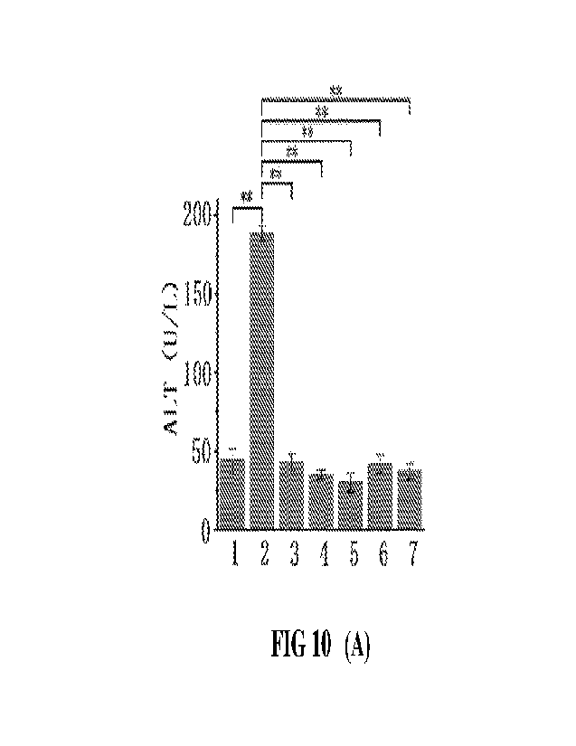
[38] FIG 10 presents bar graphs showing the effects of different doses of A-
01 and A-02 on serum (A)
ALT, (B) AST, (C) T1311,, op) MAO and (E) ALB levels in cirrhotic model mice,
where 1) denotes the blank
control group, 2) the model group, 3) the positive group treated with
sorafenib, 4) A-01 low dose group, 5)
A-01 high dose group, 6)A-02 low dose group, and 7) A-02 high dose group.
[39] FIG 11 presents bar graphs showing the effects of different doses of B-
01 and B-02 on serum (A)
ALT, (B) AST, (C) TBIL, (D) MAO and (E) ALB levels in cirrhotic model mice,
where 1) denotes the blank
control group, 2) the model group, 3) the positive group treated with
sorafenib, 4) B-01 low dose group, 5)
B-01 high dose group, 6)B-02 low dose group, and 7) B-02 high dose group.
[40] FIG 12 presents bar graphs showing the effects of high dose C on serum
(A) ALT, (B) AST, (C)
TBIL, (D) MAO and (E) ALB levels in cirrhotic model mice, where 1) denotes the
blank control group, 2)
the model group, 3) the positive group treated with sorafenib, 4) drug C high
dose group.
[41] FIG 13 presents HE staining images: (A) the blank control group; (B)
the model group; (C) the
positive control group; (D) A-01 low dose group; (E) A-01 high dose group.
[42] FIG 14 presents bar graphs showing the effects of D, E and F drugs on
serum (A) ALT, (B) AST, (C)
TBIL, (D) MAO and (E) ALB levels in cirrhotic model mice, where 1) denotes the
blank control group, 2)
the model group, 3) D drug group, 4) E drug group, and 5) F drug group.
Detailed Description of the Embodiments
[43] The present invention may be understood more readily by reference to
the following detailed
description of certain embodiments of the invention.
[44] Throughout this application, where publications are referenced, the
disclosures of these publications
are hereby incorporated by reference, in their entireties, into this
application in order to more fully describe
the state of art to which this invention pertains.
[45] Gold clusters (AuCs) are a special form of gold existing between gold
atoms and gold
nanoparticles. AuCs have a size smaller than 3 nm, and are composed of only
several to a few hundreds of
gold atoms, leading to the collapse of face-centered cubic stacking structure
of gold nanoparticles. As a
result, AuCs exhibit molecule-like discrete electronic structures with
distinct HOMO¨LUMO gap unlike the
continuous or quasi-continuous energy levels of gold nanoparticles. This leads
to the disappearance of
surface plasmon resonance effect and the corresponding plasmon resonance
absorption band (520 20 urn)
at uv-vis spectrum that possessed by conventional gold nanoparticles.
CA 03164005 2022- 7-6
WO 2021/184762
PCT/CN2020/124285
[46] The present invention provides a ligand-bound AuC.
[47] In certain embodiments, the ligand-bound AuC comprises a ligand and a
gold core, wherein the
ligand is bound to the gold core.
The binding of ligands with gold cores means that ligands form
stable-in-solution complexes with gold cores through covalent bond, hydrogen
bond, electrostatic force,
hydrophobic force, van der Waals force, etc In certain embodiments, the
diameter of the gold core is in the
range of 0.5 ¨ 3 nm. In certain embodiments, the diameter of the gold core is
in the range of 0.5 ¨2.6 nm.
[48] In certain embodiments, the ligand of the ligand-bound AuC is a thiol-
containing compound or
oligopeptide. In certain embodiments, the ligand bonds to the gold core to
form a ligand-bonded AuC via
Au-S bond.
[49] In certain embodiments, the ligand is, but not limited to, L-cysteine,
D-cysteine, or a cysteine
derivative.
In certain embodiments, the cysteine derivative is N-isobutyryl-L-
cysteine (L-N1BC),
N-isobutyryl-D-cysteine (D-NIBC), N-acetyl-L-cysteine (L-NAC), or N-acetyl-D-
cysteine (D-NAC).
[50] In certain embodiments, the ligand is, but not limited to, a cysteine-
containing oligopeptide and its
derivatives.
In certain embodiments, the cysteine-containing oligopeptide is a
cysteine-containing
dipeptide.
In certain embodiments, the cysteine-containing dipeptide is L(D)-
cysteine-L(D)-arginine
dipeptide (CR), L(D)-arginine-L(D)-cysteine dipeptide (RC), or L(D)-cysteine-L-
histidine dipeptide (CH).
In certain embodiments, the cysteine-containing oligopeptide is a cysteine-
containing tripeptide. In certain
embodiments, the cysteine-containing tripeptide is glycine-L(D)-cysteine-L(D)-
arginine tripeptide (GCR),
L(D)-proline-L(D)-cysteine-L(D)-arginine tripeptide (PCR), or L(D)-glutathione
(GSH). In certain
embodiments, the cysteine-containing oligopeptide is a cysteine-containing
tetrapeptide. In certain
embodiments, the cysteine-containing tetrapeptide is glycine-L(D)-serine-L(D)-
cysteine-L(D)-arnine
tetrapeptide (GSCR) or glycine-L(D)-cysteine-L(D)-serine-L(D)-arginine
tetrapeptide (GC SR).
[51]
In certain embodiments, the ligand is a thiol-containing compound.
In certain embodiments,
thiol-containing compound is 1- [(2 S)-2-methyl-3 -thio1-1-oxopropy1]-L(D)-
proline, thioglycollic acid,
mercaptoethanol, thiophenol, D-3-trolovol, or dodecyl mercaptan.
[52] The present invention provides a pharmaceutical composition for the
treatment of liver cirrhosis
in a subject. In certain embodiments, the subject is human. In certain
embodiments, the subject is a pet
animal such as a dog.
[53] In certain embodiments, the pharmaceutical composition comprises a
ligand-bound AuC as
disclosed above and a pharmaceutically acceptable excipient
In certain embodiments, the excipient is
phosphate-buffered solution, or physiological saline.
[54] The present invention provides a use of the above disclosed ligand-
bound AuCs for manufacturing a
6
CA 03164005 2022- 7-6
WO 2021/184762
PCT/CN2020/124285
medication for the treatment of liver cirrhosis in a subject.
[55]
The present invention provides a use of the above disclosed ligand-
bound AuCs for treating liver
cirrhosis in a subject or a method for treating liver cirrhosis in subject
using the above disclosed
ligand-bound AuCs.
In certain embodiments, the method for treatment comprises
administering a
pharmaceutically effective amount of ligand-bound AuCs to the subject. The
pharmaceutically effective
amount can be ascertained by routine in vivo studies.
[561
The following examples are provided for the sole purpose of
illustrating the principles of the
present invention; they are by no means intended to limit the scope of the
present invention.
[57] Embodiments
[58] Embodiment 1. Preparation of ligand-bound AuCs
[59] 1.1 Dissolving HAuC14 in methanol, water, ethanol, n-propanol, or
ethyl acetate to get a solution A
in which the concentration of HAuC14 is 0.01-0.03M;
[601
1.2 Dissolving a ligand in a solvent to get a solution B in which
the concentration of the ligand is
0.01-0.18M; the ligand includes, but not limited to, L-cysteine, D-cysteine
and other cysteine derivatives
such as N-isobutyryl-L-cysteine (L-NIBC), N-isobutyryl-D-cysteine (D-N1BC), N-
acetyl-L-cysteine
(L-NAC), and N-acetyl-D-cysteine (D-NAC), cysteine-containing oligopeptides
and their derivatives
including, but not limited to, dipeptides, tripeptide, tetrapeptide and other
peptides containing cysteine, such
as L(D)-cysteine-L(D)-arginine dipeptide (CR), L(D)-arginine-L(D)-cysteine
dipeptide (RC), L(D)-cysteine
L(D)-hi sti dine (CH), glycine-L(D)-cysteine-L(D)-arginine
tripeptide (GCR),
L(D)-proline-L(D)-cysteine-L(D)-arginine tripeptide (PCR), L(D)-
glutathione (GSH),
glycine-L(D)- serine-L(D)-cysteine-L(D)-arginine tetrapeptide (G
SCR) and
glycine-L(D)-cysteine-L(D)-serine-L(D)-arginine tetrapeptide (GC SR), and
other thiol-containing
compounds, such as one or more of 1-[(2S)-2-methy1-3-thio1-1-oxopropyl]-L(D)-
proline, thioglycollic acid,
mercaptoethanol, thiophenol, D-3-trolovol and dodecyl mercaptan; the solvent
is one or more of methanol,
ethyl acetate, water, ethanol, n-propanol, pentane, formic acid, acetic acid,
diethyl ether, acetone, anisole,
1-propanol, 2-propanol, 1-butanol, 2-butanol, pentanol, butyl acetate,
tributyl methyl ether, isopropyl acetate,
dimethyl sulfoxide, ethyl formate, isobutyl acetate, methyl acetate, 2-methyl-
1-propanol and propyl acetate;
[61] L3 Mixing solution A and solution B so that the mole ratio between
HAuC14 and ligand is 1:
(0.01-100), stirring them in an ice bath for 0.1-48h, adding 0.025-0.8M NaBH.4
water, ethanol or methanol
solution, continuing to stir in an ice water bath and react for 0 I ¨ 12h The
mole ratio between NaBH4 and
ligand is 1. (0.01-100),
[62] 1.4 Using MWCO 3K-30K ultrafiltration tubes to centrifuge the reaction
solution at 8000-17500
7
CA 03164005 2022- 7-6
WO 2021/184762
PCT/CN2020/124285
r/min by gradient for 10-100 min after the reaction ends to obtain ligand-
bound AuCs precipitate in different
average particle sizes. The aperture of the filtration membranes for
ultrafiltration tubes of different
MWCOs directly decides the size of ligand-bound AuCs that can pass the
membranes This step may be
optionally omitted;
[63] 1.5 Dissolving the ligand-bound AuCs precipitate in different average
particle sizes obtained in
step (1.4) in water, putting it in a dialysis bag and dialyzing it in water at
room temperature for 1-7 days;
[64] 1.6 Freeze-drying ligand-bound AuCs for 12-24h after dialysis to
obtain a powdery or flocculant
substance, i.e., ligand-bound AuCs.
[65] As detected, the particle size of the powdery or flocculant substance
obtained by the foregoing
method is smaller than 3 nm (distributed in 0.5-2.6nm in general). No obvious
absorption peak at 520 nm.
It is determined that the obtained powder or floc is ligand-bound AuCs.
[66] Embodiment 2. Preparation and characterization of AuCs bound with
different ligands
[67] 2.1 Preparation of L-NIBC-bound AuCs, i.e. L-NIBC-AuCs
[68] Taking ligand L-NIBC for example, the preparation and confirmation of
AuCs bound with ligand
L-NIBC are detailed.
[69] 2.1.1 Weigh 1.00g of HAuC14 and dissolve it in 100mL of methanol to
obtain a 0.03M solution
A;
[70] 2.1.2 Weigh 0.57g of L-NIBC and dissolve it in 100mL of glacial
acetic acid (acetic acid) to
obtain a 0.03M solution B,
[71] 2.1.3 Measure lmL of solution A, mix it with 0.5mL, lmL, 2mL, 3mL,
4mL, or 5mL of solution
B respectively (i.e. the mole ratio between HAuC14 and L-NIBC is 1:0.5, 1:1,
1:2, 1:3, 1:4, 1:5 respectively),
react in an ice bath under stirring for 2h, quickly add 1 mL of freshly
prepared 0.03M (prepared by weighing
11.3mg of NaBH4 and dissolving it in 10mL of ethanol) NaBH4 ethanol solution
when the solution turns
colorless from bright yellow, continue the reaction for 30 min after the
solution turns dark brown, and add
10mL of acetone to terminate the reaction.
[72] 2.1.4 After the reaction, the reaction solution is subjected to
gradient centrifugation to obtain
L-NIBC-AuCs powder with different particle sizes. Specific method: After the
reaction is completed, the
reaction solution is transferred to an ultrafiltration tube with MWCO of 30K
and a volume of 50 mL, and
centrifuged at 10000r/min for 20min, and the retentate in the inner tube is
dissolved in ultrapure water to
obtain powder with a particle size of about 2.6 nm. Then, the mixed solution
in the outer tube is transferred
to an ultrafiltration tube with a volume of 50 mL and MWCO of 10K, and
centrifuged at 13,000 dmin for 30
min. The retentate in the inner tube is dissolved in ultrapure water to obtain
powder with a particle size of
8
CA 03164005 2022- 7-6
WO 2021/184762
PCT/CN2020/124285
about 1.8 nm. Then the mixed solution in the outer tube is transferred to an
ultrafiltration tube with a
volume of 50 mL and MWCO of 3K, and centrifuged at 17,500r/min for 40 min. The
retentate in the inner
tube is dissolved in ultrapure water to obtain powder with a particle size of
about 1.1 nm.
[73] 2.1.5 Precipitate the powder in three different particle sizes
obtained by gradient centrifugation,
remove the solvent respectively, blow the crude product dry with N2, dissolve
it in 5mL of ultrapure water,
put it in a dialysis bag (MWCO is 3KDa), put the dialysis bag in 2L of
ultrapure water, change water every
other day, dialyze it for 7 days, freeze-dry it and keep it for future use.
[74] 2.2 Characterization of L-NIBC-AuCs
[75] Characterization experiment was conducted for the powder obtained
above (L-NIBC-AuCs)
Meanwhile, ligand L-NIBC-modified gold nanoparticles (L-N1BC-AuNPs) are used
as control. The method
for preparing gold nanoparticles with ligand being L-NIBC refers to the
reference (W. Yan, L. Xu, C. Xu, W.
Ma, H. Kuang, L. Wang and N. A. Kotov, Journal of the American Chemical
Society 2012, 134, 15114; X.
Yuan, B. Zhang, Z. Luo, Q. Yao, D. T. Leong, N. Yan and J. Xie, Angewandte
Chemie International Edition
2014, 53, 4623).
[76] 2.2.1 Observation of the morphology by transmission electron
microscope (TEM)
[77] The test powders (L-N1BC-AuCs sample and L-NIBC-AuNPs sample) were
dissolved in ultrapure
water to 2 mg/L as samples, and then test samples were prepared by hanging
drop method. More specifically,
4, of the samples were dripped on an ultrathin carbon film, volatized
naturally till the water drop
disappeared, and then observe the morphology of the samples by JEM-2100F
STEM/EDS field emission
high-resolution TEM.
[78] The four TEM images of L-NIBC-AuNPs are shown in panels B, E, H, and K
of FIG 1; the three
TEM images of L-NMC-AuCs are shown in panels B, E, and H of FIG 2.
[79] The images in FIG 2 indicate that each of L-NIBC-AuCs samples has a
uniform particle size and
good dispersibility, and the average diameter of L-NIBC-AuCs (refer to the
diameter of gold core) is 1.1 nm,
1.8 nm and 2.6 nm respectively, in good accordance with the results in panels
C, F and I of FIG 2. In
comparison, L-NIBC-AuNPs samples have a larger particle size Their average
diameter (refer to the
diameter of gold core) is 3.6 nm, 6.0 nm, 10.1 nm and 18.2 nm respectively, in
good accordance with the
results in panels C, F, I and L of FIG 1.
[80] 2.2.2 Ultraviolet (UV)-visible (vis) absorption spectra
[81] The test powders (L-NIBC-AuCs sample and L-NIBC- AuNPs sample) were
dissolved in ultrapure
water till the concentration was 10ms-L-1, and the UV-vis absorption spectra
were measured at room
temperature. The scanning range was 190-1100 nm, the sample cell was a
standard quartz cuvette with an
9
CA 03164005 2022- 7- 6
WO 2021/184762
PCT/CN2020/124285
optical path of 1 cm, and the reference cell was filled with ultrapure water.
[82] The UV-vis absorption spectra of the four L-NB3C-AuNPs samples
with different sizes are shown
in panels A, D, G and J of FIG 1, and the statistical distribution of particle
size is shown in panels C, F, land
L of FIG 1; the UV-vis absorption spectra of three L-NIBC-AuCs samples with
different sizes are shown in
panels A, D and G of FIG 2, and the statistical distribution of particle size
is shown in panels C, F and I of
FIG 2.
[831 FIG 1 indicates that due to the surface plasmon effect, L-NIBC-
AuNPs had an absorption peak at
about 520 nm. The position of the absorption peak is relevant with particle
size. When the particle size is 3.6
nm, the UV absorption peak appears at 516 nm; when the particle size is 6.0
nm, the UV absorption peak
appears at 517 nm; when the particle size is 10.1 nm, the UV absorption peak
appears at 520 nm, and when
the particle size is 18.2 nm, the absorption peak appears at 523 nm. None of
the four samples has any
absorption peak above 560 nm.
[84] FIG 2 indicates that in the UV absorption spectra of three L-NIBC-AuCs
samples with different
particle sizes, the surface plasmon effect absorption peak at 520 nm
disappeared, and two obvious
absorption peaks appeared above 560 nm and the positions of the absorption
peaks varied slightly with the
particle sizes of AuCs. This is because AuCs exhibit molecule-like properties
due to the collapse of the
face-centered cubic structure, which leads to the discontinuity of the density
of states of AuCs, the energy
level splitting, the disappearance of plasmon resonance effect and the
appearance of a new absorption peak
in the long-wave direction. It could be concluded that the three powder
samples in different particle sizes
obtained above are all ligand-bound AuCs.
[85] 2.2.3 Fourier transform infrared spectroscopy
[86] Infrared spectra were measured on a VERTEX8OV Fourier transform
infrared spectrometer
manufactured by Bruker in a solid powder high vacuum total reflection mode.
The scanning range is
4000-400 cm' and the number of scans is 64. Taking L-NIBC-AuCs samples for
example, the test samples
were L-NIBC-AuCs dry powder with three different particle sizes and the
control sample was pure L-NIBC
powder. The results are shown in FIG 3.
[87] FIG 3 shows the infrared spectrum of L-NIBC-AuCs with different
particle sizes. Compared with
pure L-NIBC (the curve at the bottom), the S-H stretching vibrations of L-NIBC-
AuCs with different
particle sizes all disappeared completely at 2500-2600 cm-', while other
characteristic peaks of L-NIBC
were still observed, proving that L-NIBC molecules were successfully bound to
the surface of AuCs via
Au-S bond. The figure also shows that the infrared spectrum of the ligand-
bound AuCs is irrelevant with
its size.
CA 03164005 2022- 7-6
WO 2021/184762
PCT/CN2020/124285
[88] AuCs bound with other ligands were prepared by a method similar to the
above method, except that
the solvent of solution B, the feed ratio between HAuC14 and ligand, the
reaction time and the amount of
NaBH4 added were slightly adjusted. For example: when L-cysteine, D-cysteine,
N-isobutyryl-L-cysteine
(L-NIBC) or N-isobutyryl-D-cysteine (D-NIBC) is used as the ligand, acetic
acid is selected as the solvent;
when dipeptide CR, dipepti de RC or 1-[(2S)-2-methyl-3-thiol-1-oxopropyl]-L-
proline is used as the ligand,
water is selected as the solvent, and so on and so forth; other steps are
similar, so no further details are
provided herein.
[89] The present invention prepared and obtained a series of ligand-bound
AuCs by the foregoing
method. The ligands and the parameters of the preparation process are shown in
Table 1.
[90] Table 1. Preparation parameters of AuCs bound with different ligands
in the present invention
Parameter
Time of
Time of reaction
reaction in Mole in an ice
Feed ratio an ice bath ratio
bath
Ligand Solvent used between under
between under
for solution B HAuC14 and stirring
HAuC14 stirring
ligand before and after
addition NaBH4 addition
of NaBH4 of
N allf-14
1 L-cysteine Acetic acid 1:3 2h 1:2
0.5h
2 D-cysteine Acetic acid 1:3 2h 1:2
0.5h
3 N-acetyl-L-cysteine Ethanol 1:4 lh 1:1
0.5h
4 N-acetyl-D-cysteine Ethanol 1:4 lh 1:1
0.5h
L-NIBC Water 1:4 0.5h 1:2 0.5h
6 D-NIBC Water 1:4 0.5h 1:2
0.5h
1:
Other cysteine Soluble
7
1:(O.1-100) 0.5h-24h (0.1-10 0.1-24h
derivatives solvent
0)
8 CR Water 1:4 22h 2:1
0.5h
9 RC Water 1:4 20h 2:1
0.5h
HC Water 1:3 12h 1:2 2h
11 CH Ethanol 1:4 16h 1:3
3h
12 GSH Water 1:2 12h 1:1
3h
13 KCP Water 1:3 15h 1:2
lh
11
CA 03164005 2022- 7-6
WO 2021/184762
PCT/CN2020/124285
14 PCR Water 1:4 16h 1:3
2h
15 GSCR Water 1:4 16h 1:3
1.5h
16 GCSR Water 1:3 12h 1:2
2h
Other oligopeptides Soluble 1(0.1-
17 1:(0.1-100) 0.5h-
24h 0.1-24h
containing cysteine solvent 100)
1-[(25)-2-methy1-3-
18 thiol-1-oxopropy1]-L- Water 1:8 ',II 1:7
lh
proline
19 Mercaptoethanol Ethanol 1:2 2h 1:1
lh
20 Thiog,lycollic acid Acetic acid 1:2
7h 1:1 lh
21 Thiophenol Ethanol 1:5 5h 1:1
lh
22 D-3-trolovol Water 1:2 211 1:1
lh
N-(2-mercaptopropio
23 Water 1:2 ,h 1:1 lh
ny1)-glycine
24 Dodecyl mercaptan Methanol 1:5 5h 1:1
lh
Other compounds Soluble 1:(0.01¨ 1(0.1-
25 0.5h-24h
0.1-24h
containing thiol solvent 100)
100)
[91] The samples listed in Table 1 are confirmed by the foregoing methods.
The characteristics of six
different ligand-bound AuCs are shown in FIG 4 (CR-AuCs), in FIG 5 (RC-AuCs),
in FIG 6 (Cap-AuCs)
(Cap denotes 1 -[(2 S)-2-m ethy1-3 -thi ol-l-oxopropyl] -L-proline), in FIG 7
(GSH-AuCs), in FIG 8
(D-NIBC-AuCs), and in FIG 9 (L-Cys-AuCs). FIGS 4-FIG 9 show UV spectra (panel
A), infrared spectra
(panel B), "[EM images (panel C), and particle size distribution (panel 1)).
[92] The results indicate that the diameters of AuCs bound with different
ligands obtained from Table 1
are all smaller than 3 nm. Ultraviolet spectra also show disappearance of
peak at 520 20 nm, and
appearance of absorption peak in other positions. The position of the
absorption peak could vary with
ligands and particle sizes as well as structures. In certain situations, there
is no special absorption peak,
mainly due to the formation of AuCs mixtures with different particles sizes
and structures or certain special
AuCs that moves the position of absorption peak beyond the range of UV-vis
spectrum. Meanwhile,
Fourier transform infrared spectra also show the disappearance of ligand thiol
infrared absorption peak
(between the dotted lines in panel B of FIGS 4-8), while other infrared
characteristic peaks are all retained,
suggesting that all ligand molecules have been successfully bound to gold
atoms to form ligand-bound AuCs,
and the present invention has successfully obtained AuCs bound with the
ligands listed in Table 1.
12
CA 03164005 2022- 7-6
WO 2021/184762
PCT/CN2020/124285
[93] Embodiment 3
[94] 3.1 Materials and animals
[95] 3.1.1 Testing Sample
[96] A-01 : ligand L-NIBC-bound gold clusters (L-NIBC-AuCs), 0.9 0.2 nm.
[97] A-02: ligand L-NIBC-bound gold clusters (L-NIBC-AuCs), 1.9 +0.5 nm.
[98] B-01: ligand L-Cys-bound gold clusters (L-Cys-AuCs), 1.0 0.2 nm.
[99] B-02: ligand L-Cys-bound gold clusters (L-Cys-AuCs), 1.7 0.3 nm.
[100] C: L-NIBC-modified nanoparticles (L-NIBC-AuNPs), 6.3 1.5 nm
[101] All testing samples were prepared following the above described
method with slight
modification, and their quality was characterized using the above described
methods.
[102] 3.1.2 Positive control sample
[103] Sorafenib.
[104] 3.1.3 Animals for experiments and groups
[105] 120 SPF male C57BL/6N mice, 6-8 weeks old and 16-20g body weight,
were purchased from
Beijing Huafukang Experimental Animal Technology Co., Ltd. (production license
number: SCXK (fing)
2019-0008). According to body weight, they were randomly divided into 12
groups (n = 10): blank control
group, model group, positive control group, A-01 low dose group, A-01 high
dose group, A-02 low dose
group, A-02 high dose group, B-01 low-dose group, B-01 high-dose group, B-02
low-dose group, B-02
high-dose group, and C high-dose group.
[106] 3.2 Modeling protocol
[107] Except for the blank control group, liver cirrhosis model of mice in
other groups was prepared
by the method of carbon tetrachloride (CC14)-induction treatment. The modeling
protocoal was as follows:
(1) Each mouse was intraperitoneally injected with 10% CC14 (diluted with
olive oil) at 7 pL/g body weight,
twice a week for a total of 8 weeks; mice of the blank control group were
injected intraperitoneally with the
same amount of olive oil solvent. (2) from the 6th week, two mice were
selected and killed 48 hours after
the last injection every week. The appearance of the liver was observed. After
the appearance was in line
with the characteristics of cirrhosis (the 8th week), the liver tissue was
fixed with formalin. HE staining
and Masson staining were used to evaluate the model of cirrhosis.
[108] 3.3 Administration
[109] After the successful modeling, the mice in the positive control group
were given
intragastrically 25 mg/kg sorafenib, the mice in the low or high dose groups
of A-01, A-02, B-01 and B-02
were given by intraperitoneal injection at 2.5 or 10 mg/kg respectively of the
corresponding test material;
13
CA 03164005 2022- 7-6
WO 2021/184762
PCT/CN2020/124285
the mice in the C high dose group were given by intraperitoneal injection at a
dose of 40 mg/kg of C; and the
mice in the blank control group and the model group were given
intraperitoneally physiological saline at 10
mL/kg. The administration was once a day for 20 consecutive days.
[110] 3.4 Biochemical testing
[111] After the administration was completed, blood was collected
from mouse orbit, and sera were
obtained for biochemical testing of albumin (ALbumin, ALB), total bilirubin
__________ alanine Alanine
aminotransferase (ALT), aspartate aminotransferase (AST) and monoamine oxidase
(MAO) using
Zhongsheng Beikong Kit and biochemical analyzer (Siemens). The detection
method was performed in strict
accordance with the kit instructions.
[112] Table 2 shows the product information of kits used for
biochemical testing
Eg;0;E;;;EM;;:=;;K;;MR;Og,Egl
vgooma:Em,
Beijing Food and Drug
Albumin Test Kit
1 ALB administration
Device (Permit)
(Bromocresol Green Method)
2014 No. 2401133
Beijing Food and Drug
Total bilirubin test kit (vanadate
2 TBil administration
Device (Permit)
oxidation method)
2014 No. 2401140
Beijing Food and Drug
Alanine aminotransferase test kit
3 ALT administration
Device (Permit)
(alanine substrate method)
2014 No. 2401158
Aspartate aminotransferase test Beijing Food and
Drug
4 kit AST administration
Device (Permit)
(aspartic acid substrate method) 2014 No. 2401157
Monoamine oxidase test kit Beijing Food and
Drug
(glutamic acid dehydrogenase MAO administration Device (Permit)
method) 20162401129
[113] 3.5 Pathological examination
[114] 3.5.1 HE staining
[115] After euthanasia, the mouse liver tissue samples were fixed
with 4% paraformaldehyde fixative
for more than 48 h
After fixation, the liver samples were dehydrated with alcohol
gradient and treated
with xylene and ethanol
Then, the liver tissues were then dipped in wax and embedded. After
the
embedded material being trimmed, attached, and repaired, the liver tissues
were sliced with a paraffin
microtome, and the slices were with a thickness of 4 ium. The main process of
RE staining is as follows:
After baked in the oven at 65 C, the slices were treated with xylene and
dehydrated with gradient ethanol.
The slices were sequentially stained with hematoxylin, blue color-enhancing
solution, and 0.5% eosin, then
treated with gradient ethanol and xylene and sealed with neutral gum. The
fibrosis of liver tissue was
14
CA 03164005 2022- 7-6
WO 2021/184762
PCT/CN2020/124285
observed with a microscope.
[116] 3.5.2 Masson staining
[117] After baked, mouse liver tissue slices were dewaxed and dehydrated.
After chromizing, the
nucleus was stained with Regaud's hematoxylin staining solution. After washing
with water, the slices
were stained with Masson's Ponceau Red Acidic Fuchsin, and the slices were
dipped in a 2% glacial acetic
acid aqueous solution and differentiated with a 1% phosphomolybdic acid
solution. After directly stained
with aniline blue or light green solution, the slices were dipped in a 0.2%
glacial acetic acid aqueous
solution for a while, then transparentized with 95% alcohol, anhydrous alcohol
and xylene, and then sealed
with neutral gum. Liver tissue was observed with a microscope.
[118] 3.6 Results
[119] 3.6.1 Successful Modeling
[120] The livers of mice in the model group were divided into round or oval
masses of different sizes
by proliferating fibrous septa. The serum ALT, TBil, and AST indexes increased
significantly compared to
that of the blank control group, the serum ALB significantly decreased
compared to the blank control group,
and the MAO index was no significant difference from the control group, but
the value also increased. All
the above results suggest that this experimental modeling was successful.
[121] 3.6.2 Effects of test drugs on alanine aminotransferase (ALT),
total bilirubin (TBil),
aspartate aminotransferase (AST), monoamine oxidase (MAO) and albumin (ALB)
[122] 3.6.2.1 Test drugs A-01 and A-02
[123] As shown in FIG 10A, the ALT activity of the model group is extremely
significantly higher
than that of the blank control group (increased from 43.5 8.1 U/L to 188.5+4.9
U/L; P <0.01), which
indicates that liver functions of the cirrhotic model mice had pathological
changes. After administration of
A-01 and A-02 at high and low doses, the ALT activity of all treated groups
decreased significantly (the
highest is 41.5+5.4 U/L for A-02 low dose group; the lowest is 30.0+5.9 U/L
for A-01 high dose group; and
42.8+5.4 U/L for positive control group), and returned to the similar level of
the blank control group or even
lower, which is significantly different from that of the model group (P
<0.01).
[124] As shown in FIG 10B, the serum AST activity of the model group was
significantly increased
compared to the blank control group (increased from 141.9+13.5 U/L to
192.0+11.3 U/L; P <0.05). After
the administration of A-01 and A-02, the AST activity of all treated groups
decreased, where the high-dose
A-01 and A-02 administration significantly reduced the AST activity (130+12.8
U/L for A-01 high dose
group, 131.3 9.9 U/L for A-02 high dose group, both P <0.01), obviously
superior to the positive control
group (165.5+11.6 U/L).
CA 03164005 2022- 7-6
WO 2021/184762
PCT/CN2020/124285
[125] As shown in FIG 10C, the concentration of TBil of the model group was
significantly higher
than that of the blank control group (increased from 1.02+0.20 jtmol/L to
2.91+0.39 mon), and there was
a significant difference from the blank control group (P <0.01). After
administration of high and low doses
of A-01 and A-02, the concentrations of TBil were all significantly reduced
(the highest is 0.91+0.13 mon;
the lowest is 0.78+0.25 mon); they are in the similar level of the blank
control group, but are extremely
significantly different from the model group (P <0.01).
[126] As shown in FIG 10D, the MAO activity of the model group is increased
compared to the
blank control group (18.8+2.9 U/L for blank control group; 21.5+0.7 U/L for
model group), but there is no
statistical difference, suggesting that the changes of the MAO activity in the
cirrhosis mice induced by
carbon tetrachloride are not significant. The administration of A-01 and A-02
did not significantly affect
the MAO activity of all treated groups, but the MAO activity of all treated
groups decreased (the highest is
19.3+1.5 U/L and the lowest is 18.5+1.9 U/L); they are in the similar to the
level of the blank control group.
In comparison, the MAO activity of the positive control group did not decrease
(21.3+2.1 U/L). This result
suggests that A-01 and A-02 may adjust the activity of MAO to the level of the
blank control group, playing
a role in the recovery of liver functions in cirrhosis mice.
[127] As shown in FIG 10E, the ALB level of the model group is
significantly decreased compared to
the blank control group (decreased from 24.2+0.6 g/L to 22.1+1.3 g/L), and
there is a significant difference
from the blank control group (P<0.05), showing that carbon tetrachloride
administration may significantly
decrease serum ALB levels. The administration of different doses of A-01 and A-
02, and positive control d
did not significantly affect the serum ALB levels.
[128] The positive drug sorafenib significantly reduced the levels of ALT,
AST, and TB1L but may
not have a relief effect on cirrhotic mice for the MAO index. The results
suggest that A-01 and A-02 have
a repairing effect on liver function in cirrhotic mice, and the effect is
better than that of the positive control
drug.
[129] 3.6.2.2 Test drugs B-01 and B-02
[130] As shown in FIG 11A, the low and high doses of B-01 and B-02 could
significantly reduce
ALT activity (the highest is 46.3+7.4 U/L; the lowest is 33.0+7.1 U/L); they
are in the level similar to the
that of the blank control group, but significantly different from the model
group (188.5+4.9 U/L; P <0.01).
[131] As shown in FIG 11B, compared with the model group (192.0+11.3 U/L),
B-01 low or dose
administration can significantly reduce AST activity to normal levels (132.3
10.0 U/L and 129.7+26.6 U/L
respectively, P<0.01), and B-02 low dose administration can significantly
reduce AST activity (149.6+21.8
U/L; P<0.05); they are in similar level to that of blank control group. But B-
02 high-dose administration
16
CA 03164005 2022- 7-6
WO 2021/184762
PCT/CN2020/124285
reduces AST activity to some extent, but there was no significant difference
(P> 005). In comparison, the
positive drug sorafenib can also decrease the AST activity to 165.5 11.6 U/L
(P<0.05), but the effect is not
as good as the administration of B-01 low and high doses and B-02 low dose.
[132] As shown in FIG 11C, low and high doses of B-01 and B-02 all
significantly reduced TBil (the
highest is 1.28 0.12 mon; the lowest is 0.96 0.15 mon); they are in a level
similar to that of the blank
control group (1.02+0.20 mon), but are significantly different from the model
group (2.91+0.39 ttmol(L;
P <0.01).
[133] As show in FIG 11D, compared with the model group (21.5+0.7 U/L), B-
01 low dose
(17.3+1.3 U/L; P<0.01) and B-02 high dose (18.3 0.6 U/L; P<0.05) significantly
reduced serum MAO
levels to the blank control group (18.8 2.9 U/L), but positive control drug
has no effect on the serum level
of MAO (21.3+2.1 U/L).
[134] As shown in FIG 11E, administration of test drugs and positive
control drug had no significant
effect on ALB levels.
[135] The above results show that B-01 and B-02 significantly reduce the
levels of ALT, AST, TBIL
and MAO, and have a certain dose-dependent effect on the liver function
recovery of cirrhotic mice, and
their effects are at least in some indicators better than the positive control
drugs.
[136] 3.6.2.3 Test drugs C
[137] As shown in FIG 12, compared with the model group, high-dose drug C
administration has no
significant improvement on the levels of (A) ALT, (B) AST, (C) TBIL, (D) MAO,
and (E) ALB compared to
the model control group, and there is even a certain deterioration trend,
suggesting that the drug C is
ineffective in improving the liver functions of cirrhotic mice and may be
toxic.
[138] 3.6.3 Pathological analyses
[139] Liver cirrhosis is pathologically characterized by diffuse fibrosis
of the liver tissue and
formation of pseudolobules. The results of RE staining pathological analyses
showed that as shown in FIG
13A, the normal liver tissue from the mice of the blank control group had
clear structure, intact liver lobules,
neatly arranged hepatocytes, radial arrangement being centered on the central
vein, normal nucleus of
hepatocytes, and only a small amount of fibrous tissue in the catchment area.
As shown in FIG 13B, in the
liver tissue of the model group, the hepatocytes were disordered, balloon-like
structures appeared, the
hepatic lobules nearly disappeared, pseudolobules (as pointed to by right-
orientated arrows in FIG 13B)
were abundantly formed, and a large number of proliferated protofibrils were
present in the liver tissues,
forming round- or oval-shaped fibrous septa (as pointed to by left-orientated
arrows in FIG 13B). As
shown in FIG 13C, compared with the model control group, the positive control
group showed significant
17
CA 03164005 2022- 7-6
WO 2021/184762
PCT/CN2020/124285
reduction of liver damages; the hepatocytes evidently have neat arrangement;
fibrous hyperplasia, while
increased, apparently reduced, not forming fibrous septa, pseudolobules nearly
disappeared, but compared
with normal liver tissues, the liver tissues in the positive control group
showed apparent increases of
inter-cellular gaps (as pointed to by downward-orientated arrows). Compared
with the model control group,
the 4 groups administered with gold clusters drugs (A-01, A-02, B-01 and B-02)
showed that their
hepatocytes significantly recovered from liver damages, as evidenced by
apparent reduction of fibrous
hyperplasia and pseudolobules, and that the recovery is dose-dependent to a
certain extent.
11401 FIG 13D and FIG 13E show the HE images that showed the effects
of the exemplary A-01 lose
and high dose drug administration respectively on the recovery of liver
damages. As shown in FIG 13D,
A-01 low dose drug administration group showed relatively neat arrangement of
hepatocytes, near
disappearance of pseudolobules, evident reduction of fibrous hyperplasia, but
the inter-hepatocytes gaps,
compared with normal liver tissues, are increased to a certain extent (as
pointed to by downward-orientated
arrows in FIG 13D). As shown in FIG 13E, in comparison with A-01 low dose drug
administration group,
A-01 high dose drug administration group had even better effects of reduction
of liver damages, complete
disappearance of pseudolobules, no observation of fibrous hyperplasia, no
discernable increases of
inter-hepatocytes gaps, and no apparent difference from normal liver tissues.
In conclusion, A-01 drug
showed better effects on recovery of liver damages than the positive control
drug.
[141] The results from Masson staining provided the same conclusions as did
the results of HE
staining.
[142] The other 3 drugs also showed similar effects of A-01 drug; no
detailed description is needed.
[143] In summary, the four test drugs A-01, A-02, B-01 and B-02
significantly reduced liver fibrous
hyperplasia and liver pseudolobules. The test results of liver function
indicators also showed the recovery
of liver function. The most significant changes were alanine aminotransferase
(ALT) and total bilirubin
(TBil). Aspartate aminotransferase (AST) and monoamino oxidase (MAO) also
recovered significantly,
while albumin (ALB) did not change significantly. The four gold clusters drugs
may significantly improve
liver function and part of the liver pathological structure in cirrhotic mice,
and the total effects are superior
to the positive control drug sorafenib, providing experimental basis for
further application in the future.
However, drug C has no obvious therapeutic effect, and cannot be used for the
treatment of liver cirrhosis.
[144] Embodiment 4
[145] 4.1 Materials and animals
[146] 4.1.1 Testing Sample
[147] D: ligand L-NAC-bound gold clusters (L-NAC-AuCs), 0.5-3 nm.
18
CA 03164005 2022- 7-6
WO 2021/184762
PCT/CN2020/124285
[148] E: ligand CR-bound gold clusters (CR-AuCs), 0.5-3 nm.
[149] F: ligand RC-bound gold clusters (RC-AuCs), 0.-3 nm.
[150] All testing samples were prepared following the above described
method with slight
modification, and their quality was characterized using the above described
methods.
[151] 4.1.2 Animals for experiments and groups
[152] 50 SPF male C57BL/6N mice, 6-8 weeks old and 16-20g body weight, were
purchased from
Beijing Huafukang Experimental Animal Technology Co., Ltd. (production license
number: SCXK (Jing)
2019-0008). According to body weight, they were randomly divided into 5 groups
(ii = 10): blank control
group, model group, D drug administration group, E drug administration group,
and F drug administration
group.
[153] 4.2 Modeling protocol
[154] Except for the blank control group, liver cirrhosis model of mice in
other groups was prepared
by the method of carbon tetrachloride (CC14)-induction treatment. The modeling
protocoal was as follows:
(1) Each mouse was intraperitoneally injected with 10% CC14 (diluted with
olive oil) at 7 iii.L/g body weight,
twice a week for a total of 8 weeks; mice of the blank control group were
injected intraperitoneally with the
same amount of olive oil solvent. (2) from the 6th week, two mice were
selected and killed 48 hours after
the last injection every week. The appearance of the liver was observed. After
the appearance was in line
with the characteristics of cirrhosis (the 8th week), the liver tissue was
fixed with formalin. HE staining
and Masson staining were used to evaluate the model of cirrhosis.
[155] 4.3 Administration
[156] After the successful modeling, the mice in the three drug
administration groups were given by
intraperitoneal injection at a dose of 40 mg/kg respectively of the
corresponding gold clusters drugs; and the
mice in the blank control group and the model group were given
intraperitoneally physiological saline at 10
mL/kg. The administration was once a day for 20 consecutive days.
[157] 4.4 Biochemical testing
[158] The reagents and protocols were the same as described in section 3.4.
[159] 4.5 Results
[160] 4.5.1 Successful Modeling
[161] The livers of mice in the model group were divided into round or oval
masses of different sizes
by proliferating fibrous septa. The serum ALT, TBil, and AST indexes increased
significantly compared to
that of the blank control group, the serum ALB significantly decreased
compared to the blank control group,
and the MAO index was no significant difference from the control group, but
the value also increased. All
19
CA 03164005 2022- 7-6
WO 2021/184762
PCT/CN2020/124285
the above results suggest that this experimental modeling was successful.
[162]
45.2 Effects of test drugs on alanine aminotransferase (ALT),
total bilirubin (TBil),
aspartate aminotransferase (AST), monoamine oxidase (MAO) and albumin (ALB)
[163] As shown in FIG 14A, the ALT activity of the model group is extremely
significantly higher
than that of the blank control group (P <0.01, **), which indicates that liver
functions of the cirrhotic model
mice had pathological changes. After administration of D, E or F drugs, the
ALT activity of all treated
groups decreased significantly, and returned to the similar level of the blank
control group, which is
significantly different from that of the model group (P <0.01).
[164] As shown in FIG 14B, the AST activity of the model group is
significantly higher than that of
the blank control group (P <0.05, *). After administration of D, E or F drugs,
the AST activity of all
treated groups decreased significantly (P <0.05, *).
[165] As shown in FIG 14C, the TBil concentration of the model group is
significantly higher than
that of the blank control group (P <0.01, **).
After administration of D, E or F drugs, the TBil
concentration of all treated groups decreased significantly to the level of
the blank control group, but are
significantly different than that of the model control group (P <0.01, **).
[166] As shown in FIG 14D, the MAO activity of the model group was
increased compared to the
blank control group, but there is no statistical difference (P >0.5),
suggesting that the changes of the MAO
activity in the cirrhosis mice induced by carbon tetrachloride are not
significant. The administration of D,
E or F drugs did not significantly affect MAO activity, but the MAO activity
of all drug administration
groups decreased to the level of the blank control group.
[167] As shown in FIG 14E, the ALB concentration of the model group is
decreased in comparison
with that of the blank control group, but the difference is not significant (P
>0.05). However, the
administration of D, E or F drugs increased the serum ALB concentration, but
the difference is not
significant (P >0.05).
[168] In summary, the three gold clusters drugs D, E and F significantly
improved liver function.
Alanine aminotransferase (ALT) and total bilimbin (TBil) showed the most
significant changes, aspartate
aminotransferase (AST) and monoamino oxidase (MAO) showed evident recovery,
and albumin (ALB) was
also improved, while not significantly. These results provide experimental
basis for further application in
the future.
[169] Other sized L-Cys-AuCs, L-NIBC-AuCs, L-NAC-AuCs, CR-AuCs, RC-AuCs,
and other
ligand-bound AuCs with different sizes also have the similar effects , while
their effects vary to certain
extents They would not be described in detail here.
CA 03164005 2022- 7-6
WO 2021/184762
PCT/CN2020/124285
[170] While the present invention has been described with reference
to particular embodiments, it
will be understood that the embodiments are illustrative and that the
invention scope is not so limited.
Alternative embodiments of the present invention will become apparent to those
having ordinary skill in the
art to which the present invention pertains. Such alternate embodiments are
considered to be encompassed
within the scope of the present invention. Accordingly, the scope of the
present invention is defined by the
appended claims and is supported by the foregoing description.
21
CA 03164005 2022- 7-6