Note: Descriptions are shown in the official language in which they were submitted.
WO 2021/146503
PCT/US2021/013550
PRESSURE ACTIVATED SURGICAL TOOL FOR USE IN SPINAL DECOMPRESSION
PROCEDURES AND METHODS OF USING THE SAME
RELATED APPLICATIONS
[0001] This application claims priority to U.S. Prov. Appin. No.
62/961,811, filed January
16, 2020, entitled "Robot-Guided Spinal Decompression" which is incorporated
herein by
reference.
FIELD OF THE INVENTION
[0002] The disclosure generally relates to spinal surgery.
Stated more particularly,
disclosed herein are systems and methods for the decompression of spinal
stenosis through
laminotomy or laminectomy techniques performed under robotic guidance.
BACKGROUND OF THE INVENTION
[0003] The human spine is a complex structure with thirty-three
individual vertebrae
stacked atop one another. The spinal column provides the main support for the
torso of the human
body allowing flexible, multi-directional movement, while protecting the
spinal cord from injury.
As shown in FIGS. 2 through 5, a human spine 100 is depicted with plural
vertebrae 102, each
with an anterior vertebral body 104 and a posterior vertebral arch 106 that
cooperate to enclose
the vertebral foramen 108 through which the spinal cord 110 passes. The
vertebral arch 106
includes a pair of laminae 112 and a spinous process 114 there between. The
vertebrae 102 are
joined by facet joints 116. Spinal nerves 118 leave the spinal cord 110
through intervertebral
foramina 120 Anterior (not shown) and posterior longitudinal ligaments 122
extend the length
of the vertebral column, and dura mater 124 envelops the spinal cord 110.
[0004] The narrowing of one or more of the foramina 108 or 120
within the spine 100
is generally referred to as spinal stenosis. That narrowing of the foramina
108 or 120 reduces the
1
CA 03164975 2022- 7- 15
WO 2021/146503
PCT/US2021/013550
space available for the comfortable and effective passage of nerves 118.
Spinal stenosis can come
to exist within the vertebral foramen (collectively, the spinal canal) 108 or
within the
intervertebral foramina 120 where spinal nerves 118 exit the spinal canal 108.
[0005] Depending on the location and the severity of the
narrowing, compression of the
spinal nerve 118 or the spinal cord 110 can produce symptoms that can range in
severity and that
can include pain, tingling, numbness, and weakness. Pain deriving from spinal
stenosis can be
sharp and can radiate into one or more of the person's arms or legs, or it may
be dull and localized
to the neck or lower back. Where numbness occurs, it may vary from reduced
sensitivity to total
numbness in an arm, leg, or other portion of the body. Spinal stenosis can
also lead to strength
deterioration, loss of coordination, and still further deleterious
consequences.
[0006] Certain instances of spinal stenosis can be treated non-
surgically, such as with
physical therapy, pain medication, activity modification, or epidural
injection. Where non-surgical
treatment is insufficient to alleviate the effects of spinal stenosis,
surgical intervention may become
necessary.
[0007] According to one known method of treatment, an invasive
fusion procedure is
performed where adjacent vertebrae 102 are fused together with screws and rods
to stabilize the
spine, normally after a spinal decompression technique has been performed.
Such fusion
procedures introduce increased surgical risk and are known to carry the risk
of unintended negative
long-term consequences.
[0008] An alternative treatment is a laminotomy procedure where
at least a portion of
the laminae 112 and/or the spinous process 114, the bony protrusion at the
back of the vertebra
102 that connects them, is removed, as depicted in FIGS. 6A, B, and C. This
removal of part of
the vertebral arch 106 is designed to decompress the spinal cord 110 and nerve
roots 118 that were
2
CA 03164975 2022- 7- 15
WO 2021/146503
PCT/US2021/013550
being pinched or inflamed by spinal stenosis. When done successfully,
laminotomy surgery can
eliminate the need for a more invasive fusion procedure. However, the
laminotomy can be
technically challenging. It requires extreme precision to remove just enough
lamina 112 or spinous
process 114 bone to decompress the spine 100 without compromising the
remaining lamina 112,
spinous process 114, facet joints 116, or stabilizing ligaments 122.
[0009] As shown in FIGS. 7A and B, another available surgical
treatment is a
laminectomy where the entire lamina 112 and spinous process 114 are removed. A
laminectomy
introduces further risk of destabilization as the posterior stabilizing
portion of the spine 100 is
removed. It is recognized to be an inherently ablative and often imprecise
procedure, one
performed on the lamina bone 112 as it resides directly over the spinal cord
110. Injury to the
spinal cord 110 can carry extreme immediate and long-term consequences to the
patient.
[0010] It is normally up to the surgeon's skill and accuracy to
cut to the required depth
successfully without injuring surrounding nerves 118 or unduly compromising
the stabilizing
anatomy, such as the facet joint 116 or interspinous ligaments 122. Deficits
in physician skill or
accuracy can lead to devastating consequences or ineffective procedures.
[0011] In other surgical techniques, it is known to use robotic
control, such as to drill
precise pilot holes for bone screws in fusion procedures. The use of such
robotic systems seeks to
provide improved accuracy and effectiveness of the surgery. However, the
application of robotic
guidance has been limited in the field.
[0012] One major obstacle to the use of robotic control in
laminotomy and laminectomy
procedures is the critical need for human differentiation between the drilling
and removal of bone
as compared to drilling into the softer tissue of ligaments, joints, and
nerves. Also preventing
robotic control is need for determining and accurately acting in relation to
the location of the
3
CA 03164975 2022- 7- 15
WO 2021/146503
PCT/US2021/013550
needed material removal.
[0013] It has been appreciated that a robotic surgical solution
capable of autonomous or
semi-autonomous operation in performing laminotomy and laminectomy procedures
would
represent a substantial advance in the art. It has been further appreciated
that the practical
application of such robot-guided procedures demands concomitant advances in
mechanical and
operational characteristics, including the effective differentiation between
bone and tissue.
SUMMARY OF THE INVENTION
[0014] [TO COME WITH FINAL CLAIM SET].
BRIEF DESCRIPTION OF THE DRAWINGS
[0015] In the accompanying drawing figures:
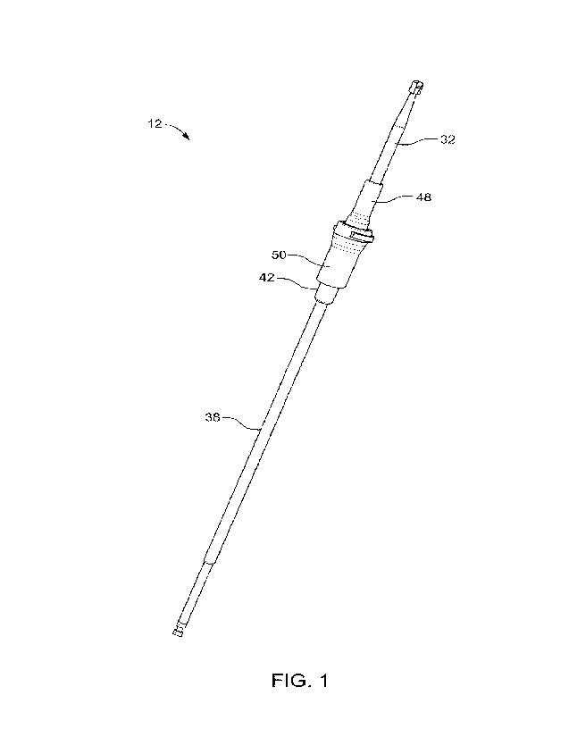
[0016] FIG. 1 is a perspective view of a surgical tool for use
in a spinal decompression
system;
[0017] FIG. 2 is a dorsal view of a portion of a human spine;
[0018] FIG. 3 is a laterally sectioned plan view of a vertebrae
of a human spine;
[0019] FIG. 4 is a side view of a portion of a human spine
including two vertebrae 104
and 104';
[0020] FIG. 5 is a perspective view of a portion of a human
spine including two vertebrae
104 and 104';
[0021] FIGS. 6A and B are top sectional views of a portion of a
human spine before and
after, respectively, a laminotomy procedure;
[0022] FIG. 6C is a dorsal view of a portion of a human spine
after a laminotomy
procedure;
[0023] FIGS. 7A and B are top sectional views of a portion of a
human spine before and
4
CA 03164975 2022- 7- 15
WO 2021/146503
PCT/US2021/013550
after, respectively, a laminectomy procedure;
[0024] FIG. 8A is a perspective view of a portion of a human
spine with a drill pattern
for a laminectomy as disclosed herein;
[0025] FIG. 8B is a perspective view of a portion of a human
spine with a drill pattern
for a laminotomy as disclosed herein;
[0026] FIG. 9 is a front view of a portion of a human spine
after partial removal of the
laminae, as disclosed herein;
[0027] FIG. 10 is a front view of a portion of a human spine
after partial removal of
laminae, as disclosed herein;
[0028] FIG. 11 is a front view of a portion of a human spine
showing a drill pattern for
partial removal of laminae, as disclosed herein
[0029] FIG. 12 is a schematic representation of a robotic-
assisted system for use in spinal
decompression procedures;
[0030] FIG. 13 is an exploded view in side elevation of a
pressure- activated surgical
tool;
[0031] FIG. 14 is a partially sectioned side view of a pressure-
activated surgical tool, as
disclosed herein;
[0032] FIG. 15 is an amplified partially sectioned view in side
elevation of the pressure-
activated surgical tool of FIG. 14;
[0033] FIG. 16 is an exploded view in side elevation of the
pressure- activated surgical
tool;
[0034] FIG. 17 is an amplified exploded view in side elevation
of the pressure- activated
surgical tool;
CA 03164975 2022- 7- 15
WO 2021/146503
PCT/US2021/013550
[0035] FIG. 18 is a partially sectioned view in side elevation
of the engaging portion of
the pressure- activated surgical tool;
[0036] FIG. 19 is an amplified side view of another embodiment
of the pressure-
activated surgical tool;
[0037] FIG. 20 is a top view of another embodiment of the
pressure activated surgical
tool.
[0038] FIG. 21 is an exploded cut-away top view of the pressure
activated surgical tool
of FIG. 21.
[0039] FIG. 22A is a perspective view of another embodiment of a
pressure-activated
surgical tool.
[0040] FIG. 22B is a detail view of the embodiment of FIG. 22A.
[0041] FIG. 23 is a schematic view in side elevation of the
pressure- activated surgical
tool drilling through a first material;
[0042] FIG. 24 is a schematic view in side elevation of the
pressure-activated surgical
tool after drilling through a first material and reaching a second material;
and
[0043]
DETAILED DESCRIPTION OF PREFERRED EMBODIMENTS
[0044] Systems and methods for use in spinal decompression
procedures are disclosed
herein are subject to widely varied embodiments. However, to ensure that one
skilled in the art
will be able to understand and, in appropriate cases, practice the invention,
certain embodiments
of the broader invention revealed herein are described below and disclosed by
the accompanying
drawing figures. The embodiments shown and described are not intended to be
limiting.
[0045] Referring now to FIG. 1, a surgical tool 12 for use in
spinal decompression
6
CA 03164975 2022- 7- 15
WO 2021/146503
PCT/US2021/013550
procedures is provided. It should be understood that this tool may be used for
any procedure in
which bone is being removed around a softer material, such as tissue. For
example, the surgical
tool 12 may also be used to perform a facetectomy in order to perform a
interbody fusion.
Specifically, the tool 12 can be combined with a robotic navigation system to
remove bone at.
the facet joint in order to provide access to the disc space. By using the
surgical tool 12, the
facetectomy can be done percutaneously through a cannula therefore allowing
for a more direct
exposure to the facet.
[0046] In one embodiment, the surgical tool 12 may be operated
manually. In another
embodiment, the surgical tool 12 may be operated as a part of a spinal
decompression system 10
in combination with a robotic arm 14 and a computer system 16, as shown in
FIG. 12.
[0047] For purposes of illustration, the surgical tool 12 will be
described as part of a
spinal decompression system 10, which may be configured to enable the
performance of
laminectomy, laminotomy, and potentially other surgical procedures under
robotic guidance with
the incorporation of mechanical, electro-mechanical, and overall advancements
in methodology
and systemic operation. In one embodiment, the robot-guided spinal
decompression system 10
permits spinal decompression to be carried out with efficiency and accuracy in
an automated
manner with reduced reliance on operator skill and dexterity during the course
of the spinal
decompression procedure.
[0048] As shown in FIGS. 7A and B, during a lumbar laminectomy,
substantially all of
the lamina 112 is removed in order to alleviate the cause of a stenosis.
However, such extensive
material removal exceeds the actual need thereby introducing excessive spinal
instability and risk
of undue damage to the vertebrae 102, the spinal cord 110, and other aspects
of the spine 100.
[0049] However, the use of a pressure activated tool in
laminectomy procedures as
7
CA 03164975 2022- 7- 15
WO 2021/146503
PCT/US2021/013550
disclosed herein, allows the user to focus on removing only the portion of
bone actually causing
the spinal stenosis. For instance, as shown in FIG. 11, a targeted laminectomy
portion 126 is
identified such that only portions of the vertebrae 102 determined to be
contributing to the
compressive stenosis are designated to be removed. The result of such focused
removal leaves
more bone intact and, as a result, a more stable structure of the spine 100
and reduced likelihood
of damage to the surrounding tissue.
[0050] Potential products of the focused removal of constricting
portions of vertebrae 102
are depicted in FIGS. 9 and 10. In the non-limiting example of FIG. 9, the
targeted laminectomy
portion 126 is generally presented as a round area of removed bone material.
In FIG. 10, the
targeted laminectomy portion 126 is still more focused, including only the
portions of lamina 112
determined, such as by pre-surgical planning through a computerized tomography
(CT) scan or
otherwise, to be contributing to the stenotic constriction.
[0051] Based on its shape and localization, the focused
laminectomy can be referred to
as a pothole laminectomy. With the pothole laminectomy, the entire lamina 112
is not removed.
Instead, a more precise procedure is undertaken with only the portion of the
lamina 112 (and
potentially the spinous process 114) identified as causing the stenosis, being
removed while the
remainder of the vertebrae 102 is preserved.
[0052] With further reference to FIGS. 13 through 18, for
example, a pressure activated
surgical tool 12 can be employed as a carving instrument to remove what has
been identified as
stenotic bone of a targeted laminectomy 126. The surgical tool 12 can operate
in a drill pattern
128 (as shown in FIG. 11), which can be predefined. While the drill pattern
128 could be followed
manually, embodiments of the present invention contemplate an automated
traversing of the drill
pattern 128 under robotic control.
8
CA 03164975 2022- 7- 15
WO 2021/146503
PCT/US2021/013550
[0053] As illustrated in FIG. 12, for example, the surgical tool
12 can be retained at the
distal end of a robotic arm 14. The surgical tool 12 can thus be manipulated
robotically by the
robotic arm 14. The robotic arm 14 can have multiple degrees of freedom to
permit adjustment
of the location and orientation of the surgical tool 12 within the range of
motion of the robotic
arm 14. In the embodiment depicted, the robotic arm 14 has a proximal portion
18 pivotable
about a vertical axis, a first intermediate portion 20 extendible and
retractable in relation to the
proximal portion 18, a second intermediate portion 22 pivotable about a
lateral axis in relation to
the first intermediate portion 20, and a distal portion 24 pivotable about a
longitudinal axis in
relation to the second intermediate portion 22. Under this construction, the
position and
orientation of the surgical tool 12 can be adjusted substantially infinitely
within the range of
motion of the robotic arm 14.
[0054] Actuation and movement of the robotic arm 14 and the
surgical tool 12 can be
partially or completely automated. Control of the robotic arm 14 and the
surgical tool 12 can, in
certain practices, be performed by commands received from one or more
computers 16, possibly
based on image information obtained by an image acquisition device 26, such as
a camera, and
image information obtained by prior analysis. The computer 16 can be local to
the robotic arm
14 and the surgical tool 12, or it could be remotely located. Where an image
acquisition device
26 is employed, it could be retained by the robotic arm 14 as shown in FIG. 12
or otherwise
disposed to perceive the relative position and operation of the robotic arm
14, the surgical tool
12, and the area of the operation.
[0055] The surgical tool 12 can be selectively powered by a
rotary power system 28 that
can be retained locally to the surgical tool 12 as in FIG. 12 or remotely.
Through automation,
manual control, or some combination thereof, the surgical tool 12 can be
caused to remove bone
9
CA 03164975 2022- 7- 15
WO 2021/146503
PCT/US2021/013550
material along the drill pattern 128, whether by repeated adjacent drilling,
by lateral movement,
or by some other movement or combination thereof. The surgical tool 12 can
thus be manipulated
under computer control to traverse the predetermined pathway of the drill
pattern 128 to achieve
a desired surgical result, which in this non-limiting example is a pothole
laminectomy.
[0056] As referenced hereinabove, it is contemplated that the
surgical path of the drill
pattern 128 could be determined by pre-surgical planning. For instance, a
patient may undergo
one or more computerized scans, such as computerized tomography (CT) scans, to
determine the
vertebrae 102 and the particular portion of the vertebrae 102 causing the
stenosis. Based on the
computer data derived from the scanning, a surgeon can plan the laminectomy,
and the required
drill pattern 128 to establish the same can be established automatically by
computer 16, manually,
or by some combination of the two or in another manner. The resulting surgical
plan retained in
electronic memory can then be electronically conveyed to the computer-
controlled robotic system
shown and described herein. Prior to surgery, predetermined reference points
on the patient can
be established and confirmed. Then, the robotic arm 14 and the surgical tool
12 can be actuated
and controlled by computer 16 to perform the planned I am i n otomy or
laminectomy according to
the surgical plan.
[0057] With continued reference to FIG. 12, the depicted
embodiment of the surgical tool
12 incorporates an engagement system 30. In one embodiment, the engagement
system 30 may
be a pressure activated engagement system 30. The engagement system 30 is
operative to engage
a drill bit 32 (or other suitable implement used for bone removal or
sculpting) for powered
rotation when the drill bit 32 engages a first material 34 of a first
predetermined resistance, such
as bone, and to disengage the drill bit 32 from rotational power when the bit
34 engages a second
material 36 at a second predetermined resistance. The engagement system 30
could be operative
CA 03164975 2022- 7- 15
WO 2021/146503
PCT/US2021/013550
to engage and disengage the drill bit 32 relative to powered rotation based on
resistance
longitudinally, axially, laterally, rotationally, or in some other direction
or combination of
directions. While non-limiting embodiments of engagement systems 30 disclosed
herein are
operative to engage and disengage a drill bit 32 based on axial or
longitudinal resistance,
engagement systems 30 are contemplated and within the scope of the invention
where lateral,
longitudinal, axial, and/or rotational resistance produces an engagement or
disengagement of the
drill bit 32.
[0058] Under the disclosed constructions, when the drill bit 32
is engaged with the bone
of the lamina 112 of a vertebra 102 (i.e. a first material 34 having a first
predetermined resistance),
for instance, the drill bit 32 can be rotated. However, when the drill bit 32
passes through the
bone to reach the underlying tissue (i.e. a second material 36 at a second
predetermined
resistance), rotational power to the bit 34 is automatically terminated,
thereby preventing injury
to the relatively soft tissue and neural elements (the second material 36)
within the vertebral
foramen 108 and elsewhere. It should be understood that the first and second
predetermined
resistances can be adjusted based on the intended use and the physical
attributes of the patient,
such as bone density.
[0059] Accordingly, working in combination, robotic control and
the engagement system
30, potentially in further combination with image guidance provided by one or
more image
acquisition devices 26, enables the removal of bone causing stenosis while
minimizing the risk
of injury to the underlying and adjacent soft tissue and neural elements. The
surgical tool 12 can
be operated to drill adjacent holes or to travel laterally in relation to bone
along a predetermined,
programmed trajectory to create the predetermined laminotomy or laminectomy
along the
predetermined drill pattern 128. Improved accuracy, consistency, uniformity,
and a higher
11
CA 03164975 2022- 7- 15
WO 2021/146503
PCT/US2021/013550
success rate can be achieved in comparison to traditional laminotomy and
laminectomy
procedures. With the engagement system 30, the robot-guided spinal
decompression system 10
thus can engage and cut when encountering bone and disengage and stop cutting
before
penetrating the tissue, including the nervous layer, underneath and adjacent
to the bone.
Accuracy and consistency are improved and the risk of surgical error is
minimized.
[0060] In one embodiment, the engagement system 30 could be
mechanical, electro-
mechanical, electronic, or some other operative engaging mechanism or
combination thereof By
way of example and not limitation, the engagement system 30 could be embodied
as a clutch
mechanism, a mechanical, electro-mechanical, or electronic pressure sensor, a
bone or tissue or
material detection system, or in any other manifestation operative to cease or
prevent rotation of
the drill bit 32 on encountering tissue or neural material but to permit
rotation of the drill bit 32
on encountering bone.
[0061] During operation of the robot-guided spinal decompression
system 10, the surgical
tool 12 can be automatically repositioned, such as by retraction and alignment
with a subsequent
drilling location or withdrawal to a storage or non-use position, on a
disengagement of the
surgical tool 12 by operation of the engagement system 30. The automatic
repositioning can be
induced, for example, by the engagement system 30 in combination with the
robotic arm 14 under
control of the computing system 16.
[0062] The structure and operation of a surgical tool 12
incorporating a pressure
engagement system 30 according to the invention can be better understood with
reference to
FIGS. 13 through 18. With reference to FIG. 13, in one embodiment, the
surgical tool 12 includes
an engagement system 30, a drill bit 32 having a proximal end and a distal
end, and a drive shaft
38. The drive shaft 38 has a proximal rod portion 40 and a distal tubular
portion 42 that terminates
12
CA 03164975 2022- 7- 15
WO 2021/146503
PCT/US2021/013550
in a contoured formation 44.
[0063]
The proximal rod portion 40 of the drive shaft 38 is configured to
engage a
connection of the rotary power system 28 of, for instance, a robotic drilling
platform. In one
embodiment, a first compression spring 46 is matingly received into the distal
tubular portion 42
of the drive shaft 38. The spring rating of the compression spring 46 will
largely control the
amount of resistance required for the drill bit 32 to be engaged (the first
predetermined resistance)
and likewise, will largely control the level of resistance at which the drill
bit 32 will disengaged
(second predetermined resistance). In one embodiment, the compression spring
46 may be a
standard coil design. In another embodiment, as shown in FIGS. 13A and19, the
spring 46 may
be a wave spring.
[0064]
In yet another embodiment, the compression spring 46 may be replaced by
an
electronic pressure sensor or strength gauge that will similarly be used to
control the amount of
resistance (or pressure) required to engage and disengage the drill bit 32.
One example of a
suitable electronic sensor may be a pressure transducers such as
potentiometric pressure
sensors, inductive pressure sensors, capacitive pressure
sensors, piezoelectric pressure
sensors, strain gauge pressure sensors, and variable reluctance pressure
sensors.
[0065]
The distal tubular portion 42 of the drive shaft 38 is, in turn,
matingly received
into an inner housing 48. An outer housing 50 is received over the proximal
portion of the inner
housing 48. The inner and outer housings 48 and 50, the first compression
spring 46, and the
distal tubular portion 42 of the drive shaft 38 are retained under compression
in the assembled
configuration of, for example, FIGS. 14 and 15 by the combined effects of a
ridge 52 on the outer
surface of the inner housing 48, a ledge 54 at a distal end of the outer
housing 50, and a cap
portion 56 proximally disposed on the outer housing 50. The inner housing 48,
the outer housing
13
CA 03164975 2022- 7- 15
WO 2021/146503
PCT/US2021/013550
50, the drive shaft 38, and the drill bit 32 are concentrically disposed.
[0066] A contoured aperture 58 is disposed through the
cylindrical wall of the inner
housing 48, and a pin 60 projects laterally from a base portion 62 of the
drill bit 32 to be received
through the contoured aperture 58. The aperture 58 has a greater component
along the
longitudinal axis of the surgical tool 12 than does the pin 60, and the base
portion 62 of the drill
bit 32 is slidably engaged with the inner housing 48.
[0067] As can be perceived by reference to FIGS. 16-18, when
sufficient axial
compressive force is applied to the drill bit 32, the base portion 62 of the
drill bit 32 will tend to
compress the first compression spring 46 and move longitudinally deeper within
the inner
housing 48. As the base portion 62 moves within the inner housing 48, the pin
60 will move
proximally in the longitudinal direction within the contoured aperture 58.
When the base portion
62 is moved sufficiently, the contoured formation 44 at the distal end of the
drive shaft 38 engages
the pin 60. When the drive shaft 38 is in rotation, it will then rotate the
drill bit 32 to permit
drilling.
[0068] However, when the axial force applied to the drill bit 32
is reduced to below the
expansive force of the first spring 46, the base portion 62 of the drill bit
32 will be released distally
within the inner housing 48 and the pin 60 will move distally in the
longitudinal direction within
the contoured aperture 58. When the base portion 62 is moved sufficiently away
from the drive
shaft 38, the contoured formation 44 at the distal tubular portion 42 of the
drive shaft 38 will
disengage the pin 60, and rotational power to the drill bit 32 will be
terminated automatically.
[0069] Under this construction, the drill bit 32 may rotate at a
speed of between 5,000
and 80,000 RPM. In one embodiment, the force required to engage the first
compression spring
46 is about 1 pound-force (lbf) to about 20 lbf. As is shown in FIGS. 23 and
24, as the drill bit
14
CA 03164975 2022- 7- 15
WO 2021/146503
PCT/US2021/013550
32 encounters sufficient resistance (i.e. a first predetermined resistance),
such as when the drill
bit 32 encounters the bone of the laminae (i.e. the first material 34), axial
force A can be applied
sufficient to compress the first compression spring 46 and to engage the drill
bit 32 with the drive
shaft 38. Rotation of the drive shaft 38 will then induce rotation of the
drill bit 32 to permit
cutting of the bone 106.
[0070] However, when the first material 34 is pierced and softer
tissue or neural material
(i.e. the second material 36) is encountered, the reduced longitudinal
resistance A (or second
predetermined resistance) will be overcome by the expanding force of the first
compression
spring 46 thereby disengaging the drill bit 32 from the drive shaft 38 so that
even continued
rotation of the drive shaft 38 will not induce further rotational cutting by
the drill bit 32.
[0071] The robot-guided spinal decompression system 10 so
disclosed can be employed
to perform laminotomies and laminectomies under robotic guidance with enhanced
precision and
minimized risks of injury to the spinal cord 110 and other tissue underlying
and adjacent to
vertebrae 102. Under computer 16 guidance, the robotic arm 14 and the surgical
tool 12 can
perform drilling operations along a predetermined robotic drill pattern 128
through the lamina
112 and other bony portions to remove only bone contributing to spinal
stenosis while sparing
bony portions, facet joints 116, ligaments 122, and other bodily components
not contributing to
stenosis. With that, the effectiveness and precision of laminotomies and
laminectomies can be
improved while impact on the strength and stability of the structure of the
spine 100 can be
minimized.
[0072] In another embodiment, as shown and described in FIGS. 19,
20, and 21, the
surgical tool 12 further includes a drive shaft extension 72 with an
engagement end 80 that is
disposed around the distal tubular portion 42 of the drive shaft 38. The
engagement end 80 is
CA 03164975 2022- 7- 15
WO 2021/146503
PCT/US2021/013550
configured to mate with a correspondingly shaped pocket 78 within the inner
housing. Therefore,
when the spring 46 is compressed by force applied by the first material, the
engagement end 80
is moved longitudinally in to and fitted within the corresponding pocket 78 to
engage the drill bit
32.
[0073] In addition, the proximal end of the inner housing 48 may
include a threaded
portion 74 that is configured to attach to a threaded portion 76 on the distal
end of the outer
housing 50. This allows the user to change the spring 46, in order to
customize the resistance
required to engage the drill bit 32. In this embodiment, the pin 60 extends
through an opening in
the distal end of the outer housing 50 and fits within an opining 82 within
the threaded portion
74 of the inner housing.
[0074] While a mechanical engagement system 30 is often shown and
described herein,
it will be understood that other engagement systems and combinations of
engagement systems
and mechanisms would be possible and within the scope of the invention. By way
of example, it
would be possible to have a longitudinal force sensor operably associated with
the surgical tool
12 to sense the resistive force experienced by the drill bit 32. The
engagement system 30 can be
operative to permit rotation of the drill bit 32 when resistance in excess of
a predetermined
resistance is encountered and to prevent rotation of the drill bit 32 when
resistance below the
predetermined resistance is encountered.
[0075] In another embodiment, as shown in FIGS. 22A and 22B, the
surgical tool 12 may
include a drill bit 32 and a drive shaft 38 that are configured to rotate
about the horizontal axis in
both a clockwise and a counter-clockwise direction. In this embodiment, the
engagement system
30 further includes a second compression spring 64 and a hexagonal engagement
system to
facilitate rotation of the drill bit 32 in the counterclockwise direction. It
should be understood
16
CA 03164975 2022- 7- 15
WO 2021/146503
PCT/US2021/013550
that while this embodiment utilizes a hexagonal geometry, other shapes, such
as star, hexalobe,
square, etc., or a friction surface may be used.
[0076] When the user cuts through a first material 34, such as
bone, the friction between
the hole created and the shaft of the drill bit 32 can be difficult to
overcome with only a static
backwards force applied manually or mechanically. Rotating the dill bit in the
opposite direction
reduces the friction to make it easier to remove the drill bit 32 from the
drilled hole. In this
embodiment, when the tension required to compress the first compression spring
46 is overcome
with forwards pressure, the male engagement hex 66 moves in to a female
engagement pocket 68
disposed toward the distal end of the system, thereby engaging the drill bit
in the clockwise
direction. However, when the tension required to compress the second
compression spring 64 is
met with backwards pressure, the male engagement hex 66 engages the female
engagement
pocket 70 disposed toward the proximal end of the outer housing in order to
allow the drill bit 32
to rotate in a counter-clockwise direction.
[0077] With certain details and embodiments of the present
invention for systems and
methods for robot-guided spinal decompression disclosed, it will be
appreciated by one skilled in
the art that numerous changes and additions could be made thereto without
deviating from the
spirit or scope of the invention. This is particularly true when one bears in
mind that the presently
preferred embodiments merely exemplify the broader invention revealed herein.
Accordingly, it
will be clear that those with major features of the invention in mind could
craft embodiments that
incorporate those major features while not incorporating all of the features
included in the
preferred embodiments.
[0078] Therefore, the claims that will ultimately be employed to
protect this invention
will define the scope of protection to be afforded to the patent holder. Those
claims shall be
17
CA 03164975 2022- 7- 15
WO 2021/146503
PCT/US2021/013550
deemed to include equivalent constructions insofar as they do not depart from
the spirit and scope
of the invention. Certain claims may express, or be interpreted to express,
certain elements as
means for performing a specific function, at times without the recital of
structure or material. As
the law demands, any such claims shall be construed to cover not only the
corresponding structure
and material expressly described in this specification but also all legally-
cognizable equivalents
thereof.
18
CA 03164975 2022- 7- 15