Note: Descriptions are shown in the official language in which they were submitted.
CA 03165115 2022-06-16
WO 2021/123953 PCT/IB2020/060751
INJECTION PUMP NEEDLE MECHANICS
FIELD
[0001] The present disclosure relates generally to injection pump needle
mechanics.
BACKGROUND
[0002] Pharmaceutical products (including large and small molecule
pharmaceuticals,
hereinafter "drugs") are administered to patients in a variety of different
ways for the treatment
of specific medical indications. A pump is a type of drug administration
device that can
administer a liquid drug to the patient. Some pumps are wearable by a patient
and can include a
reservoir, such as a vial or a cartridge, that contains the liquid drug
therein for delivery to the
patient through a needle inserted into tissue of the patient. However,
delivering the drug through
the needle can cause various adverse effects, such as patient pain and tissue
inflammation.
[0003] Accordingly, there remains a need for improved liquid drug pumps.
SUMMARY
[0004] In general, methods, systems, and devices for injection pump needle
mechanics are
provided.
[0005] In one aspect, a pump configured to deliver a drug to a patient is
provided that in one
embodiment includes a reservoir configured to contain a liquid drug therein, a
needle including a
distal tip configured to be inserted into a patient and configured to reduce
pressure at the distal
tip of the needle, and a pumping assembly configured to drive the liquid drug
from the reservoir
and into the needle for delivery of the liquid drug into the patient. The pump
can have any
number of variations.
[0006] In another aspect, a method of using a pump configured to deliver a
drug to a patient is
provided and in one embodiment includes activating a pumping assembly of the
pump to move a
liquid drug from a reservoir of the pump and into a needle of the pump. The
method can have
any number of variations.
1
CA 03165115 2022-06-16
WO 2021/123953 PCT/IB2020/060751
BRIEF DESCRIPTION OF DRAWINGS
[0007] The present invention is described by way of reference to the
accompanying figures
which are as follows:
[0008] FIG. 1 is a schematic view of an embodiment of a pump configured to
deliver a liquid
drug to a patient;
[0009] FIG. 2 is a schematic view of another embodiment of a pump configured
to deliver a
liquid drug to a patient and an embodiment of a reservoir configured to be
received in the pump;
[0010] FIG. 3 is a schematic view of the reservoir and pump of FIG. 2 coupled
together;
[0011] FIG. 4 is a schematic view of the reservoir and pump of FIG. 3 with a
conduit of the
pump penetrated into the reservoir;
[0012] FIG. 5 is a schematic view of yet another embodiment of a pump
configured to deliver a
liquid drug to a patient;
[0013] FIG. 6 is a perspective view of an embodiment of a needle injecting a
liquid drug into
tissue;
[0014] FIG. 7 is a table showing information for seven liquid injection
simulation runs;
[0015] FIG. 8 is a graphic view of a blunt tip needle of runs 1-3 of FIG. 7;
[0016] FIG. 9 is a graphic view of a spherical tip needle of runs 4 and 5 of
FIG. 7;
[0017] FIG. 10 is a graphic view of a 100 beveled tip needle of run 6 of FIG.
7;
[0018] FIG. 11 is a graphic view of a 20 beveled tip needle of run 7 of FIG.
7;
[0019] FIG. 12 is a graph of pressure versus time for run 1 of FIG. 7;
[0020] FIG. 13 is a graph of pressure versus time for run 2 of FIG. 7;
[0021] FIG. 14 is a graph of pressure versus time for run 3 of FIG. 7;
2
CA 03165115 2022-06-16
WO 2021/123953
PCT/IB2020/060751
[0022] FIG. 15 is a graph of pressure versus time for run 4 of FIG. 7;
[0023] FIG. 16 is a graph of pressure versus time for run 5 of FIG. 7;
[0024] FIG. 17 is a graph of pressure versus time for run 6 of FIG. 7;
[0025] FIG. 18 is a graph of pressure versus time for run 7 of FIG. 7;
[0026] FIG. 19 is a perspective view of a blunt tip needle with side exit
openings of an eighth
liquid injection simulation run;
[0027] FIG. 20 is a quartered view of the needle of FIG. 19 positioned in
tissue;
[0028] FIG. 21 is a graph of pressure versus time for run 8 of FIG. 19;
[0029] FIG. 22 is a perspective view of a blunt tip needle with side exit
openings of a ninth
liquid injection simulation run;
[0030] FIG. 23 is a quartered view of the needle of FIG. 22 positioned in
tissue;
[0031] FIG. 24 is a graph of pressure versus time for run 9 of FIG. 22;
[0032] FIG. 25 is a graphic view of drug distribution for run 2 of FIG. 7;
[0033] FIG. 26 is a graphic view of drug distribution for run 8 of FIG. 19;
[0034] FIG. 27 is a graphic view of drug distribution for run 9 of FIG. 22;
and
[0035] FIG. 28 is a graphic view of tissue of runs 1-7 of FIG. 7, run 8 of
FIG. 19, and run 9 of
FIG. 22.
DETAILED DESCRIPTION
[0036] Certain exemplary embodiments will now be described to provide an
overall
understanding of the principles of the structure, function, manufacture, and
use of the devices,
systems, and methods disclosed herein. One or more examples of these
embodiments are
illustrated in the accompanying drawings. A person skilled in the art will
understand that the
3
CA 03165115 2022-06-16
WO 2021/123953 PCT/IB2020/060751
devices, systems, and methods specifically described herein and illustrated in
the accompanying
drawings are non-limiting exemplary embodiments and that the scope of the
present invention is
defined solely by the claims. The features illustrated or described in
connection with one
exemplary embodiment may be combined with the features of other embodiments.
Such
modifications and variations are intended to be included within the scope of
the present
invention.
[0037] Further, in the present disclosure, like-named components of the
embodiments generally
have similar features, and thus within a particular embodiment each feature of
each like-named
component is not necessarily fully elaborated upon. Additionally, to the
extent that linear or
circular dimensions are used in the description of the disclosed systems,
devices, and methods,
such dimensions are not intended to limit the types of shapes that can be used
in conjunction
with such systems, devices, and methods. A person skilled in the art will
recognize that an
equivalent to such linear and circular dimensions can easily be determined for
any geometric
shape. A person skilled in the art will appreciate that a dimension may not be
a precise value but
nevertheless be considered to be at about that value due to any number of
factors such as
manufacturing tolerances and sensitivity of measurement equipment. Sizes and
shapes of the
systems and devices, and the components thereof, can depend at least on the
size and shape of
components with which the systems and devices will be used.
[0038] Various exemplary methods, systems, and devices for injection pump
needle mechanics
are provided.
[0039] The drug to be delivered using a pump as described herein can be any of
a variety of
drugs. Examples of drugs that can be delivered using a pump as described
herein include an
antibodies (such as monoclonal antibodies), hormones, antitoxins, substances
for the control of
pain, substances for the control of thrombosis, substances for the control of
infection, peptides,
proteins, human insulin or a human insulin analogue or derivative,
polysaccharide, DNA, RNA,
enzymes, oligonucleotides, antiallergics, antihistamines, anti-inflammatories,
corticosteroids,
disease modifying anti-rheumatic drugs, erythropoietin, and vaccines.
[0040] The needle mechanics described herein can be used with a variety of
drug delivery pumps
configured to deliver a drug to a patient. Examples of drug delivery pumps
include the pumps
4
CA 03165115 2022-06-16
WO 2021/123953 PCT/IB2020/060751
described in Intl. Pat. Pub. WO 2018/096534 entitled "Apparatus For Delivering
A Therapeutic
Substance" published May 31, 2018, in U.S. Pat. Pub. No. 2019/0134295 entitled
"Local
Disinfection For Prefilled Drug Delivery System" published May 9, 2019, in
U.S. Pat. No.
7,976,505 entitled "Disposable Infusion Device Negative Pressure Filling
Apparatus And
Method" issued July 12, 2011, and in U.S. Pat. No. 7,815,609 entitled
"Disposable Infusion
Device Positive Pressure Filling Apparatus And Method" issued October 19,
2010, which are
hereby incorporated by reference in their entireties. Other examples of drug
delivery pumps
include the SmartDose Drug Delivery Platform available from West
Pharmaceutical Services,
Inc. of Exton, PA, the OMNIPOD available from Insulet Corp. of Acton, MA, the
YpsoDose
patch injector available from Ypsomed AG of Burgdorf, Switzerland, the BD
LibertasTM
wearable injector available from Becton, Dickinson and Co. of Franklin Lakes,
NJ, the Sorrel
Medical pump available from Sorrel Medical of Netanya, Israel, the SteadyMed
PatchPump
available from SteadyMed Ltd. of Rehovot, Israel, the Sensile Medical infusion
pump available
from Sensile Medical AG of Olten, Switzerland, the SonceBoz wearable injectors
available from
SonceBoz SA of Sonceboz-Sombeval, Switzerland, enFuse available from Enable
Injections of
Cincinnati, OH, the on-body injector for Neulasta available from Amgen, Inc.
of Thousand
Oaks, CA, the Pushtronex System available from Amgen, Inc. of Thousand Oaks,
CA, and the
Imperium pump available from Unilife Corp. of King of Prussia, PA.
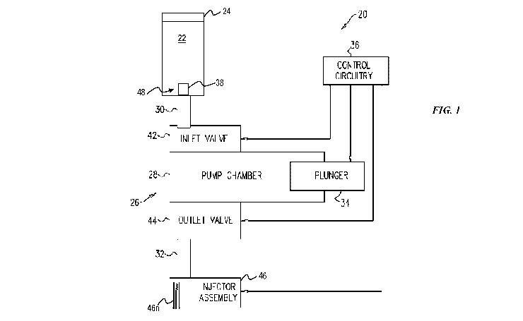
[0041] FIG. 1 illustrates an embodiment of a pump 20, e.g., a patch pump,
configured to be worn
by a patient and to deliver a drug (also referred to herein as a "therapeutic
substance") 22 to the
patient. The pump 20 can be configured to be attached to the patient in any of
a variety of ways,
as will be appreciated by a person skilled in the art, such as by including a
backing or label
configured to be removed from a body of the pump 20 to expose adhesive
attachable to the
patient. The pump 20 includes a therapeutic substance reservoir 24 containing
the drug 22
therein. The reservoir 24 can be prefilled by a medical vendor or device
manufacturer, or the
reservoir 24 can be filled by a user (e.g., the patient, the patient's
caregiver, a doctor or other
health care professional, a pharmacist, etc.) prior to use of the pump 20.
Alternatively, the
reservoir 24 can come prefilled from a medical vendor ready to be loaded or
inserted into pump
20 prior to use. The pump 20 also includes a conduit 38 through which the drug
22 is configured
to pass from the reservoir 24 and into an inlet fluid path 30 operatively
connected to an injector
assembly 46 of the pump 20 that is configured to deliver the therapeutic
substance 22 into a
CA 03165115 2022-06-16
WO 2021/123953 PCT/IB2020/060751
patient. The conduit 38 is thus a tube in which the drug 22 can flow.
[0042] The electromechanical pumping assembly 26, e.g., a motor thereof, is
operatively
connected to the reservoir 24 and is configured to cause delivery of the
therapeutic substance 22
to the patient via the injector assembly 46, e.g., through a needle 46n of the
injector assembly 46
that has been inserted into tissue of the patient. The electromechanical
pumping assembly 26 is
shaped to define a rigid pump chamber 28 that includes a therapeutic substance
inlet 30 through
which the therapeutic substance 22 is received from the conduit 30, and hence
from the reservoir
24, into the pump chamber 28. The rigid pump chamber 28 also includes a fluid
path outlet 32
through which the therapeutic substance 22 is delivered from the pump chamber
28 to the patient
via the injector assembly 46. Although the pumping assembly 26 is
electromechanical in this
illustrated embodiment, the pumping assembly of the pump 20 (and for other
embodiments of
pumps described herein) can instead be mechanical. The mechanical pumping
assembly need
not include any electronic components or controls. For example, the mechanical
pumping
assembly can include a balloon diaphragm configured to be activated to cause
delivery of a drug
through mechanical action.
[0043] The pump 20 also includes a plunger 34 slidably disposed within the
pump chamber 28
and sealably contacting an inside of the pump chamber 28. The plunger 34 is
configured to be in
direct contact with the drug 22 in the pumping chamber 28.
[0044] The pump 20 also includes control circuitry 36. The electromechanical
pumping
assembly 26 is configured to be driven to operate in two pumping phases by the
control circuitry
36. In a first pumping phase, the control circuitry 36 is configured to drive
the plunger 34 (e.g.,
slidably move the plunger 34 in the pump chamber 28) to draw the drug 22 from
the reservoir 24
into the conduit 38, then into the inlet fluid path 30, then through an inlet
valve 42 and into the
pump chamber 28. The inlet valve 42 is configured to be opened and closed such
that when the
inlet valve 42 is open there is fluid communication between the reservoir 24
and the pump
chamber 28, and when the inlet valve 42 is closed there is no fluid
communication between the
reservoir 24 and the pump chamber 28. During the first pumping phase, the
control circuitry 36
is configured to cause the inlet valve 42 to open, cause an outlet valve 44 to
close, and drive the
plunger 34 to draw the therapeutic substance 22 from the reservoir 24 into the
pump chamber 28,
6
CA 03165115 2022-06-16
WO 2021/123953 PCT/IB2020/060751
e.g., the control circuitry 36 is configured to set the inlet valve 42 and the
outlet valve 44 such
that the therapeutic substance 22 can flow only between the reservoir 24 and
the pump chamber
28. Thus, as the plunger 34 is drawn back, therapeutic substance 22 is drawn
into pump chamber
28. The control circuitry 36 causing the inlet valve 42 to open and the outlet
valve 44 to close
can be active control or can be passive control in which the valves 42, 44 are
mechanical valves
that automatically open/close due to the driving of the plunger 34.
[0045] The needle 46n of the injector assembly 46 is configured to move from
inside the pump's
housing to at least partially outside of the pump's housing for penetration
into a patient. The
electromechanical pumping assembly 26, e.g., the motor thereof as controlled
by the control
circuitry 36, is configured to cause the movement of the needle 46n. The
needle 46n movement
can occur during the first pumping phase or before the first pumping phase. In
other
embodiments, the needle 46n begins outside of the pump's housing.
[0046] In a second pumping phase, the control circuitry 36 is configured to
drive the plunger 34
to deliver the drug 22 from the pump chamber 28 through the outlet valve 44 to
the outlet fluid
path 32 and then to the injector assembly 46 for delivery into the patient
through the needle 46n.
The outlet valve 44 is configured to be opened and closed such that when the
outlet valve 44 is
open there is fluid communication between the pump chamber 28 and the patient,
and when the
outlet valve 44 is closed there is no fluid communication between the pump
chamber 28 and the
patient. During the second pumping phase, the control circuitry 36 is
configured to cause the
inlet valve 42 to close, cause the outlet valve 44 to open, and drive the
plunger 34 to deliver the
therapeutic substance 22 from the pump chamber 28 in a plurality of discrete
motions of the
plunger 34. For example, the control circuitry 36 can be configured to set the
inlet valve 42 and
the outlet valve 44 such that the therapeutic substance 22 can flow only
between the pump
chamber 28 and the patient, and the plunger 34 is incrementally pushed back
into the pump
chamber 28 in a plurality of discrete motions thereby delivering the
therapeutic substance 22 to
the patient in a plurality of discrete dosages. Similar to that discussed
above, the control
circuitry 36 causing the inlet valve 42 to close and the outlet valve 44 to
open can be active
control or can be passive control in which the valves 42, 44 are mechanical
valves that
automatically open/close due to the driving of the plunger 34.
7
CA 03165115 2022-06-16
WO 2021/123953 PCT/IB2020/060751
[0047] In some embodiments, the control circuitry 36 is configured to drive
the plunger 34 to
draw the therapeutic substance 22 into the pump chamber 28 in a single motion
of the plunger
34, e.g., the plunger 34 is pulled back in a single motion to draw a volume of
the therapeutic
substance 22 into the pump chamber 28 during the first pumping phase.
Alternatively, the
control circuitry 36 can be configured to drive the plunger 34 to draw the
therapeutic substance
22 into the pump chamber 28 in one or more discrete expansion motions of the
plunger 34, e.g.,
the plunger 34 can be pulled halfway out of the pump chamber 28 in one motion
and then the
rest of the way out of the pump chamber 28 in a second, separate motion. In
this case, a duration
of some or all expansion motions of the plunger 34 during the first pumping
phase are typically
longer than a duration of any one of the plurality of discrete motions of the
plunger 34 during the
second pumping phase.
[0048] In other embodiments, the control circuitry 36 is configured to drive
the plunger 34 such
that a duration of the first pumping phase and a duration of the second
pumping phase are
unequal. For example, a duration of the second pumping phase can be in a range
of five to fifty
times longer than the first pumping phase, e.g., at least ten times, thirty
times, fifty times, etc.
longer than a duration of the first pumping phase.
[0049] The pump 20 can also include a power supply (not shown) configured to
provide power
to components requiring power to operate, such as the control circuitry 36. In
an exemplary
embodiment, the power supply is a single power supply configured to provide
power to each
component of the pump 20 requiring power to operate, which may help reduce
cost of the pump
20, help conserve space within the pump 20 for other components, and/or help
reduce an overall
size of the pump 20. The power supply can, however, include a plurality of
power supplies,
which may help provide redundancy and/or help reduce cost of the pump 20 since
some
components, e.g., the control circuitry 36, may be manufactured with an on-
board dedicated
power supply. In an exemplary embodiment, the power supply is on-board the
pump 20, which
may facilitate use of the pump 20 at any time in any location. In other
embodiments, the power
supply can include a mechanism configured to connect the pump 20 to an
external power supply.
[0050] FIGS. 2-4 illustrate another embodiment of a pump 100, e.g., a patch
pump, configured to
be worn by a patient and to deliver a drug 148 to the patient. The pump 100 of
FIGS. 2-4 is
8
CA 03165115 2022-06-16
WO 2021/123953 PCT/IB2020/060751
generally configured and used similar to the pump 20 of FIG. 1. The pump 100
is configured to
engage with a prefilled therapeutic substance reservoir 132. Within the pump
100 is a sterile
fluid path 122 for delivering a drug 148 from the reservoir 132 to a patient
wearing the pump
100. The sterile fluid path 122 has a conduit 126 at an upstream end 124 of
the sterile fluid path
122 and has an injection assembly (also referred to herein as an "injector
assembly") 130 at a
downstream end 128 of the sterile fluid path 122.
[0051] The pump 100 and the prefilled therapeutic substance reservoir 132 are
configured to
engage with one another, such as shown by arrow 133 in FIG. 2, e.g., the
reservoir 132 is
configured to be inserted into the pump 100. When the pump 100 and the
reservoir 132 are
engaged with one another, such as is shown in FIG. 3, a sealed disinfection
chamber 134 is
defined between the sterile fluid path 122 and the reservoir 132. While the
pump 100 and the
reservoir 132 are typically sterile, the disinfection chamber 134 is (a)
initially non-sterile, and (b)
typically sealed from further bacteria or virus penetration. The conduit 126
is configured to be
driven to penetrate the disinfection chamber 134 and subsequently the
reservoir 132 when the
pump 100 and the reservoir 132 are engaged with one another, such that fluid
communication is
established between the reservoir 132 and the sterile fluid path 122, such as
is shown in FIG. 4.
[0052] The pump 100 includes a disinfection assembly 136 configured to
disinfect the
disinfection chamber 134 prior to the conduit 126 penetrating the disinfection
chamber 134 and
thus before the conduit 126 enters the reservoir 132. The pump 100 includes
control circuitry
138 configured to activate the disinfection assembly 136, to subsequently
terminate the
activation of the disinfection assembly 136, and to then drive the conduit 126
to penetrate the
disinfection chamber 134 and subsequently the reservoir 132.
[0053] Once fluid communication is established between the reservoir 132 and
the sterile fluid
path 122, the control circuitry 138 is configured to drives a pump assembly
140 to draw the drug
148 from the reservoir 132 and deliver it to the patient via the injection
assembly 130, e.g., via a
needle thereof, similar to that discussed above regarding the control
circuitry 36 and the injector
assembly 46 of FIG. 1.
[0054] FIG. 5 illustrates another embodiment of a pump 200 configured to be
worn by a patient
and to deliver a drug to the patient. The pump 200 of FIG. 5 is generally
configured and used
9
CA 03165115 2022-06-16
WO 2021/123953 PCT/IB2020/060751
similar to the pump 20 of FIG. 1. The pump 200 includes a reservoir 210
configured to contain a
liquid drug therein to be delivered from the pump 200. The pump 200 also
includes a pumping
assembly 216 configured to cause dispensing of the drug contained in the
reservoir 210 so that
the drug can be delivered to the patient. The pump 200 also includes an
injector assembly that
includes an infusion line 212, e.g., a needle. The drug is delivered from the
reservoir 210 upon
actuation of the pumping assembly 216 via the infusion line 212.
[0055] The pump 200 also includes a user interface 280 configured to provide
for interaction
with a user. The user interface 280 can be implemented on a computer having a
display screen,
such as for example a cathode ray tube (CRT) or a liquid crystal display (LCD)
or a light
emitting diode (LED) monitor for displaying information to a user. The display
screen can allow
input thereto directly (e.g., as a touch screen) or indirectly (e.g., via an
input device such as a
keypad or voice recognition hardware and software). The user interface 280 can
take the form
of, e.g., a touchscreen or a keypad.
[0056] The pump 200 also includes control circuitry that includes a processor
296 and a memory
297 in operative communication with the processor 296. Actuation of the
pumping assembly
216 is controlled by the processor 296, which is in operative communication
with the pumping
assembly 216 for controlling the pump's operation.
[0057] In at least some embodiments, the processor 296 is configured to be
programmed by a
user, e.g., the patient, a healthcare professional, etc., via the user
interface 280. The processor
296 being user-programmable enables the pump 200 to deliver the drug to the
patient in a
controlled manner specific to the patient. The user can enter parameters, such
as infusion
duration and delivery rate, via the user interface 280, such as by the user
interface 280 including
a touchscreen configured to receive touch input thereto, the user interface
280 including selector
button(s), and/or the user interface 280 including a keypad. The delivery rate
can be set by the
user to a constant infusion rate or as set intervals for periodic delivery,
typically within
pre-programmed limits. The programmed parameters for controlling the pumping
assembly 216
are stored in and retrieved by the processor 296 from the memory 297.
CA 03165115 2022-06-16
WO 2021/123953 PCT/IB2020/060751
[0058] The pump 200 also includes a power supply 295 configured to provide
power to any
components of the pump 200 that require power for operation, such as the
pumping assembly
216, the processor 296, the user interface 280, and the sensor 282.
[0059] The reservoir 210, the pumping assembly 216, the user interface 280,
the power supply
295, the processor 296, and the memory 297 are located within a housing (also
referred to herein
as a "body" of a pump) 230 of the pump 200. The infusion line 212 is partially
located within
the housing 230 and extends from the housing 230 for penetration into the
patient. The infusion
line 212 can be fixedly positioned partially within the housing 230 and
partially outside the
housing 230, as shown in FIG. 5, or the infusion line 212 can be movable,
e.g., under control of
the circuitry, from an initial position entirely within the housing 230 to a
delivery position
partially within the housing 230 and partially outside the housing 230.
[0060] The various pumps described herein are configured to deliver a drug to
a patient, e.g., the
pump 20 of FIG. 1, the pump 100 of FIGS. 2-4, and the pump 200 of FIG. 5, the
drug is
configured to be delivered into a subcutaneous tissue or muscle of the patient
through a needle of
the pump's injector assembly. The drug exits the needle into the tissue or
muscle through an
open distal tip of the needle. For example, FIG. 6 illustrates an embodiment
of a needle 300
inserted into tissue of a patient and delivering a bolus of a liquid drug 302
into subcutaneous
tissue 304 through an open distal tip 306 of the needle 300.
[0061] In general, transport of a liquid drug through interstitial space will
ultimately dictate drug
delivery and pressure distribution in a tissue into which a needle is
injecting the drug, including
backpressure at the needle interface, which may contribute to patient pain.
There are various
factors that can affect patient comfort and successful delivery of a liquid
drug into subcutaneous
tissue, such as physics factors (e.g., tissue deformation such as elastic
stress-strain), tissue
viscoelasticity such as stress/strain versus time, drug velocity and
viscosity, drug transport (e.g.,
Darcy's law), and needle material failure, fracture, or tearing; extrinsic
tissue factors (e.g.,
subcutaneous and hypodermis tissue structure and thickness, and dermis tissue
structure and
thickness); subcutaneous tissue intrinsic properties (e.g., tissue elasticity,
tissue viscoelasticity,
hydraulic permeability, and interstitial fluid and solution viscosity);
boundary conditions (e.g.,
injection speed and flow rate, absorption of drug into capillaries or
lymphatic vessels, diffusion
11
CA 03165115 2022-06-16
WO 2021/123953 PCT/IB2020/060751
across dermis/muscle-tissue boundaries (if any), and deformation of dermal and
muscle layer
boundaries); drug formulation factors (e.g., molecule size and
hydrophobicity); patient factors
(body mass index (BMI), gender, and injection location); injection site
factors (e.g., temperature
and pH); and needle factors (e.g., needle geometry). With respect to tissue
thickness,
subcutaneous tissue thickness is highly variable, which can make it difficult
to achieve patient
comfort and successful delivery of a liquid drug into subcutaneous tissue.
[0062] There are various factors indicative of patient comfort and successful
delivery of a liquid
drug into subcutaneous tissue. For example, interstitial fluid hydrostatic
pressure (buildup) can
be indicative of pressure sensed on surrounding tissue and may be related to
swelling, edema,
bleb formation, and pain. For another example, deformation of dermal and
muscle boundaries
can characterize a bulge due to injection bolus. For yet another example,
implementation of
stress or strain based traction-separation/fracture criteria can characterize
tissue damage, pain, or
potential for drug leakage.
[0063] Fluid mechanics and structural models indicate that differences in
tissue at an open distal
tip of the needle injecting drug into the tissue, as well as the design of the
needle, impact a
pressure profile during injection. This pressure profile can impact
inflammatory response, pain,
etc. In addition, this variability can lead to wearable pump performance
differences, such as
higher or lower force requirements for the pump's pumping assembly that drives
the drug from
the pump's reservoir and out the pump's needle, the ability of the pump to
operate properly, and
to achieve a certain delivery profile based on power requirements and signals
from the pump's
control circuitry. Increased space, e.g., a void of fluid or gas, a pathway of
fluid or gas, or a
pocket of fluid or gas of low resistance or differing viscosity, around the
distal tip of the needle
can reduce injection pressures because less tissue can be displaced by the
injected liquid drug.
[0064] Fluid mechanics at the needle/tissue interface thus dictates
backpressure observed during
injection. If the needle/tissue interface is small (e.g., the needle has a
small inner diameter that
defines an opening through which the drug exits the needle), the velocity
gradients and resulting
pressures are high. If the needle/tissue interface is made larger, tissue
backpressure during
injection can be significantly reduced. Placing beveled openings at the distal
tip of the needle
increases the flow area, reduces the velocity gradients, and reduces the
pressures as compared to
12
CA 03165115 2022-06-16
WO 2021/123953 PCT/IB2020/060751
a blunt distal tip. The bevel can be, e.g., about 100 or about 20 . The inner
diameter of the
needle can be, e.g., about 0.5 mm. A person skilled in the art will appreciate
that a value may
not be precisely at a value but nevertheless be considered to be about that
value because of any
of a variety of factors, such as manufacturing tolerances and sensitivity of
measurement
equipment.
[0065] In some embodiments, a needle can have only one exit opening for the
liquid drug at the
needle's distal tip. In other embodiments, the needle can have an exit opening
for the liquid drug
at the needle's tip and at least one side exit opening for the liquid drug
formed in a sidewall of
the needle. The at least one side exit opening can be a slot, a hole, a slit,
etc. formed in the
needle's sidewall so as to allow a partial portion of liquid drug in the
needle's inner lumen to exit
through the at least one side exit opening and a remainder of the liquid drug
in the needle's inner
lumen to exit through the needle's tip opening. Each side exit opening can be
a discrete opening
formed through the sidewall, or a distal portion of the needle can be formed
from a porous
structure where the porous structure's pores define side exit openings. The at
least one side exit
opening can vary in size, location, and number. In still other embodiments,
the needle can have
at least one side exit opening for the liquid drug formed in a sidewall of the
needle and not have
an exit opening for the liquid drug at the needle's tip.
[0066] FIG. 7 shows a chart of seven liquid injection simulation runs. In each
of the seven runs,
the needle gauge was 27G RW, the total liquid drug delivery volume was 15 ml,
the liquid drug
viscosity was 12 cP, and the subcutaneous liquid drug viscosity was 1 cP. The
needles in runs
1-7 have an inner diameter of 0.21082 mm, have an outer diameter of 0.4218 mm,
have a total
length of 9 mm, and sit in at 7 mm depth within the tissue. The flow rate (in
ml/min) shown in
FIG. 7 represents the liquid drug flow rate. The flow rate is constant in runs
1, 2, 4, 6, and 7 and
is not constant in runs 3 and 5. In runs 3 and 5, the flow rate ramps upward
and is then constant.
The porous resistance (in kg/(m35)) shown in FIG. 7 represents resistance at
the needle/tissue
interface at which the liquid is released into tissue. The porous resistance
is constant in run 1
and is not constant, e.g., is adaptive, in runs 2-7. The porous resistance not
being constant
indicates that viscosity of the liquid drug contributes to the porous
resistance.
13
CA 03165115 2022-06-16
WO 2021/123953 PCT/IB2020/060751
[0067] FIG. 8 illustrates the needle's blunt tip 8t of runs 1, 2, and 3. FIG.
9 illustrates the
needle's spherical tip 9t of runs 4 and 5. In general, the spherical tip 9t
allows for increased
space around the tip 9t at the needle/tissue interface because the spherical
shape moves more
tissue as compared to blunt distal tips such as the blunt tip 8t. As mentioned
above, increased
space around the distal tip of the needle can reduce injection pressures. FIG.
10 illustrates the
needle's 100 beveled tip 10t of run 6. FIG. 11 illustrates the needle's 20
beveled tip lit of run
7.
[0068] FIGS. 12-18 illustrate graphs of the pressure profiles for runs 1-7,
respectively. The top
line in each of the graphs represents average pressure at the needle's inlet
and is thus before the
liquid drug exits the needle. The bottom line in each of the graphs represents
average pressure at
the needle/tissue interface at which the liquid drug is released from the
needle into tissue. In
general, the average pressure at the needle/tissue interface being
substantially constant indicates
that the average pressure at the needle/tissue interface will be substantially
the same for different
patients and for different tissues (e.g., for drug injected into dermis tissue
versus into
subcutaneous tissue).
[0069] Comparing the graphs of runs 2 and 4 (FIGS. 13 and 15) demonstrates
pressure profile
differences for the blunt tip 8t of run 2 and the spherical tip 9t of run 4
since each of runs 2 and 4
have the same constant flow rate and the same adaptive porous resistance.
FIGS. 13 and 15
show that each of the average pressure at the needle's inlet and the average
pressure at the
needle/tissue interface are substantially constant with the spherical tip 9t
and the blunt tip 8t and
are lower with the spherical tip 9t than with the blunt tip 8t.
[0070] Comparing the graphs of runs 3 and 5 (FIGS. 14 and 16) demonstrates
pressure profile
differences for the blunt tip 8t of run 3 and the spherical tip 9t of run 5
since each of runs 3 and 5
have the same ramped and then constant flow rate and the same adaptive porous
resistance.
FIGS. 14 and 16 show that each of the average pressure at the needle's inlet
and the average
pressure at the needle/tissue interface are less steeply sloped with the
spherical tip 9t than with
the blunt tip 8t, are substantially constant after the ramping with the
spherical tip 9t and the blunt
tip 8t, and reach a lower maximum pressure with the spherical tip 9t than with
the blunt tip 8t.
14
CA 03165115 2022-06-16
WO 2021/123953 PCT/IB2020/060751
[0071] Comparing the graphs of runs 2 and 6 (FIGS. 13 and 17) demonstrates
pressure profile
differences for the blunt tip 8t of run 2 and the 100 beveled tip 10t of run 6
since each of runs 2
and 6 have the same constant flow rate and the same adaptive porous
resistance. FIGS. 13 and
17 show that each of the average pressure at the needle's inlet and the
average pressure at the
needle/tissue interface are substantially constant with the 100 beveled tip
10t and the blunt tip 8t
and are lower with the 100 beveled tip 10t than with the blunt tip 8t.
[0072] Comparing the graphs of runs 2 and 7 (FIGS. 13 and 18) demonstrates
pressure profile
differences for the blunt tip 8t of run 2 and the 20 beveled tip llt of run 7
since each of runs 2
and 7 have the same constant flow rate and the same adaptive porous
resistance. FIGS. 13 and
18 show that each of the average pressure at the needle's inlet and the
average pressure at the
needle/tissue interface are substantially constant with the 20 beveled tip
llt and the blunt tip 8t
and are lower with the 20 beveled tip llt than with the blunt tip 8t.
[0073] Comparing the graphs of runs 6 and 7 (FIGS. 17 and 18) demonstrates
pressure profile
differences for the different beveled tips 10t, 20t since each of runs 6 and 7
have the same
constant flow rate and the same adaptive porous resistance. FIGS. 17 and 18
show that each of
the average pressure at the needle's inlet and the average pressure at the
needle/tissue interface
are substantially constant with the 10 beveled tip 10t and the 20 beveled
tip llt and are lower
with the 10 beveled tip 10t than with the 20 beveled tip llt.
[0074] Two alternate liquid injection simulation runs, run 8 and run 9, were
run for run 2 (blunt
needle tip, flow rate constant at 0.8 ml/min, and adaptive porous resistance
of 108 *
(cell vselcP) kg/m35). The needles in runs 8 and 9 have an inner diameter of
0.210 mm, have
an outer diameter of 0.42 mm, have a total length of 9 mm, and sit in at 7 mm
depth within the
tissue. In general, the addition of drug ports to the sides of the needle was
observed to reduce
injection pressures and to cause drug distributions to shift towards the skin,
e.g., towards an
upper portion of the subcutaneous tissue.
[0075] In run 8, as shown in FIGS. 19 and 20, the needle has the blunt tip 8t
of run 2 and also
has a plurality of side exit openings 8s formed in a sidewall of the needle.
The side exit openings
8s in this illustrated embodiment are longitudinal slots. The side exit
openings 8s can have a
CA 03165115 2022-06-16
WO 2021/123953 PCT/IB2020/060751
variety of locations and sizes but in this illustrated embodiment are
equidistantly spaced around a
circumference of the needle, have one terminal end 2 mm from the blunt tip end
of the needle
and extend to 4 mm from the blunt tip end of the needle, have a width of 0.05
mm, and have a
length of 2 mm. Although the needle includes two side longitudinal slots 8s in
this illustrated
embodiment, the needle can have another number of side longitudinal slots.
Additionally, the
side holes can be located at more than one axial position along the needle.
[0076] FIG. 21 illustrates a graph of the pressure profile for run 8. The top
line in FIG. 21
represents average pressure at the needle's inlet and is thus before the
liquid drug exits the
needle. The bottom line in FIG. 21 represents average pressure at the
needle/tissue interface at
which the liquid drug is released from the needle into tissue.
[0077] Comparing the graphs of runs 2 and 8 (FIGS. 13 and 21) demonstrates
pressure profile
differences for the blunt tip 8t of run 2 with the needle having no side exit
openings and the blunt
tip 8t of run 9 with the needle having side exit openings in the form of slots
since runs 2 and 8
are the same except for the absence (run 2) or presence (run 8) of side exit
openings in the form
of slots. FIGS. 13 and 21 show that each of the average pressure at the
needle's inlet and the
average pressure at the needle/tissue interface are substantially constant
with and without the
side exit openings in the form of slots and are lower with the side exit
openings in the form of
slots (FIG. 21) than without the side exit openings in the form of slots (FIG.
13).
[0078] In run 9, as shown in FIGS. 22 and 23, the needle has the blunt tip 8t
of run 2 and also
has a plurality of side exit openings 8h formed in a sidewall of the needle.
The side exit
openings 8h in this illustrated embodiment are circular holes. The side exit
openings 8h can
have a variety of locations but in this illustrated embodiment include half
the side holes 8h
equidistantly spaced around a circumference of the needle at a first axial
position along the
needle's longitudinal axis, and half the side holes 8h equidistantly spaced
around the
circumference of the needle at a second, different axial position along the
needle's longitudinal
axis. The first and second axial positions can vary but in this illustrated
embodiment are 2 mm
from the blunt tip end of the needle and 4 mm from the blunt tip end of the
needle. The side exit
openings 8h can have a variety of sizes but in this illustrated embodiment
have a diameter of 0.1
mm. Although the needle includes eight side holes 8h in this illustrated
embodiment, the needle
16
CA 03165115 2022-06-16
WO 2021/123953 PCT/IB2020/060751
can have another number of side holes. Additionally, the side holes can be
located at only one
axial position along the needle or at more than two axial positions along the
needle.
[0079] FIG. 24 illustrates a graph of the pressure profile for run 9. The top
line in FIG. 24
represents average pressure at the needle's inlet and is thus before the
liquid drug exits the
needle. The bottom line in FIG. 24 represents average pressure at the
needle/tissue interface at
which the liquid drug is released from the needle into tissue.
[0080] Comparing the graphs of runs 2 and 9 (FIGS. 13 and 24) demonstrates
pressure profile
differences for the blunt tip 8t of run 2 with the needle having no side exit
openings and the blunt
tip 8t of run 9 with the needle having side exit openings in the form of holes
since runs 2 and 9
are the same except for the absence (run 2) or presence (run 9) of side exit
openings in the form
of holes. FIGS. 13 and 24 show that each of the average pressure at the
needle's inlet and the
average pressure at the needle/tissue interface are substantially constant
with and without the
side exit openings in the form of holes and are lower with the side exit
openings in the form of
holes (FIG. 24) than without the side exit openings in the form of holes (FIG.
13).
[0081] Comparing the graphs of runs 8 and 9 (FIGS. 21 and 24) demonstrates
pressure profile
differences for the blunt tip 8t of run 8 with the needle having side exit
openings in the form of
slots and the blunt tip 8t of run 9 with the needle having side exit openings
in the form of holes
since runs 8 and 9 are otherwise the same. The average pressure at the
needle/tissue interface is
substantially the same in FIGS. 21 and 24, and the average pressure at the
needle's inlet is lower
with the side exit openings in the form of slots at a single axial position
(FIG. 21) than with the
side exit openings in the form of holes at two different axial positions (FIG.
24).
[0082] Runs 8 and 9, as compared to run 2, showed that drug distribution
shifted towards the
skin, e.g., towards an upper portion of the subcutaneous tissue. FIG. 25
illustrates drug
distribution for run 2 at time 3 seconds. In run 2, 100% of the drug exited
the needle through the
blunt distal tip 8t. FIG. 26 illustrates drug distribution for run 8 at time 3
seconds. In run 8, 90%
of the drug exited the needle through the side slots 8s, and 10% of the drug
exited the needle
through the blunt distal tip 8t. FIG. 27 illustrates drug distribution for run
9 at time 3 seconds.
In run 9, 48% of the drug exited the needle through the four upper side holes
8h, 33% of the drug
17
CA 03165115 2022-06-16
WO 2021/123953 PCT/IB2020/060751
exited the needle through the four lower side holes 8h, and 19% of the drug
exited the needle
through the blunt distal tip 8t.
[0083] FIG. 28 illustrates the tissue for runs 1-9. The Symmetry Axis shown in
FIG. 28
represents a longitudinal axis of the needle. The Bolus Pressure Source shown
in FIG. 28
represents the needle/tissue interface at which the liquid drug is released
from the needle into
tissue, in particular into subcutaneous tissue. Lines in the subcutaneous
tissue of FIG. 28 shows
deformation of the subcutaneous tissue caused by the injected liquid, e.g., by
the pressure applied
to the tissue by the liquid. The deformation is greater the nearer the
needle/tissue interface, as
indicated by the more compressed lines closer to the needle/tissue interface.
Muscle underlying
the subcutaneous tissue is also illustrated in FIG. 28. Lines in the muscle of
FIG. 28 shows that
the muscle is not deforming.
[0084] A needle of the various pumps described herein, e.g., the pump 20 of
FIG. 1, the pump
100 of FIGS. 2-4, and the pump 200 of FIG. 5, can be configured to reduce
pressure at a distal
tip of the needle. In an embodiment of a needle configured to reduce pressure
at a distal tip of
the needle, the needle starts at its most distal position and is configured to
move passively based
on resistance to flow/backpressure from tissue (in which the needle is
inserted) in a proximal
direction. The needle has a hub that is hydraulically coupled to a housing of
the pump. The
hydraulic control can be one which has a (a) positive spring that holds the
needle down so that
the spring is compressed when the tissue is above a certain threshold, (b)
compressible spring
that compresses the farther in the proximal direction the needle wants to
move, the higher the
tissue pressure required, (c) grease filled chamber with constant hydraulic
resistance, (d) constant
force spring, (e) lever arm, or (f) two magnets that oppose each other.
[0085] In another embodiment of a needle configured to reduce pressure at a
distal tip of the
needle, the needle starts at some position and is configured to move in
laterally (left and right).
Moving laterally can increase space at the needle/tissue interface, which as
mentioned above can
reduce injection pressures. After being moved, the needle can return to its
initial position or can
be in a new position. The elasticity of the tissue in which the needle is
located and/or the amount
of space created by the needle's movement can dictate whether the needle is in
the new position
or returns to its initial position. The needle has a hub that is hydraulically
coupled to a housing
18
CA 03165115 2022-06-16
WO 2021/123953 PCT/IB2020/060751
of the pump. The hub is configured to provide for the lateral movement of the
needle with a
fulcrum or an intentional play of the needle in the hub. The hydraulic control
can be one which
has a (a) positive spring that holds the needle down so that the spring is
compressed when the
tissue is above a certain threshold, (b) torque spring in the hub that results
in a bending moment
resulting in lateral deflection, (c) compressible spring that compresses the
farther in the proximal
direction the needle wants to move, the higher the tissue pressure required,
(d) grease filled
chamber with constant hydraulic resistance, (e) constant force spring, (f)
lever arm, or (g) two
magnets that oppose each other.
[0086] In another embodiment of a needle configured to reduce pressure at a
distal tip of the
needle, the needle is configured to move actively in a distal direction or a
proximal direction.
Moving distally or proximally can increase space at the needle/tissue
interface, which as
mentioned above can reduce injection pressures. After being moved, the needle
can return to its
initial position or can be in a new position. The elasticity of the tissue in
which the needle is
located and/or the amount of space created by the needle's movement can
dictate whether the
needle is in the new position or returns to its initial position. The pump
includes a sensor
configured to monitor flow resistance. The pump's control circuitry is
configured to receive data
from the sensor indicative of the flow resistance. The control circuitry is
configured to control
the pump's pumping assembly, e.g., a motor thereof, such that the motor's
torque maintains the
needle at the needle's current angular position. The pump includes an encoder
configured to
confirm the needle's angular position and/or distal position. The pump
includes
electromechanical means configured to be driven by the motor (as controlled by
the control
circuitry) to cause selective movement of the needle proximally and distally
to change a location
of the needle's distal tip in the tissue and thus change a terminal end of the
liquid drug flow path
from the needle. The needle is electromechanically, operatively coupled to the
motor via
gearing, which can be independent or can be secondarily driven by a power
source based on
signal from the pump's control circuitry.
[0087] In another embodiment of a needle configured to reduce pressure at a
distal tip of the
needle, the needle is configured to move in a distal direction or a proximal
direction using an
active means of the pump in the form of a vibration mechanism of the pump.
Moving distally or
proximally can increase space at the needle/tissue interface, which as
mentioned above can
19
CA 03165115 2022-06-16
WO 2021/123953 PCT/IB2020/060751
reduce injection pressures. The vibration mechanism is configured to allow
selective distal and
proximal movement of the needle against a hydraulic pressure. After being
selectively moved,
the needle can return to its initial position or can be in a new position. The
elasticity of the tissue
in which the needle is located and/or the amount of space created by the
needle's movement can
dictate whether the needle is in the new position or returns to its initial
position. The needle has
a hub that is hydraulically coupled to a housing of the pump. The hydraulic
control can be one
which has a (a) positive spring that holds the needle down so that the spring
is compressed when
the tissue is above a certain threshold, (b) compressible spring that
compresses the farther in the
proximal direction the needle wants to move, the higher the tissue pressure
required, (c) grease
filled chamber with constant hydraulic resistance, (d) constant force spring,
(e) lever arm, or (f)
two magnets that oppose each other. The vibration of the needle acts against
the hydraulic
control. Alternatively, no hydraulic control may be present, and the needle
can remain fixed by
vibrating against a rigid base.
[0088] In another embodiment of a needle configured to reduce pressure at a
distal tip of the
needle, the needle is configured to move laterally (left and right) using an
active means of the
pump in the form of a vibration mechanism of the pump. Moving laterally can
increase space at
the needle/tissue interface, which as mentioned above can reduce injection
pressures. The
vibration mechanism is configured to allow selective lateral movement of the
needle against a
hydraulic pressure. After being selectively moved, the needle can return to
its initial position or
can be in a new position. The elasticity of the tissue in which the needle is
located and/or the
amount of space created by the needle's movement can dictate whether the
needle is in the new
position or returns to its initial position. The needle has a hub that is
hydraulically coupled to a
housing of the pump. The hydraulic control can be one which has a (a) positive
spring that holds
the needle down so that the spring is compressed when the tissue is above a
certain threshold, (b)
compressible spring that compresses the farther in the proximal direction the
needle wants to
move, the higher the tissue pressure required, (c) grease filled chamber with
constant hydraulic
resistance, (d) constant force spring, (e) lever arm, or (f) two magnets that
oppose each other.
The vibration of the needle acts against the hydraulic control. Alternatively,
no hydraulic control
may be present, and the needle can remain fixed by vibrating against a rigid
base.
CA 03165115 2022-06-16
WO 2021/123953 PCT/IB2020/060751
[0089] In another embodiment of a needle configured to reduce pressure at a
distal tip of the
needle, a closed loop system is provided including an active means that is
operatively connected
to the fluid path resistance and is configured to automatically vibrate. The
vibration can cause an
increase in space at the needle/tissue interface, which as mentioned above can
reduce injection
pressures. The pump includes a sensor configured to detect clogging of the
needle, such as by
detecting a pressure that is above a predetermined maximum amount of
acceptable pressure so as
to indicate a probable clog in the needle because of a higher pressure
existing than normal and/or
by detecting a motor current that is above a predetermined maximum amount of
acceptable
motor current so as to indicate a probable clog via the motor working harder
than normal. The
pump's control circuitry is configured to receive data from the sensor
indicative of the detected
clogging. The control circuitry is configured to control the active means to
vibrate in response
the sensor detecting clogging of the needle. The control circuitry is
configured to control the
pump's pumping assembly, e.g., a motor thereof, to control the vibration of
active means.
[0090] In any of the above embodiments of a needle configured to reduce
pressure at a distal tip
of the needle, the needle's positional changes can be during infusion or
before the start of
infusion, where there is a desire to create a pocket of space or area of least
resistance around a
terminal end of the fluid path, e.g., at a distal tip of the needle out of
which the drug flows.
[0091] As discussed herein, one or more aspects or features of the subject
matter described
herein, for example components of the control circuitry and the user
interface, can be realized in
digital electronic circuitry, integrated circuitry, specially designed
application specific integrated
circuits (ASICs), field programmable gate arrays (FPGAs) computer hardware,
firmware,
software, and/or combinations thereof. These various aspects or features can
include
implementation in one or more computer programs that are executable and/or
interpretable on a
programmable system including at least one programmable processor, which can
be special or
general purpose, coupled to receive data and instructions from, and to
transmit data and
instructions to, a storage system, at least one input device, and at least one
output device.
[0092] The computer programs, which can also be referred to as programs,
software, software
applications, applications, components, or code, include machine instructions
for a
programmable processor, and can be implemented in a high-level procedural
language, an object-
21
CA 03165115 2022-06-16
WO 2021/123953 PCT/IB2020/060751
oriented programming language, a functional programming language, a logical
programming
language, and/or in assembly/machine language. As used herein, the term
"machine-readable
medium" refers to any computer program product, apparatus and/or device, such
as for example
magnetic discs, optical disks, memory, and Programmable Logic Devices (PLDs),
used to
provide machine instructions and/or data to a programmable processor,
including a
machine-readable medium that receives machine instructions as a machine-
readable signal. The
term "machine-readable signal" refers to any signal used to provide machine
instructions and/or
data to a programmable processor. The machine-readable medium can store such
machine
instructions non-transitorily, such as for example as would a non-transient
solid-state memory or
a magnetic hard drive or any equivalent storage medium. The machine-readable
medium can
alternatively or additionally store such machine instructions in a transient
manner, such as for
example as would a processor cache or other random access memory associated
with one or
more physical processor cores.
[0093] The present disclosure has been described above by way of example only
within the
context of the overall disclosure provided herein. It will be appreciated that
modifications within
the spirit and scope of the claims may be made without departing from the
overall scope of the
present disclosure.
22